comparative evaluation of mutational and mrna expression pattern of pulmonary cytokines after...
TRANSCRIPT

Comparative Evaluation of Mutational and mRNA Expression Pattern of Pulmonary Cytokines after Infection with Aspergillus flavus and Aspergillus fumigatus
Jehane I. Eid 1, Heba M. Abdelraouf 2, Tahany M. Abdelrahman 3, Akmal A. El-Ghor 1
Author affiliations: 1 Zoology Department, Faculty of Science, Cairo University, Giza 12613, Egypt, 2 Microbiology Lab, Microanalytical Center, Faculty of Science, Cairo University, Giza 12613, Egypt, 3 Botony Department, Faculty of Science, Cairo University, Giza 12613, Egypt Correspondence to: [email protected]; [email protected] (J.I.Eid)
Received: May 05, 2014Accepted: May 19, 2014
Dis Mol Med 2014;2: 32-40DOI:10.5455/dmm.20140519115524
Key words: Invasive pulmonary as-pergillosis; A. fumigatus; A. flavus; IL-1β; IL-6; mRNA; PCR-SSCP; DNA fragmentation
AbstractInvasive pulmonary aspergillosis (IPA) is a lethal complication of persistent neutropenia. In the pathogenesis of IPA, proinflammatory cytokines seem to play a key role. Howev-er, little progress has been made in comparing the production of pulmonary cytokines in mice with IPA caused by A. fumigatus versus A. flavus at the molecular level. Therefore, it was our aim to investigate the mutational and expression pattern of proinflammatory cytokine encoding genes in the lung of immunocompetent and immunocompromised mice infected with either species. The levels of mutational events and transcription of IL-1β and IL-6 encoding genes in the pulmonary tissues of infected mice (with or without immunosuppression) were compared by means of semi-quantitative polymerase chain reaction and PCR-SSCP. Pulmonary fungal burden and histopathological changes were also assessed. The degree of DNA degredation and amounts of pulmonary IL-1β and IL-6 induced by A. fumigatus were much higher than those produced by A. flavus. The differ-ential expressions of cytokines by two species were not associated with any mutational events within the selected sequences of the cytokines encoding genes. Our data may explain why A. fumigatus, but less commonly A. flavus, cause IPA and also confirm the contribution of DNA degradation pattern associated with severe inflammation to the pathogenesis of IPA.
IntroductionThe lungs are the main portal of entry for Aspergil-
lus spp., which, in immunocompromised individuals, cause a life-threatening opportunistic infection called invasive pulmonary aspergillosis (IPA) (1-3). Previous studies have indicated that neutropenia is considered as one of the major predisposing conditions to this form of aspergillosis (4). Though the fungus is known to pre-dominantly affect the immunocompromised host, a hy-persensitization response can ensue leading to acute pneumonia in immunocompetent hosts, which is invari-ably fatal (5).
Inhalation of Aspergillus spores triggers a cascade of consequences not only determined by the immuno-logical state of those affected, but also by the virulence of the fungus (6, 7). There are many different species of Aspergillus, but the most frequent pathogenic agent
of invasive aspergillosis is A. fumigatus followed by A. flavus (3). IPA characterized by hyphal invasion and ne-crosis of pulmonary tissue, is thus leading to significant morbimortality among immunosuppressed patients (8, 9).
Host defense against IPA is mediated by cytokines and phagocytic cells of the innate immune system (10). IL-1β is an important proinflammatory cytokine that in the earliest innate response to A. fumigatus is sig-nificantly differentially expressed coinciding with an increase in phagocytosis (11). IL-6 is a multifunctional
©2014 Disease and Molecular Medicine. This is an Open Access article distributed under the terms of the Creative Commons Attribution Non-Commercial License (http://creativecommons.org/licenses/by-nc/3.0)
which permits unrestricted non-commercial use, distribution, and reproduction in any medium, provided the original work is properly cited.
Original Article
Disease and Molecular Medicinedisease &molecularmedicine w w w.d i s m o l m e d .o r g
Dis Mol Med 2014;2: 32-40
OPEN ACCESS
d m m
d m m
cytokine that is rapidly produced in aspergillosis after intranasal infection with the fungus (5, 12), and proteas-es of A. fumigatus can induce production of IL-6 by air-way epithelial cells (13, 14). Il-6 deficiency renders mice highly susceptible to IPA by increased inflammatory pathology of lungs and decreased antifungal effector functions of phagocytes (15).
Although the role of both IL-1β and Il-6 in IPA due to A. fumigatus has been previously characterized, their expression profile involved in the pathogensis of A. fla-vus has not yet been fully elucidated. Moreover, little is known about the differences of expression level of both IL-1β and iL-6 cytokines on the molecular level, be-tween immunosuppressed versus immunocompetent murine models of aspergillosis with A. fumigatus or A. flavus. In the present study, immunosuppressed and im-munocompetent mice were infected intranasally with A. fumigatus or A. flavus conidia and were monitored for parameters of fungal colonization, histopathologi-cal changes, DNA damage and IL-1β and IL-6 expression levels and mutational frequency, and their contribution to IPA progression.
Materials and Methods
Microorganism isolation and preparation of inoculaA clinical isolate of A. fumigatus and A. flavus (ob-
tained from Microbiology Lab, Microanalytical Centre, Cairo University, Egypt) were cultured on Czapek Dox agar (Difco, Detroit) slants at 37 0C for 5 days until conid-ia were fully mature. Conidia were harvested from the surface of several (two to three) agar slants into sterile saline with 0.01% Tween 20 (Fisher Scientific, Fair Lawn, N.J.), then counted by hemocytometer. The concentra-tion was adjusted in order to inoculate 4 × 10⁶ conidia per mouse in a volume of 30 µl. The viable count was confirmed by serially diluting the conidial suspension 10-fold and plating the inoculum on Czapek Dox agar.
MiceSix to eight week old male swiss albino mice (body
weight, 18-20 g), were reared and maintained in the ani-mal facility of the Department of Zoology, Cairo Univer-sity. All animals were housed under sterile conditions and provided with sterile drinking water containing tet-racycline hydrochloride (500 μg/ml, Sigma, USA), with temperatures between 22 and 24°C and a 12 h light/12 h dark photoperiod. They were caged in groups of 5 to
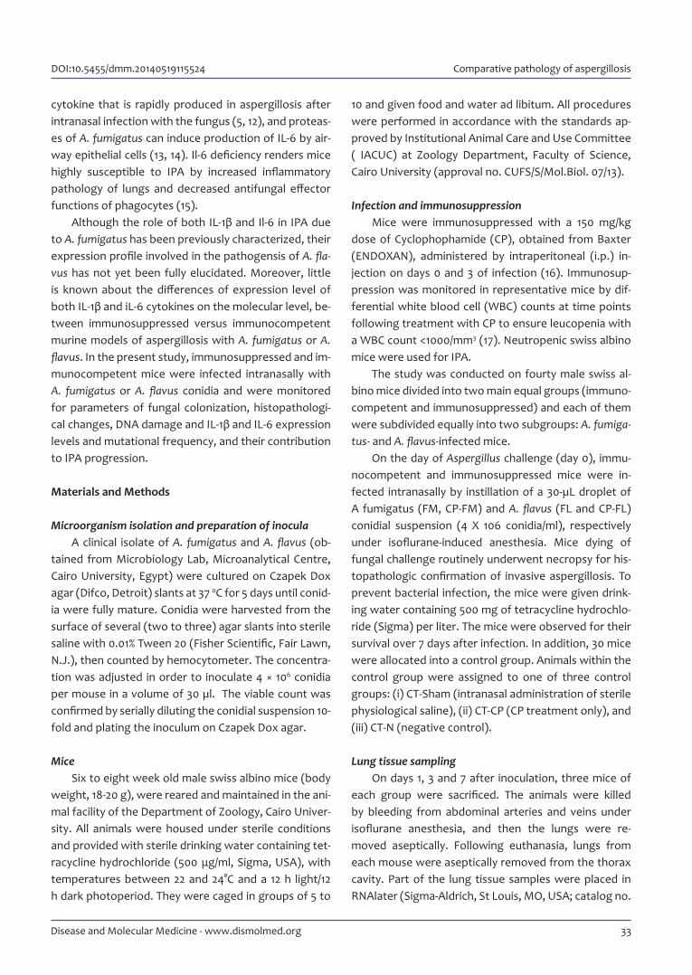
10 and given food and water ad libitum. All procedures were performed in accordance with the standards ap-proved by Institutional Animal Care and Use Committee ( IACUC) at Zoology Department, Faculty of Science, Cairo University (approval no. CUFS/S/Mol.Biol. 07/13).
Infection and immunosuppressionMice were immunosuppressed with a 150 mg/kg
dose of Cyclophophamide (CP), obtained from Baxter (ENDOXAN), administered by intraperitoneal (i.p.) in-jection on days 0 and 3 of infection (16). Immunosup-pression was monitored in representative mice by dif-ferential white blood cell (WBC) counts at time points following treatment with CP to ensure leucopenia with a WBC count <1000/mm3 (17). Neutropenic swiss albino mice were used for IPA.
The study was conducted on fourty male swiss al-bino mice divided into two main equal groups (immuno-competent and immunosuppressed) and each of them were subdivided equally into two subgroups: A. fumiga-tus- and A. flavus-infected mice.
On the day of Aspergillus challenge (day 0), immu-nocompetent and immunosuppressed mice were in-fected intranasally by instillation of a 30-µL droplet of A fumigatus (FM, CP-FM) and A. flavus (FL and CP-FL) conidial suspension (4 X 10⁶ conidia/ml), respectively under isoflurane-induced anesthesia. Mice dying of fungal challenge routinely underwent necropsy for his-topathologic confirmation of invasive aspergillosis. To prevent bacterial infection, the mice were given drink-ing water containing 500 mg of tetracycline hydrochlo-ride (Sigma) per liter. The mice were observed for their survival over 7 days after infection. In addition, 30 mice were allocated into a control group. Animals within the control group were assigned to one of three control groups: (i) CT-Sham (intranasal administration of sterile physiological saline), (ii) CT-CP (CP treatment only), and (iii) CT-N (negative control).
Lung tissue samplingOn days 1, 3 and 7 after inoculation, three mice of
each group were sacrificed. The animals were killed by bleeding from abdominal arteries and veins under isoflurane anesthesia, and then the lungs were re-moved aseptically. Following euthanasia, lungs from each mouse were aseptically removed from the thorax cavity. Part of the lung tissue samples were placed in RNAlater (Sigma-Aldrich, St Louis, MO, USA; catalog no.
DOI:10.5455/dmm.20140519115524 Comparative pathology of aspergillosis
33Disease and Molecular Medicine - www.dismolmed.org

R0901) and incubated at -80ºC for later RNA isolation. For histology, tissues were excised and were immedi-ately fixed in formalin. The other parts of the lung were kept at -20 °C until used for fungal burden assessment and DNA analysis.
Determination of fungal burden in lungsThe lung tissues were homogenized in 3 ml of dis-
tilled water. Lung homogenates were serially diluted with distilled water, and 0.1 ml of the diluted homogen-ates was plated onto Sabouraud dextrose agar (Eiken Chemical Co., Tokyo, Japan). The plates were incubated at 28°C for 40 to 48 h, and the colony forming unit (CFU) of both A. flavus and A. fumigatus were enumerated.
Histopathological changes For histological analysis, representative parts of
lung were fixed in buffered formalin and embedded in paraffin; 3-µm sections were stained with hematoxylin-eosin or PAS stain using standard protocols (18).
RNA isolation and RT-PCR analysisTotal RNA was isolated from tissues by using RNe-
asy Mini Kit (Qiagen, Germany). All RNA samples were treated with RNase-free DNase (Promega, USA) to pre-clude genomic DNA contamination before reverse tran-scription. A total of 2 μg of RNA was reverse transcribed into cDNA using Revert Aid TM First Strand cDNA Syn-thesis Kit (Fermentas, USA). The cDNA was stored at –20°C until use. Primer sequences (Bioneer, Seoul, Ko-rea) for each locus are listed in Table 1.
Reaction mixtures for Reverse Transcription-Poly-merase Chain Reaction (RT-PCR) had a final volume of 25 µl and included 200 ng cDNA, 50 nM of each primer, and 12.5 µl of 2× GoTaq Green Master Mix (Promega). PCR conditions were 5 minute at 95 °C, and 35 cycles at 95°C for 1 min, 55°C for 30 seconds and 72°c for 1 min. Cycling was done using thermal cycler Model 96 (MJ Re-search, INC., Watertown,MA, USA). The final products of RT-PCR were separated by 2% agarose gel electro-phoresis, and were detected by ethidium bromide stain-
ing. The level of mRNA for each cytokine was obtained by comparing the optical density (OD) of cytokine –spe-cific bands, quantitated by GENEQUANT software (Syn-gene), to the internal standard (β-actin).
DNA extractionGenomic DNA was prepared from mouse lung tis-
sues using a salting-out procedure performed as de-scribed (19).
Qualitative DNA fragmentationFragmented DNA was detected by running 5 μg of
genomic DNA on a 1.5% ethidium bromide-treated aga-rose gel (Sigma, UK) according to the standard protocol described by (20). The gel run at 80 volt (power supply Biorad, Model 200/2.0), and visualized under UV tran-sillminator (Statagene, USA).
PCR-SSCPThe same primers used in RT-PCR were also used in
PCR amplifications on genomic DNA. DNA sequences were amplified directly following the same conditions as for cDNA described above. The amplified PCR prod-ucts were subjected to SSCP analysis. Electrophore-sis of the denatured PCR products was carried out on a 9% polyacrylamide gel electrophoresis (acrylamide/bisacrylamide = 49:1, v/v). The gel was stained by shak-ing for 10 min in 100 ml of 1× TBE with 10 μl ethidium bro-mide (10 mg/ml) to visualize the DNA bands. The gel was placed on a UV trans-illuminator (Stratagene, USA) and pictures were taken with a Polaroid camera (Polaroid MP4 Land Camera). The gels were ethidium bromide-stained, placed on a UV trans-illuminator (Stratagene, USA), and photographed using Medidoc gel documen-tation system (Herolab, Wiesloch, Germany)
Data presentation and statisticsDuncan’s test of homogeneity was run to estimate
the similarities among the studied experimental groups. Regression analyses and correlation coefficient were used to fit the relationships between the different stud-
Table 1. Primer sequences for cytokines used for reverse transcriptional PCR.
Target cDNA Primer sequences (F) Primer sequences (R) Size of product
β actin CCCCATCGAGCACGGTATTG ATGGCGGGGGTGTTGAAGGTC 189bp
IL-1β GCCCATCCTCTGTGACTCAT AGGCCACAGGTATTTTGTCG 230bp
IL-6 AGTTGCCTTCTTGGGACTGA TCCACGATTTCCCAGAGAAC 159bp
Eid JI et al. Dis Mol Med 2014;2: 32-40
34 Disease and Molecular Medicine - www.dismolmed.org
DOI:10.5455/dmm.20140519115524 Comparative pathology of aspergillosis
ied variables. All the results were expressed as a mean of three mice ± standard deviation (SD).
Results
Survival rate and CFU determinationsThe survival rate of immunocompetent and immu-
nosuppressed mice with pulmonary aspergillosis (PA) was compared in A. fumigatus and A. flavus infected groups. Survival rates rapidly declined in CP-FM and CP-FL mice with 45% and 34% mortality, respectively within 7 days (figure 1). Whereas there was no death recorded among FM and FL mice.
Figure 1. Survival of immunocompetent and immunosuppressed mice infected with 4 × 10⁶ conidia of A. flavus (FL and CP-FL) and A. fumigatus (FM and CP-FM).
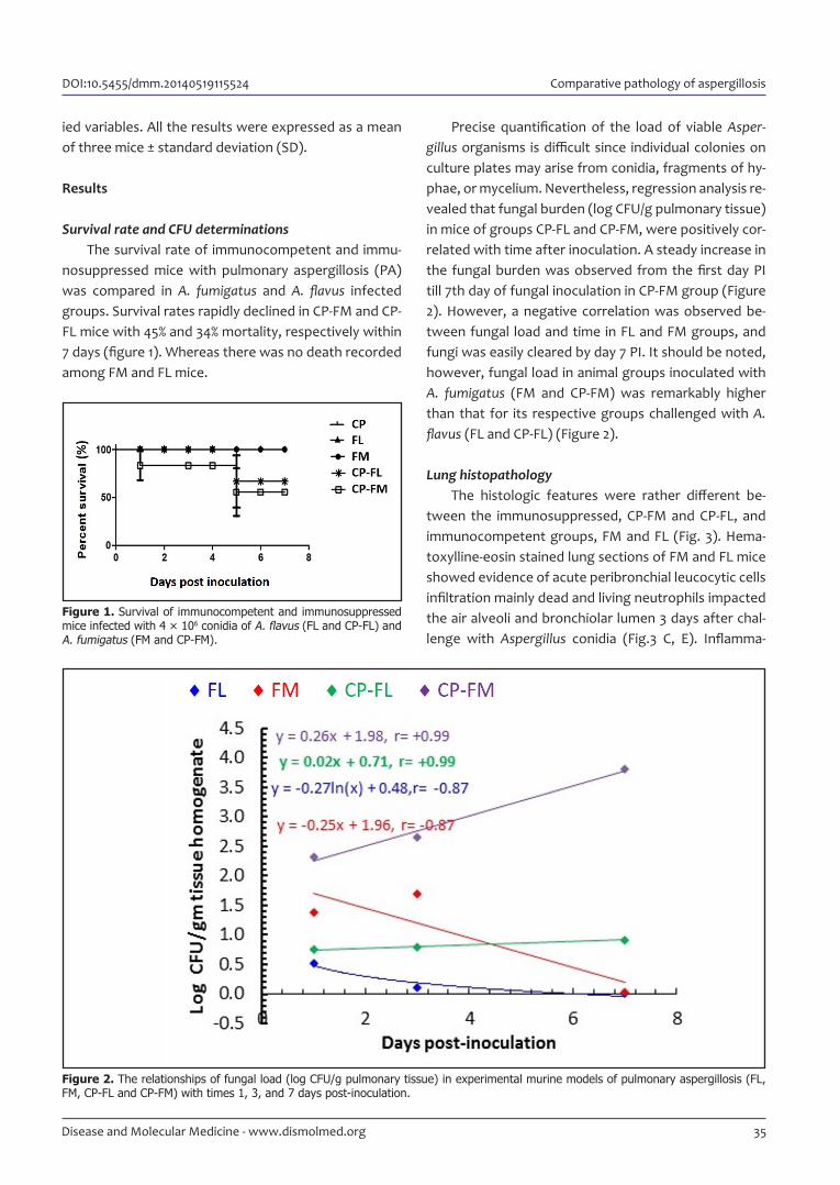
Figure 2. The relationships of fungal load (log CFU/g pulmonary tissue) in experimental murine models of pulmonary aspergillosis (FL, FM, CP-FL and CP-FM) with times 1, 3, and 7 days post-inoculation.
Precise quantification of the load of viable Asper-gillus organisms is difficult since individual colonies on culture plates may arise from conidia, fragments of hy-phae, or mycelium. Nevertheless, regression analysis re-vealed that fungal burden (log CFU/g pulmonary tissue) in mice of groups CP-FL and CP-FM, were positively cor-related with time after inoculation. A steady increase in the fungal burden was observed from the first day PI till 7th day of fungal inoculation in CP-FM group (Figure 2). However, a negative correlation was observed be-tween fungal load and time in FL and FM groups, and fungi was easily cleared by day 7 PI. It should be noted, however, fungal load in animal groups inocu lated with A. fumigatus (FM and CP-FM) was remarkably higher than that for its respective groups challenged with A. flavus (FL and CP-FL) (Figure 2).
Lung histopathologyThe histologic features were rather different be-
tween the immunosuppressed, CP-FM and CP-FL, and immunocompetent groups, FM and FL (Fig. 3). Hema-toxylline-eosin stained lung sections of FM and FL mice showed evidence of acute peribronchial leucocytic cells infiltration mainly dead and living neutrophils impacted the air alveoli and bronchiolar lumen 3 days after chal-lenge with Aspergillus conidia (Fig.3 C, E). Inflamma-
35Disease and Molecular Medicine - www.dismolmed.org

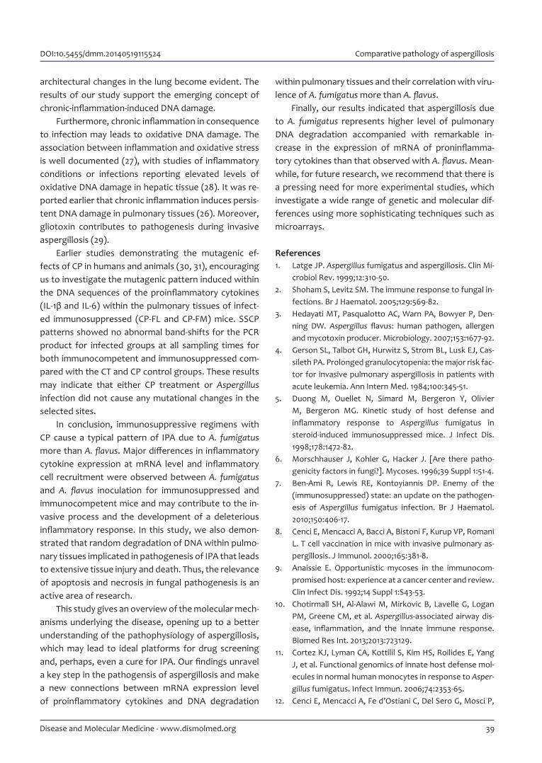
Figure 3. Representative comparative histology of lung architecture between A. fumigatus and A. flavus infections in immunocompetent (C-D, E-F) and immunosuppressed mice (G-H, I-J), respectively, 3 days after inoculation with 4 × 10⁶ Aspergillus conidia (magnification, ×100). (A) CT group showing normal histological architecture in the lung. (H&E, 100 ×) (B) CT group showing absence of fungal elements within pulmonary tissues. (PAS, 100 ×). (C) FL group showing inflammatory cellular infiltration with minimal necrosis. (H&E) (D) FL group showing occasional conidia (PAS, 100 ×). (E) FM group showing higher degree of pulmonary inflammation and necrosis (H&E). (F) FM group showing scattered fungal coinida (PAS, 100 ×), suggesting no invasive process. (G) CP-FL group showing dense inflammatory cell infiltrates and necrosis (H&E). (H) CP-FL group showing aggregations of fungal coinida (PAS, 100 ×). (I) CP-FM group showing severe pulmonary inflammation, necrosis and edema in the lung (H&E). (J) CP-FM group showing abundant fungal hyphae and conidia in the pulmonary parenchyma and angioinvasion. Fungal elements (arrows).
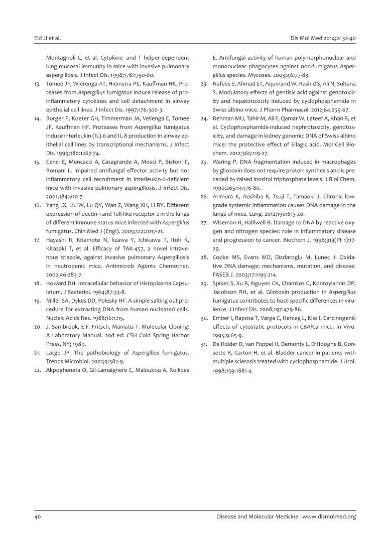
Figure 4. Mean (± SD) interleukins (IL)-1β (A) and (IL)-6 (B) levels in lung homogenates, of uninfected immunocompetent (CT), un-infected immunosuppressed (CP), and infected immunosuppressed (CP-FL and CP-FM) mice. Means with the same letter do not differ statistically at the level of P < 0.05.
tory cell infiltration was observed in peribronchial ar-eas as soon as 1 day after infection (data not shown), increased at 3 days (Fig. C, E), and had nearly resolved by 7 days (data not shown). Tissue necrosis associated with dense cell neutrophilic cells infiltrates was the main histopathologic feature of pulmonary aspergil-losis in immunosuppressed mice (Fig.3 G, I). On day 3 postchallenge, both CP-FL (Fig. 3 G) and CP-FM (Fig. 3 I) mice developed diffuse peribronchiolar leucocytic cells infiltration followed by extensive tissue necrosis that progressed from 3 days until death. Of interest, cell in-filtration in lungs of CP-FM mice was remarkably more
extensive than that of CP-FL mice (Fig.3 G, I).PAS-stained sections 3 days post-inoculation con-
firmed the difference between normal and immunosup-pressed mice infected with either Aspergillus species, in the degree of fungal conidial load within pulmonary tissues. At 3 days postchallenge in immunosuppressed mice, there was aggregations of fungal conidia in CP-FL (Fig.3 H) and more abundant in CP-FM (Fig.3 J) groups. On the other hand, we noted severe aggregations in lungs of CP-FL and CP-FM mice, but clearance of both fungal spores in lungs of FM and FL mice by 7 days after infection and normal architecture of lung histology (not shown).
Eid JI et al. Dis Mol Med 2014;2: 32-40
36 Disease and Molecular Medicine - www.dismolmed.org

Figure 5. DNA fragmentation using agarose gel electrophoresis of DNA isolated from lung homogenate of infected immunocompetent (A) and immunosuppressed (B) mice.
Figure 6. Representative 9% polyacrylamide gel showing PCR-SSCP pattern using IL- 1β (A) and IL-6 (B) primers for all groups.
Profiles of IL-1β and IL-6 in lung homogenatesThe mRNA expression level for both IL-1β and IL-6,
known to be involved in lung inflammatory responses, was assessed in lung homogenates of infected groups.
Comparisons between CP-infected groups showed significantly higher levels of IL-1b and IL-6 in CP-FM mice than in CP-FL mice at all selected time points with the highest levels for both at 1 day PI then declined gradual-ly from day 3 PI onwards, but still more significant than CP control group (Fig.4). The mRNA expression level of IL-1β was found to be higher than that of IL-6 at all se-lected time points.
In FM and FL mice lung homogenates, IL-1β and IL-6 levels rose significantly in comparison with healthy mice at 1 day after infection and later rapidly declined, while there was no significant change in mRNA expression be-tween both groups (data not shown).
Qualitative changes in pulmonary DNA fragmentationLow level of pulmonary nuclear DNA fragmentation
in normal infected mice (FM and FL), detected as slight smears apparent at all selected time points compared to intact genomic DNA in CT mice (Fig.5A). However, high level of random DNA degradation was detected as highly dense smears in immunosuppressed infected mice (CP-FM and CP-FL) at all selected time points, but still more than that for CP control mice at 24 h post-treatment (Fig.5B)
Of interest, the degree of DNA fragmentation in FM infected groups was higher than that in mice inoculated with FL.
Detection of Mutations frequency in nuclear lung DNA samples
The PCR products for IL-1β and IL-6 from pulmonary tissues of all infected and noninfected groups were ana-lyzed by SSCP to identify whether DNA harboring mu-tations within their DNA sequences. IL-1β 5’ and 3’ end fragment with length of 230bp (Fig.6A) and IL-6 with length of 159 (Fig.6B). The PCR-SSCP pattern of both DNA sequences for all infected groups shows no differ-ence between the CT group and different FM- and FL-infected groups at all selected time points.
DiscussionVery few, if none, investigations has so far been
focused on study the immunopathogenesis of invasive aspergillosis using animal models at the molecular level. Although A. flavus seems more virulent than A. fumiga-tus, the best evidence for this assumption comes from studies involving mice. Comparative studies in which
DOI:10.5455/dmm.20140519115524 Comparative pathology of aspergillosis
37Disease and Molecular Medicine - www.dismolmed.org

animals are primarily infected via the respiratory tract are lacking and required. Intranasal inoculation mimics the natural route of infection and would also be a more appropriate route more than others (1, 21). Here we in-vestigated the host response to A. fumigatus and A. fla-vus in a mouse model of aspergillosis, at the molecular level, in the context of both immunocompetence and immunosuppression.
A key step in understanding the pathogenesis of IPA is to explore the molecular mechanisms underly-ing the disease. We aimed to investigate the degree of DNA damage and mRNA expression level of proinflam-matory cytokines within pulmonary tissues of murine model with aspergillosis. Furthermore, we performed a comparative analysis of pulmonary mRNA expression pattern for proinflammatory cytokines between A. fu-migatus and A. flavus infected mice.
Because of the markedly decreased survival rate of immunocompromised mice after infection with Asper-gillus species, our model allowed us to investigate the role of proinflammtory cytokines at the molecular level in suppressing or promoting invasive aspergillosis with-in 7 days post inoculation. We evaluated the chronology of events that characterize mRNA expression profile of proinflamatory cytokines and their mutational frequen-cy, inflammatory cell recruitment, fungal tissue burden and tissue damage.
In immunocompetent mice, mRNA expression pro-file of IL-1β, and IL-6 in lung tissue was evaluated: sig-nificant increase at 1 day after A. fumgatus and A. flavus inoculation followed by a decrease from day 3 PI on-wards. It was obvious that there was a significant dif-ference between FM and FL mice in their mRNA expres-sion profile, indicating that the inflammatory process associated with A. fumigatus infection is more severe than with A. flavus. This result is in consistent with a previous study indicating that polymorphonuclear leu-kocytes induced significantly less hyphal damage to A. flavus than to A. fumigatus (22).
In addition, we have demonstrated a higher survival rate associated with lower fungal tissue burden with A. flavus significantly lower than occurs in A. fumigatus in-fections under immunosuppressive regimens. As men-tioned previously, the bigger size of A. flavus conidia may be an important factor limiting the ability of these spores to reach the alveoli (3).
Duong et al. (5) used a steroid murine model of pulmonary aspergillosis to examine cytokine responses
and reported that in pulmonary A. fumigatus, there was sustained release of IL-6 and IL-1β with extensive tissue necrosis in the lungs. In contrast, immunocompetent mice infected with A. fumigatus showed a rapid release of TNF-α, IL-6, and IL-1β and clearance of the fungus fol-lowed by rapid decrease of cytokine production. Differ-ent temporal variations in cytokine levels characterized the time course of the disease in immunocompetent and immunosuppressed animals.
In addition, the high lung levels of proinflammatory cytokines appeared to impact the lung histopathology of infected immunosuppressed mice (CP-FL and CP-FM), with extensive tissue damage, inflammatory cel-lular infiltrations, necrosis and abundant fungal growth. These results were in consistent with finding of Duong et al. (5) who reported that immunosuppressed mice in-fected with A. fumigatus developed diffuse peribronchi-olar and alveolar pneumonia characterized by a dense, acute cell infiltration followed by extensive tissue ne-crosis that progressed from 72 h until death. However, the degree of tissue damage in lungs was also correlat-ed with all the previous parameters indicating the se-verity of A. fumigatus infection than A. flavus.
Moreover, agarose gel electrophoresis of DNA showed a strong DNA smear, characteristic of necrosis, confirmed the prevalence of necrotic lesions observed within histological section of lungs in CP-FM group and with a lesser degree in CP-FL. In contrast to infected im-munosuppressed, a mild smear observed in tissues of infected immunocompetent mice (FL and FM). In CP control group, there was some degree of DNA smear-ing which was evident from previous studies indicating the genotoxicty of CP (23, 24).
In addition to genotoxic effect of cyclophospha-mide, gliotoxin, secondary metabolite produced by As-pergillus was found also to induce DNA fragmentation in macrophages (25). In contrast to an earlier study indi-cating that gliotoxin induced apoptosis in macrophages, here we proposed a random degradation of pulmonary DNA. It could be explained by the fact that gliotoxin in-duced programmed cell death within macrophages in order to invade the pulmonary tissues, however, their inflammatory reaction within pulmonary tissues due to its invasiveness and fungal growth cause random de-gredation of DNA as a hall mark for necrosis.
Arimura et al. (26) suggested that chronic systemic low-grade inflammation induces persistent DNA dam-age in alveolar epithelial and endothelial cells before
38 Disease and Molecular Medicine - www.dismolmed.org
Eid JI et al. Dis Mol Med 2014;2: 32-40
39Disease and Molecular Medicine - www.dismolmed.org
architectural changes in the lung become evident. The results of our study support the emerging concept of chronic-inflammation-induced DNA damage.
Furthermore, chronic inflammation in consequence to infection may leads to oxidative DNA damage. The association between inflammation and oxidative stress is well documented (27), with studies of inflammatory conditions or infections reporting elevated levels of oxidative DNA damage in hepatic tissue (28). It was re-ported earlier that chronic inflammation induces persis-tent DNA damage in pulmonary tissues (26). Moreover, gliotoxin contributes to pathogenesis during invasive aspergillosis (29).
Earlier studies demonstrating the mutagenic ef-fects of CP in humans and animals (30, 31), encouraging us to investigate the mutagenic pattern induced within the DNA sequences of the proinflammatory cytokines (IL-1β and IL-6) within the pulmonary tissues of infect-ed immunosuppressed (CP-FL and CP-FM) mice. SSCP patterns showed no abnormal band-shifts for the PCR product for infected groups at all sampling times for both immunocompetent and immunosuppressed com-pared with the CT and CP control groups. These results may indicate that either CP treatment or Aspergillus infection did not cause any mutational changes in the selected sites.
In conclusion, immunosuppressive regimens with CP cause a typical pattern of IPA due to A. fumigatus more than A. flavus. Major differences in inflammatory cytokine expression at mRNA level and inflammatory cell recruitment were observed between A. fumigatus and A. flavus inoculation for immunosuppressed and immunocompetent mice and may contribute to the in-vasive process and the development of a deleterious inflammatory response. In this study, we also demon-strated that random degradation of DNA within pulmo-nary tissues implicated in pathogenesis of IPA that leads to extensive tissue injury and death. Thus, the relevance of apoptosis and necrosis in fungal pathogenesis is an active area of research.
This study gives an overview of the molecular mech-anisms underlying the disease, opening up to a better understanding of the pathophysiology of aspergillosis, which may lead to ideal platforms for drug screening and, perhaps, even a cure for IPA. Our findings unravel a key step in the pathogensis of aspergillosis and make a new connections between mRNA expression level of proinflammatory cytokines and DNA degradation
within pulmonary tissues and their correlation with viru-lence of A. fumigatus more than A. flavus.
Finally, our results indicated that aspergillosis due to A. fumigatus represents higher level of pulmonary DNA degradation accompanied with remarkable in-crease in the expression of mRNA of proninflamma-tory cytokines than that observed with A. flavus. Mean-while, for future research, we recommend that there is a pressing need for more experimental studies, which investigate a wide range of genetic and molecular dif-ferences using more sophisticating techniques such as microarrays.
References 1. Latge JP. Aspergillus fumigatus and aspergillosis. Clin Mi-
crobiol Rev. 1999;12:310-50. 2. Shoham S, Levitz SM. The immune response to fungal in-
fections. Br J Haematol. 2005;129:569-82. 3. Hedayati MT, Pasqualotto AC, Warn PA, Bowyer P, Den-
ning DW. Aspergillus flavus: human pathogen, allergen and mycotoxin producer. Microbiology. 2007;153:1677-92.
4. Gerson SL, Talbot GH, Hurwitz S, Strom BL, Lusk EJ, Cas-sileth PA. Prolonged granulocytopenia: the major risk fac-tor for invasive pulmonary aspergillosis in patients with acute leukemia. Ann Intern Med. 1984;100:345-51.
5. Duong M, Ouellet N, Simard M, Bergeron Y, Olivier M, Bergeron MG. Kinetic study of host defense and inflammatory response to Aspergillus fumigatus in steroid-induced immunosuppressed mice. J Infect Dis. 1998;178:1472-82.
6. Morschhauser J, Kohler G, Hacker J. [Are there patho-genicity factors in fungi?]. Mycoses. 1996;39 Suppl 1:51-4.
7. Ben-Ami R, Lewis RE, Kontoyiannis DP. Enemy of the (immunosuppressed) state: an update on the pathogen-esis of Aspergillus fumigatus infection. Br J Haematol. 2010;150:406-17.
8. Cenci E, Mencacci A, Bacci A, Bistoni F, Kurup VP, Romani L. T cell vaccination in mice with invasive pulmonary as-pergillosis. J Immunol. 2000;165:381-8.
9. Anaissie E. Opportunistic mycoses in the immunocom-promised host: experience at a cancer center and review. Clin Infect Dis. 1992;14 Suppl 1:S43-53.
10. Chotirmall SH, Al-Alawi M, Mirkovic B, Lavelle G, Logan PM, Greene CM, et al. Aspergillus-associated airway dis-ease, inflammation, and the innate immune response. Biomed Res Int. 2013;2013:723129.
11. Cortez KJ, Lyman CA, Kottilil S, Kim HS, Roilides E, Yang J, et al. Functional genomics of innate host defense mol-ecules in normal human monocytes in response to Asper-gillus fumigatus. Infect Immun. 2006;74:2353-65.
12. Cenci E, Mencacci A, Fe d’Ostiani C, Del Sero G, Mosci P,
DOI:10.5455/dmm.20140519115524 Comparative pathology of aspergillosis
Montagnoli C, et al. Cytokine- and T helper-dependent lung mucosal immunity in mice with invasive pulmonary aspergillosis. J Infect Dis. 1998;178:1750-60.
13. Tomee JF, Wierenga AT, Hiemstra PS, Kauffman HK. Pro-teases from Aspergillus fumigatus induce release of pro-inflammatory cytokines and cell detachment in airway epithelial cell lines. J Infect Dis. 1997;176:300-3.
14. Borger P, Koeter GH, Timmerman JA, Vellenga E, Tomee JF, Kauffman HF. Proteases from Aspergillus fumigatus induce interleukin (IL)-6 and IL-8 production in airway ep-ithelial cell lines by transcriptional mechanisms. J Infect Dis. 1999;180:1267-74.
15. Cenci E, Mencacci A, Casagrande A, Mosci P, Bistoni F, Romani L. Impaired antifungal effector activity but not inflammatory cell recruitment in interleukin-6-deficient mice with invasive pulmonary aspergillosis. J Infect Dis. 2001;184:610-7.
16. Yang JX, Liu W, Lu QY, Wan Z, Wang XH, Li RY. Different expression of dectin-1 and Toll-like receptor 2 in the lungs of different immune status mice infected with Aspergillus fumigatus. Chin Med J (Engl). 2009;122:2017-21.
17. Hayashi R, Kitamoto N, Iizawa Y, Ichikawa T, Itoh K, Kitazaki T, et al. Efficacy of TAK-457, a novel intrave-nous triazole, against invasive pulmonary Aspergillosis in neutropenic mice. Antimicrob Agents Chemother. 2002;46:283-7.
18. Howard DH. Intracellular behavior of Histoplasma Capsu-latum. J Bacteriol. 1964;87:33-8.
19. Miller SA, Dykes DD, Polesky HF. A simple salting out pro-cedure for extracting DNA from human nucleated cells. Nucleic Acids Res. 1988;16:1215.
20. J. Sambrook, E.F. Fritsch, Maniatis T. Molecular Cloning: A Laboratory Manual. 2nd ed: CSH Cold Spring Harbor Press, NY; 1989.
21. Latge JP. The pathobiology of Aspergillus fumigatus. Trends Microbiol. 2001;9:382-9.
22. Akpogheneta O, Gil-Lamaignere C, Maloukou A, Roilides
E. Antifungal activity of human polymorphonuclear and mononuclear phagocytes against non-fumigatus Asper-gillus species. Mycoses. 2003;46:77-83.
23. Nafees S, Ahmad ST, Arjumand W, Rashid S, Ali N, Sultana S. Modulatory effects of gentisic acid against genotoxic-ity and hepatotoxicity induced by cyclophosphamide in Swiss albino mice. J Pharm Pharmacol. 2012;64:259-67.
24. Rehman MU, Tahir M, Ali F, Qamar W, Lateef A, Khan R, et al. Cyclophosphamide-induced nephrotoxicity, genotox-icity, and damage in kidney genomic DNA of Swiss albino mice: the protective effect of Ellagic acid. Mol Cell Bio-chem. 2012;365:119-27.
25. Waring P. DNA fragmentation induced in macrophages by gliotoxin does not require protein synthesis and is pre-ceded by raised inositol triphosphate levels. J Biol Chem. 1990;265:14476-80.
26. Arimura K, Aoshiba K, Tsuji T, Tamaoki J. Chronic low-grade systemic inflammation causes DNA damage in the lungs of mice. Lung. 2012;190:613-20.
27. Wiseman H, Halliwell B. Damage to DNA by reactive oxy-gen and nitrogen species: role in inflammatory disease and progression to cancer. Biochem J. 1996;313(Pt 1):17-29.
28. Cooke MS, Evans MD, Dizdaroglu M, Lunec J. Oxida-tive DNA damage: mechanisms, mutation, and disease. FASEB J. 2003;17:1195-214.
29. Spikes S, Xu R, Nguyen CK, Chamilos G, Kontoyiannis DP, Jacobson RH, et al. Gliotoxin production in Aspergillus fumigatus contributes to host-specific differences in viru-lence. J Infect Dis. 2008;197:479-86.
30. Ember I, Raposa T, Varga C, Herceg L, Kiss I. Carcinogenic effects of cytostatic protocols in CBA/Ca mice. In Vivo. 1995;9:65-9.
31. De Ridder D, van Poppel H, Demonty L, D’Hooghe B, Gon-sette R, Carton H, et al. Bladder cancer in patients with multiple sclerosis treated with cyclophosphamide. J Urol.
1998;159:1881-4.
Eid JI et al. Dis Mol Med 2014;2: 32-40
40 Disease and Molecular Medicine - www.dismolmed.org