comparative 3d microanatomy and histology of the eyes and central nervous systems in coleoid...
TRANSCRIPT

ORIGINAL ARTICLE
Comparative 3D microanatomy and histology of the eyesand central nervous systems in coleoid cephalopod hatchlings
Elvira Wild & Tim Wollesen & Gerhard Haszprunar &
Martin Heß
Received: 6 November 2012 /Accepted: 16 June 2014# Gesellschaft für Biologische Systematik 2014
Abstract Adaptive radiation of an animal group is the evo-lutionary variation of morphology, physiology, and behavioropening up new habitats and resources. An impressive exam-ple of the reciprocal interdependency of form and function isfound in the anatomy of cephalopod visual and central ner-vous systems. Interspecific differences of sensory organs andsignal processing structures reflect the eco-functional context,e.g., the species-specific demands emanating from habitat andforaging behavior. To substantiate this, we investigated theeyes and brain neuropils of early post-hatching stages ofsix coleoid cephalopod species (Sepia officinalis, Rossiamacrosoma, Sepietta obscura, Idiosepius notoides, Loligovulgaris, and Octopus vulgaris), showing different size andinhabiting different ethoecological niches. Comprehensive 3Dstructure data sets were produced in light microscopic resolu-tion, i.e., semithin section series of the head region (histologypresented for I. notoides, R. macrosoma, and S. obscura for the
first time) and 3D surface renderings of the neuropils, enablingthe display of all components in arbitrary perspectives andcombinations, and comparative volumetic anaylsis of homolo-gous lobe neuropils. Differing in absolute size considerably, thevisual and central nervous systems of the six species follow thesame bauplan in adult-like configuration. The visual senseobviously is of paramount importance already after hatching,but also, equilibrioception and olfaction are well developed.The species-specific shapes of various components show thatsome plasticity and distinct differences in volumetric ratios arefound, subject to their functional relevance and to differentdemands of the lifestyle on the brachial and swimming motorfunction, on camouflage, as well as on sensoric and cognitiveabilities.
Keywords Functional morphology . Volumetry . Animalvision . 3D-rendering . Interactivemodel
Introduction
Since the Cuvier-Geoffroy debate in 1830, the intrinsic mutualconditionality of structure and function is accepted as a basicbiological principle (see e.g., Lauder 1981; Boletzky et al.1997), which not least legitimates research on the functionalmorphology of visual systems (e.g., Lythgoe 1979; Archeret al. 1999). Hence, by means of morphology, valid concep-tions about functional aspects of the visual system, as well asthe peripheral and central nervous system (CNS), can befound. Eye size, photoreceptor length, and photoreceptordensity, for instance, have effects on both acuity andsensitivity (see e.g., Warrant and Locket 2004). Severalcomparative studies on animal eyes conclusively demon-strate the adaptive plasticity and functional relevance ofthese structures (Cephalopoda, e.g., Scharpf et al. 2008;Makino and Miyazaki 2010; Vertebrata, e.g., Collin and
Elvira Wild and Martin Heß contributed equally to this work.
Electronic supplementary material The online version of this article(doi:10.1007/s13127-014-0184-4) contains supplementary material,which is available to authorized users.
E. Wild :G. Haszprunar :M. Heß (*)BioZentrum LMU, Großhaderner Straße 2-4,82152 Planegg-Martinsried, Germanye-mail: [email protected]
G. HaszprunarZoologische Staatssammlung München, Münchhausenstraße 21,81247 München, Germany
G. Haszprunar :M. HeßGeoBioCenter LMU, Richard-Wagner-Straße 10, 80333 München,Germany
T. WollesenIntegrative Zoologie, Universität Wien, Althanstraße 14, 1090Wien,Austria
Org Divers EvolDOI 10.1007/s13127-014-0184-4
Partridge 1996; Fishelson et al. 2004; Arthropoda, e.g.,Kunze 1972; Warrant and McIntyre 1991).
Coleoid cephalopods possess an outstanding complex CNSwithin theMolluscawhich is even the biggest of all invertebrates(Budelmann 1995). Here, the inputs of multiple sensory organs(photoreceptors, chemoreceptors, and mechanoreceptors) areprocessed, and the complex motor activity of the tentacles,funne l , beaks , f ins , and sk in p igmen ta t ion i scontrolled (Table 1). As in vertebrates, the cephalopod brainrepresents the structural basis of the predatory and social behav-ior. Morphologically, the optic lobes are a conspicuous part ofthe highly developed visual system, and the big lens eyes cancompete with the convergently developed vertebrate eyes(Packard 1972). In contrast to vertebrates, however, the outersegments of cephalopod ectodermal photoreceptors point to-ward the light, and the first synaptic connections occur in theoptic lobes and not within the retina. Therefore, the cortex of theoptic lobes is also called “retina profunda” (Young 1974). Forvisual information is finally processed by the CNS (e.g., huge
optic lobes in cephalopods), also the nervous systems have tobe adapted to environmental conditions and habitats.Decabrachian cephalopods are visual predators virtuallystraightaway after hatching. Thus, vision is of paramountimportance (Messenger 1968; Villanueva et al. 1996), becausethe yolk is almost completely depleted, and the hatchling mustfind and catch prey to survive. Octopus paralarvae indeeddeplete their last yolk reserves during a planktic stage but startactive foraging already before getting benthic (Villanueva et al.1996). Therefore, visual systems must be adapted to theenvironmental light conditions and prey visibility in aspecies-specific way. Obviously, the hatchlings are preadaptedto the particular requirements of their habitat and habits, a factthat should be reflected, inter alia, in morphological differencesof the eyes and optic lobes of different species. Hatchlings ofSepia officinalis, e.g., already possess quite big eyes withconsiderable visual acuity, which improves with growth onlyslowly (the eyes grow slower than the rest of the body;Groeger et al. 2005, 2006). Nonetheless, size does matter also
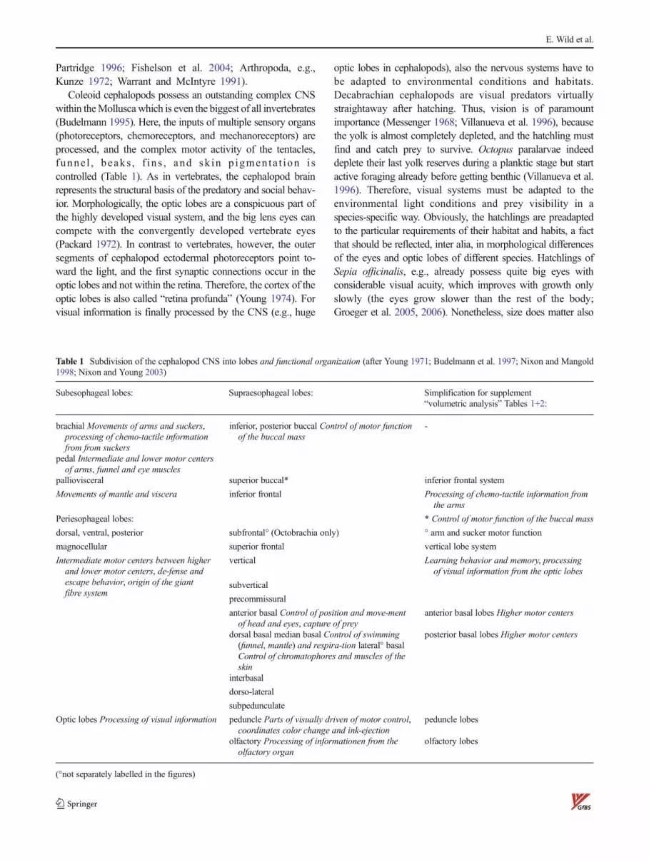
Table 1 Subdivision of the cephalopod CNS into lobes and functional organization (after Young 1971; Budelmann et al. 1997; Nixon and Mangold1998; Nixon and Young 2003)
Subesophageal lobes: Supraesophageal lobes: Simplification for supplement“volumetric analysis” Tables 1+2:
brachial Movements of arms and suckers,processing of chemo-tactile informationfrom from suckers
inferior, posterior buccal Control of motor functionof the buccal mass
-
pedal Intermediate and lower motor centersof arms, funnel and eye muscles
palliovisceral superior buccal* inferior frontal system
Movements of mantle and viscera inferior frontal Processing of chemo-tactile information fromthe arms
Periesophageal lobes: * Control of motor function of the buccal mass
dorsal, ventral, posterior subfrontal° (Octobrachia only) ° arm and sucker motor function
magnocellular superior frontal vertical lobe system
Intermediate motor centers between higherand lower motor centers, de-fense andescape behavior, origin of the giantfibre system
vertical Learning behavior and memory, processingof visual information from the optic lobes
subvertical
precommissural
anterior basal Control of position and move-mentof head and eyes, capture of prey
anterior basal lobes Higher motor centers
dorsal basal median basal Control of swimming(funnel, mantle) and respira-tion lateral° basalControl of chromatophores and muscles of theskin
posterior basal lobes Higher motor centers
interbasal
dorso-lateral
subpedunculate
Optic lobes Processing of visual information peduncle Parts of visually driven of motor control,coordinates color change and ink-ejection
peduncle lobes
olfactory Processing of informationen from theolfactory organ
olfactory lobes
(°not separately labelled in the figures)
E. Wild et al.
during allometric growth, for cell numbers of 2D receptororgans like the hemispheric retina increase with the square ofthe radius, and that of 3D brain lobes with the cube.
There are numerous studies trying to morphologically de-scribe the complex CNS of cephalopods, and Dietl (1878)introduced a nomenclature for the different brain lobes whichendures until today. Major studies on the nervous system ofOctopus and Loligo were performed by J. Z. Young (1965a,1965b, 1971, 1974, 1976b, 1977, 1979) or on several speciesin a comparative way (Wirz 1959; Frösch 1971; Nixon andMangold 1998). Here, already well-founded attempts weremade to correlate the relative volumes of brain lobes withspecies-specific habitats and habits. Recently, Kerbl et al.(2013) demonstrated an μCT-based nondestructive approachto investigate the 3D structure and volumetry of cephalopodCNS and eyes with 3-μm istotropic resolution in late embry-onic stages of the sepiolid Euprymna scolopes.
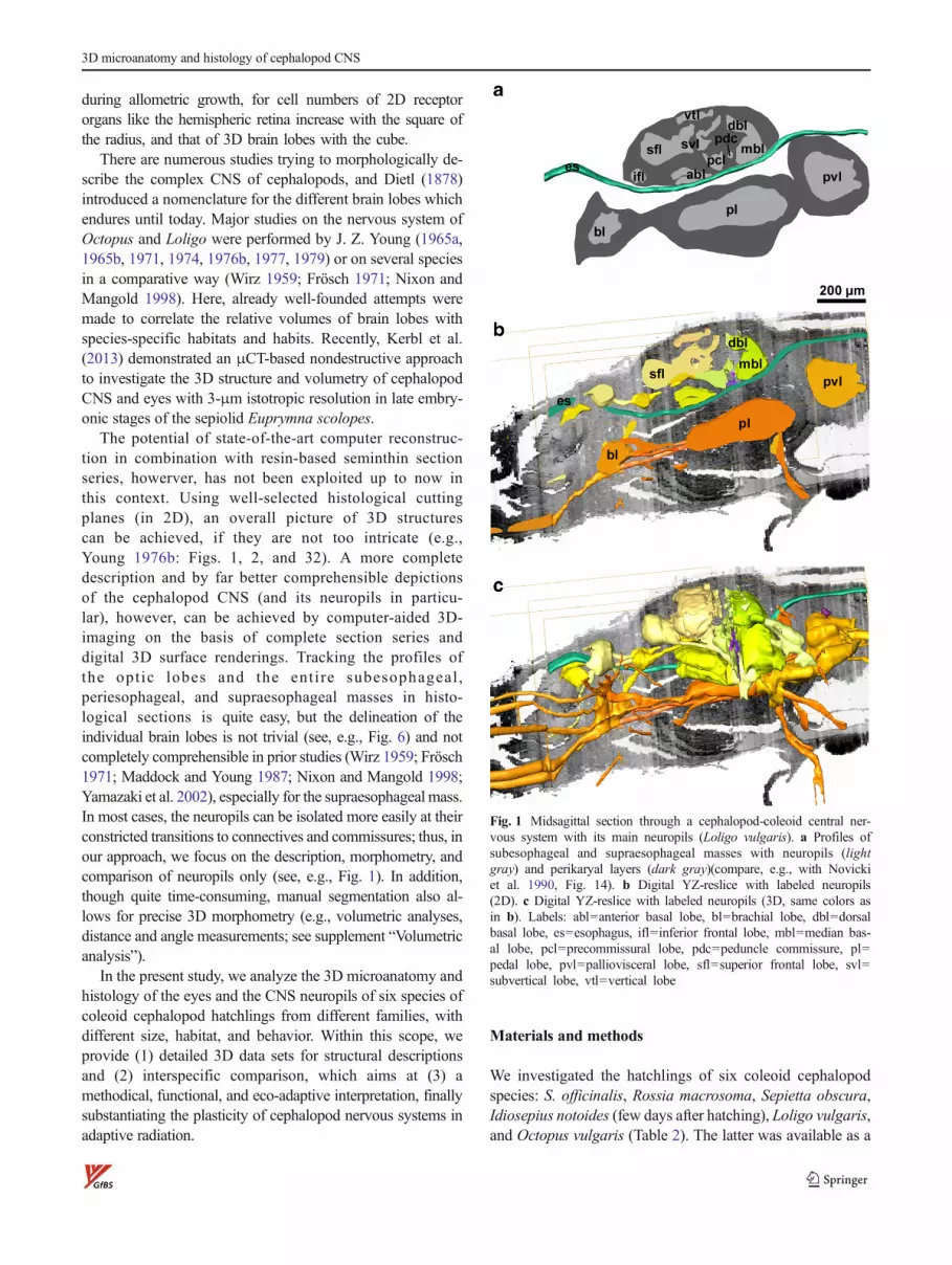
The potential of state-of-the-art computer reconstruc-tion in combination with resin-based seminthin sectionseries, howerver, has not been exploited up to now inthis context. Using well-selected histological cuttingplanes (in 2D), an overall picture of 3D structurescan be achieved, if they are not too intricate (e.g.,Young 1976b: Figs. 1, 2, and 32). A more completedescription and by far better comprehensible depictionsof the cephalopod CNS (and its neuropils in particu-lar), however, can be achieved by computer-aided 3D-imaging on the basis of complete section series anddigital 3D surface renderings. Tracking the profiles ofthe opt ic lobes and the ent i re subesophageal ,periesophageal, and supraesophageal masses in histo-logical sections is quite easy, but the delineation of theindividual brain lobes is not trivial (see, e.g., Fig. 6) and notcompletely comprehensible in prior studies (Wirz 1959; Frösch1971; Maddock and Young 1987; Nixon and Mangold 1998;Yamazaki et al. 2002), especially for the supraesophageal mass.In most cases, the neuropils can be isolated more easily at theirconstricted transitions to connectives and commissures; thus, inour approach, we focus on the description, morphometry, andcomparison of neuropils only (see, e.g., Fig. 1). In addition,though quite time-consuming, manual segmentation also al-lows for precise 3D morphometry (e.g., volumetric analyses,distance and angle measurements; see supplement “Volumetricanalysis”).
In the present study, we analyze the 3D microanatomy andhistology of the eyes and the CNS neuropils of six species ofcoleoid cephalopod hatchlings from different families, withdifferent size, habitat, and behavior. Within this scope, weprovide (1) detailed 3D data sets for structural descriptionsand (2) interspecific comparison, which aims at (3) amethodical, functional, and eco-adaptive interpretation, finallysubstantiating the plasticity of cephalopod nervous systems inadaptive radiation.
Materials and methods
We investigated the hatchlings of six coleoid cephalopodspecies: S. officinalis, Rossia macrosoma, Sepietta obscura,Idiosepius notoides (few days after hatching), Loligo vulgaris,and Octopus vulgaris (Table 2). The latter was available as a
Fig. 1 Midsagittal section through a cephalopod-coleoid central ner-vous system with its main neuropils (Loligo vulgaris). a Profiles ofsubesophageal and supraesophageal masses with neuropils (lightgray) and perikaryal layers (dark gray)(compare, e.g., with Novickiet al. 1990, Fig. 14). b Digital YZ-reslice with labeled neuropils(2D). c Digital YZ-reslice with labeled neuropils (3D, same colors asin b). Labels: abl=anterior basal lobe, bl=brachial lobe, dbl=dorsalbasal lobe, es=esophagus, ifl=inferior frontal lobe, mbl=median bas-al lobe, pcl=precommissural lobe, pdc=peduncle commissure, pl=pedal lobe, pvl=palliovisceral lobe, sfl=superior frontal lobe, svl=subvertical lobe, vtl=vertical lobe
3D microanatomy and histology of cephalopod CNS
prefabricated, Azan-stained paraffin section series (8.7 μmeach) on glass slides from the histology course stock of theUniversity of Vienna. S. obscura and R. macrosoma camefrom rearing experiments at the marine station Banyuls(Dr. Sigurd von Boletzky, Laboratoire Arago, Banyuls surMer, France): Paraffin section series (7–9 μm each) werealternately stained with Azan (azocarmine + aniline blue +orange G) and with Masson’s trichrome (iron hematoxylin +acid fuchsin + xylidine Ponceau + phospho-molybdic acid +light green). The specimens of S. officinalis hatched from eggswhich were collected in the bay of Barbariga near Rovinj(Croatia). L. vulgaris was supplied by the aquarium inRoscoff (France); I. notoides was collected from shallowseagrass meadows at Moreton Bay (Australia) and reared inaquaria for reproduction.
Systematics of the investigated species (after Young et al.2012)
Subclass: Coleoidea
Superorder: Decapodiformes
Order: Sepioidea
Family: Sepiidae S. officinalis Linnaeus, 1758
Family: Sepiolidae
Subfamily: Rossiinae R. macrosoma Delle Chiaje, 1829
Subfamily: Sepiolinae S. obscura Naef, 1916
Order: uncertain
Family: Idiosepiidae I. notoides Berry, 1921
Order: Teuthoidea
Suborder: Myopsida
Family: Loliginidae L. vulgaris Lamarck, 1798
Superorder: Octopodiformes
Order: Octopoda
Suborder: Incirrata
Family: Octopodidae O. vulgaris Cuvier, 1797
The hatchlings (S. officinalis, L. vulgaris, I. notoides) werefixed in 4% formaldehyde in 0.1-M phosphate buffer (pH 7.4)overnight, and then, the heads were cut off and embedded inepoxy resin following standard protocols. Semithin sectionseries (2.1–3μmeach, transversal planes, Table 3) were cut onan RMC MT-7000 microtome following a procedureestablished by Ruthensteiner (2008). Following a commonconvention, we use the physiological coordinate system fororientation instead of the morphological one, i.e., the mouthopening points forward (instead of ventrally), the funnel liesventrally (not caudally). Hence, transversal sections are nor-mal to the physiological anterior-posterior axis.
The sections were transferred to microscope slides,stained with Azur II-methylene blue (after Richardsonet al. 1960), and mounted with coverslips. Single sectionswere photographed with an Olympus CX41 light micro-scope (PlanC N 10x or UPlan S Apo 40x objective)
equipped with an Olympus DP25 digital camera, or com-plete microscope slides were photographed with anOlympus BX51-dotSlide system (UPlan S Apo 10x objec-tive, XC 10 camera) using extended focal imaging andautomated stitching.
The image series were converted from RGB color to gray-scale, contrast enhanced, and unsharp masked with AdobePhotoshop 6.0 (San Jose, USA). Constrained by memorylimitations of the imaging computer, the image stacks werepartially reduced in size (see Table 3). The preprocessedimages were loaded into Amira® 5.3.3 (Berlin, Germany),aligned, and segmented, i.e., the profiles of organs or otherstructures were selected manually in every image plane andattributed to separate “materials.” Therefrom, surfaces wererendered, smoothed, and displayed under any defined angle,color, transparency, and material composition. Appropriateviews of the digital 3D models were virtually “photographed”using the snapshot tool and arranged to figures with AdobePhotoshop®. Interactive 3D models were inserted in the PDFfigures following Ruthensteiner and Heß (2008) with updatedsoftware.
Results
Comprehensive 3D data sets of the head region of the hatch-lings of six coleoid cephalopod species were produced in lightmicroscopic resolution. Based on transversal section series,digital imaging, segmentation, and 3D surface rendering thesedata sets enable the detailed histological and 3D microana-tomical description as well as morphometric analyses andinterspecific comparisons of the eyes and CNS of the investi-gated cephalopod species.
Though the eyes and nervous systems of the six investigat-ed species basically follow the same bauplan, they varyinterspecifically in size (see supplement “Volumetric analy-sis”), proportions, and complexity (see comparative Figs. 2, 3,4, 5, 6, 7, 8, 9, and 10 and supplement Tables 1+2).
Sepia officinalis
The adult-like hatchling of S. officinalis (Fig. 11, supplementinteractive Fig. 1) is the biggest of the studied specimens(horizontal head diameter ca. 3.5 mm), and therefore, it hasthe biggest eyes and nervous system (Fig. 2a, supplement“Volumetric analysis” Table 1). As a representative of theDecabrachia, S. officinalis possesses eight arms and two ten-tacles. The arms are arranged in a circle around the mouthopening and the buccal mass (Fig. 11b, c), whereas the tenta-cles run between arm pairs 3 and 4. They are retractable in atentacular pocket formed by a skinny duplicature of theintegument between the bases of arms 4 and 3 and belowthe eye chamber (Fig. 11c–f). Arm pair 4 possesses lateral
E. Wild et al.

fin-like extensions in this species and is distinctly set apartto the other arms.
The peripheral nervous system of the arms and tentaclesis prototypic in Sepia hatchlings. The eyes are situatedslightly dorsal with respect to the head’s horizontal midlineand pointing horizontally (Fig. 11d) and slightly forward(Fig. 2a). They have poorly developed outer lids but athickened integument just behind the cornea. The anterioreye chambers are quite voluminous, even if one takes intoaccount some shrinkage of the eyes, indicated by thedeformed retina profiles in Fig. 3a, and lead backwardhalfway ventral of the optic lobes. The eyes (horizontaldiameter ca. 1.3 mm) “float” in these chambers, attachedto the optic lobes via their axon bundles and to the troch-lear and cranial cartilages via the oculomotoric muscles.The white body and anterior chamber organ fill the space,where the opposing curvatures of eye cup and lobusopticus detach. The vitreous body between lens (diameterca. 450 μm, distal segment ca. 20 %) and retina appearscomparatively narrow in this species, and the dark retinalpigment is thinning out in central parts of the eye cup. Theretina is flattened centrally, where it contacts the optic lobe,and also ventrally.
Frontally, the optic lobes of both sides are kept apart(minimal distance ca. 280 μm dorsally) by the buccal and
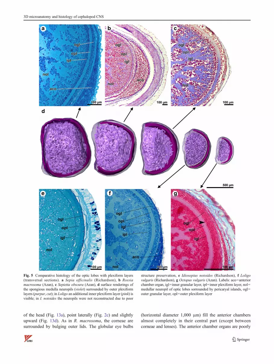
frontal lobes. At the periphery of the lobes, an outer plexiformlayer is well defined between the outer and inner granularlayer with a fine substratification in evidence (Fig. 5a).Mediodorsally, the outer plexiform layer is missing in thisspecies (Fig. 2a). An additional inner plexiform layer is notclearly discernible (but see Loligo; Fig. 5f). In the medulla,neuropils with a complex spongy structure and islands of cellnuclei alternate (both contiguous in 3D; Fig. 11e, f, g, Fig. 5a,Fig. 9a). The optic commissure is well developed and can bedivided in a dorsal (Fig. 6a; doc) and ventral optic commissure(not shown, situated a few micrometer frontally). The pedun-cle commissure lies tightly adjacent and dorsal to the dorsaloptic commissure.
Concerning the subesophageal mass, the S. officinalishatchling shows a comparatively close constellation of thebrachial and pedal lobes entailing short brachiopedal connec-tives. Ventral of the pedal lobes, the anterior funnel nervesform a distinctive neuropil node (Fig. 7a), where severalbundles of nerve fibers fan out in various directions (see alsoI. notoides, Fig. 7d and to a lesser degree L. vulgaris, Fig. 7e).Brachio-magnocellular connectives are best seen inS. officinalis within this study.
A somewhat condensed constellation of buccal and frontallobes with short cerebrobuccal connectives can also be ob-served in the frontal part of the supraesophageal mass. The
Table 2 Lifestyles of the investigated cephalopod species
Species life style hatchling life style adult
Sepia officinalis hatches adult-like, benthopelagic (Nixon and Mangold 1998) benthic, demersal, neritic? (Jereb and Roper 2005)
Rossia macrosoma hatches adult-like (Boletzky and Boletzky 1973), benthic(Boletzky 2003)
benthic 0–300 m, demersal (Jereb and Roper 2005)
Sepietta obscura benthic (Boletzky et al. 1971; Boletzky 2003) benthic with vertical migrations (Jereb and Roper 2005)
Idiosepius notoides planktic (Boletzky 2003; Yamamoto et al. 2003) necto-benthic, often attached to eelgrass (Boletzky 2003;Yamamoto et al. 2003)
Loligo vulgaris planktic (Boletzky 1974; Jereb and Roper 2010) neritic with vertical migrations, pelagic (Jereb and Roper 2010;Roper et al. 1984)
Octopus vulgaris planktic for 33 – 54 days (Nixon and Mangold 1996, 1998) benthic (e.g. Roper et al. 1984)
Table 3 Section series from the head region of cephalopod hatchlings
Species (size [mm]: mantle edge—abdominal tip, mantle width)
Number of slices usedfor reconstruction
Slice thickness [μm] Resolution [μm/px](photo)
Resolution [μm/px](reconstr.)
Origin
Sepia officinalis (6.3, 4.6) 2060** 2.6 5.15 5.15 Rovinj
Rossia macrosoma (−, 3.2) 322 7* 1.43 4 Banyuls
Sepietta obscura (2.4, 2.6) 410 8.75* 1.43 5 Banyuls
Idiosepius notoides (4.3, 2.5) 1080** 3 2.58 2.58 Australia
Loligo vulgaris (2.6, 1.55) 1188** 2.1 0.73 1.46 Roscoff
Octopus vulgaris (1.13, 0.98) 178 8.7* 0.44 1.79 Vienna
*The slice thickness (indicated on the slides incorrectly) was corrected after 3D reconstruction to get spherical lenses in both XY- and XZ-planes.
**Every fourth slice used for reconstruction
3D microanatomy and histology of cephalopod CNS

inferior buccal lobes lie ventrally to the superior buccal lobesas observable in S. obscura and I. notoides. Here, thesympathic nerves project backward and run alongside theesophagus (Fig. 9a). They could be traced only up to theanterior basal lobe. Dorsal basal lobes and peduncle lobesare found in a rather dorsal position. The palliovisceral lobesshow marked frontolateral bulges, i.e., fin and posterior chro-matophore lobes (Fig. 2a).
The statocysts are well preserved (Fig. 11g, h), most prob-ably due to its robust cartilage capsule, with a tangential
diameter of max. 830 μm. Statoliths are present, and severalcartilaginous protrusions are found bisymmetrically within thestatocyst’s lumen. Olfactory organs are oval shaped (430×260 μm2, thickness max. 100 μm) and positioned laterally justbehind the eyes (Fig. 11a, f and Fig. 2a).
Rossia macrosoma
The hatchling of R. macrosoma (Fig. 12, supplement interac-tive Fig. 2) is the second largest (horizontal head diameter ca.
Fig. 2 Comparative 3Dreconstruction of the centralnervous system: supraesophagealneuropils, optic lobes, eyes, andtentacle nerves (comparativedorsal views; a-f same scale; seealso interactive supplementaryfigures 1–6). a Sepia officinalis,b Rossia macrosoma, c Sepiettaobscura, d Idiosepius notoides,e Loligo vulgaris, f Octopusvulgaris. Labels: 3=collar nerve,7=posterior funnel nerve, 13=posterior superior oculomotoricnerve, 14=olfactory nerve,15=superior anterior ophthalmicnerve, 16=superior posteriorophthalmic nerve, 19=pallialnerve, 22=posterior head retrac-tor nerve, 24=visceral nerve,dbl=dorsal basal lobe,dll=dorso-lateral lobe, ibc=interbrachial commissure, ibg=intrabrachial ganglion, ibl=inferi-or buccal lobe, ifl=inferior frontallobe, le=lens, mbl=median basallobe, of=olfactory organ, ol=op-tic lobe (purpur=outer plexiformlayer, violett=medullar neuropils;due to poor structure preservationnot reconstructed in I. notoides),pdl=peduncle lobe, pl=pedallobe,pvl=palliovisceral lobe, re=reti-na, sbl=superior buccal lobe, sfl=superior frontal lobe, vtl=verticallobe, *=fin and chromatophorelobe
E. Wild et al.
3.1 mm) of the six species investigated in this study, has acomparatively thick epidermis, and appears dorsoventrallycompressed in the eye region (Fig. 12c, d). The arms werenot completely enclosed in the section series, but the periph-eral (arm and tentacle) nervous system reveals the standardorganization (Fig. 2b).
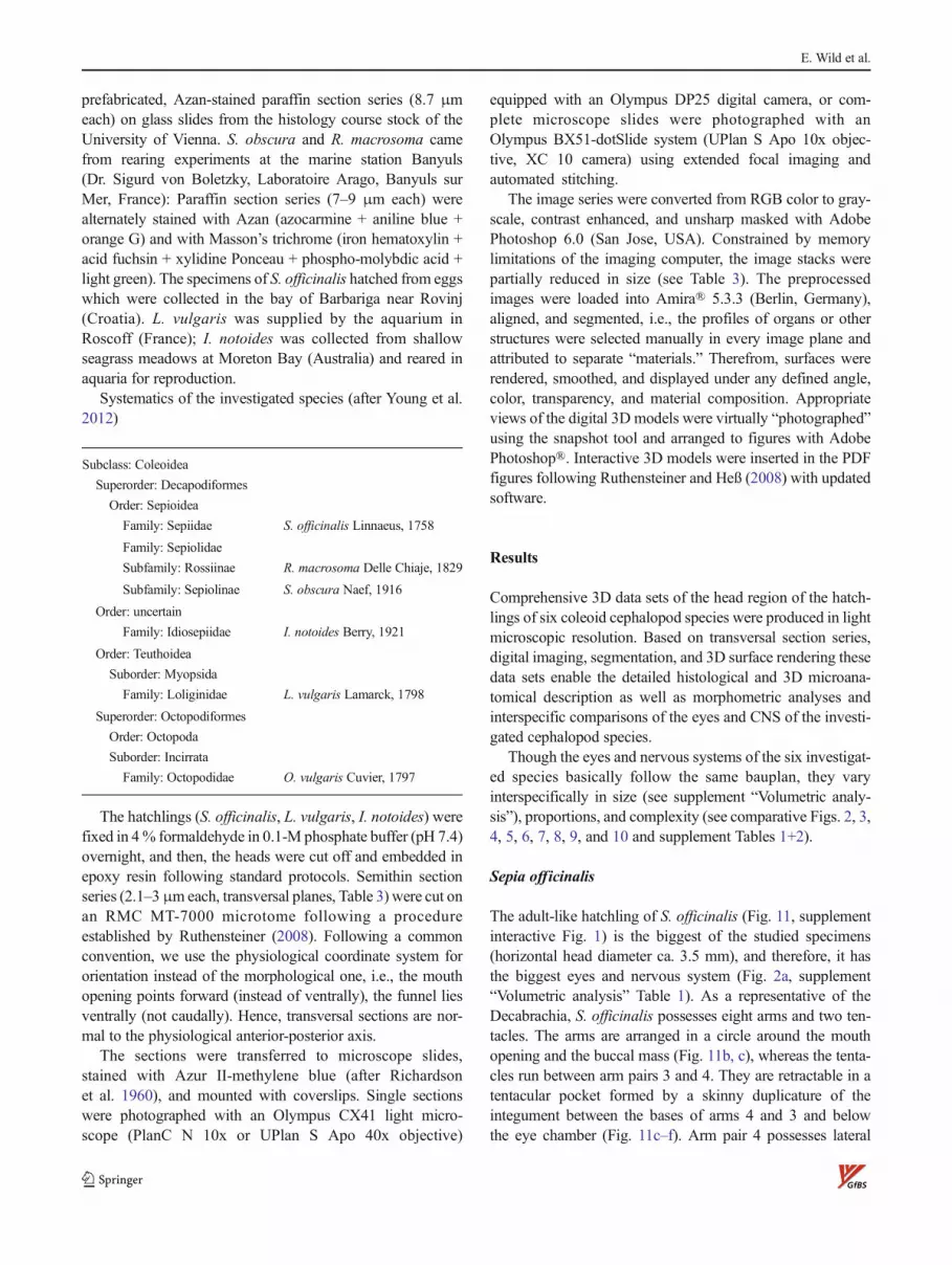
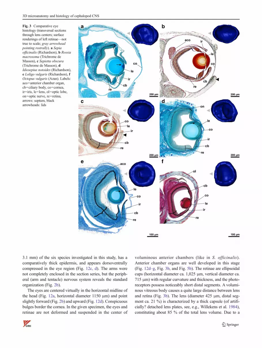
The eyes are centered virtually in the horizontal midline ofthe head (Fig. 12a, horizontal diameter 1150 μm) and pointslightly forward (Fig. 2b) and upward (Fig. 12d). Conspicuousbulges border the cornea. In the given specimen, the eyes andretinae are not deformed and suspended in the center of
voluminous anterior chambers (like in S. officinalis).Anterior chamber organs are well developed in this stage(Fig. 12d–g, Fig. 3b, and Fig. 5b). The retinae are ellipsoidalcups (horizontal diameter ca. 1,025 μm, vertical diameter ca.715 μm) with regular curvature and thickness, and the photo-receptors possess noticeably short distal segments. A volumi-nous vitreous body causes a quite large distance between lensand retina (Fig. 3b). The lens (diameter 425 μm, distal seg-ment ca. 21 %) is characterized by a thick capsule (of artifi-cially? detached lens plates, see, e.g., Willekens et al. 1984),constituting about 85 % of the total lens volume. Due to a
Fig. 3 Comparative eyehistology (transversal sectionsthrough lens centers; surfacerenderings of left retinae—nottrue to scale; gray arrowheadpointing rostrally). a Sepiaofficinalis (Richardson), b Rossiamacrosoma (Trichrome deMasson), c Sepietta obscura(Trichrome de Masson), dIdiosepius notoides (Richardson),e Loligo vulgaris (Richardson), fOctopus vulgaris (Azan). Labels:aco=anterior chamber organ,cb=ciliary body, co=cornea,ir=iris, le=lens, ol=optic lobe,on=optic nerve, re=retina,arrows: septum, blackarrowheads: lids
3D microanatomy and histology of cephalopod CNS

preparation artifact, the lens is broken at the septum, accentu-ating the connections of the ciliary body to both the distal andproximal segment of the lens (Fig. 3b).
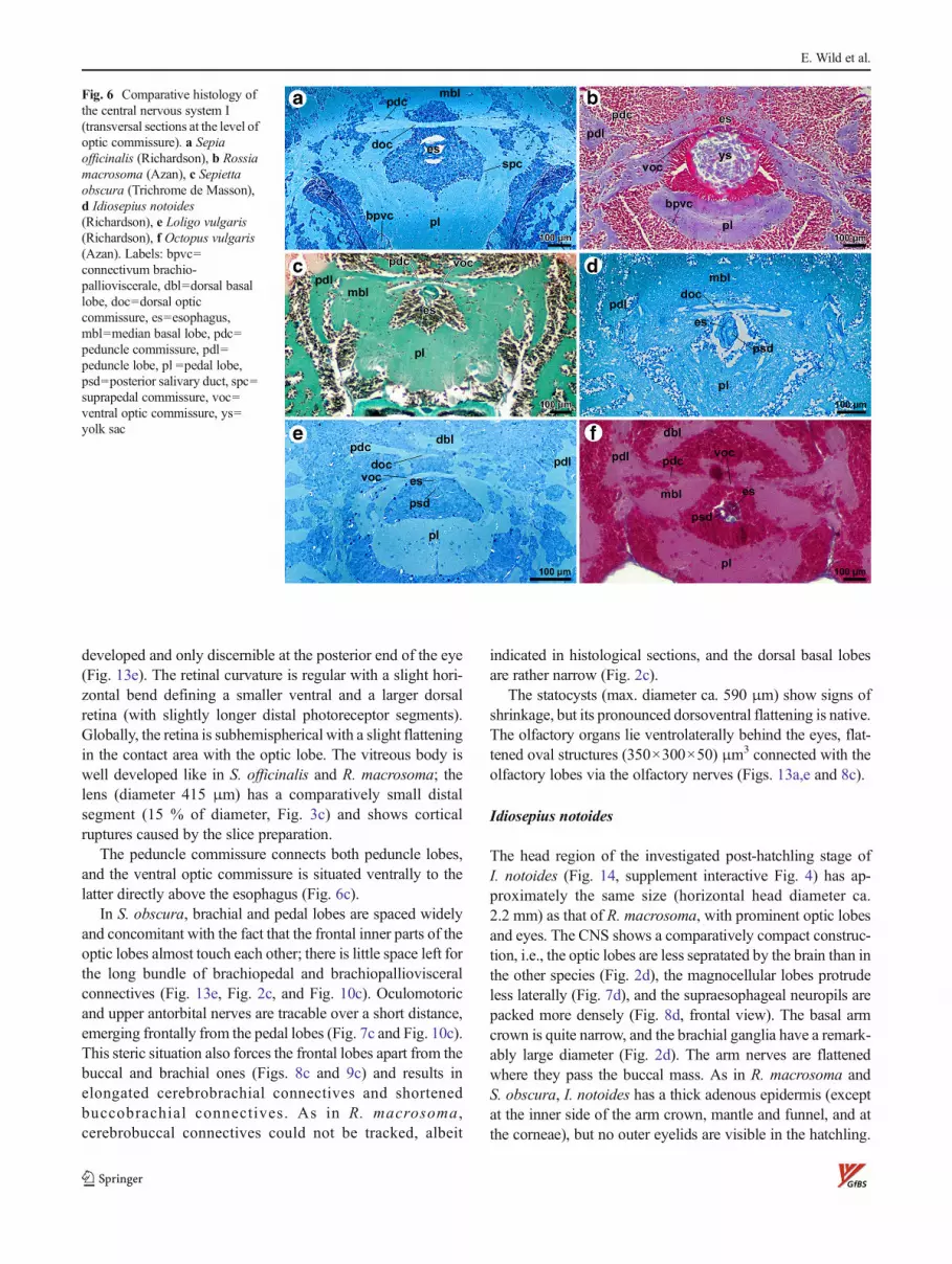
As in Sepia, the brachial lobes, esophagus, and inner yolksac, but not the buccal and frontal lobes, are located betweenthe frontal portions of the optic lobes. The very thin ventralpart of the optic commissure runs directly across the hardlyvisible esophagus under which a profile of the inner yolk sac isvisible (Fig. 6b). The peduncle commissure is only discernibleat its entrance in the right peduncle lobe.
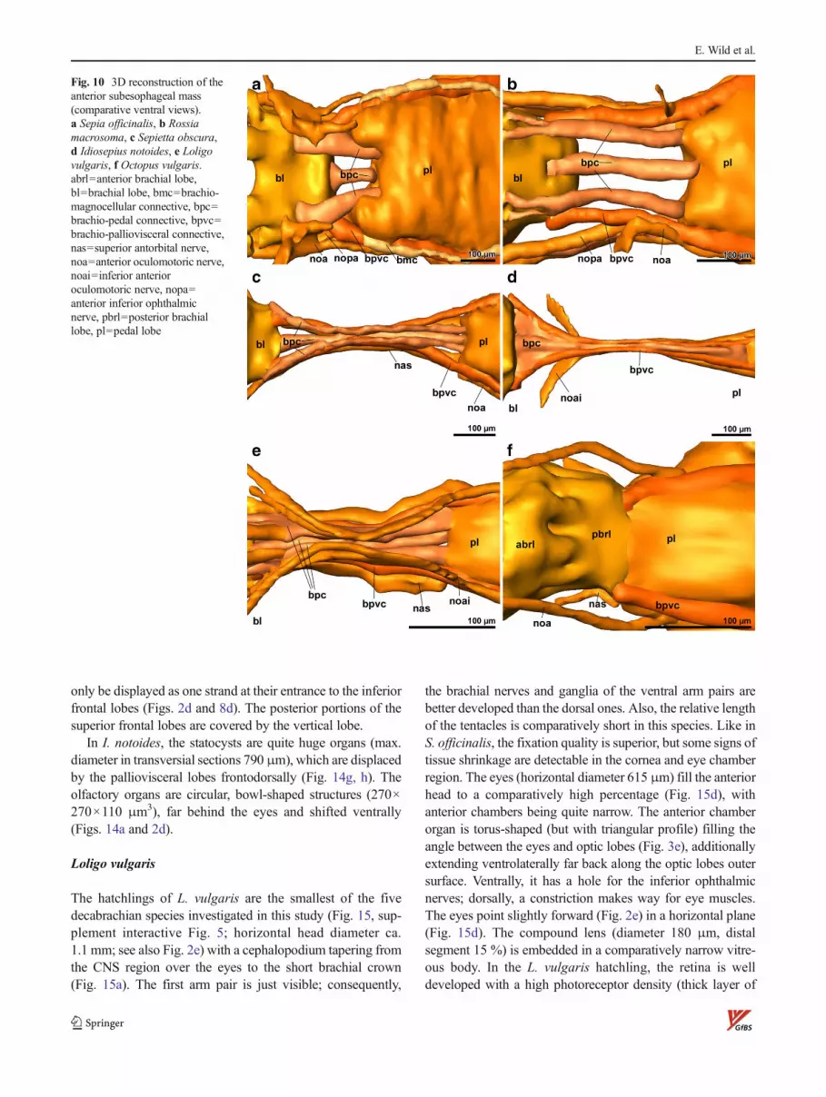
The brachiopedal connectives are elongate (twofold com-pared to S. officinalis) because of the greater distance betweenthe brachial and pedal lobes (Fig. 10b).
In the supraesophageal mass, the inferior buccal lobes liefrontally to the superior buccal lobes as in Loligo. The supe-rior buccal lobes and the inferior frontal lobes are spaced ca.500 μm by long buccobrachial and cerebrobrachial
connectives (Fig. 2b and Fig. 9b); cerebrobuccal connectivesare visible in outlines but could not be tracked. Also, thedistance between buccal and brachial lobes is quite big. Thedorsal basal lobes appear rather narrow in this species(Fig. 2b).
The statocysts are well preserved in the given specimen andcomparatively small sized with respect to the total head vol-ume (vertical diameter in transversal sections 590 μm).Olfactory organs are visible as flat “anlagen,” ventrolateralto the rear portion of the anterior chamber organ.
Sepietta obscura
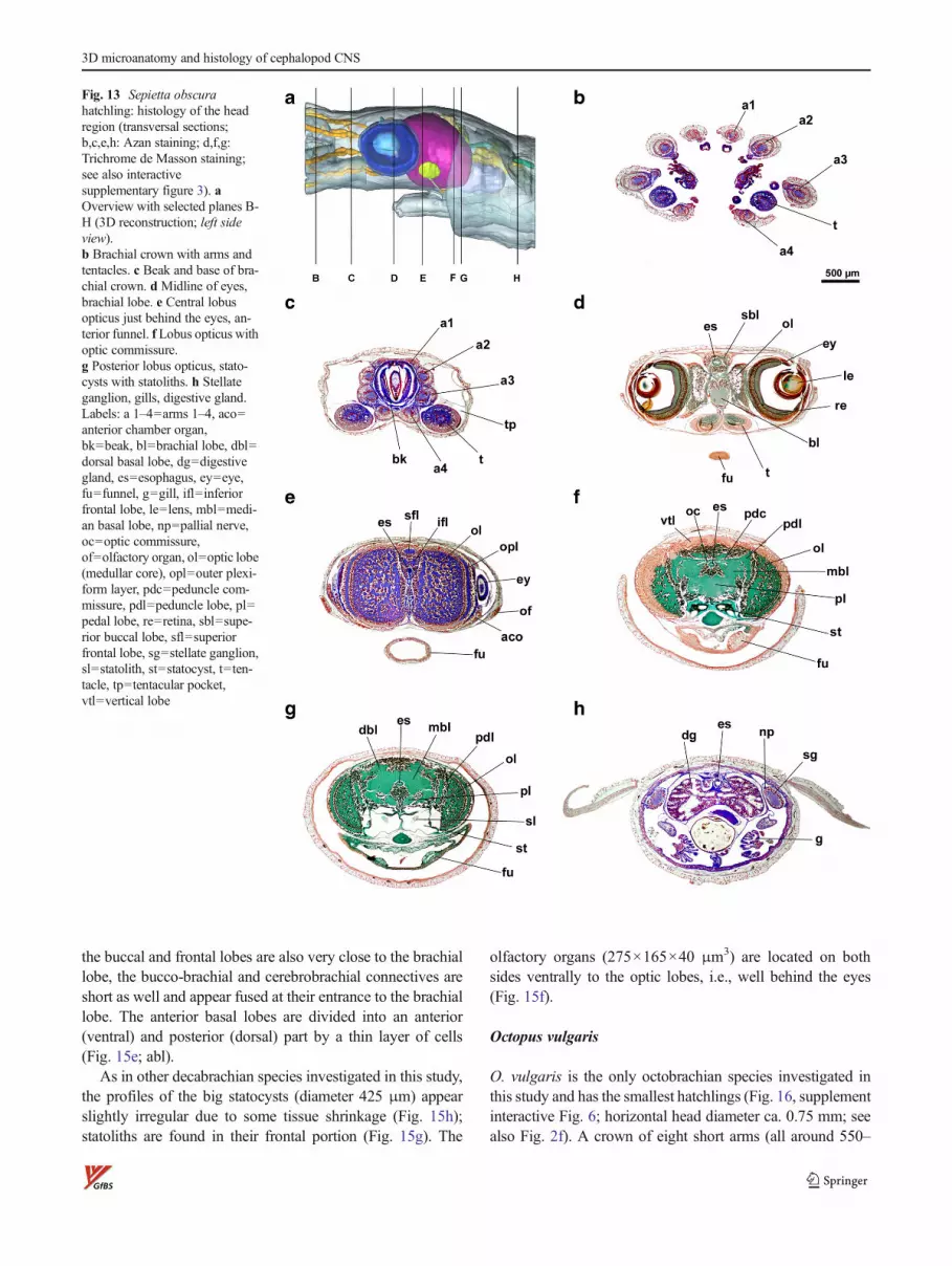
The hatchling of S. obscura (Fig. 13, supplement interactiveFig. 3; horizontal head diameter ca. 2.5 mm) has a slenderbodywith some dorsoventral compression in the head and armcrown region. The eyes are centered in the horizontal midline
Fig. 4 Comparative retinahistology (ventral portion;transversal sections at the level oflens centers). a Sepia officinalis(Richardson), b Rossiamacrosoma (Trichrome deMasson), c Sepietta obscura(Trichrome de Masson), dIdiosepius notoides (Richardson),e Loligo vulgaris (Richardson), fOctopus vulgaris (Azan). Labels:bsm=basal membrane, ds=distalsegments of photoreptors,ps=proximal segments ofphotoreceptors, rp=retinalplexus, scn=supporting cellnuclei, arrowheads periphericallythickened eye cartilage
E. Wild et al.
of the head (Fig. 13a), point laterally (Fig. 2c) and slightlyupward (Fig. 13d). As in R. macrosoma, the corneae aresurrounded by bulging outer lids. The globular eye bulbs
(horizontal diameter 1,000 μm) fill the anterior chambersalmost completely in their central part (except betweencorneae and lenses). The anterior chamber organs are poorly
Fig. 5 Comparative histology of the optic lobes with plexiform layers(transversal sections). a Sepia officinalis (Richardson), b Rossiamacrosoma (Azan), c Sepietta obscura (Azan), d surface renderings ofthe spongious medulla neuropils (violet) surrounded by outer plexiformlayers (purpur, cut); in Loligo an additional inner plexiform layer (pink) isvisible; in I. notoides the neuropils were not reconstructed due to poor
structure preservation, e Idiosepius notoides (Richardson), f Loligovulgaris (Richardson), g Octopus vulgaris (Azan). Labels: aco=anteriorchamber organ, igl=inner granular layer, ipl=inner plexiform layer, nol=medullar neuropil of optic lobes sorrounded by pericaryal islands, ogl=outer granular layer, opl=outer plexiform layer
3D microanatomy and histology of cephalopod CNS
developed and only discernible at the posterior end of the eye(Fig. 13e). The retinal curvature is regular with a slight hori-zontal bend defining a smaller ventral and a larger dorsalretina (with slightly longer distal photoreceptor segments).Globally, the retina is subhemispherical with a slight flatteningin the contact area with the optic lobe. The vitreous body iswell developed like in S. officinalis and R. macrosoma; thelens (diameter 415 μm) has a comparatively small distalsegment (15 % of diameter, Fig. 3c) and shows corticalruptures caused by the slice preparation.
The peduncle commissure connects both peduncle lobes,and the ventral optic commissure is situated ventrally to thelatter directly above the esophagus (Fig. 6c).
In S. obscura, brachial and pedal lobes are spaced widelyand concomitant with the fact that the frontal inner parts of theoptic lobes almost touch each other; there is little space left forthe long bundle of brachiopedal and brachiopalliovisceralconnectives (Fig. 13e, Fig. 2c, and Fig. 10c). Oculomotoricand upper antorbital nerves are tracable over a short distance,emerging frontally from the pedal lobes (Fig. 7c and Fig. 10c).This steric situation also forces the frontal lobes apart from thebuccal and brachial ones (Figs. 8c and 9c) and results inelongated cerebrobrachial connectives and shortenedbuccobrachial connectives. As in R. macrosoma ,cerebrobuccal connectives could not be tracked, albeit
indicated in histological sections, and the dorsal basal lobesare rather narrow (Fig. 2c).
The statocysts (max. diameter ca. 590 μm) show signs ofshrinkage, but its pronounced dorsoventral flattening is native.The olfactory organs lie ventrolaterally behind the eyes, flat-tened oval structures (350×300×50) μm3 connected with theolfactory lobes via the olfactory nerves (Figs. 13a,e and 8c).
Idiosepius notoides
The head region of the investigated post-hatchling stage ofI. notoides (Fig. 14, supplement interactive Fig. 4) has ap-proximately the same size (horizontal head diameter ca.2.2 mm) as that of R. macrosoma, with prominent optic lobesand eyes. The CNS shows a comparatively compact construc-tion, i.e., the optic lobes are less sepratated by the brain than inthe other species (Fig. 2d), the magnocellular lobes protrudeless laterally (Fig. 7d), and the supraesophageal neuropils arepacked more densely (Fig. 8d, frontal view). The basal armcrown is quite narrow, and the brachial ganglia have a remark-ably large diameter (Fig. 2d). The arm nerves are flattenedwhere they pass the buccal mass. As in R. macrosoma andS. obscura, I. notoides has a thick adenous epidermis (exceptat the inner side of the arm crown, mantle and funnel, and atthe corneae), but no outer eyelids are visible in the hatchling.
Fig. 6 Comparative histology ofthe central nervous system I(transversal sections at the level ofoptic commissure). a Sepiaofficinalis (Richardson), b Rossiamacrosoma (Azan), c Sepiettaobscura (Trichrome de Masson),d Idiosepius notoides(Richardson), e Loligo vulgaris(Richardson), f Octopus vulgaris(Azan). Labels: bpvc=connectivum brachio-pallioviscerale, dbl=dorsal basallobe, doc=dorsal opticcommissure, es=esophagus,mbl=median basal lobe, pdc=peduncle commissure, pdl=peduncle lobe, pl =pedal lobe,psd=posterior salivary duct, spc=suprapedal commissure, voc=ventral optic commissure, ys=yolk sac
E. Wild et al.

The eyes appear spacious (horizontal diameter 1,250 μm), andalmost no anterior chamber volume could be detected(Figs. 14d and 3d). They point in a horizontal/lateral direction,
both in horizontal (Fig. 2d) and transversal planes (Fig. 14d).Anterior chamber organs are barely developed. The two seg-ments of the lenses (total diameter 475 μm) have different
Fig. 7 Subesophageal neuropils. 3D reconstruction of the central ner-vous system: supraesophageal and subesophageal neuropils (comparativeleft lateral and frontal views; enlarge to 100 % in the PDF viewer; A-Fdifferent scales). a Sepia officinalis, b Rossia macrosoma, c Sepiettaobscura, d Idiosepius notoides, e Loligo vulgaris, f Octopus vulgaris.Labels: 1=inferior antorbital nerve, 2=superior antorbital nerve, 3=collarnerve, 4=crista nerve, 5=anterior funnel nerve, 6=median funnel nerve,7=posterior funnel nerve, 8=anterior oculomotoric nerve, 9=inferioranterior oculomotoric nerve, 10=superior anterior oculomotoric nerve,11=posterior oculomotoric nerve, 12=inferior posterior oculomotoric
nerve, 13=superior posterior oculomotoric nerve, 15=anterior superiorophthalmic nerve, 16=posterior superior ophthalmic nerve, 17=anteriorinferior ophthalmic nerve, 18=posterior inferior ophthalmic nerve,19=pallial nerve, 20=postorbital nerve, 21=anterior head retractornerve, 22=posterior head retractor nerve, 24=visceral nerve, bl=brachial lobe, bmc=brachio-magnocellular connective, bpvc=brachio-palliovisceral connective, es=esophagus, mcl=magnocellular lobe, pl=pedal lobe, pvl=palliovisceral lobe, sbc=suprabrachialcommissure, spc=suprapedal commissure
3D microanatomy and histology of cephalopod CNS
curvature radii and a different cortex-core partitioning. Thedistal component has a comparatively high volume fraction inthis species (>30 %), and the inner component is slightlyelongated in axial direction (Fig. 3d). The vitreous bodiesare developed regularly; the ciliary bodies are weakly
developed in this species (Fig. 3d). The retina is thickened inthe central region, and the tips of the distal photoreceptorsegments show some artificial swellings in the central anddorsal retina. The ventral retina is flattened, smoothly mergingwith the dorso-nasal retina, set of against the centrotemporal
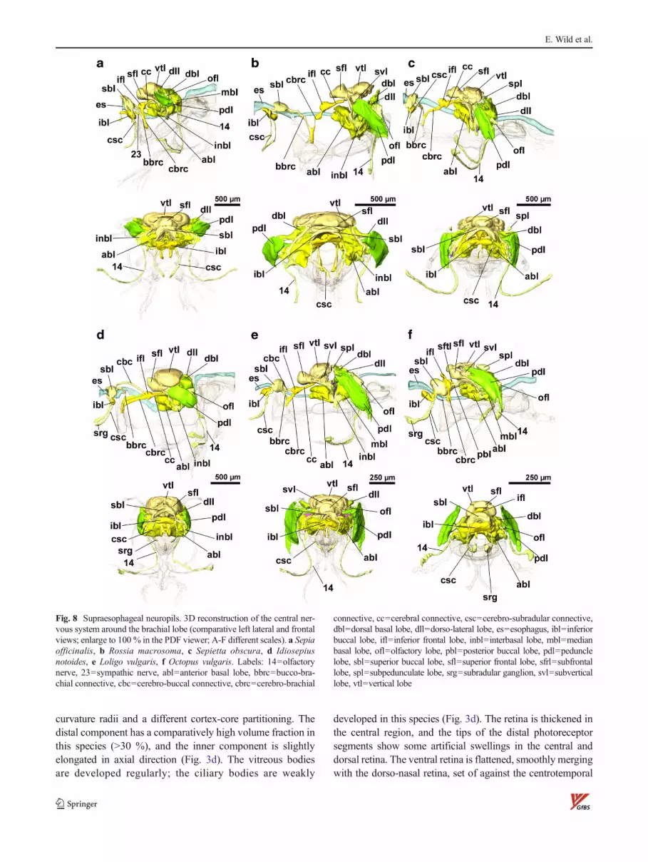
Fig. 8 Supraesophageal neuropils. 3D reconstruction of the central ner-vous system around the brachial lobe (comparative left lateral and frontalviews; enlarge to 100 % in the PDF viewer; A-F different scales). a Sepiaofficinalis, b Rossia macrosoma, c Sepietta obscura, d Idiosepiusnotoides, e Loligo vulgaris, f Octopus vulgaris. Labels: 14=olfactorynerve, 23=sympathic nerve, abl=anterior basal lobe, bbrc=bucco-bra-chial connective, cbc=cerebro-buccal connective, cbrc=cerebro-brachial
connective, cc=cerebral connective, csc=cerebro-subradular connective,dbl=dorsal basal lobe, dll=dorso-lateral lobe, es=esophagus, ibl=inferiorbuccal lobe, ifl=inferior frontal lobe, inbl=interbasal lobe, mbl=medianbasal lobe, ofl=olfactory lobe, pbl=posterior buccal lobe, pdl=pedunclelobe, sbl=superior buccal lobe, sfl=superior frontal lobe, sfrl=subfrontallobe, spl=subpedunculate lobe, srg=subradular ganglion, svl=subverticallobe, vtl=vertical lobe
E. Wild et al.

retina by a marked bend (Fig. 3d, inlet). The latter appearsto be “pushed forward” by the optic lobes with ahorseshoe-shaped thickening and a slight temporal/horizontal depression (see 3D model, supplement Fig. 4).As in S. officinalis, the dark retinal pigment is fading inthe central to ventral parts of the retina (Figs. 14d and3d).
The optic lobes are voluminous and face each other frontally(Fig. 2d) and frontoventrally (Fig. 14e, f). Its inner structure isregular, i.e., with outer granular layer, outer plexiform layer,and inner granular layer made of interwoven neuropils andsoma islands. The bad structural preservation of the medullaallowed just for the identification of the dorsal optic commis-sure (Figs. 14f and 6d); however, it was not sufficient for a 3D-rendering of inner neuropils in the optic lobe (Fig. 5e).
Similar to the situation in S. obscura, the frontal lobes and thebuccal and brachial lobes are spaced by the converging halves of
the optic lobes (Figs. 8d and 2d). In I. notoides, the brachiopedalconnectives and brachio-palliovisceral connectives are com-pressed to an even narrower cable (Fig. 10d). Specific featuresof the subesophageal CNS are (1) a “plexus” of anterior funnelnerves ventral of the pedal lobes (as in S. officinalis), (2) thepresence of a single median funnel nerve (Fig. 7d: 6), and (3) theparallel run of the posterior superior oculomotoric and ophthal-mic nerves (Fig 7d: 13). Dorsolaterally on top of thepalliovisceral lobes, the fin lobes are located.
The long cerebrobrachial connectives as well as the esoph-agus and the (posterior) salivary gland duct are laterally com-pressed in the narrow space between the optic lobes. Longcerebrobuccal connectives could be observed in I. notoides(Fig. 8d), in contrast to R. macrosoma and S. obscura withcomparatively long distances between superior buccal lobesand inferior frontal lobes (Fig. 8b, c). They run very close toeach other due to sterical reasons, and therefore, they could
Fig. 9 Comparative histology ofthe central nervous system II(transversal sections at the level ofbrachio-pedal connectives).a Sepia officinalis (Richardson),b Rossia macrosoma (Azan),c Sepietta obscura (Azan), dIdiosepius notoides (Richardson),e Loligo vulgaris (Richardson),f Octopus vulgaris (Azan). La-bels: bpc=connectivum brachio-pedale, bpvc=connectivumbrachio-pallioviscerale, nas=su-perior antorbital nerve, noa=an-terior oculomotoric nerve, noai=inferior anterior oculomotoricnerve, nopa=anterior inferiorophthalmic nerve, pbrl=posteriorbrachial lobe
3D microanatomy and histology of cephalopod CNS
only be displayed as one strand at their entrance to the inferiorfrontal lobes (Figs. 2d and 8d). The posterior portions of thesuperior frontal lobes are covered by the vertical lobe.
In I. notoides, the statocysts are quite huge organs (max.diameter in transversial sections 790 μm), which are displacedby the palliovisceral lobes frontodorsally (Fig. 14g, h). Theolfactory organs are circular, bowl-shaped structures (270×270×110 μm3), far behind the eyes and shifted ventrally(Figs. 14a and 2d).
Loligo vulgaris
The hatchlings of L. vulgaris are the smallest of the fivedecabrachian species investigated in this study (Fig. 15, sup-plement interactive Fig. 5; horizontal head diameter ca.1.1 mm; see also Fig. 2e) with a cephalopodium tapering fromthe CNS region over the eyes to the short brachial crown(Fig. 15a). The first arm pair is just visible; consequently,
the brachial nerves and ganglia of the ventral arm pairs arebetter developed than the dorsal ones. Also, the relative lengthof the tentacles is comparatively short in this species. Like inS. officinalis, the fixation quality is superior, but some signs oftissue shrinkage are detectable in the cornea and eye chamberregion. The eyes (horizontal diameter 615 μm) fill the anteriorhead to a comparatively high percentage (Fig. 15d), withanterior chambers being quite narrow. The anterior chamberorgan is torus-shaped (but with triangular profile) filling theangle between the eyes and optic lobes (Fig. 3e), additionallyextending ventrolaterally far back along the optic lobes outersurface. Ventrally, it has a hole for the inferior ophthalmicnerves; dorsally, a constriction makes way for eye muscles.The eyes point slightly forward (Fig. 2e) in a horizontal plane(Fig. 15d). The compound lens (diameter 180 μm, distalsegment 15 %) is embedded in a comparatively narrow vitre-ous body. In the L. vulgaris hatchling, the retina is welldeveloped with a high photoreceptor density (thick layer of
Fig. 10 3D reconstruction of theanterior subesophageal mass(comparative ventral views).a Sepia officinalis, b Rossiamacrosoma, c Sepietta obscura,d Idiosepius notoides, e Loligovulgaris, f Octopus vulgaris.abrl=anterior brachial lobe,bl=brachial lobe, bmc=brachio-magnocellular connective, bpc=brachio-pedal connective, bpvc=brachio-palliovisceral connective,nas=superior antorbital nerve,noa=anterior oculomotoric nerve,noai=inferior anterioroculomotoric nerve, nopa=anterior inferior ophthalmicnerve, pbrl=posterior brachiallobe, pl=pedal lobe
E. Wild et al.
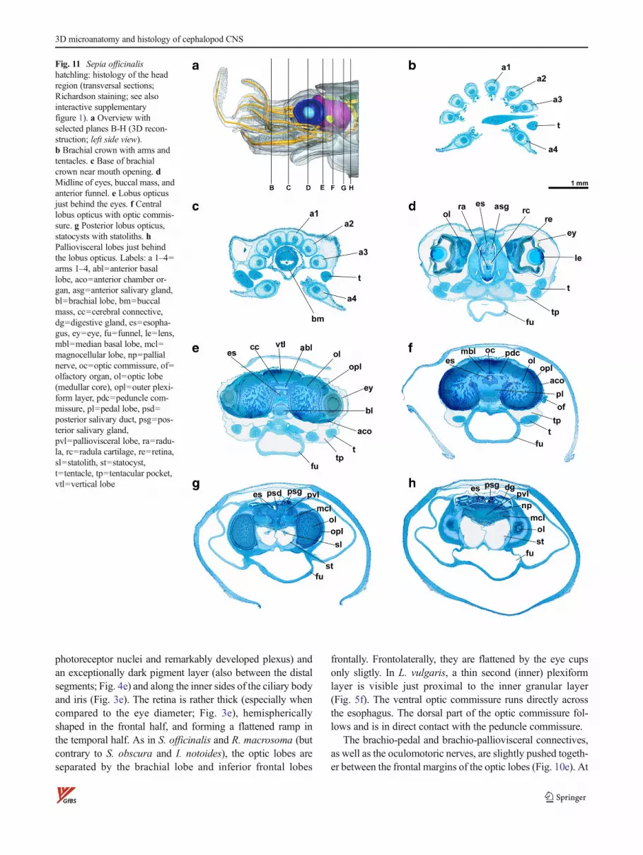
photoreceptor nuclei and remarkably developed plexus) andan exceptionally dark pigment layer (also between the distalsegments; Fig. 4e) and along the inner sides of the ciliary bodyand iris (Fig. 3e). The retina is rather thick (especially whencompared to the eye diameter; Fig. 3e), hemisphericallyshaped in the frontal half, and forming a flattened ramp inthe temporal half. As in S. officinalis and R. macrosoma (butcontrary to S. obscura and I. notoides), the optic lobes areseparated by the brachial lobe and inferior frontal lobes
frontally. Frontolaterally, they are flattened by the eye cupsonly sligtly. In L. vulgaris, a thin second (inner) plexiformlayer is visible just proximal to the inner granular layer(Fig. 5f). The ventral optic commissure runs directly acrossthe esophagus. The dorsal part of the optic commissure fol-lows and is in direct contact with the peduncle commissure.
The brachio-pedal and brachio-palliovisceral connectives,as well as the oculomotoric nerves, are slightly pushed togeth-er between the frontal margins of the optic lobes (Fig. 10e). At
Fig. 11 Sepia officinalishatchling: histology of the headregion (transversal sections;Richardson staining; see alsointeractive supplementaryfigure 1). a Overview withselected planes B-H (3D recon-struction; left side view).b Brachial crown with arms andtentacles. c Base of brachialcrown near mouth opening. dMidline of eyes, buccal mass, andanterior funnel. e Lobus opticusjust behind the eyes. f Centrallobus opticus with optic commis-sure. g Posterior lobus opticus,statocysts with statoliths. hPalliovisceral lobes just behindthe lobus opticus. Labels: a 1–4=arms 1–4, abl=anterior basallobe, aco=anterior chamber or-gan, asg=anterior salivary gland,bl=brachial lobe, bm=buccalmass, cc=cerebral connective,dg=digestive gland, es=esopha-gus, ey=eye, fu=funnel, le=lens,mbl=median basal lobe, mcl=magnocellular lobe, np=pallialnerve, oc=optic commissure, of=olfactory organ, ol=optic lobe(medullar core), opl=outer plexi-form layer, pdc=peduncle com-missure, pl=pedal lobe, psd=posterior salivary duct, psg=pos-terior salivary gland,pvl=palliovisceral lobe, ra=radu-la, rc=radula cartilage, re=retina,sl=statolith, st=statocyst,t=tentacle, tp=tentacular pocket,vtl=vertical lobe
3D microanatomy and histology of cephalopod CNS

the level of arm pair 3 on both sides, a nerve, running back-ward to the pedal lobes, escorts the superior antorbital nerves(Fig. 7e; to the right of “bl”). As soon as possible, the anteriorinferior oculomotor nerves diverge from the brachio-pedalconnectives, running alongside the inner surface of the opticlobes (Fig. 9e; noai) and bifurcate into and ventral and dorsalbranch (Fig. 7e; 9).
We have not discriminated between prebrachial andbrachial lobes in the anterior subesophageal mass as Young(1976b) did; the same is true for the pedal lobe. The
commissure between the subradular ganglia (Young 1965a,1971) was detectable only in outlines. The palliovisceral massconsists among others of prominent dorsolateral lobes (fin-and chromatophore lobes), and the crossing of the first-ordergiant fibers can be seen (Fig. 15h, arrowhead).
The inferior frontal lobes are proportionally bigger com-pared with the superior buccal lobes and as in R. macrosomapositioned frontally to the latter. As in S. officinalis, the buccaland frontal lobes of L. vulgaris lie close to each other, so thatthe cerebrobuccal connectives are quite short (Fig. 8e). Since
Fig. 12 Rossia macrosomahatchling: histology of the headregion (transversal sections;Trichrome de Masson staining,except f: Azan staining; see alsointeractive supplementaryfigure 2). a Overview withselected planes B-H (3D recon-struction; left side view).b Base of brachial crown witharms and tentacles. c Base of bra-chial crown with buccal mass. dMidline of eyes, radula. e Centrallobus opticus just behind the eyes.f Central lobus opticus with opticcommissure. g Posterior lobusopticus, statocysts with statoliths.h Palliovisceral lobes just at theend of the optic lobes. Labels: a 1–4=arms 1–4, aco=anterior cham-ber organ, bm=buccal mass, es=esophagus,ey=eye, fu=funnel, ibl=inferiorbuccal lobe, ifl=inferior frontallobe, le=lens, mcl=magnocellularlobe, oc=optic commissure, ofl=olfactory lobe, ol=optic lobe(medullar core), opl=outer plexi-form layer, pdl=peduncle lobe,pl=pedal lobe, pvl=pallioviscerallobe, ra=radula, re=retina, sl=statolith, st=statocyst,t=tentacle, tp=tentacular pocket,vtl=vertical lobe, ys=yolk sac
E. Wild et al.
the buccal and frontal lobes are also very close to the brachiallobe, the bucco-brachial and cerebrobrachial connectives areshort as well and appear fused at their entrance to the brachiallobe. The anterior basal lobes are divided into an anterior(ventral) and posterior (dorsal) part by a thin layer of cells(Fig. 15e; abl).
As in other decabrachian species investigated in this study,the profiles of the big statocysts (diameter 425 μm) appearslightly irregular due to some tissue shrinkage (Fig. 15h);statoliths are found in their frontal portion (Fig. 15g). The
olfactory organs (275×165×40 μm3) are located on bothsides ventrally to the optic lobes, i.e., well behind the eyes(Fig. 15f).
Octopus vulgaris
O. vulgaris is the only octobrachian species investigated inthis study and has the smallest hatchlings (Fig. 16, supplementinteractive Fig. 6; horizontal head diameter ca. 0.75 mm; seealso Fig. 2f). A crown of eight short arms (all around 550–
Fig. 13 Sepietta obscurahatchling: histology of the headregion (transversal sections;b,c,e,h: Azan staining; d,f,g:Trichrome de Masson staining;see also interactivesupplementary figure 3). aOverview with selected planes B-H (3D reconstruction; left sideview).b Brachial crown with arms andtentacles. c Beak and base of bra-chial crown. d Midline of eyes,brachial lobe. e Central lobusopticus just behind the eyes, an-terior funnel. f Lobus opticus withoptic commissure.g Posterior lobus opticus, stato-cysts with statoliths. h Stellateganglion, gills, digestive gland.Labels: a 1–4=arms 1–4, aco=anterior chamber organ,bk=beak, bl=brachial lobe, dbl=dorsal basal lobe, dg=digestivegland, es=esophagus, ey=eye,fu=funnel, g=gill, ifl=inferiorfrontal lobe, le=lens, mbl=medi-an basal lobe, np=pallial nerve,oc=optic commissure,of=olfactory organ, ol=optic lobe(medullar core), opl=outer plexi-form layer, pdc=peduncle com-missure, pdl=peduncle lobe, pl=pedal lobe, re=retina, sbl=supe-rior buccal lobe, sfl=superiorfrontal lobe, sg=stellate ganglion,sl=statolith, st=statocyst, t=ten-tacle, tp=tentacular pocket,vtl=vertical lobe
3D microanatomy and histology of cephalopod CNS

650 μm long) embraces the remainder of an outer yolk sac(Fig. 16a, b). The microarchitecture of the peripheral nervesystem of the arm crown resembles that of decabrachians,except it lacks tentacles. The integument is characterizedby numerous tiny spines (bunches of “Kölliker bristles”;Fig. 16b–h) that are less densely distributed in the cornea.
The eyes point ca. 45° forward (Fig. 2f) in a horizontalplane (Fig. 16d), suspended in a voluminous anterior cham-ber. Anterior chamber organs are underdeveloped, if present.The lens of the O. vulgaris hatchling (diameter 120 μm,distal segment ca. 28 %) has a comparatively small cortex(Fig. 3f), the vitreous body is of average appearance, and the
Fig. 14 Idiosepius notoideshatchling: histology of the headregion (transversal sections;Richardson staining; see alsointeractive supplementaryfigure 4). a Overview withselected planes B-H (3D recon-struction; left side view).b Brachial crown with arms andtentacles. c Base of brachialcrown near mouth opening, buc-cal mass. d Midline of eyes, bra-chial lobe. e Lobus opticus justbehind eyes. f Central lobusopticus with optic commissure,anterior funnel. g Posterior lobusopticus, statocysts with statoliths.h Palliovisceral lobes just behindthe lobus opticus, statocysts. La-bels: a 1–4=arms 1–4, bl=bra-chial lobe, bm=buccal mass, es=esophagus, ey=eye, fu=funnel,ifl=inferior frontal lobe, le=lens,mbl=median basal lobe, np=pal-lial nerve, oc=optic commissure,of=olfactory organ, ol=optic lobe(medullar core), opl=outer plexi-form layer, pdl=peduncle lobe,pl=pedal lobe,pvl=palliovisceral lobe, ra=radu-la, rc=radula cartilage, re=retina,sfl=superior frontal lobe, sl=statolith, st=statocyst, t=tentacle,tp=tentacular pocket
E. Wild et al.

retina is completely developed histogenetically with a darkpigment layer (Figs. 3f and 4f). In its 3D aspect, the retina isa hemispheric cup, slightly flattened rostrally. As inS. officinalis, the optic lobes are kept at distance by the mostfrontal parts of the CNS. Frontally, they are strongly dentedby the eye cups into a slightly concave shape (eyes notdeformed by the optic lobes in this species). Optic and
peduncle commissures were visible in histological sliceseven in the tiny specimen.
Subesophageal mass: In O. vulgaris, a suprabrachial com-missure is present (Fig. 7f; sbc), bridging the esophagusbetween the dorsal rims (related to arm pair 1) of thehorseshoe-shaped brachial lobe. The posterior part of thebrachial lobe is fused with the pedal lobes, and brachio-
Fig. 15 Loligo vulgarishatchling: histology of the headregion (transversal sections;Richardson staining; see alsointeractive supplementaryfigure 5). a Overview withselected planes B-H (3D recon-struction; left side view). b Bra-chial crown with arms and tenta-cles. c Base of brachial crownnear mouth opening. dMidline ofeyes, radula. e Lobus opticus justbehind the eyes, anterior basallobe. f Central lobus opticus withoptic commissure, pedal lobe.g Posterior lobus opticus, stato-cysts with statoliths. hPalliovisceral lobes just behindthe lobus opticus, statocysts. La-bels: a 1–4=arms 1–4, abl=ante-rior basal lobe, aco=anteriorchamber organ, doc=dorsal opticcommissure, es=esophagus, ey=eye, fu=funnel, ibl=inferior buc-cal lobe, le=lens, ipl=inner plex-iform layer, mcl=magnocellularlobe, noai=inferior anterioroculomotoric nerve,of=olfactory organ, ofl=olfactorylobe, ol=optic lobe (medullarcore), opl=outer plexiform layer,pdc=peduncle commissure, pdl=peduncle lobe, pl=pedal lobe,pvl=palliovisceral lobe, ra=radu-la, re=retina, sl=statolith,st=statocyst, svl=subverticallobe, t=tentacle, tp=tentacularpocket, voc=ventral optic com-missure, vtl=vertical lobe;arrowhead crossing of first-ordergiant axons
3D microanatomy and histology of cephalopod CNS
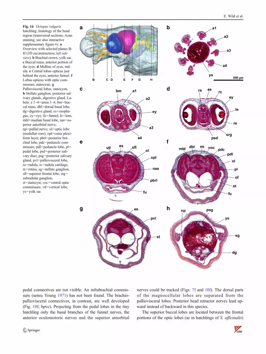
pedal connectives are not visible. An infrabrachial commis-sure (sensu Young 1971) has not been found. The brachio-palliovisceral connectives, in contrast, are well developed(Fig. 10f; bpvc). Projecting from the pedal lobes in the tinyhatchling only the basal branches of the funnel nerves, theanterior oculomotoric nerves and the superior antorbital
nerves could be tracked (Figs. 7f and 10f). The dorsal partsof the magnocellular lobes are separated from thepalliovisceral lobes. Posterior head retractor nerves lead up-ward instead of backward in this species.
The superior buccal lobes are located between the frontalportions of the optic lobes (as in hatchlings of S. officinalis)
Fig. 16 Octopus vulgarishatchling: histology of the headregion (transversal sections; Azanstaining; see also interactivesupplementary figure 6). aOverview with selected planes B-H (3D reconstruction; left sideview). b Brachial crown, yolk sac.c Buccal mass, anterior portion ofthe eyes. d Midline of eyes, rad-ula. e Central lobus opticus justbehind the eyes, anterior funnel. fLobus opticus with optic com-missure, statocysts. gPalliovisceral lobes, statocysts.h Stellate ganglion, posterior sal-ivary glands, digestive gland. La-bels: a 1–4=arms 1–4, bm=buc-cal mass, dbl=dorsal basal lobe,dg=digestive gland, es=esopha-gus, ey=eye, fu=funnel, le=lens,mbl=median basal lobe, nas=su-perior antorbital nerve,np=pallial nerve, ol=optic lobe(medullar core), opl=outer plexi-form layer, pbrl=posterior bra-chial lobe, pdc=peduncle com-missure, pdl=peduncle lobe, pl=pedal lobe, psd=posterior sali-vary duct, psg=posterior salivarygland, pvl=palliovisceral lobe,ra=radula, rc=radula cartilage,re=retina, sg=stellate ganglion,sfl=superior frontal lobe, srg=subradular ganglion,st=statocyst, voc=ventral opticcommissure, vtl=vertical lobe,ys=yolk sac
E. Wild et al.
and comparatively close to the superior frontal lobes in thisspecies. Cerebro-subradular connectives lead down to thesubradular ganglia, but a subradular commissure as describedby Marquis (1989) could not be found. The neuropils of thesuperior and posterior buccal lobes (the latter only found inoctobrachian species) are continuous, and only thincerebrobuccal connectives lead dorsally straight to the inferiorfrontal lobes (Fig. 8f). Because of the low distance betweenthe brachial lobe and the buccal and frontal lobes also, thebucco-brachial and the cerebrobrachial connectives are veryshort and appear as a fused axone bundle. The partitioning ofthe superior frontal lobes into lateral and median componentsis visible. The vertical lobe has a very characteristic shapewith five lobules (sensu Nixon and Young 2003; see Fig. 2f:vtl @ 200 % or interactive suppl. Fig. 6) in contrast to thedome-shaped vertical lobes in the decabrachian species.Embedded in the pericaryal layer between superior frontaland posterior buccal lobes, the tiny neuropils of the subfrontallobes can be detected (Fig. 8f; sftl) which are not easilydelineated from the precommissural lobe.
The posterior buccal lobes are directly connected with thesubvertical lobes, corresponding to the cerebral connectives ofthe decabrachian species. Conspicuous subpedunculate lobesare found on top of the dorsal basal lobes.
The statocysts of the O. vulgaris hatchling have circularprofiles in tangential sections (i.e., unaffected by any shrink-age; max. diameter 280μm; Fig. 16f, g) and are oval shaped inside view, slightly tapered at both ends. The olfactory organs(115×100×35 μm3) are placed directly behind the optic lobesjust below their equator.
Discussion
New quality of structure data
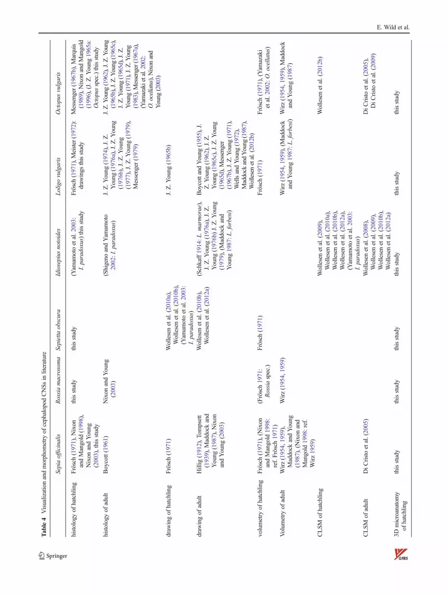
The present study provides new structural and volumetric dataof the CNS of the hatchlings of six species of coleoid cepha-lopods in light microscopical resolution (Table 4). Based ontransversal section series (i.e., complete 3D data sets) throughthe head novel computer-aided 3D reconstructions ofneuropils have been created, providing virtually explorabledigital surfacemodels. By these twomethodologies, a descrip-tion and comparison of the histology and neuropil microanat-omy of the cephalopod CNS are possible in so far unmatcheddetail. With arbitrary choice of cutting planes (raw data) andperspectives (interactive suppl. Figs. 1–6), the sterically com-plex structures get transparent and much better comprehensi-ble compared with a display of selected 2D data. In addition,digital morphometry of the segmented structures renders pos-sible interspecific comparisons of absolute and relative vol-umes for the description of allometry with high precision (see
supplement “volumetric analysis”). This finally enables toappraise the biological relevance of individual lobes of theCNS in the context of adaptative radiation and evolution of thespecies-specific modes of life.
Histological image data of the head region based onsemithin sections are presented for I. notoides, R. macrosoma,and S. obscura in this study for the first time. However, forI. notoides, the gross anatomy of the CNS and the distributionof certain neurotransmitters have been scrutinized before,employing 100–150-μm vibratome sections (Wollesen et al.2008, 2009, 2010a, 2012a). For the hatchlings of S. officinalisand L. vulgaris, the histological series have few micrometerresolution in tangential and axial dimensions (Table 3),expanding a previous approach employing sagittal sections of7–10-μm thickness (Frösch 1971). Within the tiny hatchlings,even details like the interbrachial commissures can be traced(see also Graziadei 1971: Fig. 3.12), the same is true, e.g., forthe thin cerebro-subradular connectives or the inner plexiformlayer in the lobus opticus of L. vulgaris. For Octopus hatch-lings, only few selected histological planes (Messenger 1967b;plate 27 Fig. 3; Marquis 1989; Figs. 3+4) or vibratomesections (Wollesen et al. 2012b; Figs. 6+7) have beenpublished so far.
Concerning (pseudo-)3D-rendering approaches, alreadyFrösch (1971) attempted to display the subesophageallobes of cephalopod hatchlings in perspectivic drawings,however, with schematically simplified geometry (Frösch1971; Figs. 5 and 6; see also Hillig 1912: plts. 33, 34;Richter 1913; Schkaff 1914; Pfefferkorn 1915; Tompsett1939: Figs. 55–58; Young 1971; Fig. 1.5, 1.6; Young1976b; Figs. 1+2). Due to improved computer-aided im-aging technology, the renderings presented here enablesubstantially more realistic 3D views with several options:free choice of perspective, zoom factor, color and trans-parency, cutting planes where appropriate, and free com-position of substructures (see, e.g., Figs. 7 and 8). Thereader is encouraged to explore the virtual brains of ceph-alopod hatchlings provided in PDF format (supplementFigs. 1–6) in order to learn about their morphology andto become familiar with the advantages of interactivelyexplorable surface models.
Comparative histology and microanatomy
The comparison of the histological and microanatomicalfindings of the present study on the eyes and CNS withpublished data is problematic, since data are often hetero-geneous, incomplete, of varying quality, or not existent forcertain species (Table 4).
The eye structure is largely constant in coleoid cephalopods(except oegopsids without cornea; Budelmann et al. 1997;Young and Vecchione 2004). Accordingly, the eyes of theinvestigated hatchlings are quite similar, and differences
3D microanatomy and histology of cephalopod CNS
Tab
le4
Visualizationandmorphom
etry
ofcephalopod
CNSsin
literature
Sepiaofficinalis
Rossiamacrosoma
Sepietta
obscura
Idiosepius
notoides
Loligovulgaris
Octopus
vulgaris
histologyof
hatchling
Frösch(1971),N
ixon
andMangold
(1998),
Nixon
andYoung
(2003),thisstudy
thisstudy
thisstudy
(Yam
amotoetal.2003:
I.paradoxus)thisstudy
Frösch
(1971),M
eister(1972):
draw
ings
thisstudy
Messenger(1967b),Marquis
(1989),N
ixon
andMangold
(1996),(J.Z.Y
oung
1965a:
Octopus
spec.)thisstudy
histologyof
adult
Boycott(1961)
Nixon
andYoung
(2003)
(Shigeno
andYam
amoto
2002:I.paradoxus)
J.Z.Y
oung
(1974),J.Z
.Young
(1976a),J.Z.Y
oung
(1976b),J.Z.Y
oung
(1977),J.Z
.Young
(1979),
Messenger
(1979)
J.Z.Y
oung
(1962),J.Z
.Young
(1965b),J.Z.Y
oung
(1965c),
J.Z.Y
oung
(1965d),J.Z.
Young
(1971),J.Z
.Young
(1983),M
essenger(1967a),
(Yam
azakietal.2002:
O.ocellatus),N
ixon
and
Young
(2003)
draw
ingof
hatchling
Frösch(1971)
Wollesenetal.(2010a),
Wollesenetal.(2010b),
(Yam
amotoetal.2003:
I.paradoxus)
J.Z.Y
oung
(1965b)
draw
ingof
adult
Hillig
(1912),T
ompsett
(1939),M
addock
and
Young
(1987),N
ixon
andYoung
(2003)
Wollesenetal.(2010b),
Wollesenetal.(2012a)
(Schkaff1914:L
.marmorae),
J.Z.Y
oung
(1976a),J.Z.
Young
(1976b)J.Z.Y
oung
(1979),(Maddock
and
Young
1987:L
.forbesi)
BoycottandYoung
(1955),J.
Z.Y
oung
(1962),J.Z
.Young
(1965c),J.Z.Y
oung
(1965d),Messenger
(1967b),J.Z.Y
oung
(1971),
WellsandYoung
(1972),
Maddock
andYoung
(1987),
Wollesenetal.(2012b)
volumetry
ofhatchling
Frösch(1971),(Nixon
andMangold
1998:
ref.Frösch
1971)
(Frösch1971:
Rossiaspec.)
Frösch
(1971)
Frösch(1971)
Frösch(1971),(Yam
azaki
etal.2002:
O.ocella
tus)
Volum
etry
ofadult
Wirz(1954,1959),
Maddock
andYoung
(1987),(Nixon
and
Mangold
1998:ref.
Wirz1959)
Wirz(1954,1959)
Wirz(1954,1959),(M
addock
andYoung
1987:L
.forbesi)
Wirz(1954,1959),Maddock
andYoung
(1987)
CLSM
ofhatchling
Wollesenetal.(2009),
Wollesenetal.(2010a),
Wollesenetal.(2010b),
Wollesenetal.(2012a),
(Yam
amotoetal.2003:
I.paradoxus)
Wollesenetal.(2012b)
CLSM
ofadult
DiC
ristoetal.(2005)
Wollesenetal.(2008),
Wollesenetal.(2009),
Wollesenetal.(2010b),
Wollesenetal.(2012a)
DiC
ristoetal.(2005),
DiC
ristoetal.(2009)
3Dmicroanatom
yof
hatchling
thisstudy
thisstudy
thisstudy
thisstudy
thisstudy
thisstudy
E. Wild et al.

pertain, e.g., to the size of the anterior eye chamber. Thelatter is largest in S. officinalis and R. macrosoma, mediumsized in O. vulgaris, and barely perceptible in the otherspecies. Also, the retina follows a fixed bauplan, whereasthe proportions of the single layers may be designed dif-ferently. In R. macrosoma, the distal photoreceptor seg-ments are comparatively short, which probably results ina comparatively low photon yield at the time of hatching(see also Bozzano et al. 2009; Sepioteuthis australis). Fulllength will often be reached later in development, as shownfor Sepia esculenta (Hao et al. 2010) or Sepiella japonica(Yamamoto 1985). A high amount of pigment granulesbetween the photoreceptor distal segments (L. vulgaris;Fig. 4e, and to a lesser degree also I. notoides andO. vulgaris) suggests diurnal activity or at least fixationin a light-adapted state. Concerning the bipartite cephalo-pod lens (Willekens et al. 1984; West et al. 1995), thedistal component of the O. vulgaris hatchling is still quitesmall. This is astonishing in so far, as the developmentaltime is not much shorter in this species (Mangold andBoletzky 1973; Villanueva et al. 1995). During subsequentdevelopment, the vitreous body enlarges, the retina thicknessincreases only a little, the ciliary body gets more complex, andthe anterior lens segment catches up (see e.g., Nixon andYoung 2003; Fig. 31.24). Differences in lens diameter affectachievable resolution and sensitivity in favor of the larger ones(i.e., Sepioids and I. notoides). Differences in the alignment ofoptic axes have to be interpreted carefully, because relaxationor fixation artefacts (e.g., of the eye muscles) cannotcompletely ruled out. The slightly forward orientation at leastsupports the idea of binocular vison, as it is done by formerbehavioral experiments (Wells 1958; Collewijn 1970).
The optic lobes of all investigated hatchlings are composedof a cortex with outer plexiform layer and a medulla with itsintricate neuropil mesh. In L. vulgaris, an additional innerplexiform layer is present, and in outlines also visible inS. officinalis but not in the other species investigated in thisstudy. According to Young (1974), this layer is a decabrachiancharacter (but see Budelmann et al. 1997) and should bedeveloped more or less pronounced also in R. macrosoma,S. obscura, and I. notoides (see, e.g., Wollesen et al. 2008;Fig. 2c). The structure of the medullar neuropil meshes iscompared in Fig. 5d—it is least developed in the O. vulgarishatchling. In the investigated I. notoides, a condition betweenthe stage 30 hatchling (Wollesen et al. 2010a; Fig. 7j) and theadult is found (Wollesen et al. 2008; Fig. 2f; Fig. 14e, f in thepresent study).
“Optic glands” (see, e.g., Messenger 1967b) could not befound in any hatchling. This corresponds with their endocrinenature to control sexual maturation (Wells andWells 1959; LeGall et al. 1988) and hence by their late formation (Bonichon1967), even though Wollesen et al. (2010a) could detect theprecursor cells via InFMRF expression in the hatchling of
I. notoides. In adult animals, they are situated behind the eyesnear the optic tract and innervated by the subpedunculatelobes in Octopus (Young 1971; Frösch 1974) or dorsal basallobes in Sepia (Boycott 1961; perhaps also olfactory lobes; LeGall et al. 1988).
Concerning the morphology of the CNS, the results de-scribed here generally corroborate the few published structuraldata on cephalopod hatchlings (e.g., Young 1965a; Fig. 1;Messenger 1967b; Fig. 3; Frösch 1971; Figs. 3, 4, and 6;Marquis 1989, e.g., Figs. 3, and 4; Nixon and Mangold1996; plate 1b; Nixon and Mangold 1998; Fig. 2; Nixon andYoung 2003; Figs. 5.10a, 31.8; Yamamoto et al. 2003; Figs. 8,and 9c-h; Wollesen et al. 2010b; Figs. 6c–e, 7g–j; Wollesenet al. 2012b; Figs. 6 and 7). The architecture of subesophagealand supraesophageal masses and the constellation and con-nectivity of their lobes is consonant with the coleoid CNSdesign, however, with some species-specific characteristics.The CNS of cephalopod hatchlings or paralarvae alreadyresembles the “adult” condition: In the histological sections,all important lobes and most of their emerging nerves can befound.
The neuropils of I. paradoxus emerge and grow upbetween the perikarya already in embryonal stage 22 andreach adult-like proportions at stage 30 (=hatchling)(Yamamoto et al. 2003). Albeit small, the adult-like archi-tecture already supports autonomous survival before theaccessory lobes are terminally differentiated (i.e., verticalsystem; see, e.g., Yamamoto et al. 2003; Idiosepius;Marquis 1989; Octopus; Shigeno et al. 2001a, b, c, d;teuthoid squids), and before the neuron numbers andneuropil ratios reach their final condition (if at all, becausethere is a smooth transition from juvenile to adult in an ever-growing animal; Frösch 1971). Compared to hatchlings ofO. vulgaris, the vertical lobe systems of S. officinalis,I. notoides, and to a lesser extend L. vulgaris are well devel-oped suggesting that complex cognitive abilities are manifest-ed already during this early stage (Young 1979; present study).
Nixon and Young (2003; Fig. 2.2) compare the outlines ofadult CNSs in lateral view including Sepia, Loligo, andOctopus. Sepiola represents Sepiolida, and indeed, the outlineshows some similarity with the lateral views of R. macrosoma(Fig. 8b) and S. obscura hatchlings (Fig. 8c). The character-istic flat and elongate shape of the adult Loligo CNS and thecompressed and high profile of the Octopus CNS is similar inhatchlings as well. Since the relative buccal lobe volumes areabout 2.2 % in all hatching sepiolids and S. officinalis (presentstudy), the notion that these lobes are smaller in adult sepiolidscompared to adult S. officinalis cannot be confirmed forhatchlings (Frösch 1971; present study). Frösch (1971) reportseven slightly bigger inferior buccal lobes in S. obscura than inS. officinalis.
An increasing distance between buccal and frontal lobesand between brachial and pedal lobes in the sequence
3D microanatomy and histology of cephalopod CNS
Octopodida > Sepiida > Sepiolida > Teuthida is documentedin both adult lobes (Nixon and Young 2003; Fig. 2.2) andhatchling neuropils (this study, Figs. 7 and 8). Also, I. notoidesfits in this pattern, and as in S. obscura and L. vulgaris, theoptic lobes of both sides converge anteroventrally so much,which the brachiopedal connectives are compressed. Thiscondition can be interpreted as a simple consequence of stericconstraints resulting from shortage of space in the head,concurrent with an elongate, finally torpedo-shaped body inthe nectonic teuthids. Other way round, the large S. officinalishatchling with the most complex CNS microarchitecture hasthe shortest brachiopedal and cerebrobuccal connectives ofthe investigated decabrachian species (not compressed as inR. macrosoma). This condition is interpreted as a tendencytoward brain centralization with the advantage of faster infor-mation processing (shorter distances), since behavioral reac-tions follow faster on stimulus perception, boosting, e.g.,predation success (Budelmann 1995). Generally, centraliza-tion of the CNS was assumed to be associated with a morecomplex behavioral repertoire (Nixon and Young 2003; seealso Wells 1958; Hanlon and Messenger 1988; Nixon andMangold 1998), culminating in the “intelligence” of adultoctopods. On the contrary, also theutids show highly complexschooling and mating behavior (Gilbert et al. 1990; Hanlonand Messenger 1996; Norman 2003). Difficult keeping con-ditions for these pelagic animals and hence comparatively fewbehavioral experiments may entail serious undervaluation oftheir intelligence.
A peculiarity of S. officinalis, according to Nixon andMangold (1998) is the high chromatophore number alreadyin the hatchling, which (in contrast to the transparent plankticOctopus paralarvae) does not essentially increase toward theadult, and a mature chromatic behavior (Hanlon andMessenger 1996). Correspondingly, we find considerably de-veloped chromatophore lobes in this species (Fig. 2a).
The investigated I. notoides is very similar to the I. paradoxushatchling (developmental stage 30 of Yamamoto et al. 2003),albeit slightly further developed, because it was fixed a fewdays after hatching. As these authors, we could not identifygiant axons in our material in light microscopic resolution (butsee Fig. 15h and Martin 1965 for L. vulgaris and Martin andRungger 1966 for S. officinalis). In contrast to I. paradoxus,the investigated I. notoides has already a better developedvertical lobe. However, contrary to adult I. paradoxus(Shigeno and Yamamoto 2002), dorsolateral commissuresand additional olfactory lobules could not yet be detected. Incontrast to the other investigated species, the hatchlings ofI. notoides and L. vulgaris possess an unpaired middle funnelnerve (Young 1976b; Shigeno and Yamamoto 2002) in addi-tion to the paired anterior funnel nerves.
The CNS of O. vulgaris, being the only octobrachianhatchling in this study, shows some specific characters. Anadvanced degree of lobe condensation is manifested in the
fusion of brachial and pedal lobes in which brachio-pedalconnectives disappear and a suprabrachial commissureremains present. As a consequence of a convergence ofsuperior buccal lobes and inferior frontal lobes, thecerebrobuccal connectives disappear. Young (1971) de-scribes the lobes just behind the superior buccal lobes asposterior buccal lobes (see our Fig. 8f) and—characteristicof Octobrachia—with small, dorsally outgrowing “inferiorfrontal lobes.” It was stated that the posterior buccal lobes(sending out the cerebrobrachial connectives) are homolo-gous to the inferior frontal lobes of the Decabrachia, and theposterior-buccal-to-subvertical tracts (Young 1965b) arehomologs of the cerebral connectives leading to thesubvertical lobes. The subfrontal lobes are proportionallymuch smaller than those in the adult and hence hardlydetectable. Although the vertical lobe system is prematurein the hatchling, its five gyri (Boycott and Young 1955;Young 1971) can be shown up already in the neuropil(Fig. 2f; vtl @ 200 % or interactive Fig. 6).
Structure and lifestyle
Taking account of the diverging eidonomy of the investigatedspecies and the abovementioned functional conclusionsconcerning the CNS, some relations between brain morphol-ogy and lifestyle of the hatchlings (Table 2) can be derived, asit was already done by Maddock and Young (1987) for adultcephalopods: The nervous system of coleoid cephalopods isorganized according to a general blueprint, though there areadaptations to environment and behavior which are reflectedin the design of the central nervous system.” Frösch (1971) aswell as Nixon and Mangold (1996) assume that in particular,the volume of brain lobes should reflect habitat and behaviorto some degree (see also discussion in supplement “volumet-ric analysis”). Bozzano et al. (2009) find a close relationbetween morphology and function of the visual system andemphasize the importance of its development in due time forlater survival. Considerations of that kind are publishedalso for other animal groups, e.g., for the tectum opticumof teleost species wich differ regarding to the importanceof their visual sense (Bone and Marshall 1985; Fig. 10.2)or for the proportions of visual and olfactory CNS in thekiwi (Martin et al. 2007; Fig. 2).
Least significant may be the outer “shape” of the CNS(Fig. 2) mainly imposed by the overall body shape. Anyway,there are differences between slim pelagic forms (Loligo,Idiosepius) and compact benthic forms (Sepia, Rossia), whereatthe sterical demands of the eyes and optic lobes seem to play adecisive role (e.g., Rossia vs Sepietta; Figs. 7 and 10). Quiteearly, the lobe proportions are reminiscent of the adult situation(e.g., the tiny Octopus hatchling already reveals the centraliza-tion characteristic for Octobrachia), but the CNS shape willchange during ontogeny in parts with the changing weighting
E. Wild et al.

of functionalities (as a rule, the relative size of brachial andvertical lobes increases, whereas the basal and sometimes opticlobes are bigger in the hatchlings; Wirz 1954; Maddock andYoung 1987; Nixon and Mangold 1998).
The total size of the CNS (see supplement “volumetricanalysis” Table 1) is correlated to lifestyle via body size.The small pelagic hatchlings ofO. vulgaris are equipped withall brain regions important for subsequent benthic life but withcomparatively few cells proportional to the still small numberof sensory and effectory cells under control. The plankticstage is little proactive requiring little computational power,e.g., for arm movements or chromatophore control. For awhile, it is passively drifting and lecithotrophic, structurallyreflected in the short arms and a residual outer yolk sac ofthe investigated specimen (Fig. 16a, b). Nonetheless, thenervous system has to be well-prepared at the transition tothe final benthic lifestyle for swimming, crawling, hiding,camouflage, and visual hunting. In contrast, large hatchlingsare allready able to hide from predators (camouflage,entrenching, escaping) and to feed autonomously almostimmediately after hatching (Wells 1958; Poirier et al. 2004)after a short behavioral “training/maturation” period. Afterdepleting the yolk reservoirs, they actively hunt and feed,which requires visual prey recognition, control of body pos-ture, and the coordination of all muscle groups involved(Maddock and Young 1987). Here, the anterior basal lobesplay an important role for head and eye movements and theposterior basal lobes for swimming. All these skills have to bepreadapted during embryogenesis, even though not essentialwithin the well protected (and narrow) egg milieu.
The cephalopod hatchling with the best-known lifestyle isS. officinalis with an “adult-like” behavioral repertoire(Hanlon and Messenger 1996; Nixon and Mangold 1998).Equipped with the characters described above, the hatchlingsof this species should have comparatively sharp vision withcomplex image processing, good swimming abilities and ten-tacle use, and background-correlated chromatophore controlfor effective camouflage.
Concerning the other sepioids investigated here, similar as-sumptions appear admissible, because they have likewise largehatchlings, differing even less from the adults because they donot grow as much as Sepia. Some constraints concerning nav-igation are owed to their more cryptic lifestyle. In R. macrosomagood vision (large eyes, high lobus opticus-to-retina volumeratio), differenciated arm coordination (large brachial lobes)and olfaction (comparatively big olfactory neuropils) are pres-ent. Also, S. obscura hatches with large eyes and large opticlobes, and their pedal lobe neuropils are developed slightlybeyond average. Like R. macrosoma, this species has a benthiclifestyle with self-entrenching behavior and visual hunting thatrequires complex motoric competences and good vision.
After hatching, I. notoides enters the water column(Boletzky 2003; Yamamoto et al. 2003). Its well-developed
visual system with pronounced outer plexiform layer givesevidence for advanced vision with higher order visual pro-cessing in the vertical lobe (or even some learning and mem-ory capabilites), as well as good arm and general motoriccompetences. In this developmental stage, most probably,the arms play a dominant role for hunting before the tentaclesare fully developed.
In the holopelagic L. vulgaris, we find pronounced posteriorbasal and peduncle lobe neuropils for the coordination of swim-ming movements, color change, and ink ejection (Table 1),whereas the brachial lobes are less developed. The arm motorfunction seems to play a comparatively less important role andapart from good vision olfaction seems to be an importantsensory cue (large olfactory lobe neuropils). The presence ofdense dark screening pigment in the retina of L. vulgaris andO. vulgaris fits to the pelagic, all-day active pelagic life.
For O. vulgaris, the volumetric data suggest that photore-ception and central processing of visual information still playa minor role in the (early?) planktic phase, whereas olfaction,the motoric control of the mantle and arms, and the processingof their chemo-tactile information are well prepared on theneuroanatomical side. Obviously, inferior frontal, brachial(and palliovisceral) lobe neuropils are outstandingly developed(supplement “volumetric analysis” Fig. 1) in Octobrachiaalready in the hatchlings, to support the complex arm motorfunction characteristic for this cephalopod group (Wirz 1954;Budelmann 1995).
Conclusions and outlook
3D imaging and volumetric analysis of CNS provides reliablemorphological characters for functional interpretations(even if information about the lifestyle of a given speciesis lacking) and for comparative work on the adaptiveradiation of an animal group into different (etho-)ecologicalniches. Cephalopod hatchlings are preadapted to the re-quirements of their habitat and habits, and the fine-tuningis reflected in morphological differences of the eyes, opticlobes, and various CNS neuropils. In future, this approachcan be refined by consistent segmentation of lobes intoneuropil and pericarya fractions (or even by single cellcounting; see, e.g., Heß 2007) and by use of faster rawdata acquisition via CLSM imaging of vibratome sectionseries (see, e.g., Wollesen et al. 2012b: Figs. 6+7, distinc-tion between neuropils and pericarya regions in principle ispossible) if a z-resolution of around 50 μm is tolerable, orvia μCT (see, e.g., Kerbl et al. 2013).
Acknowledgments We are verymuch obligued to Sigurd von Boletzkyfor providing us with some of his section series and for encouraging us touse them for 3D reconstructions. Furthermore, we want to express ourgratitude to Heidemarie Gensler for expert technical assistance and for
3D microanatomy and histology of cephalopod CNS
Anastasia de Motte for improving the English. The study was partiallysupported by a grant to EW by the Bayerische Eliteförderung.
References
Archer, S. N., Djamgoz, M. B. A., Loew, E. R., Partridge, J. C., &Vallerga, S. (1999). Adaptive mechanisms in the ecology of vision(p. 680). Dordrecht: Kluwer Academic Publishers.
Boletzky, S. V. (1974). The “larvae” of Cephalopoda: a review. ThalassiaJugoslavica, 10, 45–76.
Boletzky, S. V. (2003). Biology of early life stages in cephalopod mol-luscs. Advances in Marine Biology, 44, 143–203.
Boletzky, S.V.,&Boletzky,M.V.V. (1973). Observations on the embryonicand early post-embryonic development of Rossia macrosoma(Mollusca, Cephalopoda). Helgolaender Meeresuntersuchungen, 25,135–161.
Boletzky, S. V., Boletzky, M. V. V., Froesch, D., & Gätzi, V. (1971).Laboratory rearing of Sepiolinae (Mollusca, Cephalopoda).MarineBiology, 8, 62–87.
Boletzky, S. V., Fioroni, P., & Guerra, A. (1997). Proceedings of thesecond international symposium on functional morphology of ceph-alopods. Vie et milieu - Life and environment, 47(2), 187.
Bone, Q., & Marshall, N. B. (1985). Biologie der Fische. Stuttgart:Gustav Fischer Verlag.
Bonichon, A. (1967). Contribution à l´etude de la neurosécretion et de l´endocrinologie chez les Céphalpodes. I. Octopus vulgaris. Vie etmilieu - Life and environment, 18, 27–263.
Boycott, B. B. (1961). The functional organization of the brain of thecuttlefish Sepia officinalis. Proceedings of the Royal Society B:Biological Sciences, 153, 503–534.
Boycott, B. B., & Young, J. Z. (1955). A memory system in Octopusvulgaris Lamarck. Proceedings of the Royal Society B: BiologicalSciences, 153, 503–534.
Bozzano, A., Pankhurst, P. M., Moltschaniwskyj, N. A., & Villanueva, R.(2009). Eye development in southern calamary, Sepioteuthisaustralis, embryos and hatchlings. Marine Biology, 156, 1359–1373.
Budelmann, B. U. (1995). The cephalopod nervous system: what evolu-tion has made of the molluscan design. In O. Breidbach & W.Kutsch (Eds.), The nervous system of Invertebrates: an evolutionaryand comparative approach (pp. 115–138). Basel: Birkhäuser.
Budelmann, B. U., Schipp, R., & Boletzky, S. V. (1997). Cephalopoda. InF. W. Harrison & A. J. Kohn (Eds.), Microscopic anatomy ofinvertebrates (Vol. 6A: Mollusca II, pp. pp. 119–pp. 414). NewYork: Wiley-Liss.
Claes, M. F. (1996). Functional morphology of the white bodies of thecephalopod mollusc Sepia officinalis. Acta Zoologica (Stockholm),77, 173–190.
Collewijn, H. (1970). Oculomotor reactions in the cuttlefish, Sepiaofficinalis. The Journal of Experimental Biology, 52(2), 369–384.
Collin, S. P., & Partridge, J. C. (1996). Fish vision: retinal specializations inthe eyes of deep-sea teleosts. Journal of Fish Biology, 49, 157–174.
Di Cristo, C., Van Minnen, J., & Di Cosmo, A. (2005). The presence ofAPGWamide in Octopus vulgaris: a possible role in the reproduc-tive behavior. Peptides, 26(1), 53–62.
Di Cristo, C., De Lisa, E., & Di Cosmo, A. (2009). Control of GnRHexpression in the olfactory lobe of Octopus vulgaris. Peptides,30(3), 538–544.
Dietl, M. J. (1878). Untersuchungen über die Organisation des Gehirnswirbelloser Thiere (Cephalopoden, Tethys). Sitzungsberichte derAkademie der Wissenschaften in Wien, 77, 481–533.
Fishelson, L., Ayalon, G., Zverdling, A., & Holzman, R. (2004).Comparative morphology of the eye (with particular attention to the
retina) in various species of cardinal fish (Apogonidae, Teleostei). TheAnatomical Record.Part A. Discoveries in Molecular, Cellular, andEvolutionary Biology, 277(2), 249–261.
Frösch, D. (1971). Quantitative Untersuchungen am Zentralnervensystemder Schlüpfstadien von zehnmediterranen Cephalopodenarten. RevueSuisse de Zoologie, 57, 1069–1122.
Frösch, D. (1974). The subpedunculate lobe of the octopus brain: evi-dence for dual function. Brain Research, 75(2), 277–285.
Gilbert, D. L., Adelman, W. J., & Arnold, J. M. (1990). Squid asexperimental animals (p. 548). New York: Plenum Press.
Graziadei, P. (1971). The nervous system of the arms. In J. Z. Young(Ed.), The anatomy of the nervous system of Octopus vulgaris (pp.45–61). Oxford: Claredon Press.
Groeger, G., Cotton, P. A., &Williamson, R. (2005). Ontogenetic chang-es in the visual acuity of Sepia officinalis measured using theoptomotor response. Canadian Journal of Zoology, 83(2), 274–279.
Groeger, G., Chrachri, A., &Williamson, R. (2006). Changes in cuttlefishretinal sensitivity during growth. Vie et Milieu, 56(2), 167–173.
Hanlon, R. T., & Messenger, J. B. (1988). Adaptive coloration in youngcuttlefish (Sepia officinalis L.): the morphology and development ofbody patterns and their relation to behaviour. PhilosophicalTransactions of the Royal Society, B: Biological Sciences, 320,437–487.
Hanlon, R. T., &Messenger, J. B. (1996). Cephalopod behaviour (p. 232).Cambridge: Cambridge University Press.
Hao, Z.-L., Zhang, X.-M., Kudo, H., & Kaeriyama, M. (2010).Development of the retina in the cuttlefish Sepia esculenta.Journal of Shellfish Research, 29(2), 463–470.
Heß, M. (2007). Semi-automated mapping of cell nuclei in 3D-stacksfrom optical-sectioning microscopy. In P. Perner & O. Salvetti(Eds.), Advances in mass data analysis of signals and images inmedicine, biotechnology and chemistry (Lecture Notes in ComputerScience (Vol. 4826, pp. pp. 156–pp. 164). Berlin: Springer.
Hillig, R. (1912). Das Nervensystem von Sepia officinalis L. Zeitschriftfür Wissenschaftliche Zoologie, 101, 736–800.
Jereb, P., & Roper, C. F. E. (2005). Cephalopods of the world: anannotated and illustrated catalogue of cephalopod species knownto date. Volume 1. Chambered nautiluses and sepioids (Nautilidae,Sepiidae, Sepiolidae, Sepiadariidae, Idiosepiidae and Spirulidae)(Vol. 4(1), FAO species catalogue for fishery purposes). Rome:FAO. 262 p.
Jereb, P., & Roper, C. F. E. (2010). Cephalopods of the world: anannotated and illustrated catalogue of cephalopod species knownto date. Volume 2. Myopsid and oegopsid squids (FAO Speciescatalogue for fishery purposes, Vol. 4(2)). Rome: FAO. 605 p.
Kerbl, A., Handschuh, S., Nödl, M.-T., Metscher, B., Walzl, M., &Wanninger, A. (2013). Micro-CT in cephalopod research:Investigating the internal anatomy of a sepiolid squid using anon-destructive technique with special focus on the ganglionicsystem. Journal of Experimental Marine Biology and Ecology,447, 140–148.
Kunze, P. (1972). Comparative studies of arthropod superposition eyes.Journal of Comparative Physiology. A, Neuroethology, Sensory,Neural, and Behavioral Physiology, 76(4), 347–357.
Lauder, G. V. (1981). Form and function: structural analysis in evolution-ary morphology. Paleobiology, 7(4), 430–442.
Le Gall, S., Feral, C., Van Minnen, J., & Marchand, C. R. (1988).Evidence for peptidergic innervation of the endocrine optic glandin Sepia by neurons showing FMRFamide-like immunoreactivity.Brain Research, 462(1), 83–88.
Lythgoe, J. N. (1979). The ecology of vision (p. 270). Oxford: ClarendonPress.
Maddock, L., & Young, J. Z. (1987). Quantitative differences among thebrains of cephalopods. Journal of Zoology, 212(4), 739–767.
Makino, A., & Miyazaki, T. (2010). Topographical distribution of visualcell nuclei in the retina in relation to the habitat of five species of
E. Wild et al.
Decapodiformes (Cephalopoda). Journal of Molluscan Studies,76(2), 180–185.
Mangold, K., & Boletzky, S. (1973). New data on reproductive biologyand growth of Octopus vulgaris. Marine Biology, 19(1), 7–12.
Marquis, F. (1989). Die Embryonalentwicklung des Nervensystems vonOctopus vulgaris Lam. (Cephalopoda. Octopoda), eine histologischeAnalyse. Verhandlungen der naturforschenden Gesellschaft Basel,99, 23–76.
Martin, R. (1965). On the structure and embryonic development of thegiant fibre system of the squid Loligo vulgaris. Zeitschrift fürZellforschung und Mikroskopische Anatomie, 67, 77–85.
Martin, R., & Rungger, D. (1966). Zur Struktur und Entwicklung desRiesenfasersystems erster Ornung von Sepia officinalis L.(Cephalopoda). Zeitschrift für Zellforschung und MikroskopischeAnatomie, 74, 454–463.
Martin, G. R., Wilson, K.-J., Wild, J. M., Parsons, S., Kubke, M. F., &Corfield, J. (2007). Kiwi forego vision in the guidance of theirnocturnal activities. PLoS ONE, 2(2), e198.
Meister, G. (1972). Organogenese von Loligo vulgaris Lam. (Mollusca,Cephalopoda, Teuthoidea, Myopsida, Loliginidae). ZoologischeJahrbücher. Abteilung für Anatomie und Ontogenie der Tiere, 59,247–300.
Messenger, J. B. (1967a). The effects on locomotion of lesions to theviso-motor system in Octopus. Proceedings of the Royal Society B:Biological Sciences, 167, 252–281.
Messenger, J. B. (1967b). The peduncle lobe: a viso-motor centre inOctopus. Proceedings of the Royal Society B: Biological Sciences,167, 225–251.
Messenger, J. B. (1968). The visual attack of the cuttlefish, Sepiaofficinalis. Animal Behaviour, 16(2), 342–357.
Messenger, J. B. (1979). The nervous system of Loligo: IV. The peduncleand olfactory lobes. Philosophical Transactions of the RoyalSociety, B: Biological Sciences, 285(1008), 275–309.
Nixon, M., & Mangold, K. (1996). The early life of Octopus vulgaris(Cephalopoda: Octopodidae) in the plankton and at settlement: achange in lifestyle. Journal of Zoology, 239(2), 301–327.
Nixon, M., &Mangold, K. (1998). The early life of Sepia officinalis, andthe contrast with that ofOctopus vulgaris (Cephalopoda). Journal ofZoology, 245(04), 407–421.
Nixon, M., & Young, J. Z. (2003). The brains and lives of cephalopods(p. 408). Oxford: Oxford University Press.
Norman, M. (2003). Cephalopods - A world guide (2nd ed., p. 320).Hackenheim: Conch books.
Novicki, A., Budelmann, B. U., & Hanlon, R. T. (1990). Brain pathwaysof the chromatophore system in the squid Lolliguncula brevis. BrainResearch, 519(1–2), 315–323.
Packard, A. (1972). Cephalopods and fish: the limits of convergence.Biological Reviews, 47(2), 241–307.
Pfefferkorn, A. (1915). Das Nervensystem der Octopoden. Zeitschrift fürWissenschaftliche Zoologie, 114, 425–531.
Poirier, R., Chichery, R., & Dickel, L. (2004). Effects of rearing condi-tions on sand digging efficiency in juvenile cuttlefish. BehaviouralProcesses, 67(2), 273–279.
Richardson, K. C., Jarett, L., & Finke, E. H. (1960). Embedding in epoxyresins for ultrathin sectioning in electron microscopy. StainTechnology, 35, 313–323.
Richter, K. (1913). Das Nervensystem der Oegopsiden. Zeitschrift fürWissenschaftliche Zoologie, 106, 289–408.
Roper, C. F. E., Sweeney,M. J., &Nauen, C. E. (1984).Cephalopods of theworld. An annotated and illustrated catalogue of species of interest tofisheries (Vol. 3, FAO Fisheries Synopsis). Rome: FAO. 277 p.
Ruthensteiner, B. (2008). Soft Part 3D visualization by serial sectioningand computer reconstruction. Zoosymposia, 1, 63–100.
Ruthensteiner, B., & Heß, M. (2008). Embedding 3D models of biolog-ical specimens in PDF publications. Microscopy Research andTechnique, 71(11), 778–786.
Scharpf, E., Haszprunar, G., & Heß, M. Coleoid cephalopod retinae - acomparative morphological approach. In 3rd InternationalSymposium Coleoid Cephalopods through Time, Luxemburg, 2008(pp. 25–27).
Schkaff, B. (1914). Zur Kenntnis des Nervensystems der Myopsiden.Zeitschrift für Wissenschaftliche Zoologie, 109, 591–630.
Shigeno, S., & Yamamoto, M. (2002). Organization of the nervous systemin the pygmy cuttlefish, Idiosepius paradoxusOrtmann (Idiosepiidae,Cephalopoda). Journal of Morphology, 254(1), 65–80.
Shigeno, S., Kidokoro, H., Tsuchiya, K., Segawa, S., & Yamamoto, M.(2001a). Development of the brain in the oegopsid squid, Todarodespacificus: an atlas from hatchling to juvenile. Zoological Science,18(8), 1081–1096.
Shigeno, S., Kidokoro, H., Tsuchiya, K., Segawa, S., & Yamamoto, M.(2001b). Development of the brain in the oegopsid squid, Todarodespacificus: an atlas up to the hatching stage. Zoological Science,18(4), 527–541.
Shigeno, S., Tsuchiya, K., & Segawa, S. (2001c). Conserved topolog-ical patterns and heterochronies in loliginid cephalopods: com-parative developmental morphology of the oval squid Sepioteuthislessoniana. Invertebrate Reproduction & Development, 39(3),161–174.
Shigeno, S., Tsuchiya, K., & Segawa, S. (2001d). Embryonic andparalarval development of the central nervous system of the loliginidsquid Sepioteuthis lessoniana. The Journal of ComparativeNeurology, 437(4), 449–475.
Tompsett, D. H. (1939). Sepia (L.M.B.C. memoirs on typical Britishmarine plants and animals, Vol. 32): University Press. 184 p.
Villanueva, R., Nozais, C., & Boletzky, S. V. (1995). The planktic life ofoctopuses. Nature, 377(6545), 107–107.
Villanueva, R., Nozais, C., & Boletzky, S. V. (1996). Swimming behav-iour and food searching in planktic Octopus vulgaris Cuvier fromhatching to settlement. Journal of ExperimentalMarine Biology andEcology, 208(1–2), 169–184.
Warrant, E. J., & Locket, N. A. (2004). Vision in the deep sea. BiologicalReviews, 79(3), 671–712.
Warrant, E. J., & McIntyre, P. (1991). Strategies for retinal design inarthropod eyes of low F-number. Journal of Comparative Physiology.A, Neuroethology, Sensory, Neural, and Behavioral Physiology,168(4), 499–512.
Wells,M. J. (1958). Factors affecting reactions toMysis by newly hatchedSepia. Behaviour, 13(1–2), 96–111.
Wells, M. J., & Wells, J. (1959). Hormonal control of sexual maturity inOctopus. The Journal of Experimental Biology, 36(1), 1–33.
Wells, M. J., & Young, J. Z. (1972). The median inferior frontal lobe andtouch learning in the octopus. The Journal of Experimental Biology,56(2), 381–402.
West, J. A., Sivak, J. G., & Doughty, M. J. (1995). Microscopicalevaluation of the crystalline lens of the squid (Loligo opalescens)during embryonic development. Experimental Eye Research, 60(1),19–35.
Willekens, B., Vrensen, G., Jacob, T., & Duncan, G. (1984). The ultra-structure of the lens of the cephalopod Sepiola: a scanning electronmicroscopic study. Tissue & Cell, 16(6), 941–950.
Wirz, K. (1954). Etudes quantitatives sur le systeme nerveux desCephalopodes. Comptes Rendus Hebdomadaires des Séances del’Académie des Sciences, 238(12), 1353–1355.
Wirz, K. (1959). Étude biométrique du système nerveux des céphalopodes:Presses universitaires de France. 40 p.
Wollesen, T., Loesel, R., &Wanninger, A. (2008). FMRFamide-like immu-noreactivity in the central nervous system of the cephalopod mollusc,Idiosepius notoides. Acta Biologica Hungarica, 59, 111–116.
Wollesen, T., Loesel, R., & Wanninger, A. (2009). Pygmy squids andgiant brains: mapping the complex cephalopod CNS by phalloidinstaining of vibratome sections and whole-mount preparations.Journal of Neuroscience Methods, 179(1), 63–67.
3D microanatomy and histology of cephalopod CNS
Wollesen, T., Cummins, S. F., Degnan, B. M., & Wanninger, A. (2010a).FMRFamide gene and peptide expression during central nervoussystem development of the cephalopod mollusk, Idiosepiusnotoides. Evolution & Development, 12(2), 113–130.
Wollesen, T., Degnan, B. M., & Wanninger, A. (2010b). Expression ofserotonin (5-HT) during CNS development of the cephalopodmollusk, Idiosepius notoides. Cell and Tissue Research, 342(2),161–178.
Wollesen, T., Nishiguchi, M. K., Seixas, P., Degnan, B.M., &Wanninger,A. (2012a). The VD1/RPD2 alpha 1-neuropeptide is highlyexpressed in the brain of cephalopod mollusks. Cell and TissueResearch, 348(3), 439–452.
Wollesen, T., Sukhsangchan, C., Seixas, P., Nabhitabhata, J., &Wanninger, A. (2012b). Analysis of neurotransmitter distributionin brain development of benthic and pelagic octopod cephalopods.Journal of Morphology, 273(7), 776–790.
Yamamoto, M. (1985). Ontogeny of the visual system in thecuttlefish Sepiella japonica. I. Morphological differentiationof the visual cell. The Journal of Comparative Neurology,232(3), 347–361.
Yamamoto, M., Shimazaki, Y., & Shigeno, S. (2003). Atlas of theembryonic brain in the pygmy squid, Idiosepius paradoxus.Zoological Science, 20(2), 163–179.
Yamazaki, A., Yoshida, M., & Uematsu, K. (2002). Post-hatchingdevelopment of the brain in Octopus ocellatus. ZoologicalScience, 19(7), 763–771.
Young, J. Z. (1962). The optic lobes of Octopus vulgaris. PhilosophicalTransactions of the Royal Society, B: Biological Sciences, 245(718),19–58.
Young, J. Z. (1965a). The buccal nervous system of Octopus.Philosophical Transactions of the Royal Society, B: BiologicalSciences, 249(755), 27–44.
Young, J. Z. (1965b). The centres for touch discrimination in Octopus.Philosophical Transactions of the Royal Society, B: BiologicalSciences, 249(755), 45–67.
Young, J. Z. (1965c). The nervous pathways for poisoning, eating andlearning in Octopus. The Journal of Experimental Biology, 43(3),581–593.
Young, J. Z. (1965d). The organization of a memory system. Proceedingsof the Royal Society B: Biological Sciences, 163(992), 285–320.
Young, J. Z. (1970). Neurovenous tissues in cephalopods. PhilosophicalTransactions of the Royal Society, B: Biological Sciences, 257(815),309–321.
Young, J. Z. (1971). The anatomy of the nervous system of Octopusvulgaris (p. 690). Oxford: Clarendon Press.
Young, J. Z. (1974). The central nervous system of Loligo. I. The opticlobe. Philosophical Transactions of the Royal Society B: BiologicalSciences, 267(885), 263–302.
Young, J. Z. (1976a). The ‘cerebellum’ and the control of eye movementsin cephalopods. Nature, 264(5586), 572–574.
Young, J. Z. (1976b). The nervous system of Loligo. II. Suboesophagealcentres. Philosophical Transactions of the Royal Society B:Biological Sciences, 274(930), 101–167.
Young, J. Z. (1977). The nervous system of Loligo. III. Higher motorcentres: the basal supraoesophageal lobes. PhilosophicalTransactions of the Royal Society B: Biological Sciences,276(948), 351–398.
Young, J. Z. (1979). The nervous system of Loligo. V. The vertical lobecomplex. Philosophical Transactions of the Royal Society, B:Biological Sciences, 285(1009), 311–354.
Young, J. Z. (1983). The distributed tactile memory system of Octopus.Proceedings of the Royal Society B: Biological Sciences, 218(1211),135–176.
Young, R. E., & Vecchione, M. (2004). Oegopsida Orbigny, 1845.Version 18 August 2004 (under construction). http://tolweb.org/Oegopsida/19407/2004.08.18
Young, R. E., Vecchione, M., & Mangold, K. M. (2012). CephalopodaCuvier 1797. Octopods, squids, nautiluses, etc. Version 04 July2012 (under construction). http://tolweb.org/Cephalopoda/19386/2012.07.04.
E. Wild et al.