combined eeg/meg can outperform single modality eeg or meg source reconstruction in presurgical...
TRANSCRIPT

RESEARCH ARTICLE
Combined EEG/MEG Can Outperform SingleModality EEG or MEG Source Reconstructionin Presurgical Epilepsy DiagnosisÜmit Aydin1,2*, Johannes Vorwerk1, Matthias Dümpelmann3, Philipp Küpper1,4,Harald Kugel5, Marcel Heers3,6, JörgWellmer6, Christoph Kellinghaus4, Jens Haueisen2,Stefan Rampp6,7, Hermann Stefan7, Carsten H. Wolters1
1 Institute for Biomagnetism and Biosignalanalysis, WestfälischeWilhelms-Universität Münster, Münster,Germany, 2 Institute for Biomedical Engineering and Informatics, Technische Universität Ilmenau, Ilmenau,Germany, 3 Epilepsy Center, Universitätsklinikum Freiburg, Freiburg im Breisgau, Germany, 4 Departmentof Neurology, Klinikum Osnabrück, Osnabrück, Germany, 5 Department of Clinical Radiology,UniversitätsklinikumMünster, Münster, Germany, 6 Ruhr-Epileptology Department of Neurology,Universitätsklinikum Knappschaftskrankenhaus Bochum, Bochum, Germany, 7 Epilepsy Center,Department of Neurology, Universitätsklinikum Erlangen, Erlangen, Germany
AbstractWe investigated two important means for improving source reconstruction in presurgical ep-
ilepsy diagnosis. The first investigation is about the optimal choice of the number of epileptic
spikes in averaging to (1) sufficiently reduce the noise bias for an accurate determination of
the center of gravity of the epileptic activity and (2) still get an estimation of the extent of the
irritative zone. The second study focuses on the differences in single modality EEG (80-
electrodes) or MEG (275-gradiometers) and especially on the benefits of combined EEG/
MEG (EMEG) source analysis. Both investigations were validated with simultaneous ste-
reo-EEG (sEEG) (167-contacts) and low-density EEG (ldEEG) (21-electrodes). To account
for the different sensitivity profiles of EEG and MEG, we constructed a six-compartment fi-
nite element head model with anisotropic white matter conductivity, and calibrated the skull
conductivity via somatosensory evoked responses. Our results show that, unlike single mo-
dality EEG or MEG, combined EMEG uses the complementary information of both modali-
ties and thereby allows accurate source reconstructions also at early instants in time
(epileptic spike onset), i.e., time points with low SNR, which are not yet subject to propaga-
tion and thus supposed to be closer to the origin of the epileptic activity. EMEG is further-
more able to reveal the propagation pathway at later time points in agreement with sEEG,
while EEG or MEG alone reconstructed only parts of it. Subaveraging provides important
and accurate information about both the center of gravity and the extent of the epileptogenic
tissue that neither single nor grand-averaged spike localizations can supply.
PLOS ONE | DOI:10.1371/journal.pone.0118753 March 11, 2015 1 / 29
OPEN ACCESS
Citation: Aydin Ü, Vorwerk J, Dümpelmann M,Küpper P, Kugel H, Heers M, et al. (2015) CombinedEEG/MEG Can Outperform Single Modality EEG orMEG Source Reconstruction in Presurgical EpilepsyDiagnosis. PLoS ONE 10(3): e0118753. doi:10.1371/journal.pone.0118753
Academic Editor: Daniele Marinazzo, UniversiteitGent, BELGIUM
Received: September 12, 2014
Accepted: January 6, 2015
Published: March 11, 2015
Copyright: © 2015 Aydin et al. This is an openaccess article distributed under the terms of theCreative Commons Attribution License, which permitsunrestricted use, distribution, and reproduction in anymedium, provided the original author and source arecredited.
Data Availability Statement: All relevant data arewithin the paper and its supporting information files.
Funding: This work was supported by DFG(Deutsche Forschungsgemeinschaft) projectWO1425/2-1 and the Priority Program 1665 of theDFG (WO1425/5-1) for ÜA, JV and CHW; DFGproject STE380/14-1 for SR and HS; and by theMedical Faculty of Ruhr University Bochum, by aFoRUM research grant (K062-11) for MH and JW.The funders had no role in study design, datacollection and analysis, decision to publish, orpreparation of the manuscript.

IntroductionNoninvasive EEG and MEG are important tools for presurgical epilepsy diagnosis, they canguide the placement of invasive electrodes, the current gold standard in presurgical epilepsy di-agnosis, and in some cases they can even supply sufficient information for a surgical interven-tion without invasive recordings [1–12]. Especially with increasing use of realistic andindividual head models, improved MRI co-registration approaches and high sensor numbers,the accuracy and precision of noninvasive source reconstructions have increased notably[13–17]. It has also been recently shown that there is good agreement between noninvasiveEEG and MEG source reconstructions and fMRI responses [18].
Sensitivity differences of EEG and MEG with regard to source locationand orientationEEG and MEG contain complementary information [19]. Although the sources that produceEEG and MEG recordings are the same, their distinct properties cause them to produce differ-ent sensor signals. Patients with detectable epileptic activity only in EEG or MEG illustrate theimportance of simultaneous measurements for epileptic spike detection [20–22]. Furthermore,with regard to the reconstruction of the sources underlying the measured signals, unlike MEG,which measures almost only quasi-tangential sources, EEG can measure both quasi-tangentialand quasi-radial sources [23–30]. When compared to the EEG, MEG is thus also less sensitiveto all deeper sources, not only because the signal decays with the square of the distance fromthe source to the measurement sensors (MEG shares this drawback with the EEG), but also be-cause deeper sources become more quasi-radial. On the other hand, in comparison to EEG,MEG achieves higher SNRs (signal-to-noise-ratios) for more lateral quasi-tangential sources,also because the measured signals are nearly not contaminated by mainly quasi-radial biologi-cal noise [31]. The signal topographies of EEG and MEG are almost orthogonal to each otherand, because the low skull conductivity smears out the EEG, the distance between the poles ofdipolar EEG topographies is in practice greater than for MEG. Therefore, the simultaneous ac-quisition of EEG and MEG increases the probability of measuring the important aspects of thesignal topographies by at least one of the two modalities, thus stabilizing thesource reconstructions.
Sensitivity differences of EEG and MEG with regard to volumeconductionNoninvasive EEG measures the signals passing through the poorly conducting skull, whichspatially smoothens and attenuates the electric potentials. On the contrary, the skull conductiv-ity has small to negligible effects on magnetic fields recorded by the MEG. Therefore, EEG isshown to be very sensitive to uncertainty and variations in skull conductivities while MEG islargely insensitive to these changes [29], [32]. Furthermore, studies showing the importance ofmodeling skull inhomogeneity in EEG address the need for distinguishing the higher resistiveskull compacta and lower resistive skull spongiosa compartments [33–35]. Both EEG andMEG are sensitive to differences in the cerebrospinal fluid (CSF), the gray and the white mattercompartments, and thus accurate modeling of these compartments is important [29], [36].White matter is known to be anisotropic and diffusion tensor imaging (DTI) provides the nec-essary information to model this [37], [38]. Earlier studies revealed that it might be important,especially for deeper sources, to model white matter anisotropy for accurate EEG and MEGsource reconstructions [30], [39]. Therefore, in this study, we constructed and used a six-compartment (scalp, skull compacta, skull spongiosa, CSF, gray matter, white matter) finite
Combined EEG/MEGCan Outperform EEG or MEG Alone in Spike Localization
PLOS ONE | DOI:10.1371/journal.pone.0118753 March 11, 2015 2 / 29
Competing Interests: The authors have declaredthat no competing interests exist.

element (FE) head volume conductor model with anisotropic white matter compartment. Inaddition, we calibrated the head model by means of adjusting skull conductivity to ensure thebest fit to the somatosensory evoked potential and field data to reduce localization errors dueto highly unrealistic assumptions on the patient’s individual skull conductivity [40–42].
Combined EEG and MEG source reconstructionThe different sensitivity profiles and especially the complementarity of EEG and MEG as ex-plained in the previous two paragraphs encourage researchers to increase the synergy by mea-suring and evaluating EEG and MEG simultaneously [21], [24], [25], [27], [42]. In this paperwe investigated if EMEG (combined EEG/MEG) can add additional information to single mo-dality EEG or MEG with regard to source reconstruction of epileptic activity. For this purpose,we especially focused on time instants at spike onset, which on the one hand have limited reli-ability due to smaller SNRs but which on the other hand are more immune to misleading local-izations due to propagation.
Extent of the epileptogenic tissueDetermining the location and the extent of the epileptogenic tissue is of great importance forsuccessful surgery and seizure freedom. Köhling et al. [43] and Speckmann et al. [44] employedoptical imaging on epileptic human neocortical slices removed during epilepsy surgery to showthat the activated cortical areas during epileptic activity are focal and their spatial positionschange in a dynamic manner within the epileptic tissue. In line with these results, many studiesused the size of the area producing interictal epileptic spikes, the irritative zone, as an indicatorof the focality and the chance of seizure freedom after surgery [5], [17], [45]-[47]. For this pur-pose, usually each single spike is localized separately and then the scatter is calculated from thedistance of each spike localization to the centroid location. While this approach seems reason-able for high SNRs, it was shown in Bast et al. [2] that the scatter size depends highly on theSNR for EEG single spike localizations. On the other hand, it is possible to average epilepticspikes with similar origins (having a sufficiently similar EEG/MEG topography) to improveSNR and to achieve more reliable source reconstructions [2], [11]. However, the latter ap-proach does no longer provide much information on the actual size of the underlying irritativezone, because it often uses a single dipole that only represents the center of gravity of a largeractivated cortical patch. At first view, distributed source models and current density ap-proaches might seem more appropriate, but the reconstructed extent in commonly used cur-rent density approaches mainly depends on the chosen approach/norm and huge differencesin spatial dispersion have been shown for one and the same underlying source [48]. Here, wepropose the concept of subaveraging with the aim of accurately reconstructing the center ofgravity and at the same time not losing all information about the possible extent of the irritativezone. One further main topic investigated here is thus assessing the sensitivity of single spikelocalizations on SNR in order to better estimate both the center and the spatial distribution ofthe epileptic activity. We used source reconstructions of EEG, MEG and EMEG to outline theirspecific performance. Multiple subaverages with different numbers of spikes were producedwith a bootstrap like algorithm and these subaverages were compared with each other and withsingle spike reconstructions.
Structure of our study and main contributionsThe remainder of our study is structured in the following way: In the Patient and Methods sec-tion we present the medical history of the patient, the construction of the individual FE headmodel and the source reconstruction procedure including epileptic spike detection and
Combined EEG/MEGCan Outperform EEG or MEG Alone in Spike Localization
PLOS ONE | DOI:10.1371/journal.pone.0118753 March 11, 2015 3 / 29
subaveraging. Results and Discussion sections are both divided into the following two subsec-tions: In subsection “Effects of Epileptic Spike Averaging on Source Reconstruction” we presentand discuss the differences between different subaverages on the calculated center of gravityand extent of the scatter. In subsection “Comparison of EEG, MEG and EMEG Source Recon-structions” we use our findings on subaveraging in order to assess the differences betweenEEG, MEG and EMEG source reconstructions during different phases from spike onset to laterpropagation at the spike peak. Our results in the latter subsection confirm our hypotheses that,unlike single modality EEG or MEG alone, combined EMEG in calibrated multi-compartmentrealistic head models allows meaningful source reconstructions at early instants in time, i.e., attime points with low SNR (spike onset). These early time points are not yet subject to propaga-tion and thus closer to the origin of the epileptic activity. Furthermore, we show that combinedEMEG source analysis reveals the propagation pathways at later time points in complete agree-ment to sEEG, while single modality EEG or MEGmight only be sensitive to, however, comple-mentary, parts of the epileptic activity.
Patient and Methods
Ethics StatementThe patient and her parent signed written consent forms and all procedures have been ap-proved by the ethics committee of the University of Erlangen, Faculty of Medicine on10.05.2011 (Ref. No. 4453).
PatientA 17 years old female suffering from pharmaco-resistant focal epilepsy since the age of six hasbeen investigated. In one of the 3T MRIs the radiologist made a remark of a suspected left tem-poro-mesial FCD that could not be confirmed in later MRI investigations. An FDG-PET scanshowed a diffuse and extended left fronto-temporal hypometabolism. The video-EEG (surfaceEEG) recorded seizures with temporal and frontal semiology and the EEG showed early tempo-ral left and bifrontal/frontal left seizure activity. One of the hypotheses before the invasive EEGwas that, while there are several interictal spike foci, there might be an initial temporal leftonset with propagation to frontal areas. However, the invasive EEG showed that this is nottrue, but instead there were three seizure onset zones: frontal basal mesial, rostral frontal andtemporal. Resective epilepsy surgery was refused after the invasive work-up due to multifocalactivity and an unfavorable risk-benefit ratio of any resective surgical intervention.
MRI MeasurementsT1-weighted (T1w), T2-weighted (T2w) and diffusion-tensor (DT) MRI scans were acquiredwith a 3T scanner (Gyroscan Intera/Achieva 3.0T, System Release 2.5 (Philips, Best, NL)). Ad-ditionally, a diffusion weighted data set with flat diffusion gradients but with reversed encodinggradients was measured to be used for susceptibility correction [49]. T1w and T2w MRIs had1.17 mm and DT-MRI had 1.875 mm edge length for the measured cubic voxels. MR imageswere resampled to 1 mm isotropic resolution, used as the resolution of the FE mesh throughoutthis study. The total acquisition time required for these four scans was 28 minutes.
Head Model ConstructionFSL (http://www.fmrib.ox.ac.uk/fsl) routines-FLIRT and-BETSURF were used to register T1wand T2wMRIs, and to distinguish between scalp (which includes all extracranial tissues), skulland brain. The results of the automatic segmentation algorithm were afterwards inspected and
Combined EEG/MEGCan Outperform EEG or MEG Alone in Spike Localization
PLOS ONE | DOI:10.1371/journal.pone.0118753 March 11, 2015 4 / 29
manually corrected. FSL-FAST [50] and Freesurfer (http://surfer.nmr.mgh.harvard.edu/) werelater used to distinguish CSF, white and gray matter. The skull spongiosa-compacta classifica-tion was implemented by eroding the skull estimate and performing a threshold based segmen-tation on the T2wMRI (limited to the skull estimate).
The information on anisotropy was included to the white matter using the DT-MRIs. FSL-FLIRT was used for eddy current correction and registration of the DTMRI to T2w MRI. Thetwo data sets with flat diffusion gradients but with reversed encoding gradients were used forsusceptibility correction using a diffeomorphic nonlinear correction approach with a variation-al multiscale nonlinear image registration framework as implemented in the freely-availableSPM (http://www.diffusiontools.com/documentation/hysco.html) and FAIR (http://www.mic.uni-luebeck.de/people/jan-modersitzki/software/fair.html) software packages [49]. Followingthe susceptibility correction, diffusion tensors were determined with FSL-DTIFIT [51]. As alast step, the conductivity tensors were calculated from the artifact-corrected and registered dif-fusion tensors using an effective medium approach as described in [37], [38].
A geometry adapted hexahedral FE mesh, which was created by shifting the nodes on mate-rial interfaces to increase the conformance to the real geometry and to mitigate the staircase ef-fects [52], was constructed based on the labeled MRI using SimBio-VGRID (http://www.rheinahrcampus.de/~medsim/vgrid/index.html) [53]. The overall construction of the headmodel took about two days, most of it for the manual correction and optimization of the auto-matic segmentation results.
Simultaneous EEG/MEGMeasurements80 channel EEG, 275 channel whole head MEG (plus 29 reference channels for synthetic gradi-ometers) (CTF, VSMMedTech Ltd.) and electrocardiography (ECG) were measured simulta-neously in a magnetically shielded room. The amplifiers for EEG system were supplied withthe MEG and use the same system clock to ensure synchrony. The EEG cap had 74 Ag/AgClsintered ring electrodes placed equidistantly according to the 10–10 System (EASYCAPGmbH, Herrsching, Germany). In addition to those 74 electrodes, 6 additional channels wereavailable and used for both eye movement detection (with a bipolar software montage) and asadditional EEG channels for source reconstruction. Electrode locations were digitized with aPolhemus Fastrak system (Polhemus Incorporated, Colchester, Vermont, U.S.A.) prior to mea-surement. The patient was measured in lying position to reduce head movements and to avoiderroneous CSF effects due to brain shift when combining EEG/MEG and MRI [54]. Six runswere acquired in total. The first run was 7 minutes long with the aim to measure somatosenso-ry evoked responses following electrical stimulation of the left median nerve for head modelcalibration. The next 5 runs (8 minutes each) were spontaneous measurements without anystimulation for spike detection. During the measurements the position of the head inside theMEG scanner was constantly measured via three coils that are placed on nasion, left ear andright ear canal and only the runs with maximum head movement lower than 8 mm were usedin further analysis.
Epileptic Spike DetectionAs a first step the 3 runs with minimal head movement were filtered with an 80 Hz low pass fil-ter, resampled to 250 Hz, concatenated, and corrected for ECG artifacts using BESA (http://www.besa.de). For this purpose, the ECG channel was selected for detection and averaging,and principal component analysis was used for minimizing the artifact. The measurementswere then examined and epileptic spikes were marked by 3 clinical reviewers (PK, CK, SR).From these markings, 10 clear left temporal spikes, which belong to the most frequent spike
Combined EEG/MEGCan Outperform EEG or MEG Alone in Spike Localization
PLOS ONE | DOI:10.1371/journal.pone.0118753 March 11, 2015 5 / 29
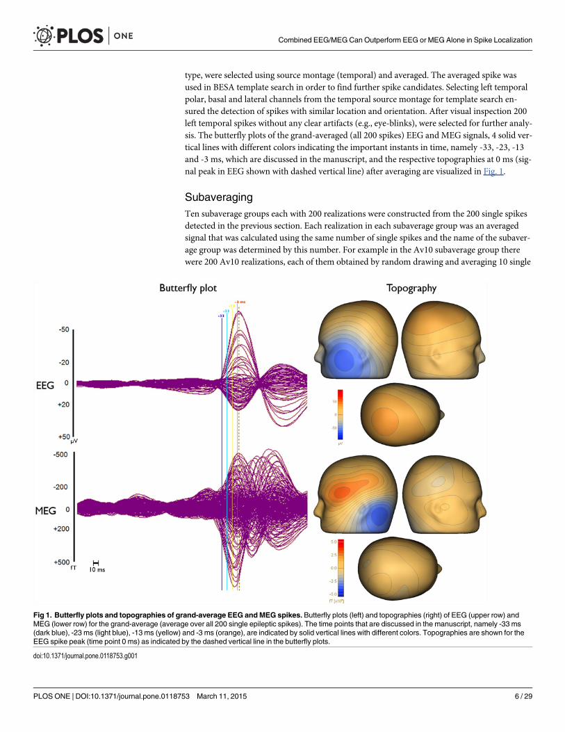
type, were selected using source montage (temporal) and averaged. The averaged spike wasused in BESA template search in order to find further spike candidates. Selecting left temporalpolar, basal and lateral channels from the temporal source montage for template search en-sured the detection of spikes with similar location and orientation. After visual inspection 200left temporal spikes without any clear artifacts (e.g., eye-blinks), were selected for further analy-sis. The butterfly plots of the grand-averaged (all 200 spikes) EEG and MEG signals, 4 solid ver-tical lines with different colors indicating the important instants in time, namely -33, -23, -13and -3 ms, which are discussed in the manuscript, and the respective topographies at 0 ms (sig-nal peak in EEG shown with dashed vertical line) after averaging are visualized in Fig. 1.
SubaveragingTen subaverage groups each with 200 realizations were constructed from the 200 single spikesdetected in the previous section. Each realization in each subaverage group was an averagedsignal that was calculated using the same number of single spikes and the name of the subaver-age group was determined by this number. For example in the Av10 subaverage group therewere 200 Av10 realizations, each of them obtained by random drawing and averaging 10 single
Fig 1. Butterfly plots and topographies of grand-average EEG andMEG spikes. Butterfly plots (left) and topographies (right) of EEG (upper row) andMEG (lower row) for the grand-average (average over all 200 single epileptic spikes). The time points that are discussed in the manuscript, namely -33 ms(dark blue), -23 ms (light blue), -13 ms (yellow) and -3 ms (orange), are indicated by solid vertical lines with different colors. Topographies are shown for theEEG spike peak (time point 0 ms) as indicated by the dashed vertical line in the butterfly plots.
doi:10.1371/journal.pone.0118753.g001
Combined EEG/MEGCan Outperform EEG or MEG Alone in Spike Localization
PLOS ONE | DOI:10.1371/journal.pone.0118753 March 11, 2015 6 / 29

spikes. The random drawing was performed with Matlab (The Mathworks, Inc.) and it was en-sured that none of the spikes was chosen more than once in the same realization. The subaver-age groups that were constructed with this procedure were Av5, Av10, . . ., Av50 withincrements of 5. Additionally, the group containing all 200 single spikes was denoted Av1.
The continuous EEG and MEG data were imported into Curry (http://www.neuroscan.com/curry.cfm), filtered from 1 to 100 Hz and divided into 400 ms long epochs (200 ms beforeand 200 ms after each EEG spike peak). The SNRs were calculated by dividing the signal powerat a certain time point, which was used for source reconstruction, by the variance of the noisedetermined from the interval -200 to -70 ms. The SNRs of EEG, MEG and EMEG signals werecalculated separately for each time instant (from -33ms to 0 ms) and only the signals withSNRs higher than three in the corresponding modality and subaverage were included in thefurther analysis.
Source Reconstruction ProcedureA cortically-constrained deviation scan inverse approach was used for source reconstruction[55], [56]. For this purpose, a source space with 2 mm resolution was calculated. A customwritten Matlab code ensured that all source space points were located inside the gray mattercompartment and sufficiently far away from other tissues. This ensured the closest node of theFE mesh for each source space point to only belong to elements that were labeled as gray mat-ter. Thus, the so-called Venant condition, being crucial to avoid unrealistic source modelingwhen using the FE-Venant approach, was satisfied [57]. The SimBio software (https://www.mrt.uni-jena.de/simbio and the SimBio integration into Fieldtrip: http://fieldtrip.fcdonders.nl/development/simbio) was then used to calculate EEG and MEG leadfield matrices for the givensource space, FE head model and sensor configurations. The Venant source modeling ap-proach was selected together with standard piecewise trilinear basis functions and an FE trans-fer matrix approach using an algebraic multigrid preconditioned conjugate gradient solver toobtain numerically accurate and computationally efficient forward EEG and MEG solutions[53], [57], [58]. The calculation of the leadfield matrix for 80 channels EEG and 275 channelMEG took about 60 minutes and 6 hours respectively on a standard workstation (Intel Core i7-860 Processor, 2.80 GHz and 16 GB RAM). The leadfield matrices were then imported intoCurry and single dipole scans were calculated from -33 to 0 ms with 0 corresponding to thepeak of the EEG signal. Unlike classical dipole fit algorithms, the cortically-constraint deviationscan provides goodness-of-fit (GOF) values for all source space points and the location withthe highest GOF was then used as the final deviation scan result.
Unlike MEG source reconstructions in which we used regularization to avoid implausibleresults, which might occur due to MEG’s insensitivity to radial components, for EEG andEMEG we did not use any regularization. Furthermore, only the MEG sensors over the lefthemisphere were used for MEG and EMEG source reconstructions to improve SNR and GOF.
In order to perform combined source reconstruction, EEG and MEG signals were trans-ferred to a unitless common space. This was achieved by using the SNRs of each electrode andgradiometer instead of the original signals [25].
In the results section we also use, instead of spike clusters, so-called centroid dipoles, de-fined by the mean location and orientation of all deviation scan result dipoles belonging to thecorresponding cluster. In addition, in spike clusters the distances of each deviation scan recon-struction (that passes the SNR criterion) to the centroid dipole were determined and used tocalculate the mean distance and its standard deviation for each cluster. Then, the reconstruc-tions in which the distance to the centroid exceeds mean plus two times the standard deviation
Combined EEG/MEGCan Outperform EEG or MEG Alone in Spike Localization
PLOS ONE | DOI:10.1371/journal.pone.0118753 March 11, 2015 7 / 29

for this cluster were excluded from the cluster and not used for further analysis (see Algorithm1 in [40] for details).
Skull Conductivity CalibrationEEG and, to a lesser extent, EMEG source reconstructions are sensitive to the conductivities ofthe highly isolating skull tissues [25], [40], [42]. However, skull conductivity values are quitecontroversial in literature with huge interindividual variance [26], [33], [59], [60]. We thereforecalibrated the skull conductivity using the N20 component of the measured and reconstructedsomatosensory evoked potential (SEP) and field (SEF) data as described and evaluated in detailin [40]. Similar calibration procedures have also been proposed by [41], [42].
The tissue conductivities (S/m) that are used in this study are [40], [61], [62]: Scalp (0.43),CSF (1.79), gray matter (0.33), anisotropic white matter along with the skull conductivity val-ues individually calibrated for the patient: skull compacta (0.0024) and skull spongiosa(0.0084).
Stereo-EEGMeasurementssEEG relying on 14 intracerebral depth electrodes with 167 contacts in total, showed indepen-dent epileptic activity with left frontal and temporal origins from 3 different seizure onsetzones: i) left fronto-basal mesial, ii) left temporal and iii) left frontal parasagittal. In this paperwe focused only on the left temporal spikes due to their high occurrence rate. 24 contacts from5 different electrodes and 8 contacts from 2 electrodes were active during temporal interictalspikes and seizure onset, respectively. Examples of an averaged spike and a single spike mea-sured simultaneously with sEEG and low density scalp EEG (ldEEG) (21 electrodes) are shownin Fig. 2. Based on information from the clinical report we represent the sEEG contacts withthree different colors: The green spheres represent the sEEG contacts that measured only inter-ictal activity, red spheres represent the sEEG contacts active during the seizure onset and theblue spheres represent the contacts near the left temporal lobe that measured neither interictalnor seizure onset activity. All contacts that were active during seizure onset were also measur-ing interictal spikes and were thus within the irritative zone. The locations of the sEEG contactswere marked manually using the post-operative T1-MRI and computed tomography imagesthat had been registered to the pre-operative T1-MRI using an affine registration scheme.
Square Distance Index (SDI)In order to quantify the amount of agreement between noninvasive source reconstructions andsEEG, we used a formula which weights each dipole by the inverse of its square distance toeach active sEEG contact.
SDIi ¼PN
j¼11
d2jþ1
N� 100 1
Where i represents a specific sEEG contact, e.g., A1, which measures frequent interictal activity,N is the number of dipoles that passes the SNR criterion (SNR>3) for the respective modalityand dj represents the Euclidian distance between the j’th dipole and the sEEG contact i. The ad-dition of 1 ensures the appropriate weighting for perfect localizations (dj = 0). A high value ofthis index at a certain sEEG contact indicates concentrated dipole localizations in the vicinityof this contact. On the other hand, high differences in SDIs for different sEEG contactsindicate that the localizations highlight only a certain area within the irritative zone and notthe whole. Thus, the mean of SDIs over all sEEG contacts should be high, while its standard
Combined EEG/MEGCan Outperform EEG or MEG Alone in Spike Localization
PLOS ONE | DOI:10.1371/journal.pone.0118753 March 11, 2015 8 / 29

Fig 2. Locations of sEEG contacts inside the brain and epileptic activity measured with sEEG andldEEG. Examples for an averaged (top right block) and a single epileptic spike (bottom right block) measuredsimultaneously with sEEG (left column in each block) and 21 electrodes low density scalp EEG (ldEEG) (rightcolumn in each block). Maximum of sEEG is at the HP2 contact as shown by the vertical lines at the peak ofthis contact. In the upper two brain figures (left block) only the active (that measures interictal or ictal signals)
Combined EEG/MEGCan Outperform EEG or MEG Alone in Spike Localization
PLOS ONE | DOI:10.1371/journal.pone.0118753 March 11, 2015 9 / 29
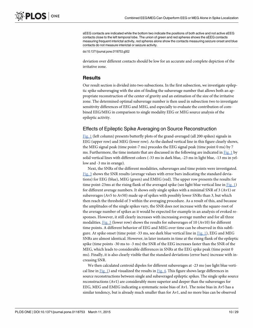
deviation over different contacts should be low for an accurate and complete depiction of theirritative zone.
ResultsOur result section is divided into two subsections. In the first subsection, we investigate epilep-tic spike subaveraging with the aim of finding the subaverage number that allows both an ap-propriate reconstruction of the center of gravity and an estimation of the size of the irritativezone. The determined optimal subaverage number is then used in subsection two to investigatesensitivity differences of EEG and MEG, and especially to evaluate the contribution of com-bined EEG/MEG in comparison to single modality EEG or MEG source analysis of theepileptic activity.
Effects of Epileptic Spike Averaging on Source ReconstructionFig. 1 (left column) presents butterfly plots of the grand-averaged (all 200 spikes) signals inEEG (upper row) and MEG (lower row). As the dashed vertical line in this figure clearly shows,the MEG signal peak (time point-7 ms) precedes the EEG signal peak (time point 0 ms) by 7ms. Furthermore, the time instants that are discussed in the following are indicated in Fig. 1 bysolid vertical lines with different colors (-33 ms in dark blue, -23 ms in light blue, -13 ms in yel-low and -3 ms in orange).
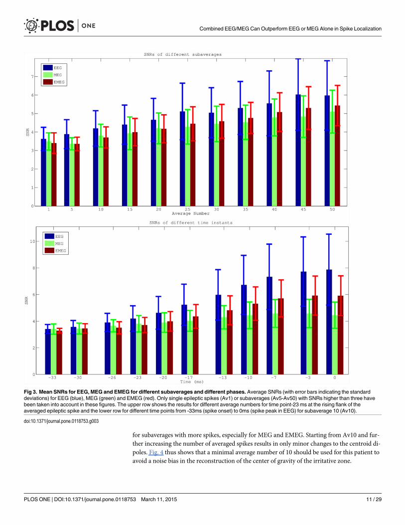
Next, the SNRs of the different modalities, subaverages and time points were investigated.Fig. 3 shows the SNR results (average values with error bars indicating the standard devia-tions) for EEG (blue), MEG (green) and EMEG (red). The upper row presents the results fortime point-23ms at the rising flank of the averaged spike (see light blue vertical line in Fig. 1)for different average numbers. It shows only single spikes with a minimal SNR of 3 (Av1) orsubaverages (Av5 to Av50) made up of spikes with possibly lower SNRs than 3, but whichthen reach the threshold of 3 within the averaging procedure. As a result of this, and becausethe amplitudes of the single spikes vary, the SNR does not increase with the square-root ofthe average number of spikes as it would be expected for example in an analysis of evoked re-sponses. However, it still clearly increases with increasing average number and for all threemodalities. Fig. 3 (lower row) shows the results for subaverages of 10 (Av10) for differenttime points. A different behavior of EEG and MEG over time can be observed in this subfi-gure. At spike onset (time point -33 ms, see dark blue vertical line in Fig. 1), EEG and MEGSNRs are almost identical. However, in later instants in time at the rising flank of the epilepticspike (time points -30 ms to -3 ms) the SNR of the EEG increases faster than the SNR of theMEG, which leads to considerable differences in SNRs at the EEG spike peak (time point 0ms). Finally, it is also clearly visible that the standard deviations (error bars) increase with in-creasing SNR.
We then calculated centroid dipoles for different subaverages at -23 ms (see light blue verti-cal line in Fig. 1) and visualized the results in Fig. 4. This figure shows large differences insource reconstructions between single and subaveraged epileptic spikes. The single spike sourcereconstructions (Av1) are considerably more superior and deeper than the subaverages forEEG, MEG and EMEG indicating a systematic noise bias of Av1. The noise bias in Av5 has asimilar tendency, but is already much smaller than for Av1, and no more bias can be observed
sEEG contacts are indicated while the bottom two indicate the positions of both active and not active sEEGcontacts close to the left temporal lobe. The union of green and red spheres shows the sEEG contactsmeasuring frequent interictal activity, red spheres alone show the contacts measuring seizure onset and bluecontacts do not measure interictal or seizure activity.
doi:10.1371/journal.pone.0118753.g002
Combined EEG/MEGCan Outperform EEG or MEG Alone in Spike Localization
PLOS ONE | DOI:10.1371/journal.pone.0118753 March 11, 2015 10 / 29
for subaverages with more spikes, especially for MEG and EMEG. Starting from Av10 and fur-ther increasing the number of averaged spikes results in only minor changes to the centroid di-poles. Fig. 4 thus shows that a minimal average number of 10 should be used for this patient toavoid a noise bias in the reconstruction of the center of gravity of the irritative zone.
Fig 3. Mean SNRs for EEG, MEG and EMEG for different subaverages and different phases. Average SNRs (with error bars indicating the standarddeviations) for EEG (blue), MEG (green) and EMEG (red). Only single epileptic spikes (Av1) or subaverages (Av5-Av50) with SNRs higher than three havebeen taken into account in these figures. The upper row shows the results for different average numbers for time point-23 ms at the rising flank of theaveraged epileptic spike and the lower row for different time points from -33ms (spike onset) to 0ms (spike peak in EEG) for subaverage 10 (Av10).
doi:10.1371/journal.pone.0118753.g003
Combined EEG/MEGCan Outperform EEG or MEG Alone in Spike Localization
PLOS ONE | DOI:10.1371/journal.pone.0118753 March 11, 2015 11 / 29
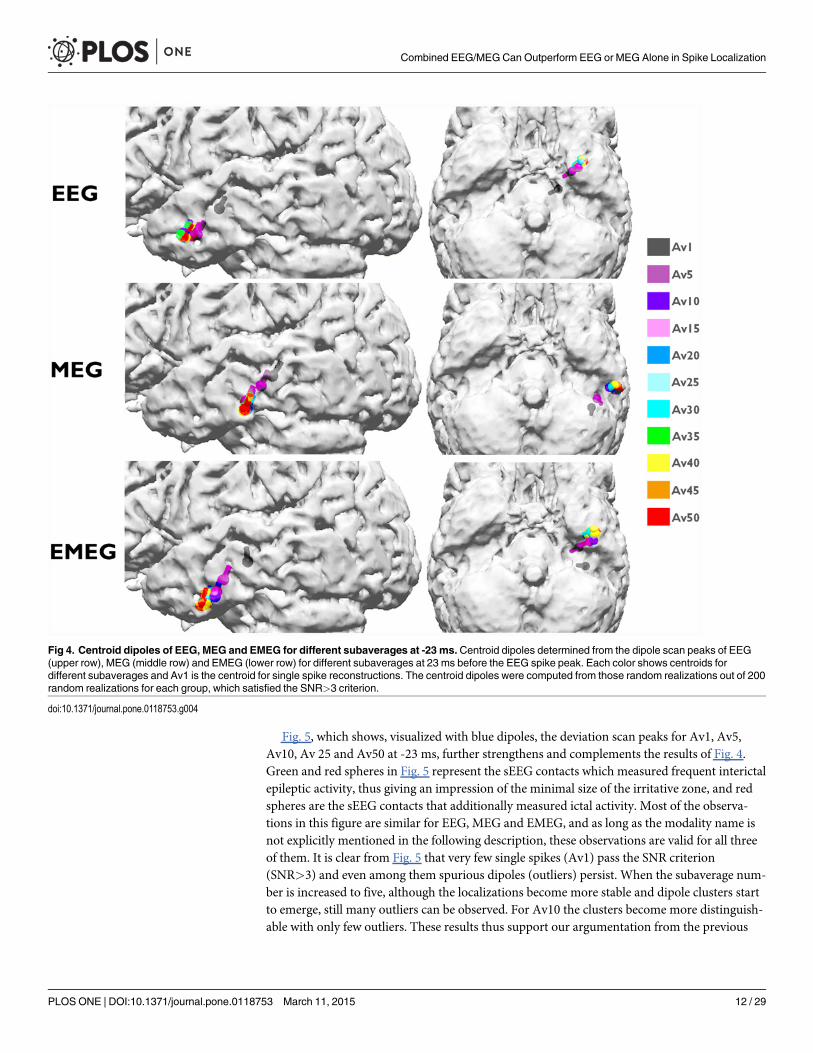
Fig. 5, which shows, visualized with blue dipoles, the deviation scan peaks for Av1, Av5,Av10, Av 25 and Av50 at -23 ms, further strengthens and complements the results of Fig. 4.Green and red spheres in Fig. 5 represent the sEEG contacts which measured frequent interictalepileptic activity, thus giving an impression of the minimal size of the irritative zone, and redspheres are the sEEG contacts that additionally measured ictal activity. Most of the observa-tions in this figure are similar for EEG, MEG and EMEG, and as long as the modality name isnot explicitly mentioned in the following description, these observations are valid for all threeof them. It is clear from Fig. 5 that very few single spikes (Av1) pass the SNR criterion(SNR>3) and even among them spurious dipoles (outliers) persist. When the subaverage num-ber is increased to five, although the localizations become more stable and dipole clusters startto emerge, still many outliers can be observed. For Av10 the clusters become more distinguish-able with only few outliers. These results thus support our argumentation from the previous
Fig 4. Centroid dipoles of EEG, MEG and EMEG for different subaverages at -23 ms.Centroid dipoles determined from the dipole scan peaks of EEG(upper row), MEG (middle row) and EMEG (lower row) for different subaverages at 23 ms before the EEG spike peak. Each color shows centroids fordifferent subaverages and Av1 is the centroid for single spike reconstructions. The centroid dipoles were computed from those random realizations out of 200random realizations for each group, which satisfied the SNR>3 criterion.
doi:10.1371/journal.pone.0118753.g004
Combined EEG/MEGCan Outperform EEG or MEG Alone in Spike Localization
PLOS ONE | DOI:10.1371/journal.pone.0118753 March 11, 2015 12 / 29
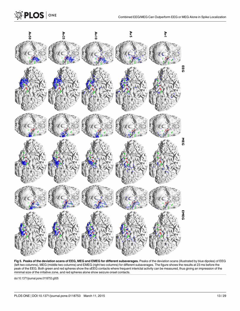
Fig 5. Peaks of the deviation scans of EEG, MEG and EMEG for different subaverages. Peaks of the deviation scans (illustrated by blue dipoles) of EEG(left two columns), MEG (middle two columns) and EMEG (right two columns) for different subaverages. The figure shows the results at 23 ms before thepeak of the EEG. Both green and red spheres show the sEEG contacts where frequent interictal activity can be measured, thus giving an impression of theminimal size of the irritative zone, and red spheres alone show seizure onset contacts.
doi:10.1371/journal.pone.0118753.g005
Combined EEG/MEGCan Outperform EEG or MEG Alone in Spike Localization
PLOS ONE | DOI:10.1371/journal.pone.0118753 March 11, 2015 13 / 29

paragraph (Fig. 4) to use a minimal average number of 10 for this patient. The following argu-mentation now delivers the complementary information that, from the chosen subaveragenumbers, Av10 even seems to be optimal: Av25 results again differ from Av10, especially withregard to a further decrease in the scatter of dipoles. When increasing the subaverage numberto 50 the scatter of the dipoles further decreases and MEG epileptic spikes are localized morelateral than for Av10 and Av25. Now the dipole scatters can be evaluated with the informationfrom the sEEG. Fig. 5 shows that for Av10 the dipole scatter covers almost all active sEEG con-tacts, i.e., it covers the minimal size of the irritative zone. For Av25 the clusters are already toofocal, missing the active HA8–10 and all HP contacts. Av50 is even more focal, missing evenmore of the active sEEG contacts (additionally to HA8–10 and all HP, also HA1–5 are outsidethe estimated irritative zone) and thus strongly underestimating the size of the irritative zone.
Comparison of EEG, MEG and EMEG Source ReconstructionsFor comparing EEG, MEG and EMEG localizations, based on the results of the previous sub-section, the focus will be on Av10 results. This choice is based on Figs. 4 and 5, which showthat a minimal average number of 10 is needed to sufficiently reduce noise bias and appropri-ately reconstruct the center of gravity of the irritative zone and that higher average numbers re-sult in too focal dipole clusters that lead to an underestimation of the extent of theirritative zone.
The Av10 EEG reconstructions in Fig. 5 are mainly localized in an area close to the pole ofthe temporal lobe and close to sEEG TA contacts (Temporal Anterior, see Fig. 2). On the otherhand, no activity is localized near HP1–3 and HA1–5 (the hippocampus posterior and anteriorcontacts). In MEG the localizations are more posterior than in EEG with clusters in the vicinityof HA8–10 (the posterior lateral neocortical contacts that, in contrast to their label, are not lo-cated in the hippocampus anterior, see Fig. 2), and close to HP contacts although no clusterwas formed around them. Unlike EEG there are no localizations in the vicinity of TA in MEG.In EMEG, noninvasive reconstructions cover all active sEEG contacts. EMEG even shows lo-calizations in the vicinity of the HP contacts, where neither the sensitivity of EEG (see EEGside-view in the second column of Av10 in Fig. 5) nor of MEG (see MEG bottom-view in thethird column of Av10 in Fig. 5) was sufficient to reconstruct any activity.
The plots in Fig. 6 add quantitative information to Av10 source reconstructions visualizedin Fig. 5. The plots show the SDIs (upper subfigure) and the percentage of dipoles that are clos-er than 10 mm to each sEEG contact measuring frequent interictal activity (lower subfigure).In these plots, the contacts that are also part of the seizure onset zone (amygdala contacts A1–3, and hippocampus anterior contacts HA1–5) are enclosed within rectangles with dotted lines.The upper subfigure clearly shows that most of the EEG localizations are clustered near the TAcontacts and the lower subfigure shows that there are dipoles within 10 mm at only 6 out ofoverall 24 interictal and 1 out of 8 ictal contacts. MEG values for the same measures are 10 outof 24 interictal and 2 out of 8 ictal, and the localizations are clustered especially near the poste-rior lateral neocortical contacts HA8–10 and HP contacts. On the other hand, for EMEG 23out of 24 interictal and 7 out of 8 ictal contacts have at least one noninvasive localization within10 mm. In the SDI plot the EEG SDIs for TA contacts are considerably higher than for othercontacts, while for MEG the SDIs at contacts HA8–10 are larger. For EMEG the SDI index isalmost equally distributed over the sEEG contacts and does not show a huge variation as inEEG and MEG. The means and standard deviations of SDIs for EEG, MEG and EMEG demon-strate this behavior well. The average SDIs for EEG and MEG are 0.22±0.15 and 0.18±0.11, re-spectively, and with 0.24 the average SDI for EMEG is higher and, most importantly, with 0.05its standard deviation is considerably lower than for EEG or MEG.
Combined EEG/MEGCan Outperform EEG or MEG Alone in Spike Localization
PLOS ONE | DOI:10.1371/journal.pone.0118753 March 11, 2015 14 / 29

Fig 6. Quantitative comparison of EEG, MEG and EMEG source reconstructions and sEEG contacts. Square distance index and the percentage ofdipoles closer than 10 mm for each sEEG contact. The values are given for EEG, MEG and EMEG for subaverages of 10 at -23 ms. The sEEG contactsenclosed by dashed lines are within the seizure onset zone.
doi:10.1371/journal.pone.0118753.g006
Combined EEG/MEGCan Outperform EEG or MEG Alone in Spike Localization
PLOS ONE | DOI:10.1371/journal.pone.0118753 March 11, 2015 15 / 29
As already noted, the MEG signal peak precedes the EEG maximum by approximately 7 ms(Fig. 1). In order to investigate which sources are dominating the EEG and MEG signals, wecompared the timings of epileptic spikes in sEEG and the simultaneously measured ldEEG(Fig. 2). We observed that simultaneously with the ldEEG peak, sEEG contact TA4 is also at itspeak value. On the other hand, the peak of the HP2 contact, the contact which measures thehighest amplitude in sEEG, is occurring 7.5 ms before the ldEEG and TA4 peaks. Moreover,the peaks of A1–3 and HA1–5, i.e., the seizure onset contacts, are also preceding the TA con-tacts and are almost simultaneous with the HP contacts.
Fig. 7 shows the pathways from spike onset to late propagation determined from EEG(upper two rows), MEG (middle two rows) and EMEG (lower two rows). Av10 deviation scanreconstructions for 4 different time points are visualized from -33 ms (spike onset, left column;also see dark blue vertical line in Fig. 1) to -3 ms (late propagation phase close to EEG peak,right column; also see orange vertical line in Fig. 1) in steps of 10 ms. The first striking observa-tion in this figure is the considerably higher stability of EMEG source reconstructions at spikeonset in comparison to single modality EEG and MEG. The EMEG source reconstructions attime point -33 ms are correctly clustered close to the seizure onset zone (red spheres at A1–3and HA1–5 contacts). In contrast, EEG is strongly dominated by noise with source reconstruc-tions spreading over a wide region. Although MEG reconstructions at spike onset are alreadybetter than EEG, they are still too lateral and spread over a too large region and the low SNRstill leads to many spurious reconstructions. The results for EEG, MEG and EMEG at the prop-agation phase also differ between one another. EMEG source reconstructions show that duringthe 10 ms period from spike onset to time point-23 ms, at the rising flank of the signal, the re-constructed activity spreads from amygdala and hippocampus to a wider area over the tempo-ral lobe, then covering all active sEEG contacts. At time points-13 ms and -3 ms, thereconstructed EMEG activity accumulates near the pole of the temporal lobe. The propagationpaths shown by single modality EEG and MEG differ quite much from the one of EMEG and,when compared to the sEEG findings and the EMEG reconstructions, are both incomplete. ForEEG, the first stable (by improved SNR) source reconstructions shown in Fig. 7 are the ones at-23 ms in the vicinity of the pole of the temporal lobe. EEG alone completely misses the moreposterior activity close to the HA and HP contacts (see Fig. 2) in this early propagation phase(see especially the first row and second column in Fig. 7). At later instants in time the EEG isonly able to reconstruct activity at the tip of the temporal pole. With regard to the MEG, at -23ms, source reconstructions are at more posterior temporal areas covering especially the HA8–10 contacts (see Fig. 2 and second row in Fig. 7) very well. Later on at time points-13 ms and -3ms, the reconstructed MEG activity travels to more anterior and temporobasal regions. Duringthe whole propagation phase MEG alone completely misses the temporo-polar activity close tothe TA contacts (fourth row in Fig. 7).
Corresponding to the visualizations of the propagation pathway in Fig. 7, the plots in Fig. 8add quantitative information on Av10 EMEG source reconstructions for the 4 different timepoints (please see supporting information for the same figure for EEG (S1 Fig.) and MEG (S2Fig.)). The upper subfigure, presenting the SDIs, shows that at -33 ms (spike onset) the sourcelocalizations are mostly clustered near the A and HA1–5 contacts (seizure onset). At later timepoints, i.e., closer to the spike peak, the reconstructed activity propagates to E and TA contacts.The lower subfigure, presenting the percentage of dipoles that are closer than 10 mm to eachsEEG contact measuring frequent interictal activity, shows that at -33 ms 7 contacts are cov-ered by the noninvasive EMEG reconstructions. Among them, 6 are ictal contacts (it covers 6out of 8 ictal contacts) and the other one is the HP1 contact, which peaks earlier than the TAcontacts, as shown in Fig. 2. While the EMEG reconstructed activity continuously decreases
Combined EEG/MEGCan Outperform EEG or MEG Alone in Spike Localization
PLOS ONE | DOI:10.1371/journal.pone.0118753 March 11, 2015 16 / 29
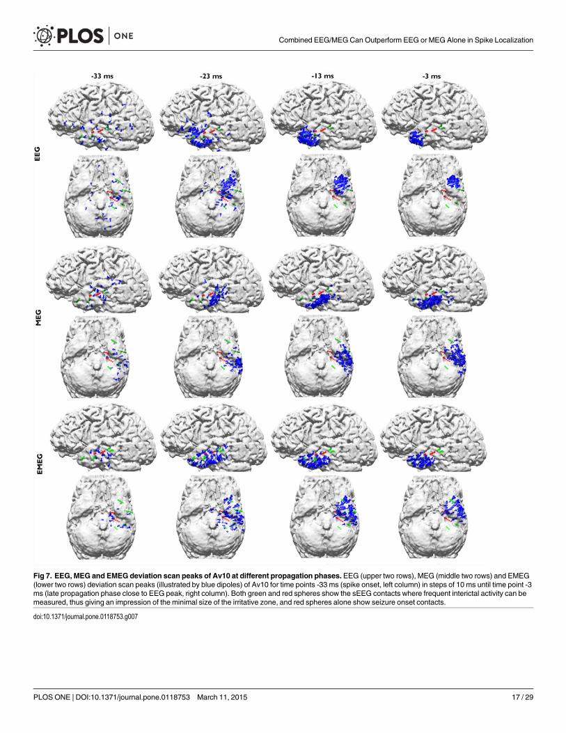
Fig 7. EEG, MEG and EMEG deviation scan peaks of Av10 at different propagation phases. EEG (upper two rows), MEG (middle two rows) and EMEG(lower two rows) deviation scan peaks (illustrated by blue dipoles) of Av10 for time points -33 ms (spike onset, left column) in steps of 10 ms until time point -3ms (late propagation phase close to EEG peak, right column). Both green and red spheres show the sEEG contacts where frequent interictal activity can bemeasured, thus giving an impression of the minimal size of the irritative zone, and red spheres alone show seizure onset contacts.
doi:10.1371/journal.pone.0118753.g007
Combined EEG/MEGCan Outperform EEG or MEG Alone in Spike Localization
PLOS ONE | DOI:10.1371/journal.pone.0118753 March 11, 2015 17 / 29
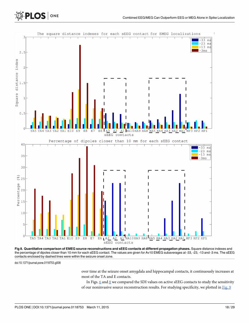
over time at the seizure onset amygdala and hippocampal contacts, it continuously increases atmost of the TA and E contacts.
In Figs. 6 and 8 we compared the SDI values on active sEEG contacts to study the sensitivityof our noninvasive source reconstruction results. For studying specificity, we plotted in Fig. 9
Fig 8. Quantitative comparison of EMEG source reconstructions and sEEG contacts at different propagation phases. Square distance indexes andthe percentage of dipoles closer than 10 mm for each sEEG contact. The values are given for Av10 EMEG subaverages at -33, -23, -13 and -3 ms. The sEEGcontacts enclosed by dashed lines were within the seizure onset zone.
doi:10.1371/journal.pone.0118753.g008
Combined EEG/MEGCan Outperform EEG or MEG Alone in Spike Localization
PLOS ONE | DOI:10.1371/journal.pone.0118753 March 11, 2015 18 / 29

Fig 9. SDI values for EEG, MEG and EMEG at -33 ms. The SDI bars are colored according to the measuredactivity: seizure onset contacts (red), only interictal contacts (green) and not active contacts (blue).
doi:10.1371/journal.pone.0118753.g009
Combined EEG/MEGCan Outperform EEG or MEG Alone in Spike Localization
PLOS ONE | DOI:10.1371/journal.pone.0118753 March 11, 2015 19 / 29
the SDI values for EEG, MEG and EMEG at -33 ms, now also including the not active sEEGcontacts. The SDIs were colored according to the measured activity: seizure onset contacts(red), interictal contacts (green) and not active contacts (blue). The figure shows that not onlythe sensitivity but also the specificity of EMEG results are superior to EEG and MEG alone.EMEG SDIs of sEEG contacts are gradually decreasing with distance to the seizure onset con-tacts (see HA6 to HA15 and A4 to A9). EEG and MEG source reconstructions alone not onlyfailed to highlight the seizure onset due to low SNRs but also their specificities were qualitative-ly inferior to our EMEG results.
DiscussionThis section starts with the most important results first, the ordering of the subchapters wastherefore inverted.
Comparison of EEG, MEG and EMEG Source ReconstructionsPropagation phenomenon: Problems and opportunities for noninvasive source reconstruc-tion. Propagation of interictal epileptic activity is a well-known phenomenon which might leadto misinterpretations and spurious diagnosis if not taken into account. In order to cope with it,many studies suggested reconstructing sources at the middle of the rising flank instead of thepeak of the epileptic spike (see [63] and references therein). Although this might seem to be agood compromise between low SNRs at the spike onset and propagation at the spike peak, theactivity at the middle of the rising flank might have already been subject to propagation, e.g., inmesial temporal lobe epilepsy, as shown in this study. On the other side, in cases where thepropagation pathway is always identical over different spikes, like the case discussed here,propagation provides also a great opportunity: Propagation of activity from low SNR locations(in our case the deep amygdala and hippocampal structures) to locations with much higherSNRs (in our case the pole of the temporal lobe; see Fig. 3 with regard to the increase in SNR)enables the examiner to find spikes and thus supply the necessary triggers for averaging, whichin turn might then enable revealing the preceding activity with lower SNR. This has beenshown in our study where we were able to accurately (with regard to our sEEG validation mea-sure, see further discussion below) reconstruct the complete pathway of the epileptic activityfrom onset to spike peak using subaveraging techniques and combined EEG and MEG sourceanalysis. Similar scenarios are discussed in the literature. For example, simultaneous scalp andintracranial EEG studies showed that, while scalp EEG might not directly be able to distinguishthe activity from deeper structures from the noise, it might be possible to extract the EEG signalby averaging intracranial epileptic spikes [64], [65].
EEG, MEG and EMEG source reconstructions at spike onset. Despite the strategies ex-plained in the previous paragraph, as we have shown in Figs. 7 and 8, at very early instants intime (-33 ms, see dark blue vertical line in Fig. 1) single modality EEG or MEG source recon-structions might not be reliable enough to draw conclusions on the origin of the epileptic spike.In our results, EEG was strongly dominated by noise and although MEG source reconstruc-tions were more stable, they had a lateral bias with still too many spurious dipole positions.The source reconstructions with single modality EEG or MEG stabilized at later instants intime (see time point-23 ms in Fig. 7), but the activity had already been subject to propagationby then. One of the most important and clinically relevant findings of this study is thus theability of EMEG to benefit from the complementary information of EEG and MEG at especiallythese very early instants in time and to thereby stabilize the source reconstructions in cases oflow SNR. As shown in Figs. 7 and 8, at -33 ms the source reconstructions of EMEG are mainly
Combined EEG/MEGCan Outperform EEG or MEG Alone in Spike Localization
PLOS ONE | DOI:10.1371/journal.pone.0118753 March 11, 2015 20 / 29

clustered near amygdala and hippocampus, i.e., regions within the seizure onset zone as esti-mated from sEEG.
Differences of EEG, MEG and EMEG source reconstructions in revealing the propaga-tion pathway. The EEG, MEG and EMEG source reconstructions differed not only at spikeonset but also at later instants in time. At -23 ms (see light blue vertical line in Fig. 1), whileboth EEG and MEG source reconstructions were clustered in the vicinity of spiking sEEG con-tacts, they highlighted different contacts. EEG localizations were mainly clustered near TA con-tacts, while MEG results were close to the posterior lateral neocortical contacts HA8–10 and,partially, to HP. In agreement with our findings, in temporal lobe epilepsy, posterior MEGsource reconstructions in comparison to EEG were also observed in other studies [66], [67].The main reason for this difference might be the increased size of the active patch at this timepoint due to propagation. Considering the wide extent of active cortex measured with sEEG inthis study, our hypothesis is that the peak of the EEG deviation scan was found at the temporalpole because of its considerable radial source orientation component. As a result, this activationdid not contribute much to the MEG signals. On the other hand, the activity arising from espe-cially the posterior lateral neocortical contacts HA8–10 and from the HP contacts was moretangentially oriented, leading to higher SNRs in MEG, and thus MEG was mainly focusing onthis part of the cortex. Furthermore, the averaged MEG signal peak was not synchronous withthe EEG peak; it preceded the EEG by approximately 7 ms (see dashed vertical line in Fig. 1).In order to determine the sources dominating the EEG and MEG signals, we investigated thetime relationship between the peaks of the simultaneously measured sEEG and low-densityEEG (ldEEG) epileptic spikes. We observed that the peaks of the ldEEG and the TA4 contactwere simultaneous, and our EEG localizations were clustered around TA4. Although MEG andsEEG were not measured simultaneously, we might extrapolate the results of the simultaneousldEEG-sEEG to the simultaneous EEG-MEG measurements and comment on the timings ofthe measured signals. Considering the fact that the MEG peak is also approximately 7 ms be-fore the EEG we might state that the MEG maximum is concurrent in time with the HP2 con-tact. This means, the peaks of the MEG and the HP2 contact are almost simultaneous andmight explain why MEG was also localized closer to HP2. All these results fit well to our hy-potheses that a larger activated cortical patch is underlying the measured activity and that EEGand MEG focus on only parts and, due to their distinct sensitivities, to non-identical parts ofthis activity. Although EEG and MEG source reconstructions were able to highlight just a sub-set of spiking sEEG contacts, EMEG results were covering almost all relevant sEEG contactswith only a few spurious localizations. EMEG localizations were not simply the union of EEGand MEG results but a rather complicated interplay of both modalities compensating their rel-ative shortcomings. For example at -23 ms, in Figs. 6 (especially the lower subfigure) and 7, nomajor dipole cluster was noticeable neither with EEG nor with MEG around the E contacts insEEG, while there were clear clusters around these active contacts in EMEG. This also supportsthe idea that combining EEG and MEG can supply important additional information that can-not be achieved by localizing EEG and MEG alone, and then comparing their results. There-fore, whenever it is technically feasible to measure EEG and MEG simultaneously, it might beimportant to not only analyze single modality EEG and MEG but also to compare with com-bined EMEG reconstructions to obtain accurate localization results. Furthermore, the asyn-chronous EEG and MEG peaks along with the more complete overview on the propagationpathways provided only by EMEG, as shown here, might also help distinguishing between theprimary and secondary interictal areas as reported in [68].
Close to the spike peak (-3 ms) the source reconstructions were more anterior in compari-son to earlier instants in time and were clustered close to the pole of the temporal lobe. TheSNR values shown in the lower subfigure of Fig. 3 support our findings that epileptic activity
Combined EEG/MEGCan Outperform EEG or MEG Alone in Spike Localization
PLOS ONE | DOI:10.1371/journal.pone.0118753 March 11, 2015 21 / 29

had started at deeper areas and then propagated to the pole of the temporal lobe at the spikepeak. The SNRs of EEG and MEG at spike onset were almost identical, but later on, the in-crease in EEG SNR was higher than in MEG due to the mainly radial source orientation in thearea of the temporal pole.
Effects of Epileptic Spike Averaging on Source ReconstructionThe decision between localizing each single epileptic spike separately and averaging spikes withsimilar topographies before source reconstruction is a highly disputed issue in presurgical epi-lepsy diagnosis, and both approaches have their merits and drawbacks. Single spike localiza-tions might be used to estimate the size of the irritative zone [5], [17], [43–46]. However, theselocalizations suffer from low SNRs as also shown in our study. On the other hand, averagingsimilar spikes might increase the SNR and thus, the reliability of the localizations remarkably[2], but information on the extent might get lost. In this paper, motivated by the findings ofBast et al. [2] and Wennberg and Cheyne [69] for EEG, andWennberg and Cheyne [67] forMEG source reconstructions, we calculated multiple subaverages in order to investigate the ef-fects of SNR and averaging on EEG, MEG and EMEG source reconstructions. This enabled usto compare the effects of averaging and the resulting SNR in a step-by-step fashion.
Systematic localization bias in single spike source reconstructions due to low SNR. Ourresults show that the centroid dipoles obtained from epileptic spike clusters differ considerablybetween different subaverages. We observed that spikes with lower number of subaverages andthus lower SNRs were localized more mesial and superior in comparison to those with highernumber of averages (and higher SNRs) at -23 ms (Fig. 4). The reason for this localization biasmight be due to background activity, which can be considered as noise in our case. Since at thistime instant the propagation had already occurred, the noise bias shifted the localizations fromlateral parts of the temporal lobe into deeper regions in the brain. Our data support this hy-pothesis by showing higher localization differences in the left-right (LR) and superior-inferior(SI) than in the anterior-posterior (AP) axis. The lateral regions of the left temporal lobe are sit-uated farther away from the center of the brain in LR and SI axes, while in AP axis they areclose to the center. In agreement to our results and our hypothesis, the studies of Wennbergand Cheyne [67], [69] also showed similar shifts to the center of the brain.
Preselection criteria to improve single and subaveraged spike source reconstructions.Different preselection criteria for epileptic spikes have been suggested to avoid errors in singlespike localizations [5], [11], [46]. We followed these criteria and localized only the spikes withSNR higher than three. This preselection strategy resulted in more reasonable localizations but,on the other hand, the number of single spikes that satisfied this condition also got smaller.From the 200 measured single spikes, only 20 EMEG spikes passed the criterion at 0 ms (spikepeak with highest SNR). At -23 ms this number was even reduced to only 7 for EMEG andeven among them spurious localizations persisted (see EMEG results for Av1 in Fig. 5). There-fore, we recommend (1) to use subaverages and (2) to observe the changes in centroid dipolesand scatter size with increasing averages.
Estimation of the optimal subaverage number. For the estimation of the optimal subaver-age number, we recommend the following procedure: A subaverage should be selected that av-erages enough spikes (in our case Av10) so that its centroid dipole does no longer differ muchfrom the centroids of the subaverages with more spikes (in our case Av25 and Av50). Sinceeven for an extended source the center of gravity would always result in the same position innoise free set-up, the changes in the centroid dipole for different subaverages are mainly due toinsufficient SNR. By selecting Av10 in which the location of the centroid dipole does not differmuch from Av50, we reduced the effects of noise on dipole scatter [70–72]. Averaging more
Combined EEG/MEGCan Outperform EEG or MEG Alone in Spike Localization
PLOS ONE | DOI:10.1371/journal.pone.0118753 March 11, 2015 22 / 29
spikes may not be favorable, as this may artificially reduce the scatter size leading to an under-estimation of the extent of the irritative zone. Nevertheless, even for the optimal subaveragenumber estimated with this procedure, the effects of spatial averaging on scatter size will persistand possibly lead to a slight underestimation of the size of the irritative zone. However, thenegative influence will be much smaller than localizing single spikes with insufficient SNRs. Inthis study Av10 was a good compromise, but this number might surely be different for otherpatients. The better performance of Av10 in comparison to Av50 might be surprising since thehigher SNRs of Av50 might be expected to result in better localizations. However, in the lightof our results and the relevant literature we can question this expectation at least for localiza-tion of interictal spikes: In [73], among 19 patients with Engel I or II outcomes, the resectedareas in four cases were concordant to only single spikes, in two to only averaged spikes, and infive to both single and averaged spike localizations. A possible explanation for the latter resultscan be sought in the light of the publications of [43] and [44], in which using optical imagingthey showed that the origins of the epileptic activity change in a stochastic way within a certainregion. This questions the assumption that spikes from the same irritative zone have exactlythe same origin and waveform, and can be used as an argument against averaging.
Topology of the irritative zone. Another important aspect is the topology of the irritativezone. In our study, the irritative zone had a convex shape so that the center of gravity was partof the zone. However, in case of a concave shape, this might change. As an example, the centerof gravity of a half-moon-shaped concave topology might be outside the structure (see, e.g., thehalf-moon-shaped single spike localizations of patient 5 in Fig. 1B in [73]). However, even inthe latter case, using the centroid localization change between different subaverages is still animportant measure, because a centroid shift between single spike and subaveraged spike locali-zations will still indicate a systematic shift of single spike localizations due to noise.
Relationship between size of dipole scatter, SNR and extent of the irritative zone. Oishiet al. [46] showed in an MEG study that 8 out of 9 patients in which the spike cluster coincidedentirely with the ictal onset zone determined by subdural EEG (and resected afterwards) be-came seizure free whereas the ratio was just 3 out of 11 for the cases where spike cluster andictal onset zone either only coincided partly or did not coincide at all. In agreement with thestudies of [5], [17], [45], [74], this shows the potential benefit of single spike localizations. Onthe other hand, our results show that the amount of scatter is highly correlated with the num-ber of subaverages and the SNR, especially for relatively low SNRs. Our results on scatter sizeare mainly in line with Bast et al. [2] and Wennberg and Cheyne [67], [69]. However, we addi-tionally show here that the effects of subaveraging and SNR on scatter size are valid for all in-vestigated modalities, i.e., EEG, MEG and EMEG. Furthermore, while the other studies rely onsimpler volume conduction modeling such as spherical shell models used in Bast et al. [2], thehigh-resolution six-compartment head model with calibrated skull conductivity and aniso-tropic representation of the white matter compartment as proposed in our study does not onlyenable simultaneous analysis of EEG and MEG, but also has the potential to improve localiza-tion accuracy for single modality EEG or MEG or in combined EMEG analysis. The latter is es-pecially important in the temporal lobe, where a sphere approximation of the skull can resultin significant errors for both EEG (e.g., [15]) and MEG (e.g., [75], [76]). Our results are also inagreement with EEG simulations of Kobayashi et al. [77] showing dipole clusters to becomeless erroneously distributed with increasing SNR. However, in summary, the identification ofthe exact size of the irritative zone still remains a difficult problem because, as also shown inthis study, scatter varies significantly with SNR, spike selection criterion andsubaverage number.
Reconstructing slightly distributed activity using a single dipole model might lead to a smalldepth-bias (sources that are localized slightly too deep). Here, we took three measures to
Combined EEG/MEGCan Outperform EEG or MEG Alone in Spike Localization
PLOS ONE | DOI:10.1371/journal.pone.0118753 March 11, 2015 23 / 29
alleviate such depth-bias and to accurately (as validated by the sEEG) reconstruct the center ofthe underlying activity: 1) We used a cortically-constrained source space, which prevents erro-neous localizations inside white matter. 2) We constructed a head model that distinguishesCSF, gray matter, and anisotropic white matter instead of a homogeneous brain, in which thetopographies for dipoles with different depths and locations would have been more similar andhomogeneous. 3) We preferred a dipole scan instead of a dipole fit to ensure finding the globaloptimum of the cost function over the cortically-constrained source space.
It is important to state that in this study the aim was not to estimate the extent of a patch inwhich all neurons are active simultaneously and always in the same way but to estimate the ex-tent of a patch in which the origin of the activity is different for each spike. Using optical imag-ing on epileptic human neocortical slices removed during epilepsy surgery, Köhling et al. [43]and Speckmann et al. [44] showed that the activated cortical areas during epileptic waves arefocal and their spatial positions change in a dynamic manner within the epileptic tissue. Thisfinding was the main reason why a subaveraging procedure was selected instead of averagingall spikes. Therefore, in this study our aim with investigating the dipole scatter was not to deter-mine the extent of a patch that always follows exactly the same activation pattern but to benefitfrom the small differences on the activation pattern within the epileptogenic zone due to thedynamic and stochastic behavior of each spike as shown in [43,44].
With regard to the chosen inverse approach, besides the cortically-constrained deviationscan as employed here (see, e.g., [38], [55], [56]), promising results were also achieved with cur-rent density approaches [38], with hierarchical Bayesian modeling frameworks [48], [78] andwith spatio-temporal current density approaches [79], [80] in non-invasively reconstructingnetworks of (epileptic) activity from EEG and/or MEG. However, also those methods need toembed correct prior knowledge in some form into the inverse approach and it still needs to beshown that the methodology is stable even in the presence of low SNR in realistic epilepsy data-sets [78], [79]. Furthermore, in Bouet et al. [81], using frequency domain beamformers, the de-termination of the spiking volume was possible in 16 out of 21 patients with sensitivity (76%)and specificity (67%), as also validated through sEEG measurements. However, Steinsträteret al. [82] showed that beamformer approaches are sensitive to head volume conductor proper-ties. Therefore, in a future study, it will be interesting to combine other inverse methods withthe subaveraging, the head modeling and the combined EEG/MEG procedure as presentedhere and to evaluate their quality by means of the intracranial EEG recordings.
Limitations of the current study. There are two important aspects that need attention re-garding the interpretation of the results presented here: 1) We used sEEG measurements forvalidation purposes because this is widely accepted as the “gold standard” in presurgical epilep-sy diagnosis. sEEG has advantages over noninvasive EEG because not only the target sourcesare closer to the measurement sensors but also the attenuation and smoothing of the signalsdue to volume conduction, especially due to the highly insulating skull, are avoided. Despitethese advantages, sEEG still lacks the ability to show the ground truth due to its low spatial res-olution and overall coverage caused by the limited number of electrodes and contacts. 2) Al-though we have simultaneously measured EEG/MEG and ldEEG/sEEG, we did not measureEEG/MEG/sEEG simultaneously. Thus, we cannot be sure that all epileptic spikes that werevisible in sEEG were also visible in EEG and MEG. Nevertheless, we verified that most sEEGspikes were also visible in ldEEG and, as Fig. 7 (EMEG, -23ms) shows, the irritative zones de-termined by sEEG (green and red contacts) and by noninvasive EEG/MEG (blue dipoles) werewell in agreement with each other.
Combined EEG/MEGCan Outperform EEG or MEG Alone in Spike Localization
PLOS ONE | DOI:10.1371/journal.pone.0118753 March 11, 2015 24 / 29

ConclusionsIn this study, a high-resolution realistic six-compartment finite element head model with an-isotropic white matter and calibrated skull conductivity enabled us to take into account the dif-ferent sensitivity profiles of EEG, MEG and EMEG in reconstructing the underlying sources inthe brain, and to make reliable interpretations on the effects of spike averaging and SNR. Ourstudy shows that EMEG source analysis can increase accuracy and confidence in source recon-structions significantly, which might have important clinical implications especially for localiz-ing at spike onset and for revealing propagation pathways as complete as possible.Furthermore, subaveraging might provide important and accurate information that neithersingle nor grand-averaged spike reconstructions can give. However, the extent of dipole scatterwill still be correlated to the number of subaverages and to the SNR. Although an advancedhead model as used in this study can improve the accuracy of source reconstructions, also stud-ies that use more homogenized ways of forward modeling such as the classical three compart-ment (skin, skull, brain) approach might still benefit from the subaveraging pipeline and thecalibration procedure for combining EEG and MEG as presented in this study.
Supporting InformationS1 Dataset. Dataset underlying the results presented. The spreadsheet contains all deviationscans of EMEG (page 1), EEG (page 2), and MEG (page 3) for subaverages Av5, Av10, Av15,Av20, Av25, Av30, Av35, Av40, Av45, Av50 and single spikes (Av1) at -33, -23, -13, and -3 ms.For each deviation scan signal-to-noise-ratio “SNR”, “Residual variance”, the coordinates ofthe deviation scan dipole “Locations”, the normals to determine the orientation “Normals”,and the strengths of the dipoles “Strengths” are given. The fourth page contains the names ofthe sEEG electrodes “Electrode name”, “Contact number”, and their coordinates “Locations”.(XLSX)
S1 Fig. Quantitative comparison of EEG source reconstructions and sEEG contacts at dif-ferent propagation phases. Square distance indexes and the percentage of dipoles closer than10 mm for each sEEG contact. The values are given for Av10 EEG subaverages at -33, -23, -13and -3 ms. The sEEG contacts enclosed by dashed lines were within the seizure onset zone.(TIF)
S2 Fig. Quantitative comparison of MEG source reconstructions and sEEG contacts at dif-ferent propagation phases. Square distance indexes and the percentage of dipoles closer than10 mm for each sEEG contact. The values are given for Av10 MEG subaverages at -33, -23, -13and -3 ms. The sEEG contacts enclosed by dashed lines were within the seizure onset zone.(TIF)
Author ContributionsConceived and designed the experiments: ÜAMDHK JW CK JH SR HS CHW. Performed theexperiments: ÜAMDMHHK. Analyzed the data: ÜA JV MD PK CK SR CHW. Contributedreagents/materials/analysis tools: ÜA JV MD CHW.Wrote the paper: ÜA JV MD PK HKMHJW CK JH SR HS CHW.
References1. Agirre-Arrizubieta Z, Thai NJ, Valentín A, Furlong PL, Seri S, Selway RP, et al. (2014): The value of
Magnetoencephalography to guide electrode implantation in epilepsy. Brain Topogr. 27(1):197–207.doi: 10.1007/s10548-013-0330-x PMID: 24249204
Combined EEG/MEGCan Outperform EEG or MEG Alone in Spike Localization
PLOS ONE | DOI:10.1371/journal.pone.0118753 March 11, 2015 25 / 29

2. Bast T, Boppel T, Rupp A, Harting I, Hoechstetter K, Fauser S, et al. (2006): Noninvasive source locali-zation of interictal EEG spikes: effects of signal-to-noise ratio and averaging. J. Clin. Neurophysiol.23(6):487–97. PMID: 17143137
3. Boon P, D’Have M, Vanrumste B, van Hoey G, Vonck K, vanWalleghem P, et al. (2002): Ictal source lo-calization in presurgical patients with refractory epilepsy. J. Clin. Neurophysiol. 19(5):461–8. PMID:12477991
4. Ebersole JS (1999): Non-invasive pre-surgical evaluation with EEG/MEG source analysis. Electroen-cephalogr. Clin. Neurophysiol. 50:167–74. PMID: 10689459
5. Fischer MJ, Scheler G, Stefan H (2005): Utilization of magnetoencephalography results to obtainfavourable outcomes in epilepsy surgery. Brain 128:153–7. PMID: 15563514
6. Gavaret M, Badier JM, Marquis P, Bartolomei F, Chauvel P (2004): Electric source imaging in temporallobe epilepsy. J. Clin. Neurophysiol. 21(4):267–82. PMID: 15509916
7. Knowlton RC, Razdan SN, Limdi N, Elgavish RA, Killen J, Blount J, et al. (2009): Effect of epilepsy mag-netic source imaging on intracranial electrode placement. Ann Neurol. 65(6):716–23. doi: 10.1002/ana.21660 PMID: 19557860
8. Pataraia E, Lindinger G, Deecke L, Mayer D, Baumgartner C (2005): Combined MEG/EEG analysis ofthe interictal spike complex in mesial temporal lobe epilepsy. Neuroimage 24(3):607–14. PMID:15652296
9. Rampp S, Stefan H (2007): Magnetoencephalography in presurgical epilepsy diagnosis. Expert Rev.Med. Devices 4(3):335–47. PMID: 17488228
10. Salayev KA, Nakasato N, Ishitobi M, Shamoto H, Kanno A, Iinuma K, et al. (2006): Spike orientationmay predict epileptogenic side across cerebral sulci containing the estimated equivalent dipole. Clin.Neurophysiol. 117(8):1836–43. PMID: 16798082
11. Stefan H, Hummel C, Scheler G, Genow A, Druschky K, Tilz C, et al. (2003): Magnetic brain source im-aging of focal epileptic activity: a synopsis of 455 cases. Brain 126: 2396–405. PMID: 12876149
12. SutherlingWW, Mamelak AN, Thyerlei D, Maleeva T, Minazad Y, Philpott L, et al. (2008): Influence ofmagnetic source imaging for planning intracranial EEG in epilepsy. Neurology 71(13):990–6. doi: 10.1212/01.wnl.0000326591.29858.1a PMID: 18809834
13. Brodbeck V, Spinelli L, Lascano AM, Wissmeier M, Vargas MI, Vulliemoz S, et al. (2011): Electroen-cephalographic source imaging: a prospective study of 152 operated epileptic patients. Brain134(Pt 10):2887–97. doi: 10.1093/brain/awr243 PMID: 21975586
14. Fuchs M, Wagner M, Kastner J (2007): Development of Volume Conductor and Source Models to Lo-calize Epileptic Foci. J. Clin. Neurophysiol. 24:101–19. PMID: 17414966
15. Huiskamp G, Vroeijenstijn M, van Dijk R, Wieneke G, van Huffelen AC (1999): The need for correct re-alistic geometry in the inverse EEG problem. IEEE Trans. Biomed. Eng. 46(11):1281–87. PMID:10582412
16. Roth BJ, Ko D, von Albertini-Carletti IR, Scaffidi D, Sato S (1997): Dipole localization in patients with ep-ilepsy using the realistically shaped head model. Electroenc. Clin. Neurophysiol. 102:159–66. PMID:9129570
17. Scheler G, Fischer MJ, Genow A, Hummel C, Rampp S, Paulini A, et al. (2007): Spatial relationship ofsource localizations in patients with focal epilepsy: Comparison of MEG and EEG with a three sphericalshells and a boundary element volume conductor model. Hum. Brain. Mapp. 28:315–22. PMID:16933294
18. Heers M, Hedrich T, An D, Dubeau F, Gotman J, Grova C, et al. (2014): Spatial correlation of hemody-namic changes related to interictal epileptic discharges with electric and magnetic source imaging.Hum Brain Mapp. 35(9):4396–414. doi: 10.1002/hbm.22482 PMID: 24615912
19. Dassios G, Fokas AS, Hadjiloizi D (2007): On the complementarity of electroencephalography andmagnetoencephalography. Inverse Problems 23:2541–49.
20. Barkley GL, Baumgartner C (2003): MEG and EEG in epilepsy. J. Clin. Neurophysiol. 20(3):163–78.PMID: 12881663
21. Iwasaki M, Pestana E, Burgess RC, Lüders HO, Shamoto H, Nakasato N (2005): Detection of epilepti-form activity by human interpreters: blinded comparison between electroencephalography and magne-toencephalography. Epilepsia 46(1):59–68. PMID: 15660769
22. Knake S, Halgren E, Shiraishi H, Hara K, Hamer HM, Grant PE, et al. (2006): The value of multichannelMEG and EEG in the presurgical evaluation of 70 epilepsy patients. Epilepsy Res. 69(1):80–86. PMID:16516443
23. Baillet S, Garnero L, Marin G, Hugonin JP (1999): Combined MEG and EEG Source Imaging by Minimi-zation of Mutual Information. IEEE Trans. Biomed. Eng. 46(5):522–34. PMID: 10230131
Combined EEG/MEGCan Outperform EEG or MEG Alone in Spike Localization
PLOS ONE | DOI:10.1371/journal.pone.0118753 March 11, 2015 26 / 29

24. Cohen D, Cuffin BN (1987): A method for combining MEG and EEG to determine the sources. Phys.Med. Biol. 32(1):85–89. PMID: 3823144
25. Fuchs M, Wagner M, Wischmann HA, Kohler T, Theissen A, Drenckhahn R, et al. (1998): Improvingsource reconstructions by combining bioelectric and biomagnetic data. Electroencephalogr. Clin. Neu-rophysiol. 107(2):93–111. PMID: 9751281
26. Hämäläinen M, Hari R, Ilmoniemi R, Knuutila J, Lounasmaa O (1993): Magnetoencephalography: theo-ry, instrumentation, and applications to noninvasive studies of the working human brain. Rev. Mod.Phys. 65:413–97.
27. Liu AK, Dale AM, Belliveau JW (2002): Monte Carlo simulation studies of EEG and MEG localizationaccuracy. Hum. Brain Mapp. 16(1):47–62. PMID: 11870926
28. Sarvas J (1987): Basic mathematical and electromagnetic concepts of the biomagnetic inverse prob-lem. Phys. Med. Biol. 32(1):11–22. PMID: 3823129
29. Vorwerk J, Cho J-H, Rampp S, Hamer H, Knösche TR, Wolters CH (2014): A Guideline for Head Vol-ume Conductor Modeling in EEG and MEG. Neuroimage, DOI: 10.1016/j.neuroimage.2014.06.040
30. Wolters CH, Anwander A, Tricoche X, Weinstein D, Koch MA, MacLeod RS (2006): Influence of tissueconductivity anisotropy on EEG/MEG field and return current computation in a realistic headmodel: Asimulation and visualization study using high-resolution finite element modeling. Neuroimage 30(3):813–26. PMID: 16364662
31. Haueisen J, Funke M, Güllmar D, Eichardt R (2012): Tangential and Radial Epileptic Spike Activity: Dif-ferent Sensitivity in EEG and MEG. J. Clin. Neurophysiol. 29(4): 327–32. doi: 10.1097/WNP.0b013e3182624491 PMID: 22854766
32. Lew S, Sliva DD, Choe MS, Grant PE, Okada Y, et al. (2013): Effects of sutures and fontanels on MEGand EEG source analysis in a realistic infant head model. Neuroimage 76:282–93. doi: 10.1016/j.neuroimage.2013.03.017 PMID: 23531680
33. Akhtari M, Bryant HC, Mamelak AN, Flynn ER, Heller L, Shih JJ, et al. (2002): Conductivities of three-layer live human skull. Brain Topogr. 14(3):151–67. PMID: 12002346
34. Dannhauer M, Lanfer B, Wolters CH, Knösche TR (2011): Modeling of the human skull in EEG sourceanalysis. Hum. Brain Mapp. 32:1383–99. doi: 10.1002/hbm.21114 PMID: 20690140
35. Montes-Restrepo V, van Mierlo P, Strobbe G, Staelens S, Vandenberghe S, Hallez H (2014): Influenceof Skull Modeling Approaches on EEG Source Localization. Brain Topogr. 27:95–111. doi: 10.1007/s10548-013-0313-y PMID: 24002699
36. Haueisen J, Ramon C, Eiselt M, Brauer H, Nowak H (1997): Influence of tissue resistivities on neuro-magnetic fields and electric potentials studied with a finite element model of the head. IEEE Trans.Biomed. Eng. 44(8): 727–35. PMID: 9254986
37. Tuch DS, Wedeen VJ, Dale AM, George JS, Belliveau JW (2001): Conductivity tensor mapping of thehuman brain using diffusion tensor MRI. P. Natl. Acad. Sci. 98:11697–701. PMID: 11573005
38. Rullmann M, Anwander A, Dannhauer M, Warfield SK, Duffy FH, Wolters CH (2009): EEG source anal-ysis of epileptiform activity using a 1mm anisotropic hexahedra finite element head model. Neuroimage44:399–410. doi: 10.1016/j.neuroimage.2008.09.009 PMID: 18848896
39. Güllmar D, Haueisen J, Reichenbach JR (2010): Influence of anisotropic electrical conductivity in whitematter tissue on the EEG/MEG forward and inverse solution. A high-resolution whole head simulationstudy. Neuroimage 51:145–63. doi: 10.1016/j.neuroimage.2010.02.014 PMID: 20156576
40. Aydin Ü, Vorwerk J, Küpper P, Heers M, Kugel H, Galka A, et al. (2014): Combining EEG and MEG forthe reconstruction of epileptic activity using a calibrated realistic volume conductor model. PLoS One.9(3):e93154. doi: 10.1371/journal.pone.0093154 PMID: 24671208
41. Gonçalves SI, de Munck JC, Verbunt JPA, Heethaar RM, Lopes da Silva FH (2003): In vivo measure-ment of brain and skull resistivities using an EIT based method and the combined analysis of SEP/SEFdata, IEEE Trans. Biomed. Eng. 50(9):1124–27. PMID: 12943281
42. Huang MX, Song T, Hagler DJ, Podgorny I, Jousmaki V, Cui L, et al. (2007): A novel integrated MEGand EEG analysis method for dipolar sources. Neuroimage 37:731–48. PMID: 17658272
43. Köhling R, Höhling JM, Straub H, Kuhlmann D, Kuhnt U, Tuxhorn I, et al. (2000): Optical monitoring ofneuronal activity during spontaneous sharp waves in chronically epileptic human neocortical tissue. J.Neurophysiol. 84(4):2161–65. PMID: 11024103
44. Speckmann EJ, Elger CE, Gorji A (2012): Neurophysiologic basis of EEG and DC potentials. In Scho-mer and Lopes da Silva (eds) Niedermeyer’s Electroencephalography: Basic Principles, Clinical Appli-cations and Related Fields, Sixth Edition, Lippincott, Williams &Wilkens.
45. Genow A, Hummel C, Scheler G, Hopfengärtner R, Kaltenhäuser M, Buchfelder M, et al. (2004): Epilep-sy surgery, resection volume and MSI localization in lesional frontal lobe epilepsy. Neuroimage21:444–9. PMID: 14741681
Combined EEG/MEGCan Outperform EEG or MEG Alone in Spike Localization
PLOS ONE | DOI:10.1371/journal.pone.0118753 March 11, 2015 27 / 29

46. Oishi M, Kameyama S, Masuda H, Tohyama J, Kanazawa O, Sasagawa M, et al. (2006): Single andmultiple clusters of magnetoencephalographic dipoles in neocortical epilepsy: significance in character-izing the epileptogenic zone. Epilepsia 47(2):355–64. PMID: 16499760
47. Pataraia E, Simos PG, Castillo EM, Billingsley RL, Sarkari S, Wheless JW, et al. (2004): Does magne-toencephalography add to scalp video-EEG as a diagnostic tool in epilepsy surgery? Neurology 62(6):943–8. PMID: 15037697
48. Lucka F, Pursiainen S, Burger M, Wolters CH (2012): Hierarchical Bayesian inference for the EEG in-verse problem using realistic FE head models: depth localization and source separation for focal prima-ry currents. Neuroimage 61(4):1364–82. doi: 10.1016/j.neuroimage.2012.04.017 PMID: 22537599
49. Ruthotto L, Kugel H, Olesch J, Fischer B, Modersitzki J, Burger M, et al. (2012): Diffeomorphic suscepti-bility artifact correction of diffusion-weighted magnetic resonance images. Phys. Med. Biol. 57(18):5715–31. doi: 10.1088/0031-9155/57/18/5715 PMID: 22941943
50. Zhang Y, Brady M, Smith S (2001): Segmentation of brain MR images through a hiddenMarkov randomfield model and the expectation-maximization algorithm. IEEE Trans Med Imaging 20: 45–57. doi: 10.1109/42.906424 PMID: 11293691
51. Behrens TEJ, Woolrich MW, Jenkinson M, Johansen-Berg H, Nunes RG, Clare S, et al. (2003): Char-acterization and propagation of uncertainty in diffusion-weighted MR imaging. Magn. Reson. Med.50:1077–88. PMID: 14587019
52. Camacho DL, Hopper RH, Lin GM, Myers BS (1997): An improved method for finite element mesh gen-eration of geometrically complex structures with application to the skullbase. J Biomech 30: 1067–1070. PMID: 9391875
53. Wolters CH, Anwander A, Berti G, Hartmann U (2007): Geometry-adapted hexahedral meshes improveaccuracy of finite element method based EEG source analysis. IEEE Trans. Biomed. Eng. 54(8):1446–53. PMID: 17694865
54. Rice JK, Rorden C, Little JS, Parra LC (2013): Subject position affects EEGmagnitudes. NeuroImage64: 476–484. doi: 10.1016/j.neuroimage.2012.09.041 PMID: 23006805
55. Fuchs M, Wischmann HA, Wagner M, Drenckhahn R, Kohler T, (1998b): Source reconstructions byspatial deviation scans, In: Advances in Biomagnetism Research: BIOMAG 96, Eds. Aine C., OkadaY., Stroink G., Swithenby S., Wood C., Springer Verlag, New York, pp. 213–6.
56. Mosher JC, Lewis PS, Leahy RM (1992): Multiple dipole modeling and localization from spatio-temporalMEG data. IEEE Trans. Biomed. Eng. 39(6):541–57. PMID: 1601435
57. Vorwerk J, Clerc M, Burger M, Wolters CH (2012): Comparison of boundary element and finite elementapproaches to the EEG forward problem. Biomed. Tech. (Berlin), Sep 6, 57 Suppl 1.
58. de Munck JC, Wolters CH, Clerc M (2012): EEG and MEG: forward modeling. In Brette & Desthexhe(eds) Handbook of Neural Activity Measurement, Cambridge University Press.
59. Akhtari M, Bryant HC, Mamelak AN, Heller L, Shih JJ, Mandelkern M, et al. (2000): Conductivities ofthree-layer human skull. Brain Topogr. 13: 29–42. PMID: 11073092
60. Hoekema R, Wieneke GH, Leijten FSS, van Veelen CWM, van Rijen PC, HuiskampGJ, et al. (2003):Measurement of the conductivity of skull, temporarily removed during epilepsy surgery. Brain Topogr16: 29–38. PMID: 14587967
61. Baumann SB, Wozny DR, Kelly SK, Meno FM (1997): The electrical conductivity of human cerebrospi-nal fluid at body temperature. IEEE Trans Biomed Eng 44: 220–223. PMID: 9216137
62. Ramon C, Schimpf P, Haueisen J, HolmesM, Ishimaru A (2004): Role of Soft Bone, CSF and Gray Mat-ter in EEG Simulations. Brain Topogr 16: 245–8. PMID: 15379221
63. Lantz G, Spinelli L, Seeck M, de Peralta Menendez RG, Sottas CC, Michel CM (2003): Propagation ofinterictal epileptiform activity can lead to erroneous source localizations: a 128-channel EEGmappingstudy. J. Clin. Neurophysiol. 20(5):311–19. PMID: 14701992
64. Merlet I, Garcia-Larrea L, Ryvlin P, Isnard J, Sindou M, Mauguiere F (1998): Topographical reliability ofmesio-temporal sources of interictal spikes in temporal lobe epilepsy. Electroencephalogr Clin Neuro-physiol. 107(3):206–12. PMID: 9803951
65. Zumsteg D, Friedman A, Wieser HG, Wennberg RA. (2006): Propagation of interictal discharges in tem-poral lobe epilepsy: correlation of spatiotemporal mapping with intracranial foramen ovale electrode re-cordings. Clin Neurophysiol. 117(12):2615–26. PMID: 17029950
66. Ebersole JS, Ebersole SM (2010): Combining MEG and EEG source modeling in epilepsy evaluations.J. Clin. Neurophysiol. 27(6):360–71. doi: 10.1097/WNP.0b013e318201ffc4 PMID: 21076318
67. Wennberg R, Cheyne D (2013a): Reliability of MEG source imaging of anterior temporal spikes: Analy-sis of an intracranially characterized spike focus. Clin. Neurophysiol. 125(5):903–18.
Combined EEG/MEGCan Outperform EEG or MEG Alone in Spike Localization
PLOS ONE | DOI:10.1371/journal.pone.0118753 March 11, 2015 28 / 29

68. Badier JM, Chauvel P (1995): Spatio-temporal characteristics of paroxysmal interictal events in humantemporal lobe epilepsy. J. Physiol. Paris 89(4–6):255–64. PMID: 8861816
69. Wennberg R, Cheyne D (2013b): EEG source imaging of anterior temporal lobe spikes: Validity and re-liability. Clin. Neurophysiol. 125(5):886–902.
70. Mosher JC, Spencer ME, Leahy RM, Lewis PS (1993) Error bounds for EEG and MEG dipole source lo-calization. Electroencephalogr Clin Neurophysiol. 86(5):303–21. PMID: 7685264
71. Fuchs M, Wagner M, Kastner J (2004) Confidence limits of dipole source reconstruction results. ClinNeurophysiol. 115(6):1442–51. PMID: 15134713
72. Ahlfors S, Han J (2014) Characterizing properties of MEG and EEG signals generated by extendedsources, Biomag 2014, 19th Int.Conf. on Biomagnetism, Aug. 24–28, Halifax, Canada.
73. Guggisberg AG, Kirsch HE, Mantle MM, Barbaro NM, Nagarajan SS (2008); Fast oscillations associat-ed with interictal spikes localize the epileptogenic zone in patients with partial epilepsy. Neuroimage39(2):661–8. PMID: 17977022
74. Papanicolaou AC, Pataraia E, Billingsley-Marshall R, Castillo EM, Wheless JW, Swank P, et al. (2005):Toward the substitution of invasive electroencephalography in epilepsy surgery. J Clin Neurophysiol.22(4):231–7. PMID: 16093894
75. Crouzeix A, Yvert B, Bertrand O, Pernier J (1999): An evaluation of dipole reconstruction accuracy withspherical and realistic head models in MEG. Clin Neurophysiol. 110(12):2176–88. PMID: 10616124
76. Hämäläinen MS, Sarvas J (1989) Realistic conductivity geometry model of the human head for interpre-tation of neuromagnetic data. IEEE Trans Biomed Eng, 36: 165–71. PMID: 2917762
77. Kobayashi K, Yoshinaga H, Ohtsuka Y, Gotman J (2005): Dipole modeling of epileptic spikes can beaccurate or misleading. Epilepsia 46(3):397–408. PMID: 15730537
78. Lucka F, Aydin Ü, Vorwerk J, Burger M, Wolters CH (2013): Hierarchical Fully-Bayesian Inference forCombined EEG/MEG Source Analysis of Evoked Responses: From Simulations to Real Data. First Int.Conf on Basic and Clinical multimodal Imaging (BaCI), Sept. 5–8, Geneva, Switzerland.
79. Dannhauer M, Lämmel E, Wolters CH, Knösche TR (2013): Spatio-temporal regularization in linear dis-tributed source reconstruction from EEG/MEG: a critical evaluation. Brain Topogr. 26(2):229–46. doi:10.1007/s10548-012-0263-9 PMID: 23112100
80. Hamid L, Aydin U, Wolters C, Stephani U, Siniatchkin M, Galka A (2013) MEG-EEG fusion by Kalmanfiltering within a source analysis framework. Conf. Proc. IEEE Eng. Med. Biol. Soc. 4819–22. doi: 10.1109/EMBC.2013.6610626 PMID: 24110813
81. Bouet R, Jung J, Delpuech C, Ryvlin P, Isnard J, Guenot M, et al. (2012): Towards source volume esti-mation of interictal spikes in focal epilepsy using magnetoencephalography. Neuroimage 59(4):3955–66. doi: 10.1016/j.neuroimage.2011.10.052 PMID: 22036998
82. Steinsträter O, Sillekens S, Junghoefer M, Burger M, Wolters CH (2010): Sensitivity of beamformersource analysis to deficiencies in forward modeling. Hum. Brain Mapp. (12: ):1907–27. doi: 10.1002/hbm.20986 PMID: 21086549
Combined EEG/MEGCan Outperform EEG or MEG Alone in Spike Localization
PLOS ONE | DOI:10.1371/journal.pone.0118753 March 11, 2015 29 / 29