coarse-grained simulations of dna overstretching
TRANSCRIPT
Coarse-grained simulations of DNA overstretchingFlavio Romano,1 Debayan Chakraborty,1, a) Jonathan P. K. Doye,1, b) Thomas E. Ouldridge,2 and Ard A. Louis21)Physical and Theoretical Chemistry Laboratory, Department of Chemistry, University of Oxford, South Parks Road,Oxford, OX1 3QZ, United Kingdom2)Rudolf Peierls Centre for Theoretical Physics, University of Oxford, 1 Keble Road, Oxford, OX1 3NP,United Kingdom
(Dated: 30 January 2013)
We use a recently developed coarse-grained model to simulate the overstretching of duplex DNA. Overstretch-ing at 23◦C occurs at 74 pN in the model, about 6–7 pN higher than the experimental value at equivalentsalt conditions. Furthermore, the model reproduces the temperature dependence of the overstretching forcewell. The mechanism of overstretching is always force-induced melting by unpeeling from the free ends.That we never see S-DNA (overstretched duplex DNA), even though there is clear experimental evidence forthis mode of overstretching under certain conditions, suggests that S-DNA is not simply an unstacked buthydrogen-bonded duplex, but instead probably has a more exotic structure.
PACS numbers: 87.14.gk,87.15.A-,87.15.La
I. INTRODUCTION
DNA in vivo is not just a passive molecular bearerof information, but is an active molecule that is ableto respond structurally to cellular signals, be it throughchanges in solutions conditions, protein binding or theaction of molecular machines. Some of these changes aremediated by very specific responses to the biochemicaldetails of the protein binding, say, but in many instancesthis control is achieved through the mechanical responseof DNA, e.g. enzymes that apply structural control by ad-justing the supercoiling of DNA. For these reasons, therehas been much interest in the fundamental mechanicalproperties of DNA,1–3 particularly as there are now themeans to study these properties in unprecedented detailusing single-molecule techniques.
One particular focus has been the response of DNA totension. At low forces the B-DNA duplex responds elasti-cally and is well described by the worm-like chain model.4
However, at higher forces, typically in the 60–70 pN rangeat room temperature (although the precise value dependson solution conditions5–7), DNA undergoes a dramaticoverstretching in which it extends by about 70% overa few pN. Perhaps somewhat surprisingly, even thoughthe first detailed results on DNA overstretching were re-ported in 1996,8,9 the nature of the overstretched stateis only now beginning to be fully resolved and has beena controversial topic for much of this period.
The two main proposals are that yielding correspondsto a transitions to a new overstretched form of double-stranded DNA (normally termed S-DNA),8,9 or to force-induced melting.10–12 Features interpreted as pointingto an S-DNA mechanism include (i) the lack of com-plete strand separation after overstretching, as might be
a)Present address: Department of Chemistry, University of Cam-bridge, Lensfield Road, Cambridge CB2 1EW, United Kingdomb)Author for correspondence
expected for force-induced melting, (ii) that the force-extension curves after overstretching often do not ini-tially follow that expected for single-stranded DNA (ss-DNA), (iii) that further transitions can sometimes beseen at higher force (that are taken to correspond toforce-induced melting and strand dissociation) and (iv)the reversibility of the transition under some conditions.By contrast, the dependence of the overstretching forceFover on temperature7,13 and solution conditions (e.g.pH5 and salt concentration6,7) fits well with the force-induced melting picture; i.e. conditions which destabilizethe duplex (e.g. higher temperature, lower salt) lead toa lower overstretching force. Furthermore, the hysteresissometimes seen has been interpreted as due to the slownature of reassociation after the melting of a long DNAmolecule.
New impetus has come to this debate from the re-cent fluorescence experiments of van Mameren et al.that clearly showed that at room temperature and at 5–150 mM salt overstretching occurred by unpeeling fromthe free ends.14 Although this experiment was inter-preted by some as proof of the force-induced melt-ing hypothesis (and hence that the S-DNA hypothesiswas unnecessary),15,16 this conclusion was disputed byothers.17–19 Furthermore, subsequent experiments7,20–28
have shown this interpretation to be too simplistic, andprovide a basis for a more balanced position. In particu-lar, these experiments have shown that the two modes ofoverstretching can occur depending on conditions, withS-DNA being favoured at low temperature, high salt,high G-C content and short time scales. One of the com-plications is that at room temperature the two mech-anisms can compete. So for lower salt concentrationsunpeeling is dominant, as in the experiments of van Ma-maren et al.14 and the impressive high-resolution follow-up study of Gross et al. which resolved a saw-tooth struc-ture in the overstretching force-extension curves that cor-related with the ease with which different parts of thesequence could unpeel.27 However, at higher salt, an S-DNA mechanism becomes more feasible. For example,
arX
iv:1
209.
5892
v2 [
q-bi
o.B
M]
29
Jan
2013
2
in the elegant experiment of Paik et al. at 150 mM saltthe transition occurred with some hysteresis when freeends were present (presumably due to unpeeling), butoccurred reversibly at the same overstretching force fora DNA construct that had no free ends and was still tor-sionally unconstrained (presumably by an S-DNA mech-anism).
There has also been a lot of work using theory and sim-ulation that has aimed to provide further insights intothe nature of the overstretching transition. The theo-retical approaches usually use simple polymer models todescribe each of the different possible states with param-eters fitted to reproduce experimental behaviour.17,29–43
In particular, the work of Cocco and Marko,32 andWhitelam and coworkers17,35,36 has provided importantinsights into the competition between different processes,such as S-DNA formation versus unpeeling. Further-more, such approaches are particularly well-suited to thelong length and time scales that are typical of exper-iments on DNA stretching. However, as these modelslack a detailed molecular representation of the underlyingphenomena, the questions these approaches can addressis inevitably limited.
At the other extreme, one has simulations of DNAstretching using fully-atomistic models.44–54 These havethe potential to provide answers to detailed structuralquestions that are inaccessible to experiment. However,firstly, the computational cost of such simulations meansthat the pulling rates are often extremely fast, makingit likely that much of what is observed is not at equi-librium. Secondly, it is not clear how well the atomisticpotentials will perform under the more extreme condi-tions associated with overstretching. Thirdly, in some ofthe simulations, constraints or boundary conditions areused that restrict the range of possible behaviour, e.g.torsional constraints or the absence of free ends. Forthese reasons, it is perhaps not surprising that these sim-ulations only observe overstretching at forces well abovethe experimental values (all above 100 pN, some substan-tially more) and that the transitions are much broader.Furthermore, although they have the potential to identifythe structure of S-DNA, no consensus has been reached.
In the middle are simulations using coarse-grainedmodels that have a simplifed representation of DNA andso allow longer time-scale and length-scale processes tobe more easily accessible. These can be divided into twotypes. Firstly, simple models that attempt to obtainqualitative insights into the stretching behaviour thatstem from capturing some of the basic physical ingre-dients of DNA.38,55,56 Secondly, there are coarse-grainedDNA models that are fitted in order to provide a quan-titatively accurate description of DNA’s behaviour.57–59
For some of these models, the stretching behaviour hasbeen simulated.60–63 However, the observed overstretch-ing forces are also typically much higher than experiment(e.g. 320 pN in Ref. 61, 500-1100 pN in Ref. 62 and 400-550 pN in Ref. 63).
Here, we simulate DNA overstretching for our recently
(b)(a)
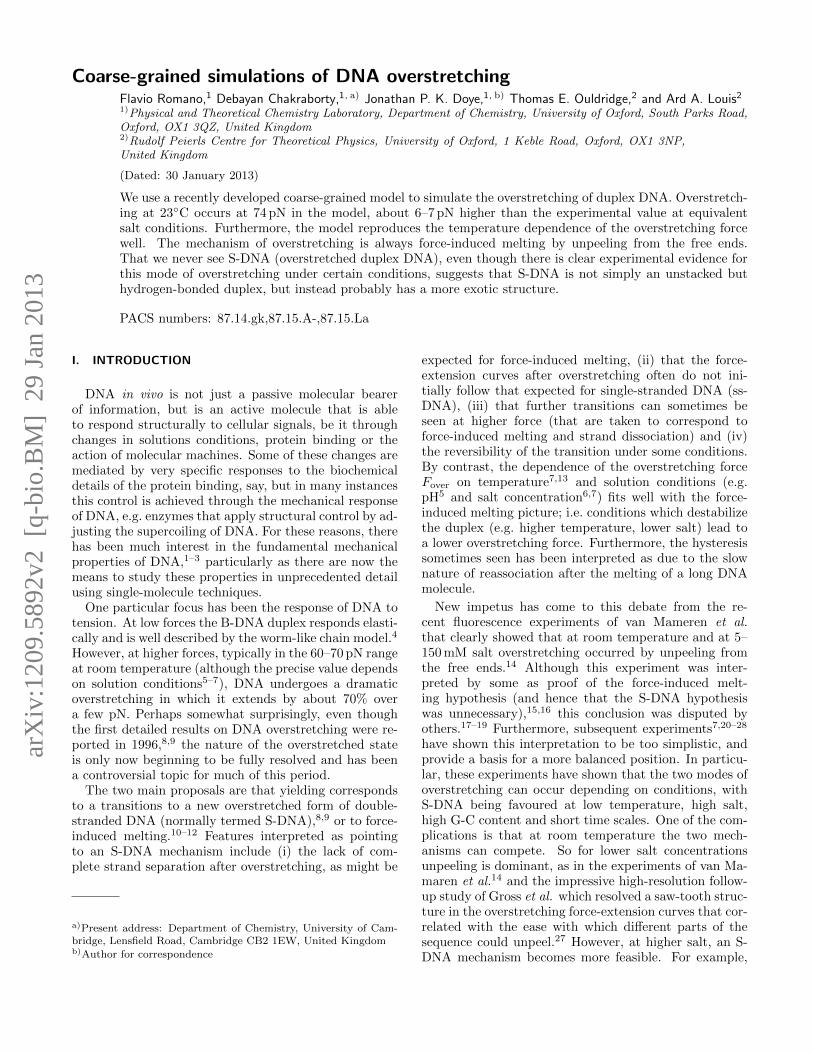
1 nucleotide
backbone base
FIG. 1. (a) Two nucleotides represented by our model,showing the rigid nucleotide unit and the backbone and baseregions. (b) A short DNA duplex as represented by our model.
developed coarse-grained DNA potential, which providesa quantitative description of many of the structural, ther-modynamic and mechanical properties of DNA.64,65 Oneaim of the work is to provide an independent test of therobustness of the model by applying it to phenomena towhich it has not been fitted. A second aim is to providefundamental insights into the nature of the overstretch-ing transition, both by visualizing some of the mechanis-tic aspects of the transitions, and by deducing from thesuccesses and failures of the model which ingredients areneeded to capture different aspects of the transition. Weproceed as follows. In the Methods section, we describeour DNA potential, the simulation techniques used andsome of the theory of the thermodynamics of pulling. Inthe Results section, we focus in detail on the room tem-perature overstretching behaviour, before more brieflyexamining the temperature dependence of overstretch-ing, including looking for the re-entrant behaviour thathas been predicted to occur near melting.10,13,33,34,38,39,55
II. METHODS
A. DNA model
We use the coarse-grained model of DNA recently de-veloped in our group.64,65 In particular, the model hasbeen designed to provide a good description of the struc-tural, thermodynamic and mechanical properties of bothdouble- and single-stranded DNA, all features that arevery important if we are to be able to describe DNAoverstretching. In addition, the wide range of appli-cations for which the model has been successfully usedgive us further confidence in its robustness. These ap-plications so far include DNA nanotweezers,64 kissinghairpins,66 DNA walkers,67–69 the formation of cruci-form DNA70 and the nematic transition of dense solu-tions of short duplexes;71 furthermore, investigations intohybridization, strand-exchange reactions, plectonemes,DNA origami and phase transitions in solutions of DNA
3
junctions are ongoing.The model describes each nucleotide as a rigid ob-
ject (Fig. 1(a)) that has interactions correspondingto backbone connectivity, excluded volume, stacking,hydrogen-bonding between complementary bases andcross-stacking. Each nucleotide has three collinear in-teraction sites (corresponding to the centres of the back-bone, stacking and hydrogen-bonding interactions) anda vector perpendicular to this axis to capture the pla-narity of the bases. We would like to emphasise that theattractive interactions in the model are not isotropic, butdepend on the relative orientation of the nucleotides. Itis these angular modulations that ensure that the DNAhelix is right-handed and pairs in an anti-parallel man-ner. The full form of the potential has been given in Refs.65 and 67. A simulation code incorporating the potentialis available to download.72
We should note a number of simplifying features of themodel that are relevant to the current study. Firstly, wedo not attempt to model the electrostatics explicitly, butinstead have fitted the potential parameters for a particu-lar salt concentration, namely 500 mM. At this concentra-tion the Debye screening length is short-ranged and theeffects of the electrostatics are included in the excluded-volume interactions. Fortunately, this salt concentrationis also one of those commonly used in experiments onoverstretching. However, as a consequence of this sim-plification we cannot of course examine how overstretch-ing depends on salt concentration. At room tempera-ture and 500 mM salt, experiments indicate that both theforce-induced melting and S-DNA modes of overstretch-ing compete.7,22,23
A second of the model’s simplifications is that it ig-nores sequence dependence in the interactions except forthe Watson-Crick nature of the base pairing (i.e. the hy-drogen bonding term in the potential only occurs forcomplementary bases). Therefore, each base pair has thesame average interaction strength irrespective of its iden-tity and its neighbours. This “average-base” approxima-tion can be an advantage when one is interested in thegeneric behaviour of DNA, as for the most part we arehere, but of course it excludes us from, for example, ex-amining how the overstretching depends on G-C content.A sequence-dependent version of the model has very re-cently been developed.73
Thirdly, the double helix in our model (Fig. 1(b)) issymmetrical with the helical grooves being of the samesize. This simplification, however, is likely to be rela-tively unimportant for the current study.
B. Pulling Schemes
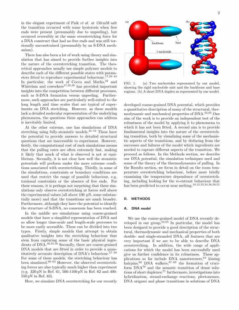
The three ways of pulling DNA that we will considerare shown in Fig. 2. Our main focus will be on schemeI, in which a force is imposed on both ends of the samestrand, and the free ends allow the duplex to overstretchby unpeeling. Scheme II is similar in that a force is
FIG. 2. Schematic representation of the three pullingschemes used. The arrows on the DNA are in the 3′-5′ di-rection.
exerted on only one of the two strands at each end ofthe duplex, thus again allowing unpeeling. However, asthe forces are applied to different strands, dissociationwill be completely irreversible when pulled at constantforce. In our simulations, we pull on both 3′ ends ofeach strand, but given the symmetric nature of the he-lix in our model and the simplified representation of thenucleotides, we do not expect any differences for pullingboth 5′ ends. In experiments differences between 5′-5′
and 3′-3′ pulling are only found at forces well above thatfor overstretching.74
Finally, in scheme III, as both ends of both strands arepulled, force-induced melting by unpeeling is suppressed.Therefore, if the DNA is to melt, it would have to be bybubble formation. Note the system in scheme III is nottorsionally constrained, i.e. the ends are free to rotate.
C. Thermodynamics of pulling
Before we consider our pulling simulations, it is impor-tant to understand the effect of imposing a force on thethermodynamics of our systems.10,32 The force providesan additional source of work being done on the system.Assuming the force F acts along the z direction, thisgives an additional term Fdz to the change in internalenergy and hence to the change in Helmholtz free en-ergy dA, and A is therefore a function of z. However,it will usually be more convenient to consider a systemunder constant force, and so we introduce the free energyA′ = A−Fz to describe such a constant-force ensemble.It follows that the change in free energy on applying aforce is
A′(F ) −A′(F = 0) = −∫ F
0
z(F )dF. (1)
Therefore, the force-extension curves of dsDNA and ss-DNA determine the relative stabilization of these twoforms by force. Furthermore, assuming overstretching
4
occurs by force-induced melting, one just needs a correctdescription of the zero-force thermodynamics and theforce-extension curves to correctly predict overstretching.
Force-induced unpeeling of DNA occurs at the force atwhich the free energy per base pair of the ssDNA anddsDNA states are equal, leading to coexistence of thetwo forms. In other words, at this overstretching forceFover(T ), the average free energy change for a duplex tounpeel by one more base pair is zero. We wish to em-phasise that overstretching, in the force-induced meltingpicture, is not determined by an equilibrium between ds-DNA and fully-dissociated ssDNA, but rather by whenthe duplex becomes unstable with respect to unpeeling.In this sense overstretching is distinct from melting, asthe translational entropy gain from complete dissociationof the two strands plays no role. However, in the bulklimit of infinitely long chains, the contribution of trans-lational entropy is negligible and melting and unpeelingtransitions are equivalent. In this limit, the melting tem-perature Tm(F ) can be defined as the temperature atwhich the average free energy change for the loss of onebase pair from a duplex is zero at a given force F , andthe curves Fover(T ) and Tm(F ) then give equivalent rep-resentations of the overstretching transition in the force-temperature plane. However, for finite chains Tm(F ) willdepend on concentration, and this equivalence will bebroken.
We also note that care should be taken when apply-ing this approach to the different schemes in Fig. 2. Forscheme I only one of the strands is force-bearing whensingle stranded. By contrast, in scheme III the force oneach single strand is F/2. Finally, in scheme II, equi-librium is not well-defined, as for any non-zero force themost stable state is two dissociated single strands becausethey can then increase their separation without limit.
D. Simulation methods
We use a mixture of Monte Carlo and Brownian dy-namics to simulate DNA stretching for our model. Weuse Monte Carlo to obtain the equilibrium behaviour ofthe system at a particular force, and Brownian dynamicsto study the stretching dynamics of DNA when the forceis increased or decreased (linearly) as a function of time.
To aid the equilibration in our Monte Carlo simula-tions, we use the virtual move Monte Carlo approach ofWhitelam and coworkers.75 This algorithm is a type of“cluster-move” Monte Carlo that allows clusters of nu-cleotides to be moved at each step, where the clustersthat are constructed reflect both the configuration andthe proposed move. The collective motion of nucleotidesthat this algorithm introduces allows more efficient sam-pling for our model compared to Monte Carlo with single-particle moves.
For the dynamics simulations we use the simple Brow-nian thermostat introduced in Ref. 76. The system isevolved according to Newtonian dynamics, but every
nNewt time steps a fraction p of the particles have all threecomponents of their velocities drawn from the Maxwelldistribution. On time scales longer than pnNewt, the dy-namics is Brownian.
Modifying these simulation algorithms to incorporatethe forces exerted on the terminal bases consistent withthe different schemes in Fig. 2 is straightforward. Toperform Monte Carlo at constant force it is necessaryto include an additional term in the energy −
∑i
Fizi,
where Fi is the external force imposed on nucleotide i.In Monte Carlo, this gives rise to an extra term in theBoltzmann factor in the standard Metropolis acceptancecriterion, namely exp(β
∑i
Fi∆zi). For the Brownian dy-
namics, implementing the constant force is just a matterof adding the external forces to the forces acting on therelevant nucleotides.
III. RESULTS
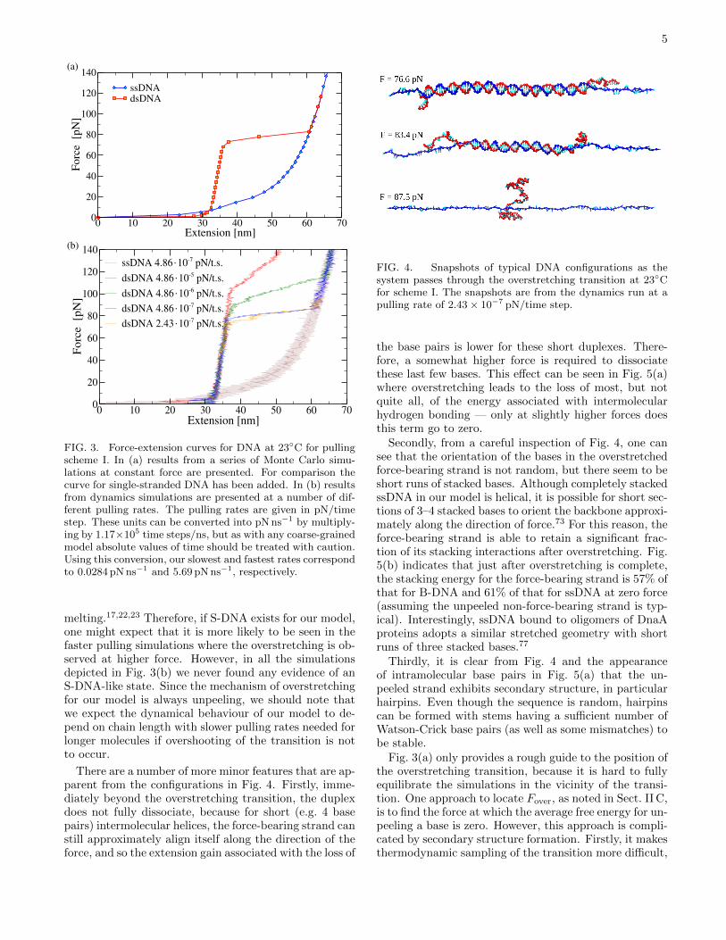
In all cases, we consider a 100 base-pair duplex witha random sequence. We first consider pulling by schemeI in detail. Force-extension curves at “room tempera-ture” (23◦C) are presented in Fig. 3 for Monte Carlosimulations at different forces, and for dynamics sim-ulations at different pulling rates. This data shows anumber of clear features. Firstly, the MC data (whichwe expect to be closer to “equilibrium”) shows a clearand narrow overstretching transition at a force of about77 pN. At the slowest pulling rates, the dynamics resultsalso show a clear overstretching plateau at a very similarforce. However, as the pulling rate increases, there is anincreasing tendency to overshoot this transition, and foroverstretching to start at a higher force and for the tran-sition to be spread over a wider range of force, simplybecause the time scale associated with the overstretch-ing transition is no longer fast compared to the pullingrate. These effects of pulling rate are also relevant tothe all-atom simulations as they provide an indicationof the consequences of being increasingly far from equi-librium. Indeed, our results at faster pulling rates some-what resemble the force-extension curves seen in all-atomsimulations.45–54 We should also note that although ourslowest pulling rates are significantly slower than thoseused in all-atom simulations, they are still much fasterthan in typical experiments.
Secondly, beyond the overstretching transition, theforce-extension curves follow that for ssDNA, thus indi-cating that DNA overstretching in our model is a resultof force-induced melting. This conclusion is confirmedin Fig. 4 which depicts typical configurations as the sys-tem passes through the overstretching transition. Fromthese shapshots, it can be clearly seen that the increasein extension is a result of DNA unpeeling from the freeends. In particular, there is no sign of any S-DNA-likestate. Experiments suggest that the transition from B- toS-DNA is able to occur more rapidly than force-induced
5
0 10 20 30 40 50 60 70Extension [nm]
0
20
40
60
80
100
120
140F
orc
e [
pN
]ssDNAdsDNA
0 10 20 30 40 50 60 70Extension [nm]
0
20
40
60
80
100
120
140
Fo
rce
[pN
]
ssDNA 4.86.10-7 pN/t.s.
dsDNA 4.86.10-5 pN/t.s.
dsDNA 4.86.10-6 pN/t.s.
dsDNA 4.86.10-7 pN/t.s.
dsDNA 2.43.10-7 pN/t.s.
(a)
(b)
FIG. 3. Force-extension curves for DNA at 23◦C for pullingscheme I. In (a) results from a series of Monte Carlo simu-lations at constant force are presented. For comparison thecurve for single-stranded DNA has been added. In (b) resultsfrom dynamics simulations are presented at a number of dif-ferent pulling rates. The pulling rates are given in pN/timestep. These units can be converted into pN ns−1 by multiply-ing by 1.17×10⁵ time steps/ns, but as with any coarse-grainedmodel absolute values of time should be treated with caution.Using this conversion, our slowest and fastest rates correspondto 0.0284 pN ns−1 and 5.69 pN ns−1, respectively.
melting.17,22,23 Therefore, if S-DNA exists for our model,one might expect that it is more likely to be seen in thefaster pulling simulations where the overstretching is ob-served at higher force. However, in all the simulationsdepicted in Fig. 3(b) we never found any evidence of anS-DNA-like state. Since the mechanism of overstretchingfor our model is always unpeeling, we should note thatwe expect the dynamical behaviour of our model to de-pend on chain length with slower pulling rates needed forlonger molecules if overshooting of the transition is notto occur.
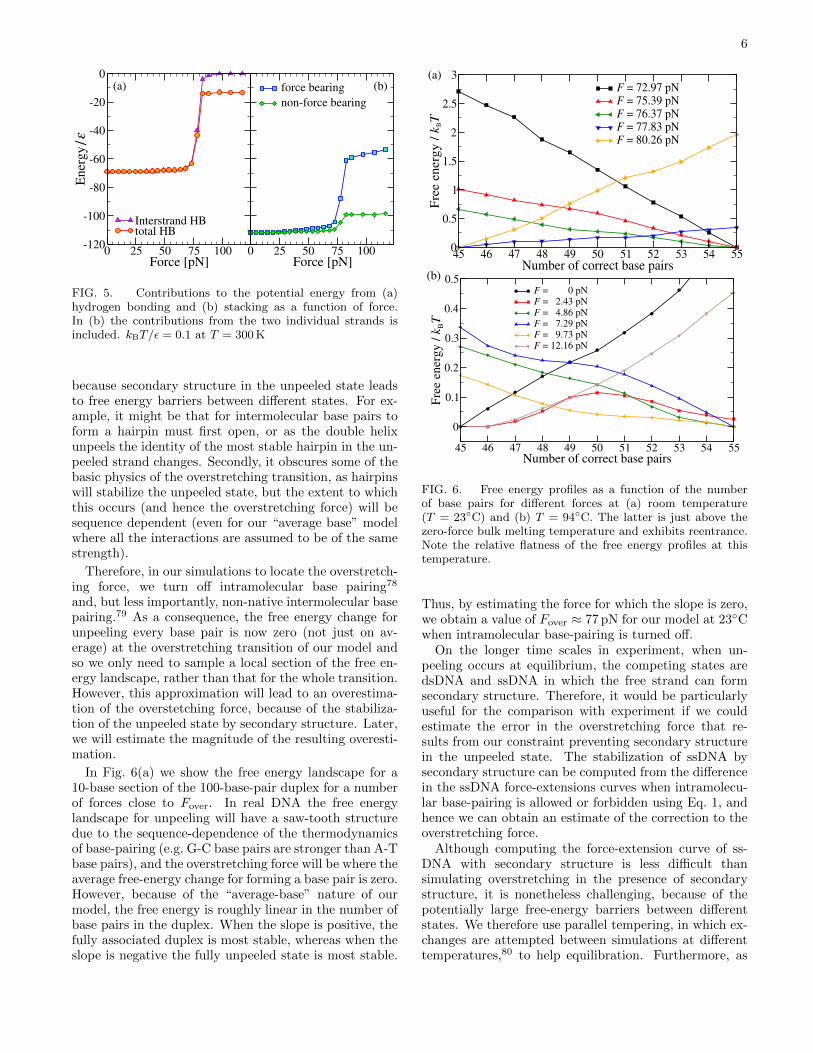
There are a number of more minor features that are ap-parent from the configurations in Fig. 4. Firstly, imme-diately beyond the overstretching transition, the duplexdoes not fully dissociate, because for short (e.g. 4 basepairs) intermolecular helices, the force-bearing strand canstill approximately align itself along the direction of theforce, and so the extension gain associated with the loss of
FIG. 4. Snapshots of typical DNA configurations as thesystem passes through the overstretching transition at 23◦Cfor scheme I. The snapshots are from the dynamics run at apulling rate of 2.43 × 10⁻⁷ pN/time step.
the base pairs is lower for these short duplexes. There-fore, a somewhat higher force is required to dissociatethese last few bases. This effect can be seen in Fig. 5(a)where overstretching leads to the loss of most, but notquite all, of the energy associated with intermolecularhydrogen bonding — only at slightly higher forces doesthis term go to zero.
Secondly, from a careful inspection of Fig. 4, one cansee that the orientation of the bases in the overstretchedforce-bearing strand is not random, but there seem to beshort runs of stacked bases. Although completely stackedssDNA in our model is helical, it is possible for short sec-tions of 3–4 stacked bases to orient the backbone approxi-mately along the direction of force.73 For this reason, theforce-bearing strand is able to retain a significant frac-tion of its stacking interactions after overstretching. Fig.5(b) indicates that just after overstretching is complete,the stacking energy for the force-bearing strand is 57% ofthat for B-DNA and 61% of that for ssDNA at zero force(assuming the unpeeled non-force-bearing strand is typ-ical). Interestingly, ssDNA bound to oligomers of DnaAproteins adopts a similar stretched geometry with shortruns of three stacked bases.77
Thirdly, it is clear from Fig. 4 and the appearanceof intramolecular base pairs in Fig. 5(a) that the un-peeled strand exhibits secondary structure, in particularhairpins. Even though the sequence is random, hairpinscan be formed with stems having a sufficient number ofWatson-Crick base pairs (as well as some mismatches) tobe stable.
Fig. 3(a) only provides a rough guide to the position ofthe overstretching transition, because it is hard to fullyequilibrate the simulations in the vicinity of the transi-tion. One approach to locate Fover, as noted in Sect. II C,is to find the force at which the average free energy for un-peeling a base is zero. However, this approach is compli-cated by secondary structure formation. Firstly, it makesthermodynamic sampling of the transition more difficult,
6
0 25 50 75 100Force [pN]
-120
-100
-80
-60
-40
-20
0E
ner
gy
/ε
Interstrand HBtotal HB
0 25 50 75 100Force [pN]
force bearing
non-force bearing
(a) (b)
FIG. 5. Contributions to the potential energy from (a)hydrogen bonding and (b) stacking as a function of force.In (b) the contributions from the two individual strands isincluded. kBT/ε = 0.1 at T = 300 K
because secondary structure in the unpeeled state leadsto free energy barriers between different states. For ex-ample, it might be that for intermolecular base pairs toform a hairpin must first open, or as the double helixunpeels the identity of the most stable hairpin in the un-peeled strand changes. Secondly, it obscures some of thebasic physics of the overstretching transition, as hairpinswill stabilize the unpeeled state, but the extent to whichthis occurs (and hence the overstretching force) will besequence dependent (even for our “average base” modelwhere all the interactions are assumed to be of the samestrength).
Therefore, in our simulations to locate the overstretch-ing force, we turn off intramolecular base pairing78
and, but less importantly, non-native intermolecular basepairing.79 As a consequence, the free energy change forunpeeling every base pair is now zero (not just on av-erage) at the overstretching transition of our model andso we only need to sample a local section of the free en-ergy landscape, rather than that for the whole transition.However, this approximation will lead to an overestima-tion of the overstetching force, because of the stabiliza-tion of the unpeeled state by secondary structure. Later,we will estimate the magnitude of the resulting overesti-mation.
In Fig. 6(a) we show the free energy landscape for a10-base section of the 100-base-pair duplex for a numberof forces close to Fover. In real DNA the free energylandscape for unpeeling will have a saw-tooth structuredue to the sequence-dependence of the thermodynamicsof base-pairing (e.g. G-C base pairs are stronger than A-Tbase pairs), and the overstretching force will be where theaverage free-energy change for forming a base pair is zero.However, because of the “average-base” nature of ourmodel, the free energy is roughly linear in the number ofbase pairs in the duplex. When the slope is positive, thefully associated duplex is most stable, whereas when theslope is negative the fully unpeeled state is most stable.
45 46 47 48 49 50 51 52 53 54 55Number of correct base pairs
0
0.5
1
1.5
2
2.5
3
Fre
e e
nerg
y /
kBT
F = 72.97 pNF = 75.39 pNF = 76.37 pNF = 77.83 pNF = 80.26 pN
45 46 47 48 49 50 51 52 53 54 55Number of correct base pairs
0
0.1
0.2
0.3
0.4
0.5
Fre
e en
erg
y /
kBT
F = 0 pNF = 2.43 pNF = 4.86 pNF = 7.29 pNF = 9.73 pNF = 12.16 pN
(b)
(a)
FIG. 6. Free energy profiles as a function of the numberof base pairs for different forces at (a) room temperature(T = 23◦C) and (b) T = 94◦C. The latter is just above thezero-force bulk melting temperature and exhibits reentrance.Note the relative flatness of the free energy profiles at thistemperature.
Thus, by estimating the force for which the slope is zero,we obtain a value of Fover ≈ 77 pN for our model at 23◦Cwhen intramolecular base-pairing is turned off.
On the longer time scales in experiment, when un-peeling occurs at equilibrium, the competing states aredsDNA and ssDNA in which the free strand can formsecondary structure. Therefore, it would be particularlyuseful for the comparison with experiment if we couldestimate the error in the overstretching force that re-sults from our constraint preventing secondary structurein the unpeeled state. The stabilization of ssDNA bysecondary structure can be computed from the differencein the ssDNA force-extensions curves when intramolecu-lar base-pairing is allowed or forbidden using Eq. 1, andhence we can obtain an estimate of the correction to theoverstretching force.
Although computing the force-extension curve of ss-DNA with secondary structure is less difficult thansimulating overstretching in the presence of secondarystructure, it is nonetheless challenging, because of thepotentially large free-energy barriers between differentstates. We therefore use parallel tempering, in which ex-changes are attempted between simulations at differenttemperatures,80 to help equilibration. Furthermore, as
7
0 5 10 15 20 25 30 35 40 45Extension [nm]
0
4
8
12
16F
orc
e [
pN
]secondary structure (average)
no secondary structure
FIG. 7. Force-extension curves for ssDNA in the presence orabsence of secondary structure. As the effects of secondarystructure depend on sequence, the results for four differentrandom sequences (as well as their average) are depicted. Thearea of the shaded region corresponds to the average free en-ergy of stabilization of ssDNA by secondary structure.
the stabilization will be sequence-dependent, one wouldalso want to average the correction over different se-quences when comparing to experiments for long DNAs.We therefore performed our simulations for five differentsequences. However, for the sequence that formed thestrongest secondary structure, we were unable to achieveequilibrium. The force-extension curves of the four othersequences are depicted in Fig. 7 (along with their av-erage) and compared to that for ssDNA with no sec-ondary structure. In particular, the secondary structuremakes the ssDNA less extensible at low force. However,as the force increases the secondary structure becomesdestabilized such that beyond 10 pN there is little differ-ence between the force-extension curves when secondarystructure is or is not allowed. This secondary-structureinduced low-force feature is also apparent in experimen-tal pulling curves for ssDNA at 500 mM salt, but disap-pears at lower salt.82 For comparison, fully complemen-tary hairpins are destabilized at about 16–17pN for thesame salt conditions as here.82
The area between the curve for ssDNA with in-tramolecular base-pairing forbidden and the averagecurve with secondary structure present gives the averagestabilization of a 100-base strand due to secondary struc-ture. At room temperature, this stabilization is 0.23 kTper base on average. Given this value and the free-energyprofiles in Fig. 6, we can then estimate the force at whichthe average free-energy change for unpeeling a base pairis zero when secondary structure in the unpeeled strandis allowed. This gives a correction of about 3 pN at roomtemperature, although this may be a slight underesti-mate, as we have not included the sequence that had thestrongest secondary structure in our estimate.
Hence, our best estimate of the overstretching force forour model at 23◦C is Fover ≈ 74 pN. This value is satisfy-ingly close to the oft-quoted 65 pN for room temperatureoverstretching, and much closer than has been previously
0 20 40 60 80 100Temperature [oC]
0
20
40
60
80
Ov
erst
retc
hin
g F
orc
e
[pN
] our modelwith sec. structureExpt. (Williams)
Expt. (Zhang)
80 85 90 950
10
20
30
FIG. 8. Dependence of the overstretching force on temper-ature. The main results for our model are from simulationswhere intramolecular base-pairing was turned off, but we alsoinclude results where a correction for secondary structure for-mation in the unpeeled chain has been applied for tempera-tures betwen 23◦C and 43◦C — above the latter temperaturethere is no need for a correction as the secondary structureis thermally unstable. Also included are the experimental re-sults from Refs. 7 and 13 at a salt concentration of 500 mM.Note that the results of Zhang et al. are for the onset (notthe midpoint) of the transition. The inset provides an ex-pansion of the high temperature region where non-monotonicbehaviour is observed. The horizontal line in the inset is theforce at which ssDNA and dsDNA have the same extension.
achieved for any coarse-grained model or atomistic simu-lation. More precisely, at 500 mM salt, the concentrationat which our coarse-grained model has been fitted, Wen-ner et al. obtain a value of 67 pN,6 and and in the morerecent study of Zhang et al. the onset and mid-point ofthe transitions were found to occur at 65 pN and 68 pN,respectively.7
The same approach can be used to study the temper-ature dependence of Fover, including for room tempera-ture and above the secondary structure correction factor— note that above 45◦C the correction factor goes tozero, because by this temperature all of the secondarystructure in the single strand has melted. As the tem-perature is increased, the duplex is destabilized relativeto ssDNA, so Fover is expected to decrease. This be-haviour is exactly what we see for our model in Fig. 8.Also included in this figure is the experimental data ofRefs. 7 and 13. Our results show a very similar tem-perature dependence over the temperature range studiedin experiments, albeit with our results being displacedto slightly higher force by a similar amount to that al-ready seen at room temperature. In particular, Zhanget al. find a slope of −0.44 pN K−1 at and above roomtemperature,7 which is similar to the −0.46 pN K−1 thatwe obtain for our model. Interestingly, Zhang et al. finda change in the sign of the slope below 20◦C which theyinterpret as a crossover to an S-DNA mechanism. Ourresults show no such change in slope, consistent with thefact that overstretching always occurs by unpeeling inour model.
8
One particularly interesting feature at high temper-ature and low force is the non-monotonic behaviour ofFover(T ). There is a narrow range of temperature justabove the bulk (i.e. infinitely long strands) zero-forcemelting temperature where there are two transitions asa function of force. This behaviour is illustrated in theinset to Fig. 8 and also in Fig. 6(b) where there are twochanges of sign in the slope of the free-energy profile as afunction of force for a temperature in this window. At lowand high force it is favourable for the duplex to unpeel,but there is an intermediate force range (∼2.5–10 pN at94◦C) where duplex DNA is most stable. This type ofbehaviour has been predicted for a number of theoreti-cal models10,13,33,34,38,39,55 and occurs because which ofdsDNA or ssDNA is more extensible depends on the mag-nitude of force. At low force dsDNA is more extensiblebecause it has a larger persistence length and so has asmaller entropy cost for aligning with the force. How-ever, when the extension approaches the contour lengthof dsDNA the force required to further extend the duplexgrows rapidly, and ssDNA becomes more extensible. Thecrossover between these two regimes occurs at approxi-mately 6.5 pN (i.e. when the two force-extension curvesin Fig. 3(a) cross). For forces below 6.5 pN, dsDNA isstabilized with respect to ssDNA by increasing force, butabove this value of the force, the opposite is true. Hence,the turning point in Fover(T ) is expected to occur at thisforce.
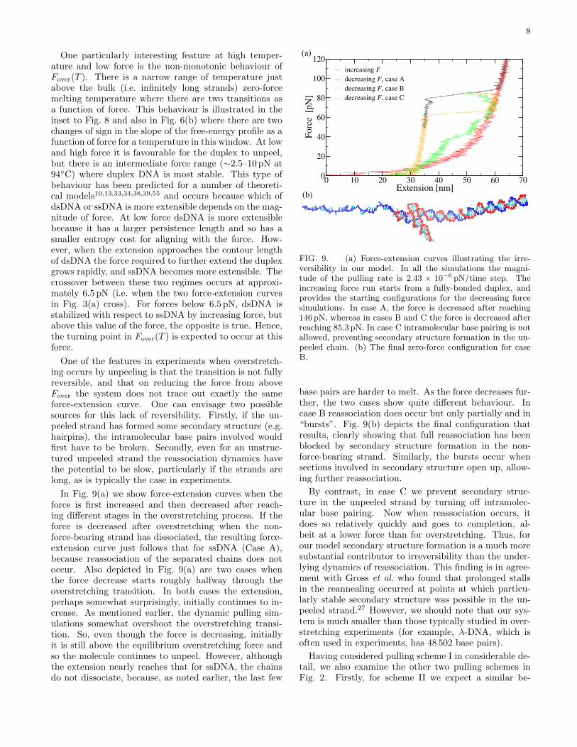
One of the features in experiments when overstretch-ing occurs by unpeeling is that the transition is not fullyreversible, and that on reducing the force from aboveFover the system does not trace out exactly the sameforce-extension curve. One can envisage two possiblesources for this lack of reversibility. Firstly, if the un-peeled strand has formed some secondary structure (e.g.hairpins), the intramolecular base pairs involved wouldfirst have to be broken. Secondly, even for an unstruc-tured unpeeled strand the reassociation dynamics havethe potential to be slow, particularly if the strands arelong, as is typically the case in experiments.
In Fig. 9(a) we show force-extension curves when theforce is first increased and then decreased after reach-ing different stages in the overstretching process. If theforce is decreased after overstretching when the non-force-bearing strand has dissociated, the resulting force-extension curve just follows that for ssDNA (Case A),because reassociation of the separated chains does notoccur. Also depicted in Fig. 9(a) are two cases whenthe force decrease starts roughly halfway through theoverstretching transition. In both cases the extension,perhaps somewhat surprisingly, initially continues to in-crease. As mentioned earlier, the dynamic pulling sim-ulations somewhat overshoot the overstretching transi-tion. So, even though the force is decreasing, initiallyit is still above the equilibrium overstretching force andso the molecule continues to unpeel. However, althoughthe extension nearly reaches that for ssDNA, the chainsdo not dissociate, because, as noted earlier, the last few
0 10 20 30 40 50 60 70Extension [nm]
0
20
40
60
80
100
120
Fo
rce
[p
N]
increasing F
decreasing F, case A
decreasing F, case B
decreasing F, case C
(b)
(a)
FIG. 9. (a) Force-extension curves illustrating the irre-versibility in our model. In all the simulations the magni-tude of the pulling rate is 2.43 × 10⁻⁶ pN/time step. Theincreasing force run starts from a fully-bonded duplex, andprovides the starting configurations for the decreasing forcesimulations. In case A, the force is decreased after reaching146 pN, whereas in cases B and C the force is decreased afterreaching 85.3 pN. In case C intramolecular base pairing is notallowed, preventing secondary structure formation in the un-peeled chain. (b) The final zero-force configuration for caseB.
base pairs are harder to melt. As the force decreases fur-ther, the two cases show quite different behaviour. Incase B reassociation does occur but only partially and in“bursts”. Fig. 9(b) depicts the final configuration thatresults, clearly showing that full reassociation has beenblocked by secondary structure formation in the non-force-bearing strand. Similarly, the bursts occur whensections involved in secondary structure open up, allow-ing further reassociation.
By contrast, in case C we prevent secondary struc-ture in the unpeeled strand by turning off intramolec-ular base pairing. Now when reassociation occurs, itdoes so relatively quickly and goes to completion, al-beit at a lower force than for overstretching. Thus, forour model secondary structure formation is a much moresubstantial contributor to irreversibility than the under-lying dynamics of reassociation. This finding is in agree-ment with Gross et al. who found that prolonged stallsin the reannealing occurred at points at which particu-larly stable secondary structure was possible in the un-peeled strand.27 However, we should note that our sys-tem is much smaller than those typically studied in over-stretching experiments (for example, λ-DNA, which isoften used in experiments, has 48 502 base pairs).
Having considered pulling scheme I in considerable de-tail, we also examine the other two pulling schemes inFig. 2. Firstly, for scheme II we expect a similar be-
9
10 20 30 40 50 60 70Extension [nm]
0
20
40
60
80
100F
orc
e [
pN
]scheme Ischeme II
(a)
(b)
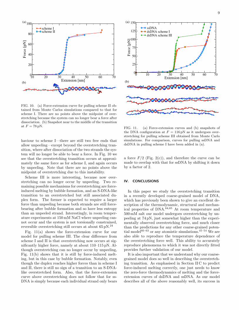
FIG. 10. (a) Force-extension curve for pulling scheme II ob-tained from Monte Carlos simulations compared to that forscheme I. There are no points above the midpoint of over-stretching because the system can no longer bear a force afterdissociation. (b) Snapshot near to the middle of the transitionat F = 78 pN.
haviour to scheme I—there are still two free ends thatallow unpeeling—except beyond the overstretching tran-sition, where after dissociation of the two strands the sys-tem will no longer be able to bear a force. In Fig. 10 wesee that the overstretching transition occurs at approxi-mately the same force as for scheme I, and again occursby unpeeling. Note that there are no points above themidpoint of overstretching due to this instability.
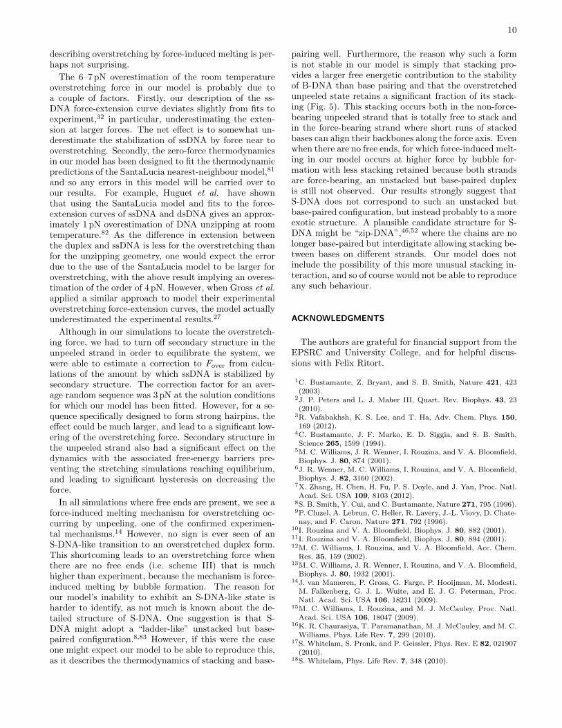
Scheme III is more interesting, because now over-stretching can no longer occur by unpeeling. Two re-maining possible mechanisms for overstretching are force-induced melting by bubble formation, and an S-DNA-liketransition to an overstretched but still associated du-plex form. The former is expected to require a largerforce than unpeeling because both strands are still force-bearing after bubble formation and so have less entropythan an unpeeled strand. Interestingly, in room temper-ature experiments at 150 mM NaCl where unpeeling can-not occur and the system is not torsionally constrained,reversible overstretching still occurs at about 65 pN.24
Fig. 11(a) shows the force-extension curve for ourmodel for pulling scheme III. The clear difference fromscheme I and II is that overstretching now occurs at sig-nificantly higher force, namely at about 110–115 pN. Al-though overstretching can no longer occur by unpeeling,Fig. 11(b) shows that it is still by force-induced melt-ing, but in this case by bubble formation. Notably, eventhough the duplex reaches higher forces than in scheme Iand II, there is still no sign of a transition to an S-DNA-like overstretched form. Also, that the force-extensioncurve above overstretching does not follow that for ss-DNA is simply because each individual strand only bears
0 10 20 30 40 50 60 70Extension [nm]
0
25
50
75
100
125
150
Forc
e [
pN
]
ssDNAdsDNA scheme IdsDNA scheme III
(b)
(a)
FIG. 11. (a) Force-extension curves and (b) snapshots ofthe DNA configuration at F = 116 pN as it undergoes over-stretching for pulling scheme III obtained from Monte Carlosimulations. For comparison, curves for pulling ssDNA anddsDNA in pulling scheme I have been added in (a).
a force F/2 (Fig. 2(c)), and therefore the curve can bemade to overlap with that for ssDNA by shifting it downby a factor of 2.
IV. CONCLUSIONS
In this paper we study the overstretching transitionin a recently developed coarse-grained model of DNA,which has previously been shown to give an excellent de-scription of the thermodynamic, structural and mechan-ical properties of DNA.64,65 At room temperature and500 mM salt our model undergoes overstretching by un-peeling at 74 pN, just somewhat higher than the experi-mentally observed overstretching force, and much closerthan the predictions for any other coarse-grained poten-tial model60–63 or any atomistic simulations.45–54 We arealso able to reproduce the temperature dependence ofthe overstretching force well. This ability to accuratelyreproduce phenomena to which it was not directly fittedprovides further validation of our model.
It is also important that we understand why our coarse-grained model does so well in describing the overstretch-ing transition. As emphasised in Section II C to predictforce-induced melting correctly, one just needs to knowthe zero-force thermodynamics of melting and the force-extension curves of dsDNA and ssDNA. As our modeldescribes all of the above reasonably well, its success in
10
describing overstretching by force-induced melting is per-haps not surprising.
The 6–7 pN overestimation of the room temperatureoverstretching force in our model is probably due toa couple of factors. Firstly, our description of the ss-DNA force-extension curve deviates slightly from fits toexperiment,32 in particular, underestimating the exten-sion at larger forces. The net effect is to somewhat un-derestimate the stabilization of ssDNA by force near tooverstretching. Secondly, the zero-force thermodynamicsin our model has been designed to fit the thermodynamicpredictions of the SantaLucia nearest-neighbour model,81
and so any errors in this model will be carried over toour results. For example, Huguet et al. have shownthat using the SantaLucia model and fits to the force-extension curves of ssDNA and dsDNA gives an approx-imately 1 pN overestimation of DNA unzipping at roomtemperature.82 As the difference in extension betweenthe duplex and ssDNA is less for the overstretching thanfor the unzipping geometry, one would expect the errordue to the use of the SantaLucia model to be larger foroverstretching, with the above result implying an overes-timation of the order of 4 pN. However, when Gross et al.applied a similar approach to model their experimentaloverstretching force-extension curves, the model actuallyunderestimated the experimental results.27
Although in our simulations to locate the overstretch-ing force, we had to turn off secondary structure in theunpeeled strand in order to equilibrate the system, wewere able to estimate a correction to Fover from calcu-lations of the amount by which ssDNA is stabilized bysecondary structure. The correction factor for an aver-age random sequence was 3 pN at the solution conditionsfor which our model has been fitted. However, for a se-quence specifically designed to form strong hairpins, theeffect could be much larger, and lead to a significant low-ering of the overstretching force. Secondary structure inthe unpeeled strand also had a significant effect on thedynamics with the associated free-energy barriers pre-venting the stretching simulations reaching equilibrium,and leading to significant hysteresis on decreasing theforce.
In all simulations where free ends are present, we see aforce-induced melting mechanism for overstretching oc-curring by unpeeling, one of the confirmed experimen-tal mechanisms.14 However, no sign is ever seen of anS-DNA-like transition to an overstretched duplex form.This shortcoming leads to an overstretching force whenthere are no free ends (i.e. scheme III) that is muchhigher than experiment, because the mechanism is force-induced melting by bubble formation. The reason forour model’s inability to exhibit an S-DNA-like state isharder to identify, as not much is known about the de-tailed structure of S-DNA. One suggestion is that S-DNA might adopt a “ladder-like” unstacked but base-paired configuration.8,83 However, if this were the caseone might expect our model to be able to reproduce this,as it describes the thermodynamics of stacking and base-
pairing well. Furthermore, the reason why such a formis not stable in our model is simply that stacking pro-vides a larger free energetic contribution to the stabilityof B-DNA than base pairing and that the overstretchedunpeeled state retains a significant fraction of its stack-ing (Fig. 5). This stacking occurs both in the non-force-bearing unpeeled strand that is totally free to stack andin the force-bearing strand where short runs of stackedbases can align their backbones along the force axis. Evenwhen there are no free ends, for which force-induced melt-ing in our model occurs at higher force by bubble for-mation with less stacking retained because both strandsare force-bearing, an unstacked but base-paired duplexis still not observed. Our results strongly suggest thatS-DNA does not correspond to such an unstacked butbase-paired configuration, but instead probably to a moreexotic structure. A plausible candidate structure for S-DNA might be “zip-DNA”,46,52 where the chains are nolonger base-paired but interdigitate allowing stacking be-tween bases on different strands. Our model does notinclude the possibility of this more unusual stacking in-teraction, and so of course would not be able to reproduceany such behaviour.
ACKNOWLEDGMENTS
The authors are grateful for financial support from theEPSRC and University College, and for helpful discus-sions with Felix Ritort.
1C. Bustamante, Z. Bryant, and S. B. Smith, Nature 421, 423(2003).
2J. P. Peters and L. J. Maher III, Quart. Rev. Biophys. 43, 23(2010).
3R. Vafabakhsh, K. S. Lee, and T. Ha, Adv. Chem. Phys. 150,169 (2012).
4C. Bustamante, J. F. Marko, E. D. Siggia, and S. B. Smith,Science 265, 1599 (1994).
5M. C. Williams, J. R. Wenner, I. Rouzina, and V. A. Bloomfield,Biophys. J. 80, 874 (2001).
6J. R. Wenner, M. C. Williams, I. Rouzina, and V. A. Bloomfield,Biophys. J. 82, 3160 (2002).
7X. Zhang, H. Chen, H. Fu, P. S. Doyle, and J. Yan, Proc. Natl.Acad. Sci. USA 109, 8103 (2012).
8S. B. Smith, Y. Cui, and C. Bustamante, Nature 271, 795 (1996).9P. Cluzel, A. Lebrun, C. Heller, R. Lavery, J.-L. Viovy, D. Chate-nay, and F. Caron, Nature 271, 792 (1996).
10I. Rouzina and V. A. Bloomfield, Biophys. J. 80, 882 (2001).11I. Rouzina and V. A. Bloomfield, Biophys. J. 80, 894 (2001).12M. C. Williams, I. Rouzina, and V. A. Bloomfield, Acc. Chem.
Res. 35, 159 (2002).13M. C. Williams, J. R. Wenner, I. Rouzina, and V. A. Bloomfield,
Biophys. J. 80, 1932 (2001).14J. van Mameren, P. Gross, G. Farge, P. Hooijman, M. Modesti,
M. Falkenberg, G. J. L. Wuite, and E. J. G. Peterman, Proc.Natl. Acad. Sci. USA 106, 18231 (2009).
15M. C. Williams, I. Rouzina, and M. J. McCauley, Proc. Natl.Acad. Sci. USA 106, 18047 (2009).
16K. R. Chaurasiya, T. Paramanathan, M. J. McCauley, and M. C.Williams, Phys. Life Rev. 7, 299 (2010).
17S. Whitelam, S. Pronk, and P. Geissler, Phys. Rev. E 82, 021907(2010).
18S. Whitelam, Phys. Life Rev. 7, 348 (2010).
11
19O. Krichevsky, Phys. Life Rev. 7, 350 (2010).20C. P. Calderon, W.-H. Chen, K.-J. Lin, N. C. Harris, and C.-H.
Kiang, J. Phys.: Condens. Matter 21, 034114 (2009).21C. U. Murade, V. Subramaniam, C. Otto, and M. L. Bennink,
Nucleic Acids Res. 38, 3423 (2010).22H. Fu, H. Chen, J. F. Marko, and J. Yan, Nucleic Acids Res. 38,
5594 (2010).23H. Fu, H. Chen, X. Zhang, Y. Qu, J. F. Marko, and J. Yan,
Nucleic Acids Res. 39, 3473 (2011).24D. H. Paik and T. T. Perkins, J. Am. Chem. Soc. 133, 3219
(2011).25P. Bianco, L. Bongini, L. Melli, M. Dolfi, and V. Lombardi, Bio-
phys. J. 101, 866 (2011).26M. Maaloum, A.-F. Beker, and P. Muller, Phys. Rev. E 83,
031903 (2011).27P. Gross, N. Laurens, L. B. Oddershede, U. Bockelmann, E. J. G.
Peterman, and G. J. L. Wuite, Nature Physics 7, 731 (2011).28N. Bosaeus, A. H. El-Sagheer, T. Brown, S. B. Smith, B. Ak-
erman, C. Bustamante, and B. Norden, Proc. Natl. Acad. Sci.USA 109, 15179 (2012).
29P. Cizeau and J.-L. Viovy, Biopolymers 42, 383 (1997).30A. Ahsan, J. Rudnick, and R. Bruinsma, Biophys. J. 74, 132
(1998).31C. Storm and P. C. Nelson, Phys. Rev. E 67, 051906 (2003).32S. Cocco, J. Yan, J.-F. Leger, D. Chatenay, and J. F. Marko,
Phys. Rev. E 70, 011910 (2004).33A. Hanke, M. G. Ochoa, and R. Metzler, Phys. Rev. Lett. 100,
018106 (2008).34S. J. Rahi, M. P. Hertzbert, and M. Kardar, Phys. Rev. E 78,
051910 (2008).35S. Whitelam, S. Pronk, and P. Geissler, J. Chem. Phys. 129,
205101 (2008).36S. Whitelam, S. Pronk, and P. Geissler, Biophys. J. 94 (2008).37B. Chakrabarti and D. R. Nelson, J. Phys. Chem. 113, 3831
(2009).38D. Marenduzzo, A. Maritan, E. Orlandini, F. Seno, and
A. Trovato, J. Stat. Mech, L04001(2009).39T. R. Einert, D. B. Staple, H.-J. Kreuzer, and R. R. Netz, Bio-
phys. J. 99, 578 (2010).40F. A. Massuci, I. P. Castillo, and C. P. Vicente, arXiv:1006.2327.41C. Nisoli and A. R. Bishop, Phys. Rev. Lett. 107, 068102 (2011).42A. Fiasconaro and F. Falo, Phys. Rev. E 86, 023902 (2012).43M. Manghi, N. Destainville, and J. Palmeri, Eur. Phys. J. E 35,
110 (2012).44A. Lebrun and R. Lavery, Nuc. Acids Res. 24, 2260 (1996).45S. A. Harris, Z. A. Sands, and C. A. Laughton, Biophys. J. 88,
1684 (2005).46R. Lohikoski, J. Timonen, and A. Laaksonen, Chem. Phys. Lett.407, 23 (2005).
47J. Wereszczynski and I. Andricioaei, Proc. Natl. Acad. Sci. USA103, 16200 (2006).
48B. Luan and A. Aksimentiev, Phys. Rev. Lett. 101, 101 (2008).49H. Li and T. Gisler, Eur. Phys. J. E 30, 325 (2009).50M. Santosh and P. K. Maiti, J. Phys.: Condens. Matter 21,
034113 (2009).51J. Rezac, P. Hobza, and S. A. Harris, Biophys. J. 98, 101 (2010).52A. Balaeff, S. L. Craig, and D. N. Beratan, J. Phys. Chem. A115, 9377 (2011).
53M. Santosh and P. K. Maiti, Biophys. J. 101, 1393 (2011).54M. Wolter, M. Elstner, and T. Kubar, J. Phys. Chem. A 115,
11238 (2011).
55D. Marenduzzo, E. Orlandini, F. Seno, and A. Trovato, Phys.Rev. E 81, 051926 (2010).
56A. R. Singh, D. Giri, and S. Kumar, J. Chem. Phys. 132, 235105(2010).
57J. J. de Pablo, Ann. Rev. Phys. Chem. 62, 555 (2011).58F. Lankas, in Innovations in biomolecular modelling and simu-lation, Vol. 2, edited by T. Schlick (Royal Society of Chemistry,2012) pp. 3–32.
59J. P. K. Doye, T. E. Ouldridge, A. A. Louis, F. Romano, P. Sulc,
C. Matek, and B. Snodin, Phys. Chem. Chem. Phys.(to be sub-mitted).
60S. Niewieczerza l and M. Cieplak, J. Phys.: Condens. Matter 21,474221 (2009).
61A. V. Savin, M. A. Mazo, I. P. Kikot, and A. V. Onufriev,arXiv:1110.3165.
62A. Kocsis and D. Swigon, Int. J. Nonlinear Mech. 47, 639 (2012).63C. W. Hsu, M. Fyta, G. Lakatos, S. Melchionna, and E. Kaxiras,
J. Chem. Phys. 137, 105102 (2012).64T. E. Ouldridge, A. A. Louis, and J. P. K. Doye, Phys. Rev. Lett104, 178101 (2010).
65T. E. Ouldridge, A. A. Louis, and J. P. K. Doye, J. Chem. Phys.134, 085101 (2011).
66F. Romano, A. Hudson, J. P. K. Doye, T. E. Ouldridge, andA. A. Louis, J. Chem. Phys. 136, 215102 (2012).
67T. E. Ouldridge, Coarse-grained modelling of DNA and DNAself-assembly, Ph.D. thesis, University of Oxford (2011); http:
//tinyurl.com/7ycbx7c.68T. E. Ouldridge, R. Hoare, A. A. Louis, J. P. K. Doye, J. Bath,
and A. J. Turberfield, ACS NANO submitted.69P. Sulc, T. E. Ouldridge, F. Romano, J. P. K. Doye, and A. A.
Louis, Natural Computing, submitted; arXiv:1212.4536.70C. Matek, T. E. Ouldridge, A. Levy, J. P. K. Doye, and A. A.
Louis, J. Phys. Chem. B 116, 11616 (2012).71C. de Michele, L. Rovigatti, T. Bellini, and F. Sciortino, Soft
Matter: 8, 8388 (2012).72http://dna.physics.ox.ac.uk.73P. Sulc, F. Romano, T. E. Ouldridge, L. Rovigatti, J. P. K. Doye,
and A. A. Louis, J. Chem. Phys. 137, 135101 (2012).74C. Danilowicz, C. Limouse, K. Hatch, A. Conover, V. W. Coljee,
N. Kleckner, and M. Prentiss, Proc. Natl. Acad. Sci. USA 106,13196 (2009).
75S. Whitelam and P. Geissler, J. Chem. Phys. 127, 154101 (2007).76J. Russo, P. Tartaglia, and F. Sciortino, J. Chem. Phys. 131,
014504 (2009).77K. E. Duderstadt, K. Chuang, and J. M. Berger, Nature 478,
209 (2011).78This could also be achieved by choosing a sequence with no com-
plementary bases in the same strand.79In some simulations with repetitive sequences (not reported here)
we have seen a tendency for the unpeeled strand to rebind tothe force-bearing strand in a non-native site for short stretches(short so that the helix of the force-bearing strand can still ap-proximately allign along the direction of force).
80D. J. Earl and M. W. Deem, Phys. Chem. Chem. Phys. 7, 3910(2005).
81J. SantaLucia, Jr. and D. Hicks, Annu. Rev. Biophys. Biomol.Struct. 33, 415 (2004).
82J. M. Huguet, C. V. Bizarro, N. Forns, S. B. Smith, C. Busta-mante, and F. Ritort, Proc. Natl. Acad. Sci. USA 107, 15431(2010).
83T. Thundat, D. P. Allison, and R. J. Warmack, Nucl. Acids Res.22, 4224 (1994).