clpb variants associated with autosomal-recessive mitochondrial disorder with cataract, neutropenia,...
TRANSCRIPT
Please cite this article in press as: Saunders et al., CLPB Variants Associated with Autosomal-Recessive Mitochondrial Disorder with Cataract,Neutropenia, Epilepsy, and Meth..., The American Journal of Human Genetics (2015), http://dx.doi.org/10.1016/j.ajhg.2014.12.020
REPORT
CLPB Variants Associated with Autosomal-RecessiveMitochondrial Disorder with Cataract, Neutropenia,Epilepsy, and Methylglutaconic Aciduria
Carol Saunders,1,2,* Laurie Smith,1,3 Flemming Wibrand,4 Kirstine Ravn,4 Peter Bross,5
Isabelle Thiffault,1 Mette Christensen,4 Andrea Atherton,3 Emily Farrow,1,3 Neil Miller,1
Stephen F. Kingsmore,1,2,3 and Elsebet Ostergaard4,*
3-methylglutaconic aciduria (3-MGA-uria) is a nonspecific finding associated with mitochondrial dysfunction, including defects of
oxidative phosphorylation. 3-MGA-uria is classified into five groups, of which one, type IV, is genetically heterogeneous. Here we report
five children with a form of type IV 3-MGA-uria characterized by cataracts, severe psychomotor regression during febrile episodes,
epilepsy, neutropenia with frequent infections, and death in early childhood. Four of the individuals were of Greenlandic descent,
and one was North American, of Northern European and Asian descent. Through a combination of homozygosity mapping in the
Greenlandic individuals and exome sequencing in the North American, we identified biallelic variants in the caseinolytic peptidase
B homolog (CLPB). The causative variants included one missense variant, c.803C>T (p.Thr268Met), and two nonsense variants,
c.961A>T (p.Lys321*) and c.1249C>T (p.Arg417*). The level of CLPB protein was markedly decreased in fibroblasts and liver of affected
individuals. CLPB is proposed to function as amitochondrial chaperone involved in disaggregation of misfolded proteins, resulting from
stress such as heat denaturation.
3-methylglutaconic aciduria (3-MGA-uria) is a nonspecific
biochemical finding associated with a group of inborn
errors of metabolism, particularly mitochondrial disorders.
3-MGA is a branched-chain organic acid and intermediate
of leucine degradation and themevalonate shunt pathway.
The latter is essential for the synthesis of cholesterol, coen-
zyme Q, dolichols, and isoprenoids, which are needed for
sterol synthesis and acetyl-CoA metabolism in mitochon-
dria.1–4 The clinical features of the 3-MGA-uria syndromes
are diverse and are classified into five types, each with
significant heterogeneity.5,6 In all types, with the excep-
tion of 3-MGA-uria type I (MIM 250950), the activities of
3-methylglutaconyl-CoA hydratase and other enzymes of
leucine degradation are normal, and the 3-MGA-uria is
thought to be secondary to defects in phospholipid remod-
eling or integrity of mitochondrial membranes, leading to
electron transport chain dysfunction.7,8 3-MGA-uria type I
is an inborn error of leucine metabolism, caused by vari-
ants in AUH (MIM 600529). AUH encodes 3-methylglu-
taconyl-CoA hydratase, which catalyzes the fifth step of
leucine catabolism, whereby 3-methylglutaconyl-CoA is
converted to 3-hydroxy-3-methylglutaryl-coenzyme A.9
3-MGA-uria type I might present as an acute, life-threat-
ening condition in childhood with nonspecific features
such as seizures, intellectual disability, or abnormal liver
function. It might also present as leukoencephalopathy
in adults.5
3-MGA-uria type II (MIM 302060), or Barth syndrome, is
an X-linked recessive disorder caused by variants in TAZ
1Center for Pediatric Genomic Medicine, Children’s Mercy Hospital, Kansas C
Children’s Mercy Hospital, Kansas City, MO 64108, USA; 3Department of Pedia
of Clinical Genetics, Copenhagen University Hospital Rigshospitalet, 2100 Co
versity and Aarhus University Hospital, 8200 Aarhus, Denmark
*Correspondence: [email protected] (C.S.), [email protected] (
http://dx.doi.org/10.1016/j.ajhg.2014.12.020. �2015 by The American Societ
The
(MIM 300394), which encodes taffazin, a cardiolipin
transacylase in the inner mitochondrial membrane.10
Clinical findings include cardiomyopathy, myopathy,
short stature, neutropenia, hypocholesterolemia, dysmor-
phism, and cognitive difficulties. 3-MGA-uria type III
(MIM 258501), or Costeff syndrome, is caused by variants
in OPA3 (MIM 165300), characterized by early-onset optic
atrophy, extrapyramidal signs, spasticity, ataxia, dysar-
thria, and cognitive deficiency.11 OPA3 is a mitochondrial
outer membrane protein involved in maintenance of
the respiratory chain. 3-MGA-uria type V (MIM 610198)
features early-onset dilated cardiomyopathy, nonprogres-
sive cerebellar ataxia, testicular dysgenesis, and growth
failure. Type V is caused by variants in DNAJC19 (MIM
608977), which encodes a mitochondrial cochaper-
one.7,12 DNAJC19 forms a complex with prohibitins
(PHB) and lipid scaffolds in the inner membrane of mito-
chondria that is necessary for mitochondrial morphogen-
esis, neuronal survival, and phospholipid remodeling. In
particular, this complex regulates cardiolipin remodeling
in cardiac mitochondrial membranes through tafazzin.7,8
Thus, 3-MGA-uria types V and II affect a common pathway
related to mitochondrial membrane metabolism, defects
in which lead to loss of electron transport chain function
and energy production.
3-MGA-uria type IV (MIM 250951), the ‘‘unclassified
type,’’ includes all other forms of 3-MGA-uria with normal
3-methylglutaconyl-CoA hydratase enzyme activity. A
diagnosis of 3-MGA-uria type IV is complicated by the large
ity, MO 64108, USA; 2Department of Pathology and Laboratory Medicine,
trics, Children’s Mercy Hospital, Kansas City, MO 64108, USA; 4Department
penhagen, Denmark; 5Research Unit for Molecular Medicine, Aarhus Uni-
E.O.)
y of Human Genetics. All rights reserved.
American Journal of Human Genetics 96, 1–8, February 5, 2015 1

Table 1. Biochemical Findings in Individuals with CLPB Variants
Subject 1 2 3 4 5
White blood count (WBC), 3 109/l (reference, 4.0–14.0 3 109/l) 1.4–9.3 2.8–7.1 2.3–7.7 NA 2.69–8.19 3 103 mcl (reference, 5–21)
Absolute neutrophil count (ANC), 3 109/l (reference:neutropenia < 1.5 3 109/l, severe neutropenia < 0.5 3 109/l)
0–0.8 0.1–1.9 0.3 NA 0.00–0.39 3 103 mcl (reference, 1.5–9.0)
Platelets NA NA NA NA 66–188 3 103 mcl (reference, 150–450)
Blood lactate, mmol/l (reference, <1.7 mmol/l) <7.7 3.8 NA NA 2.2–5.5 (0.7–2.1)
CSF lactate, mmol/l (reference, <1.7 mmol/l) 5.89 2.11 NA NA
Urine methylglutaconic acid, mg/g creatinine (reference,<15 mg/g creatinine)
70–250 250 210–260 80–220 193 mmol/mol creatinine (reference, <15)
NA, not available.
Please cite this article in press as: Saunders et al., CLPB Variants Associated with Autosomal-Recessive Mitochondrial Disorder with Cataract,Neutropenia, Epilepsy, and Meth..., The American Journal of Human Genetics (2015), http://dx.doi.org/10.1016/j.ajhg.2014.12.020
number of implicated genes, including those involved in
mitochondrial DNA depletion syndromes, mitochondrial
DNA deletion syndromes, MELAS (MIM 540000), Smith-
Lemli-Opitz syndrome (MIM 270400), and glycogen stor-
agedisease type1b (MIM232220).5,6,13,14Many individuals
with type IV 3-MGA-uria have pathogenic variants in
nuclear-encoded mitochondrial genes involved in respira-
tory chain function.5,15,16 In addition, two genes have
been associated with specific type IV 3-MGA-uria disorders.
First, variants in SERAC1 (MIM 614725), which encodes an
enzyme involved in phosphatidylglycerol remodeling,
are associated with MEGDEL syndrome (MIM 614739),
characterized by progressive spasticity, dystonia, deaf-
ness, psychomotor retardation, hypocholesterolemia, and
Leigh-like lesions on MRI.17 Second, variants in TMEM70
(MIM 612418) are associated with an isolated mitochon-
drial complex V deficiency and neonatal hypotonia,
hypertrophic cardiomyopathy, psychomotor retardation,
cataract, and hyperammonemia.6
Molecular diagnosis of 3-MGA-uria is complicated
by marked clinical and locus heterogeneity, and many
affected individuals lack a specific genetic etiology because
of the number of diverse targets, some of which have yet to
be identified. Individuals with 3-MGA-uria present with
variable symptoms and organ involvement, with predom-
inantly progressive neurological impairment in combina-
tion with 3-MGA-uria and other biochemical findings
suggestive of mitochondrial dysfunction.
Here, we describe the identification of variants in CLPB,
associated with a distinct 3-MGA-uria syndrome character-
ized by cataracts, severe psychomotor regression during
febrile episodes, epilepsy, neutropenia with frequent infec-
tions, and death in early childhood. Four individuals,
including one pair of siblings, were from Greenland; the
fifth was a North American, of Northern European and
Asian descent. None of the families from Greenland were
known to be related.
Written informed consent was obtained from all individ-
uals investigated or their guardians, and the research
protocols were performed in accordance with the ethical
standards of the respective national and international
committees on human subject research.
2 The American Journal of Human Genetics 96, 1–8, February 5, 2015
Individual 1 was female, the first child born to healthy
parents after an uncomplicated pregnancy, weighing
2,680 g at term. At the age of 6 months, she was diagnosed
with bilateral cataracts. Although psychomotor develop-
ment was apparently normal, by 9 months of age, hypoto-
nia was identified. Severe dehydration and respiratory
distress was precipitated by a febrile illness at 11 months
of age, after which developmental regression occurred
along with severe hypotonia and development of extrapy-
ramidal symptoms (buccolingual movements, myoclonus,
and choreoathetosis) and, subsequently, seizures. No dys-
morphic features were noted. Hepatic transaminases were
elevated and associated with marked macronodular steato-
sis on liver biopsy. Her clinical course featured intermittent
leukopenia, moderate to severe neutropenia, and frequent
infections (Table 1). Bone marrow examination showed a
profound maturation arrest. Blood and cerebrospinal fluid
(CSF) lactic acid levels were elevated, and urine organic
acid screening demonstrated moderate 3-MGA-uria, with
marked increase during the first febrile episode. She died
at 11 months of age with heart failure, kidney failure,
and pneumothorax.
Individual 2 was the younger brother of individual 1.
Pregnancy and birth were uncomplicated. He was born at
term with a birth weight of 3,090 g and a head circumfer-
ence of 34 cm. Like his sister, his early development
was normal until a febrile illness at 9 months of age,
when he developed a very similar phenotype and labora-
tory findings. No dysmorphic features were noted. He
required tube feeding (Table 1). Analysis of bone marrow
showed deficient granulopoiesis. Magnetic resonance im-
aging (MRI) of the brain showed symmetrical hyperinten-
sities in the globus pallidus and cerebral atrophy. Muscle
histology was normal. He died at 3 years of age; cause of
death was not reported.
Individual 3 was female, and the first child born to
healthy parents, weighing 3,000 g and measuring 51 cm
at birth. Her early development was apparently normal,
but from age 6 months, she was noted to have psy-
chomotor retardation with regression and hypotonia.
No dysmorphic features were noted. At 1 year of age,
she was admitted to the hospital because of generalized

Please cite this article in press as: Saunders et al., CLPB Variants Associated with Autosomal-Recessive Mitochondrial Disorder with Cataract,Neutropenia, Epilepsy, and Meth..., The American Journal of Human Genetics (2015), http://dx.doi.org/10.1016/j.ajhg.2014.12.020
epilepsy. She was microcephalic (42.7 cm, 2 SD below the
normal range), with hypertonicity of the upper extremities
and hyperreflexia. Ophthalmologic examination showed
slight zonular cataracts bilaterally. A brain MRI was
normal. She had leukopenia and severe neutropenia. Urine
3-MGA was moderately elevated. She died at 2.5 years of
age from pneumonia.
Individual 4 was female, the third child born to healthy
parents after an uncomplicated pregnancy and term birth,
weighing 2,700 g and measuring 50 cm. Her two older
siblings were healthy. She had pneumonia three times in
the first 4 months of life. During the last episode, she
was lethargic and had convulsions, featuring opisthoto-
nus. She was nondysmorphic. She subsequently had severe
hypotonia, psychomotor retardation, repeated episodes of
opisthotonus, hyperreflexia, dystonia, and few sponta-
neous movements. She had growth retardation and micro-
cephaly (�3 SD). She developed severe generalized epilepsy
at the age of 2 years. Liver transaminases were normal.
There was chronic neutropenia but not leukopenia.
3-MGA excretion was moderately elevated. Brain CT at
age 6 months was normal. She died at 4 years of age; cause
of death was not reported.
Individual 5 was a female infant born to a 26-year-old
G2P1 011 mother. She was delivered at 39 weeks gestation
by elective Caesarean section, because of breech presenta-
tion. Her birth weight was 2,466 g. Pregnancy was compli-
cated by polyhydramnios, growth retardation, poor fetal
movements, and suspected arthrogryposis. Prenatal testing
included normal maternal serum screening and a normal
46, XX karyotype. At birth, she was cyanotic with no
observed respiratory effort. She was noted to have a rigid
trunk, extremities, neck, and jaw; her extremities were
fixed in partial flexion with fisting of hands in upper ex-
tremities and with hip flexion and lower leg extension.
In addition, she hadmild upper and lower extremity rhizo-
melia and contractures at elbows, wrists, fingers, and hips.
Her facial appearance was unusual, including a slanting
forehead, downslanting palprebral fissures with periorbital
fullness, small appearing globes, prominent nasal bridge,
bulbous nose with perinasal creases, and micrognathia.
Neurologic exam revealed tremors, 1þ/4 deep tendon
reflex at both knees, and absent deep tendon reflexes
at the elbows. In addition, she had congenital cataracts
and mild bilateral pelviectasis. She had at least two
events featuring oxygen desaturation and bradycardia
that required positive pressure ventilation. Due to poor
neurological prognosis, care was redirected and she expired
on day 8 of life after respiratory failure.
A brain MRI was consistent with observed microcephaly
and demonstrated generalized decreased parenchymal vol-
ume without evidence of brain malformations or recent
ischemia, although a left choroid plexus hemorrhage
without hydrocephalus was incidentally identified. An
electroencephalogram (EEG) showed long runs of general-
ized attenuation without seizure activity. Biochemical
testing, including serum amino acid, urine organic acid,
The
and urine amino acid profiles, revealed moderately
elevated levels of 3-MGA in addition to nonspecific eleva-
tions of urinary amino acids. Other notable laboratory
findings included significant neutropenia and thrombocy-
topenia. In addition, she had a bleeding diathesis with
elevated prothrombin time, partial thromboplastin time,
and decreased fibrinogen, which required infusion of
blood products. Testing for factor V Leiden and prothrom-
bin variants was negative.
The current Greenlandic population of ~55,000
descended from a presumably small number of Inuit
founders from Canada who lived in relative isolation for
centuries, accounting for a founder effect and high carrier
rate for certain rare conditions. Assuming a common
founder in the four Greenlandic individuals, a genome-
wide search for homozygosity was performed with the
Affymetrix GeneChip 50cK Xba array, v.2.0 (Affymetrix).
This revealed a single homozygous region of 4.5 Mb
on chromosome 11, between markers rs826056 and
rs1938685, encompassing 62 genes. The Maestro database
was used to search for genes with a predicted mitochon-
drial localization (score R 4).22 The highest-scoring genes
were UCP2 (8.6), MRPL48 (4.7), CLPB (4.1), and FAM86C
(1.6). Sequencing analysis revealed no variants in UCP2,
MRPL48, or FAM86C. Analysis of CLPB (RefSeq accession
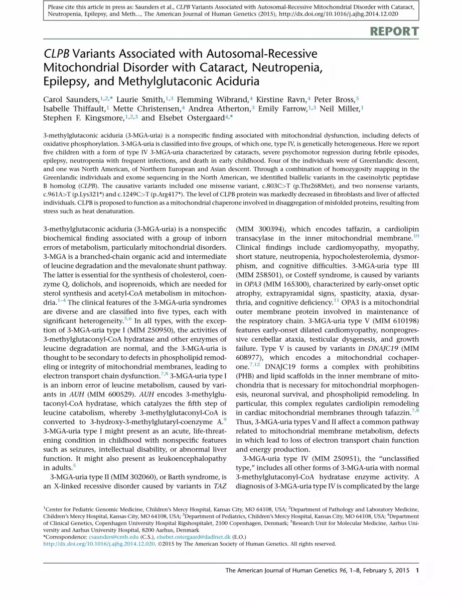
number NM_030813.3) showed a homozygous missense
variant, c.803C>T (p.Thr268Met), in all four affected chil-
dren (Figure 1A). The parents of individuals 1 and 2 were
each heterozygous for the variant; the parents of the other
two children were not available. To assess the potential
pathogenicity of this variant, the relative incidence,
conservation, in silico predictions for pathogenicity, and
potential effect on splicing was evaluated. To rule out a
splicing defect, cDNA derived from fibroblasts was ampli-
fied and sequenced, revealing a normally spliced mRNA
(data not shown) with band of normal size (Figure 1B).
The variant results in the substitution of a highly
conserved polar threonine for a nonpolar methionine
(Figure 1C), which is located in one of the ankyrin do-
mains (Figure 1D) and predicted to be pathogenic
by SIFT, PolyPhen-2, and MutationTaster. A TaqMan assay
was developed to assess the carrier frequency of c.803C>T
variant in Greenlandic controls: 6 of 184 samples were
determined to be heterozygous, corresponding to a carrier
frequency of 3.3%, which is comparable to carrier fre-
quencies of other founder variants in the Greenlandic pop-
ulation. It was not found in 2,180 samples sequenced at
Children’s Mercy Hospital (CMH) or in 13,000 alleles in
the Exome Variant Server but was reported with a carrier
frequency of 1 out of 662 in ClinSeq (rs200032855).
Sequencing of CLPB was performed in two additional indi-
viduals of Danish descent with a similar phenotype, but no
variants were identified.
In parallel, independent exome sequencing analysis was
performed on individual 5 (CMH193) and her two healthy
parents. DNA was prepared utilizing the KAPA Biosystems
library preparation kit (KAPA Biosystems) followed by
American Journal of Human Genetics 96, 1–8, February 5, 2015 3
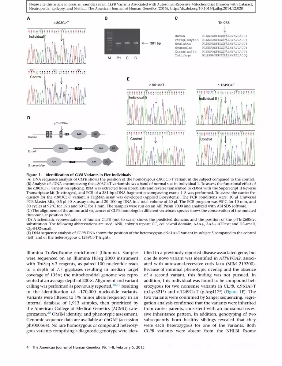
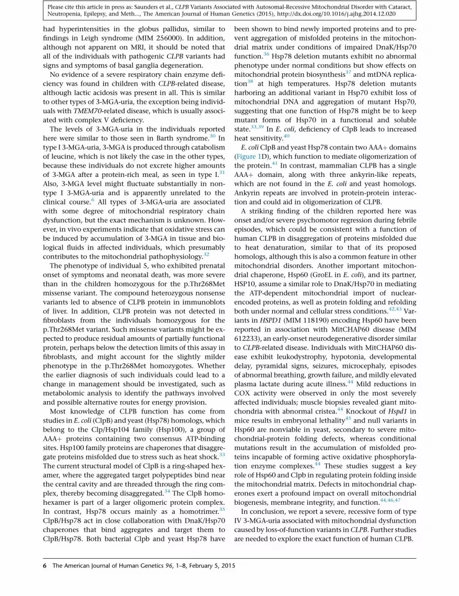
Figure 1. Identification of CLPB Variants in Five Individuals(A) DNA sequence analysis of CLPB shows the position of the homozygous c.803C>T variant in the subject compared to the control.(B) Analysis of cDNA encompassing the c.803C>T variant shows a band of normal size in individual 1. To assess the functional effect ofthe c.803C>T variant on splicing, RNA was extracted from fibroblasts and reverse transcribed to cDNA with the SuperScript II ReverseTranscriptase kit (Invitrogen), and PCR of a 381 bp cDNA fragment encompassing exons 4–8 was performed. To assess the carrier fre-quency for the c.803C>T variant, a TaqMan assay was developed (Applied Biosystems). The PCR conditions were: 10 ml UniversalPCR Master Mix, 0.5 ml 40 3 assay mix, and 20–100 ng DNA in a total volume of 20 ml. The PCR program was 95�C for 10 min, and50 cycles at 92�C for 15 s and 60�C for 1 min. The samples were run on an ABI Prism 7000 and analyzed with ABI SDS software.(C) The alignment of the amino acid sequences of CLPB homologs in different vertebrate species shows the conservation of the mutatedthreonine at position 268.(D) A schematic representation of human CLPB (not to scale) shows the predicted domains and the position of the p.Thr268Metsubstitution. The following abbreviations are used: ANK, ankyrin repeat; CC, coiled-coil domain; AAAþ, AAAþ ATPase; and D2-small,ClpB-D2-small.(E) DNA sequence analysis of CLPB DNA shows the position of the heterozygous c.961A>T variant in subject 5 compared to the control(left) and of the heterozygous c.1249C>T (right).
Please cite this article in press as: Saunders et al., CLPB Variants Associated with Autosomal-Recessive Mitochondrial Disorder with Cataract,Neutropenia, Epilepsy, and Meth..., The American Journal of Human Genetics (2015), http://dx.doi.org/10.1016/j.ajhg.2014.12.020
Illumina TruSeqExome enrichment (Illumina). Samples
were sequenced on an Illumina HiSeq 2000 instrument
with TruSeq v.3 reagents, as paired 100 nucleotide reads
to a depth of 7.7 gigabases resulting in median target
coverage of 1353; the mitochondrial genome was repre-
sented at an average depth of 2003. Alignment and variant
calling was performed as previously reported,18,19 resulting
in the identification of ~170,000 nucleotide variants.
Variants were filtered to 1% minor allele frequency in an
internal database of 1,913 samples, then prioritized by
the American College of Medical Genetics (ACMG) cate-
gorization,20 OMIM identity, and phenotypic assessment.
Genomic sequence data are available at dbGAP (accession
phs000564). No rare homozygous or compound heterozy-
gous variants comprising a diagnostic genotype were iden-
4 The American Journal of Human Genetics 96, 1–8, February 5, 2015
tified in a previously reported disease-associated gene, but
one de novo variant was identified in ATP6VOA2, associ-
ated with autosomal-recessive cutis laxa (MIM 219200).
Because of minimal phenotypic overlap and the absence
of a second variant, this finding was not pursued. In
addition, this individual was found to be compound het-
erozygous for two nonsense variants in CLPB, c.961A>T
(p.Lys321*) and c.1249C>T (p.Arg417*) (Figure 1E). The
two variants were confirmed by Sanger sequencing. Segre-
gation analysis confirmed that the variants were inherited
from carrier parents, consistent with an autosomal-reces-
sive inheritance pattern. In addition, genotyping of two
subsequently born healthy siblings revealed that they
were each heterozygous for one of the variants. Both
CLPB variants were absent from the NHLBI Exome

Figure 2. Analysis of CLPB Protein Levelsand In-Gel Activity of Complexes I and IV(A) In-gel enzyme activity in liver fromindividual 5 shows normal activity of com-plexes I and IV.(B) Analysis by SDS-PAGE of CLPB proteinin different human tissues showing ubiqui-tous localization of CLPB.(C and D) Immunoblot analysis of CLPB infibroblasts from individuals 3 and 4 andliver from subject 5 (D) shows absenceof CLPB protein. In brief, mitochondrial
protein was isolated from fibroblasts and liver as previously reported.23 The samples (25–30 cmg protein/lane) were run on a 12% SDSpolyacrylamide gel and transferred to a PVDF membrane. The membrane was probed with a polyclonal antibody against CLPB (AtlasAntibodies) at a 1:1,000 dilution and developed with a 1:1,000 dilution of goat anti-rabbit antibody (Dako). An antibody against porin(Proteintech) was used as a loading control at a 1:1,000 dilution, and SUCLA2was used as a reference at a 1:1,000 dilution. The secondaryantibody was goat anti-mouse at a 1:1,000 dilution (Dako). The bands were visualized with the Supersignal West Pico and Femto sub-strates (Thermo Fisher Scientific) and MicroChemi imaging (DNR Bioimaging Systems).
Please cite this article in press as: Saunders et al., CLPB Variants Associated with Autosomal-Recessive Mitochondrial Disorder with Cataract,Neutropenia, Epilepsy, and Meth..., The American Journal of Human Genetics (2015), http://dx.doi.org/10.1016/j.ajhg.2014.12.020
Sequencing Project (EVS) and an internal variant database
of 2,180 samples. In addition, truncating CLPB variants
were absent from the internal data set and rare in the
EVS; homozygous truncating variants were absent. CLPB
c.1249C>T has been reported in dbSNP as rs200203460,
but with unknown frequency.
Respiratory chain enzyme assays were performed on
frozen muscle and liver from individual 5 as part of a meta-
bolic autopsy at the Center For Inherited Disorders of En-
ergy Metabolism (CIDEM) (Cleveland, OH). Muscle tissue
showed generally decreased enzyme activities, probably
due to tissue deterioration. In frozen liver, the activity of
rotenone-sensitive NADH-cytochrome c reductase (reflect-
ing complexes I and III) was significantly below the control
range (14%), as was complex III activity (28%), whereas
complexes I and II had normal activity (88% and 73%,
respectively). Complex IV activity was not assayed. Citrate
synthase activity was 90% of control. In addition, ATP
production in digitonin-permeabilized fibroblasts and
in-gel enzyme activity in frozen liver was performed as
reported.21,23 ATP production in digitonin-permeabilized
fibroblasts from individual 1 energized with either
10 mM pyruvate plus 10 mM malate, 10 mM glutamate
plus 10 mM malate, or 10 mM succinate plus rotenone
was normal (37.9 nmol/min/mg protein, reference
31.8 5 4.5 [mean 5 SD]; 41.0 nmol/min/mg protein,
reference 33.0 5 5.8; and 19.2 nmol/min/mg, reference
14.9 5 3.3, respectively). Analysis of in-gel enzyme
activity showed normal activities of complexes I and IV
in the liver of individual 5 (Figure 2A).
To investigate normal localization of CLPB protein,
immunoblot analysis of several human tissues was per-
formed, demonstrating ubiquitous presence of CLPB,
although protein abundance was more variable than other
mitochondrial proteins (Figure 2B). Immunoblot analysis
performed on fibroblasts from individuals with the
p.Thr268Met variant (Figure 2C), as well as in liver from
individual 5 (with two nonsense variants) showed absence
of CLPB protein (Figure 2D).
Our data indicate that biallelic recessive variants in CLPB
result in a specific clinical and biochemical phenotype
The
associated with 3-MGA-uria (summarized in Table 1). The
onset of clinical disease in CLPB-associated 3-MGA-uria
ranged from prenatal in one individual with poly-
hydramnios, intrauterine growth retardation, and poor
movements to postnatal onset between 4 and 9 months
of age. Postnatal onset was characterized by development
of cataracts, psychomotor regression or lethargy, and
frequent febrile illnesses complicated by development of
seizures and psychomotor regression. Neutropenia was a
consistent finding in all. Cataracts were identified in four
of five individuals and microcephaly in three of five. Life
expectancy was short, with death by 8 days in the child
with prenatal onset, and from 11 months to 4 years of
age in the remaining individuals.
Many of the clinical findings in the individuals with
CLPB-associated 3-MGA-uria were similar to those seen in
other mitochondrial disorders, such as onset coincident
with systemic illness and the presence of epilepsy, psycho-
motor regression, andmicrocephaly.Neutropenia,however,
is an uncommonfinding in those with othermitochondrial
disorders, exceptMGA-uria type II (Barth syndrome), where
it is found in the majority of affected individuals.22 As in
Barth syndrome, bone marrow histology showed arrested
granulopoiesis as the proximal cause of neutropenia.
Cataract is infrequently reported in mitochondrial disor-
ders, although it is present in Sengers syndrome (MIM
212350)24 and in individuals with TMEM70- and GFER-
related (MIM 60094) disease.25–27 The main mechanism
of cataract development is unknown. However, variants
in GFER, AGK (MIM 610345), TMEM70, TAZ, and SERAC1
affect mitochondrial membrane integrity28 or lipid
metabolism of the mitochondrial membranes.29 Hence,
cataract formation in the individuals reported here can
be explained by disrupted protein-lipid interactions
affecting oxidative phosphorylation enzymes, increasing
oxidative stress, and stress-induced protein damage. In
addition, it might be speculated that a contributing factor
to CLPB-related disease is the resultant accumulation of
aggregated proteins.
Although cerebral MRI was normal in one individual,
two others showed cerebral atrophy, of whom one also
American Journal of Human Genetics 96, 1–8, February 5, 2015 5
Please cite this article in press as: Saunders et al., CLPB Variants Associated with Autosomal-Recessive Mitochondrial Disorder with Cataract,Neutropenia, Epilepsy, and Meth..., The American Journal of Human Genetics (2015), http://dx.doi.org/10.1016/j.ajhg.2014.12.020
had hyperintensities in the globus pallidus, similar to
findings in Leigh syndrome (MIM 256000). In addition,
although not apparent on MRI, it should be noted that
all of the individuals with pathogenic CLPB variants had
signs and symptoms of basal ganglia degeneration.
No evidence of a severe respiratory chain enzyme defi-
ciency was found in children with CLPB-related disease,
although lactic acidosis was present in all. This is similar
to other types of 3-MGA-uria, the exception being individ-
uals with TMEM70-related disease, which is usually associ-
ated with complex V deficiency.
The levels of 3-MGA-uria in the individuals reported
here were similar to those seen in Barth syndrome.30 In
type I 3-MGA-uria, 3-MGA is produced through catabolism
of leucine, which is not likely the case in the other types,
because these individuals do not excrete higher amounts
of 3-MGA after a protein-rich meal, as seen in type I.31
Also, 3-MGA level might fluctuate substantially in non-
type I 3-MGA-uria and is apparently unrelated to the
clinical course.6 All types of 3-MGA-uria are associated
with some degree of mitochondrial respiratory chain
dysfunction, but the exact mechanism is unknown. How-
ever, in vivo experiments indicate that oxidative stress can
be induced by accumulation of 3-MGA in tissue and bio-
logical fluids in affected individuals, which presumably
contributes to the mitochondrial pathophysiology.32
The phenotype of individual 5, who exhibited prenatal
onset of symptoms and neonatal death, was more severe
than in the children homozygous for the p.Thr268Met
missense variant. The compound heterozygous nonsense
variants led to absence of CLPB protein in immunoblots
of liver. In addition, CLPB protein was not detected in
fibroblasts from the individuals homozygous for the
p.Thr268Met variant. Such missense variants might be ex-
pected to produce residual amounts of partially functional
protein, perhaps below the detection limits of this assay in
fibroblasts, and might account for the slightly milder
phenotype in the p.Thr268Met homozygotes. Whether
the earlier diagnosis of such individuals could lead to a
change in management should be investigated, such as
metabolomic analysis to identify the pathways involved
and possible alternative routes for energy provision.
Most knowledge of CLPB function has come from
studies in E. coli (ClpB) and yeast (Hsp78) homologs, which
belong to the Clp/Hsp104 family (Hsp100), a group of
AAAþ proteins containing two consensus ATP-binding
sites. Hsp100 family proteins are chaperones that disaggre-
gate proteins misfolded due to stress such as heat shock.33
The current structural model of ClpB is a ring-shaped hex-
amer, where the aggregated target polypeptides bind near
the central cavity and are threaded through the ring com-
plex, thereby becoming disaggregated.34 The ClpB homo-
hexamer is part of a larger oligomeric protein complex.
In contrast, Hsp78 occurs mainly as a homotrimer.35
ClpB/Hsp78 act in close collaboration with DnaK/Hsp70
chaperones that bind aggregates and target them to
ClpB/Hsp78. Both bacterial Clpb and yeast Hsp78 have
6 The American Journal of Human Genetics 96, 1–8, February 5, 2015
been shown to bind newly imported proteins and to pre-
vent aggregation of misfolded proteins in the mitochon-
drial matrix under conditions of impaired DnaK/Hsp70
function.36 Hsp78 deletion mutants exhibit no abnormal
phenotype under normal conditions but show effects on
mitochondrial protein biosynthesis37 and mtDNA replica-
tion38 at high temperatures. Hsp78 deletion mutants
harboring an additional variant in Hsp70 exhibit loss of
mitochondrial DNA and aggregation of mutant Hsp70,
suggesting that one function of Hsp78 might be to keep
mutant forms of Hsp70 in a functional and soluble
state.33,39 In E. coli, deficiency of ClpB leads to increased
heat sensitivity.40
E. coliClpB and yeast Hsp78 contain two AAAþ domains
(Figure 1D), which function to mediate oligomerization of
the protein.41 In contrast, mammalian CLPB has a single
AAAþ domain, along with three ankyrin-like repeats,
which are not found in the E. coli and yeast homologs.
Ankyrin repeats are involved in protein-protein interac-
tion and could aid in oligomerization of CLPB.
A striking finding of the children reported here was
onset and/or severe psychomotor regression during febrile
episodes, which could be consistent with a function of
human CLPB in disaggregation of proteins misfolded due
to heat denaturation, similar to that of its proposed
homologs, although this is also a common feature in other
mitochondrial disorders. Another important mitochon-
drial chaperone, Hsp60 (GroEL in E. coli), and its partner,
HSP10, assume a similar role to DnaK/Hsp70 in mediating
the ATP-dependent mitochondrial import of nuclear-
encoded proteins, as well as protein folding and refolding
both under normal and cellular stress conditions.42,43 Var-
iants in HSPD1 (MIM 118190) encoding Hsp60 have been
reported in association with MitCHAP60 disease (MIM
612233), an early-onset neurodegenerative disorder similar
to CLPB-related disease. Individuals with MitCHAP60 dis-
ease exhibit leukodystrophy, hypotonia, developmental
delay, pyramidal signs, seizures, microcephaly, episodes
of abnormal breathing, growth failure, andmildly elevated
plasma lactate during acute illness.44 Mild reductions in
COX activity were observed in only the most severely
affected individuals; muscle biopsies revealed giant mito-
chondria with abnormal cristea.44 Knockout of Hspd1 in
mice results in embryonal lethality45 and null variants in
Hsp60 are nonviable in yeast, secondary to severe mito-
chondrial-protein folding defects, whereas conditional
mutations result in the accumulation of misfolded pro-
teins incapable of forming active oxidative phosphoryla-
tion enzyme complexes.44 These studies suggest a key
role of Hsp60 and Clpb in regulating protein folding inside
the mitochondrial matrix. Defects in mitochondrial chap-
erones exert a profound impact on overall mitochondrial
biogenesis, membrane integrity, and function.44,46,47
In conclusion, we report a severe, recessive form of type
IV 3-MGA-uria associated with mitochondrial dysfunction
caused by loss-of-function variants inCLPB. Further studies
are needed to explore the exact function of human CLPB.

Please cite this article in press as: Saunders et al., CLPB Variants Associated with Autosomal-Recessive Mitochondrial Disorder with Cataract,Neutropenia, Epilepsy, and Meth..., The American Journal of Human Genetics (2015), http://dx.doi.org/10.1016/j.ajhg.2014.12.020
Acknowledgments
We thank the families for their participation. We thank Dr. Lili
Miles for the EM studies. This work was supported by the Clare
Giannini Fund and a grant from The Danish Council for Indepen-
dent Research j Medical Sciences to E.O. (12-127702).
Received: October 27, 2014
Accepted: December 19, 2014
Published: January 15, 2015
Web References
The URLs for data presented herein are as follows:
1000 Genomes, http://browser.1000genomes.org
ClinSeq, http://genome.gov/20519355
dbGaP, http://www.ncbi.nlm.nih.gov/gap
dbSNP, http://www.ncbi.nlm.nih.gov/projects/SNP/
NHLBI Exome Sequencing Project (ESP) Exome Variant
Server, http://evs.gs.washington.edu/EVS/
OMIM, http://www.omim.org/
PolyPhen-2, www.genetics.bwh.harvard.edu/pph2/
RefSeq, http://www.ncbi.nlm.nih.gov/RefSeq
SIFT, http://sift.bii.a-star.edu.sg/
UCSC Genome Browser, http://genome.ucsc.edu
References
1. Gunay-Aygun,M. (2005). 3-Methylglutaconic aciduria: a com-
mon biochemical marker in various syndromes with diverse
clinical features. Mol. Genet. Metab. 84, 1–3.
2. Marinier, E., Lincoln, B.C., Garneau, M., David, F., and
Brunengraber, H. (1987). Contribution of the shunt pathway
of mevalonate metabolism to the regulation of cholesterol
synthesis in rat liver. J. Biol. Chem. 262, 16936–16940.
3. Rauthan, M., and Pilon, M. (2011). The mevalonate pathway
in C. elegans. Lipids Health Dis. 10, 243.
4. Weinstock, S.B., Kopito, R.R., Endemann, G., Tomera, J.F.,
Marinier, E., Murray, D.M., and Brunengraber, H. (1984). The
shunt pathway of mevalonate metabolism in the isolated
perfused rat liver. J. Biol. Chem. 259, 8939–8944.
5. Wortmann, S.B., Duran, M., Anikster, Y., Barth, P.G., Sperl, W.,
Zschocke, J., Morava, E., and Wevers, R.A. (2013). Inborn
errors of metabolism with 3-methylglutaconic aciduria as
discriminative feature: proper classification and nomencla-
ture. J. Inherit. Metab. Dis. 36, 923–928.
6. Wortmann, S.B., Rodenburg, R.J., Jonckheere, A., de Vries,
M.C., Huizing, M., Heldt, K., van den Heuvel, L.P., Wendel,
U., Kluijtmans, L.A., Engelke, U.F., et al. (2009). Biochemical
and genetic analysis of 3-methylglutaconic aciduria type IV:
a diagnostic strategy. Brain 132, 136–146.
7. Richter-Dennerlein, R., Korwitz, A., Haag, M., Tatsuta, T., Dar-
gazanli, S., Baker, M., Decker, T., Lamkemeyer, T., Rugarli, E.I.,
and Langer, T. (2014). DNAJC19, a mitochondrial cochaper-
one associated with cardiomyopathy, forms a complex with
prohibitins to regulate cardiolipin remodeling. Cell Metab.
20, 158–171.
8. Lamari, F., Mochel, F., Sedel, F., and Saudubray, J.M. (2013).
Disorders of phospholipids, sphingolipids and fatty acids
The
biosynthesis: toward a new category of inherited metabolic
diseases. J. Inherit. Metab. Dis. 36, 411–425.
9. IJlst, L., Loupatty, F.J., Ruiter, J.P., Duran, M., Lehnert, W., and
Wanders, R.J. (2002). 3-Methylglutaconic aciduria type I is
caused by mutations in AUH. Am. J. Hum. Genet. 71, 1463–
1466.
10. Johnston, J., Kelley, R.I., Feigenbaum, A., Cox, G.F., Iyer, G.S.,
Funanage, V.L., and Proujansky, R. (1997). Mutation charac-
terization and genotype-phenotype correlation in Barth syn-
drome. Am. J. Hum. Genet. 61, 1053–1058.
11. Anikster, Y., Kleta, R., Shaag, A., Gahl, W.A., and Elpeleg, O.
(2001). Type III 3-methylglutaconic aciduria (optic atrophy
plus syndrome, or Costeff optic atrophy syndrome): identifi-
cation of the OPA3 gene and its founder mutation in Iraqi
Jews. Am. J. Hum. Genet. 69, 1218–1224.
12. Davey, K.M., Parboosingh, J.S., McLeod, D.R., Chan, A., Casey,
R., Ferreira, P., Snyder, F.F., Bridge, P.J., and Bernier, F.P. (2006).
Mutation of DNAJC19, a human homologue of yeast inner
mitochondrial membrane co-chaperones, causes DCMA
syndrome, a novel autosomal recessive Barth syndrome-like
condition. J. Med. Genet. 43, 385–393.
13. Kelley, R.I., and Kratz, L. (1995). 3-methylglutaconic acidemia
in Smith-Lemli-Opitz syndrome. Pediatr. Res. 37, 671–674.
14. Law, L.K., Tang, N.L., Hui, J., Lam, C.W., and Fok, T.F. (2003).
3-methyglutaconic aciduria in a Chinese patient with
glycogen storage disease Ib. J. Inherit. Metab. Dis. 26, 705–
709.
15. Wortmann, S.B., Kluijtmans, L.A., Rodenburg, R.J., Sass, J.O.,
Nouws, J., van Kaauwen, E.P., Kleefstra, T., Tranebjaerg, L.,
de Vries, M.C., Isohanni, P., et al. (2013). 3-Methylglutaconic
aciduria—lessons from 50 genes and 977 patients. J. Inherit.
Metab. Dis. 36, 913–921.
16. Wortmann, S.B., and Morava, E. (2011). 3-methylglutaconic
aciduria type IV: a syndrome with an evolving phenotype.
Clin. Dysmorphol. 20, 168–169.
17. Tort, F., Garcıa-Silva, M.T., Ferrer-Cortes, X., Navarro-Sastre,
A., Garcia-Villoria, J., Coll, M.J., Vidal, E., Jimenez-Almazan,
J., Dopazo, J., Briones, P., et al. (2013). Exome sequencing
identifies a newmutation in SERAC1 in a patient with 3-meth-
ylglutaconic aciduria. Mol. Genet. Metab. 110, 73–77.
18. Bell, C.J., Dinwiddie, D.L., Miller, N.A., Hateley, S.L.,
Ganusova, E.E., Mudge, J., Langley, R.J., Zhang, L., Lee, C.C.,
Schilkey, F.D., et al. (2011). Carrier testing for severe child-
hood recessive diseases by next-generation sequencing. Sci.
Transl. Med. 3, ra4.
19. Saunders, C.J., Miller, N.A., Soden, S.E., Dinwiddie, D.L., Noll,
A., Alnadi, N.A., Andraws, N., Patterson, M.L., Krivohlavek,
L.A., Fellis, J., et al. (2012). Rapid whole-genome sequencing
for genetic disease diagnosis in neonatal intensive care units.
Sci. Transl. Med. 4, ra135.
20. Richards, C.S., Bale, S., Bellissimo, D.B., Das, S., Grody, W.W.,
Hegde, M.R., Lyon, E., and Ward, B.E.; Molecular Subcommit-
tee of the ACMG Laboratory Quality Assurance Committee
(2008). ACMG recommendations for standards for interpreta-
tion and reporting of sequence variations: Revisions 2007.
Genet. Med. 10, 294–300.
21. Zerbetto, E., Vergani, L., and Dabbeni-Sala, F. (1997). Quanti-
fication of muscle mitochondrial oxidative phosphorylation
enzymes via histochemical staining of blue native polyacryl-
amide gels. Electrophoresis 18, 2059–2064.
22. Roberts, A.E., Nixon, C., Steward, C.G., Gauvreau, K., Maisen-
bacher, M., Fletcher, M., Geva, J., Byrne, B.J., and Spencer, C.T.
American Journal of Human Genetics 96, 1–8, February 5, 2015 7

Please cite this article in press as: Saunders et al., CLPB Variants Associated with Autosomal-Recessive Mitochondrial Disorder with Cataract,Neutropenia, Epilepsy, and Meth..., The American Journal of Human Genetics (2015), http://dx.doi.org/10.1016/j.ajhg.2014.12.020
(2012). The Barth Syndrome Registry: distinguishing disease
characteristics and growth data from a longitudinal study.
Am. J. Med. Genet. A. 158A, 2726–2732.
23. Pedersen, C.B., Zolkipli, Z., Vang, S., Palmfeldt, J., Kjeldsen,
M., Stenbroen, V., Schmidt, S.P., Wanders, R.J., Ruiter, J.P., Wi-
brand, F., et al. (2010). Antioxidant dysfunction: potential risk
for neurotoxicity in ethylmalonic aciduria. J. Inherit. Metab.
Dis. 33, 211–222.
24. van Ekeren, G.J., Stadhouders, A.M., Smeitink, J.A., and
Sengers, R.C. (1993). A retrospective study of patients with
the hereditary syndrome of congenital cataract, mitochon-
drial myopathy of heart and skeletal muscle and lactic
acidosis. Eur. J. Pediatr. 152, 255–259.
25. Spiegel, R., Khayat, M., Shalev, S.A., Horovitz, Y., Mandel, H.,
Hershkovitz, E., Barghuti, F., Shaag, A., Saada, A., Korman,
S.H., et al. (2011). TMEM70 mutations are a common cause
of nuclear encoded ATP synthase assembly defect: further
delineation of a new syndrome. J. Med. Genet. 48, 177–182.
26. Di Fonzo, A., Ronchi, D., Lodi, T., Fassone, E., Tigano, M.,
Lamperti, C., Corti, S., Bordoni, A., Fortunato, F., Nizzardo,
M., et al. (2009). Themitochondrial disulfide relay system pro-
tein GFER is mutated in autosomal-recessive myopathy with
cataract and combined respiratory-chain deficiency. Am. J.
Hum. Genet. 84, 594–604.
27. Atay, Z., Bereket, A., Turan, S., Haliloglu, B., Memisoglu, A.,
Khayat, M., Shalev, S.A., and Spiegel, R. (2013). A novel homo-
zygous TMEM70 mutation results in congenital cataract and
neonatal mitochondrial encephalo-cardiomyopathy. Gene
515, 197–199.
28. Kratochvılova, H., Hejzlarova, K., Vrbacky, M., Mra�cek, T., Kar-
banova, V., Tesa�rova, M., Gombitova, A., Cmarko, D., Wittig,
I., Zeman, J., and Hou�st�ek, J. (2014). Mitochondrial mem-
brane assembly of TMEM70 protein. Mitochondrion 15, 1–9.
29. Mayr, J.A. (2014). Lipid metabolism in mitochondrial mem-
branes. J. Inherit. Metab. Dis. Published online August 1,
2014. http://dx.doi.org/10.1007/s10545-014-9748-x.
30. Mazurova, S., Tesa�rova, M., Magner, M., Hou�st’kova, H.,
Hansıkova, H., Augustınova, J., Tomek, V., Vondra�ckova, A.,
Zeman, J., and Honzık, T. (2013). Novel mutations in the
TAZ gene in patients with Barth syndrome. Prague Med.
Rep. 114, 139–153.
31. Ensenauer, R., Muller, C.B., Schwab, K.O., Gibson, K.M., Bran-
dis, M., and Lehnert, W. (2000). 3-Methylglutaconyl-CoA
hydratase deficiency: a new patient with speech retardation
as the leading sign. J. Inherit. Metab. Dis. 23, 341–344.
32. Fernandes, C.G., da Rosa, M.S., Seminotti, B., Pierozan, P.,
Martell, R.W., Lagranha, V.L., Busanello, E.N., Leipnitz, G.,
and Wajner, M. (2013). In vivo experimental evidence that
the major metabolites accumulating in 3-hydroxy-3-meth-
ylglutaryl-CoA lyase deficiency induce oxidative stress in
striatum of developing rats: a potential pathophysiological
mechanism of striatal damage in this disorder. Mol. Genet.
Metab. 109, 144–153.
33. von Janowsky, B., Major, T., Knapp, K., and Voos, W. (2006).
The disaggregation activity of the mitochondrial ClpB homo-
log Hsp78 maintains Hsp70 function during heat stress.
J. Mol. Biol. 357, 793–807.
8 The American Journal of Human Genetics 96, 1–8, February 5, 2015
34. Weibezahn, J., Tessarz, P., Schlieker, C., Zahn, R., Maglica, Z.,
Lee, S., Zentgraf, H., Weber-Ban, E.U., Dougan, D.A., Tsai,
F.T., et al. (2004). Thermotolerance requires refolding of aggre-
gated proteins by substrate translocation through the central
pore of ClpB. Cell 119, 653–665.
35. Leidhold, C., von Janowsky, B., Becker, D., Bender, T., and
Voos, W. (2006). Structure and function of Hsp78, the mito-
chondrial ClpB homolog. J. Struct. Biol. 156, 149–164.
36. Schmitt, M., Neupert, W., and Langer, T. (1995). Hsp78, a Clp
homologue within mitochondria, can substitute for chap-
erone functions of mt-hsp70. EMBO J. 14, 3434–3444.
37. Schmitt, M., Neupert, W., and Langer, T. (1996). The molecu-
lar chaperone Hsp78 confers compartment-specific thermo-
tolerance to mitochondria. J. Cell Biol. 134, 1375–1386.
38. Germaniuk, A., Liberek, K., and Marszalek, J. (2002). A bicha-
perone (Hsp70-Hsp78) system restores mitochondrial DNA
synthesis following thermal inactivation of Mip1p polymer-
ase. J. Biol. Chem. 277, 27801–27808.
39. Moczko, M., Schonfisch, B., Voos, W., Pfanner, N., and
Rassow, J. (1995). The mitochondrial ClpB homolog Hsp78
cooperates with matrix Hsp70 in maintenance of mitochon-
drial function. J. Mol. Biol. 254, 538–543.
40. Squires, C.L., Pedersen, S., Ross, B.M., and Squires, C. (1991).
ClpB is the Escherichia coli heat shock protein F84.1.
J. Bacteriol. 173, 4254–4262.
41. Mogk, A., Schlieker, C., Strub, C., Rist, W., Weibezahn, J., and
Bukau, B. (2003). Roles of individual domains and conserved
motifs of the AAAþ chaperone ClpB in oligomerization,
ATP hydrolysis, and chaperone activity. J. Biol. Chem. 278,
17615–17624.
42. Baker, M.J., Frazier, A.E., Gulbis, J.M., and Ryan, M.T. (2007).
Mitochondrial protein-import machinery: correlating struc-
ture with function. Trends Cell Biol. 17, 456–464.
43. Bross, P., Magnoni, R., and Bie, A.S. (2012). Molecular
chaperone disorders: defective Hsp60 in neurodegeneration.
Curr. Top. Med. Chem. 12, 2491–2503.
44. Magen, D., Georgopoulos, C., Bross, P., Ang, D., Segev, Y.,
Goldsher, D., Nemirovski, A., Shahar, E., Ravid, S., Luder, A.,
et al. (2008). Mitochondrial hsp60 chaperonopathy causes
an autosomal-recessive neurodegenerative disorder linked to
brain hypomyelination and leukodystrophy. Am. J. Hum.
Genet. 83, 30–42.
45. Christensen, J.H., Nielsen, M.N., Hansen, J., Fuchtbauer, A.,
Fuchtbauer, E.M., West, M., Corydon, T.J., Gregersen, N.,
and Bross, P. (2010). Inactivation of the hereditary spastic
paraplegia-associated Hspd1 gene encoding the Hsp60
chaperone results in early embryonic lethality in mice. Cell
Stress Chaperones 15, 851–863.
46. Raturi, A., and Simmen, T. (2013). Where the endoplasmic
reticulum and the mitochondrion tie the knot: the mitochon-
dria-associated membrane (MAM). Biochim. Biophys. Acta
1833, 213–224.
47. Cheng, M.Y., Hartl, F.U., Martin, J., Pollock, R.A., Kalousek, F.,
Neupert, W., Hallberg, E.M., Hallberg, R.L., and Horwich, A.L.
(1989). Mitochondrial heat-shock protein hsp60 is essential
for assembly of proteins imported into yeast mitochondria.
Nature 337, 620–625.