clinical studies in familial vcp myopathy associated with paget disease of bone and frontotemporal...
TRANSCRIPT

Clinical Studies in Familial VCP Myopathy Associated With PagetDisease of Bone and Frontotemporal Dementia
Virginia. E. Kimonis1,*, Sarju G. Mehta1, Erin C. Fulchiero1, Dana Thomasova1, MarziaPasquali2, Kym Boycott3, Edward G. Neilan1, Alex Kartashov4, Mark S. Forman5, StuartTucker6, Katerina Kimonis1, Steven Mumm7, Michael P. Whyte7, Charles D. Smith8, andGiles D. J. Watts1
1 Division of Genetics and Metabolism, Children’s Hospital, Harvard Medical School, Boston, Massachusetts
2 Department of Pathology, School of Medicine, University of Utah, Salt Lake City, Utah
3 University of Calgary, Alberta Children’s Hospital, Calgary, AB, Canada
4 Clinical Research Program (CRP), Division of Biostatistics, Children’s Hospital, Boston, Massachusetts
5 Division of Neuropathology, Department of Pathology and Laboratory Medicine, University ofPennsylvania, Pennsylvania
6 Eastover Internal Medicine, Carolinas HealthCare System, Charlotte, North Carolina
7 Division of Bone and Mineral Diseases, Washington University School of Medicine and Barnes-JewishHospital Research Institute, St. Louis, Missouri
8 Department of Neurology and Sanders-Brown Center on Aging, University of Kentucky, Lexington,Kentucky
AbstractInclusion body myopathy with Paget disease of the bone (PDB) and/or frontotemporal dementia(IBMPFD, OMIM 167320), is a progressive autosomal dominant disorder caused by mutations inthe Valousin-containing protein (VCP, p97 or CDC48) gene. IBMPFD can be difficult to diagnose.We assembled data on a large set of families to illustrate the number and type of misdiagnoses thatoccurred. Clinical analysis of 49 affected individuals in nine families indicated that 42 (87%) ofindividuals had muscle disease. The majority were erroneously diagnosed with limb girdle musculardystrophy (LGMD), facioscapular muscular dystrophy, peroneal muscular dystrophy, late adult onsetdistal myopathy, spinal muscular atrophy, scapuloperoneal muscular dystrophy, or amyotrophiclateral sclerosis (ALS) among others. Muscle biopsies showed rimmed vacuoles characteristic of aninclusion body myopathy in 7 of 18 patients (39%), however, inclusion body myopathy was correctlydiagnosed among individuals in only families 5 and 15. Frontotemporal dementia (FTD) wasdiagnosed in 13 individuals (27%) at a mean age of 57 years (range 48.9–60.2 years); however,several individuals had been diagnosed with Alzheimer disease. Histopathological examination ofbrains of three affected individuals revealed a pattern of ubiquitin positive neuronal intranuclearinclusions and dystrophic neurites. These families expand the clinical phenotype in IBMPFD, acomplex disorder caused by mutations in VCP. The presence of PDB in 28 (57%) individuals suggeststhat measuring serum alkaline phosphatase (ALP) activity may be a useful screen for IBMPFD inpatients with myopathy.
*Correspondence to: Virginia. E. Kimonis, M.D., M.R.C.P., Department of Pediatrics, Division of Genetics and Metabolism, Universityof California, Irvine, 101 The City Drive, ZOT 4482 Orange, CA 92868. E-mail: [email protected].
NIH Public AccessAuthor ManuscriptAm J Med Genet A. Author manuscript; available in PMC 2008 July 15.
Published in final edited form as:Am J Med Genet A. 2008 March 15; 146A(6): 745–757.
NIH
-PA Author Manuscript
NIH
-PA Author Manuscript
NIH
-PA Author Manuscript

Keywordsautosomal dominant; hereditary inclusion body myopathy; limb-girdle muscular dystrophy; Pagetdisease of bone; frontotemporal dementia; chromosome 9p13.3-12; VCP (valosin-containingprotein)
INTRODUCTIONHereditary inclusion body myopathy (HIBM) with early-onset Paget disease of bone (PDB)and frontotemporal dementia (FTD), or IBMPFD, is an autosomal dominant disorder (MIM167320) associated with a variable expressivity. Kimonis et al. [2000] described a family with11 affected members with autosomal dominant limb-girdle muscular dystrophy (LGMD)associated with early-onset PDB. The association of inclusion body myopathy and FTD wasestablished by Kovach et al. [2001] among 49 affected individuals from the original familyand three other unrelated families. This disorder is now called IBMPFD: HIBM, PDB, andFTD and it is caused by mutations in the valosin-containing protein (VCP; 601023) [Watts etal., 2004]. In this report, we review the clinical variability in 49 individuals among ninefamilies, where the diagnosis was confirmed by the presence of VCP mutations and illustratethe difficulty in making an accurate diagnosis.
PATIENTS AND METHODSClinical Evaluation and Diagnosis
The study was reviewed and approved by Children’s Hospital Boston IRB. This is a cross-section study of individuals referred from North American clinical centers including those self-referred. VCP mutations were confirmed in 49 individuals of nine families, and major clinicalcharacteristics derived from medical records and family histories.
We present the details of the three principal physical findings: myopathy, PDB, and dementia.The diagnosis of myopathy was based on the presence of muscular weakness, elevated creatinekinase measurements, and in several patients by EMG and/or muscle biopsy findings. Physicalexamination findings used to make a diagnosis of myopathy included various combinations ofan abnormal gait, lumbar lordosis from the proximal weakness, difficulty raising the arms orclimbing stairs, and mild weakness of the hands. Tendon reflexes were reduced or absent.Sections of unfixed muscle were stained by standard procedures. Indirect immunofluorescenceanalysis and immunoblotting of skeletal muscle were performed and electron microscopy wasdone on unfixed frozen sections [Askanas et al., 1993; Mirabella et al., 1996; Askanas andEngel, 1998].
Clinical symptoms suggestive of PDB included spine or hip pain, pathologic fractures, or longbone or cranial bone deformity. Whenever possible, PDB was confirmed by skeletalradiographic surveys. Typical findings of PDB include coarse trabeculation, corticalthickening, and spotty sclerosis. Radio-nuclide scans were used to demonstrate increased bonyuptake and are more sensitive indicators of PDB than plain survey films. The diagnosis of PDBwas made by nuclide scans in 13 patients and radiographs in 21.
FTD was diagnosed by neuropsychological assessments and imaging studies when available,together with typical clinical features of behavioral alteration (e.g., personal/socialunawareness, perseveration, abulia, disinhibition), early expressive or receptive languagedysfunction, and relative preservation of memory, orientation, or praxis [Miller et al., 1997].Neuropsychological evaluations included the administration of a combination of the followingtests: Neurobehavioral Cognitive Status Examination, Boston Naming Test, Wechsler Adult
Kimonis et al. Page 2
Am J Med Genet A. Author manuscript; available in PMC 2008 July 15.
NIH
-PA Author Manuscript
NIH
-PA Author Manuscript
NIH
-PA Author Manuscript

Intelligence Scale, Controlled Oral Word Association Test, Categories Verbal Fluency Test,Eisenson Reading Passages, Hooper Visual Organization Test, Draw a Clock by Command,Rey Osterreith Complex Figure Drawing, Bender Visual-Motor Gestalt Test, Benton VisualRetention Test, Memory for Unrelated Sentences Test, Memory Assessment Scale,Trailmaking Tests A & B, Stroop Neuropsychological Screening Test, Grooved Pegboard,McCarthy Verbal Fluency Test, Electronic Finger Tapping Test, and the Multi-scoreDepression Inventory. In several deceased individuals, medical history was abstracted fromtheir medical records.
Antibody StudiesWe present a summary of the neuropathology studies previously described in detail [Formanet al., 2006].
Statistical AnalysisTables of clinical variables for statistical analysis were generated by cross-tabulation of theavailable clinical and laboratory data for the affected members of the nine families. Data arepresented as means ± SE unless otherwise specified.
Clinical Reports (Grouped by Family)Family 5—The proband (IV:3) of this four-generation family (Fig. 1A) of German extractionwas diagnosed with limb girdle muscular dystrophy (LGMD). She noticed weakness of theleft index finger followed by weakness of both hands and reduced wrist mobility, and toeweakness, and pain associated with tripping in her 20’s. The distal weakness was followed byproximal weakness in the arms and legs resulting in difficulty climbing stairs in her late 30s.She has shortness of breath and has sleep apnea. Examination revealed proximal and distalweakness of hand and foot extensors, and decreased deep tendon reflexes. Muscle biopsyrevealed non-specific myopathic changes with fiber size variation, necrosis, centrally locatednuclei, focal endomysial fibrosis, and rimmed vacuoles within the muscle fibers consistentwith inclusion body myopathy. PDB was not apparent on bone scans. Her brother, individual(IV:4) had a 10–15 years history of progressive distal and proximal muscle weakness and backand hip pain from PDB and was using an automated wheelchair since the age of 39 years. Heryounger brother (IV:5) had chronic low back pain and was diagnosed with PDB after findingelevated alkaline phosphatase (ALP) levels at age 39 years, following enrollment in our study.Individual IV:15, a 33-year-old male cousin of the proband, developed proximal muscleweakness, pain, spasms, and tremors in the arms and legs and severe back pain at 23 years, thelatter attributed to PDB. A muscle biopsy of the left deltoid muscle revealed significantvariation of muscle fiber size with fiber splitting, fragmentation, and atrophic and hypertrophicfibers and rimmed vacuoles within muscle fibers, consistent with inclusion body myopathy.
Family 6—This Canadian family of English origin includes two affected siblings and theirmother (II:2) (Fig. 1B), the latter of whom developed progressive weakness in the legs, arms,trunk, and neck in her 40’s and was diagnosed with motor neuron disease. At 56 years, she wasconfined to a wheelchair and a year later she was fitted with a collar for severe neck muscleweakness. X-ray revealed PDB of the spine, pelvis, skull, and legs, and osteoporosis. She alsohad bilateral sensorineural hearing loss. One of her daughters (III:1) had onset of progressiveproximal muscle weakness and fatigability in the limbs at 21 years and was diagnosed withLGMD at 37 years. She has weakness in her hands and neck but is mobile with an automatedwheelchair. Nerve conduction studies were suggestive of axonal sensory neuropathy in herlegs. She has restrictive lung disease. She has a history of fractures, and was diagnosed withPDB at 35 years. Her brother (III:4) was diagnosed with PDB at 30 years after sustaining aright humeral fracture. He had a subsequent history of fractures of the forearm, ribs, and left
Kimonis et al. Page 3
Am J Med Genet A. Author manuscript; available in PMC 2008 July 15.
NIH
-PA Author Manuscript
NIH
-PA Author Manuscript
NIH
-PA Author Manuscript

clavicle. EMG studies in both siblings showed neuropathic and myopathic features. Musclebiopsy revealed variation in muscle fiber size and atrophic fibers with <1% rimmed vacuolatedfibers.
Family 7—This three-generation family includes eight affected, three of whom are deceased(Fig. 1C). The proband (III:6) had leg muscle cramps in her 30’s and was diagnosed withLGMD at age 47 years. She developed diaphragmatic involvement and required positivepressure oxygen at night. A muscle biopsy revealed non-specific myopathic findings. Herfather (II:10) was diagnosed with amyotrophic lateral sclerosis (ALS) and died at the age of61 years. Her grandmother (I:2) had myopathy and was diagnosed with Parkinson’s disease.Two paternal aunts (II:3, II:5), two paternal uncles (II:6, II:8), and two cousins (III:1, III:2)were diagnosed with either LGMD or ALS, and several with PDB. One paternal aunt (II:3),who was diagnosed with scapuloperoneal muscular dystrophy (SPMD), became wheelchair-bound and developed respiratory distress at age 70 years. An EMG at age 63 years revealedsevere myopathy with myotonia. Another aunt (II:5), with similar features had difficultybreathing since age 61 years. and used a motorized scooter since age 66 years. Her son (III:1)had leg weakness since age 34 years, reporting difficulty climbing stairs and lifting his armsoverhead. Individual III:3, diagnosed with PDB, had back problems and pain in her hips sinceher 20s, and had onset of muscle weakness at age 42 years.
Family 9—The female proband (V:2) of this five-generation family was diagnosed at 35 yearswith PDB at a routine examination based on an elevated ALP of 859 IU/L (Fig. 1D). Bonescan revealed abnormal sclerosis and mild enlargement in the right clavicle, proximal lefthumerus, distal left femur, left hemipelvis, and thoracic spine. Nerve conduction studies andEMG of the left arm were normal. Her mother (IV:2) was considered unaffected, however haddifficulty walking up stairs, and demonstrates a mild peripheral myopathy. A maternal aunt(IV:4) became bed-bound from myopathy at 44 years. She was diagnosed with dementia at age61 years. Physical exam revealed bilateral foot drop and clawing of the left hand. CT of thebrain revealed mild generalized atrophy involving the bitemporal lobes and perisylvian areas.Her son (V:3) was diagnosed at age 40 years. with SPMD.
Family 10—The proband of this four-generation family (pedigree not shown) was diagnosedwith LGMD at age 46 years. He has progressive proximal weakness, was unable to lift his armsabove his head, had problems climbing stairs, and developed nocturnal hypoventilation at 39years. He had onset of back pain for 10 years and was diagnosed with PDB in his right hip andL3 vertebrae by X-rays and bone scan at age 46 years. His mother died from myopathy at age66 years. His brother was diagnosed with myopathy at age 50 years, but chose not to participatein this study.
Family 11—Clinical data in nine individuals from this family were previously reported byTucker et al. [1982] (Fig. 1E). They were previously diagnosed with autosomal dominant lowermotor neuron disease and PDB. Medical information on several of these deceased individualsis included in Table I. We have established contact with individuals from successivegenerations of the original family. The proband for our study (V:4) was diagnosed with SPMDalthough his predominant illness was PDB. He developed back pain at age 24 years, andsustained a non-displaced fracture of the distal left fibula, which was slow to heal after a fallat work at age 31 years. He has suffered repetitive leg fractures. Laboratory testing revealedan elevated ALP level (>1,000 IU/L), X-rays revealed increased trabecular pattern of PDB ofthe right tibia, and bone scan revealed multiple areas of increased uptake throughout theskeleton. Because of concern for metastatic bone disease, he had a biopsy of the right pelvisat age 31 years, which revealed disruption of the normal pattern of cortical and cancellousbone, with a proliferation of fibrous connective tissue in the medullary spaces, hypertrophic
Kimonis et al. Page 4
Am J Med Genet A. Author manuscript; available in PMC 2008 July 15.
NIH
-PA Author Manuscript
NIH
-PA Author Manuscript
NIH
-PA Author Manuscript

trabeculi with mosaic patterns, and focal new bone formation consistent with early PDB. Inthe areas where the medullary fibrous proliferation was obvious, the pattern suggested fibrousdysplasia. He has had difficulty lifting his arms since the age of 40 years, and difficulty walkingsince the age of 46 years.
Ultrastructural studies of osteoclasts of bone in four members of this family revealed nuclearinclusions, identical to those of patients with PDB (Fig. 2A,B). The nuclear inclusions consistedof straight, tubular structures of ~15 nm diameter. The nuclear inclusions seen in muscle (Fig.2C) and osteoclasts (Fig. 2A,B) were structurally very similar.
Family 13—Individual III:8 in this four-generation family of German extraction, an avidsportswoman, developed climbing stairs and back pain at age 43 years (Fig. 1F). A trial of animmunosuppressant for her diagnosis of polymyositis was unsuccessful. EMG studies revealedmyopathic changes (legs >arms) and median nerve entrapment at the wrist and ulnarentrapment at the elbow. Two muscle biopsies from the quadriceps showed both neurogenicand myopathic changes with normal staining for dystrophin, alpha and delta sarcoglycans,caveolin, and spectrin, with normal electron microscopy. Individual III:7, diagnosed withLGMD at age 42 years, showed signs of fast progression of his myopathy until his demise atthe age of 58 years. In his final year, he became paranoid and depressed, however, he was notdiagnosed with dementia. An autopsy was not obtained. His mother (II:5) was diagnosed withPDB at the age of 42 years. She had several limb fractures and had a hip replacement. Non-specific myopathy was diagnosed in her mid 40’s, progression of which led to transitioning toa wheelchair over several years. She developed bizarre, inappropriate behavior in her 50’s, wasdiagnosed with Alzheimer disease at age 60 years, and died at age 68 years.
Family 15—The proband of Scottish ancestry (III:I) first noted distal and proximal muscleweakness at age 38 years (Fig. 1G). He was initially diagnosed with facioscapulohumeralmuscular dystrophy (FSH), this diagnosis being later revised to LGMD. He developed PDBin his 50’s. His mother became wheelchair-bound because of myopathy and later developeddementia. His sister (III:2) also developed myopathy, as did three maternal cousins and theirmother prior to her demise. Individual III:4 developed progressive weakness at age 39 years,and had an elevated ALP. Muscle biopsy of the patient’s right deltoid showed small, scatteredfoci of interstitial chronic inflammation, including focally prominent lymphocytes, indicativeof a mild inflammatory component as well as foci of fiber degeneration/regeneration withoutprominently increased fibrous tissue. Rimmed vacuoles were focally present, though relativelyinconspicuous. Staining was negative for amyloid, neurofilament, and tau; however,immunohistochemical stain for ubiquitin showed unusually prominent staining and moderatebut variable desmin immunoreactivity and increased cytoplasmic vimentin staining.Ultrastructural inclusions showing features characteristic of inclusion body myopathy orinclusion body myositis were relatively prominent within nuclei. X-rays have beenunremarkable. Individual III:7 developed weakness of his left shoulder and atrophy of hisdeltoid muscle in his 20s and was initially diagnosed with FSH and spinal muscular atrophy.EMG at 36 years showed active denervation in both arms in a C5-7 distribution and no evidenceof a myopathic process. Muscle biopsy showed moderately severe chronic-active myopathicchanges with numerous fibers showing one or multiple rimmed vacuoles (Fig. 3A,B). He hada left C5-6 laminectomy, however, he later developed weakness of the right arm and leg.Although he has had back pain for several years, and elevated ALP, he was not previouslydiagnosed with PDB.
Family 16—This family was evaluated extensively by CDS for a history of autosomaldominant early-onset Alzheimer’s disease, later established as FTD (Fig. 1H). Individual II:6had bilateral foot drop in her 40s and died at the age of 59 years. Brain autopsy showed grossatrophy in the frontal and dorsolateral temporal lobes. Microscopically, neurofibrillary tangles
Kimonis et al. Page 5
Am J Med Genet A. Author manuscript; available in PMC 2008 July 15.
NIH
-PA Author Manuscript
NIH
-PA Author Manuscript
NIH
-PA Author Manuscript
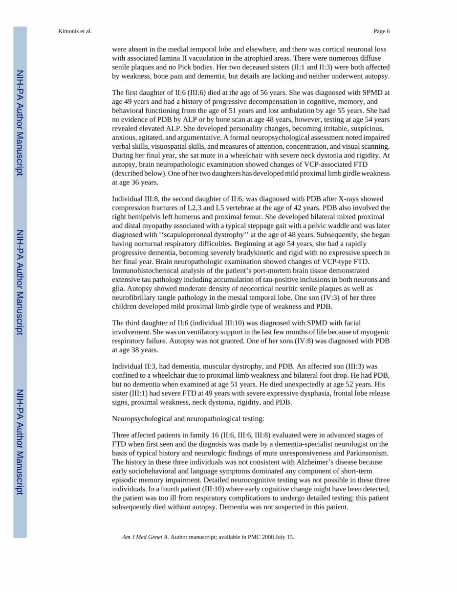
were absent in the medial temporal lobe and elsewhere, and there was cortical neuronal losswith associated lamina II vacuolation in the atrophied areas. There were numerous diffusesenile plaques and no Pick bodies. Her two deceased sisters (II:1 and II:3) were both affectedby weakness, bone pain and dementia, but details are lacking and neither underwent autopsy.
The first daughter of II:6 (III:6) died at the age of 56 years. She was diagnosed with SPMD atage 49 years and had a history of progressive decompensation in cognitive, memory, andbehavioral functioning from the age of 51 years and lost ambulation by age 55 years. She hadno evidence of PDB by ALP or by bone scan at age 48 years, however, testing at age 54 yearsrevealed elevated ALP. She developed personality changes, becoming irritable, suspicious,anxious, agitated, and argumentative. A formal neuropsychological assessment noted impairedverbal skills, visuospatial skills, and measures of attention, concentration, and visual scanning.During her final year, she sat mute in a wheelchair with severe neck dystonia and rigidity. Atautopsy, brain neuropathologic examination showed changes of VCP-associated FTD(described below). One of her two daughters has developed mild proximal limb girdle weaknessat age 36 years.
Individual III:8, the second daughter of II:6, was diagnosed with PDB after X-rays showedcompression fractures of L2,3 and L5 vertebrae at the age of 42 years. PDB also involved theright hemipelvis left humerus and proximal femur. She developed bilateral mixed proximaland distal myopathy associated with a typical steppage gait with a pelvic waddle and was laterdiagnosed with ‘‘scapuloperoneal dystrophy’’ at the age of 48 years. Subsequently, she beganhaving nocturnal respiratory difficulties. Beginning at age 54 years, she had a rapidlyprogressive dementia, becoming severely bradykinetic and rigid with no expressive speech inher final year. Brain neuropathologic examination showed changes of VCP-type FTD.Immunohistochemical analysis of the patient’s port-mortem brain tissue demonstratedextensive tau pathology including accumulation of tau-positive inclusions in both neurons andglia. Autopsy showed moderate density of neocortical neuritic senile plaques as well asneurofibrillary tangle pathology in the mesial temporal lobe. One son (IV:3) of her threechildren developed mild proximal limb girdle type of weakness and PDB.
The third daughter of II:6 (individual III:10) was diagnosed with SPMD with facialinvolvement. She was on ventilatory support in the last few months of life because of myogenicrespiratory failure. Autopsy was not granted. One of her sons (IV:8) was diagnosed with PDBat age 38 years.
Individual II:3, had dementia, muscular dystrophy, and PDB. An affected son (III:3) wasconfined to a wheelchair due to proximal limb weakness and bilateral foot drop. He had PDB,but no dementia when examined at age 51 years. He died unexpectedly at age 52 years. Hissister (III:1) had severe FTD at 49 years with severe expressive dysphasia, frontal lobe releasesigns, proximal weakness, neck dystonia, rigidity, and PDB.
Neuropsychological and neuropathological testing:
Three affected patients in family 16 (II:6, III:6, III:8) evaluated were in advanced stages ofFTD when first seen and the diagnosis was made by a dementia-specialist neurologist on thebasis of typical history and neurologic findings of mute unresponsiveness and Parkinsonism.The history in these three individuals was not consistent with Alzheimer’s disease becauseearly sociobehavioral and language symptoms dominated any component of short-termepisodic memory impairment. Detailed neurocognitive testing was not possible in these threeindividuals. In a fourth patient (III:10) where early cognitive change might have been detected,the patient was too ill from respiratory complications to undergo detailed testing; this patientsubsequently died without autopsy. Dementia was not suspected in this patient.
Kimonis et al. Page 6
Am J Med Genet A. Author manuscript; available in PMC 2008 July 15.
NIH
-PA Author Manuscript
NIH
-PA Author Manuscript
NIH
-PA Author Manuscript

Detailed neuropathologic examination by MSF in a 56 years female (III: 6) from family 16typifies findings in IBMPFD. Post-mortem examination revealed a 940-gram brain with mildfrontal lobe atrophy. Microscopic examination revealed mild superficial microvacuolation,neuronal loss, and gliosis throughout the neocortex including the frontal, temporal, and parietallobes. The limbic system was also affected with mild neuron loss and gliosis. Thioflavin Sstaining revealed a low density of neuritic plaques in the neocortex and a low density ofneurofibrillary tangles in the entorhinal cortex and amygdala. Immunohistochemistry withantibodies to β-amyloid, phosphorylated tau, α-synuclein, and ubiquitin revealed a moderatedensity of predominantly diffuse senile plaques in the neocortex (Fig. 4A–D) and a low densityof tau pathology in the hippocampus and entorhinal cortex. Tau pathology was not identifiedin the neocortex (Fig. 4B) and there were no Lewy bodies (Fig. 4C). Immunohistochemistryfor ubiquitin revealed a moderate to high density of ubiquitinated inclusions throughout theneocortex consisting of abundant nuclear inclusion and dystrophic neurites with only a lowdensity of ubiquitin positive cytoplasmic inclusions (Fig. 4D). Similar, but less robust,ubiquitin pathology was also detected in the hippocampal formation, subcortical nuclei, andbrainstem. Moreover, the ubiquitin staining detected the low density of neuritic plaquesobserved with the Thioflavin S stain.
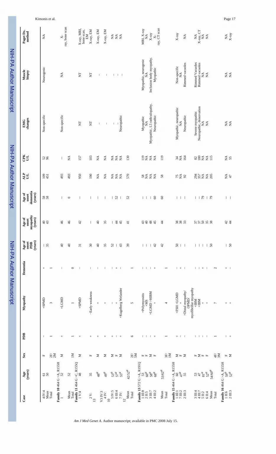
SUMMARY OF CLINICAL DATA IN NINE FAMILIESOn reviewing the specific diagnoses among individuals from these families in whom recordscould be obtained, LGMD was the most common diagnosis made in 11 (38%) of 29 individuals,SPMD in eight (28%), ALS in three (10%), SMA in two (7%), diabetic neuropathy in two(7%), and inclusion body myositis, multiple sclerosis, polymyositis, FSH, and distal myopathy/oculopharyngeal muscular dystrophy/myofibrillar myopathy in one individual each (3%), theremaining individuals being diagnosed with a non-specific myopathy. Several individuals hadmore than one diagnosis made over the course of their illness. Review of the clinical featuresin the nine families indicate that 42/49 (86%) affected individuals with VCP mutations hadprogressive proximal muscle weakness and occasionally distal muscle involvement. The onsetof weakness was at a mean age of 42 years (range 24–61 years), however, the pattern variedwidely, even within families. Mean CPK levels in affected individuals was 154.4 ± 121.4 IU/L compared to 113.6 ± 29.3 in the unaffected VCP mutation negative relatives (data not shown).Muscle biopsy specimens revealed typical myopathic features including variation in musclefiber size and mildly increased endomysial connective tissue in patchy regions of the biopsy.Large focal regions of ‘‘myopathic grouping’’ were observed in some biopsies. Rimmedvacuoles and cytoplasmic inclusions characteristic of HIBM but also seen in idiopathicinclusion body myositis and in a number of other hereditary distal myopathy types were presentin 39% of muscles studied. Two individual from families 5 and 15 were correctly diagnosedwith HIBM and in an additional, two individuals the diagnosis was revised from the initialdiagnosis of LGMD to HIBM (Table I).
PDB seen in 28/49 (57%) patients occurred early (mean age 40 years, with a range of 23–65years) with typical distribution in the spine, pelvis, and skull and later progression involvedother bones. Elevated ALP levels were seen in 16 individuals diagnosed with PDB (mean 359.3IU/L, range 58–1,508 IU/L; unaffected relatives mean 86.8 IU/L, normal population 30–130IU/L). Ultrastructural studies of bone biopsy osteoclasts in four members of family number 6revealed nuclear and cytoplasmic inclusions morphologically identical to that seen in classicPDB.
The onset of dementia seen in 27% of individuals studied occurred at a mean age of 57 years(range 48.9–60.2 years). It was characterized by personality change and language dysfunction,usually an expressive dysphasia, with early relative sparing of memory, dystonia (particularlyneck dystonia), rigidity without tremor and severe gait instability. These features together with
Kimonis et al. Page 7
Am J Med Genet A. Author manuscript; available in PMC 2008 July 15.
NIH
-PA Author Manuscript
NIH
-PA Author Manuscript
NIH
-PA Author Manuscript

findings of early relative sparing of memory, the impairment of executive skills, absence ofcortical sensory or visuospatial abnormalities, absence of vertical gaze abnormalities, tremoror other involuntary movements, onset in the fifth and sixth decade, and rapid progression,made the clinical classification of FTD secure. Two of 13 patients had significant visual orauditory hallucinations. The end stage is a wheelchair or bed-confined, mute, rigid individualwith no ability to interact meaningfully with the environment. In the case of family 16, thethree affected patients were in advanced stages of FTD when first seen, and the diagnosis wasmade by a dementia-specialist neurologist on the basis of typical history and neurologicalfindings of mute unresponsiveness and Parkinsonism. The history in these three was notconsistent with Alzheimer’s disease. Neuropathological findings from three individuals fromfamily 16 who were diagnosed with dementia showed characteristic findings includingubiquitin-positive neuronal intranuclear inclusions, dystrophic neurites, and only rareintracytoplasmic VCP positive inclusions.
DISCUSSIONClinical, biochemical, and histopathological data of 49 individuals from nine families with arecently described disorder autosomal dominant proximal myopathy associated with PDB andFTD was analyzed for their usefulness in establishing diagnostic criteria. The CPK levels inaffected individuals were only mildly elevated, EMG and muscle biopsies revealed non-specific myopathic changes, and only 39% of muscle biopsies showed rimmed vacuoles andcytoplasmic inclusions characteristic of HIBM, the vast majority of individuals revealing non-specific changes.
In contrast to classic PDB seen in 1–2% of the >50 years population [Klein and Norman,1995], the PDB seen in 57% of patients with IBMPFD presented earlier (mean age of 40 years)with typical distribution in the spine, pelvis, and skull and later progression to involve otherbones. Elevation of serum ALP was found to be a good indicator of PDB. Fractures were nottypically seen except among affected individuals in family 6 with the severe VCP A232Emutation. The association of familial PDB and neuromuscular disorders is a rare combination,having been reported in a few unrelated families with distinct muscular phenotypes. We haveidentified the descendants of one family with the familial disorder of combined lower motorneuron degeneration and skeletal disorganization reported by Tucker et al. [1982]. This familyhas the VCP R155Q mutation. At least two of Tucker’s patients with lower motor neurondegeneration and PDB developed dementia as a terminal event. Evaluation of an affecteddescendant of this family indicates resemblance to members of the other families.
The VCP protein is widely expressed and is a member of the AAA-ATPase super-family[Beyer, 1997]. It has been implicated in many cellular functions [Wang et al., 2003; Woodman,2003], and is required for the proteasomal degradation of phosphorylated IκB-α [Dai et al.,1998; Asai et al., 2002], an essential step in NF-κB activation. Interestingly, the causativemutations in VCP all affect the highly conserved CDC 48 domain, which is involved inubiquitin-binding [Dai and Li, 2001; Rape et al., 2001]. The PDB-causing mutations in theSQSTM1 gene also affect the ubiquitin-binding domain of the gene product, p62 [Layfield andHocking, 2004], suggesting that the disease processes in PDB associated with SQSTM1 andIBMPFD may be related [Daroszewska and Ralston, 2006]. We believe that IBMPFD iscurrently underdiagnosed among the patients with myopathy and/or dementia. Using anelevated ALP as a screen, with confirmation by radiography or bone scan, should identify morecases of syndromic PDB.
Dementia, mainly of the frontotemporal type, was diagnosed in 27% individuals (mean age ofonset 57 years (range 48.9–60.2 years)). The novel pattern of brain histopathology from threeindividuals from family 16 who were diagnosed with dementia is different from that previously
Kimonis et al. Page 8
Am J Med Genet A. Author manuscript; available in PMC 2008 July 15.
NIH
-PA Author Manuscript
NIH
-PA Author Manuscript
NIH
-PA Author Manuscript

reported for sporadic and familial FTD with ubiquitin inclusions also known as FTLD withmotor neuron type inclusions or motor neuron disease inclusion dementia [McKhann et al.,2001], autosomal-dominant FTD linked to the tau gene on chromosome 17 [Spillantini et al.,1998], to an unknown gene on chromosome 3 [Brown, 1998] and the familial corticobasalsyndrome associated with mutations in the progranulin gene [Masellis et al., 2006]. Forman etal. [2006] analyzed neuropathologic changes in eight patients with VCP mutations (six withdementia including these three cases from family 16). Characteristic findings include ubiquitin-positive neuronal intranuclear inclusions and dystrophic neurites. There was no biochemicalalteration in the VCP protein and only rare intracytoplasmic inclusions were detected withantibodies to VCP in contrast to the reported findings in a 55-year German patient with VCPgene mutations [Schröder et al., 2005]. The overlap between the histology and composition ofthe brain protein accumulations in FTD, for example, ubiquitin epitopes, and those found inthe muscle fibers of individuals with IBM [Askanas and Engel, 2001] suggests that cellulardeterioration may result from a common cascade of events. Mehta et al. [2007] studiedApolipoprotein-E (APOE) as a risk factor for the dementia seen in approximately one-third ofpatients with IBMPFD based on its known modifier effect in Alzheimer’s disease. Theyidentified a possible association with the presence of one or more alleles of the APOE 4genotype and FTD (P = 0.002), but not with IBM (P = 0.9), or PDB (P = 0.9). They did notobserve an association with FTD and the H2 MAPT haplotype.
The variable presentation of IBMPFD has been noted by other authors. Haubenberger et al.[2005] discovered a novel missense mutation in VCP (R159H, 688G>A) in four Austriansiblings, ages ranging from 60 to 70 years, who presented with both progressive proximalmyopathy and PDB in whom dementia was excluded in all members by neuropsychologicalassessment. In contrast, the majority of patients in two French kindreds [Guyant-Marechal etal., 2006] have been diagnosed with dementia in 100 and 90% of the individuals with a novelR93C missense mutation and a common R155C mutation, respectively. Neuropsychologicalexamination of six affected individuals from the first family identified with dementia wascharacterized by aggressive behavior, impaired judgment, and paranoia. Fifty percentpresented with selectively severe atrophy and symmetric weakness of the proximal and distalmuscles of the upper limbs and distal legs (mean age of onset 57 years) and 50% of the patientssuffered from PDB at a mean age of onset of 55 years associated with foraminal lumbar stenosisor hearing loss in individuals. In a second family of 30 individuals, previously mistakenlydiagnosed with myotonic dystrophy and reported to be linked to chromosome 15q21-24 by LeBer et al. [2004], 10 presented with proximal and axial muscle weakness with clinical andelectrical myotonia and associated FTD. Histological analyses confirmed PDB in sixindividuals. Hepatitis and variable diagnoses of non-specific hepatic fibrosis, cytolysis, andcholestasis was reported.
We hope to increase awareness of this under-diagnosed disorder which shares features withLGMD, FSH, SPMD, late adult onset distal myopathy, spinal muscular atrophy, ALS, andnon-specific dementias, among others, suggesting possible underlying common pathogenicmechanisms. An elevated blood ALP may be a useful screen for PDB, which is seen in overhalf the patients. Testing for VCP mutations should be considered in any individual with twoor more associated features of IBMPFD, or, with one or more features and a suspicious familyhistory. Understanding the basic molecular pathways of VCP disease will hopefully lead tospecific therapies as in the case of Paget disease of bone [Deftos, 2005]. Studying this relativelyrare disease has important implications for common disorders such as the muscular dystrophies,myopathies, PDB and related bone disorders and the dementias.
Kimonis et al. Page 9
Am J Med Genet A. Author manuscript; available in PMC 2008 July 15.
NIH
-PA Author Manuscript
NIH
-PA Author Manuscript
NIH
-PA Author Manuscript

ELECTRONIC-DATABASE INFORMATIONOnline Mendelian Inheritance in Man (OMIM) http://www3.ncbi.nlm.nih.gov/omim (forLGMD with PDB [MIM 605382], IBM1 [MIM 147420], IBM2 [MIM 600737], IBM3 [MIM605637], PDB2 [MIM 602080], DMRV, [MIM 605820], FTD presenting with Parkinsonism[MIM 600274], FTD presenting with ALS [MIM 105550]), Pagetoid amyotrophic lateralsclerosis [MIM 167320].
Acknowledgements
We thank the families and their health care providers for their enthusiastic participation and contribution in our researchstudies in particular Dr. Zachary Simmons, Dr. Javed Towfighi, Dr. Rabi Tawil, Dr. Fred Singer and other colleagesand collaborators who referred patients for this study.
Grant sponsor: NIAMS; Grant sponsor: National Institutes of Health; Grant numbers: RO1 AR050236-01A1, R03AR 46869; Grant sponsor: Muscular Dystrophy Association; Paget Foundation.
ReferencesAsai T, Tomita Y, Nakatsuka S, Hoshida Y, Myoui A, Yoshikawa H, Aozasa K. VCP (p97) regulates
NF kappaB signaling pathway, which is important for metastasis of osteosarcoma cell line. Jpn JCancer Res 2002;93:296–304. [PubMed: 11927012]
Askanas V, Engel WK. Sporadic inclusion-body myositis and its similarities to Alzheimer disease brain.Recent approaches to diagnosis and pathogenesis, and relation to aging. Scand J Rheumatol1998;27:389–405. [PubMed: 9855208]
Askanas V, Engel WK. Inclusion-body myositis: Newest concepts of pathogenesis and relation to agingand Alzheimer disease. J Neuropathol Exp Neurol 2001;60:1–14. [PubMed: 11202170]
Askanas V, Alvarez RB, Engel WK. Beta-amyloid precursor epitopes in muscle fibers of inclusion bodymyositis. Ann Neurol 1993;34:551–560. [PubMed: 7692809]
Beyer A. Sequence analysis of the AAA protein family. Protein Sci 1997;6:2043–2058. [PubMed:9336829]
Brown J. Chromosome 3-linked frontotemporal dementia. Cell Mol Life Sci 1998;54:925–927. [PubMed:9791535]
Dai RM, Li CC. Valosin-containing protein is a multi-ubiquitin chain-targeting factor required inubiquitin-proteasome degradation. Nat Cell Biol 2001;3:740–744. [PubMed: 11483959]
Dai RM, Chen E, Longo DL, Gorbea CM, Li CC. Involvement of valosin-containing protein, an ATPaseCo-purified with IkappaBalpha and 26 S proteasome, in ubiquitin-proteasome-mediated degradationof IkappaBalpha. J Biol Chem 1998;273:3562–3573. [PubMed: 9452483]
Daroszewska A, Ralston SH. Mechanisms of disease: Genetics of Paget’s disease of bone and relateddisorders. Nat Clin Pract Rheumatol 2006;2:270–277. [PubMed: 16932700]
Deftos LJ. Treatment of Paget’s disease-taming the wild osteoclast. NEJM 2005;353:872–875. [PubMed:16135831]
Forman MS, Mackenzie IR, Markesbery WR, Swanson E, Cairns NJ, Boyer PJ, Jhaveri BS, KarlawishJH, McKeel DW, Watts GD, Markesbery WR, Smith CD, Kimonis VE. Novel ubiquitin brainpathology in frontotemporal dementia with inclusion body myopathy and Paget’s disease. JNeuropathol Exp Neurol 2006;65:571–581. [PubMed: 16783167]
Guyant-Marechal L, Duyckaerts C, Bou J, Dugny F, Le Ber I, Frebourg T, Hannequin D, Campion D.VCP mutations in two kindreds with prominent frontotemporal dementia: Clinical andneuropathological features. Neurology 2006;67:644–651. [PubMed: 16790606]
Haubenberger D, Bittner RE, Rauch-Shorny S, Zimprich F, Mannhalter C, Wagner L, Mineva I, Vass K,Auff E, Zimprich A. Inclusion body myopathy and Paget disease is linked to a novel mutation in theVCP gene. Neurology 2005;65:1304–1305. [PubMed: 16247064]
Kimonis VE, Kovach MJ, Waggoner B, Leal S, Salam A, Rimer L, Davis K, Khardori R, Gelber D.Clinical and molecular studies in a unique family with autosomal dominant limb-girdle musculardystrophy and Paget disease of bone. Genet Med 2000;2:232–241. [PubMed: 11252708]
Kimonis et al. Page 10
Am J Med Genet A. Author manuscript; available in PMC 2008 July 15.
NIH
-PA Author Manuscript
NIH
-PA Author Manuscript
NIH
-PA Author Manuscript

Klein RM, Norman A. Diagnostic procedures for Paget’s disease. Radiologic, pathologic, and laboratorytesting. Endocrinol Metab Clin North Am 1995;24:437–450. [PubMed: 7656898]
Kovach MJ, Waggoner B, Leal SM, Gelber D, Khardori R, Levenstien MA, Shanks CA, Gregg G, Al-Lozi MT, Miller T, Rakowicz W, Lopate G, Florence J, Glosser G, Simmons Z, Morris JC, WhyteMP, Pestronk A, Kimonis VE. Clinical delineation and localization to chromosome 9p13.3-p12 of aunique dominant disorder in four families: Hereditary inclusion body myopathy, Paget disease ofbone, and frontotemporal dementia. Mol Genet Metab 2001;74:458–475. [PubMed: 11749051]
Layfield R, Hocking LJ. SQSTM1 and Paget’s disease of bone. Calcif Tissue Int 2004;75:347–357.[PubMed: 15365659]
Le Ber I, Martinez M, Campion D, Laquerriere A, Betard C, Bassez G, Girard C, Saugier-Veber P, RauxG, Sergeant N, Magnier P, Maisonobe T, Eymard B, Duyckaerts C, Delacourte A, Frebourg T,Hannequin D. A non-DM1, non-DM2 multisystem myotonic disorder with frontotemporal dementia:Phenotype and suggestive mapping of the DM3 locus to chromosome 15q 21-24. Brain2004;127:1979–1992. [PubMed: 15215218]
Masellis M, Momeni P, Meschino W, Heffner R Jr, Elder J, Sato C, Liang Y, St George-Hyslop P, HardyJ, Bilbao J, Black S, Rogaeva E. Novel splicing mutation in the progranulin gene causing familialcorticobasal syndrome. Brain 2006;129:3115–3123. [PubMed: 17030534]
McKhann GM, Albert MS, Grossman M, Miller B, Dickson D, Trojanowski JQ. Clinical and pathologicaldiagnosis of frontotemporal dementia: Report of the work group on frontotemporal dementia andPick’s disease. Arch Neurol 2001;58:1803–1809. [PubMed: 11708987]
Mehta S, Watts GD, Adamson JL, Hutton M, Umberger G, Xiong S, Ramdeen S, Lovell MA, KimonisVE, Smith C. APOE is a potential modifier gene in an autosomal dominant form of frontotemporaldementia (IBMPFD). Genet Med 2007;9:9–13. [PubMed: 17224685]
Miller BL, Ikonte C, Ponton M, Levy M, Boone K, Darby A, Berman N, Mena I, Cummings JL. A studyof the Lund-Manchester research criteria for frontotemporal dementia: Clinical and single-photonemission CT correlations. Neurology 1997;48:937–942. [PubMed: 9109881]
Mirabella M, Alvarez RB, Bilak M, Engel WK, Askanas V. Difference in expression of phosphorylatedtau epitopes between sporadic inclusion-body myositis and hereditary inclusion-body myopathies. JNeuropathol Exp Neurol 1996;55:774–786. [PubMed: 8965093]
Rape M, Hoppe T, Gorr I, Kalocay M, Richly H, Jentsch S. Mobilization of processed, membrane-tetheredSPT23 transcription factor by CDC48 (UFD1/NPL4), a ubiquitin-selective chaperone. Cell2001;107:667–677. [PubMed: 11733065]
Schröder R, Watts GD, Mehta SG. Mutant valosin-containing protein causes a novel type offrontotemporal dementia. Ann Neurol 2005;57:457–461. [PubMed: 15732117]
Spillantini MG, Murrell JR, Goedert M, Farlow MR, Klug A, Ghetti B. Mutation in the tau gene in familialmultiple system tauopathy with presenile dementia. Proc Natl Acad Sci USA 1998;95:7737–7741.[PubMed: 9636220]
Tucker WSJ, Hubbard WH, Stryker TD, Morgan SW, Evans OB, Freemon FB, Theil GB. A new familialdisorder of combined lower motor neuron degeneration and skeletal disorganization. Trans AssocAm Phys 1982;95:126–134. [PubMed: 7182974]
Wang Q, Song C, Li CC. Hexamerization of p97-VCP is promoted by ATP binding to the D1 domainand required for ATPase and biological activities. Biochem Biophys Res Commun 2003;300:253–260. [PubMed: 12504076]
Watts G, Wymer J, Kovach M, Mehta S, Mumm S, Darvish D, Pestronk A, Whyte M, Kimonis V.Inclusion body myopathy associated with Paget disease of bone and frontotemporal dementia iscaused by mutant valosin-containing protein. Nat Genet 2004;36:377–381. [PubMed: 15034582]
Woodman PG. p97, a protein coping with multiple identities. J Cell Sci 2003;116:4283–4290. [PubMed:14514884]
Kimonis et al. Page 11
Am J Med Genet A. Author manuscript; available in PMC 2008 July 15.
NIH
-PA Author Manuscript
NIH
-PA Author Manuscript
NIH
-PA Author Manuscript

Fig. 1.Pedigrees of IBMPFD families. The filled in top right corner of a symbol represents myopathy,the bottom right corner of a symbol represents Paget disease of the bone (PDB) and the bottomleft corner of a symbol represents frontotemporal dementia (FTD).
Kimonis et al. Page 12
Am J Med Genet A. Author manuscript; available in PMC 2008 July 15.
NIH
-PA Author Manuscript
NIH
-PA Author Manuscript
NIH
-PA Author Manuscript

Fig. 2.Electron microscopy of typical inclusions in IBMPFD muscle, compared with inclusions inPagetic nuclei. A: Low magnification of inclusions in a nucleus of a Pagetic osteoclast(×20,000). B: High magnification of PHF in a nucleus of a Pagetic osteoclast (×40,000). C:Nuclei almost completely filled with collections of PHF (paired helical filaments), whichappear as inclusions by light microscopy (×16,000).
Kimonis et al. Page 13
Am J Med Genet A. Author manuscript; available in PMC 2008 July 15.
NIH
-PA Author Manuscript
NIH
-PA Author Manuscript
NIH
-PA Author Manuscript

Fig. 3.A: Muscle biopsy from individual III:7, age 47 years from family 15 showed moderately severechronic-active myopathic changes with numerous fibers showing one or multiple rimmedvacuoles (white arrows). B: A single fiber showing a hyaline sarcoplasmic inclusion (yellowarrow) (H and E staining). [Color figure can be viewed in the online issue, which is availableat www.interscience.wiley.com.]
Kimonis et al. Page 14
Am J Med Genet A. Author manuscript; available in PMC 2008 July 15.
NIH
-PA Author Manuscript
NIH
-PA Author Manuscript
NIH
-PA Author Manuscript

Fig. 4.Neuropathology of IBMPFD. Frontal neocortex from patient III:6, family 16 wasimmunostained with antibodies to B-amyloid (A, 4G8), phosphorylated tau (B, PHF1), alphasynuclein (C, Syn303), and ubiquitin (D, Chemicon). A moderate density of predominantlydiffuse senile plaques was present throughout the neocortex (A) and there was no tau (B) orLewy body (C) pathology identified. In contrast, there were abundant ubiquitin positiveinclusions (D) that consisted of nuclear inclusions (inset, panel D) and dystrophic neurites withonly a low density of cytoplasmic inclusion. Scale bar = 200 μm.
Kimonis et al. Page 15
Am J Med Genet A. Author manuscript; available in PMC 2008 July 15.
NIH
-PA Author Manuscript
NIH
-PA Author Manuscript
NIH
-PA Author Manuscript

NIH
-PA Author Manuscript
NIH
-PA Author Manuscript
NIH
-PA Author Manuscript
Kimonis et al. Page 16TA
BLE
IC
linic
al a
nd L
abor
ator
y D
ata
of A
ffec
ted
Indi
vidu
als
Cas
eA
ge(y
ears
)Se
xPD
BM
yopa
thy
Dem
entia
Age
of
onse
tPD
B(y
ears
)
Age
of
onse
tm
yopa
thy
(yea
rs)
Age
of
onse
tde
men
tia(y
ears
)
AL
PU
/LC
PK U/L
EM
Gch
ange
sM
uscl
ebi
opsy
Page
t Dx.
met
hod
Fam
ily 5
463
C >
T, R
155C
1
IV:3
55F
−+L
GM
D >
HIB
M−
–34
–71
/80
271
Myo
path
icIn
clus
ion
body
myo
path
y,rim
med
vac
uole
sX
-ra
y, b
one
scan
2
IV:4
41M
++S
MA
>LG
MD
−33
31–
6856
4M
yopa
thic
Incl
usio
n bo
dy m
yopa
thy
X-r
ay
3 IV
:539
M+
−−
40–
–13
8847
1–
–X
-ra
y, b
one
scan
4
IV:
1533
M+
+Inc
lusi
on b
ody
myo
sitis
−23
24–
7325
1M
yopa
thic
Incl
usio
n bo
dy m
yopa
thy
X-
ray,
bon
e sc
an
5 II
I:458
aM
++
+32
3257
NA
NA
NA
NA
NA
6
III:6
60a
M+
++
34N
AN
AN
AN
AN
A
7 II
I:958
aF
++
+56
NA
NA
NA
NA
NA
8
III:
1162
aM
++
−32
32–
NA
NA
NA
NA
NA
9
II:4
47a
F+
++
3230
46N
AN
AN
AN
AN
A
Mea
n42
/57a
3231
5340
238
9
Tota
l3F
/6M
88
4
Fam
ily 6
695
C >
A, A
232E
1
II:2
63a
F+
+ALS
, SN
HL
−50
–N
AN
AM
yopa
thic
NA
X-r
ay
2 II
I:255
F+
+LG
MD
−37
–12
0420
0M
yopa
thic
/neu
roge
nic
Myo
path
ic n
euro
path
icX
-ray
, tot
albo
dy b
one
imag
ing
3
III:5
49M
++L
GM
D−
3039
–30
0612
4M
yopa
thic
Myo
path
ic, n
euro
path
icX
-ra
y, b
one
scan
M
ean
52/6
3a30
42–
2105
162
To
tal
2F/
1M3
30
Fam
ily 7
464
G >
A, R
155H
1
III:6
54F
++L
GM
D/S
PMD
−51
47–
265
75D
iffus
e de
nerv
atio
nN
on-s
peci
fic m
yopa
thic
X-
ray,
bon
e sc
an
2 II
:567
F+
+LG
MD
−65
60–
149
76N
AN
on-s
peci
fic m
yopa
thic
X-
ray,
bon
e sc
an
3 II
I:143
M+
+LG
MD
/D
iabe
tic n
euro
path
y−
4039
–26
121
0A
ctiv
e de
nerv
atio
nIn
clus
ion
body
myo
path
yX
-ray
4
II:6
65a
M−
+−
–35
–N
AN
AN
AN
AN
A
5 II
:855
aM
−+
−–
50–
NA
NA
NA
NA
NA
6
II:
1061
aM
−+A
LS−
–61
–N
AN
AN
AN
AN
A
7
II:3
71F
−+L
GM
D/S
PMD
−–
56–
6670
Seve
re m
yopa
thy,
myo
toni
aN
on-s
peci
fic, n
euro
geni
cN
A
8 II
I:245
F+
−Dia
betic
neu
ropa
thy
−45
––
354
113
Myo
path
icN
on-s
peci
ficX
-ra
y, b
one
scan
M
ean
56/6
0a50
5021
910
9
Tota
l4F
/4M
47
0
Fam
ily 9
283
C >
G, R
95G
1
V:3
42M
−+S
PMD
−–
40–
157
163
NA
NA
NA
2
V:2
36F
+−
−35
––
1200
93N
AN
AX
-ra
y, b
one
scan
3
IV:5
60M
−+A
LS−
–50
–33
974
Non
-spe
cific
NA
NA
Am J Med Genet A. Author manuscript; available in PMC 2008 July 15.
NIH
-PA Author Manuscript
NIH
-PA Author Manuscript
NIH
-PA Author Manuscript
Kimonis et al. Page 17C
ase
Age
(yea
rs)
Sex
PDB
Myo
path
yD
emen
tiaA
ge o
fon
set
PDB
(yea
rs)
Age
of
onse
tm
yopa
thy
(yea
rs)
Age
of
onse
tde
men
tia(y
ears
)
AL
PU
/LC
PK U/L
EM
Gch
ange
sM
uscl
ebi
opsy
Page
t Dx.
met
hod
4
IV:4
61F
−+S
PMD
+–
4058
109
52N
on-s
peci
ficN
euro
geni
cN
A
Mea
n50
3543
5845
196
To
tal
2F/
2M1
31
Fam
ily 1
0 46
4 G
>A
, R15
5H
152
M+
+LG
MD
−40
46–
493
–N
on-s
peci
ficN
AX
-ra
y, b
one
scan
M
ean
5240
460
493
NA
To
tal
1M1
10
Fam
ily 1
1 46
4 G
>C
, R15
5Q
1 V
:448
M+
+SPM
D−
3142
–95
015
7N
TN
TX
-ray
, MR
I,bo
ne sc
an,
EM
2 V
:13
35F
+−E
arly
wea
knes
s−
30–
–19
010
3N
TN
TX
-ray
, EM
V3
IV:3
48a
M+
+−
4040
–N
AN
A_
_X
-ray
, EM
4
IV:
1049
aM
++
−35
35–
NA
NA
__
X-r
ay, E
M
5
IV:5
53a
M+
−−
52–
–N
AN
A_
_N
A
6 II
I:457
aF
−+
+–
4452
NA
NA
__
NA
7
IV:
1255
aM
++K
ugel
berg
Wel
ande
r−
4345
–N
AN
AN
euro
path
ic_
NA
M
ean
42/5
2a39
4152
570
130
To
tal
2F/
5M6
51
Fam
ily 1
3 57
2 G
>A
, R19
1Q
1 II
I:853
F−
+Pol
ymyo
sitis
−–
43–
5811
9M
yopa
thic
Myo
path
ic, n
euro
geni
cM
RI,
X-r
ay
2 II
:560
aF
−+M
S−
–40
–N
AN
AN
AN
AN
A
3 II
I:758
aM
−+L
GM
D >
HIB
M−
–48
–N
AN
AM
yopa
thic
, L5
radi
culo
path
y.In
clus
ion
body
myo
path
y.X
-ray
4
III:2
68a
F+
++
4245
60N
AN
AN
euro
path
icM
yopa
thic
X-
ray,
CT
scan
M
ean
53/6
2a42
4460
5811
9
Tota
l3F
/1M
14
1
Fam
ily 1
5 46
4 G
>A
, R15
5H
1 II
I:160
M+
+FSH
>LG
MD
−50
38–
7534
Myo
path
ic, n
euro
path
icN
on-s
peci
ficX
-ray
7
III:2
47a
F−
++
–38
–N
AN
AN
AN
A
2 II
I:355
M−
+Dis
tal m
yopa
thy/
OPM
D/
myo
fibril
lar m
yopa
thy
−–
33–
9216
4N
euro
path
icR
imm
ed v
acuo
les
NA
3
III:4
53F
−+I
BM
−–
37–
294
82Se
vere
myo
path
icR
imm
ed V
acuo
les
NA
4
III:7
47M
−+I
BM
−–
37–
357
180
Neu
ropa
thic
, den
erva
tion
Rim
med
vac
uole
sX
-ray
, CT
5
II:2
81a
F−
++
–50
79N
AN
AN
AN
AN
A
6 II
:452
aF
−+
−–
35–
NA
NA
NA
NA
NA
M
ean
54/6
0a50
3879
205
115
To
tal
4F/
3M1
72
Fam
ily 1
6 46
4 G
>A
, R15
5H
1 II
:659
aF
−+
+–
42–
NA
NA
NA
NA
NA
2
III:3
52a
M+
+−
5044
–47
55N
AN
AX
-ray
Am J Med Genet A. Author manuscript; available in PMC 2008 July 15.

NIH
-PA Author Manuscript
NIH
-PA Author Manuscript
NIH
-PA Author Manuscript
Kimonis et al. Page 18C
ase
Age
(yea
rs)
Sex
PDB
Myo
path
yD
emen
tiaA
ge o
fon
set
PDB
(yea
rs)
Age
of
onse
tm
yopa
thy
(yea
rs)
Age
of
onse
tde
men
tia(y
ears
)
AL
PU
/LC
PK U/L
EM
Gch
ange
sM
uscl
ebi
opsy
Page
t Dx.
met
hod
3
III:6
56a
F−
+SPM
D+
–49
5117
198
NA
NA
NA
4
III:8
61a
F+
+SPM
D+
4248
5411
6022
4N
euro
nopa
thy
axon
opat
hyN
AX
-ray
5
III:
1058
F−
+SPM
D+
–50
5812
232
NA
NA
NA
6
IV:8
43M
+−
–38
––
8411
4N
AN
AX
-ray
, MR
I,bo
ne sc
an
Mea
n51
/57a
4347
5431
710
5
Tota
l4F
/2M
35
4
TO
TA
L50
/58a
24F/
25M
28/5
7%43
/87%
13/2
7%40
4257
457
154
a Age
indi
vidu
als d
ecea
sed,
NA
, not
ava
ilabl
e; N
T, n
ot te
sted
; LG
MD
, lim
b gi
rdle
mus
cula
r dys
troph
y; A
LS, a
myo
tropi
c la
tera
l scl
eros
is; O
PMD
, ocu
loph
aryn
geal
mus
cula
r dys
troph
y; S
PMD
, sca
pulo
pero
neal
mus
cula
r dys
troph
y; M
S, m
ultip
le sc
lero
sis;
HIB
M/IB
M,
here
dita
ry/in
clus
ion
body
myo
path
y; C
PK, c
reat
ine
phos
phok
inas
e; n
orm
al ra
nge
4–15
0 U
/L; A
LK, a
lkal
ine
phos
phat
ase
norm
al ra
nge
30–1
20 U
/L; E
M, e
lect
ron
mic
rosc
opy.
Am J Med Genet A. Author manuscript; available in PMC 2008 July 15.