chloroparva pannonica gen. et sp. nov. (trebouxiophyceae, chlorophyta) – a new picoplanktonic...
TRANSCRIPT

Chloroparva pannonica gen. et sp. nov. (Trebouxiophyceae, Chlorophyta) – a new
picoplanktonic green alga from a turbid, shallow soda pan
BOGLARKA SOMOGYI1*, TAMAS FELFOLDI
2, KATALIN SOLYMOSI3, JUDIT MAKK
2, ZALAN GABOR HOMONNAY2,
GYORGYI HORVATH4, ERIKA TURCSI
5, BELA BODDI3, KAROLY MARIALIGETI
2AND LAJOS VOROS
1
1Balaton Limnological Research Institute of the Hungarian Academy of Sciences, H – 8237, Tihany, Klebelsberg Kuno 3, Hungary2Department of Microbiology, Eotvos Lorand University, H – 1117, Budapest, Pazmany Peter 1/c, Hungary
3Department of Plant Anatomy, Eotvos Lorand University, H – 1117, Budapest, Pazmany Peter 1/c, Hungary4Department of Pharmacognosy, University of Pecs, Medical School, H – 7624, Pecs, Rokus 2, Hungary
5Department of Biochemistry and Medical Chemistry, University of Pecs, Medical School, H – 7624, Pecs, Szigeti 12, Hungary
SOMOGYI B., FELFOLDI T., SOLYMOSI K., MAKK J., HOMONNAY Z.G., HORVATH G., TURCSI E., BODDI B., MARIALIGETI
K. AND VOROS L. 2011. Chloroparva pannonica gen. et sp. nov. (Trebouxiophyceae, Chlorophyta) – a newpicoplanktonic green alga from a turbid, shallow soda pan. Phycologia 50: 1–10. DOI: 10.2216/10-08.1
We describe Chloroparva pannonica Somogyi, Felfoldi & Voros gen. et sp. nov., a new trebouxiophycean picoplanktonicalga isolated from a turbid, shallow soda pan in Hungary. The cells are spherical to oval, less than 2 mm in diameter,with simple ultrastructure typical to small green algae. Cells divide by autosporulation, forming two daughter cells perautosporangium. Cell wall structure consists of an outer trilaminar layer, an inner microfibrillar layer and an electron-transparent layer covering the plasma membrane. The trilaminar layer of the mother cell wall often persists around theautospores. Typical chlorophyte pigments have been found, including chlorophyll a and b and lutein as the dominantcarotenoid. The main fatty acid was oleic acid. The phylogenetic position of the new chlorophyte confirms the proposalof a new genus within the Trebouxiophyceae. Based on its 18S rRNA gene sequence, this isolate is distantly related toNannochloris eucaryotum UTEX 2502, Chlorella minutissima C-1.1.9 and C. minutissima SAG 1.80 (# 97.6% 18S rRNAgene pairwise similarities).
KEY WORDS: Chloroparva pannonica, New chlorophyte species, 18S rRNA gene, Picoplankton, Turbid soda pan
INTRODUCTION
Photoautotrophic picoplankton comprises small (, 2 mm)
prokaryotic picocyanobacteria and eukaryotic photo-
trophs. These small cells dominate photosynthetic biomass
and primary production in many marine ecosystems and
can also play an important role in freshwaters (Callieri
2008; Vaulot et al. 2008). Among freshwater ecosystems –
in some low transparent or turbid lakes/pans – extremely
high picoplankton abundances (106–108 cells ml21) and
contributions (90–100%) have been detected (Carrick &
Schelske 1997; Hepperle & Krienitz 2001; Voros et al. 2008;
Felfoldi et al. 2009; Somogyi et al. 2009). During the last
decades, the eukaryotic component of picophytoplankton
in freshwaters has received much less attention than
picocyanobacteria, presumably due to their characteristic
autumn–winter appearance in the temperate zone (Callieri
2008; Voros et al. 2009).
Picoeukaryotic algal species had been described formerly
based on morphological and biochemical characters. As
these tiny cells have limited morphological features, some
studies simply referred them to as ‘little green balls’,
‘Chlorophyta isolates’, ‘Chlorella-like cells’ or ‘Nanno-
chloris-like algae’ (Stockner 1991; Huss et al. 1999; Henley
et al. 2004; Krienitz et al. 2004). The ‘classical’ taxonomy
was based on the following criteria and methods: cell
morphology and ultrastructure, cell division pattern, cell
wall structure and composition and biochemical and
physiological characters (Huss et al. 1999; Henley et al.
2004; Krienitz et al. 2004). The introduction of phylogenetic
methods into their taxonomy based mainly on 18S rRNA
gene analysis, led to the upset of the original morphology-
based classification and clarifies that these small cells
presumably evolved by convergent evolution (Lewin et al.
2000; Callieri 2008). Nevertheless, the combination of
molecular and morphological approaches offers the most
favourable approach to understanding picophytoplankton
diversity (Callieri 2008).
The introduction of molecular methods into phycology
caused algologists to change their way of thinking as
particular algal strains instead of species would be the basic
taxonomical items in case of some groups. During the last
years, more and more algal taxa have been revised, and
many examples demonstrated that different algal strains
identified morphologically as the same species proved to
belong to different taxa based on phylogenetic analysis
(Huss et al. 1999; Krienitz et al. 1999; Fawley et al. 2004;
Henley et al. 2004; Krienitz et al. 2004). In the absence of
sufficient information, taxonomical status of some algal
strains remained uncertain as they could not be referred to
any existing taxa. One of these examples is the taxonomic
revision of ‘Nannochloris-like’ algae executed by Henley
et al. (2004) proposing the new genus Picochlorum Henley,
Hironaka, Guillou, M. Buchheim, J. Buchheim, M. Fawley
& K. Fawley for 13 marine or saline autosporing taxa and* Corresponding author ([email protected]).
Phycologia (2011) Volume 50 (1), 1–10 Published 7 January 2011
1

providing a new species diagnosis of Picochlorum oklaho-
mensis Hironaka. The ‘original, morphology-based’ Nanno-
chloris genus was restricted to Nannochloris bacillaris
Naumann and Chlorella sp. Yanaqocha RA1., while
Nannochloris eucaryotum (Wilhelm, Eisenbeis, Wild &
Zahn) Menzel & Wild UTEX 2502 and Chlorella minu-
tissima Fott & Novakova C-1.1.9 formed a problematic
sister group (branching deeper than the other isolates). At
this time, neither can be definitively assigned to any existing
genus (Henley et al. 2004).
Besides the study of Henley et al. (2004), several
taxonomic revisions could be cited, constructing monophy-
letic taxa within the phylum Chlorophyta; however, further
development needs more information by means of isolation
of new algal strains or application of culture independent
molecular phylogenetic techniques (Huss et al. 1999;
Krienitz et al. 1999; Fawley et al. 2004; Henley et al.
2004; Krienitz et al. 2004; Luo et al. 2010). Here we present
the formal description of a new picoeukaryotic algal
species, Chloroparva pannonica, isolated from Boddi-szek
pan (Hungary), whereby this study contributes to the
continuous advance of the molecular phylogenetics and
taxonomy of Trebouxiophyceae (Chlorophyta).
MATERIAL AND METHODS
Site description
Boddi-szek pan is a turbid soda pan located in the Danube-
Tisza Interfluve in Hungary (46u469N, 19u089E; for a
geographical map see Felfoldi et al. 2009), with a total
surface area of about 117 ha. The pan is an intermittent
shallow water body that frequently dries out entirely by the
end of the summer. The maximum water depth is less than
30 cm, and due to the high concentration of inorganic
suspended particles (up to 2900 mg l21) and dissolved
coloured (humic) substances, the Secchi-disk transparency
ranges only between 3 and 7 cm (Voros et al. 2006, 2008).
The pan has alkaline water, characterised by the dominance
of Na+ and HCO32 ions with pH values between 8.9 and 9.8
(Voros et al. 2006). The salinity varies between hypo-
and mesosaline ranges corresponding to the season and
water level with conductivity values between 2200 and
16,500 ms cm21 (Voros et al. 2006).
Isolation and culture methods
The picoeukaryotic strain ACT 0608 was isolated from the
water of Boddi-szek pan in December 2005. The isolation
was carried out on a modified brackish water medium as
previously described (Somogyi et al. 2009). Unialgal
cultures were established by serial streakings on 1.5% agar
plates and single colony isolations. For maintenance, the
cultures were transferred to modified BG11 medium, in
which only one tenth of the recommended micronutrient
solution was used (Rippka et al. 1979). Maintenance
cultures in liquid media were kept at 21uC and
100 mmol m22 s21 of cool-white fluorescent light (Tungs-
ram F33) on a 14:10 h light:dark cycle.
Microscopical methods
Living cells from both young and old cultures were
examined under Olympus BX51 microscope equipped with
differential interference or phase-contrast optics and
Olympus DP71 digital camera. For cell size calculations,
approximately 400 cells were manually measured using
Olympus CellD software. For scanning electron microscopy
(SEM), samples from a growing culture were fixed in
glutaraldehyde (5% in 0.1 M phosphate buffer) and filtered
onto 0.2 mm polycarbonate filter (Millipore). Filters were
dehydrated in acetone dilution series (30, 50, 70, 80, 90 and
100% twice), then critical point dried with liquid CO2 after
being infiltrated with amyl-acetate and finally coated with
gold. Cells were examined using HITACHI S-2600N
scanning electron microscope at an accelerating voltage of
20 kV. For transmission electron microscopy (TEM),
samples were fixed in 2.5% glutaraldehyde overnight at
4uC. For this, 3 ml of algal culture were mixed with a
fixative solution containing 250 ml glutaraldehyde (50%)
and 1.75 ml Na-K phosphate buffer (70 mM, pH 7.2). The
samples were harvested by centrifugation (4000 3 g, 5 min,
20uC), then the pellet was resuspended and mixed into
drops of solidifying agar solution (2%) according to
Solymosi et al. (2006). The solidified agar plates were cut
into small pieces and postfixed in 1% OsO4 for 2 h. Fixing
solutions were diluted in 70 mM Na-K phosphate buffer
(pH 7.2), and the same buffer was used for rinsing after
fixation steps. After dehydration in alcohol series, the
samples were embedded in Durcupan ACM epoxy resin
(Fluka Chemie AG). Ultrathin (70 and 50 nm) sections
were cut with a Reichert Jung ULTRACUT E microtome
(Reichert-Jung AG). The sections were stained with 5%
uranyl acetate dissolved in methanol for 5 min and treated
with Reynold’s lead citrate solution for 5 min. Cells were
investigated using Hitachi 7100 TEM at 75-kV accelerating
voltage.
Pigment analysis
For pigment analysis, the culture was grown at 25uC in a
14:10 h light:dark cycle under cold fluorescent illumination
(Tungsram F33) of about 60 mmol m22 s21. The isolation,
identification and quantification of chlorophyll a and b
were performed according to Mantoura & Llewellyn (1983).
Cells were harvested by centrifugation (15,000 3 g, 15 min),
and pigments were extracted in acetone helped with
ultrasonic homogenizator. After centrifugation (15,000 3
g, 10 min), chlorophyll a and b were analysed by high-
pressure liquid chromatography (HPLC), equipped with
fluorescence detector (Waters) on a 10-mm, 3.9 3 300-mm
inner-diameter (i.d.) mBondapack C18 reverse-phase column
[Waters system using solvent as 10% ion-pairing reagent
(Mantoura & Llewellyn 1983), 10% water and 80%
methanol (MeOH); flow rate: 1 ml min21]. The excitation
wavelength of the detector was 420 nm, and the emission
wavelength was 650 nm. The wet weight (biomass) of the
algal suspension was calculated on the basis of cell density
[determined by epifluorescence microscopy according to
MacIsaac & Stockner (1993)] and cell volumes assuming a
specific gravity of 1.0.
2 Phycologia, Vol. 50 (1), 2011
The isolation, identification and quantification of carot-
enoids were performed according to Haugan et al. (1995). A
lyophilised alga suspension was extracted three times with
MeOH and once with diethyl ether, then the methanolic
extracts were combined and transferred into the mixture of
toluene and hexane in a separatory funnel. After evapora-
tion of this solution by rotary evaporator, the residue was
dissolved in diethyl ether. The ethereal solutions were
combined, and the total extract was saponified with 30%
KOH/MeOH in heterogeneous phase (Molnar & Szabolcs
1979). The carotenoid composition was analysed by HPLC
on the basis of their UV/VIS spectroscopic properties
(gradient pump: Dionex P680; detector: Dionex PDA-100;
t 5 45 min; l 5 450 nm) on an end-capped C18 column (250
3 4.6-mm i.d.; Merck LiChrospher 100 RP-18; 5 mm) using
as solvents 12% H2O/MeOH (A), MeOH (B), 50% acetone/
MeOH (C) [gradient program: 100% A (0–2 min); 80% A
and 20% B (2–10 min); 50% A and 50% B (10–18 min); 100%
B (18–27 min); 100% C (27–34 min; 100% B (34–43 min);
100% A (43–56 min), flow rate: 1.250 ml min21]. To specify
the carotenoid composition, the total extract was distribut-
ed between MeOH:H2O (9:1) and hexane. The distribution
resulted in a hypophasic and epiphasic fraction (hypophasic
and epiphasic carotenoids; Molnar & Szabolcs 1979), which
were reanalysed separately by HPLC providing more strict
quantitative analysis. For the identification of pigments,
authentic reference samples were used.
Fatty acid analysis
For fatty acid analysis, the culture was grown at 8 and 21uCin a 14:10-h light:dark cycle under cold fluorescent
illumination (Tungsram F33) of about 60 mmol m22 s21.
Cells were harvested by centrifugation (15,000 3 g, 15 min)
in the stationary phase (after 3 weeks). Cellular fatty acids
were extracted, and methylation was performed according
to Stead et al. (1992). Nonadecanoic acid (19:0; Sigma-
Aldrich) was added as an internal standard. Fatty acid
methyl esters (FAME) were analysed by gas-liquid chro-
matography (HP 5890 GC). The separation of FAMEs was
performed using HP-1 dimethylpolysiloxane column, and
37-Component FAME Mix (Supelco) was used as quality
standard. Fatty acids were identified through comparison
with the retention times of FAME standards.
Phylogenetic analysis
Genomic DNA was extracted according to the procedure
described previously by Somogyi et al. (2009). Polymerase
chain reaction (PCR) amplification of the 18S rRNA gene
was performed with a final volume of 50 ml using
approximately 2 ml of genomic DNA, 0.2 mM of each
deoxynucleotide, 2 mM MgCl2, 1 U LC Taq DNA
polymerase (Fermentas), 13 PCR buffer (Fermentas),
0.325 mM of Euk328f and Euk329r primers (Table 1) and
400 ng of BSA (Fermentas). PCR amplicon was purified
with the PCR-MTM Clean Up System (Viogene). Sequenc-
ing was carried out with the BigDyeH Terminator v3.1
Cycle Sequencing Kit (Applied Biosystems) using the
primers listed in Table 1. Chromatograms were corrected
manually with Chromas 1.45 software (Technelysium Pty
Ltd). The generated sequences were compared to the
GenBank nucleotide database using the Blast program
(Altschul et al. 1997). The obtained 18S rRNA gene
sequence was submitted to GenBank under the accession
number FJ013257. Alignment of various trebouxiophycean
algal sequences was generated with ClustalW (Thompson et
al. 1994) and corrected manually using the MEGA4
software (Tamura et al. 2007). Phylogenetic analysis was
performed with the software PAUP* version 4.0b10
(Swofford 2002) and MrBayes version 3.1 (Huelsenbeck
& Ronquist 2001) using 1671 nucleotide positions. Likeli-
hood settings were calculated with Modeltest version 3.7
(Posada & Crandall 1998).
RESULTS
Chloroparva pannonica Somogyi, Felfoldi & Voros gen. et
sp. nov.
Cellulae virides, forma ab rotunda ad ovalis, 1–2.3 3 1.2–
2.6 mm diametro, in lacibus alcalius, semisalinis viventes.
Nucleus unicus, mitochondrius unicus, chloroplastus unicus
lateraliter positus sine pyrenoide, granulis amyli interdum
praesentibus. Inter pigmenta chloroplasti chlorophyllae a,b,
luteinum, violaxanthinum, noexanthinum et b-carotenum
adsunt. Flagella nulla. Paries cellulae tristratus cum strato
microfibrillaris internus. Reproductio asexualis autosporis in
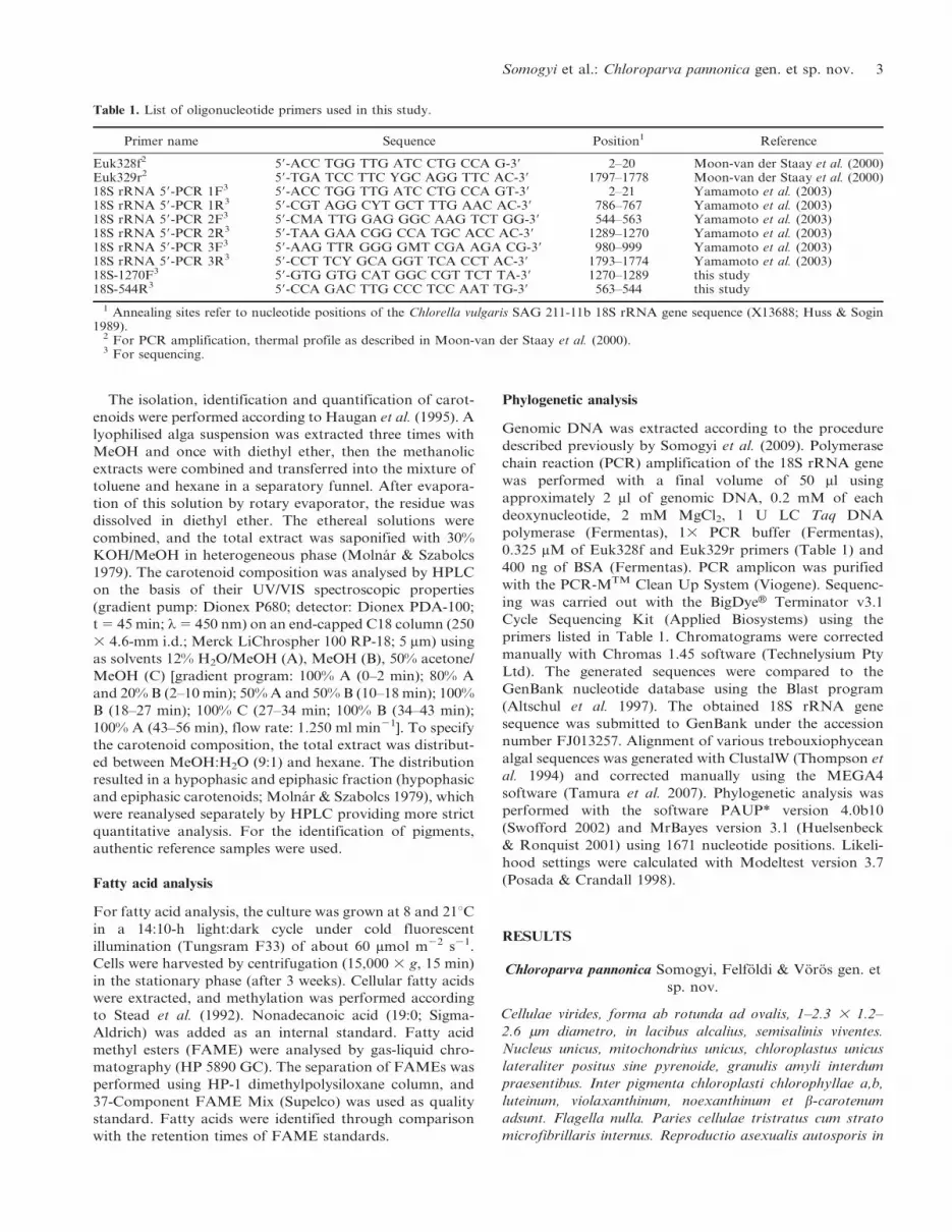
Table 1. List of oligonucleotide primers used in this study.
Primer name Sequence Position1 Reference
Euk328f2 59-ACC TGG TTG ATC CTG CCA G-39 2–20 Moon-van der Staay et al. (2000)Euk329r2 59-TGA TCC TTC YGC AGG TTC AC-39 1797–1778 Moon-van der Staay et al. (2000)18S rRNA 59-PCR 1F3 59-ACC TGG TTG ATC CTG CCA GT-39 2–21 Yamamoto et al. (2003)18S rRNA 59-PCR 1R3 59-CGT AGG CYT GCT TTG AAC AC-39 786–767 Yamamoto et al. (2003)18S rRNA 59-PCR 2F3 59-CMA TTG GAG GGC AAG TCT GG-39 544–563 Yamamoto et al. (2003)18S rRNA 59-PCR 2R3 59-TAA GAA CGG CCA TGC ACC AC-39 1289–1270 Yamamoto et al. (2003)18S rRNA 59-PCR 3F3 59-AAG TTR GGG GMT CGA AGA CG-39 980–999 Yamamoto et al. (2003)18S rRNA 59-PCR 3R3 59-CCT TCY GCA GGT TCA CCT AC-39 1793–1774 Yamamoto et al. (2003)18S-1270F3 59-GTG GTG CAT GGC CGT TCT TA-39 1270–1289 this study18S-544R3 59-CCA GAC TTG CCC TCC AAT TG-39 563–544 this study
1 Annealing sites refer to nucleotide positions of the Chlorella vulgaris SAG 211-11b 18S rRNA gene sequence (X13688; Huss & Sogin1989).
2 For PCR amplification, thermal profile as described in Moon-van der Staay et al. (2000).3 For sequencing.
Somogyi et al.: Chloroparva pannonica gen. et sp. nov. 3

partes duas; reproductio sexualis ignota. 18S rRNA sequen-
tia genetica (FJ013257) demonstrat differentias a speciebus
ceteris Trebouxiophycearum.
Cells green, spherical to oval, with a diameter of 1–2.3 3
1.2–2.6 mm, growing in alkaline, soda waters. One nucleus,
one mitochondrion, one lateral chloroplast without pyre-
noid, starch grains sometimes present. Chloroplast pig-
ments comprise chlorophylls a and b, lutein, violaxanthin,
neoxanthin and b-carotene. Flagella absent. Cell wall
trilaminate, with an inner microfibrillar layer. Reproduc-
tion by autosporulation into two daughter cells. Sexual
reproduction unknown. Analysis of 18S rRNA gene
sequence (FJ013257) shows differences from sequences of
other trebouxiophycean species.
HOLOTYPE: A lyophilised specimen has been deposited in
the Algological Collection of the Hungarian Natural
History Museum (under BP 7964).
ISOTYPE: Live culture has been submitted to the SAG
Culture Collection of Algae (University of Gottingen)
under SAG 2358.
ICONOTYPE: Fig. 9.
TYPE LOCALITY: An alkaline soda pan in the Danube-Tisza
Interfluve, Hungary.
Morphology, ultrastructure and reproduction
The cell size ranged from 1 to 2.3 mm in width and from 1.2
to 2.6 mm in length, with an average size of 1.4 3 1.7 mm.
SEM observation showed that the cell surface was either
smooth or wrinkled, but the surface of the dividing cells was
always wrinkled (Figs 1, 2). TEM observation revealed that
the cell organization was very simple. There were no
flagella, no basal bodies detectable by electron microscopy.
Each cell had a single nucleus and a single, parietal, usually
cup-shaped and bilobed chloroplast, containing stacked
thylakoid lamellae composed typically of four to six
thylakoid sacs (Figs 3, 8). Often one or two electron-dense
plastoglobuli could be observed within the plastids (Fig. 3),
but starch grains were only rarely found under the
experimental conditions (i.e. in the relatively young algal
culture). A single elongated mitochondrion, with cristae,
lied in the central cytoplasm, closely associated with the
concavity of the chloroplast and also with the nucleus
(Figs 3, 5). Vacuoles were also observed that contained
either electron-transparent material or sometimes electron-
dense bodies (Figs 3, 9). Peroxisome was also found
(Fig. 5). Chloroplast, nucleus and vacuole occupy a major
portion of the cell; the remainder is filled by the cytoplasm
containing several ribosomes (Figs 3, 5).
The cell wall was strictly regular, consisted of electron-
dense and electron-transparent layers and had an average
thickness of 28–63 nm. An electron-transparent layer
covered the plasma membrane, and this layer was followed
by a denser, fibrillar and relatively thick layer (inner
microfibrillar sheet) above which a trilaminar sheath was
observed consisting of an electron-dense, an electron-
transparent and another, outermost electron-dense layer
(Figs 4, 6). No mucilaginous envelope was observed on the
outer surface of the cells (Figs 4, 6).
Cells divided by autosporulation, generally giving rise to
two daughter cells (Figs 7–10). The remnants of the mother
cell wall (usually the trilaminar layer) were often observed
loosely surrounding the daughter cells after autosporulation
(Fig. 10). The remnants of the grandmother cell wall were
occasionally found around the autospores outside the
mother cell wall (Fig. 9). After the release of the daughter
cells, a ghost-like, curling, empty wall was sometimes
observed. No evidence for sexual reproduction was observed.
Figs 1–2. Scanning electron micrographs of Chloroparva pannonica(stub ACT 0608).
Fig. 1. Vegetative cell with a smooth surface. Scale bar 5 1 mm.Fig. 2. Vegetative cell with a wrinkled surface. Scale bar 5 1 mm.
Figs 3–6. Transmission electron micrographs of Chloroparvapannonica. C, chloroplast; CW, cell wall; M, mitochondrion; N,nucleus; P, peroxisome; PG, plastoglobule; PM, plasma membrane;V, vacuole.
Fig. 3. Vegetative cell. Scale bar 5 0.5 mm.Fig. 4. A close-up from Fig. 3 showing the cell wall structure:trilaminar outer layer of the vegetative cell (arrowhead), granulo-fibrillar inner layer and electron-transparent layer covering theplasma membrane (arrow). Scale bar 5 0.2 mm.Fig. 5. Vegetative cell. Scale bar 5 0.5 mm.Fig. 6. A close-up from Fig. 5 showing the cell wall structure.Notation as in Fig. 4. Scale bar 5 0.2 mm.
4 Phycologia, Vol. 50 (1), 2011

Pigment composition
Chloroparva pannonica contained typical chlorophyte pig-
ments, including chlorophylls a and b and lutein as the
dominant carotenoid. The chlorophyll a (tr: 6.5 min) and b
(tr: 9.1 min) content of C. pannonica was 4.66 and
2.27 mg g21 wet weight (biomass), respectively. The chloro-
phyll b:a ratio was 0.48. During the carotenoid analysis, the
proportion of the hypophasic and epiphasic fraction was
2.5. The main carotenoids were (all-E)- lutein, (all-E)-
violaxanthin, (99Z)-neoxanthin and b-carotene (b,b-
carotene) (Table 2).
Fatty acid content
The total fatty acid content of C. pannonica was 45 mg g21
dry cell mass at 8uC and 35 mg g21 dry cell mass at 21uC(Table 3). At 8uC, saturated fatty acids (SFA) constituted
7.2%, monounsaturated fatty acids (MUFA) 88.9% and
polyunsaturated fatty acids (PUFA) 3.8% of the total fatty
acid content. At 21uC, SFA composed 5.7%, MUFA 93.7%
and PUFA 0.5% of the total fatty acid content. The main
fatty acid of C. pannonica was oleic acid (18:1n-9)
constituting approximately 90% of the total extracted fatty
acid content.
18S rRNA gene analysis
Sequence analysis of C. pannonica (ACT 0608) resulted in
2329 nucleotides. Based on the database search, the closest
relatives were N. eucaryotum UTEX 2502, C. minutissima
C-1.1.9 and C. minutissima SAG 1.80 with 97.5–97.6%
pairwise similarities. Molecular analysis of the 18S rRNA
gene sequence placed the new picoalgal isolate on a dis-
tinct branch within the Trebouxiophyceae, Chlorophyta
(Fig. 11). The new isolate contains an intron in its 18S
rRNA gene with the same insertion position but different
length as in Koliella spiculiformis (Vischer) Hindak,
Actinastrum hantzschii Lagerheim SAG 2015 and Micracti-
nium sp. Fresinius CCAP 211/92 (Fig. 12).
DISCUSSION
Molecular biological methods appear to be useful tools to
assign taxa with a limited number of morphological
features. The introduction of DNA-based phylogenetic
analysis in the taxonomy of freshwater and saline
planktonic green algae resulted in the construction of new
genera (Henley et al. 2004; Krienitz et al. 2004; Fawley et al.
2005); although the nomenclature and systematics of
several small coccoid chlorophyte isolates are still waiting
for revision (Henley et al. 2004). Due to these uncertainties,
currently there is no general pairwise similarity value of 18S
rRNA gene that defines chlorophyte genera. Krienitz et al.
(2004) found that the taxa of Chlorella and Parachlorella
clade (Trebouxiophyceae, Chlorophyta), including several
genera (Dicloster, Didymogenes, Closteriopsis, Parachlor-
ella, Micractinium etc.), showed a range of 18S rRNA
sequence similarities from 97.4 to 99.5%. The phylogenetic
analysis of the ‘Nannochloris-like’ algae (Trebouxiophyceae,
Chlorophyta) revealed that these isolates (the Nannochloris-
like clade, containing the genera Picochlorum, Nannochloris,
Marvania, etc.) possess only 3.75% 18S rRNA gene
sequence heterogenity (Henley et al. 2004). Sequence
analysis of C. pannonica revealed that this isolate is
distantly related to other isolates of the Trebouxiophyceae
(# 97.6% 18S rRNA gene pairwise similarities); therefore,
the phylogenetic position of the new chlorophyte confirms
the proposal of a new genus (Fig. 11).
The closest relatives of C. pannonica are the members of
the ‘problematic group’ according to the work of Henley et
al. (2004): N. eucaryotum UTEX 2502, C. minutissima C-
1.1.9 and C. minutissima SAG 1.80. In contrast to the
members of this group, C. pannonica possesses one intron
(presumably a group I intron) in its 18S rRNA gene,
located in an insertion position previously described in
other related trebouxiophycean isolates (Fig. 12). Group I
intron insertions are relatively frequent in the ribosomal
RNA of green algae and have sporadic, highly biased
distribution (Haugen et al. 2005; Hoshina & Imamura
2008). The majority of these mobile genetic elements is
predicted to spread with reverse splicing (Woodson & Cech
1989; Bhattacharya et al. 2005) and has insertion positions
Figs 7–10. Transmission electron micrographs of Chloroparvapannonica during autosporulation. C, chloroplast; GMCW, grand-mother cell wall; MCW, mother cell wall; N, nucleus; PM, plasmamembrane; V, vacuole.
Fig. 7. Mother cell with two autospores. Scale bar 5 0.5 mm.Fig. 8. A close-up from Fig. 7 showing the structure of themother cell wall and the evolving daughter cell wall. Thedaughter cell wall has a fine trilaminar layer (arrowhead), a firmgranulo-fibrillar inner layer and a thin electron-transparentlayer. Scale bar 5 0.2 mm.Fig. 9. Mother cell with two autospores. Asterisk indicates anelectron-dense body. Scale bar 5 0.5 mm.Fig. 10. Two daughter cells at a late phase of autosporulation.Scale bar 5 0.5 mm.
Somogyi et al.: Chloroparva pannonica gen. et sp. nov. 5

restricted to only a few sites within the rRNA genes
(Hoshina & Imamura 2008). In addition to the two
distantly related chlorophyte genera, Choricystis (Treboux-
iophyceae) and Mychonastes (Chlorophyceae), the new
Chloroparva is the third freshwater picoeukaryotic algal
genus that contains introns in the 18S rRNA gene (Krienitz
et al. 1996; Hepperle & Schlegel 2002; Luo et al. 2010). Due
to the uneven occurrence (Fig. 12), introns in the 18S
rRNA gene should not be regarded as useful phylogenetic
markers among chlorophytes or among picoeukaryotic
algae.
The taxonomy of the members of the ‘problematic group’
is somewhat confusing. The type strain – Mainz 1 – of N.
eucaryotum was renamed based on its 18S rRNA gene
sequence to Picochlorum eukaryotum (Wilhelm, Eisenbeis,
Wild & Zahn) Henley, Hironaka, Guillou, M. Buchheim, J.
Buchheim, M. Fawley & K. Fawley by Henley et al. (2004).
According to Wilhelm et al. (1982), Mainz 1 strain had
small cells (1.5 mm on average) surrounded by a smooth and
thin cell wall, but dividing cells showed a rougher surface.
Menzel & Wild (1989) described that the cells divided by
autosporulation and that the two daughter cells remained
enveloped by the mother cell wall. After the release of the
daughter cells, the empty parental envelope displayed a
characteristic curling. Nannochloris eucaryotum UTEX
2502 is supposed to be the relative of N. eucaryotum SAG
55.87 according to the catalogue of SAG and also UTEX
Culture Collections. As described by Yamamoto et al.
(2001), N. eucaryotum UTEX 2502 had spherical cells of
2.5 mm in diameter, divided by autosporulation, which
resulted in two, three and four round daughter cells and a
persistent mother cell wall. In a latter study, Yamamoto et
al. (2003) noted that N. eucaryotum UTEX 2502 was
Table 3. Selected fatty acid content (in mg g21 dry weight) ofChloroparva pannonica in the stationary phase of growth at 8uCand 21uC in comparison to Choricystis minor and Pseudodictyos-phaerium jurisii in the stationary phase of growth at 20uC.
Fatty acids (FA)
C. pannonicaC. minor1 P. jurisii1
8uC 21uC 20uC 20uC
Saturated fatty acids (SFA)
14:0 0.90 0.55 1.09 0.2016:0 0.92 0.44 6.59 3.1317:0 0.00 0.08 — —18:0 0.99 0.91 0.95 0.4020:0 0.23 0.00 — —21:0 0.20 0.00 — —
Monounsaturated fatty acids (MUFA)
14:1 n-5 0.34 0.30 — —16:1 n-7 0.00 0.00 0.64 0.1017:1 n-7 0.37 0.28 — —18:1 n-9 39.26 31.94 0.68 0.45
Polyunsaturated fatty acids (PUFA)
18:2 n-6 0.49 0.12 0.90 0.8518:3 n-6 0.36 0.00 0.04 0.0818:3 n-3 0.00 0.00 1.80 0.7720:3 n-3 0.16 0.06 — —20:3 n-6 0.30 0.00 — —20:4 n-6 0.00 0.00 0.14 0.1420:5 n-3 0.00 0.00 0.46 0.1722:2 n-6 0.42 0.00 — —22:5 n-3 0.00 0.00 0.04 0.1522:6 n-3 0.00 0.00 0.22 0.15
S FA 44.95 34.69 18.99 10.37S SFA 3.25 1.99 8.63 3.73S MUFA 39.98 32.52 1.32 0.55S PUFA 1.73 0.18 3.60 2.31
1 According to Krienitz & Wirth (2006).
Table 2. The carotenoid composition of the total extract, the hypophasic and the epiphasic fractions isolated from Chloroparva pannonicalyophilised cell mass.
Peak Carotenoid UV/VIS (nm)
Total extract Hypophasic fraction Epiphasic fraction
tr (min) % tr (min) % tr (min) %
1 (all-E)-neoxanthin 468 441/4421 416 12.00 t2 10.73 1.1 — —2 (99Z)-neoxanthin 463/4641 435 412 14.26 7.4 12.92 5.8 — —3 (all-E)-violaxanthin 467 438 415 15.33 12.6 14.00 12.3 13.95 t2
4 epimers of luteoxanthin 448 420 398 16.75 17.15 t2 15.58 16.00 t2 — —5 (all-E)-lutein-5,6-epoxide 467 439/4411 416 19.85 t2 18.71 1.0 — —6 (Z)-isomers of lutein-5,6-epoxide 465 438 413 21.65 22.04 t2 20.58 21.00 t2 — —7 (all-E)-lutein 470 443 (420) 23.58 66.4 22.79 73.5 22.78 43.18 (9Z) + (99Z)-lutein 465 438 (417) 26.54 2.8 26.10 3.3 26.09 1.29 (13Z) + (139Z)-lutein 463 436 (415) 3303 27.08 2.4 26.71 3.0 26.69 2.0
10 (15Z)-lutein 466 439 (417) 3303 27.50 t2 27.16 t2 27.15 t2
11 (all-E)-b-cryptoxanthin 474 447 424 36.17 1.6 — — 36.15 36.234 9.04
12 (9Z) + (99Z)-b-cryptoxanthin 470 443 420 36.29 t2 — —13 c (b,y)-carotene + d (e,y)-carotene5 487 458/4576433 37.85 t2 — — 37.93 5.314 a-carotene (b,e-carotene) 473 445 (420) 4206 38.95 t2 — — 39.12 2.215 b-carotene (b,b-carotene) 477 452 39.55 6.8 — — 39.58 37.216 (9Z)-b-carotene 472 446 40.00 t2 — — 39.95 t2
1 In case of the hypophasic fraction.2 In traces.3 Peak 330 (cis-peak) with high intensity is characteristic to the (13Z)-, (139Z)- and (15Z)-isomers of carotenoids.4 (all-E) + (9Z) + (99Z)-b-cryptoxanthin, UV/VIS: 475 447 (422) nm.5 Tentatively identified.6 In case of the epiphasic fraction.
6 Phycologia, Vol. 50 (1), 2011

heterogeneous, and they purified it by single colony
isolation resulting in KSW0203. Interestingly, N. eucaryo-
tum KSW0203 was shown to be an autosporic taxon with
larger cells compared to their earlier results (autospores 3–
4 mm, mother cells 5–6 mm in diameter). Notwithstanding,
Henley et al. (2004) obtained the identical 18S rRNA gene
sequence for N. eucaryotum UTEX 2502 as Yamamoto et
al. (2003) for N. eucaryotum KSW0203. According to
Tschermak-Woess (1998), N. eucaryotum SAG 55.87 had
ellipsoidal cells somewhat less than 2 3 2 mm, and the size
Fig. 11. Maximum likelihood (ML) tree of 18S rRNA gene sequences retrieved from Chloroparva pannonica isolate ACT 0608 and theclosest relatives (Trebouxiophyceae, Chlorophyta). ML analysis was conducted heuristically using the tree-bisection-reconnection optionwith likelihood settings of the best-fit model [TrN+I+G model; Lset Base 5 (0.2533 0.2111 0.2705), Nst 5 6, Rmat 5 (1.0000 2.0131 1.00001.0000 5.0758), Rates 5 gamma, Shape 5 0.6136; Pinvar 5 0.6952; the same values were used for maximum parsimony (MP)]. Bootstrapvalues greater than 70 (based on 100 and 1000 replicates for ML and MP, respectively) and Bayesian (B) posterior probabilities (3 100)from 500,000 generations are shown (order: ML/MP/B). Asterisk marks sequences that are also present in Fig. 12.
Somogyi et al.: Chloroparva pannonica gen. et sp. nov. 7

of mother cells reached 3.5 mm. The long persistence of the
mother cell wall surrounding the autospores was also
described (Tschermak-Woess 1998). Cell division was
found by autosporulation, with two, three or rarely four
autospores per mother cell resulting from two temporally
separated processes of autosporulation (Tschermak-Woess
1998). As Henley et al. (2004) suggested, sequencing of
strain SAG 55.87 would be necessary to reveal the relation
between these two strains.
The type strain – Lefevre no. 87 – of C. minutissima was
renamed based on its 18S rRNA gene sequence to
Mychonastes homosphaera Kalina & Puncocharova by Huss
et al. (1999). For strain C-1.1.9 (and there are other algal
strains misinterpreted as C. minutissima, e.g. SAG 1.80),
which is unrelated to M. homosphaera, Huss et al. (1999)
proposed keeping the name C. minutissima provisionally
until more information becomes available. According to our
knowledge, detailed description is available only for strain
Lefevre no. 87. Nevertheless, C. minutissima C-1.1.9 is
absent in any current culture collection as implied by Henley
et al. (2004). The detailed study of C. minutissima SAG 1.80
would be necessary – parallel with sequencing N. eucar-
yotum SAG 55.87 – to clarify the taxonomical status of the
closest relatives of the proposed novel genus, C. pannonica.
Based on the above descriptions, C. pannonica have
smaller cells (usually less than 2 mm) than N. eucaryotum
UTEX 2502 and SAG 55.87 (Figs 1, 2). Unfortunately,
detailed electron microscopy–based description is not
available in the case of these strains; however, the
autosporulation and the persistent mother cell wall
surrounding the autospores suggest similarity to C.
pannonica (Figs 9, 10). According to Yamamoto et al.
(2003), autosporulation is an ancestral character, but until
now the taxonomical value of the mode of cell division is
not clear. In C. pannonica, formation of only two
autospores per mother cell was observed, in contrast to
the three or four autospores in the other strains; however,
the number of autospores may depend on culture condi-
tions (Krienitz et al. 1996). The cell wall structure of C.
pannonica (Figs 4, 6) is very similar to that of Choricystis
minor (Skuja) Fott as described by Menzel & Wild (1989)
and Krienitz et al. (1996). According to Krienitz et al.
(1996), the cell wall of C. minor consists of a thin (10–20 mm)
trilaminar layer, which is closely associated with an inner
fibrillar layer, and a relatively diffuse layer of low electron
optical density is often located between this inner layer and
plasmalemma. According to Menzel & Wild (1989), in the
course of autosporulation, the inner layer (presumably
cellulose) of the still closed mother cell wall disappears,
leaving only the trilaminar sheath. We assume that in case
of C. pannonica, the inner microfibrillar layer of the mother
cell wall disappears similarly to C. minor, and only the
trilaminar layer (of the mother cell wall or occasionally the
grandmother cell wall) persists for a long time around the
autospores (Figs 9, 10). Based on SEM micrographs, the
surface of the cells was either smooth or wrinkled (Figs 1,
2). TEM micrographs clearly indicated that the wrinkled
cell surface was not the result of a network of ribs on the
cell wall as in M. homosphaera (Hanagata et al. 1999). We
hypothesize that the long persistence of the mother cell wall
around the daughter cells – which is clearly observable on
TEM micrographs (Figs 9, 10) – results in this wrinkled
appearance (Fig. 2). In contrast with this, cells after the
detachment of the mother cell wall remnants have a smooth
surface (Fig. 1).
Menzel & Wild (1989) and Krienitz et al. (1996)
described the typical curling of the mother cell wall after
autosporulation, as it is observable in the case of many
trebouxiophycean species (e.g. P. eukaryotum) and also in
the case of C. pannonica. This curling suggests that the
trilaminar cell wall layer contains sporopollenin, which
provides a defence mechanism against drying out and
enzymatic disintegration (Menzel & Wild 1989). In the case
of C. pannonica, the toleration of desiccation is presumably
a very important feature, as this alga has to survive the
summer period, when the pans usually dry out.
The fatty acid content of C. pannonica was higher at 8uCthan at 21uC. The relative contribution of polyunsaturated
fatty acids increased significantly (7.4 times) due to the
temperature decrease (Table 3). Picoeukaryotes show char-
acteristic seasonal appearance in the temperate zone: they
dominate the pico-fraction from autumn to spring (Callieri
Fig. 12. Position and length of introns within the 18S rRNA gene presented in Chloroparva pannonica isolate ACT 0608 and in its closestrelatives. Introns are indicated with black boxes; nucleotide positions are shown according to the Chlorella vulgaris SAG 211-11b 18S rRNAgene sequence (X13688; Huss & Sogin 1989); nt, nucleotides.
8 Phycologia, Vol. 50 (1), 2011

2008; Voros et al. 2009). This characteristic seasonal
dynamic was also observable in the case of Hungarian
soda pans, and C. pannonica was isolated from a winter
phytoplankton sample. The winter predominance of
picoeukaryotes was correlated with their lower light and
temperature requirement (Somogyi et al. 2009). Besides
this, the increased PUFA content at lower temperature
might be an important feature in the temperature acclima-
tization. Krienitz & Wirth (2006) analysed the fatty acid
content of the most abundant freshwater picoplanktonic
green algal species, C. minor and Pseudodictyosphaerium
jurisii (Hindak) Hindak. According to their results, C.
minor contained 64% palmitic acid (16:0) and 13% alpha-
linolenic acid (18:3 n-3) similarly to P. jurisii, which
contained 57% palmitic acid and 12% alpha-linolenic acid
(Table 3). Several studies suggest that the fatty acid content
and composition of nanoplanktonic green algae could be
notably different; however, saturated fatty acids [palmitic
acid or stearic acid (18:0)] were dominant (29–70%) in the
case of the studied species (Chlorella sp., Monoraphidium
sp., Scenedesmus sp. and Stichococcus sp.) at 20uC (Makulla
2000; Teoh et al. 2004; Krienitz & Wirth 2006). Based on
these results, the extremely high monounsaturated oleic
acid ratio of C. pannonica seems to be a distinctive feature.
In conclusion, the new chlorophyte isolate originating
from a turbid, shallow soda pan proved to be a new species
– belonging to a new genus within the Trebouxiophyceae –
based on its 18S rRNA gene sequence. Chloroparva
pannonica has a minimal set of cell organelles similarly to
other pico-sized chlorophytes. The pigment composition
showed a typical chlorophycean pattern. The long persis-
tence of the mother cell wall surrounding the autospores
proved to be somewhat unique.
ACKNOWLEDGEMENTS
The study was sponsored by the Hungarian Research Fund
(OTKA K 73369 and partially by K 76176, K 60121).
Gyorgyi Horvath was supported by a scholarship from the
University of Pecs, Medical School (PTE AOK KA). Tamas
Felfoldi was supported by a scholarship from the Ministry of
Education and Culture, Hungary (DFO 0051/2009). The
authors thank Emil Boros for his help with fieldwork, Attila
Kovacs for helpful discussions and Balazs Nemeth and
Erzsebet Lakatos for their technical assistance. The authors
are grateful to Csilla Jonas for skillful assistance in electron
microscopic sample preparation and ultrathin sectioning and
to Laszlo Hiripi for chlorophyll a and b analysis. We also
thank Peter Takacs for his help with the Latin diagnosis.
REFERENCES
ALTSCHUL S.F., MADDEN T.L., SCHAFFER A.A., ZHANG J., ZHANG
Z., MILLER W. & LIPMAN D.J. 1997. Gapped BLAST and PSI-BLAST: a new generation of protein database search programs.Nucleic Acids Research 25: 3389–3402.
BHATTACHARYA D., REEB V., SIMON D.M. & LUTZONI F. 2005.Phylogenetic analyses suggest reverse splicing spread of group Iintrons in fungal ribosomal DNA. BMC Evolutionary Biology 5:68.
CALLIERI C. 2008. Picophytoplankton in freshwater ecosystems:the importance of small-sized phototrophs. Freshwater Reviews1: 1–28.
CARRICK H.J. & SCHELSKE C.L. 1997. Have we overlooked theimportance of small phytoplankton in productive waters?Limnology and Oceanography 42: 1613–1621.
FAWLEY M.W., FAWLEY K.P. & BUCHHEIM M.A. 2004. Moleculardiversity among communities of freshwater microchlorophytes.Microbial Ecology 48: 489–499.
FAWLEY M.W., FAWLEY K.P. & OWEN H.A. 2005. Diversity andecology of small coccoid green algae from Lake Itasca,Minnesota, USA, including Meyerella planktonica, gen. et sp.nov. Phycologia 44: 35–48.
FELFOLDI T., SOMOGYI B., MARIALIGETI K. & VOROS L. 2009.Characterization of photoautotrophic picoplankton assemblagesin turbid, alkaline lakes of the Carpathian Basin (CentralEurope). Journal of Limnology 68: 385–395.
HANAGATA N., MALINSKY-RUSHANSKY N. & DUBINSKY Z. 1999.Eukaryotic picoplankton, Mychonastes homosphaera (Chloro-phyceae, Chlorophyta), in Lake Kinneret, Israel. PhycologicalResearch 47: 263–269.
HAUGAN J.A., AAKERMANN T. & LIAAEN-JENSEN S. 1995. Example2: macroalgae and microalgae. In: Carotenoids vol. 1A: isolationand analysis (Ed. by G. Britton, S. Liaaen-Jensen & H. Pfander),pp. 215–226. Birkhauser Verlag, Basel.
HAUGEN P., SIMON D.M. & BHATTACHARYA D. 2005. The naturalhistory of group I introns. Trends in Genetics 21: 111–119.
HENLEY W.J., HIRONAKA J.L., GUILLOU L., BUCHHEIM M.A.,BUCHHEIM J.A., FAWLEY M.W. & FAWLEY K.P. 2004. Phyloge-netic analysis of the ‘Nannochloris-like’ algae and diagnoses ofPicochlorum oklahomensis gen. et sp. nov. (Trebouxiophyceae,Chlorophyta). Phycologia 43: 641–652.
HEPPERLE D. & KRIENITZ L. 2001. Systematics and ecology ofchlorophyte picoplankton in German inland waters along anutrient gradient. International Review of Hydrobiology 86:269–284.
HEPPERLE D. & SCHLEGEL I. 2002. Molecular diversity ofeukaryotic picoalgae from three lakes in Switzerland. Interna-tional Review of Hydrobiology 87: 1–10.
HOSHINA R. & IMAMURA N. 2008. Eu-Chlorella large subunitrDNA sequences and group I introns in ribosomal DNA of theparamecian symbiotic alga NC64A. Phycological Research 56:21–32.
HUELSENBECK J.P. & RONQUIST F. 2001. MrBayes: Bayesianinference of phylogeny. Bioinformatics 17: 754–755.
HUSS V.A.R. & SOGIN M.L. 1989. Primary structure of theChlorella vulgaris small subunit ribosomal RNA coding region.Nucleic Acids Research 17: 1255.
HUSS V.A.R., FRANK C., HARTMANN E.C., HIRMER M., KLOBOU-
CEK A., SEIDEL B.M., WENZELER P. & KESSLER E. 1999.Biochemical taxonomy and molecular phylogeny of the genusChlorella sensu lato (Chlorophyta). Journal of Phycology 35:587–598.
KRIENITZ L. & WIRTH M. 2006. The high content of polyunsat-urated fatty acids in Nannochloropsis limnetica (Eustigmatophy-ceae) and its implication for food web interactions, freshwateraquaculture and biotechnology. Limnologica 36: 204–210.
KRIENITZ L., HUSS V.A.R. & HUMMER C. 1996. PicoplanktonicChoricystis species (Chlorococcales, Chlorophyta) and theproblems surrounding the morphologically similar ‘Nanno-chloris-like algae’. Phycologia 35: 332–341.
KRIENITZ L., TAKEDA H. & HEPPERLE D. 1999. Ultrastructure, cellwall composition, and phylogenetic position of Pseudodictyo-sphaerium jurisii (Chlorococcales, Chlorophyta) including acomparison with other picoplanktonic green algae. Phycologia38: 100–107.
KRIENITZ L., HEGEWALD E.H., HEPPERLE D., HUSS V.A.R., ROHR
T. & WOLF M. 2004. Phylogenetic relationship of Chlorella andParachlorella gen. nov. (Chlorophyta, Trebouxiophyceae). Phy-cologia 43: 529–542.
LEWIN R.A., KRIENITZ L., GOERICKE R., TAKEDA H. & HEPPERLE
D. 2000. Picocystis salinarum gen. et sp. nov. (Chlorophyta) – anew picoplanktonic green alga. Phycologia 39: 560–565.
Somogyi et al.: Chloroparva pannonica gen. et sp. nov. 9

LUO W., PROSCHOLD T., BOCK C. & KRIENITZ L. 2010. Genericconcept in Chlorella-related coccoid green algae (Chlorophyta,Trebouxiophyceae). Plant Biology 12: 545–553.
MACISAAC E.A. & STOCKNER J.G. 1993. Enumeration ofphototrophic picoplankton by autofluorescence microscopy. In:Handbook of methods in aquatic microbial ecology (Ed. by P.F.Kemp, B.F. Sherr, E.B. Sherr & J.J. Cole), pp. 187–197. LewisPublishers, Boca Raton, FL.
MAKULLA A. 2000. Fatty acid composition of Scenedesmusobliquus: correlation to dilution rates. Limnologica 30: 162–168.
MANTOURA R.F.C. & LLEWELLYN C.A. 1983. The rapid determi-nation of algal chlorophyll and carotenoid pigments and theirbreakdown products in natural waters by reverse-phase highperformance liquid chromatography. Analytica Chimica Acta151: 297–314.
MENZEL K. & WILD A. 1989. A comparative ultrastructuralinvestigation of some Nannochloris species (Chlorococcales) withparticular reference to the systematic position of Nannochlorumeucaryotum. Botanica Acta 102: 152–158.
MOLNAR P. & SZABOLCS J. 1979. Alkaline permanganate oxidationof carotenoid epoxides and furanoids. Acta Chimica AcademiaeScientiarum Hungaricae 99: 155–173.
MOON-vAN DER STAAY S.Y., VAN DER STAAY G.W.M., GUILLOU
L., VAULOT D., CLAUSTRE H. & MEDLIN L.K. 2000. Abundanceand diversity of prymnesiophytes in the picoplankton communityfrom the equatorial Pacific Ocean inferred from 18S rDNAsequences. Limnology and Oceanography 45: 98–109.
POSADA D. & CRANDALL K.A. 1998. Modeltest: testing the modelof DNA substitution. Bioinformatics 14: 817–818.
RIPPKA R., DERUELLES J., WATERBURY J.B., HERDMAN M. &STAINER R.Y. 1979. Generic assignments, strain histories andproperties of pure cultures of cyanobacteria. Journal of GeneralMicrobiology 111: 1–61.
SOLYMOSI K., MYSLIWA-KURDZIEL B., BOKA K., STRZAŁKA K. &BODDI B. 2006. Disintegration of the prolamellar body structureat high concentrations of Hg2+. Plant Biology 8: 627–635.
SOMOGYI B., FELFOLDI T., VANYOVSZKI J., AGYI A., MARIALIGETI
K. & VOROS L. 2009. Winter bloom of picoeukaryotes inHungarian shallow turbid soda pans and the role of light andtemperature. Aquatic Ecology 43: 735–744.
STEAD D.E., SELLWOOD J.E., WILSON J. & VINEY I. 1992.Evaluation of a commercial microbial identification systembased on fatty acid profiles for rapid, accurate identification ofplant pathogenic bacteria. Journal of Applied Bacteriology 72:315–321.
STOCKNER J.G. 1991. Autotrophic picoplankton in freshwaterecosystems: the view from the summit. Internationale Revue dergesamten Hydrobiologie 76: 483–492.
SWOFFORD D.L. 2002. PAUP*: phylogenetic analysis usingparsimony (*and other methods), version 4.0b10. SinauerAssociates, Sunderland, MA.
TAMURA K., DUDLEY J., NEI M. & KUMAR S. 2007. MEGA4:molecular evolutionary genetics analysis (MEGA) softwareversion 4.0. Molecular Biology and Evolution 24: 1596–1599.
TEOH M.L., CHU W.L., MARCHANT H. & PHANG S.M. 2004.Influence of culture temperature on the growth, biochemicalcomposition and fatty acid profiles of six Antarctic microalgae.Journal of Applied Phycology 16: 421–430.
THOMPSON J.D., HIGGINS D.G. & GIBSON T.J. 1994. Clustal W:improving the sensitivity of progressive multiple sequencealignment through sequence weighting, position-specific gappenalties and weight matrix choice. Nucleic Acids Research 22:4673–4680.
TSCHERMAK-WOESS E. 1998. Life cycle and supplementarycomments on the light microscopic morphology of Nannochloriseucaryota. Plant Biology 1: 214–218.
VAULOT D., EIKREM W., VIPREY M. & MOREAU H. 2008. Thediversity of small eukaryotic phytoplankton (# 3 mm) in marineecosystems. FEMS Microbiology Reviews 32: 795–820.
VOROS L., BOROS E., SCHMIDT A., V.-BALOGH K., NEMETH B.,SOMOGYI B. & MOZES A. 2006. Physical and chemicalenvironment of phytoplankton in soda pans having whitecoloured water. Hidrologiai Kozlony 86: 139–141. (in Hungarianwith English summary)
VOROS L., SOMOGYI B. & BOROS E. 2008. Birds cause netheterotrophy in shallow lakes. Acta Zoologica AcademiaeScientiarum Hungaricae 54: 23–34.
VOROS L., MOZES A. & SOMOGYI B. 2009. A five-year study ofautotrophic winter picoplankton in Lake Balaton, Hungary.Aquatic Ecology 43: 727–734.
WILHELM C., EISENBEIS G., WILD A. & ZAHN R. 1982. Nanno-chlorum eucaryotum: a very reduced coccoid species of marineChlorophyceae. Zeitschrift fur Naturforschung 37c: 107–114.
WOODSON S.A. & CECH T.R. 1989. Reverse self-splicing of theTetrahymena group I intron: implication for the directionality ofsplicing and for intron transposition. Cell 57: 335–345.
YAMAMOTO M., NOZAKI H. & KAWANO S. 2001. Evolutionaryrelationships among multiple modes of cell division in thegenus Nannochloris (Chlorophyta) revealed by genome size, actingene multiplicity and phylogeny. Journal of Phycology 37:106–120.
YAMAMOTO M., NOZAKI H., MIYAZAWA Y., KOIDE T. & KAWANO
S. 2003. Relationship between presence of a mother cell wall andspeciation in the unicellular microalga Nannochloris (Chloro-phyta). Journal of Phycology 39: 172–184.
Received 3 February 2010; accepted 11 May 2010
Associate editor: Lothar Krienitz
10 Phycologia, Vol. 50 (1), 2011