chimica farmaceutica - ese - salento university publishing
TRANSCRIPT
Chimica Farmaceutica

337
FAR-PL-01 Orthosteric and Allosteric Ligands Selectively Acting at Cholinergic Receptor Subtypes Marco De Amici, Clelia Dallanoce, Giovanni Grazioso, Carlo De Micheli Dipartimento di Scienze Farmaceutiche “Pietro Pratesi” dell’Università degli Studi di Milano, Via L. Mangiagalli 25, 20133 Milano, Italy [email protected]
The presentation will focus on the results recently achieved by our research group in the design, synthesis and pharmacological evaluation of selective ligands which target cholinergic receptor subtypes, belonging to both the nicotinic (nAChR) and the muscarinic (mAChR) acetylcholine receptor families.
A set of spirocyclic derivatives will be illustrated, in which the simultaneous presence of the quinuclidinyl and 2-isoxazolinyl moieties, coupled with suitable stereoelectronic features of the substituent at position 3 of the spirocyclic ring, engendered a selective agonist profile at the homomeric neuronal 7 nAChRs [1]. The most promising compound in the series has been further investigated in preclinical studies and in in vivo models of CNS disorders and neuropathic pain. A group of novel hybrid peptides structurally related to natural -conotoxins MII and PIA will be also presented, which behave as competitive antagonists able to discriminate 62* and 32* nAChR subtypes [2].
The five mAChR subtypes bind their physiological transmitter in the highly conserved orthosteric site within the transmembrane domains of the receptors. Orthosteric muscarinic agonists have negligible binding selectivity and poor signaling specificity. A less conserved allosteric site has been also characterized at the extracellular entrance of the binding pocket of mAChRs. To gain subtype-selective M2 receptor activation, we designed a group of putative bitopic compounds, i. e. hybrid derivatives fusing highly potent, unselective oxotremorine-like orthosteric activators with M2-selective bis(ammonio)alkane-type allosteric fragments. The new ligands interacted simultaneously with both recognition areas of the receptor protein, thus allowing the exploitation of favorable features of the orthosteric and the allosteric site by a single ligand molecule. The orthosteric interaction provided high affinity binding and activation of M2 mAChRs. The allosteric interaction yielded receptor subtype-selectivity and, in addition, could modulate efficacy and activate pathway-specific intracellular signaling [3].
[1] C.Dallanoce, P.Magrone, C.Matera, F.Frigerio, G.Grazioso, M.De Amici,
S.Fucile, V.Piccari, K.Frydenvang, L.Pucci, C.Gotti, F.Clementi, C.De Micheli, ChemMedChem, 6, 2011, 889-903.
[2] M.De Amici, G.Grazioso, C.Dallanoce, C.De Micheli et al., submitted. [3] K.Mohr, C.Tränkle, E.Kostenis, E.Barocelli, M.De Amici, U.Holzgrabe, Br.J.Pharmacol., 159, 2010, 997-1008.
338
FAR-PL-02 Modulation of Hydrolysis of Fatty Acid Ethanolamides: Rational Drug Design for Novel Therapeutic Opportunities
MarcoMor
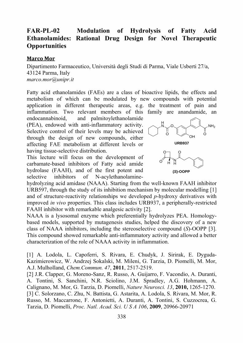
Dipartimento Farmaceutico, Università degli Studi di Parma, Viale Usberti 27/a, 43124 Parma, Italy [email protected] Fatty acid ethanolamides (FAEs) are a class of bioactive lipids, the effects and metabolism of which can be modulated by new compounds with potential application in different therapeutic areas, e.g. the treatment of pain and inflammation. Two relevant members of this family are anandamide, an endocannabinoid, and palmitoylethanolamide (PEA), endowed with anti-inflammatory activity. Selective control of their levels may be achieved through the design of new compounds, either affecting FAE metabolism at different levels or having tissue-selective distribution. This lecture will focus on the development of carbamate-based inhibitors of Fatty acid amide hydrolase (FAAH), and of the first potent and selective inhibitors of N-acylethanolamine-hydrolyzing acid amidase (NAAA). Starting from the well-known FAAH inhibitor URB597, through the study of its inhibition mechanism by molecular modelling [1] and of structure-reactivity relationships we developed p-hydroxy derivatives with improved in vivo properties. This class includes URB937, a peripherally-restricted FAAH inhibitor with remarkable analgesic activity [2]. NAAA is a lysosomal enzyme which preferentially hydrolyzes PEA. Homology-based models, supported by mutagenesis studies, helped the discovery of a new class of NAAA inhibitors, including the stereoselective compound (S)-OOPP [3]. This compound showed remarkable anti-inflammatory activity and allowed a better characterization of the role of NAAA activity in inflammation. [1] A. Lodola, L. Capoferri, S. Rivara, E. Chudyk, J. Sirirak, E. Dyguda-Kazimierowicz, W. Andrzej Sokalski, M. Mileni, G. Tarzia, D. Piomelli, M. Mor, A.J. Mulholland, Chem.Commun. 47, 2011, 2517-2519. [2] J.R. Clapper, G. Moreno-Sanz, R. Russo, A. Guijarro, F. Vacondio, A. Duranti, A. Tontini, S. Sanchini, N.R. Sciolino, J.M. Spradley, A.G. Hohmann, A. Calignano, M. Mor, G. Tarzia, D. Piomelli, Nature Neurosci. 13, 2010, 1265-1270. [3] C. Solorzano, C. Zhu, N. Battista, G. Astarita, A. Lodola, S. Rivara, M. Mor, R. Russo, M. Maccarrone, F. Antonietti, A. Duranti, A. Tontini, S. Cuzzocrea, G. Tarzia, D. Piomelli, Proc. Natl. Acad. Sci. U S A 106, 2009, 20966-20971
OHN
O
NH2
OOH
NH
OO
O H
URB937
(S)-OOPP
339
FAR-PL-03 Advances in the Characterization of New Challenging GPCRs Gloria Cristalli School of Pharmacy, Medicinal Chemistry Unit, University of Camerino, Via S. Agostino 1, 62032 Camerino, Italy. [email protected] Purinergic receptors are classified in P2X, a ligand-gated ion channel family, activated by ATP and ADP, and P1 and P2Y, two G protein-coupled receptor (GPCR) families, activated by adenosine and purine/pyrimidine nucleotides, respectively. They are widely distributed in the body and involved in several cellular functions, most of them still poorly understood, although in recent years a number of clinical applications of purinergic receptor ligands have been proposed and few compounds are already on the market or in clinical trials. Purinoceptor families include also purinergic-like receptors, which need to be better characterized and, between them, the recently deorphanized GPR17 seems to be dually activated by uracil nucleotides and cysteinyl-leukotrienes. GPR17 was found to be highly expressed in organs typically undergoing ischemic damage, thus representing a potential target for new therapeutic approaches to acute and chronic neurodegenerative diseases. Characterization of this receptor has been performed on transfected 1321N1 cells by using [35S]GTPγS binding assay. Known and newly synthesized nucleotides were screened and proved to behave as ligands for this receptor with a wide range of activity. Moreover, an innovative and non-radioactive functional cAMP assay was validated, which showed a strong correlation with the data obtained with [35S]GTPγS binding assay and confirmed that GPR17 is coupled with a Gαi [1]. Furthermore, specific binding sites for [3H]-guanosine, which are not recognized by other purinergic receptor ligands, were detected on membrane preparations from rat brain. These findings supported the hypothesis of the existence of a specific GPCR for guanosine that could account for the actions played by this naturally occurring purine nucleoside. An innovative assay was optimized for the characterization at rat brain membranes of the putative guanosine binding site by using a series of known and novel guanosine derivatives, prepared by modifying the purine and the sugar moiety of guanosine. Results of these experiments proved that guanosine, 6-thioguanosine, and their derivatives activate a new GPCR, which is different from the well characterized adenosine receptors [2]. [1] M. Buccioni, G. Marucci, D. Dal Ben, D. Giacobbe, C. Lambertucci, L. Soverchia, A.
Thomas, R. Volpini, G. Cristalli, Adv. Pharmacol. Sci., in press. [2] R. Volpini, G. Marucci, M. Buccioni, D. Dal Ben, C. Lambertucci, C. Lammi, R.C. Mishra, A.
Thomas, G. Cristalli, ChemMedChem., DOI: 10.1002/cmdc.201100100, VIP (the paper was judged to be very important by the referees).
340
FAR-PL-04 Multidisciplinary and Multitarget Approaches in the Search for Novel Drugs in the Treatment of Neurodegenerative Diseases Angelo Carotti Department of Medicinal Chemistry, University of Bari “Aldo Moro”, Via Orabona 4, I-70125 Bari, Italy. E-mail: [email protected]
Research in medicinal chemistry has recently shifted towards the design of
multitarget/multipotent agents that, interfering with different biological pathways critical for either the onset or the progression of a given disease, may have higher therapeutic benefits compared to single-target–selective drugs [1,2]. Multitarget ligands would be simpler to develop in a clinical setting and present lower risks of drug-drug interactions compared to multicomponent drugs and drug combinations. On the other hand, the design and optimization of multiple ligands exhibiting high and, more importantly, well-balanced affinities at selected targets, is quite a daunting task.
Within this challenging scenario, we developed new multitarget ligands which act as reversible, dual MAO-B and acetylcholinesterase (AChE) inhibitors [3,4] or as ChE inhibitors and beta-amyloid (A) anti-aggregating agents [5], with the potential for treating Alzheimer’s disease (AD) [6]. In a parallel research, we have been working along the amyloid hypothesis of AD that has led to a deeper understanding of the pathology of AD and has provided insight into the design of novel potential drugs [7]. According to this hypothesis, the increase of Aβ production and aggregation into low-molecular weight oligomers, fibrils and, ultimately, amyloid plaques are the leading cause of AD. The reduction of both Aβ formation (with β- and -secretase modulators) [8] and aggregation and the increase of Aβ clearance (with active and passive immunization) [9] are promising therapeutic strategies for AD. Pursuant to the development of a fast spectrofluorimetric method for the kinetic analysis of Aβ aggregation [10], a screening of medium-sized molecular libraries was carried out and several classes of novel anti-aggregating agents, including two anticancer drugs, have been discovered.[11,12] The optimization of the most interesting molecules afforded compounds capable of blocking Aβ-fibril formation at a sub-micromolar concentration. Spectroscopic, analytical and biophysical methods have been used to elucidate the inhibition mechanism of Aβ aggregation. Among them, capillary electrophoresis proved particularly efficient to detect the oligomeric species targeted by the compounds blocking A fibril formation.[12] Finally, molecular dynamics simulations on carefully conceived model systems have shed light on A fibril formation and on how small molecules may hinder the early phase of A aggregation.[13]
341
References [1] Morphy R., Rankovic Z. J. Med. Chem. 2005, 48, 6523-43; [2] Cavalli A., Bolognesi ML, Minarini A., Rosini M., Tumiatti V., Recanatini M., Melchiorre C. J. Med. Chem. 2008, 51, 347-72; [3] Carotti A. ACS-EFMC Meeting, Frontiers in CNS and Oncology Medicinal Chemistry, Siena, Italy, October 7-9, 2007; [4] Pisani L., Catto M., Leonetti F., Nicolotti O., Stefanachi A., Campagna F., Carotti A. Curr. Med. Chem. 2011, in press; [5] B. Tasso et al: Poster presentation at this Meeting; [6] Querfurth HW, LaFerla FM. N Engl J Med. 2010, 362, 329-44; [7] Prins ND, Visser PJ, Scheltens P. Alzheimer’s Research & Therapy 2010, 2, 28; [8] Chopra K., Misra S., Kuhad A. Expert Opin. Pharmacother. 2011, 12, 335-50; [9] Lemere CA, Masliah E. Na.t Rev. Neurol. 2010, 6, 108-19; [10] Cellamare S., Stefanachi A., Stolfa DA, Basile T., Catto M., Campagna F., Sotelo E., Acquafredda P., Carotti. A. Bioorg. Med. Chem. 2008, 16, 4810-22; [11] Catto M., Aliano R., Carotti A., Cellamare S., Palluotto F., Purgatorio R., De Stradis A., Campagna F. Eur. J. Med. Chem. 2010, 45, 1359-66; [12] Colombo R., Carotti A., Catto M., Racchi M., Lanni C., Verga L., Caccialanza G., De Lorenzi E. Electrophoresis. 2009, 30, 1418-29; [13] Convertino M., Pellarin R., Catto M., Carotti A., Caflisch A. Proteins 2009, 18, 792-800.
342
FAR-KN-01 Furthering the Understand of Polypharmacology in Nuclear Receptor Superfamily. Antonio Macchiarulo, Marco Cellanetti, Andrea Carotti, Roberto Pellicciari Dipartimento di Chimica e Tecnologia del Farmaco, Università degli Studi di Perugia, Via del Liceo 1, 06123, Perugia. Email: [email protected] Recent years have seen an increasing awareness that drugs often bind to more than one molecular target, exhibiting polypharmacology [1]. Although this aspect has commonly been considered as undesirable promiscuity being responsible for unwanted side effects, in many cases it is a key component to the therapeutic efficacy of drugs [2]. The knowledge on polypharmacology can therefore aid the explanation of why some drugs work better than expected, or why other drugs have diverse side effects, albeit acting on the same target. Polypharmacology is the result of poor ligand specificity that combines with protein promiscuity [3]. Accordingly, in order to further the understand of polypharmacology, both ligand-based and protein-based computational techniques are being developed that provide predictions of proteins to which ligands are likely to bind [4]. In this communication, we investigate aspects of polypharmacology in the superfamily of human nuclear receptors (NRs). Human NRs comprise 48 members of ligand-dependent transcription factors that offer important druggable targets for therapeutic interventions in multiple disease areas [5]. Many NRs are promiscuous with respect to the wide range of ligands that act as modulators, and many NR modulators are not specific with respect to the number of NRs they bind. The construction of a target-centric chemical space [6, 7] and the application of integrative approaches are discussed as instrumental in charting key components and interactions of NR binding sites, with the aim of aiding the rationalization and optimization of selectivity and/or multi-target profile of selected NR ligands. [1] A.L. Hopkins, Nature. 462, 2009, 167. [2] M.J. Keiser MJ, J.J. Irwin, B.K. Shoichet, Biochemistry, 49, 2010, 10267. [3] A. Macchiarulo, I. Nobeli, J.M. Thornton, Nat. Biotechnol., 22, 2004, 1039. [4] A.D. Boran, R. Iyengar, Curr. Opin. Drug Discov. Devel. 13, 2010, 297. [5] A. Aranda, A. Pascual, Physiol. Rev. 81, 2001, 1269. [6] A. Macchiarulo, R. Pellicciari, J. Mol. Graph. Model. 26, 2007, 728. [7] A. Macchiarulo, R. Nuti, G. Eren, R. Pellicciari, J. Chem. Inf. Model. 49,
2009, 900.
343
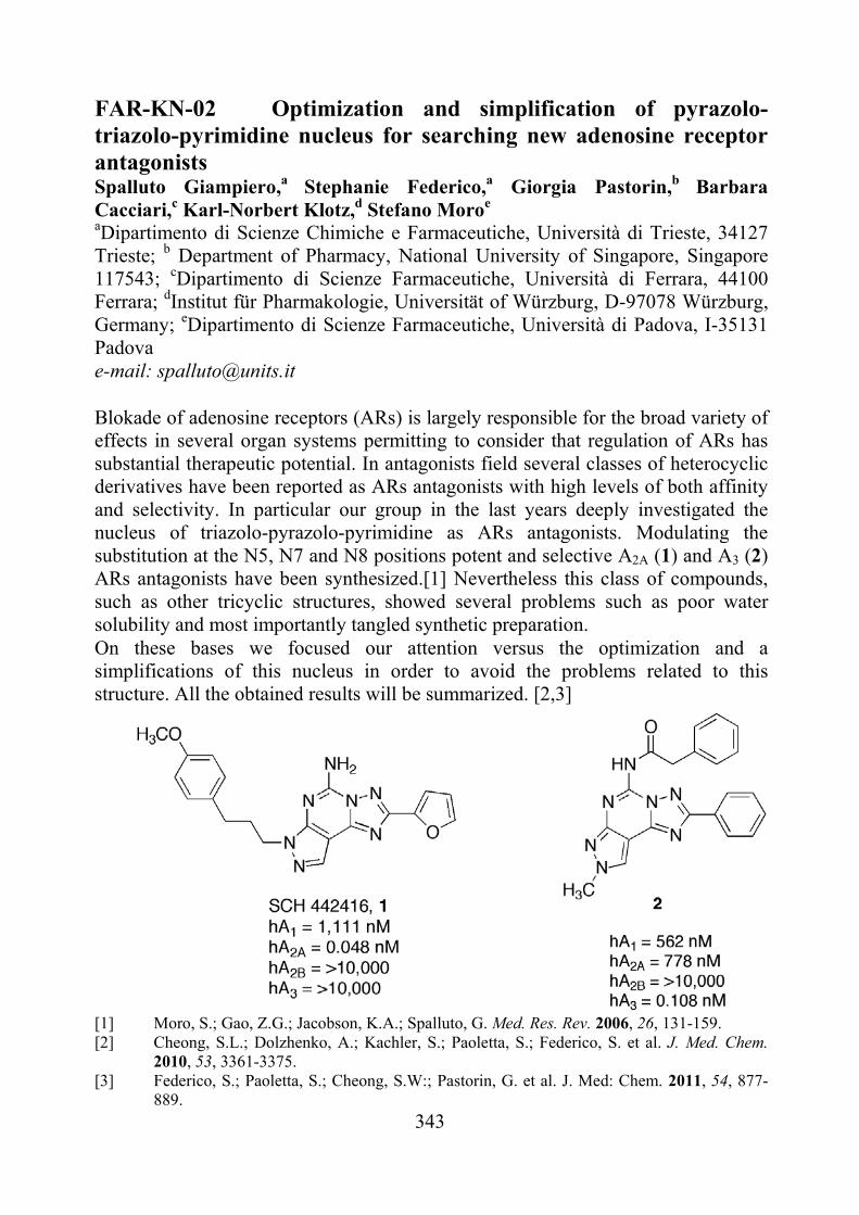
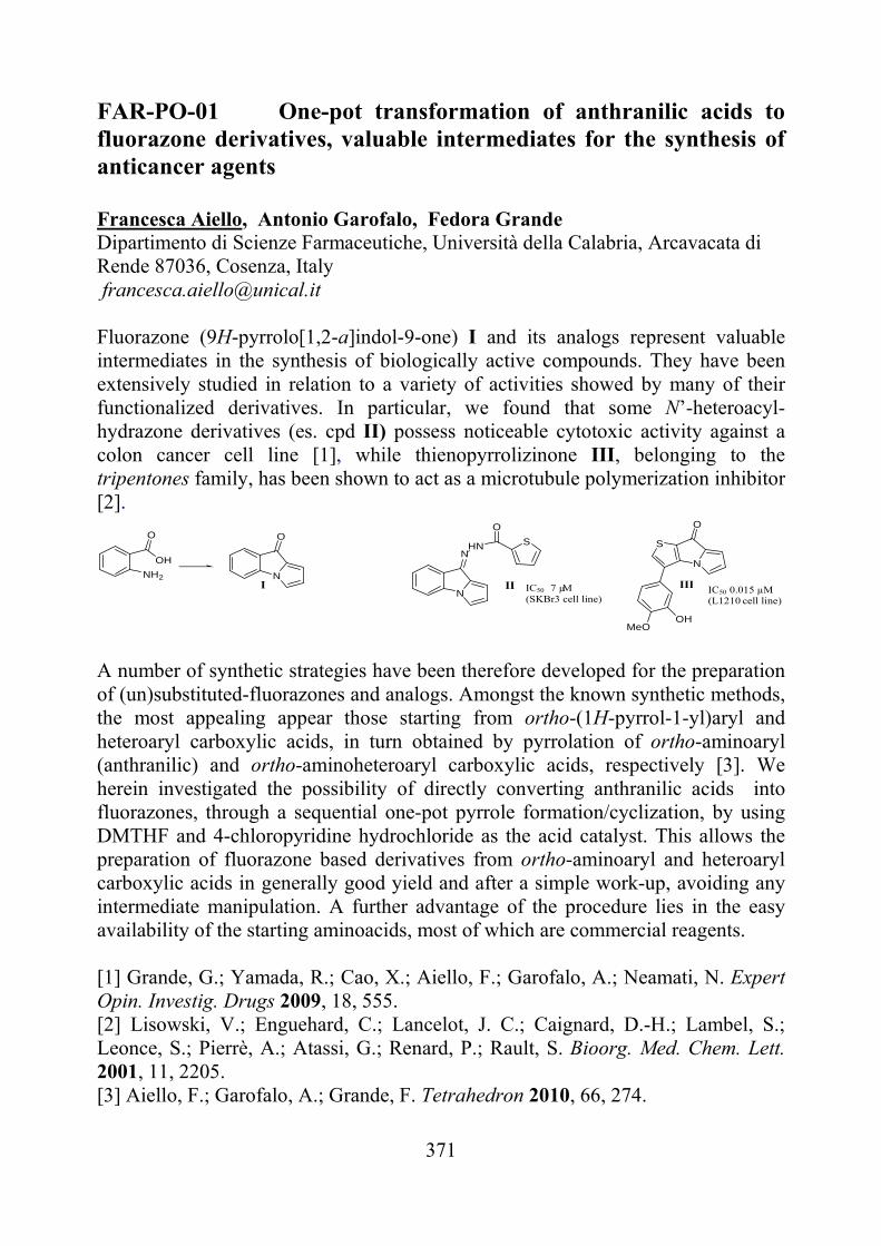
FAR-KN-02 Optimization and simplification of pyrazolo-triazolo-pyrimidine nucleus for searching new adenosine receptor antagonists Spalluto Giampiero,a Stephanie Federico,a Giorgia Pastorin,b Barbara Cacciari,c Karl-Norbert Klotz,d Stefano Moroe aDipartimento di Scienze Chimiche e Farmaceutiche, Università di Trieste, 34127 Trieste; b Department of Pharmacy, National University of Singapore, Singapore 117543; cDipartimento di Scienze Farmaceutiche, Università di Ferrara, 44100 Ferrara; dInstitut für Pharmakologie, Universität of Würzburg, D-97078 Würzburg, Germany; eDipartimento di Scienze Farmaceutiche, Università di Padova, I-35131 Padova e-mail: [email protected] Blokade of adenosine receptors (ARs) is largely responsible for the broad variety of effects in several organ systems permitting to consider that regulation of ARs has substantial therapeutic potential. In antagonists field several classes of heterocyclic derivatives have been reported as ARs antagonists with high levels of both affinity and selectivity. In particular our group in the last years deeply investigated the nucleus of triazolo-pyrazolo-pyrimidine as ARs antagonists. Modulating the substitution at the N5, N7 and N8 positions potent and selective A2A (1) and A3 (2) ARs antagonists have been synthesized.[1] Nevertheless this class of compounds, such as other tricyclic structures, showed several problems such as poor water solubility and most importantly tangled synthetic preparation. On these bases we focused our attention versus the optimization and a simplifications of this nucleus in order to avoid the problems related to this structure. All the obtained results will be summarized. [2,3]
[1] Moro, S.; Gao, Z.G.; Jacobson, K.A.; Spalluto, G. Med. Res. Rev. 2006, 26, 131-159. [2] Cheong, S.L.; Dolzhenko, A.; Kachler, S.; Paoletta, S.; Federico, S. et al. J. Med. Chem.
2010, 53, 3361-3375. [3] Federico, S.; Paoletta, S.; Cheong, S.W:; Pastorin, G. et al. J. Med: Chem. 2011, 54, 877-
889.
344
FAR-KN-03 Immobilized enzymes as Efficient tools in drug discovery Manuela Bartolini, Francesca Mancini, Angela De Simone, Marina Naldi, Vincenza Andrisano University of Bologna, Department of Pharmaceutical Sciences, Via Belmeloro 6, 40126 Bologna, Italy. [email protected] Immobilised enzyme reactors (IMERs) have proven to be a useful and economic alternative to conventional in-solution methods, when increased reliability, automation and fast data output are required. In particular, considering the high cost and difficulty in over-expression, isolation and purification of recombinant enzymes, this analytical technique represents an extremely useful approach to preserve the activity when a small amount of enzyme is available. In the IMER format, enzymes are ready to be reused and can be coupled to chromatographic systems and appropriate detectors (UV-Vis, MS, FL). This coupling generally increases automation, reproducibility and analyses accuracy and reduces sample handling and operator time consumption.
In the field of drug discovery, IMERs can be reliably applied to different phases of the drug discovery pathway, i.e., to rapidly screen for potential drugs candidates (lead selection), to characterize the mode of action at specific targets and perform SAR studies (lead optimization), and to determine ADMET parameters (early ADMET studies). In fact, in a second stage following the screening step, selected hits need to be further characterized in terms of mechanism of action and kinetic parameters.
In this talk, a few IMERs applications will be presented, useful in all the steps of drug discovery and development. At this regard, acetyl-, butyryl-cholinesterase and BACE-1 (beta secretase) immobilized reactors were validated for the screening and determination of the mechanism of action and inhibitory constants of new leads for the treatment of Alzheimer’s disease in a highly reliable and automated mode. Remarkably, besides representing valid tools to screen new reversible inhibitors, immobilized reactors were also used to characterize pseudo-irreversible inhibitors.
In drug development stage, the monolithic disk-shaped mini-columns (2 mm x 6 mm I.D.) containing immobilized 2D6 and 3A4 isoforms of cytochrome P450 were developed as tools for phase I drug metabolism studies, for the early estimates of the drug metabolism, toxicity and possible drug-drug interactions.
345
FAR-KN-04 From the central benzodiazepine receptor to the adenosine receptors exploiting the 3-diketoindole moiety. Barbara Cosimelli Dipartimento di Chimica Farmaceutica e Tossicologica, Università degli Studi di Napoli “Federico II”, Via Montesano 49, 80131 Napoli, Italy [email protected]
The first non-xantinic antagonists at the adenosine receptors (ARs) were reverse agonists at the benzodiazepine receptor (BzR) showing SNC stimulating properties [1].
Some time ago our research group disclosed 3-aryl[1,2,4]triazino[4,3-a]benzimidazol-4-(10H)-ones as A1 AR antagonists designed by modifying analogous compounds binding to the BzR [2].
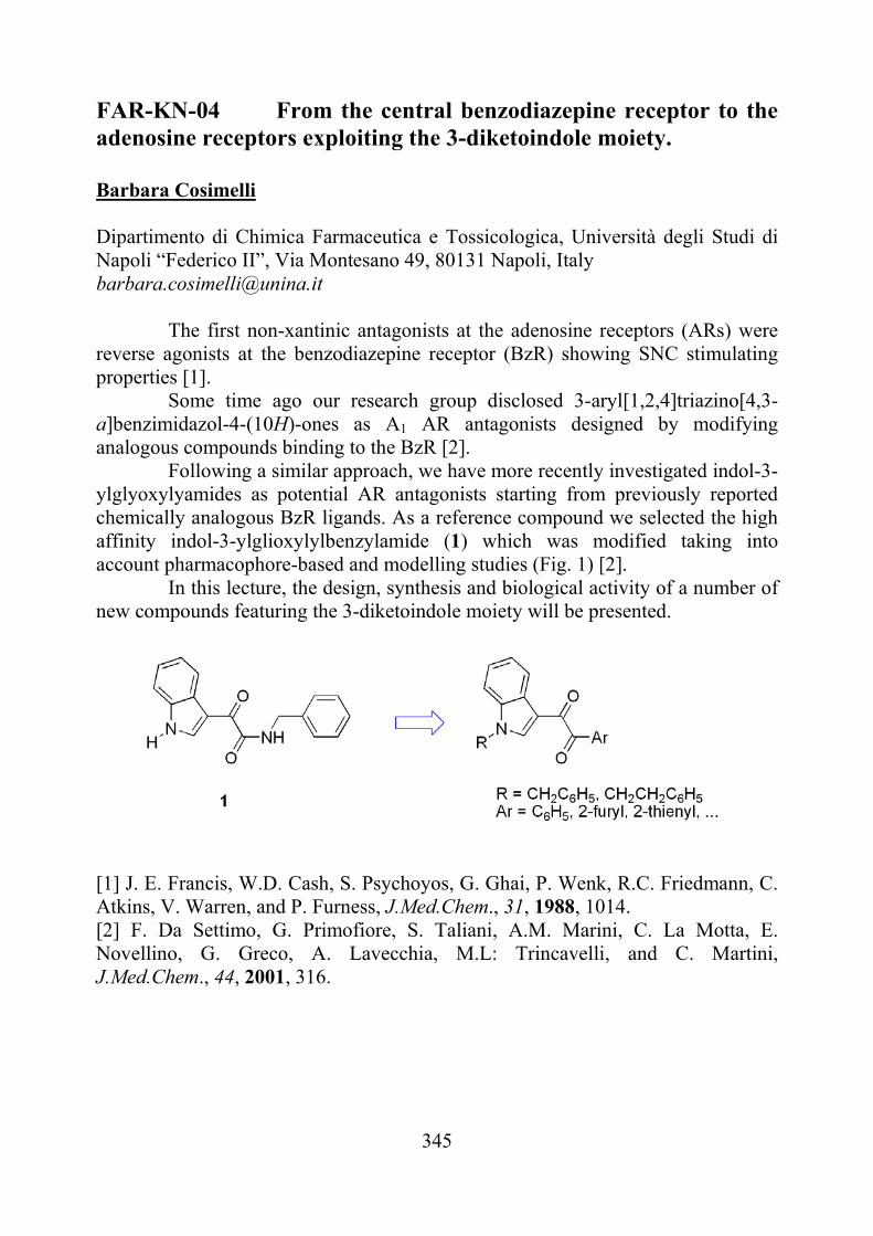
Following a similar approach, we have more recently investigated indol-3-ylglyoxylyamides as potential AR antagonists starting from previously reported chemically analogous BzR ligands. As a reference compound we selected the high affinity indol-3-ylglioxylylbenzylamide (1) which was modified taking into account pharmacophore-based and modelling studies (Fig. 1) [2].
In this lecture, the design, synthesis and biological activity of a number of new compounds featuring the 3-diketoindole moiety will be presented.
[1] J. E. Francis, W.D. Cash, S. Psychoyos, G. Ghai, P. Wenk, R.C. Friedmann, C. Atkins, V. Warren, and P. Furness, J.Med.Chem., 31, 1988, 1014. [2] F. Da Settimo, G. Primofiore, S. Taliani, A.M. Marini, C. La Motta, E. Novellino, G. Greco, A. Lavecchia, M.L: Trincavelli, and C. Martini, J.Med.Chem., 44, 2001, 316.
346
FAR-KN-05 Design and in vivo evaluation of PET radiotracers for imaging P-gp expression Nicola A. Colabufo, Francesco Berardi, Mariangela Cantore, Marialessandra Contino, Carmela Inglese, Mauro Niso, Roberto Perrone Dipartimento Farmacochimico, Universitá degli Studi di Bari, via Orabona,4, 70125, Bari, Italy [email protected] ABC transporters, in particular P-gp, BCRP, and MRPs are highly expressed in various physiological barriers such as blood-brain barrier, blood-testis barrier, blood-tumor barrier [1]. They modulate the accumulation of various drugs by active efflux transport. It has been demonstrated that changes in ABC transporter expression and function are involved in various neurodegenerative pathologies such as Alzheimer’s and Parkinson’s disease as well as epilepsy [2]. Moreover, the overexpression of these transporters in tumour cells causes Multidrug Resistance. PET radiotracers allow a noninvasive in vivo imaging of transporter function and expression. Recently several probes have been developed but their unfavorable pharmacodynamic and pharmacokinetic properties limited the in vivo investigation [3]. The design of new imaging probes to visualize efflux transporters is complicated by the overlapping substrate recognition pattern of different ABC transporter types. Three probes for PET analysis displaying favorable preclinical studies will be presented: - [11C]MC266, a P-gp substrate, to image the pump activity; - [11C]MC18, a P-gp inhibitor, to detect the pump expression; - [11C]MC113, a P-gp substrate, to identify chemosensitive and chemoresistant tumors [4] [1] W. Löscher, H. Potschka, Prog. Neurobiol. 76, 2005, 22. [2] N.A. Colabufo, F. Berardi, M. Cantore, M. Contino, C. Inglese, M. Niso, R. Perrone, J. Med. Chem. 53, 2010, 1883. [3] F. Bauer, C. Kuntner, J.P. Bankstahl, T. Wanek, M. Bankstahl, J. Stanek, S. Mairinger, B. Dörner, W. Löscher, M. Müller, T. Erker, O. Langer O. Bioorg Med Chem. 18, 2010, 5489. [4] A. Van Waarde, N.K. Ramakrishnan, A.A. Rybczynska, P.H. Elsinga, F. Berardi, J.R. de Jong, C. Kwizera, R. Perrone, M. Cantore, J.W.A. Sijbesma, R.A. Dierckx, N.A. Colabufo, J. Med. Chem. 523, 2009, 4524
347
FAR-KN-06 Design, synthesis and biological evaluation of potent and selective non-hydroxamic matrix metalloproteinases inhibitors Paolo Tortorella Dipartimento Farmaco-Chimico, Università degli Studi ”Aldo Moro”, Via Orabona 4, 70126 Bari (Italy). [email protected] In the last 20 years, a great variety of synthetic, low molecular weight MMP inhibitors (MMPIs) have been synthesized and tested, and some of them entered phase III clinical trials as anticancer drugs, although none has reached clinical utility. A general structure for an effective MMP inhibitor includes a zinc-binding group (ZBG) capable to bind the catalytic zinc (II) ion of these proteinases, at least one functional group that provides crucial H-bonding interactions with the enzyme backbone and one or more side chains giving rise to effective van der Waals
interactions with the enzyme subsites. The hydroxamic acid group is by far the most commonly used ZBG in inhibitor design and has generally been found to be the most effective. Hydroxamate binds the catalytic zinc (II) ion in a bidentate fashion, blocking substrate access to the active site and rendering the metal incapable of peptide hydrolysis. The failure of hydroxamic acid-based MMPIs in vivo may stem from poor pharmacokinetics (low oral bioavailability and short half-life), from the ability to bind other metal ions, and from the lack of specificity due to very strong binding to the catalytic zinc ion. As a consequence, it has been
pointed out that the design of selective inhibitors should involve weaker ZBGs to effectively modulate affinity by variation of substituents on the molecule scaffold. With a single coordinate bond to the metal center, inhibitors with monodentate ZBGs (such as carboxylic acids or phosphonic acids) are generally weaker inhibitors. We have been studying non-hydroxamic MMPIs for a long time, with a particular attention towards phosphonic derivatives.1-3 In this lecture, the design, synthesis, structure-activity relationship and in vitro pharmacological evaluation of new phosphonic MMPIs will be presented. [1] G. Pochetti, E. Gavuzzo, C. Campestre, M. Agamennone, P. Tortorella, V. Consalvi, C. Gallina,
O. Hiller, H. Tschesche, P. A. Tucker, F. Mazza, J Med Chem 49(3), 2006, 923. [2] A. R. Folgueras, A. Fueyo, O. Garcia-Suarez, J. Cox, A. Astudillo, P. Tortorella, C. Campestre,
A. Gutierrez-Fernandez, M. Fanjul-Fernandez, C. J. Pennington, D. R. Edwards, C. M. Overall, C. Lopez-Otin, The Journal of biological chemistry 283(14), 2008, 9465.
[3] M. T. Rubino, M. Agamennone, C. Campestre, G. Fracchiolla, A. Laghezza, F. Loiodice, E. Nuti, A. Rossello, P. Tortorella, ChemMedChem 4(3), 2009, 352.
348
FAR-KN-07 4-Phenyl-2-propionamidotetralin derivatives: useful ligands to define the stereochemical requirements for MT2-selective melatonin receptor antagonists Gilberto Spadoni,a Annalida Bedini,a Simone Lucarini,a Giorgio Tarzia,a Silvia Rivara,b Marco Mor,b Valeria Lucinic aDipartimento di Scienze Biomolecolari dell’Università di Urbino “Carlo Bo”, Piazza Rinascimento 6, 61029 Urbino, Italy bDipartimento Farmaceutico dell’Università di Parma, V.le G. P. Usberti 27/A, 43124 Parma, Italy cDipartimento di Farmacologia, Chemioterapia e Tossicologia Medica dell’Università di Milano, Via Vanvitelli 32, 20129 Milano, Italy [email protected]
Understanding the therapeutic potential of melatonin (N-acetyl-5-methoxytryptamine, MLT) has become an interesting topic in medicinal chemistry research, and MT1 and MT2 receptors are emerging as possible therapeutic targets for sleep disorders and depression. Three therapeutic agents (Circadin®, Rozerem®, and Valdoxan®) are already in use, and other compounds are currently under study for the treatment of sleep disturbances or depression.[1] Consistent information is available for non-selective MT1/MT2 ligands, and several molecular models, both ligand- and receptor-based, have been proposed to rationalize their SARs.[2] On the contrary, only limited data on MT1 or MT2 subtype-selective compounds are available up to now, and a clear definition of the structural requirements for subtype selectivity is still lacking. During the present decade, SAR investigations on melatonin receptor ligands were therefore aimed at both the discovery of new chemical classes and the definition of structural requirements for subtype selectivity.
Conformational restriction of bioactive molecules is a valuable tool for investigating the topographical and chemical features of small-molecule ligands. For instance, the -aminotetralin skeleton has been successfully used as a rigid template for the synthesis of non-indolic melatonin-like agents, and several other substances possessing important biological activities. 4-Phenyl-2-propionamidotetralin (4-P-PDOT) [3] is a prototypical MT2-selective ligand employed in pharmacological tests to discriminate the role of MT1 and MT2 receptors in MLT mediated effects. Despite its pharmacological application, the SARs for its derivatives have been poorly explored.
In this lecture the design, synthesis [4] and pharmacological characterization of 4-phenyl-2-amidotetralin derivatives will be described, focusing on their SAR, active conformation and configuration. A convenient protocol providing access to all four 4-P-PDOT enantiomers (ee >99%), and the determination of their absolute configuration will also be described. Binding data
349
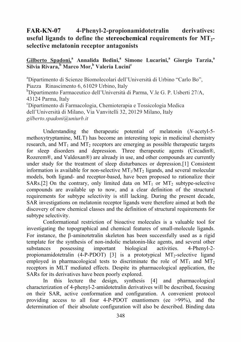
on each single stereoisomer, conformational analysis and pharmacophore-based superpositions led to a new chiral pharmacophore model which can be applied to both melatonin receptor agonists and MT2-selective antagonists. Validation of this pharmacophore model has been achieved synthesizing conformationally constrained tetrahydronaphthalene derivatives.
Superposition of (2S,4S)- and (2R,4S)-4-P-PDOT [1] G. Spadoni, A. Bedini, S. Rivara, and M. Mor, CNS Neurosci. Ther., (2010), DOI: 10.1111/j.1755-5949.2010.00197.x. [2] S. Rivara, M. Mor, A. Bedini, G. Spadoni, and G. Tarzia, Curr. Top. Med. Chem., 8, 2008, 954. [3] M.L. Dubocovich, M.I. Masana, S. Iacob, and D.M. Sauri, Naunyn-Schmiedeberg's Arch. Pharmacol. 355, 1997, 365. [4] S. Lucarini, A. Bedini, G. Spadoni, and G. Piersanti, Org. Biomol. Chem. 6, 2008, 147
350
FAR-KN-08 Antimicrobial PhotoDynamic Therapy: a new tool for the treatment of localized infections Donata Dei Molteni Therapeutics, Via Fiorentina 1, Siena; Via Barontini 8, Scandicci (FI) [email protected]
The pandemic diffusion of new microbial infections, as well as the onset of resistance toward antibiotic treatment of many pathogens, urge the development of new antimicrobial therapies as an alternative to the use of classical drugs.[1]
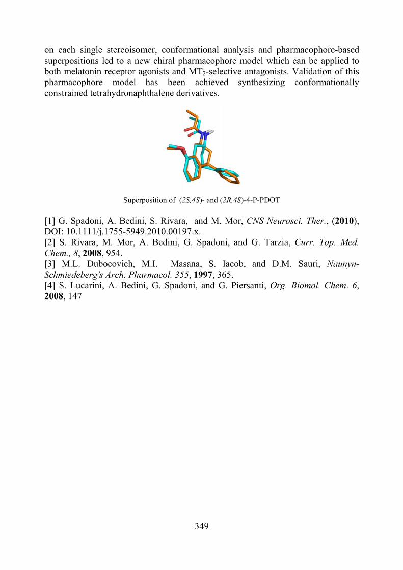
In this area, and particularly for the treatment of localized infectious diseases, the use PhotoDynamic Therapy (PDT) could represent a new appealing strategy to combact pathogens, help the wounds healing, reduce the risk of systemic infections and limit the spread of resistance.[2]
Molteni Therapeutics (MT) is involved since many years in the synthesis, analytical characterization and pharmaco-biological testing of new photosensitizers belonging to several classes of derivatives (Zinc and Silicon phthalocyanines [3], as well as porphyrins [4]), with the aim to discover new PDT agents and to elucidate the structure-activity relationships of these active compounds.
Up to now, very good results in the photoinactivation of yeasts and bacteria have been obtained with many of the synthesized compounds. In particular, the use of Zn(II)-phthalocyanines bearing quaternary ammonium groups has been extensively studied, starting from Discovery to Development for a restricted number of selected molecules. RLP068, the lead compound of MT pipeline, is currently being evaluated in a Phase IIa Clinical Trial.
In this presentation a brief introduction on PDT principles and an overview of the work done by MT on photosensitizers, spanning from synthetic and analytical data to in vitro and in vivo biological results, will be given.
[1] European Centre for Disease Prevention and Control “Annual epidemiological
report on communicable diseases in Europe 2008”; Center for Global Development’s Drug Resistance Working Group “The race against drug resistance”, 2010.
[2] Wainwright Journal of Antimicrobial Chemotherapy 1998, 42, 13-28; Garland et al. Future Med. Chem. 2009, 1(4), 667-691.
[3] Roncucci et al. EP0906758 (2011), EP1164135 (2004), EP1381611 (2005), EP1356813 (2005), EP1444236 (2007), EP1883641 (2008), EP1883640 (2009).
[4] Roncucci et al EP1558616 (2008).
351
FAR-OR-01 Drugs of abuse analysis: are Dried Blood Spots suitable for "on street" controls? Laura Mercolini1, Roberto Mandrioli1, Chiara Marcheselli1, Giovanni Serpelloni2, Maria Augusta Raggi1 1 Laboratory of Pharmaco-Toxicological Analysis, Department of Pharmaceutical Sciences, Faculty of Pharmacy, University of Bologna, Via Belmeloro 6, 40126 Bologna, Italy 2 Department of Antidrug Policies, Presidency of the Council of Ministers, Via Po 16/a, 00198 Roma, Italy [email protected]
Driving under the influence (DUI) of psychotropic substances, such as drugs of abuse (DoA) and/or alcohol, is one of the leading causes of traffic collisions. Furthermore, car accidents are the first cause of death (and of acquired disability) for young people under the age of 35. As a consequence, roadside controls are routinely performed by law enforcement agencies as part of prevention/dissuasion policies. Within this framework, the correct and timely sampling of a significant biological matrix, followed by a reliable qualitative/quantitative analysis, is the cornerstone of a fair assessment of the DUI state. For alcohol intake, the use of breathalizers has reached a satisfactory level of reliability and speed. However, assessing the actual state of drug intoxication is much more complicated. It is important to have at disposal fast and reliable analytical methods, able to provide good results for the identification and the quantitation of the most important DoA.
The Laboratory of Pharmaco-Toxicological Analysis develops advanced methods for the analysis of DoA in different biological fluids and tissues, in particular in an innovative matrix: Dried Blood Spots (DBS) [1]. DBS are obtained from blood drops collected on filter paper from a simple finger prick; the technique represents a very attractive and feasible alternative to the traditional blood sampling. It is especially useful “on street”, because is minimally invasive and allows sample collection, transport and storage, granting good stability without requiring refrigeration nor other pre-treatments. A few original analytical methods are for the analysis of different DoA in human DBS, for the purpose of reliable “on street” drug testing.The methods are based on LC-ESI-MS/MS (triple quadrupole) and samples are directly injected into the system, after a very fast solvent extraction. The results obtained until now on cocaine, cannabinoids and their main metabolites are promising, in terms of extraction yields, sensitivity and selectivity. Assays are in progress in order to fully validate the methods.
[1] L. Mercolini, R. Mandrioli, G. Gerra and M.A. Raggi, Analysis of cocaine and two metabolites in dried blood spots by liquid chromatography with fluorescence detection: A novel test for cocaine and alcohol intake, J. Chromatogr. A, 46, 2010, 7242.
352
FAR-OR-02 Selective serotonergic 5-HT7 receptor agonists as a new therapeutic venue for treatment of cognitive disorders: focus on Fragile X syndrome. Enza Lacivita,a Paola De Giorgio,a Lucia Ciranna,b Lara Costa,b Francesco Berardi,a Roberto Perrone,a Marcello Leopoldo.a aDipartimento Farmaco-Chimico, Via Orabona, 4; 70125, Bari, Italy bDipartimento di Scienze Bio-Mediche, Sezione di Fisiologia, Viale Andrea Doria, 6; 95125, Catania, Italy [email protected] Serotonin (5-HT) affects the excitability of hippocampal neurons and hippocampal-dependent cognitive functions. We have recently shown that 5-HT7 receptor activation modulates glutamate AMPA receptor-mediated basal transmission [1] and metabotropic glutamate receptor-induced long-term depression (mGluR-LTD) in the CA3-CA1 synapse of mouse hippocampus. Activation of 5-HT7 receptors is able to reverse mGluR-LTD also in the Fmr1 knockout (KO) mice model of Fragile X syndrome (FXS) [2], the most common form of hereditary intellectual disability, in which hippocampal mGluR-LTD is abnormally enhanced [3], suggesting that a pharmacological treatment selectively targeting 5-HT7 receptors might be considered in the therapy of FXS. LP-211, a selective 5-HT7 receptor agonist [4], is able to modulate AMPA-mediated synaptic currents and to reverse mGluR-LTD in the CA3-CA1 hippocampal synapse similarly to 8-OH-DPAT, the standard 5-HT7 non selective agonist.
HN
O
N( )5
N
NCLP-211
5-HT7 Ki: 0.58 nM; 5-HT1A Ki: 188 nM
On the basis of these results, we have undertaken a new project for the identification of new selective 5-HT7 receptor agonists endowed with pharmacodynamic and pharmakocinetic properties suitable for in vivo use. This will allow to clarify the therapeutic potential of 5-HT7 receptor agonists in the pharmacological treatment of cognitive disorders. [1] L.Costa, C.Trovato, S.A.Musumeci, M.V.Catania, L. Ciranna Hippocampus, 2011, in press;
doi: 10.1002/hipo.20940. [2] L. Costa, M. Spatuzza, C. Trovato, C.M.Bonaccorso, S.A. Musumeci, M.V. Catania, L.
Ciranna, article submitted; presented in abstract form (757.25/M18 Neuroscience Meeting Planner. San Diego, CA: Society for Neuroscience, 2010).
[3] M.F.Bear, K.M.Huber, S.T.Warren Trend Neurosci. 27, 2004, 370. [4] M.Leopoldo, E.Lacivita, P.De Giorgio, C.Fracasso, S.Guzzetti, S.Caccia, M.Contino,
N.A.Colabufo, F.Berardi, R.Perrone, J. Med. Chem. 51, 2008, 5813.
353
FAR-OR-03 The Medicinal Chemist’s Toolbox: Versatile Approaches for the Rapid Identification of Promising Biologically Active Hits Marco Radi and Maurizio Botta Dipartimento Farmaco Chimico Tecnologico dell'Università degli Studi di Siena, Via Aldo Moro, 53100, Siena, Italy [email protected] In the last few years, the synergistic relationship between organic chemistry, molecular modeling and biology has played a growing role in the identification of new potential drug candidates. A key contribution to this successful multidisciplinary approach has been given by modern instruments/techniques which have significantly accelerated the drug-discovery process. Despite molecular modeling approaches have significantly speed-up the identification of potential hits from large libraries of compounds, synthetic chemistry still play a key role in producing new chemical entities both for the discovery and optimization phase. Within the medicinal chemist’s “toolbox”, high-speed chemical techniques have become an important device for the rapid identification of new biologically active compounds. Parallel synthesis, microwave assisted techniques, click-chemistry and multicomponent reactions represent nowadays commonly used techniques for rapid identification of novel hit compounds and for the hit-to-lead optimization of promising inhibitors. An overview on the application of these modern techniques to the synthesis of different heterocyclic scaffold with antiviral, antitumor and antitubercular activities will be given [1]. [1] a) M. Radi, et al. J.Comb.Chem., 7, 2005, 117. b) D. Castagnolo, et al.
Tetrahedron-Asymmetry, 18, 2007, 1345. c) M. Radi, et al. Bioorg. Med. Chem. Lett., 18, 2008, 1207. d) M. Radi, et al. Tetrahedron Lett., 49, 2008, 4464. e) M. Radi, et al. Nucleosides, Nucleotides & Nucleic Acids, 28, 2009, 504. f) M. Radi, et al. Tetrahedron Lett., 50, 2009, 6572.
354
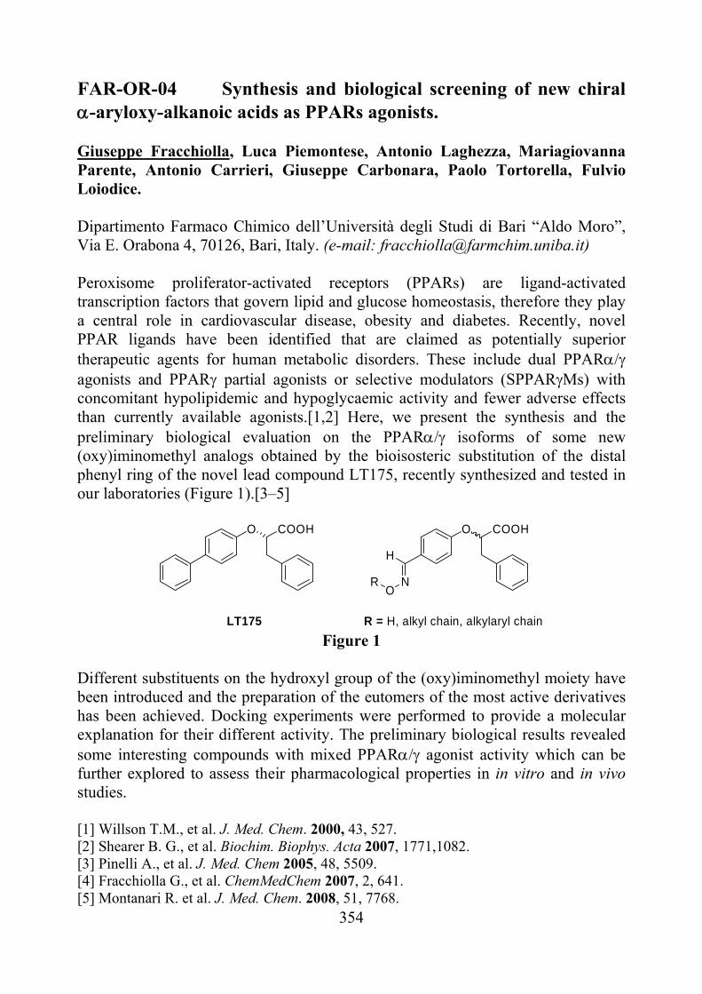
FAR-OR-04 Synthesis and biological screening of new chiral -aryloxy-alkanoic acids as PPARs agonists. Giuseppe Fracchiolla, Luca Piemontese, Antonio Laghezza, Mariagiovanna Parente, Antonio Carrieri, Giuseppe Carbonara, Paolo Tortorella, Fulvio Loiodice. Dipartimento Farmaco Chimico dell’Università degli Studi di Bari “Aldo Moro”, Via E. Orabona 4, 70126, Bari, Italy. (e-mail: [email protected]) Peroxisome proliferator-activated receptors (PPARs) are ligand-activated transcription factors that govern lipid and glucose homeostasis, therefore they play a central role in cardiovascular disease, obesity and diabetes. Recently, novel PPAR ligands have been identified that are claimed as potentially superior therapeutic agents for human metabolic disorders. These include dual PPAR agonists and PPAR partial agonists or selective modulators (SPPARMs) with concomitant hypolipidemic and hypoglycaemic activity and fewer adverse effects than currently available agonists.[1,2] Here, we present the synthesis and the preliminary biological evaluation on the PPAR/ isoforms of some new (oxy)iminomethyl analogs obtained by the bioisosteric substitution of the distal phenyl ring of the novel lead compound LT175, recently synthesized and tested in our laboratories (Figure 1).[3–5]
O COOH O COOH
H
NO
R
LT175 R = H, alkyl chain, alkylaryl chain Figure 1
Different substituents on the hydroxyl group of the (oxy)iminomethyl moiety have been introduced and the preparation of the eutomers of the most active derivatives has been achieved. Docking experiments were performed to provide a molecular explanation for their different activity. The preliminary biological results revealed some interesting compounds with mixed PPAR agonist activity which can be further explored to assess their pharmacological properties in in vitro and in vivo studies. [1] Willson T.M., et al. J. Med. Chem. 2000, 43, 527. [2] Shearer B. G., et al. Biochim. Biophys. Acta 2007, 1771,1082. [3] Pinelli A., et al. J. Med. Chem 2005, 48, 5509. [4] Fracchiolla G., et al. ChemMedChem 2007, 2, 641. [5] Montanari R. et al. J. Med. Chem. 2008, 51, 7768.
355
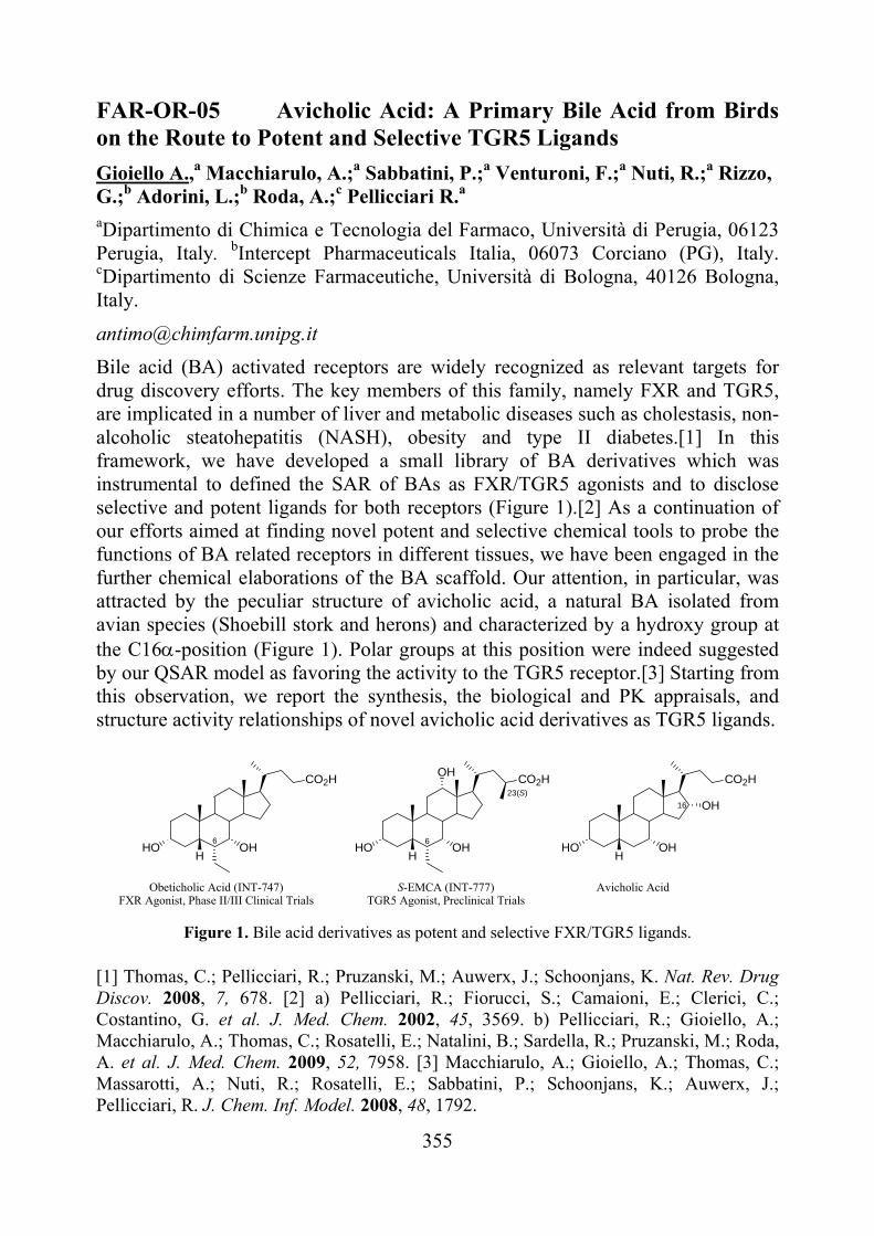
FAR-OR-05 Avicholic Acid: A Primary Bile Acid from Birds on the Route to Potent and Selective TGR5 Ligands
Gioiello A.,a Macchiarulo, A.;a Sabbatini, P.;a Venturoni, F.;a Nuti, R.;a Rizzo, G.;b Adorini, L.;b Roda, A.;c Pellicciari R.a aDipartimento di Chimica e Tecnologia del Farmaco, Università di Perugia, 06123 Perugia, Italy. bIntercept Pharmaceuticals Italia, 06073 Corciano (PG), Italy. cDipartimento di Scienze Farmaceutiche, Università di Bologna, 40126 Bologna, Italy.
Bile acid (BA) activated receptors are widely recognized as relevant targets for drug discovery efforts. The key members of this family, namely FXR and TGR5, are implicated in a number of liver and metabolic diseases such as cholestasis, non-alcoholic steatohepatitis (NASH), obesity and type II diabetes.[1] In this framework, we have developed a small library of BA derivatives which was instrumental to defined the SAR of BAs as FXR/TGR5 agonists and to disclose selective and potent ligands for both receptors (Figure 1).[2] As a continuation of our efforts aimed at finding novel potent and selective chemical tools to probe the functions of BA related receptors in different tissues, we have been engaged in the further chemical elaborations of the BA scaffold. Our attention, in particular, was attracted by the peculiar structure of avicholic acid, a natural BA isolated from avian species (Shoebill stork and herons) and characterized by a hydroxy group at the C16-position (Figure 1). Polar groups at this position were indeed suggested by our QSAR model as favoring the activity to the TGR5 receptor.[3] Starting from this observation, we report the synthesis, the biological and PK appraisals, and structure activity relationships of novel avicholic acid derivatives as TGR5 ligands.
Avicholic Acid
OHHO
CO2H
H
OH
Obeticholic Acid (INT-747)FXR Agonist, Phase II/III Clinical Trials
OHHO
CO2H
H
S-EMCA (INT-777)TGR5 Agonist, Preclinical Trials
OHHO
CO2H
H
OH
6 6
23(S)
16
Figure 1. Bile acid derivatives as potent and selective FXR/TGR5 ligands.
[1] Thomas, C.; Pellicciari, R.; Pruzanski, M.; Auwerx, J.; Schoonjans, K. Nat. Rev. Drug Discov. 2008, 7, 678. [2] a) Pellicciari, R.; Fiorucci, S.; Camaioni, E.; Clerici, C.; Costantino, G. et al. J. Med. Chem. 2002, 45, 3569. b) Pellicciari, R.; Gioiello, A.; Macchiarulo, A.; Thomas, C.; Rosatelli, E.; Natalini, B.; Sardella, R.; Pruzanski, M.; Roda, A. et al. J. Med. Chem. 2009, 52, 7958. [3] Macchiarulo, A.; Gioiello, A.; Thomas, C.; Massarotti, A.; Nuti, R.; Rosatelli, E.; Sabbatini, P.; Schoonjans, K.; Auwerx, J.; Pellicciari, R. J. Chem. Inf. Model. 2008, 48, 1792.
356
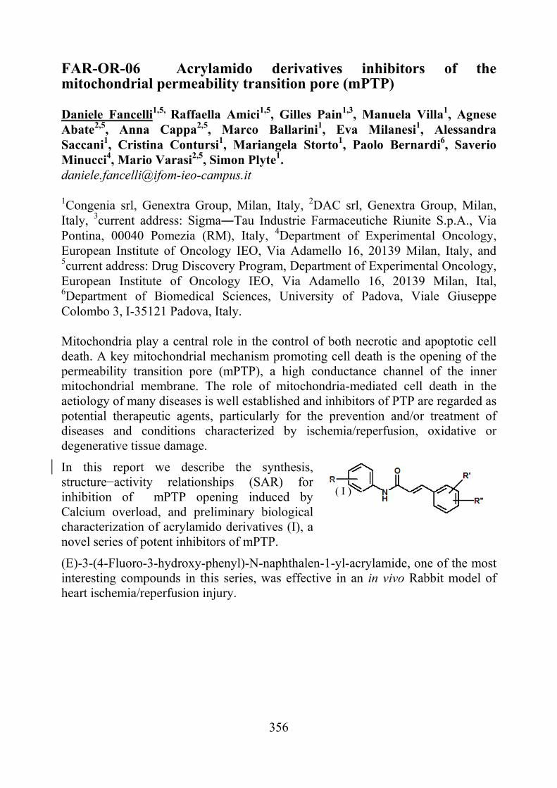
FAR-OR-06 Acrylamido derivatives inhibitors of the mitochondrial permeability transition pore (mPTP)
Daniele Fancelli1,5, Raffaella Amici1,5, Gilles Pain1,3, Manuela Villa1, Agnese Abate2,5, Anna Cappa2,5, Marco Ballarini1, Eva Milanesi1, Alessandra Saccani1, Cristina Contursi1, Mariangela Storto1, Paolo Bernardi6, Saverio Minucci4, Mario Varasi2,5, Simon Plyte1. [email protected] 1Congenia srl, Genextra Group, Milan, Italy, 2DAC srl, Genextra Group, Milan, Italy, 3current address: Sigma―Tau Industrie Farmaceutiche Riunite S.p.A., Via Pontina, 00040 Pomezia (RM), Italy, 4Department of Experimental Oncology, European Institute of Oncology IEO, Via Adamello 16, 20139 Milan, Italy, and 5current address: Drug Discovery Program, Department of Experimental Oncology, European Institute of Oncology IEO, Via Adamello 16, 20139 Milan, Ital, 6Department of Biomedical Sciences, University of Padova, Viale Giuseppe Colombo 3, I-35121 Padova, Italy. Mitochondria play a central role in the control of both necrotic and apoptotic cell death. A key mitochondrial mechanism promoting cell death is the opening of the permeability transition pore (mPTP), a high conductance channel of the inner mitochondrial membrane. The role of mitochondria-mediated cell death in the aetiology of many diseases is well established and inhibitors of PTP are regarded as potential therapeutic agents, particularly for the prevention and/or treatment of diseases and conditions characterized by ischemia/reperfusion, oxidative or degenerative tissue damage.
In this report we describe the synthesis, structure−activity relationships (SAR) for inhibition of mPTP opening induced by Calcium overload, and preliminary biological characterization of acrylamido derivatives (I), a novel series of potent inhibitors of mPTP.
(E)-3-(4-Fluoro-3-hydroxy-phenyl)-N-naphthalen-1-yl-acrylamide, one of the most interesting compounds in this series, was effective in an in vivo Rabbit model of heart ischemia/reperfusion injury.
( I )
357
FAR-OR-07 Microfluidics for radio-tracers labeling
Valentina Arimaa, Monica Biancoa, Antonella Zacheoa , Alessandra Zizzaria, Lucia Marraa, Giancarlo Pascalib, Piero Salvadorib, Elisabetta Perronea, Ross Rinaldia a National Nanotechnology Laboratory (CNR- Istituto di Nanoscience) - Distretto Tecnologico ISUFI - Università del Salento, via Arnesano, 73100 Lecce, Italy, NNL, Lecce, Italy b CNR-Istituto di Fisiologia clinica, Via Moruzzi1, 56124, Pisa, Italy *Corresponding author: [email protected]
One of the most interesting technological novelties in the field of
radiochemistry is the use of microfluidic devices to perform efficient, rapid, cost effective reactions in a user-friendly environment. Microreactors have considerable advantages in radiochemistry where short-life positron-emitters are used to produce radiotracers for molecular imaging with positron emission tomography (PET) 1-3.
A lots of advantages may be expected from this technology, such as the use of smaller amounts of radioactive precursors for saving precious materials, the possibility to work in safer conditions, to accurately control the reaction parameters and to use cheap, interchangeable, disposable and quality-assured radiochemistry processors4.
This work provides an overview of materials and microfluidic networks suitable for radiochemistry at microscale. Several micro devices are realized to perform on-chip reactions and separations. Preliminary results demonstrate the effectiveness of the proposed microfluidic platforms for radiopharmaceutical applications.
[1] Lee CC, Sui GD, Elizarov A, Shu CYJ, Shin YS,Dooley AN, et al. Science 310:1793–1796 (2005) [2] Lu SY, Watts P, Chin FT, Hong J, Musachio JL, Briard E, et al. LabChip 4:523–525 (2004) [3] Lu S, Giamis AM and Pike Vw, Current Radiopharmaceuticals, 2, 49-55, (2009).
358
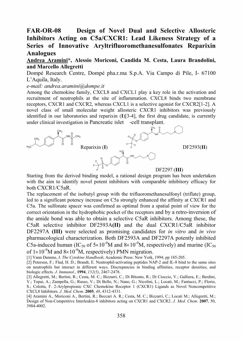
FAR-OR-08 Design of Novel Dual and Selective Allosteric Inhibitors Acting on C5a/CXCR1: Lead Likeness Strategy of a Series of Innovative Aryltrifluoromethanesulfonates Reparixin Analogues Andrea Aramini*, Alessio Moriconi, Candida M. Cesta, Laura Brandolini, and Marcello Allegretti Dompé Research Centre, Dompé pha.r.ma S.p.A. Via Campo di Pile, I- 67100 L’Aquila, Italy. e-mail: [email protected] Among the chemokine family, CXCL8 and CXCL1 play a key role in the activation and recruitment of neutrophils at the site of inflammation. CXCL8 binds two membrane receptors, CXCR1 and CXCR2, whereas CXCL1 is a selective agonist for CXCR2[1-2]. A novel class of small molecular weight allosteric CXCR1 inhibitors was previously identified in our laboratories and reparixin (I)[3-4], the first drug candidate, is currently under clinical investigation in Pancreatic islet �-cell transplant.
NH
OCH3
CH3
CH3
SCH3
O
O
Reparixin (I) O
S
OO
F
FF
NH
CH3 O
N
DF2593(II)
N
O
CH3
O
NH
S
OF
F
FO DF2297 (III)
Starting from the derived binding model, a rational design program has been undertaken with the aim to identify novel potent inhibitors with comparable inhibitory efficacy for both CXCR1/C5aR. The replacement of the isobutyl group with the trifluoromethanesulfonyl (triflate) group, led to a significant potency increase on C5a strongly enhanced the affinity at CXCR1 and C5a. The sulfonate spacer was confirmed as optimal from a spatial point of view for the correct orientation in the hydrophobic pocket of the receptors and by a retro-inversion of the amide bond was able to obtain a selective C5aR inhibitors. Among these, the C5aR selective inhibitor DF2593A(II) and the dual CXCR1/C5aR inhibitor DF2297A (III) were selected as promising candidates for in vitro and in vivo pharmacological characterization. Both DF2593A and DF2297A potently inhibited C5a-induced human (IC50 of 510-9M and 810-9M, respectively) and murine (IC50 of 110-9M and 810-9M, respectively) PMN migration. [1] Vann Damme, J. The Cytokine Handbook; Academic Press: New York, 1994; pp 185-205. [2] Petersen, F.; Flad, H. D.; Brandt, E. Neutrophil-activating peptides NAP-2 and IL-8 bind to the same sites on neutrophils but interact in different ways. Discrepancies in binding affinities, receptor densities, and biologic effects. J. Immunol., 1994, 152(5), 2467-2478. [3] Allegretti, M.; Bertini, R.; Cesta, M. C.; Bizzarri, C.; Di Bitonto, R.; Di Cioccio, V.; Galliera, E.; Berdini, V.; Topai, A.; Zampella, G.; Russo, V.; Di Bello, N.; Nano, G.; Nicolini, L.; Locati, M.; Fantucci, P.; Florio, S.; Colotta, F. 2-Arylpropionic CXC Chemokine Receptor 1 (CXCR1) Ligands as Novel Noncompetitive CXCL8 Inhibitors. J. Med. Chem. 2005, 48, 4312-4331. [4] Aramini A., Moriconi A., Bertini, R.; Beccari A. R.; Cesta, M. C.; Bizzarri, C.; Locati M.; Allegretti, M.; Design of Non-Competitive Interleukin-8 inhibitors acting on CXCR1 and CXCR2. J. Med. Chem. 2007, 50, 3984-4002.
359
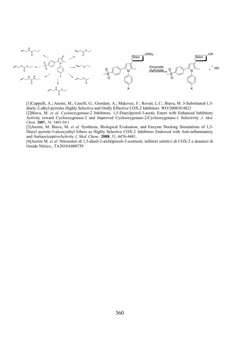
FAR-OR-09 3-Substituted-1,5-Diaryl-2-Alkylpyrroles Nitroesters, Highly Selective COX-2 Inhibitors and Nitric Oxide Donors Angela Di Capua,a* Salvatore Valenti,a Andrea Cappelli,a Lidia Sautebin,b Carla Ghelardini,c Vincenzo Calderone,d Paola Patrignani,e Antonio Giordanif, Mariangela Biava,g and Maurizio Anzini.a
aUniversità degli Studi di Siena, Via A. Moro, 53100 Siena; bUniversità di Napoli “Federico II”, Via D. Montesano 49, I-80131 Napoli; cUniversità di Firenze, Viale G. Pieraccini 6, I-50139 Firenze;d Università di Pisa, Via Bonanno 6, 56126 Pisa; eUniversità di Chieti “G. D’Annunzio” e CeSI, Via dei Vestini 31, 66013 Chieti; fRottapharm Madaus, Via Valosa di Sopra 7, 20052 Monza; gUniversità degli Studi di Roma,“La Sapienza”, P.le A. Moro 5, I-00185 Roma *e-mail: [email protected] A new generation of selective cyclooxygenase-2 (COX-2) inhibitors (coxibs) was developed to circumvent the major side effects of cyclooxygenase-1 (COX-1) and COX-2 inhibitors (stomach ulceration and nephrotoxicity). A series of previously patented 1,5-Diarylpyrrol-3-acetic esters and 1,5-Diarylpyrrole-3-alkoxyethyl ethers proved to be potent and selective COX-2 inhibitors in in vitro cell culture assay.ref The potential anti-inflammatory and antinociceptive activities of these compounds were evaluated in vivo, where they showed a very good activity against both carrageenan-induced hyperalgesia and edema in the rat paw test. 1,2,3 These classes of compounds (International Patent: PCT/EP2006/065011 and WO 2008/014821 A1) were at the basis of the development of new compounds, the COX-2 inhibiting nitric oxide (NO) donors (CINODs). CINODs are a new class of anti-inflammatory and analgesic drugs that may minimize gastrointestinal toxicity compared with standard non-steroidal anti-inflammatory drugs (NSAIDs) along with reduced cardiovascular risks associated with the Coxibs (celecoxib, valdecoxib, etc.) by virtue of their nitric oxide donation. This project was based on the synthesis of NO-donor’s compounds characterized by modifications of the side chain at position 3 of 1,5 diarylpirrole derivatives in which the 1,5-diarylpyrrole scaffold is linked to a nitric oxide moiety. In particular, the aim of the project is the extended functionalization of position 3 by means of “linkers” with different stereo-electronics properties in order to obtain “hybrid molecules” that after releasing NO are able to show elevated selective COX-2 activity in vitro along with elevated anti-inflammatory and anti-nociceptive activity in in vivo animal models.4
360
[1]Cappelli, A.; Anzini, M.; Caselli, G.; Giordani, A.; Makovec, F.; Rovati, L.C.; Biava, M. 3-Substituted-1,5-diarly-2-alkyl-pyrroles Highly Selective and Orally Effective COX-2 Inhibitors. WO/2008/014821 [2]Biava, M. et al. Cyclooxygenase-2 Inhibitors. 1,5-Diarylpyrrol-3-acetic Esters with Enhanced Inhibitory Activity toward Cyclooxygenase-2 and Improved Cyclooxygenase-2/Cyclooxygenase-1 Selectivity J. Med. Chem. 2007, 50, 5403-5411
[3]Anzini, M. Biava, M. et al. Synthesis, Biological Evaluation, and Enzyme Docking Simulations of 1,5-Diaryl pyrrole-3-alcoxyethyl Ethers as Highly Selective COX-2 Inhibitors Endowed with Anti-inflammatory and AntinociceptiveActivity J. Med. Chem., 2008, 51, 4476-4481. [4]Anzini M. et al. Nitroesteri di 1,5-diaril-2-alchilpirroli-3-sostituiti, inibitori selettivi di COX-2 e donatori di Ossido Nitrico., TA2010A000739
NS
O
O
O
O
O
O
O
HN
HN
O
HN
O
HN O
O
O O
O
X
361
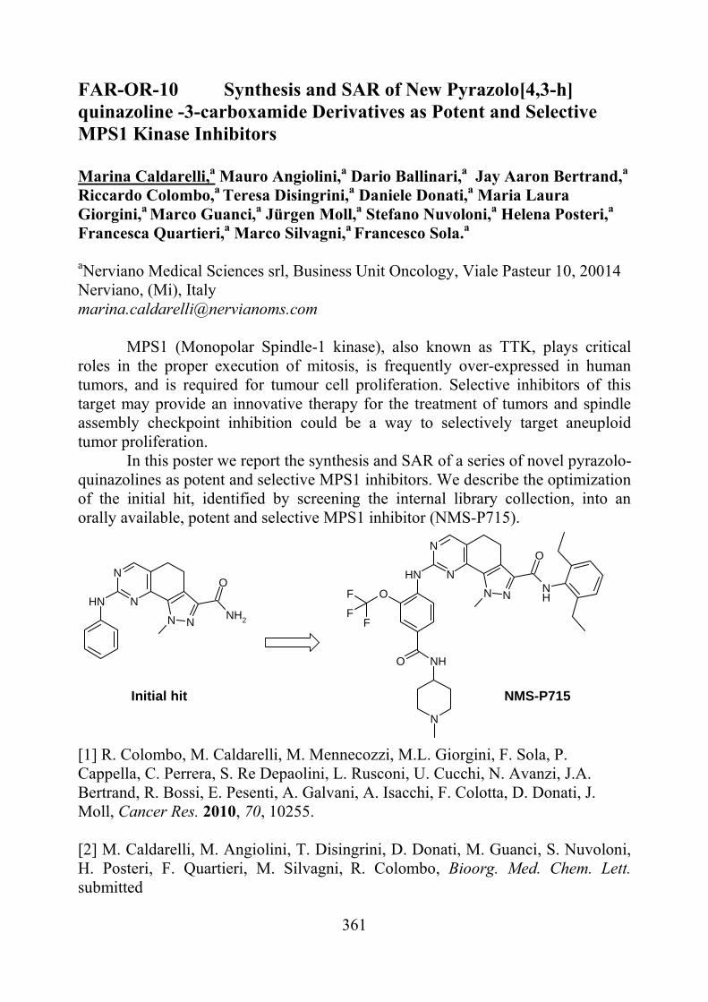
FAR-OR-10 Synthesis and SAR of New Pyrazolo[4,3-h] quinazoline -3-carboxamide Derivatives as Potent and Selective MPS1 Kinase Inhibitors Marina Caldarelli,a Mauro Angiolini,a Dario Ballinari,a Jay Aaron Bertrand,a Riccardo Colombo,a Teresa Disingrini,a Daniele Donati,a Maria Laura Giorgini,a Marco Guanci,a Jürgen Moll,a Stefano Nuvoloni,a Helena Posteri,a Francesca Quartieri,a Marco Silvagni,a Francesco Sola.a
aNerviano Medical Sciences srl, Business Unit Oncology, Viale Pasteur 10, 20014 Nerviano, (Mi), Italy [email protected] MPS1 (Monopolar Spindle-1 kinase), also known as TTK, plays critical roles in the proper execution of mitosis, is frequently over-expressed in human tumors, and is required for tumour cell proliferation. Selective inhibitors of this target may provide an innovative therapy for the treatment of tumors and spindle assembly checkpoint inhibition could be a way to selectively target aneuploid tumor proliferation. In this poster we report the synthesis and SAR of a series of novel pyrazolo-quinazolines as potent and selective MPS1 inhibitors. We describe the optimization of the initial hit, identified by screening the internal library collection, into an orally available, potent and selective MPS1 inhibitor (NMS-P715).
NH N
N
N NNH2
ONH N
N
N NNH
O
OF
FF
O NH
N
Initial hit NMS-P715
[1] R. Colombo, M. Caldarelli, M. Mennecozzi, M.L. Giorgini, F. Sola, P. Cappella, C. Perrera, S. Re Depaolini, L. Rusconi, U. Cucchi, N. Avanzi, J.A. Bertrand, R. Bossi, E. Pesenti, A. Galvani, A. Isacchi, F. Colotta, D. Donati, J. Moll, Cancer Res. 2010, 70, 10255. [2] M. Caldarelli, M. Angiolini, T. Disingrini, D. Donati, M. Guanci, S. Nuvoloni, H. Posteri, F. Quartieri, M. Silvagni, R. Colombo, Bioorg. Med. Chem. Lett. submitted
362
FAR-OR-11 The Click Chemistry Approach in the Discovery of Potent and Selective PI3K Inhibitors Tracey Pirali,a Clarissa Landoni,a Alberto Massarotti,a Emilio Hirsch,b Elisa Ciraolo,b Giovanni Sorbaa
a Dipartimento di Scienze Chimiche, Alimentari, Farmaceutiche e Farmacologiche dell’Università degli Studi del Piemonte Orientale, via Bovio 6, 28100, Novara, Italy; b Centro di Biotecnologie Molecolari dell’Università di Torino, Via Nizza 52, 10126, Torino, Italy [email protected]
Click chemistry has recently made an outstanding contribution to medicinal chemistry research [1]. This term, coined by K. B. Sharpless, refers to a new synthetic approach which exploits nearly perfect reactions. Among them, the copper-catalyzed Huisgen cycloaddition between azides and alkynes plays a prominent role.
Over the past few years, we have exploited this reaction in the discovery of resveratrol [2] and estrogenic analogues [3], HDAC inhibitors [4], and, lately, PI3K inhibitors. Phosphatidylinositol-3-kinases (PI3Ks), a class of lipid kinases, are an emerging target in antitumoral therapy as they play a major role in proliferation and survival in a wide variety of human cancers [5].
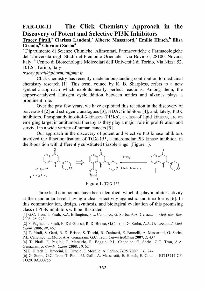
Our approach in the discovery of potent and selective PI3 kinase inhibitors involved the functionalisation of TGX-155, a micromolar PI3 kinase inhibitor, in the 8-position with differently substituted triazole rings (Figure 1).
Figure 1: TGX-155
Three lead compounds have been identified, which display inhibitor activity at the nanomolar level, having a clear selectivity against and isoforms [6]. In this communication, design, synthesis, and biological evaluation of this promising class of PI3K inhibitors will be illustrated. [1] G.C. Tron, T. Pirali, R.A. Billington, P.L. Canonico, G. Sorba, A.A. Genazzani, Med. Res. Rev. 2008, 28, 278 [2] F. Pagliai, T. Pirali, E. Del Grosso, R. Di Brisco, G.C. Tron, G. Sorba, A.A. Genazzani, J. Med. Chem. 2006, 49, 467 [3] T. Pirali, S. Gatti, R. Di Brisco, S. Tacchi, R. Zaninetti, E. Brunelli, A. Massarotti, G. Sorba, P.L. Canonico, L. Moro, A.A. Genazzani, G.C. Tron, ChemMedChem 2007, 2, 437 [4] T. Pirali, F. Pagliai, C. Mercurio, R. Boggio, P.L. Canonico, G. Sorba, G.C. Tron, A.A. Genazzani, J. Comb. Chem. 2008, 10, 624 [5] E. Hirsch, L. Braccini, E. Ciraolo, F. Morello, A. Perino, TIBS 2009, 34, 244 [6] G. Sorba, G.C. Tron, T. Pirali, U. Galli, A. Massarotti, E. Hirsch, E. Ciraolo, BIT13714-CF: TO2010A000956
363
FAR-OR-12 Synthesis and Biological Characterization of 4-Spirochromane Analogues as New HDAC Inhibitors Florian Thaler,a,b Agnese Abate,c,b Andrea Colombo,d Giacomo Carenzi,c,b Roberto Boggio,a Giulio Dondio,d Stefania Gagliardi,d Saverio Minucci,e,f Mario Varasic,b Ciro Mercurioc a Congenia srl, Genextra Group, Via Adamello 16, 20139 Milan, Italy; b Drug Discovery Program,
Department of Experimental Oncology, European Institute of Oncology, Via Adamello 16, 20139
Milan, Italy; c DAC srl, Genextra Group, Via Adamello 16, 20139 Milan, Italy; d NiKem Research
srl, Via Zambeletti 25, 20021 Baranzate (MI), Italy; e European Institute of Oncology, Via Adamello
16, 20139 Milan, Italy; f University of Milan, University of Milan, Via Celoria 26, 20133 Milan,
Italy *corresponding author: [email protected]
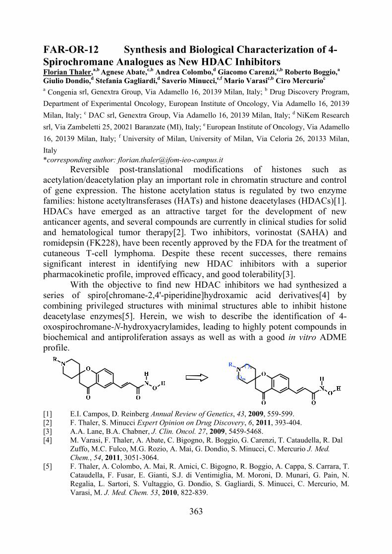
Reversible post-translational modifications of histones such as acetylation/deacetylation play an important role in chromatin structure and control of gene expression. The histone acetylation status is regulated by two enzyme families: histone acetyltransferases (HATs) and histone deacetylases (HDACs)[1]. HDACs have emerged as an attractive target for the development of new anticancer agents, and several compounds are currently in clinical studies for solid and hematological tumor therapy[2]. Two inhibitors, vorinostat (SAHA) and romidepsin (FK228), have been recently approved by the FDA for the treatment of cutaneous T-cell lymphoma. Despite these recent successes, there remains significant interest in identifying new HDAC inhibitors with a superior pharmacokinetic profile, improved efficacy, and good tolerability[3].
With the objective to find new HDAC inhibitors we had synthesized a series of spiro[chromane-2,4'-piperidine]hydroxamic acid derivatives[4] by combining privileged structures with minimal structures able to inhibit histone deacetylase enzymes[5]. Herein, we wish to describe the identification of 4-oxospirochromane-N-hydroxyacrylamides, leading to highly potent compounds in biochemical and antiproliferation assays as well as with a good in vitro ADME profile.
[1] E.I. Campos, D. Reinberg Annual Review of Genetics, 43, 2009, 559-599. [2] F. Thaler, S. Minucci Expert Opinion on Drug Discovery, 6, 2011, 393-404. [3] A.A. Lane, B.A. Chabner, J. Clin. Oncol. 27, 2009, 5459-5468. [4] M. Varasi, F. Thaler, A. Abate, C. Bigogno, R. Boggio, G. Carenzi, T. Cataudella, R. Dal
Zuffo, M.C. Fulco, M.G. Rozio, A. Mai, G. Dondio, S. Minucci, C. Mercurio J. Med. Chem., 54, 2011, 3051-3064.
[5] F. Thaler, A. Colombo, A. Mai, R. Amici, C. Bigogno, R. Boggio, A. Cappa, S. Carrara, T. Cataudella, F. Fusar, E. Gianti, S.J. di Ventimiglia, M. Moroni, D. Munari, G. Pain, N. Regalia, L. Sartori, S. Vultaggio, G. Dondio, S. Gagliardi, S. Minucci, C. Mercurio, M. Varasi, M. J. Med. Chem. 53, 2010, 822-839.

364
FAR-OR-13 Mannich bases as novel irreversible Epidermal Growth Factor Receptor inhibitors F. Vacondio,a C. Carmi,a A. Lodola,a C. Silva,a S. Rivara,a E. Galvani,b A.Cavazzoni,bR.R.Alfieri,bP.G.Petronini,bM.Mora
aDipartimento Farmaceutico, Università degli Studi di Parma, Viale Usberti 27/a, 43124 Parma, Italy; bDipartimento di Medicina Sperimentale, Università degli Studi di Parma, Via Volturno 39, 43125 Parma, Italy [email protected] Second generation, irreversible inhibitors of Epidermal Growth Factor Receptor (EGFR) are characterized by a recognition scaffold, resembling those of clinically-employed reversible inhibitors, and by a cysteine-reactive warhead, able to covalently interact with a conserved cysteine residue in the kinase domain of the ErbB family members (C797 in EGFR) [1]. Cysteine-trapping groups reported in literature so far (acrylamides, -chloroacetamides) are endowed with high intrinsic reactivity. This could cause rapid metabolic deactivation of the inhibitor or, at worst, enhanced in vivo toxicity, due to the aspecific reactivity towards off-target thiol-containing nucleophiles. We have recently started systematic exploration on the role and reactivity of warheads for irreversible EGFR inhibition, introducing different cysteine-trapping groups on the 4-anilinoquinazoline scaffold [2]. In the present work, our attention is focused on a new set of inhibitors, in which a -aminocarbonyl group (Mannich base) is linked to different scaffolds through an amide bond. These derivatives proved to be as efficient as the irreversible acrylamide derivative PD168393 in inhibiting EGFR-TK activity in different cell lines. For both acrylamide and Mannich-base derivatives, inhibition persisted for 8 hours after the wash-out of the compound from cell medium. However, their in vitro reactivity profile markedly diverged, with Mannich bases being stable in the presence of low MW thiols (GSH, cysteine, cysteamine), compared to the highly reactive acrylamide. A combined approach, employing: i. fluorimetric analysis of inhibitors incubated with cell-free EGFR, ii. quantification by HPLC-MS of inhibitors and their metabolites in A549 cell lines and in cell lysates, showed that Mannich bases can act as prodrugs, partially releasing acrylamide in the cellular environment. [1] C.P. Belani, Cancer Invest. 28, 2010, 413-23. [2] C. Carmi, A. Cavazzoni, S. Vezzosi, F. Bordi, F. Vacondio, C. Silva, S. Rivara, A. Lodola, R. R. Alfieri, S. La Monica, M. Galetti, A. Ardizzoni, P. G. Petronini, M. Mor, J. Med. Chem., 53, 2010, 2038-2050.
365
FAR-OR-14 Exploring the interaction capacities of TRPM8 channel by docking analyses and MD simulations Alessandro Pedretti, Alessandra Labozzetta, Andrea R. Beccari, Alessio Moriconi, Giulio Vistoli Dipartimento di Scienze Farmaceutiche “Pietro Pratesi”, Facoltà di Farmacia, Università degli Studi di Milano, Via Mangiagalli, 25, I-20133 Milano, Italy. Dompé SpA, R&D division, Via Campo di Pile - 67100 L'Aquila (Italy). [email protected] The transient receptor potential (TRP) superfamily is a large group of ion channels that has received increased attention in recent years. TRPM8, on which this study is focused, belongs to the subfamily of thermo-TRP channels which are triggered by diverse chemical and physical stimuli and whose precise activation mechanism is still unknown. Specifically, TRPM8 is activated by cold temperature, ligands such as menthol and icilin (a synthetic derivative), positive membrane potential and the endogenous signaling lipid, PIP2. Therefore, TRPM8 could find therapeutic applications in several pathological conditions, including neurogenic inflammation, neuropathic pain, overactive bladder and prostate cancer. [1] An homology model of the TRPM8 tetramer was recently generated using a fragmental strategy by some of us. [2] Beside the global architecture of the TRPM8 channel, such a model revealed the key residues involved in ligand recognition and suggested that the agonist binding is able to induce a cascade of conformational shifts which globally may orchestrate the channel opening (at least partially). Such a mechanism was then confirmed by classic all-atoms MD simulations which evidenced how agonists are able to trigger such structural changes whereas agonists block the channel in its starting conformations. Considering the rather nonspecific nature of TRPM8 binding site and the resulting difficulty of predicting ligand bioactivity by docking calculations, adaptive biasing force (ABF) MD simulations [3] were exploited to derive the free energies involved in TRPM8 activation and the obtained energy values are in line with the activity of a representative set of TRPM8 ligands. These results emphasize that suitably targeted MD runs can be fast enough to be systematically applied to predict the bioactivity of rather large ligand datasets. [1] Jordt SE, Ehrlich BE., Subcell Biochem. 2007, 45, 253-71 [2] Pedretti A, Marconi C, Bettinelli I, Vistoli G., Biochim Biophys Acta. 2009, 1788, 973-82 [3] Darve E., Pohorille A. J. Chem. Phys. 2001, 115, 9169-9183.
366
FAR-OR-15 Discovery of new positive allosteric modulators of GABAB receptor Claudia Mugnaini,a Valentina Pedani,a Serena Paquini,a Simone Brogi,a Andrea Tafi,a Mauro A.M. Carai,b Giancarlo Colombo,b Carla Lobina,b Federico Corellia aDipartimento Farmaco Chimico Tecnologico, Università degli Studi di Siena Via De Gasperi, 2 53100 Siena, Italia bIstituto CNR di Neuroscienze S.S. 554, Km. 4,500 09042 Monserrato (Cagliari), Italia [email protected] GABA is the main inhibiting neurotransmitter in the CNS. It modulates the neuronal activity by mediating its action via GABAA, GABAB and GABAC receptors The GABAB receptor belongs to the family 3 of the G-protein coupled receptors and it is an heterodimer made of two similar but distinct subunits. Although drugs activating the GABAB receptor were found to have a number of possible therapeutic actions, these were limited because of tolerance and undesired side effects which include sedation, myorelaxing activity and hypothermia.1 Allosteric modulators are molecules that bind to a site on a receptor which is topographically distinct from the orthosteric-binding pocket. They have little or no intrinsic agonistic activity of their own but induce conformational changes in the receptor protein, which affect its interaction with the endogenous neurotransmitter. Thus, positive allosteric modulators (PAMs) of GABAB receptor appear as a better alternative to GABAB agonists, allowing the specific enhancement of receptor activity when and where needed, and as such, are less prone to tolerance in contrast to the pure agonists (such as baclofen) that constantly activate the receptor in any region where it is expressed. These compounds are valuable anxiolytics and effectively reduce craving for drugs of abuse such as alcohol, nicotine and cocaine.2 Four companies have invested substantial resources into the search of PAMs of GABAB receptors (Novartis, Roche, AstraZeneca and Addex) with significant differences in the target indications. These medicinal chemistry efforts have enlarged the number of scaffolds, which lead to potent compounds and thus expanded our knowledge on structure-activity relationships significantly. Nevertheless, substantial efforts are still needed in order to optimize drugs for a given indication to get all the ADMET parameters right as well, such as metabolic stability, brain penetration for the CNS indications (or no brain penetration for the peripheral indications), sufficient water solubility, no alerts in Ames and genotoxicity tests, and several other parameters. Our research in the area started from a structure-based computational approach which allowed, through a virtual screening, the identification of a number of possible PAMs of GABAB receptor. Among those selected, one molecule gave interesting results both in vitro and in vivo studies. [1] Froestl, W. Future Med. Chem. 2011, 3, 163-175 [2] Pin, J.-P.; Prézeau, L. Curr. Neuropharm. 2007, 5, 195-201
367
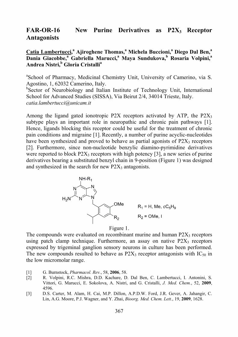
FAR-OR-16 New Purine Derivatives as P2X3 Receptor Antagonists Catia Lambertucci,a Ajiroghene Thomas,a Michela Buccioni,a Diego Dal Ben,a Dania Giacobbe,a Gabriella Marucci,a Maya Sundukova,b Rosaria Volpini,a Andrea Nistri,b Gloria Cristallia aSchool of Pharmacy, Medicinal Chemistry Unit, University of Camerino, via S. Agostino, 1, 62032 Camerino, Italy. bSector of Neurobiology and Italian Institute of Technology Unit, International School for Advanced Studies (SISSA), Via Beirut 2/4, 34014 Trieste, Italy. [email protected] Among the ligand gated ionotropic P2X receptors activated by ATP, the P2X3
subtype plays an important role in neuropathic and chronic pain pathways [1]. Hence, ligands blocking this receptor could be useful for the treatment of chronic pain conditions and migraine [1]. Recently, a number of purine acyclic-nucleotides have been synthesized and proved to behave as partial agonists of P2X3 receptors [2]. Furthermore, since non-nucleotide benzylic diamino-pyrimidine derivatives were reported to block P2X3 receptors with high potency [3], a new series of purine derivatives bearing a substituted benzyl chain in 9-position (Figure 1) was designed and synthesized in the search for new P2X3 antagonists.
Figure 1.
The compounds were evaluated on recombinant murine and human P2X3 receptors using patch clamp technique. Furthermore, an assay on native P2X3 receptors expressed by trigeminal ganglion sensory neurons in culture has been performed. The new compounds resulted to behave as P2X3 receptor antagonists with IC50 in the low micromolar range. [1] G. Burnstock, Pharmacol. Rev., 58, 2006, 58. [2] R. Volpini, R.C. Mishra, D.D. Kachare, D. Dal Ben, C. Lambertucci, I. Antonini, S.
Vittori, G. Marucci, E. Sokolova, A. Nistri, and G. Cristalli, J. Med. Chem., 52, 2009, 4596.
[3] D.S. Carter, M. Alam, H. Cai, M.P. Dillon, A.P.D.W. Ford, J.R. Gever, A. Jahangir, C. Lin, A.G. Moore, P.J. Wagner, and Y. Zhai, Bioorg. Med. Chem. Lett., 19, 2009, 1628.
368
FAR-OR-17 A novel human recombinant antibody (dAb) against a synthetic glycopeptide cross-reacts with human auto-antibodies, biomarkers of multiple sclerosis Francesca Niccheri,a Feliciana Real-Fernandez,b Matteo Ramazzotti,a Anna Maria Papini,b Donatella Degl’Innocenti,a and Paolo Roveroc aDipartimento di Scienze Biochimiche, Università di Firenze, Viale Morgagni 50, 50134 Firenze, Italia. bDipartimento di Chimica ‘Ugo Schiff’, Università di Firenze, Via della Lastruccia 13, 50019 Sesto Fiorentino, Italia. cDipartimento di Scienze Farmaceutiche, Università di Firenze, Via Ugo Schiff 6, 50019 Sesto Fiorentino, Italia. E-mail responsabile presentazione: [email protected]
We have previously described a synthetic glycopeptide, termed CSF114(Glc), able to detect specific auto-antibodies in sera of patient affected by Multiple Sclerosis (MS), an inflammatory, demyelinating disease of the central nervous system [1]. The pathogenesis of MS involves an autoimmune mechanism against myelin auto-antigens, even if the target antigens remain elusive. Accordingly, we focused our attention on both the characterization of the antigenic properties of CSF114(Glc) and the identification of the native auto-antigen(s) recognized by anti-CSF114(Glc) auto-antibodies.
In this context, we have recently used the glycopeptide CSF114(Glc), coated on magnetic beads, to select by phage display specific human domain antibodies (dAb) from a domain antibodies library [2]. Purified dAbs (15 kDa) were characterized by Biacore for binding specificity to CSF114(Glc) versus unglycosylated CSF114, showing a good specificity and affinity.
Subsequent Biacore experiments demonstrated that these recombinant dAbs cross-react with anti-CSF114(Glc) auto-antibodies isolated from MS patients’ sera by immuno-affinity chromatography. Accordingly, the new recombinant dAb may be used for the characterization of the native auto-antigens in human tissues or as a positive control in an in vitro diagnostic assay based on CSF114(Glc). [1] F.Lolli, B.Mulinacci, A.Carotenuto, B.Bonetti, G.Sabatino, B.Mazzanti,
A.M.D'Ursi, E.Novellino, M.Pazzagli, L.Lovato, M.C.Alcaro, E.Peroni, M.C.Pozo-Carrero, F.Nuti, L.Battistini, G.Borsellino, M.Chelli, P.Rovero, A.M.Papini, Proc. Natl. Acad. Sci. 102, 2005, 10273.
[2] C.M.Lee, N.Iorno, F.Sierro, D.Christ, Nature Protoc. 2, 2007, 3001.
369
FAR-OR-18 Exploring the space of histidine containing dipeptides in search of novel efficient RCS sequestering agents Giulio Vistoli, Valentina Straniero, Laura Fumagalli, Danilo De Maddis, Matteo Lo Monte, Alessandro Pedretti, Marina Carini, Ermanno Valoti, Giancarlo Aldini Department of Pharmaceutical Sciences "Pietro Pratesi". Università degli Studi di Milano, via Mangiagalli 25, 20133, Milan, Italy. Experimental evidence confirmed that reactive carbonyl species (RCS) are involved in the pathogenesis of several human diseases including diabetes related disorders and distress metabolic syndrome. Hence RCS, beside to be considered biomarkers of oxidative damage, can be also seen as potential targets for the development of bioactive compounds acting as detoxifying agents of RCS (carbonyl quenching compounds). We found that the endogenous dipeptide carnosine (-alanyl-L-histidine) is a selective and potent RCS sequestering agent, even though its clinical application is limited due to the rapid hydrolysis in blood by a specific dipeptidase (carnosinase). With a view to finding stable and effective agents, several carnosine derivatives were recently proposed in literature and some of these compounds proved promising in vivo in suitable animal models. Despite the mentioned variety of carnosine analogues, the chemical space of the proteinogenic histidine containing dipeptides was never exhaustively investigated. On these grounds, the study is focused on the synthesis, physicochemical profiling, in silico analysis and biological evaluation of a set of diastereoisomeric pairs of histidine containing dipeptides suitably chosen to cover a large part of the accessible chemical space. In detail, the examined peptides were designed as diastereoisomeric pairs in order to delve the configurational effects on the activity which could shed additional light on the quenching mechanism. Finally, some relevant physicochemical properties (namely pKi, log P and log D7.4) were experimentally determined to clarify the main factors governing the quenching activity and their relationships with in silico determined descriptors were also investigated.
370
FAR-OR-19 Recombinant Albumin as Chiral Selector in Enantioselective HPLC Marco Pistolozzia, Guy Felixb, Dave Meadc, Carlo Bertuccia
aDepartment of Pharmaceutical Chemistry, University of Bologna, via Belmeloro 6, 40126-Bologna, Italy bCNRS, Aix-Marseille Université, CINaM UPR 3118, Marseille, France cNovozymes Biopharma UK Ltd, Castle Court, 59 Castle Boulevard, Nottigham NG7 1FD, United Kingdom
[email protected] Human serum albumin (HSA) has been successfully used as chiral selector in enantioselective HPLC. These HSA-based columns usually present the problem of a significant variation of the chromatographic performances depending not only on the immobilization procedure, but also on the origin of the anchored protein from different sera. This makes difficult the application of developed and validated HPLC methods. Recombinant human albumin, rHA (RECOMBUMIN®, Novozymes Biopharma UK Limited) can overcome the problem, because of the high homogeneity of the structure and binding properties of the protein samples. RECOMBUMIN®, produced from Novozymes’ Saccaromyces cerevisiae was purified and defatted before use. The protein was then characterized for its conformation and its binding properties by circular dichroism (CD). In particular the analysis of the CD at high energy showed substantially the same secondary structure for the recombinant albumin with respect to the serum albumin. In addition the binding of selected markers for the most important binding sites (i.e. phenylbutazone for site I, diazepam for site II, and bilirubin for site III) was proved to be stereoselective. A sample of RECOMBUMIN® was then anchored in situ to an epoxy silica matrix of a HPLC column. The anchoring method was validated by checking the binding parameters of known ligands on the immobilized protein. The RECOMBUMIN® -based column was efficiently used for the enantioselective analysis of a variety of chiral drugs and amino acid derivatives. As an example enantiomeric resolution was obtained for rac-warfarin ( = 2.1), rac-lorazepam hemisuccinate ( = 5.3), N-benzoyl-DL-leucine (=2.8), using 1-propanol/phosphate buffer (pH 7.5) 15/85, 0.6 ml/min flow rate. The obtained values of enantioselectivity are comparable or higher with respect to those obtained with the corresponding HSA-based columns, under the same experimental conditions. The rHA based column has also a great potential for its use as affinity support for characterizing the binding of new active compounds in terms of KD and location of primary binding site.
371
FAR-PO-01 One-pot transformation of anthranilic acids to fluorazone derivatives, valuable intermediates for the synthesis of anticancer agents Francesca Aiello, Antonio Garofalo, Fedora Grande Dipartimento di Scienze Farmaceutiche, Università della Calabria, Arcavacata di Rende 87036, Cosenza, Italy [email protected] Fluorazone (9H-pyrrolo[1,2-a]indol-9-one) I and its analogs represent valuable intermediates in the synthesis of biologically active compounds. They have been extensively studied in relation to a variety of activities showed by many of their functionalized derivatives. In particular, we found that some N’-heteroacyl-hydrazone derivatives (es. cpd II) possess noticeable cytotoxic activity against a colon cancer cell line [1], while thienopyrrolizinone III, belonging to the tripentones family, has been shown to act as a microtubule polymerization inhibitor [2].
S
N
O
OHMeO
N
O
IIIN
NHN
O
S
I IINH2
O
OH
IC50 7 µ M (SKBr3 cell line)
IC 50 0.015 µM ( L1210
cell line )
A number of synthetic strategies have been therefore developed for the preparation of (un)substituted-fluorazones and analogs. Amongst the known synthetic methods, the most appealing appear those starting from ortho-(1H-pyrrol-1-yl)aryl and heteroaryl carboxylic acids, in turn obtained by pyrrolation of ortho-aminoaryl (anthranilic) and ortho-aminoheteroaryl carboxylic acids, respectively [3]. We herein investigated the possibility of directly converting anthranilic acids into fluorazones, through a sequential one-pot pyrrole formation/cyclization, by using DMTHF and 4-chloropyridine hydrochloride as the acid catalyst. This allows the preparation of fluorazone based derivatives from ortho-aminoaryl and heteroaryl carboxylic acids in generally good yield and after a simple work-up, avoiding any intermediate manipulation. A further advantage of the procedure lies in the easy availability of the starting aminoacids, most of which are commercial reagents. [1] Grande, G.; Yamada, R.; Cao, X.; Aiello, F.; Garofalo, A.; Neamati, N. Expert Opin. Investig. Drugs 2009, 18, 555. [2] Lisowski, V.; Enguehard, C.; Lancelot, J. C.; Caignard, D.-H.; Lambel, S.; Leonce, S.; Pierrè, A.; Atassi, G.; Renard, P.; Rault, S. Bioorg. Med. Chem. Lett. 2001, 11, 2205. [3] Aiello, F.; Garofalo, A.; Grande, F. Tetrahedron 2010, 66, 274.
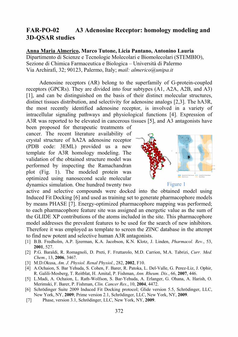
372
FAR-PO-02 A3 Adenosine Receptor: homology modeling and 3D-QSAR studies Anna Maria Almerico, Marco Tutone, Licia Pantano, Antonino Lauria Dipartimento di Scienze e Tecnologie Molecolari e Biomolecolari (STEMBIO), Sezione di Chimica Farmaceutica e Biologica – Università di Palermo Via Archirafi, 32; 90123, Palermo, Italy; mail: [email protected]
Adenosine receptors (AR) belong to the superfamily of G-protein-coupled receptors (GPCRs). They are divided into four subtypes (A1, A2A, A2B, and A3) [1], and can be distinguished on the basis of their distinct molecular structures, distinct tissues distribution, and selectivity for adenosine analogs [2,3]. The hA3R, the most recently identified adenosine receptor, is involved in a variety of intracellular signaling pathways and physiological functions [4]. Expression of A3R was reported to be elevated in cancerous tissues [5], and A3 antagonists have been proposed for therapeutic treatments of cancer. The recent literature availability of crystal structure of hA2A adenosine receptor (PDB code: 3EML) provided us a new template for A3R homology modeling. The validation of the obtained structure model was performed by inspecting the Ramachandran plot (Fig. 1). The modeled protein was optimized using nanosecond scale molecular dynamics simulation. One hundred twenty two active and selective compounds were docked into the obtained model using Induced Fit Docking [6] and used as training set to generate pharmacophore models by means PHASE [7]. Energy-optimized pharmacophore mapping was performed; to each pharmacophore feature site was assigned an energetic value as the sum of the GLIDE XP contributions of the atoms included in the site. This pharmacophore model addresses the prevalent features to be used for the search of new inhibitors. Therefore it was employed as template to screen the ZINC database in the attempt to find new potent and selective human A3R antagonists. [1] B.B. Fredholm, A.P. Ijzerman, K.A. Jacobson, K.N. Klotz, J. Linden, Pharmacol. Rev., 53,
2001, 527. [2] P.G. Baraldi, R. Romagnoli, D. Preti, F. Fruttarolo, M.D. Carrion, M.A. Tabrizi, Curr. Med.
Chem., 13, 2006, 3467. [3] M.D.Okusa, Am. J. Physiol. Renal Physiol., 282, 2002, F10. [4] A.Ochaion, S. Bar Yehuda, S. Cohen, F. Barer, R. Patoka, L. Del-Valle, G. Perez-Liz, J. Ophir,
R. Galili-Mosberg, T. Reitblat, H. Amital, P. Fishman, Ann. Rheum. Dis., 66, 2007, 446. [5] L.Madi, A. Ochaion, L. Rath-Wolfson, S. Bar-Yehuda, A. Erlanger, G. Ohana, A. Harish, O.
Merimski, F. Barer, P. Fishman, Clin. Cancer Res., 10, 2004, 4472. [6] Schrödinger Suite 2009 Induced Fit Docking protocol; Glide version 5.5, Schrödinger, LLC,
New York, NY, 2009; Prime version 2.1, Schrödinger, LLC, New York, NY, 2009. [7] Phase, version 3.1, Schrödinger, LLC, New York, NY, 2009.
Figure 1
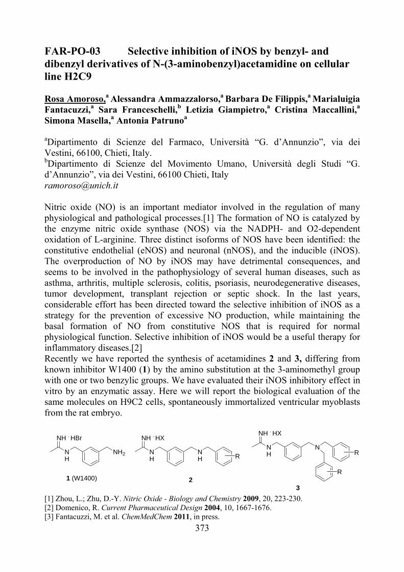
373
FAR-PO-03 Selective inhibition of iNOS by benzyl- and dibenzyl derivatives of N-(3-aminobenzyl)acetamidine on cellular line H2C9 Rosa Amoroso,a Alessandra Ammazzalorso,a Barbara De Filippis,a Marialuigia Fantacuzzi,a Sara Franceschelli,b Letizia Giampietro,a Cristina Maccallini,a Simona Masella,a Antonia Patrunoa aDipartimento di Scienze del Farmaco, Università “G. d’Annunzio”, via dei Vestini, 66100, Chieti, Italy. bDipartimento di Scienze del Movimento Umano, Università degli Studi “G. d’Annunzio”, via dei Vestini, 66100 Chieti, Italy [email protected] Nitric oxide (NO) is an important mediator involved in the regulation of many physiological and pathological processes.[1] The formation of NO is catalyzed by the enzyme nitric oxide synthase (NOS) via the NADPH- and O2-dependent oxidation of L-arginine. Three distinct isoforms of NOS have been identified: the constitutive endothelial (eNOS) and neuronal (nNOS), and the inducible (iNOS). The overproduction of NO by iNOS may have detrimental consequences, and seems to be involved in the pathophysiology of several human diseases, such as asthma, arthritis, multiple sclerosis, colitis, psoriasis, neurodegenerative diseases, tumor development, transplant rejection or septic shock. In the last years, considerable effort has been directed toward the selective inhibition of iNOS as a strategy for the prevention of excessive NO production, while maintaining the basal formation of NO from constitutive NOS that is required for normal physiological function. Selective inhibition of iNOS would be a useful therapy for inflammatory diseases.[2] Recently we have reported the synthesis of acetamidines 2 and 3, differing from known inhibitor W1400 (1) by the amino substitution at the 3-aminomethyl group with one or two benzylic groups. We have evaluated their iNOS inhibitory effect in vitro by an enzymatic assay. Here we will report the biological evaluation of the same molecules on H9C2 cells, spontaneously immortalized ventricular myoblasts from the rat embryo.
[1] Zhou, L.; Zhu, D.-Y. Nitric Oxide - Biology and Chemistry 2009, 20, 223-230. [2] Domenico, R. Current Pharmaceutical Design 2004, 10, 1667-1676. [3] Fantacuzzi, M. et al. ChemMedChem 2011, in press.
NH
NH . HBr
NH2
1 (W1400)
NH
NH . HX
NH R
2
NH
NH . HX
N
R
R
3
374
FAR-PO-04 Follicle Stimulating Hormone: evaluation of glycan content by liquid chromatography-mass spectrometry Concetta Andreozzi,a Paolo Caccia,b Angela Lombardi,a Vincenzo Pavonea
aDepartment of Chemistry “Paolo Corradini”, University Federico II of Naples, Via Cintia, I-80126 Naples, Italy. bIBSA Institut Biochimique S.A., Lugano, Switzerland [email protected]
Follicle Stimulating Hormone (FSH) is a glycoprotein, member of the
gonadotropins family, which also includes luteinizing hormone (LH), chorionic gonadotropin (CG) and thyroid stimulating hormone (TSH). FSH is secreted from the pituitary gland and regulates reproduction in mammals. In females, FSH targets a receptor (FSHR) expressed only on granulosa cells, and induces the maturation of ovarian follicles. In males, FSH stimulates Sertoli cell proliferation in testes and supports spermatogenesis [1]. FSH and the other members of the gonadotropin family have closely related structures, composed of two non covalently linked - and -subunits. Both subunits are glycosylated. The glycosylation state plays a key role in determining and modulating biological functions in vivo; therefore, glycoproteins intended for pharmaceutical use in humans need to be carefully characterized, both in terms of their amino acid sequence and oligosaccharide structure. As a consequence, it is essential to develop methodologies for a fast and accurate monitoring of gonadotropin glycosylation.
Due to their extensive structural heterogeneity, the elucidation of glycosylation patterns in glycoproteins, such as the FSH, remains one of the most challenging problems in the proteomic analysis of post-translational modifications. The glycosylation state is usually studied after decomposition of the intact proteins to the proteolytic peptide level [2], or after glycan release by hydrazinolysis [3]. These approaches are often laborious, and give no information on the whole protein [4]. Here we report the oligosaccharide profile of commercial FSH preparations, as obtained by intact molecular mass analysis and bioinformatic tools. The results obtained by RP-HPLC/IT-TOF mass spectrometry show a predominance of highly sialylated, highly branched glycans in human urinary FSH if compared to the recombinant FSH expressed in rodent cell lines. [1] K.M.Fox, J.A.Dias, and P.Van Roey, Mol.Endocrinol., 15, 2001, 378. [2] A.Amoresano, R.Siciliano, S.Orrù, R.Napoleoni, V.Altarocca, E.De Luca,
A.Sirna, and P.Pucci, Eur.J.Biochem., 242, 1996, 608. [3] A.Gervais, Y.A.Hammel, S.Pelloux, P.Lepage, G.Baer, N.Carte,
O.Sorokine, J.M.Strub, R.Koerner, E.Leize, and A.Van Dorsselaer, Glycobiology, 13, 2003, 179.
H.Toll, P.Berger, A.Hofmann, A.Hildebrandt, H.Oberacher, H.P.Lenhof and C.G.Huber, Electrophoresis, 27, 2006, 2734.
375
FAR-PO-05 The Libra Project: An Italian chemical library for drug discovery Tiziano Bandiera, Enzo Brambilla, Andrea Cavalli, Daniele Piomelli, Vincenzo Rizzo Drug Discovery and Development, Italian Institute of Technology, via Morego 30, 16163 Genoa [email protected] The aim of the LIBRA project is to collect small organic molecules, produced within scientific institutions in Italy, and distribute them in a format suitable for biochemical screening. The idea of building such a collection arose out of the observation that the Italian organic and medicinal chemistry communities annually produce a wealth of interesting novel small molecules. Often, these molecules undergo just one or very few tests, whereas with an appropriate organization and logistics up to 100 different biochemical or phenotype tests might be feasible with only 3-5 mg of a typical compound (MW < 500). One of the objectives of LIBRA is to offer new opportunities of screening, thus encouraging public-public and public-private collaborations. The Drug Discovery and Development unit of the Italian Institute of Technology decided to share part of its storage facility in favor of this initiative and to develop a procedure comprising several automated steps for the preparation of solutions, physico-chemical characterization, storage, cherry-picking and distribution on microplates of the LIBRA’s chemical collection. Details of the whole process will be illustrated, including methods and validation on the first experimental sets. Specifically: storage as DMSO solutions (10 mM), frozen and under nitrogen, will follow a well-established practice of modern screening centers; distribution (the smallest quantities compatible with screening needs) will occur in 96-well microplates with appropriate liquid handling work stations. The collection will be limited to molecules with published or patented structure, in order to limit intellectual property issues, but is a must of LIBRA to protect the interest of the provider while keeping the collection appealing for the screening needs of all users. The construction of a web site to advertise and display the available molecular structures is part of the project, such as to offer a “One Stop Showcase” for biochemists and pharmacologists with specific screening initiatives ready to start. A highly requested option, delivery of selected portions of the collection (cherry picking), will be possible.
376
FAR-PO-06 NEW PYRROLE-BASED SELECTIVE COX-2
INHIBITING NITRIC OXIDE DONORS: SYNTHESIS, IN VITRO AND IN VIVO
EVALUATION Claudio Battilocchio,a Giulio Cesare Porretta,a Giovanna Poce,a Salvatore Alfonso,a Antonio Giordani,b Lidia Sautebin,c Carla Ghelardini,d Vincenzo Calderone,e Paola Patrignani,f Maurizio Anzini,g and Mariangela Biavaa
a Dipartimento di Chimica e Tecnologie del Farmaco, Università di Roma “Sapienza”, P.le A. Moro, 5, 00185 Roma. [email protected] bRottapharm S.p.A., via Valosa di Sopra 7, I-20052 Monza; c Università di Napoli “Federico II”, Via D. Montesano 49, I-80131 Napoli; dUniversità di Firenze, Viale G. Pieraccini 6, I-50139 Firenze ;eUniversità di Pisa, Via Bonanno 6, 56126 Pisa; fUniversità di Chieti “G. D’Annunzio” e CeSI, Via dei Vestini 31, I-66013 Chieti; gDipartimento Farmaco Chimico Tecnologico, Università degli Studi di Siena, Polo scientifico. Miniato, Via A. Moro, snc, I-53100 Siena.
The development of COX-inhibiting nitric oxide donors (CINODs) has been
driven by the pursuit for the search of novel analgesic/anti-inflammatory agents devoid of adverse side effects, especially those related to traditional Non Steroidal Anti-inflammatory Drugs (t-NSAIDs)[1]. Starting from new diarylpyrrole-based selective COX-2 inhibitors previously individuated by some of us [2,3] and taking into account all the structural features that were responsible for COX-2 inhibition and selectivity, we planned the synthesis of novel COX-2 selective CINODs, in which the nitric oxide (NO) release aims at cutting down the side effects displayed by COX-2-like structures [4]. Thus, for improving the biological profile of these class of compounds, we synthesized new derivatives introducing a nitrated amidic side chain. All compounds were evaluated to assess their inhibition towards both COX-1 and COX-2 through a cell-based assay, NO-dependent vasorelaxing effect and in vivo analgesic and anti-inflammatory profiles. [1] Fiorucci S. Science direct, Digestive and Liver Disease , 2007 , 39, 1043-1051. [2] Biava, M.; Porretta, G. C.; Poce, G.; Battilocchio, C.; Manetti, F.; Botta, M.; Forli, S.; Sautebin, L.; Rossi, A.; Pergola, C.; Ghelardini, C.; Galeotti, N.; Makovec, F.; Giordani, A.; Anzellotti, P.; Patrignani, P.; Anzini, M. J. Med. Chem. 2010, 53, 723–33. [3] Biava, M.; Porretta, G. C.; Poce, G.; Battilocchio, C.; Botta, M.; Manetti, F.; Rovini, M.; Cappelli, A.; Sautebin, L.; Rossi, A.; Pergola, C.; Ghelardini, C.; Galeotti, N.; Makovec, F.; Giordani, A.; Anzellotti, P.; Tacconelli, S.; Patrignani, P.; Anzini, M. Curr. Med. Chem. 18(10), 1540-1554, 2011. [4] Biava, M.; Porretta, G.C.; Poce, G.; Battilocchio, C.; Alfonso, S.; Rovini, M.; Giorgi, G.; Calderone, V.; Martelli, A.; Testai, L.; Sautebin, L.; Rossi, A.; Papa, G.; Ghelardini, C.; Di Cesare Mannelli, L.; Giordani, A.; Anzellotti, P.; Bruno, A.; Patrignani, P.; Anzini, M. J. Med. Chem. Submitted for publication.
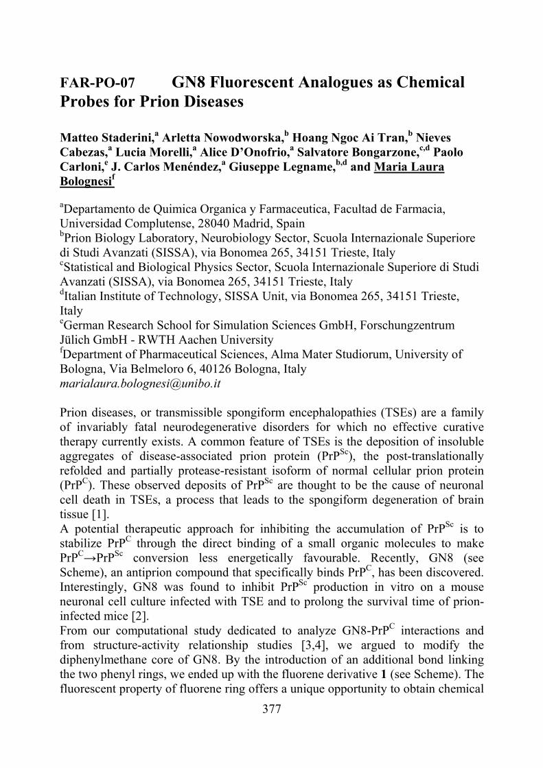
377
FAR-PO-07 GN8 Fluorescent Analogues as Chemical Probes for Prion Diseases Matteo Staderini,a Arletta Nowodworska,b Hoang Ngoc Ai Tran,b Nieves Cabezas,a Lucia Morelli,a Alice D’Onofrio,a Salvatore Bongarzone,c,d Paolo Carloni,e J. Carlos Menéndez,a Giuseppe Legname,b,d and Maria Laura Bolognesif
aDepartamento de Quimica Organica y Farmaceutica, Facultad de Farmacia, Universidad Complutense, 28040 Madrid, Spain bPrion Biology Laboratory, Neurobiology Sector, Scuola Internazionale Superiore di Studi Avanzati (SISSA), via Bonomea 265, 34151 Trieste, Italy cStatistical and Biological Physics Sector, Scuola Internazionale Superiore di Studi Avanzati (SISSA), via Bonomea 265, 34151 Trieste, Italy dItalian Institute of Technology, SISSA Unit, via Bonomea 265, 34151 Trieste, Italy eGerman Research School for Simulation Sciences GmbH, Forschungzentrum Jülich GmbH - RWTH Aachen University fDepartment of Pharmaceutical Sciences, Alma Mater Studiorum, University of Bologna, Via Belmeloro 6, 40126 Bologna, Italy [email protected] Prion diseases, or transmissible spongiform encephalopathies (TSEs) are a family of invariably fatal neurodegenerative disorders for which no effective curative therapy currently exists. A common feature of TSEs is the deposition of insoluble aggregates of disease-associated prion protein (PrPSc), the post-translationally refolded and partially protease-resistant isoform of normal cellular prion protein (PrPC). These observed deposits of PrPSc are thought to be the cause of neuronal cell death in TSEs, a process that leads to the spongiform degeneration of brain tissue [1]. A potential therapeutic approach for inhibiting the accumulation of PrPSc is to stabilize PrPC through the direct binding of a small organic molecules to make PrPC→PrPSc conversion less energetically favourable. Recently, GN8 (see Scheme), an antiprion compound that specifically binds PrPC, has been discovered. Interestingly, GN8 was found to inhibit PrPSc production in vitro on a mouse neuronal cell culture infected with TSE and to prolong the survival time of prion-infected mice [2]. From our computational study dedicated to analyze GN8-PrPC interactions and from structure-activity relationship studies [3,4], we argued to modify the diphenylmethane core of GN8. By the introduction of an additional bond linking the two phenyl rings, we ended up with the fluorene derivative 1 (see Scheme). The fluorescent property of fluorene ring offers a unique opportunity to obtain chemical
378
probes which may elucidate the mechanism of action of GN8 derivatives in vitro and in vivo. Herein, we report the synthesis of a small library of fluorene-based analogues of GN8 and the evaluation of their antiprion activity in TSE infected cells.
NH
ON
NH
ON
RR
R R
1
NH
NH
O
N
O
N
GN8 [1] Prusiner, S. B. Science 1991, 252, 1515. [2] Kuwata, K.; Nishida, N.; Matsumoto, T.; Kamatari, Y. O.; Hosokawa-Muto, J.;
Kodama, K.; Nakamura, H. K.; Kimura, K.; Kawasaki, M.; Takakura, Y.; Shirabe, S.; Takata, J.; Kataoka, Y.; Katamine, S. Proc. Natl. Acad. Sci. USA 104, 2007, 11921.
[3] Kimura, T.; Hosokawa-Muto, J.; Kamatari, Y. O.; Kuwata, K. Bioorg. Med. Chem. Lett. 21, 2011, 1502.
[4] Kranjc, A.; Bongarzone, S.; Rossetti, G.; Biarnés, X.; Cavalli, A.; Bolognesi, M. L.; Roberti, M.; Legname, G.; Carloni, P. J. Chem. Theory Comput. 5, 2009, 2565.
379
FAR-PO-08 New selective PDE4D inhibitors devoid of sedative effects in mice Olga Bruno,a Chiara Brullo,a Alessia Romussi,a Massimo Rocca,a Alessandro Balbi,a Lisa Flammini,b Simona Bertoni,b Elisabetta Barocellib
aDipartimento di Scienze Farmaceutiche dell’Università degli Studi di Genova, V.le Benedetto XV, 3- 16132 Genova-Italy. bDipartimento di Scienze Farmacologiche, Biologiche e Chimiche Applicate dell’Università degli Studi di Parma, V.le delle Scienze 27/a- 43100 Parma-Italy
Strategies designed to enhance cerebral cAMP by phosphodiesterases inhibition have been proposed as symptomatic treatments to counteract cognitive deficits. We have recently reported a series of new 3-cyclopentyloxy-4-methoxybenzaldehyde derivatives endowed with selective inhibitory activity toward the long-form PDE4D3 [1]. In a recent pharmacological study GEBR-7b, the most active of them, increased hippocampal cAMP and did not influence A levels; furthermore, in the object recognition test it improved spatial as well as object memory performance, at doses that did not cause emesis-like behaviour in rodents [2]. Therefore, our results further support the hypothesis that specific PDE4D isoforms play a critical role in the mediation of memory processes, as recently reported by other researchers [3-4]. It is well known that rolipram, a PDE4 inhibitor lacking in isoforms selectivity, combines pro-cognitive properties with undesirable sedative effects, decreasing both locomotion and rearing in rodents [5]. Recently, Li et al. observed no changes in locomotor activity between PDE4D knock out and wild type mice, suggesting that PDE4D inhibition couldn’t induce sedation [4]. With the aim to develop PDE4 inhibitors endowed with a better tolerability, we synthesized a new series of analogues of previous compounds and evaluated “in vitro” their inhibitory activity on different PDE4 isoforms as well as their ability to affect spontaneous locomotor activity. Some of them (compounds 4a, 5b, 7b, selective toward PDE4D isoforms and compounds 6b and 9b, inactive toward all PDE4 isoforms), when studied in open field test in mice, did not affect locomotor activity at doses comparable with those of rolipram able to decrease locomotor activity and prolong immobility time. Compound 11, having a non-selective rolipram-like enzymatic profile, caused a similar even if not significant reduction in spontaneous motor activity.
In conclusion, these findings further support the idea that selective PDE4D inhibition may represent an innovative cognitive-enhancing pharmacological strategy lacking side-effects such as emesis and sedation.
[1] O. Bruno, et al. J. Med. Chem. 52, 2009, 6546. [2] O. Bruno, et al., Br. J. Pharm. 2011 DOI: 10.1111/j.1476-5381.2011.01524.x [3] A.B. Burgin, et al. Nat. Biotechnol, 28, 2010, 63. [4] Y.F. Li, et al. J. Neurosci. 31, 2011, 172. [5] J.S. Silvestre, et al. Pharmacol., Biochem. Behav. 64, 1999, 1.
380
FAR-PO-09 Synthesis and Enzymatic Evaluation of NAD Mimics as Nicotinamide Adenine Dinucleotide Kinase (NADK) Inhibitors Loredana Cappellacci,a Riccardo Petrelli,a,b Krzysztof Felczak,b Patrizia Vita,a Ilaria Torquati,a Palmarisa Franchetti,a Michele Di Stefano,c Giulio Magni,c Mario Grifantini,a Krzysztof W. Pankiewiczb aSchool of Pharmacy, Medicinal Chemistry Unit, University of Camerino, via S. Agostino 1, 62032 Camerino, Italy bCenter for Drug Design, University of Minnesota, 516 Delaware St SE, Minneapolis, MN 55455, USA cDipartimento di Patologia Molecolare e Terapie Innovative, sezione di Biochimica, Università Politecnica delle Marche, via Ranieri, 60131 Ancona, Italy E-mail: [email protected]
NAD kinase (NADK) is a key enzyme that regulates supply of NADP in the cell. At this time no other pathway of NADP biosynthesis has been found in prokaryotic or eukaryotic cells. Human NAD kinase catalyzes a magnesium-dependent phosphorylation of the 2’-hydroxyl group of the adenosine ribose moiety of NAD using ATP as phosphoryl donor to give NADP. Bacterial enzymes can use inorganic polyphosphates as phosphoryl donors in addition to ATP. Significant differences between the human and the mycobacterium enzyme were found that might allow for construction of inhibitors with selectivity against these proteins. Therefore, M. tubercolosis NADK has became an appealing new target for the development of potential drugs against multi-drug resistant (MDR) and extensively drug resistant (XDR) tubercolosis (TB).
Recently, we have reported that dinucleoside disulfide NAD mimics, such as diadenosine disulfide (DTA), were found to be moderate inhibitors of M. tubercolosis and human NADKs. A restriction of the conformation of adenine moiety to syn by substitution with a bulky bromine atom at the C8 of one or two adenine rings of DTA, furnished the most potent inhibitors of both human and mycobacterium NADK reported so far. On the contrary, fixing the sugar conformation in the “North” or “South” conformation by introduction of a methyl group at the 2’- or 3’-position of the ribose ring was detrimental for NADK inhibitory activity [1].
To further investigate the structural features of the ribose moiety, herein we report the synthesis and the NADK inhibitory activity of 2’-deoxy-, 3’-deoxy-, and 2’,3’-dideoxy-DTA. The results of this study will be presented. [1] R. Petrelli, Y.Y. Sham, L. Chen, K. Felczak, E. Bennett, D. Wilson, C. Aldrich, J.S. Yu, L. Cappellacci, P. Franchetti, M. Grifantini, F. Mazzola, M. Di Stefano, G. Magni, and K. Pankiewicz, Bioorg. Med. Chem., 17, 2009, 5656.
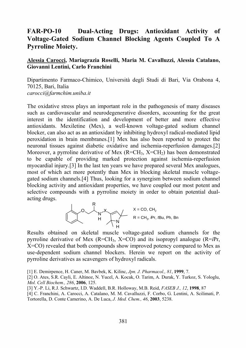
381
FAR-PO-10 Dual-Acting Drugs: Antioxidant Activity of Voltage-Gated Sodium Channel Blocking Agents Coupled To A Pyrroline Moiety. Alessia Carocci, Mariagrazia Roselli, Maria M. Cavalluzzi, Alessia Catalano, Giovanni Lentini, Carlo Franchini Dipartimento Farmaco-Chimico, Università degli Studi di Bari, Via Orabona 4, 70125, Bari, Italia [email protected]
The oxidative stress plays an important role in the pathogenesis of many diseases such as cardiovascular and neurodegenerative disorders, accounting for the great interest in the identification and development of better and more effective antioxidants. Mexiletine (Mex), a well-known voltage-gated sodium channel blocker, can also act as an antioxidant by inhibiting hydroxyl radical-mediated lipid peroxidation in brain membranes.[1] Mex has also been reported to protect the neuronal tissues against diabetic oxidative and ischemia-reperfusion damages.[2] Moreover, a pyrroline derivative of Mex (R=CH3, X=CH2) has been demonstrated to be capable of providing marked protection against ischemia-reperfusion myocardial injury.[3] In the last ten years we have prepared several Mex analogues, most of which act more potently than Mex in blocking skeletal muscle voltage-gated sodium channels.[4] Thus, looking for a synergism between sodium channel blocking activity and antioxidant properties, we have coupled our most potent and selective compounds with a pyrroline moiety in order to obtain potential dual-acting drugs.
X = CO, CH2ONH
R
X
NH
R = CH3, iPr, tBu, Ph, Bn
Results obtained on skeletal muscle voltage-gated sodium channels for the pyrroline derivative of Mex (R=CH3, X=CO) and its isopropyl analogue (R=iPr, X=CO) revealed that both compounds show improved potency compared to Mex as use-dependent sodium channel blockers. Herein we report on the activity of pyrroline derivatives as scavengers of hydroxyl radicals.
[1] E. Demirpence, H. Caner, M. Bavbek, K. Kilinc, Jpn. J. Pharmacol., 81, 1999, 7. [2] O. Ates, S.R. Cayli, E. Altinoz, N. Yucel, A. Kocak, O. Tarim, A. Durak, Y. Turkoz, S. Yologlu, Mol. Cell Biochem., 286, 2006, 125. [3] Y.-P. Li, R.J. Schwartz, I.D. Waddell, B.R. Holloway, M.B. Reid, FASEB J., 12, 1998, 87 [4] C. Franchini, A. Carocci, A. Catalano, M. M. Cavalluzzi, F. Corbo, G. Lentini, A. Scilimati, P. Tortorella, D. Conte Camerino, A. De Luca, J. Med. Chem., 46, 2003, 5238.
382
FAR-PO-11 Monitoring the effect of antagonist on P53-MDM2 interaction with NMR spectroscopy. Recent results. A. Carotenuto,a D. Brancaccio,a A. Limatola,a S. Rea,b V. De Pasquale.b, L. M. Pavone,b E. Novellinoa
aDipartimento di Chimica Farmaceutica e Tossicologica, via Montesano 49, 80131 Napoli, Italy. bDipartimento di Biochimica e Biotecnologie Mediche, via Pansini 5, 80131 Napoli, Italy. [email protected] The tremendous challenge of inhibiting therapeutically important protein-protein interactions has created the opportunity to extend traditional medicinal chemistry to a new class of targets and to explore nontraditional strategies. Holak and co. have recently described a two-dimensional 15N-HSQC based NMR assay for studying the effect of antagonists on protein-protein interactions [1]. The method, named AIDA (for antagonist induced dissociation assay), provides information on whether an antagonist of a protein-protein interaction is strong enough to dissociate the complex and whether its action is through denaturation, precipitation, or release of a protein in its functional folded state. AIDA requires the use of a large protein fragment (larger than 30 kDa) to bind to a small reporter protein (less than 20 kDa). In appropriate conditions (flexible residues), 1D proton NMR spectra may suffice for monitoring the states of proteins in complexes upon treatment with ligands. Because of the highly flexible nature of the N-terminal domain of p53, p53-MDM2 complex is suitable for 1D proton NMR application. In particular, the NHε side chains of W23 and W53 produce sharp lines in the free p53 1D proton spectrum. On formation of the complex with MDM2, W23 signal disappears, since W23, together with the p53 residues 17-26, comprises the primary binding site for MDM2. Hence it is possible to determine the activity of potential p53-MDM2 antagonists acquiring the 1H NMR spectrum of the protein complex. We have recently applied this method to a series of spiro(oxindole-3,3’-thiazolidine) derivatives thus discriminating the compounds able to block the p53-MDM2 interaction [2]. The method is now applied to new series of peptide and small molecule compounds to screen their p53-MDM2 antagonist properties. [1] L. D’Silva, P. Ozdowy, M. Krajewski, U. Rothweiler, M. Singh, and T. A. Holak, J. Am. Chem. Soc., 127, 2005, 13220. [2] I. Gomez-Monterrey, A. Bertamino, A. Porta, A. Carotenuto, S. Musella, C. Aquino, I. Granata, M. Sala, D. Brancaccio, D. Picone, C. Ercole, P. Stiuso, P. Campiglia, P. Grieco, P. Ianelli, B. Maresca, and E. Novellino, J. Med. Chem., 53, 2010, 8319.

383
FAR-PO-12 Targeting the FXR Nuclear Receptor through a Virtual Screening Approach Andrea Carotti,a Antimo Gioiello,a Antonio Macchiarulo,a Maura Marinozzi,a Paride Liscio,a Paola Sabbatini,a Giovanni Rizzo,b Luciano Adorini,b Roberto Pellicciaria
aDipartimento di Chimica e Tecnologia del Farmaco, Università degli Studi di Perugia, Via del Liceo 1, 06123, Perugia, Italy bIntercept Pharmaceuticals Italia Srl, Via Togliatti, 06073, Corciano, Perugia, Italy [email protected]
Farnesoid X Receptor (FXR) belongs to a family of nuclear receptors that are ligand-inducible transcription factors. FXR is highly expressed in the liver and intestine and is activated by bile acids, such as chenodeoxycholic acid (CDCA) and cholic acid (CA), and their corresponding conjugates at physiological concentrations. FXR functions as a bile acid sensor in enterohepatic tissues, regulating several target genes associated with bile acid synthesis and transport [1]. Activation of FXR by bile acids or synthetic agonists results in transcriptional repression of cholesterol 7α-hydroxylase (CYP7A1), the rate-limiting enzyme in the bile acid biosynthesis pathway, induction of the small heterodimer partner (SHP), a transcriptional repressor found in the liver and intestine, an induction of genes encoding for some bile acid transport proteins, such as intestinal bile acid-binding protein (IBABP) [2] and bile salt export pump (BSEP) [3]. Furthermore, bile acid-mediated FXR activation is recently recognized as a major underlying pathway for energy homeostasis and glucose as well as lipid metabolism.
FXR has also been suggested to counteract pro-inflammatory and pro-atherogenic responses in cardiovascular diseases.
All these evidences make FXR a promising potential target for the treatment of a variety of metabolic disorders, including hyperlipidemia, cholelithiasis, cholestasis, and diabetes mellitus.
Over the past few years, many efforts have been dedicated to the search of highly potent steroidal- and nonsteroidal FXR modulators. With the aim of discovering novel classes of nonsteroidal FXR ligands, we initiated a virtual screening protocol based on the information retrieved by the crystal structures of FXR in complex with agonists. A multistep approach using ligand and structure-based techniques will be presented.
[1] T.T.Lu, M.Makishima, J.J.Repa, K.Schoonjans, T.A.Kerr, J.Auwerx, and
D.J.Mangelsdorf, Mol. Cell, 6, 2000, 507. [2] J.Li, P.C.Pircher, I.G.Schulman, and S.K.Westin, J. Biol. Chem., 280, 2005,
7427. [3] T.M.Willson, S.A.Jones, J.T.Moore, and S.A.Kliewer, Med. Res. Rev., 21,
2001, 513.
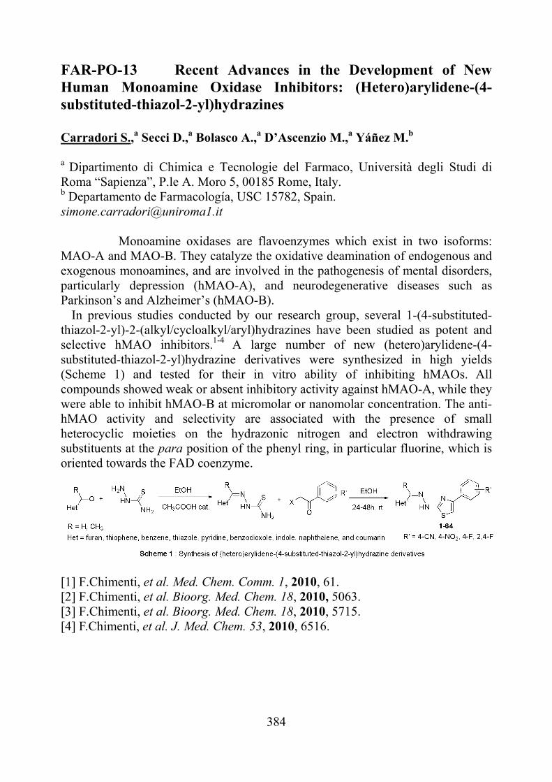
384
FAR-PO-13 Recent Advances in the Development of New Human Monoamine Oxidase Inhibitors: (Hetero)arylidene-(4-substituted-thiazol-2-yl)hydrazines
Carradori S.,a Secci D.,a Bolasco A.,a D’Ascenzio M.,a Yáñez M.b
a Dipartimento di Chimica e Tecnologie del Farmaco, Università degli Studi di Roma “Sapienza”, P.le A. Moro 5, 00185 Rome, Italy. b Departamento de Farmacología, USC 15782, Spain. [email protected]
Monoamine oxidases are flavoenzymes which exist in two isoforms: MAO-A and MAO-B. They catalyze the oxidative deamination of endogenous and exogenous monoamines, and are involved in the pathogenesis of mental disorders, particularly depression (hMAO-A), and neurodegenerative diseases such as Parkinson’s and Alzheimer’s (hMAO-B).
In previous studies conducted by our research group, several 1-(4-substituted-thiazol-2-yl)-2-(alkyl/cycloalkyl/aryl)hydrazines have been studied as potent and selective hMAO inhibitors.1-4 A large number of new (hetero)arylidene-(4-substituted-thiazol-2-yl)hydrazine derivatives were synthesized in high yields (Scheme 1) and tested for their in vitro ability of inhibiting hMAOs. All compounds showed weak or absent inhibitory activity against hMAO-A, while they were able to inhibit hMAO-B at micromolar or nanomolar concentration. The anti-hMAO activity and selectivity are associated with the presence of small heterocyclic moieties on the hydrazonic nitrogen and electron withdrawing substituents at the para position of the phenyl ring, in particular fluorine, which is oriented towards the FAD coenzyme.
[1] F.Chimenti, et al. Med. Chem. Comm. 1, 2010, 61. [2] F.Chimenti, et al. Bioorg. Med. Chem. 18, 2010, 5063. [3] F.Chimenti, et al. Bioorg. Med. Chem. 18, 2010, 5715. [4] F.Chimenti, et al. J. Med. Chem. 53, 2010, 6516.
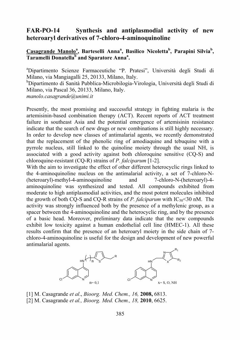
385
FAR-PO-14 Synthesis and antiplasmodial activity of new heteroaryl derivatives of 7-chloro-4-aminoquinoline Casagrande Manoloa, Barteselli Annaa, Basilico Nicolettab, Parapini Silviab, Taramelli Donatellab and Sparatore Annaa.
aDipartimento Scienze Farmaceutiche “P. Pratesi”, Università degli Studi di Milano, via Mangiagalli 25, 20133, Milano, Italy. bDipartimento di Sanità Pubblica-Microbilogia-Virologia, Università degli Studi di Milano, via Pascal 36, 20133, Milano, Italy. [email protected] Presently, the most promising and successful strategy in fighting malaria is the artemisinin-based combination therapy (ACT). Recent reports of ACT treatment failure in southeast Asia and the potential emergence of artemisinin resistance indicate that the search of new drugs or new combinations is still highly necessary. In order to develop new classes of antimalarial agents, we recently demonstrated that the replacement of the phenolic ring of amodiaquine and tebuquine with a pyrrole nucleus, still linked to the quinoline moiety through the usual NH, is associated with a good activity against both chloroquine sensitive (CQ-S) and chloroquine-resistant (CQ-R) strains of P. falciparum [1-2]. With the aim to investigate the effect of other different heterocyclic rings linked to the 4-aminoquinoline nucleus on the antimalarial activity, a set of 7-chloro-N-(heteroaryl)-methyl-4-aminoquinoline and 7-chloro-N-(heteroaryl)-4-aminoquinoline was synthesized and tested. All compounds exhibited from moderate to high antiplasmodial activities, and the most potent molecules inhibited the growth of both CQ-S and CQ-R strains of P. falciparum with IC50<30 nM. The activity was strongly influenced both by the presence of a methylenic group, as a spacer between the 4-aminoquinoline and the heterocyclic ring, and by the presence of a basic head. Moreover, preliminary data indicate that the new compounds exhibit low toxicity against a human endothelial cell line (HMEC-1). All these results confirm that the presence of an heteroaryl moiety in the side chain of 7-chloro-4-aminoquinoline is useful for the design and development of new powerful antimalarial agents.
NCl
HN S
R
n
NCl
HN X
NR1
R2
n= 0,1 x= S, O, NH
[1] M. Casagrande et al., Bioorg. Med. Chem., 16, 2008, 6813. [2] M. Casagrande et al., Bioorg. Med. Chem., 18, 2010, 6625.
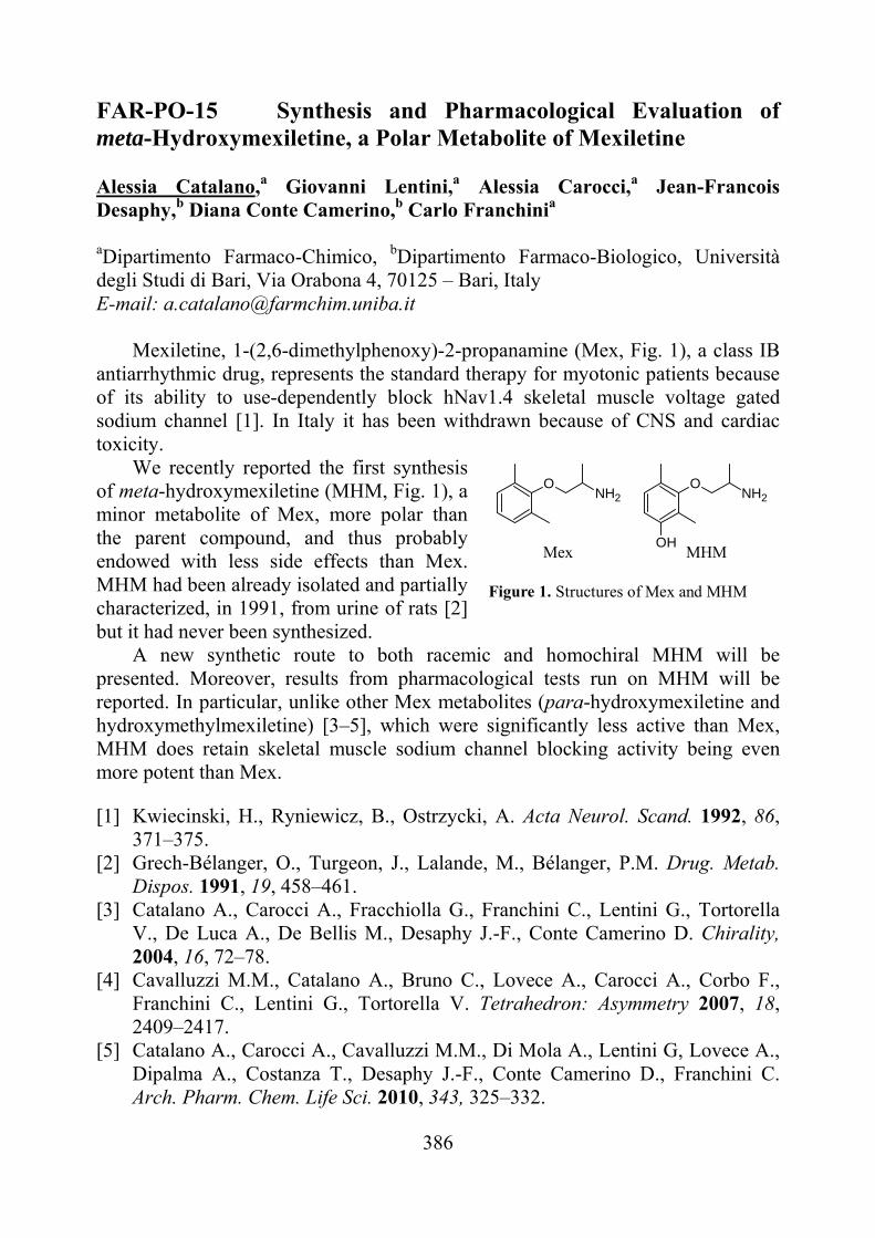
386
FAR-PO-15 Synthesis and Pharmacological Evaluation of meta-Hydroxymexiletine, a Polar Metabolite of Mexiletine Alessia Catalano,a Giovanni Lentini,a Alessia Carocci,a Jean-Francois Desaphy,b Diana Conte Camerino,b Carlo Franchinia
aDipartimento Farmaco-Chimico, bDipartimento Farmaco-Biologico, Università degli Studi di Bari, Via Orabona 4, 70125 – Bari, Italy E-mail: [email protected]
Mexiletine, 1-(2,6-dimethylphenoxy)-2-propanamine (Mex, Fig. 1), a class IB
antiarrhythmic drug, represents the standard therapy for myotonic patients because of its ability to use-dependently block hNav1.4 skeletal muscle voltage gated sodium channel [1]. In Italy it has been withdrawn because of CNS and cardiac toxicity.
We recently reported the first synthesis of meta-hydroxymexiletine (MHM, Fig. 1), a minor metabolite of Mex, more polar than the parent compound, and thus probably endowed with less side effects than Mex. MHM had been already isolated and partially characterized, in 1991, from urine of rats [2] but it had never been synthesized.
A new synthetic route to both racemic and homochiral MHM will be presented. Moreover, results from pharmacological tests run on MHM will be reported. In particular, unlike other Mex metabolites (para-hydroxymexiletine and hydroxymethylmexiletine) [3–5], which were significantly less active than Mex, MHM does retain skeletal muscle sodium channel blocking activity being even more potent than Mex.
[1] Kwiecinski, H., Ryniewicz, B., Ostrzycki, A. Acta Neurol. Scand. 1992, 86, 371–375.
[2] Grech-Bélanger, O., Turgeon, J., Lalande, M., Bélanger, P.M. Drug. Metab. Dispos. 1991, 19, 458–461.
[3] Catalano A., Carocci A., Fracchiolla G., Franchini C., Lentini G., Tortorella V., De Luca A., De Bellis M., Desaphy J.-F., Conte Camerino D. Chirality, 2004, 16, 72–78.
[4] Cavalluzzi M.M., Catalano A., Bruno C., Lovece A., Carocci A., Corbo F., Franchini C., Lentini G., Tortorella V. Tetrahedron: Asymmetry 2007, 18, 2409–2417.
[5] Catalano A., Carocci A., Cavalluzzi M.M., Di Mola A., Lentini G, Lovece A., Dipalma A., Costanza T., Desaphy J.-F., Conte Camerino D., Franchini C. Arch. Pharm. Chem. Life Sci. 2010, 343, 325–332.
MHM
ONH2
OH
ONH2
Mex
Figure 1. Structures of Mex and MHM
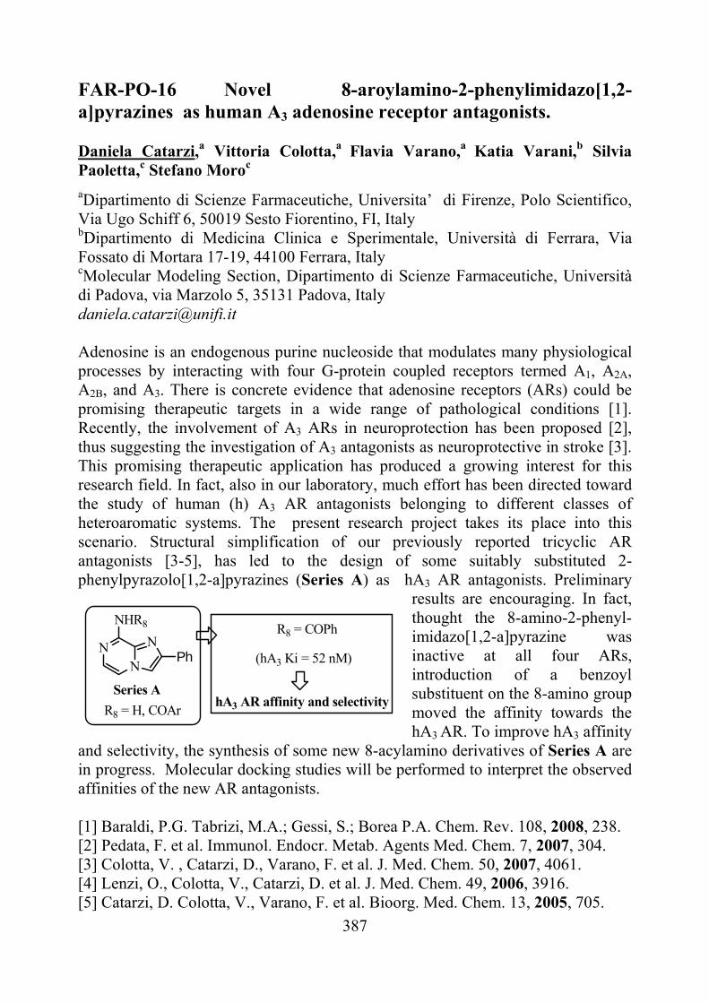
387
NN
NHR8
NPh
R8 = COPh
(hA3 Ki = 52 nM)
hA3 AR affinity and selectivityR8 = H, COAr
Series A
FAR-PO-16 Novel 8-aroylamino-2-phenylimidazo[1,2-a]pyrazines as human A3 adenosine receptor antagonists. Daniela Catarzi,a Vittoria Colotta,a Flavia Varano,a Katia Varani,b Silvia Paoletta,c Stefano Moroc aDipartimento di Scienze Farmaceutiche, Universita’ di Firenze, Polo Scientifico, Via Ugo Schiff 6, 50019 Sesto Fiorentino, FI, Italy bDipartimento di Medicina Clinica e Sperimentale, Università di Ferrara, Via Fossato di Mortara 17-19, 44100 Ferrara, Italy
cMolecular Modeling Section, Dipartimento di Scienze Farmaceutiche, Università di Padova, via Marzolo 5, 35131 Padova, Italy [email protected]
Adenosine is an endogenous purine nucleoside that modulates many physiological processes by interacting with four G-protein coupled receptors termed A1, A2A, A2B, and A3. There is concrete evidence that adenosine receptors (ARs) could be promising therapeutic targets in a wide range of pathological conditions [1]. Recently, the involvement of A3 ARs in neuroprotection has been proposed [2], thus suggesting the investigation of A3 antagonists as neuroprotective in stroke [3]. This promising therapeutic application has produced a growing interest for this research field. In fact, also in our laboratory, much effort has been directed toward the study of human (h) A3 AR antagonists belonging to different classes of heteroaromatic systems. The present research project takes its place into this scenario. Structural simplification of our previously reported tricyclic AR antagonists [3-5], has led to the design of some suitably substituted 2-phenylpyrazolo[1,2-a]pyrazines (Series A) as hA3 AR antagonists. Preliminary
results are encouraging. In fact, thought the 8-amino-2-phenyl-imidazo[1,2-a]pyrazine was inactive at all four ARs, introduction of a benzoyl substituent on the 8-amino group moved the affinity towards the hA3 AR. To improve hA3 affinity
and selectivity, the synthesis of some new 8-acylamino derivatives of Series A are in progress. Molecular docking studies will be performed to interpret the observed affinities of the new AR antagonists. [1] Baraldi, P.G. Tabrizi, M.A.; Gessi, S.; Borea P.A. Chem. Rev. 108, 2008, 238. [2] Pedata, F. et al. Immunol. Endocr. Metab. Agents Med. Chem. 7, 2007, 304. [3] Colotta, V. , Catarzi, D., Varano, F. et al. J. Med. Chem. 50, 2007, 4061. [4] Lenzi, O., Colotta, V., Catarzi, D. et al. J. Med. Chem. 49, 2006, 3916. [5] Catarzi, D. Colotta, V., Varano, F. et al. Bioorg. Med. Chem. 13, 2005, 705.
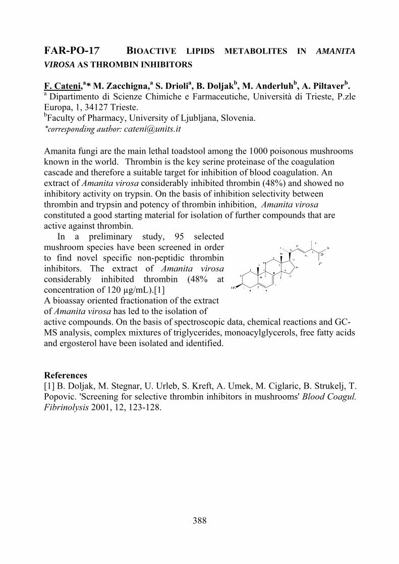
388
FAR-PO-17 BIOACTIVE LIPIDS METABOLITES IN AMANITA
VIROSA AS THROMBIN INHIBITORS F. Cateni,a* M. Zacchigna,a S. Driolia, B. Doljakb, M. Anderluhb, A. Piltaverb. a Dipartimento di Scienze Chimiche e Farmaceutiche, Università di Trieste, P.zle Europa, 1, 34127 Trieste. bFaculty of Pharmacy, University of Ljubljana, Slovenia. *corresponding author: [email protected]
Amanita fungi are the main lethal toadstool among the 1000 poisonous mushrooms known in the world. Thrombin is the key serine proteinase of the coagulation cascade and therefore a suitable target for inhibition of blood coagulation. An extract of Amanita virosa considerably inhibited thrombin (48%) and showed no inhibitory activity on trypsin. On the basis of inhibition selectivity between thrombin and trypsin and potency of thrombin inhibition, Amanita virosa constituted a good starting material for isolation of further compounds that are active against thrombin.
In a preliminary study, 95 selected mushroom species have been screened in order to find novel specific non-peptidic thrombin inhibitors. The extract of Amanita virosa considerably inhibited thrombin (48% at concentration of 120 µg/mL).[1]
A bioassay oriented fractionation of the extract of Amanita virosa has led to the isolation of active compounds. On the basis of spectroscopic data, chemical reactions and GC-MS analysis, complex mixtures of triglycerides, monoacylglycerols, free fatty acids and ergosterol have been isolated and identified.
References [1] B. Doljak, M. Stegnar, U. Urleb, S. Kreft, A. Umek, M. Ciglaric, B. Strukelj, T. Popovic. 'Screening for selective thrombin inhibitors in mushrooms' Blood Coagul. Fibrinolysis 2001, 12, 123-128.
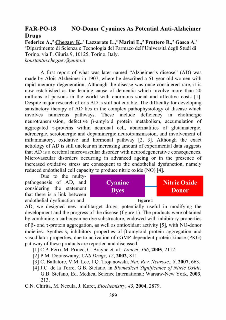
389
FAR-PO-18 NO-Donor Cyanines As Potential Anti-Alzheimer Drugs Federico A.,a Chegaev K.,a Lazzarato L.,a Marini E.,a Fruttero R.,a Gasco A.a
aDipartimento di Scienza e Tecnologia del Farmaco dell’Università degli Studi di Torino, via P. Giuria 9, 10125, Torino, Italy. [email protected]
A first report of what was later named “Alzheimer’s disease” (AD) was
made by Alois Alzheimer in 1907, where he described a 51-year old women with rapid memory degeneration. Although the disease was once considered rare, it is now established as the leading cause of dementia which involve more than 20 millions of persons in the world with enormous social and affective costs [1]. Despite major research efforts AD is still not curable. The difficulty for developing satisfactory therapy of AD lies in the complex pathophysiology of disease which involves numerous pathways. These include deficiency in cholinergic neurotransmission, defective -amyloid protein metabolism, accumulation of aggregated -proteins within neuronal cell, abnormalities of glutamatergic, adrenergic, serotonergic and dopaminergic neurotransmission, and involvement of inflammatory, oxidative and hormonal pathway [2, 3]. Although the exact aetiology of AD is still unclear an increasing amount of experimental data suggests that AD is a cerebral microvascular disorder with neurodegenerative consequences. Microvascular disorders occurring in advanced ageing or in the presence of increased oxidative stress are consequent to the endothelial dysfunction, namely reduced endothelial cell capacity to produce nitric oxide (NO) [4].
Due to the multy-pathogenesis of AD, and considering the statement that there is a link between endothelial dysfunction and AD, we designed new multitarget drugs, potentially useful in modifying the development and the progress of the disease (figure 1). The products were obtained by combining a carbocyanine dye substructure, endowed with inhibitory properties of - and -protein aggregation, as well as antioxidant activity [5], with NO-donor moieties. Synthesis, inhibitory properties of -amyloid protein aggregation and vasodilator properties, due to activation of cGMP-dependent protein kinase (PKG) pathway of these products are reported and discussed.
[1] C.P. Ferri, M. Prince, C. Brayne et. al., Lancet, 366, 2005, 2112. [2] P.M. Doraiswamy, CNS Drugs, 12, 2002, 811. [3] C. Ballatore, V.M. Lee, J.Q. Trojanowski, Nat. Rev. Neurosc., 8, 2007, 663. [4] J.C. de la Torre, G.B. Stefano, in Biomedical Significance of Nitric Oxide.
G.B. Stefano, Ed. Medical Science International: Warsaw-New York, 2003, 213.
C.N. Chirita, M. Necula, J. Kuret, Biochemistry, 43, 2004, 2879.
Figure 1
CyanineDyes
Nitric OxideDonor
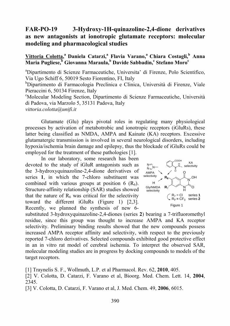
390
FAR-PO-19 3-Hydroxy-1H-quinazoline-2,4-dione derivatives as new antagonists at ionotropic glutamate receptors: molecular modeling and pharmacological studies Vittoria Colotta,a Daniela Catarzi,a Flavia Varano,a Chiara Costagli,b Anna Maria Pugliese,b Giovanna Maraula,b Davide Sabbadin,c Stefano Moroc aDipartimento di Scienze Farmaceutiche, Universita’ di Firenze, Polo Scientifico, Via Ugo Schiff 6, 50019 Sesto Fiorentino, FI, Italy bDipartimento di Farmacologia Preclinica e Clinica, Università di Firenze, Viale Pieraccini 6, 50134 Firenze, Italy
cMolecular Modeling Section, Dipartimento di Scienze Farmaceutiche, Università di Padova, via Marzolo 5, 35131 Padova, Italy [email protected]
Glutamate (Glu) plays pivotal roles in regulating many physiological
processes by activation of metabotrobic and ionotropic receptors (iGluRs), these latter being classified as NMDA, AMPA and Kainate (KA) receptors. Excessive glutamatergic transmission is involved in several neurological disorders, including hypoxia/ischemia brain damage and epilepsy, thus the blockade of iGluRs could be employed for the treatment of these pathologies [1].
In our laboratory, some research has been devoted to the study of iGluR antagonists such as the 3-hydroxyquinazoline-2,4-dione derivatives of series 1, in which the 7-chloro substituent was combined with various groups at position 6 (R6). Structure-affinity relationship (SAR) studies showed that the nature of R6 was critical for the selectivity toward the different iGluRs (Figure 1) [2,3]. Recently, we planned the synthesis of new 6-substituted 3-hydroxyquinazoline-2,4-diones (series 2) bearing a 7-trifluoromethyl residue, since this group was thought to increase AMPA and KA receptor selectivity. Preliminary binding results showed that the new compounds possess increased AMPA receptor affinity and selectivity, with respect to the previously reported 7-chloro derivatives. Selected compounds exhibited good protective effect in an in vitro rat model of cerebral ischemia. To interpret the observed SAR, molecular modeling studies are in progress by docking compounds to models of the target receptors. [1] Traynelis S. F., Wollmuth, L.P. et al Pharmacol. Rev. 62, 2010, 405. [2] V. Colotta, D. Catarzi, F. Varano et al, Bioorg. Med. Chem. Lett. 14, 2004, 2345. [3] V. Colotta, D. Catarzi, F. Varano et al, J. Med. Chem. 49, 2006, 6015.
NH
NR6
R7 O
O
OH
NNN
Gly/NMDAselectivity
AMPA selectivity
H
HN
O
COOHKA
selectivity
R7 = Cl series 1R7 = CF3 series 2
Figure 1
391
FAR-PO-20 Indolylarylsulfones as HIV-1 Non-Nucleoside Reverse Transcriptase Inhibitors. New Cyclic Substituents at the Indole-2-carboxamide.
Antonio Coluccia,a Giuseppe La Regina,a Valerio Gatti,a Francesco Piscitelli,a Valeria Famiglini,a Andrea Brancale,b Govanni Maga,c Jan Balzarini,d Ettore Novellino,e Romano Silvestri.a
aIstituto Pasteur - Fondazione Cenci Bolognetti, Dipartimento di Chimica e Tecnologie del Farmaco, Sapienza Università di Roma, Piazzale Aldo Moro 5, I-00185 Roma, Italy. bWelsh School of Pharmacy, Cardiff University, King Edward VII Avenue, Cardiff, CF10 3NB, UK. cIstituto di Genetica Molecolare CNR, Via Abbiategrasso 207, I-27100 Pavia, Italy. dRega Institute for Medical Research, Katholieke Universiteit Leuven, B-3000 Leuven, Belgium. eDipartimento di Chimica Farmaceutica e Tossicologica, Università di Napoli Federico II, Via Domenico Montesano 49, I-80131, Napoli, Italy. [email protected]
New indolylarylsulfone (IAS) [1,2] derivatives bearing cyclic substituents at the indole-2-carboxamide linked through a methylene/ethylene spacer are potent inhibitors of the HIV-1 WT replication with inhibitory concentrations in the low nanomolar range.
We report the results of docking and molecular dynamics experiments on this family of compounds. Analysis of the proposed binding pose into the WT and mutated RTs for the highly active compounds 5-chloro-3-((3,5-dimethylphenyl)sulfonyl)-N-(pyrroldin-1-yl)-1H-indole-2-carboxamide (1), it is possible to highlight a series of crucial interactions: (i) a H-bond between the indole NH and Lys101; (ii) the 3,5-dimethylphenyl moiety forms hydrophobic interactions in an aromatic cleft formed by Tyr181, Tyr188, and Trp229; (iii) a H-bond between the nitrogen atom of the heterocycle and the Glu138:B.
The model proposed is in good agreement with the biological results: 1 EC50
3.3 nM; IC50wt 12nM; IC50L100I 26nM, IC50K103N 93nM
[1] G. La Regina, A. Coluccia, A. Brancale, F. Piscitelli, V. Gatti, G. Maga, J. Balzarini, E. Novellino, R. Silvestri. J. Med. Chem., 54, 2011, 1587.
[2] F. Piscitelli, A. Coluccia, A. Brancale, G. La Regina, C. Giordano, J. Balzarini, G. Maga, E. Novellino, R. Silvestri. J. Med. Chem., 52, 2009, 1922.
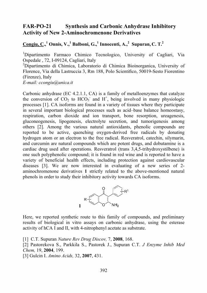
392
FAR-PO-21 Synthesis and Carbonic Anhydrase Inhibitory Activity of New 2-Aminochromenone Derivatives Congiu, C.,1 Onnis, V.,1 Balboni, G.,1 Innocenti, A.,2 Supuran, C. T.2
1Dipartimento Farmaco Chimico Tecnologico, University of Cagliari, Via Ospedale , 72, I-09124, Cagliari, Italy 2Dipartimento di Chimica, Laboratorio di Chimica Bioinorganica, University of Florence, Via della Lastruccia 3, Rm 188, Polo Scientifico, 50019-Sesto Fiorentino (Firenze), Italy E-mail: [email protected] Carbonic anhydrase (EC 4.2.1.1, CA) is a family of metalloenzymes that catalyze the conversion of CO2 to HCO3
- and H+, being involved in many physiologic processes [1]. CA isoforms are found in a variety of tissues where they participate in several important biological processes such as acid–base balance homeostasy, respiration, carbon dioxide and ion transport, bone resorption, ureagenesis, gluconeogenesis, lipogenesis, electrolyte secretion, and tumorigenesis among others [2]. Among the various natural antioxidants, phenolic compounds are reported to be active, quenching oxygen-derived free radicals by donating hydrogen atom or an electron to the free radical. Resveratrol, catechin, silymarin, and curcumin are natural compounds which are potent drugs, and dobutamine is a cardiac drug used after operations. Resveratrol (trans 3,4,5-trihydroxystilbene) is one such polyphenolic compound; it is found in red wine and is reported to have a variety of beneficial health effects, including protection against cardiovascular diseases [3]. We are now interested in evaluating of a new series of 2-aminochromenone derivatives 1 strictly related to the above-mentioned natural phenols in order to study their inhibitory activity towards CA isoforms.
1 Here, we reported synthetic route to this family of compounds, and preliminary results of biological in vitro assays on carbonic anhydrase, using the esterase activity of hCA I and II, with 4-nitrophenyl acetate as substrate. [1] C.T. Supuran Nature Rev Drug Discov, 7, 2008, 168. [2] Pastorekova S., Parkkila S., Pastorek J., Supuran C.T. J Enzyme Inhib Med Chem, 19, 2004, 199. [3] Gulcin I. Amino Acids, 32, 2007, 431.
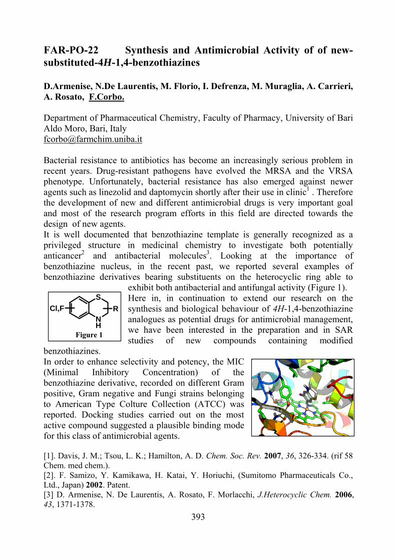
393
NH
S
RCl,F
Figure 1
FAR-PO-22 Synthesis and Antimicrobial Activity of of new-substituted-4H-1,4-benzothiazines D.Armenise, N.De Laurentis, M. Florio, I. Defrenza, M. Muraglia, A. Carrieri, A. Rosato, F.Corbo. Department of Pharmaceutical Chemistry, Faculty of Pharmacy, University of Bari Aldo Moro, Bari, Italy [email protected] Bacterial resistance to antibiotics has become an increasingly serious problem in recent years. Drug-resistant pathogens have evolved the MRSA and the VRSA phenotype. Unfortunately, bacterial resistance has also emerged against newer agents such as linezolid and daptomycin shortly after their use in clinic1 . Therefore the development of new and different antimicrobial drugs is very important goal and most of the research program efforts in this field are directed towards the design of new agents. It is well documented that benzothiazine template is generally recognized as a privileged structure in medicinal chemistry to investigate both potentially anticancer2 and antibacterial molecules3. Looking at the importance of benzothiazine nucleus, in the recent past, we reported several examples of benzothiazine derivatives bearing substituents on the heterocyclic ring able to
exhibit both antibacterial and antifungal activity (Figure 1). Here in, in continuation to extend our research on the synthesis and biological behaviour of 4H-1,4-benzothiazine analogues as potential drugs for antimicrobial management, we have been interested in the preparation and in SAR studies of new compounds containing modified
benzothiazines. In order to enhance selectivity and potency, the MIC (Minimal Inhibitory Concentration) of the benzothiazine derivative, recorded on different Gram positive, Gram negative and Fungi strains belonging to American Type Colture Collection (ATCC) was reported. Docking studies carried out on the most active compound suggested a plausible binding mode for this class of antimicrobial agents. [1]. Davis, J. M.; Tsou, L. K.; Hamilton, A. D. Chem. Soc. Rev. 2007, 36, 326-334. (rif 58 Chem. med chem.). [2]. F. Samizo, Y. Kamikawa, H. Katai, Y. Horiuchi, (Sumitomo Pharmaceuticals Co., Ltd., Japan) 2002. Patent. [3] D. Armenise, N. De Laurentis, A. Rosato, F. Morlacchi, J.Heterocyclic Chem. 2006, 43, 1371-1378.
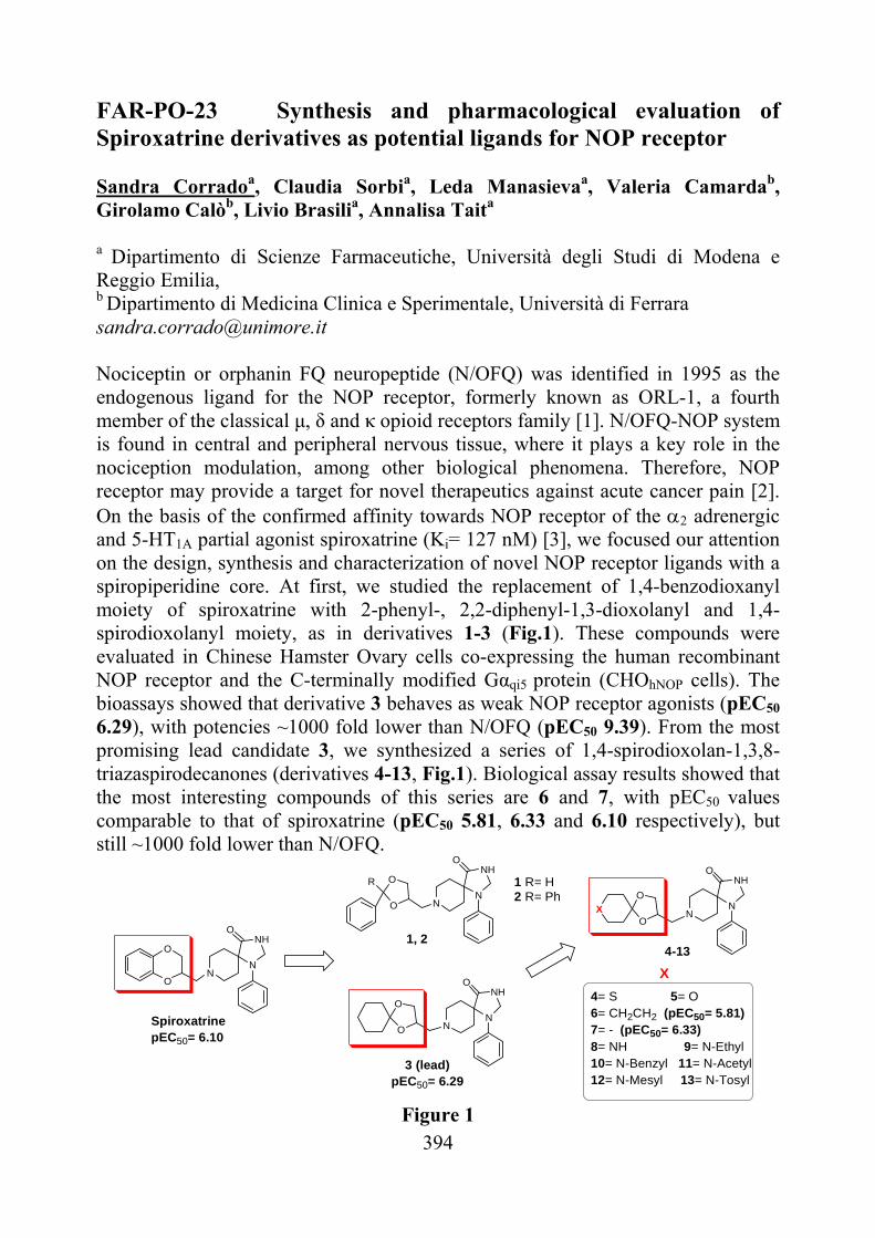
394
FAR-PO-23 Synthesis and pharmacological evaluation of Spiroxatrine derivatives as potential ligands for NOP receptor Sandra Corradoa, Claudia Sorbia, Leda Manasievaa, Valeria Camardab, Girolamo Calòb, Livio Brasilia, Annalisa Taita
a Dipartimento di Scienze Farmaceutiche, Università degli Studi di Modena e Reggio Emilia, b Dipartimento di Medicina Clinica e Sperimentale, Università di Ferrara [email protected] Nociceptin or orphanin FQ neuropeptide (N/OFQ) was identified in 1995 as the endogenous ligand for the NOP receptor, formerly known as ORL-1, a fourth member of the classical μ, δ and κ opioid receptors family [1]. N/OFQ-NOP system is found in central and peripheral nervous tissue, where it plays a key role in the nociception modulation, among other biological phenomena. Therefore, NOP receptor may provide a target for novel therapeutics against acute cancer pain [2]. On the basis of the confirmed affinity towards NOP receptor of the 2 adrenergic and 5-HT1A partial agonist spiroxatrine (Ki= 127 nM) [3], we focused our attention on the design, synthesis and characterization of novel NOP receptor ligands with a spiropiperidine core. At first, we studied the replacement of 1,4-benzodioxanyl moiety of spiroxatrine with 2-phenyl-, 2,2-diphenyl-1,3-dioxolanyl and 1,4-spirodioxolanyl moiety, as in derivatives 1-3 (Fig.1). These compounds were evaluated in Chinese Hamster Ovary cells co-expressing the human recombinant NOP receptor and the C-terminally modified Gαqi5 protein (CHOhNOP cells). The bioassays showed that derivative 3 behaves as weak NOP receptor agonists (pEC50 6.29), with potencies ~1000 fold lower than N/OFQ (pEC50 9.39). From the most promising lead candidate 3, we synthesized a series of 1,4-spirodioxolan-1,3,8-triazaspirodecanones (derivatives 4-13, Fig.1). Biological assay results showed that the most interesting compounds of this series are 6 and 7, with pEC50 values comparable to that of spiroxatrine (pEC50 5.81, 6.33 and 6.10 respectively), but still ~1000 fold lower than N/OFQ.
O
O
NN
NHO
SpiroxatrinepEC50= 6.10
O
O
R
O
OX
1, 2
1 R= H2 R= Ph
4-13
X
4= S 5= O6= CH2CH2 (pEC50= 5.81) 7= - (pEC50= 6.33)8= NH 9= N-Ethyl 10= N-Benzyl 11= N-Acetyl12= N-Mesyl 13= N-Tosyl
O
O
3 (lead)pEC50= 6.29
NN
NHO
NN
NHO
NN
NHO
Figure 1
395
The key reaction to obtain derivatives 1-13 is the nucleophilic substitution between 1-phenyl-1,3,8-triaza-spiro[4,5]decan-4-one and the toluensulfonate- or 2-chloromethyl-intermediates resulting from the appropriate ketone or aldehyde, in the presence of Na2CO3 and dimethylformamide as solvent [4]. [1] Zaveri N. Life Science, 2003, 73, 663-678; [2] Lambert D. Nature reviews, 2008, 7, 694-710; [3] Calo’ G., Bigoni R., Rizzi A., Guerrini R., Salvadori S., Regoli D. Peptides, 2000, 21(7), 935-947; [4] Heine H. G. et al. Unites States Patent 6060482, 2000.
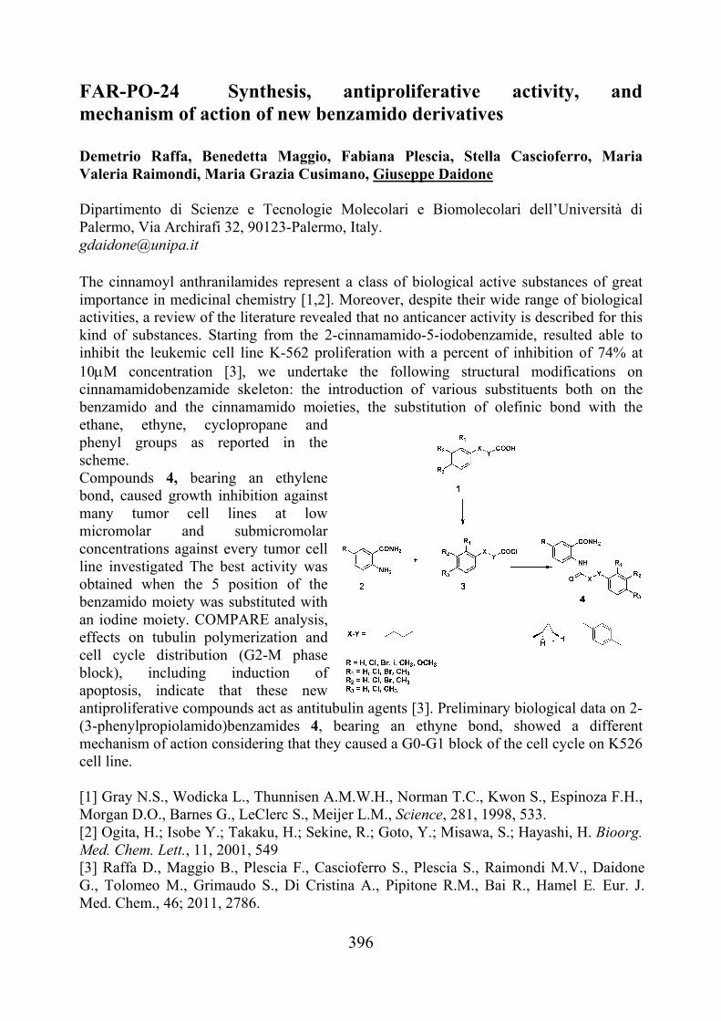
396
FAR-PO-24 Synthesis, antiproliferative activity, and mechanism of action of new benzamido derivatives Demetrio Raffa, Benedetta Maggio, Fabiana Plescia, Stella Cascioferro, Maria Valeria Raimondi, Maria Grazia Cusimano, Giuseppe Daidone Dipartimento di Scienze e Tecnologie Molecolari e Biomolecolari dell’Università di Palermo, Via Archirafi 32, 90123-Palermo, Italy. [email protected] The cinnamoyl anthranilamides represent a class of biological active substances of great importance in medicinal chemistry [1,2]. Moreover, despite their wide range of biological activities, a review of the literature revealed that no anticancer activity is described for this kind of substances. Starting from the 2-cinnamamido-5-iodobenzamide, resulted able to inhibit the leukemic cell line K-562 proliferation with a percent of inhibition of 74% at 10M concentration [3], we undertake the following structural modifications on cinnamamidobenzamide skeleton: the introduction of various substituents both on the benzamido and the cinnamamido moieties, the substitution of olefinic bond with the ethane, ethyne, cyclopropane and phenyl groups as reported in the scheme. Compounds 4, bearing an ethylene bond, caused growth inhibition against many tumor cell lines at low micromolar and submicromolar concentrations against every tumor cell line investigated The best activity was obtained when the 5 position of the benzamido moiety was substituted with an iodine moiety. COMPARE analysis, effects on tubulin polymerization and cell cycle distribution (G2-M phase block), including induction of apoptosis, indicate that these new antiproliferative compounds act as antitubulin agents [3]. Preliminary biological data on 2-(3-phenylpropiolamido)benzamides 4, bearing an ethyne bond, showed a different mechanism of action considering that they caused a G0-G1 block of the cell cycle on K526 cell line. [1] Gray N.S., Wodicka L., Thunnisen A.M.W.H., Norman T.C., Kwon S., Espinoza F.H., Morgan D.O., Barnes G., LeClerc S., Meijer L.M., Science, 281, 1998, 533. [2] Ogita, H.; Isobe Y.; Takaku, H.; Sekine, R.; Goto, Y.; Misawa, S.; Hayashi, H. Bioorg. Med. Chem. Lett., 11, 2001, 549 [3] Raffa D., Maggio B., Plescia F., Cascioferro S., Plescia S., Raimondi M.V., Daidone G., Tolomeo M., Grimaudo S., Di Cristina A., Pipitone R.M., Bai R., Hamel E. Eur. J. Med. Chem., 46; 2011, 2786.
397
FAR-PO-25 Rationalization of the Efficacy at the Human A3 Adenosine Receptor of 2- and N6-Substituted Adenosine and NECA Derivatives Diego Dal Ben,a Michela Buccioni,a Catia Lambertucci,a Gabriella Marucci,a Rosaria Volpini,a Karl-Norbert Klotz,b Gloria Cristalli.a aSchool of Pharmacy, Medicinal Chemistry Unit, University of Camerino, Via S. Agostino 1, 62032 Camerino, Italy. bInstitut für Pharmakologie und Toxikologie, Universität of Würzburg, Versbacher Str. 9 D-97078 Würzburg, Germany. [email protected] A3 adenosine receptor (A3AR) subtype is highly expressed in lungs, liver, and immune cells, and at lower densities in heart and brain and it is involved in a variety of key physiological processes such as release of inflammatory mediators and inhibition of tumor necrosis factor-α production. Agonists of this receptor have been recently analysed for pharmaceutical development based on their anti-inflammatory, anticancer, and cardioprotective effects [1,2]. On this basis, the design and synthesis of potent and selective A3AR agonists could be helpful to provide tools for further characterization and evaluation of the physio-pathological role of this receptor and for the development of new drugs. Some years ago, we have reported the synthesis and binding affinity of a number of adenosine (Ado) derivatives bearing in 2-position (ar)-alkynyl chains, endowed with good affinity and different degrees of selectivity for the human A3AR [3]. The replacement in these compounds of the hydroxymethyl group in 4’-position of the sugar moiety with an ethylcarboxamido function or the introduction in N6-position of small alkyl groups enhanced A3AR affinity and selectivity, leading to compounds with A3AR Ki values at low nanomolar level. These molecules were tested for their ability to inhibit forskolin-stimulated adenylyl cyclase activity. Results showed that all the 5’-N-ethylcarboxamidoAdo (NECA) derivatives behave as full agonists of A3AR, while the 4’-unmodified Ado derivatives present a partial agonist profile at the same receptor. The N6-substituents do not change the partial agonist activity observed for Ado derivatives unsubstituted at N6-position. Molecular modeling analyses, carried out by using the recently solved A2AAR crystal structure as template for homology modeling studies, helped to get a rationalization of the different pharmacological profile of the presented molecules at the human A3AR. [1] G. Cristalli, R. Volpini, Curr. Top. Med. Chem., 3, 2003, 355. [2] K.A. Jacobson, Z.G. Gao, Nat. Rev. Drug Dis., 5, 2006, 247. [3] R. Volpini, S. Costanzi, C. Lambertucci, S. Taffi, S. Vittori, K.-N. Klotz, G. Cristalli, J. Med. Chem., 45, 2002, 3271.
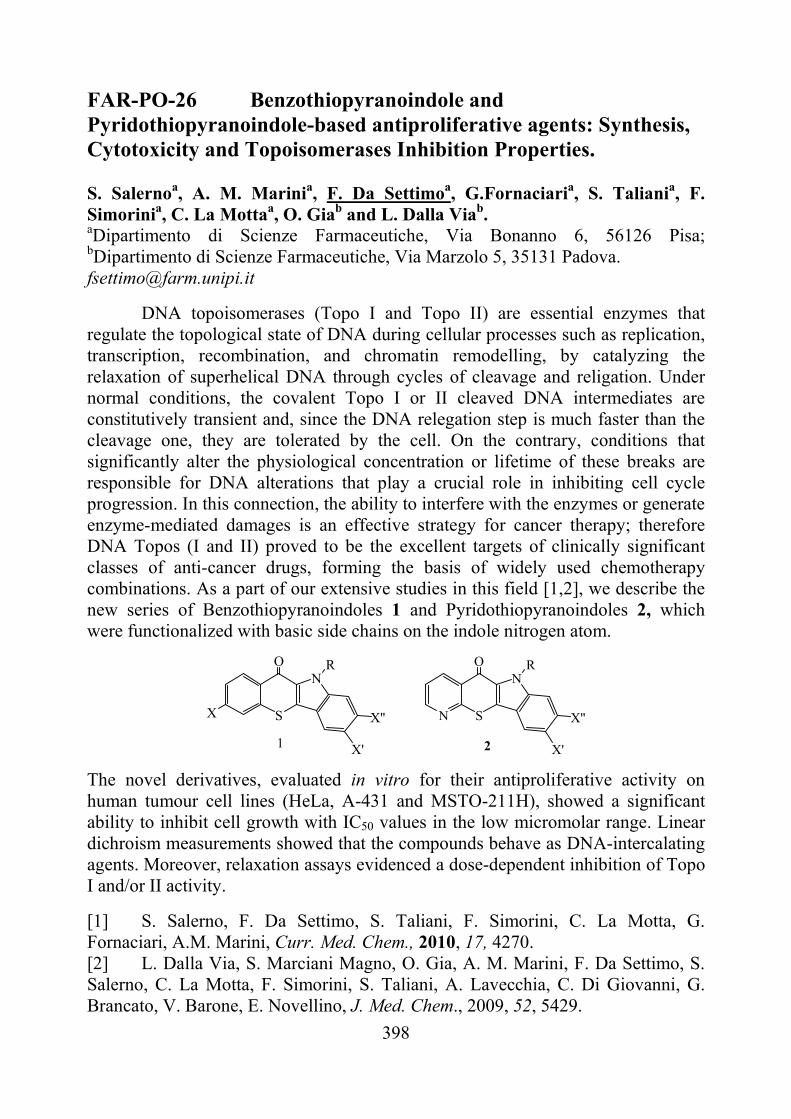
398
FAR-PO-26 Benzothiopyranoindole and Pyridothiopyranoindole-based antiproliferative agents: Synthesis, Cytotoxicity and Topoisomerases Inhibition Properties. S. Salernoa, A. M. Marinia, F. Da Settimoa, G.Fornaciaria, S. Taliania, F. Simorinia, C. La Mottaa, O. Giab and L. Dalla Viab. aDipartimento di Scienze Farmaceutiche, Via Bonanno 6, 56126 Pisa; bDipartimento di Scienze Farmaceutiche, Via Marzolo 5, 35131 Padova. [email protected]
DNA topoisomerases (Topo I and Topo II) are essential enzymes that regulate the topological state of DNA during cellular processes such as replication, transcription, recombination, and chromatin remodelling, by catalyzing the relaxation of superhelical DNA through cycles of cleavage and religation. Under normal conditions, the covalent Topo I or II cleaved DNA intermediates are constitutively transient and, since the DNA relegation step is much faster than the cleavage one, they are tolerated by the cell. On the contrary, conditions that significantly alter the physiological concentration or lifetime of these breaks are responsible for DNA alterations that play a crucial role in inhibiting cell cycle progression. In this connection, the ability to interfere with the enzymes or generate enzyme-mediated damages is an effective strategy for cancer therapy; therefore DNA Topos (I and II) proved to be the excellent targets of clinically significant classes of anti-cancer drugs, forming the basis of widely used chemotherapy combinations. As a part of our extensive studies in this field [1,2], we describe the new series of Benzothiopyranoindoles 1 and Pyridothiopyranoindoles 2, which were functionalized with basic side chains on the indole nitrogen atom.
N S
ON
X'
R
X''S
O
X
N
X'
R
X''
1 2
The novel derivatives, evaluated in vitro for their antiproliferative activity on human tumour cell lines (HeLa, A-431 and MSTO-211H), showed a significant ability to inhibit cell growth with IC50 values in the low micromolar range. Linear dichroism measurements showed that the compounds behave as DNA-intercalating agents. Moreover, relaxation assays evidenced a dose-dependent inhibition of Topo I and/or II activity.
[1] S. Salerno, F. Da Settimo, S. Taliani, F. Simorini, C. La Motta, G. Fornaciari, A.M. Marini, Curr. Med. Chem., 2010, 17, 4270. [2] L. Dalla Via, S. Marciani Magno, O. Gia, A. M. Marini, F. Da Settimo, S. Salerno, C. La Motta, F. Simorini, S. Taliani, A. Lavecchia, C. Di Giovanni, G. Brancato, V. Barone, E. Novellino, J. Med. Chem., 2009, 52, 5429.
399
FAR-PO-27 Refinement of the Translocator Protein Pharmacophore/Receptor Model via Structure-activity Relationships of Novel N,N-Dialkylindolylglyoxylamides S. Taliani,a E. Barresi,a I. Pugliesi,a F. Simorini,a C. La Motta,a S. Salerno,a A.M. Marini,a F. Da Settimo,a B. Cosimelli,c G. Greco,c E. Novellino,c E. Da Pozzo,b B. Costa,b C. Martini.b aDip. di Scienze Farmaceutiche, cDip. di Psichiatria, Neurobiologia, Farmacologia e Biotecnologia, via Bonanno, 6, Università di Pisa, 56126 Pisa. bDip. di Chimica Farmaceutica e Tossicologica, Università di Napoli “Federico II”, via D. Montesano, 49, 80131 Napoli. [email protected] The Translocator Protein (18 kDa) (TSPO), is widely expressed in glial cells and in peripheral tissues, and is involved in a variety of biological processes (steroidogenesis, cell growth and differentiation, apoptosis induction...) [1,2] TSPO basal expression is alterated in several disorders, including a variety of tumours, neuropathologies and neuroinflammation, anxiety and mood disorders. [1,2] In last years, we have described potent and selective TSPO ligands, the N,N-dialkyl-(2-phenylindol-3-yl)glyoxylamides I, [3,4] which allowed the development of newfluorescent or radio-labeled probes targeting this protein [5-7]. In this study, we reported a novel series of indolglyoxylamides, designed to investigate the binding role of the lipophilic pocket L1 of the hypothesized pharmacophore/receptor model.[1,2] Specifically, compounds featuring a bulky biphenyl II or 2-napthyl III group, phenyl with hydrophilic substituents IV, and fur-2-yl or thien-3-yl rings V at the 2-position of the indole nucleus, were synthesized and biologically evaluated. [1] Papadopoulos, V. et al. Trends Pharmacol. Sci., 27, 2006, 402. [2] Casellas, P. et al. Neurochem. Int., 40, 2002, 475. [3] Primofiore, G. et al. J. Med. Chem., 47, 2004, 1852. [4] Da Settimo. F. et al. J. Med. Chem., 51, 2008, 5798. [5] Taliani, S. et al. J. Med. Chem., 50, 2007, 404. [6] Taliani, S. et al. J. Med. Chem., 53, 2010, 4085. [7] Pike, V.W. et al. J. Med. Chem., 54, 2011, 366.
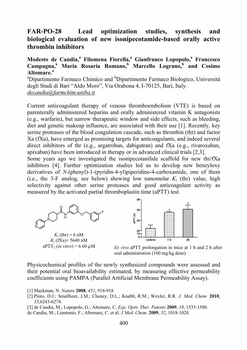
400
Ex vivo aPTT prolongation in mice at 1 h and 2 h after oral administration (100 mg/kg dose).
FAR-PO-28 Lead optimization studies, synthesis and biological evaluation of new isonipecotamide-based orally active thrombin inhibitors Modesto de Candia,a Filomena Fiorella,a Gianfranco Lopopolo,a Francesco Campagna,a Maria Rosaria Romano,b Marcello Lograno,b and Cosimo Altomare.a aDipartimento Farmaco Chimico and bDipartimento Farmaco Biologico, Università degli Studi di Bari “Aldo Moro”, Via Orabona 4, I-70125, Bari, Italy. [email protected] Current anticoagulant therapy of venous thromboembolism (VTE) is based on parenterally administered heparins and orally administered vitamin K antagonists (e.g., warfarin), but narrow therapeutic window and side effects, such as bleeding, diet and genetic makeup influence, are associated with their use [1]. Recently, key serine proteases of the blood coagulation cascade, such as thrombin (thr) and factor Xa (fXa), have emerged as promising targets for anticoagulants, and indeed several direct inhibitors of thr (e.g., argatroban, dabigatran) and fXa (e.g., rivaroxaban, apixaban) have been introduced in therapy or in advanced clinical trials [2,3]. Some years ago we investigated the isonipecotanilide scaffold for new thr/fXa inhibitors [4]. Further optimization studies led us to develop new benzyloxy derivatives of N-(phenyl)-1-(pyridin-4-yl)piperidine-4-carboxamide, one of them (i.e., the 3-F analog, see below) showing low nanomolar Ki (thr) value, high selectivity against other serine proteases and good anticoagulant activity as measured by the activated partial thromboplastin time (aPTT) test. Physicochemical profiles of the newly synthesized compounds were assessed and their potential oral bioavailability estimated, by measuring effective permeability coefficients using PAMPA (Parallel Artificial Membrane Permeability Assay). [1] Mackman, N. Nature 2008, 451, 914-918. [2] Pinto, D.J.; Smallheer, J.M.; Cheney, D.L.; Knabb, R.M.; Wexler, R.R. J. Med. Chem. 2010,
53,6243-6274. [3] de Candia, M.; Lopopolo, G.; Altomare, C. Exp. Opin. Ther. Patents 2009, 19, 1535-1580. de Candia, M.; Liantonio, F.; Altomare, C. et al. J Med. Chem. 2009, 52, 1018-1028.
Ki (thr) = 6 nM Ki (fXa)= 5640 nM
aPTT2 (in vitro) = 6.60 M
N
N
NH
O
OF
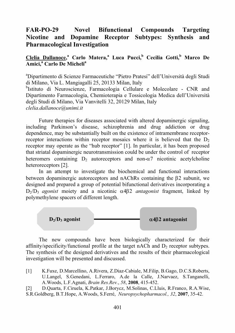
401
FAR-PO-29 Novel Bifunctional Compounds Targeting Nicotine and Dopamine Receptor Subtypes: Synthesis and Pharmacological Investigation Clelia Dallanoce,a Carlo Matera,a Luca Pucci,b Cecilia Gotti,b Marco De Amici,a Carlo De Michelia aDipartimento di Scienze Farmaceutiche “Pietro Pratesi” dell’Università degli Studi di Milano, Via L. Mangiagalli 25, 20133 Milan, Italy bIstituto di Neuroscienze, Farmacologia Cellulare e Molecolare - CNR and Dipartimento Farmacologia, Chemioterapia e Tossicologia Medica dell’Università degli Studi di Milano, Via Vanvitelli 32, 20129 Milan, Italy [email protected]
Future therapies for diseases associated with altered dopaminergic signaling, including Parkinson’s disease, schizophrenia and drug addiction or drug dependence, may be substantially built on the existence of intramembrane receptor-receptor interactions within receptor mosaics where it is believed that the D2 receptor may operate as the “hub receptor” [1]. In particular, it has been proposed that striatal dopaminergic neurotransmission could be under the control of receptor heteromers containing D2 autoreceptors and non-7 nicotinic acetylcholine heteroreceptors [2].
In an attempt to investigate the biochemical and functional interactions between dopaminergic autoreceptors and nAChRs containing the 2 subunit, we designed and prepared a group of potential bifunctional derivatives incorporating a D2/D3 agonist moiety and a nicotinic 42 antagonist fragment, linked by polymethylene spacers of different length.
The new compounds have been biologically characterized for their affinity/specificity/functional profile at the target nACh and D2 receptor subtypes. The synthesis of the designed derivatives and the results of their pharmacological investigation will be presented and discussed. [1] K.Fuxe, D.Marcellino, A.Rivera, Z.Diaz-Cabiale, M.Filip, B.Gago, D.C.S.Roberts,
U.Langel, S.Genedani, L.Ferraro, A.de la Calle, J.Narvaez, S.Tanganelli, A.Woods, L.F.Agnati, Brain Res.Rev., 58, 2008, 415-452.
[2] D.Quarta, F.Ciruela, K.Patkar, J.Borycz, M.Solinas, C.Lluis, R.Franco, R.A.Wise, S.R.Goldberg, B.T.Hope, A.Woods, S.Ferré, Neuropsychopharmacol., 32, 2007, 35-42.
D2/D3 agonist 42 antagonist
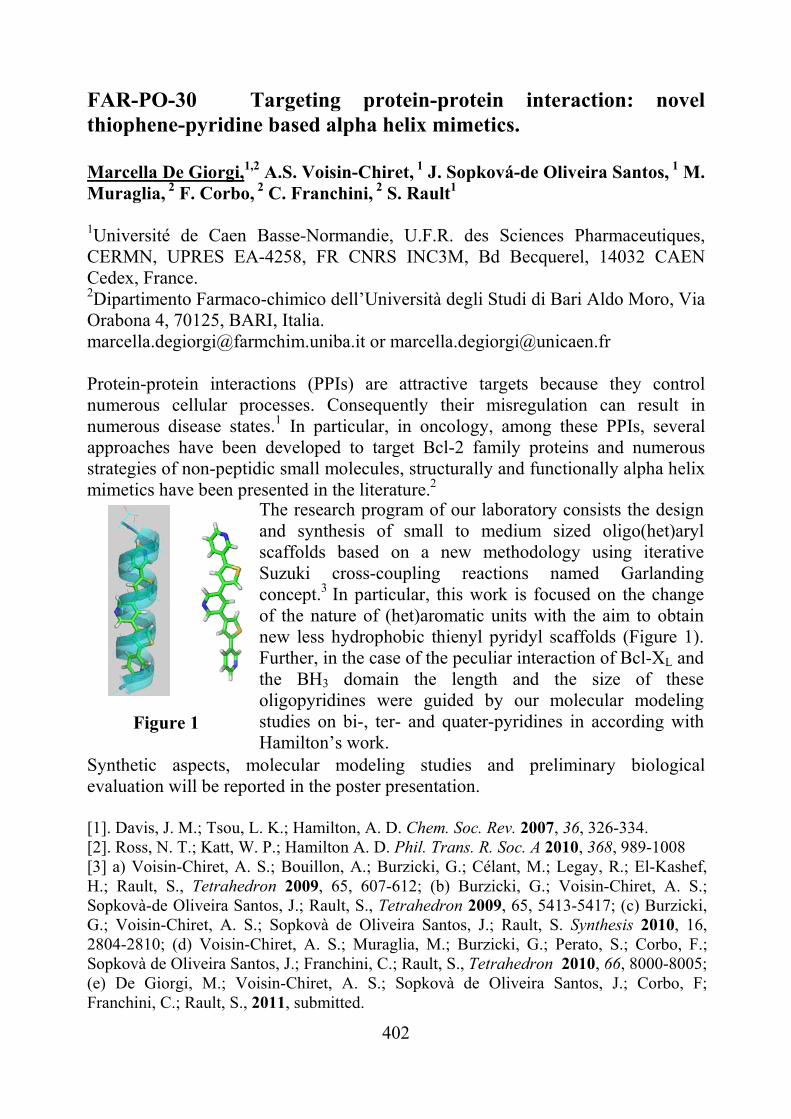
402
FAR-PO-30 Targeting protein-protein interaction: novel thiophene-pyridine based alpha helix mimetics. Marcella De Giorgi,1,2 A.S. Voisin-Chiret,
1 J. Sopková-de Oliveira Santos, 1 M.
Muraglia, 2 F. Corbo,
2 C. Franchini, 2 S. Rault1 1Université de Caen Basse-Normandie, U.F.R. des Sciences Pharmaceutiques, CERMN, UPRES EA-4258, FR CNRS INC3M, Bd Becquerel, 14032 CAEN Cedex, France. 2Dipartimento Farmaco-chimico dell’Università degli Studi di Bari Aldo Moro, Via Orabona 4, 70125, BARI, Italia. [email protected] or [email protected] Protein-protein interactions (PPIs) are attractive targets because they control numerous cellular processes. Consequently their misregulation can result in numerous disease states.1 In particular, in oncology, among these PPIs, several approaches have been developed to target Bcl-2 family proteins and numerous strategies of non-peptidic small molecules, structurally and functionally alpha helix mimetics have been presented in the literature.2 Figure 1 Synthetic aspects, molecular modeling studies and preliminary biological evaluation will be reported in the poster presentation. [1]. Davis, J. M.; Tsou, L. K.; Hamilton, A. D. Chem. Soc. Rev. 2007, 36, 326-334. [2]. Ross, N. T.; Katt, W. P.; Hamilton A. D. Phil. Trans. R. Soc. A 2010, 368, 989-1008 [3] a) Voisin-Chiret, A. S.; Bouillon, A.; Burzicki, G.; Célant, M.; Legay, R.; El-Kashef, H.; Rault, S., Tetrahedron 2009, 65, 607-612; (b) Burzicki, G.; Voisin-Chiret, A. S.; Sopkovà-de Oliveira Santos, J.; Rault, S., Tetrahedron 2009, 65, 5413-5417; (c) Burzicki, G.; Voisin-Chiret, A. S.; Sopkovà de Oliveira Santos, J.; Rault, S. Synthesis 2010, 16, 2804-2810; (d) Voisin-Chiret, A. S.; Muraglia, M.; Burzicki, G.; Perato, S.; Corbo, F.; Sopkovà de Oliveira Santos, J.; Franchini, C.; Rault, S., Tetrahedron 2010, 66, 8000-8005; (e) De Giorgi, M.; Voisin-Chiret, A. S.; Sopkovà de Oliveira Santos, J.; Corbo, F; Franchini, C.; Rault, S., 2011, submitted.
The research program of our laboratory consists the design and synthesis of small to medium sized oligo(het)aryl scaffolds based on a new methodology using iterative Suzuki cross-coupling reactions named Garlanding concept.3 In particular, this work is focused on the change of the nature of (het)aromatic units with the aim to obtain new less hydrophobic thienyl pyridyl scaffolds (Figure 1). Further, in the case of the peculiar interaction of Bcl-XL and the BH3 domain the length and the size of these oligopyridines were guided by our molecular modeling studies on bi-, ter- and quater-pyridines in according with Hamilton’s work.

403
FAR-PO-31 3-Chloroethyl-pyrrolo[2,1-d][1,2,3,5]tetrazines: synthesis and antitumor activity
Patrizia Diana,a Anna Carbone,a Paola Barraja,a Alessandra Montalbano,a Daniela Vedaldi,b Alessia Salvador,b Francesco Dall'Acqua,b Paola Brun,c Girolamo Cirrincionea
aDipartimento STEMBIO, Università di Palermo, Via Archirafi 32, 90123, Palermo, Italy; bDipartimento di Scienze Farmaceutiche, Università di Padova, Via Marzolo 5, 3513, Padova, Italy; cDipartimento di Istologia, Microbiologia e Biotecnologie Mediche – Università di Padova, Via Gabelli 63, 35121, Padova, Italy [email protected]
Temozolomide is a 3-methyl-imidazotetrazine currently in the market with trade name Temodal® and is used in the treatment of melanoma, mycosis fungoides, and brain tumors [1-4]. Temozolomide itself is a prodrug, spontaneously hydrolyzing at physiologic pH into MTIC (3-methyl-(triazen-1-yl)imidazole-4-carboxamide) and a methyldiazonium ion. This latter acts as a DNA methylating agent mainly at the N7 position of guanine [5-7]. We synthesized pyrrolo[2,1-d][1,2,3,5]tetrazinones that hold the deaza skeleton of temozolomide. Surprisingly the 3-methyl derivatives were the least active whereas the 3-aryl compounds had remarkable antineoplastic activity having GI50 values from the low micromolar-nanomolar range. Pyrrolotetrazinones showed a mechanism of action different from that observed by temozolomide. Infact they inhibit microtubule polymerization, induce G2/M arrest of cell cycle and cause apoptosis through the mitochondrial pathway. Considering the previous results we synthesised new 3-chloroethyl-pyrrolotetrazinones to verify whether the mode of action would resemble to that of temozolomide or to that of the deaza-analogues. Cytotoxicity was evaluated 72 h after incubation with compounds in several human cell lines. Generally compounds were cytotoxic in Jurkat cells, whereas one of them presents an antiproliferative activity in all cell lines with GI50 20-0.6 μM. Moreover, since the resistance onset is one of the main problems for classical anticancer drugs, the cytotoxicity of this latter compound was also studied in a P-glycoprotein over-expressing cell line and its antiproliferative effect was maintained. We also performed cell cycle analysis to investigate the kind of cellular death (necrosis or apoptosis) and it resulted that compounds induced cell death by apoptosis. However, no clear block in some specific phase was detected. Other studies were also carried out to check the involvement of mitochondria in the apoptotic process after JC-1 cell staining. Other experiments to identify their mechanism of action are now in the pipeline; in particular since temozolomide is a DNA alkylator, DNA will be considered as the possible target for these new compounds. [1] Bleehen NM, et al J Clin Oncol 1995, 13:910–913; [2] O’Reilly SM, et al Eur J Cancer 1993, 29A:940–942; [3] Newlands ES, et al Br J Cancer 1992, 65:287–291; [4] Newlands ES, et al Cancer Treat Rep 1997, 23:35–61; [5] Catapano CV, et al Cancer Res 1987, 47:4884–4889; [6] Denny BJ, et al Biochem.1994, 33:9045–9051; [7] Curtin NJ, et al A Clin Cancer Res 2004, 10:881–889.
404
FAR-PO-32 Synthesis, Biological Evaluation, and Pharmacokinetic Profile of 1,5-Diarylpyrrole-3-Propoxyethyl Ethers as Selective Cyclooxygenase-2 Inhibitors Endowed with Anti-inflammatory and Antinociceptive Activity Angela Di Capua,a* Michele Rovini,a Giulia Ferretti,a Andrea Cappelli,a Lidia Sautebin,b Carla Ghelardini,c Paola Patrignani,d Antonio Giordani,e Mariangela Biava,f and Maurizio Anzini.a
aUniversità degli Studi di Siena, Via A. Moro, 53100 Siena; bUniversità di Napoli “Federico II”, Via D. Montesano 49, I-80131 Napoli; cUniversità di Firenze, Viale G. Pieraccini 6, I-50139 Firenze; dUniversità di Chieti “G. D’Annunzio” e CeSI, Via dei Vestini 31, 66013 Chieti; eRottapharm Madaus, Via Valosa di Sopra 7, 20052 Monza; fUniversità degli Studi di Roma,“La Sapienza”, P.le A. Moro 5, I-00185 Roma *e-mail: [email protected] A new generation of selective cyclooxygenase-2 (COX-2) inhibitors (coxibs) was developed to circumvent the major side effects of cyclooxygenase-1 (COX-1) and COX-2 inhibitors (stomach ulceration and nephrotoxicity).1Structural features of known selective cyclooxygenase-2 inhibitors and of COX isozymes led our research group to synthesize the novel class of 1,5-diarylpyrrole derivatives.A series of previously patented 1,5-diarylpyrrol-3-acetic esters and 1,5-diarylpyrrole-3-alkoxyethyl ethers proved to be potent and selective COX-2 inhibitors in in vitro cell culture assay.1,2 The potential anti-inflammatory and antinociceptive activities of these compounds were evaluated in vivo, where they showed a very good activity against both carrageenan-induced hyperalgesia and edema in the rat paw test. In particular, compounds 1 and 2 appear to be equipotent with rofecoxib. In the Human Whole Blood (HWB) test, compound 2 demonstrated to be as selective as valdecoxib. The potential anti-inflammatory and antinociceptive activities of compounds 1 and 2 were evaluated in vivo, where they showed a very good activity against both carrageenan induced hyperalgesia and carrageenan induced oedema in the rat paw, with a complete remission 1 hour after the administration.2,3
Compound 2 was then selected for a preliminary pharmacokinetic analysis in male Sprague−Dawley rats. Unfortunately, plasma levels of this compound showed a low oral bioavailability, probably due to a rapid first-pass metabolism (Fig. 1). So a new series of fluorinated derivatives, 4 and 5, was synthetized in order to obtain an improved parmacokinetic profile: substitution of the 4’-H by the fluorine atom can profoundly change the conformational preferences of a small molecule because of size and stereoelectronic effects4 along with the possibility for 4’-F to exert an advantageous metabolic obstruction.
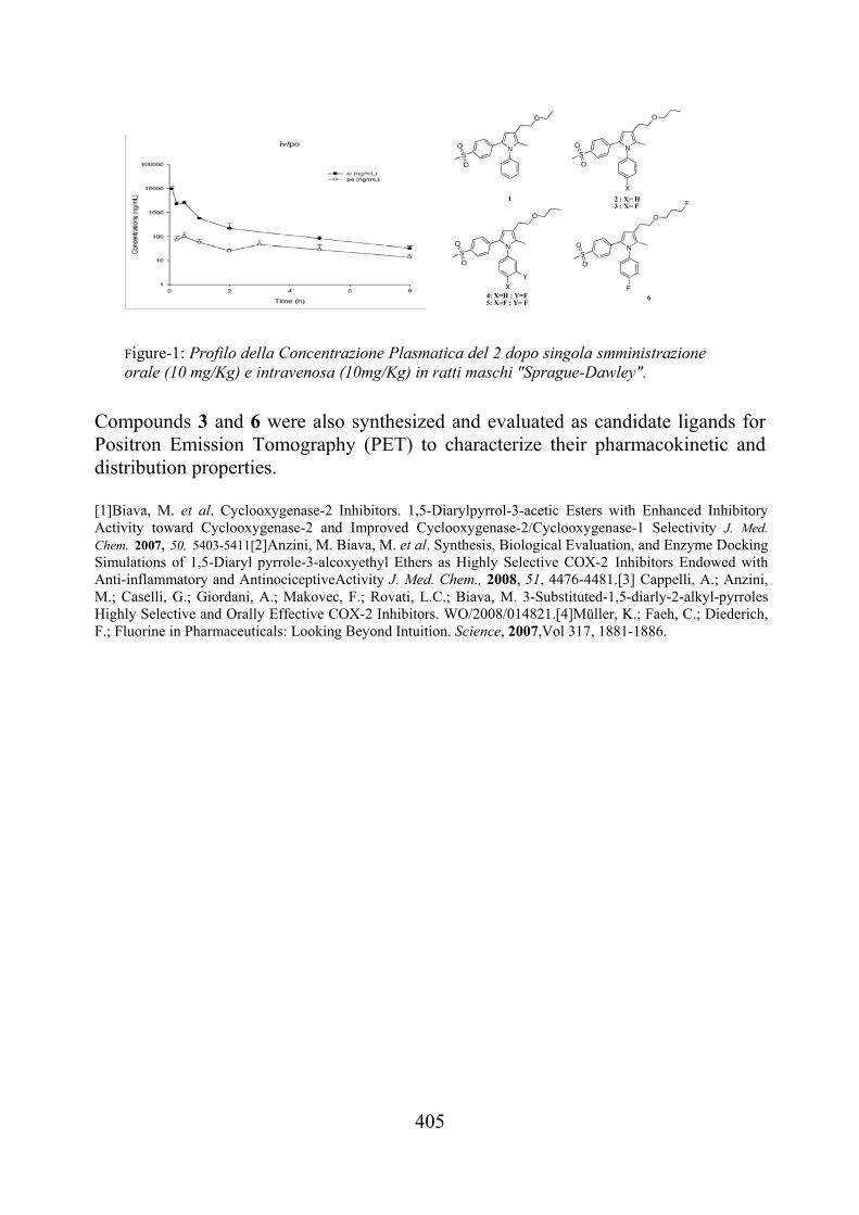
405
Compounds 3 and 6 were also synthesized and evaluated as candidate ligands for Positron Emission Tomography (PET) to characterize their pharmacokinetic and distribution properties. [1]Biava, M. et al. Cyclooxygenase-2 Inhibitors. 1,5-Diarylpyrrol-3-acetic Esters with Enhanced Inhibitory Activity toward Cyclooxygenase-2 and Improved Cyclooxygenase-2/Cyclooxygenase-1 Selectivity J. Med. Chem. 2007, 50, 5403-5411[2]Anzini, M. Biava, M. et al. Synthesis, Biological Evaluation, and Enzyme Docking Simulations of 1,5-Diaryl pyrrole-3-alcoxyethyl Ethers as Highly Selective COX-2 Inhibitors Endowed with Anti-inflammatory and AntinociceptiveActivity J. Med. Chem., 2008, 51, 4476-4481.[3] Cappelli, A.; Anzini, M.; Caselli, G.; Giordani, A.; Makovec, F.; Rovati, L.C.; Biava, M. 3-Substituted-1,5-diarly-2-alkyl-pyrroles Highly Selective and Orally Effective COX-2 Inhibitors. WO/2008/014821.[4]Müller, K.; Faeh, C.; Diederich, F.; Fluorine in Pharmaceuticals: Looking Beyond Intuition. Science, 2007,Vol 317, 1881-1886.
Figure-1: Profilo della Concentrazione Plasmatica del 2 dopo singola smministrazioneorale (10 mg/Kg) e intravenosa (10mg/Kg) in ratti maschi "Sprague-Dawley".
N
O
SO
O N
O
SO
O
N
O
S
O
O
X
X
Y
N
O
SO
O
F
F1 2 : X= H
3 : X= F
4: X=H ; Y=F5: X=F ; Y= F
6
406
FAR-PO-33 Elucidating the selective binding behaviour of a series of COXs inhibitors Francesco Saverio Di Leva,a Jagdish Suresh Patel,a Vittorio Limongelli,b,c Davide Branduardi,d Luciana Marinelli,b Ettore Novellino,b Michele Parrinello,c and Andrea Cavallia,e a Department of Drug Discovery and Development, Istituto Italiano di Tecnologia, Via Morego, 30, 16163 Genova, Italy b Dipartimento di Chimica Farmaceutica e Tossicologica, Università degli Studi di Napoli “Federico II”, Via D. Montesano 49, I-80131 Naples, Italy c Department of Chemistry and Applied Biosciences, ETH Zürich, USI Campus, Via G. Buffi 13, CH-6900 Lugano, Switzerland d Theoretical Molecular Biophysics Group, Max Planck Institute for Biophysics, Max-von-Laue-Straße 3, 60438 Frankfurt am Main, Germany e Department of Pharmaceutical Sciences, Alma Mater Studiorum – Bologna University, Via Belmeloro 6, 40126 Bologna, Italy [email protected]
Nonsteroidal antinflammatory drugs (NSAIDs) inhibit the cyclooxygenase enzymes (COXs), and are widely used for the treatment of inflammation, pain, and cancers. A selective inhibition of the COX-2 isoform is desirable, as this is overexpressed during inflammatory events.1 Therefore, many efforts have been directed towards the development of COX-2 selective inhibitors. Unfortunately, most of these inhibitors have been found to be highly cardiotoxic, as they produce a strong prostacyclin/thromboxane imbalance.2 Therefore, a deep understanding of the molecular bases of COXs selective inhibition is of great demand. Recently, we have successfully used metadynamics1 to study the binding behavior of a highly potent COX-2 selective inhibitor, SC-558, in both COX-1 and COX-2 isoforms.2 Following the line traced in our previous work, we have here extended the metadynamics studies on a series of potent COXs inhibitors endowed with different selectivity profiles. Our results provide useful computational tools to design small organic molecules with fine-tuned COX-1/COX-2 potency and selectivity. Furthermore, these results can be of paramount importance in the design of less toxic novel anti-inflammatory drug candidates.
[1] G. D. Anderson, S. D. Hauser, K. L. McGarity, M. E. Bremer, P. C. Isakson and S. A. Gregory, J. Clin. Invest., 97, 1996, 2672.
[2] L.-C. Chen and D. M Ashcroft, Pharmacoepidemiology and Drug Safety, 16, 2007, 762.
[3] A. Laio and M. Parrinello, Proc. Natl. Acad. Sci. U.S.A., 99, 2002, 12562. [4] V. Limongelli, M. Bonomi, L. Marinelli, F. L. Gervasio, A. Cavalli, E. Novellino and M. Parrinello, Proc. Natl. Acad. Sci. U.S.A., 107, 2010, 5411.
407
FAR-PO-34 5-Alkylamino-pyrazolo[4,3-e]1,2,4-triazolo[1,5-c]pyrimidines. Influence of the Substituent on the Affinity at the Adenosine Receptor Subtypes. Stephanie Federico,a Silvia Paoletta,b Siew Lee Cheong,c Barbara Cacciari,d Karl-Norbert Klotz,e Giorgia Pastorin,c Stefano Moro,b Giampiero Spalluto.a
aDipartimento di Scienze Chimiche e Farmaceutiche, Università di Trieste, Piazzale Europa 1, 34127 Trieste, Italy; bMolecular Modeling Section, Dipartimento di Scienze Farmaceutiche, Universita` di Padova, Via Marzolo 5, 35131 Padova, Italy; c Department of Pharmacy, National University of Singapore, 3 Science Drive 2,
Block S15, 05-PI-03, 117543 Singapore; dDipartimento di Scienze Farmaceutiche, Università di Ferrara, via Fossato di Mortara 17-19, I-44100 Ferrara, Italy; eInstitut für Pharmakologie, Universität of Würzburg, D-97078 Würzburg, Germany. [email protected]
It is well known that adenosine interacts with G-protein coupled receptors
that were classified in four receptors subtypes: A1, A2A, A2B and A3 adenosine receptors (AR). Adenosine receptor antagonists have several potential therapeutic applications. In particular, A1 antagonists are diuretic. Because antagonism towards A2A AR gave neuroprotection, antagonists for this receptor subtype are useful in neurodegenerative diseases, in particular for the treatment of Parkinson's disease. A2B AR antagonists could be used in asthma and diabetes, while A3 antagonism is useful in glaucoma, moreover, due to the fact that A3AR is widely expressed in some cancer cells, A3AR antagonists could be used for tumoral diagnosis.[1]
It was demonstrated by several studies that pyrazolo-triazolo-pyrimidine (PTP) core is a good template to obtain potent and selective adenosine receptor antagonists.[2] Until now all the PTP derivatives substituted at the 5 position bear phenylureidic or phenylacetamidic moieties, with a very poor variability.[3] So we have synthesized a novel class of 5-alkylamino-pyrazolo[4,3-e]1,2,4-triazolo[1,5-c]pyrimidines. This kind of substitution allows the introduction of several different amines, such as alkyl, cycloalkyl and benzyl amines, but also disubstituted-amines and hydroxylamines, which enabling the investigation of both the volume and nature of the receptor binding cavity.
Good results were obtained towards the A3 adenosine receptor subtype, in particular when a 5-bulky substituent, such as a benzhydrylamine, was present (KihA3=0.83 nM). Moreover a stereoselectivity by the binding pocket was observed for the derivatives substituted with a 1-phenyl-ethylamine, where the S isomer was 12-fold more potent than the R isomer at the A3 AR. [1] K.A.Jacobson, Z.-G.Gao. Nature Rev.DrugDisc., 5, 2006, 247. [2] S.Moro, Z.-G.Gao, K.A.Jacobson, G.Spalluto. Med.Res.Rev., 26, 2006, 131. [3] S.L.Cheong, G.Venkatesan, P.Paira, R.Jothibasu, A.L.Mandel, S.Federico, G.Spalluto, and G.Pastorin. Int.J.Med.Chem. In press: doi:10.1155/2011/480652
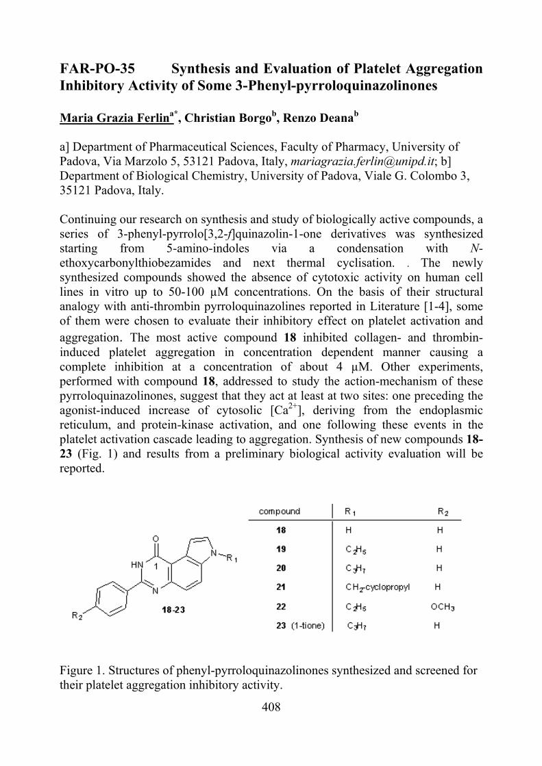
408
FAR-PO-35 Synthesis and Evaluation of Platelet Aggregation Inhibitory Activity of Some 3-Phenyl-pyrroloquinazolinones Maria Grazia Ferlina*, Christian Borgob, Renzo Deanab a] Department of Pharmaceutical Sciences, Faculty of Pharmacy, University of Padova, Via Marzolo 5, 53121 Padova, Italy, [email protected]; b] Department of Biological Chemistry, University of Padova, Viale G. Colombo 3, 35121 Padova, Italy. Continuing our research on synthesis and study of biologically active compounds, a series of 3-phenyl-pyrrolo[3,2-f]quinazolin-1-one derivatives was synthesized starting from 5-amino-indoles via a condensation with N-ethoxycarbonylthiobezamides and next thermal cyclisation. . The newly synthesized compounds showed the absence of cytotoxic activity on human cell lines in vitro up to 50-100 µM concentrations. On the basis of their structural analogy with anti-thrombin pyrroloquinazolines reported in Literature [1-4], some of them were chosen to evaluate their inhibitory effect on platelet activation and aggregation. The most active compound 18 inhibited collagen- and thrombin-induced platelet aggregation in concentration dependent manner causing a complete inhibition at a concentration of about 4 µM. Other experiments, performed with compound 18, addressed to study the action-mechanism of these pyrroloquinazolinones, suggest that they act at least at two sites: one preceding the agonist-induced increase of cytosolic [Ca2+], deriving from the endoplasmic reticulum, and protein-kinase activation, and one following these events in the platelet activation cascade leading to aggregation. Synthesis of new compounds 18-23 (Fig. 1) and results from a preliminary biological activity evaluation will be reported.
Figure 1. Structures of phenyl-pyrroloquinazolinones synthesized and screened for their platelet aggregation inhibitory activity.
409
[1] Dixit, A., Kashaw, S.K., Gaur, S., Saxena, A.K. Bioorg. Med. Chem., 2004, 12, 3591-3598. [2] Ahn H.S., Arik L., Boykow G., Burnett D.A., Caplen M.A., Czarniecki M., Domalski M.S., Foster C., Manna M., Stamford A.W., Wu Y. Bioorg. Med. Chem. Lett. 1999, 9, 2073-2078. [3] Huang, L-J.; Hsieh, M-C.; Teng, C-M.; Lee, K-H.; Kuo, S-C. Bioorg. Med. Chem. 1998, 6, 1657-1662. [4] Verstraete M., Zoldhely P. Drugs 1995; 49, 856–884.
410
FAR-PO-36 NO-Donor Carnosine Derivatives As Potential Neuroprotective Agents Montanaro G.,a Bertinaria M.,a Giorgis M.,a Rolando B.,a Fruttero R.,a Gasco A.a aDipartimento di Scienza e Tecnologia del Farmaco dell’Università degli Studi di Torino, via P. Giuria 9, 10125, Torino, Italy. [email protected]
Ageing affects many cellular processes which favor chronic
neurodegenerative diseases, among them Alzheimer’s disease (AD). AD is a complex pathology which involves more than 20 million individuals in the world.[1] AD is characterized by memory loss and other cognitive deficiencies, by the progressive dissolution of the personality as well as of the intellectual capacities. AD is accompanied by damages to brain capillaries, impairment of cholinergic transmission in both the hippocampus and the cerebral cortex as well as by extracellular accumulation of -amyloid (A plaques and intracellular tau protein aggregates in different area of the brain. In addition, it has been shown that oxidative stress, related to an abnormal production of reactive oxygen species (ROS) and nitrogen species (RNS) and/or depletion of the antioxidant defences, is an important early event in AD.[2] An increasing amount of experimental data suggests that AD is a cerebral microvascular disorder with neurodegenerative consequences, rather than the opposite. Microvascular disorders occurring in advanced ageing or in the presence of increased oxidative stress are consequent to the endothelial dysfunction, namely to a reduced capacity of the endothelial cells to produce nitric oxide (NO).[3] On this basis we designed new “multifunctional” molecules potentially useful in modifying the development and the progress of AD. These products were obtained by combining the structure of L-carnosine, a natural dipeptide endowed with a complex and multifactorial antioxidant action, with nitrooxy NO-donor moieties. These two pharmacophores were joined through an amide bond which, in a previous work, allowed us to obtain compounds stable in human serum, overcoming the major drawback of carnosine, while preserving its beneficial properties.[4] Synthesis, serum stability, vasodilating properties, antioxidant activity, ability to scavenge reactive carbonyl species (RCS) as well as copper (II) chelating properties of the obtained models are reported. [1] C.P. Ferri et al., Lancet, 366, 2005, 2112. [2] A. Nunomura, et al., J. Neuropath. Exp. Neur., 65, 2006, 631. [3] J.C. de la Torre, G.B. Stefano, in Biomedical Significance of Nitric Oxide. G.B. Stefano, Ed. Medical Science International: Warsaw-New York, 2003, 213. [4] M. Bertinaria et al. J. Med. Chem., 54, 2011, 611.
411
FAR-PO-37 Eremurus persicus Boiss: ethnobotanical relevance, analytical fingerprint and preliminary biological results of root extracts Karzan Mahmooda,1, Raffaella Gaggeria,1, Cinzia Bosellia,2, Barbara Mannuccib, Giovanni Vidaric,d, Simona Collinaa,1,d, Daniela Rossia,1
a Dipartimento di Scienze del Farmaco, Università degli Studi di Pavia, Via Taramelli 12, 27100, Pavia, Italy, 1: Sezione di Chimica Farmaceutica, 2: Sezione di Farmacologia; bCentro Grandi Strumenti (CGS), Università degli Studi di Pavia, Via Bassi 21, 27100, Pavia, Italy; cDipartimento di Chimica, Università degli Studi di Pavia, Via Taramelli 12, 27100, Pavia, Italy; dCentro Interdipartimentale di Studi e Ricerche di Etnobiofarmacia (C.I.St.R.E.), Università degli Studi di Pavia, Via Taramelli 12, 27100, Pavia, Italy. [email protected]
Recently, the drug discovery process has been frequently focused on the screening of plant extracts commonly used in traditional medicines as source of novel therapeutic agents. In this context, the object of the present study was to evaluate the folk claims of Eremurus persicus. Genus Eremurus (Asphodelaceae), with its 45 species, is distributed over large area in Central Asia [1]. Eremurus persicus (Jaub. & Spach) Boiss is a medicinal plant of Kurdistan; its root extracts are used by native people as folk remedies for managing wounds, inflammatory and skin diseases. Basing on our previous experience [2] several extracts were prepared by varying either the solvent mixture or the extraction procedure. All prepared extracts were tested in vitro to evaluate anti-tyrosinase and free radical scavenging properties, since these are relevant bioactivities related to skin disorders and inflammation [3]. The ethanolic extract obtained from maceration of plant material pre-treated with petroleum ether gave rise to the most interesting antiradical and antityrosinase effect, therefore it has been selected as the hit extract. The phytochemical fingerprint of the hit extract was investigated by HPLC-UV-PAD coupled by either CD or MS detector and herein presented together with the preliminary biological results reached till now. [1] K. N. Safar et al, Iran. J. Bot., 15, 2009 [2] R. Gaggeri et al, The Open Natural Products Journal, 3, 2010 [3] I. Süntar et al, J. of Ethnopharmacol., 135, 2011 The Authors would like to thank Regione Lombardia (PROKURDUP Project) for financial support
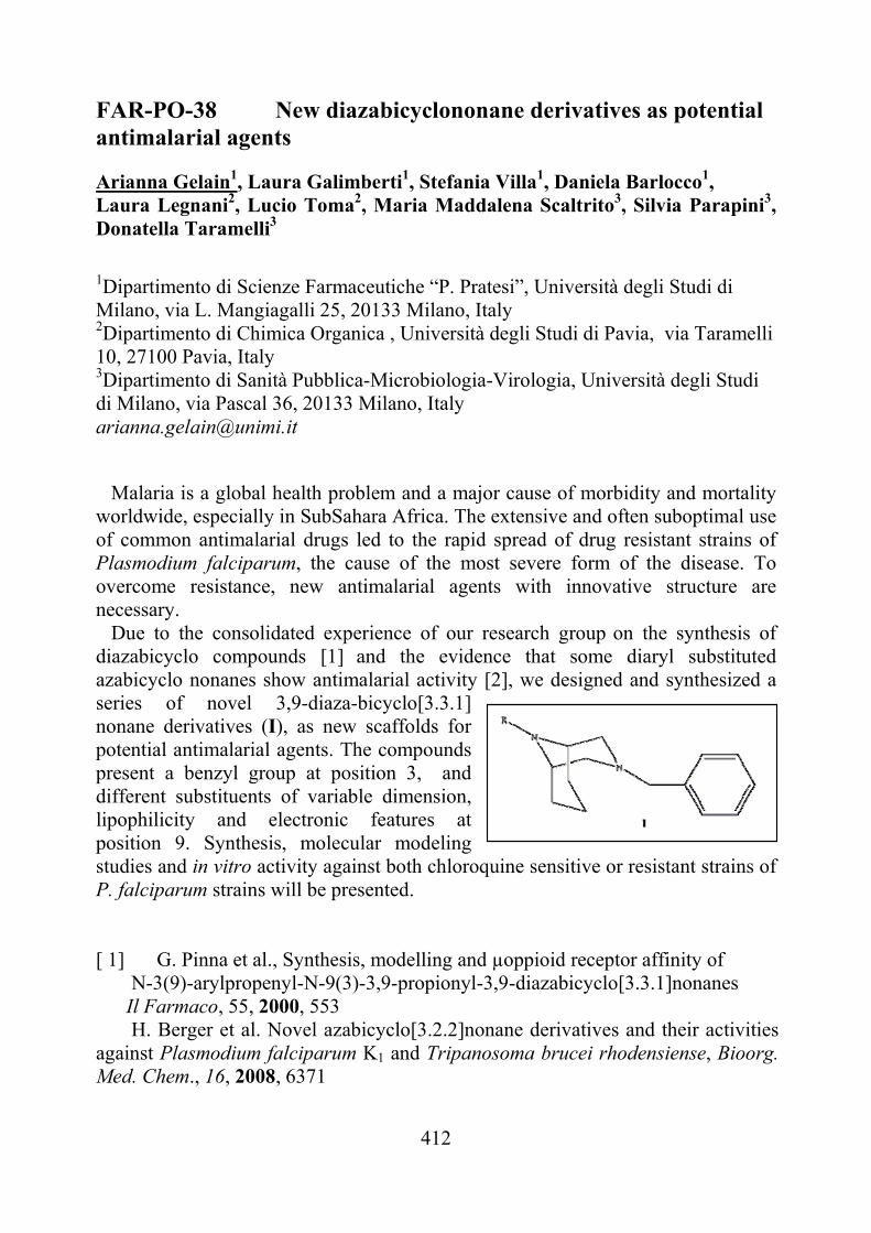
412
FAR-PO-38 New diazabicyclononane derivatives as potential antimalarial agents Arianna Gelain1, Laura Galimberti1, Stefania Villa1, Daniela Barlocco1, Laura Legnani2, Lucio Toma2, Maria Maddalena Scaltrito3, Silvia Parapini3, Donatella Taramelli3 1Dipartimento di Scienze Farmaceutiche “P. Pratesi”, Università degli Studi di Milano, via L. Mangiagalli 25, 20133 Milano, Italy 2Dipartimento di Chimica Organica , Università degli Studi di Pavia, via Taramelli 10, 27100 Pavia, Italy 3Dipartimento di Sanità Pubblica-Microbiologia-Virologia, Università degli Studi di Milano, via Pascal 36, 20133 Milano, Italy [email protected] Malaria is a global health problem and a major cause of morbidity and mortality worldwide, especially in SubSahara Africa. The extensive and often suboptimal use of common antimalarial drugs led to the rapid spread of drug resistant strains of Plasmodium falciparum, the cause of the most severe form of the disease. To overcome resistance, new antimalarial agents with innovative structure are necessary. Due to the consolidated experience of our research group on the synthesis of diazabicyclo compounds [1] and the evidence that some diaryl substituted azabicyclo nonanes show antimalarial activity [2], we designed and synthesized a series of novel 3,9-diaza-bicyclo[3.3.1] nonane derivatives (I), as new scaffolds for potential antimalarial agents. The compounds present a benzyl group at position 3, and different substituents of variable dimension, lipophilicity and electronic features at position 9. Synthesis, molecular modeling studies and in vitro activity against both chloroquine sensitive or resistant strains of P. falciparum strains will be presented. [ 1] G. Pinna et al., Synthesis, modelling and µoppioid receptor affinity of N-3(9)-arylpropenyl-N-9(3)-3,9-propionyl-3,9-diazabicyclo[3.3.1]nonanes Il Farmaco, 55, 2000, 553 H. Berger et al. Novel azabicyclo[3.2.2]nonane derivatives and their activities against Plasmodium falciparum K1 and Tripanosoma brucei rhodensiense, Bioorg. Med. Chem., 16, 2008, 6371
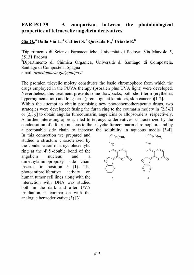
413
FAR-PO-39 A comparison between the photobiological properties of tetracyclic angelicin derivatives.
Gia O.,a Dalla Via L.,a Caffieri S. a Quezada E.,b Uriarte E.b
aDipartimento di Scienze Farmaceutiche, Università di Padova, Via Marzolo 5, 35131 Padova bDipartimento di Chimica Organica, Università di Santiago di Compostela, Santiago di Compostela, Spagna email: [email protected]
The psoralen tricyclic moiety constitutes the basic chromophore from which the drugs employed in the PUVA therapy (psoralen plus UVA light) were developed. Nevertheless, this treatment presents some drawbacks, both short-term (erythema, hyperpigmentation) and long-term (premalignant keratoses, skin cancers)[1-2]. Within the attempt to obtain promising new photochemotherapeutic drugs, two strategies were developed: fusing the furan ring to the coumarin moiety in [2,3-h] or [2,3-f] to obtain angular furocoumarin, angelicins or allopsoralens, respectively. A further interesting approach led to tetracyclic derivatives, characterized by the condensation of a fourth nucleus to the tricyclic furocoumarin chromophore and by a protonable side chain to increase the solubility in aqueous media [3-4].In this connection we prepared and studied a structure characterized by the condensation of a cyclohexenylic ring at the 4,5-double bond of the angelicin nucleus and a dimethylaminopropoxy side chain inserted in position 5 (1). The photoantiproliferative activity on human tumor cell lines along with the interaction with DNA was studied both in the dark and after UVA irradiation in comparison with the analogue benzoderivative (2) [3].
OO
O
N(Me)2
OOO
O
N(Me)2
O
1 2
414
The isolation and characterization of the furan side photoadduct with the pyrimidine base thymine is also reported. Finally, the ability to interact with topoisomerase II in the dark was investigated. [1] N. J. Lowe, V. Chizhevsky, and H. Gabriel, Clin. Dermatol., 15, 1997, 745. [2] R. S. Stern, J. Am. Acad. Dermatol., 44, 2001, 755. [3] L. Santana, E. Uriarte, L. Dalla Via, and O. Gia, Bioorg. Med. Chem. Lett., 10, 2000, 135. [4] L. Dalla Via, S. Mammi, E. Uriarte, L. Santana, I. Lampronti, R. Gambari, and O. Gia, J. Med. Chem., 49, 2006, 4317.
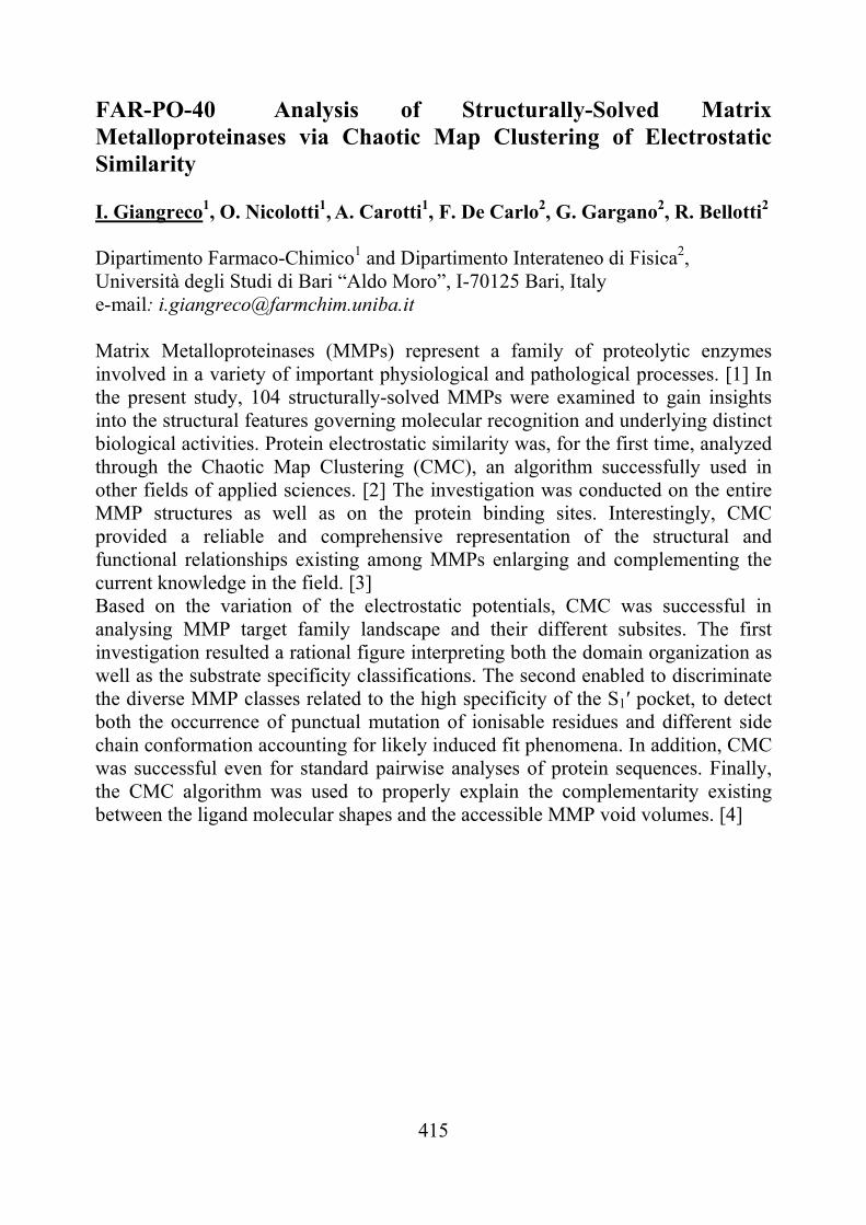
415
FAR-PO-40 Analysis of Structurally-Solved Matrix Metalloproteinases via Chaotic Map Clustering of Electrostatic Similarity I. Giangreco1, O. Nicolotti1, A. Carotti1, F. De Carlo2, G. Gargano2, R. Bellotti2 Dipartimento Farmaco-Chimico1 and Dipartimento Interateneo di Fisica2, Università degli Studi di Bari “Aldo Moro”, I-70125 Bari, Italy e-mail: [email protected] Matrix Metalloproteinases (MMPs) represent a family of proteolytic enzymes involved in a variety of important physiological and pathological processes. [1] In the present study, 104 structurally-solved MMPs were examined to gain insights into the structural features governing molecular recognition and underlying distinct biological activities. Protein electrostatic similarity was, for the first time, analyzed through the Chaotic Map Clustering (CMC), an algorithm successfully used in other fields of applied sciences. [2] The investigation was conducted on the entire MMP structures as well as on the protein binding sites. Interestingly, CMC provided a reliable and comprehensive representation of the structural and functional relationships existing among MMPs enlarging and complementing the current knowledge in the field. [3] Based on the variation of the electrostatic potentials, CMC was successful in analysing MMP target family landscape and their different subsites. The first investigation resulted a rational figure interpreting both the domain organization as well as the substrate specificity classifications. The second enabled to discriminate the diverse MMP classes related to the high specificity of the S1′ pocket, to detect both the occurrence of punctual mutation of ionisable residues and different side chain conformation accounting for likely induced fit phenomena. In addition, CMC was successful even for standard pairwise analyses of protein sequences. Finally, the CMC algorithm was used to properly explain the complementarity existing between the ligand molecular shapes and the accessible MMP void volumes. [4]
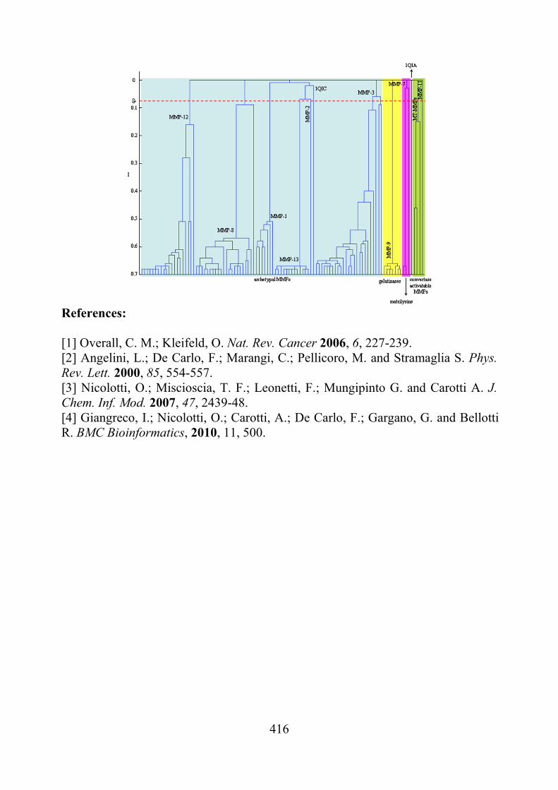
416
References: [1] Overall, C. M.; Kleifeld, O. Nat. Rev. Cancer 2006, 6, 227-239. [2] Angelini, L.; De Carlo, F.; Marangi, C.; Pellicoro, M. and Stramaglia S. Phys. Rev. Lett. 2000, 85, 554-557. [3] Nicolotti, O.; Miscioscia, T. F.; Leonetti, F.; Mungipinto G. and Carotti A. J. Chem. Inf. Mod. 2007, 47, 2439-48. [4] Giangreco, I.; Nicolotti, O.; Carotti, A.; De Carlo, F.; Gargano, G. and Bellotti R. BMC Bioinformatics, 2010, 11, 500.
417
FAR-PO-41 Allyphenyline Analogues Potentially Useful in the Management of Chronic Pain and Opioid Addiction. Eleonora Diamanti,a Fabio Del Bello,a Mario Giannella,a Valerio Mammoli,a Alessandro Piergentili,a Wilma Quaglia,a Antonio Carrieri,b Giuseppe Carbonara,b Giuseppe Fracchiolla,b and Maria Pigini.a
a Scuola di Scienze del Farmaco e dei Prodotti della Salute, Università, via S. Agostino 1, 62032, Camerino, Italy bDipartimento Farmacochimico, Università, via Orabona 4, 70125, Bari, Italy [email protected] We have previously demonstrated that 2-adrenergic (2-AR) molecules, related to the non-subtype selective antagonist 1, displayed different activity at 2A-, 2B- and 2C-AR subtypes, depending on the peculiar nature of the ortho substituent in the Ar ring. For example, phenyl and thienyl groups converted 1 into the efficacious agonists 2 (biphenyline) and 3, respectively, activating 2A- and 2C-subtypes. Allyl or cyclopropyl groups of lower steric bulk turned the biological profile of the antagonist 1 only at 2C-subtype: 4 (allyphenyline) and 5 were potent 2C-agonists, whereas efficiently antagonized 2A-AR. From in vivo study 4 significantly enhanced morphine analgesia (due to 2C-AR agonism), was devoid of sedative side effects (due to 2A-AR antagonism) as well as prevented and contrasted morphine tolerance and dependence at very low dose (0.05 mg/Kg) [1]. Encouraged by these results, we prepared and studied compounds 6-12 inspired by 4. They were characterized by ortho substituents of moderate steric bulk and positive or negative and contributions in all the combinations. A molecular modeling study performed on 2-12 highlighted that the desired 2C-agonism/2A-antagonism combination was displayed by ligands having favoured extended conformation (i.e. 4 and 5). In contrast, the activation of both 2A- and 2C-subtypes was produced by ligands endowed with favoured folded conformation (i.e. 2 and 3).
2 3 4 5
This work was supported by the Monte dei Paschi di Siena Foundation Award.
[1] F. Del Bello et al. J. Med. Chem. 53, 2010, 7825-7835 and references therein.
418
FAR-PO-42 Design, synthesis and biological evaluation of peptidomimetic boronic acids targeting 20S proteasome R. Ettari,a N. Micale,a C. Bonaccorso,aT. Schirmeister,b M. Zappalà,a S. Grassoa aDipartimento Farmaco-Chimico, Università di Messina, Viale Annunziata, 98168 Messina, Italy, bInstitute of Pharmacy and Food Chemistry, University of Würzburg, Am Hubland, D-97074 Würzburg, Germany E-mail: [email protected]
The ubiquitin-proteasome pathway is the major proteolytic system of all eukaryotic cells and plays a very important role in the degradation of many proteins critical for cell division, growth activation, signaling and transcription. A deregulation of this system could lead to an anarchic cell proliferation and to a tumor development [1]. For these reasons, ubiquitin/proteasome inhibition has become a new and significant strategy for the drug development in cancer treatment.
Within the present work, the strategy targeting proteasome has been delineated with the aim of developing efficacious antitumor agents. This approach was based on the design of conformationally constrained peptidomimetics by reducing the peptidic character of the tripeptide boronate Z-Leu-Leu-Leu-B(OH)2 (MG262, Ki=18 pM, Figure 1), reversible inhibitor of the chymotrypsin-like activity of the 20S proteasome. This strategy has been accomplished by bioisosteric replacement of the P3 Leu residue with a 2-pyridone nucleus [2]. This structural modification could ensure stability towards degradation by enzymes, enhancement of oral bioavailability, reduction of conformational freedom of peptides, improvement of the selectivity towards the target enzyme.
Further modifications of new conformationally constrained peptidomimetics involved the P2 site according to the features of the dipeptide boronate bortezomib (Velcade®, Figure 1), the first proteasome inhibitor approved by FDA for the treatment of haematological malignancies such as multiple myeloma and mantle cell lymphoma. In this context we introduced bulky substituents at the P2 site (i.e. Phe residue, Figure 1), whereas the P1 site was kept constant because of its importance for substrate specificity. Additional changes involving the N-terminal protective group have been realized in order to improve the binding properties of the new inhibitors (Figure 1).
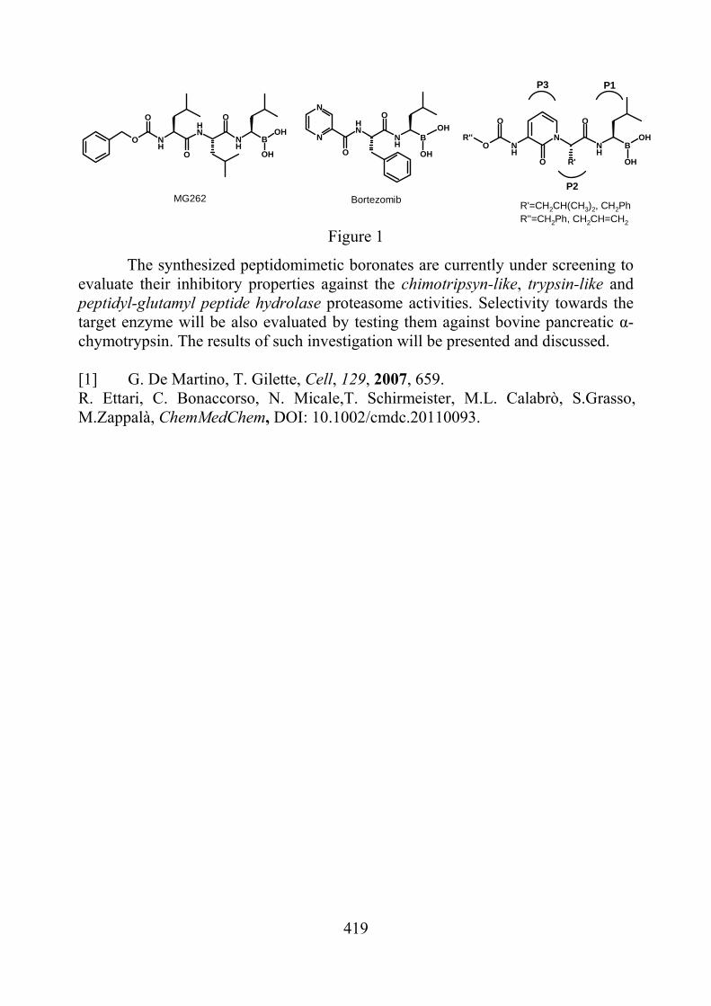
419
N
O
NH
R'
O
BNH
OR''
O
OH
OH
N
NNH
NH
BOH
O
O
OH
O NH
NH
NH
B
O
O
O
OH
OH
BortezomibMG262
P3
P2
P1
R'=CH2CH(CH3)2, CH2PhR''=CH2Ph, CH2CH=CH2
Figure 1
The synthesized peptidomimetic boronates are currently under screening to evaluate their inhibitory properties against the chimotripsyn-like, trypsin-like and peptidyl-glutamyl peptide hydrolase proteasome activities. Selectivity towards the target enzyme will be also evaluated by testing them against bovine pancreatic α-chymotrypsin. The results of such investigation will be presented and discussed. [1] G. De Martino, T. Gilette, Cell, 129, 2007, 659. R. Ettari, C. Bonaccorso, N. Micale,T. Schirmeister, M.L. Calabrò, S.Grasso, M.Zappalà, ChemMedChem, DOI: 10.1002/cmdc.20110093.
420
FAR-PO-43 Praziquantel analogues containing NO-donor furoxans and related furazans as agents active against Schistosoma mansoni. Francesca Vottero,a Stefano Guglielmo,a Roberta Fruttero,a Alberto Gasco,a Daniela Cortese,a,b Latasha Day,b Valerie Kommer,b and David L. Williams.b aDipartimento di Scienza e Tecnologia del Farmaco dell’Università degli Studi di Torino, Via P. Giuria 9, 10125, Turin, Italy b Department of Microbiology and Immunology, Rush University Medical Center, Chicago, IL, 60612, USA [email protected]
Today around 200 million people worldwide are parasitized by several species of Schistosoma and in particular S. mansoni, and more than 200 000 people die every year from this neglected disease. Praziquantel (PZQ) is the only drug currently administered. The mechanism of action of this drug has not been elucidated; however, inhibition of calcium channels and inhibition of adenosine uptake have been suggested to be involved in PZQ activity. Since PZQ-resistant worms have been identified, the development of new anti-schistosomiasis drugs is urgently needed [1].
Recently, several furoxans (1,2,5-oxadiazole 2-oxides) have been shown to be endowed with good activity against S. mansoni. The proposed mechanism of action consists in the inhibition of thioredoxin glutathione reductase (TGR), an essential enzyme for parasite redox balance, through nitrosylation of cysteine and/or selenocysteine residues of the protein. This reaction takes place as a consequence of the interaction of the compounds with the enzyme and subsequent NO release [2].
On this basis we decided to conjugate PZQ and furoxan pharmacophores in a single molecule in order to obtain dual drugs exploiting antischistosomal properties through two different mechanisms of action. Through slight modifications of an inexpensive and straightforward synthesis [3], six novel PZQ derivatives were prepared, three of them bearing furoxan moieties with the remainder bearing the corresponding furazan (1,2,5-oxadiazole) moieties and devoid of NO-donor properties.
All the compounds have been subjected to structural and pharmacological characterization for their activity against adult ex vivo worms and their capability to inhibit TGR activity. Moreover in vivo studies are in progress in order to evaluate whether the compounds are able to decrease worm burdens in infected mice. [1] A. Dömling and K. Khoury, ChemMedChem, 5, 2010, 1420. [2] A. Simeonov et al, PLoS NTDs, 2, 2008, e127; A. Sayed et al., Nature Med., 14, 2008, 407;
G. Rai et al, J. Med. Chem., 52, 2009, 6474. [3] J. H. Kim et al, Tetrahedron, 54, 1998, 7395.
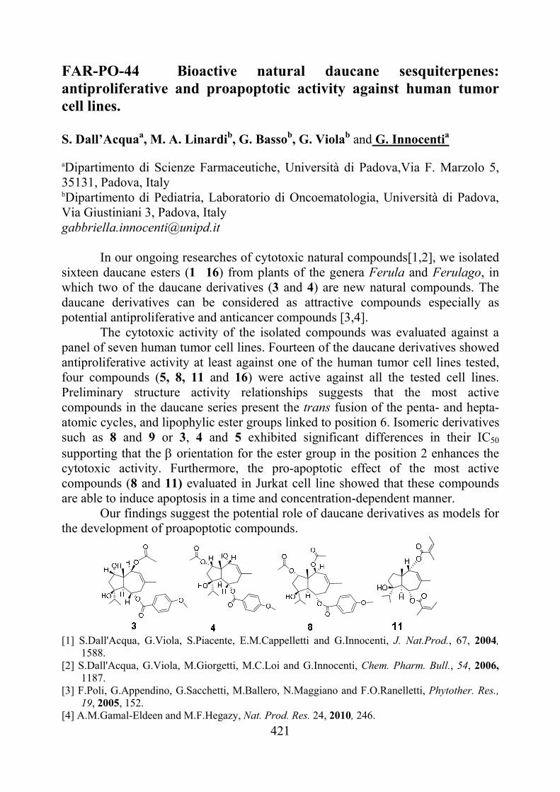
421
FAR-PO-44 Bioactive natural daucane sesquiterpenes: antiproliferative and proapoptotic activity against human tumor cell lines. S. Dall’Acquaa, M. A. Linardib, G. Bassob, G. Violab and G. Innocentia
aDipartimento di Scienze Farmaceutiche, Università di Padova,Via F. Marzolo 5, 35131, Padova, Italy bDipartimento di Pediatria, Laboratorio di Oncoematologia, Università di Padova, Via Giustiniani 3, Padova, Italy [email protected]
In our ongoing researches of cytotoxic natural compounds[1,2], we isolated
sixteen daucane esters (1�16) from plants of the genera Ferula and Ferulago, in which two of the daucane derivatives (3 and 4) are new natural compounds. The daucane derivatives can be considered as attractive compounds especially as potential antiproliferative and anticancer compounds [3,4].
The cytotoxic activity of the isolated compounds was evaluated against a panel of seven human tumor cell lines. Fourteen of the daucane derivatives showed antiproliferative activity at least against one of the human tumor cell lines tested, four compounds (5, 8, 11 and 16) were active against all the tested cell lines. Preliminary structure activity relationships suggests that the most active compounds in the daucane series present the trans fusion of the penta- and hepta- atomic cycles, and lipophylic ester groups linked to position 6. Isomeric derivatives such as 8 and 9 or 3, 4 and 5 exhibited significant differences in their IC50 supporting that the orientation for the ester group in the position 2 enhances the cytotoxic activity. Furthermore, the pro-apoptotic effect of the most active compounds (8 and 11) evaluated in Jurkat cell line showed that these compounds are able to induce apoptosis in a time and concentration-dependent manner.
Our findings suggest the potential role of daucane derivatives as models for the development of proapoptotic compounds.
[1] S.Dall'Acqua, G.Viola, S.Piacente, E.M.Cappelletti and G.Innocenti, J. Nat.Prod., 67, 2004,
1588. [2] S.Dall'Acqua, G.Viola, M.Giorgetti, M.C.Loi and G.Innocenti, Chem. Pharm. Bull., 54, 2006,
1187. [3] F.Poli, G.Appendino, G.Sacchetti, M.Ballero, N.Maggiano and F.O.Ranelletti, Phytother. Res.,
19, 2005, 152. [4] A.M.Gamal-Eldeen and M.F.Hegazy, Nat. Prod. Res. 24, 2010, 246.
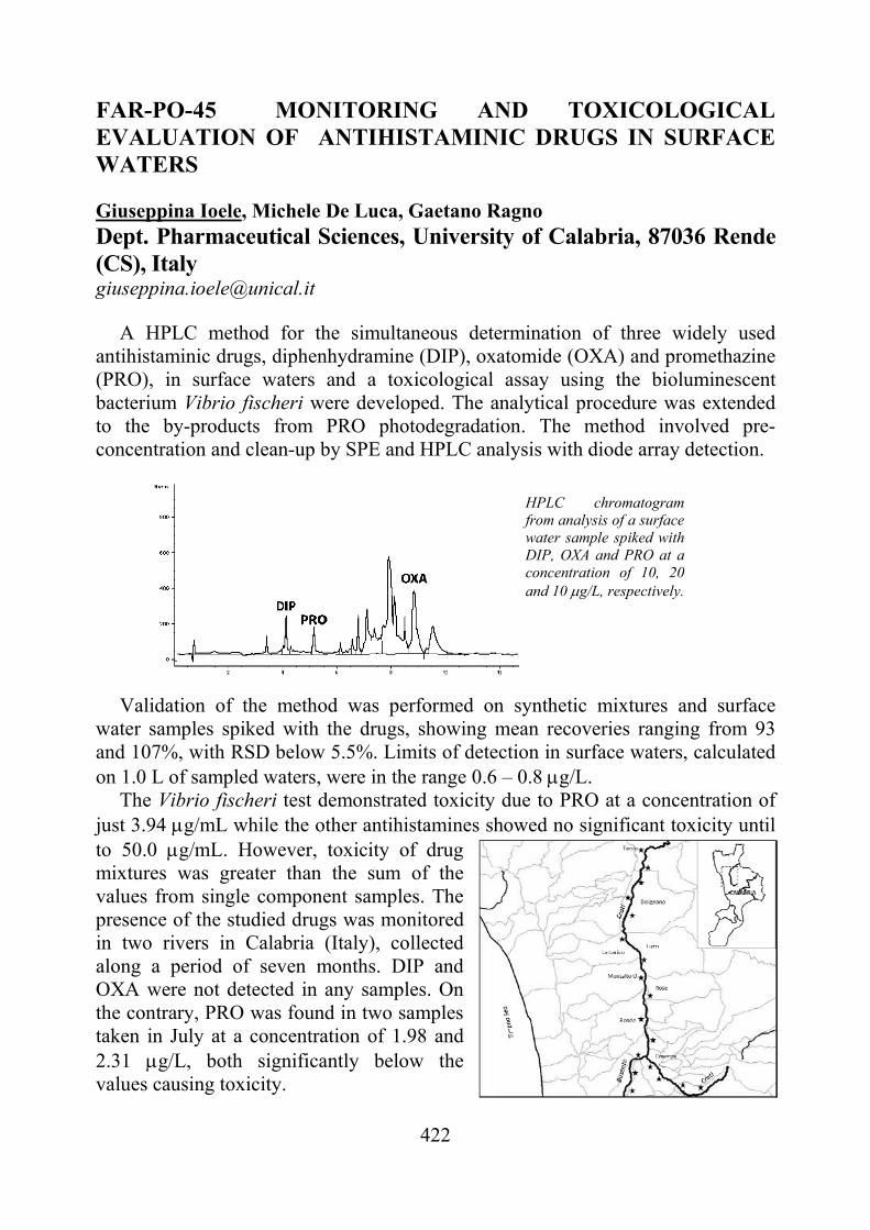
422
FAR-PO-45 MONITORING AND TOXICOLOGICAL EVALUATION OF ANTIHISTAMINIC DRUGS IN SURFACE WATERS
Giuseppina Ioele, Michele De Luca, Gaetano Ragno Dept. Pharmaceutical Sciences, University of Calabria, 87036 Rende (CS), Italy [email protected]
A HPLC method for the simultaneous determination of three widely used
antihistaminic drugs, diphenhydramine (DIP), oxatomide (OXA) and promethazine (PRO), in surface waters and a toxicological assay using the bioluminescent bacterium Vibrio fischeri were developed. The analytical procedure was extended to the by-products from PRO photodegradation. The method involved pre-concentration and clean-up by SPE and HPLC analysis with diode array detection.
Validation of the method was performed on synthetic mixtures and surface water samples spiked with the drugs, showing mean recoveries ranging from 93 and 107%, with RSD below 5.5%. Limits of detection in surface waters, calculated on 1.0 L of sampled waters, were in the range 0.6 – 0.8 g/L.
The Vibrio fischeri test demonstrated toxicity due to PRO at a concentration of just 3.94 g/mL while the other antihistamines showed no significant toxicity until to 50.0 g/mL. However, toxicity of drug mixtures was greater than the sum of the values from single component samples. The presence of the studied drugs was monitored in two rivers in Calabria (Italy), collected along a period of seven months. DIP and OXA were not detected in any samples. On the contrary, PRO was found in two samples taken in July at a concentration of 1.98 and 2.31 g/L, both significantly below the values causing toxicity.
HPLC chromatogram from analysis of a surface water sample spiked with DIP, OXA and PRO at a concentration of 10, 20 and 10 g/L, respectively.
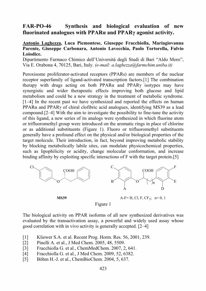
423
FAR-PO-46 Synthesis and biological evaluation of new fluorinated analogues with PPARα and PPARγ agonist activity. Antonio Laghezza, Luca Piemontese, Giuseppe Fracchiolla, Mariagiovanna Parente, Giuseppe Carbonara, Antonio Lavecchia, Paolo Tortorella, Fulvio Loiodice. Dipartimento Farmaco Chimico dell’Università degli Studi di Bari “Aldo Moro”, Via E. Orabona 4, 70125, Bari, Italy. (e-mail: [email protected])
Peroxisome proliferator-activated receptors (PPARs) are members of the nuclear receptor superfamily of ligand-activated transcription factors.[1] The combination therapy with drugs acting on both PPARα and PPARγ isotypes may have synergistic and wider therapeutic effects improving both glucose and lipid metabolism and could be a new strategy in the treatment of metabolic syndrome. [1–4] In the recent past we have synthesized and reported the effects on human PPARα and PPARγ of chiral clofibric acid analogues, identifying MS39 as a lead compound.[2–4] With the aim to investigate the possibility to fine-tune the activity of this ligand, a new series of its analogs were synthesized in which fluorine atom or trifluoromethyl group were introduced on the aromatic rings in place of chlorine or as additional substituents (Figure 1). Fluoro or trifluoromethyl substituents generally have a profound effect on the physical and/or biological properties of the target molecule. Their introduction, in fact, beyond improving metabolic stability by blocking metabolically labile sites, can modulate physicochemical properties, such as lipophilicity or acidity, change molecular conformation, and increase binding affinity by exploiting specific interactions of F with the target protein.[5]
Cl
O
COOHC
O
COOH
A
B
F
E
D
n
A-F= H, Cl, F, CF3; n= 0, 1MS39 Figure 1
The biological activity on PPAR isoforms of all new synthesized derivatives was evaluated by the transactivation assay, a powerful and widely used assay whose good correlation with in vivo activity is generally accepted. [2–4] [1] Kliewer S.A. et al. Recent Prog. Horm. Res. 56, 2001, 239. [2] Pinelli A. et al., J Med Chem. 2005, 48, 5509. [3] Fracchiolla G. et al., ChemMedChem. 2007, 2, 641. [4] Fracchiolla G. et al., J Med Chem. 2009, 52, 6382. [5] Böhm H.-J. et al., ChemBioChem. 2004, 5, 637.
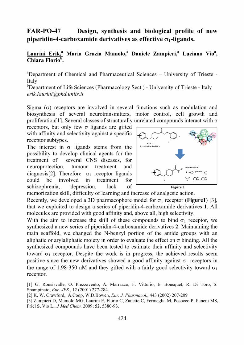
424
FAR-PO-47 Design, synthesis and biological profile of new piperidin-4-carboxamide derivatives as effective 1-ligands. Laurini Erik,a Maria Grazia Mamolo,a Daniele Zampieri,a Luciano Vioa, Chiara Floriob. aDepartment of Chemical and Pharmaceutical Sciences – University of Trieste - Italy bDepartment of Life Sciences (Pharmacology Sect.) - University of Trieste - Italy [email protected] Sigma () receptors are involved in several functions such as modulation and biosynthesis of several neurotransmitters, motor control, cell growth and proliferation[1]. Several classes of structurally unrelated compounds interact with σ receptors, but only few σ ligands are gifted with affinity and selectivity against a specific receptor subtypes. The interest in ligands stems from the possibility to develop clinical agents for the treatment of several CNS diseases, for neuroprotection, tumour treatment and diagnosis[2]. Therefore 1 receptor ligands could be involved in treatment for schizophrenia, depression, lack of memorization skill, difficulty of learning and increase of analgesic action. Recently, we developed a 3D pharmacophore model for 1 receptor (Figure1) [3], that we exploited to design a series of piperidin-4-carboxamide derivatives 1. All molecules are provided with good affinity and, above all, high selectivity. With the aim to increase the skill of these compounds to bind 1 receptor, we synthesized a new series of piperidin-4-carboxamide derivatives 2. Maintaining the main scaffold, we changed the N-benzyl portion of the amide groups with an aliphatic or arylaliphatic moiety in order to evaluate the effect on binding. All the synthesized compounds have been tested to estimate their affinity and selectivity toward 1 receptor. Despite the work is in progress, the achieved results seem positive since the new derivatives showed a good affinity against 1 receptors in the range of 1.98-350 nM and they gifted with a fairly good selectivity toward 1 receptor. [1] G. Ronsisvalle, O. Prezzavento, A. Marrazzo, F. Vittorio, E. Bousquet, R. Di Toro, S. Spampinato, Eur. JPS., 12 (2001) 277-284. [2] K. W. Crawford, A.Coop, W.D.Bowen, Eur. J. Pharmacol., 443 (2002) 207-209 [3] Zampieri D, Mamolo MG, Laurini E, Florio C, Zanette C, Fermeglia M, Posocco P, Paneni MS, Pricl S, Vio L., J Med Chem. 2009; 52, 5380-93.
Figure 2
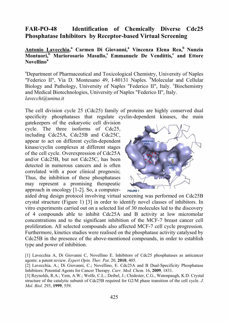
425
FAR-PO-48 Identification of Chemically Diverse Cdc25 Phosphatase Inhibitors by Receptor-based Virtual Screening Antonio Lavecchia,a Carmen Di Giovanni,a Vincenza Elena Rea,b Nunzia Montuori,b Mariorosario Masullo,c Emmanuele De Vendittis,c and Ettore Novellinoa
aDepartment of Pharmaceutical and Toxicological Chemistry, University of Naples "Federico II", Via D. Montesano 49, I-80131 Naples. bMolecular and Cellular Biology and Pathology, University of Naples "Federico II", Italy. cBiochemistry and Medical Biotechnologies, University of Naples "Federico II", Italy. [email protected] The cell division cycle 25 (Cdc25) family of proteins are highly conserved dual specificity phosphatases that regulate cyclin-dependent kinases, the main gatekeepers of the eukaryotic cell division cycle. The three isoforms of Cdc25, including Cdc25A, Cdc25B and Cdc25C, appear to act on different cyclin-dependent kinase/cyclin complexes at different stages of the cell cycle. Overexpression of Cdc25A and/or Cdc25B, but not Cdc25C, has been detected in numerous cancers and is often correlated with a poor clinical prognosis; Thus, the inhibition of these phosphatases may represent a promising therapeutic approach in oncology [1-2]. So, a computer-aided drug design protocol involving virtual screening was performed on Cdc25B crystal structure (Figure 1) [3] in order to identify novel classes of inhibitors. In vitro experiments carried out on a selected list of 30 molecules led to the discovery of 4 compounds able to inhibit Cdc25A and B activity at low micromolar concentrations and to the significant inhibition of the MCF-7 breast cancer cell proliferation. All selected compounds also affected MCF-7 cell cycle progression. Furthermore, kinetics studies were realised on the phosphatase activity catalysed by Cdc25B in the presence of the above-mentioned compounds, in order to establish type and power of inhibition. [1] Lavecchia A, Di Giovanni C, Novellino E. Inhibitors of Cdc25 phosphatases as anticancer agents: a patent review. Expert Opin. Ther. Pat. 20, 2010, 405. [2] Lavecchia, A.; Di Giovanni, C.; Novellino, E. Cdc25A and B Dual-Specificity Phosphatase Inhibitors: Potential Agents for Cancer Therapy. Curr. Med. Chem. 16, 2009, 1831. [3] Reynolds, R.A.; Yem, A.W.; Wolfe, C.L.; Deibel, J.; Chidester, C.G., Watenpaugh, K.D. Crystal structure of the catalytic subunit of Cdc25B required for G2/M phase transition of the cell cycle. J. Mol. Biol. 293, 1999, 559.
426
FAR-PO-49 Label-free, Reagentless, Straightforward Capillary Electrophoretic Method to Individuate Potent Calmodulin Ligands Lentini G., Bruno, C., Cavalluzzi M. M., Lovece A., Franchini C.
Dipartimento Farmaco-Chimico, Università degli Studi di Bari ‘Aldo Moro’, Via Orabona 4, 70125 – Bari, Italy [email protected]
In the last decade, drug repositioning (drug repurposing or indication switch)
strategies have become more and more attracting as useful ways for drug discovery [15], and promiscuous ligands let envisage the way toward a new generation of efficacious drugs [6].
Calmodulin (CaM) ligands are a chemically heterogeneous class of biologically active compounds, many of which behave as anthypertensive and antianginous, neuroleptic, anxiolytic, antidepressant, antiarrhythmic, antiestrogen, and antineoplastic agents. Several CaM ligands display polypharmacology. Thus, this class of compounds might be mined to detect leads for repositioning: both new and clinically established CaM ligands might prove useful in some of the above mentioned therapeutic areas.
Here we propose a label-free, reagentless, straightforward affinity capillary electrophoretic (ACE) method to screen compounds as CaM ligands. Apparent dissociation constants between bovine brain CaM and various small ligands were found in good agreement with those reported in the literature. The method was successfully used to demonstrate that lubeluzole―a well-known neuroprotective agent―is a high-affinity CaM ligand.
[6] Wermuth C. G. Med. Chem. Res. 2001, 10, 431–439. [7] Wermuth C. G. J. Med. Chem. 2004, 47, 1303–1314. [8] Dueñas-González A., García-López P., Alonso Herrera L., Medina-Franco J.
L., González-Fierro A., Candelaria M. Mol. Cancer 2008, 7, 82. [9] Riveiro M. E., De Kimpe N., Moglioni A., Vázquez R., Monczor F., Shayo C.,
Davio C. Curr. Med. Chem. 2010, 17, 13251338. [10] Gan F., Cao B., Wu D., Chen Z., Hou T., Mao X. Curr. Med. Chem. 2011, 18,
15091514. Hopkins A. L., Mason J. S., Overington J. P. Curr. Opin. Struc. Biol. 2006, 16, 127136.
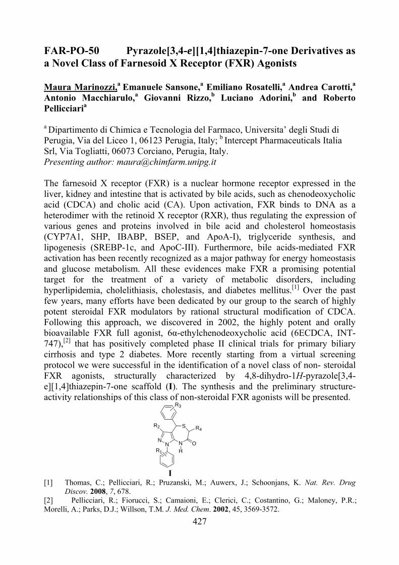
427
FAR-PO-50 Pyrazole[3,4-e][1,4]thiazepin-7-one Derivatives as a Novel Class of Farnesoid X Receptor (FXR) Agonists Maura Marinozzi,a Emanuele Sansone,a Emiliano Rosatelli,a Andrea Carotti,a Antonio Macchiarulo,a Giovanni Rizzo,b Luciano Adorini,b and Roberto Pellicciaria
a Dipartimento di Chimica e Tecnologia del Farmaco, Universita’ degli Studi di Perugia, Via del Liceo 1, 06123 Perugia, Italy; b Intercept Pharmaceuticals Italia Srl, Via Togliatti, 06073 Corciano, Perugia, Italy. Presenting author: [email protected]
The farnesoid X receptor (FXR) is a nuclear hormone receptor expressed in the liver, kidney and intestine that is activated by bile acids, such as chenodeoxycholic acid (CDCA) and cholic acid (CA). Upon activation, FXR binds to DNA as a heterodimer with the retinoid X receptor (RXR), thus regulating the expression of various genes and proteins involved in bile acid and cholesterol homeostasis (CYP7A1, SHP, IBABP, BSEP, and ApoA-I), triglyceride synthesis, and lipogenesis (SREBP-1c, and ApoC-III). Furthermore, bile acids-mediated FXR activation has been recently recognized as a major pathway for energy homeostasis and glucose metabolism. All these evidences make FXR a promising potential target for the treatment of a variety of metabolic disorders, including hyperlipidemia, cholelithiasis, cholestasis, and diabetes mellitus.[1] Over the past few years, many efforts have been dedicated by our group to the search of highly potent steroidal FXR modulators by rational structural modification of CDCA. Following this approach, we discovered in 2002, the highly potent and orally bioavailable FXR full agonist, 6α-ethylchenodeoxycholic acid (6ECDCA, INT-747),[2] that has positively completed phase II clinical trials for primary biliary cirrhosis and type 2 diabetes. More recently starting from a virtual screening protocol we were successful in the identification of a novel class of non- steroidal FXR agonists, structurally characterized by 4,8-dihydro-1H-pyrazole[3,4-e][1,4]thiazepin-7-one scaffold (I). The synthesis and the preliminary structure-activity relationships of this class of non-steroidal FXR agonists will be presented.
NN N
SR2
HO
R4
R1
R3
I [1] Thomas, C.; Pellicciari, R.; Pruzanski, M.; Auwerx, J.; Schoonjans, K. Nat. Rev. Drug
Discov. 2008, 7, 678. [2] Pellicciari, R.; Fiorucci, S.; Camaioni, E.; Clerici, C.; Costantino, G.; Maloney, P.R.; Morelli, A.; Parks, D.J.; Willson, T.M. J. Med. Chem. 2002, 45, 3569-3572.
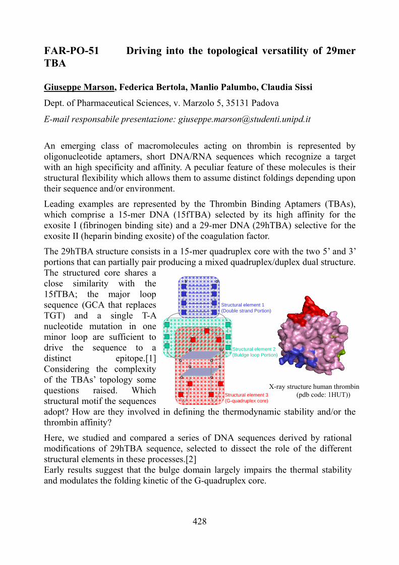
428
FAR-PO-51 Driving into the topological versatility of 29mer TBA Giuseppe Marson, Federica Bertola, Manlio Palumbo, Claudia Sissi
Dept. of Pharmaceutical Sciences, v. Marzolo 5, 35131 Padova
E-mail responsabile presentazione: [email protected]
An emerging class of macromolecules acting on thrombin is represented by oligonucleotide aptamers, short DNA/RNA sequences which recognize a target with an high specificity and affinity. A peculiar feature of these molecules is their structural flexibility which allows them to assume distinct foldings depending upon their sequence and/or environment.
Leading examples are represented by the Thrombin Binding Aptamers (TBAs), which comprise a 15-mer DNA (15fTBA) selected by its high affinity for the exosite I (fibrinogen binding site) and a 29-mer DNA (29hTBA) selective for the exosite II (heparin binding exosite) of the coagulation factor.
The 29hTBA structure consists in a 15-mer quadruplex core with the two 5’ and 3’ portions that can partially pair producing a mixed quadruplex/duplex dual structure. The structured core shares a close similarity with the 15fTBA; the major loop sequence (GCA that replaces TGT) and a single T-A nucleotide mutation in one minor loop are sufficient to drive the sequence to a distinct epitope.[1] Considering the complexity of the TBAs’ topology some questions raised. Which structural motif the sequences adopt? How are they involved in defining the thermodynamic stability and/or the thrombin affinity?
Here, we studied and compared a series of DNA sequences derived by rational modifications of 29hTBA sequence, selected to dissect the role of the different structural elements in these processes.[2] Early results suggest that the bulge domain largely impairs the thermal stability and modulates the folding kinetic of the G-quadruplex core.
G G
G G
5’ 3’
G G
Structural element 3 (G-quadruplex core)
Structural element 1 (Double strand Portion)
Structural element 2 (Buldge loop Portion)
G G
X-ray structure human thrombin (pdb code: 1HUT))
429
Similarly, the length of the double strand portion, does not seem to influence protein binding affinity although it plays a significant role in defining the folded DNA thermal stability. Finally we designed and characterized an ExositeI/Exosite II DNA binder that merges 15fTBA structural elements in the 29hTBA general structure. Our work is aimed to define the minimal structural motifs required to preserve the target affinity, to finally translate them to not-oligonucleotide molecules (e.g. PNA) characterized by better drug-like properties. [1] I., Smirnov, Effect of Loop Sequence and Size on DNA Aptamer Stability. Biochemistry, 39, 2000, 1462-1468. [2] F., Macaya, Structural and functional characterization of potent antithrombotic oligonucleotides possessing both quadruplex and duplex motif. Biochemistry, 34, 1995, 4478-4492.
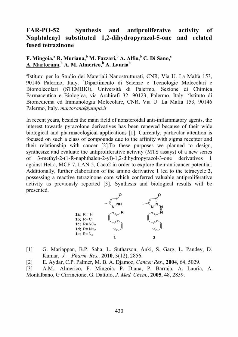
430
FAR-PO-52 Synthesis and antiproliferatve activity of Naphtalenyl substituted 1,2-dihydropyrazol-5-one and related fused tetrazinone F. Mingoia,a R. Muriana,b M. Fazzari,b A. Alfio,b C. Di Sano,c A. Martorana,b A. M. Almerico,b A. Lauriab
aIstituto per lo Studio dei Materiali Nanostrutturati, CNR, Via U. La Malfa 153, 90146 Palermo, Italy. bDipartimento di Scienze e Tecnologie Molecolari e Biomolecolari (STEMBIO), Università di Palermo, Sezione di Chimica Farmaceutica e Biologica, via Archirafi 32. 90123, Palermo, Italy. cIstituto di Biomedicina ed Immunologia Molecolare, CNR, Via U. La Malfa 153, 90146 Palermo, Italy. [email protected]
In recent years, besides the main field of nonsteroidal anti-inflammatory agents, the interest towards pyrazolone derivatives has been renewed because of their wide biological and pharmacological applications [1]. Currently, particular attention is focused on such a class of compounds due to the affinity with sigma receptor and their relationship with cancer [2].To these purposes we planned to design, synthesize and evaluate the antiproliferative activity (MTS assays) of a new series of 3-methyl-2-(1-R-naphthalen-2-yl)-1,2-dihydropyrazol-3-one derivatives 1 against HeLa, MCF-7, LAN-5, Caco2 in order to explore their anticancer potential. Additionally, further elaboration of the amino derivative 1 led to the tetracycle 2, possessing a reactive tetrazinone core which conferred valuable antiproliferative activity as previously reported [3]. Synthesis and biological results will be presented.
NHN
O
1a; R = H 1b; R= Cl 1c; R= NO21d; R= NH21e; R= N3
R
NN
O
N
N
1 2 [1] G. Mariappan, B.P. Saha, L. Sutharson, Anki, S. Garg, L. Pandey, D.
Kumar, J. Pharm. Res., 2010, 3(12), 2856. [2] E. Aydar, C.P. Palmer, M. B. A. Djamoz, Cancer Res., 2004, 64, 5029. [3] A.M., Almerico, F. Mingoia, P. Diana, P. Barraja, A. Lauria, A. Montalbano, G Cirrincione, G. Dattolo, J. Med. Chem., 2005, 48, 2859.
431
FAR-PO-53 Identification of Putative Guanosine Receptor in Rat Brain Gabriella Marucci, Michela Buccioni, Diego Dal Ben, Catia Lambertucci,
Ajiroghene Thomas, RosariaVolpini, Gloria Cristalli. School of Pharmacy, Medicinal Chemistry Unit, University of Camerino, Via S. Agostino 1, 62032 Camerino, Italy. [email protected] G protein coupled receptors (GPCRs), recognizing adenosine and purine and pyrimidine nucleotides as extracellular messengers, have been characterized and classified as purinergic P1 and P2 receptors, respectively. However, some experimental data suggest that other nucleosides as guanosine, inosine and hypoxanthine can also act as signalling molecules via the activation of specific membrane receptors [1]. In particular, extracellular guanosine seems to possess many trophic effects, including promotion, division, and growth of astrocytes and other neuron-like cells. Binding studies, performed on rat brain membranes using [3H]guanosine, have shown that the compound interacts with binding sites, which are distinct from the well-characterized ATP and adenosine receptors. Furthermore, guanosine itself and 6-thioguanosine are equally effective in displacing [3H]guanosine from rat brain membranes [2]. Hence, potent and selective agonists and antagonists are highly needed for the characterization of the physiopathological profile of the new putative guanosine receptor. Starting from these observations and aimed at developing a new assay that allows to evaluate the potency of ligands at the putative guanosine receptor, Eu-GTP assay was performed in rat brain. This technique, using the principle of the [35S]GTPγS binding assay, replaces the radioactive material with Eu-GTP and exploits the unique fluorescence properties of Europium lanthanide chelate. New molecules, prepared by modifying the purine and sugar moieties of guanosine at the 6- and 5’-positions, were tested using Eu-GTP assay. Results showed that the new compounds are able to activate the putative guanosine receptor more than guanosine itself. On the other hand, this functional assay seems to demonstrate that a GPCR activated by guanosine is present in rat brain [3]. [1] F. Caciagli, P. Illes, Purinergic Signalling, 1, 2005, 297. [2] U. Traversa, G. Bombi, E. Camaioni, A. Macchiarulo, G. Costantino, C.
Palmieri, F. Caciagli, R. Pellicciari, Bioorg. Med. Chem.,11(24), 2003, 5417.
[3] R. Volpini, G. Marucci, M. Buccioni, D. Dal Ben, C. Lambertucci, C. Lammi, R.C. Mishra, A. Thomas, G. Cristalli, ChemMedChem., DOI: 10.1002/cmdc.201100100, VIP (the paper was judged to be very important by the referees).
432
FAR-PO-54 3-[2-(4-aryl-1,3-thiazol-2-yl)hydrazin-1-ylidene]-1H-indol-2-ones as new potential dual inhibitors of polimerase and ribonuclease HIV-1 RT associated function R. Meleddu1, A. Corona2, S. Distinto3, F. Esposito2, M. L. Sanna1, C. Cardia1, E. Tramontano2 and E. Maccioni1. 1) Department Farmaco Chimico Tecnologico, University of Cagliari, Via
Ospedale 72, Cagliari, 09124, Italy. [email protected] 2) Department di Scienze Applicate ai Biosistemi, University of Cagliari,
Cittadella Universitaria di Monserrato, 09042, Monserrato, Cagliari, Italy. 3) Department di Scienze Farmacobiologiche, Università degli Studi di Catanzaro
"Magna Graecia", 88021, Roccelletta di Borgia (CZ), Italy.
RT is an essential enzyme for viral replication which has two associated catalytic functions: a DNA or RNA dependent polymerase activity and a ribonuclease H (RNase H) activity.[1-2] The two catalytic sites are distinc but inter-dependent and mutations in the polymerase domain affect the RNase H activity, and vice versa. Furthermore, both RT associated activites are essential for virus replication. Thus, both enzyme functions are attractive targets for drug development. Aiming to identify new scaffolds capable to combine inhibitory activity on both enzymatic functions, some of us performed a combined shape-, 2D-fingerprint-, and pharmacophore-based virtual screening and identified a 3-substituted-2-indolinone derivative as a promising dual inhibitor of both RT functions.[3] 2-indolinone derivatives are of biological interest and their biological properties have been recently investigated.[4-6] However their activity on HIV-1 RT has never been investigated. Prompted by the virtual screening results we have synthesised a series of 3-[2-(4-aryl-1,3-thiazol-2-yl)hydrazin-1-ylidene]-1H-indol-2-ones and evaluate their biological activity as potential dual inhibitors of the Human Immunodeficiency virus-1 (HIV-1) Reverse Transcriptase (RT). Furthermore we have investigated on the site of interaction on the HIV-1 RT. In this presentation we wish to report on our first approach dedicated to the synthesis and the biological behaviour investigation of new rationally designed RNase H and RDDP dual inhibitors. [1] Telesnitsky, A.; et al, In Retroviruses, Cold Spring Harbor Laboratory Press: 1997; pp 121-160. [2] Hughes, S. H.; et al, In Ribonucleases H, Editions Inserm: Paris, 1998; pp 195-224. [3] Distinto, S.; et al, (submitted for publication). [4] Roth G. J.; et al,; J. Med. Chem., 2010, 53, 7287–7295. [5]. Andreani A.; et al, J. Med. Chem., 2008, 51, 7508–7513. [6] Kilian H.; et al, J. Med. Chem., 2010, 53, 1383–1386.
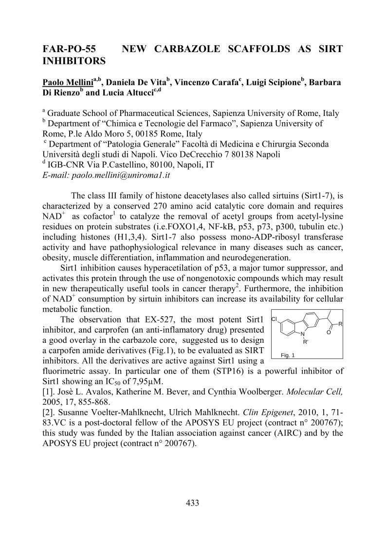
433
FAR-PO-55 NEW CARBAZOLE SCAFFOLDS AS SIRT INHIBITORS Paolo Mellinia,b, Daniela De Vitab, Vincenzo Carafac, Luigi Scipioneb, Barbara Di Rienzob and Lucia Altuccic,d
a Graduate School of Pharmaceutical Sciences, Sapienza University of Rome, Italy b Department of “Chimica e Tecnologie del Farmaco”, Sapienza University of Rome, P.le Aldo Moro 5, 00185 Rome, Italy c Department of “Patologia Generale” Facoltà di Medicina e Chirurgia Seconda Università degli studi di Napoli. Vico DeCrecchio 7 80138 Napoli d IGB-CNR Via P.Castellino, 80100, Napoli, IT E-mail: [email protected]
The class III family of histone deacetylases also called sirtuins (Sirt1-7), is characterized by a conserved 270 amino acid catalytic core domain and requires NAD+ as cofactor1 to catalyze the removal of acetyl groups from acetyl-lysine residues on protein substrates (i.e.FOXO1,4, NF-kB, p53, p73, p300, tubulin etc.) including histones (H1,3,4). Sirt1-7 also possess mono-ADP-ribosyl transferase activity and have pathophysiological relevance in many diseases such as cancer, obesity, muscle differentiation, inflammation and neurodegeneration. Sirt1 inhibition causes hyperacetilation of p53, a major tumor suppressor, and activates this protein through the use of nongenotoxic compounds which may result in new therapeutically useful tools in cancer therapy2. Furthermore, the inhibition of NAD+ consumption by sirtuin inhibitors can increase its availability for cellular metabolic function. The observation that EX-527, the most potent Sirt1 inhibitor, and carprofen (an anti-inflamatory drug) presented a good overlay in the carbazole core, suggested us to design a carpofen amide derivatives (Fig.1), to be evaluated as SIRT inhibitors. All the derivatives are active against Sirt1 using a fluorimetric assay. In particular one of them (STP16) is a powerful inhibitor of Sirt1 showing an IC50 of 7,95µM. [1]. Josè L. Avalos, Katherine M. Bever, and Cynthia Woolberger. Molecular Cell, 2005, 17, 855-868. [2]. Susanne Voelter-Mahlknecht, Ulrich Mahlknecht. Clin Epigenet, 2010, 1, 71-83.VC is a post-doctoral fellow of the APOSYS EU project (contract n° 200767); this study was funded by the Italian association against cancer (AIRC) and by the APOSYS EU project (contract n° 200767).
N O
RCl
R'
Fig. 1
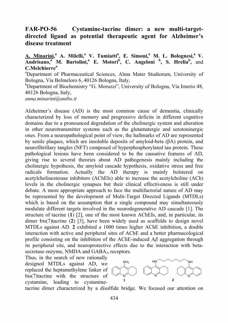
434
FAR-PO-56 Cystamine-tacrine dimer: a new multi-target-directed ligand as potential therapeutic agent for Alzheimer’s disease treatment A. Minarini,a A. Milelli,a V. Tumiattia, E. Simoni,a M. L. Bolognesi,a V. Andrisano,a M. Bartolini,a E. Motorib, C. Angeloni b, S. Hreliab, and C.Melchiorrea aDepartment of Pharmaceutical Sciences, Alma Mater Studiorum, University of Bologna, Via Belmeloro 6, 40126 Bologna, Italy, bDepartment of Biochemistry “G. Moruzzi”, University of Bologna, Via Irnerio 48, 40126 Bologna, Italy, [email protected] Alzheimer’s disease (AD) is the most common cause of dementia, clinically characterized by loss of memory and progressive deficits in different cognitive domains due to a pronounced degradation of the cholinergic system and alteration in other neurotransmitter systems such as the glutamatergic and serotoninergic ones. From a neuropathological point of view, the hallmarks of AD are represented by senile plaques, which are insoluble deposits of amyloid-beta (βA) protein, and neurofibrillary tangles (NFT) composed of hyperphosphorylated tau protein. These pathological lesions have been considered to be the causative features of AD, giving rise to several theories about AD pathogenesis mainly including the cholinergic hypothesis, the amyloid cascade hypothesis, oxidative stress and free radicals formation. Actually the AD therapy is mainly bolstered on acetylcholinesterase inhibitors (AChEIs) able to increase the acetylcholine (ACh) levels in the cholinergic synapses but their clinical effectiveness is still under debate. A more appropriate approach to face the multifactorial nature of AD may be represented by the development of Multi-Target Directed Ligands (MTDLs) which is based on the assumption that a single compound may simultaneously modulate different targets involved in the neurodegenerative AD cascade [1]. The structure of tacrine (1) [2], one of the most known AChEIs, and, in particular, its dimer bis(7)tacrine (2) [3], have been widely used as scaffolds to design novel MTDLs against AD. 2 exhibited a 1000 times higher AChE inhibition, a double interaction with active and peripheral sites of AChE and a better pharmacological profile consisting on the inhibition of the AChE-induced Aβ aggregation through its peripheral site, and neuroprotective effects due to the interaction with beta-secretase enzyme, NMDA and GABAA receptors. Thus, in the search of new rationally designed MTDLs against AD, we replaced the heptamethylene linker of bis(7)tacrine with the structure of cystamine, leading to cystamine-tacrine dimer characterized by a disulfide bridge. We focused our attention on
N
NH2
N
HN
N1 2
NH
435
cystamine for its biological activities as antioxidant, cyto- and neuroprotective agent [4]. In this study we demonstrated that the cystamine-tacrine dimer, in comparison to bis(7)tacrine, is endowed with a lower toxicity, it is able to inhibit AChE, BChE, self and AChE induced Aβ aggregation in the same range of the reference compound and to protect the neuroblastoma SH-SY5Y cell line against H2O2-induced damage by activating the extracellular signal-regulated kinase 1 and 2 (ERK1/2) and Akt/protein kinase B (PKB) pathways. (This research was supported by grants from MIUR, Rome (PRIN), and University of Bologna (RFO)) (1) Cavalli, A., Bolognesi, ML et al. J. Med. Chem., 51, 2008, 347. (2) Tumiatti, V., Minarini, A. et al. Curr. Med. Chem., 17, 2010, 1825. (3) Pang, Y. P., Quiram, P. et al. J. Biol. Chem., 271, 1996, 23646. (4) Wood, P. L., Khan, M. A. et al. Brain Research, 1158, 2007, 158.
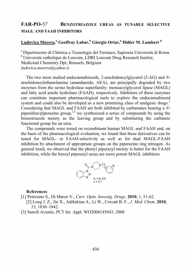
436
FAR-PO-57 BENZOTRIAZOLE UREAS AS TUNABLE SELECTIVE
MAGL AND FAAH INHIBITORS Ludovica Morera,a Geoffray Labar,b Giorgio Ortar,a Didier M. Lambert b
a Dipartimento di Chimica e Tecnologie del Farmaco, Sapienza Università di Roma b Université catholique de Louvain, LDRI Louvain Drug Research Institut, Medicinal Chemistry Dpt, Brussels, Belgium [email protected]
The two most studied endocannabinoids, 2-arachidonoylglycerol (2-AG) and N-arachidonoylethanolamine (anandamide, AEA), are principally degraded by two enzymes from the serine hydrolase superfamily: monoacylglycerol lipase (MAGL) and fatty acid amide hydrolase (FAAH), respectively. Inhibitors of these enzymes can constitute important pharmacological tools to explore the endocannabinoid system and could also be developed as a new promising class of analgesic drugs.1 Considering that MAGL and FAAH are both inhibited by carbamates bearing a N-piperidine/piperazine group,2,3 we synthesized a series of compounds by using the benzotriazole moiety as the leaving group and by substituting the carbamic functional group for an urea.
The compounds were tested on recombinant human MAGL and FAAH and, on the basis of the pharmacological evaluation, we found that these derivatives can be tuned for MAGL- or FAAH-selectivity as well as for dual MAGL-FAAH inhibition by attachment of appropriate groups on the piperazine ring nitrogen. As general trend, we observed that the phenyl piperazyl moiety is better for the FAAH inhibition, while the benzyl piperazyl ureas are more potent MAGL inhibitors.
References
[1] Petrosino S., Di Marzo V., Curr. Opin. Investig. Drugs. 2010, 1, 51-62. [2] Long J. Z., Jin X., Adibekian A., Li W., Cravatt B. F., J. Med. Chem. 2010,
53, 1830–1842. [3] Sanofi Aventis, PCT Int. Appl. WO2008145843, 2008
437
FAR-PO-58 NOVEL POTENT AND SELECTIVE FAAH REVERSIBLE INHIBITORS
Raffaella Napolitano,a Patrizia Minetti,a Walter Cabri,a Antonio Caprioli,a Franco Borsini,a Stefania Butini,b Margherita Brindisi,b Samuele Maramai,b, Chiara De Simone,c Mauro Maccarronec
.
aSigma-Tau, R&D, Via Pontina km 30,400 - 00040 Pomezia, Italy bEuropean Research Centre for Drug Discovery and Development (NatSynDrugs) Siena University, Via Aldo Moro 2, 53100 Siena, Italy, cDipartimento Scienze Biomediche, Università di Teramo, and CERC/Fondazione S. Lucia, Roma, Italy E-mail: [email protected]
Endocannabinoids are a class of signalling lipids such as N-arachidonoyl ethanolamine (anandamide, AEA) and 2-arachidonoyl glycerol (2-AG), which activate cannabinoid receptors CB1 and CB2 to modulate a range of effects including pain, inflammation, appetite, motility, sleep, and thermoregulation, cognitive and emotional states. AEA and related bioactive fatty acid amides are inactivated by the membrane-bound serine hydrolase fatty acid amide hydrolase (FAAH). The development of FAAH inhibitors represents an elegant alternative to CB receptor agonists. Accordingly, inactivation of FAAH may have beneficial effects on pain and anxiety without the side effects (hypomotility, hypothermia, and catalepsy). To preserve this lack of “cannabinoid side effects”, the inhibitors should not interfere with CB1 receptor, which is involved in most of the unwanted effects of exogenous cannabinoids.
With the aim to discover new scaffolds for selective FAAH inhibition we identified a series of potent and selective FAAH inhibitors characterized by a phenyl-1- pyrrole structure bearing differently functionalized lateral chains to improve hydrophylic and pharmacokinetic properties. Some representative hits proved to be extremely potent and selective FAAH inhibitors, reversibly binding the enzyme.1
[1] Walter Cabri, Patrizia Minetti, Giuseppe Campiani, Stefania Butini; "New carbamate derivatives as modulators of fatty Acid Amide Hydrolase (FAAH Inhibitors)"; EP09155528
438
FAR-PO-59 The effect of the Cu(II) salt anion in a ligand exchange system operating with a chiral mobile phase Benedetto Natalini, Roccaldo Sardella, Andrea Carotti, Federica Ianni, Maria Eugenia García Rubiño, Antonio Macchiarulo, Roberto Pellicciari Dipartimento di Chimica e Tecnologia del Farmaco, Università degli Studi di Perugia, Via del Liceo 1, 06123, Perugia, Italy [email protected]
With the use of a chiral ligand-exchange chromatography (CLEC) system operating with the O-benzyl-(S)-serine [(S)-OBS] [1,2] as the chiral mobile phase (CMP) additive to the eluent, the effect of the copper(II) anion type was evaluated on the thermodynamic parameters, retention (k) and separation () factors, by rationally changing the following experimental chromatographic conditions: column temperature, absolute configuration of the chiral selector, and salt concentration. The CLEC-CMP analysis was carried out on ten amino acidic racemates and with nine cupric salts. While the group of analytes comprised both aliphatic (leucine, isoleucine, nor-leucine, proline, valine, nor-valine, and -methyl-valine) and aromatic (1-aminoindan-1,5-dicarboxylic acid, phenylglycine, and tyrosine) species, representative organic (formate, methanesulphonate, and trifluoroacetate) and inorganic (bromide, chloride, fluoride, nitrate, perchlorate, and sulphate) Cu(II) salts were selected as the metal source into the eluent. This route of investigation was pursued with the aim of identifying analogies among the employed Cu(II) salts, by observing the profile of variation of the selected chromatographic parameters, upon a change of the above experimental conditions. All the data were collected and analyzed through a statistical approach (PCA and k-means clustering) that revealed the presence of two behavioural classes of cupric salts, sharing the same variation profile for k and values. Interestingly, this clustering can be explained in terms of ESP (Electrostatic Surface Potential) balance values, obtained by an ab initio calculation operated on the cupric salts. [1] B.Natalini, R.Sardella, and R.Pellicciari, Curr.Anal.Chem., 1, 2005, 85. [2] B.Natalini, N.Giacchè, R.Sardella, F.Ianni, A.Macchiarulo, and R.Pellicciari, J.Chromatogr.A, 1217, 2010, 7523.
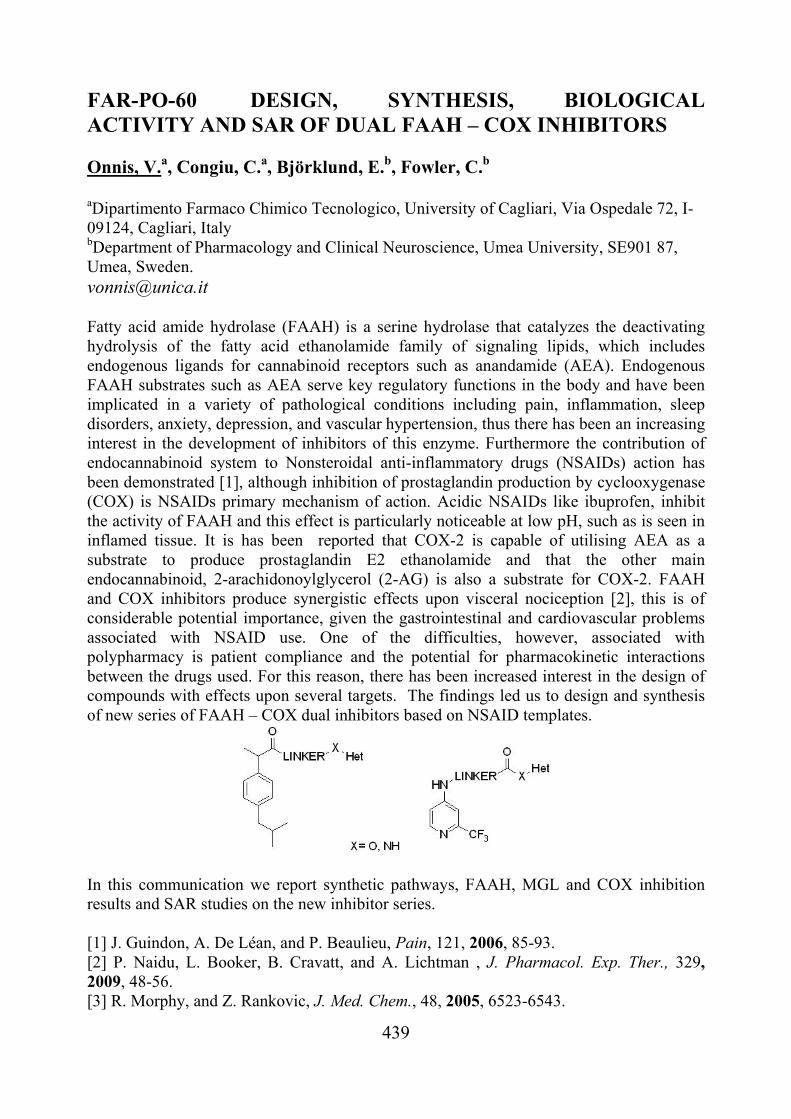
439
FAR-PO-60 DESIGN, SYNTHESIS, BIOLOGICAL ACTIVITY AND SAR OF DUAL FAAH – COX INHIBITORS Onnis, V.a, Congiu, C.a, Björklund, E.b, Fowler, C.b aDipartimento Farmaco Chimico Tecnologico, University of Cagliari, Via Ospedale 72, I-09124, Cagliari, Italy bDepartment of Pharmacology and Clinical Neuroscience, Umea University, SE901 87, Umea, Sweden. [email protected] Fatty acid amide hydrolase (FAAH) is a serine hydrolase that catalyzes the deactivating hydrolysis of the fatty acid ethanolamide family of signaling lipids, which includes endogenous ligands for cannabinoid receptors such as anandamide (AEA). Endogenous FAAH substrates such as AEA serve key regulatory functions in the body and have been implicated in a variety of pathological conditions including pain, inflammation, sleep disorders, anxiety, depression, and vascular hypertension, thus there has been an increasing interest in the development of inhibitors of this enzyme. Furthermore the contribution of endocannabinoid system to Nonsteroidal anti-inflammatory drugs (NSAIDs) action has been demonstrated [1], although inhibition of prostaglandin production by cyclooxygenase (COX) is NSAIDs primary mechanism of action. Acidic NSAIDs like ibuprofen, inhibit the activity of FAAH and this effect is particularly noticeable at low pH, such as is seen in inflamed tissue. It is has been reported that COX-2 is capable of utilising AEA as a substrate to produce prostaglandin E2 ethanolamide and that the other main endocannabinoid, 2-arachidonoylglycerol (2-AG) is also a substrate for COX-2. FAAH and COX inhibitors produce synergistic effects upon visceral nociception [2], this is of considerable potential importance, given the gastrointestinal and cardiovascular problems associated with NSAID use. One of the difficulties, however, associated with polypharmacy is patient compliance and the potential for pharmacokinetic interactions between the drugs used. For this reason, there has been increased interest in the design of compounds with effects upon several targets. The findings led us to design and synthesis of new series of FAAH – COX dual inhibitors based on NSAID templates.
In this communication we report synthetic pathways, FAAH, MGL and COX inhibition results and SAR studies on the new inhibitor series. [1] J. Guindon, A. De Léan, and P. Beaulieu, Pain, 121, 2006, 85-93. [2] P. Naidu, L. Booker, B. Cravatt, and A. Lichtman , J. Pharmacol. Exp. Ther., 329, 2009, 48-56. [3] R. Morphy, and Z. Rankovic, J. Med. Chem., 48, 2005, 6523-6543.
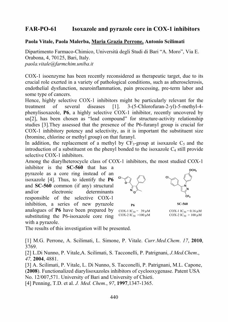
440
FAR-PO-61 Isoxazole and pyrazole core in COX-1 inhibitors Paola Vitale, Paola Malerba, Maria Grazia Perrone, Antonio Scilimati
Dipartimento Farmaco-Chimico, Università degli Studi di Bari “A. Moro”, Via E. Orabona, 4, 70125, Bari, Italy. [email protected] COX-1 isoenzyme has been recently reconsidered as therapeutic target, due to its crucial role exerted in a variety of pathological conditions, such as atherosclerosis, endothelial dysfunction, neuroinflammation, pain processing, pre-term labor and some type of cancers. Hence, highly selective COX-1 inhibitors might be particularly relevant for the treatment of several diseases [1]. 3-(5-Chlorofuran-2-yl)-5-methyl-4-phenylisoxazole, P6, a highly selective COX-1 inhibitor, recently uncovered by us[2], has been chosen as “lead compound” for structure-activity relationship studies [3].They assessed that the presence of the P6-furanyl group is crucial for COX-1 inhibitory potency and selectivity, as it is important the substituent size (bromine, chlorine or methyl group) on that furanyl. In addition, the replacement of a methyl by CF3-group at isoxazole C5 and the introduction of a substituent on the phenyl bonded to the isoxazole C4 still provide selective COX-1 inhibitors. Among the diarylheterocycle class of COX-1 inhibitors, the most studied COX-1 inhibitor is the SC-560 that has a pyrazole as a core ring instead of an isoxazole [4]. Thus, to identify the P6 and SC-560 common (if any) structural and/or electronic determinants responsible of the selective COX-1 inhibition, a series of new pyrazole analogues of P6 have been prepared by substituting the P6-isoxazole core ring with a pyrazole. The results of this investigation will be presented. [1] M.G. Perrone, A. Scilimati, L. Simone, P. Vitale. Curr.Med.Chem. 17, 2010, 3769. [2] L.Di Nunno, P. Vitale,A. Scilimati, S. Tacconelli, P. Patrignani, J.Med.Chem., 47, 2004, 4881. [3] A. Scilimati, P. Vitale, L. Di Nunno, S. Tacconelli, P. Patrignani, M.L. Capone, (2008). Functionalized diarylisoxazoles inhibitors of cyclooxygenase. Patent USA No. 12/007,571. University of Bari and University of Chieti. [4] Penning, T.D. et al. J. Med. Chem., 97, 1997,1347-1365.
NN
CF3
OCH3
Cl
SC-560
COX-1 IC50 = 0.16MCOX-2 IC50 > 100 M
NO
O
Cl
COX-1 IC50 = 39 MCOX-2 IC50 >100 M
P6
441
FAR-PO-62 Reactivity of platinum- based drugs toward selected proteins O. Pinato,a C. Musetti,a N. Farrell,b C. Sissia
a Dipartimento di Scienze Farmaceutiche dell’Università degli Studi di Padova, Via F. Marzolo 5, 35131, Padova, Italy b Deparment of Chemistry, Virginia Commonwealth University 1001 W. Main St., Richmond VA, USA [email protected]
Anticancer platinum drugs represent one of the most successful groups of compounds clinically used. Their biological mechanism of action is referred to platination of nuclear DNA, preferentially at guanine sites: the formation of stable DNA adducts is recognized as a DNA damage event and can ultimately drive to the apoptotic cell death.
From the discovery of their anticancer properties, research has been largely focused on the description of the DNA adduct formation by Pt drugs [1]. Although it was proposed that Pt-protein adducts could play an important role in modulating pharmacokinetics, resistance mechanisms, drug activity and side effects during this anticancer treatment, poor attention has been dedicated to characterize the interaction of platinum compounds with selected proteins [2].
In order to investigate the nature of metallodrug-protein interaction, we monitored the reactivity of different platinum compounds, included the trans planar platinum ammines [3] with model proteins such as bovine α-lactalbumin and hen egg lysozyme. The reactions were performed for 24 hours at 37 °C using different protein-Pt compound molar ratios. The reaction mixtures, containing the putative protein-Pt adducts, were resolved by SDS-PAGE gel electrophoresis and identified by electrospray ionization mass spectrometry technique (ESI-MS), a powerful technique to investigate metallodrugs-proteins interactions [4].
Our results clearly confirmed the formation of the metallodrug-protein adducts. In particular, we observed that the tested Pt compounds specifically react with each selected model protein according to different kinetics. Additionally, the structural features of Pt drugs have been shown to play a crucial role in promoting the reactivity and the selectivity toward tested proteic substrates. [1] D. Wang, S.J. Lippard, Nat. Rev. Drug Discovery, 4, 2005, 307. [2] E. Espinosa, J. Feliu, P. Zamora, M. Gonzalez Baron, J.J Sanchez., A.
Ordonez, and J.J. Espinosa, Lung Cancer,12, 1995, 67. [3] C. Musetti, A.A. Nazarov, N.P. Farrell, C. Sissi, ChemMedChem., 6, 2011,
1283. [4] A.R. Timerbaev, C.G. Hartinger, S.S. Aleksenko, and B.K. Keppler, Chem. Rev., 106, 2006, 2224.
442
FAR-PO-63 1-Aryl-5-(1H-pyrrol-1-yl)-1H-pyrazole-3-carboxamide: an effective scaffold for the design of either CB1 or CB2 receptor ligands
Francesco Piscitellia,*, Alessia Ligrestib, Giuseppe La Reginaa, Valerio Gattia, Antonella Brizzic, Serena Pasquinic, Marco Allaràb, Mauro Antonio Maria Caraid, Ettore Novellinoe, Giancarlo Colombod, Vincenzo Di Marzob, Federico Corellic, and Romano Silvestria aIstituto Pasteur – Fondazione Cenci Bolognetti, Dipartimento di Chimica e Tecnologie del Farmaco, Sapienza Università di Roma, Piazzale Aldo Moro 5, I-00185 Roma, Italy; bIstituto di Chimica Biomolecolare, Consiglio Nazionale delle Ricerche, Via Campi Flegrei 34, Comprensorio Olivetti, I-80078 Pozzuoli, Napoli, Italy; cDipartimento Farmaco Chimico Tecnologico, Università di Siena, Polo Scientifico Universitario San Miniato, Via Alcide De Gasperi 2, I-53100 Siena, Italy; dIstituto di Neuroscienze, Sezione di Cagliari, Consiglio Nazionale delle Ricerche, S.S. 554, km 4.500, I-09042 Cagliari, Italy; eDipartimento di Chimica Farmaceutica e Tossicologica, Università di Napoli Federico II, Via Domenico Montesano 49, I-80131, Napoli, Italy. *[email protected]
The endocannabinoid system is involved in pain, immunosuppression, vascular
disease, appetite management and locomotor disorders [1]. The CB1 inverse agonist Rimonabant (1), launched for the treatment of overweight, obesity and associated cardiovascular and metabolic disorders, was withdrawn because of psychiatric adverse events. There is a need of effective drugs to treat obesity because of the: (i) rapid increase of overweight people in the developed countries, and (ii) restricted therapeutic potential displayed by the FDA approved drugs sibutramine and orlistat [2].
New 1-aryl-5-(1H-pyrrol-1-yl)-1H-pyrazole-3-carboxamides were synthesized as CB receptor ligands. Compound 11 (CB1 Ki = 2.3 nM, CB1 SI = 163.6) showed CB1 receptor affinity and selectivity superior to Rimonabant and AM251. Acute administration of 2 mg/kg 11 resulted in preferentially reduced intake of sucrose rather than intake of regular food in rats. On the other hand, compound 23 (CB2 Ki = 0.51 nM, CB2 SI = 30.0) showed signficant affinity and selectivity for the CB2 receptor. The results presented here show that the 1-aryl-5-(1H-pyrrol-1-yl)-1H-pyrazole-3-carboxamide may serve as an effective scaffold for the design of either CB1 or CB2 receptor ligands. [1] V. Di Marzo, Targeting the endocannabinoid system: to enhance or reduce? Nat. Rev. Drug Discov. 7, 2008, 438-455. [2] R.S. Padwal, S.R. Majumdar, Drug treatments for obesity: orlistat, sibutramine, and rimonabant, Lancet 369, 2007, 71-77.
443
FAR-PO-64 Capillary electrophoresis coupled to laser induced fluorescence detection for the analysis of penicillamine in a non-conventional matrix Emanuele Morganti, Maria Addolorata Saracino, Francesca Bugamelli, Anna Ferranti, Nadia Ghedini, Maria Augusta Raggi Laboratory of Pharmaco-Toxicological Analysis, Department of Pharmaceutical Sciences of the University of Bologna, Via Belmeloro 6, 40126 Bologna, Italy [email protected]
Penicillamine (DL-2-amino-3-mercapto-3-methyl-butanoic acid) is a chelating agent derived from the hydrolysis of penicillin, lacking any antibiotic properties. The therapeutic form is D-penicillamine, while L-penicillamine is toxic. This drug is used in the treatment of severe active rheumatoid arthritis and acts by reducing the number of T-lymphocytes, inhibiting macrophage function and preventing collagen from cross-linking. It is also used as a chelating agent in Wilson's disease (a rare genetic disorder of copper metabolism), in cystinuria and in the treatment of heavy metal poisoning [1].
Adverse effects are frequent and may include: membranous glomerulonephritis, antibody-mediated myasthenic syndrome, drug-induced systemic lupus erythematosus, toxic myopathies and elastosis perforans serpiginosa. This last one may persist even after the therapy withdrawal [2].
Thus, to evaluate toxic effects in clinical cases we are developing a method based on capillary electrophoresis coupled to laser induced fluorescence detection (CE-LIF) in specific biological matrices such as epithelium.
The analysis is carried out in a fused silica capillary, using a carbonate buffer as the background electrolyte. Satisfactory sensitivity was obtained by exciting the molecule at 488 nm after a derivatisation step with 5-(iodoacetamido)fluorescein (IAF). Preliminary results are promising and the validation of the method is in progress. At the same time, we are developing another technique based on reversed phase liquid chromatography with amperometric detection to analyse penicillamine in epithelium samples.
[1] S. Sweetman (Ed.), Martindale: The Complete Drug Reference Thirty-sixth edition, The Pharmaceutical Press, London, 2009, 1456. [2] L. Atzori, A.L. Pinna, M. Pau and N. Aste, D-penicillamine elastosis perforans serpiginosa: Description of two cases and review of the literature. Dermatol. Online J. 17, 2011, 3.
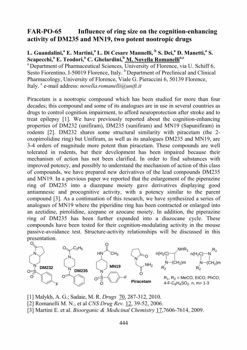
444
FAR-PO-65 Influence of ring size on the cognition-enhancing activity of DM235 and MN19, two potent nootropic drugs L. Guandalini,a E. Martini,a L. Di Cesare Mannelli, b S. Dei,a D. Manetti,a S. Scapecchi,a E. Teodori,a C. Ghelardini,b M. Novella Romanellia,c a Department of Pharmaceutical Sciences, University of Florence, via U. Schiff 6, Sesto Fiorentino, I-50019 Florence, Italy. b Department of Preclinical and Clinical Pharmacology, University of Florence, Viale G. Pieraccini 6, 50139 Florence, Italy. c e-mail address: [email protected] Piracetam is a nootropic compound which has been studied for more than four decades; this compound and some of its analogues are in use in several countries as drugs to control cognition impairment, to afford neuroprotection after stroke and to treat epilepsy [1]. We have previously reported about the cognition-enhancing properties of DM232 (unifiram), DM235 (sunifiram) and MN19 (Sapunifiram) in rodents [2]. DM232 shares some structural similarity with piracetam (the 2-oxopirrolidine ring) but Unifiram, as well as its analogues DM235 and MN19, are 3-4 orders of magnitude more potent than piracetam. These compounds are well tolerated in rodents, but their development has been impaired because their mechanism of action has not been clarified. In order to find substances with improved potency, and possibly to understand the mechanism of action of this class of compounds, we have prepared new derivatives of the lead compounds DM235 and MN19. In a previous paper we reported that the enlargement of the piperazine ring of DM235 into a diazepane moiety gave derivatives displaying good antiamnesic and procognitive activity, with a potency similar to the parent compound [3]. As a continuation of this research, we have synthesized a series of analogues of MN19 where the piperidine ring has been contracted or enlarged into an azetidine, pirrolidine, azepane or azocane moiety. In addition, the piperazine ring of DM235 has been further expanded into a diazocane cycle. These compounds have been tested for their cognition-modulating activity in the mouse passive-avoidance test. Structure-activity relationships will be discussed in this presentation.
N
N
O
SOO
F
N
N
ON
SOO
F
HN
O
CH3
DM232DM235
MN19
R1, R2 = MeCO, EtCO, PhCO,4-F-C6H4SO2. n, m= 1-3
O C2H5
n(H2C)
N (CH2)m
NHR1
R2
Nn(H2C)
N (CH2)mR2
R1
N O
O
NH2
Piracetam
[1] Malykh, A. G.; Sadaie, M. R. Drugs 70, 287-312, 2010. [2] Romanelli M. N., et al CNS Drug Rev. 12, 39-52, 2006. [3] Martini E. et al. Bioorganic & Medicinal Chemistry 17,7606-7614, 2009.
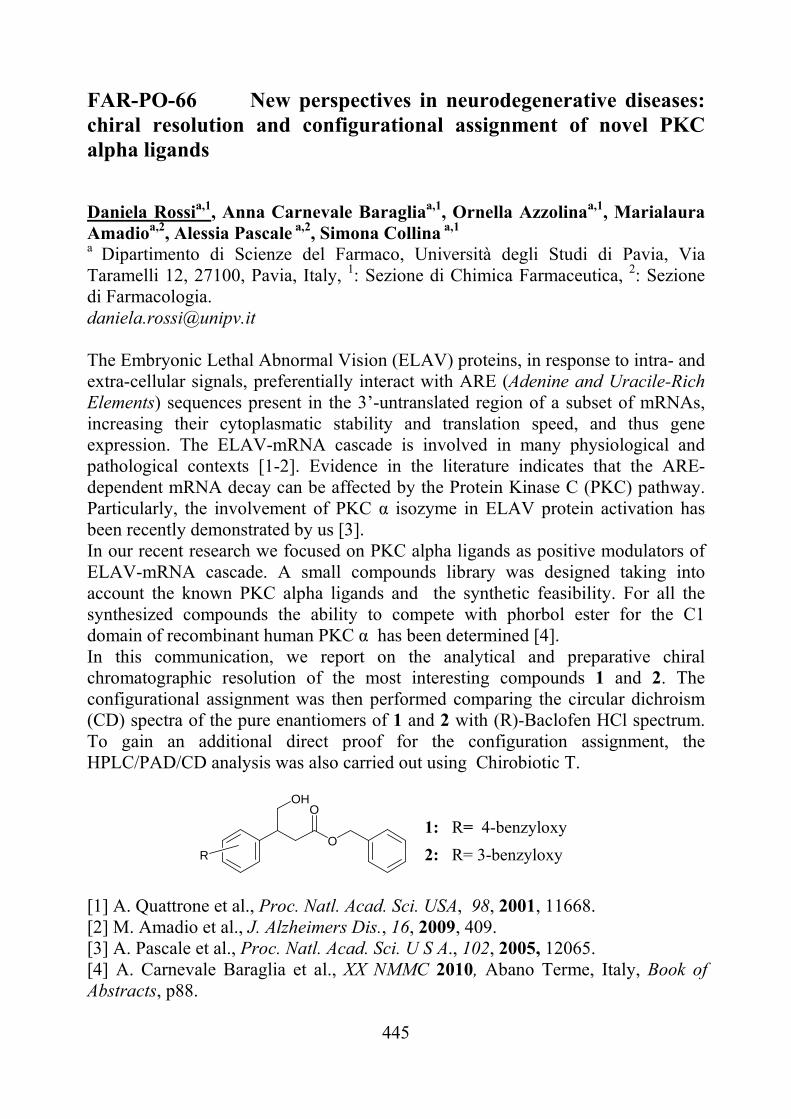
445
FAR-PO-66 New perspectives in neurodegenerative diseases: chiral resolution and configurational assignment of novel PKC alpha ligands
Daniela Rossia,1, Anna Carnevale Baragliaa,1, Ornella Azzolinaa,1, Marialaura Amadioa,2, Alessia Pascale a,2, Simona Collina a,1 a Dipartimento di Scienze del Farmaco, Università degli Studi di Pavia, Via Taramelli 12, 27100, Pavia, Italy, 1: Sezione di Chimica Farmaceutica, 2: Sezione di Farmacologia. [email protected]
The Embryonic Lethal Abnormal Vision (ELAV) proteins, in response to intra- and extra-cellular signals, preferentially interact with ARE (Adenine and Uracile-Rich Elements) sequences present in the 3’-untranslated region of a subset of mRNAs, increasing their cytoplasmatic stability and translation speed, and thus gene expression. The ELAV-mRNA cascade is involved in many physiological and pathological contexts [1-2]. Evidence in the literature indicates that the ARE-dependent mRNA decay can be affected by the Protein Kinase C (PKC) pathway. Particularly, the involvement of PKC α isozyme in ELAV protein activation has been recently demonstrated by us [3]. In our recent research we focused on PKC alpha ligands as positive modulators of ELAV-mRNA cascade. A small compounds library was designed taking into account the known PKC alpha ligands and the synthetic feasibility. For all the synthesized compounds the ability to compete with phorbol ester for the C1 domain of recombinant human PKC α has been determined [4]. In this communication, we report on the analytical and preparative chiral chromatographic resolution of the most interesting compounds 1 and 2. The configurational assignment was then performed comparing the circular dichroism (CD) spectra of the pure enantiomers of 1 and 2 with (R)-Baclofen HCl spectrum. To gain an additional direct proof for the configuration assignment, the HPLC/PAD/CD analysis was also carried out using Chirobiotic T.
RO
OOH
1: R= 4-benzyloxy
2: R= 3-benzyloxy
[1] A. Quattrone et al., Proc. Natl. Acad. Sci. USA, 98, 2001, 11668. [2] M. Amadio et al., J. Alzheimers Dis., 16, 2009, 409. [3] A. Pascale et al., Proc. Natl. Acad. Sci. U S A., 102, 2005, 12065. [4] A. Carnevale Baraglia et al., XX NMMC 2010, Abano Terme, Italy, Book of Abstracts, p88.
446
FAR-PO-67 Structure-Activity Relationships studies of GRK2 inhibitors Peptides
Marina Salaa, Isabel Gomez-Monterreyb, Ermelinda Vernieria, Guido Iaccarinoc, Alfonso Carotenutob, Paolo Griecob, Ettore Novellinob, Pietro Campigliaa. aDepartment of Pharmaceutical and Biomedical Science, University of Salerno. bDepartment of Pharmaceutical and Toxicological Chemistry, University of Naples “Federico II”. cDepartment of Clinical Medicine Cardiovascular and Immunological Sciences, University of Naples “ Federico II”. [email protected] G protein-coupled receptor kinase 2 (GRK2) regulates cell signaling by promoting agonist-specific desensitization of several metabolism-related GPCRs, including the -adrenergic receptors, endothelin, and glucose-dependent insulinotropic polypeptides. Interestingly, GRK2 expression and function has been shown to be altered in several pathological conditions. Thus, the upregulation of GRK2 and corresponding desensitization of these metabolism-related GPCRs seem play an important role in the onset or progression of diseases such as heart failure, myocardial ischemia, hypertension and Type 2 diabetes. In these diseases, understanding of the molecular mechanisms leading to altered GRK2 levels, as well as the identification of GRK2 inhibitors is a very active field of research. In this communication we report the preliminary results obtained with a small libraries of short analogues of peptides KRX107 and KRX124 derived from HJ loop of GRK2/3 [1]. 1 and 2 show a positive effect on glucose metabolism in animal models of Type 2 diabetes, increasing insuline sensitivity and improving glucose homeostasis and emerge as a valuable starting point for the development of a novel class of GRK2 inhibitors. [1] Anis Y. et al. Diabetologia, 2004, 47:1232–1244.
1 KRX107 G L L R r H S 2 KRX124 G L L R r H S I
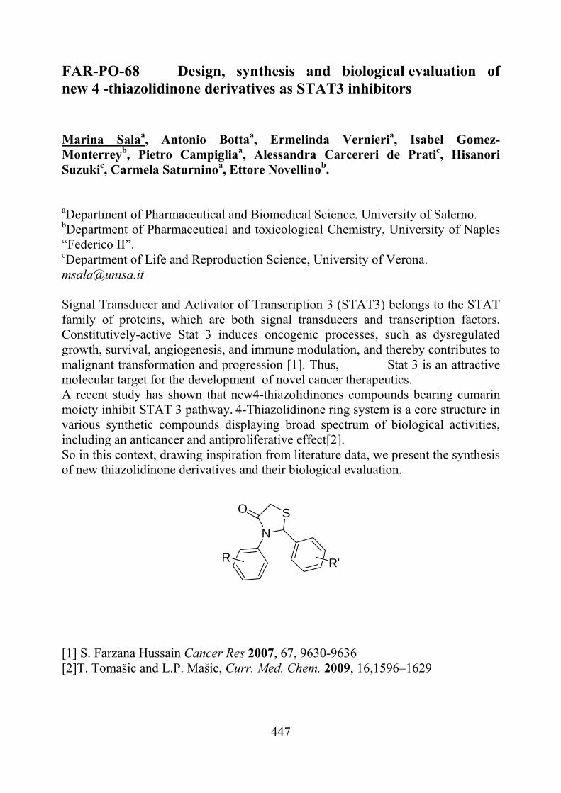
447
FAR-PO-68 Design, synthesis and biological evaluation of new 4 -thiazolidinone derivatives as STAT3 inhibitors Marina Salaa, Antonio Bottaa, Ermelinda Vernieria, Isabel Gomez-Monterreyb, Pietro Campigliaa, Alessandra Carcereri de Pratic, Hisanori Suzukic, Carmela Saturninoa, Ettore Novellinob. aDepartment of Pharmaceutical and Biomedical Science, University of Salerno. bDepartment of Pharmaceutical and toxicological Chemistry, University of Naples “Federico II”. cDepartment of Life and Reproduction Science, University of Verona. [email protected] Signal Transducer and Activator of Transcription 3 (STAT3) belongs to the STAT family of proteins, which are both signal transducers and transcription factors. Constitutively-active Stat 3 induces oncogenic processes, such as dysregulated growth, survival, angiogenesis, and immune modulation, and thereby contributes to malignant transformation and progression [1]. Thus, Stat 3 is an attractive molecular target for the development of novel cancer therapeutics. A recent study has shown that new4-thiazolidinones compounds bearing cumarin moiety inhibit STAT 3 pathway. 4-Thiazolidinone ring system is a core structure in various synthetic compounds displaying broad spectrum of biological activities, including an anticancer and antiproliferative effect[2]. So in this context, drawing inspiration from literature data, we present the synthesis of new thiazolidinone derivatives and their biological evaluation.
N
SO
R'R
[1] S. Farzana Hussain Cancer Res 2007, 67, 9630-9636 [2]T. Tomašic and L.P. Mašic, Curr. Med. Chem. 2009, 16,1596–1629
448
FAR-PO-69 EXPLORING -BEND STRUCTURES IN THE NEUROPEPTIDE S SEQUENCE
Salvadori Severoa, Guerrini Remoa, Trapella Claudioa, Marzola Ericaa, Ruzza Chiarab, Pulga Aliceb and Calo’ Girolamob aDepartment of Pharmaceutical Sciences and Biotechnology Centre University of Ferrara, bDepartment of Experimental and Clinical Medicine, Section of Pharmacology and National Institute of Neuroscience; Via Fossato di Mortara 19, 44100 Ferrara, Italy. [email protected]
Neuropeptide S (NPS) was identified as the endogenous ligand of an orphan receptor now referred to as NPSR. NPS injected supraspinally in rodents has been shown to modulate arousal, sleep-wakefulness cycle, anxiety-related behaviours and feeding. To identify novel NPSR ligands, structure-activity relationship studies were performed on the NPS sequence. In the context of such studies [D-Xaa5]NPS analogues were recognized as the first NPSR antagonists, among these molecules [tBu-D-Gly5]NPS was the most potent (pKB 7.06) [1].
The presence of Asn4-Gly5 in the N-terminal region hints at the possibility for this domain to assume, in its bioactive conformation, a regular turn centered on Asn4-Gly5, in fact these residues are found with high frequency in position i+1 and i+2 respectively of many natural turns of globular proteins [2].
Thus, we designated NPS analogues in which Asn4 or Asn4-Gly5 was substituted in turn with Pro/D-Pro or Orn in order to stabilize I, II or II’ beta turns centered in this position. The NPSR agonist and antagonist properties of these NPS analogues were investigated by measuring the intracellular calcium levels in response to NPS in cells expressing the recombinant murine receptor. [1] R. Guerrini, V. Camarda, C. Trapella, G. Caló, A. Rizzi, C. Ruzza, S. Fiorini, E.
Marzola, R.K. Reinscheid, D. Regoli, S. Salvadori, J. Med. Chem. 52, 2009, 4068-71.
[2] E.G. Hutchinson, J.M. Thornton, Protein Sci. 3, 1994, 3207-16.
449
FAR-PO-70 NEW PROMISING SCAFFOLDS FOR THE INHIBITION
OF MONOAMINE OXIDASE B Maria Luisa Sanna,a Rita Meleddu,a Maria Cristina Cardia,a Simona Distinto,b Stefano Alcaro,b and Elias Maccionia
a Dipartimento Farmaco Chimico Tecnologico, University of Cagliari, Via Ospedale, 72 Cagliari (CA), Italy; b Dipartimento di Scienze Farmaco Biologiche, University “Magna Graecia” of Catanzaro, Campus “S. Venuta”, 88100 Catanzaro (CZ), Italy [email protected]
Monoamine oxidase (MAO) (EC 1.4.3.4) are a family of enzymes responsible for the deactivation of active monoamines such as epinephrine, norepinephrine, dopamine, and serotonin [1]. MAO exist as 2 isoenzymes, A and B, with different affinities for various amines as substrates. The activity of monoamine oxidase helps to maintain neuron firing rates throughout the body within homeostatic limits. Because of the vital role that MAO play in the inactivation of neurotransmitters, MAO dysfunction is thought to be responsible for a number of psychiatric and neurological disorders, and, as a result, MAO inhibitors (MAOi) are studied for the treatment of several psychiatric and neurological disorders. Part of the biochemical activity of monoamine oxidase generates hydroxyl radicals, very toxic members of the oxygen free radical group, that may be involved in neurodegenerative disorders such as Parkinson's disease. Therefore, MAO-B inhibitors are coadjutant in the treatment of Parkinson’s diseases [2]. MAO-A inhibitors are used as antidepressant and anxiolytic drugs. Furthermore, the activity of MAO-B is enhanced by aging and in Alzheimer’s diseases patients [3]. Aiming at the identification of new leads for the selective inhibition of the B isoform of MAO, we have recently reported on the design, the synthesis, and the biological properties of a wide selection of differently substituted heterocyclic nuclei [4]. In this work we wish to present further studies on MAO inhibitors, particularly focusing on the structural modifications leading to an increase of MAO-B activity and selectivity. Thus the synthesis, the biological properties and the SARs of different series of 3-acetyl-2,5-diaryl-2,3-dihydro-1,3,4-oxadiazoles and 3,5-diaryl-4,5-dihydroisoxazoles will be reported. Moreover the influence of stereochemistry on the activity and the selectivity towards the different enzymatic isoforms will be discussed. [1] Wouters, J., 1998, Curr. Med. Chem., 5, 137-162 [2] Cesura, A.M., et al., 1992, Prog. Drug Res., 38, 171-297 [3] Saura, J., et al., 1994, Neuroscience, 62, 15-30 [4] Maccioni, E., et al., 2010, Eur.J.Med.Chem., 45, 4490-4498, and references therein cited.
450
FAR-PO-71 HPLC/ELSD analysis of conjugated bile acids: a way to assist flow chemistry processes Benedetto Natalini, Federica Ianni, Roccaldo Sardella, Francesco Venturoni, Antimo Gioiello, Roberto Pellicciari Dipartimento di Chimica e Tecnologia del Farmaco, Università degli Studi di Perugia, Via del Liceo 1, 06123, Perugia, Italy [email protected]
The employment of flow chemistry [1] in the process optimization of the N-acyl amidation of natural and unnatural bile acids (BAs), has required the connection with an in-line analysis and validation method for the determination of the reaction yields as well as of purity grade of the synthesized glyco- and tauro-conjugated derivatives. In this framework, an unique HPLC method was successfully established and validated for chenodeoxycholic (CDCA), cholic (CA), deoxycholic (DCA), and ursodeoxycholic (UDCA) acids, as well as the corresponding glyco- and tauro-conjugated forms. Because of the shared absence of relevant chromophoric moieties in the sample structure, an Evaporative Light Scattering Detector (ELSD) [2] was profitably utilized for the analysis of such steroidal species. For each of the investigated compounds, all the runs were contemporarily carried out on the free and the two relative conjugated variants. The different ELSD response of the free and the corresponding conjugated BAs, imposed to build-up separate calibration curves. In all the cases, very good precision and accuracy (evaluated both in the short and long period) along with appreciably low LOD and LOQ values turned out.
[1] S.V.Ley, and I.R.Baxendale, Nat.Rev.DrugDiscovery, 1, 2002, 573. [2] B.Natalini, R.Sardella, A.Gioiello, G.Carbone, M.Dawgul, and R.Pellicciari, J.Sep.Sci., 32, 2009, 2022.
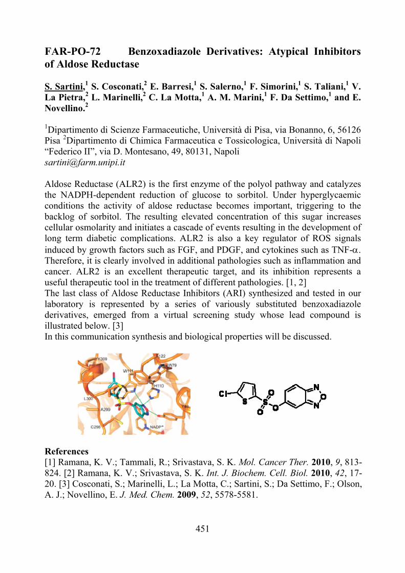
451
FAR-PO-72 Benzoxadiazole Derivatives: Atypical Inhibitors of Aldose Reductase S. Sartini,1 S. Cosconati,2 E. Barresi,1 S. Salerno,1 F. Simorini,1 S. Taliani,1 V. La Pietra,2 L. Marinelli,2 C. La Motta,1 A. M. Marini,1 F. Da Settimo,1 and E. Novellino.2
1Dipartimento di Scienze Farmaceutiche, Università di Pisa, via Bonanno, 6, 56126 Pisa 2Dipartimento di Chimica Farmaceutica e Tossicologica, Università di Napoli “Federico II”, via D. Montesano, 49, 80131, Napoli [email protected]
Aldose Reductase (ALR2) is the first enzyme of the polyol pathway and catalyzes the NADPH-dependent reduction of glucose to sorbitol. Under hyperglycaemic conditions the activity of aldose reductase becomes important, triggering to the backlog of sorbitol. The resulting elevated concentration of this sugar increases cellular osmolarity and initiates a cascade of events resulting in the development of long term diabetic complications. ALR2 is also a key regulator of ROS signals induced by growth factors such as FGF, and PDGF, and cytokines such as TNF-. Therefore, it is clearly involved in additional pathologies such as inflammation and cancer. ALR2 is an excellent therapeutic target, and its inhibition represents a useful therapeutic tool in the treatment of different pathologies. [1, 2] The last class of Aldose Reductase Inhibitors (ARI) synthesized and tested in our laboratory is represented by a series of variously substituted benzoxadiazole derivatives, emerged from a virtual screening study whose lead compound is illustrated below. [3] In this communication synthesis and biological properties will be discussed.
References [1] Ramana, K. V.; Tammali, R.; Srivastava, S. K. Mol. Cancer Ther. 2010, 9, 813-824. [2] Ramana, K. V.; Srivastava, S. K. Int. J. Biochem. Cell. Biol. 2010, 42, 17-20. [3] Cosconati, S.; Marinelli, L.; La Motta, C.; Sartini, S.; Da Settimo, F.; Olson, A. J.; Novellino, E. J. Med. Chem. 2009, 52, 5578-5581.
452
FAR-PO-73 Novel Pyrazolopyrimidine Derivatives as Therapeutic Agents for the Treatment of Medullary Thyroid Carcinoma S. Sartini,1 M. Morelli,1 A. Patti,1 F. Simorini,1 S. Taliani,1 S. Salerno,1 A. M. Marini,1 S. Cosconati,2 V. La Pietra,2 L. Marinelli,2 A. Fioravanti,3 T. Di Desidero,3 G. Bocci,3 S. M. Ferrari,3 P. Fallahi,3 A. Antonelli,3 C. La Motta,1 F. Da Settimo,1 and E. Novellino.2 1Dipartimento di Scienze Farmaceutiche, Università di Pisa, via Bonanno, 6, 56126 Pisa. 2Dipartimento di Chimica Farmaceutica e Tossicologica, Università di Napoli “Federico II”, via D. Montesano, 49, 80131, Napoli. 3Dipartimento di Medicina Interna, Via Roma 67, 56126, Pisa. [email protected]
Medullary thyroid carcinoma (MTC) is a malignant endocrine tumour originating from parafollicular calcitonin-producing C cells. Mainly sporadic, it may be also present in an inherited form, accounting for up to 8% of thyroid cancers. At present, the treatment of choice for both forms of MTC relies in their complete surgical resection. However, disease can persist or recur, with local and distant metastases which are often fatal. The limited effectiveness of conventional cytotoxic agents in thyroid carcinomas suggests the need of novel therapeutic strategies. Recent advances in the knowledge of pathogenic mechanisms leading to MTC identified receptor tyrosine kinase RET as a new and promising target. ‘Gain of function' mutations and rearrangement of RET activate the kinase activity of the receptor, thus providing mitogenic and survival signals to calcitonin-producing C cells. As mutations of RET have been identified in about 98% of inherited MTC cases, at the germ-line level, and in 30% to 50% of the sporadic forms, at the somatic level, this protein represent a sound target for the molecular therapy of most people affected by MTC [1,2]. Different approaches have been considered to repress the kinase function of RET, the most pursued one being the use of small molecules able to compete with the ATP site of the catalytic domain of this receptor. Recently, we described a number of pyrazolo[3,4-d]pyrimidine derivatives as effective inhibitors of both RET and VEGFR2 [3,4]. Here we present the synthesis and the functional evaluation of novel compounds of the same series, characterized by suitable substituents in the positions 3 and 6 of the heterocyclic core. References [1] Schlumberger, M.; Carlomagno, F.; Baudin, E. et al. Nat. Clin. Pract. Endocrinol. Metab. 2008, 4, 22-32. [2] Santoro, M.; Carlomagno, F. Nat. Clin. Pract. Endocrinol. Metab. 2006, 2, 42-52. [3] Antonelli, A.; Bocci, G.; La Motta, C. et al. Clin. Endocrinol. Metab. 2011, 96, E288-296. [4] Bocci, G.; Fioravanti, A.; La Motta, C. et al. Biochem Pharmacol. 2011, 81, 1309-1316.
453
FAR-PO-74 Antiproliferative activity of “Lycopersicon esculentum” leaves (var. Paul Robenson): preliminary study. Carmela Saturninoa, Ada Popoloa, Annalisa Spagnuoloa, Esther Sirignanoa, Antonio Bottaa, Marina Salaa, Giuseppina Tommonarob, Rocco De Priscob, Aldo Pintoa.
aDepartment of Pharmaceutical Sciences and Biomedics , University of Salerno, Fisciano(SA), Italy, Via Ponte Don Melillo 84084 Fisciano (SA) Italy; bInstitute of Biomolecular Chemistry, CNR, Pozzuoli (NA), Italy; [email protected] Among plants, the Lycopersicon esculentum (Solanaceae) is the most important for its beneficial effects on health. Several epidemiological studies have shown the benefits of tomato consumption in the cancer and cardiovascular disease prevention. In traditional medicine, also, the leaves of chopped tomatoes are used as a remedy for insect bites, against diarrhea, dysentery, gonorrhea, anal irritation and eyes infections. These properties could be ascribed to alpha-tomatine, contained only in the green part of the plant, with antibiotic, insecticide, insectifuge, fungicide and antibacterial properties. This study was aimed to evaluate the cytotoxic activity in vitro of Black Tomato leaves extracts (var. Paul Robenson) on human embryonic kidney (HEK-293), rat glioma (C6) and human breast cancer cells (MCF-7). Data obtained by the MTT test, showed that both hydrophilic and chloroform fractions exert a citotoxic activity comparable to that of cis-platinum (used as a reference drug) on C6 cell line. No significant activity was exerted by all three extracts on MCF-7 and HEK-293 cell lines. Regarding MCF-7, our data was in agreement with a previous study of Friedman et al. who demonstrate that these cells are insensitive to lower concentration of glycoalkaloid contained in tomato and that lower concentration of the extracts cause an initial increase in cell growth. Regarding HEK-293 the inactivity of our extract could be advantageous because these are human cell but not tumor cells. Further studies needed to explore these preliminary observations that indicate the involvement of the extracts of tomato leaves in the reduction of tumour cell proliferation.
454
FAR-PO-75 6-Substituted carbazoles: synthesis and biological evaluation Maria Stefania Sinicropi,a Anna Caruso,a Adele Chimento,b Hussein El-Kashef,c Jean-Charles Lancelot,d Antonella Panno,a Vincenzo Pezzi,b Carmela Saturnino,e Rosa Sirianni,b Sylvain Rault.d
aDepartment of Pharmaceutical Sciences and bDepartment of Pharmaco-Biology, University of Calabria, 87036 Arcavacata di Rende (CS), Italy. cChemistry Department, Faculty of Science, Assiut University, 71516, Assiut, Egypt and dCRMN, Université de Caen Basse-Normandie, Boulevard Becquerel, 14032 Caen Cedex, France. eDepartment of Pharmaceutical Sciences, University of Salerno, Fisciano (SA), Italy. e-mail: [email protected] Most anticancer drugs, both synthetic and natural, interact with DNA or its precursors, causing irreversible damage to DNA and inhibiting the synthesis of new genetic materials [1]. 9H-Carbazole is found in coal-tar creosote and exhibits a widespread range of applications. Several 9H-carbazole possess various pharmacological activities [2]. The aim of this work was the synthesis and the biological evaluation of a series of different-substituted carbazoles 2,3. Thus, the conventional methodology for carbazole ring construction was applied using the substituted indole 1 as a starting material [2]. Alkylation of the carbazole NH was gave the N-alkylated derivatives 3 in good yields. The anti-proliferation activity of the carbazoles prepared was evaluated using MTT methodology. Thus, different concentrations of 0.1-, 1-, and 10 μM of carbazoles 2 and 3 were tested against human breast adenocarcinoma (MCF-7) and human endometrial cancer Ishikawa (ISK) cell lines. The compounds under testing have reduced significantly the proliferation of both cell lines after 96h (for MCF-7 cells) and 24h (for ISK cells) treatments. These results indicate that these compounds could be used as a new therapeutic approach for the anti-proliferation treatment of the cancer. [1] M.K. Roy, et al., British Journal of Pharmacology 145, 2005, 145. [2] A. Caruso, et al., Tetrahedron 65, 2009, 10400.
455
FAR-PO-76 Group 4 metallocenes : synthesis, characterization and cytotoxicity activity Esther Sirignano b ,Carmela Saturnino b, Mariagrazia Napoli a, Antonio Botta b, Ada Popolob, Aldo Pinto b,Pasquale Longo a
a Dipartimento di Chimica, Università di Salerno, Via Ponte don Melillo, 84084 Fisciano (SA), Italy b Dipartimento di Scienze Farmaceutiche e Biomediche, Università di Salerno, Via Ponte don Melillo, 84084 Fisciano (SA), Italy [email protected] Novel titanocene derivatives and zirconium analogues, having cyclopentadienyl-alcoxyde or sulfoxyde ligands, were synthesized [1,2] and fully characterized by NMR, FT-IR, and elemental analysis. Some of these complexes showed a good cytotoxic activity [3] on human breast cancer (MCF-7) cell lines and on HEK-293. Additionally,a study on the rate of hydrolysis of these compounds showed that the leaving groups significantly affect the rate of hydrolysis of cyclopentadienyl groups too. The different activity of synthesized compounds was tentatively related to the rate of hydrolysis.
[1] H. Huang, Y. Qian, Synthesis (1987) 910-913. [2] A.A.H. Zeijden, C. Mattheis, R. Fröhlich, Organometallics 16 (1997) 2651-2658. [3] T. Mosmann, J. Immunol. Methods 65 (1983) 55-63.
456
FAR-PO-77 Modulation of thiol homeostasis induced by a novel H2S-releasing compound
Valerio Tazzaria, Daniela Giustarinib, Ranieri Rossib, Anna Sparatorea
aDipartimento Scienze Farmaceutiche “P.Pratesi”, Università degli Studi di Milano, via Mangiagalli 25, 20133, Milano, Italy. bDipartimento di Biologia Evolutiva, Laboratorio di Farmacologia e Tossicologia, Università di Siena, Siena , Italy. [email protected] Recently, the physiological function of hydrogen sulfide (H2S) has been discovered and a potential therapeutic use of this gas for the treatment of diseases characterized by its altered concentrations has been suggested. A possible approach for a therapeutic administration of H2S is represented by molecules able to release H2S in a controlled manner, mimicking what happens physiologically. Dithiolethiones have been found to behave as H2S donors in physiological conditions. N-Acetylcysteine (NAC) is under investigation as potential therapeutic agent against several different pathologies characterized by the occurrence of oxidative stress and a decrease in GSH although results deriving from large, multi-center, prospective clinical trials are on most case contradictory and inconclusive. It is possible that the scarce efficacy of NAC is due to its low oral bioavailability (about 8%). We have recently observed that both dithiolethione containing molecules and the derivative of NAC, N-acetylcysteine ethylester (NACET) are able to significantly reduce circulating and tissue levels of hyperomocysteinemia (hCys), probably via an increase of the thiol to disulfide ratio in extracellular fluids. Mild hCys is considered an independent risk factor for cardiovascular and cerebrovascular disease. Starting from these observations, we synthesized new dithiolethione–cysteine hybrids (ACS94, ACS96, ACS97) with the assumption that they could have synergic effect in reducing plasma hCys, as well (by tissue glutathione increase) correcting the redox imbalance process present in several diseases. The effects on thiols pool in different organs and in plasma, after iv or oral administration of NAC (10mg/kg) or equimolar ACS94 to healthy rats and after ip administration of paracetamol (as a model of hepatic toxicity), have been investigated. The results clearly indicate that ACS94 protects from paracetamol induced hepatic toxicity better than NAC and that ACS94 prevents paracetamol induced thiol depletion in kidney and liver. In addition a more significant decrease of hCys compared to NAC, was observed in some rat target organs and in plasma.
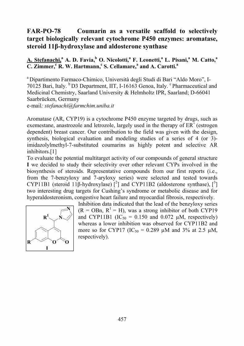
457
FAR-PO-78 Coumarin as a versatile scaffold to selectively target biologically relevant cytochrome P450 enzymes: aromatase, steroid 11β-hydroxylase and aldosterone synthase A. Stefanachi,a A. D. Favia,b O. Nicolotti,a F. Leonetti,a L. Pisani,a M. Catto,a C. Zimmer,c R. W. Hartmann,c S. Cellamare,a and A. Carotti.a
a Dipartimento Farmaco-Chimico, Università degli Studi di Bari “Aldo Moro”, I-70125 Bari, Italy. b D3 Department, IIT, I-16163 Genoa, Italy. c Pharmaceutical and Medicinal Chemistry, Saarland University & Helmholtz IPR, Saarland; D-66041 Saarbrücken, Germany e-mail: [email protected] Aromatase (AR, CYP19) is a cytochrome P450 enzyme targeted by drugs, such as exemestane, anastrozole and letrozole, largely used in the therapy of ER+ (estrogen dependent) breast cancer. Our contribution to the field was given with the design, synthesis, biological evaluation and modeling studies of a series of 4 (or 3)-imidazolylmethyl-7-substituted coumarins as highly potent and selective AR inhibitors.[1] To evaluate the potential multitarget activity of our compounds of general structure I we decided to study their selectivity over other relevant CYPs involved in the biosynthesis of steroids. Representative compounds from our first reports (i.e., from the 7-benzyloxy and 7-aryloxy series) were selected and tested towards CYP11B1 (steroid 11β-hydroxylase) [2] and CYP11B2 (aldosterone synthase), [3] two interesting drug targets for Cushing’s syndrome or metabolic disease and for hyperaldosteronism, congestive heart failure and myocardial fibrosis, respectively.
Inhibition data indicated that the lead of the benzyloxy series (R = OBn, R1 = H), was a strong inhibitor of both CYP19 and CYP11B1 (IC50 = 0.150 and 0.072 µM, respectively) whereas a lower inhibition was observed for CYP11B2 and more so for CYP17 (IC50 = 0.289 µM and 3% at 2.5 µM, respectively).
O OR
N
N
R1
I
458
A similar trend was observed for most of the analyzed benzyloxy-substituted congeners, the most potent and selective CYP11B1 inhibitor being the 3'-trifluoromethoxybenzyloxy derivative which exhibited an IC50 value of 5 nM. Preliminary structure-affinity and structure-selectivity relationships will be presented and discussed. [1]A. Stefanachi, A. D. Favia, O. Nicolotti, F. Leonetti, L. Pisani, Catto, M.; C. Zimmer, R. W. Hartmann, A. Carotti, J. Med. Chem., 54, 2011, 1613 and refs. therein. [2]U. E. Hille, C. Zimmer, C. A. Vock, R. W. Hartmann, ACS Med. Chem. Lett., 2, 2011, 2. [3]M. Voets, I. Antes, C. Scherer, U. Muller-Vieira, K. Biemel, C. Barassin, S. Marchais-Oberwinkler, R. W. Hartmann, J. Med. Chem. 48, 2005, 6632.
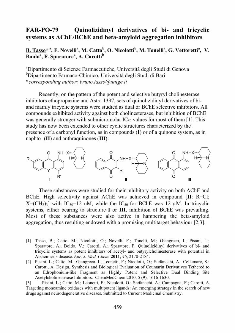
459
FAR-PO-79 Quinolizidinyl derivatives of bi- and tricyclic systems as AChE/BChE and beta-amyloid aggregation inhibitors B. Tasso*,a, F. Novellia, M. Cattob, O. Nicolottib, M. Tonellia, G. Vettorettia, V. Boidoa, F. Sparatorea, A. Carottib aDipartimento di Scienze Farmaceutiche, Università degli Studi di Genova bDipartimento Farmaco-Chimico, Università degli Studi di Bari *corresponding author: [email protected]
Recently, on the pattern of the potent and selective butyryl cholinesterase inhibitors ethopropazine and Astra 1397, sets of quinolizidinyl derivatives of bi- and mainly tricyclic systems were studied as dual or BChE selective inhibitors. All compounds exhibited activity against both cholinesterases, but inhibition of BChE was generally stronger with submicromolar IC50 values for most of them [1]. This study has now been extended to other cyclic structures characterized by the presence of a carbonyl function, as in compounds (I) or of a quinone system, as in naphto- (II) and anthraquinones (III):
Y
R
O
R'
NH XNH
O
O
R
NH XNH
O
O
NH XNH
I II III
These substances were studied for their inhibitory activity on both AChE and BChE. High selectivity against AChE was achieved in compound [II: R=Cl; X=(CH2)3] with IC50=12 nM, while the IC50 for BChE was 12 M. In tricyclic systems, either bearing to structure I or III, inhibition of BChE was prevailing. Most of these substances were also active in hampering the beta-amyloid aggregation, thus resulting endowed with a promising multitarget behaviour [2,3]. [1] Tasso, B.; Catto, M.; Nicolotti, O.; Novelli, F.; Tonelli, M.; Giangreco, I.; Pisani, L.;
Sparatore, A.; Boido, V.; Carotti, A.; Sparatore, F. Quinolizidinyl derivatives of bi- and tricyclic systems as potent inhibitors of acetyl- and butyrylcholinesterase with potential in Alzheimer’s disease. Eur. J. Med. Chem. 2011, 46, 2170-2184.
[2] Pisani, L.; Catto, M.; Giangreco, I.; Leonetti, F.; Nicolotti, O.; Stefanachi, A.; Cellamare, S.; Carotti, A. Design, Synthesis and Biological Evaluation of Coumarin Derivatives Tethered to an Edrophonium-like Fragment as Highly Potent and Selective Dual Binding Site Acetylcholinesterase Inhibitors. ChemMedChem 2010, 5 (9), 1616-1630.
[3] Pisani, L.; Catto, M.; Leonetti, F.; Nicolotti, O.; Stefanachi, A.; Campagna, F.; Carotti, A. Targeting monoamine oxidases with multipotent ligands: An emerging strategy in the search of new drugs against neurodegenerative diseases. Submitted to Current Medicinal Chemistry.
460
FAR-PO-80 Nutraceutical properties of red grape juice and its stability after processing for the formulation of food supplements
Gian Carlo Tenorea, Giacomo Pepeb, Luigi Coppolac, Ettore Novellinoa, Pietro Campigliab. aDepartment of Pharmaceutical and Toxicological Chemistry, University of Naples “Federico II”. bDepartment of Pharmaceutical and Biomedical Sciences, University of Salerno. c Coppola S.p.a., via S. Maria la Carità, Vicinale De Risi, Scafati (SA). [email protected] Experimental data have increasingly suggested that cellular oxidative damage induced by reactive oxygen species (ROS) has a relevant pathophysiological role in several types of human diseases, such as atherosclerosis and cancer [1]. Foods, particularly fruits and vegetables, have an important role in maintaining physiological redox equilibrium. These foods supply several antioxidants, such as vitamin C and polyphenolic compounds, to the body. Grapes are rich in phenolic compounds, such as flavonoids and resveratrol, which are mainly found in red grape products [2]. It has been already reported that grape juice compounds can prevent: (i) platelet aggregation, (ii) LDL oxidation and oxidative damage to DNA, (iii) coronary diseases and atherosclerosis [3]. Data on the effects of thermal treatments and subsequent storage on polyphenolics in fruits and vegetables are limited. However, several studies reported that processing may result in significant alterations in antioxidant compounds [4], especially anthocyanins, which may deeply influence antioxidant capacity, color and nutritional quality of food and food products. The aim of the present study was to evaluate the polyphenolic profile and antioxidant activity of red grape juice before and after freeze-drying. Scientific data on the composition of individual polyphenols in grape juice are scarce. In this study the sample was compared with the best red wines in the market and then the antioxidant activity of red grape juice was tested by DPPH and FRAP assays and compared with authentic standard. The sample after lyophilization revealed to keep quite unchanged both the polyphenolic composition and the antioxidant capacity when compared with the fresh product. [1] C. Dani et al., Food and Chem. Toxicol., 45, 2007, 2574-2580. [2] T. Fuleki et al., J. Agric. Food Chem., 51, 2003, 640-646. [3] K.W. Singletary, J. Agric. Food Chem., 51, 2003, 7280-7286. [4] T.J. Hager et al., J. Agric. Food Chem., 56, 2008, 689-695.
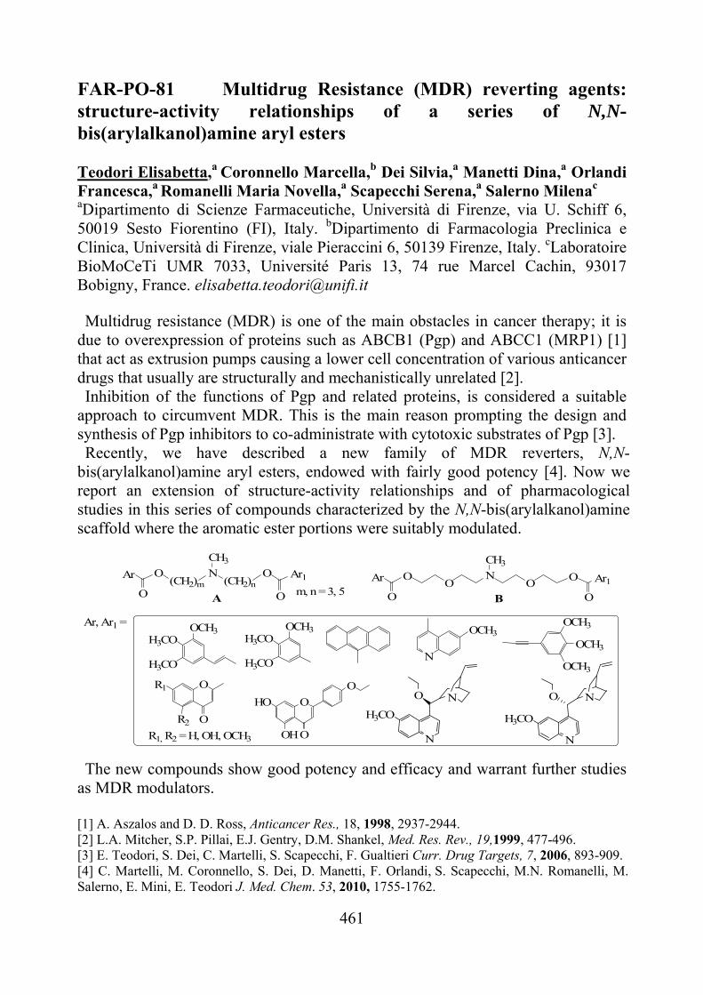
461
FAR-PO-81 Multidrug Resistance (MDR) reverting agents: structure-activity relationships of a series of N,N-bis(arylalkanol)amine aryl esters
Teodori Elisabetta,a Coronnello Marcella,b Dei Silvia,a Manetti Dina,a Orlandi Francesca,a Romanelli Maria Novella,a Scapecchi Serena,a Salerno Milenac
aDipartimento di Scienze Farmaceutiche, Università di Firenze, via U. Schiff 6, 50019 Sesto Fiorentino (FI), Italy. bDipartimento di Farmacologia Preclinica e Clinica, Università di Firenze, viale Pieraccini 6, 50139 Firenze, Italy. cLaboratoire BioMoCeTi UMR 7033, Université Paris 13, 74 rue Marcel Cachin, 93017 Bobigny, France. [email protected] Multidrug resistance (MDR) is one of the main obstacles in cancer therapy; it is
due to overexpression of proteins such as ABCB1 (Pgp) and ABCC1 (MRP1) [1] that act as extrusion pumps causing a lower cell concentration of various anticancer drugs that usually are structurally and mechanistically unrelated [2]. Inhibition of the functions of Pgp and related proteins, is considered a suitable
approach to circumvent MDR. This is the main reason prompting the design and synthesis of Pgp inhibitors to co-administrate with cytotoxic substrates of Pgp [3]. Recently, we have described a new family of MDR reverters, N,N-
bis(arylalkanol)amine aryl esters, endowed with fairly good potency [4]. Now we report an extension of structure-activity relationships and of pharmacological studies in this series of compounds characterized by the N,N-bis(arylalkanol)amine scaffold where the aromatic ester portions were suitably modulated.
N
CH3
(CH2)n(CH2)mO O N
CH3
O OA BO
Ar
O
Ar1 O
O
Ar O
O
Ar1
O
OR2
R1
O
O
HO
OH
O
H3CO
H3CO
OCH3H3CO
H3CO
OCH3
N
OCH3
OCH3
OCH3
OCH3
R1, R2 = H, OH, OCH3
O
N
H3CO
N O
N
H3CO
N
m, n = 3, 5
Ar, Ar1 =
The new compounds show good potency and efficacy and warrant further studies as MDR modulators.
[1] A. Aszalos and D. D. Ross, Anticancer Res., 18, 1998, 2937-2944. [2] L.A. Mitcher, S.P. Pillai, E.J. Gentry, D.M. Shankel, Med. Res. Rev., 19,1999, 477-496. [3] E. Teodori, S. Dei, C. Martelli, S. Scapecchi, F. Gualtieri Curr. Drug Targets, 7, 2006, 893-909. [4] C. Martelli, M. Coronnello, S. Dei, D. Manetti, F. Orlandi, S. Scapecchi, M.N. Romanelli, M. Salerno, E. Mini, E. Teodori J. Med. Chem. 53, 2010, 1755-1762.
462
FAR-PO-82 Liposome functionalized by Olygobranched Neurotensin peptides to delivery doxorubicin to tumour cells Diego Tesauro,a Antonella Accardo,a Barbara Ziacoc Chiara Falcianib , Barbara Lellib, Luisa Braccib, Giancarlo Morelli a
a Dipartimento delle Scienze Biologiche & CIRPeB, Università Federico II di Napoli & IBB CNR, Via Mezzocannone,16 80134 Napoli. bDipartimento di Biologia Molecolare, Università di Siena, Via Fiorentina 1, 53100 Siena c Invectors s.r.l. Via Pietro Castellino 111, 80131 Napoli In last years nanoparticles have been generated a strong interest for their potential application as in vivo carriers of active principles [1]. Especially liposome display unique pharmacokinetic properties slowly releasing drugs loaded in the inner aqueous cavity. Recently we have developed supramolecular aggregates decorated by bioactive peptide able to recognize overexpressed receptors on tumour cells membrane [2]. Here we present the synthesis, the structure and the in vitro behaviour of liposomes obtained by co-aggregation of the DOPC phospholipid with a new synthetic amphiphilic molecule, NT4Lys(C18)2, which contains a lysine scaffold derivatized with a lipophilic moiety and a tetra-branched neurotensin (NT1-13) peptides or a truncated form (NT8–13). The liposome were filled with the cytotoxic drug doxorubicin (Doxo). The synthesis was carried out on solid phase following a Fmoc strategy. The size of liposome was determinated by Dynamic light scattering measurements which indicate a value for the hydrodynamic radius (RH) of 88.3 ± 4.4 nm. The selective internalization and cytotoxicity of liposomes as compared to pure DOPC liposomes, were tested in HT29 human colon adenocarcinoma and TE671 human rhabdomyosarcoma cells, both of which express neurotensin receptors. FACS analysis indicates an increase in fluorescence signal of the NT4-liposomes, in both cell lines and the cytotoxicity is increased four-fold with respect to DOPC. These effects could to be ascribed to the higher rate of internalization for DOPC-NT4Lys(C18)2-Doxo liposomes, due to stronger binding driven by a lower dissociation constant of the NT4-liposomes that bind the membrane onto a specific protein, in contrast to DOPC liposomes, which approach the plasma membrane unselectively. [1] Torchilin Nat Rev drug 4, 2005, 145. [2] A. Accardo, et al. Molecular Biosystems, 6, 2010, 878.
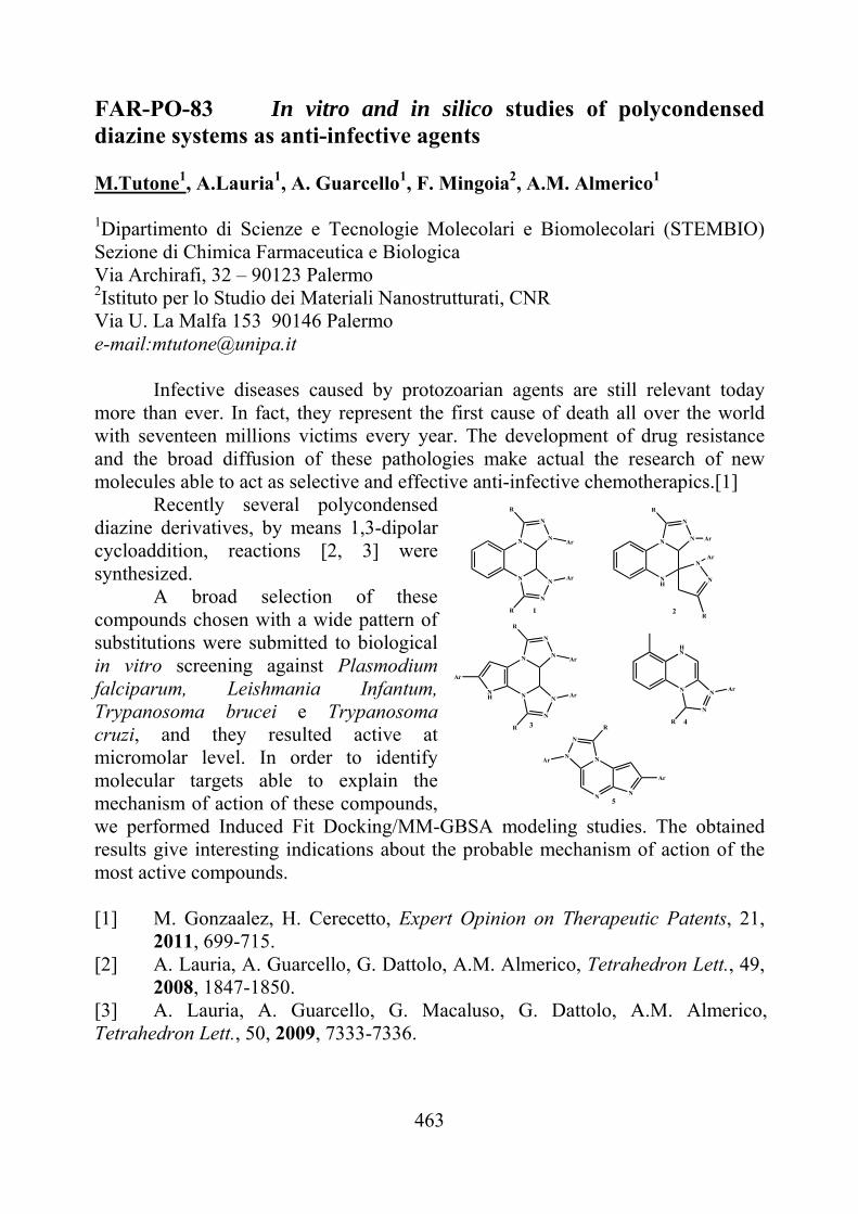
463
FAR-PO-83 In vitro and in silico studies of polycondensed diazine systems as anti-infective agents M.Tutone1, A.Lauria1, A. Guarcello1, F. Mingoia2, A.M. Almerico1
1Dipartimento di Scienze e Tecnologie Molecolari e Biomolecolari (STEMBIO) Sezione di Chimica Farmaceutica e Biologica Via Archirafi, 32 – 90123 Palermo 2Istituto per lo Studio dei Materiali Nanostrutturati, CNR Via U. La Malfa 153 90146 Palermo e-mail:[email protected]
Infective diseases caused by protozoarian agents are still relevant today
more than ever. In fact, they represent the first cause of death all over the world with seventeen millions victims every year. The development of drug resistance and the broad diffusion of these pathologies make actual the research of new molecules able to act as selective and effective anti-infective chemotherapics.[1]
Recently several polycondensed diazine derivatives, by means 1,3-dipolar cycloaddition, reactions [2, 3] were synthesized.
A broad selection of these compounds chosen with a wide pattern of substitutions were submitted to biological in vitro screening against Plasmodium falciparum, Leishmania Infantum, Trypanosoma brucei e Trypanosoma cruzi, and they resulted active at micromolar level. In order to identify molecular targets able to explain the mechanism of action of these compounds, we performed Induced Fit Docking/MM-GBSA modeling studies. The obtained results give interesting indications about the probable mechanism of action of the most active compounds. [1] M. Gonzaalez, H. Cerecetto, Expert Opinion on Therapeutic Patents, 21,
2011, 699-715. [2] A. Lauria, A. Guarcello, G. Dattolo, A.M. Almerico, Tetrahedron Lett., 49,
2008, 1847-1850. [3] A. Lauria, A. Guarcello, G. Macaluso, G. Dattolo, A.M. Almerico, Tetrahedron Lett., 50, 2009, 7333-7336.
N
N N
N
N
N
R
Ar
R
Ar
NH
N N
N
N
NAr
Ar
R
R
N
N N
N
N
N
R
Ar
R
Ar
NH
Ar
N
HN
N
NAr
R
N
N
N
N
N
Ar
R
Ar
1 2
3 4
5
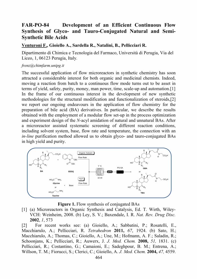
464
FAR-PO-84 Development of an Efficient Continuous Flow Synthesis of Glyco- and Tauro-Conjugated Natural and Semi-Synthetic Bile Acids
Venturoni F., Gioiello A., Sardella R., Natalini, B., Pellicciari R.
Dipartimento di Chimica e Tecnologia del Farmaco, Università di Perugia, Via del Liceo, 1, 06123 Perugia, Italy.
The successful application of flow microreactors in synthetic chemistry has soon attracted a considerable interest for both organic and medicinal chemists. Indeed, moving a reaction from batch to a continuous flow mode turns out to be asset in terms of yield, safety, purity, money, man power, time, scale-up and automation.[1] In the frame of our continuous interest in the development of new synthetic methodologies for the structural modification and functionalization of steroids,[2] we report our ongoing endeavours in the application of flow chemistry for the preparation of bile acid (BA) derivatives. In particular, we describe the results obtained with the employment of a modular flow set-up in the process optimization and experiment design of the N-acyl amidation of natural and unnatural BAs. After a microreactor assisted systematic screening of different reaction conditions, including solvent system, base, flow rate and temperature, the connection with an in-line purification method allowed us to obtain glyco- and tauro-conjugated BAs in high yield and purity.
CO2H
R2HOH T °C
10 ml
PS-SA
BPR
R2HOH
NH2HO2C
O
NH
CO2H
N OEt
OEtO
+
+
EEDQ
Glycine
Organic Solvent
H2O
Base
Flow Rate
Flow Rate
BA
GBA
R2HOH
O
NH
TBA
SO3H
or
HO3S
Taurine
NH2
or
R
R1
R1
R1
R
R
Figure 1. Flow synthesis of conjugated BAs [1] (a) Microreactors in Organic Synthesis and Catalysis, Ed. T. Wirth, Wiley-
VCH: Weinheim, 2008. (b) Ley, S. V.; Baxendale, I. R. Nat. Rev. Drug Disc. 2002, 1, 573
[2] For recent works see: (a) Gioiello, A.; Sabbatini, P.; Rosatelli, E., Macchiarulo, A.; Pellicciari, R. Tetrahedron 2011, 67, 1924. (b) Sato, H.; Macchiarulo, A.; Thomas, C.; Gioiello, A.; Une, M.; Hofmann, A. F.; Saladin, R.; Schoonjans, K.; Pellicciari, R.; Auwerx, J. J. Med. Chem. 2008, 51, 1831. (c) Pellicciari, R.; Costantino, G.; Camaioni, E.; Sadeghpour, B. M.; Entrena, A.; Willson, T. M.; Fiorucci, S.; Clerici, C.; Gioiello, A. J. Med. Chem. 2004, 47, 4559.
465
FAR-PO-85 Design, synthesis and biological evaluation of PTPRJ binding Peptides Ermelinda Vernieria, Isabel Gomez-Monterreyb, Marina Salaa, Alfonso Carotenutob, Paolo Griecob, Francesco Trapassoc, Stefano Alcarod, Francesco Ortusod, Ettore Novellinob, Pietro Campiglia.a
aDepartment of Pharmaceutical and Biomedical Sciences, University of Salerno. bDepartment of Pharmaceutical and Toxicological Chemistry, University of Naples “Federico II”. cDepartment of Experimental and Clinical Medicine, University of Catanzaro. dDepartment of Pharmacobiological Science, University of Catanzaro. [email protected] PTPRJ (a receptor-type protein tyrosine phosphatase also named DEP-1, HPTPeta, or CD148) is of particular interest for its role in human and experimental tumorigenesis. After its discovery, a consistent body of literature showed the inhibiting effect of the PTPRJ on several players of the mitogenic signal in both normal and cancer cells. In fact, PTPRJ is able to interact and dephosphorylate numerous receptor tyrosine kinases (RTKs) such as PDGFR, HGFR, RET, and EGFR whose aberration or overexpression in cancer cells is responsible of self-sufficiency cell growth. The role of PTPRJ on the inhibition of RTKs was also extended to VEGFR, whose activity is required for the formation of new vessels in tumor progression (angiogenesis).[1] These findings indicate a tumor suppressor activity for PTPRJ and make it an interesting candidate for the generation of novel therapeutic strategies. In a previous study, we described the isolation and characterization of synthetic PTPRJ binding peptides from a combinatorial phage display library. The cyclic peptides 1 and 2 induced dephosphorylation of PTPRJ targets and moderate cell growth inhibition in HeLa and Huvec tumor cell lines.
1 [Cys-His-His-Asn-Leu-Thr-His-Ala-Cys]-OH 2 [Cys-Leu-His-His-Tyr-His-Gly-Ser-Cys]-OH
In this communication, we present the synthesis, the biological activities and the structure-activity relationships of new analogues of peptides 1 and 2 which we considered as a valuable starting point for the development of a novel class of PTPRJ agonists. [1] Takamune Takahashi et al., Blood, 2006, Volume 108, Number 4.
466
FAR-PO-86 Structure-Activity Relationships Studies of CaMKIINtide Analogues Ermelinda Vernieria, Isabel Gomez-Monterreyb, Marina Salaa, Alessia Bertaminob, Simona Musellab, Maddalena Illarioc, Maria Rosaria Ruscianoc,
Paolo Griecob, Guido Iaccarinod, Carlo Franchinie, Ettore Novellinob, Pietro Campigliaa.
aDepartment of Pharmaceutical and Biomedical Science, University of Salerno. bDepartment of Pharmaceutical and Toxicological Chemistry, University of Naples “Federico II”. cDepartment of Experimental Pharmacology, University of Naples, “Federico II”. d Department of Clinical Medicine Cardiovascular and Immunological Sciences, University of Naples “ Federico II”. eDepartment of Medicinal Chemistry, University of Bari.
[email protected] Ca2+ /calmodulin (CaM)-dependent protein kinase is a multifunctional Ser/Thr protein kinase that plays an important role in many cellular function including cell division, differentiation, cardiac contraction and synaptic plasticity [1].
Over the past decade, several CaMKII inhibitors have been reported to study CaMKII function. Most of these compounds showed low potency and absence of highly specific inhibition. In the research of major selectivity the natural CaMKII inhibitor protein, CaM-KIIN, provides a promising alternative, because it potently inhibits CaMKII but not CaMKI, CaMKIV, PKA or PKC. In previous studies COOH-terminal truncations of CaM-KIIN indicated that its inhibitory potency and activities resided largely in a 27 aminoacid residues. This peptide, named CaM-KNtide (KRPPKLGQIGR SKRVVIEDDRIDDVLK) showed a similar IC50 value (50 nM) for both the total and the Ca2+-independent CaM-KII activities. As part of our current interest in the study of CaMKII-dependent cell signaling that regulates some many physiological function, we directed our efforts toward the identification of a novel peptide CaMKII inhibitors. Here, we present synthesis, conformational studies and biological evaluation of different CaM-KNtide analogues. [1]Tsui, J.; Inagaki, M.; Schulman, H. J Biol Chem. 2005, Mar 11, 280(10), 9210-6.
[2]Chang B.H., Mukherji S., Soderling T.R. Proc. Natl. Acad. Sci. USA 1998, 95, 10890-10895.
467
FAR-PO-87 [1.1.1.]Cryptand: A Bifunctional Kinetic Molecular Device for 1H NMR Automatic Determination of pH-Rate Profile of Drugs Iole Villaria, Giuseppe Alibrandib, Carmelo Lo Vecchiob, Antonino Villaria aDipartimento Farmaco-Chimico, Facoltà di Farmacia, Università di Messina, Villaggio SS. Annunziata, 98166 Messina. bDipartimento di Chimica Inorganica, Chimica Analitica e Chimica Fisica, Università di Messina, Viale F. Stagno d’Alcontres 31, 98166 Messina. [email protected] A bifunctional molecular device is a chemical species able to perform two specific works at a molecular level. [1.1.1]Cryptand [1a] falls into this definition because it is able 1) to change slowly, in an almost linear way,[2] the pH in a reaction medium, working as a molecular titrator, and 2) to measure it, as a molecular pH-meter, making it possible to carry out automatic 1H NMR reaction monitoring for kinetic or thermodynamic investigation, without using physical devices like autoburette and pH-meter. The device performs this role by means of a mechanism [1b] where 1) a fast and reversible protonation followed by a slow, irreversible and highly specific entrapping of the hydrogen ion produce the increase of pH and 2) the partition between unreacted protonated and deprotonated species, in fast equilibrium with each other, produces an averaged CH2N chemical shift linked to the pH by the Henderson-Hasselbalch equation (Eqn. 1). This kinetic molecular device (KMD), used before for spectrophotometric automatic titrations,[3a] can be used to carry out unprecedented 1H NMR automatic titrations [3b] for the determination of the pKa of molecules of pharmaceutical interest. In this contribution it has been used to carry out, for the first time, a variable-pH kinetic experiment where the hydrolysis of aspirin, a classic pH-sensitive reaction, has been followed by 1H NMR while the pH was changing in a controlled way. The kinetic profile obtained has been processed using a mathematical model (Eqn. 2; A = 1H NMR peak area of acetylsalicylic acid) to obtain the entire pH-rate profile of the drug in the range explored.
pH pKa log
a
b (1)
-
1
A
dA
dt kobs[pH(t )] (2)

468
The bifunctionality allows to obtain a variable-pH monitoring without using an added molecular pH-meter. In this way the device system takes only ca. 1 ppm of the entire spectrum and leaves all the rest available for the reaction monitoring.
[1] a) J. Cheney, J. M. Lehn, J. Chem. Soc. Chem. Commun. 1972, 487–488; b) P. B. Smith, J. L. Dye, J. Cheney, J. M. Lehn, J. Am. Chem. Soc. 1981, 103, 6044–6048.
[2] a) G. Alibrandi, Angew. Chem., Int. Ed., 47, 2008, 3026–3028; b) Alibrandi, G.; Lo Vecchio, C.; Lando, G. Angew. Chem., Int. Ed, 121, 2009, 6450–6452. [3] a) Alibrandi, G.; Lo Vecchio, C.; Villari, A; Villari, I. Chem. Eur. J., 16, 2010, 7700–7703; b) Alibrandi, G.; Arena, G. C.; Lando, G.; Lo Vecchio, C.; Parisi, F. M. Chem. Eur. J., 17, 2011, 1419–1422.
469
FAR-PO-88 The discovery of a carnosine derivative (FL-926-A16) as selective and efficient sequestering agent of cytotoxic carbonyl species: from molecular design to preclinical studies Vistoli G.(1), Orioli M.(1), Regazzoni L.(1), Canevotti, R.(2), Gamberoni L.(2), Menini S.(3), Pugliese G.(3), Carini M.(1), Aldini G(1) (1)Department of Pharmaceutical Sciences "Pietro Pratesi". Università degli Studi di Milano, via Mangiagalli 25, 20133, Milan, Italy. (2)Flamma S.p.A., Chignolo d'Isola (BG), Italy. (3)Department of Clinical and Molecular Medicine, La Sapienza University, Rome, Italy [email protected] Reactive carbonyl species (RCS) are involved in the pathogenesis of several human diseases. Hence RCS, beside to be considered a biomarker of oxidative damage, are also potential targets for the development of bioactive compounds acting as detoxifying agents of RCS (carbonyl quenching compounds) [1]. We found that the natural dipeptide carnosine (-alanyl-L-histidine) is a selective and potent RCS sequestering agent, even if its clinical application is limited due to the rapid hydrolysis in blood by a specific dipeptidase (carnosinase). Consequently, we developed a drug discovery approach aimed to design, synthesize and evaluate novel CAR peptidomimetics which, beside to maintain or improve the reactivity and selectivity of carnosine towards RCS, are recognized by hPepT1 and hence transported through an active transport but not recognized by carnosinase. The metabolic stability of the synthesized compounds was studied by incubating them with rat plasma and human serum as well as with rat and human liver fractions and reactivity (HNE as substrate) and selectivity (pyridoxal) studied in in vitro models. The pharmacokinetic profile for the most promising derivative (FL-926-A16) was then investigated in rats and its ability to quench HNE and to reduce protein carbonylation and tissue damage was demonstrated in different animal models (db/db mouse and nApoE null mice). [1] Aldini G, Orioli M, Rossoni G, Savi F, Braidotti P, Vistoli G, Yeum KJ, Negrisoli G, Carini M., J Cell Mol Med, 2010, Jun 1. [Epub ahead of print].
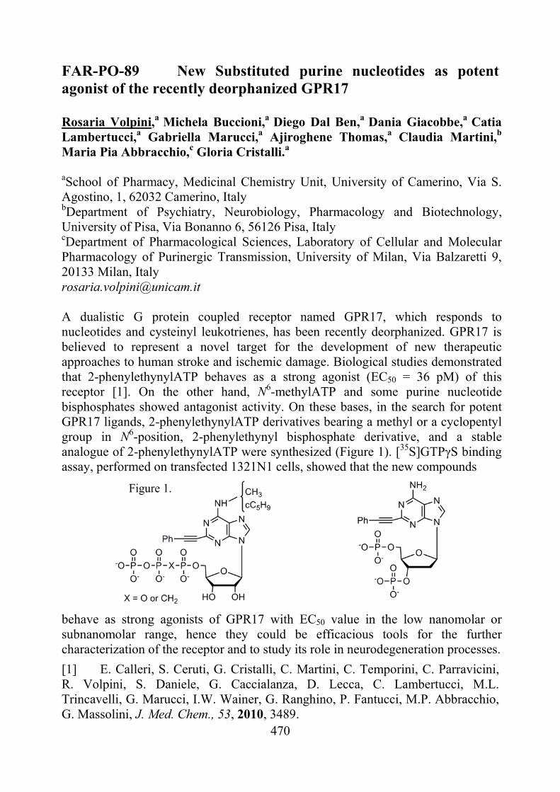
470
FAR-PO-89 New Substituted purine nucleotides as potent agonist of the recently deorphanized GPR17 Rosaria Volpini,a Michela Buccioni,a Diego Dal Ben,a Dania Giacobbe,a Catia Lambertucci,a Gabriella Marucci,a Ajiroghene Thomas,a Claudia Martini,b Maria Pia Abbracchio,c Gloria Cristalli.a aSchool of Pharmacy, Medicinal Chemistry Unit, University of Camerino, Via S. Agostino, 1, 62032 Camerino, Italy bDepartment of Psychiatry, Neurobiology, Pharmacology and Biotechnology, University of Pisa, Via Bonanno 6, 56126 Pisa, Italy cDepartment of Pharmacological Sciences, Laboratory of Cellular and Molecular Pharmacology of Purinergic Transmission, University of Milan, Via Balzaretti 9, 20133 Milan, Italy [email protected] A dualistic G protein coupled receptor named GPR17, which responds to nucleotides and cysteinyl leukotrienes, has been recently deorphanized. GPR17 is believed to represent a novel target for the development of new therapeutic approaches to human stroke and ischemic damage. Biological studies demonstrated that 2-phenylethynylATP behaves as a strong agonist (EC50 = 36 pM) of this receptor [1]. On the other hand, N6-methylATP and some purine nucleotide bisphosphates showed antagonist activity. On these bases, in the search for potent GPR17 ligands, 2-phenylethynylATP derivatives bearing a methyl or a cyclopentyl group in N6-position, 2-phenylethynyl bisphosphate derivative, and a stable analogue of 2-phenylethynylATP were synthesized (Figure 1). [35S]GTPγS binding assay, performed on transfected 1321N1 cells, showed that the new compounds
behave as strong agonists of GPR17 with EC50 value in the low nanomolar or subnanomolar range, hence they could be efficacious tools for the further characterization of the receptor and to study its role in neurodegeneration processes.
[1] E. Calleri, S. Ceruti, G. Cristalli, C. Martini, C. Temporini, C. Parravicini, R. Volpini, S. Daniele, G. Caccialanza, D. Lecca, C. Lambertucci, M.L. Trincavelli, G. Marucci, I.W. Wainer, G. Ranghino, P. Fantucci, M.P. Abbracchio, G. Massolini, J. Med. Chem., 53, 2010, 3489.
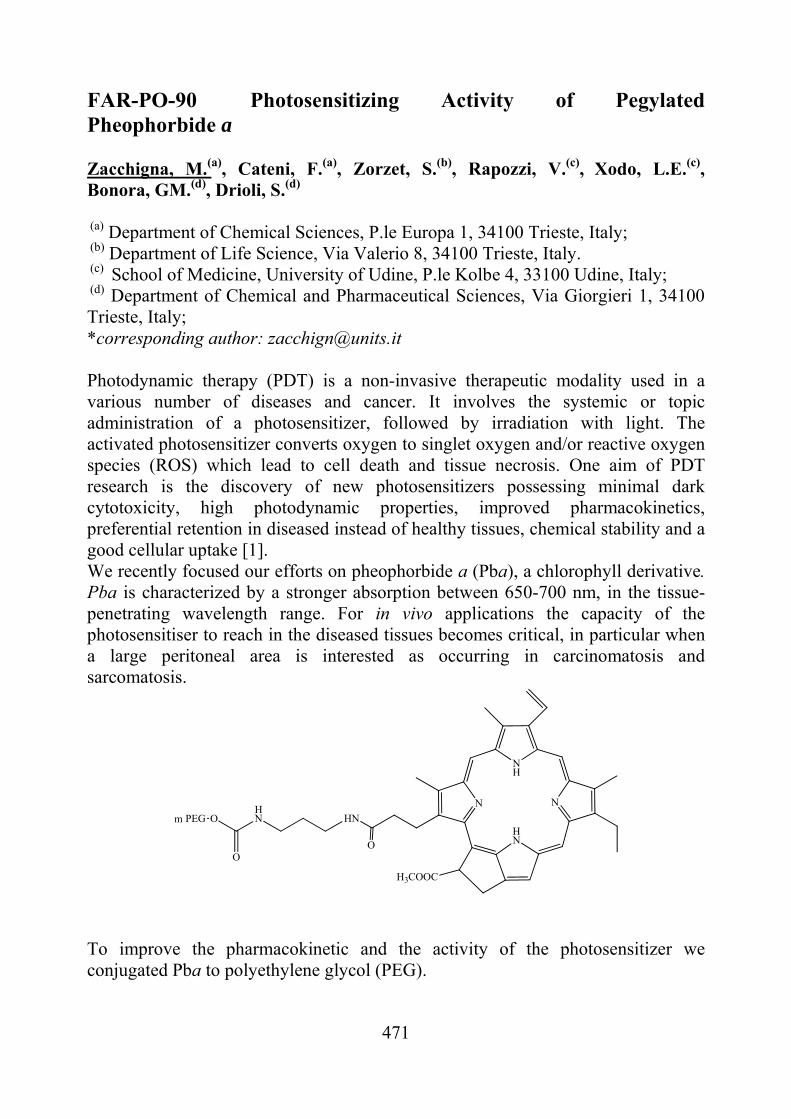
471
FAR-PO-90 Photosensitizing Activity of Pegylated Pheophorbide a Zacchigna, M.(a), Cateni, F.(a), Zorzet, S.(b), Rapozzi, V.(c), Xodo, L.E.(c), Bonora, GM.(d), Drioli, S.(d) (a) Department of Chemical Sciences, P.le Europa 1, 34100 Trieste, Italy; (b) Department of Life Science, Via Valerio 8, 34100 Trieste, Italy. (c) School of Medicine, University of Udine, P.le Kolbe 4, 33100 Udine, Italy; (d) Department of Chemical and Pharmaceutical Sciences, Via Giorgieri 1, 34100 Trieste, Italy; *corresponding author: [email protected] Photodynamic therapy (PDT) is a non-invasive therapeutic modality used in a various number of diseases and cancer. It involves the systemic or topic administration of a photosensitizer, followed by irradiation with light. The activated photosensitizer converts oxygen to singlet oxygen and/or reactive oxygen species (ROS) which lead to cell death and tissue necrosis. One aim of PDT research is the discovery of new photosensitizers possessing minimal dark cytotoxicity, high photodynamic properties, improved pharmacokinetics, preferential retention in diseased instead of healthy tissues, chemical stability and a good cellular uptake [1]. We recently focused our efforts on pheophorbide a (Pba), a chlorophyll derivative. Pba is characterized by a stronger absorption between 650-700 nm, in the tissue-penetrating wavelength range. For in vivo applications the capacity of the photosensitiser to reach in the diseased tissues becomes critical, in particular when a large peritoneal area is interested as occurring in carcinomatosis and sarcomatosis.
N
NH
N
HN
H3COOC
O
HNHN
O
OPEGm
To improve the pharmacokinetic and the activity of the photosensitizer we conjugated Pba to polyethylene glycol (PEG).
472
In vivo the pharmacokinetic analysis performed on living female C57/BL6 mice bearing a subcutaneous melanoma mass, showed that injected mPEG-Pba distributes all over the body, with an higher uptake in the tumor respect to free Pba. Moreover, preliminary data suggest that PEG-Pba in mice bearing B78-H1 amelanotic melanoma reduces the tumor growth after light activation in comparison with Pba. [1] Josefsen, L.B. and Boyle, R.W., 2008. Photodynamic therapy and the development of metal-based photosensitisers. Met. Based Drugs, 276109-276133.
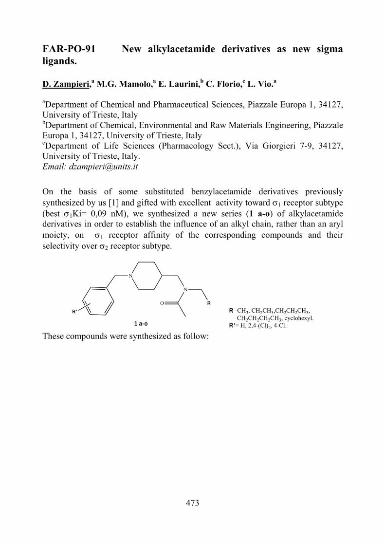
473
FAR-PO-91 New alkylacetamide derivatives as new sigma ligands. D. Zampieri,a M.G. Mamolo,a E. Laurini,b C. Florio,c L. Vio.a
aDepartment of Chemical and Pharmaceutical Sciences, Piazzale Europa 1, 34127, University of Trieste, Italy bDepartment of Chemical, Environmental and Raw Materials Engineering, Piazzale Europa 1, 34127, University of Trieste, Italy cDepartment of Life Sciences (Pharmacology Sect.), Via Giorgieri 7-9, 34127, University of Trieste, Italy. Email: [email protected]
On the basis of some substituted benzylacetamide derivatives previously synthesized by us [1] and gifted with excellent activity toward 1 receptor subtype (best 1Ki= 0,09 nM), we synthesized a new series (1 a-o) of alkylacetamide derivatives in order to establish the influence of an alkyl chain, rather than an aryl moiety, on 1 receptor affinity of the corresponding compounds and their selectivity over 2 receptor subtype.
N
N
RO
R' R=CH3, CH2CH3,CH2CH2CH3, CH2CH2CH2CH3, cyclohexyl.R'= H, 2,4-(Cl)2, 4-Cl.1 a-o
These compounds were synthesized as follow:
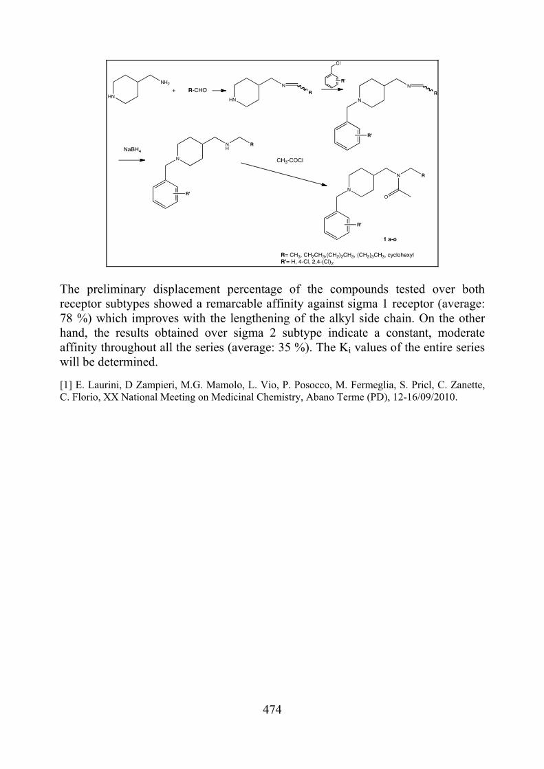
474
The preliminary displacement percentage of the compounds tested over both receptor subtypes showed a remarcable affinity against sigma 1 receptor (average: 78 %) which improves with the lengthening of the alkyl side chain. On the other hand, the results obtained over sigma 2 subtype indicate a constant, moderate affinity throughout all the series (average: 35 %). The Ki values of the entire series will be determined. [1] E. Laurini, D Zampieri, M.G. Mamolo, L. Vio, P. Posocco, M. Fermeglia, S. Pricl, C. Zanette, C. Florio, XX National Meeting on Medicinal Chemistry, Abano Terme (PD), 12-16/09/2010.
475
FAR-PO-92 Design, synthesis and molecular modeling studies on peptidomimetic vinyl esters as falcipan-2 inhibitors Roberta Ettari,a, Nicola Micale,a Floriana Bova,a Giovanni Grazioso,b Tanja Schirmeister,c Silvana Grassoa, Maria Zappalàa
aDipartimento Farmaco-Chimico, University of Messina, Viale Annunziata, 98168 Messina, Italy bDipartimento di Scienze Farmaceutiche “Pietro Pratesi”, Università degli Studi di Milano, Via Mangiagalli 25, 20133 Milano, Italy cInstitute of Pharmacy and Food Chemistry, University of Würzburg, Am Hubland, D-97074 Würzburg, Germany E-mail: [email protected]
Malaria is currently endemic in 106 countries, with an estimated 225
million clinical cases and nearly 781,000 deaths in 2009 [1]. To date resistance has emerged to all common antimalarial drugs, including recently artemisinins. In this context a new important target for antimalarial drug design is represented by falcipain-2 (FP-2), a hemoglobinase of P. falciparum food vacuole [2]. FP-2 is able also orchestrate selective proteolytic events during the release of malaria parasite from human red blood cells, because of its ability to cleavate the cytoskeletal proteins ankyrin and the band 4.1 protein.
In this regard our research group actively worked in the last years on the development of novel peptidomimetic FP-2 inhibitors, containing a 1,4-benzodiazepine (BDZ) scaffold [3] introduced into the dipeptide sequence D-Ser-Gly. Among all the FP-2 inhibitors synthesized, the Michael acceptor vinyl ester 1 [4] (Figure 1) has been shown to be the most potent and selective FP-2 inhibitor. In order to investigate the structure-activity relationship of the lead compound 1, novel vinyl ester derivatives have been designed (Figure 1) [5], by introducing different substituents on the fused benzene ring or at C-4’ of the phenyl substituent on the BDZ scaffold. Additionally the 4-chloro-2-trifluoromethylphenyl group, linked by means of a carbamoyl moiety to the side chain of the P3 site serine, has been replaced with other aromatic rings or with cyclo(alkyl)groups in such a way to investigate the ability of the P3 pocket to accommodate groups of different size. Lately the methyl substituent of the parent inhibitor 1 has been replaced with an ethyl and isobutyl or benzyl group in order to evaluate size and characteristics of the P1’ liphophilic pocket. In the present work we now report docking studies in order to explain the different degree of biological activity of synthesized inhibitors as well as their reversible or irreversible inhibition mode of FP-2.

476
NH
O
O
NN
O
NH
O
R'''COOR
R'
R''
NH
O
O
NN
O
NH
O
Cl
CF3
COOCH3
1
R=Et, i-Bu, BnR'=H, ClR''=H, Br, OCH3
R''' =4-MeO-C6H4, 1-naphthyl,n-C4H9, c-C6H11
Figure 1
[1] World Malaria Report 2010 http://www.who.int/malaria/world_malaria_report_2010/en;
[2] S.X. Wang, K.C. Pandey, J.R. Somoza, et al. Proc. Natl. Acad. Sci. U.S.A. 103, 2006, 11503.
[3] N. Micale, A. P. Kozikowski, R. Ettari, S. Grasso, M. Zappalà, A. Kumar, M. Hanspal, J.-J. Jeong, A. H. Chishti. J. Med. Chem., 49, 2006, 3064.
[4] R. Ettari, N. Micale, T. Schirmeister, R. Vičík, E. Nizi, M. E. Di Francesco, S. Grasso, M. Zappalà, J. Med. Chem., 52, 2009, 2157.
[5] F. Bova, R. Ettari, N. Micale C. Carnovale, T. Schirmeister, C. Gelhaus, M. Leippe, S. Grasso, M. Zappalà. Bioorg. Med. Chem., 18, 2010, 4928.
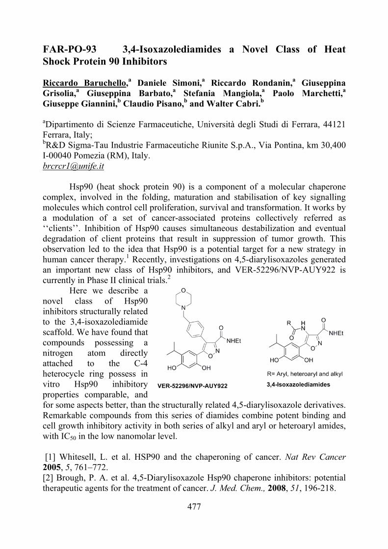
477
FAR-PO-93 3,4-Isoxazolediamides a Novel Class of Heat Shock Protein 90 Inhibitors Riccardo Baruchello,a Daniele Simoni,a Riccardo Rondanin,a Giuseppina Grisolia,a Giuseppina Barbato,a Stefania Mangiola,a Paolo Marchetti,a Giuseppe Giannini,b Claudio Pisano,b and Walter Cabri.b aDipartimento di Scienze Farmaceutiche, Università degli Studi di Ferrara, 44121 Ferrara, Italy; bR&D Sigma-Tau Industrie Farmaceutiche Riunite S.p.A., Via Pontina, km 30,400 I-00040 Pomezia (RM), Italy. [email protected]
Hsp90 (heat shock protein 90) is a component of a molecular chaperone complex, involved in the folding, maturation and stabilisation of key signalling molecules which control cell proliferation, survival and transformation. It works by a modulation of a set of cancer-associated proteins collectively referred as ‘‘clients’’. Inhibition of Hsp90 causes simultaneous destabilization and eventual degradation of client proteins that result in suppression of tumor growth. This observation led to the idea that Hsp90 is a potential target for a new strategy in human cancer therapy.1 Recently, investigations on 4,5-diarylisoxazoles generated an important new class of Hsp90 inhibitors, and VER-52296/NVP-AUY922 is currently in Phase II clinical trials.2
Here we describe a novel class of Hsp90 inhibitors structurally related to the 3,4-isoxazolediamide scaffold. We have found that compounds possessing a nitrogen atom directly attached to the C-4 heterocycle ring possess in vitro Hsp90 inhibitory properties comparable, and for some aspects better, than the structurally related 4,5-diarylisoxazole derivatives. Remarkable compounds from this series of diamides combine potent binding and cell growth inhibitory activity in both series of alkyl and aryl or heteroaryl amides, with IC50 in the low nanomolar level. [1] Whitesell, L. et al. HSP90 and the chaperoning of cancer. Nat Rev Cancer 2005, 5, 761–772. [2] Brough, P. A. et al. 4,5-Diarylisoxazole Hsp90 chaperone inhibitors: potential therapeutic agents for the treatment of cancer. J. Med. Chem., 2008, 51, 196-218.
478
FAR-PO-94 Quantification of artemisinin in Artemisia annua Herbal Tea and test In Vitro for Anti-Malarial Activity Adele Idoloa, Tiziana Grassi a, Marcello Guidoa, Paride Papadiab, Luciano Villanovac, Alessandro Merendinoc, Francesco Paolo Fanizzib, Antonella De Donnoa. aDepartment of Biological and Environmental Science and Technology, Laboratory of Hygiene, University of the Salento, via Prov.le Lecce-Monteroni, 73100 Lecce, Italy. bDepartment of Biological and Environmental Science and Technology, Laboratory of General and Inorganic Chemistry, University of the Salento, 73100 Lecce, Italy. cDepartment of Research and Development, LACHIFARMA S.r.l., 73100 Zollino (Lecce), Italy [email protected]
A. annua was usually used to prepare a tea and if it contains effective amounts of artemisinin, it might be used today as a self-reliant treatment of malaria [1]. Artemisia annua tea has proven itself to be a very effective treatment for malaria in various clinical trials, but to date, his efficacy has not been investigated in vitro. Therefore, we have carried out a study for the evaluation of effects of A. annua tea on Plasmodium falciparum cultures in vitro. We also determined the concentration of artemisinin in herbal tea preparation.
The compound was tested against chloroquine-sensitive D10 and chloroquine-resistant W2 strains of P. falciparum using the parasite lactate dehydrogenase assay [2]. The quantification of artemisinin in the extract of leaves of A. annua was obtained using an 1H NMR method.
The in vitro tests conducted in this study confirm the clinical efficacy demonstrated by the tea of A. annua in vivo on both chloroquine-sensitive D10 and chloroquine-resistant W2 strains. The concentration of artemisinin in A. annua tea is lower with respect to that of pure artemisinin responsible for the same antimalarial activity. The artemisinin present in the tea is probably co-solubilised with other ingredients, some of which may also have antimalarial activity and act synergistically with it. The presence of other active ingredients suggests that A. annua is a natural artemisinin combination therapy. These compounds also merit further research, to see whether their presence hinders the development of parasite resistance compared to pure artemisinin [3]. [1] C.H.Blanke, G.B.Naisabha, M.B.Balema, G.M.Mbaruku, L.Heide and
M.S.Muller, Trop. Doct., 38, 2008, 113–6. [2] M.T.Makler, J.M.Ries, J.A.Williams, J.E.Bamcroft, R.C.Piper,
B.L.Gibbins, et al, Am.J.Trop.Med.Hyg., 48(6), 1993, 739-41. [3] Work supported by Regione Puglia Progetto Strategico PS70.