chimeric calicivirus-like particles elicit specific immune responses in pigs
TRANSCRIPT

C
EPa
b
c
d
e
f
g
a
ARRAA
KRV3FP
1
tairttaparo
IS
0d
Vaccine 30 (2012) 2427–2439
Contents lists available at SciVerse ScienceDirect
Vaccine
journa l homepage: www.e lsev ier .com/ locate /vacc ine
himeric calicivirus-like particles elicit specific immune responses in pigs
. Crisci a, L. Frailea,b, N. Morenoc, E. Blancoc, R. Cabezónd, C. Costae, T. Mussáa, M. Baratelli a,
. Martinez-Orellanaa, L. Gangesa, J. Martínez f, J. Bárcenac, M. Montoyaa,g,∗
Centre de Recerca en Sanitat Animal (CReSA), UAB-IRTA, Campus de la Universitat Autònoma de Barcelona, 08193 Bellaterra, SpainUniversitat de Lleida, Lleida, SpainCentro de Investigación en Sanidad Animal (CISA-INIA), Valdeolmos, 28130 Madrid, SpainFundació Clínic per la Recerca Biomèdica, Centre Esther Koplowitz, Barcelona, SpainNew Therapies of Genes and Transplants Group, Institut d’Investigació Biomèdica de Bellvitge (IDIBELL), L’Hospitalet de Llobregat, 08908 Barcelona, SpainDepartament de Sanitat i Anatomia Animals, Universitat Autònoma de Barcelona, SpainInstitut de Recerca i Tecnologia Agroalimentàries (IRTA), Barcelona, Spain
r t i c l e i n f o
rticle history:eceived 8 October 2011eceived in revised form 20 January 2012ccepted 22 January 2012vailable online 3 February 2012
eywords:HDV-VLPsaccine vectorAMDV
a b s t r a c t
Virus-like particles (VLPs) have received considerable attention due to their potential application in vet-erinary vaccines and, in particular, VLPs from rabbit haemorrhagic disease virus (RHDV) have successfullyshown to be good platforms for inducing immune responses against an inserted foreign epitope in mice.The aim of this study was to assess the immunogenicity of chimeric RHDV-VLPs as vaccine vectors in pigs.For this purpose, we have generated chimeric VLPs containing a well-known T epitope of 3A protein offoot-and-mouth disease virus (FMDV). Firstly, RHDV-VLPs were able to activate immature porcine bonemarrow-derived dendritic cells (poBMDCs) in vitro. Secondly, pigs were inoculated twice in a two-weekinterval with chimeric RHDV-VLPs at different doses intranasally or intramuscularly. One intramuscu-larly treated group was also inoculated with adjuvant MontanideTM ISA 206 at the same time. SpecificIgG and IgA antibodies against RHDV-VLPs were induced and such levels were higher in the adjuvanted
igs group compared with other groups. Interestingly, anti-RHDV-VLP IgA responses were higher in groupsinoculated intramuscularly than those that received the VLPs intranasally. Two weeks after the last immu-nisation, specific IFN-�-secreting cells against 3A epitope and against RHDV-VLPs were detected in PBMCsby ELISPOT. The adjuvanted group exhibited the highest IFN-�-secreting cell numbers and lymphoprolif-
onseimer
erative specific T cell respon the potential use of ch
. Introduction
Virus-like particles (VLPs) have received considerable atten-ion due to their potential application in vaccines, gene therapynd drug delivery [1]. Indeed, VLPs are robust protein cagesn the nanometer range exhibiting well-defined geometry andemarkable uniformity [1] that mimic the overall structure ofhe native virions. VLPs are appealing as vaccine candidates forheir inherent properties of stability and non-replicative form,nd have advantages in terms of immunogenicity and safety overrevious approaches [2]. In fact, lacking the genome of the virus
voids any of the risks associated with virus replication, reversion,ecombination or re-assortment [3]. The strong immunogenicityf VLPs, the high productivity of expression systems, their ability∗ Corresponding author at: Centre de Recerca en Sanitat Animal (CReSA), UAB-RTA, Campus de la Universitat Autònoma de Barcelona, 08193 Bellaterra, Barcelona,pain. Tel.: +34 93 581 4562; fax: +34 93 581 4490.
E-mail address: [email protected] (M. Montoya).
264-410X/$ – see front matter © 2012 Elsevier Ltd. All rights reserved.oi:10.1016/j.vaccine.2012.01.069
s against 3A epitope and RHDV-VLP. This is the first immunological reportic RHDV-VLPs as antigen carriers in pigs.
© 2012 Elsevier Ltd. All rights reserved.
to achieve a rapid implementation at production scale and thecost-effectiveness have encouraged researchers in the field [4].Additionally, VLPs have self-adjuvanting ability and stimulatestrong B-cell-mediated and cellular immune responses [5].
The two most successful VLP-based vaccines licensed andapproved for use in humans are hepatitis B and human papillo-mavirus vaccines and progresses have been made in developingVLPs for hepatitis C virus, Ebola virus, Marburg virus, SARScoronavirus and Chikungunya virus [6]. Also in veterinary vacci-nology, new VLP-based vaccine candidates have been elaboratedfor different viruses including porcine parvovirus [4], porcineencephalomyocarditis [7], canine parvovirus [8,9], goose par-vovirus [10], equine rhinitia A virus [11], bluetongue virus [12,13],chicken anaemia virus [14,15] and infectious bursal disease virus[16]. Although multiple candidates are in course of study in the vet-erinary field [17], only porcine circovirus type 2 (PCV2) VLP-based
®
vaccine, Ingelvac CircoFLEX developed by Boehringer Ingelheim(Germany) is commercially available. Besides, for the same virus,another sub-unit commercial vaccine, Porcilis PCV® (Intervet Inter-national, The Netherlands) is commercialised.
2 ine 30
tvfd
hffbcaumitgccnsu
pagatiibpposl[pc
csdsipcttodtiRath
TO
428 E. Crisci et al. / Vacc
In addition of being used to induce immune responses againsthe particle itself, animal VLPs have been successfully used asaccine vectors for inducing immune responses against insertedoreign immunogenic epitopes (chimeric VLPs) [18–20] or for drugelivery [1].
VLPs derived from rabbit haemorrhagic disease virus (RHDV)ave been shown to be highly immunogenic and protect rabbits
rom a lethal challenge with the virus [21–23]. These VLPs areormed from 180 copies of the single coat protein (VP60), assem-led into an icosahedral capsid [24]. Recently, we have shown thathimeric RHDV-VLPs harbouring a model cytotoxic T-cell epitopet the N-terminal end of the VP60 monomers were able to stim-late a powerful specific cytotoxic response in vivo and to protectice from viral challenge [18]. Moreover, a tumour antigen chem-
cally coupled to RHDV-VLPs has been shown to delay or preventhe development of tumours [19]. The well-documented immuno-enicity of VLPs is probably due to their interaction with dendriticells (DCs) [25]. RHDV-VLPs enter both human and murine DCs bylathrin-dependent pinocytosis and phagocytosis. Win et al. foundo evidence for receptor-mediated acquisition by DCs from eitherpecies, which may be related to the fact that RHDV does not nat-rally infect mice or humans [26].
Foot-and-mouth disease virus (FMDV) is a picornavirus thatroduces a highly transmissible and devastating disease in farmnimals and other cloven-hoofed livestock [27]. FMDV shows a highenetic and antigenic variability, reflected in the seven serotypesnd the numerous variants described to date [28]. Several T-cell epi-opes frequently recognised by natural host lymphocytes have beendentified in FMDV proteins. One of these T-cell epitopes, locatedn residues 21–35 of FMDV NS protein 3A, is efficiently recognisedy lymphocytes from infected pigs. This epitope was capable torovide adequate T-helper co-operation when synthesised juxta-osed to the B-cell antigenic site VP1, inducing significant levelsf serotype-specific anti-FMDV activity in vitro [29]. Its amino acidequence is conserved among FMDV types A, O, and C, showingimited variation among isolates from the seven FMDV serotypes30]. Cubillos et al. have shown the successful use of a dendrimericeptide, using such 3A and VP1 epitopes, to protect pigs against ahallenge with FMDV [31].
Taking into account the strong potential of RHDV-VLPs as vac-ine vectors, our aim was to develop these VLPs as a deliveryystem for the multimeric presentation of immunogenic epitopeserived from pathogens relevant for animal health, in livestockpecies. As a first approach we generated chimeric RHDV-VLPsncorporating a well defined T-helper epitope [29] from FMDV 3Arotein (RHDV-3A-VLPs), and analysed their ability to induce spe-ific immune responses in pigs. The foreign epitope was inserted athe N-terminus of VP60 protein, which is predicted to be buried inhe internal face of the VLPs. Firstly, the immunogenic potentialf RHDV-VLPs in immature porcine bone marrow derived den-ritic cells (poBMDCs) in vitro was studied. RHDV-VLPs were ableo stimulate poBMDCs in vitro. Then, fifty conventional pigs weremmunised with two doses at two week interval with chimeric
HDV-3A-VLPs using different routes. Acute phase proteins werelso analysed in chimeric RHDV-3A-VLP immunised pigs sinceheir responses comprise innate reactions [32]. Moreover, specificumoral and cellular responses against the inserted epitope andable 1ligonucleotide primers used for cloning.
Primer Sequence (5′–3′)a
Bac1F GACTCCAAGTGTGTGGGTGAAGTCNT3A15F TTTGAGGGCATGGTACACGACTCCATTAAAGCCCGCNT3A15R GAGTCGTGTACCATGCCCTCAAAGAATTCAATGGCTVP60PR TCCGAAGATCTCAGACATAAGAAAAGCCATTG
a Restriction site sequences are underlined. Start and stop codons are shown in boldfac
(2012) 2427–2439
the vector were specifically induced. This is the first immunologi-cal report on the potential use of RHDV-VLPs as vectors for antigenpresentation in pigs in which specific immunity against a foreignepitope was induced.
2. Materials and methods
2.1. Virus and cells
Derivatives of Autographa californica nuclear polyhedrosis virus(AcMNPV) were used to obtain the recombinant baculovirusesexpressing RHDV-VLPs. Baculoviruses were propagated in insectcell lines grown in suspension or monolayer cultures at 28 ◦C inTNM-FH medium (Sigma, St. Louis, MO, USA) supplemented with5% foetal calf serum (FCS), as described previously [33]. Spodopterafrugiperda cells (SF9) were used for generation of recombinant bac-uloviruses, plaque assays, and the preparation of high titre viralstocks. Trichoplusia ni cells (H5) were used for high level expressionof recombinant proteins.
2.2. Construction of recombinant baculovirus transfer vectors
The baculovirus transfer vector chosen was plasmid pBac-PAK8XB. This plasmid is a derivative of pBacPAK8 (Clontech, TakaraBio Europe), in which several restriction sites were eliminatedfrom the multiple cloning site [24]. The full-length VP60 gene ofRHDV was subcloned in pBacPAK8XB, generating plasmid pMVP60[24]. The sequence coding the T-helper epitope AAIEFFEGMVHD-SIK, derived from the 3A protein of FMDV [29], was insertedat the 5′ end of the VP60 gene by performing two sequentialPCRs. First, two separate PCRs were performed using the primerpairs Bac1F/NT3A15R and NT3A15F/VP60PR (Table 1), and plasmidpMVP60 as template. The PCR products obtained were gel puri-fied, denatured and annealed together in a secondary PCR reactionin which the extended template was amplified using the externalprimers Bac1F/VP60PR. The PCR product obtained was cloned intothe unique BglII restriction site of pBacPAK8XB generating pNT15.The inserted sequence in the resulting recombinant plasmid wasverified by sequence analysis.
2.3. Generation of recombinant baculoviruses
The recombinant baculoviruses were produced using the Bac-PAK baculovirus expression system (Clontech, Takara Bio Europe)as described previously [24]. Briefly, monolayers of SF9 insectcells were cotransfected with recombinant transfer vectors andBsu36I triple-cut AcMNPV DNA [34] using lipofectamine (Invit-rogen). Recombinant baculoviruses were selected on the basis oftheir LacZ-negative phenotypes, plaque purified, and propagatedas described elsewhere [35].
2.4. Expression and purification of the recombinant RHDV-VLPs
The recombinant constructs VP60 and NT15 were expressedin H5 insect cell-cultures infected with the corresponding recom-binant baculoviruses, as previously described [33]. To purify the
ACAGCGCCGGCCATATGAAGATCTTCTAGGATCGATCCG
e.

ine 30
sbittV6ps1NbctpV(ciPada
2d
fe(om(g(StGtaamdcddi1tc12wIdcwo
2
wW1
E. Crisci et al. / Vacc
elf-assembled VLPs, H5 cell monolayers were infected with recom-inant baculoviruses at a multiplicity of infection of 10. After
ncubation (4 days, 28 ◦C), infected cells were gently dislodged intohe growth medium and collected. The resulting suspensions werehen washed three times with 0.2 M phosphate-buffered saline forLPs (PBS-V, consisting of 0.2 M sodium phosphate, 0.1 M NaCl, pH.0) in order to separate intact cells from the culture medium. Theellets were then resuspended in distilled water, subjected to mildonication and treated with DNAse I (Roche Applied Science) forh at RT. Next, samples were adjusted to 2% Sarkosyl (sodium-lauroylsarcosine, Sigma) and 5 mM EDTA in PBS-V, and incu-ated overnight (ON) at 4 ◦C. Subsequently, the cell lysates werelarified by low-speed centrifugation and the supernatant was cen-rifuged at 27,000 rpm for 2 h with a Beckman SW28 rotor. Theelleted material was resuspended in PBS-V, extracted twice withertrel XF (Fluka, Sigma–Aldrich), and subjected to centrifugation
at 35,000 rpm for 2.5 h with a Beckman SW55 rotor) through aushion of 1.5 ml of PBS-V with 15% Opti-prep (a 60% solution ofodixanol in water, Invitrogen). Pellets were finally resuspended inBS-V containing protease inhibitors (Complete, Roche) and storedt 4 ◦C. The protein concentrations of the VLP preparations wereetermined with a bicinchoninic acid protein assay kit (BCA proteinssay kit, Pierce, Thermo Scientific Inc., USA).
.5. Porcine bone-marrow derived and human monocyte-derivedendritic cell generation
Porcine bone marrow (BM) haematopoietic cells were obtainedrom femurs of clinically healthy Large White × Landrace pigs ofight weeks of age. Porcine bone marrow derived dendritic cellspoBMDCs) were generated by an eight-day protocol as previ-usly described by [36] with some modifications [37]. Briefly, bonearrow haematopoietic cells were resuspended in RPMI-1640
Lonza, Walkesville, USA) culture medium containing 2 mM of l-lutamine (Invitrogen®, Barcelona, Spain), 100 U/ml of Polymixin BSigma–Aldrich Quimica, S.A., Madrid, Spain) 10% of FCS (Euroclone,ziano, Italy) and 100 �g/ml of penicillin with 100 U/ml of strep-omycin (Invitrogen®, Barcelona, Spain). Recombinant porcineM-CSF (R&D Systems, Spain) was added at 100 ng/ml to the cells
hree times during the culture within 2 days intervals. Stimuli weredded at day 8 and on day 9, mature poBMDCs were harvestednd analysed by flow cytometry and cytokine production. Humanonocyte derived dendritic cells (huMoDCs) were prepared as
escribed previously [38]. Briefly, peripheral blood mononuclearells (PBMC) were isolated by Ficoll 1.077 (Sigma–Aldrich, Spain)ensity gradient centrifugation after leukapheresis of healthyonors performed at the Banc de Sang i Teixits (Barcelona). For
solation by adherence, PBMC were seeded in a concentration of× 108 per 75 cm2 tissue culture flasks (Corning, Spain) in 8 ml cul-
ure medium and incubated for 1–2 h at 37 ◦C, 5% CO2. Non adherentells were washed away extensively with PBS. To the adherent cells,0 ml of X-VIVO 15 medium (BioWhittaker, Walkersville, MD) with% human serum (Sigma–Aldrich, Spain) was added, of which halfas replaced on day one with culture medium supplemented with
L-4 (300 U/ml) and GM-CSF (450 U/ml) (Miltenyi Biotec, Spain). Atay 3, all cells were harvested using cold PBS and seeded at 1 × 10⁶
ells in 2 ml fresh culture medium per well of a 6-well plate. Stimuliere added at day 6 and mature DCs were harvested and analysed
n day 7 by flow cytometry and cytokine production.
.6. Stimulation of poBMDCs and huMoDCs
For in vitro studies, poBMDCs and huMoDCs were stimulatedith different concentrations of RHDV-VLPs (10 and 50 �g/ml).e used as positive controls LPS (Sigma) at a concentration of
�g/ml for huMoDCs and 10 �g/ml for poBMDCs, and poly:IC
(2012) 2427–2439 2429
(50 �g/ml, Sigma) for poBMDCs. Briefly, stimuli were plated withpoBMDCs after 8 days of culture (5 × 10⁵ cells/well) in 96-wellplates or with huMoDCs after 6-day culture (106 cells/well) in 6-well plates. Activation of DCs was analysed by flow cytometry at24 h post-stimulation or by cytokines release in supernatant at dif-ferent time-points (4, 8, 16, 24 h post-stimulation) using specificELISAs. Non-stimulated cells cultured in media served as controls.Data were obtained from four poBMDC experiments, correspond-ing each one to a different animal, and for huMoDCs from twodifferent individuals.
2.7. Flow cytometry analyses of poBMDCs and huMoDCs
Flow cytometry analysis of poBMDCs was performed usingan indirect labelling for CD172a, SLA-I, SLA-II, CD4, CD1c,CD11R3, CD11R1, SWC1, CD40, CD80/86, CD86 and CD163 anddirect labelling for CD14 and CD16. Unless specified below,the reagents were detected by hybridoma supernatants. Briefly,5 × 105 cells/50 �l/well were labelled during 1 h at 4 ◦C for eachCD marker, using 50 �l of antibody solution. Anti-CD172a (SWC3,BA1C11), anti-SLAI (4B7/8), anti-SLAII (1F12), anti-CD1c (76-7-4),anti-CD4 (76-12-4), anti-CD11R1 (MIL4, IgG1, Serotec), anti-CD11R3 (2F4/11), purified anti-human CD40 (G28.4, Biolegend,San Diego, CA, USA), anti-SWC1 (76-6-7), anti-CD14-FITC (MIL2,Serotec, bioNova cientifica, Madrid, Spain), anti-CD16-FITC (G7,Serotec), CTLA4-mIg (Ancell, MN, USA), purified anti-porcine CD86(mouse mAb 5B9.88, Alexion Pharmaceuticals, Cheshire, CT, USA)[39] and anti-CD163 (2A10/11) were used. After incubation, theywere washed with cold PBS with 2% FCS by centrifugation at 450 × g,4 ◦C for 5 min. Then, the secondary antibody R-phycoerythryn anti-mouse IgG (Jackson ImmunoResearch, Suffolk, UK) diluted 1:300was added when required. Cells were incubated for 1 h at 4 ◦C, theywere washed as before and resuspended in PBS with 2% FCS. Stainedcells were acquired on Coulter® EPICS XL-MCL cytometer and ana-lysed by EXPO 32 ADC v.1.2 program. A gate strategy was appliedin 85% of living cells using the forward and side scatter (FS/SS).
To characterise and compare the phenotype of the huMoDCpopulations the following mAbs or appropriate isotype controlswere used: anti-CD14, CD80, CD83, CD86, CCR7, MHC-I and MHC-II(all from BD Biosciences, Mountain View, CA). Primary antibodieswere followed by staining with PE-labelled goat-anti-mouse (BDBioscience). Flow cytometry was performed with FACSCanto IITM
with FACSDiva software (BD Biosciences). The histogram analysiswas performed with Weasel v3.0 (Walter and Eliza Hall Institute,Melbourne, Australia).
2.8. Cytokine ELISAs
Cytokine levels in conditioned cell supernatants were assayedby ELISA for porcine IFN-�, TNF-�, IL-18, IL-6 and IL-10. Differenttime-points (4, 8, 16, 24 h) were tested. For each ELISA, triplicatewells of stimulated- or un-stimulated-cell supernatants were usedand all the results were analysed with KC Junior Program (BioTek Instruments, Inc.) using the filter Power Wave XS reader. Todetect IFN-�, an in-house ELISA for anti-IFN-� (around 4 U/mlof detection limit) was performed using commercial antibodiespurchased from PBL Biomedical Laboratories (Piscataway, NJ, USA)as previously described [40]. For TNF-�, IL-6 and IL-10 detection,a DuoSet® ELISA Development system (R&D Systems, Abingdon,UK) was used following manufacturer’s instructions. The limit ofdetection was around: 148 pg/ml for TNF-�, 70 pg/ml for IL-6 and50 pg/ml for IL-10. For IL-18 secretion, the kit Pig IL-18 Module
Set BMS672MST (Bender Med Systems, Vienna, Austria) wasused following manufacturer’s instructions (limit of detectionwas around 74.5 pg/ml). Supernatants of human DC cultureswere collected after stimulation for 24 h. IL-10, IFN-� (Bender
2430 E. Crisci et al. / Vaccine 30
Table 2Experimental design.
Group Route Doses (�g/pig)
A – – (n = 5)B
1IN
20 (n = 5)2 60 (n = 5)3 180 (n = 5)
C1
IM + ADJ
20 (n = 5)2 60 (n = 5)3 180 (n = 5)
D1 20 (n = 5)
Ma3r
2
Wlrvtbaai0t
bA(aoiwdtRewtC2so
Cvhl8
2
m(
IM2 60 (n = 5)3 180 (n = 5)
edSystems) and TNF-� (Mabtech) were analysed by ELISAccording to the manufacturer’s guidelines using Spectramax40PC 384 reader (Molecular Devices). The detection limits wereespectively 4.7 pg/ml, 7.8 pg/ml, 4.7 pg/ml and 15.6 pg/ml.
.9. Experimental design
At the age of 6–7 weeks, fifty male conventional pigs (Largehite × Landrace) were selected from a high health status farm
ocated in the Northern part of Spain; these pigs were porcineeproductive and respiratory syndrome virus (PRRSV), influenzairus and Mycoplasma hyopneumoniae negative at the beginning ofhe experiment. Animals were clinically healthy when the studyegan. Pigs received non-medicated commercial feed ad libitumnd had free access to drinking water. Animals were housed inn experimental farm (CEP, Torrelameau, Lleida, Spain) contain-ng 12–14 piglets per pen. The space available for the animals was.75 m2/pig. The building was equipped with manual mechanismso control ventilation.
At the beginning of the experiment pigs were identified, dou-le ear-tagged and randomly distributed into four groups, namely(n = 5), B (n = 15), C (n = 15) and D (n = 15) balanced by weight
Table 2). Pigs of group A remained untreated and were used as neg-tive controls. Groups B, C and D were inoculated twice with 1 mlf RHDV-3A-VLPs in a two-week interval. Subgroups were organ-sed as summarised in Table 2. Group B was intranasally inoculated
ith 20, 60 and 180 �g per dose of chimeric RHDV-3A-VLPs onay 0. Group C and D animals were intramuscularly inoculated inhe right neck muscle with abovementioned amounts of chimericHDV-3A-VLPs and pigs from group C received the different dosesmulsified with the adjuvant MontanideTM ISA 206 (SEPPIC) in 1 mlith equal proportions. Pigs were monitored daily for immunisa-
ion reactions and samples of blood and saliva (using Salivette®
otton Swab from SARSTEDT, Spain) were collected at day 0, 14 and8 after the beginning of the experiment. Fourteen days after theecond immunisation pigs were euthanised with an intravenousverdose of sodium pentobarbital.
The experiment received prior approval from the Ethicalommittee for Animal Experimentation of the Institution (Uni-ersitat Autònoma de Barcelona). The treatment, housing andusbandry conditions conformed to the European Union Guide-
ines (The Council of the European Communities 1986, EU directive6/609/EEC).
.10. Detection of haptoglobin and pentraxin 3 in serum
Haptoglobin (Hp) was quantified by a spectrophotometricethod (haemoglobin binding assay) with commercial reagents
Tridelta Development Limited, Ireland) and performed on an
(2012) 2427–2439
automatic analyser (Olympus AU400, Hamburg, Germany) aspreviously described [41].
Porcine pentraxin 3 (PTX3) concentration in sera was deter-mined as previously described [42] by sandwich ELISA againstmurine PTX3 (MuPTX3) with the following antibodies: 2C3 andbiotinylated 6B11. Values of OD were analysed at 450 nm and wecount as positive sera those giving OD values above the cut-off ofthe assay (0.09).
2.11. Detection of specific anti-RHDV-VLP antibodies by ELISA
Antibodies against RHDV-VLP were examined in serum sam-ples collected at days 14 and 28 by ELISA. Briefly, Maxisorp 96-wellELISA plates (Nunc) were coated with RHDV-VLP (50 ng/well), incarbonate/bicarbonate buffer (pH 9.6), ON at 4 ◦C. Duplicate four-fold dilution series of each serum sample were made, starting at1/50. Fifty microlitre volumes were used throughout. Specific anti-bodies were detected with horseradish peroxidase conjugated withprotein G (Pierce).
RHDV-VLP-specific IgG1, IgG2 (in sera) and IgA (in sera andsaliva) were measured following the same procedure described butreplacing the protein G-HRPO by monoclonal antibodies specific forthese isotypes, supplied by Serotec, and using as secondary anti-body a goat anti-mouse IgG (H + L)-HRP (Invitrogen). In the case ofsaliva, two consecutive incubations with sample were performedbefore adding the commercial monoclonal antibody to porcine IgA,in order to increase the sensitivity of the assay. Colour develop-ment was obtained after addition of the substrate chromophore,OPD (total Ig analyses) or TMB (isotyping) and stopped by adding aH2SO4 solution. Plates were read in an automatic microplate reader(Fluostar Omega) at 492 and 450 nm, respectively. Antibody titres(total Ig and isotypes) were therefore expressed as the reciprocallog10 of serum dilutions giving the absorbance recorded in the con-trol wells (serum collected day 0) plus twice standard deviation.These data were calculated by interpolation.
2.12. Detection of specific anti-FMDV epitope T-3A antibodies byELISA
Serum samples were examined for the presence of antibodiesagainst NSP 3A T-cell epitope, displayed in the RHDV-VLP. Assaywas performed in 96-well Immobilizer Amino Plates (Nunc) coatedwith 4 �g/well of T-3A synthetic peptide (kindly supplied by D.Andreu and B.G. De la Torre, Pompeu Fabra University, Spain),in PBS buffer and ON at 4 ◦C. Duplicate serum samples diluted1/50 were assayed and specific antibodies were detected withhorseradish peroxidase conjugated with protein G (Pierce), andcolour development was obtained after addition of TMB substrate.We count as positive sera those giving OD values above the cut-offof the assay (0.2).
2.13. ELISPOT assay
Two and four weeks after the first immunisation, (PBMC) werecollected and analysed for specific IFN-� production by ELISPOTset following manufacturer’s instructions (Becton Dickinson, UK).PBMCs were isolated by Histopaque-1.077® gradient and plated induplicate at 5 × 10⁵/100 �l/well in RPMI-1640 supplemented with10% FCS into 96-well plates (MultiScreen® MAHAS4510 Millipore)coated ON at 4 ◦C with 5 �g/ml anti-pig IFN-�-specific capturemAb (P2G10, Becton Dickinson, UK) 100 �l/well. For the in vitroantigen recall, 35 �g/ml of 3A peptide or 20 �g/ml RHDV-VLPs
were used as stimuli. As positive control, cells were incubated with10 �g/ml phytohaemagglutinin (PHA) (Sigma) and cells incubatedin the absence of antigen were use as negative control. Plateswere cultured for 72 h at 37 ◦C, then incubated with 2 �g/ml of
ine 30
bbIw(cso
2
dupBamRCi4oaDbi(p(
2
(dipwf
m1tii
2
wawWit(ot
3
3F
e
E. Crisci et al. / Vacc
iotinylated anti-IFN-� mAb (P2C11, Becton Dickinson), followedy streptavidin–horseradish peroxidase conjugates (Jacksonmmunoresearch Lab.). The presence of IFN-�-producing cells
as visualised using 3-amino-9-ethylcarbazole (AEC) substrateSigma). The background values (number of spots in negativeontrol wells) were subtracted from the respective counts of thetimulated cells and immune responses were expressed as numberf spots per million of PBMCs.
.14. Lymphoproliferation assay
Proliferation assays of swine lymphocytes were performed asescribed previously [43]. Blood was collected in 5 �M EDTA andsed immediately for the preparation of PBMCs [44]. Assays wereerformed in 96-well round-bottomed microtitre plates (Nunc).riefly, 2.5 × 10⁵ PBMCs per well were cultured in triplicate, infinal volume of 200 �l, in complete RPMI, 10% FCS, 50 �M 2-ercaptoethanol, in the presence of various concentrations of (i)
HDV-VLP at 20 �g/ml and (ii) 3A synthetic peptide, at 20 �g/ml.ultures with medium alone or with mock-infected cells were
ncluded as controls. Cells were incubated at 37 ◦C in 5% CO2 fordays. Following incubation, each well was pulsed with 0.5 �Ci
f [methyl-3H]thymidine for 18 h. The cells were collected usingcell harvester and the incorporation of radioactivity into the
NA was measured by liquid scintillation counting with a Micro-eta counter (Pharmacia). Results were expressed as stimulation
ndexes (SI), which were calculated as the mean counts per minutecpm) of stimulated cultures/mean cpm of cultures grown in theresence of medium alone (peptide) or mock-stimulated cellsRHDV-VLPs).
.15. Pathological analysis
The objective of the pathologic studies at the site of injectionright neck muscle, brachiocephalicus) was to establish any reactionue to the intramuscular immunisation. Tissue samples from the
noculation point were fixed in 10% buffered formalin and routinelyrocessed for histopathology. Sections 4 �m thick were cut, stainedith haematoxylin and eosin (H–E) and observed in a blinded-
ashion method.Lesions were classified regarding to the severity of the inflam-
atory reaction. Thus, lesions scores were as follows: 0 (no lesions);(mild): small accumulation of cells in the perimuscular adipose
issue; 2 (moderate): muscular tissue was mildly infiltrated bynflammation; 3 (severe): extended areas of muscular tissue werenfiltrated and loss of muscular fibres and fibrosis were observed.
.16. Statistical analysis
All statistical analyses were performed using SPSS 15.0 soft-are (SPSS Inc., Chicago, IL, USA). For all analyses, pig was used
s the experimental unit. The significance level (˛) was set at 0.05ith statistical tendencies reported when p < 0.10. The Shapiroilk’s and the Levene test were used to evaluate the normal-
ty of the distribution of the examined quantitative variables andhe homogeneity of variances, respectively. A non-parametric testMann–Whitney) was chosen to compare the different valuesbtained for all the immunological parameters between groups allhe sampling times.
. Results
.1. Generation of chimeric RHDV-VLPs carrying 3A T epitope of
MDVIn order to analyse the potential of RHDV-VLPs as a deliv-ry system for foreign T cell epitopes in pigs, we produced
(2012) 2427–2439 2431
recombinant baculoviruses expressing a RHDV VP60 constructharbouring an epitope derived from the 3A protein of FMDV (AAIEF-FEGMVHDSIK) fused to the N-terminus. Expression was verified bySDS-10% PAGE (data not shown). Preparations were characterisedby electron microscopy to confirm that the chimeric VP60 constructself-assembled into VLPs (data not shown).
3.2. Maturation of poBMDCs after RHDV-VLP stimulation
Once chimeric RHDV-VLPs expressing 3A epitope were gener-ated, their immunogenicity was studied by analysing whether ornot RHDV-VLPs were able to stimulate poBMDCs in vitro. Con-sistent with previous reports [36,37], poBMDCs phenotype atday eight of culture was CD172a+, SLAI+, SLAII+, CD1+, CD4−,CD11R1−, CD14+, CD16+, CD40−, CD80/86+ and CD163low. Atthis time, the population of poBMDCs was rather homogeneousand they constituted our starting culture for further experi-ments. Porcine BMDCs maturation induced by RHDV-VLPs wasevaluated after 24 h of stimulation. Pulsing poBMDCs with RHDV-VLPs resulted in increased levels of maturation markers SLA-II,CD80/86 and CD86 that in some cases reached higher levels thatafter LPS stimulation (Fig. 1). A dose-dependent up-regulationof SLA-II and CD80/86 was found (Suppl. Fig. 1). No differ-ences were found in all other surface markers assessed (data notshown).
3.3. RHDV-VLPs induce secretion of cytokines by DCs
To investigate whether RHDV-VLPs were capable of inducingcytokine production in DCs, levels of TNF-�, IL-18, IL-6, IL-10,IL-12 and IFN-� were determined in supernatants of poBMDCscultured in the presence or absence of different concentrationof RHDV-VLPs at different time-points. LPS or poly:IC were usedas positive controls. RHDV-VLPs induced the production of TNF-� and IL-6, with maximum levels between 16 and 24 h (Fig. 2)and the differences with un-stimulated DCs showed a strongstatistical tendency (0.07 < p value < 0.05). Secretion of IL-6 andTNF-� in response to the different RHDV-VLPs occurred moreslowly than that induced by LPS or poly:IC, which rapidly inducedinflammatory cytokine as early as 4 h after stimulation. Moreover,RHDV-VLP-stimulation was lower than LPS or poly:IC induc-tion (Fig. 2) with a statistical tendency (0.08 < p value < 0.05).RHDV-VLPs did not induce DCs to secrete IFN-�, IL-10 and IL-18, at least not above the detection limit of the assay (data notshown).
3.4. Detection of acute phase proteins in serum
Then, groups of conventional pigs were inoculated through dif-ferent routes and with different amount of chimeric RHDV-VLPs asstated in Section 2 and Table 2. Firstly, acute phase proteins such ashaptoglobin (Hp) and PTX3 were analysed to determine whetherthey could influence immune responses in the treated animals. Pro-tein levels were tested in sera after each RHDV-3A-VLP injection.No differences were found in Hp production between the variouscohorts studied at day 14 and day 28 (data not shown). On the con-trary, PTX3 levels in sera were higher in animals from groups C1and D1 compared with control animals at day 14, but then PTX3decreased at day 28 and only animals in group D1 remained statis-tically higher than control animals (Table 3). Moreover, animals ingroup C1 exhibited PTX3 levels statistically higher when compared
with all animals in group B at day 14, although these differenceswere no longer observed at day 28 (Table 3). All the other groupsshowed no significant differences at either day 14 or 28 (data notshown).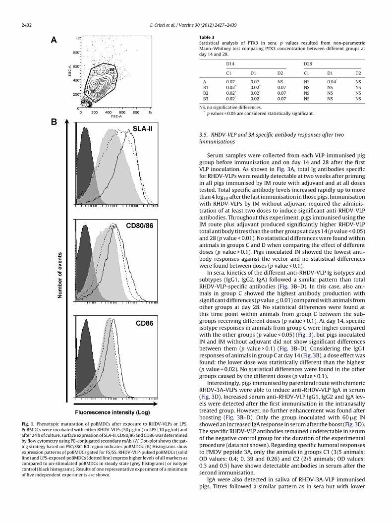
2432 E. Crisci et al. / Vaccine 30 (2012) 2427–2439
Fig. 1. Phenotypic maturation of poBMDCs after exposure to RHDV-VLPs or LPS.PoBMDCs were incubated with either RHDV-VLPs (50 �g/ml) or LPS (10 �g/ml) andafter 24 h of culture, surface expression of SLA-II, CD80/86 and CD86 was determinedby flow cytometry using PE-conjugated secondary mAb. (A) Dot-plot shows the gat-ing strategy based on FSC/SSC. R0 region indicates poBMDCs. (B) Histograms showexpression patterns of poBMDCs gated for FS/SS. RHDV-VLP-pulsed poBMDCs (solidline) and LPS-exposed poBMDCs (dotted line) express higher levels of all markers ascompared to un-stimulated poBMDCs in steady state (grey histograms) or isotypecontrol (black histograms). Results of one representative experiment of a minimumof five independent experiments are shown.
Table 3Statistical analysis of PTX3 in sera. p values resulted from non-parametricMann–Whitney test comparing PTX3 concentration between different groups atday 14 and 28.
D14 D28
C1 D1 D2 C1 D1 D2
A 0.07 0.07 NS NS 0.04* NSB1 0.02* 0.02* 0.07 NS NS NSB2 0.02* 0.02* 0.07 NS NS NSB3 0.02* 0.02* 0.07 NS NS NS
NS, no significative differences.* p values < 0.05 are considered statistically significant.
3.5. RHDV-VLP and 3A specific antibody responses after twoimmunisations
Serum samples were collected from each VLP-immunised piggroup before immunisation and on day 14 and 28 after the firstVLP inoculation. As shown in Fig. 3A, total Ig antibodies specificfor RHDV-VLPs were readily detectable at two weeks after primingin all pigs immunised by IM route with adjuvant and at all dosestested. Total specific antibody levels increased rapidly up to morethan 4 log10 after the last immunisation in those pigs. Immunisationwith RHDV-VLPs by IM without adjuvant required the adminis-tration of at least two doses to induce significant anti-RHDV-VLPantibodies. Throughout this experiment, pigs immunised using theIM route plus adjuvant produced significantly higher RHDV-VLPtotal antibody titres than the other groups at days 14 (p value < 0.05)and 28 (p value < 0.01). No statistical differences were found withinanimals in groups C and D when comparing the effect of differentdoses (p value > 0.1). Pigs inoculated IN showed the lowest anti-body responses against the vector and no statistical differenceswere found between doses (p value < 0.1).
In sera, kinetics of the different anti-RHDV-VLP Ig isotypes andsubtypes (IgG1, IgG2, IgA) followed a similar pattern than totalRHDV-VLP-specific antibodies (Fig. 3B–D). In this case, also ani-mals in group C showed the highest antibody production withsignificant differences (p value ≤ 0.01) compared with animals fromother groups at day 28. No statistical differences were found atthis time point within animals from group C between the sub-groups receiving different doses (p value > 0.1). At day 14, specificisotype responses in animals from group C were higher comparedwith the other groups (p value < 0.05) (Fig. 3), but pigs inoculatedIN and IM without adjuvant did not show significant differencesbetween them (p value > 0.1) (Fig. 3B–D). Considering the IgG1responses of animals in group C at day 14 (Fig. 3B), a dose effect wasfound: the lower dose was statistically different than the highest(p value = 0.02). No statistical differences were found in the othergroups caused by the different doses (p value > 0.1).
Interestingly, pigs immunised by parenteral route with chimericRHDV-3A-VLPs were able to induce anti-RHDV-VLP IgA in serum(Fig. 3D). Increased serum anti-RHDV-VLP IgG1, IgG2 and IgA lev-els were detected after the first immunisation in the intranasallytreated group. However, no further enhancement was found afterboosting (Fig. 3B–D). Only the group inoculated with 60 �g INshowed an increased IgA response in serum after the boost (Fig. 3D).The specific RHDV-VLP antibodies remained undetectable in serumof the negative control group for the duration of the experimentalprocedure (data not shown). Regarding specific humoral responsesto FMDV peptide 3A, only the animals in groups C1 (3/5 animals;OD values: 0.4; 0. 39 and 0.26) and C2 (2/5 animals; OD values:0.3 and 0.5) have shown detectable antibodies in serum after the
second immunisation.IgA were also detected in saliva of RHDV-3A-VLP immunisedpigs. Titres followed a similar pattern as in sera but with lower

E. Crisci et al. / Vaccine 30 (2012) 2427–2439 2433
Fig. 2. Cytokine production of poBMDCs pulsed with VLPs and LPS or poly:IC in vitro. Immature poBMDCs were pulsed with RHDV-VLPs (50 �g/ml) or LPS (10 �g/ml) orp quants in m7 inimu
aCwDtpinwo1(sp
3b
RnpaiiIsRm
oly:IC (50 �g/ml). After 4, 8, 16 and 24 h of culture, secreted TNF-� and IL-6 werehown in pg/ml (mean ± standard deviation). As controls, poBMDCs were cultured0 pg/ml for IL-6. Results of triplicate wells of one representative experiment of a m
nti-RHDV-VLP antibody levels (Fig. 3E); indeed, animals in groupreached around 2.2 log10 titres at day 28. Significant differencesere found between animals in group C and B (p value < 0.03), and1 and B (p value < 0.05) at day 28. Animals from group D1 showed
he same responses as adjuvanted group (no statistical difference,value > 0.1) and also, no differences between doses were found
n animals from group C. Interestingly, mucosal immunisation didot elicit high amount of local specific RHDV-VLP IgA antibodies,hereas parenteral inoculation could stimulate higher production
f specific RHDV-VLP mucosal IgA locally in saliva (Fig. 3E). At day4 no statistical differences were observed between all the groupsp value > 0.1). The negative control group remained negative inaliva for specific RHDV-VLP antibodies during the experimentalrocedure (data not shown).
.6. RHDV-VLP and 3A specific cellular immune responses elicitedy chimeric RHDV-3A-VLP immunisation
To get further insight into the immune responses induced byHDV-3A-VLPs, cell-mediated immune responses in pigs immu-ised with chimeric RHDV-3A-VLPs were studied by analysingorcine PBMCs isolated after each immunisation. Taking intoccount that 3A sequence is an immunodominant T cell epitope,t was conceivable to assume that a good vaccine vector carry-ng such epitope would induce specific 3A IFN-�-secreting cells.
ndeed, after the last inoculation of animals with RHDV-3A-VLPs,pecific IFN-�-secreting cells against 3A but also against the vectorHDV-VLP were detected in PBMCs of pigs by ELISPOT (Fig. 4A). Ani-als in group C showed the highest production of IFN-�-secretingified by commercially available ELISAs. Cytokine levels in culture supernatants areedium alone (Mock). The limit of detection was around 148 pg/ml for TNF-� andm of three independent experiments are shown.
cells against RHDV-VLP and 3A compared with the other groups.However, no statistical difference was found between the differentadjuvanted doses of RHDV-3A-VLPs (p value > 0.1) (Fig. 4A). Ani-mals in group C showed a high statistical difference with animalsfrom group B (p value = 0.01), whereas only a strong tendency wasfound between animals from group C1 or C2 and group D (0.09 < pvalue < 0.06). An interesting finding was that RHDV-3A-VLP immu-nisation was able to induce specific RHDV-VLP IFN-�-secreting cellsalso after the first inoculation in all the groups, mainly in animalsfrom group C1 (Suppl. Fig. 2). Likewise, specific 3A IFN-�-secretingcells were detected at day 14 in all the groups, mainly in animalsfrom group C1 (Suppl. Fig. 2). Responses at day 14 were lower thatat day 28 and no statistical differences were found between the sub-groups receiving different doses of RHDV-3A-VLPs (p value > 0.1).As expected, control pigs (Fig. 4A and Suppl. Fig. 2) or pigs prior toimmunisation did not show any significant response.
The data were also consistent with the measurement of thelymphoproliferation assay indicating that the RHDV-3A-VLPs werecapable of inducing cellular immune responses against the for-eign antigen and the vector. Lymphoproliferation assay results hadthe same pattern as the ELISPOT results and animals immunisedwith the adjuvant were able to induce higher responses against thevector and the peptide compared with the other groups (Fig. 4B).Stimulation indexes (SI) from animals from group C had lower lev-els against the peptide 3A compared with the vector RHDV-VLP
(Fig. 4B) but no statistical differences were detected as a result of thedifferent doses within the same group (p value > 0.1). Conversely,different results were obtained when PBMCs at day 28 were stimu-lated with RHDV-VLP. In this case, animals from group C showed the
2434 E. Crisci et al. / Vaccine 30 (2012) 2427–2439
Fig. 3. Specific humoral responses of RHDV-3A-VLP immunised pigs against the vector RHDV-VLP in serum and saliva at day 14 after the second immunisation. (A) Anti-RHDV-VLP total Ig antibodies in serum. (B) Anti-RHDV-VLP IgG1 antibodies in serum. (C) Anti-RHDV-VLP IgG2 antibodies in serum. (D) Anti-RHDV-VLP IgA antibodies ins ps depD e), 60r 492 ofw
hdtnpuR
erum. (E) Anti-RHDV-VLP IgA antibodies in saliva. Pigs are divided in different grou(IM) (dotted line). Pigs are also divided depending on the VLP dose: 20 �g (triangl
eciprocals of the last dilution of sera (log10), calculated by interpolation to give an Aells) of each group.
ighest responses, but also a significant dose effect whose 20 �g-ose–response was significantly higher (p value = 0.02) comparedo the 180 �g-dose response (Fig. 4C). Moreover, animals immu-
ised IN with 180 �g showed comparable cellular responses thanigs vaccinated IM with 180 �g of RHDV-3A-VLPs (Fig. 4C). No stim-lation was observed in PBMCs from control pigs or pigs prior toHDV-3A-VLP immunisation (data not shown).ending on the inoculation route: B (IN) (grey line), C (IM + ADJ) (solid black line) and�g (rhomb) and 180 �g (square), summarised in the legend. Titres are expressed as1.0 OD unit. Each value corresponds to geometric mean of all the animals (duplicate
3.7. Pathological analysis
Macroscopically, no injection site reactions were observed after
immunisation and all swine were healthy during the immuni-sation period. Histopathological analysis of the inoculation pointrevealed a similar type of lesion in those pigs with microscopicalalterations. This consisted in a focal inflammatory granulomatous
E. Crisci et al. / Vaccine 30 (2012) 2427–2439 2435
A 3A
350
400
RHDV-VLP
350
400
150
200
250
300
150
200
250
300
A B1 B2 B3 C1 C2 C3 D1 D2 D30
50
100
IM+ADJIN IM
Groups
A B1 B2 B3 C1 C2 C3 D1 D2 D30
50
100
IM+ADJIN IM
Groups
Sp
ots
/10
6 P
BM
Cs
Sp
ots
/10
6 P
BM
Cs
B RHDV-VLP 3A
25
30
35
40
25
30
35
40
0
5
10
15
20
SI
0
5
10
15
20SI
A B3B2B1 C1 C2 C3 D3D2D1
IM+ADJIN IM
Groups
A B1 B2 B3 C1 C2 C3 D1 D2 D3
IM+ADJIN IM
Groups
C RHDV-3A-VLP
40
45
50
20
25
30
35
SI
A B1 B2 B3 C1 C2 C3 D1 D2 D30
5
10
15
IN IM+ADJ IM
Groups
Fig. 4. Specific cellular responses of RHDV-3A-VLP immunised pig against the vector RHDV-VLP, against the peptide 3A and against chimeric RHDV-3A-VLP at day 28. Pigsare divided in different groups, depending on the inoculation route: intranasal (IN), intramuscular + adjuvant (IM + ADJ) and intramuscular (IM). Specific RHDV-VLP and3AT IFN-�-producing cells are detected by ELISPOT (A). The background values (number of spots in negative control wells) were subtracted from the respective countsof the stimulated cells and the immune responses were expressed as number of spots per million of PBMCs for each animal. Shown are the results of duplicate wells ofone representative experiment. Specific RHDV-VLP (B), 3A (B) and RHDV-3A-VLP (C) T-cell proliferation is detected by lymphoproliferation assay. Data are shown as SI(stimulation indexes, see Section 2) of each animal. Results of triplicate wells of one representative experiment are shown.

2436 E. Crisci et al. / Vaccine 30 (2012) 2427–2439
Fig. 5. Histopathological analysis of injection point (brachiocephalicus muscle) of pigs at day 14 after the second immunisation (H–E stain, bar = 200 �m). Lesions werec ted ino ue; (Dd core (0
rgcoFp
ileag0gavwho
3c
etotifat
lassified as (A) absence; (B) mild: small focus of granulomatous inflammation locaccupying the perimuscular fat with mild infiltration of the adjacent muscular tissiffuse granulomatous inflammation. (E) In the graphic is shown the pathological s
eaction composed by abundant macrophages and multinucleatediant cells surrounding droplets of foreign lipid material. In theytoplasm of some multinucleated giant cells, small accumulationsf fragmented, fibrillar and eosinophilic material could be observed.ew lymphocytes, plasma cells and eosinophils were present in theeriphery of the lesion (Fig. 5).
From the thirty-three studied pigs (3 control pigs and 30ntramuscularly injected), 23 (69.7%) showed no histopathologicalesions (Fig. 5A), 2 (6.1%) had mild lesions (Fig. 5B), 3 (9.1%) mod-rate lesions (Fig. 5C) and 5 (15.1%) severe lesions (Fig. 5D). Theverage and standard deviation of the histopathological scores byroups were as follows, group A: 0 (0), group C1: 1 (1.4), group C2:.6 (0.89), group C3: 2.8 (0.45), group D1: 0 (0), group D2 0.2 (0.45),roup D3: 0 (0) (Fig. 5). Statistical differences were found betweennimals in group C3 and group A (p value = 0.02) or all group D (palue < 0.007). Moreover, the animals from the groups immunisedith the highest dose of chimeric RHDV-VLPs showed significantigher lesion score compared with other groups immunised withther doses (p value < 0.05).
.8. Maturation of huMoDCs after RHDV-VLP stimulation andytokine production
Once chimeric RHDV-VLPs potential as vaccine vector wasstablished in pigs, as it was previously established in mice [18],he question was whether these properties could be applied inther systems. In the mice system as well as in the swine sys-em, there was a direct relationship between activation of BMDC
n vitro and induction of significant specific responses in vivo. There-ore, human monocyte derived DCs (huMoDCs) were tested in vitros a surrogate model for possible human use. At day six of cul-ure, the un-stimulated huMoDCs phenotype (data not shown)the perimuscular adipose tissue; (C) moderate: granulomatous reaction is partially) severe: adipose and muscular tissues are infiltrated and almost substituted by a–3) for all the groups indicated as mean (bars) ± standard deviation (lines).
was consistent with previous observations [38]. After stimulationwith RHDV-VLPs, huMoDCs exhibited a similar pattern of activa-tion than poBMDCs with up-regulation of MHC-II and CD86 (Fig. 6).The activation level was dose-dependent (data not shown). In thissystem, LPS stimulation of surface markers was higher or equal toRHDV-VLP stimulation (Fig. 6). No differences were found whenassessing all the other surface markers indicated (data not shown).No increased expression of maturation markers could be detectedin un-stimulated DCs (data not shown). However, RHDV-VLPs didnot induce huMoDCs to secrete IL-10, IL-12, TNF-� and IFN-� or theproduction did not result in appreciable levels (data not shown).
4. Discussion and conclusions
In this work, the potential of RHDV-VLPs as vaccine vectors forantigen presentation in pigs was investigated for the first time,using a well-known T helper epitope derived from the 3A proteinof FMDV. New subunit vaccines are getting a foothold in veterinaryvaccinology and VLPs are one of the most appealing approaches,opening up new frontiers in animal vaccines. VLPs are proteinshells that mimic the overall structure of the virions and are oftenantigenically indistinguishable from native virus, maintaining anon-replicative form. In addition to being effective vaccines againstthe corresponding virus from which they are derived, VLPs canalso be used as carrier molecules to present foreign chosen epi-topes, DNA, drugs and other small molecules to the immune system.VLPs derived from RHDV have shown to be a suitable vector forthe presentation of foreign T-cell epitopes in mice [18,19], but this
knowledge has not been translated into applications suitable forrelevant animal health diseases affecting livestock animals.Firstly, the results show that RHDV-VLPs have the ability to stim-ulate immature poBMDCs in vitro by up-regulating SLA-II molecule

E. Crisci et al. / Vaccine 30 (2012) 2427–2439 2437
Fig. 6. Phenotypic maturation of huMoDCs after exposure to RHDV-VLPs or LPS. huMoDCs were incubated with either RHDV-VLPs (50 �g/ml) or LPS (1 �g/ml), and after24 h of culture, surface expression of MHC-II and CD86 was determined by flow cytometry using PE-labelled secondary mAb. (A) Dot-plot shows the gating strategy basedon FSC/SSC. R0 region indicates huMoDCs. (B) Histograms show expression patterns on huMoDCs gated for FS/SS. RHDV-VLP-pulsed huMoDCs (solid line) and LPS-exposedh ulateR
aTLnovsoei[wwittcnmwPL(icwicewo(n
riuiirw
uMoDCs (dotted line) express higher levels of all markers as compared with un-stimesults of one representative experiment out of two independent experiments.
s well as co-stimulatory molecules (CD80/86, in particular CD86).he kinetics of the induction of cell surface markers was similar forPS and RHDV-VLPs, because it required 24 h for the activated phe-otype to be displayed. Moreover, RHDV-VLPs induced the releasef IL-6 and TNF-( by poBMDCs in culture supernatant. In our initro porcine system, the induction of surface markers and cytokineecretion in DC was dose-dependent. Our results confirmed previ-us studies performed in mice and humans with other VLPs. Lenzt al. have shown that human papillomavirus VLPs have the abil-ty to stimulate immature murine BMDCs with comparable results45]. Likewise, human DCs pulsed with rodent polyomavirus VLPsere maturated by up-regulation of CD86, MHC-I and MHC-II andere found to secrete IL-12 [46]. The fact that all these VLPs, includ-
ng RHDV-VLPs, induce the production of TNF-� and IL-6, suggestshat the interaction between RHDV-VLPs and DCs activates throughhe NF-�B transcription factor pathway, which stimulates bothytokines [47]. To exclude any possible interference of contami-ants (e.g. LPS) in our VLPs preparations, porcine DC cultures wereaintained in conditioned medium with Polymixin B, an antibiotichich will attach to any traces of LPS in the medium. In fact, when
olymixin B was not removed from the culture, addition of 1 �g/mlPS in the medium as positive control did not activate DC in vitrodata not shown). This conditioned medium was maintained dur-ng the DC cultures, avoiding any interference with possible minoroncentrations of LPS. Furthermore, several in vitro experimentsere performed including a mock VLP preparation (prepared from
nsect cells infected with wild type baculovirus). The VLPs used foromparison were RHDV-VLP-2 and RHDV-VLP-306, used in Criscit al. [18]. When murine bone marrow derived-DC were incubatedith different stimuli overnight, mock VLP showed the lowest level
f TNF-� production, comparable with TNF-� production of PBS-Vthe dilution buffer of the VLP samples) and un-stimulated DC (dataot shown).
Secondly, we tested the immunogenicity of chimeric VLPs car-ying the T helper epitope derived from the FMDV 3A protein in vivon pigs. Acute phase proteins were first determined to evaluate annspecific reaction to the immunisation. Our results show that VLP
mmunisation did not alter Hp serum concentration, whereas PTX3nduction was present only at early stage of immunisation. Hp iseleased rapidly by liver during the course of innate reaction [32],hereas PTX3 is produced locally mainly in DCs [48]. These results
d poBMDCs in steady state (grey histograms) or isotype control (black histograms).
indicated that in our experimental system the sanitary condition,stress status and housing of pigs did not influence Hp concentra-tion, as shown in another study in pig [32]; in the same way, theimmunisation did not alter Hp concentration during the study. Onthe contrary, PTX3 concentration in serum increased at early stagesof immunisation, mainly in the intramuscularly inoculated group,when compared with intranasal group. This effect could be due tothe immunogenicity of RHDV-VLPs that induce PTX3 production inporcine BMDCs (Crisci et al., submitted for publication) and/or tothe fact that in our system the intramuscular injection seems to bemore effective in the stimulation of immune responses.
Taking into consideration humoral responses, higher increasesin antibody levels against RHDV were induced in groupscontaining adjuvant after the second immunisation. Only adjuvant-immunised animals were able to elicit anti-FMDV antibodies twoweeks after the second immunisation and the response to the car-rier were higher than to the 3A epitope. These results were notunexpected since the 3A protein-derived epitope is mainly a Thelper epitope and the insertion site on the VLPs, predicted to beburied, might not be the optimal location to enhance a humoralresponse. Even though, presence of adjuvant seemed to enhancethe capacity to induce anti-FMDV antibodies. When different routesof delivery were tested, intranasal versus intramuscular immu-nisation, different responses were also obtained. The parenteralinjection of chimeric VLPs was more effective than the mucosaladministration for eliciting specific antibody responses; indeed,higher anti-RHDV-VLP IgA responses in saliva were observed ingroups inoculated intramuscularly. Taking into consideration thekinetic analysis of antibody induction by different VLP doses, itis noteworthy that the time for reaching the highest antibodylevels was more dependent on the presence of adjuvant ratherthan VLPs dosage. The lowest dose of VLPs was able to inducein saliva similar responses to the one exhibited by animals inthe adjuvant-immunised group at day 28. However, consideringthe general picture, our results provide evidence that immuneresponses induced after homologous prime-boost immunisationwith chimeric RHDV-VLPs are more dependent on presence or
absence of adjuvant than on the VLP-dosage in pig. The addi-tion of an adjuvant plays an important role by enhancing immuneresponses. This strategy has shown to be effective in high humoralresponses induction. However, further studies are required to get
2 ine 30
iiu
tribwvpadsmrrburis
sitipa[Imarptpotioiiad(piedtni
aiturchstcft
438 E. Crisci et al. / Vacc
nsight into the protective capacity of these responses, for examplencluding a FMDV neutralising B-cell epitope as the one successfullysed in dendrimeric peptides [31].
Adjuvants stimulate immune system, but can lead to unin-ended stimulations and different adverse reactions, which canesult in unwanted side effects such as fever and granulomatousnflammation. The acceptability of the side effects is determinedy the species in which the adjuvant is applied. The decision to useater in oil in water double emulsion MontanideTM ISA 206 adju-
ant was based on the immunogenicity results previously shown inigs [49,50]. Interestingly, no macroscopic lesions were observedfter immunisations and animals maintained the healthy statusuring all the experimental period. The histopathological studieshow a local recall of immune cells detected at the injection site,ainly with the higher dose of chimeric RHDV-VLPs. Thus, this local
eaction may indicate that the adjuvant might promote immuneesponse by recruiting professional APC to the immunisation site,y increasing the delivery of antigen to APCs or by improving theptake of the antigen by DCs, enhancing the efficacy of immuneesponses. Thus, our chimeric VLPs could be considered anothermportant candidate to add to the list of FMDV-VLP-based vaccines,ince 3A epitope is shared by different serotypes.
The type of cellular immune response was determined by mea-uring IFN-�, a marker for T helper type 1 responses. Driving themmune response towards Th1 responses may be an attractive fea-ure of RHDV-VLPs as immunity associated with a Th1 responses thought to be essential for the control of several pathogens,articularly viral infections. IFN-� stimulates MHC expression inntigen-presenting cells and efficiently inhibits FMDV replication51]. Chimeric RHDV-VLP immunised animals were able to elicitFN-� producing cells against the vector and the FMDV epitope,
ainly in the adjuvanted group, indicating that Montanide hasn adjuvanting effect for the establishment of an effector T cellesponse in the anti-viral immunity. For the development of aotent immunisation, a prolonged immune response is requiredo provide protection against a subsequent infection. Hence, theotential of chimeric RHDV-VLPs immunisation to sustain a mem-ry T cell response was investigated after two weeks. Results showhat immunisation with chimeric RHDV-VLPs was sufficient tonduce a specific cellular memory response. Although inductionf neutralising antibodies is considered to be the most importantmmune correlate with FMDV protection, specific T cells are alsonduced in convalescent and conventionally vaccinated animalsnd are relevant for protection [52]. A previous study with den-rimeric epitope based on 3A peptide and a relevant B-epitopeFMDV site-A) has shown that high stimulation indexes (SI) wereresent in protected animals against FMDV challenge [31]. Thus, it
s conceivable to speculate that chimeric VLP-immunised animalsxhibiting similar SI to 3A peptide in Cubillos et al. might exert someegree of response against FMDV infection. An additional study haso be performed to get insight the efficacy of RHDV-3A-VLP immu-isation, since the aim of this study was only to investigate the
mmunogenicity of the vector in pigs.The 3A epitope has been already described and characterised as
T helper immunodominant epitope [29]. As such, the 3A epitopes SLA-II restricted and therefore, a certain degree of variability inhe response was expected when working with an outbred pop-lation, as the one used in this study. Thus, some animals willespond better than others, being some of them unresponsive. If weonsider dendrimeric peptide-based vaccination, previous studiesave shown similar variability in the cellular response than the onehown in our study [31,50]. However, another plausible explana-
ion for our results would be that a comprehensive kinetic on theellular response against chimeric RHDV-VLPs has not been per-ormed in each animal and therefore the optimal window to detecthe T cells responses might have been lost for some animals.(2012) 2427–2439
Nonetheless, we analysed the responses in animals from thegroups inoculated with RHDV-3A-VLPs plus the adjuvant, sinceresponses in these groups were more homogeneous and they canbe evaluated better than in their counterparts. In the adjuvantedgroups, 100% of the animals responded to the vector RHDV-VLPby ELISPOT and by lymphoproliferation. Considering the specificresponse to the 3A peptide, 11/15 animals (73%) responded in theELISPOT assay and 10/15 (66%) in the lymphoproliferation assay.Generally speaking, pigs giving the greatest response to the vectorwere the ones that elicited the highest response to the peptide. Tak-ing into account the variability in an outbred population, these dataindicated a clear relationship between the responses to RHDV-VLPsand the responses to the 3A peptide.
Earlier results obtained in our lab indicated that in order toinduce an efficient cellular response with chimeric RHDV-VLPs, theforeign T-cell epitope should be inserted at the N-terminal endof the VP60 capsid protein, which locates at the internal face ofthe VLPs [18]. On the other hand, we have recently obtained verypromising results concerning the ability of chimeric RHDV-VLPs toinduce a potent neutralising humoral response against inserted for-eign B-cell epitopes in mice (unpublished results), indicating thatthe best suited insertion site for such epitopes is located at the pre-dicted exposed loop within the P2 subdomain of the RHDV capsidprotein, previously identified by our group [18]. Taken together, ourresults open the way for generating chimeric RHDV-VLPs designedfor co-delivering foreign T and B-cell epitopes (inserted at theN-terminal and at predicted exposed loop of VP60 protein, respec-tively), which might eventually be used to immunise pigs using theexperimental conditions established in this study.
Additionally, when DCs were stimulated in vitro by RHDV-VLPs, there was an important degree of immunogenicity elicitedby RHDV-VLPs in vivo, not only in mice [18] but also in swine (thisreport). Therefore, if RHDV-VLPs were able to stimulate human DCsit might be reasonable to speculate that they might be a good vac-cine vector for human diseases as well. Indeed, RHDV-VLPs wereable to stimulate immature human monocyte derived DCs in vitro,paving the way for further uses of RHDV-VLPs as vaccine vectors inhumans.
In conclusion, in this study we could demonstrate the strongpotential and immunogenicity of RHDV-VLPs in pig and, in this way,their suitability as appealing vaccine vectors for veterinary viralvaccinology.
Acknowledgements
We thank Pérez D., López Jiménez R. and Guerra B. for thetechnical assistance; Andreu D. y García de la Torre B. (PompeuFabra University, Barcelona) for the 3A peptide; SEPPIC for theadjuvant; Benitez D. (CIBERehd-Hospital Clinic, Barcelona) for thein vitro experiments with human monocyte derived dendritic cellsystem; Domínguez J. for porcine antibodies; Benitez D. for criti-cally reviewing the manuscript (DC.CAT group, the Catalan groupfor DCs studies).
This work was partly funded by the Project AGL2009-12945-C02and AGL2010-22200-C02 and BIO2008-04487-C03-01 by the Span-ish Government. PhD studies of Crisci E., Moreno N. and BaratelliM. are funded by a doctoral FPI grant of Spanish Ministry of Scienceand Innovation. PhD studies of Mussá T. are supported by a doctoralgrant from the AECID.
Appendix A. Supplementary data
Supplementary data associated with this article can be found, inthe online version, at doi:10.1016/j.vaccine.2012.01.069.

ine 30
R
[
[
[
[
[
[
[
[
[
[
[
[
[
[
[
[
[
[
[
[
[
[
[
[
[
[
[
[
[
[
[
[
[
[
[
[
[
[
[
[
[
[
E. Crisci et al. / Vacc
eferences
[1] Zhao Q, Chen W, Chen Y, Zhang L, Zhang J, Zhang Z. Self-assembled virus-likeparticles from rotavirus structural protein VP6 for targeted drug delivery. Bio-conjug Chem 2011;22(March (3)):346–52.
[2] Roy P, Noad R. Virus-like particles as a vaccine delivery system: myths andfacts. Hum Vaccin 2008;4(January–February (1)):5–12.
[3] Jennings GT, Bachmann MF. The coming of age of virus-like particle vaccines.Biol Chem 2008;389(May (5)):521–36.
[4] Antonis AF, Bruschke CJ, Rueda P, Maranga L, Casal JI, Vela C, et al. A novel recom-binant virus-like particle vaccine for prevention of porcine parvovirus-inducedreproductive failure. Vaccine 2006;24(June (26)):5481–90.
[5] Grgacic EV, Anderson DA. Virus-like particles: passport to immune recognition.Methods 2006;40(September (1)):60–5.
[6] Plummer EM, Manchester M. Viral nanoparticles and virus-like particles:platforms for contemporary vaccine design. Wiley Interdiscip Rev NanomedNanobiotechnol 2010;(September).
[7] Jeoung HY, Lee WH, Jeong W, Shin BH, Choi HW, Lee HS, et al. Immunogenicityand safety of virus-like particle of the porcine encephalomyocarditis virus inpig. Virol J 2011;8:170.
[8] Saliki JT, Mizak B, Flore HP, Gettig RR, Burand JP, Carmichael LE, et al. Canineparvovirus empty capsids produced by expression in a baculovirus vector:use in analysis of viral properties and immunization of dogs. J Gen Virol1992;73(February (Pt 2)):369–74.
[9] Lopez de Turiso JA, Cortes E, Martinez C, Ruiz de Ybanez R, Simarro I, Vela C,et al. Recombinant vaccine for canine parvovirus in dogs. J Virol 1992;66(May(5)):2748–53.
10] Ju H, Wei N, Wang Q, Wang C, Jing Z, Guo L, et al. Goose parvovirus structuralproteins expressed by recombinant baculoviruses self-assemble into virus-likeparticles with strong immunogenicity in goose. Biochem Biophys Res Commun2011;409(May (1)):131–6.
11] Lynch SE, Gilkerson JR, Symes SJ, Huang JA, Tatarczuch L, Hartley CA. Equinerhinitis A virus-like particle expressing DNA vaccine induces a virus neutralis-ing immune response in mice. Virus Res 2011;158(June (1–2)):294–7.
12] Roy P, French T, Erasmus BJ. Protective efficacy of virus-like particles for blue-tongue disease. Vaccine 1992;10(1):28–32.
13] Roy P, Urakawa T, Van Dijk AA, Erasmus BJ. Recombinant virus vaccine forbluetongue disease in sheep. J Virol 1990;64(May (5)):1998–2003.
14] Noteborn MH, Verschueren CA, Koch G, Van der Eb AJ. Simultaneous expressionof recombinant baculovirus-encoded chicken anaemia virus (CAV) proteins VP1and VP2 is required for formation of the CAV-specific neutralizing epitope. J GenVirol 1998;79(December (Pt 12)):3073–7.
15] Koch G, van Roozelaar DJ, Verschueren CA, van der Eb AJ, Noteborn MH.Immunogenic and protective properties of chicken anaemia virus proteinsexpressed by baculovirus. Vaccine 1995;13(8):763–70.
16] Martinez-Torrecuadrada JL, Saubi N, Pages-Mante A, Caston JR, Espuna E, CasalJI. Structure-dependent efficacy of infectious bursal disease virus (IBDV) recom-binant vaccines. Vaccine 2003;21(July (23)):3342–50.
17] Brun A, Barcena J, Blanco E, Borrego B, Dory D, Escribano JM, et al. Currentstrategies for subunit and genetic viral veterinary vaccine development. VirusRes 2011;157(April (1)):1–12.
18] Crisci E, Almanza H, Mena I, Cordoba L, Gomez-Casado E, Caston JR,et al. Chimeric calicivirus-like particles elicit protective anti-viral cytotoxicresponses without adjuvant. Virology 2009;387(May (2)):303–12.
19] Peacey M, Wilson S, Perret R, Ronchese F, Ward VK, Young V, et al. Virus-likeparticles from rabbit hemorrhagic disease virus can induce an anti-tumorresponse. Vaccine 2008;26(October (42)):5334–7.
20] Sedlik C, Saron M, Sarraseca J, Casal I, Leclerc C. Recombinant parvovirus-likeparticles as an antigen carrier: a novel nonreplicative exogenous antigen toelicit protective antiviral cytotoxic T cells. Proc Natl Acad Sci U S A 1997;94(July(14)):7503–8.
21] Perez-Filgueira DM, Resino-Talavan P, Cubillos C, Angulo I, Barderas MG,Barcena J, et al. Development of a low-cost, insect larvae-derived recombinantsubunit vaccine against RHDV. Virology 2007;364(August (2)):422–30.
22] Angulo E, Barcena J. Towards a unique and transmissible vaccine against myx-omatosis and rabbit haemorrhagic disease for rabbit populations. Wildlife Res2007;34(7):567–77.
23] Laurent S, Vautherot JF, Madelaine MF, Le Gall G, Rasschaert D. Recombi-nant rabbit hemorrhagic disease virus capsid protein expressed in baculovirusself-assembles into virus like particles and induces protection. J Virol1994;68(October (10)):6794–8.
24] Barcena J, Verdaguer N, Roca R, Morales M, Angulo I, Risco C, et al. Thecoat protein of rabbit hemorrhagic disease virus contains a molecular switchat the N-terminal region facing the inner surface of the capsid. Virology2004;322(April (1)):118–34.
25] Garcea RL, Gissmann L. Virus-like particles as vaccines and vessels for the deliv-ery of small molecules. Curr Opin Biotechnol 2004;15(December (6)):513–7.
26] Win SJ, Ward VK, Dunbar PR, Young SL, Baird MA. Cross-presentation of epi-
topes on virus-like particles via the MHC I receptor recycling pathway. ImmunolCell Biol 2011;89(August (6)):681–8.27] Cao Y, Lu Z, Sun J, Bai X, Sun P, Bao H, et al. Synthesis of empty capsid-likeparticles of Asia I foot-and-mouth disease virus in insect cells and theirimmunogenicity in guinea pigs. Vet Microbiol 2009;137(May (1–2)):10–7.
[
(2012) 2427–2439 2439
28] VanBekkum. Correlation between serum antibody level and protection againstchallenge with f.m.d. Virus. Report of the Meeting of the Research Groupof the Standing Technical Committee of the European Commission For TheControl of Foot-And-Mouth Disease Brescia, Italy: FAO; 1969. Report No.:AN-EUFMD/69/11 APP. 3.
29] Blanco E, Garcia-Briones M, Sanz-Parra A, Gomes P, De Oliveira E, ValeroML, et al. Identification of T-cell epitopes in nonstructural proteins offoot-and-mouth disease virus. J Virol 2001;75(April (7)):3164–74.
30] Carrillo C, Tulman ER, Delhon G, Lu Z, Carreno A, Vagnozzi A, et al. Com-parative genomics of foot-and-mouth disease virus. J Virol 2005;79(May(10)):6487–504.
31] Cubillos C, de la Torre BG, Jakab A, Clementi G, Borras E, Barcena J, et al.Enhanced mucosal immunoglobulin A response and solid protection againstfoot-and-mouth disease virus challenge induced by a novel dendrimeric pep-tide. J Virol 2008;82(July (14)):7223–30.
32] Heegaard PM, Stockmarr A, Pineiro M, Carpintero R, Lampreave F, CampbellFM, et al. Optimal combinations of acute phase proteins for detecting infectiousdisease in pigs. Vet Res 2011;42(January–February (1)):50.
33] Almanza H, Cubillos C, Angulo I, Mateos F, Caston JR, van der Poel WH, et al.Self-assembly of the recombinant capsid protein of a swine norovirus intovirus-like particles and evaluation of monoclonal antibodies cross-reactivewith a human strain from genogroup II. J Clin Microbiol 2008;46(December(12)):3971–9.
34] Kitts PA, Possee RD. A method for producing recombinant baculovirus expres-sion vectors at high frequency. Biotechniques 1993;14(May (5)):810–7.
35] King LA, Possee RD. The baculovirus expression system. A laboratory manual.London: Chapman & Hall; 1992.
36] Carrasco CP, Rigden RC, Schaffner R, Gerber H, Neuhaus V, Inumaru S, et al.Porcine dendritic cells generated in vitro: morphological, phenotypic and func-tional properties. Immunology 2001;104(October (2)):175–84.
37] Mussa T, Rodriguez-Carino C, Pujol M, Cordoba L, Busquets N, Crisci E, et al.Interaction of porcine conventional dendritic cells with swine influenza virus.Virology 2011;420(November (2)):125–34.
38] de Vries IJ, Eggert AA, Scharenborg NM, Vissers JL, Lesterhuis WJ, Boerman OC,et al. Phenotypical and functional characterization of clinical grade dendriticcells. J Immunother 2002;25(September–October (5)):429–38.
39] Costa C, Barber DF, Fodor WL. Human NK cell-mediated cytotoxicity triggeredby CD86 and Gal alpha 1,3-Gal is inhibited in genetically modified porcine cells.J Immunol 2002;168(April (8)):3808–16.
40] Kekarainen T, Montoya M, Dominguez J, Mateu E, Segales J. Porcine circovirustype 2 (PCV2) viral components immunomodulate recall antigen responses.Vet Immunol Immunopathol 2008;124(July (1–2)):41–9.
41] Saco Y, Fraile L, Gimenez M, Alegre A, Lopez-Jimenez R, Cortey M, et al. Serumacute phase proteins as biomarkers of pleuritis and cranio-ventral pulmonaryconsolidation in slaughter-aged pigs. Res Vet Sci 2011;91(August (1)):52–7.
42] Salio M, Chimenti S, De Angelis N, Molla F, Maina V, Nebuloni M, et al. Cardio-protective function of the long pentraxin PTX3 in acute myocardial infarction.Circulation 2008;117(February (8)):1055–64.
43] Blanco E, McCullough K, Summerfield A, Fiorini J, Andreu D, Chiva C, et al.Interspecies major histocompatibility complex-restricted Th cell epitopeon foot-and-mouth disease virus capsid protein VP4. J Virol 2000;74(May(10)):4902–7.
44] Saiz JC, Rodriguez A, Gonzalez M, Alonso F, Sobrino F. Heterotypic lympho-proliferative response in pigs vaccinated with foot-and-mouth disease virus.Involvement of isolated capsid proteins. J Gen Virol 1992;73(October (Pt10)):2601–7.
45] Lenz P, Day PM, Pang YY, Frye SA, Jensen PN, Lowy DR, et al. Papillomavirus-likeparticles induce acute activation of dendritic cells. J Immunol 2001;166(May(9)):5346–55.
46] Gedvilaite A, Dorn DC, Sasnauskas K, Pecher G, Bulavaite A, Lawatscheck R,et al. Virus-like particles derived from major capsid protein VP1 of differentpolyomaviruses differ in their ability to induce maturation in human dendriticcells. Virology 2006;354(October (2)):252–60.
47] Yang J, Jiang H, Yang J, Ding JW, Chen LH, Li S, Zhang XD. Valsartanpreconditioning protects against myocardial ischemia–reperfusion injurythrough TLR4/NF-kappaB signaling pathway. Mol Cell Biochem 2009;330(April(1–2)):39–46.
48] Bottazzi B, Doni A, Garlanda C, Mantovani A. An integrated view ofhumoral innate immunity: pentraxins as a paradigm. Annu Rev Immunol2010;28(March):157–83.
49] Barnett PV, Pullen L, Williams L, Doel TR. International bank for foot-and-mouthdisease vaccine: assessment of Montanide ISA 25 and ISA 206, two commer-cially available oil adjuvants. Vaccine 1996;14(September (13)):1187–98.
50] Tarradas J, Monso M, Munoz M, Rosell R, Fraile L, Frias MT, et al. Par-tial protection against classical swine fever virus elicited by dendrimericvaccine-candidate peptides in domestic pigs. Vaccine 2011;29(June (26)):4422–9.
51] Zhang ZD, Hutching G, Kitching P, Alexandersen S. The effects of gamma inter-
feron on replication of foot-and-mouth disease virus in persistently infectedbovine cells. Arch Virol 2002;147(November (11)):2157–67.52] Kenneth C, McCullough, Sobrino F. Immunology of foot and mouth disease. In:Domingo Esteban, Sobrino Francisco, editors. Foot and mouth disease. Currentperspectives. CRC Press; 2004., doi:10.1201/9781420037968.ch8 [chapter 8].