chimeric antigen receptor t cells targeting il-1rap
TRANSCRIPT
HAL Id: tel-03575497https://tel.archives-ouvertes.fr/tel-03575497
Submitted on 15 Feb 2022
HAL is a multi-disciplinary open accessarchive for the deposit and dissemination of sci-entific research documents, whether they are pub-lished or not. The documents may come fromteaching and research institutions in France orabroad, or from public or private research centers.
L’archive ouverte pluridisciplinaire HAL, estdestinée au dépôt et à la diffusion de documentsscientifiques de niveau recherche, publiés ou non,émanant des établissements d’enseignement et derecherche français ou étrangers, des laboratoirespublics ou privés.
Chimeric antigen receptor T cells targeting IL-1RAP : apromising new cellular immunotherapy to treat acute
myeloid leukemiaRim Trad
To cite this version:Rim Trad. Chimeric antigen receptor T cells targeting IL-1RAP : a promising new cellular im-munotherapy to treat acute myeloid leukemia. Human health and pathology. Université BourgogneFranche-Comté, 2021. English. �NNT : 2021UBFCE027�. �tel-03575497�
THESE DE DOCTORAT DE L’ETABLISSEMENT UNIVERSITE BOURGOGNE FRANCHE-COMTE
PREPAREE A L’ECOLE DOCTORALE ENVIROBBEMENT-SANTE
Ecole doctorale n° 554
L’ECOLE DOCTORALE ENVIROBBEMENT-SANTE
Doctorat de Médecine, cancérologie, génétique, hématologie, immunologie
Par
Mlle TRAD Rim
Chimeric antigen receptor T cells targeting IL-1RAP: a promising new cellular
immunotherapy to treat acute myeloid leukemia
Thèse présentée et soutenue à « l’UFR Santé-Université de Franche-Comté », le «
05 Novembre 2021 »
Composition du Jury :
Pr. André BARUCHEL, Hôpital universitaire Robert Debré (APHP), Paris, France Président
Pr. Sophie PARK, Université de Grenoble Alpes, Grenoble, France Rapporteur
Dr. Jean-Christophe BORIES, Hôpital Saint-Louis, Paris, France Rapporteur
Dr. Loïc REPPEL, CHRU de Nancy, Université de Lorraine, France Examinateur
Dr. Christophe FERRAND, UMR1098-EFS- UBFC, Besançon, France Directeur de thèse
Dr. Walid WARDA, Université de Bourgogne Franche-Comté, Besançon, France Codirecteur de thèse
Cellules T réceptrices d'antigènes chimériques ciblant IL-1 RAP : une nouvelle immunothérapie cellulaire prometteuse pour traiter la leucémie myéloïde aiguë
Remerciements
Je tiens d’abord à remercier l’ensemble des membres du jury, qui m'ont fait l'honneur de bien vouloir
étudier avec attention mon travail : Pr Sophie PARK et Dr Jean-Christophe BORIES pour avoir accepté
d'être rapporteurs de cette thèse. Pr André BARUCHEL et Dr Loïc REPPEL pour avoir accepté d'examiner
cette thèse.
J’exprime ma plus sincère reconnaissance au Dr Christophe FERRAND mon directeur de thèse et au Dr
Walid WARDA mon co-directeur de thèse. Je vous remercie de m’avoir donné ma chance et fait
confiance. Merci pour votre patience, votre disponibilité, votre enthousiasme inépuisable et surtout
vos judicieux conseils, qui ont contribué à alimenter ma réflexion. Merci de me faire sentir à l’aise le
long de mon travail avec vous. Merci également au Dr Marina DESCHAMPS, merci pour votre aide et
votre gentillesse.
Pr Philippe SAAS, le directeur du laboratoire UMR1098. Merci de m’avoir accueilli dans votre
laboratoire. Merci pour vos conseils, et votre gentillesse. Merci également au Pr Olivier ADOTEVI, le
directeur de l’équipe TIMC, merci de m’avoir accueilli dans votre équipe, pour vos conseils, votre
encouragement et votre enthousiasme.
Bien sûr je n’oublie pas de remercier Dr Yahya SALMA mon co-directeur de thèse. Je vous remercie de
m’avoir donné votre confiance, votre encouragement, votre gentillesse et toute votre aide.
Ma thèse fut l’occasion de rencontrer et d’échanger avec de nombreuses personnes, étudiants,
techniciens, ingénieurs, chercheurs. Merci tout particulièrement à mes supers collègues :
A Mathieu NETO DA ROCHA, Clémentine NICOD, Lucie BOUQUET, Rafik HADERBACH, Mathieu
GONCALVES, Maxime FREDON et Margaux POUSSARD. Vous avez été très sympas et inoubliables. Un
grand merci pour votre aide. J’ai passé un précieux temps avec vous !
Merci à l’ensemble des chercheurs de l’unité pour leurs conseils avisés durant toutes ces années.
Enfin j’adresse mes plus sincères et fidèles remerciements à mes parents et mon frère. Merci pour
votre soutien, votre amour, votre encouragement et votre confiance, qui m’ont accompagné tout au
long de ma thèse, sans vous dans ma vie je ne pouvais pas n’être là ni nulle part. Merci du cœur !
Rim
Table of contents
List of Figures and Tables .........................................................................................................................3
List of abbreviations .................................................................................................................................2
Abstract ....................................................................................................................................................5
Résumé .....................................................................................................................................................6
Introduction ..............................................................................................................................................7
Chapter 1: Acute Myeloid Leukemia (AML) .............................................................................................7
I. AML physiopathology .......................................................................................................................7
A. Genetic and Cytogenetic abnormalities .......................................................................................8
B. Diagnostic .................................................................................................................................. 10
II. History of AML treatment ............................................................................................................. 10
A. Conventional chemotherapy ..................................................................................................... 10
B. Non-targeted therapy................................................................................................................ 11
C. Targeted therapy ....................................................................................................................... 12
D. Immunotherapy ......................................................................................................................... 13
1. Passive immunotherapy ........................................................................................................ 14
2. Active immunotherapy .......................................................................................................... 15
3. Adoptive immunotherapy ..................................................................................................... 16
E. Genetically modified T Lymphocytes ........................................................................................ 17
III. Relapse of AML patients and need for new therapeutic alternatives ...................................... 18
A. Resistance to treatments .......................................................................................................... 18
1. Definition of IL-1RAP ............................................................................................................. 20
2. Function of IL-1RAP ............................................................................................................... 21
3. Targeting of IL-1RAP .............................................................................................................. 23
Chapter 2: 1. Cellular immunotherapy in AML using CART-cells .......................................................... 24
I. Principle of CAR development ....................................................................................................... 24
II. CAR structure ................................................................................................................................. 25
A. Extracellular and stimulation domain ....................................................................................... 26
B. Hinge region .............................................................................................................................. 27
C. Transmembrane domain ........................................................................................................... 27
D. Intracellular and signaling domain ............................................................................................ 28
III. CAR vector transfer ................................................................................................................... 30
IV. Clinical trials using CART-cells ................................................................................................... 33
A. Strategy of CART-cells production for patients’ treatment....................................................... 33
B. Clinical trials of CART-cells in hematological malignancies ....................................................... 35
V. Limitations and challenges of CART-cell therapy .......................................................................... 37
VI. Toxicities of CART therapy ......................................................................................................... 38
VII. Strategies to overcome CART therapy related toxicities .......................................................... 41
A. Enhance the safety of CART therapy ......................................................................................... 41
B. Optimize the efficiency of CART therapy .................................................................................. 43
Chapter 2: 2. CART-cells immunotherapy in AML ................................................................................. 46
I. Therapeutic approaches in AML using CART-cells and clinical trials ............................................. 46
A. The ongoing use of CART-cells therapy in AML ......................................................................... 46
II. CART therapy limitations in AML ................................................................................................... 49
A. Lack of an AML-specific target antigen ..................................................................................... 49
B. Persistence of CART-cells .......................................................................................................... 50
C. Challenges in CART-cells production ......................................................................................... 52
D. Epitope masking ........................................................................................................................ 53
E. Immunosuppression worn by AML toward CART-cells therapy ................................................ 54
III. Targeting the AML tumor microenvironment with CART-Cells ................................................. 58
IV. Approaches to overcome CART-cells resistance in AML and to manage toxicities .................. 60
A. Genome editing using CRISPR/Cas9: ......................................................................................... 60
B. Identify new AML specific neoantigens..................................................................................... 61
C. Strategies to mitigate GvHD risks: ............................................................................................. 62
D. Combine CART-cells treatment with checkpoints inhibitors ..................................................... 65
Main objectives of this work ................................................................................................................. 66
Results (Submitted article) .................................................................................................................... 67
Discussion ............................................................................................................................................ 111
Conclusion ........................................................................................................................................... 114
Annexes: published articles ................................................................................................................. 138
1
List of Figures and Tables
Figures
Figure 1: Acute myeloid leukemia hematopoiesis.. ................................................................................ 7
Figure 2: Clonal evolution from diagnosis to relapse, after accumulation of new mutations. ............... 9
Figure 3: Percentage of AML patients’ survival ..................................................................................... 11
Figure 4: Targeted therapies available and in development in AML .................................................... 13
Figure 5: Immunotherapeutic approaches in AML ............................................................................... 14
Figure 6: Immunotherapies using genetically modified immune cells in AML ..................................... 18
Figure 7: IL-1 production and signaling. ................................................................................................ 21
Figure 8: IL-1RAP signaling pathway promotes AML cell survival ......................................................... 23
Figure 9: Different generations of the CAR receptor ............................................................................ 25
Figure 10: Schematic representation of the different domains of the CAR .......................................... 26
Figure 11: CAR scFV structure. scFV: single chain variable fragment ................................................... 27
Figure 12: A brief of the different CAR co-stimulatory molecules and their functions. ....................... 29
Figure 13: Signaling mechanisms of conventional TCR and CAR receptor ............................................ 30
Figure 14: Different mechanisms of engineering immune effector cells with the CAR transgene. ...... 31
Figure 15: Structure of a lentiviral virus ................................................................................................ 32
Figure 16: Production of lentiviral viruses containing the CAR transgene. ........................................... 33
Figure 17: Strategy of CART-cells production for clinical use to treat patients .................................... 34
Figure 18: A close automated manufacturing system for CART-cells production in GMP-like grade ... 35
Figure 19: CART-cells related toxicities ................................................................................................. 40
Figure 20: overcoming toxicities rest on the suicide gene co-expression in CART-cells ....................... 42
Figure 21: Targeted activation strategies to overcome toxicities. ........................................................ 44
Figure 22: Switchable CAR strategies to overcome toxicities. .............................................................. 45
Figure 23: mRNA electroporation to engineer CAR/TCR T-cells ........................................................... 52
Figure 24: iCasp9 safety switch system mechanism ............................................................................. 54
Figure 25: Mechanisms of AML induced immunosuppression ............................................................. 55
Figure 26: Overcoming the immunosuppression of AML microenvironment ...................................... 59
Figure 27: Different strategies of gene editing using CRISPR/Cas9 system .......................................... 61
Figure 28: Antileukemic cytotoxic mechanisms of yᵹ T-cells applications. ........................................... 63
Figure 29: CAR NK cells anti-tumor function in AML ............................................................................. 64
Figure 30: CART-cells secret scFv anti-PD-1 as combined immunotherapy .......................................... 65
Tables
Table 1: Recurrent gene mutations in AML……….. ..... .………………………………………………………………………..9
Table 2: Mechanisms of AML LSC resistance against immunotherapies .............................................. 19
Table 3: Selected clinical trials using CART-cells for hematological malignancies………………….……….….37
Table 4: Toxicities associated with CART-cells treatment in hematological and solid tumors …..….……40
Table 5: CART-cell therapy applications in AML ………………………………………………………………………………..47
2
List of abbreviations
A CD: cluster of differentiation
AML: Acute myeloid leukemia CIK: Cytokines Induced Killer
ANLL: Acute non-lymphoblastic leukemia CML: Chronic Myeloid Leukemia
AL : acute leukemia CRISPR/Cas9: Clustered Regularly Interspaced
ASCT: Allogenic stem cell transplantation Short Palindromic Repeats/Cas9Cas9: CRISPR
ASLX1: Additional Sex Combs Like 1 associated protein 9
ADCT: Adoptive Cell Transfer CLL: chronic lymphocytic leukaemia
AP-1: Activator protein-1 CRS: cytokine release syndrome
ADCC: Antibody-dependent cell cytotoxicity CRES: Cytokine Release Encephalopathy
AICD: Activation Induced Cell Death Syndrome
APC: antigen-presenting cell CSF: cerebrospinal fluid
AAV: Adeno-Associated Virus CDC: complement-dependent cytotoxicity
Ad: Adenovirus CLL-1: C-type Lectine-Like-1
ASH: American Society of Hematology cCAR: compound CAR
ATG: anti-thymocyte globulin CEA: carcinoembryonic antigen
ATP: adenosine triphosphate D
A2AR: adenosine receptor DOT1L: Disruptor of telomeric silencing 1-like
B DART: Dual-Affinity Re-targeting antibodies
BET: Bromodomain and extra-terminal protein DC: dendritic cell
BCL2: B-Cell Lymphoma 2 DLBCL: diffuse large B-cell lymphoma
BiTE: Bispecific T-cell Engager DNMT: DNA methyl transferase
BiKE: Bispecific Killer Engagers E
BCMA: B-cell maturation antigen EZH2: Enhancer of Zeste Homolog 2
BissCAR: bispecific CAR EMA: European Medicines Agency
C EGFR: epidermal growth factor receptor
CTLs: cytotoxic T lymphocytes F
CAR: Chimeric Antigen Receptor FLT3: Fms-Like tyrosine kinase 3
CiTE: Checkpoint inhibitor T-cell Engager FLT3-ITD/TKD: FLT3-Internal Tendem
CTLA-4: Cytotoxic T Lymphocyte Associated protein 4 Duplication/Tyrosine kinase domain
3
Fv: Foamy virus iCasp9: inducible caspase 9
FDA: Food and Drug Administration Agency iCAR: inhibitory CAR
FITC: Fluorescein isothiocyanate IDO: Indoleamine 2,3 dioxygenase
G K
GMTLs: genetically modified T lymphocytes KIRs: killer inhibitory receptors
GO: GemtuzumAb ozogamicin L
GMP: Good Manufacturing Process LDS1: Lysine specific demethylase1
GMCSF: Granulocyte-macrophage colony LDC: Low Doses Cytarabine
stimulating factor LAG3: Lymphocyte Activation Gene
GVHD: graft-versus-host disease 3 protein
GSV: Ganciclovir LSC: Leukemia Stem Cell
GVL: graft-versus-leukemia LDL: Low-density lipoprotein
GITR: glucocorticoid induced tumor necrosis factor LXR: liver-X nuclear hormone
receptor related protein receptor
H M
HSC : Hematopoietic stem cell MDS : Myelodysplastic syndrome
HPC : hematopoietic progenitor cell MDM2: murine double minute 2
HMA: Hypomethylating agents mAbs: Monoclonal antibodies
HIV: human immunodeficiency virus MM: multiple myeloma
HSV-tk: herpes simplex virus thymidine kinase MHC: Major Histocompatibility
HLA: human leukocyte antigen Complex
I MCL: mantle cell lymphoma
IDH1/2: Isocitrate dehydrogenase 1/2 MAS: macrophage activation
ICD: Intermediate cytarabine dose syndrome
IS: Immune System MRD: measurable residual disease
IL-1RAP: Interleukin-1 Receptor Accessory Protein MDSCs: myeloid-derived suppressor
IL-1: interleukin-1 cells
IFNγ: interferon gamma MSC: mesenchymal stromal cells
ICOS: Inducible T-cell COStimulator N
ICANS: immune effector cell-associated NPM1: Nucleolar phosphoprotein
neurotoxicity syndrome B23 or Numatrin
4
NK: Natural killer TNFα: Tumor Necrosis Factor alpha
NF-Kappa-B: Nuclear factor-kappa B Tcm: central memory T-cells
NFAT: nuclear factor of the activated T-cell Tem: effector memory T-cells
NHL: non-Hodgkin's lymphoma Treg: regulatory T lymphocyte
NAD: nicotinamide adenine dinucleotide TALEN: nucleoside transcription
P activator-like effector nuclease
PD-1/PDL-1: Programmed Death-1/Programmed TAT: Trans-Activator of
Death Ligand-1 Transcription
PR-1: Protein-1 TLS: tumor lysis syndrome
PDX: patient derived xenograft TAA: Tumor-Associated Antigens
R Tan-CAR: Tandem-CAR
RT: reverse transcriptase TF: transcription factor
REV: Regulator of Expression of Virion TRE3G: tet response element 3G
ROS: reactive oxygen species TIGIT: T-cell immunoglobulin and
S immunoreceptor tyrosine-based
ScFV: single cell fragment variable inhibitory motif domain
SB: Sleeping Beauty V
synNotch: synthetic Notch VH: Heavy chain
sCAR: Switchable CAR VL: Light chain
T VSV-G: Vesicular Stomatitis Virus
TET2: Ten-Eleven Translocation 2 Glycoprotein
TKI: Tyrosine Kinase Inhibitor W
TP53: tumor protein 53 WT1: Wilm's Tumor gene 1
tgTCR: transgenic T-cell receptor Z
TIM3: T-cell Immunoglobulin and Mucin domain ZFNs: Zinc fingers
containing protein 3
TILs: Tumor Infiltrating Lymphocytes
TRiKE: Trispecific Killer Engager
TIR: Toll interleukin receptor
TLs: T Lymphocytes
TRUCKs: T-cells redirected for universal cytokine-mediated killing
5
Abstract
Acute myeloid leukemia (AML) remains a very difficult disease to cure due to the persistence of
leukemic stem cells (LSCs), a subpopulation of AML cells with self-renewal, and chemorefractory capacity.
AML LSCs are the origin of refractory/relapsed (R/R) disease in 80% of AML patients not receiving
allogeneic hematopoietic stem cell transplantation (allo-HSCT). Targeted therapies combined with
chemotherapy and bone marrow transplantation have improved the prognosis. Immunotherapy
[monoclonal / bispecific antibodies, checkpoint inhibitors, Chimeric Antigen Receptor Lymphocytes (CAR
T-cells)] offers great hope for improvement in treatment. However, treatment of R/R AML is still a
substantial challenge and is associated with poor prognosis and low chance for cure, especially for elderly
patients. Hence the necessity of new alternatives with robust anti-leukemic activity while avoiding T-cell
cytotoxicity against healthy tissues for treating AML patients. Interleukin-1 Receptor Accessory Protein (IL-
1RAP) has been identified as being involved in the oncogenic pathway of AML, but also as a potential target
for cytotoxic blast elimination. We have previously established the proof of concept in-vitro and in-vivo
that a third-generation CART-cell targeting IL-1RAP was able to eliminate LSCs in Chronic Myeloid Leukemia
(CML). In this study, we showed that the IL-1RAP protein is overexpressed on the surface of LSCs in all
subtypes of AML and confirmed it as an interesting and promising target in AML compared to the most
common potential AML targets. We hypothesized that third-generation IL-1RAP CART-cells could eliminate
AML LSCs. We first demonstrated that IL-1RAP CART-cells could be produced from T-cells of AML patients
at the time of diagnosis but also at relapse. Characterization of IL-1RAP CART-cells showed expression of
checkpoint markers at the end of the production process. Interestingly, we showed, in-vitro and in-vivo,
the effectiveness of IL-1RAP CART-cells against AML cell lines expressing different levels of IL-1RAP and the
cytotoxicity of autologous CART-cells against primary cells from AML patients at diagnosis or at relapse. In
patient-derived AML xenograft (PDX) models, we confirmed that IL-1RAP CART-cells are able to circulate
in peripheral blood and to migrate in the bone marrow and spleen and are cytotoxic against primary AML
cells.
Keywords: Acute myeloid leukemia (AML); Hematopoietic stem cells (HSCs); Leukemic stem cells (LSCs);
Interleukin-1 receptor accessory protein (IL-1RAP); Chimeric antigen receptor (CAR); T lymphocytes (TLs);
relapse.
6
Résumé
La leucémie aiguë myéloïde (LAM) reste une maladie très difficile à guérir en raison de la persistance
des cellules souches leucémiques (CSLs), une sous-population de cellules de la LAM avec un auto-
renouvellement et une capacité chimioréfractaire. Les CSLs de LAM sont à l'origine d'une maladie
réfractaire/récidive (R/R) chez 80 % des patients atteints de LAM ne recevant pas d'allogreffe de
cellules souches hématopoïétiques (allo-CSH). Les thérapies ciblées associées à la chimiothérapie et à
la greffe de moelle osseuse ont amélioré le pronostic. L'immunothérapie [anticorps monoclonaux /
bispécifiques, inhibiteurs de points de contrôle, lymphocytes à récepteurs chimériques d'antigènes
(cellules CAR T)] offre un grand espoir d'amélioration du traitement. Cependant, le traitement de la
R/R LAM reste un substantiel défi et est associé à un mauvais pronostic et à de faibles chances de
guérison, en particulier chez les patients âgés. D'où la nécessité de nouvelles alternatives avec une
activité anti-leucémique robuste tout en évitant la cytotoxicité des lymphocytes T contre les tissus
sains pour le traitement des patients atteints de LAM. La protéine accessoire du récepteur de
l'interleukine-1 (IL-1RAP) a été identifiée comme étant impliquée dans la voie oncogène de la LAM,
mais aussi comme une cible potentielle pour l'élimination cytotoxique des blastes. Nous avons
précédemment établi la preuve de concept in-vitro et in-vivo qu'un CART-cell de troisième génération
ciblant IL-1RAP était capable d'éliminer les LSCs dans la leucémie myéloïde chronique (LMC). Dans
cette étude, nous avons montré que la protéine IL-1RAP est surexprimée à la surface des LSCs dans
tous les sous-types de LAM et l'avons confirmée comme une cible intéressante et prometteuse dans
la LAM par rapport aux cibles potentielles les plus courantes de LAM. Nous avons émis l'hypothèse que
les IL-1RAP CART-cells de troisième génération pourraient éliminer les LSCs de la LAM. Nous avons
d'abord montré que les IL-1RAP CART-cells pouvaient être produites à partir de cellules T de patients
atteints de LAM au moment du diagnostic mais aussi lors de la rechute. La caractérisation des IL-1RAP
CART-cells a montré l'expression de marqueurs de point de contrôle à la fin du processus de
production. Fait intéressant, nous avons montré, in-vitro et in-vivo, l'efficacité des IL-1RAP CART-cells
contre des lignées cellulaires LAM exprimant différents niveaux d'IL-1RAP et la cytotoxicité des IL-1RAP
CART-cells autologues contre des cellules primaires de patients atteints de LAM au moment du
diagnostic ou à la rechute. Dans les modèles de xénogreffe de LAM dérivées de patients, nous avons
confirmé que les IL-1RAP CART-cells sont capables de circuler dans le sang périphérique et de migrer
dans la moelle osseuse et la rate et sont cytotoxiques contre les cellules primaires de la LAM.
Mots clés : Leucémie aiguë myéloïde (LAM) ; Cellules souches hématopoïétiques (CSHs) ; Cellules
souches leucémiques (CSLs); protéine accessoire du récepteur de l'interleukine-1 (IL-1RAP); récepteur
d'antigène chimérique (CAR); Lymphocytes T (LT); rechute.
7
Introduction
Chapter 1: Acute Myeloid Leukemia (AML)
I. AML physiopathology
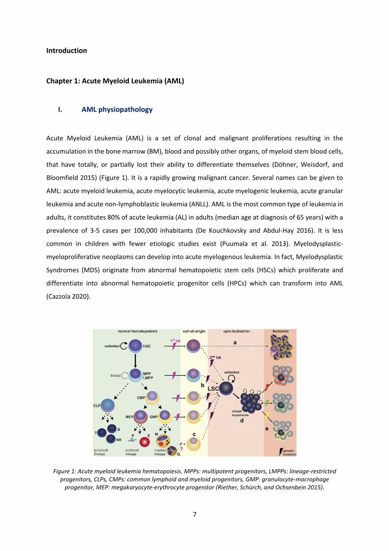
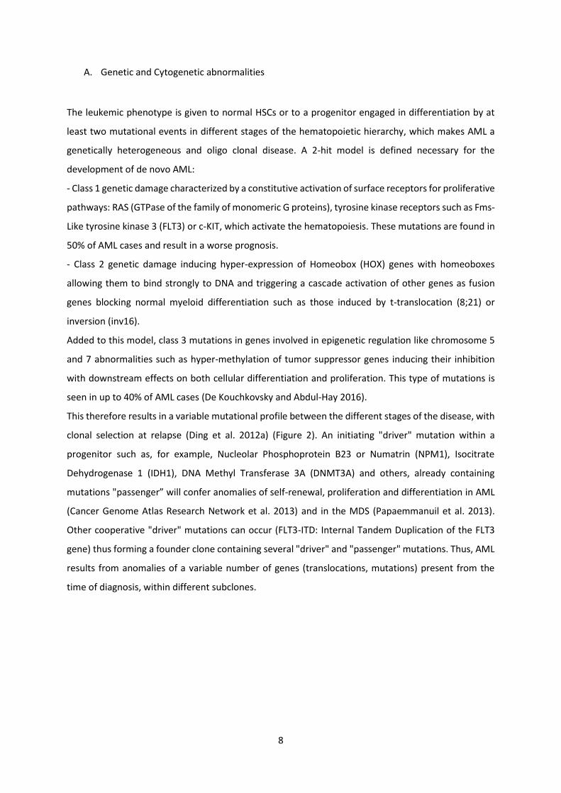
Acute Myeloid Leukemia (AML) is a set of clonal and malignant proliferations resulting in the
accumulation in the bone marrow (BM), blood and possibly other organs, of myeloid stem blood cells,
that have totally, or partially lost their ability to differentiate themselves (Döhner, Weisdorf, and
Bloomfield 2015) (Figure 1). It is a rapidly growing malignant cancer. Several names can be given to
AML: acute myeloid leukemia, acute myelocytic leukemia, acute myelogenic leukemia, acute granular
leukemia and acute non-lymphoblastic leukemia (ANLL). AML is the most common type of leukemia in
adults, it constitutes 80% of acute leukemia (AL) in adults (median age at diagnosis of 65 years) with a
prevalence of 3-5 cases per 100,000 inhabitants (De Kouchkovsky and Abdul-Hay 2016). It is less
common in children with fewer etiologic studies exist (Puumala et al. 2013). Myelodysplastic-
myeloproliferative neoplasms can develop into acute myelogenous leukemia. In fact, Myelodysplastic
Syndromes (MDS) originate from abnormal hematopoietic stem cells (HSCs) which proliferate and
differentiate into abnormal hematopoietic progenitor cells (HPCs) which can transform into AML
(Cazzola 2020).
Figure 1: Acute myeloid leukemia hematopoiesis. MPPs: multipotent progenitors, LMPPs: lineage-restricted progenitors, CLPs, CMPs: common lymphoid and myeloid progenitors, GMP: granulocyte-macrophage
progenitor, MEP: megakaryocyte-erythrocyte progenitor (Riether, Schürch, and Ochsenbein 2015).
8
A. Genetic and Cytogenetic abnormalities
The leukemic phenotype is given to normal HSCs or to a progenitor engaged in differentiation by at
least two mutational events in different stages of the hematopoietic hierarchy, which makes AML a
genetically heterogeneous and oligo clonal disease. A 2-hit model is defined necessary for the
development of de novo AML:
- Class 1 genetic damage characterized by a constitutive activation of surface receptors for proliferative
pathways: RAS (GTPase of the family of monomeric G proteins), tyrosine kinase receptors such as Fms-
Like tyrosine kinase 3 (FLT3) or c-KIT, which activate the hematopoiesis. These mutations are found in
50% of AML cases and result in a worse prognosis.
- Class 2 genetic damage inducing hyper-expression of Homeobox (HOX) genes with homeoboxes
allowing them to bind strongly to DNA and triggering a cascade activation of other genes as fusion
genes blocking normal myeloid differentiation such as those induced by t-translocation (8;21) or
inversion (inv16).
Added to this model, class 3 mutations in genes involved in epigenetic regulation like chromosome 5
and 7 abnormalities such as hyper-methylation of tumor suppressor genes inducing their inhibition
with downstream effects on both cellular differentiation and proliferation. This type of mutations is
seen in up to 40% of AML cases (De Kouchkovsky and Abdul-Hay 2016).
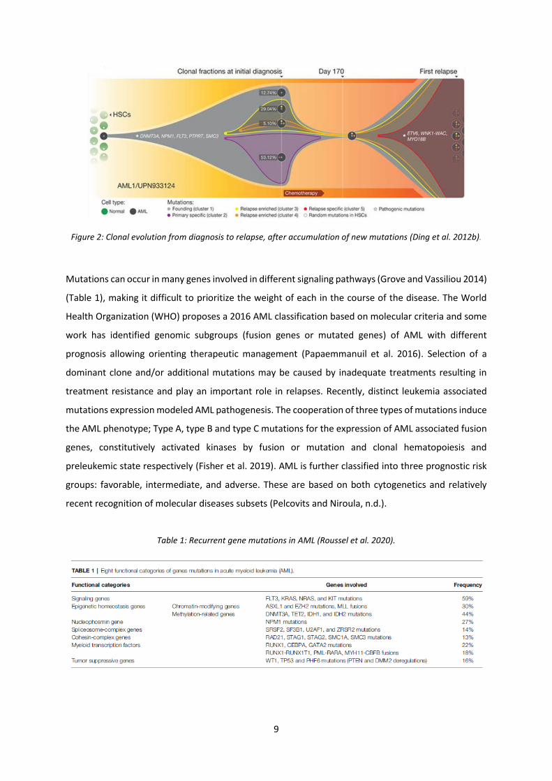
This therefore results in a variable mutational profile between the different stages of the disease, with
clonal selection at relapse (Ding et al. 2012a) (Figure 2). An initiating "driver" mutation within a
progenitor such as, for example, Nucleolar Phosphoprotein B23 or Numatrin (NPM1), Isocitrate
Dehydrogenase 1 (IDH1), DNA Methyl Transferase 3A (DNMT3A) and others, already containing
mutations "passenger” will confer anomalies of self-renewal, proliferation and differentiation in AML
(Cancer Genome Atlas Research Network et al. 2013) and in the MDS (Papaemmanuil et al. 2013).
Other cooperative "driver" mutations can occur (FLT3-ITD: Internal Tandem Duplication of the FLT3
gene) thus forming a founder clone containing several "driver" and "passenger" mutations. Thus, AML
results from anomalies of a variable number of genes (translocations, mutations) present from the
time of diagnosis, within different subclones.
9
Figure 2: Clonal evolution from diagnosis to relapse, after accumulation of new mutations (Ding et al. 2012b).
Mutations can occur in many genes involved in different signaling pathways (Grove and Vassiliou 2014)
(Table 1), making it difficult to prioritize the weight of each in the course of the disease. The World
Health Organization (WHO) proposes a 2016 AML classification based on molecular criteria and some
work has identified genomic subgroups (fusion genes or mutated genes) of AML with different
prognosis allowing orienting therapeutic management (Papaemmanuil et al. 2016). Selection of a
dominant clone and/or additional mutations may be caused by inadequate treatments resulting in
treatment resistance and play an important role in relapses. Recently, distinct leukemia associated
mutations expression modeled AML pathogenesis. The cooperation of three types of mutations induce
the AML phenotype; Type A, type B and type C mutations for the expression of AML associated fusion
genes, constitutively activated kinases by fusion or mutation and clonal hematopoiesis and
preleukemic state respectively (Fisher et al. 2019). AML is further classified into three prognostic risk
groups: favorable, intermediate, and adverse. These are based on both cytogenetics and relatively
recent recognition of molecular diseases subsets (Pelcovits and Niroula, n.d.).
Table 1: Recurrent gene mutations in AML (Roussel et al. 2020).
10
B. Diagnostic
The clinical signs are the consequence of the proliferation of blasts in the bone marrow and their
spread in the blood. From genetic studies, it has become clear that the evolution of human AML is a
multi-step process.
The myelogram is essential for the diagnosis and characterization of blasts (Auer body, myelo
peroxidase positive staining, myeloid markers as cluster of differentiation (CD) CD34, CD13, CD33)
(Döhner et al. 2017). The number of blasts must be greater than 20% of the total cells in the BM or
circulating cells in the Peripheral Blood (PB) (Narayanan and Weinberg 2020), except for hemopathies
with translocations inv16, t (8;21) and t (15;17) or an extramedullary tissue infiltrate. Certain cases
require urgent management, in particular hyperleukocytosis (> 50G/L), severe hemorrhagic
syndromes, metabolic disorders (lysis syndrome, renal failure).
Symptoms related to cytopenias in the blood are more or less marked: Anemic syndrome, Infectious
syndrome, Hemorrhagic syndrome.
The tumor syndrome is inconstant: Lymphadenopathy is rare; splenomegaly is encountered in 15 - 20%
of cases, leading to acute myeloid leukemia.
Gingival hyperplasia and skin localizations (leukemids) are more common for AL with a monocyte
component, sometimes neurological locations (as in ALL) (De Kouchkovsky and Abdul-Hay 2016).
II. History of AML treatment
Treatment of AML is generally divided into 2 phases: an induction phase and a consolidation phase.
The first is intended to stabilize the patient's condition by reducing the tumor mass in the blood and
the number of blasts in the BM, until complete remission. The second phase is to prevent relapse, after
the patient has recovered from the induction phase; this phase can go as far as transplant strategies.
A. Conventional chemotherapy
The induction phase in AML, generally consists of high doses of cytotoxic chemotherapy with
cytarabine and an anthracycline (Daunorubicin), type "7 + 3" which means 7 days of continuous
infusion of cytarabine with the addition of Daunorubin daily for the first 3 days (Huguet and Récher
2012) (Pelcovits and Niroula, n.d.). Although this option allows a reduction in tumor mass, it is
nevertheless associated with high toxicity such as aplasia, infection, hemorrhages, inflammation, etc.,
11
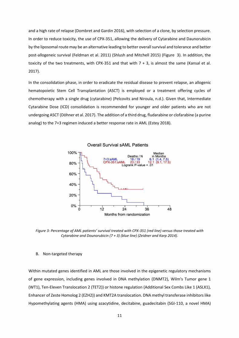
and a high rate of relapse (Dombret and Gardin 2016), with selection of a clone, by selection pressure.
In order to reduce toxicity, the use of CPX-351, allowing the delivery of Cytarabine and Daunorubicin
by the liposomal route may be an alternative leading to better overall survival and tolerance and better
post-allogeneic survival (Feldman et al. 2011) (Shlush and Mitchell 2015) (Figure 3). In addition, the
toxicity of the two treatments, with CPX-351 and that with 7 + 3, is almost the same (Kansal et al.
2017).
In the consolidation phase, in order to eradicate the residual disease to prevent relapse, an allogenic
hematopoietic Stem Cell Transplantation (ASCT) is employed or a treatment offering cycles of
chemotherapy with a single drug (cytarabine) (Pelcovits and Niroula, n.d.). Given that, Intermediate
Cytarabine Dose (ICD) consolidation is recommended for younger and older patients who are not
undergoing ASCT (Döhner et al. 2017). The addition of a third drug, fludarabine or clofarabine (a purine
analog) to the 7+3 regimen induced a better response rate in AML (Estey 2018).
Figure 3: Percentage of AML patients’ survival treated with CPX-351 (red line) versus those treated with Cytarabine and Dounorubicin (7 + 3) (blue line) (Zeidner and Karp 2014).
B. Non-targeted therapy
Within mutated genes identified in AML are those involved in the epigenetic regulatory mechanisms
of gene expression, including genes involved in DNA methylation (DNMT2), Wilm's Tumor gene 1
(WT1), Ten-Eleven Translocation 2 (TET2)) or histone regulation (Additional Sex Combs Like 1 (ASLX1),
Enhancer of Zeste Homolog 2 (EZH2)) and KMT2A translocation. DNA methyl transferase inhibitors like
Hypomethylating agents (HMA) using azacytidine, decitabine, guadecitabin (SGI-110, a novel HMA)
12
and many others are known to have significant clinical activity in the treatment of AML (Dombret et
al. 2015). New agents, like OTX015, a Bromodomain and extra-terminal (BET) protein inhibitor,
pinometostat, a Disruptor of telomeric silencing 1-like (DOT1L) inhibitor and iadademstat (ORY-1001),
a Lysine specific demethylase 1 (LDS1) showed a clinically significant activity in relapse and refractory
AML (Cerrano and Itzykson 2021).
C. Targeted therapy
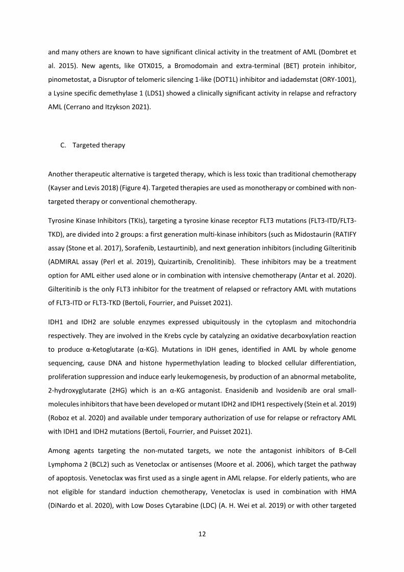
Another therapeutic alternative is targeted therapy, which is less toxic than traditional chemotherapy
(Kayser and Levis 2018) (Figure 4). Targeted therapies are used as monotherapy or combined with non-
targeted therapy or conventional chemotherapy.
Tyrosine Kinase Inhibitors (TKIs), targeting a tyrosine kinase receptor FLT3 mutations (FLT3-ITD/FLT3-
TKD), are divided into 2 groups: a first generation multi-kinase inhibitors (such as Midostaurin (RATIFY
assay (Stone et al. 2017), Sorafenib, Lestaurtinib), and next generation inhibitors (including Gilteritinib
(ADMIRAL assay (Perl et al. 2019), Quizartinib, Crenolitinib). These inhibitors may be a treatment
option for AML either used alone or in combination with intensive chemotherapy (Antar et al. 2020).
Gilteritinib is the only FLT3 inhibitor for the treatment of relapsed or refractory AML with mutations
of FLT3-ITD or FLT3-TKD (Bertoli, Fourrier, and Puisset 2021).
IDH1 and IDH2 are soluble enzymes expressed ubiquitously in the cytoplasm and mitochondria
respectively. They are involved in the Krebs cycle by catalyzing an oxidative decarboxylation reaction
to produce α-Ketoglutarate (α-KG). Mutations in IDH genes, identified in AML by whole genome
sequencing, cause DNA and histone hypermethylation leading to blocked cellular differentiation,
proliferation suppression and induce early leukemogenesis, by production of an abnormal metabolite,
2-hydroxyglutarate (2HG) which is an α-KG antagonist. Enasidenib and Ivosidenib are oral small-
molecules inhibitors that have been developed or mutant IDH2 and IDH1 respectively (Stein et al. 2019)
(Roboz et al. 2020) and available under temporary authorization of use for relapse or refractory AML
with IDH1 and IDH2 mutations (Bertoli, Fourrier, and Puisset 2021).
Among agents targeting the non-mutated targets, we note the antagonist inhibitors of B-Cell
Lymphoma 2 (BCL2) such as Venetoclax or antisenses (Moore et al. 2006), which target the pathway
of apoptosis. Venetoclax was first used as a single agent in AML relapse. For elderly patients, who are
not eligible for standard induction chemotherapy, Venetoclax is used in combination with HMA
(DiNardo et al. 2020), with Low Doses Cytarabine (LDC) (A. H. Wei et al. 2019) or with other targeted
13
therapies (H. Liu 2021). The association azacytitdine-venotoclacx (Phase III VIALE-A assay) (Bertoli,
Fourrier, and Puisset 2021) is a first line oral treatment for relapse or refractory AML patients ineligible
for intensive chemotherapy (Bertoli, Fourrier, and Puisset 2021).
The tumor protein 53 (TP53) mutation has been associated with a poor prognosis in both AML and
MDS (Short, Rytting, and Cortes 2018) (Hunter and Sallman 2020). Despite of being “undruggable”,
many studies have been done to target TP53 mutation. 10-days of decitabine was used as a first line
therapy for older AML patients. A novel, first-in-class small molecule APR-246 (Eprenetapopt) alone or
combined with azacytidine induce apoptosis in TP53 mutated cancer cells in MDS and AML. In addition,
inhibitors targeting the interaction between TP53 and murine double minute 2 (MDM2) like idasanutlin
(a second-generation-nutlin molecule) binding MDM2 and leading to decrease TP53 transcriptional
activity. Idasanutlin was also used with cytarabine and venetoclax (H. Liu 2021).
Figure 4: Targeted therapies available and in development in AML (Roussel et al. 2020).
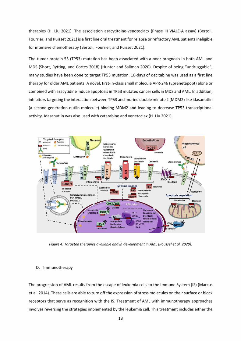
D. Immunotherapy
The progression of AML results from the escape of leukemia cells to the Immune System (IS) (Marcus
et al. 2014). These cells are able to turn off the expression of stress molecules on their surface or block
receptors that serve as recognition with the IS. Treatment of AML with immunotherapy approaches
involves reversing the strategies implemented by the leukemia cell. This treatment includes either the
14
use of agents allowing elimination of the tumor cell (coupled antibodies), or reactivation and
recruitment of immunocompetent cells (vaccines, cytotoxic T lymphocytes (CTLs)). Plus, by injecting
genetically modified T lymphocytes (GMTLs) to redirect them against the tumor such as Chimeric
Antigen Receptor (CAR) T-cells and transgenic T-cell receptor (tgTCR) (Yang et al. 2017a) (Figure 5).
Figure 5: Immunotherapeutic approaches in AML (Yang et al. 2017b).
1. Passive immunotherapy
Different membrane antigens (such as CD33, CD123, CD47, CD64…) are expressed on the surface of
leukemia cells allowing their identification, and especially their targeting with antibodies in order to
be able to deliver cytotoxic drugs in a specific manner. On the other hand, targeting AML
microenvironment is still under investigation.
Monoclonal antibodies (mAbs) are used alone or with a conjugated drug, targeting several leukemia
cells’ membrane antigens (Morsink and Walter 2019a). Only few clinical trials have shown a
satisfactory response in AML (Shang and Zhou 2019a) (Morsink and Walter 2019b). CD33-targeting
with an anti-CD33 mAb coupled to a toxin (GemtuzumAb ozogamicin (GO)) is the most advanced
strategy actually used but anti-CD33 mAb could not provide the results expected in monotherapy
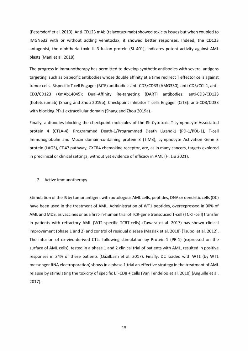
15
(Petersdorf et al. 2013). Anti-CD123 mAb (talacotuzumab) showed toxicity issues but when coupled to
IMGN632 with or without adding venetoclax, it showed better responses. Indeed, the CD123
antagonist, the diphtheria toxin IL-3 fusion protein (SL-401), indicates potent activity against AML
blasts (Mani et al. 2018).
The progress in immunotherapy has permitted to develop synthetic antibodies with several antigens
targeting, such as bispecific antibodies whose double affinity at a time redirect T effector cells against
tumor cells. Bispecific T cell Engager (BiTE) antibodies: anti-CD3/CD33 (AMG330), anti-CD3/CCl-1, anti-
CD3/CD123 (XmAb14045); Dual-Affinity Re-targeting (DART) antibodies: anti-CD3/CD123
(flotetuzumab) (Shang and Zhou 2019b); Checkpoint inhibitor T cells Engager (CiTE): anti-CD3/CD33
with blocking PD-1 extracellular domain (Shang and Zhou 2019a).
Finally, antibodies blocking the checkpoint molecules of the IS: Cytotoxic T-Lymphocyte-Associated
protein 4 (CTLA-4), Programmed Death-1/Programmed Death Ligand-1 (PD-1/PDL-1), T-cell
Immunoglobulin and Mucin domain-containing protein 3 (TIM3), Lymphocyte Activation Gene 3
protein (LAG3), CD47 pathway, CXCR4 chemokine receptor, are, as in many cancers, targets explored
in preclinical or clinical settings, without yet evidence of efficacy in AML (H. Liu 2021).
2. Active immunotherapy
Stimulation of the IS by tumor antigen, with autologous AML cells, peptides, DNA or dendritic cells (DC)
have been used in the treatment of AML. Administration of WT1 peptides, overexpressed in 90% of
AML and MDS, as vaccines or as a first-in-human trial of TCR-gene transduced T-cell (TCRT-cell) transfer
in patients with refractory AML (WT1-specific TCRT-cells) (Tawara et al. 2017) has shown clinical
improvement (phase 1 and 2) and control of residual disease (Maslak et al. 2018) (Tsuboi et al. 2012).
The infusion of ex-vivo-derived CTLs following stimulation by Protein-1 (PR-1) (expressed on the
surface of AML cells), tested in a phase 1 and 2 clinical trial of patients with AML, resulted in positive
responses in 24% of these patients (Qazilbash et al. 2017). Finally, DC loaded with WT1 (by WT1
messenger RNA electroporation) shows in a phase 1 trial an effective strategy in the treatment of AML
relapse by stimulating the toxicity of specific LT-CD8 + cells (Van Tendeloo et al. 2010) (Anguille et al.
2017).
16
3. Adoptive immunotherapy
In 2011, Hanahan and Weinberg (Hanahan and Weinberg 2011) described immunomodulating
properties within tumors characteristics. Some tumors are infiltrated by effector cells for innate
immunity and adaptive immunity with a good prognosis, so Adoptive Cell Transfer therapies (ADCT)
can be a promising immunotherapy in AML.
Allogeneic BM transplantation, a first and benchmark cell immunotherapy, is indicated in first
complete remission in AML (Koreth et al. 2009). Even though allogeneic transplantation is associated
with high procedural toxicity (TRM: Transplant-Related Mortality), it remains the only curative option
in patients at unfavorable risk (Kassim and Savani 2017).
Autologous Tumor Infiltrating Lymphocytes (TILs) cultured in ex-vivo, in the presence of IL-2, were the
first adoptive cellular immunotherapies. TILs are not suitable for hematologic tumors, because of the
difficulties in obtaining these cells, although recent work has shown the possibility of obtaining them
from the microenvironment of the BM (Borrello and Noonan 2016). TILs from AML patients’ BM are
expandable ex-vivo and possess anti-tumor activity (Teo et al. 2019) (L. Wei et al. 2019).
AML blast cells escape the vigilance of Natural killer (NK) cells, through the overexpression of specific
ligands for immunoglobulin-like killer inhibitory receptors (KIRs) expressed on the surface of NK cells.
The use of NK cells may therefore be an interesting avenue in the treatment of AML (Carlsten and Järås
2019). Clinical studies have shown the feasibility of using NK cells from a healthy donor stimulated ex-
vivo by IL-2, which aims to amplify the activating signal and inhibit alloreactivity with human leukocyte
antigen (HLA) molecules from the patient's tumor cells (Koehl et al. 2004). In addition, Bispecific Killer
Engagers (BiKE) (anti-CD16/CD33) and Trispecific Killer Engager (TRiKE) (anti-CD16/IL-15/CD33, anti-
CD33/CD123/CD16 is under investigation (Braciak et al. 2018)). NK cells activated by different
cytokines for Cytokines Induced Killer cells (CIK) are made more cytotoxic and more proliferative than
those activated with IL-2 (Linn et al. 2009). Immunotherapy using Cytokine-induced memory-like NK
cells demonstrates the ability to fight AML leukemia cells in a first-in-human phase 1 clinical trial
(Romee et al. 2016).
17
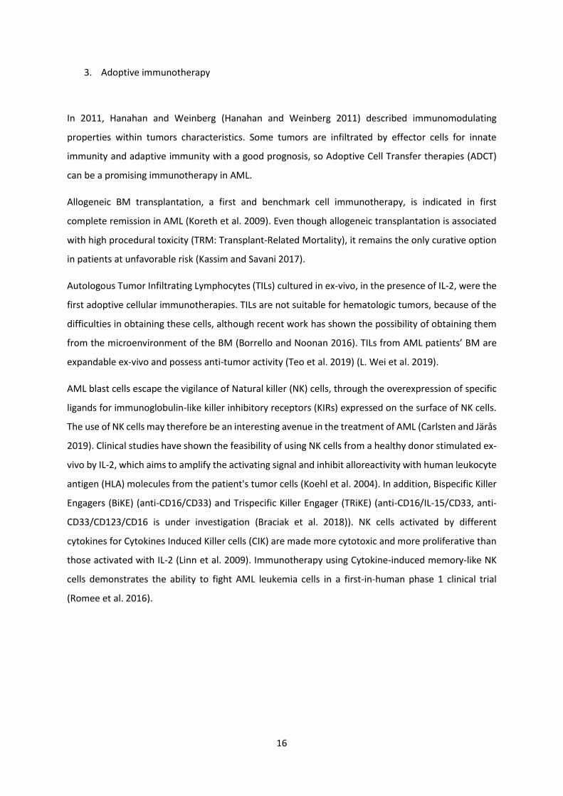
E. Genetically modified T Lymphocytes
Cancer immunotherapy is based on the use of immune effector cells cytotoxicity against leukemia cells.
The potential of T-cells to eradicate tumors has inspired new immunotherapy strategies and has led to
the development of GMTL to express a specific receptor of malignant cells (Turtle et al. 2012).
As ADCT, autologous T-cells can be redirected to leukemia cells of an AML patient by two recently used
methods based on the genetic modification of these T-cells; either by generating T-cells with a tgTCR
specific to one tumor antigen, or by generating T-cells expressing a CAR against a tumor antigen (Figure
6).
Several tgTCRT-cells targeting a tumor antigen are used in AML with potent anti-tumor efficacy.
However, tgTCRT-cells therapy may be associated with off-target toxicities induced by mispairing
between the endogenous and the introduced TCR chains and it is limited to HLA restriction and the
small number of suitable targets (Bonte et al. 2020).
In AML, classically, blasts escape the IS by down-regulating HLA molecules, resulting in an altered TCR/
HLA/tumor antigen interaction. Therefore, immunotherapy using T-cells free of HLA-recognized (CART-
cells) will be more preferable.
CART-cells therapy is a novel ADCT therapy with promising results in BCL and multiple myeloma (MM)
using CD19 CAR, CD22 CAR -T-cells (Park et al. 2018). CART-cells targeting various antigens (CD33,
CD123, FLT3, CLL-1, etc.) are under clinical investigations for AML treatment. These assays showed
potent efficacy but also toxicities on healthy hematopoietic cells (Shang and Zhou 2019a). The ideal
antigenic target in AML has not yet been identified.
18
Figure 6: Immunotherapies using genetically modified immune cells in AML (Roussel et al. 2020).
III. Relapse of AML patients and need for new therapeutic alternatives
A. Resistance to treatments
Despite the progress made in the treatment of AML over the past 10 years, survival has not been
significantly improved especially in elderly patients, mainly due to relapses. Resistance to treatments
occurs due to acquisition and/or enrichment of clones in the AML tumor cell with activation of the
signaling pathways such as FLT3 (FLT3-ITD mutation) or RAS or bi-allelic mutations affecting the
function of TP53. Moreover, combination of venetoclax and azacitidin may select monocytic disease
in AML, which will lead to treatment resistance and relapse (Pei et al. 2020).
The strong interactions of AML blasts with the IS suggest avenues for improving treatments, thanks to
immunotherapy approaches, particularly in maintaining remission during the maintenance phase
(Table 2). Currently, various research groups are trying to identify appropriate targets to develop
alternative immunotherapies to treat patients refractory or relapsing from conventional treatments or
existing immunotherapies.
19
Several studies aimed to target the Leukemia Stem Cell (LSC), which is responsible of the maintenance
and the propagation of the AML phenotype after treatments, and prevent relapse by targeting various
markers expressed on the LSC’s surface (Gentles et al. 2010).
The Interleukin-1 Receptor Accessory Protein (IL-1RAP) has been identified for its expression on LSC
(Tasian, Bornhäuser, and Rutella 2018), thus as a potential target to destroy AML leukemia cells. This
approach will make it possible to target AML blasts expressing the IL-1RAP protein, in order to provide
an alternative treatment to CART-Cells approaches conventionally targeting CD33 or CD123.
Table 2: Mechanisms of AML LSC resistance against immunotherapies and some possible solutions (Valent et al. 2020).
20
B. IL-1 Receptor Accessory Protein (IL-1RAP)
1. Definition of IL-1RAP
IL-1RAP (also called IL-1RAcP, IL-1R3 or C3orf13) is a protein encoded by chromosome 3q28. It belongs
to the interleukin-1 (IL-1) family of proteins. It is a protein expressed on the surface of cells and forms
a complex with the receptor for IL-1α, IL-1β, and IL-33 and it is essential for their signaling. There are
five known forms of alternative splicing for IL-1RAP mRNA. This protein exists in two forms, a
membrane form (variant 1, 3, and 4) and a secreted soluble form (variant 2 and 5). The membrane
form of IL-1RAP induces an intracellular signal after binding to IL-1. While the soluble form has a
neutralizing effect of IL-1 (D. E. Smith et al. 2003). IL-1 is a pro-inflammatory cytokine in response to
infection, stress or tissue damage via the (Nuclear factor-kappa B) NF-Kappa-B pathway to infection
(Figure 7).
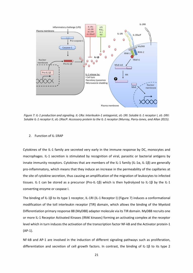
21
Figure 7: IL-1 production and signaling. IL-1Ra: Interleukin-1 antagonist, sIL-1RI: Soluble IL-1 receptor I, sIL-1RII: Soluble IL-1 receptor II, sIL-1RacP: Accessory protein to the IL-1 receptor (Murray, Parry-Jones, and Allan 2015).
2. Function of IL-1RAP
Cytokines of the IL-1 family are secreted very early in the immune response by DC, monocytes and
macrophages. IL-1 secretion is stimulated by recognition of viral, parasitic or bacterial antigens by
innate immunity receptors. Cytokines that are members of the IL-1 family (IL-1α, IL-1β) are generally
pro-inflammatory, which means that they induce an increase in the permeability of the capillaries at
the site of cytokine secretion, thus causing an amplification of the migration of leukocytes to infected
tissues. IL-1 can be stored as a precursor (Pro-IL-1β) which is then hydrolyzed to IL-1β by the IL-1
converting enzyme or caspase I.
The binding of IL-1β to its type 1 receptor, IL-1RI (IL-1 Receptor I) (Figure 7) induces a conformational
modification of the toll interleukin receptor (TIR) domain, which allows the binding of the Myeloid
Differentiation primary response 88 (MyD88) adapter molecule via its TIR domain. MyD88 recruits one
or more IL-1 Receptor Activated Kinases (IRAK kinases) forming an activating complex at the receptor
level which in turn induces the activation of the transcription factor NF-kB and the Activator protein-1
(AP-1).
NF-kB and AP-1 are involved in the induction of different signaling pathways such as proliferation,
differentiation and secretion of cell growth factors. In contrast, the binding of IL-1β to its type 2
22
receptor IL-1RII, does not induce signaling because IL-1RII lacks the intracellular TIR fragment. IL-1Ryα
(IL-1 receptor antagonist) has an effect that opposes the effects of IL-1β by competing with IL-1RI. In
addition, the soluble forms of IL-1RI (sIL-1RI), IL-1RII (sIL-1RII), and IL-1RAP (sIL-1RAP) have an
inhibitory effect. They can bind to IL-1β and therefore prevent its binding to its IL-1RI membrane
receptor.
A transcriptomic analysis, compares the populations of Leukemic Hematopoietic Stem Cells (LHSC)
(AML with monosomy 7), primitive -LT (Lin-/CD34+/CD38-/CD90+ : long-term LHSCs), less primitive-ST
(Lin-/CD34+/ CD38-/CD90- : short-term LHSC) and more differentiated GMP (Lin-
/CD34+/CD38+/CD123+/CD45RA+ : Granulocytes/Macrophages progenitors) respectively with the
same populations of healthy subjects. Among a list of 11 genes, common to the three types of
populations, an overexpression of the IL-1RAP protein emerges. Thus, the IL-1RAP gene expression
allows discriminating normal HSC and LSC.
IL-1RAP overexpression is observed in Chronic Myeloid Leukemia (CML) correlated with the mutation
of BCR-ABL gene, in ALL with Philadelphia chromosome, and in MDS and AML stem and progenitors
cells (with normal karyotype or AML-7/7q- (monosomie 7 or deletion of 7q) but not on healthy
hematopoietic cells (Barreyro et al. 2012) (Askmyr et al. 2013).
In an inflammatory microenvironment such as in AML pathogenesis, IL-1RAP has an oncogenic effect
through two tyrosine kinase receptors pathways FLT3 and c-kit (Mitchell et al. 2018) which promotes
the proliferation of leukemic cells and drives HSC clonal evolution (Pietras et al. 2016). IL-1RAP
signaling axis plays an important role in enhancing inflammation in the leukemic niche via p38 MAPK
and NF-Kβ signaling pathways (De Boer et al. 2020) (Figure 8). In addition, IL-1RAP has a crucial role in
the regulation of tumor microenvironment-related inflammatory factors in solid tumors ((Lv et al.
2021).
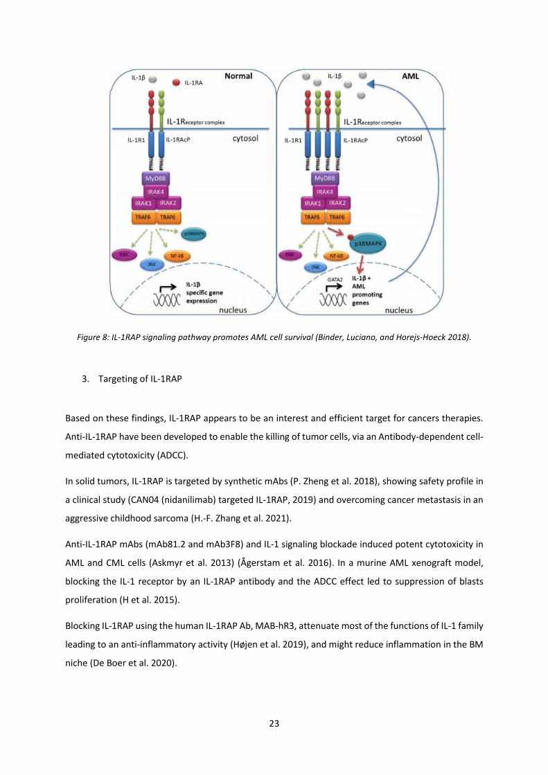
23
Figure 8: IL-1RAP signaling pathway promotes AML cell survival (Binder, Luciano, and Horejs-Hoeck 2018).
3. Targeting of IL-1RAP
Based on these findings, IL-1RAP appears to be an interest and efficient target for cancers therapies.
Anti-IL-1RAP have been developed to enable the killing of tumor cells, via an Antibody-dependent cell-
mediated cytotoxicity (ADCC).
In solid tumors, IL-1RAP is targeted by synthetic mAbs (P. Zheng et al. 2018), showing safety profile in
a clinical study (CAN04 (nidanilimab) targeted IL-1RAP, 2019) and overcoming cancer metastasis in an
aggressive childhood sarcoma (H.-F. Zhang et al. 2021).
Anti-IL-1RAP mAbs (mAb81.2 and mAb3F8) and IL-1 signaling blockade induced potent cytotoxicity in
AML and CML cells (Askmyr et al. 2013) (Ågerstam et al. 2016). In a murine AML xenograft model,
blocking the IL-1 receptor by an IL-1RAP antibody and the ADCC effect led to suppression of blasts
proliferation (H et al. 2015).
Blocking IL-1RAP using the human IL-1RAP Ab, MAB-hR3, attenuate most of the functions of IL-1 family
leading to an anti-inflammatory activity (Højen et al. 2019), and might reduce inflammation in the BM
niche (De Boer et al. 2020).
24
IL-1RAP was also targeted with CART-cells in CML. IL-1RAP CART-cells efficiently eliminate quiescent
tumor HSCs, which fall outside the spectrum of action of tyrosine kinase inhibitors in-vitro and in-vivo
in a xenograft murine model (Warda et al. 2019a).
Recently, a novel therapy was developed in targeting IL-1RAP in AML using a bioreducible lipidoid-
encapsulated CRISPR associated protein 9 (Cas9)/single guide IL-1RAP RNA ribonucleoprotein (Ho et
al. 2021). This strategy provides an effective attenuation of AML LSC growth.
In addition, bispecific Abs have been developed in order to target IL-1RAP and Thomsen–Friedenreich
in CML to increase the specificity towards LSC by using additional biomarkers (Eldesouki et al. 2021).
Chapter 2: 1. Cellular immunotherapy in AML using CART-cells
I. Principle of CAR development
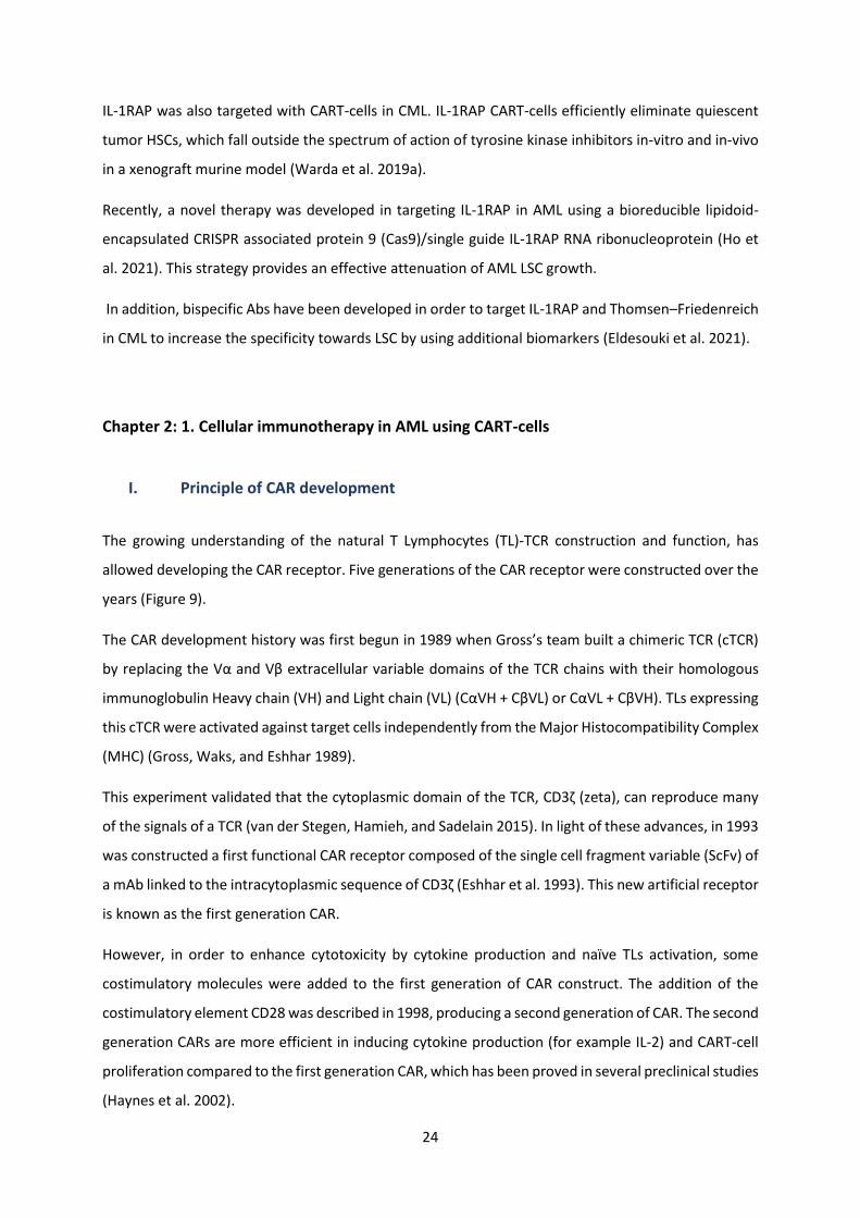
The growing understanding of the natural T Lymphocytes (TL)-TCR construction and function, has
allowed developing the CAR receptor. Five generations of the CAR receptor were constructed over the
years (Figure 9).
The CAR development history was first begun in 1989 when Gross’s team built a chimeric TCR (cTCR)
by replacing the Vα and Vβ extracellular variable domains of the TCR chains with their homologous
immunoglobulin Heavy chain (VH) and Light chain (VL) (CαVH + CβVL) or CαVL + CβVH). TLs expressing
this cTCR were activated against target cells independently from the Major Histocompatibility Complex
(MHC) (Gross, Waks, and Eshhar 1989).
This experiment validated that the cytoplasmic domain of the TCR, CD3ζ (zeta), can reproduce many
of the signals of a TCR (van der Stegen, Hamieh, and Sadelain 2015). In light of these advances, in 1993
was constructed a first functional CAR receptor composed of the single cell fragment variable (ScFv) of
a mAb linked to the intracytoplasmic sequence of CD3ζ (Eshhar et al. 1993). This new artificial receptor
is known as the first generation CAR.
However, in order to enhance cytotoxicity by cytokine production and naïve TLs activation, some
costimulatory molecules were added to the first generation of CAR construct. The addition of the
costimulatory element CD28 was described in 1998, producing a second generation of CAR. The second
generation CARs are more efficient in inducing cytokine production (for example IL-2) and CART-cell
proliferation compared to the first generation CAR, which has been proved in several preclinical studies
(Haynes et al. 2002).
25
The third generation CAR contains two costimulatory domains, resulting in a more potent persistence
and other CART-cell functions in treated patients (Jinjuan Wang et al. 2007).
For further improve the killing function of CART-cells against tumors, a fourth generation of CAR, also
named T-cells redirected for universal cytokine-mediated killing (TRUCKs), has been developed based
on the second generation. It can induce cytokine production for example IL-12 through the nuclear
factor of the activated T-cell (NFAT) which can direct the TL to express transgenic products
(Chmielewski and Abken 2015). Recently, a novel fifth generation of CAR was developed containing
intracellular domains of cytokine receptors, such as IL-2Rβ chain fragment, to enhance anti-tumor
effects (Kagoya et al. 2018).
Figure 9: Different generations of the CAR receptor (Jin et al. 2021).
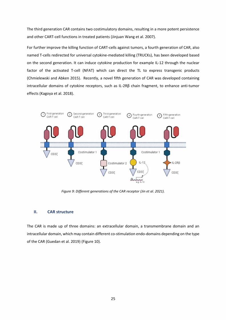
II. CAR structure
The CAR is made up of three domains: an extracellular domain, a transmembrane domain and an
intracellular domain, which may contain different co-stimulation endo-domains depending on the type
of the CAR (Guedan et al. 2019) (Figure 10).
26
Figure 10: Schematic representation of the different domains of the CAR (Skorka et al. 2020).
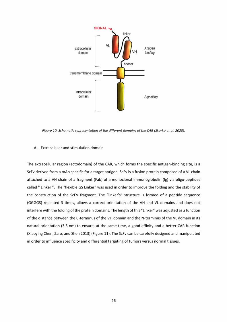
A. Extracellular and stimulation domain
The extracellular region (ectodomain) of the CAR, which forms the specific antigen-binding site, is a
ScFv derived from a mAb specific for a target antigen. ScFv is a fusion protein composed of a VL chain
attached to a VH chain of a fragment (Fab) of a monoclonal immunoglobulin (Ig) via oligo-peptides
called " Linker ”. The "flexible GS Linker" was used in order to improve the folding and the stability of
the construction of the ScFV fragment. The “linker’s” structure is formed of a peptide sequence
(GGGGS) repeated 3 times, allows a correct orientation of the VH and VL domains and does not
interfere with the folding of the protein domains. The length of this “Linker” was adjusted as a function
of the distance between the C-terminus of the VH domain and the N-terminus of the VL domain in its
natural orientation (3.5 nm) to ensure, at the same time, a good affinity and a better CAR function
(Xiaoying Chen, Zaro, and Shen 2013) (Figure 11). The ScFv can be carefully designed and manipulated
in order to influence specificity and differential targeting of tumors versus normal tissues.
27
Figure 11: CAR ScFV structure. ScFV: single chain variable fragment (Hughes-Parry, Cross, and Jenkins 2020).
B. Hinge region
The “Hinge” or “spacer” part of the CAR construct links the extracellular part to the intracellular part
of this receptor. It plays an important role in activating the CART-cell. The role of this region is to
provide flexibility to the ScFv, whose "hinge" length plays a role in enhancing the expression of ScFv at
the TL membrane (Guest et al. 2005). These properties have been described as modulating effector
cell/target cell interactions, thus affecting the strength of the activation signal of the CART-cell. The
hinge region can be of different types such as an extracellular fragment derived from CD28, TCRβ chain,
CD8α, or NKG2D or an Ig-like domain with the Fc regions of an IgG antibody (IgG1 hinge CH2-CH3) due
to the stability of the protein domain (Lipowska-Bhalla et al. 2012). The position of the target epitope
regarding to the target cell surface determine the need of an extracellular spacer domain (A. A.
Hombach et al. 2007). Studies have shown that the hinge region has a role in the activation and
secretion of cytokines by CART-cells and regulates the CAR signaling threshold (Fujiwara et al. 2020).
C. Transmembrane domain
The transmembrane domain (TM) connects the ectodomain to the endodomain and serves as the
anchor to the cell membrane. Few studies have been done on the transmembrane domain of the CAR.
An earlier study had used a CD3ᵹ transmembrane domain in the CAR construct and subsequently
showed that this domain is important for stability of membrane expression of the CAR (Romeo, Amiot,
and Seed 1992). In 2010, Bridgeman et al. have shown that the biochemical interactions that occur
28
between the wild-type CD3ᵹ transmembrane domain and other components of the endogenous
TCR/CD3 complex are important for the optimal activity of the CD3ᵹ CAR (Bridgeman et al. 2010).
Various transmembrane regions have also been employed in CARs including those derived from CD28,
CD3, CD8, CD4, or FcꜫRIy. Using CD19 CART-cells with TNFRS19 TM showed clinical promoting results
(Caimi et al. 2019). The TM can regulates the amount of CAR signaling via control of CAR expression
level. Similar to the hinge domain, changing the length of the TM can affect the CART-cell proliferation
and further clinical studies are needed to prove the advantages of the TM for decreasing tonic signals
and increasing CART-cell persistence (Fujiwara et al. 2020).
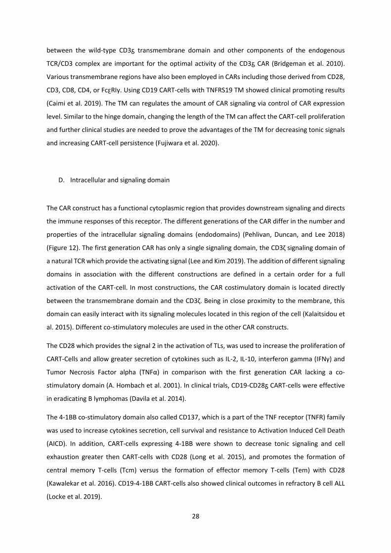
D. Intracellular and signaling domain
The CAR construct has a functional cytoplasmic region that provides downstream signaling and directs
the immune responses of this receptor. The different generations of the CAR differ in the number and
properties of the intracellular signaling domains (endodomains) (Pehlivan, Duncan, and Lee 2018)
(Figure 12). The first generation CAR has only a single signaling domain, the CD3ζ signaling domain of
a natural TCR which provide the activating signal (Lee and Kim 2019). The addition of different signaling
domains in association with the different constructions are defined in a certain order for a full
activation of the CART-cell. In most constructions, the CAR costimulatory domain is located directly
between the transmembrane domain and the CD3ζ. Being in close proximity to the membrane, this
domain can easily interact with its signaling molecules located in this region of the cell (Kalaitsidou et
al. 2015). Different co-stimulatory molecules are used in the other CAR constructs.
The CD28 which provides the signal 2 in the activation of TLs, was used to increase the proliferation of
CART-Cells and allow greater secretion of cytokines such as IL-2, IL-10, interferon gamma (IFNγ) and
Tumor Necrosis Factor alpha (TNFα) in comparison with the first generation CAR lacking a co-
stimulatory domain (A. Hombach et al. 2001). In clinical trials, CD19-CD28ᵹ CART-cells were effective
in eradicating B lymphomas (Davila et al. 2014).
The 4-1BB co-stimulatory domain also called CD137, which is a part of the TNF receptor (TNFR) family
was used to increase cytokines secretion, cell survival and resistance to Activation Induced Cell Death
(AICD). In addition, CART-cells expressing 4-1BB were shown to decrease tonic signaling and cell
exhaustion greater then CART-cells with CD28 (Long et al. 2015), and promotes the formation of
central memory T-cells (Tcm) versus the formation of effector memory T-cells (Tem) with CD28
(Kawalekar et al. 2016). CD19-4-1BB CART-cells also showed clinical outcomes in refractory B cell ALL
(Locke et al. 2019).
29
Additional preclinical studies discovered more molecules, which can be used for co-stimulation of the
CAR in combination with CD28 and 4-1BB.
The Inducible T-cell COStimulator (ICOS) molecule, also called CD278, is a member of the CD28
superfamily. This co-stimulator plays a promoter role in the differentiation of TL-CD4+ Helper 1 (Th1)
and Helper 2 (Th2) subsets and their effector functions in cytokines production (IL-10). CART-cells with
the two intracellular signaling domains ICOS and 4-1BB demonstrate increased efficacy in solid tumor
models regarding the use of only 4-1BB (Guedan et al. 2018).
Combined 4-1BB and OX40 (CD134), two agonist of the TNFR costimulatory receptors, has been shown
to generate very high effector T-cells with longer survival, more differentiation and production of
greater quantity of cytokines compared to T-cells stimulated with only 4-1BB (Konstorum et al. 2019).
In addition, OX40 stimulates the secretion of pro-inflammatory cytokines such as IL-4, IL-6 and IFNγ by
CART-Cells, thus inhibiting the suppressive activity of regulatory TL (Treg), and can induce
differentiation of CART-cells to a memory phenotype allowing the escape of AICD (Redmond, Ruby,
and Weinberg 2009).
Figure 12: A brief of the different CAR co-stimulatory molecules and their functions. ITAM: Immunoreceptor tyrosine-based activation motif (CD3ᵹ), CM: Co-stimulatory Molecule (Cartellieri et al. 2010).
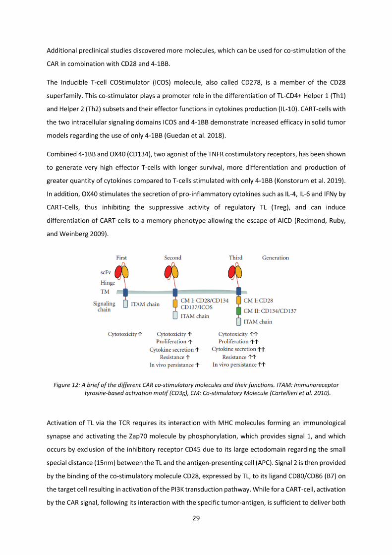
Activation of TL via the TCR requires its interaction with MHC molecules forming an immunological
synapse and activating the Zap70 molecule by phosphorylation, which provides signal 1, and which
occurs by exclusion of the inhibitory receptor CD45 due to its large ectodomain regarding the small
special distance (15nm) between the TL and the antigen-presenting cell (APC). Signal 2 is then provided
by the binding of the co-stimulatory molecule CD28, expressed by TL, to its ligand CD80/CD86 (B7) on
the target cell resulting in activation of the PI3K transduction pathway. While for a CART-cell, activation
by the CAR signal, following its interaction with the specific tumor-antigen, is sufficient to deliver both
30
signals 1 and 2, resulting in T-cell activation and triggering of an immune response. The special distance
between the CART-cell and the target tumor cell is not yet known. Indeed, the activation of the BiTE
needs the presence of two independent signals. Signal 1 is delivered upon BiTE ligation with target
tumor cell through secreting bispecific antibodies, and signal 2 is delivered by CD28/B7 (Y. Wang et al.
2017) (Figure 13 A-B-C).
Figure 13: Signaling mechanisms of conventional TCR and CAR receptor (Y. Wang et al. 2017).
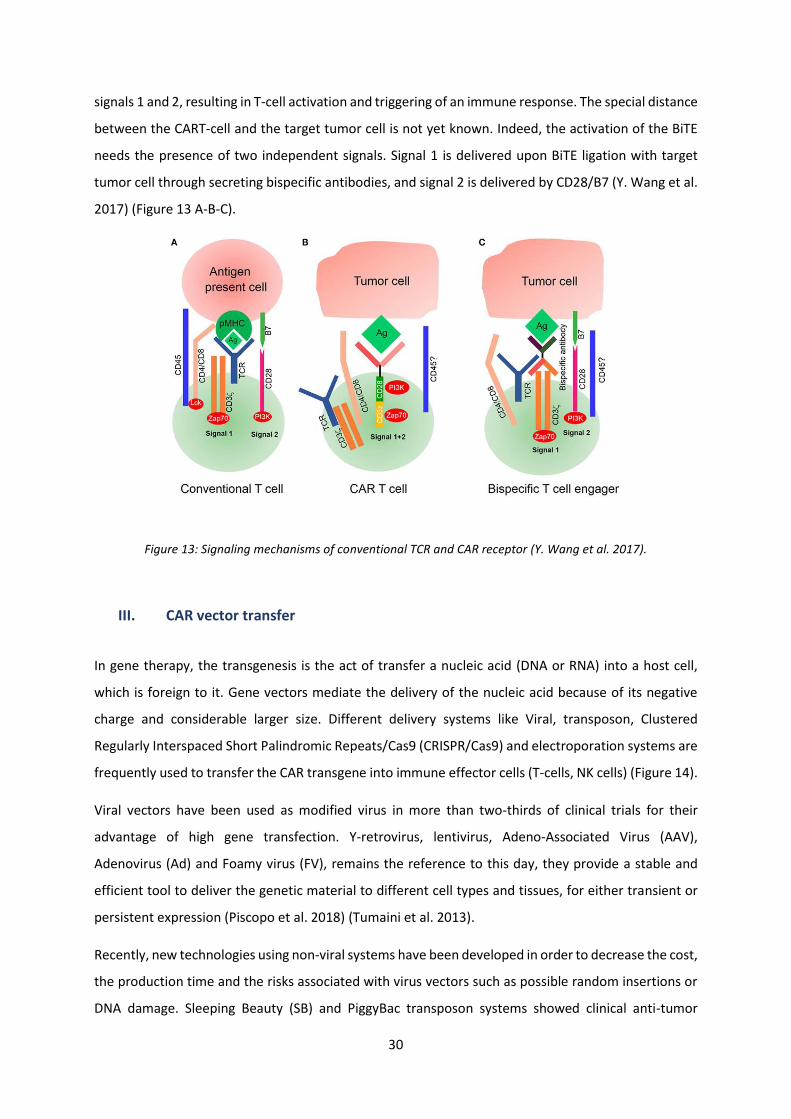
III. CAR vector transfer
In gene therapy, the transgenesis is the act of transfer a nucleic acid (DNA or RNA) into a host cell,
which is foreign to it. Gene vectors mediate the delivery of the nucleic acid because of its negative
charge and considerable larger size. Different delivery systems like Viral, transposon, Clustered
Regularly Interspaced Short Palindromic Repeats/Cas9 (CRISPR/Cas9) and electroporation systems are
frequently used to transfer the CAR transgene into immune effector cells (T-cells, NK cells) (Figure 14).
Viral vectors have been used as modified virus in more than two-thirds of clinical trials for their
advantage of high gene transfection. Y-retrovirus, lentivirus, Adeno-Associated Virus (AAV),
Adenovirus (Ad) and Foamy virus (FV), remains the reference to this day, they provide a stable and
efficient tool to deliver the genetic material to different cell types and tissues, for either transient or
persistent expression (Piscopo et al. 2018) (Tumaini et al. 2013).
Recently, new technologies using non-viral systems have been developed in order to decrease the cost,
the production time and the risks associated with virus vectors such as possible random insertions or
DNA damage. Sleeping Beauty (SB) and PiggyBac transposon systems showed clinical anti-tumor
31
efficiency and CART-cells persistence with producing a higher ratio of Tcm (Clauss et al. 2018) (Bishop
et al. 2019). SB-modified CART-cells also demonstrate potent outcomes in-vitro and in-vivo in
chemoresistant AML patient derived xenograft (PDX) (Rotiroti et al. 2020).
Among the evolution of the manufacturing technology, gene editing methods such as Zinc finger (ZFNs)
and nucleoside transcription activator-like effector nucleases (TALENs) were developed and used in
the CAR therapy (Qasim et al. 2017). The breakthrough in the genetic “editing” was the CRISPR/Cas9
technology. Using a short RNA guide (gRNA) to direct any desired region in the genome, it provides a
powerful tool to enhance the ability of engineered T-cells to fight cancer cells and decrease the
immunogenicity. PD-1 disrupted CART-cells in solid tumors using CRISPR/Cas9 system showed
preclinical tumor-killing efficacy (Hu et al. 2019) (H. Zhu et al. 2020). In AML, CRISPR/Cas9 system was
used recently to target the IL-1RAP protein providing an effective strategy to improve AML therapy
(Ho et al. 2021). In addition, this system is actually in use to enhance CART-cells efficacy by deletion of
immunosuppressive factors (Giuffrida et al. 2021).
The electroporation of CAR mRNA in NK and T-cells was described as efficient enough, with low
electroporation-related apoptosis. It demonstrated successful clinical anti-tumor effects in solid
tumors (Beatty et al. 2014). Despite the short lifetime and transiency of its expression, CAR mRNA
degradation over time allows a complete removal of the CAR from the patient without the need for
suicide genes (Angel and Yanik 2010). This system is being investigated in early clinical trials. However,
some limits are also described using this system such as the long ex-vivo culture time to generate
therapeutic doses of GMTLs and the severe cell damage following the electroporation.
Figure 14: Different mechanisms of engineering immune effector cells with the CAR transgene (Oldham and Medin 2017).
32
Lentivirus
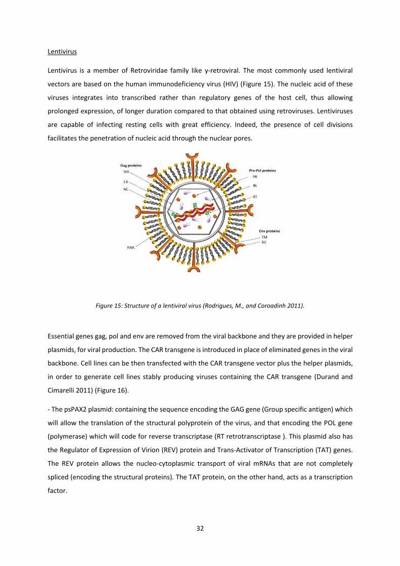
Lentivirus is a member of Retroviridae family like y-retroviral. The most commonly used lentiviral
vectors are based on the human immunodeficiency virus (HIV) (Figure 15). The nucleic acid of these
viruses integrates into transcribed rather than regulatory genes of the host cell, thus allowing
prolonged expression, of longer duration compared to that obtained using retroviruses. Lentiviruses
are capable of infecting resting cells with great efficiency. Indeed, the presence of cell divisions
facilitates the penetration of nucleic acid through the nuclear pores.
Figure 15: Structure of a lentiviral virus (Rodrigues, M., and Coroadinh 2011).
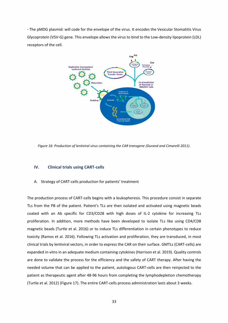
Essential genes gag, pol and env are removed from the viral backbone and they are provided in helper
plasmids, for viral production. The CAR transgene is introduced in place of eliminated genes in the viral
backbone. Cell lines can be then transfected with the CAR transgene vector plus the helper plasmids,
in order to generate cell lines stably producing viruses containing the CAR transgene (Durand and
Cimarelli 2011) (Figure 16).
- The psPAX2 plasmid: containing the sequence encoding the GAG gene (Group specific antigen) which
will allow the translation of the structural polyprotein of the virus, and that encoding the POL gene
(polymerase) which will code for reverse transcriptase (RT retrotranscriptase ). This plasmid also has
the Regulator of Expression of Virion (REV) protein and Trans-Activator of Transcription (TAT) genes.
The REV protein allows the nucleo-cytoplasmic transport of viral mRNAs that are not completely
spliced (encoding the structural proteins). The TAT protein, on the other hand, acts as a transcription
factor.
33
- The pMDG plasmid: will code for the envelope of the virus. It encodes the Vesicular Stomatitis Virus
Glycoprotein (VSV-G) gene. This envelope allows the virus to bind to the Low-density lipoprotein (LDL)
receptors of the cell.
Figure 16: Production of lentiviral virus containing the CAR transgene (Durand and Cimarelli 2011).
IV. Clinical trials using CART-cells
A. Strategy of CART-cells production for patients’ treatment
The production process of CART-cells begins with a leukopheresis. This procedure consist in separate
TLs from the PB of the patient. Patient’s TLs are then isolated and activated using magnetic beads
coated with an Ab specific for CD3/CD28 with high doses of IL-2 cytokine for increasing TLs
proliferation. In addition, more methods have been developed to isolate TLs like using CD4/CD8
magnetic beads (Turtle et al. 2016) or to induce TLs differentiation in certain phenotypes to reduce
toxicity (Ramos et al. 2016). Following TLs activation and proliferation, they are transduced, in most
clinical trials by lentiviral vectors, in order to express the CAR on their surface. GMTLs (CART-cells) are
expanded in-vitro in an adequate medium containing cytokines (Harrison et al. 2019). Quality controls
are done to validate the process for the efficiency and the safety of CART therapy. After having the
needed volume that can be applied to the patient, autologous CART-cells are then reinjected to the
patient as therapeutic agent after 48-96 hours from completing the lymphodepletion chemotherapy
(Turtle et al. 2012) (Figure 17). The entire CART-cells process administration lasts about 3 weeks.
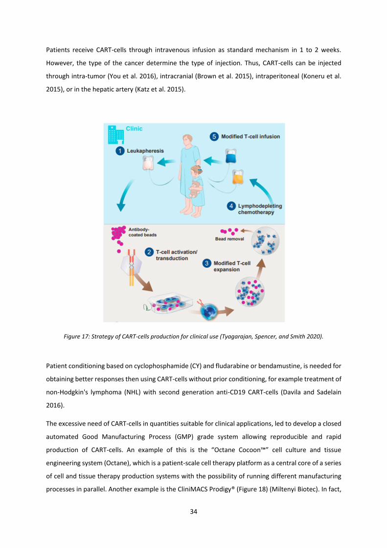
34
Patients receive CART-cells through intravenous infusion as standard mechanism in 1 to 2 weeks.
However, the type of the cancer determine the type of injection. Thus, CART-cells can be injected
through intra-tumor (You et al. 2016), intracranial (Brown et al. 2015), intraperitoneal (Koneru et al.
2015), or in the hepatic artery (Katz et al. 2015).
Figure 17: Strategy of CART-cells production for clinical use (Tyagarajan, Spencer, and Smith 2020).
Patient conditioning based on cyclophosphamide (CY) and fludarabine or bendamustine, is needed for
obtaining better responses then using CART-cells without prior conditioning, for example treatment of
non-Hodgkin's lymphoma (NHL) with second generation anti-CD19 CART-cells (Davila and Sadelain
2016).
The excessive need of CART-cells in quantities suitable for clinical applications, led to develop a closed
automated Good Manufacturing Process (GMP) grade system allowing reproducible and rapid
production of CART-cells. An example of this is the “Octane Cocoon™” cell culture and tissue
engineering system (Octane), which is a patient-scale cell therapy platform as a central core of a series
of cell and tissue therapy production systems with the possibility of running different manufacturing
processes in parallel. Another example is the CliniMACS Prodigy® (Figure 18) (Miltenyi Biotec). In fact,
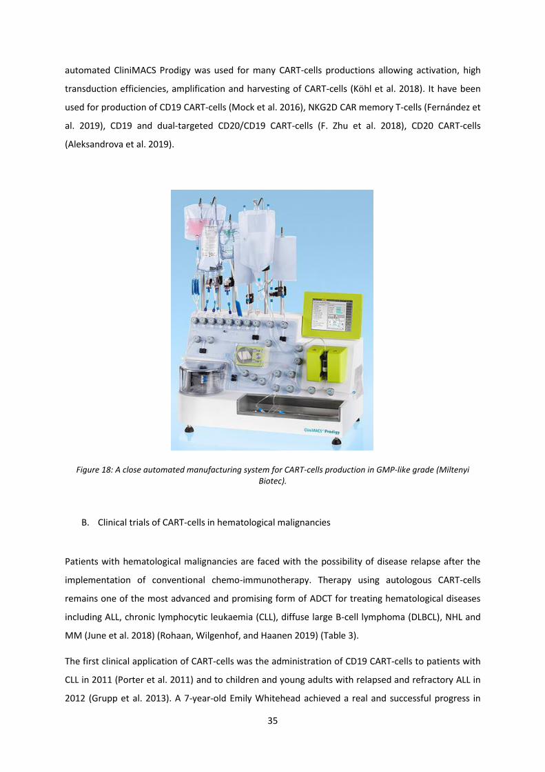
35
automated CliniMACS Prodigy was used for many CART-cells productions allowing activation, high
transduction efficiencies, amplification and harvesting of CART-cells (Köhl et al. 2018). It have been
used for production of CD19 CART-cells (Mock et al. 2016), NKG2D CAR memory T-cells (Fernández et
al. 2019), CD19 and dual-targeted CD20/CD19 CART-cells (F. Zhu et al. 2018), CD20 CART-cells
(Aleksandrova et al. 2019).
Figure 18: A close automated manufacturing system for CART-cells production in GMP-like grade (Miltenyi Biotec).
B. Clinical trials of CART-cells in hematological malignancies
Patients with hematological malignancies are faced with the possibility of disease relapse after the
implementation of conventional chemo-immunotherapy. Therapy using autologous CART-cells
remains one of the most advanced and promising form of ADCT for treating hematological diseases
including ALL, chronic lymphocytic leukaemia (CLL), diffuse large B-cell lymphoma (DLBCL), NHL and
MM (June et al. 2018) (Rohaan, Wilgenhof, and Haanen 2019) (Table 3).
The first clinical application of CART-cells was the administration of CD19 CART-cells to patients with
CLL in 2011 (Porter et al. 2011) and to children and young adults with relapsed and refractory ALL in
2012 (Grupp et al. 2013). A 7-year-old Emily Whitehead achieved a real and successful progress in
36
CART-cells therapy (Rosenbaum 2017). Novel therapies are being investigated in CLL treatment with
CD20 CART-cells (Hosing et al. 2013). CD20 CART-cells have demonstrated potential antitumor activity
for treatment of indolent NHL and mantle cell lymphoma (MCL) (Till et al. 2008).
Thereby, two products of CART-cells were licensed by the US Food and Drug Administration Agency
(FDA) in 2017 and the European Medicines Agency (EMA) in 2018. Tisagenlecleucel (Novartis’ Kymriah)
(Y. Liu et al. 2017) and Axicabtagene ciloleuce (Kite/Gilead’s Yescarta) (Viardot et al. 2019). They are
autologous CD19 CART-cells used respectively for patients with B-cell ALL who do not respond to
treatment or have relapsed (Jacoby et al. 2018) based on the phase II clinical results (ELIANA study),
and for relapsed or refractory (R/R) DLBCL based on the phase I/II clinical results (ZUMA study). Other
than CD19, CD20 and CD22 antigens highly expressed by DBLCL cells could become potential targets
for CART therapy.
CART-cells targeting the B-cell maturation antigen (BCMA) have been used in multiple clinical trials for
MM treatment (ABECMA, idecabtagene vicleucel (ide-cel) (H. Huang, Wu, and Hu 2020). Different
other therapeutic targets for CART-cells were used in MM, CD138 CART-cells showed potent results in
phase I/II clinical trials (Guo et al. 2016). Several clinical trials also have focused on using CD19 CART-
cells for MM treatment (Garfall et al. 2018), for R/R MCL (TECARTUS, brexucabtagene autoleucel) and
for R/R large B-cell lymphoma (LBCL) (BREYANZI, lisocabtagene maraleucel).
The promoting progress in CAR technology allowed its use in other hematological malignancies and
solid tumors. Even the use of dual CART-cells, CAR-NK cells or CAR NKT cells.
The American Society of Hematology (ASH) meeting in 2019 presented new therapeutic agents for
treatment of NHL using lisocabtagene maraleucel (liso-cel; JCAR017) in R/R LBCL and ZUMA-2
(Gilead/Kite’s KTE-X19) in R/R MCL. Liso-cel differs from Yescarta and Kymriah, as it consists of a
specified ratio of CD4+/CD8+ T-cells.
The number of clinical trials using CAR-NK cells is limited. Recently, the administration of CD19 CAR-
NK cells to relapsed or refractory patients with NHL and CLL showed a response to treatment without
the development of major toxic effects (E. Liu et al. 2020). However, most of CAR-NK clinical trials are
still in early planning or recruiting. Similarly, clinical trials using autologous CAR NKT cells for the
treatment of R/R metastatic neuroblastoma are ongoing with promising results.
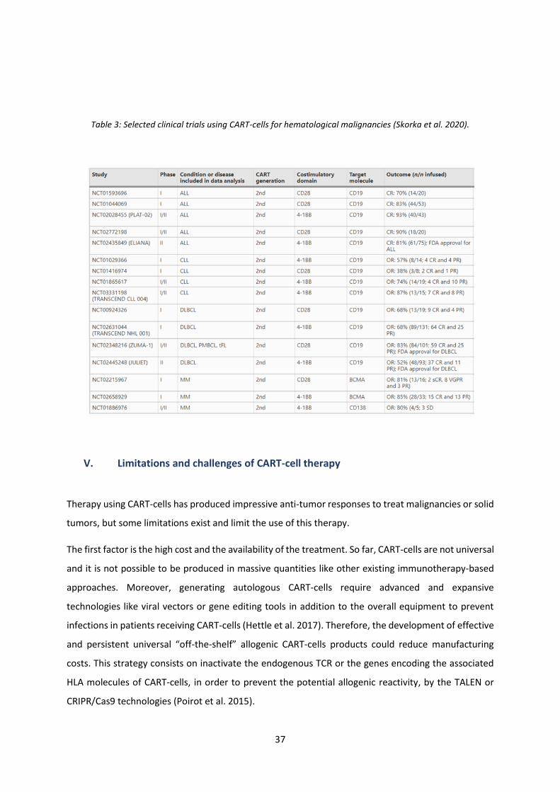
37
Table 3: Selected clinical trials using CART-cells for hematological malignancies (Skorka et al. 2020).
V. Limitations and challenges of CART-cell therapy
Therapy using CART-cells has produced impressive anti-tumor responses to treat malignancies or solid
tumors, but some limitations exist and limit the use of this therapy.
The first factor is the high cost and the availability of the treatment. So far, CART-cells are not universal
and it is not possible to be produced in massive quantities like other existing immunotherapy-based
approaches. Moreover, generating autologous CART-cells require advanced and expansive
technologies like viral vectors or gene editing tools in addition to the overall equipment to prevent
infections in patients receiving CART-cells (Hettle et al. 2017). Therefore, the development of effective
and persistent universal “off-the-shelf” allogenic CART-cells products could reduce manufacturing
costs. This strategy consists on inactivate the endogenous TCR or the genes encoding the associated
HLA molecules of CART-cells, in order to prevent the potential allogenic reactivity, by the TALEN or
CRIPR/Cas9 technologies (Poirot et al. 2015).
38
The risk of developing a resistance to CART therapy is also an important limiting factor. In fact, the
insufficiency of expansion or limited persistence of CART-cells, the loss or the down-regulation of
tumor-antigen in relapsed disease, all these factors can induce resistance to the treatment (N. N. Shah
and Fry 2019). Accordingly, administering combinations of CARs against multiple targets will overcome
relapse owing to the antigen-negative tumor cells (Orlando et al. 2018) or combining CART therapy
with blocking of inhibitory Abs such as anti-PD-1 mAb (Chong et al. 2017).
The healthy status of patients undergoing CART-cells therapy is also required. Before T-cells
administration, the lymphodepletion chemotherapy can expose vulnerable patients to infections (S. D.
Smith et al. 2019).
VI. Toxicities of CART therapy
Although CART-cells therapy has revolutionized the treatment of cancers, it is still associated with
several toxicities and safety issues. CART-cells application presents frequent toxic side effects with
significant ricks (Table 4) (Miliotou and Papadopoulou 2018). It will be simpler to classify these
toxicities into five categories: on-target on-tumor, on-target off-tumor, off-target, neurotoxicity, and
other toxicities (Figure 19) (Sun et al. 2018).
On-Target On-Tumor Toxicity (a): The most common toxicity specific to the administration of CART-
cells is the on-target on tumor toxicity. It is characterized by an excessive release of cytokines or tumor
cell necrosis. The excessive cytokine release may result in cytokine release syndrome (CRS), which can
vary from mild moderate, to severe potentially fatal forms. CRS is triggered often during the first week
after CART-cells infusion, as a first rapid immune reaction of activation and proliferation of injected
CART-cells leading to a massive secretion of pro-inflammatory cytokines such as TNF-α, IL-6, IL-10 and
IFN-γ (Hay et al. 2017). In addition, the rapid devastation of a large quantity of tumor cells can develop
a tumor lysis syndrome (TLS) (Howard, Jones, and Pui 2011).
In rare cases, CRS can progress into other immune-related toxicities such as prolonged cytopenias,
macrophage activation syndrome (MAS) (A. R. Shah et al. 2016), Cytokine Release Encephalopathy
Syndrome (CRES) (Sattva S. Neelapu et al. 2017).
On-Target Off-Tumor Toxicity (b): The most detrimental toxicity is when injected CART-cells attack
normal tissues that have the shared expression of the targeted tumor-antigen. This on-target off-
tumor toxicity of CART-cells on nonpathogenic tissues expressing low levels of the same antigen
expressed on tumor cells may be fatal in many cases (C. H. Lamers et al. 2013) (A. Hombach, Hombach,
39
and Abken 2010). Thus, the selection of target antigen, which is strictly specific to the tumor or to
generate CART-cells with lower affinity for the targeted antigen and less recognition of normal cells
expressing low levels of this antigen (Song et al. 2015), in addition, to infuse lower doses of CART-cells
to patients could potentially overcome this toxicity (Ahmed et al. 2015).
Off-Target Toxicity (c): The off-target toxicity occurs when infused CART-cells attacks another antigen
than the one they target or activate themselves independently from the antigen recognition due
sometimes to their artificial synthetic construct. For example, anti-HER2/neu CART-cells (trastuzumab)
carrying the IgG1-derived CH2CH3 domain in the spacer extracellular domain may interact with the Fc
receptor expressed on innate immune cells such as macrophages and NK cells, and resulting of an
antigen-independent activation (A. Hombach, Hombach, and Abken 2010). Likely, to date, this toxicity
cross-reactive antigen has not been evident in CART-cells clinical trials. However, it was reported a
fatal off target toxicity with tgTCR T-cells (Kötter, Andresen, and Krüger 2014).
Neurotoxicity (d): In most cases, CRS is associated with immune effector cell-associated neurotoxicity
syndrome (ICANS) in patients receiving CD19 CART-cells therapy. Neurotoxicity can appear in the days
to weeks post transfusion in patients with different types of cancers (Rubin et al. 2019). In most
patients with neurotoxicity, CART-cells have been found in cerebrospinal fluid (CSF) without reporting
a clear expression of CD19 in the affected brain areas. Suggestion that this infiltration may be caused
by hyperthermia and IL-6, released during CRS (Prudent and Breitbart 2017).
Other toxicities (e-f): Some other toxicities are also triggered by CART-cells infusion, such as (1)
Immunosuppression: caused by the lymphodepleting and nonmyeloablative regimen before the
infusion. It comes along with anemia, coagulopathy, and neutropenic sepsis (Dudley et al. 2008). (2)
Immunogenicity: due to immune reaction against the ScFv, which derives from a mouse mAb, leading
to sever anaphylaxis (C. H. J. Lamers et al. 2011) (Gorovits and Koren 2019). (3) Genotoxicity: may
results from the integrating viral vectors used for the engineered T-cells which may pose a potential
risk of oncogenic insertional mutagenesis including modifications of normal gene expression and
function (Hacein-Bey-Abina et al. 2008).
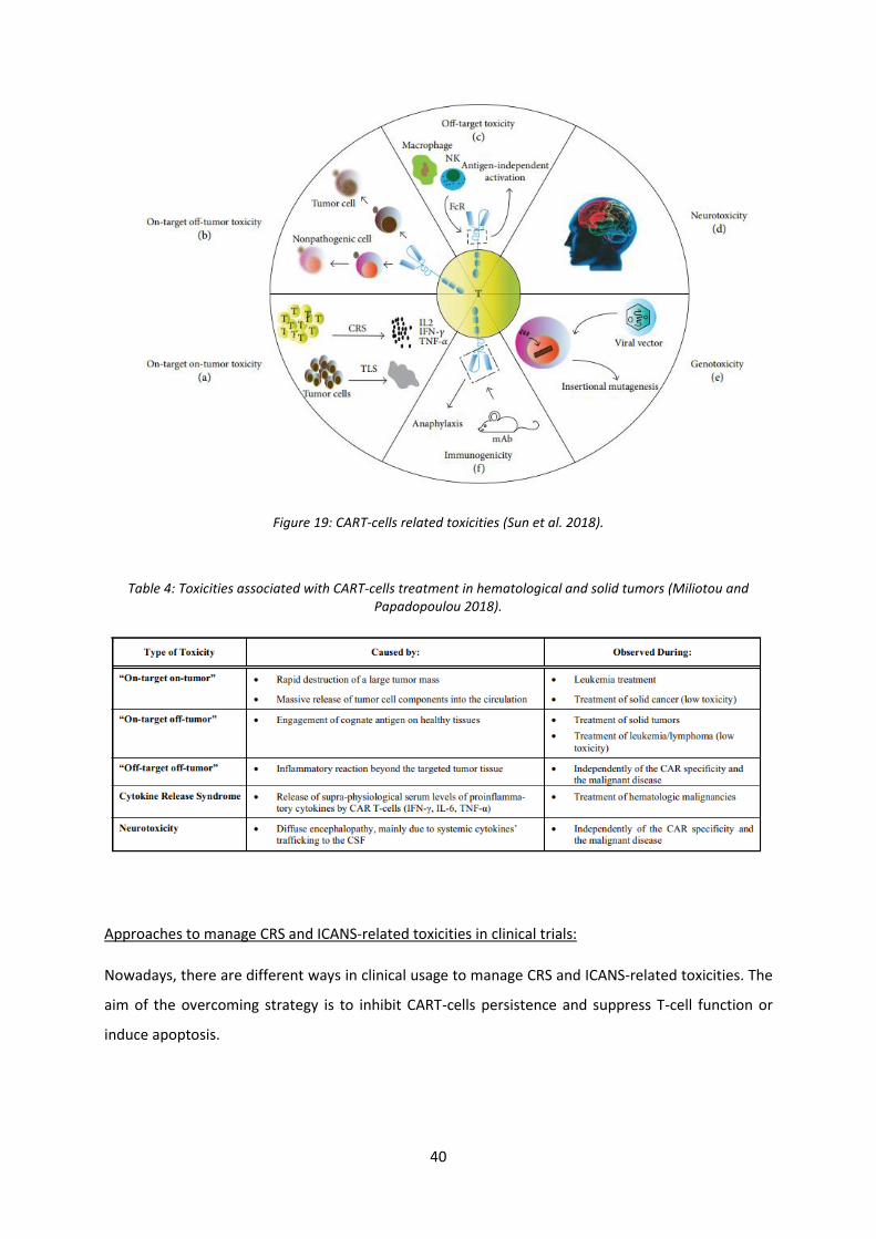
40
Figure 19: CART-cells related toxicities (Sun et al. 2018).
Table 4: Toxicities associated with CART-cells treatment in hematological and solid tumors (Miliotou and Papadopoulou 2018).
Approaches to manage CRS and ICANS-related toxicities in clinical trials:
Nowadays, there are different ways in clinical usage to manage CRS and ICANS-related toxicities. The
aim of the overcoming strategy is to inhibit CART-cells persistence and suppress T-cell function or
induce apoptosis.
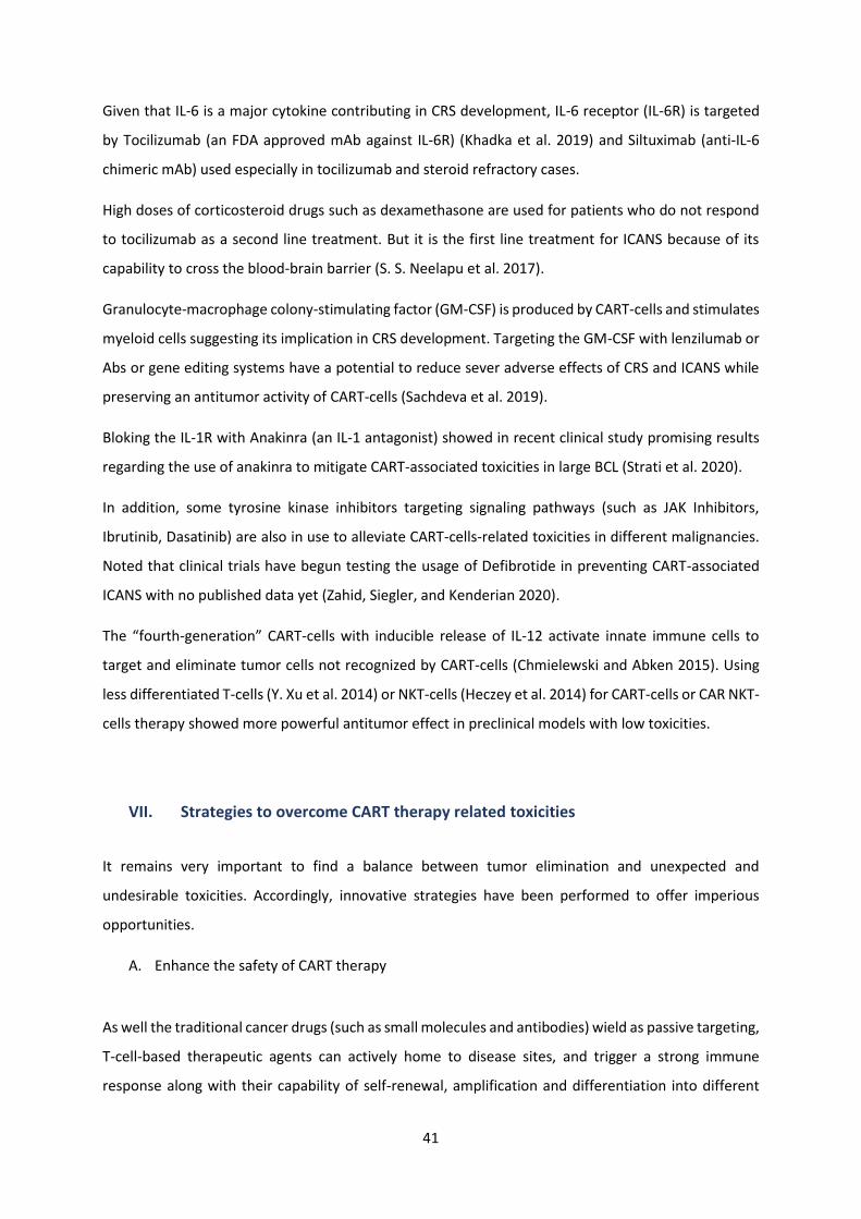
41
Given that IL-6 is a major cytokine contributing in CRS development, IL-6 receptor (IL-6R) is targeted
by Tocilizumab (an FDA approved mAb against IL-6R) (Khadka et al. 2019) and Siltuximab (anti-IL-6
chimeric mAb) used especially in tocilizumab and steroid refractory cases.
High doses of corticosteroid drugs such as dexamethasone are used for patients who do not respond
to tocilizumab as a second line treatment. But it is the first line treatment for ICANS because of its
capability to cross the blood-brain barrier (S. S. Neelapu et al. 2017).
Granulocyte-macrophage colony-stimulating factor (GM-CSF) is produced by CART-cells and stimulates
myeloid cells suggesting its implication in CRS development. Targeting the GM-CSF with lenzilumab or
Abs or gene editing systems have a potential to reduce sever adverse effects of CRS and ICANS while
preserving an antitumor activity of CART-cells (Sachdeva et al. 2019).
Bloking the IL-1R with Anakinra (an IL-1 antagonist) showed in recent clinical study promising results
regarding the use of anakinra to mitigate CART-associated toxicities in large BCL (Strati et al. 2020).
In addition, some tyrosine kinase inhibitors targeting signaling pathways (such as JAK Inhibitors,
Ibrutinib, Dasatinib) are also in use to alleviate CART-cells-related toxicities in different malignancies.
Noted that clinical trials have begun testing the usage of Defibrotide in preventing CART-associated
ICANS with no published data yet (Zahid, Siegler, and Kenderian 2020).
The “fourth-generation” CART-cells with inducible release of IL-12 activate innate immune cells to
target and eliminate tumor cells not recognized by CART-cells (Chmielewski and Abken 2015). Using
less differentiated T-cells (Y. Xu et al. 2014) or NKT-cells (Heczey et al. 2014) for CART-cells or CAR NKT-
cells therapy showed more powerful antitumor effect in preclinical models with low toxicities.
VII. Strategies to overcome CART therapy related toxicities
It remains very important to find a balance between tumor elimination and unexpected and
undesirable toxicities. Accordingly, innovative strategies have been performed to offer imperious
opportunities.
A. Enhance the safety of CART therapy
As well the traditional cancer drugs (such as small molecules and antibodies) wield as passive targeting,
T-cell-based therapeutic agents can actively home to disease sites, and trigger a strong immune
response along with their capability of self-renewal, amplification and differentiation into different
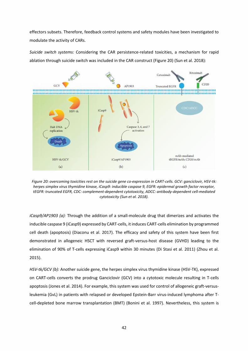
42
effectors subsets. Therefore, feedback control systems and safety modules have been investigated to
modulate the activity of CARs.
Suicide switch systems: Considering the CAR persistence-related toxicities, a mechanism for rapid
ablation through suicide switch was included in the CAR construct (Figure 20) (Sun et al. 2018):
Figure 20: overcoming toxicities rest on the suicide gene co-expression in CART-cells. GCV: ganciclovir, HSV-tk: herpes simplex virus thymidine kinase, iCasp9: inducible caspase 9, EGFR: epidermal growth factor receptor, tEGFR: truncated EGFR, CDC: complement-dependent cytotoxicity, ADCC: antibody-dependent cell-mediated
cytotoxicity (Sun et al. 2018).
iCasp9/AP1903 (a): Through the addition of a small-molecule drug that dimerizes and activates the
inducible caspase 9 (iCasp9) expressed by CART-cells, it induces CART-cells elimination by programmed
cell death (apoptosis) (Diaconu et al. 2017). The efficacy and safety of this system have been first
demonstrated in allogeneic HSCT with reversed graft-versus-host disease (GVHD) leading to the
elimination of 90% of T-cells expressing iCasp9 within 30 minutes (Di Stasi et al. 2011) (Zhou et al.
2015).
HSV-tk/GCV (b): Another suicide gene, the herpes simplex virus thymidine kinase (HSV-TK), expressed
on CART-cells converts the prodrug Ganciclovir (GCV) into a cytotoxic molecule resulting in T-cells
apoptosis (Jones et al. 2014). For example, this system was used for control of allogeneic graft-versus-
leukemia (GvL) in patients with relapsed or developed Epstein-Barr virus-induced lymphoma after T-
cell-depleted bone marrow transplantation (BMT) (Bonini et al. 1997). Nevertheless, this system is
43
limited by the immunogenicity engendered by viral enzymes and the long time (several days) to
achieve full elimination (Chalmers et al. 2001).
mAb-mediated tEGFR/mAb (c): Alternative safety switches built on targeting a specific surface antigen
expressed by CART-cells such as CD20 and truncated epidermal growth factor receptor (EGFR). These
epitope tags are recognized by FDA-approved mAb such as cetuximab (Paszkiewicz et al. 2016) and
rituximab (Tasian et al. 2017a) respectively, allowing CART-cells apoptosis through complement-
dependent cytotoxicity (CDC) and ADCC.
B. Optimize the efficiency of CART therapy
Suicide switches are effective at stopping the toxicity of CART-cells, however they induce an
irreversible ending of the therapeutic treatment. Otherwise, non-cytotoxic reversible systems may be
practical for controlling unfavorable toxicity without limiting the therapeutic effects.
1- Targeting Two Tumor-Associated Antigens (TAA): Consisting on CART-cells whose activation can be
guide through combinatorial antigen-targeting activation with separated signals, such as Dual CAR,
Tandem-CAR (Tan-CAR), inhibitory CAR (iCAR) and synthetic Notch (synNotch) (Figure 21) (Sun et al.
2018).

44
Figure 21: Targeted activation strategies to overcome toxicities. ScFV: single-chain variable fragment of antibody, CM: costimulatory molecule, ITAM: immune-receptor tyrosine-based activation motif, TAA: tumor-
associated antigen, NTA: normal tissue antigen, CAR: chimeric antigen receptor, iCAR: inhibitory CAR, TanCAR: tandem CAR, synNotch CAR: synthetic notch CAR, TF: transcription factor (Sun et al. 2018).
Dual CAR (a): A dual CAR is a bispecific CAR generated by modifying T-cells by “splitting” the activation
signal and the endo-costimulatory signal in two different CAR constructs to express two CARs targeting
different TAA to guarantee that they will be activated only against tumor cells (Kloss et al. 2013).
Tan-CAR (b): Another bispecific CAR, the Tandem CAR, which unlike the dual CAR is a single CAR specific
for two antigens due to the expression of two tandemly arranged ScFvs combined to the same signaling
domain (Grada et al. 2013).
inhibitory CARs (iCARs) (c): The iCAR construct include an ScFv specific to an antigen expressed on
normal tissues. This ScFv is coupled to the intracellular signaling domain of an inhibitory receptor such
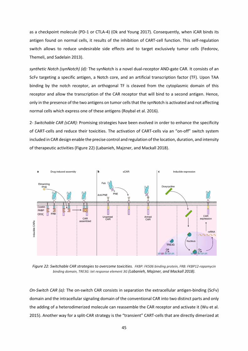
45
as a checkpoint molecule (PD-1 or CTLA-4) (Ok and Young 2017). Consequently, when iCAR binds its
antigen found on normal cells, it results of the inhibition of CART-cell function. This self-regulation
switch allows to reduce undesirable side effects and to target exclusively tumor cells (Fedorov,
Themeli, and Sadelain 2013).
synthetic Notch (synNotch) (d): The synNotch is a novel dual-receptor AND-gate CAR. It consists of an
ScFv targeting a specific antigen, a Notch core, and an artificial transcription factor (TF). Upon TAA
binding by the notch receptor, an orthogonal TF is cleaved from the cytoplasmic domain of this
receptor and allow the transcription of the CAR receptor that will bind to a second antigen. Hence,
only in the presence of the two antigens on tumor cells that the synNotch is activated and not affecting
normal cells which express one of these antigens (Roybal et al. 2016).
2- Switchable CAR (sCAR): Promising strategies have been evolved in order to enhance the specificity
of CART-cells and reduce their toxicities. The activation of CART-cells via an “on-off” switch system
included in CAR design enable the precise control and regulation of the location, duration, and intensity
of therapeutic activities (Figure 22) (Labanieh, Majzner, and Mackall 2018).
Figure 22: Switchable CAR strategies to overcome toxicities. FKBP: FK506 binding protein, FRB: FKBP12-rapamycin
binding domain, TRE3G: tet response element 3G (Labanieh, Majzner, and Mackall 2018).
On-Switch CAR (a): The on-switch CAR consists in separation the extracellular antigen-binding (ScFv)
domain and the intracellular signaling domain of the conventional CAR into two distinct parts and only
the adding of a heterodimerized molecule can reassemble the CAR receptor and activate it (Wu et al.
2015). Another way for a split-CAR strategy is the “transient” CART-cells that are directly dimerized at
46
the hinge domain with the addition of a small molecule (Juillerat et al. 2016). These two procedures
transform conventional CART-cells into smart CART-cells more safer, more precise and more efficient
(E. Zhang and Xu 2017).
Switch CAR using recombinant Abs (b): Furthermore, another type of switchable CAR was created using
a system of two antibody fragments (Fab) as bifunctional molecules. In this system, a tumor antigen-
specific Fab molecule engrafted with the neoepitope peptide (PNE) or FITC (Fluorescein
isothiocyanate) that binds to the CAR receptor containing the antigen-specific scFv anti-PNE or anti-
FITC (M. S. Kim et al. 2015).
Inducible CAR (c): The CAR expression can be induced by the addition of a small molecule such as
doxycycline or tetracycline. This molecule induces a conformational change in tet response element
3G (TRE3G) that enables the transcription of CAR mRNA and the CAR expression (Sakemura et al. 2016).
Chapter 2: 2. CART-cells immunotherapy in AML
I. Therapeutic approaches in AML using CART-cells and clinical trials
AML cancer is globally a rare disease affecting more commonly older adults and among men compared
to women. The rate of new cases of AML was 4.3 per 100,000 men and women per year based on
2014–2018 cases, age-adjusted, with a 5-year related survival around 29.5%.
A. The ongoing use of CART-cells therapy in AML
In AML application, CART-cell therapy is at its dawn, with a limited number of reported clinical trials.
In 2013, Ritchie et al. published the first reported phase 1 clinical trial using CART-cells in AML. They
used a second generation CD28-ζ CAR directed against the Lewis Y (LeY) antigen (NCT01716364).
Although only limited efficacy was observed, this study was very important, as it has given a clear
demonstration of CART-cell biological activity in AML patients in the absence of observed
hematopoietic toxicities (Ritchie et al. 2013). Nowadays, there are more than twenty CART-cell clinical
trials applying patients with AML registered with the clinicaltrials.gov (Table 5), substantially targeting
CD33, CD123, C-type Lectine-Like-1 (CLL-1/CLEC12A), NKG2DL, FLT3 (CD135), CD38 or CD44v6
(Fiorenza and Turtle 2021). Whereas more AML antigens are evaluated in preclinical for being targeted
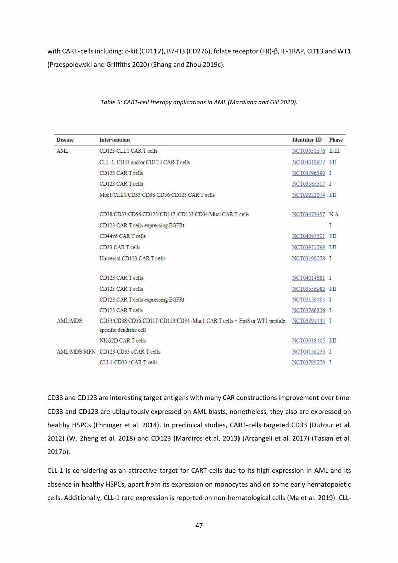
47
with CART-cells including: c-kit (CD117), B7-H3 (CD276), folate receptor (FR)-β, IL-1RAP, CD13 and WT1
(Przespolewski and Griffiths 2020) (Shang and Zhou 2019c).
Table 5: CART-cell therapy applications in AML (Mardiana and Gill 2020).
CD33 and CD123 are interesting target antigens with many CAR constructions improvement over time.
CD33 and CD123 are ubiquitously expressed on AML blasts, nonetheless, they also are expressed on
healthy HSPCs (Ehninger et al. 2014). In preclinical studies, CART-cells targeted CD33 (Dutour et al.
2012) (W. Zheng et al. 2018) and CD123 (Mardiros et al. 2013) (Arcangeli et al. 2017) (Tasian et al.
2017b).
CLL-1 is considering as an attractive target for CART-cells due to its high expression in AML and its
absence in healthy HSPCs, apart from its expression on monocytes and on some early hematopoietic
cells. Additionally, CLL-1 rare expression is reported on non-hematological cells (Ma et al. 2019). CLL-
48
1 CART-cells generated by Zhou, is a CAR of 3rd generation with two co-stimulatory domains CD28 and
4-1BB coupled to the CD3-ζ chain. CLL-1 CART-cells had efficient anti-tumor activity against AML cells,
with no cytotoxicity toward healthy HSCs in preclinical studies (Jinghua Wang et al. 2018).
A phase 1 clinical trial of autologous second generation CART-cells targeting CD33 (CD33 CART-cells)
reported a partial response in a 41-year-old male with relapsed and refractory AML (NCT01864902).
This patient showed a remarkable transient reduction of AML blasts in the bone marrow until he
relapsed with a disease progression that occurred at 9 weeks after CART-cells treatment. A significant
CRS was observed (Q. Wang et al. 2015). In order to reduce CD33 toxicity on healthy HSCs, the genetic
inactivation of CD33 gene in these cells prior to transplantation allowed to avoid on-tumor off-target
toxicities (M. Y. Kim et al. 2018).
Recently completed a phase 1 clinical trial of CD123 CART-cells produced via mRNA electroporation
(NCT02623582) with the intention of preventing long-term persistence of CART-cells thus avoiding the
risk of sever toxicities (Cummins et al. 2017b). While no measurable anti-tumor responses were
reported in R/R AML patients, CRS demonstrated CART-cells bioactivity. A transient detection of CART-
cells was noted with no adverse effects regarding healthy tissues expressing CD123. This favorable
safety profile encouraged the development of a lentiviral CD123 CART-cells clinical trial. A phase 1 trial
(NCT02159495, NCT03766126) of R/R AML patients, treated with second generation autologous
CD123 CART-cells, experienced complete remission with no treatment-related cytopenias (Budde et
al. 2017). The modification of anti-CD123 ScFv affinity with VH and VL chains of different mAbs can
decrease the BM toxicity of AML mice (Arcangeli et al. 2017). Moreover, the combination of CD123
CART-cells with ASCT offer a promising strategy for treating R/R AML patients (Testa, Pelosi, and
Castelli 2019).
Up to 2021, an estimate of 65 AML patients have been treated with CART-cell therapy. Only a quarter
of whom have achieved complete remission. The majority of the CD33 CART-cell therapies resulted in
partial responses, with no responses observed in all 31 patients treated with NKG2DL CART-cells for
three studies (Fiorenza and Turtle 2021). In a phase I study (NCT03018405, NCT02203825), seven of
eight R/R AML patients treated with NKG2DL CART-cells (CYAD-01), without prior preconditioning
chemotherapy, showed promising responses (Sallman et al. 2018). Promising and interesting results
have been reported by 3 patients who reached complete remission within one month after being
infused with CLL1 CART-cells (Bu et al. 2020).
In order to overcome the lack of tumor-specific antigen resulting in AML heterogeneity and to prevent
toxicity due to the common antigens between leukemic blasts and normal tissues, novel targeting
strategies are being investigated using dual-targeting CART-cells. A transcriptomic and surfaceome
49
proteomic analysis profiled human AML cells and normal hematopoietic cells, reported unique pairings
of antigens detected on AML blasts but not co-expressed on normal cells. Consequently, given that
one antigen is expressed on normal hematopoietic and non-hematopoietic tissues, dual-targeting
CART-cells will induce less toxicity toward normal cells (Perna et al. 2017).
In a first in human phase 1 study (NCT03795779), Liu et al. evaluated compound CAR (cCAR) T-cells
targeting two AML antigens, CD33 and CLL-1. It was reported that the majority of CD33 positive AML
cells are also positive for CLL-1 expression.
The dual CD33/CLL-1 CART-cell construct consists of two individually complete and functional CAR
receptors on the surface of a T-cell with a CD52 safety switch. Two R/R AML patients treated with
multiple lines of chemotherapy were the unique responders that have been reported from this trial so
far, with a measurable residual disease (MRD)-negative remission so they were able to proceed to allo-
HSCT (F. Liu et al. 2018).
In preclinical studies, a compound CD33/CD123 CART-cells demonstrate anti-leukemic activity and
prolonged animal survival in AML cell line xenograft models compared to a single antigen-redirected
CART-cells (Petrov et al. 2018). Furthermore, compound CD123/CLL-1 CART-cells may be useful in AML
targeting with limiting healthy HCSs toxicities (Shang and Zhou 2019b).
Another novel preclinical targeting strategy, recently published developed a bispecific and split CAR
(BissCAR) targeting CD13 and TIM3. CD13 is expressed in healthy HSCs but highly overexpressed in
AML cells, while TIM3 is only expressed in AML cells. Accordantly, this BissCART-cell was capable to
effectively eradicate patient-derived AML cells (He et al. 2020). Thus, BissCART therapy might be a
hopeful approach for developing an effective CART-cell therapy for AML.
II. CART therapy limitations in AML
A. Lack of an AML-specific target antigen
The main challenge limiting the application of CART-cells in AML is lack of suitable target antigen. AML
cells express various cell surface myeloid antigens mainly CD33 and CD123 that are often co-expressed
on normal HSPCs and their myeloid and/or lymphoid progenitors (Cummins and Gill 2019). This
fundamental biological barrier of CART-cells use remain a primary problem in AML treatment resulting
in an intolerable myeloablation and major toxicities as observed with CD33 CART- and CD123 CART-
50
cells. Despite the high anti-tumor efficacy in preclinical studies, there was several undesired toxicities
on normal cells.
An optimal AML target should be mightily expressed on the surface of the AML blasts in particularly
on LSCs in most AML cases and with no or lower expression on normal hematopoietic and non-
hematopoietic tissues.
The antigenic heterogeneity of AML give some potential targets for CART-cells therapy, each with
advantages and disadvantages.
Classification of AML target antigens:
Leukemia-specific antigens: These antigens resulting from aberrant proteins encoded by leukemia
mutations are usually expressed intracellularly (for example, NUP98 fusion proteins, mutated FLT-3
and NPM1, IDH-1) and exclusively in malignant clones so they might be ideal targets. To date, no clinical
trials evaluating these leukemia-specific neoantigens.
Lineage-restricted antigens: These antigens are restricted to the myeloid lineage and usually expressed
on the surface of myeloid cells. The majority of current clinical trials of antibodies or CART-cells in AML
patients target this type of antigens, such as CD33, CD123, CLL-1/CLEC12A, IL-1RAP, CD135/FLT-3).
Leukemia-associated antigens: Leukemia-associated antigens are overexpressed on AML cells
comparable to healthy tissues. Usually these antigens are not lineage specific which make their
expression on healthy hematopoietic cells less likely. Nevertheless, they may be found on non-
hematopoietic tissues, causing on-target off-tumor toxicities. For instance, WT1 and PRAME are under
early-phase clinical trials evaluation in AML patients (Lichtenegger et al. 2020).
B. Persistence of CART-cells
Since CART-cells target highly expressed non-leukemia specific antigens including CD33 and CD123
resulting in the AML-BM failure, limiting the long-term CART-cell persistence in-vivo could offer an
important opportunity to prevent toxicities. The considerable strategy of a “safety switch” system that
consists on incorporating a suicide gene into T-cells allowing the elimination of these cells in-vivo has
been evaluated in various preclinical and clinical studies. A suicide gene that has been employed for a
long time in CART-cell treatment is the HSV-tk. Upon administration of the prodrug Ganciclovir, this
suicide gene is able to transform this prodrug into a toxic compound that stop the DNA replication.
51
Hence, providing a selective depletion of T-cells expressing the HSV-tk gene (Bordignon et al. 1995).
However, the immunogenicity of the viral enzyme associated with the HSV-tk/Ganciclovir system and
the long latency to activation and effects manifestation, are considerable limits for its use, as the
management of toxicity require immediate termination.
A more safe suicide system utilized is the iCasp9/AP1903 system. In this system, the intracellular
domain of a pro-apoptotic protein the caspase 9 is fused to the extracellular binding domain of the
FK506 drug protein resulting in the co-expression of iCasp9 in CART-cells. Following the administration
of a synthetic molecule drug called AP1903, a dimerization of the fusion proteins occurs allowing a
rapid ablation of CART-cells (Gargett and Brown 2014) (Figure 20 (b)). The iCasp9 suicide system was
clinically evaluated in haploidentical stem cell transplantation background (Di Stasi et al. 2011). Then,
it was explored by Hoyos et al. in a preclinical situation using CART-cell therapy (Hoyos et al. 2010).
Thereafter, the iCasp9 suicide system has been integrated in the CAR vector construct of many clinical
trials (such as clinicaltrials.gov identifier: 02992210, 01822652). So far, the AP1903 administration has
never been needed thus its efficacy in CART-cells setting is not proved yet. Rapamycin caspase 9
(rapaCDasp9) is also a safe suicide system, based on the fusion of both FRB and FKBP12 with the
catalytic domain of caspase 9, used for CD19 CART-cells elimination in preclinical studies (Stavrou et
al. 2018).
Another alternative for a safety switch is to use ΔEGFR/cetuximab or ΔCD20/rituximab systems (Figure
20 (c)). These two systems are based on engineering CART-cells to co-express a truncated (Δ) well-
characterized cell surface antigen against a clinically approved mAb (such as ΔEGFR targetable by
cetuximab, and ΔCD20 targetable by rituximab). Through antibody-based elimination, these systems
are able to eradicate transferred CART-cells by ADCC or CDC (H. Li and Zhao 2017).
Some approaches use non-specific drugs to eliminate infused CART-cells as well as endogenous T-cells
principally anti-thymocyte globulin (ATG), or the anti-CD52 mAb (alemtuzumab) (Ali et al. 2017).
In another hand, in order to limit CART-cells persistence, some other different strategies do not require
administration of exogenous antibodies such as using the mRNA electroporation for incorporation of
the CAR transgene into T-cells, by which the degradation of the mRNA inherently limit CART-cells
function (Cummins et al. 2017b) (Figure 23).
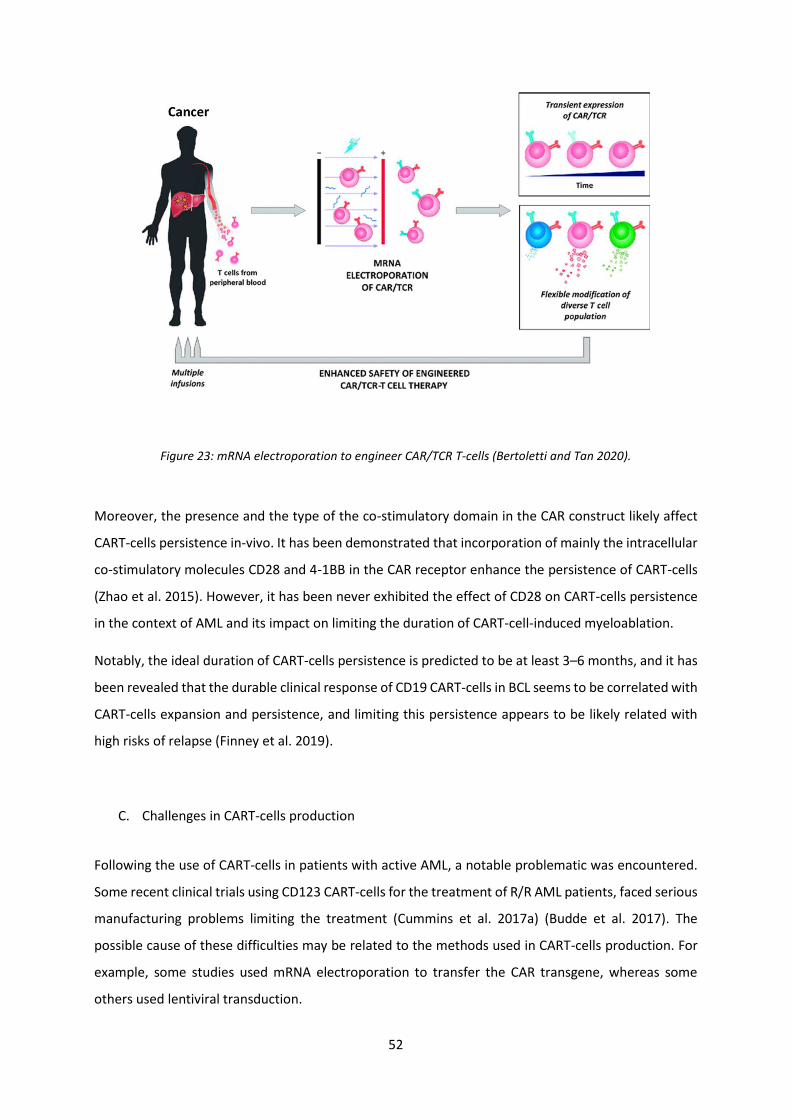
52
Figure 23: mRNA electroporation to engineer CAR/TCR T-cells (Bertoletti and Tan 2020).
Moreover, the presence and the type of the co-stimulatory domain in the CAR construct likely affect
CART-cells persistence in-vivo. It has been demonstrated that incorporation of mainly the intracellular
co-stimulatory molecules CD28 and 4-1BB in the CAR receptor enhance the persistence of CART-cells
(Zhao et al. 2015). However, it has been never exhibited the effect of CD28 on CART-cells persistence
in the context of AML and its impact on limiting the duration of CART-cell-induced myeloablation.
Notably, the ideal duration of CART-cells persistence is predicted to be at least 3–6 months, and it has
been revealed that the durable clinical response of CD19 CART-cells in BCL seems to be correlated with
CART-cells expansion and persistence, and limiting this persistence appears to be likely related with
high risks of relapse (Finney et al. 2019).
C. Challenges in CART-cells production
Following the use of CART-cells in patients with active AML, a notable problematic was encountered.
Some recent clinical trials using CD123 CART-cells for the treatment of R/R AML patients, faced serious
manufacturing problems limiting the treatment (Cummins et al. 2017a) (Budde et al. 2017). The
possible cause of these difficulties may be related to the methods used in CART-cells production. For
example, some studies used mRNA electroporation to transfer the CAR transgene, whereas some
others used lentiviral transduction.
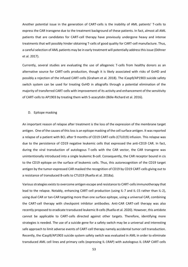
53
Another potential issue in the generation of CART-cells is the inability of AML patients’ T-cells to
express the CAR transgene due to the treatment background of these patients. In fact, almost all AML
patients that are candidates for CART-cell therapy have previously undergone heavy and intense
treatments that will possibly hinder obtaining T-cells of good quality for CART-cell manufacture. Thus,
a careful selection of AML patients may be in early treatment will potentially address this issue (Döhner
et al. 2017).
Currently, several studies are evaluating the use of allogeneic T-cells from healthy donors as an
alternative source for CART-cells production, though it is likely associated with risks of GvHD and
possibly a rejection of the infused CART-cells (Graham et al. 2018). The iCasp9/AP1903 suicide safety
switch system can be used for treating GvHD in allografts through a potential elimination of the
majority of transferred CART-cells with improvement of its activity and enhancement of the sensitivity
of CART-cells to AP1903 by treating them with 5-azacytidin (Bôle-Richard et al. 2016).
D. Epitope masking
An important reason of relapse after treatment is the loss of the expression of the membrane target
antigen. One of the causes of this loss is an epitope masking of the cell surface antigen. It was reported
a relapse of a patient with BCL after 9 months of CD19 CART-cells (CTL019) infusion. This relapse was
due to the persistence of CD19 negative leukemic cells that expressed the anti-CD19 CAR. In fact,
during the viral transduction of autologous T-cells with the CAR vector, the CAR transgene was
unintentionally introduced into a single leukemic B-cell. Consequently, the CAR receptor bound in cis
to the CD19 epitope on the surface of leukemic cells. Thus, this autorecognition of the CD19 target
antigen by the tumor-expressed CAR masked the recognition of CD19 by CD19 CART-cells giving out to
a resistance of transduced B-cells to CTL019 (Ruella et al. 2018a).
Various strategies exists to overcome antigen escape and resistance to CART-cells immunotherapy that
lead to the relapse. Notably, enhancing CART-cell production (using IL-7 and IL-15 rather than IL-2),
using dual CAR or tan-CAR targeting more than one surface epitope, using a universal CAR, combining
the CART-cell therapy with checkpoint inhibitor antibodies. Anti-CAR CART-cell therapy was also
recently proposed to eradicate transduced leukemic B-cells (Ruella et al. 2020). However, this antidote
cannot be applicable to CART-cells directed against other targets. Therefore, identifying more
strategies is needed. The use of a suicide gene for a safety switch may be a universal and interesting
safe approach to limit adverse events of CART-cell therapy namely accidental tumor cell transduction.
Recently, the iCasp9/AP1903 suicide system safety switch was evaluated in AML in order to eliminate
transduced AML cell lines and primary cells (expressing IL-1RAP) with autologous IL-1RAP CART-cells
54
for triggering an epitope masking (Figure 24) (Warda et al. 2021). The elimination of >85% of tumor
cells IL-1RAP+/CAR+ in a 24 hours of exposure to AP1903 (Rimiducid) encourage the use of this
approach in a phase I trial.
Figure 24: iCasp9 safety switch system mechanism for an epitope masking model in autologous AML tumor cells (Warda et al. 2021).
E. Immunosuppression worn by AML toward CART-cells therapy
While AML has long been identified being immune-responsive, featured by the GVL effects following
allo-HSCT, it is also considered as an immunosuppressive or immune-evasive disease. A wide range of
AML immunosuppressive mechanisms have been described based on in-vitro and preclinical studies
(Figure 25) (Mardiana and Gill 2020). Identifying the immunosuppressive pathways responsible for
AML evasion and relapse remains very crucial to develop appropriate efficient therapies.
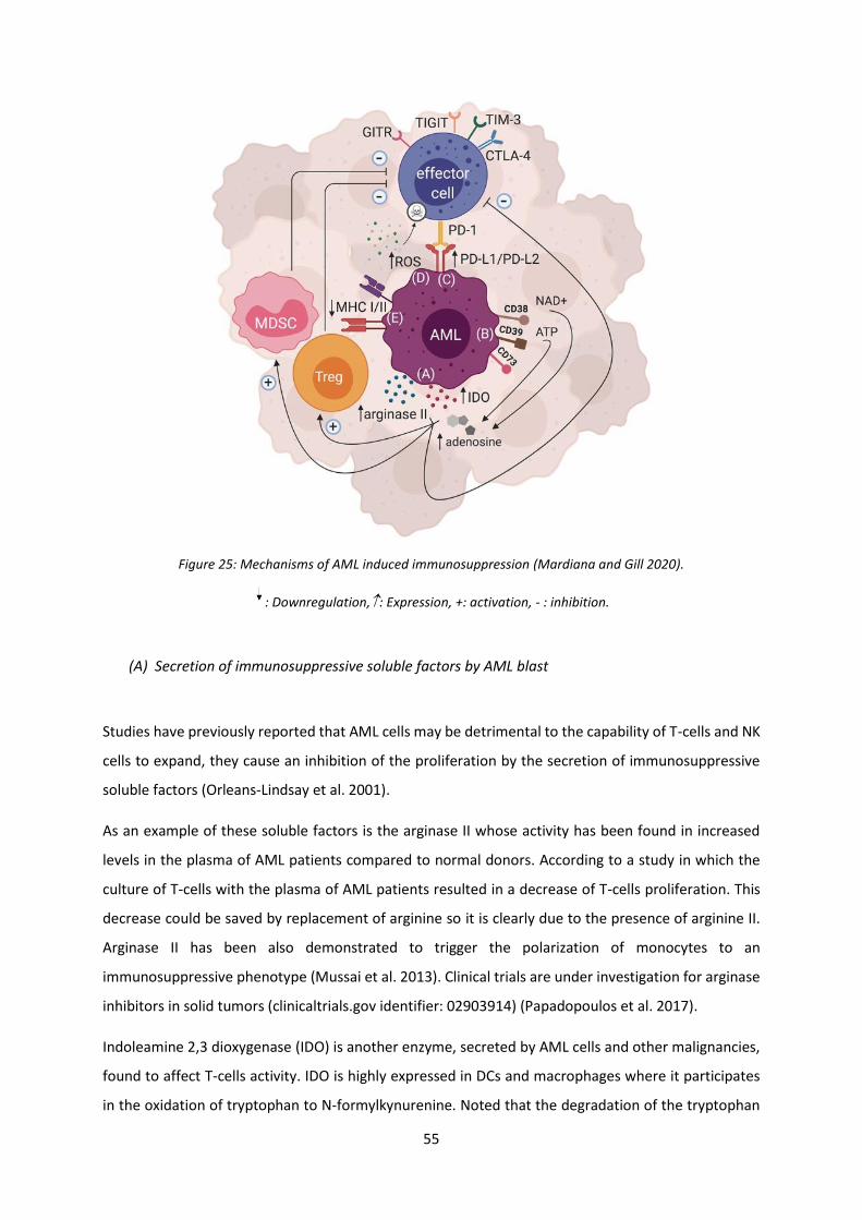
55
Figure 25: Mechanisms of AML induced immunosuppression (Mardiana and Gill 2020).
: Downregulation,: Expression, +: activation, - : inhibition.
(A) Secretion of immunosuppressive soluble factors by AML blast
Studies have previously reported that AML cells may be detrimental to the capability of T-cells and NK
cells to expand, they cause an inhibition of the proliferation by the secretion of immunosuppressive
soluble factors (Orleans-Lindsay et al. 2001).
As an example of these soluble factors is the arginase II whose activity has been found in increased
levels in the plasma of AML patients compared to normal donors. According to a study in which the
culture of T-cells with the plasma of AML patients resulted in a decrease of T-cells proliferation. This
decrease could be saved by replacement of arginine so it is clearly due to the presence of arginine II.
Arginase II has been also demonstrated to trigger the polarization of monocytes to an
immunosuppressive phenotype (Mussai et al. 2013). Clinical trials are under investigation for arginase
inhibitors in solid tumors (clinicaltrials.gov identifier: 02903914) (Papadopoulos et al. 2017).
Indoleamine 2,3 dioxygenase (IDO) is another enzyme, secreted by AML cells and other malignancies,
found to affect T-cells activity. IDO is highly expressed in DCs and macrophages where it participates
in the oxidation of tryptophan to N-formylkynurenine. Noted that the degradation of the tryptophan
56
can hamper the proliferation and the differentiation of T-cells. Besides, another function of IDO
enzyme is to stimulate Treg conversion and increase their immunosuppressive activity. It was reported
an increased level of kynurenine in AML patients associated with a reduced overall survival. Clinical
trials are currently ongoing for IDO inhibitors in AML (clinicaltrials.gov identifier: 02835729) and other
types of malignancies (Soliman et al. 2016). Indeed, IDO inhibition was shown to upgrade CART-cells
efficacy in preclinical studies (Ninomiya et al. 2015) (Q. Huang et al. 2018).
(B) Enhance immunosuppressive cells’ activity
One of the immunosuppressive manifestations of AML cells is that they can recruit and enhance the
expansion and the activity of immunosuppressive immune cells such as Tregs and myeloid-derived
suppressor cells (MDSCs) (Epperly, Gottschalk, and Velasquez 2020). AML cells as well as Tregs express
ectonucleotideases (CD39 and CD73) enzymes that function is to breakdown the adenosine
triphosphate (ATP) or nicotinamide adenine dinucleotide (NAD+) producing large number of
adenosine. Extracellular adenosine aggregation enhance Tregs activity and alter the effector function
of T-cells (Dulphy et al. 2014). MDSCs are also found in high levels in patients with AML. This expansion
was shown to be raised by both primary AML cells and AML cell lines in a contact-dependent manner
through the MUC-1 oncoprotein (Pyzer et al. 2017).
In the context of CART-cell therapy, many potential approaches were investigated in order to address
the issue of the inhibitory function of abundant immunosuppressive cells against CART-cells. As such,
blocking the adenosine immunosuppression activity by an antagonists targeting the adenosine
receptor (A2AR) administered in combination with CART-cell therapy (Beavis et al. 2017) or by the
deletion of this receptor in the CAR construct using CRISPR/Cas9 system (Giuffrida et al. 2021). In the
aim to target MDSCs immunosuppression, some clinical trials have been initiated in multiple
malignancies using small molecules to reduce the recruitment of MDSCs, for example chemokine
inhibitors or the liver-X nuclear hormone receptor (LXR) agonist (Fleming et al. 2018). Furthermore,
the in-vivo depletion of MDSCs increased CART-cells anti-tumor effects (Di et al. 2019). Several
strategies also have been evaluated to enhance CART-cells expansion and persistence including IL-8 or
IL-15 transgene expression, addition of different cytokines (IL-7, IL-15, and IL-21) and many others.
However, these strategies are not yet investigated in AML.
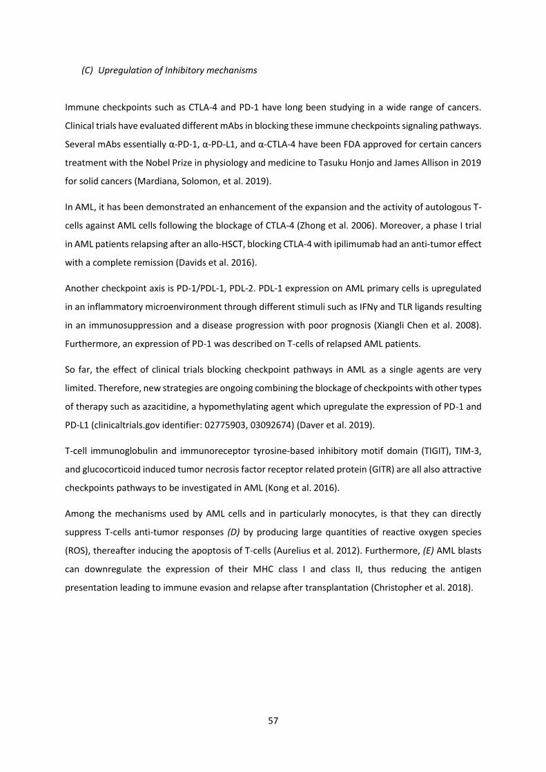
57
(C) Upregulation of Inhibitory mechanisms
Immune checkpoints such as CTLA-4 and PD-1 have long been studying in a wide range of cancers.
Clinical trials have evaluated different mAbs in blocking these immune checkpoints signaling pathways.
Several mAbs essentially α-PD-1, α-PD-L1, and α-CTLA-4 have been FDA approved for certain cancers
treatment with the Nobel Prize in physiology and medicine to Tasuku Honjo and James Allison in 2019
for solid cancers (Mardiana, Solomon, et al. 2019).
In AML, it has been demonstrated an enhancement of the expansion and the activity of autologous T-
cells against AML cells following the blockage of CTLA-4 (Zhong et al. 2006). Moreover, a phase I trial
in AML patients relapsing after an allo-HSCT, blocking CTLA-4 with ipilimumab had an anti-tumor effect
with a complete remission (Davids et al. 2016).
Another checkpoint axis is PD-1/PDL-1, PDL-2. PDL-1 expression on AML primary cells is upregulated
in an inflammatory microenvironment through different stimuli such as IFNγ and TLR ligands resulting
in an immunosuppression and a disease progression with poor prognosis (Xiangli Chen et al. 2008).
Furthermore, an expression of PD-1 was described on T-cells of relapsed AML patients.
So far, the effect of clinical trials blocking checkpoint pathways in AML as a single agents are very
limited. Therefore, new strategies are ongoing combining the blockage of checkpoints with other types
of therapy such as azacitidine, a hypomethylating agent which upregulate the expression of PD-1 and
PD-L1 (clinicaltrials.gov identifier: 02775903, 03092674) (Daver et al. 2019).
T-cell immunoglobulin and immunoreceptor tyrosine-based inhibitory motif domain (TIGIT), TIM-3,
and glucocorticoid induced tumor necrosis factor receptor related protein (GITR) are all also attractive
checkpoints pathways to be investigated in AML (Kong et al. 2016).
Among the mechanisms used by AML cells and in particularly monocytes, is that they can directly
suppress T-cells anti-tumor responses (D) by producing large quantities of reactive oxygen species
(ROS), thereafter inducing the apoptosis of T-cells (Aurelius et al. 2012). Furthermore, (E) AML blasts
can downregulate the expression of their MHC class I and class II, thus reducing the antigen
presentation leading to immune evasion and relapse after transplantation (Christopher et al. 2018).
58
III. Targeting the AML tumor microenvironment with CART-Cells
As represented before, the microenvironment could play a substantial role in the regulation of the
functions and toxicities of CART-cells. The deregulation of several costimulatory ligands by the
microenvironment in addition to the immunosuppressive and hypoxic nature of AML-BM niche, they
may presumably hamper CART-cell functionality. To date, only one study using CD44v6 CART-cells has
assessed the role of the BM-mesenchymal stromal cells (MSC) in CART-cell resistance in AML and
further investigation is required (Casucci et al. 2013).
In aim to hamper the tumor-associated immunosuppressive environment, different CAR constructs
have been evolved such as: the fourth-generation CAR that have been engineered to produce immune-
stimulatory cytokines (IL-12) (Charrot and Hallam 2019), to release PD-1-blocking ScFv (Sarwish Rafiq
et al. 2018) with preclinical anti-tumor efficacy, or to enhance endogenous anti-tumor immune
response by constitutively express CD40L (Curran et al. 2015). Additionally, a switch CAR with the
extracellular domain of an inhibitory T-cell receptor (like PD-1) coupled to an intracellular
costimulatory signal have been described (X. Liu et al. 2016). Furthermore, many other strategies are
also used (Figure 26).
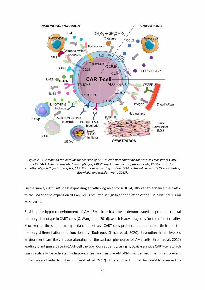
59
Figure 26: Overcoming the immunosuppression of AML microenvironment by adoptive cell transfer of CART-cells. TAM: Tumor-associated macrophages, MDSC: myeloid-derived suppressor cells, VEGFR: vascular
endothelial growth factor receptor, FAP: fibroblast activating protein, ECM: extracellular matrix (Gowrishankar, Birtwistle, and Micklethwaite 2018).
Furthermore, c-kit CART-cells expressing a trafficking receptor (CXCR4) allowed to enhance the traffic
to the BM and the expansion of CART-cells resulted in significant depletion of the BM c-kit+ cells (Arai
et al. 2018).
Besides, the hypoxic environment of AML-BM niche have been demonstrated to promote central
memory phenotype in CART-cells (X. Wang et al. 2016), which is advantageous for their functionality.
However, at the same time hypoxia can decrease CART-cells proliferation and hinder their effector
memory differentiation and functionality (Rodriguez-Garcia et al. 2020). In another hand, hypoxic
environment can likely induce alteration of the surface phenotype of AML cells (Sironi et al. 2015)
leading to antigen escape in CART-cell therapy. Consequently, using hypoxia-sensitive CART-cells which
can specifically be activated in hypoxic sites (such as the AML-BM microenvironment) can prevent
undesirable off-site toxicities (Juillerat et al. 2017). This approach could be credibly assessed to
60
eradicate residual LSC persisting in the hypoxic BM niche after chemotherapy and responsible for the
relapse. More investigations are required to better understand the effect of the AML-BM niche on
CART-cells functions for developing greater and more efficient therapeutic targets.
IV. Approaches to overcome CART-cells resistance in AML and to manage toxicities
A. Genome editing using CRISPR/Cas9:
Toward the prevention of undesired prolonged myeloablation effect and for a long-term CART-cells
persistence, a novel strategy has been evaluated based on editing out the CAR target antigen from a
donor allograft. This technique consist on the deletion of the CAR target antigen for example CD33 in
the HSPCs of the donor using CRISPR/Cas9 technology (Figure 27) (C. Liu et al. 2017) and then
transplant these CD33-/- HSPCs into the patient before administrating CD33 CART-cells manufactured
from the same donor. Thereby, CD33-/- allograft HSPCs will trigger a normal hematopoiesis always
with continued persistence of CART-cells. In fact, a group of researchers has demonstrated by
CRISPR/Cas9 genome editing that autologous CD33 CART-cells (from the donor) did not affect CD33-/-
HSPCs in-vitro and in AML murine xenograft and resulting in an efficient elimination of leukemia
without myelotoxicity. They have also exhibit that CD33-deficient HSPCs have consistently kept their
hematopoietic and immunological functions. Accordingly, genetically engineering the donor derived
HSPCs represent an innovative approach to create an artificial AML-specific antigen immunotherapy
with avoiding on-target, off-tumor toxicity. Indeed, a clinical trial implying the use of allogeneic CD33-
/- HSCT prior to CART-cell infusion is currently being investigated at the University of Pennsylvania for
patients with R/R AML (M. Y. Kim et al. 2018).
Besides CD33, CD123 is also a potential AML target antigen that may be edited using a similar
approach. However, since CD123 plays an important role in hematopoiesis as it is the alpha-chain IL-
3R, the complete deletion of CD123 in hematopoietic cells will results in an abnormal hematopoietic
system (Testa, Pelosi, and Frankel 2014). Therefore, two alternative approaches could be potentially
used: the deletion of the epitope on the CD123 receptor that can be recognized by the CART-cells, as
well as the knockdown of the CD123 expression in donor-derived HSPCs instead of a complete
knockout of its expression. Herewith, the expression of CD123 on hematopoietic allograft cells will be
minimized to a level below the CART-cells activation threshold, so CART-cells will not be activated
against normal HSPCs while preserving a normal hematopoiesis with CD123 presence. So far, this
approach is under investigation.
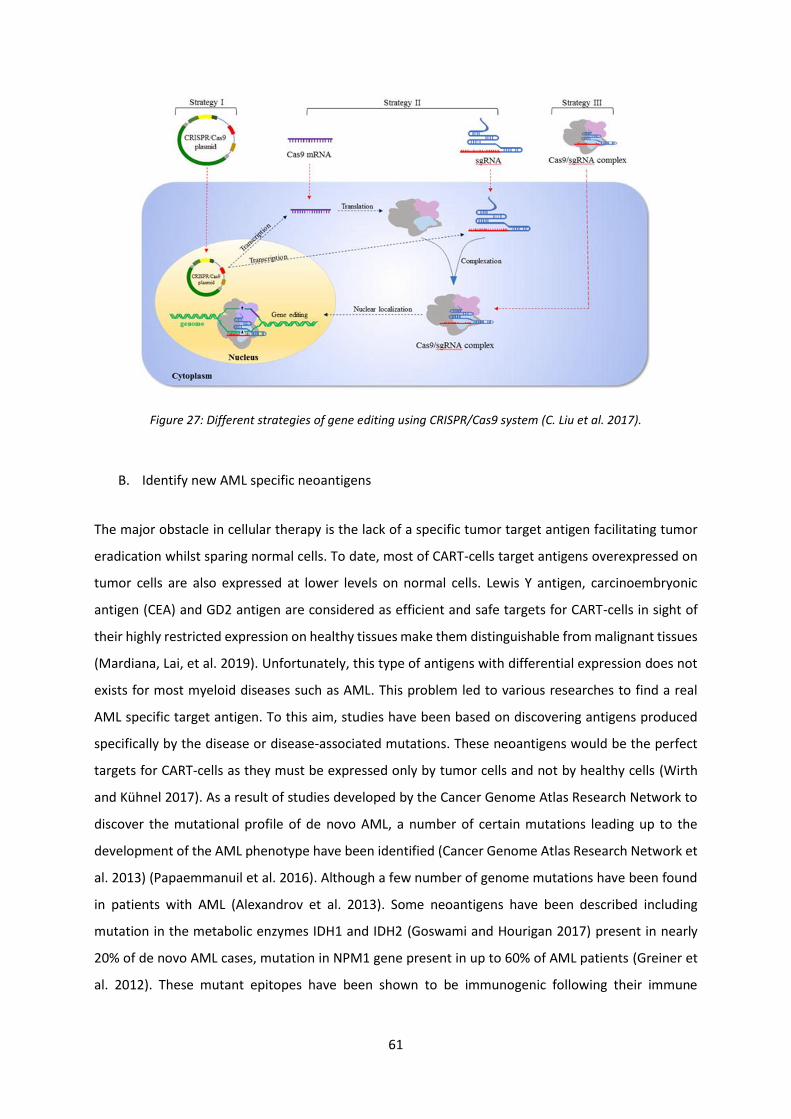
61
Figure 27: Different strategies of gene editing using CRISPR/Cas9 system (C. Liu et al. 2017).
B. Identify new AML specific neoantigens
The major obstacle in cellular therapy is the lack of a specific tumor target antigen facilitating tumor
eradication whilst sparing normal cells. To date, most of CART-cells target antigens overexpressed on
tumor cells are also expressed at lower levels on normal cells. Lewis Y antigen, carcinoembryonic
antigen (CEA) and GD2 antigen are considered as efficient and safe targets for CART-cells in sight of
their highly restricted expression on healthy tissues make them distinguishable from malignant tissues
(Mardiana, Lai, et al. 2019). Unfortunately, this type of antigens with differential expression does not
exists for most myeloid diseases such as AML. This problem led to various researches to find a real
AML specific target antigen. To this aim, studies have been based on discovering antigens produced
specifically by the disease or disease-associated mutations. These neoantigens would be the perfect
targets for CART-cells as they must be expressed only by tumor cells and not by healthy cells (Wirth
and Kühnel 2017). As a result of studies developed by the Cancer Genome Atlas Research Network to
discover the mutational profile of de novo AML, a number of certain mutations leading up to the
development of the AML phenotype have been identified (Cancer Genome Atlas Research Network et
al. 2013) (Papaemmanuil et al. 2016). Although a few number of genome mutations have been found
in patients with AML (Alexandrov et al. 2013). Some neoantigens have been described including
mutation in the metabolic enzymes IDH1 and IDH2 (Goswami and Hourigan 2017) present in nearly
20% of de novo AML cases, mutation in NPM1 gene present in up to 60% of AML patients (Greiner et
al. 2012). These mutant epitopes have been shown to be immunogenic following their immune
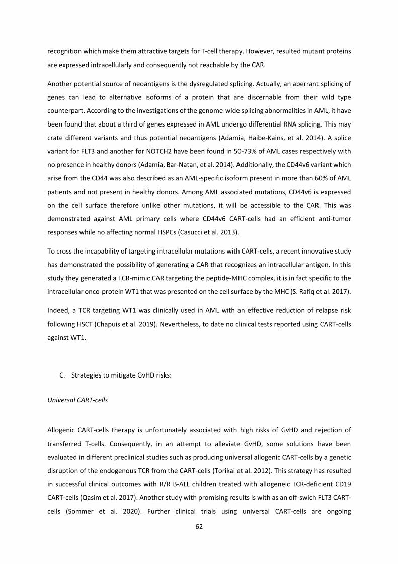
62
recognition which make them attractive targets for T-cell therapy. However, resulted mutant proteins
are expressed intracellularly and consequently not reachable by the CAR.
Another potential source of neoantigens is the dysregulated splicing. Actually, an aberrant splicing of
genes can lead to alternative isoforms of a protein that are discernable from their wild type
counterpart. According to the investigations of the genome-wide splicing abnormalities in AML, it have
been found that about a third of genes expressed in AML undergo differential RNA splicing. This may
crate different variants and thus potential neoantigens (Adamia, Haibe-Kains, et al. 2014). A splice
variant for FLT3 and another for NOTCH2 have been found in 50-73% of AML cases respectively with
no presence in healthy donors (Adamia, Bar-Natan, et al. 2014). Additionally, the CD44v6 variant which
arise from the CD44 was also described as an AML-specific isoform present in more than 60% of AML
patients and not present in healthy donors. Among AML associated mutations, CD44v6 is expressed
on the cell surface therefore unlike other mutations, it will be accessible to the CAR. This was
demonstrated against AML primary cells where CD44v6 CART-cells had an efficient anti-tumor
responses while no affecting normal HSPCs (Casucci et al. 2013).
To cross the incapability of targeting intracellular mutations with CART-cells, a recent innovative study
has demonstrated the possibility of generating a CAR that recognizes an intracellular antigen. In this
study they generated a TCR-mimic CAR targeting the peptide-MHC complex, it is in fact specific to the
intracellular onco-protein WT1 that was presented on the cell surface by the MHC (S. Rafiq et al. 2017).
Indeed, a TCR targeting WT1 was clinically used in AML with an effective reduction of relapse risk
following HSCT (Chapuis et al. 2019). Nevertheless, to date no clinical tests reported using CART-cells
against WT1.
C. Strategies to mitigate GvHD risks:
Universal CART-cells
Allogenic CART-cells therapy is unfortunately associated with high risks of GvHD and rejection of
transferred T-cells. Consequently, in an attempt to alleviate GvHD, some solutions have been
evaluated in different preclinical studies such as producing universal allogenic CART-cells by a genetic
disruption of the endogenous TCR from the CART-cells (Torikai et al. 2012). This strategy has resulted
in successful clinical outcomes with R/R B-ALL children treated with allogeneic TCR-deficient CD19
CART-cells (Qasim et al. 2017). Another study with promising results is with as an off-swich FLT3 CART-
cells (Sommer et al. 2020). Further clinical trials using universal CART-cells are ongoing
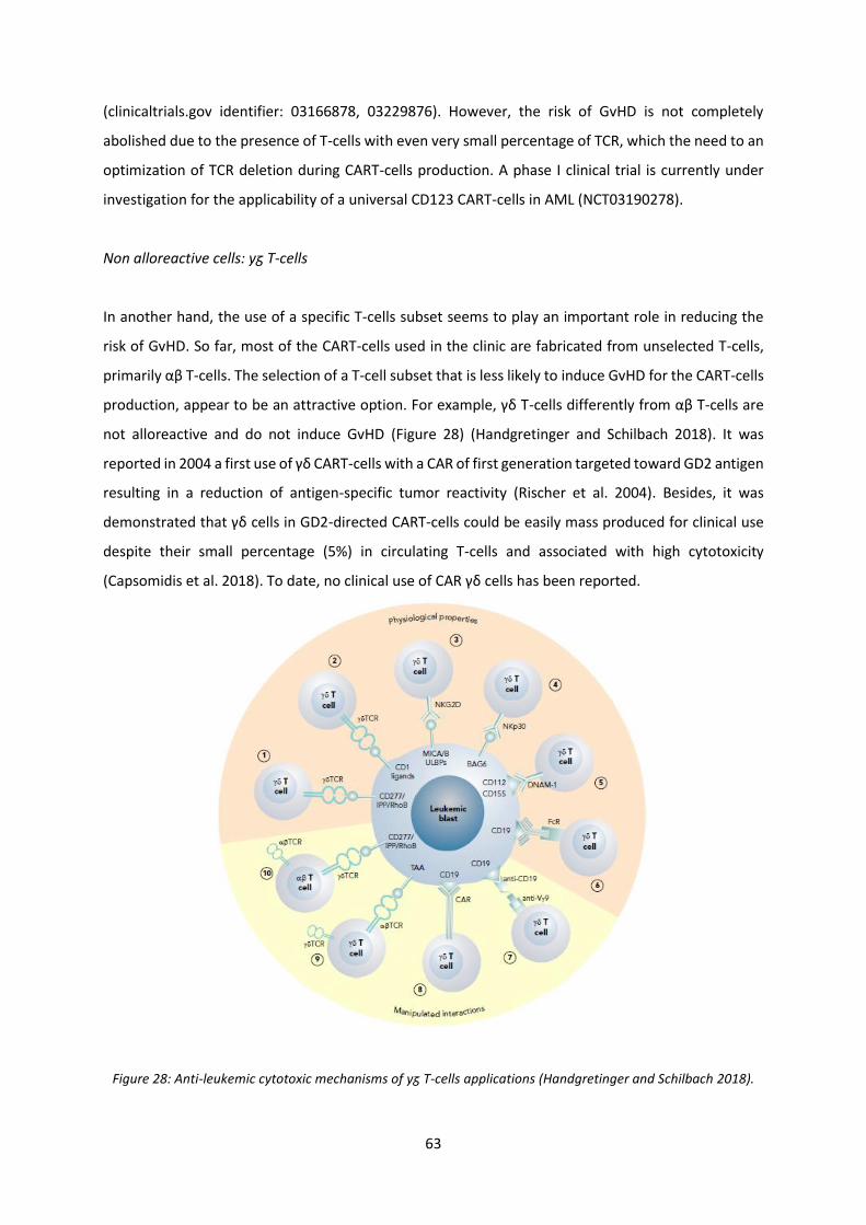
63
(clinicaltrials.gov identifier: 03166878, 03229876). However, the risk of GvHD is not completely
abolished due to the presence of T-cells with even very small percentage of TCR, which the need to an
optimization of TCR deletion during CART-cells production. A phase I clinical trial is currently under
investigation for the applicability of a universal CD123 CART-cells in AML (NCT03190278).
Non alloreactive cells: yᵹ T-cells
In another hand, the use of a specific T-cells subset seems to play an important role in reducing the
risk of GvHD. So far, most of the CART-cells used in the clinic are fabricated from unselected T-cells,
primarily αβ T-cells. The selection of a T-cell subset that is less likely to induce GvHD for the CART-cells
production, appear to be an attractive option. For example, γδ T-cells differently from αβ T-cells are
not alloreactive and do not induce GvHD (Figure 28) (Handgretinger and Schilbach 2018). It was
reported in 2004 a first use of γδ CART-cells with a CAR of first generation targeted toward GD2 antigen
resulting in a reduction of antigen-specific tumor reactivity (Rischer et al. 2004). Besides, it was
demonstrated that γδ cells in GD2-directed CART-cells could be easily mass produced for clinical use
despite their small percentage (5%) in circulating T-cells and associated with high cytotoxicity
(Capsomidis et al. 2018). To date, no clinical use of CAR γδ cells has been reported.
Figure 28: Anti-leukemic cytotoxic mechanisms of yᵹ T-cells applications (Handgretinger and Schilbach 2018).
64
Other effector cells: NKT and NK cells
Alternatively, it is possible to use other effector cells to mitigate GvHD risks such as CIK-cells, NKT-cells
and NK-cells for the CAR engineering and the AML treatment (Figure 29) (Burger et al. 2019).
Figure 29: CAR NK cells anti-tumor function in AML (Burger et al. 2019).
As published in preclinical studies, using CD123 CAR CIK-cells and CD33 CAR CIK-cells in-vivo promoted
a very efficient reduction of AML leukemia burden with limited toxicity on normal HSCs with only
CD123 CAR CIK-cells (Tettamanti et al. 2013) (Pizzitola et al. 2014) (Rotiroti et al. 2020). Currently, CAR
NK-cells are being investigated in phase I/II clinical trials, targeting different antigens mainly CD19 but
also others including CD7, CD33, MUC-I, and Her2 (Kloess, Kretschmer, et al. 2019). CD33 CAR NK-92
cells was evaluated in a phase 1 trial (Tang et al. 2018) and recently engineered CD33 targeted CAR
NK-cells, using CRISPR/Cas9 gene editing and AAV-mediated gene delivery, were able to kill CD33+
AML cells in-vitro (Naeimi Kararoudi et al. 2020). CD123-specific CAR cord blood NK-cells eliminate
CD123+ AML cells which support their potential use in the clinic for patients with relapsed AML
(Kerbauy et al. 2017). However, CAR NK therapy was associated with a poor CAR NK-cells persistence,
thus in order to enhance CAR persistence it was recently reported an in-vitro activity of affinity
optimized CD38 CAR NK-cells in AML. In another hand, the expression of an NKG2D CAR on NK-cells
naturally expressing the NKG2D receptor enhances NK-cell activity with a poor risk to a downregulation
encountered with endogenous NKG2D in AML (Gurney and O’Dwyer 2021). A phase I clinical trial of
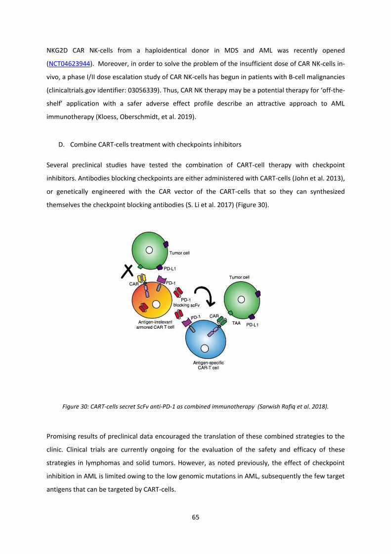
65
NKG2D CAR NK-cells from a haploidentical donor in MDS and AML was recently opened
(NCT04623944). Moreover, in order to solve the problem of the insufficient dose of CAR NK-cells in-
vivo, a phase I/II dose escalation study of CAR NK-cells has begun in patients with B-cell malignancies
(clinicaltrials.gov identifier: 03056339). Thus, CAR NK therapy may be a potential therapy for ‘off-the-
shelf’ application with a safer adverse effect profile describe an attractive approach to AML
immunotherapy (Kloess, Oberschmidt, et al. 2019).
D. Combine CART-cells treatment with checkpoints inhibitors
Several preclinical studies have tested the combination of CART-cell therapy with checkpoint
inhibitors. Antibodies blocking checkpoints are either administered with CART-cells (John et al. 2013),
or genetically engineered with the CAR vector of the CART-cells that so they can synthesized
themselves the checkpoint blocking antibodies (S. Li et al. 2017) (Figure 30).
Figure 30: CART-cells secret ScFv anti-PD-1 as combined immunotherapy (Sarwish Rafiq et al. 2018).
Promising results of preclinical data encouraged the translation of these combined strategies to the
clinic. Clinical trials are currently ongoing for the evaluation of the safety and efficacy of these
strategies in lymphomas and solid tumors. However, as noted previously, the effect of checkpoint
inhibition in AML is limited owing to the low genomic mutations in AML, subsequently the few target
antigens that can be targeted by CART-cells.
66
Main objectives of this work
Given the toxicities and the high relapses that are associated with the current treatments in AML, a
need of a safe and curative alternative treatment strategy for AML patients is crucial. Following our
previous work using CART-cells targeted the IL-1RAP protein in CML that showed promising results, we
made the proof of concept that IL-1RAP CART-cells are highly functional and cytotoxic against leukemic
blasts that express IL-1RAP at their surface. Therefore, the objective of this project was to develop a
new therapeutic strategy in AML using adoptive CART-cell immunotherapy targeting an antigen
specifically expressed by AML LSCs who are the origin of the relapse. With published data that
demonstrated an expression of IL-1RAP on the surface of AML cells, the aim of this thesis is to evaluate
the IL-1RAP molecular and cellular expression in AML primary cells and AML cell lines using our own
anti-IL-1RAP monoclonal antibody (mAb). Then we validate the functionality of IL-1RAP CART-cells,
that we produce using a CAR of 3rd generation specific for IL-1RAP, against AML primary cells (diagnosis
or R/R) and AML cell lines in-vitro and in-vivo in xenograft murine models and PDX models. We also
characterize the immunophenotyping profile and the exhaustion status (checkpoints expression) of IL-
1RAP CART-cells produced from AML patients (diagnosis or R/R).
67
Results (Submitted article)
Chimeric antigen receptor T cells targeting IL-1RAP: a promising new cellular
immunotherapy to treat acute myeloid leukemia
Scientific category: Myeloid neoplasia
Title page: AML as a candidate for IL-1RAP CAR T cell immunotherapy
Key points: Identification of a novel antigen expressed by AML cells (IL-1RAP) allowed the
development of an alternative AML treatment: CAR-T cells targeting IL-1RAP.
R. Trad (1); W. Warda (1,2); V. Alcazer (3); M. Neto Da Rocha (1,2); A. Berceanu (4); C. Nicod (1); R.
Haderbache (1); X. Roussel (1,4); Yohan Desbrosses (4); E. Daguindau (1,4); F. Renosi (1); C. Roumier
(4); L. Bouquet (1); S. Biichle (1); M. Guiot (1); E. Seffar (1); D. Caillot (6); S. Depil (7); E. Deconinck
(1,4); M. Deschamps (1,2); C. Ferrand (1,2)
INSERM UMR1098 RIGHT, EFS BFC, Université de Franche Comté, 25000, Besançon, France
CanCell Therapeutics, 25000, Besançon, France
Department of Clinical Hematology, C.H. Lyon Sud Jules Courmond, Pierre-Bénite, 69000, Lyon, France
Department of Clinical Hematology, C.H. Univ, 25000, Besançon, France
Laboratory of Hematology, CHU Lille, 59000 Lille, France
Department of Clinical Hematology, CHU François Mitterand, 21000, Dijon, France
Department of Clinical Hematology, Centre Léon Bérard, Lyon, 69000, France
Corresponding author & reprint requests:
Christophe Ferrand, PhD
INSERM UMR1098 RIGHT, Etablissement Français du Sang – Bourgogne/Franche-Comté, Université
de Franche-Comté
8 rue du Dr Jean-François Xavier Girod
25020 Besançon cedex, France

69
Abstract
Acute myeloid leukemia (AML) remains a very difficult disease to cure due to the persistence of
leukemic stem cells (LSCs), which are resistant to different lines of chemotherapy and are the basis of
refractory/relapsed (R/R) disease in 80% of AML patients not receiving allogeneic transplantation. In this
study, we showed that the IL-1RAP protein is overexpressed on the cell surface of LSCs in all subtypes of
AML and confirmed it as an interesting and promising target in AML compared to the most common
potential AML targets. After establishing the proof of concept for the efficacy of CAR T cells targeting IL-
1RAP in chronic myeloid leukemia (CML), we hypothesized that third-generation IL-1RAP CAR T cells could
eliminate AML LSCs. We first demonstrated that IL-1RAP CAR T cells can be produced from AML T cells at
the time of diagnosis but also at relapse. The characterization of IL-1RAP CAR T cells showed an expression
of checkpoint markers at the end of the production. In vitro and in vivo, we showed the effectiveness of
IL-1RAP CAR T cells against AML cell lines expressing different levels of IL-1RAP and the cytotoxicity of
autologous IL-1RAP CAR T cells against primary cells from AML patients at diagnosis or at relapse. In
patient-derived relapsed AML xenograft models, we confirmed that IL-1RAP CAR T cells are able to circulate
in peripheral blood and to migrate in the bone marrow and spleen and are cytotoxic against primary AML
cells.
70
INTRODUCTION
Despite several successive improvement in treatment, acute myeloid leukemia (AML), a
hematological malignancy of leukemic stem cells (1), remains difficult to treat and to cure (2).
The current conventional initial “7+3” treatment consists of an induction phase with high doses of
cytarabine and anthracycline (daunorubicin) chemotherapy followed by a consolidation phase of
chemotherapy or allogenic stem cell transplantation (SCT) for high-risk patients. With the knowledge
of the molecular mutation landscape of AML leukemogenesis, other targeted therapy options have
emerged with lower toxicity, such as FLT3 (Fms-like tyrosine kinase) inhibitors like midostaurin and
gilteritinib, the IDH1/2 mutant inhibitors ivosidenib and enasidenib and the BCL2 inhibitor venetoclax
in combination with hypomethylating agents (3).
Immunotherapy targeting leukemia-specific AML antigens has been explored to improve the
outcome of AML patients, such as conjugated monoclonal antibodies (CD33-GO, gemtuzumab
ozogamicin, and antibodies targeting CD44, CD123 or CD47) (4), bispecific T cell engager (BiTE)
antibodies (targeting CD3/CD33 or CD3/CD123), and immune checkpoint inhibitors (targeting PD-
1/PDL-1, anti-TIM-3, and anti-CTLA4) (5).
Since SCT is not suitable for every AML patient and based on the success and subsequent approval
of cellular gene therapy CD19-targeted immunotherapies (6), these technologies, particularly chimeric
antigen receptor (CAR) T-cells, are being translated into robust anti-AML therapies for
refractory/relapsed (R/R) patients (7). Several AML cell surface targets have been explored, like CD33,
CD123, CD44v6, CLL-1, and B7H6 (8); however, they have potential toxicities due to their frequent
expression on healthy HSCs or progenitors (9) and can lead to ablation of all myeloid progeny, thus
requiring investigation of other targets.
IL-1 receptor accessory protein (IL-1RAP) forms a complex with the IL-1α, IL-1β, and IL-33 receptors
on the surface of hematopoietic cells. The IL-1RAP protein has been shown to be overexpressed on the
surface of leukemic stem cells (LSCs) of AML, myelodysplastic syndrome (MDS) and chronic myeloid
leukemia (CML) but not on normal HSCs (10). It is a proinflammatory protein that has an oncogenic
effect in AML via the FLT3 and C-kit pathways (11) that promotes leukemic proliferation over normal
hematopoiesis (12) and has been shown to be related to some solid tumors (13).
Currently, IL-1RAP is targeted only by monoclonal antibodies (14-16), which are still under clinical
evaluation (ClinicalTrials.gov: NCT03267316 and NCT04452214) in solid tumors (17). The goals of such
antibodies are to block the interaction with the IL-1 receptor, impair tumor progression and enhance

71
antibody-dependent cellular cytotoxicity (ADCC) by recruiting natural killer (NK) cells. In both cases,
the antibodies did not generate a persistent memory effect compared with CAR T cells.
Based on our previous work demonstrating the proof of concept that 3rd generation IL-1RAP CAR T
cells (18, 19) are an interesting and robust immunotherapy approach in CML that does not affect
healthy HSCs, in this work, we evaluated this innovative cell immunotherapy in AML to demonstrate
its feasibility for a future first phase I clinical trial.
72
Methods
Additional details of the materials and methods section are available in the supplemental methods.
Transcriptomic and RNAseq in-silico analysis
Gene expression profiling (GEP) was performed using Human Genome U133 Plus 2.0 arrays
(Affymetrix/ThermoFisher, Santa Clara, CA), as previously described (20). A total of 244 samples were
subjected to transcriptomic analysis, among which 74 AML samples included different French-
American-British subtypes: 28 AML0, 11 AML1, 17 AML2 including t(8;21), 12 AML4 including t(inv16),
6 AML5, 60 T-ALL, 24 B-ALL and 13 blastic plasmacytoid dendritic cell neoplasm (BPDCN) samples. Gene
expression intensity values were log-transformed and normalized using the robust multiarray average
(RMA) algorithm with DNA-Chip Analyzer DCHIP software (21)[1]. Gene expression data from 32
peripheral blood samples from healthy donors available in the Gene Expression Omnibus (22) database
were included.
Raw RNA-seq data of sorted normal bone marrow and peripheral blood mononuclear cells (n=49
samples from 9 healthy donors) and leukemic bone marrow cells (n=32 samples from 12 AML patients)
were retrieved from the NCBI Gene Expression Omnibus (GEO) portal under the accession number
GSE74246 (23).Statistical analysis was performed using R software v 3.6.0 (see supplemental
methods).
Tumor cell lines, primary AML cells and patient sample collection
The human AML tumor cell lines Molm-13 (ACC-554), HL-60 (CCL-240), EOL-1 (ACC-386), HEL (ACC-
11), Mono-Mac-6 (ACC-124), THP-1 (TIB-202) and Ma9RAS (MLL/AF9), the CML cell lines KU812 (CRL-
2099) and K562 (CCL-243), and the human embryonic kidney epithelial cell line 239T (CRL-3216) were
obtained from ATCC and stored in a master cell bank. Cells derived from working cell banks were used
for the present study. Peripheral blood mononuclear cells (PBMCs) were isolated by Ficoll gradient
density centrifugation using Ficoll-Paque (Velizy-Villacoublay, France) from anonymous blood samples
collected from healthy donors at a French blood center (Besançon, France). AML primary cell collection
from AML patients was performed at the time of diagnosis (Cohort French Innovative Leukemia
Organization (FILO) Cell Biobank; AML collection (N° BB-0033-00073, declaration 2009-944 and
authorization AC 201261739)) and at follow-up and relapse (AML-CAR Collection, N° CPP2019-03-
022a/2018-A03300-55, harvested from Besançon and Dijon clinical centers). Patients and donors
provided written informed consent, and the study was conducted in accordance with ethical guidelines
73
(Declaration of Helsinki) and approved by the local ethics committee of the CPP-Sud-Ouest et Outre-
Mer IV (France).
Determination of IL-1RAP mRNA expression, western blotting, and determination of the number of
IL-1RAP antigenic sites
Relative IL-1RAP mRNA expression was determined by RT-QRT-PCR (real-time quantitative reverse
transcription-polymerase chain reaction) using the Hs_00895050_m1 TaqMan qPCR gene expression
assay (Thermo Fisher Scientific, Illkirch-Graffenstaden, France) targeting the mRNA variant codon for
the intracellular domain of the cell surface protein. IL-1RAP protein expression analysis was performed
by western blotting on AML cell lines (5.106 cells) using our own primary antibody targeting IL1RAP
(#A3C3 mAb; BL-43, Diaclone, France; (diluted 1,000x) and an antibody targeting β-actin (clone AC15,
#A5441, Sigma-Aldrich, St Louis, MO, USA) (diluted 1,000x) as an internal loading control.
The number of IL-1RAP antigenic sites in AML cell lines and primary cells from 30 AML patients (Filo
collection) was determined by flow cytometry (FCM) using the #A3C3 mAb and an anti-mouse IgG-1
mAb according to manufacturer’s recommendations (CELLQUANT Calibrator, Biocytex, Marseille,
France)
Production of lentiviral construct supernatant and genetically modified CART-cells
The CAR construct and the self-inactivating lentiviral vector have been previously described (19).
Briefly, the extracellular receptor domain, derived from the IL-1RAP monoclonal antibody (mAb,
#A3C3), is linked to a third-generation intracellular T-cell activating domain (CD28/4-1BB/CD3). The
vector (Supplemental Figure S1A) also carries a safety switch consisting of a suicide gene (inducible
caspase 9 (iCASP9) and a truncated CD19 gene (∆CD19) encoding the cell surface marker). Lentiviral
(LV) supernatant was harvested from tritransfected 293T cells (pMDG, pPAX2 and transgene plasmids)
(Supplemental Figure S1B). Cells taken from healthy donor or AML patient samples, after initial T cell
selection and activation using anti-CD3/CD28 magnetic beads and interleukin-2 (IL-2), were transduced
with either IL-1RAP CAR or mock (lacking the CAR sequence) LV supernatant. After 6 days, genetically
modified CD3+/CD19+ T cells were evaluated by flow cytometry and expanded for 9 days.
Flow cytometry immunophenotyping
Patient primary AML cell immunophenotyping was performed using a panel of mAbs targeting
human CD45, CD34 (stem cells), CD38 (progenitor cells), CD33 (AML blasts), CD14 (monocytes), and
74
CD123 (AML blasts), including our own murine FITC-labeled IL-1RAP mAb (#A3C3, clone BL-43, from
which the CAR was derived) and/or #H6E11 (clone BR-58; Diaclone, Besançon, France). Transduced T
cells were stained using both anti-CD3 and anti-CD19 antibodies. T lymphocytes from AML patients
were stained using an immunophenotyping panel containing mAbs targeting CD3, CD8, CCR7 and CD95
and a checkpoint inhibitor panel containing mAbs targeting PD-1, LAG-3 and TIM-3. Stained cells were
collected by a CANTO II cytometer (BD Biosciences, Le Pont-de-Claix, France) or an LSR Fortessa flow
cytometer and analyzed using DIVA software (BD Biosciences, Le Pont-de-Claix, France). Suitable
matched isotype controls were used for analysis. The relative fluorescence intensity ratio (RFI) was
calculated by dividing the mean fluorescence intensity (MFI) of IL-1RAP staining by that of the isotype
control mAb. Cells were considered positive for IL-1RAP expression at an RFI > 1. The mAbs used for
phenotyping, intracellular staining, cytometry and other cytometry reagents are described in
Supplemental Table S1.
In vitro cellular analysis: cell activation, IFNg intracellular expression and cytotoxicity analysis
IL-1RAP CAR T cells were cocultured with target tumor cells (AML cell lines or primary AML blasts)
overnight at an effector:target (E:T) ratio of 1:5 for the detection of IFN-γ intracellular expression. An
IL-1RAP CAR T cell proliferation assay was performed after 3 days of coculture (E:T ratio= 1:3) and
assessed by flow cytometry after V450 e-Fluor staining. The cytotoxicity of IL-1RAP CAR T cells against
AML leukemic cells was assessed after incubation for 24 h at different E:T ratios by flow cytometry
using trucount Absolute Counting tubes (ref: 340334, BD biosciences, Le Pont-de-Claix, France). Cell
death was evaluated by 7-amino actinomycin D (7-AAD+) labeling. Effector cells were distinguished
from target cells with a previously described V450 e-Fluor labeling method. Gating on CD3+/CD19+
cells and on IL-1RAP+ cells allowed discrimination of CAR T cells from IL-1RAP+ tumor AML cells. In the
case of allogeneic mismatch, alloreactivity was taken into account by subtracting the cytotoxicity of
untransduced T cells (C0) or mock-treated T (MockT) cells at the respective E:T ratio. C0 and MockT
cells were used as controls.
Xenograft murine models and patient-derived xenograft murine model
Six- to 8-week-old NSG mice (Jackson Laboratory, Sacramento, CA, USA) were sublethally irradiated
(250 cGy) on day 4 prior to tumor injection. One day later, each mouse was injected intravenously with
1.106 HL-60 (IL-1RAPLow), Molm-13 (IL-1RAPInt) or Mono-Mac-6 (IL-1RAPHigh) luciferase-expressing AML
cells or primary cells from AML patients (taken at diagnosis or relapse). Following AML cell engraftment
(day 0), mice were treated intravenously with MockT or IL-1RAP CAR T cells (10.106 cells) and assessed
75
for leukemia progression on days 3, 5, 10, 14, 17 and 21 by in vivo bioluminescent imaging or
determination of the percentage of human CD45+/IL-1RAP+ cells in mouse blood for the PDX model.
In the PDX model, at D32 and D71, NSGS mice (triple transgenic NSG-SGM3 mice expressing human
IL-3, GM-CSF and SCF, Jackson Laboratory, Sacramento, CA, USA) were sacrificed, and bone marrow or
spleen cells were collected and identified by FCM staining (hCD45+/CD3-/CD34+). Monitoring of CAR
T cells was performed by digital PCR as previously described (Haderbach et al). CART-cells
Effector/Memory phenotype was assessed by the expression of CD95/CCR7 among CD45RO+/CD45RA-
or CD45RO-/CD45RA+ cells. Mouse experiments were approved by the local ethical committees
(CELEAG and protocol 11007R, Veterinary Services for Animal Health & Protection).
Statistical analysis and graph constructions
The results are expressed as the mean [minimum–maximum] for MFI (mean fluorescence intensity)
and RFI (mean fluorescence intensity ratio) and as the mean ± SD for all other results. All statistical
analyses in this study were performed using GraphPad Prism 7 (GraphPad Software, San Diego, CA,
USA). Comparisons between two groups were assessed by ANOVA and t tests. A p-value < 0.05 was
considered statistically significant. Other analyses were performed with Anaconda and R software.
Some data were plotted with R using the ggplot2 and ggnewscale packages. The code is available upon
request from the authors.
76
Results
Compared to the large number of AML cell surface-targeted markers, IL-1RAP is a promising marker
expressed at various levels in primary AML blasts
In silico analysis of IL-1RAP expression was performed with a public dataset of 81 samples of sorted
bone marrow and peripheral blood cells from 9 healthy donors and 12 high-risk AML patients (23).
First, IL-1RAP mRNA expression was compared to that of the other current AML cell surface targets in
ongoing CAR T cell studies (24). Differential gene expression analysis between AML cells (LSCs or blasts)
and normal hematopoietic progenitor and stem cells (HPSCs) revealed significant overexpression of IL-
1RAP in LSCs and blasts (fold changes of 21.7 and 15, respectively), with a significant false discovery
rate (FDR)-adjusted p-value (~1.10e-20). The IL-1RAP marker was highly discriminatory compared to
common AML cell surface targets, such as TIM3, CLEC12A (CLL-1), IL3RA (CD123), MICA/MICB
(NKG2DL), CD44, CD33 and FLT3 (Figure 1A).
As reported in Figure 1B, comparison of IL-1RAP expression between total normal PB/BM and AML
cells clearly showed a higher level in blasts and LSCs. Importantly, IL-1RAP is not expressed in B, T (CD4+
or CD8+) or NK cells but is known to be expressed in monocyte PB subpopulations. In addition, all BM
progenitor subpopulations expressed IL-1RAP. Using TCGA LAML cohort (n=139), we found that a high
IL-1RAP expression was associated with a poor disease-free survival (DFS) and overall survival (OS).
This association stood significant in a multivariate Cox model using age and ELN2017 group as
confounding variables (HR 1.60 for high versus low IL1-RAP expression level for both DFS and OS, p-
value=0.037 and 0.014 respectively) (Supplemental Figure S2). Finally, Affymetrix transcriptional
profiling of the IL-1RAP mRNA encoding the membrane expressed isoform (mRNA variant 1) showed
that it was overexpressed in all AML FAB subtypes compared to the soluble IL-1RAP isoform (variant
5) (Figure 1C).
Il-1RAP is expressed at various levels on the cell surface of AML cell lines and primary AML cells
By RT-qPCR, we detected mRNA in different AML cell lines (n=6) (Supplemental Figure S3A). IL1-RAP
protein was detected by western blotting in different AML cell lines (Figure 2A). Cell surface expression
at various levels was identified in 7 AML cell lines (2< RFI <4.3) (Figure 2B) as well as in primary AML
blasts and LSCs (1.8 < RFI < 3.6, n=30), while it was consistent on monocytes from AML patients (1.6 <
RFI < 2.3, n=30) (Figure 2C). No expression of IL-1RAP was detected on normal hematopoietic cells and
normal monocytes. Clinical information of AML patients are provided in supplemental table 2.
Absolute quantification of the number of IL-1RAP antigen sites confirmed the different levels of
expression in different antigen cell lines (n=7) (Supplemental Figure S3B) as well as in primary AML
blasts (n=11) (Figure 2D). The absolute number of IL-1RAP antigen sites was not statistically different
according to ELN group. To model IL-1RAP cell surface expression for further analysis, we classified 3
77
AML cell lines and defined them as "low (HL-60)", "intermediate (Molm-13)" and "high (Mono-Mac-6)"
IL-1RAP expressers.
Efficient generation of IL-1RAP CAR T cells from T cells taken from AML patients at either diagnosis
or relapse with memory profiles and checkpoint inhibitor expression
Using our lentiviral vector (19), we were able to efficiently transduce T cells from healthy donors with
either the mock (75.29% ± 22.7) or IL-1RAP CAR (74.06% ± 16.48) T cell supernatant (n=24). More
interestingly, T cells from AML patients taken either at diagnosis (D) or at the time of relapse (R/R)
could be transduced (90.4% ± 6.58 and 88.1% ± 9.28, respectively, for mock or IL-1RAP CAR T cell
supernatant, n=12) without noticeable differences from T cells from healthy donors (Figure 3A). We
then identified the ratio of CD4+ and CD8+ cells before CAR T cell production (D0) and at the end of
the process (D10) using either IL-2 or IL-7/15 as cytokines in the culture medium. As reported in Figure
3B, similar to IL-1RAP CAR T cells from healthy donors, IL-1RAP CAR T cells produced from AML patients
conserved the CD4+/CD8+ ratio during the culture. Ultimately, the IL-1RAP CAR T cells (either CD4+ or
CD8+) produced from T cells taken from AML patients (D or R/R) showed the same profile of phenotype
that was observed with T cells taken from healthy donors with a conservation of this phenotype during
culture with either IL-2 or IL-7/15 (Figure 3C). A slight tendency of acquisition a naïve/memory
phenotype by the end of the production process was observed with CD8+ CAR T cells.
IL-1RAP CAR T-cells are able to specifically proliferate, secrete IFN and kill AML cell lines
independent of IL-1RAP cell surface expression levels
The HL-60 (IL-1RAPLow), Molm-13 (IL-1RAPInt) and Mono-Mac-6 (IL-1RAPHigh) IL-1RAP+ AML cell lines
were used for in vitro and in vivo functional analysis. For the proliferation test, IL-1RAP CAR T cells were
cocultured for 3 days with the 3 AML cell lines at a 1:3 Effector:Target (E:T) ratio. We demonstrated
that IL-1RAP CAR T cells proliferated at 71.5 ± 18.09%, 81.58 ± 6.4%, and 80.3 ± 15.3% when cultured
with the low, int and high IL-1RAP cell surface expression cell lines (n=4), respectively, independent of
the IL-1RAP expression level, and these results were different than those achieved with culture in the
absence of targets or with coculture of targets with untransduced T cells (C0) or MockT cells (Figure
4A). Similarly, in the presence of IL-1RAP-positive targets, CD4+ [27.23 ± 7.5% (low), 41.63 ± 12.2%
(int), and 58.25 ± 12.9% (high)] or CD8+ [26.8 ± 6.7% (low), 38.73 ± 3.7% (int), and 71.75 ± 17.2% (high)]
cells were activated and expressed IFN. Interestingly, the level of IL-1RAP cell surface expression
affected the levels of IFN expression (Figure 4B). Finally, IL-1RAP CAR T cell cytotoxicity was evaluated
78
at different E:T ratios against the 3 AML cell lines and the negative IL-1RAP cell line K562. After
coculture, in contrast to MockT cells or IL-1RAP CAR T cells that were unable to kill IL-1RAP-negative
K562 cells, IL-1RAP CAR T cells were able to statistically eliminate CML KU812 IL-1RAP+ cells (99 ± 0.1%)
and all AML cell lines at different E:T ratios (95.3 ± 1.52% (Low); 99 ± 0.1% (Int); 99.3 ± 0.57% (High); a
high and significant cytotoxic effect was observed at a low ratio E:T = 10:1, n=3) (figure 5A).
Interestingly, specificity was confirm by coculturing IL-1RAP CAR T cells with different healthy
peri(solid)tumor tissues, thus in an inflammatory context (Supplemental Figure S4).
IL-1RAP CAR T cells clearly decrease the cell numbers of leukemia-derived AML tumor cell lines in
vitro and decrease the tumor burden in an in vivo xenograft murine model
To test the efficacy of IL-1RAP CAR T cells in vivo in murine xenograft models, we generated 3
luciferase-positive AML cell lines to be injected intravenously into immunodeficient NSG mice. After
AML tumor engraftment, IL-1RAP CAR T cell-treated mice showed a rapid decrease in tumor burden in
peripheral blood, whereas AML tumors (formed from cells with low, intermediate or high IL-1RAP cell
surface expression) progressed to very high tumor burdens or until mouse death (day 21) in the
untreated group and group treated with untransduced T cells (Figure 5B). The luminescence
measurement confirmed a significant decrease at day 21 (average radiance >10e5 p/s/cm2/sr versus
average radiance <10e3 p/s/cm2 for untreated or T cell-treated mice and IL-1RAP CAR T cell-infused
mice, P<0,001, n=3 mice/group). Taken together, these results show that IL-1RAP CAR T cells efficiently
controlled AML cell line growth in vivo.
IL-1RAP CAR T cells generated from healthy donors or AML patients (at diagnosis or relapse) are
efficient against primary AML cells (taken from patients at diagnosis or relapse)
We then produced IL-1RAP CAR T cells from PBMCs of healthy donors or harvested from AML patients
either at the time of diagnosis or at the time of relapse. We evaluated the ability of allogeneic (from a
donor or an AML patient) or autologous (from an AML patient) CAR T cells to kill primary AML blasts
(taken at diagnosis or relapse) after 24 h of coculture at different E:T ratios (0:1, 1:2, 1:1, 3:1, 5:1 and
10:1). As reported in Figure 6, we confirmed that IL-1RAP CAR T cells prepared from healthy donor T
cells were able to effectively kill primary AML blasts extracted at diagnosis or relapse at as low as a 1:1
E:T ratio. Moreover, IL-1RAP CAR T cells produced from T cells taken from AML patients at diagnosis
or after relapse were also cytotoxic against primary AML blasts in allogeneic and autologous settings
also at a low E:T ratio of 1:1. Finally, and more interestingly, autologous IL-1RAP CAR T cells produced
from PBMCs taken from a relapsed AML patient with FLT3+NPM1+CD33+ disease (following several
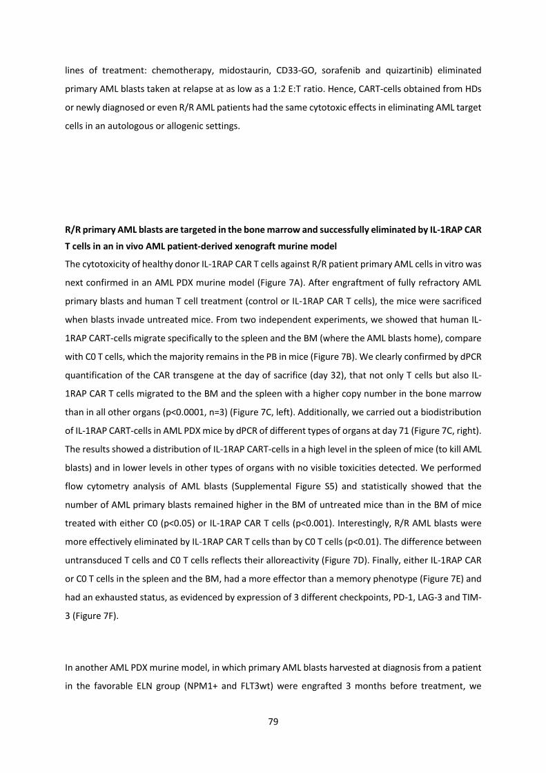
79
lines of treatment: chemotherapy, midostaurin, CD33-GO, sorafenib and quizartinib) eliminated
primary AML blasts taken at relapse at as low as a 1:2 E:T ratio. Hence, CART-cells obtained from HDs
or newly diagnosed or even R/R AML patients had the same cytotoxic effects in eliminating AML target
cells in an autologous or allogenic settings.
R/R primary AML blasts are targeted in the bone marrow and successfully eliminated by IL-1RAP CAR
T cells in an in vivo AML patient-derived xenograft murine model
The cytotoxicity of healthy donor IL-1RAP CAR T cells against R/R patient primary AML cells in vitro was
next confirmed in an AML PDX murine model (Figure 7A). After engraftment of fully refractory AML
primary blasts and human T cell treatment (control or IL-1RAP CAR T cells), the mice were sacrificed
when blasts invade untreated mice. From two independent experiments, we showed that human IL-
1RAP CART-cells migrate specifically to the spleen and the BM (where the AML blasts home), compare
with C0 T cells, which the majority remains in the PB in mice (Figure 7B). We clearly confirmed by dPCR
quantification of the CAR transgene at the day of sacrifice (day 32), that not only T cells but also IL-
1RAP CAR T cells migrated to the BM and the spleen with a higher copy number in the bone marrow
than in all other organs (p<0.0001, n=3) (Figure 7C, left). Additionally, we carried out a biodistribution
of IL-1RAP CART-cells in AML PDX mice by dPCR of different types of organs at day 71 (Figure 7C, right).
The results showed a distribution of IL-1RAP CART-cells in a high level in the spleen of mice (to kill AML
blasts) and in lower levels in other types of organs with no visible toxicities detected. We performed
flow cytometry analysis of AML blasts (Supplemental Figure S5) and statistically showed that the
number of AML primary blasts remained higher in the BM of untreated mice than in the BM of mice
treated with either C0 (p<0.05) or IL-1RAP CAR T cells (p<0.001). Interestingly, R/R AML blasts were
more effectively eliminated by IL-1RAP CAR T cells than by C0 T cells (p<0.01). The difference between
untransduced T cells and C0 T cells reflects their alloreactivity (Figure 7D). Finally, either IL-1RAP CAR
or C0 T cells in the spleen and the BM, had a more effector than a memory phenotype (Figure 7E) and
had an exhausted status, as evidenced by expression of 3 different checkpoints, PD-1, LAG-3 and TIM-
3 (Figure 7F).
In another AML PDX murine model, in which primary AML blasts harvested at diagnosis from a patient
in the favorable ELN group (NPM1+ and FLT3wt) were engrafted 3 months before treatment, we
80
showed a higher overall survival (OS) of PDX mice treated with IL-1RAP CAR T cells (n=5) than of
untreated animals (n=2) or C0 T cell-treated (n=5) (240 days versus 98 and 126 days for untreated mice
and C0 T cell-treated mice, respectively; n=5 mice per group; P < 0.01) (Figure 7G). Importantly, no
visible toxicities were observed on dead mice treated with IL-1RAP CART-cells.
81
Discussion
AML remains one of the most malignant hematological diseases and has a poor outcome, especially
for high-risk patients, despite improvements in hematopoietic stem cell transplantation and several
other treatments. Indeed, though approximately 40%-45% of young patients and 10%-20% of elderly
patients can be cured, the cure rate for R/R AML patients is lower than 10%, suggesting the necessity
of new alternatives for treating patients (25). Immunotherapy using CAR T cells, particularly CD19 CAR
T cells, which have shown remarkable results in ALL and lymphoma (26) as well as in MM (27), is an
interesting strategy to explore in AML. Several cell surface AML antigen targets, mainly CD123, CD33,
CLL1, FLT3, and CD44v6, are under investigation at preclinical stages; however, none are truly perfect
targets due to their hematopoietic toxicity (for example, they can induce profound myeloablation)
(28). Thus, there is a need to continue investigating new AML markers targetable by CAR T cells.
In this work, we investigated IL-1RAP, a novel and unexplored AML cell surface marker expressed
by leukemia cells but not healthy hematopoietic stem cells, progenitors, or T and B lymphocytes,
though it is expressed by circulating monocytes (19). IL-1RAP, the accessory protein of the IL-1
receptor, plays a crucial role in immunity, inflammation and cancer. It is involved in a tumor
microenvironment that favors leukemia proliferation (12, 13), potentiates multiple oncogenic signaling
pathways and oncogenic pathways and contributes to AML oncogenesis (11, 29). The need for IL-1RAP
for tumorigenesis and its overexpression in cancer make it an interesting target for avoiding further
selection and escape because of antigen loss after CAR T cell immunotherapy, as occurs with CD19 (30)
or BCMA (31) CAR T cell immunotherapies. Michaud et al. demonstrated that soluble IL-1RAP is present
at a low level in healthy people , and Warda et al. showed that high concentrations of soluble IL-1RAP
did not result in staining of IL1RAP CAR-expressing T cells and did not affect CAR T cytotoxicity against
IL1RAP+ target cells.
Currently, only a monoclonal antibody targeting IL-1RAP (nidanilimab) is under investigation in
phase I/II clinical trials (NCT03267316 and NCT04452214, www.clinicaltrials.gov) in pancreatic and
triple-negative breast cancer. This antibody is used as a blocking agent in association with
pembrolizumab or to enhance chemotherapy (15) and has shown a manageable safety profile (22).
Regarding toxicity, in addition to our previous work (19), where we showed absence of targeting of the
healthy hematopoietic stem cells, we give here another argument in favor of IL-1RAP targeting by
showing absence of degranulation of IL-1RAP CAR T cell against healthy peritumoral tissues from
different solid tumors, thus in a inflammatory context.
We demonstrated that IL-1RAP mRNA is expressed in all AML FAB subtypes and that the IL-RAP
protein is expressed at different levels on the cell surface of AML blasts independent of ELN
82
classification. Interestingly, we showed in 30 AML patient cohorts that most primary IL-1RAP+ blasts
coexpressed CD123, making this target suitable for bispecific CAR T cell targeting, which will increase
specificity via “and/or” CAR activation signaling and presumably without increasing toxicity (32).
Following our previous work in CML demonstrating a proof of concept for targeting IL-1RAP (19),
we here showed in a preclinical study that this new alternative treatment has efficacy in AML using an
innovative approach: targeting the cell surface marker IL-1RAP, which is expressed by both leukemic
stem cells and primary AML blasts. The fact that the cytotoxicity and efficacy of IL-1RAP CAR T cells
were the same against both cells with low IL-1RAP and cells with high IL-1RAP expression (primary AML
cells and AML cell lines) suggests their efficacy.
Interestingly, while chemotherapy and other targeted therapies are recognized as early-line
treatments currently, it is important to demonstrate that T cells from R/R AML patients can be used
for CAR T cell generation. Indeed, nonresponder AML patients are eligible for targeted therapies
following chemotherapy, and some of these patients receive FLT3 tyrosine kinase inhibitor drugs (33)
that may interact with intrinsic tyrosine kinases (34) involved in CAR transduction signals, such as Fyn,
ZAP-70, and lck(35). We were able to transduce T cells taken from AML patients at diagnosis and also
at relapse with high efficiency, and we demonstrated that autologous CAR T cells are effective in vitro
against primary R/R AML blasts. Moreover, the presence of a suicide gene within our lentiviral
construct will protect against adverse events in cases of unexpected leukemic cell transduction, as
previously shown (18). This mechanism will also help in the case of persistent adverse events affecting
monocytes by functioning as a safety switch in order to eliminate CAR T cells and allow a mature
myeloid cell recovery. The functionality of the safety switch system has been evaluated in killing
persistent IL-1RAP CAR T cells (18).
Characterization of the IL-1RAP CAR T cells at the end of the production process (supplemental
Figure S6) and when in PDX mice, showed that they acquire the expression of exhaustion markers such
as PD1, TIM3 and LAG3. Interestingly, the expression of these markers did not affect the function of
IL-1RAP CART-cells in killing AML blasts. Further studies are needed in order to evaluate the expression
of these markers’ ligands (such as PDL1) on the surface of AML cells. Importantly, these results allow
using inhibitors of these markers in combination with the CART-cells in case of unqualified patient's T
lymphocytes (because of early treatment lines), their activity can be increased by inhibiting the
exhaustion markers (36). Importantly, the persistence of IL-1RAP CART-cells in the bone marrow and
the spleen of PDX mice was confirmed by dPCR for approximately 2 months after treatment with no
visible side effects on treated mice. This persistence may be due to the co-stimulation molecule 4-1BB
present in the CAR construct.
83
In conclusion, this work clearly confirms the potential of IL-1RAP as a target in AML and the strong
anti-leukemic effects of strategies targeting this marker both in vitro and in vivo, laying the foundation
for a promising future first-in-human clinical trial in R/R AML. IL-1RAP CAR T cells have not yet been
evaluated in humans, and doing so will help determine the optimal IL-1RAP CAR T cell dose and
evaluate the efficacy and safety of this approach.
Support
o Association « Nausicaa Combat sa Leucémie » (nonprofit association - loi 1901 FR); Gift 2017-03,
2018, 2019 and 2020
o Association semons l’Espoir (nonprofit association - loi 1901 FR); Gift 2020
o Ligue contre le cancer, Comité de Montbéliard, Grant 12/2020
o Canceropôle Est, Grant 2020
o This work was supported by the MiMedi project funded by BPI France (grant no. DOS0060162/00)
and the European Union through the European Regional Development Fund of the Region
Bourgogne-Franche-Comte (grant no. FC0013440).
o French Blood Center, Grants 2018 and 2019
o EU project T2EVOLVE grant agreement No. 945393
o UMR1098 RIGHT is a member of the OPALE Carnot Institute.
o Rafik Haderbache has a fellowship from Grand Besançon Metropole (GBM).
84
Acknowledgments
o Diaclone, Besancon, France for providing monoclonal antibodies against IL-1RAP
o All technicians operating the biomonitoring platform (UMR1098 RIGHT) for technical support with
AML sample collection
o Elise Robert (Besançon Hospital headquarters) for the management of AML biobank collection
o FILO "French Innovative Leukemia Organization" for providing samples from the FILOThèque AML
Biobank (N° BB-0033-00073, declaration 2009-944 and authorization AC 201261739)

85
Disclosure of Conflicts of Interest
o CF: Consulting: Novartis, BMS, Incyte, and Daichii. Research grants: Novartis, Daiichi, Bellicum
Pharmaceutical
o MD and CF: co-founders of CanCell Therapeutics SAS, Besançon, France
o SD: Founder of ErVaccine
o VA: ErVaccine consultant
86
Author’s contributions
Conceptualization and study design: RT, WW, MD, CF
Methodology, experiments and data analysis: RT, WW, ES, MNdR, LB, RH, CN, SB, FR, MD, CF
Original draft and figure construction: RT, WW, MNdR, CN, VA, ES, RH, MD CF
Clinical advice and patient collection: AB, XR, YD, ED, CF
Writing, review and/or revision of the manuscript: VA, SD, WW, RT, MD, CF
Overall supervision and research funding: WW, MD, CF
All authors reviewed the manuscript and contributed to improving the paper.
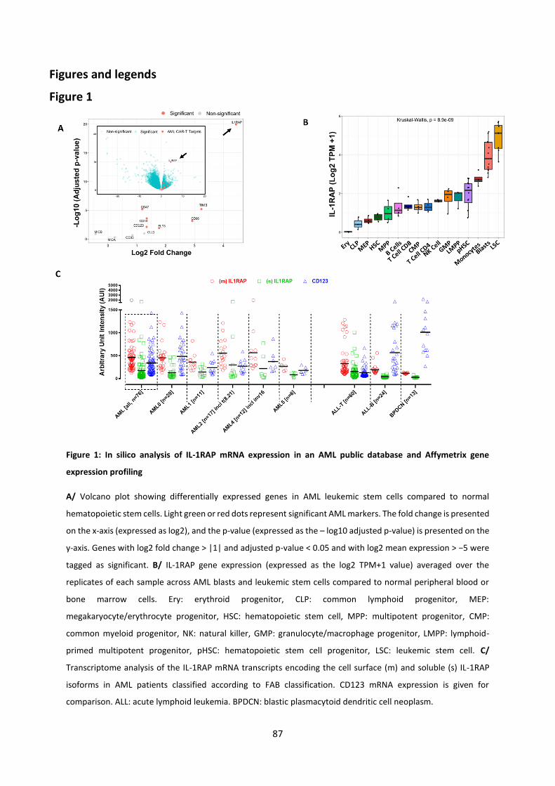
87
Figures and legends
Figure 1
Figure 1: In silico analysis of IL-1RAP mRNA expression in an AML public database and Affymetrix gene
expression profiling
A/ Volcano plot showing differentially expressed genes in AML leukemic stem cells compared to normal
hematopoietic stem cells. Light green or red dots represent significant AML markers. The fold change is presented
on the x-axis (expressed as log2), and the p-value (expressed as the – log10 adjusted p-value) is presented on the
y-axis. Genes with log2 fold change > |1| and adjusted p-value < 0.05 and with log2 mean expression > −5 were
tagged as significant. B/ IL-1RAP gene expression (expressed as the log2 TPM+1 value) averaged over the
replicates of each sample across AML blasts and leukemic stem cells compared to normal peripheral blood or
bone marrow cells. Ery: erythroid progenitor, CLP: common lymphoid progenitor, MEP:
megakaryocyte/erythrocyte progenitor, HSC: hematopoietic stem cell, MPP: multipotent progenitor, CMP:
common myeloid progenitor, NK: natural killer, GMP: granulocyte/macrophage progenitor, LMPP: lymphoid-
primed multipotent progenitor, pHSC: hematopoietic stem cell progenitor, LSC: leukemic stem cell. C/
Transcriptome analysis of the IL-1RAP mRNA transcripts encoding the cell surface (m) and soluble (s) IL-1RAP
isoforms in AML patients classified according to FAB classification. CD123 mRNA expression is given for
comparison. ALL: acute lymphoid leukemia. BPDCN: blastic plasmacytoid dendritic cell neoplasm.
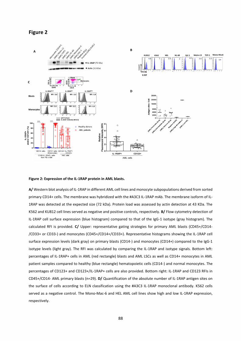
88
Figure 2
Figure 2: Expression of the IL-1RAP protein in AML blasts.
A/ Western blot analysis of IL-1RAP in different AML cell lines and monocyte subpopulations derived from sorted
primary CD14+ cells. The membrane was hybridized with the #A3C3 IL-1RAP mAb. The membrane isoform of IL-
1RAP was detected at the expected size (72 kDa). Protein load was assessed by actin detection at 43 KDa. The
K562 and KU812 cell lines served as negative and positive controls, respectively. B/ Flow cytometry detection of
IL-1RAP cell surface expression (blue histogram) compared to that of the IgG-1 isotype (gray histogram). The
calculated RFI is provided. C/ Upper: representative gating strategies for primary AML blasts (CD45+/CD14-
/CD33+ or CD33-) and monocytes (CD45+/CD14+/CD33+). Representative histograms showing the IL-1RAP cell
surface expression levels (dark gray) on primary blasts (CD14-) and monocytes (CD14+) compared to the IgG-1
isotype levels (light gray). The RFI was calculated by comparing the IL-1RAP and isotype signals. Bottom left:
percentages of IL-1RAP+ cells in AML (red rectangle) blasts and AML LSCs as well as CD14+ monocytes in AML
patient samples compared to healthy (blue rectangle) hematopoietic cells (CD14-) and normal monocytes. The
percentages of CD123+ and CD123+/IL-1RAP+ cells are also provided. Bottom right: IL-1RAP and CD123 RFIs in
CD45+/CD14- AML primary blasts (n=29). D/ Quantification of the absolute number of IL-1RAP antigen sites on
the surface of cells according to ELN classification using the #A3C3 IL-1RAP monoclonal antibody. K562 cells
served as a negative control. The Mono-Mac-6 and HEL AML cell lines show high and low IL-1RAP expression,
respectively.
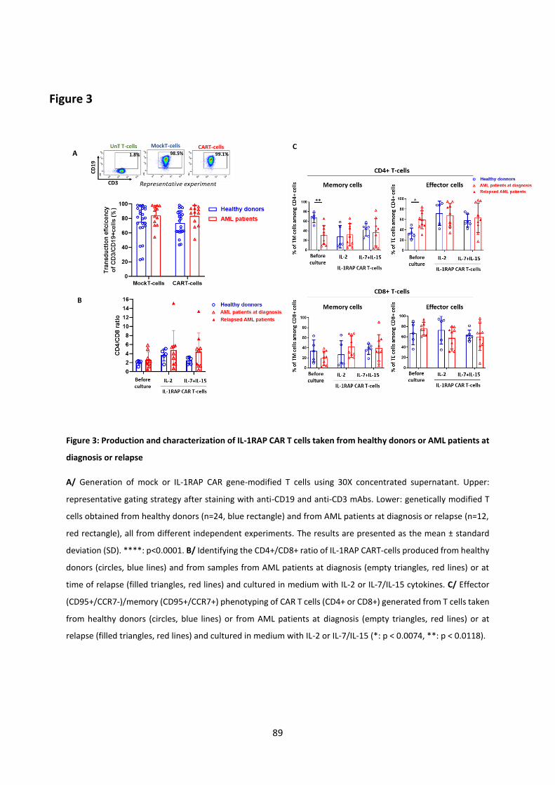
89
Figure 3
Figure 3: Production and characterization of IL-1RAP CAR T cells taken from healthy donors or AML patients at
diagnosis or relapse
A/ Generation of mock or IL-1RAP CAR gene-modified T cells using 30X concentrated supernatant. Upper:
representative gating strategy after staining with anti-CD19 and anti-CD3 mAbs. Lower: genetically modified T
cells obtained from healthy donors (n=24, blue rectangle) and from AML patients at diagnosis or relapse (n=12,
red rectangle), all from different independent experiments. The results are presented as the mean ± standard
deviation (SD). ****: p<0.0001. B/ Identifying the CD4+/CD8+ ratio of IL-1RAP CART-cells produced from healthy
donors (circles, blue lines) and from samples from AML patients at diagnosis (empty triangles, red lines) or at
time of relapse (filled triangles, red lines) and cultured in medium with IL-2 or IL-7/IL-15 cytokines. C/ Effector
(CD95+/CCR7-)/memory (CD95+/CCR7+) phenotyping of CAR T cells (CD4+ or CD8+) generated from T cells taken
from healthy donors (circles, blue lines) or from AML patients at diagnosis (empty triangles, red lines) or at
relapse (filled triangles, red lines) and cultured in medium with IL-2 or IL-7/IL-15 (*: p < 0.0074, **: p < 0.0118).
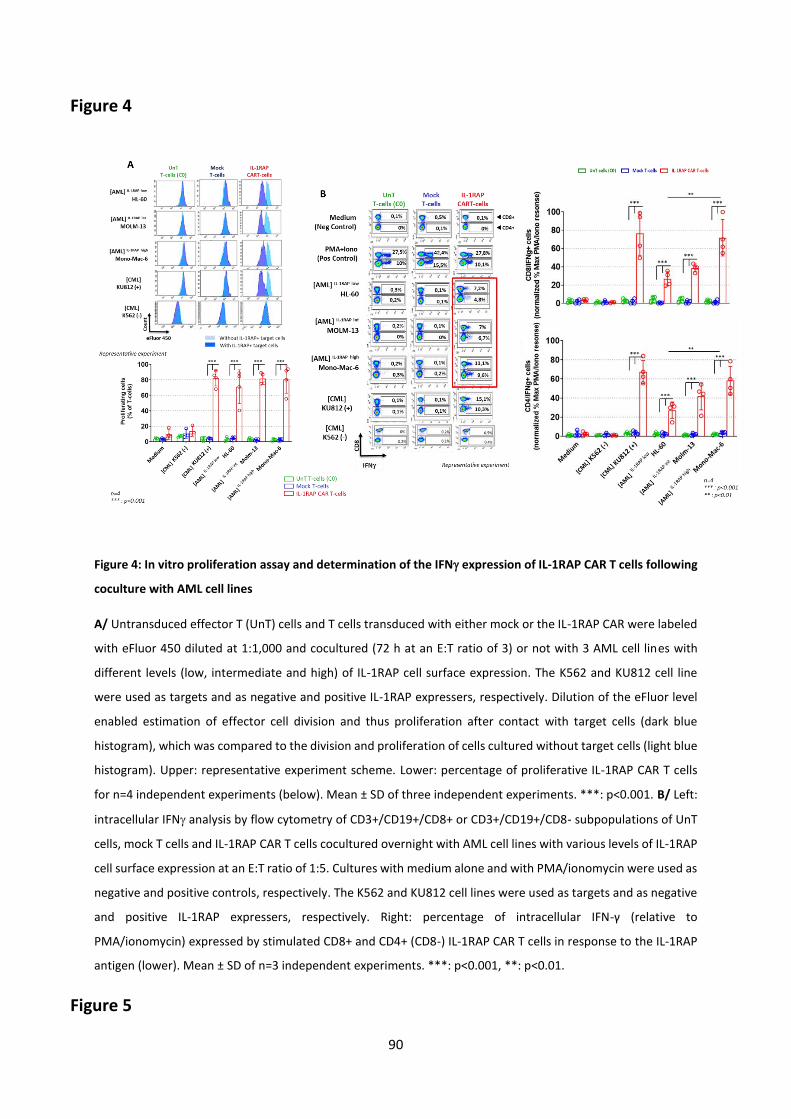
90
Figure 4
Figure 4: In vitro proliferation assay and determination of the IFN expression of IL-1RAP CAR T cells following
coculture with AML cell lines
A/ Untransduced effector T (UnT) cells and T cells transduced with either mock or the IL-1RAP CAR were labeled
with eFluor 450 diluted at 1:1,000 and cocultured (72 h at an E:T ratio of 3) or not with 3 AML cell lines with
different levels (low, intermediate and high) of IL-1RAP cell surface expression. The K562 and KU812 cell line
were used as targets and as negative and positive IL-1RAP expressers, respectively. Dilution of the eFluor level
enabled estimation of effector cell division and thus proliferation after contact with target cells (dark blue
histogram), which was compared to the division and proliferation of cells cultured without target cells (light blue
histogram). Upper: representative experiment scheme. Lower: percentage of proliferative IL-1RAP CAR T cells
for n=4 independent experiments (below). Mean ± SD of three independent experiments. ***: p<0.001. B/ Left:
intracellular IFN analysis by flow cytometry of CD3+/CD19+/CD8+ or CD3+/CD19+/CD8- subpopulations of UnT
cells, mock T cells and IL-1RAP CAR T cells cocultured overnight with AML cell lines with various levels of IL-1RAP
cell surface expression at an E:T ratio of 1:5. Cultures with medium alone and with PMA/ionomycin were used as
negative and positive controls, respectively. The K562 and KU812 cell lines were used as targets and as negative
and positive IL-1RAP expressers, respectively. Right: percentage of intracellular IFN-γ (relative to
PMA/ionomycin) expressed by stimulated CD8+ and CD4+ (CD8-) IL-1RAP CAR T cells in response to the IL-1RAP
antigen (lower). Mean ± SD of n=3 independent experiments. ***: p<0.001, **: p<0.01.
Figure 5
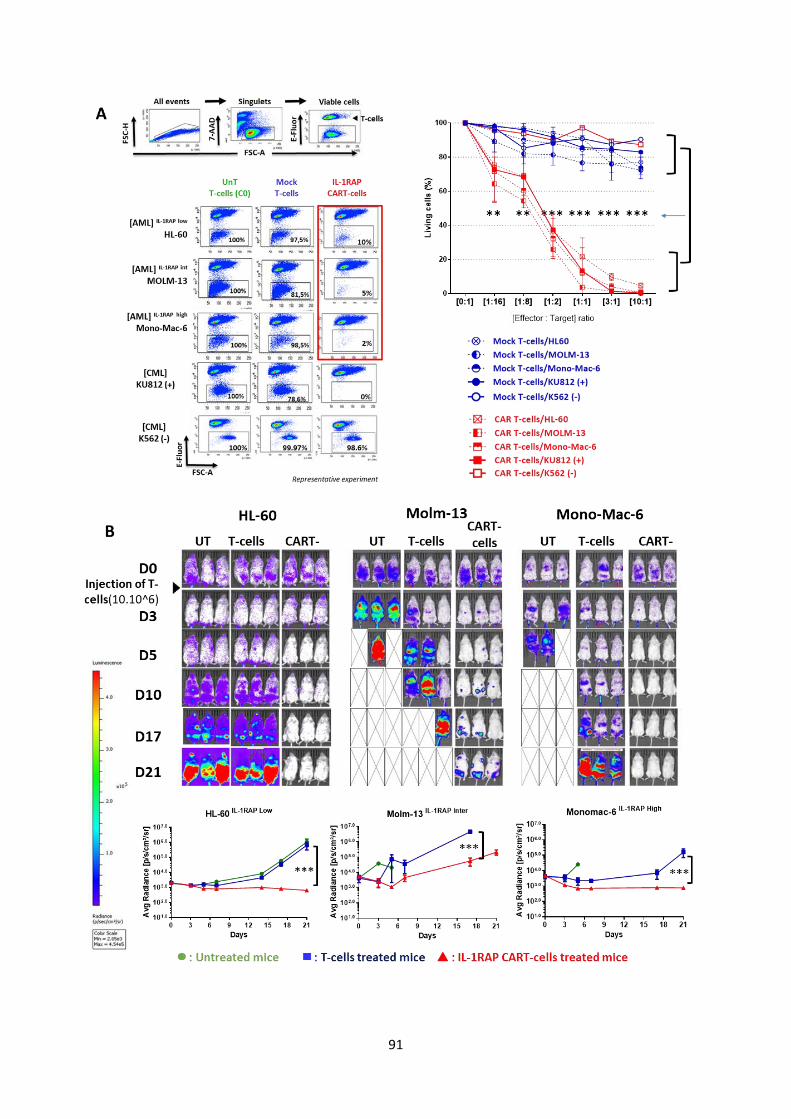
91
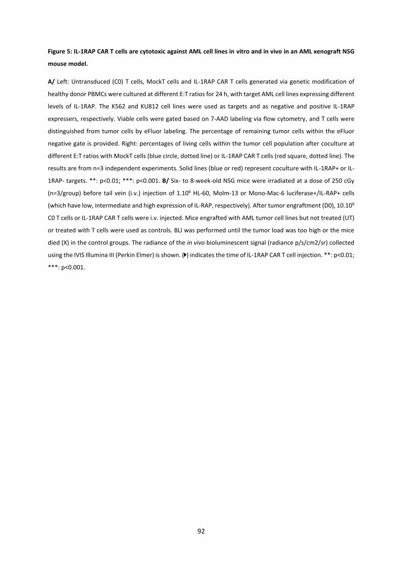
92
Figure 5: IL-1RAP CAR T cells are cytotoxic against AML cell lines in vitro and in vivo in an AML xenograft NSG
mouse model.
A/ Left: Untransduced (C0) T cells, MockT cells and IL-1RAP CAR T cells generated via genetic modification of
healthy donor PBMCs were cultured at different E:T ratios for 24 h, with target AML cell lines expressing different
levels of IL-1RAP. The K562 and KU812 cell lines were used as targets and as negative and positive IL-1RAP
expressers, respectively. Viable cells were gated based on 7-AAD labeling via flow cytometry, and T cells were
distinguished from tumor cells by eFluor labeling. The percentage of remaining tumor cells within the eFluor
negative gate is provided. Right: percentages of living cells within the tumor cell population after coculture at
different E:T ratios with MockT cells (blue circle, dotted line) or IL-1RAP CAR T cells (red square, dotted line). The
results are from n=3 independent experiments. Solid lines (blue or red) represent coculture with IL-1RAP+ or IL-
1RAP- targets. **: p<0.01; ***: p<0.001. B/ Six- to 8-week-old NSG mice were irradiated at a dose of 250 cGy
(n=3/group) before tail vein (i.v.) injection of 1.106 HL-60, Molm-13 or Mono-Mac-6 luciferase+/IL-RAP+ cells
(which have low, intermediate and high expression of IL-RAP, respectively). After tumor engraftment (D0), 10.106
C0 T cells or IL-1RAP CAR T cells were i.v. injected. Mice engrafted with AML tumor cell lines but not treated (UT)
or treated with T cells were used as controls. BLI was performed until the tumor load was too high or the mice
died (X) in the control groups. The radiance of the in vivo bioluminescent signal (radiance p/s/cm2/sr) collected
using the IVIS Illumina III (Perkin Elmer) is shown. ( ) indicates the time of IL-1RAP CAR T cell injection. **: p<0.01;
***: p<0.001.
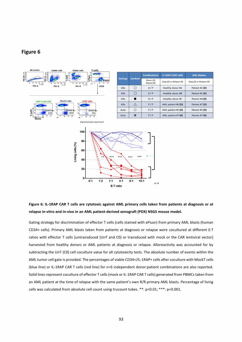
93
Figure 6
Figure 6: IL-1RAP CAR T cells are cytotoxic against AML primary cells taken from patients at diagnosis or at
relapse in-vitro and in-vivo in an AML patient-derived xenograft (PDX) NSGS mouse model.
Gating strategy for discrimination of effector T cells (cells stained with eFluor) from primary AML blasts (human
CD34+ cells). Primary AML blasts taken from patients at diagnosis or relapse were cocultured at different E:T
ratios with effector T cells [untransduced (UnT and C0) or transduced with mock or the CAR lentiviral vector]
harvested from healthy donors or AML patients at diagnosis or relapse. Alloreactivity was accounted for by
subtracting the UnT (C0) cell coculture value for all cytotoxicity tests. The absolute number of events within the
AML tumor cell gate is provided. The percentages of viable CD34+/IL-1RAP+ cells after coculture with MockT cells
(blue line) or IL-1RAP CAR T cells (red line) for n=6 independent donor:patient combinations are also reported.
Solid lines represent coculture of effector T cells (mock or IL-1RAP CAR T cells) generated from PBMCs taken from
an AML patient at the time of relapse with the same patient’s own R/R primary AML blasts. Percentage of living
cells was calculated from absolute cell count using trucount tubes. **: p<0.01; ***: p<0.001.
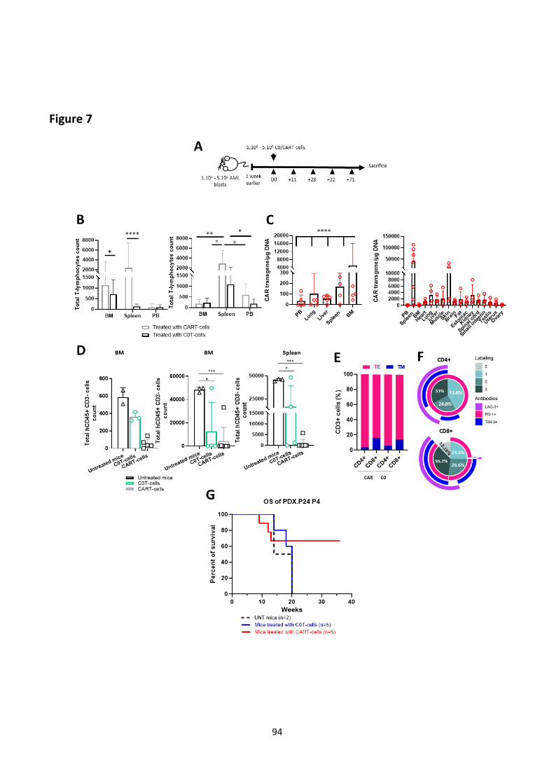
94
Figure 7
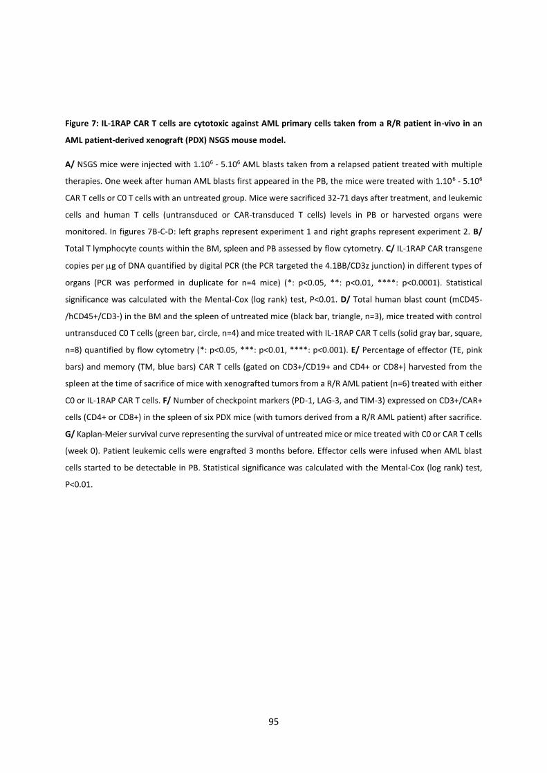
95
Figure 7: IL-1RAP CAR T cells are cytotoxic against AML primary cells taken from a R/R patient in-vivo in an
AML patient-derived xenograft (PDX) NSGS mouse model.
A/ NSGS mice were injected with 1.106 - 5.106 AML blasts taken from a relapsed patient treated with multiple
therapies. One week after human AML blasts first appeared in the PB, the mice were treated with 1.106 - 5.106
CAR T cells or C0 T cells with an untreated group. Mice were sacrificed 32-71 days after treatment, and leukemic
cells and human T cells (untransduced or CAR-transduced T cells) levels in PB or harvested organs were
monitored. In figures 7B-C-D: left graphs represent experiment 1 and right graphs represent experiment 2. B/
Total T lymphocyte counts within the BM, spleen and PB assessed by flow cytometry. C/ IL-1RAP CAR transgene
copies per g of DNA quantified by digital PCR (the PCR targeted the 4.1BB/CD3z junction) in different types of
organs (PCR was performed in duplicate for n=4 mice) (*: p<0.05, **: p<0.01, ****: p<0.0001). Statistical
significance was calculated with the Mental-Cox (log rank) test, P<0.01. D/ Total human blast count (mCD45-
/hCD45+/CD3-) in the BM and the spleen of untreated mice (black bar, triangle, n=3), mice treated with control
untransduced C0 T cells (green bar, circle, n=4) and mice treated with IL-1RAP CAR T cells (solid gray bar, square,
n=8) quantified by flow cytometry (*: p<0.05, ***: p<0.01, ****: p<0.001). E/ Percentage of effector (TE, pink
bars) and memory (TM, blue bars) CAR T cells (gated on CD3+/CD19+ and CD4+ or CD8+) harvested from the
spleen at the time of sacrifice of mice with xenografted tumors from a R/R AML patient (n=6) treated with either
C0 or IL-1RAP CAR T cells. F/ Number of checkpoint markers (PD-1, LAG-3, and TIM-3) expressed on CD3+/CAR+
cells (CD4+ or CD8+) in the spleen of six PDX mice (with tumors derived from a R/R AML patient) after sacrifice.
G/ Kaplan-Meier survival curve representing the survival of untreated mice or mice treated with C0 or CAR T cells
(week 0). Patient leukemic cells were engrafted 3 months before. Effector cells were infused when AML blast
cells started to be detectable in PB. Statistical significance was calculated with the Mental-Cox (log rank) test,
P<0.01.
96
Supplemental
Chimeric antigen receptor T cells targeting IL-1RAP: a promising new cellular
immunotherapy to treat acute myeloid leukemia
Trad et al
97
Supplemental material and Methods
Transcriptomic and RNAseq in silico analyses
Gene expression profiling (GEP) was performed using Human Genome U133 Plus 2.0 arrays
(Affymetrix/ThermoFisher, Santa Clara, CA). Gene expression data available in the Gene Expression
Omnibus (22) database were included for 32 peripheral blood samples from healthy donors
(GSE48060: GSM1167102-21 and GSM1167123; GSE54992: GSM1327541,42,44,46,49,50, GSE20489:
GSM514766,71,76,81,86) and 26 bone marrow samples from healthy donors (GSE3526: GSM80576,
577, 602-604; GSE18674: GSM463920; GSE32725: GSM813068-71; GSE33075: GSM818813-19,23,24;
GSE41130: GSM1008985-91).
RNAseq dataset in silico analysis
Total single-strand RNAseq data were available for sorted samples from healthy donors (n= 9, 32
samples) and high risk AML patients (n=12, 49 samples) representing in total 81 samples.
RNAseq data: For the sorted RNAseq samples, data were downloaded from the GEO portal (accession
number GSE74246). Raw data were aligned to the human reference genome (GRCh38) using Bowtie2.
Transcripts were counted using HTSeq with Gencode v33 annotation, normalized to transcripts per
kilobase million (TPM) values and log2+1 transformed. Differential expression analysis was performed
using DESeq2 on the raw counts. TCGA LAML RNAseq raw counts and log2 RSEM+1 normalized counts
were downloaded from the UCSC Xena Browser (https://xenabrowser.net/datapages/ last access:
March 2020).
Gene set enrichment analysis: For gene set enrichment analysis (GSEA), TCGA raw counts were
normalized using DESeq2. The whole cohort (n=152) was then divided into 3 terciles according to
IL1RAP expression. GSEA was performed using all human pathways from the Enrichment Map
repository (http://download.baderlab.org/EM_Genesets/current_release/Human/symbol/, last
access: May 2020) on a filtered cohort to compare the low versus high terciles of IL1RAP expression
(n=101). An enrichment map was then plotted using Cytoscape v3.8.0 and EnrichmentMap v3.2.1.
(PLoS One, 2010 Nov 15;5(11): e13984) with custom filtering parameters (cutoffs: p-value 0.001, FDR
Q-value: 0.05, overlap: 0.5, normalized enrichment score > 2 or < -2). Common pathways were first
annotated using AutoAnnotate v1.3.3 (F1000Res, 2016; 5: 1717.) and then manually annotated. The
few remaining isolated nodes with no other relations were discarded.*
98
Survival analysis: The TCGA cohort was filtered to include only intensively treated patients (n=139),
divided at the median according to IL1RAP expression and separated into a high and low expression
group. Kaplan-Meier survival curves and multivariate Cox models were generated using the survminer
and survival R packages.
All statistical analyses were performed using R software v 3.6.0.
Lentiviral vector construction
A mouse anti-hIL-1RAP mAb was generated with the standard hybridoma technique using BALB/c mice
immunized with human IL-1RAP recombinant protein. The selected antibody (clone #A3C3) was
characterized by western blotting, enzyme-linked immunosorbent assay (ELISA) against recombinant
IL-1RAP protein, immunohistochemistry, and confocal microscopy and using primary samples from
patients with CML. Molecular characterization was performed by Sanger sequencing.
We designed a CAR plasmid (pSDY-iC9-IL-1RAPCAR-dCD19) using a self-inactivating lentiviral backbone
carrying the scFv of mAb #A3C3, an iCASP9 safety cassette and a cell surface-expressed marker (ΔCD19)
for monitoring and potential cell selection (Figure S1A). The three transgenes were all separated by
peptide cleavage sequences (P2A and T2A) and under the control of the elongation factor 1 alpha (EF1)
promoter in addition to the enhancer sequence SP163. The mock plasmid carried the same construct
but lacking the gene sequence encoding the specific scFv of IL-1RAP.
Determination of IL-1RAP mRNA expression and western blotting
IL-1RAP mRNA was extracted from AML cell lines and primary cells from AML patients (5.106 cells lysed
in RLT buffer) following the manufacturer’s protocols (RNaesy Mini Kit, Qiagen, Cat n°: 74106). The
relative IL-1RAP mRNA expression was determined by RT-qPCR using the Hs_00895050_m1 TaqMan
qPCR gene expression assay (Thermo Fisher Scientific) targeting the mRNA variant codon for the
intracellular domain of the cell surface protein. PCR was performed using a CFX96 TM Real Time System
(C1000TM Thermal Cycler, BioRad) according to the following program: enzymatic activation at 95°C
within 20 sec-3 min (1 cycle), denaturation at 95°C within 1-3 sec (40 cycles), and
pairing/elongation/acquisition at 60°C for 15 sec (40 cycles). The results were analyzed using BioRad
CFX Manager Software.
For western blotting, AML cell lines (5.106 cells) were sonicated and suspended in RIPA buffer
supplemented with a protease inhibitor cocktail. Proteins were transferred to membranes and probed
overnight with primary antibodies targeting IL-1RAP: #A3C3 mAb (clone A3C3, 8423A3C3, Diaclone)
99
(diluted 1:3e3) and anti-β-actin antibody (clone AC15, #A5441, Sigma-Aldrich, St Louis, MO, USA)
(diluted 1:10e3) as an internal loading control. For immunodetection, an HRP-anti-mouse IgG was
added (#515-035-062, Jackson ImmunoResearch, USA). Detection was performed by
chemiluminescence using a camera and Bio-1D software (Vilber-Lourmat).
Functional tests in vitro
Effector C0 (untransduced), mock and IL-1RAP CAR T cells (1.106 cells/mL) were labeled with Cell
Proliferation Dye e-Fluor-V450 (ref: 65-0842-85, eBioscience) following the manufacturer’s protocol.
Labeled cells were cultured in a 96-well plate at an appropriate E:T ratio with target cells at 37°C for
the needed time. The final volume/well was of 200 µL. After coculture, cells were labeled with 7-AAD
(Cat N°: 51- 68981E, BD) and anti-CD3-PE, anti-CD19-APC (for evaluating T-cells) and anti-IL-1RAP (for
evaluating target cells) antibodies.
A CD107a-PE degranulation assay (Clone H4A3, BD Biosciences) and an intracellular IFN-γ-PE
expression assay (BD Cytofix/CytopermTM Plus Kit, ref: 555028) were performed according to the
manufacturers’ protocols.
Xenograft murine models and the patient-derived xenograft murine model
AML cell lines used for the in vivo tests were transduced with a luciferase lentiviral vector (pLenti CMV
V5-Luc Blast vector, Addgene) and blasticidin-selected. Six- to 8-week-old NSG-S (triple transgenic NSG-
SGM3 mice expressing human IL3, GM-CSF and SCF, Jackson Laboratory, Sacramento, CA, USA) were
sublethally irradiated (250 cGy) on day 4 prior to tumor injection. One day later, each mouse was
injected intravenously, with 1.106 HL-60 (IL-1RAPLow), Molm-13 (IL-1RAPInt) or Mono-Mac-6 (IL-
1RAPHigh) luciferase-expressing AML cells suspended in 300 µL of PBS or primary cells from AML
patients. Following AML cell engraftment (day 0), mice were treated intravenously with MockT or IL-
1RAP CAR T cells (10.106 cells in 300 µL of PBS) and assessed for leukemia progression on days 3, 5, 10,
14, 17 and 21 by BLI measurements or determination of the percentage of human CD45+/IL-1RAP+
cells in mouse blood for the PDX mice. Mice received 3 mg of luciferin intraperitoneally (VivoGlo 150
Luciferin, Promega, Fitchburg, WI, USA) within 10 min of imaging using the IVIS® Lumina III system
(PerkinElmer). Untreated mice and mice treated with C0 T cells or MockT cells were used as controls.
In the PDX models, mice were treated earlier on D5 after AML blast engraftment. At D32, mice were
sacrificed, and cells from organs were collected and identified by FCM staining (hCD45+/CD3-/CD34+).
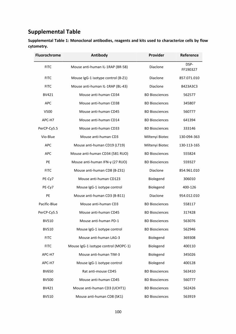
100
Supplemental Table
Supplemental Table 1: Monoclonal antibodies, reagents and kits used to characterize cells by flow
cytometry.
Fluorochrome Antibody Provider Reference
FITC Mouse anti-human IL-1RAP (BR-58) Diaclone DSP-
FF190327
FITC Mouse IgG-1 isotype control (B-Z1) Diaclone 857.071.010
FITC Mouse anti-human IL-1RAP (BL-43) Diaclone 8423A3C3
BV421 Mouse anti-human CD34 BD Biosciences 562577
APC Mouse anti-human CD38 BD Biosciences 345807
V500 Mouse anti-human CD45 BD Biosciences 560777
APC-H7 Mouse anti-human CD14 BD Biosciences 641394
PerCP-Cy5.5 Mouse anti-human CD33 BD Biosciences 333146
Vio-Blue Mouse anti-human CD3 Miltenyi Biotec 130-094-363
APC Mouse anti-human CD19 (LT19) Miltenyi Biotec 130-113-165
APC Mouse anti-human CD34 (581 RUO) BD Biosciences 555824
PE Mouse anti-human IFN-γ (27 RUO) BD Biosciences 559327
FITC Mouse anti-human CD8 (B-Z31) Diaclone 854.961.010
PE-Cy7 Mouse anti-human CD123 Biolegend 306010
PE-Cy7 Mouse IgG-1 isotype control Biolegend 400-126
PE Mouse anti-human CD3 (B-B11) Diaclone 954.012.010
Pacific-Blue Mouse anti-human CD3 BD Biosciences 558117
PerCP-Cy5.5 Mouse anti-human CD45 BD Biosciences 317428
BV510 Mouse anti-human PD-1 BD Biosciences 563076
BV510 Mouse IgG-1 isotype control BD Biosciences 562946
FITC Mouse anti-human LAG-3 Biolegend 369308
FITC Mouse IgG-1 isotype control (MOPC-1) Biolegend 400110
APC-H7 Mouse anti-human TIM-3 Biolegend 345026
APC-H7 Mouse IgG-1 isotype control Biolegend 400128
BV650 Rat anti-mouse CD45 BD Biosciences 563410
BV500 Mouse anti-human CD45 BD Biosciences 560777
BV421 Mouse anti-human CD3 (UCHT1) BD Biosciences 562426
BV510 Mouse anti-human CD8 (SK1) BD Biosciences 563919
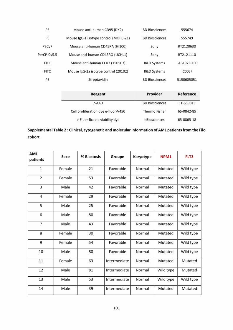
101
PE Mouse anti-human CD95 (DX2) BD Biosciences 555674
PE Mouse IgG-1 isotype control (MOPC-21) BD Biosciences 555749
PECy7 Mouse anti-human CD45RA (HI100) Sony RT2120630
PerCP-Cy5.5 Mouse anti-human CD45RO (UCHL1) Sony RT2121110
FITC Mouse anti-human CCR7 (150503) R&D Systems FAB197F-100
FITC Mouse IgG-2a isotype control (20102) R&D Systems IC003F
PE Streptavidin BD Biosciences 5150605051
Reagent Provider Reference
7-AAD BD Biosciences 51-68981E
Cell proliferation dye e-fluor-V450 Thermo Fisher 65-0842-85
e-Fluor fixable viability dye eBiosciences 65-0865-18
Supplemental Table 2 : Clinical, cytogenetic and molecular information of AML patients from the Filo
cohort.
AML
patients Sexe % Blastosis Groupe Karyotype NPM1 FLT3
1 Female 21 Favorable Normal Mutated Wild type
2 Female 53 Favorable Normal Mutated Wild type
3 Male 42 Favorable Normal Mutated Wild type
4 Female 29 Favorable Normal Mutated Wild type
5 Male 25 Favorable Normal Mutated Wild type
6 Male 80 Favorable Normal Mutated Wild type
7 Male 43 Favorable Normal Mutated Wild type
8 Female 30 Favorable Normal Mutated Wild type
9 Female 54 Favorable Normal Mutated Wild type
10 Male 80 Favorable Normal Mutated Wild type
11 Female 63 Intermediate Normal Mutated Mutated
12 Male 81 Intermediate Normal Wild type Mutated
13 Male 53 Intermediate Normal Wild type Wild type
14 Male 39 Intermediate Normal Mutated Mutated
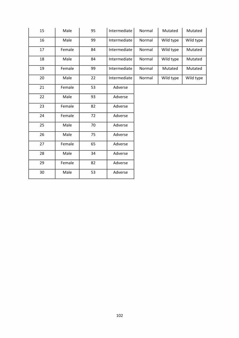
102
15 Male 95 Intermediate Normal Mutated Mutated
16 Male 99 Intermediate Normal Wild type Wild type
17 Female 84 Intermediate Normal Wild type Mutated
18 Male 84 Intermediate Normal Wild type Mutated
19 Female 99 Intermediate Normal Mutated Mutated
20 Male 22 Intermediate Normal Wild type Wild type
21 Female 53 Adverse
22 Male 93 Adverse
23 Female 82 Adverse
24 Female 72 Adverse
25 Male 70 Adverse
26 Male 75 Adverse
27 Female 65 Adverse
28 Male 34 Adverse
29 Female 82 Adverse
30 Male 53 Adverse

103
Supplemental Figures
Figure S1
Figure S1: Production of IL-1RAP CAR lentiviral vector. A/ Schematic construct of the IL-1RAP lentiviral vector
(above) and the control Mock lentiviral vector (below). B/ 293T cells were transfected wutg pMDG, pPAX2 and
transgene plasmids to produce IL-1RAP CAR and mock supernatants. The efficiency of the transfection was
measured by flow cytometry by gating on CD19+ cells (left, representative experiment). The transfection
efficiencies were 90.19 ± 13.99 and 91.54 ± 10.5 for 293T mock cells and 293T CAR cells, respectively.
Untransfected (UnT) 293T cells were used as a control. The results are also presented in bar histograms with the
mean ± standard deviation (SD) from n=17 independent experiments (right).
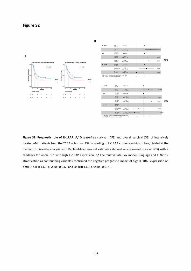
104
Figure S2
Figure S2: Prognostic role of IL-1RAP. A/ Disease-free survival (DFS) and overall survival (OS) of intensively
treated AML patients from the TCGA cohort (n=139) according to IL-1RAP expression (high or low; divided at the
median). Univariate analysis with Kaplan-Meier survival estimates showed worse overall survival (OS) with a
tendency for worse DFS with high IL-1RAP expression. B/ The multivariate Cox model using age and ELN2017
stratification as confounding variables confirmed the negative prognostic impact of high IL-1RAP expression on
both DFS (HR 1.60, p-value: 0.037) and OS (HR 1.60, p-value: 0.014).
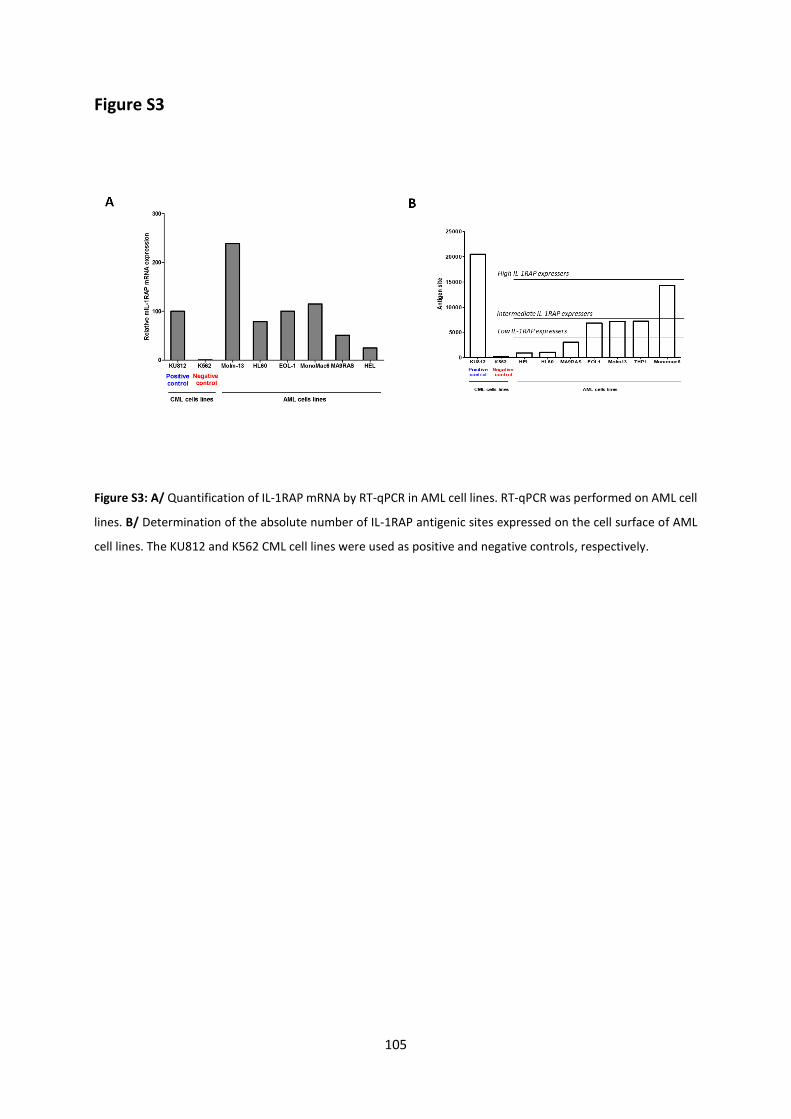
105
Figure S3
Figure S3: A/ Quantification of IL-1RAP mRNA by RT-qPCR in AML cell lines. RT-qPCR was performed on AML cell
lines. B/ Determination of the absolute number of IL-1RAP antigenic sites expressed on the cell surface of AML
cell lines. The KU812 and K562 CML cell lines were used as positive and negative controls, respectively.
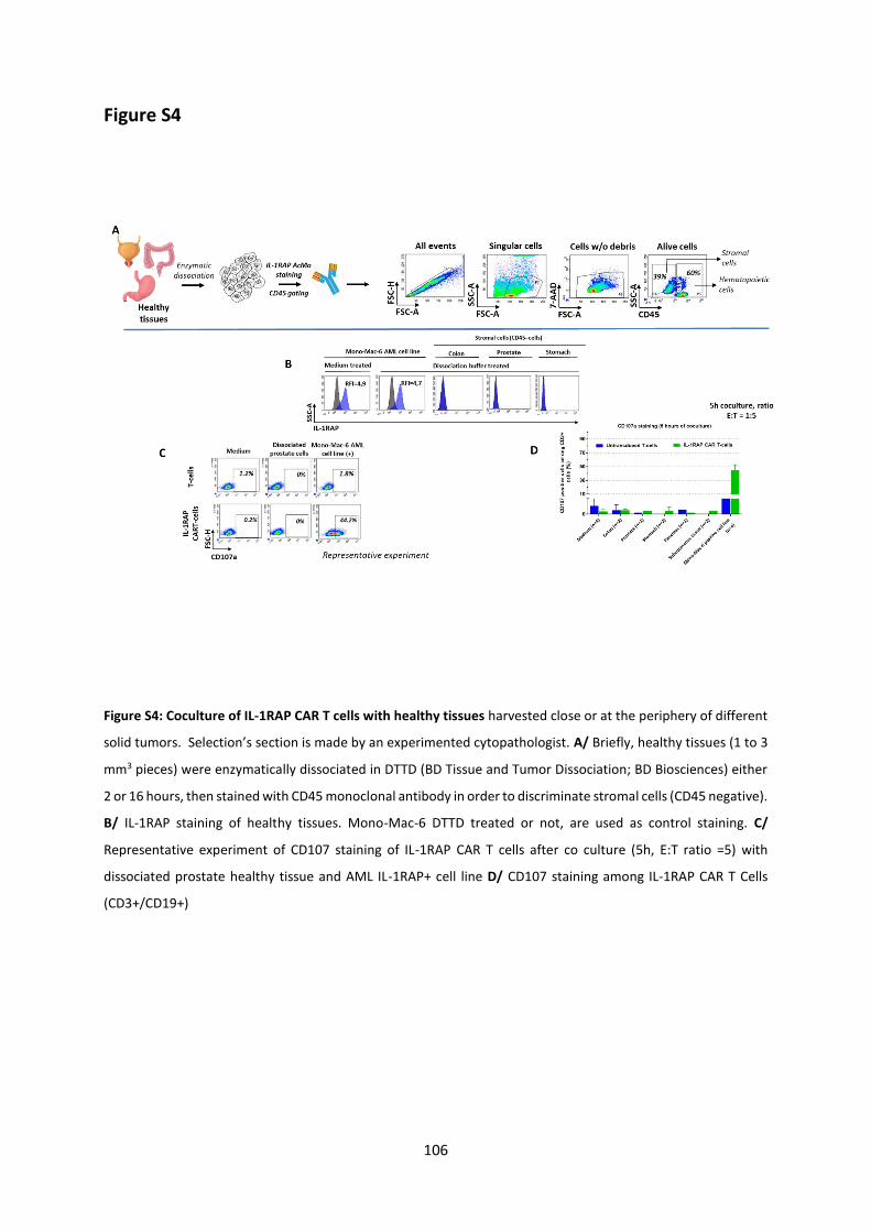
106
Figure S4
Figure S4: Coculture of IL-1RAP CAR T cells with healthy tissues harvested close or at the periphery of different
solid tumors. Selection’s section is made by an experimented cytopathologist. A/ Briefly, healthy tissues (1 to 3
mm3 pieces) were enzymatically dissociated in DTTD (BD Tissue and Tumor Dissociation; BD Biosciences) either
2 or 16 hours, then stained with CD45 monoclonal antibody in order to discriminate stromal cells (CD45 negative).
B/ IL-1RAP staining of healthy tissues. Mono-Mac-6 DTTD treated or not, are used as control staining. C/
Representative experiment of CD107 staining of IL-1RAP CAR T cells after co culture (5h, E:T ratio =5) with
dissociated prostate healthy tissue and AML IL-1RAP+ cell line D/ CD107 staining among IL-1RAP CAR T Cells
(CD3+/CD19+)
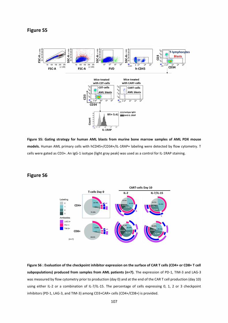
107
Figure S5
Figure S5: Gating strategy for human AML blasts from murine bone marrow samples of AML PDX mouse
models. Human AML primary cells with hCD45+/CD34+/IL-1RAP+ labeling were detected by flow cytometry. T
cells were gated as CD3+. An IgG-1 isotype (light gray peak) was used as a control for IL-1RAP staining.
Figure S6
Figure S6 : Evaluation of the checkpoint inhibitor expression on the surface of CAR T cells (CD4+ or CD8+ T cell
subpopulations) produced from samples from AML patients (n=7). The expression of PD-1, TIM-3 and LAG-3
was measured by flow cytometry prior to production (day 0) and at the end of the CAR T cell production (day 10)
using either IL-2 or a combination of IL-7/IL-15. The percentage of cells expressing 0, 1, 2 or 3 checkpoint
inhibitors (PD-1, LAG-3, and TIM-3) among CD3+CAR+ cells (CD4+/CD8+) is provided.
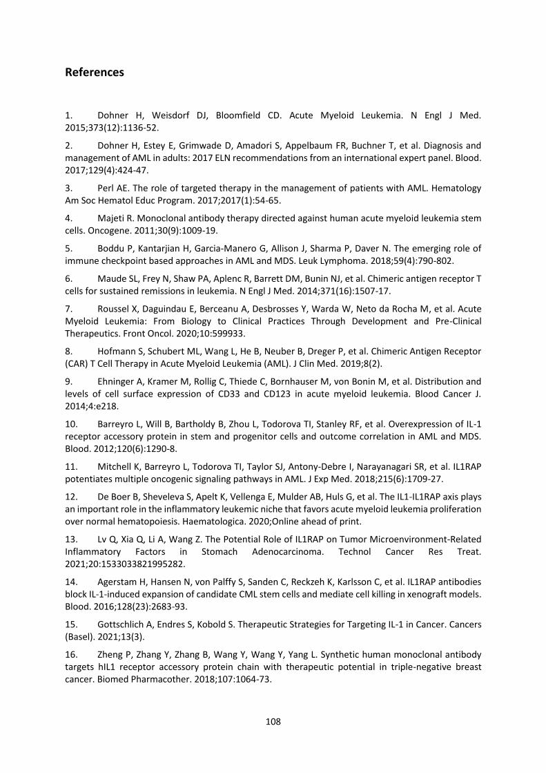
108
References
1. Dohner H, Weisdorf DJ, Bloomfield CD. Acute Myeloid Leukemia. N Engl J Med. 2015;373(12):1136-52.
2. Dohner H, Estey E, Grimwade D, Amadori S, Appelbaum FR, Buchner T, et al. Diagnosis and management of AML in adults: 2017 ELN recommendations from an international expert panel. Blood. 2017;129(4):424-47.
3. Perl AE. The role of targeted therapy in the management of patients with AML. Hematology Am Soc Hematol Educ Program. 2017;2017(1):54-65.
4. Majeti R. Monoclonal antibody therapy directed against human acute myeloid leukemia stem cells. Oncogene. 2011;30(9):1009-19.
5. Boddu P, Kantarjian H, Garcia-Manero G, Allison J, Sharma P, Daver N. The emerging role of immune checkpoint based approaches in AML and MDS. Leuk Lymphoma. 2018;59(4):790-802.
6. Maude SL, Frey N, Shaw PA, Aplenc R, Barrett DM, Bunin NJ, et al. Chimeric antigen receptor T cells for sustained remissions in leukemia. N Engl J Med. 2014;371(16):1507-17.
7. Roussel X, Daguindau E, Berceanu A, Desbrosses Y, Warda W, Neto da Rocha M, et al. Acute Myeloid Leukemia: From Biology to Clinical Practices Through Development and Pre-Clinical Therapeutics. Front Oncol. 2020;10:599933.
8. Hofmann S, Schubert ML, Wang L, He B, Neuber B, Dreger P, et al. Chimeric Antigen Receptor (CAR) T Cell Therapy in Acute Myeloid Leukemia (AML). J Clin Med. 2019;8(2).
9. Ehninger A, Kramer M, Rollig C, Thiede C, Bornhauser M, von Bonin M, et al. Distribution and levels of cell surface expression of CD33 and CD123 in acute myeloid leukemia. Blood Cancer J. 2014;4:e218.
10. Barreyro L, Will B, Bartholdy B, Zhou L, Todorova TI, Stanley RF, et al. Overexpression of IL-1 receptor accessory protein in stem and progenitor cells and outcome correlation in AML and MDS. Blood. 2012;120(6):1290-8.
11. Mitchell K, Barreyro L, Todorova TI, Taylor SJ, Antony-Debre I, Narayanagari SR, et al. IL1RAP potentiates multiple oncogenic signaling pathways in AML. J Exp Med. 2018;215(6):1709-27.
12. De Boer B, Sheveleva S, Apelt K, Vellenga E, Mulder AB, Huls G, et al. The IL1-IL1RAP axis plays an important role in the inflammatory leukemic niche that favors acute myeloid leukemia proliferation over normal hematopoiesis. Haematologica. 2020;Online ahead of print.
13. Lv Q, Xia Q, Li A, Wang Z. The Potential Role of IL1RAP on Tumor Microenvironment-Related Inflammatory Factors in Stomach Adenocarcinoma. Technol Cancer Res Treat. 2021;20:1533033821995282.
14. Agerstam H, Hansen N, von Palffy S, Sanden C, Reckzeh K, Karlsson C, et al. IL1RAP antibodies block IL-1-induced expansion of candidate CML stem cells and mediate cell killing in xenograft models. Blood. 2016;128(23):2683-93.
15. Gottschlich A, Endres S, Kobold S. Therapeutic Strategies for Targeting IL-1 in Cancer. Cancers (Basel). 2021;13(3).
16. Zheng P, Zhang Y, Zhang B, Wang Y, Wang Y, Yang L. Synthetic human monoclonal antibody targets hIL1 receptor accessory protein chain with therapeutic potential in triple-negative breast cancer. Biomed Pharmacother. 2018;107:1064-73.
109
17. Askmyr M, Agerstam H, Hansen N, Gordon S, Arvanitakis A, Rissler M, et al. Selective killing of candidate AML stem cells by antibody targeting of IL1RAP. Blood. 2013;121(18):3709-13.
18. Warda W, Da Rocha MN, Trad R, Haderbache R, Salma Y, Bouquet L, et al. Overcoming target epitope masking resistance that can occur on low-antigen-expresser AML blasts after IL-1RAP chimeric antigen receptor T cell therapy using the inducible caspase 9 suicide gene safety switch. Cancer Gene Ther. 2021.
19. Warda W, Larosa F, Neto Da Rocha M, Trad R, Deconinck E, Fajloun Z, et al. CML Hematopoietic Stem Cells Expressing IL1RAP Can Be Targeted by Chimeric Antigen Receptor-Engineered T Cells. Cancer Res. 2019;79(3):663-75.
20. Ceroi A, Masson D, Roggy A, Roumier C, Chague C, Gauthier T, et al. LXR agonist treatment of blastic plasmacytoid dendritic cell neoplasm restores cholesterol efflux and triggers apoptosis. Blood. 2016;128(23):2694-707.
21. Li C, Wong WH. Model-based analysis of oligonucleotide arrays: expression index computation and outlier detection. Proc Natl Acad Sci U S A. 2001;98(1):31-6.
22. Awada A, Eskens F, Robbrecht D, Lassen UN, Soerensen MM, Steeghs N, et al. Results from a first-in-man, open label, safety and tolerability trial of CAN04 (nidanilimab), a fully humanized monoclonal antibody against the novel antitumor target, IL1RAP, in patients with solid tumor malignancies. Journal of Clinical Oncology. 2019;37(15_suppl):2504-.
23. Corces MR, Buenrostro JD, Wu B, Greenside PG, Chan SM, Koenig JL, et al. Lineage-specific and single-cell chromatin accessibility charts human hematopoiesis and leukemia evolution. Nat Genet. 2016;48(10):1193-203.
24. Mardiana S, Gill S. CAR T Cells for Acute Myeloid Leukemia: State of the Art and Future Directions. Front Oncol. 2020;10:697.
25. Bose P, Vachhani P, Cortes JE. Treatment of Relapsed/Refractory Acute Myeloid Leukemia. Curr Treat Options Oncol. 2017;18(3):17.
26. Frigault MJ, Maus MV. State of the art in CAR T cell therapy for CD19+ B cell malignancies. J Clin Invest. 2020;130(4):1586-94.
27. Munshi NC, Anderson LD, Jr., Shah N, Madduri D, Berdeja J, Lonial S, et al. Idecabtagene Vicleucel in Relapsed and Refractory Multiple Myeloma. N Engl J Med. 2021;384(8):705-16.
28. Gill S, Tasian SK, Ruella M, Shestova O, Li Y, Porter DL, et al. Preclinical targeting of human acute myeloid leukemia and myeloablation using chimeric antigen receptor-modified T cells. Blood. 2014;123(15):2343-54.
29. Litmanovich A, Khazim K, Cohen I. The Role of Interleukin-1 in the Pathogenesis of Cancer and its Potential as a Therapeutic Target in Clinical Practice. Oncol Ther. 2018;6(2):109-27.
30. Nie Y, Lu W, Chen D, Tu H, Guo Z, Zhou X, et al. Mechanisms underlying CD19-positive ALL relapse after anti-CD19 CAR T cell therapy and associated strategies. Biomark Res. 2020;8:18.
31. Samur MK, Fulciniti M, Aktas Samur A, Bazarbachi AH, Tai YT, Prabhala R, et al. Biallelic loss of BCMA as a resistance mechanism to CAR T cell therapy in a patient with multiple myeloma. Nat Commun. 2021;12(1):868.
32. Jain MD, Davila ML. Concise Review: Emerging Principles from the Clinical Application of Chimeric Antigen Receptor T Cell Therapies for B Cell Malignancies. Stem Cells. 2018;36(1):36-44.
33. Gebru MT, Wang HG. Therapeutic targeting of FLT3 and associated drug resistance in acute myeloid leukemia. J Hematol Oncol. 2020;13(1):155.
110
34. Fasslrinner F, Arndt C, Koristka S, Feldmann A, Altmann H, von Bonin M, et al. Midostaurin abrogates CD33-directed UniCAR and CD33-CD3 bispecific antibody therapy in acute myeloid leukaemia. Br J Haematol. 2019;186(5):735-40.
35. Wolleschak D, Mack TS, Perner F, Frey S, Schnoder TM, Wagner MC, et al. Clinically relevant doses of FLT3-kinase inhibitors quizartinib and midostaurin do not impair T-cell reactivity and function. Haematologica. 2014;99(6):e90-3.
36. Jimbu L, Mesaros O, Popescu C, Neaga A, Berceanu I, Dima D, et al. Is There a Place for PD-1-PD-L Blockade in Acute Myeloid Leukemia? Pharmaceuticals (Basel). 2021;14(4).
111
Discussion
AML remains a rare but lethal malignancy with a poor prognosis, especially for high-risk patients.
AML treatment is always in progress with novel approaches that have emerged since 2017. Despite
the progress made over the last ten years in the treatment of AML and improvements in allogeneic
HSCT, and given that AML is an aggressive disease with a clonal evolution, it is in most cases related to
a fatal relapse after treatment. This relapse owing to the resistance and the persistence of leukemia
cells, specifically LSCs, which is a subpopulation of AML cells with self-renewal, and chemorefractory
capacity.
Actually, the relapse after conventional chemotherapy, targeted therapy or immunotherapy remains
a major problem in AML patients and a crucial cause of death. Hence the necessity of new alternatives
for treating AML patients (Thol and Heuser 2021). In the field of CART-cells immunotherapies, the
stellar success of autologous CD19 CART-cells in different types of B-cell malignancies such as in ALL
and lymphoma (Frigault and Maus 2020) as well as in multiple myeloma (MM) (Munshi et al. 2021)
have yielded significant efforts to translate this triumph to myeloid malignancies such as AML. Almost
all antigens expressed on the surface of LSC, are targeted with at least one therapeutic pathway
(targeted therapies, immunotherapies). A surface protein, the IL-1 receptor accessory protein (IL-
1RAP) has been defined as a selective marker of LSCs while the other antigens are also expressed by
normal HSCs (Askmyr et al. 2013). The pro-inflammatory protein IL-1RAP plays a crucial role in
immunity, inflammation and cancer (De Boer et al. 2020) (Lv et al. 2021).
In this work, we investigated IL-1RAP, as a novel and unexplored potential target in AML. We have
reproduced what has already been demonstrated that IL-1RAP is overexpressed by AML leukemia cells
specifically LSCs and not by healthy HSPCs, or T and B lymphocytes, though it is expressed by circulating
monocytes (Askmyr et al. 2013). We demonstrated that IL-1RAP mRNA is expressed in LSCs and blasts
in all AML FAB subtypes and was discriminatory compared to common AML cell surface targets. As
published data, we showed that the IL-1RAP protein is expressed at three different levels “Low”,
“Intermediate” and “High” on the cell surface of AML blasts (Askmyr et al. 2013) and independently of
ELN classification.
Interestingly, we showed in a cohort of 30 AML patients that most primary IL-1RAP+ blasts co-
expressed CD123, making this target suitable for bispecific CART-cell targeting, which will increase
specificity via “and/or” CAR activation signaling and presumably without increasing toxicity (Jain and
Davila 2018).
112
In our preceded work in CML, we established the proof of concept for targeting IL-1RAP with CART-
cells (Warda et al. 2019b). We demonstrated that IL-1RAP CART-cells efficiently eliminate CML
resistant cells after therapy with tyrosine kinase inhibitors (Warda et al. 2019b). Furthermore, we
showed the absence of toxicities toward healthy HSCs that were not targeted by IL-1RAP CART-cells
(Warda et al. 2019b). Here, we show in a preclinical study that this new alternative treatment has
efficacy in AML. We targeted the cell surface biomarker IL-1RAP that is expressed on leukemic stem
cells and primary AML blasts using CART-cells specific for IL-1RAP. This innovative approach showed
efficiency and high cytotoxicity against both cells AML cell lines and primary AML cells either with low
expression of IL-1RAP or with high IL-1RAP expression. We were able to produce IL-1RAP CART-cells of
3d generation from healthy donors and from AML patients at diagnosis or at relapse with high
efficiency. We demonstrated that allogeneic and interestingly autologous IL-1RAP CART-cells are
effective in-vitro against primary R/R AML blasts and LSCs and allogenic IL-1RAP CART-cells were able
to eradicated AML blasts and LSCs from R/R AML patients in the bone marrow and spleen of PDX mice
in-vivo. Therefore, CART-cells offer a strong treatment option apart from chemotherapy and targeted
therapies, which are recognized as early-line treatments.
Moreover, the characterization of T-cells taken from diagnostic AML patients and from R/R AML
patients showed an acquisition of at least one checkpoint marker such as PD-1, TIM-3 and LAG-3 at the
end of the production process in-vitro as in-vivo in PDX mice. Further studies are needed to evaluate
the surface expression of the ligands of these markers such as PDL-1 on AML cells for a possible
combination of IL-1RAP CART-cells therapy with checkpoints inhibitors (Jimbu et al. 2021).
In addition, these T-cells showed a conservation of the phenotype profile during in-vitro culture
with a slight tendency of acquisition a naïve/memory phenotype by CD8+ CART-cells at the end of the
production process (Schnorfeil et al. 2015) (Williams et al. 2019). No impact of the cytokine type added
in the culture medium (IL-2 or IL-7/IL-15). In PDX mice, an acquisition of an effector phenotype was
observed after the sacrifice.
Importantly, the persistence of IL-1RAP CART-cells in the bone marrow and the spleen of PDX mice
was confirmed by dPCR for approximately 2 months after treatment. This persistence may be due to
the co-stimulation molecule 4-1BB present in the CAR construct.
From a clinical point of view, in order to circumvent the problem of inducing a graft-versus-host
disease, following the transfusion of allogeneic TL, we incorporated within our lentiviral construct an
iCASP9/AP1903 suicide system. This safety switch system will protect patients who received CART-cells
against adverse events such as off-target toxicities, neurological toxicities, unexpected leukemic cell
transduction, as previously shown (Ruella et al. 2018b) or when persistent adverse events affect

113
monocytes and many other toxicities in order to eliminate CART-cells and allow a mature myeloid cell
recovery. In addition, this system could be facilitate the use of allogeneic IL-1RAP CART-cells in AML.
We are also working on a clinical transfer of IL-1RAP CART-cells treatment to a phase I/IIa trial on
R/R AML patients with a GMP like-grade Prodigy system.
114
Conclusion
Our synthetic mAb is specific for IL-1RAP protein expressed on the surface of AML cell lines and primary
cells and not expressed on normal HSPC except monocytes. With a high lentiviral transduction, IL-1RAP
CART-cells were produced.
Subsequently, the functionality of IL-1RAP CART-cells was evaluated on different AML cell lines
expressing the IL-1RAP protein at different levels (Low, intermediate and high, respectively). Despite
this difference in IL-1RAP expression, the stimulation and activation of IL-1RAP CART-cells was similar
after co-culture with the target lines, regard to the level of cell proliferation, the expression of IFNγ
and induction of target mortality in-vitro, and in-vivo with xenograft murine model.
Additionally, IL-1RAP CART-cells were highly cytotoxic in-vitro and in-vivo with PDX murine model
against AML primary cells from AML patients at diagnosis or specially when relapsed following
chemotherapy or targeted therapy treatments.
Accordingly, this work clearly confirms the potential of IL-1RAP as a target in AML and the strong
anti-leukemic effects of strategies targeting this marker both in-vitro and in-vivo, laying the foundation
for a promising future first-in-human clinical trial in R/R AML. IL-1RAP CART-cells have not yet been
evaluated in humans, and doing so will help determine the optimal IL-1RAP CART-cells dose and
evaluate the efficacy and safety of this approach.
115
References
Adamia, Sophia, Michal Bar-Natan, Benjamin Haibe-Kains, Patrick M. Pilarski, Christian Bach, Samuel Pevzner, Teresa Calimeri, et al. 2014. “NOTCH2 and FLT3 Gene Mis-Splicings Are Common Events in Patients with Acute Myeloid Leukemia (AML): New Potential Targets in AML.” Blood 123 (18): 2816–25. https://doi.org/10.1182/blood-2013-02-481507.
Adamia, Sophia, Benjamin Haibe-Kains, Patrick M. Pilarski, Michal Bar-Natan, Samuel Pevzner, Herve Avet-Loiseau, Laurence Lode, et al. 2014. “A Genome-Wide Aberrant RNA Splicing in Patients with Acute Myeloid Leukemia Identifies Novel Potential Disease Markers and Therapeutic Targets.” Clinical Cancer Research: An Official Journal of the American Association for Cancer Research 20 (5): 1135–45. https://doi.org/10.1158/1078-0432.CCR-13-0956.
Ågerstam, Helena, Nils Hansen, Sofia von Palffy, Carl Sandén, Kristian Reckzeh, Christine Karlsson, Henrik Lilljebjörn, et al. 2016. “IL1RAP Antibodies Block IL-1-Induced Expansion of Candidate CML Stem Cells and Mediate Cell Killing in Xenograft Models.” Blood 128 (23): 2683–93. https://doi.org/10.1182/blood-2015-11-679985.
Ahmed, Nabil, Vita S. Brawley, Meenakshi Hegde, Catherine Robertson, Alexia Ghazi, Claudia Gerken, Enli Liu, et al. 2015. “Human Epidermal Growth Factor Receptor 2 (HER2) -Specific Chimeric Antigen Receptor-Modified T Cells for the Immunotherapy of HER2-Positive Sarcoma.” Journal of Clinical Oncology: Official Journal of the American Society of Clinical Oncology 33 (15): 1688–96. https://doi.org/10.1200/JCO.2014.58.0225.
Aleksandrova, Krasimira, Jana Leise, Christoph Priesner, Anette Melk, Fanni Kubaink, Hinrich Abken, Andreas Hombach, et al. 2019. “Functionality and Cell Senescence of CD4/ CD8-Selected CD20 CAR T Cells Manufactured Using the Automated CliniMACS Prodigy® Platform.” Transfusion Medicine and Hemotherapy: Offizielles Organ Der Deutschen Gesellschaft Fur Transfusionsmedizin Und Immunhamatologie 46 (1): 47–54. https://doi.org/10.1159/000495772.
Alexandrov, Ludmil B., Serena Nik-Zainal, David C. Wedge, Samuel A. J. R. Aparicio, Sam Behjati, Andrew V. Biankin, Graham R. Bignell, et al. 2013. “Signatures of Mutational Processes in Human Cancer.” Nature 500 (7463): 415–21. https://doi.org/10.1038/nature12477.
Ali, Robert, Jeremy Ramdial, Sandra Algaze, and Amer Beitinjaneh. 2017. “The Role of Anti-Thymocyte Globulin or Alemtuzumab-Based Serotherapy in the Prophylaxis and Management of Graft-Versus-Host Disease.” Biomedicines 5 (4): E67. https://doi.org/10.3390/biomedicines5040067.
Angel, Matthew, and Mehmet Fatih Yanik. 2010. “Innate Immune Suppression Enables Frequent Transfection with RNA Encoding Reprogramming Proteins.” PLOS ONE 5 (7): e11756. https://doi.org/10.1371/journal.pone.0011756.
Anguille, Sébastien, Ann L. Van de Velde, Evelien L. Smits, Viggo F. Van Tendeloo, Gunnar Juliusson, Nathalie Cools, Griet Nijs, et al. 2017. “Dendritic Cell Vaccination as Postremission Treatment to Prevent or Delay Relapse in Acute Myeloid Leukemia.” Blood 130 (15): 1713–21. https://doi.org/10.1182/blood-2017-04-780155.
Antar, Ahmad I., Zaher K. Otrock, Elias Jabbour, Mohamad Mohty, and Ali Bazarbachi. 2020. “FLT3 Inhibitors in Acute Myeloid Leukemia: Ten Frequently Asked Questions.” Leukemia 34 (3): 682–96. https://doi.org/10.1038/s41375-019-0694-3.
Arai, Yasuyuki, Uimook Choi, Cristina I. Corsino, Sherry M. Koontz, Masaki Tajima, Colin L. Sweeney, Mary A. Black, Steven A. Feldman, Mary C. Dinauer, and Harry L. Malech. 2018. “Myeloid Conditioning with C-Kit-Targeted CAR-T Cells Enables Donor Stem Cell Engraftment.” Molecular Therapy: The Journal of the American Society of Gene Therapy 26 (5): 1181–97. https://doi.org/10.1016/j.ymthe.2018.03.003.
Arcangeli, Silvia, Maria Caterina Rotiroti, Marco Bardelli, Luca Simonelli, Chiara Francesca Magnani, Andrea Biondi, Ettore Biagi, Sarah Tettamanti, and Luca Varani. 2017. “Balance of Anti-CD123 Chimeric Antigen Receptor Binding Affinity and Density for the Targeting of Acute Myeloid
116
Leukemia.” Molecular Therapy: The Journal of the American Society of Gene Therapy 25 (8): 1933–45. https://doi.org/10.1016/j.ymthe.2017.04.017.
Askmyr, Maria, Helena Ågerstam, Nils Hansen, Sandra Gordon, Alexandros Arvanitakis, Marianne Rissler, Gunnar Juliusson, Johan Richter, Marcus Järås, and Thoas Fioretos. 2013. “Selective Killing of Candidate AML Stem Cells by Antibody Targeting of IL1RAP.” Blood 121 (18): 3709–13. https://doi.org/10.1182/blood-2012-09-458935.
Aurelius, Johan, Fredrik B. Thorén, Ali A. Akhiani, Mats Brune, Lars Palmqvist, Markus Hansson, Kristoffer Hellstrand, and Anna Martner. 2012. “Monocytic AML Cells Inactivate Antileukemic Lymphocytes: Role of NADPH Oxidase/Gp91(Phox) Expression and the PARP-1/PAR Pathway of Apoptosis.” Blood 119 (24): 5832–37. https://doi.org/10.1182/blood-2011-11-391722.
Barreyro, Laura, Britta Will, Boris Bartholdy, Li Zhou, Tihomira I. Todorova, Robert F. Stanley, Susana Ben-Neriah, et al. 2012. “Overexpression of IL-1 Receptor Accessory Protein in Stem and Progenitor Cells and Outcome Correlation in AML and MDS.” Blood 120 (6): 1290–98. https://doi.org/10.1182/blood-2012-01-404699.
Beatty, Gregory L., Andrew R. Haas, Marcela V. Maus, Drew A. Torigian, Michael C. Soulen, Gabriela Plesa, Anne Chew, et al. 2014. “Mesothelin-Specific Chimeric Antigen Receptor MRNA-Engineered T Cells Induce Anti-Tumor Activity in Solid Malignancies.” Cancer Immunology Research 2 (2): 112–20. https://doi.org/10.1158/2326-6066.CIR-13-0170.
Beavis, Paul A., Melissa A. Henderson, Lauren Giuffrida, Jane K. Mills, Kevin Sek, Ryan S. Cross, Alexander J. Davenport, et al. 2017. “Targeting the Adenosine 2A Receptor Enhances Chimeric Antigen Receptor T Cell Efficacy.” The Journal of Clinical Investigation 127 (3): 929–41. https://doi.org/10.1172/JCI89455.
Bertoletti, Antonio, and Anthony Tanoto Tan. 2020. “Challenges of CAR- and TCR-T Cell-Based Therapy for Chronic Infections.” The Journal of Experimental Medicine 217 (5): e20191663. https://doi.org/10.1084/jem.20191663.
Bertoli, Sarah, Lydie Fourrier, and Florent Puisset. 2021. “Les Thérapies Orales En 2021 Dans Les Leucémies Aiguës Myéloïdes : Un Travail d’équipe ! L’expérience Toulousaine.” Hématologie 27 (2): 36–49. https://doi.org/10.1684/hma.2021.1632.
Binder, Stephanie, Michela Luciano, and Jutta Horejs-Hoeck. 2018. “The Cytokine Network in Acute Myeloid Leukemia (AML): A Focus on pro- and Anti-Inflammatory Mediators.” Cytokine & Growth Factor Reviews 43 (October): 8–15. https://doi.org/10.1016/j.cytogfr.2018.08.004.
Bishop, D. C., L. E. Clancy, J. Burgess, G. Mathew, E. Atkins, S. Advic, K. Maddock, et al. 2019. “Matched Sibling Donor-Derived Piggybac CAR19 T Cells Induce Remission of Relapsed/Refractory CD19+ Malignancy Following Haematopoietic Stem Cell Transplant.” Cytotherapy 21 (5, Supplement): S9. https://doi.org/10.1016/j.jcyt.2019.03.562.
Bôle-Richard, E., C. Gamonet, J.-M. Certoux, I. Idirene, F. Larosa, E. Deconinck, A.-M. Mosseley, et al. 2016. “Exposure to Hypomethylating Agent, 5-Azacytidine, May Improve ICasp9 Suicide Gene Therapy for Treating GvHD in Allografts.” Gene Therapy 23 (8): 664–72. https://doi.org/10.1038/gt.2016.39.
Bonini, C., G. Ferrari, S. Verzeletti, P. Servida, E. Zappone, L. Ruggieri, M. Ponzoni, et al. 1997. “HSV-TK Gene Transfer into Donor Lymphocytes for Control of Allogeneic Graft-versus-Leukemia.” Science (New York, N.Y.) 276 (5319): 1719–24. https://doi.org/10.1126/science.276.5319.1719.
Bonte, Sarah, Stijn De Munter, Glenn Goetgeluk, Joline Ingels, Melissa Pille, Lore Billiet, Tom Taghon, Georges Leclercq, Bart Vandekerckhove, and Tessa Kerre. 2020. “T-Cells with a Single Tumor Antigen-Specific T-Cell Receptor Can Be Generated in Vitro from Clinically Relevant Stem Cell Sources.” Oncoimmunology 9 (1): 1727078. https://doi.org/10.1080/2162402X.2020.1727078.
Bordignon, C., C. Bonini, S. Verzeletti, N. Nobili, D. Maggioni, C. Traversari, R. Giavazzi, P. Servida, E. Zappone, and E. Benazzi. 1995. “Transfer of the HSV-Tk Gene into Donor Peripheral Blood Lymphocytes for in Vivo Modulation of Donor Anti-Tumor Immunity after Allogeneic Bone Marrow Transplantation.” Human Gene Therapy 6 (6): 813–19. https://doi.org/10.1089/hum.1995.6.6-813.
117
Borrello, Ivan, and Kimberly A. Noonan. 2016. “Marrow-Infiltrating Lymphocytes - Role in Biology and Cancer Therapy.” Frontiers in Immunology 7: 112. https://doi.org/10.3389/fimmu.2016.00112.
Braciak, Todd A., Claudia C. Roskopf, Sarah Wildenhain, Nadja C. Fenn, Christian B. Schiller, Ingo A. Schubert, Uwe Jacob, et al. 2018. “Dual-Targeting Triplebody 33-16-123 (SPM-2) Mediates Effective Redirected Lysis of Primary Blasts from Patients with a Broad Range of AML Subtypes in Combination with Natural Killer Cells.” Oncoimmunology 7 (9): e1472195. https://doi.org/10.1080/2162402X.2018.1472195.
Bridgeman, John S., Robert E. Hawkins, Steve Bagley, Morgan Blaylock, Mark Holland, and David E. Gilham. 2010. “The Optimal Antigen Response of Chimeric Antigen Receptors Harboring the CD3zeta Transmembrane Domain Is Dependent upon Incorporation of the Receptor into the Endogenous TCR/CD3 Complex.” Journal of Immunology (Baltimore, Md.: 1950) 184 (12): 6938–49. https://doi.org/10.4049/jimmunol.0901766.
Brown, Christine E., Behnam Badie, Michael E. Barish, Lihong Weng, Julie R. Ostberg, Wen-Chung Chang, Araceli Naranjo, et al. 2015. “Bioactivity and Safety of IL13Rα2-Redirected Chimeric Antigen Receptor CD8+ T Cells in Patients with Recurrent Glioblastoma.” Clinical Cancer Research: An Official Journal of the American Association for Cancer Research 21 (18): 4062–72. https://doi.org/10.1158/1078-0432.CCR-15-0428.
Bu, Chaoke, Zhiyong Peng, Min Luo, Guangchao Li, and Chunfu Li. 2020. “Phase I Clinical Trial of Autologous CLL1 CAR-T Therapy for Pediatric Patients with Relapsed and Refractory Acute Myeloid Leukemia.” Blood 136 (Supplement 1): 13–13. https://doi.org/10.1182/blood-2020-140648.
Budde, Lihua, Joo Y Song, Young Kim, Suzette Blanchard, Jamie Wagner, Anthony S. Stein, Lihong Weng, et al. 2017. “Remissions of Acute Myeloid Leukemia and Blastic Plasmacytoid Dendritic Cell Neoplasm Following Treatment with CD123-Specific CAR T Cells: A First-in-Human Clinical Trial.” Blood 130 (Suppl_1): 811–811. https://doi.org/10.1182/blood.V130.Suppl_1.811.811.
Burger, Michael C., Congcong Zhang, Patrick N. Harter, Annette Romanski, Florian Strassheimer, Christian Senft, Torsten Tonn, Joachim P. Steinbach, and Winfried S. Wels. 2019. “CAR-Engineered NK Cells for the Treatment of Glioblastoma: Turning Innate Effectors Into Precision Tools for Cancer Immunotherapy.” Frontiers in Immunology 0. https://doi.org/10.3389/fimmu.2019.02683.
Caimi, Paolo Fabrizio, Jane Reese, Folashade Otegbeye, Dina Schneider, Kamal Chamoun, Kirsten M Boughan, Brenda W. Cooper, et al. 2019. “Phase 1 Trial of Anti-CD19 Chimeric Antigen Receptor T (CAR-T) Cells with Tumor Necrosis Alfa Receptor Superfamily 19 (TNFRSF19) Transmembrane Domain.” Journal of Clinical Oncology 37 (15_suppl): 2539–2539. https://doi.org/10.1200/JCO.2019.37.15_suppl.2539.
Cancer Genome Atlas Research Network, Timothy J. Ley, Christopher Miller, Li Ding, Benjamin J. Raphael, Andrew J. Mungall, A. Gordon Robertson, et al. 2013. “Genomic and Epigenomic Landscapes of Adult de Novo Acute Myeloid Leukemia.” The New England Journal of Medicine 368 (22): 2059–74. https://doi.org/10.1056/NEJMoa1301689.
Capsomidis, Anna, Gabriel Benthall, Heleen H. Van Acker, Jonathan Fisher, Anne M. Kramer, Zarah Abeln, Yvonne Majani, et al. 2018. “Chimeric Antigen Receptor-Engineered Human Gamma Delta T Cells: Enhanced Cytotoxicity with Retention of Cross Presentation.” Molecular Therapy: The Journal of the American Society of Gene Therapy 26 (2): 354–65. https://doi.org/10.1016/j.ymthe.2017.12.001.
Carlsten, Mattias, and Marcus Järås. 2019. “Natural Killer Cells in Myeloid Malignancies: Immune Surveillance, NK Cell Dysfunction, and Pharmacological Opportunities to Bolster the Endogenous NK Cells.” Frontiers in Immunology 10: 2357. https://doi.org/10.3389/fimmu.2019.02357.
Cartellieri, Marc, Michael Bachmann, Anja Feldmann, Claudia Bippes, Slava Stamova, Rebekka Wehner, Achim Temme, and Marc Schmitz. 2010. “Chimeric Antigen Receptor-Engineered T Cells for Immunotherapy of Cancer.” Journal of Biomedicine and Biotechnology 2010: 1–13. https://doi.org/10.1155/2010/956304.
118
Casucci, Monica, Benedetta Nicolis di Robilant, Laura Falcone, Barbara Camisa, Margherita Norelli, Pietro Genovese, Bernhard Gentner, et al. 2013. “CD44v6-Targeted T Cells Mediate Potent Antitumor Effects against Acute Myeloid Leukemia and Multiple Myeloma.” Blood 122 (20): 3461–72. https://doi.org/10.1182/blood-2013-04-493361.
Cazzola, Mario. 2020. “Myelodysplastic Syndromes.” Edited by Dan L. Longo. New England Journal of Medicine 383 (14): 1358–74. https://doi.org/10.1056/NEJMra1904794.
Cerrano, Marco, and Raphael Itzykson. 2021. “New Therapeutic Options for Older Unfit AML Patients (Excluding Targeted Therapies).” Hématologie 27 (2): 24–35. https://doi.org/10.1684/hma.2021.1631.
Chalmers, David, Christophe Ferrand, Jane F Apperley, Junia V Melo, Saskia Ebeling, Isobel Newton, Anne Duperrier, et al. 2001. “Elimination of the Truncated Message from the Herpes Simplex Virus Thymidine Kinase Suicide Gene.” Molecular Therapy 4 (2): 146–48. https://doi.org/10.1006/mthe.2001.0433.
Chapuis, Aude G., Daniel N. Egan, Merav Bar, Thomas M. Schmitt, Megan S. McAfee, Kelly G. Paulson, Valentin Voillet, et al. 2019. “T Cell Receptor Gene Therapy Targeting WT1 Prevents Acute Myeloid Leukemia Relapse Post-Transplant.” Nature Medicine 25 (7): 1064–72. https://doi.org/10.1038/s41591-019-0472-9.
Charrot, Sarah, and Simon Hallam. 2019. “CAR-T Cells: Future Perspectives.” HemaSphere 3 (2): e188. https://doi.org/10.1097/HS9.0000000000000188.
Chen, Xiangli, Shuhu Liu, Liancai Wang, Wanggang Zhang, Yuqiang Ji, and Xiaorong Ma. 2008. “Clinical Significance of B7-H1 (PD-L1) Expression in Human Acute Leukemia.” Cancer Biology & Therapy 7 (5): 622–27. https://doi.org/10.4161/cbt.7.5.5689.
Chen, Xiaoying, Jennica L. Zaro, and Wei-Chiang Shen. 2013. “Fusion Protein Linkers: Property, Design and Functionality.” Advanced Drug Delivery Reviews 65 (10): 1357–69. https://doi.org/10.1016/j.addr.2012.09.039.
Chmielewski, Markus, and Hinrich Abken. 2015. “TRUCKs: The Fourth Generation of CARs.” Expert Opinion on Biological Therapy 15 (8): 1145–54. https://doi.org/10.1517/14712598.2015.1046430.
Chong, Elise A., J. Joseph Melenhorst, Simon F. Lacey, David E. Ambrose, Vanessa Gonzalez, Bruce L. Levine, Carl H. June, and Stephen J. Schuster. 2017. “PD-1 Blockade Modulates Chimeric Antigen Receptor (CAR)-Modified T Cells: Refueling the CAR.” Blood 129 (8): 1039–41. https://doi.org/10.1182/blood-2016-09-738245.
Christopher, Matthew J., Allegra A. Petti, Michael P. Rettig, Christopher A. Miller, Ezhilarasi Chendamarai, Eric J. Duncavage, Jeffery M. Klco, et al. 2018. “Immune Escape of Relapsed AML Cells after Allogeneic Transplantation.” The New England Journal of Medicine 379 (24): 2330–41. https://doi.org/10.1056/NEJMoa1808777.
Clauss, Julian, Matthias Obenaus, Csaba Miskey, Zoltán Ivics, Zsuzsanna Izsvák, Wolfgang Uckert, and Mario Bunse. 2018. “Efficient Non-Viral T-Cell Engineering by Sleeping Beauty Minicircles Diminishing DNA Toxicity and MiRNAs Silencing the Endogenous T-Cell Receptors.” Human Gene Therapy 29 (5): 569–84. https://doi.org/10.1089/hum.2017.136.
Cummins, Katherine D, Noelle Frey, Anne Marie Nelson, Aliza Schmidt, Selina Luger, Randi E. Isaacs, Simon F Lacey, et al. 2017a. “Treating Relapsed / Refractory (RR) AML with Biodegradable Anti-CD123 CAR Modified T Cells.” Blood 130 (Supplement 1): 1359–1359. https://doi.org/10.1182/blood.V130.Suppl_1.1359.1359.
———. 2017b. “Treating Relapsed / Refractory (RR) AML with Biodegradable Anti-CD123 CAR Modified T Cells.” Blood 130 (December): 1359. https://doi.org/10.1182/blood.V130.Suppl_1.1359.1359.
Cummins, Katherine D., and Saar Gill. 2019. “Will CAR T Cell Therapy Have a Role in AML? Promises and Pitfalls.” Seminars in Hematology 56 (2): 155–63. https://doi.org/10.1053/j.seminhematol.2018.08.008.
Curran, Kevin J., Beatrijs A. Seinstra, Yan Nikhamin, Raymond Yeh, Yelena Usachenko, Dayenne G. van Leeuwen, Terence Purdon, Hollie J. Pegram, and Renier J. Brentjens. 2015. “Enhancing Antitumor Efficacy of Chimeric Antigen Receptor T Cells through Constitutive CD40L Expression.” Molecular
119
Therapy: The Journal of the American Society of Gene Therapy 23 (4): 769–78. https://doi.org/10.1038/mt.2015.4.
Daver, Naval, Guillermo Garcia-Manero, Sreyashi Basu, Prajwal C. Boddu, Mansour Alfayez, Jorge E. Cortes, Marina Konopleva, et al. 2019. “Efficacy, Safety, and Biomarkers of Response to Azacitidine and Nivolumab in Relapsed/Refractory Acute Myeloid Leukemia: A Nonrandomized, Open-Label, Phase II Study.” Cancer Discovery 9 (3): 370–83. https://doi.org/10.1158/2159-8290.CD-18-0774.
Davids, Matthew S., Haesook T. Kim, Pavan Bachireddy, Caitlin Costello, Rebecca Liguori, Alexandra Savell, Alexander P. Lukez, et al. 2016. “Ipilimumab for Patients with Relapse after Allogeneic Transplantation.” The New England Journal of Medicine 375 (2): 143–53. https://doi.org/10.1056/NEJMoa1601202.
Davila, Marco L., Isabelle Riviere, Xiuyan Wang, Shirley Bartido, Jae Park, Kevin Curran, Stephen S. Chung, et al. 2014. “Efficacy and Toxicity Management of 19-28z CAR T Cell Therapy in B Cell Acute Lymphoblastic Leukemia.” Science Translational Medicine 6 (224): 224ra25. https://doi.org/10.1126/scitranslmed.3008226.
Davila, Marco L., and Michel Sadelain. 2016. “Biology and Clinical Application of CAR T Cells for B Cell Malignancies.” International Journal of Hematology 104 (1): 6–17. https://doi.org/10.1007/s12185-016-2039-6.
De Boer, Bauke, Sofia Sheveleva, Katja Apelt, Edo Vellenga, André B. Mulder, Gerwin Huls, and Jan Jacob Schuringa. 2020. “The IL1-IL1RAP Axis Plays an Important Role in the Inflammatory Leukemic Niche That Favors Acute Myeloid Leukemia Proliferation over Normal Hematopoiesis.” Haematologica Online ahead of print (October). https://doi.org/10.3324/haematol.2020.254987.
De Kouchkovsky, I., and M. Abdul-Hay. 2016. “‘Acute Myeloid Leukemia: A Comprehensive Review and 2016 Update.’” Blood Cancer Journal 6 (7): e441–e441. https://doi.org/10.1038/bcj.2016.50.
Di, Shengmeng, Min Zhou, Zeyan Pan, Ruixin Sun, Muhua Chen, Hua Jiang, Bizhi Shi, Hong Luo, and Zonghai Li. 2019. “Combined Adjuvant of Poly I:C Improves Antitumor Effects of CAR-T Cells.” Frontiers in Oncology 9: 241. https://doi.org/10.3389/fonc.2019.00241.
Di Stasi, Antonio, Siok-Keen Tey, Gianpietro Dotti, Yuriko Fujita, Alana Kennedy-Nasser, Caridad Martinez, Karin Straathof, et al. 2011. “Inducible Apoptosis as a Safety Switch for Adoptive Cell Therapy.” The New England Journal of Medicine 365 (18): 1673–83. https://doi.org/10.1056/NEJMoa1106152.
Diaconu, Iulia, Brandon Ballard, Ming Zhang, Yuhui Chen, John West, Gianpietro Dotti, and Barbara Savoldo. 2017. “Inducible Caspase-9 Selectively Modulates the Toxicities of CD19-Specific Chimeric Antigen Receptor-Modified T Cells.” Molecular Therapy: The Journal of the American Society of Gene Therapy 25 (3): 580–92. https://doi.org/10.1016/j.ymthe.2017.01.011.
DiNardo, Courtney D., Brian A. Jonas, Vinod Pullarkat, Michael J. Thirman, Jacqueline S. Garcia, Andrew H. Wei, Marina Konopleva, et al. 2020. “Azacitidine and Venetoclax in Previously Untreated Acute Myeloid Leukemia.” New England Journal of Medicine 383 (7): 617–29. https://doi.org/10.1056/NEJMoa2012971.
Ding, Li, Timothy J. Ley, David E. Larson, Christopher A. Miller, Daniel C. Koboldt, John S. Welch, Julie K. Ritchey, et al. 2012a. “Clonal Evolution in Relapsed Acute Myeloid Leukaemia Revealed by Whole-Genome Sequencing.” Nature 481 (7382): 506–10. https://doi.org/10.1038/nature10738.
———. 2012b. “Clonal Evolution in Relapsed Acute Myeloid Leukaemia Revealed by Whole-Genome Sequencing.” Nature 481 (7382): 506–10. https://doi.org/10.1038/nature10738.
Döhner, Hartmut, Elihu Estey, David Grimwade, Sergio Amadori, Frederick R. Appelbaum, Thomas Büchner, Hervé Dombret, et al. 2017. “Diagnosis and Management of AML in Adults: 2017 ELN Recommendations from an International Expert Panel.” Blood 129 (4): 424–47. https://doi.org/10.1182/blood-2016-08-733196.
Döhner, Hartmut, Daniel J. Weisdorf, and Clara D. Bloomfield. 2015. “Acute Myeloid Leukemia.” The New England Journal of Medicine 373 (12): 1136–52. https://doi.org/10.1056/NEJMra1406184.
120
Dombret, Hervé, and Claude Gardin. 2016. “An Update of Current Treatments for Adult Acute Myeloid Leukemia.” Blood 127 (1): 53–61. https://doi.org/10.1182/blood-2015-08-604520.
Dombret, Hervé, John F. Seymour, Aleksandra Butrym, Agnieszka Wierzbowska, Dominik Selleslag, Jun Ho Jang, Rajat Kumar, et al. 2015. “International Phase 3 Study of Azacitidine vs Conventional Care Regimens in Older Patients with Newly Diagnosed AML with >30% Blasts.” Blood 126 (3): 291–99. https://doi.org/10.1182/blood-2015-01-621664.
Dudley, Mark E., James C. Yang, Richard Sherry, Marybeth S. Hughes, Richard Royal, Udai Kammula, Paul F. Robbins, et al. 2008. “Adoptive Cell Therapy for Patients with Metastatic Melanoma: Evaluation of Intensive Myeloablative Chemoradiation Preparative Regimens.” Journal of Clinical Oncology: Official Journal of the American Society of Clinical Oncology 26 (32): 5233–39. https://doi.org/10.1200/JCO.2008.16.5449.
Dulphy, Nicolas, Guylaine Henry, Patrice Hemon, Zena Khaznadar, Hervé Dombret, Nicolas Boissel, Armand Bensussan, and Antoine Toubert. 2014. “Contribution of CD39 to the Immunosuppressive Microenvironment of Acute Myeloid Leukaemia at Diagnosis.” British Journal of Haematology 165 (5): 722–25. https://doi.org/10.1111/bjh.12774.
Durand, Stéphanie, and Andrea Cimarelli. 2011. “The inside out of Lentiviral Vectors.” Viruses 3 (2): 132–59. https://doi.org/10.3390/v3020132.
Dutour, A., V. Marin, I. Pizzitola, S. Valsesia-Wittmann, D. Lee, E. Yvon, H. Finney, et al. 2012. “In Vitro and In Vivo Antitumor Effect of Anti-CD33 Chimeric Receptor-Expressing EBV-CTL against CD33 Acute Myeloid Leukemia.” Advances in Hematology 2012: 683065. https://doi.org/10.1155/2012/683065.
Ehninger, A., M. Kramer, C. Röllig, C. Thiede, M. Bornhäuser, M. von Bonin, M. Wermke, et al. 2014. “Distribution and Levels of Cell Surface Expression of CD33 and CD123 in Acute Myeloid Leukemia.” Blood Cancer Journal 4 (June): e218. https://doi.org/10.1038/bcj.2014.39.
Eldesouki, Raghda E., Chengxiang Wu, Fayez M. Saleh, Eman Abdel-Moemen Mohammed, Soha Younes, Naglaa Elsayed Hassan, Theresa C. Brown, et al. 2021. “Identification and Targeting of Thomsen–Friedenreich and IL1RAP Antigens on Chronic Myeloid Leukemia Stem Cells Using Bi-Specific Antibodies.” OncoTargets and Therapy 14: 609. https://doi.org/10.2147/OTT.S255299.
Epperly, Rebecca, Stephen Gottschalk, and M. Paulina Velasquez. 2020. “A Bump in the Road: How the Hostile AML Microenvironment Affects CAR T Cell Therapy.” Frontiers in Oncology 10 (February): 262. https://doi.org/10.3389/fonc.2020.00262.
Eshhar, Z., T. Waks, G. Gross, and D. G. Schindler. 1993. “Specific Activation and Targeting of Cytotoxic Lymphocytes through Chimeric Single Chains Consisting of Antibody-Binding Domains and the Gamma or Zeta Subunits of the Immunoglobulin and T-Cell Receptors.” Proceedings of the National Academy of Sciences of the United States of America 90 (2): 720–24. https://doi.org/10.1073/pnas.90.2.720.
Estey, Elihu H. 2018. “Acute Myeloid Leukemia: 2019 Update on Risk-Stratification and Management.” American Journal of Hematology 93 (10): 1267–91. https://doi.org/10.1002/ajh.25214.
Fedorov, Victor D., Maria Themeli, and Michel Sadelain. 2013. “PD-1- and CTLA-4-Based Inhibitory Chimeric Antigen Receptors (ICARs) Divert off-Target Immunotherapy Responses.” Science Translational Medicine 5 (215): 215ra172. https://doi.org/10.1126/scitranslmed.3006597.
Feldman, Eric J., Jeffrey E. Lancet, Jonathan E. Kolitz, Ellen K. Ritchie, Gail J. Roboz, Alan F. List, Steven L. Allen, et al. 2011. “First-In-Man Study of CPX-351: A Liposomal Carrier Containing Cytarabine and Daunorubicin in a Fixed 5:1 Molar Ratio for the Treatment of Relapsed and Refractory Acute Myeloid Leukemia.” Journal of Clinical Oncology 29 (8): 979–85. https://doi.org/10.1200/JCO.2010.30.5961.
Fernández, Lucía, Adrián Fernández, Isabel Mirones, Adela Escudero, Leila Cardoso, María Vela, Diego Lanzarot, et al. 2019. “GMP-Compliant Manufacturing of NKG2D CAR Memory T Cells Using CliniMACS Prodigy.” Frontiers in Immunology 10: 2361. https://doi.org/10.3389/fimmu.2019.02361.
Finney, Olivia C., Hannah M. Brakke, Stephanie Rawlings-Rhea, Roxana Hicks, Danielle Doolittle, Marisa Lopez, Robert B. Futrell, et al. 2019. “CD19 CAR T Cell Product and Disease Attributes Predict
121
Leukemia Remission Durability.” The Journal of Clinical Investigation 129 (5): 2123–32. https://doi.org/10.1172/JCI125423.
Fiorenza, Salvatore, and Cameron J. Turtle. 2021. “CAR-T Cell Therapy for Acute Myeloid Leukemia: Preclinical Rationale, Current Clinical Progress, and Barriers to Success.” BioDrugs: Clinical Immunotherapeutics, Biopharmaceuticals and Gene Therapy 35 (3): 281–302. https://doi.org/10.1007/s40259-021-00477-8.
Fisher, James Neil, Natarajaswamy Kalleda, Vaia Stavropoulou, and Juerg Schwaller. 2019. “The Impact of the Cellular Origin in Acute Myeloid Leukemia: Learning From Mouse Models.” HemaSphere 3 (1): e152. https://doi.org/10.1097/HS9.0000000000000152.
Fleming, Viktor, Xiaoying Hu, Rebekka Weber, Vasyl Nagibin, Christopher Groth, Peter Altevogt, Jochen Utikal, and Viktor Umansky. 2018. “Targeting Myeloid-Derived Suppressor Cells to Bypass Tumor-Induced Immunosuppression.” Frontiers in Immunology 9: 398. https://doi.org/10.3389/fimmu.2018.00398.
Frigault, Matthew J., and Marcela V. Maus. 2020. “State of the Art in CAR T Cell Therapy for CD19+ B Cell Malignancies.” The Journal of Clinical Investigation 130 (4): 1586–94. https://doi.org/10.1172/JCI129208.
Fujiwara, Kento, Ayaka Tsunei, Hotaka Kusabuka, Erika Ogaki, Masashi Tachibana, and Naoki Okada. 2020. “Hinge and Transmembrane Domains of Chimeric Antigen Receptor Regulate Receptor Expression and Signaling Threshold.” Cells 9 (5): E1182. https://doi.org/10.3390/cells9051182.
Garfall, Alfred L., Edward A. Stadtmauer, Wei-Ting Hwang, Simon F. Lacey, Jan Joseph Melenhorst, Maria Krevvata, Martin P. Carroll, et al. 2018. “Anti-CD19 CAR T Cells with High-Dose Melphalan and Autologous Stem Cell Transplantation for Refractory Multiple Myeloma.” JCI Insight 3 (8): 120505. https://doi.org/10.1172/jci.insight.120505.
Gargett, Tessa, and Michael P. Brown. 2014. “The Inducible Caspase-9 Suicide Gene System as a ‘Safety Switch’ to Limit on-Target, off-Tumor Toxicities of Chimeric Antigen Receptor T Cells.” Frontiers in Pharmacology 5: 235. https://doi.org/10.3389/fphar.2014.00235.
Gentles, Andrew J., Sylvia K. Plevritis, Ravindra Majeti, and Ash A. Alizadeh. 2010. “Association of a Leukemic Stem Cell Gene Expression Signature with Clinical Outcomes in Acute Myeloid Leukemia.” JAMA 304 (24): 2706–15. https://doi.org/10.1001/jama.2010.1862.
Giuffrida, Lauren, Kevin Sek, Melissa A. Henderson, Junyun Lai, Amanda X. Y. Chen, Deborah Meyran, Kirsten L. Todd, et al. 2021. “CRISPR/Cas9 Mediated Deletion of the Adenosine A2A Receptor Enhances CAR T Cell Efficacy.” Nature Communications 12 (1): 3236. https://doi.org/10.1038/s41467-021-23331-5.
Gorovits, Boris, and Eugen Koren. 2019. “Immunogenicity of Chimeric Antigen Receptor T-Cell Therapeutics.” BioDrugs: Clinical Immunotherapeutics, Biopharmaceuticals and Gene Therapy 33 (3): 275–84. https://doi.org/10.1007/s40259-019-00354-5.
Goswami, Meghali, and Christopher S. Hourigan. 2017. “Novel Antigen Targets for Immunotherapy of Acute Myeloid Leukemia.” Current Drug Targets 18 (3): 296–303. https://doi.org/10.2174/1389450116666150223120005.
Gowrishankar, Kavitha, Lucy Birtwistle, and Kenneth Micklethwaite. 2018. “Manipulating the Tumor Microenvironment by Adoptive Cell Transfer of CAR T-Cells.” Mammalian Genome 29 (11–12): 739–56. https://doi.org/10.1007/s00335-018-9756-5.
Grada, Zakaria, Meenakshi Hegde, Tiara Byrd, Donald R. Shaffer, Alexia Ghazi, Vita S. Brawley, Amanda Corder, et al. 2013. “TanCAR: A Novel Bispecific Chimeric Antigen Receptor for Cancer Immunotherapy.” Molecular Therapy. Nucleic Acids 2 (July): e105. https://doi.org/10.1038/mtna.2013.32.
Graham, Charlotte, Agnieszka Jozwik, Andrea Pepper, and Reuben Benjamin. 2018. “Allogeneic CAR-T Cells: More than Ease of Access?” Cells 7 (10): E155. https://doi.org/10.3390/cells7100155.
Greiner, Jochen, Yoko Ono, Susanne Hofmann, Anita Schmitt, Elmar Mehring, Marlies Götz, Philippe Guillaume, et al. 2012. “Mutated Regions of Nucleophosmin 1 Elicit Both CD4(+) and CD8(+) T-Cell Responses in Patients with Acute Myeloid Leukemia.” Blood 120 (6): 1282–89. https://doi.org/10.1182/blood-2011-11-394395.
122
Gross, G, T Waks, and Z Eshhar. 1989. “Expression of Immunoglobulin-T-Cell Receptor Chimeric Molecules as Functional Receptors with Antibody-Type Specificity.” Proceedings of the National Academy of Sciences of the United States of America 86 (24): 10024–28.
Grove, Carolyn S., and George S. Vassiliou. 2014. “Acute Myeloid Leukaemia: A Paradigm for the Clonal Evolution of Cancer?” Disease Models & Mechanisms 7 (8): 941–51. https://doi.org/10.1242/dmm.015974.
Grupp, Stephan A., Michael Kalos, David Barrett, Richard Aplenc, David L. Porter, Susan R. Rheingold, David T. Teachey, et al. 2013. “Chimeric Antigen Receptor–Modified T Cells for Acute Lymphoid Leukemia.” New England Journal of Medicine 368 (16): 1509–18. https://doi.org/10.1056/NEJMoa1215134.
Guedan, Sonia, Hugo Calderon, Avery D. Posey, and Marcela V. Maus. 2019. “Engineering and Design of Chimeric Antigen Receptors.” Molecular Therapy - Methods & Clinical Development 12 (March): 145–56. https://doi.org/10.1016/j.omtm.2018.12.009.
Guedan, Sonia, Avery D. Posey, Carolyn Shaw, Anna Wing, Tong Da, Prachi R. Patel, Shannon E. McGettigan, et al. 2018. “Enhancing CAR T Cell Persistence through ICOS and 4-1BB Costimulation.” JCI Insight 3 (1): 96976. https://doi.org/10.1172/jci.insight.96976.
Guest, Ryan D., Robert E. Hawkins, Natalia Kirillova, Eleanor J. Cheadle, Jennifer Arnold, Allison O’Neill, Joely Irlam, et al. 2005. “The Role of Extracellular Spacer Regions in the Optimal Design of Chimeric Immune Receptors: Evaluation of Four Different ScFvs and Antigens.” Journal of Immunotherapy (Hagerstown, Md.: 1997) 28 (3): 203–11. https://doi.org/10.1097/01.cji.0000161397.96582.59.
Guo, Bo, Meixia Chen, Qingwang Han, Fan Hui, Hanren Dai, Wenying Zhang, Yajing Zhang, Yao Wang, Hongli Zhu, and Weidong Han. 2016. “CD138-Directed Adoptive Immunotherapy of Chimeric Antigen Receptor (CAR)-Modified T Cells for Multiple Myeloma.” Journal of Cellular Immunotherapy 2 (1): 28–35. https://doi.org/10.1016/j.jocit.2014.11.001.
Gurney, Mark, and Michael O’Dwyer. 2021. “Realizing Innate Potential: CAR-NK Cell Therapies for Acute Myeloid Leukemia.” Cancers 13 (7): 1568. https://doi.org/10.3390/cancers13071568.
H, Ågerstam, Karlsson C, Hansen N, Sandén C, Askmyr M, von Palffy S, Högberg C, et al. 2015. “Antibodies Targeting Human IL1RAP (IL1R3) Show Therapeutic Effects in Xenograft Models of Acute Myeloid Leukemia.” Proceedings of the National Academy of Sciences of the United States of America 112 (34). https://doi.org/10.1073/pnas.1422749112.
Hacein-Bey-Abina, Salima, Alexandrine Garrigue, Gary P. Wang, Jean Soulier, Annick Lim, Estelle Morillon, Emmanuelle Clappier, et al. 2008. “Insertional Oncogenesis in 4 Patients after Retrovirus-Mediated Gene Therapy of SCID-X1.” The Journal of Clinical Investigation 118 (9): 3132–42. https://doi.org/10.1172/JCI35700.
Hanahan, Douglas, and Robert A. Weinberg. 2011. “Hallmarks of Cancer: The next Generation.” Cell 144 (5): 646–74. https://doi.org/10.1016/j.cell.2011.02.013.
Handgretinger, Rupert, and Karin Schilbach. 2018. “The Potential Role of Γδ T Cells after Allogeneic HCT for Leukemia.” Blood 131 (10): 1063–72. https://doi.org/10.1182/blood-2017-08-752162.
Harrison, Richard P., Ezequiel Zylberberg, Simon Ellison, and Bruce L. Levine. 2019. “Chimeric Antigen Receptor-T Cell Therapy Manufacturing: Modelling the Effect of Offshore Production on Aggregate Cost of Goods.” Cytotherapy 21 (2): 224–33. https://doi.org/10.1016/j.jcyt.2019.01.003.
Hay, Kevin A., Laïla-Aïcha Hanafi, Daniel Li, Juliane Gust, W. Conrad Liles, Mark M. Wurfel, José A. López, et al. 2017. “Kinetics and Biomarkers of Severe Cytokine Release Syndrome after CD19 Chimeric Antigen Receptor-Modified T-Cell Therapy.” Blood 130 (21): 2295–2306. https://doi.org/10.1182/blood-2017-06-793141.
Haynes, Nicole M., Joseph A. Trapani, Michèle W. L. Teng, Jacob T. Jackson, Loretta Cerruti, Stephen M. Jane, Michael H. Kershaw, Mark J. Smyth, and Phillip K. Darcy. 2002. “Single-Chain Antigen Recognition Receptors That Costimulate Potent Rejection of Established Experimental Tumors.” Blood 100 (9): 3155–63. https://doi.org/10.1182/blood-2002-04-1041.
123
He, Xin, Zijie Feng, Jian Ma, Sunbin Ling, Yan Cao, Buddha Gurung, Yuan Wu, et al. 2020. “Bispecific and Split CAR T Cells Targeting CD13 and TIM3 Eradicate Acute Myeloid Leukemia.” Blood 135 (10): 713–23. https://doi.org/10.1182/blood.2019002779.
Heczey, Andras, Daofeng Liu, Gengwen Tian, Amy N. Courtney, Jie Wei, Ekaterina Marinova, Xiuhua Gao, et al. 2014. “Invariant NKT Cells with Chimeric Antigen Receptor Provide a Novel Platform for Safe and Effective Cancer Immunotherapy.” Blood 124 (18): 2824–33. https://doi.org/10.1182/blood-2013-11-541235.
Hettle, Robert, Mark Corbett, Sebastian Hinde, Robert Hodgson, Julie Jones-Diette, Nerys Woolacott, and Stephen Palmer. 2017. “The Assessment and Appraisal of Regenerative Medicines and Cell Therapy Products: An Exploration of Methods for Review, Economic Evaluation and Appraisal.” Health Technology Assessment (Winchester, England) 21 (7): 1–204. https://doi.org/10.3310/hta21070.
Ho, Tzu-Chieh, Hye Sung Kim, Yumei Chen, Yamin Li, Mark W. LaMere, Caroline Chen, Hui Wang, et al. 2021. “Scaffold-Mediated CRISPR-Cas9 Delivery System for Acute Myeloid Leukemia Therapy.” Science Advances 7 (21): eabg3217. https://doi.org/10.1126/sciadv.abg3217.
Højen, Jesper Falkesgaard, Marie Louise Vindvad Kristensen, Amy S. McKee, Megan Taylor Wade, Tania Azam, Lars P. Lunding, Dennis M. de Graaf, et al. 2019. “IL-1R3 Blockade Broadly Attenuates the Functions of Six Members of the IL-1 Family, Revealing Their Contribution to Models of Disease.” Nature Immunology 20 (9): 1138–49. https://doi.org/10.1038/s41590-019-0467-1.
Hombach, A., A. A. Hombach, and H. Abken. 2010. “Adoptive Immunotherapy with Genetically Engineered T Cells: Modification of the IgG1 Fc ‘spacer’ Domain in the Extracellular Moiety of Chimeric Antigen Receptors Avoids ‘off-Target’ Activation and Unintended Initiation of an Innate Immune Response.” Gene Therapy 17 (10): 1206–13. https://doi.org/10.1038/gt.2010.91.
Hombach, A., A. Wieczarkowiecz, T. Marquardt, C. Heuser, L. Usai, C. Pohl, B. Seliger, and H. Abken. 2001. “Tumor-Specific T Cell Activation by Recombinant Immunoreceptors: CD3 Zeta Signaling and CD28 Costimulation Are Simultaneously Required for Efficient IL-2 Secretion and Can Be Integrated into One Combined CD28/CD3 Zeta Signaling Receptor Molecule.” Journal of Immunology (Baltimore, Md.: 1950) 167 (11): 6123–31. https://doi.org/10.4049/jimmunol.167.11.6123.
Hombach, Andreas A., Verena Schildgen, Claudia Heuser, Ricarda Finnern, David E. Gilham, and Hinrich Abken. 2007. “T Cell Activation by Antibody-like Immunoreceptors: The Position of the Binding Epitope within the Target Molecule Determines the Efficiency of Activation of Redirected T Cells.” Journal of Immunology (Baltimore, Md.: 1950) 178 (7): 4650–57. https://doi.org/10.4049/jimmunol.178.7.4650.
Hosing, Chitra, Partow Kebriaei, William Wierda, Bipulendu Jena, Laurence J. N. Cooper, and Elizabeth Shpall. 2013. “CARs in Chronic Lymphocytic Leukemia – Ready to Drive.” Current Hematologic Malignancy Reports 8 (1): 10.1007/s11899-012-0145-y. https://doi.org/10.1007/s11899-012-0145-y.
Howard, Scott C., Deborah P. Jones, and Ching-Hon Pui. 2011. “The Tumor Lysis Syndrome.” New England Journal of Medicine 364 (19): 1844–54. https://doi.org/10.1056/NEJMra0904569.
Hoyos, V., B. Savoldo, C. Quintarelli, A. Mahendravada, M. Zhang, J. Vera, H. E. Heslop, C. M. Rooney, M. K. Brenner, and G. Dotti. 2010. “Engineering CD19-Specific T Lymphocytes with Interleukin-15 and a Suicide Gene to Enhance Their Anti-Lymphoma/Leukemia Effects and Safety.” Leukemia 24 (6): 1160–70. https://doi.org/10.1038/leu.2010.75.
Hu, Wanghong, Zhenguo Zi, Yanling Jin, Gaoxin Li, Kang Shao, Qiliang Cai, Xiaojing Ma, and Fang Wei. 2019. “CRISPR/Cas9-Mediated PD-1 Disruption Enhances Human Mesothelin-Targeted CAR T Cell Effector Functions.” Cancer Immunology, Immunotherapy: CII 68 (3): 365–77. https://doi.org/10.1007/s00262-018-2281-2.
Huang, He, Heng-Wei Wu, and Yong-Xian Hu. 2020. “Current Advances in Chimeric Antigen Receptor T-Cell Therapy for Refractory/Relapsed Multiple Myeloma.” Journal of Zhejiang University. Science. B 21 (1): 29–41. https://doi.org/10.1631/jzus.B1900351.
124
Huang, Qian, Jiajia Xia, Lei Wang, Xu Wang, Xiaodong Ma, Qipan Deng, Yong Lu, et al. 2018. “MiR-153 Suppresses IDO1 Expression and Enhances CAR T Cell Immunotherapy.” Journal of Hematology & Oncology 11 (1): 58. https://doi.org/10.1186/s13045-018-0600-x.
Hughes-Parry, Hannah E., Ryan S. Cross, and Misty R. Jenkins. 2020. “The Evolving Protein Engineering in the Design of Chimeric Antigen Receptor T Cells.” International Journal of Molecular Sciences 21 (1): 204. https://doi.org/10.3390/ijms21010204.
Huguet, F., and C. Récher. 2012. “Leucémies aiguës de l’adulte.” EMC - Traité de médecine AKOS 7 (3): 1–9. https://doi.org/10.1016/S1634-6939(12)56275-7.
Hunter, Anthony M., and David A. Sallman. 2020. “Targeting TP53 Mutations in Myelodysplastic Syndromes.” Hematology/Oncology Clinics of North America 34 (2): 421–40. https://doi.org/10.1016/j.hoc.2019.11.004.
Jacoby, Elad, Bella Bielorai, Abraham Avigdor, Orit Itzhaki, Daphna Hutt, Vered Nussboim, Amilia Meir, et al. 2018. “Locally Produced CD19 CAR T Cells Leading to Clinical Remissions in Medullary and Extramedullary Relapsed Acute Lymphoblastic Leukemia.” American Journal of Hematology 93 (12): 1485–92. https://doi.org/10.1002/ajh.25274.
Jain, Michael D., and Marco L. Davila. 2018. “Concise Review: Emerging Principles from the Clinical Application of Chimeric Antigen Receptor T Cell Therapies for B Cell Malignancies.” STEM CELLS 36 (1): 36–44. https://doi.org/10.1002/stem.2715.
Jimbu, Laura, Oana Mesaros, Cristian Popescu, Alexandra Neaga, Iulia Berceanu, Delia Dima, Mihaela Gaman, and Mihnea Zdrenghea. 2021. “Is There a Place for PD-1-PD-L Blockade in Acute Myeloid Leukemia?” Pharmaceuticals (Basel, Switzerland) 14 (4): 288. https://doi.org/10.3390/ph14040288.
Jin, Ke-Tao, Bo Chen, Yu-Yao Liu, H. uan-Rong Lan, and Jie-Ping Yan. 2021. “Monoclonal Antibodies and Chimeric Antigen Receptor (CAR) T Cells in the Treatment of Colorectal Cancer.” Cancer Cell International 21 (1): 83. https://doi.org/10.1186/s12935-021-01763-9.
John, Liza B., Christel Devaud, Connie P. M. Duong, Carmen S. Yong, Paul A. Beavis, Nicole M. Haynes, Melvyn T. Chow, Mark J. Smyth, Michael H. Kershaw, and Phillip K. Darcy. 2013. “Anti-PD-1 Antibody Therapy Potently Enhances the Eradication of Established Tumors by Gene-Modified T Cells.” Clinical Cancer Research: An Official Journal of the American Association for Cancer Research 19 (20): 5636–46. https://doi.org/10.1158/1078-0432.CCR-13-0458.
Jones, Benjamin S., Lawrence S. Lamb, Frederick Goldman, and Antonio Di Stasi. 2014. “Improving the Safety of Cell Therapy Products by Suicide Gene Transfer.” Frontiers in Pharmacology 5: 254. https://doi.org/10.3389/fphar.2014.00254.
Juillerat, Alexandre, Alan Marechal, Jean Marie Filhol, Yannick Valogne, Julien Valton, Aymeric Duclert, Philippe Duchateau, and Laurent Poirot. 2017. “An Oxygen Sensitive Self-Decision Making Engineered CAR T-Cell.” Scientific Reports 7 (January): 39833. https://doi.org/10.1038/srep39833.
Juillerat, Alexandre, Alan Marechal, Jean-Marie Filhol, Julien Valton, Aymeric Duclert, Laurent Poirot, and Philippe Duchateau. 2016. “Design of Chimeric Antigen Receptors with Integrated Controllable Transient Functions.” Scientific Reports 6 (1): 18950. https://doi.org/10.1038/srep18950.
June, Carl H., Roddy S. O’Connor, Omkar U. Kawalekar, Saba Ghassemi, and Michael C. Milone. 2018. “CAR T Cell Immunotherapy for Human Cancer.” Science (New York, N.Y.) 359 (6382): 1361–65. https://doi.org/10.1126/science.aar6711.
Kagoya, Yuki, Shinya Tanaka, Tingxi Guo, Mark Anczurowski, Chung-Hsi Wang, Kayoko Saso, Marcus O. Butler, Mark D. Minden, and Naoto Hirano. 2018. “A Novel Chimeric Antigen Receptor Containing a JAK-STAT Signaling Domain Mediates Superior Antitumor Effects.” Nature Medicine 24 (3): 352–59. https://doi.org/10.1038/nm.4478.
Kalaitsidou, Milena, Gray Kueberuwa, Antje Schütt, and David Edward Gilham. 2015. “CAR T-Cell Therapy: Toxicity and the Relevance of Preclinical Models.” Immunotherapy 7 (5): 487–97. https://doi.org/10.2217/imt.14.123.
125
Kansal, Anuraag, Mark Du, Oscar Herrera-Restrepo, Robert Leipold, Robert J. Ryan, Arthur C. Louie, and Karen Chung. 2017. “Cost-Effectiveness of CPX-351 Versus 7+3 Regimen in the Treatment of Treatment-Related Acute Myeloid Leukemia (TAML) or AML with Myelodysplasia-Related Changes (MRC).” Blood 130 (Supplement 1): 4674–4674. https://doi.org/10.1182/blood.V130.Suppl_1.4674.4674.
Kassim, Adetola A., and Bipib N. Savani. 2017. “Hematopoietic Stem Cell Transplantation for Acute Myeloid Leukemia: A Review.” Hematology/Oncology and Stem Cell Therapy, SI:Proceedings of WBMT, 10 (4): 245–51. https://doi.org/10.1016/j.hemonc.2017.05.021.
Katz, Steven C., Rachel A. Burga, Elise McCormack, Li Juan Wang, Wesley Mooring, Gary R. Point, Pranay D. Khare, et al. 2015. “Phase I Hepatic Immunotherapy for Metastases Study of Intra-Arterial Chimeric Antigen Receptor-Modified T-Cell Therapy for CEA+ Liver Metastases.” Clinical Cancer Research: An Official Journal of the American Association for Cancer Research 21 (14): 3149–59. https://doi.org/10.1158/1078-0432.CCR-14-1421.
Kawalekar, Omkar U., Roddy S. O’Connor, Joseph A. Fraietta, Lili Guo, Shannon E. McGettigan, Avery D. Posey, Prachi R. Patel, et al. 2016. “Distinct Signaling of Coreceptors Regulates Specific Metabolism Pathways and Impacts Memory Development in CAR T Cells.” Immunity 44 (2): 380–90. https://doi.org/10.1016/j.immuni.2016.01.021.
Kayser, Sabine, and Mark J. Levis. 2018. “Advances in Targeted Therapy for Acute Myeloid Leukaemia.” British Journal of Haematology 180 (4): 484–500. https://doi.org/10.1111/bjh.15032.
Kerbauy, Lucila Nassif, Sonny Ang, Enli Liu, Pinaki P Banerjee, Yewen Wu, Hila Shaim, Francesca Lorraine Wei Inng Lim, et al. 2017. “Cord Blood NK Cells Engineered to Express a Humanized CD123-Targeted Chimeric Antigen Receptor (CAR) and IL-15 As Off-the-Shelf Therapy for Acute Myeloid Leukemia.” Blood 130 (Supplement 1): 4453–4453. https://doi.org/10.1182/blood.V130.Suppl_1.4453.4453.
Khadka, Roman H., Reona Sakemura, Saad S. Kenderian, and Aaron J. Johnson. 2019. “Management of Cytokine Release Syndrome: An Update on Emerging Antigen-Specific T Cell Engaging Immunotherapies.” Immunotherapy 11 (10): 851–57. https://doi.org/10.2217/imt-2019-0074.
Kim, Min Soo, Jennifer S. Y. Ma, Hwayoung Yun, Yu Cao, Ji Young Kim, Victor Chi, Danling Wang, et al. 2015. “Redirection of Genetically Engineered CAR-T Cells Using Bifunctional Small Molecules.” Journal of the American Chemical Society 137 (8): 2832–35. https://doi.org/10.1021/jacs.5b00106.
Kim, Miriam Y., Kyung-Rok Yu, Saad S. Kenderian, Marco Ruella, Shirley Chen, Tae-Hoon Shin, Aisha A. Aljanahi, et al. 2018. “Genetic Inactivation of CD33 in Hematopoietic Stem Cells to Enable CAR T Cell Immunotherapy for Acute Myeloid Leukemia.” Cell 173 (6): 1439-1453.e19. https://doi.org/10.1016/j.cell.2018.05.013.
Kloess, Stephan, Anna Kretschmer, Lilly Stahl, Stephan Fricke, and Ulrike Koehl. 2019. “CAR-Expressing Natural Killer Cells for Cancer Retargeting.” Transfusion Medicine and Hemotherapy: Offizielles Organ Der Deutschen Gesellschaft Fur Transfusionsmedizin Und Immunhamatologie 46 (1): 4–13. https://doi.org/10.1159/000495771.
Kloess, Stephan, Olaf Oberschmidt, Julia Dahlke, Xuan-Khang Vu, Christine Neudoerfl, Arnold Kloos, Tanja Gardlowski, et al. 2019. “Preclinical Assessment of Suitable Natural Killer Cell Sources for Chimeric Antigen Receptor Natural Killer-Based ‘Off-the-Shelf’ Acute Myeloid Leukemia Immunotherapies.” Human Gene Therapy 30 (4): 381–401. https://doi.org/10.1089/hum.2018.247.
Kloss, Christopher C., Maud Condomines, Marc Cartellieri, Michael Bachmann, and Michel Sadelain. 2013. “Combinatorial Antigen Recognition with Balanced Signaling Promotes Selective Tumor Eradication by Engineered T Cells.” Nature Biotechnology 31 (1): 71–75. https://doi.org/10.1038/nbt.2459.
Koehl, Ulrike, Jan Sörensen, Ruth Esser, Stefanie Zimmermann, Hans Peter Grüttner, Torsten Tonn, Christian Seidl, Erhard Seifried, Thomas Klingebiel, and Dirk Schwabe. 2004. “IL-2 Activated NK Cell Immunotherapy of Three Children after Haploidentical Stem Cell Transplantation.” Blood Cells, Molecules & Diseases 33 (3): 261–66. https://doi.org/10.1016/j.bcmd.2004.08.013.
126
Köhl, Ulrike, Stanislava Arsenieva, Astrid Holzinger, and Hinrich Abken. 2018. “CAR T Cells in Trials: Recent Achievements and Challenges That Remain in the Production of Modified T Cells for Clinical Applications.” Human Gene Therapy 29 (5): 559–68. https://doi.org/10.1089/hum.2017.254.
Koneru, Mythili, Roisin O’Cearbhaill, Swati Pendharkar, David R. Spriggs, and Renier J. Brentjens. 2015. “A Phase I Clinical Trial of Adoptive T Cell Therapy Using IL-12 Secreting MUC-16(Ecto) Directed Chimeric Antigen Receptors for Recurrent Ovarian Cancer.” Journal of Translational Medicine 13 (March): 102. https://doi.org/10.1186/s12967-015-0460-x.
Kong, Yaxian, Liuluan Zhu, Todd D. Schell, Jianhong Zhang, David F. Claxton, W. Christopher Ehmann, Witold B. Rybka, Melissa R. George, Hui Zeng, and Hong Zheng. 2016. “T-Cell Immunoglobulin and ITIM Domain (TIGIT) Associates with CD8+ T-Cell Exhaustion and Poor Clinical Outcome in AML Patients.” Clinical Cancer Research: An Official Journal of the American Association for Cancer Research 22 (12): 3057–66. https://doi.org/10.1158/1078-0432.CCR-15-2626.
Konstorum, Anna, Anthony T. Vella, Adam J. Adler, and Reinhard C. Laubenbacher. 2019. “A Mathematical Model of Combined CD8 T Cell Costimulation by 4-1BB (CD137) and OX40 (CD134) Receptors.” Scientific Reports 9 (1): 10862. https://doi.org/10.1038/s41598-019-47333-y.
Koreth, John, Richard Schlenk, Kenneth J. Kopecky, Sumihisa Honda, Jorge Sierra, Benjamin J. Djulbegovic, Martha Wadleigh, et al. 2009. “Allogeneic Stem Cell Transplantation for Acute Myeloid Leukemia in First Complete Remission: Systematic Review and Meta-Analysis of Prospective Clinical Trials.” JAMA 301 (22): 2349–61. https://doi.org/10.1001/jama.2009.813.
Kötter, Sebastian, Christian Andresen, and Martina Krüger. 2014. “Titin: Central Player of Hypertrophic Signaling and Sarcomeric Protein Quality Control.” Biological Chemistry 395 (11): 1341–52. https://doi.org/10.1515/hsz-2014-0178.
Labanieh, Louai, Robbie G. Majzner, and Crystal L. Mackall. 2018. “Programming CAR-T Cells to Kill Cancer.” Nature Biomedical Engineering 2 (6): 377–91. https://doi.org/10.1038/s41551-018-0235-9.
Lamers, Cor H. J., Ralph Willemsen, Pascal van Elzakker, Sabine van Steenbergen-Langeveld, Marieke Broertjes, Jeannette Oosterwijk-Wakka, Egbert Oosterwijk, Stefan Sleijfer, Reno Debets, and Jan W. Gratama. 2011. “Immune Responses to Transgene and Retroviral Vector in Patients Treated with Ex Vivo-Engineered T Cells.” Blood 117 (1): 72–82. https://doi.org/10.1182/blood-2010-07-294520.
Lamers, Cor Hj, Stefan Sleijfer, Sabine van Steenbergen, Pascal van Elzakker, Brigitte van Krimpen, Corrien Groot, Arnold Vulto, et al. 2013. “Treatment of Metastatic Renal Cell Carcinoma with CAIX CAR-Engineered T Cells: Clinical Evaluation and Management of on-Target Toxicity.” Molecular Therapy: The Journal of the American Society of Gene Therapy 21 (4): 904–12. https://doi.org/10.1038/mt.2013.17.
Lee, Young-Ho, and Chan Hyuk Kim. 2019. “Evolution of Chimeric Antigen Receptor (CAR) T Cell Therapy: Current Status and Future Perspectives.” Archives of Pharmacal Research 42 (7): 607–16. https://doi.org/10.1007/s12272-019-01136-x.
Li, Hua, and Yangbing Zhao. 2017. “Increasing the Safety and Efficacy of Chimeric Antigen Receptor T Cell Therapy.” Protein & Cell 8 (8): 573–89. https://doi.org/10.1007/s13238-017-0411-9.
Li, Si, Natnaree Siriwon, Xiaoyang Zhang, Shuai Yang, Tao Jin, Feng He, Yu Jeong Kim, et al. 2017. “Enhanced Cancer Immunotherapy by Chimeric Antigen Receptor-Modified T Cells Engineered to Secrete Checkpoint Inhibitors.” Clinical Cancer Research: An Official Journal of the American Association for Cancer Research 23 (22): 6982–92. https://doi.org/10.1158/1078-0432.CCR-17-0867.
Lichtenegger, Felix S., Frauke M. Schnorfeil, Maurine Rothe, Katrin Deiser, Torben Altmann, Veit L. Bücklein, Thomas Köhnke, et al. 2020. “Toll-like Receptor 7/8-Matured RNA-Transduced Dendritic Cells as Post-Remission Therapy in Acute Myeloid Leukaemia: Results of a Phase I Trial.” Clinical & Translational Immunology 9 (3): e1117. https://doi.org/10.1002/cti2.1117.
Linn, Yeh C., Siew Kee J. Lau, Bee H. Liu, Lee H. Ng, Hao X. Yong, and Kam M. Hui. 2009. “Characterization of the Recognition and Functional Heterogeneity Exhibited by Cytokine-
127
Induced Killer Cell Subsets against Acute Myeloid Leukaemia Target Cell.” Immunology 126 (3): 423–35. https://doi.org/10.1111/j.1365-2567.2008.02910.x.
Lipowska-Bhalla, Grazyna, David E. Gilham, Robert E. Hawkins, and Dominic G. Rothwell. 2012. “Targeted Immunotherapy of Cancer with CAR T Cells: Achievements and Challenges.” Cancer Immunology, Immunotherapy 61 (7): 953–62. https://doi.org/10.1007/s00262-012-1254-0.
Liu, Chang, Li Zhang, Hao Liu, and Kun Cheng. 2017. “Delivery Strategies of the CRISPR-Cas9 Gene-Editing System for Therapeutic Applications.” Journal of Controlled Release 266 (November): 17–26. https://doi.org/10.1016/j.jconrel.2017.09.012.
Liu, Enli, David Marin, Pinaki Banerjee, Homer A. Macapinlac, Philip Thompson, Rafet Basar, Lucila Nassif Kerbauy, et al. 2020. “Use of CAR-Transduced Natural Killer Cells in CD19-Positive Lymphoid Tumors.” The New England Journal of Medicine 382 (6): 545–53. https://doi.org/10.1056/NEJMoa1910607.
Liu, Fang, Yuanzhen Cao, Kevin Pinz, Yu Ma, Masayuki Wada, Kevin Chen, Gina Ma, et al. 2018. “First-in-Human CLL1-CD33 Compound CAR T Cell Therapy Induces Complete Remission in Patients with Refractory Acute Myeloid Leukemia: Update on Phase 1 Clinical Trial.” Blood 132 (Supplement 1): 901–901. https://doi.org/10.1182/blood-2018-99-110579.
Liu, Hongtao. 2021. “Emerging Agents and Regimens for AML.” Journal of Hematology & Oncology 14 (1): 49. https://doi.org/10.1186/s13045-021-01062-w.
Liu, Xiaojun, Raghuveer Ranganathan, Shuguang Jiang, Chongyun Fang, Jing Sun, Soyeon Kim, Kheng Newick, et al. 2016. “A Chimeric Switch-Receptor Targeting PD1 Augments the Efficacy of Second-Generation CAR T Cells in Advanced Solid Tumors.” Cancer Research 76 (6): 1578–90. https://doi.org/10.1158/0008-5472.CAN-15-2524.
Liu, Y., X. Chen, W. Han, and Y. Zhang. 2017. “Tisagenlecleucel, an Approved Anti-CD19 Chimeric Antigen Receptor T-Cell Therapy for the Treatment of Leukemia.” Drugs of Today (Barcelona, Spain: 1998) 53 (11): 597–608. https://doi.org/10.1358/dot.2017.53.11.2725754.
Locke, Frederick L., Armin Ghobadi, Caron A. Jacobson, David B. Miklos, Lazaros J. Lekakis, Olalekan O. Oluwole, Yi Lin, et al. 2019. “Long-Term Safety and Activity of Axicabtagene Ciloleucel in Refractory Large B-Cell Lymphoma (ZUMA-1): A Single-Arm, Multicentre, Phase 1-2 Trial.” The Lancet. Oncology 20 (1): 31–42. https://doi.org/10.1016/S1470-2045(18)30864-7.
Long, Adrienne H., Waleed M. Haso, Jack F. Shern, Kelsey M. Wanhainen, Meera Murgai, Maria Ingaramo, Jillian P. Smith, et al. 2015. “4-1BB Costimulation Ameliorates T Cell Exhaustion Induced by Tonic Signaling of Chimeric Antigen Receptors.” Nature Medicine 21 (6): 581–90. https://doi.org/10.1038/nm.3838.
Lv, Qing, Qinghua Xia, Anshu Li, and Zhiyong Wang. 2021. “The Potential Role of IL1RAP on Tumor Microenvironment-Related Inflammatory Factors in Stomach Adenocarcinoma.” Technology in Cancer Research & Treatment 20 (January): 1533033821995282. https://doi.org/10.1177/1533033821995282.
Ma, Hongbing, Iyer Swaminathan Padmanabhan, Simrit Parmar, and Yuping Gong. 2019. “Targeting CLL-1 for Acute Myeloid Leukemia Therapy.” Journal of Hematology & Oncology 12 (1): 41. https://doi.org/10.1186/s13045-019-0726-5.
Mani, Rajeswaran, Swagata Goswami, Bhavani Gopalakrishnan, Rahul Ramaswamy, Ronni Wasmuth, Minh Tran, Xiaokui Mo, et al. 2018. “The Interleukin-3 Receptor CD123 Targeted SL-401 Mediates Potent Cytotoxic Activity against CD34+CD123+ Cells from Acute Myeloid Leukemia/Myelodysplastic Syndrome Patients and Healthy Donors.” Haematologica 103 (8): 1288–97. https://doi.org/10.3324/haematol.2018.188193.
Marcus, Assaf, Benjamin G. Gowen, Thornton W. Thompson, Alexandre Iannello, Michele Ardolino, Weiwen Deng, Lin Wang, Nataliya Shifrin, and David H. Raulet. 2014. “Recognition of Tumors by the Innate Immune System and Natural Killer Cells.” Advances in Immunology 122: 91–128. https://doi.org/10.1016/B978-0-12-800267-4.00003-1.
Mardiana, Sherly, and Saar Gill. 2020. “CAR T Cells for Acute Myeloid Leukemia: State of the Art and Future Directions.” Frontiers in Oncology 10 (May): 697. https://doi.org/10.3389/fonc.2020.00697.
128
Mardiana, Sherly, Junyun Lai, Imran Geoffrey House, Paul Andrew Beavis, and Phillip Kevin Darcy. 2019. “Switching on the Green Light for Chimeric Antigen Receptor T-Cell Therapy.” Clinical & Translational Immunology 8 (5): e1046. https://doi.org/10.1002/cti2.1046.
Mardiana, Sherly, Benjamin J. Solomon, Phillip K. Darcy, and Paul A. Beavis. 2019. “Supercharging Adoptive T Cell Therapy to Overcome Solid Tumor-Induced Immunosuppression.” Science Translational Medicine 11 (495): eaaw2293. https://doi.org/10.1126/scitranslmed.aaw2293.
Mardiros, Armen, Cedric Dos Santos, Tinisha McDonald, Christine E. Brown, Xiuli Wang, L. Elizabeth Budde, Lauren Hoffman, et al. 2013. “T Cells Expressing CD123-Specific Chimeric Antigen Receptors Exhibit Specific Cytolytic Effector Functions and Antitumor Effects against Human Acute Myeloid Leukemia.” Blood 122 (18): 3138–48. https://doi.org/10.1182/blood-2012-12-474056.
Maslak, Peter G., Tao Dao, Yvette Bernal, Suzanne M. Chanel, Rong Zhang, Mark Frattini, Todd Rosenblat, et al. 2018. “Phase 2 Trial of a Multivalent WT1 Peptide Vaccine (Galinpepimut-S) in Acute Myeloid Leukemia.” Blood Advances 2 (3): 224–34. https://doi.org/10.1182/bloodadvances.2017014175.
Miliotou, Androulla N., and Lefkothea C. Papadopoulou. 2018. “CAR T-Cell Therapy: A New Era in Cancer Immunotherapy.” Current Pharmaceutical Biotechnology 19 (1): 5–18. https://doi.org/10.2174/1389201019666180418095526.
Mitchell, Kelly, Laura Barreyro, Tihomira I. Todorova, Samuel J. Taylor, Iléana Antony-Debré, Swathi-Rao Narayanagari, Luis A. Carvajal, et al. 2018. “IL1RAP Potentiates Multiple Oncogenic Signaling Pathways in AML.” The Journal of Experimental Medicine 215 (6): 1709–27. https://doi.org/10.1084/jem.20180147.
Mock, Ulrike, Lauren Nickolay, Brian Philip, Gordon Weng-Kit Cheung, Hong Zhan, Ian C. D. Johnston, Andrew D. Kaiser, et al. 2016. “Automated Manufacturing of Chimeric Antigen Receptor T Cells for Adoptive Immunotherapy Using CliniMACS Prodigy.” Cytotherapy 18 (8): 1002–11. https://doi.org/10.1016/j.jcyt.2016.05.009.
Moore, Joseph, Karen Seiter, Jonathan Kolitz, Wendy Stock, Francis Giles, Matt Kalaycio, David Zenk, and Guido Marcucci. 2006. “A Phase II Study of Bcl-2 Antisense (Oblimersen Sodium) Combined with Gemtuzumab Ozogamicin in Older Patients with Acute Myeloid Leukemia in First Relapse.” Leukemia Research 30 (7): 777–83. https://doi.org/10.1016/j.leukres.2005.10.025.
Morsink, Linde M., and Roland B. Walter. 2019a. “Novel Monoclonal Antibody-Based Therapies for Acute Myeloid Leukemia.” Best Practice & Research. Clinical Haematology 32 (2): 116–26. https://doi.org/10.1016/j.beha.2019.05.002.
———. 2019b. “Novel Monoclonal Antibody-Based Therapies for Acute Myeloid Leukemia.” Best Practice & Research. Clinical Haematology 32 (2): 116–26. https://doi.org/10.1016/j.beha.2019.05.002.
Munshi, Nikhil C., Larry D. Anderson, Nina Shah, Deepu Madduri, Jesús Berdeja, Sagar Lonial, Noopur Raje, et al. 2021. “Idecabtagene Vicleucel in Relapsed and Refractory Multiple Myeloma.” The New England Journal of Medicine 384 (8): 705–16. https://doi.org/10.1056/NEJMoa2024850.
Murray, Katie N., Adrian R. Parry-Jones, and Stuart M. Allan. 2015. “Interleukin-1 and Acute Brain Injury.” Frontiers in Cellular Neuroscience 9. https://doi.org/10.3389/fncel.2015.00018.
Mussai, Francis, Carmela De Santo, Issa Abu-Dayyeh, Sarah Booth, Lynn Quek, Rosanna M. McEwen-Smith, Amrana Qureshi, Francesco Dazzi, Paresh Vyas, and Vincenzo Cerundolo. 2013. “Acute Myeloid Leukemia Creates an Arginase-Dependent Immunosuppressive Microenvironment.” Blood 122 (5): 749–58. https://doi.org/10.1182/blood-2013-01-480129.
Naeimi Kararoudi, Meisam, Shibi Likhite, Ezgi Elmas, Maura Schwartz, Kinnari Sorathia, Kenta Yamamoto, Nitin Chakravarti, Branden S Moriarity, Kathrin Meyer, and Dean Anthony Lee. 2020. “CD33 Targeting Primary CAR-NK Cells Generated By CRISPR Mediated Gene Insertion Show Enhanced Anti-AML Activity.” Blood 136 (Supplement 1): 3–3. https://doi.org/10.1182/blood-2020-142494.
Narayanan, Damodaran, and Olga K. Weinberg. 2020. “How I Investigate Acute Myeloid Leukemia.” International Journal of Laboratory Hematology 42 (1): 3–15. https://doi.org/10.1111/ijlh.13135.
129
Neelapu, S. S., F. L. Locke, N. L. Bartlett, L. J. Lekakis, D. Miklos, C. A. Jacobson, I. Braunschweig, et al. 2017. “Axicabtagene Ciloleucel (Axi-Cel; Kte-C19) in Patients with Refractory Aggressive Non-Hodgkin Lymphomas (Nhl): Primary Results of the Pivotal Trial Zuma-1.” Hematological Oncology 35 (S2): 28–28. https://doi.org/10.1002/hon.2437_7.
Neelapu, Sattva S., Frederick L. Locke, Nancy L. Bartlett, Lazaros J. Lekakis, David B. Miklos, Caron A. Jacobson, Ira Braunschweig, et al. 2017. “Axicabtagene Ciloleucel CAR T-Cell Therapy in Refractory Large B-Cell Lymphoma.” The New England Journal of Medicine 377 (26): 2531–44. https://doi.org/10.1056/NEJMoa1707447.
Ninomiya, Soranobu, Neeharika Narala, Leslie Huye, Shigeki Yagyu, Barbara Savoldo, Gianpietro Dotti, Helen E. Heslop, Malcolm K. Brenner, Cliona M. Rooney, and Carlos A. Ramos. 2015. “Tumor Indoleamine 2,3-Dioxygenase (IDO) Inhibits CD19-CAR T Cells and Is Downregulated by Lymphodepleting Drugs.” Blood 125 (25): 3905–16. https://doi.org/10.1182/blood-2015-01-621474.
Ok, Chi Young, and Ken H. Young. 2017. “Checkpoint Inhibitors in Hematological Malignancies.” Journal of Hematology & Oncology 10 (1): 103. https://doi.org/10.1186/s13045-017-0474-3.
Oldham, Robyn A. A., and Jeffrey A. Medin. 2017. “Practical Considerations for Chimeric Antigen Receptor Design and Delivery.” Expert Opinion on Biological Therapy 17 (8): 961–78. https://doi.org/10.1080/14712598.2017.1339687.
Orlando, Elena J., Xia Han, Catherine Tribouley, Patricia A. Wood, Rebecca J. Leary, Markus Riester, John E. Levine, et al. 2018. “Genetic Mechanisms of Target Antigen Loss in CAR19 Therapy of Acute Lymphoblastic Leukemia.” Nature Medicine 24 (10): 1504–6. https://doi.org/10.1038/s41591-018-0146-z.
Orleans-Lindsay, J. K., L. D. Barber, H. G. Prentice, and M. W. Lowdell. 2001. “Acute Myeloid Leukaemia Cells Secrete a Soluble Factor That Inhibits T and NK Cell Proliferation but Not Cytolytic Function--Implications for the Adoptive Immunotherapy of Leukaemia.” Clinical and Experimental Immunology 126 (3): 403–11. https://doi.org/10.1046/j.1365-2249.2001.01692.x.
Papadopoulos, K., F. Tsai, T. Bauer, Lucas Muigai, Liang Yu, M. Bennett, K. Orford, and S. Fu. 2017. “CX-1158-101: A First-in-Human Phase 1 Study of CB-1158, a Small Molecule Inhibitor of Arginase, as Monotherapy and in Combination with an Anti-PD-1 Checkpoint Inhibitor in Patients (Pts) with Solid Tumors.” https://doi.org/10.1200/JCO.2017.35.15_SUPPL.3005.
Papaemmanuil, Elli, Moritz Gerstung, Lars Bullinger, Verena I. Gaidzik, Peter Paschka, Nicola D. Roberts, Nicola E. Potter, et al. 2016. “Genomic Classification and Prognosis in Acute Myeloid Leukemia.” New England Journal of Medicine 374 (23): 2209–21. https://doi.org/10.1056/NEJMoa1516192.
Papaemmanuil, Elli, Moritz Gerstung, Luca Malcovati, Sudhir Tauro, Gunes Gundem, Peter Van Loo, Chris J. Yoon, et al. 2013. “Clinical and Biological Implications of Driver Mutations in Myelodysplastic Syndromes.” Blood 122 (22): 3616–27. https://doi.org/10.1182/blood-2013-08-518886.
Park, Jae H., Isabelle Rivière, Mithat Gonen, Xiuyan Wang, Brigitte Sénéchal, Kevin J. Curran, Craig Sauter, et al. 2018. “Long-Term Follow-up of CD19 CAR Therapy in Acute Lymphoblastic Leukemia.” The New England Journal of Medicine 378 (5): 449–59. https://doi.org/10.1056/NEJMoa1709919.
Paszkiewicz, Paulina J., Simon P. Fräßle, Shivani Srivastava, Daniel Sommermeyer, Michael Hudecek, Ingo Drexler, Michel Sadelain, et al. 2016. “Targeted Antibody-Mediated Depletion of Murine CD19 CAR T Cells Permanently Reverses B Cell Aplasia.” The Journal of Clinical Investigation 126 (11): 4262–72. https://doi.org/10.1172/JCI84813.
Pehlivan, Katherine C., Brynn B. Duncan, and Daniel W. Lee. 2018. “CAR-T Cell Therapy for Acute Lymphoblastic Leukemia: Transforming the Treatment of Relapsed and Refractory Disease.” Current Hematologic Malignancy Reports 13 (5): 396–406. https://doi.org/10.1007/s11899-018-0470-x.
Pei, Shanshan, Daniel A. Pollyea, Annika Gustafson, Brett M. Stevens, Mohammad Minhajuddin, Rui Fu, Kent A. Riemondy, et al. 2020. “Monocytic Subclones Confer Resistance to Venetoclax-Based
130
Therapy in Patients with Acute Myeloid Leukemia.” Cancer Discovery 10 (4): 536–51. https://doi.org/10.1158/2159-8290.CD-19-0710.
Pelcovits, Ari, and Rabin Niroula. n.d. “Acute Myeloid Leukemia: A Review,” 3. Perl, Alexander E., Giovanni Martinelli, Jorge E. Cortes, Andreas Neubauer, Ellin Berman, Stefania
Paolini, Pau Montesinos, et al. 2019. “Gilteritinib or Chemotherapy for Relapsed or Refractory FLT3-Mutated AML.” New England Journal of Medicine 381 (18): 1728–40. https://doi.org/10.1056/NEJMoa1902688.
Perna, Fabiana, Samuel H. Berman, Rajesh K. Soni, Jorge Mansilla-Soto, Justin Eyquem, Mohamad Hamieh, Ronald C. Hendrickson, Cameron W. Brennan, and Michel Sadelain. 2017. “Integrating Proteomics and Transcriptomics for Systematic Combinatorial Chimeric Antigen Receptor Therapy of AML.” Cancer Cell 32 (4): 506-519.e5. https://doi.org/10.1016/j.ccell.2017.09.004.
Petersdorf, Stephen H., Kenneth J. Kopecky, Marilyn Slovak, Cheryl Willman, Thomas Nevill, Joseph Brandwein, Richard A. Larson, et al. 2013. “A Phase 3 Study of Gemtuzumab Ozogamicin during Induction and Postconsolidation Therapy in Younger Patients with Acute Myeloid Leukemia.” Blood 121 (24): 4854–60. https://doi.org/10.1182/blood-2013-01-466706.
Petrov, Jessica C., Masayuki Wada, Kevin G. Pinz, Lulu E. Yan, Kevin H. Chen, Xiao Shuai, Hua Liu, et al. 2018. “Compound CAR T-Cells as a Double-Pronged Approach for Treating Acute Myeloid Leukemia.” Leukemia 32 (6): 1317–26. https://doi.org/10.1038/s41375-018-0075-3.
Pietras, Eric M., Cristina Mirantes-Barbeito, Sarah Fong, Dirk Loeffler, Larisa V. Kovtonyuk, SiYi Zhang, Ranjani Lakshminarasimhan, et al. 2016. “Chronic Interleukin-1 Exposure Drives Haematopoietic Stem Cells towards Precocious Myeloid Differentiation at the Expense of Self-Renewal.” Nature Cell Biology 18 (6): 607–18. https://doi.org/10.1038/ncb3346.
Piscopo, Nicole J., Katherine P. Mueller, Amritava Das, Peiman Hematti, William L. Murphy, Sean P. Palecek, Christian M. Capitini, and Krishanu Saha. 2018. “Bioengineering Solutions for Manufacturing Challenges in CAR T Cells.” Biotechnology Journal 13 (2). https://doi.org/10.1002/biot.201700095.
Pizzitola, I., F. Anjos-Afonso, K. Rouault-Pierre, F. Lassailly, S. Tettamanti, O. Spinelli, A. Biondi, E. Biagi, and D. Bonnet. 2014. “Chimeric Antigen Receptors against CD33/CD123 Antigens Efficiently Target Primary Acute Myeloid Leukemia Cells in Vivo.” Leukemia 28 (8): 1596–1605. https://doi.org/10.1038/leu.2014.62.
Poirot, Laurent, Brian Philip, Cécile Schiffer-Mannioui, Diane Le Clerre, Isabelle Chion-Sotinel, Sophie Derniame, Pierrick Potrel, et al. 2015. “Multiplex Genome-Edited T-Cell Manufacturing Platform for ‘Off-the-Shelf’ Adoptive T-Cell Immunotherapies.” Cancer Research 75 (18): 3853–64. https://doi.org/10.1158/0008-5472.CAN-14-3321.
Porter, David L., Bruce L. Levine, Michael Kalos, Adam Bagg, and Carl H. June. 2011. “Chimeric Antigen Receptor-Modified T Cells in Chronic Lymphoid Leukemia.” The New England Journal of Medicine 365 (8): 725–33. https://doi.org/10.1056/NEJMoa1103849.
Prudent, Vasthie, and William S. Breitbart. 2017. “Chimeric Antigen Receptor T-Cell Neuropsychiatric Toxicity in Acute Lymphoblastic Leukemia.” Palliative & Supportive Care 15 (4): 499–503. https://doi.org/10.1017/S147895151600095X.
Przespolewski, Amanda C., and Elizabeth A. Griffiths. 2020. “BITES and CARS and Checkpoints, Oh My! Updates Regarding Immunotherapy for Myeloid Malignancies from the 2018 Annual ASH Meeting.” Blood Reviews 43 (September): 100654. https://doi.org/10.1016/j.blre.2020.100654.
Puumala, Susan E., Julie A. Ross, Richard Aplenc, and Logan G. Spector. 2013. “Epidemiology of Childhood Acute Myeloid Leukemia.” Pediatric Blood & Cancer 60 (5): 728–33. https://doi.org/10.1002/pbc.24464.
Pyzer, Athalia Rachel, Dina Stroopinsky, Hasan Rajabi, Abigail Washington, Ashujit Tagde, Maxwell Coll, Jacqueline Fung, et al. 2017. “MUC1-Mediated Induction of Myeloid-Derived Suppressor Cells in Patients with Acute Myeloid Leukemia.” Blood 129 (13): 1791–1801. https://doi.org/10.1182/blood-2016-07-730614.
Qasim, Waseem, Hong Zhan, Sujith Samarasinghe, Stuart Adams, Persis Amrolia, Sian Stafford, Katie Butler, et al. 2017. “Molecular Remission of Infant B-ALL after Infusion of Universal TALEN Gene-
131
Edited CAR T Cells.” Science Translational Medicine 9 (374): eaaj2013. https://doi.org/10.1126/scitranslmed.aaj2013.
Qazilbash, M. H., E. Wieder, P. F. Thall, X. Wang, R. Rios, S. Lu, S. Kanodia, et al. 2017. “PR1 Peptide Vaccine Induces Specific Immunity with Clinical Responses in Myeloid Malignancies.” Leukemia 31 (3): 697–704. https://doi.org/10.1038/leu.2016.254.
Rafiq, S., T. J. Purdon, A. F. Daniyan, M. Koneru, T. Dao, C. Liu, D. A. Scheinberg, and R. J. Brentjens. 2017. “Optimized T-Cell Receptor-Mimic Chimeric Antigen Receptor T Cells Directed toward the Intracellular Wilms Tumor 1 Antigen.” Leukemia 31 (8): 1788–97. https://doi.org/10.1038/leu.2016.373.
Rafiq, Sarwish, Oladapo O. Yeku, Hollie J. Jackson, Terence J. Purdon, Dayenne G. van Leeuwen, Dylan J. Drakes, Mei Song, et al. 2018. “Targeted Delivery of a PD-1-Blocking ScFv by CAR-T Cells Enhances Anti-Tumor Efficacy in Vivo.” Nature Biotechnology 36 (9): 847–56. https://doi.org/10.1038/nbt.4195.
Ramos, Carlos A., Barbara Savoldo, Vicky Torrano, Brandon Ballard, Huimin Zhang, Olga Dakhova, Enli Liu, et al. 2016. “Clinical Responses with T Lymphocytes Targeting Malignancy-Associated κ Light Chains.” The Journal of Clinical Investigation 126 (7): 2588–96. https://doi.org/10.1172/JCI86000.
Redmond, William L., Carl E. Ruby, and Andrew D. Weinberg. 2009. “The Role of OX40-Mediated Co-Stimulation in T-Cell Activation and Survival.” Critical Reviews in Immunology 29 (3): 187–201. https://doi.org/10.1615/critrevimmunol.v29.i3.10.
Riether, C., C. M. Schürch, and A. F. Ochsenbein. 2015. “Regulation of Hematopoietic and Leukemic Stem Cells by the Immune System.” Cell Death & Differentiation 22 (2): 187–98. https://doi.org/10.1038/cdd.2014.89.
Rischer, Markus, Sibylle Pscherer, Susanne Duwe, Josef Vormoor, Heribert Jürgens, and Claudia Rossig. 2004. “Human Gammadelta T Cells as Mediators of Chimaeric-Receptor Redirected Anti-Tumour Immunity.” British Journal of Haematology 126 (4): 583–92. https://doi.org/10.1111/j.1365-2141.2004.05077.x.
Ritchie, David S., Paul J. Neeson, Amit Khot, Stefan Peinert, Tsin Tai, Kellie Tainton, Karen Chen, et al. 2013. “Persistence and Efficacy of Second Generation CAR T Cell against the LeY Antigen in Acute Myeloid Leukemia.” Molecular Therapy: The Journal of the American Society of Gene Therapy 21 (11): 2122–29. https://doi.org/10.1038/mt.2013.154.
Roboz, Gail J., Courtney D. DiNardo, Eytan M. Stein, Stéphane de Botton, Alice S. Mims, Gabrielle T. Prince, Jessica K. Altman, et al. 2020. “Ivosidenib Induces Deep Durable Remissions in Patients with Newly Diagnosed IDH1-Mutant Acute Myeloid Leukemia.” Blood 135 (7): 463–71. https://doi.org/10.1182/blood.2019002140.
Rodrigues, Ana, Paula M., and Ana Coroadinh. 2011. “Production of Retroviral and Lentiviral Gene Therapy Vectors: Challenges in the Manufacturing of Lipid Enveloped Virus.” In Viral Gene Therapy, edited by Ke Xu. InTech. https://doi.org/10.5772/18615.
Rodriguez-Garcia, Alba, Asis Palazon, Estela Noguera-Ortega, Daniel J. Powell, and Sonia Guedan. 2020. “CAR-T Cells Hit the Tumor Microenvironment: Strategies to Overcome Tumor Escape.” Frontiers in Immunology 11 (June): 1109. https://doi.org/10.3389/fimmu.2020.01109.
Rohaan, Maartje W., Sofie Wilgenhof, and John B. A. G. Haanen. 2019. “Adoptive Cellular Therapies: The Current Landscape.” Virchows Archiv: An International Journal of Pathology 474 (4): 449–61. https://doi.org/10.1007/s00428-018-2484-0.
Romee, Rizwan, Maximillian Rosario, Melissa M. Berrien-Elliott, Julia A. Wagner, Brea A. Jewell, Timothy Schappe, Jeffrey W. Leong, et al. 2016. “Cytokine-Induced Memory-like Natural Killer Cells Exhibit Enhanced Responses against Myeloid Leukemia.” Science Translational Medicine 8 (357): 357ra123. https://doi.org/10.1126/scitranslmed.aaf2341.
Romeo, Charles, Martine Amiot, and Brian Seed. 1992. “Sequence Requirements for Induction of Cytolysis by the T Cell Antigen Fc Receptor ζ Chain.” Cell 68 (5): 889–97. https://doi.org/10.1016/0092-8674(92)90032-8.
132
Rosenbaum, Lisa. 2017. “Tragedy, Perseverance, and Chance — The Story of CAR-T Therapy.” New England Journal of Medicine 377 (14): 1313–15. https://doi.org/10.1056/NEJMp1711886.
Rotiroti, Maria Caterina, Chiara Buracchi, Silvia Arcangeli, Stefania Galimberti, Maria Grazia Valsecchi, Vincenzo Maria Perriello, Tamas Rasko, et al. 2020. “Targeting CD33 in Chemoresistant AML Patient-Derived Xenografts by CAR-CIK Cells Modified with an Improved SB Transposon System.” Molecular Therapy: The Journal of the American Society of Gene Therapy 28 (9): 1974–86. https://doi.org/10.1016/j.ymthe.2020.05.021.
Roussel, Xavier, Etienne Daguindau, Ana Berceanu, Yohan Desbrosses, Walid Warda, Mathieu Neto da Rocha, Rim Trad, Eric Deconinck, Marina Deschamps, and Christophe Ferrand. 2020. “Acute Myeloid Leukemia: From Biology to Clinical Practices Through Development and Pre-Clinical Therapeutics.” Frontiers in Oncology 10. https://doi.org/10.3389/fonc.2020.599933.
Roybal, Kole T., Levi J. Rupp, Leonardo Morsut, Whitney J. Walker, Krista A. McNally, Jason S. Park, and Wendell A. Lim. 2016. “Precision Tumor Recognition by T Cells With Combinatorial Antigen-Sensing Circuits.” Cell 164 (4): 770–79. https://doi.org/10.1016/j.cell.2016.01.011.
Rubin, Daniel B., Husain H. Danish, Ali Basil Ali, Karen Li, Sarah LaRose, Andrew D. Monk, David J. Cote, et al. 2019. “Neurological Toxicities Associated with Chimeric Antigen Receptor T-Cell Therapy.” Brain: A Journal of Neurology 142 (5): 1334–48. https://doi.org/10.1093/brain/awz053.
Ruella, Marco, David M. Barrett, Olga Shestova, Jessica Perazzelli, Avery D. Posey Jr, Seok Jae Hong, Miroslaw Kozlowski, et al. 2020. “A Cellular Antidote to Specifically Deplete Anti-CD19 Chimeric Antigen Receptor–Positive Cells.” Blood 135 (7): 505–9. https://doi.org/10.1182/blood.2019001859.
Ruella, Marco, Jun Xu, David M. Barrett, Joseph A. Fraietta, Tyler J. Reich, David E. Ambrose, Michael Klichinsky, et al. 2018a. “Induction of Resistance to Chimeric Antigen Receptor T Cell Therapy by Transduction of a Single Leukemic B Cell.” Nature Medicine 24 (10): 1499–1503. https://doi.org/10.1038/s41591-018-0201-9.
———. 2018b. “Induction of Resistance to Chimeric Antigen Receptor T Cell Therapy by Transduction of a Single Leukemic B Cell.” Nature Medicine 24 (10): 1499–1503. https://doi.org/10.1038/s41591-018-0201-9.
Sachdeva, Mohit, Philippe Duchateau, Stéphane Depil, Laurent Poirot, and Julien Valton. 2019. “Granulocyte-Macrophage Colony-Stimulating Factor Inactivation in CAR T-Cells Prevents Monocyte-Dependent Release of Key Cytokine Release Syndrome Mediators.” The Journal of Biological Chemistry 294 (14): 5430–37. https://doi.org/10.1074/jbc.AC119.007558.
Sakemura, Reona, Seitaro Terakura, Keisuke Watanabe, Jakrawadee Julamanee, Erina Takagi, Kotaro Miyao, Daisuke Koyama, et al. 2016. “A Tet-On Inducible System for Controlling CD19-Chimeric Antigen Receptor Expression upon Drug Administration.” Cancer Immunology Research 4 (8): 658–68. https://doi.org/10.1158/2326-6066.CIR-16-0043.
Sallman, David A, Tessa Kerre, Xavier Poire, Violaine Havelange, Philippe Lewalle, Marco L. Davila, Eunice S. Wang, et al. 2018. “Remissions in Relapse/Refractory Acute Myeloid Leukemia Patients Following Treatment with NKG2D CAR-T Therapy without a Prior Preconditioning Chemotherapy.” Blood 132 (Supplement 1): 902–902. https://doi.org/10.1182/blood-2018-99-111326.
Schnorfeil, Frauke M., Felix S. Lichtenegger, Katharina Emmerig, Miriam Schlueter, Julia S. Neitz, Rika Draenert, Wolfgang Hiddemann, and Marion Subklewe. 2015. “T Cells Are Functionally Not Impaired in AML: Increased PD-1 Expression Is Only Seen at Time of Relapse and Correlates with a Shift towards the Memory T Cell Compartment.” Journal of Hematology & Oncology 8 (July): 93. https://doi.org/10.1186/s13045-015-0189-2.
Shah, Abdul Rashid, Tariq Muzzafar, Rita Assi, Dawid Schellingerhout, Zeev Estrov, Gevorg Tamamyan, Hagop Kantarjian, and Naval Daver. 2016. “Hemophagocytic Lymphohistiocytosis in Adults: An under Recognized Entity.” BBA Clinical 7 (December): 36–40. https://doi.org/10.1016/j.bbacli.2016.12.002.
Shah, Nirali N., and Terry J. Fry. 2019. “Mechanisms of Resistance to CAR T Cell Therapy.” Nature Reviews Clinical Oncology 16 (6): 372–85. https://doi.org/10.1038/s41571-019-0184-6.
133
Shang, Yufeng, and Fuling Zhou. 2019a. “Current Advances in Immunotherapy for Acute Leukemia: An Overview of Antibody, Chimeric Antigen Receptor, Immune Checkpoint, and Natural Killer.” Frontiers in Oncology 9. https://doi.org/10.3389/fonc.2019.00917.
———. 2019b. “Current Advances in Immunotherapy for Acute Leukemia: An Overview of Antibody, Chimeric Antigen Receptor, Immune Checkpoint, and Natural Killer.” Frontiers in Oncology 9. https://doi.org/10.3389/fonc.2019.00917.
———. 2019c. “Current Advances in Immunotherapy for Acute Leukemia: An Overview of Antibody, Chimeric Antigen Receptor, Immune Checkpoint, and Natural Killer.” Frontiers in Oncology 9 (September): 917. https://doi.org/10.3389/fonc.2019.00917.
Shlush, Liran I., and Amanda Mitchell. 2015. “AML Evolution from Preleukemia to Leukemia and Relapse.” Best Practice & Research Clinical Haematology 28 (2–3): 81–89. https://doi.org/10.1016/j.beha.2015.10.004.
Short, Nicholas J., Michael E. Rytting, and Jorge E. Cortes. 2018. “Acute Myeloid Leukaemia.” Lancet (London, England) 392 (10147): 593–606. https://doi.org/10.1016/S0140-6736(18)31041-9.
Sironi, Silvia, Michaela Wagner, Alexander Kuett, Heidrun Drolle, Harald Polzer, Karsten Spiekermann, Christina Rieger, and Michael Fiegl. 2015. “Microenvironmental Hypoxia Regulates FLT3 Expression and Biology in AML.” Scientific Reports 5 (November): 17550. https://doi.org/10.1038/srep17550.
Skorka, Katarzyna, Katarzyna Ostapinska, Aneta Malesa, and Krzysztof Giannopoulos. 2020. “The Application of CAR-T Cells in Haematological Malignancies.” Archivum Immunologiae et Therapiae Experimentalis 68 (6): 34. https://doi.org/10.1007/s00005-020-00599-x.
Smith, Dirk E., Roberta Hanna, null Della Friend, Heather Moore, Hongbo Chen, Ann M. Farese, Thomas J. MacVittie, G. Duke Virca, and John E. Sims. 2003. “The Soluble Form of IL-1 Receptor Accessory Protein Enhances the Ability of Soluble Type II IL-1 Receptor to Inhibit IL-1 Action.” Immunity 18 (1): 87–96. https://doi.org/10.1016/s1074-7613(02)00514-9.
Smith, Stephen D., Prathima Reddy, Alexandra Sokolova, Victor A. Chow, Ryan C. Lynch, Mazyar A. Shadman, Brian G. Till, et al. 2019. “Eligibility for CAR T-Cell Therapy: An Analysis of Selection Criteria and Survival Outcomes in Chemorefractory DLBCL.” American Journal of Hematology 94 (4): E117–E116. https://doi.org/10.1002/ajh.25411.
Soliman, Hatem H., Susan E. Minton, Hyo Sook Han, Roohi Ismail-Khan, Anthony Neuger, Fatema Khambati, David Noyes, et al. 2016. “A Phase I Study of Indoximod in Patients with Advanced Malignancies.” Oncotarget 7 (16): 22928–38. https://doi.org/10.18632/oncotarget.8216.
Sommer, Cesar, Hsin-Yuan Cheng, Duy Nguyen, Danielle Dettling, Yik Andy Yeung, Janette Sutton, Moustafa Hamze, et al. 2020. “Allogeneic FLT3 CAR T Cells with an Off-Switch Exhibit Potent Activity against AML and Can Be Depleted to Expedite Bone Marrow Recovery.” Molecular Therapy: The Journal of the American Society of Gene Therapy 28 (10): 2237–51. https://doi.org/10.1016/j.ymthe.2020.06.022.
Song, De-Gang, Qunrui Ye, Mathilde Poussin, Lin Liu, Mariangela Figini, and Daniel J. Powell. 2015. “A Fully Human Chimeric Antigen Receptor with Potent Activity against Cancer Cells but Reduced Risk for Off-Tumor Toxicity.” Oncotarget 6 (25): 21533–46. https://doi.org/10.18632/oncotarget.4071.
Stavrou, Maria, Brian Philip, Charlotte Traynor-White, Christopher G. Davis, Shimobi Onuoha, Shaun Cordoba, Simon Thomas, and Martin Pule. 2018. “A Rapamycin-Activated Caspase 9-Based Suicide Gene.” Molecular Therapy: The Journal of the American Society of Gene Therapy 26 (5): 1266–76. https://doi.org/10.1016/j.ymthe.2018.03.001.
Stegen, Sjoukje J. C. van der, Mohamad Hamieh, and Michel Sadelain. 2015. “The Pharmacology of Second-Generation Chimeric Antigen Receptors.” Nature Reviews. Drug Discovery 14 (7): 499–509. https://doi.org/10.1038/nrd4597.
Stein, Eytan M., Courtney D. DiNardo, Amir T. Fathi, Daniel A. Pollyea, Richard M. Stone, Jessica K. Altman, Gail J. Roboz, et al. 2019. “Molecular Remission and Response Patterns in Patients with Mutant-IDH2 Acute Myeloid Leukemia Treated with Enasidenib.” Blood 133 (7): 676–87. https://doi.org/10.1182/blood-2018-08-869008.
134
Stone, Richard M., Sumithra J. Mandrekar, Ben L. Sanford, Kristina Laumann, Susan Geyer, Clara D. Bloomfield, Christian Thiede, et al. 2017. “Midostaurin plus Chemotherapy for Acute Myeloid Leukemia with a FLT3 Mutation.” The New England Journal of Medicine 377 (5): 454–64. https://doi.org/10.1056/NEJMoa1614359.
Strati, Paolo, Sairah Ahmed, Partow Kebriaei, Loretta J. Nastoupil, Catherine M. Claussen, Grace Watson, Sandra B. Horowitz, et al. 2020. “Clinical Efficacy of Anakinra to Mitigate CAR T-Cell Therapy–Associated Toxicity in Large B-Cell Lymphoma.” Blood Advances 4 (13): 3123–27. https://doi.org/10.1182/bloodadvances.2020002328.
Sun, Shangjun, He Hao, Ge Yang, Yi Zhang, and Yang Fu. 2018. “Immunotherapy with CAR-Modified T Cells: Toxicities and Overcoming Strategies.” Journal of Immunology Research 2018: 1–10. https://doi.org/10.1155/2018/2386187.
Tang, Xiaowen, Lin Yang, Zheng Li, Ansel P. Nalin, Haiping Dai, Ting Xu, Jia Yin, et al. 2018. “First-in-Man Clinical Trial of CAR NK-92 Cells: Safety Test of CD33-CAR NK-92 Cells in Patients with Relapsed and Refractory Acute Myeloid Leukemia.” American Journal of Cancer Research 8 (6): 1083–89.
Tasian, Sarah K., Martin Bornhäuser, and Sergio Rutella. 2018. “Targeting Leukemia Stem Cells in the Bone Marrow Niche.” Biomedicines 6 (1). https://doi.org/10.3390/biomedicines6010022.
Tasian, Sarah K., Saad S. Kenderian, Feng Shen, Marco Ruella, Olga Shestova, Miroslaw Kozlowski, Yong Li, et al. 2017a. “Optimized Depletion of Chimeric Antigen Receptor T Cells in Murine Xenograft Models of Human Acute Myeloid Leukemia.” Blood 129 (17): 2395–2407. https://doi.org/10.1182/blood-2016-08-736041.
———. 2017b. “Optimized Depletion of Chimeric Antigen Receptor T Cells in Murine Xenograft Models of Human Acute Myeloid Leukemia.” Blood 129 (17): 2395–2407. https://doi.org/10.1182/blood-2016-08-736041.
Tawara, Isao, Shinichi Kageyama, Yoshihiro Miyahara, Hiroshi Fujiwara, Tetsuya Nishida, Yoshiki Akatsuka, Hiroaki Ikeda, et al. 2017. “Safety and Persistence of WT1-Specific T-Cell Receptor Gene-Transduced Lymphocytes in Patients with AML and MDS.” Blood 130 (18): 1985–94. https://doi.org/10.1182/blood-2017-06-791202.
Teo, Yi Wei Bryan, Yeh Ching Linn, Yeow Tee Goh, Shang Li, and Liam Pock Ho. 2019. “Tumor Infiltrating Lymphocytes from Acute Myeloid Leukemia Marrow Can Be Reverted to CD45RA+ Central Memory State by Reactivation in SIP (Simulated Infective Protocol).” Immunobiology 224 (4): 526–31. https://doi.org/10.1016/j.imbio.2019.05.001.
Testa, Ugo, Elvira Pelosi, and Germana Castelli. 2019. “CD123 as a Therapeutic Target in the Treatment of Hematological Malignancies.” Cancers 11 (9). https://doi.org/10.3390/cancers11091358.
Testa, Ugo, Elvira Pelosi, and Arthur Frankel. 2014. “CD 123 Is a Membrane Biomarker and a Therapeutic Target in Hematologic Malignancies.” Biomarker Research 2 (February): 4. https://doi.org/10.1186/2050-7771-2-4.
Tettamanti, Sarah, Virna Marin, Irene Pizzitola, Chiara F. Magnani, Greta M. P. Giordano Attianese, Elisabetta Cribioli, Francesca Maltese, et al. 2013. “Targeting of Acute Myeloid Leukaemia by Cytokine-Induced Killer Cells Redirected with a Novel CD123-Specific Chimeric Antigen Receptor.” British Journal of Haematology 161 (3): 389–401. https://doi.org/10.1111/bjh.12282.
Thol, Felicitas, and Michael Heuser. 2021. “Treatment for Relapsed/Refractory Acute Myeloid Leukemia.” HemaSphere 5 (6): e572. https://doi.org/10.1097/HS9.0000000000000572.
Till, Brian G., Michael C. Jensen, Jinjuan Wang, Eric Y. Chen, Brent L. Wood, Harvey A. Greisman, Xiaojun Qian, et al. 2008. “Adoptive Immunotherapy for Indolent Non-Hodgkin Lymphoma and Mantle Cell Lymphoma Using Genetically Modified Autologous CD20-Specific T Cells.” Blood 112 (6): 2261–71. https://doi.org/10.1182/blood-2007-12-128843.
Torikai, Hiroki, Andreas Reik, Pei-Qi Liu, Yuanyue Zhou, Ling Zhang, Sourindra Maiti, Helen Huls, et al. 2012. “A Foundation for Universal T-Cell Based Immunotherapy: T Cells Engineered to Express a CD19-Specific Chimeric-Antigen-Receptor and Eliminate Expression of Endogenous TCR.” Blood 119 (24): 5697–5705. https://doi.org/10.1182/blood-2012-01-405365.
135
Tsuboi, A., Y. Oka, T. Kyo, Y. Katayama, O. A. Elisseeva, M. Kawakami, S. Nishida, et al. 2012. “Long-Term WT1 Peptide Vaccination for Patients with Acute Myeloid Leukemia with Minimal Residual Disease.” Leukemia 26 (6): 1410–13. https://doi.org/10.1038/leu.2011.343.
Tumaini, Barbara, Daniel W. Lee, Tasha Lin, Luciano Castiello, David F. Stroncek, Crystal Mackall, Alan Wayne, and Marianna Sabatino. 2013. “Simplified Process for the Production of Anti-CD19-CAR-Engineered T Cells.” Cytotherapy 15 (11): 1406–15. https://doi.org/10.1016/j.jcyt.2013.06.003.
Turtle, Cameron J., Laïla-Aïcha Hanafi, Carolina Berger, Michael Hudecek, Barbara Pender, Emily Robinson, Reed Hawkins, et al. 2016. “Immunotherapy of Non-Hodgkin’s Lymphoma with a Defined Ratio of CD8+ and CD4+ CD19-Specific Chimeric Antigen Receptor-Modified T Cells.” Science Translational Medicine 8 (355): 355ra116. https://doi.org/10.1126/scitranslmed.aaf8621.
Turtle, Cameron J., Michael Hudecek, Michael C. Jensen, and Stanley R. Riddell. 2012. “Engineered T Cells for Anti-Cancer Therapy.” Current Opinion in Immunology 24 (5): 633–39. https://doi.org/10.1016/j.coi.2012.06.004.
Tyagarajan, Seshu, Tom Spencer, and Jonathan Smith. 2020. “Optimizing CAR-T Cell Manufacturing Processes during Pivotal Clinical Trials.” Molecular Therapy - Methods & Clinical Development 16 (March): 136–44. https://doi.org/10.1016/j.omtm.2019.11.018.
Valent, Peter, Karin Bauer, Irina Sadovnik, Dubravka Smiljkovic, Daniel Ivanov, Harald Herrmann, Yüksel Filik, Gregor Eisenwort, Wolfgang R. Sperr, and Werner Rabitsch. 2020. “Cell-Based and Antibody-Mediated Immunotherapies Directed against Leukemic Stem Cells in Acute Myeloid Leukemia: Perspectives and Open Issues.” STEM CELLS Translational Medicine 9 (11): 1331–43. https://doi.org/10.1002/sctm.20-0147.
Van Tendeloo, Viggo F., Ann Van de Velde, Ann Van Driessche, Nathalie Cools, Sébastien Anguille, Kristin Ladell, Emma Gostick, et al. 2010. “Induction of Complete and Molecular Remissions in Acute Myeloid Leukemia by Wilms’ Tumor 1 Antigen-Targeted Dendritic Cell Vaccination.” Proceedings of the National Academy of Sciences of the United States of America 107 (31): 13824–29. https://doi.org/10.1073/pnas.1008051107.
Viardot, Andreas, Verena Wais, Elisa Sala, and Sixten Koerper. 2019. “Chimeric Antigen Receptor (CAR) T-Cell Therapy as a Treatment Option for Patients with B-Cell Lymphomas: Perspectives on the Therapeutic Potential of Axicabtagene Ciloleucel.” Cancer Management and Research 11: 2393–2404. https://doi.org/10.2147/CMAR.S163225.
Wang, Jinghua, Siyu Chen, Wei Xiao, Wende Li, Liang Wang, Shuo Yang, Weida Wang, et al. 2018. “CAR-T Cells Targeting CLL-1 as an Approach to Treat Acute Myeloid Leukemia.” Journal of Hematology & Oncology 11 (1): 7. https://doi.org/10.1186/s13045-017-0553-5.
Wang, Jinjuan, Michael Jensen, Yukang Lin, Xingwei Sui, Eric Chen, Catherine G. Lindgren, Brian Till, et al. 2007. “Optimizing Adoptive Polyclonal T Cell Immunotherapy of Lymphomas, Using a Chimeric T Cell Receptor Possessing CD28 and CD137 Costimulatory Domains.” Human Gene Therapy 18 (8): 712–25. https://doi.org/10.1089/hum.2007.028.
Wang, Quan-shun, Yao Wang, Hai-yan Lv, Qing-wang Han, Hui Fan, Bo Guo, Li-li Wang, and Wei-dong Han. 2015. “Treatment of CD33-Directed Chimeric Antigen Receptor-Modified T Cells in One Patient with Relapsed and Refractory Acute Myeloid Leukemia.” Molecular Therapy: The Journal of the American Society of Gene Therapy 23 (1): 184–91. https://doi.org/10.1038/mt.2014.164.
Wang, Xiuli, ChingLam W. Wong, Ryan Urak, Ellie Taus, Brenda Aguilar, Wen-Chung Chang, Armen Mardiros, et al. 2016. “Comparison of Naïve and Central Memory Derived CD8+ Effector Cell Engraftment Fitness and Function Following Adoptive Transfer.” Oncoimmunology 5 (1): e1072671. https://doi.org/10.1080/2162402X.2015.1072671.
Wang, Yuedi, Feifei Luo, Jiao Yang, Chujun Zhao, and Yiwei Chu. 2017. “New Chimeric Antigen Receptor Design for Solid Tumors.” Frontiers in Immunology 8. https://doi.org/10.3389/fimmu.2017.01934.
Warda, Walid, Mathieu Neto Da Rocha, Rim Trad, Rafik Haderbache, Yahya Salma, Lucie Bouquet, Xavier Roussel, Clémentine Nicod, Marina Deschamps, and Christophe Ferrand. 2021. “Overcoming Target Epitope Masking Resistance That Can Occur on Low-Antigen-Expresser AML
136
Blasts after IL-1RAP Chimeric Antigen Receptor T Cell Therapy Using the Inducible Caspase 9 Suicide Gene Safety Switch.” Cancer Gene Therapy, January. https://doi.org/10.1038/s41417-020-00284-3.
Warda, Walid, Fabrice Larosa, Mathieu Neto Da Rocha, Rim Trad, Eric Deconinck, Ziad Fajloun, Cyril Faure, et al. 2019a. “CML Hematopoietic Stem Cells Expressing IL1RAP Can Be Targeted by Chimeric Antigen Receptor–Engineered T Cells.” Cancer Research 79 (3): 663–75.
———. 2019b. “CML Hematopoietic Stem Cells Expressing IL1RAP Can Be Targeted by Chimeric Antigen Receptor-Engineered T Cells.” Cancer Research 79 (3): 663–75. https://doi.org/10.1158/0008-5472.CAN-18-1078.
Wei, Andrew H., Stephen A. Strickland, Jing-Zhou Hou, Walter Fiedler, Tara L. Lin, Roland B. Walter, Anoop Enjeti, et al. 2019. “Venetoclax Combined With Low-Dose Cytarabine for Previously Untreated Patients With Acute Myeloid Leukemia: Results From a Phase Ib/II Study.” Journal of Clinical Oncology 37 (15): 1277–84. https://doi.org/10.1200/JCO.18.01600.
Wei, Liya, Zhenkun Wang, Zhuo Zhang, Yinghua Li, Shengjin Fan, Yanqiu Zhao, Zhiyu Liu, et al. 2019. “Assessment of the Presence and Anti-Tumor Potential of Tumor-Infiltrating Lymphocytes in Patients with Acute Myeloid Leukemia.” Cancer Management and Research 11: 3187–96. https://doi.org/10.2147/CMAR.S199817.
Williams, Patrick, Sreyashi Basu, Guillermo Garcia-Manero, Christopher S. Hourigan, Karolyn A. Oetjen, Jorge E. Cortes, Farhad Ravandi, et al. 2019. “The Distribution of T-Cell Subsets and the Expression of Immune Checkpoint Receptors and Ligands in Patients with Newly Diagnosed and Relapsed Acute Myeloid Leukemia.” Cancer 125 (9): 1470–81. https://doi.org/10.1002/cncr.31896.
Wirth, Thomas C., and Florian Kühnel. 2017. “Neoantigen Targeting-Dawn of a New Era in Cancer Immunotherapy?” Frontiers in Immunology 8: 1848. https://doi.org/10.3389/fimmu.2017.01848.
Wu, Chia-Yung, Kole T. Roybal, Elias M. Puchner, James Onuffer, and Wendell A. Lim. 2015. “Remote Control of Therapeutic T Cells through a Small Molecule-Gated Chimeric Receptor.” Science (New York, N.Y.) 350 (6258): aab4077. https://doi.org/10.1126/science.aab4077.
Xu, Yang, Ming Zhang, Carlos A. Ramos, April Durett, Enli Liu, Olga Dakhova, Hao Liu, et al. 2014. “Closely Related T-Memory Stem Cells Correlate with in Vivo Expansion of CAR.CD19-T Cells and Are Preserved by IL-7 and IL-15.” Blood 123 (24): 3750–59. https://doi.org/10.1182/blood-2014-01-552174.
Yang, Dan, Xiuqun Zhang, Xuezhong Zhang, and Yanli Xu. 2017a. “The Progress and Current Status of Immunotherapy in Acute Myeloid Leukemia.” Annals of Hematology 96 (12): 1965–82. https://doi.org/10.1007/s00277-017-3148-x.
———. 2017b. “The Progress and Current Status of Immunotherapy in Acute Myeloid Leukemia.” Annals of Hematology 96 (12): 1965–82. https://doi.org/10.1007/s00277-017-3148-x.
You, Fengtao, Licui Jiang, Bozhen Zhang, Qiang Lu, Qiao Zhou, Xiaoyang Liao, Hong Wu, et al. 2016. “Phase 1 Clinical Trial Demonstrated That MUC1 Positive Metastatic Seminal Vesicle Cancer Can Be Effectively Eradicated by Modified Anti-MUC1 Chimeric Antigen Receptor Transduced T Cells.” Science China. Life Sciences 59 (4): 386–97. https://doi.org/10.1007/s11427-016-5024-7.
Zahid, Anas, Elizabeth L. Siegler, and Saad S. Kenderian. 2020. “CART Cell Toxicities: New Insight into Mechanisms and Management.” Clinical Hematology International 2 (4): 149–55. https://doi.org/10.2991/chi.k.201108.001.
Zeidner, Joshua F., and Judith E. Karp. 2014. “Reason for CPXcitement in AML.” Blood 123 (21): 3211–12. https://doi.org/10.1182/blood-2014-04-568725.
Zhang, Erhao, and Hanmei Xu. 2017. “A New Insight in Chimeric Antigen Receptor-Engineered T Cells for Cancer Immunotherapy.” Journal of Hematology & Oncology 10 (January): 1. https://doi.org/10.1186/s13045-016-0379-6.
Zhang, Hai-Feng, Christopher S. Hughes, Wei Li, Jian-Zhong He, Didier Surdez, Amal M. El-Naggar, Hongwei Cheng, et al. 2021. “Proteomic Screens for Suppressors of Anoikis Identify IL1RAP as a Promising Surface Target in Ewing Sarcoma.” Cancer Discovery, January. https://doi.org/10.1158/2159-8290.CD-20-1690.
137
Zhao, Zeguo, Maud Condomines, Sjoukje J. C. van der Stegen, Fabiana Perna, Christopher C. Kloss, Gertrude Gunset, Jason Plotkin, and Michel Sadelain. 2015. “Structural Design of Engineered Costimulation Determines Tumor Rejection Kinetics and Persistence of CAR T Cells.” Cancer Cell 28 (4): 415–28. https://doi.org/10.1016/j.ccell.2015.09.004.
Zheng, Pengfei, Yifu Zhang, Bin Zhang, Yanan Wang, Yanlei Wang, and Lu Yang. 2018. “Synthetic Human Monoclonal Antibody Targets HIL1 Receptor Accessory Protein Chain with Therapeutic Potential in Triple-Negative Breast Cancer.” Biomedicine & Pharmacotherapy = Biomedecine & Pharmacotherapie 107 (November): 1064–73. https://doi.org/10.1016/j.biopha.2018.07.099.
Zheng, Wenting, Carol E. O’Hear, Rajshekhar Alli, Jacob H. Basham, Hossam A. Abdelsamed, Lance E. Palmer, Lindsay L. Jones, Ben Youngblood, and Terrence L. Geiger. 2018. “PI3K Orchestration of the in Vivo Persistence of Chimeric Antigen Receptor-Modified T Cells.” Leukemia 32 (5): 1157–67. https://doi.org/10.1038/s41375-017-0008-6.
Zhong, R. K., M. Loken, T. A. Lane, and E. D. Ball. 2006. “CTLA-4 Blockade by a Human MAb Enhances the Capacity of AML-Derived DC to Induce T-Cell Responses against AML Cells in an Autologous Culture System.” Cytotherapy 8 (1): 3–12. https://doi.org/10.1080/14653240500499507.
Zhou, Xiaoou, Gianpietro Dotti, Robert A. Krance, Caridad A. Martinez, Swati Naik, Rammurti T. Kamble, April G. Durett, et al. 2015. “Inducible Caspase-9 Suicide Gene Controls Adverse Effects from Alloreplete T Cells after Haploidentical Stem Cell Transplantation.” Blood 125 (26): 4103–13. https://doi.org/10.1182/blood-2015-02-628354.
Zhu, Fenlu, Nirav Shah, Huiqing Xu, Dina Schneider, Rimas Orentas, Boro Dropulic, Parameswaran Hari, and Carolyn A. Keever-Taylor. 2018. “Closed-System Manufacturing of CD19 and Dual-Targeted CD20/19 Chimeric Antigen Receptor T Cells Using the CliniMACS Prodigy Device at an Academic Medical Center.” Cytotherapy 20 (3): 394–406. https://doi.org/10.1016/j.jcyt.2017.09.005.
Zhu, Haifeng, Yongping You, Zhouming Shen, and Lei Shi. 2020. “EGFRvIII-CAR-T Cells with PD-1 Knockout Have Improved Anti-Glioma Activity.” Pathology Oncology Research: POR 26 (4): 2135–41. https://doi.org/10.1007/s12253-019-00759-1.
138
Annexes: published articles
Annexe 1: CML Hematopoietic Stem Cells Expressing IL-1RAP Can Be Targeted by Chimeric Antigen Receptor-Engineered T Cells, Cancer Research 2019 Walid Warda, Fabrice Larosa, Mathieu Neto Da Rocha, Rim Trad, Eric Deconinck, Ziad Fajloun, Cyril Faure, Denis Caillot, Marius Moldovan, Severine Valmary-Degano, Sabeha Biichle, Etienne Daguindau, Francine Garnache-Ottou, Sebastien Tabruyn, Olivier Adotevi, Marina Deschamps, and Christophe Ferrand.
Annexe 2: IL-1RAP, un candidat pour l’immunothérapie par CAR T-cells, Médecine/Sciences, 2019 Mathieu Neto da Rocha, Rim Trad, Walid Warda, Rafik Haderbache, Lucie Bouquet, Clémentine Nicod, Marina Deschamps, Christophe Ferrand.
Annexe 3: Acute Myeloid Leukemia: From Biology to Clinical Practices Through Development and Pre-Clinical Therapeutics, Frontiers in Oncology 2020 Xavier Roussel, Etienne Daguindau, Ana Berceanu, Yohan Desbrosses, Walid Warda, Mathieu Neto da Rocha, Rim Trad, Eric Deconinck, Marina Deschamps and Christophe Ferrand.
Annexe 4: Overcoming target epitope masking resistance that can occur on low-antigen-expresser AML blasts after IL-1RAP chimeric antigen receptor T cell therapy using the inducible caspase 9 suicide gene safety switch, Cancer gene therapy 2021 Walid Warda, Mathieu Neto Da Rocha, Rim Trad, Rafik Haderbache, Yahya Salma, Lucie Bouquet, Xavier Roussel, Clémentine Nicod, Marina Deschamps & Christophe Ferrand.
Annexe 5: Droplet digital PCR allows vector copy number assessment and monitoring of experimental CAR T cells in murine xenograft models or approved CD19 CAR T cell-treated patients, Journal of Translational Medicine, 2021 Rafik Haderbache, Walid Warda, Eric Hervouet, Mathieu Neto da Rocha, Rim Trad, Vincent Allain, Clementine Nicod, Catherine Thieblemeont, Nicolas Boissel, Pauline Varlet, Ibrahim Yakoub Agha, Lucie Bouquet, Melanie Guiot, Fabienne Venet, Pierre Sujobert, Xavier Roussel, Paul-Oliver Rouzaire, Denis Caillot, Olivier Casasnovas, Jean Christophe Bories, Emmanuel Bachy, Sophie Caillat-Zucman, Marina Deschamps, Christophe Ferrand.
Annexe 6: Coated recombinant target protein helps explore IL-1RAP CAR T-cell functionality in vitro, submitted article (Cytotherapy, 2021) Mathieu Neto Da Rocha, Melanie Guiot, Clementine Nicod, Rim Trad, Lucie Bouquet, Rafik Haderbache, Walid Warda, Pierre-Emmanuel Baurand, Chloe Jouanneau, Philippe Dulieu, Marina Deschamps and Christophe Ferrand.