characterization of a venom peptide from a crassispirid gastropod
TRANSCRIPT
Characterization of a Venom Peptide from a CrassispiridGastropod
April B. Cabanga, Julita S. Imperialb,*, Joanna Gajewiakb, Maren Watkinsb, Patrice ShowersCornelib, Baldomero M. Oliverab, and Gisela P. Concepciona
aMarine Science Institute, University of the Philippines, Diliman, Quezon City 1101, PhilippinesbDepartment of Biology, University of Utah, 257 South 1400 East, Salt Lake City, UT 84112, USA
AbstractThe crassispirids are a large branch of venomous marine gastropods whose venoms have not beeninvestigated previously. We demonstrate that crassispirids comprise a major group of toxoglossatesnails in a clade distinct from all turrids whose venoms have been analyzed. The isolation andbiochemical definition of the first venom component from any crassispirid is described.
Crassipeptide cce9a from Crassispira cerithina (Anton, 1838) was purified from crude venom byfollowing biological activity elicited in young mice, lethargy and a lack of responsiveness toexternal stimuli. Using Edman sequencing and mass spectrometry, the purified peptide was shownto be 29 amino acid residues long, with the sequence:GSCGLPCHENRRCGWACYCDDGICKPLRV.
The sequence assignment was verified through the analysis of a cDNA clone encoding the peptide.The peptide was chemically synthesized and folded; the synthetic peptide was biologically activeand coelution with the native venom peptide was demonstrated. When injected into mice ofvarious ages, the peptide elicited a striking shift in behavioral phenotype between 14 and 16 days,from lethargy to hyperactivity.
KeywordsVenom; Neuropharmacology; Developmental change; Crassipeptide; Mollusc
1. IntroductionThe venom peptides from the cone snails (conotoxins, conopeptides) have become standardreagents in neuroscience, with significant biomedical applications (Terlau and Olivera,2004). Many of the most important conopeptides were originally discovered through asimple assay: injection into the central nervous system of a mouse. This relativelyunsophisticated assay was surprisingly effective in identifying different neuroactive peptidesin Conus venoms, and proved to be key to identifying peptides that were later found to haveimportant applications in neuroscience and biomedicine (Olivera et al., 1990). The peptide,
*Corresponding author: [email protected], Tel: 801-581-8370, Fax: 801-585-5010.Conflict of Interest StatementThe authors declare that there are no conflicts of interest.Publisher's Disclaimer: This is a PDF file of an unedited manuscript that has been accepted for publication. As a service to ourcustomers we are providing this early version of the manuscript. The manuscript will undergo copyediting, typesetting, and review ofthe resulting proof before it is published in its final citable form. Please note that during the production process errors may bediscovered which could affect the content, and all legal disclaimers that apply to the journal pertain.
NIH Public AccessAuthor ManuscriptToxicon. Author manuscript; available in PMC 2012 December 1.
Published in final edited form as:Toxicon. 2011 December 1; 58(8): 672–680. doi:10.1016/j.toxicon.2011.09.001.
NIH
-PA Author Manuscript
NIH
-PA Author Manuscript
NIH
-PA Author Manuscript
ε-conotoxin MVIIA (Olivera et al., 1987), originally characterized using this approach, wasapproved as a commercial drug, Prialt (Teichert and Olivera, 2010). The sleeper (Olivera etal., 1985) and sluggish peptides (Craig et al., 1998), which were purified using thebehavioral phenotype they elicited, have reached human clinical trials for pain and epilepsy,respectively (Teichert and Olivera, 2010). Thus, for cone snail venoms, using an in vivobehavioral phenotype as an assay has clearly been productive for identifying novelneuroactive venom peptides.
However, cone snails in the family Conidae comprise only a minor component of the totalbiodiversity of venomous marine molluscs. Other groups include the auger snails in thefamily Terebridae and an enormous biodiversity of largely small, deep-water venomousgastropods traditionally included in a single family, Turridae (Powell, 1966); these threefamilies (i.e., Conidae, Terebridae and Turridae) are generally grouped together in thesuperfamily Conoidea. While cone snails and auger snails each comprise several hundredspecies, it is now believed that the various “turrids” (broadly defined) comprise over 10,000species (Bouchet et al., 2002). Recent phylogenetic data has suggested that “turrid” speciesfall into several major branches, each comprising well over 103 species.
Studies on the venom of some of the larger turrid species, which all fall within the classicalsubfamily Turrinae (Bouchet and Rocroi, 2005; Powell, 1967), have been reported (Aguilaret al., 2009; Heralde et al., 2008; López-Vera et al., 2004). It is now clear that the othermajor branches of “turrids” are not closely related to the Turrinae, and probably should beregarded as separate families. In this report, we characterize for the first time a peptide fromthe venom of a “turrid”, Crassispira cerithina (Fig. 1), a species that does not belong to theturrine branch. Instead, it belongs to a highly biodiverse group that has variously beenreferred to as the family Crassispiridae or subfamily Crassispirinae and more rarely asPseudomelatomidae or Drillidae. In this article, we will refer to this major clade ofvenomous gastropods as the “crassispirids.”
We adopted the general approach used previously for characterizing venom peptides fromConus, i.e., injecting venom components into the central nervous system of a mouse. Onevenom peptide from Crassispira cerithina proved to have an unusual activity when assayed;this peptide has been purified and synthesized. The characterization of this peptideelucidates for the first time a venom component from the highly biodiverse crassispirids, amajor group of venomous animal species that is likely to exceed both the cone snails and theauger snails in number of species.
2. Materials and Methods2.1. Specimens
Live C. cerithina snails were collected by hookah divers at a depth of 10–20 m off OlangoIsland, Cebu, Philippines. These divers also provided most of the other species used in themolecular phylogeny analysis. Live specimens were dissected on ice to get the venom ducts.Ducts for peptide analysis were stored in liquid nitrogen on-site and at −70 °C in thelaboratory prior to use. For RNA work, snails were dissected in RNAlater® (Ambion, Inc.,USA) and 50 ducts were pooled per tube with RNAlater®. Foot samples were preserved in95% ethanol for molecular analysis. Voucher specimens were preserved in 95% ethanol anddeposited at The Marine Science Institute, University of the Philippines, Quezon City,Philippines.
2.2. DNA extraction, amplification, and sequencingGenomic DNA was extracted from 10 mg of foot tissue using the Gentra® Puregene® DNAIsolation Kit (Qiagen, Inc., CA, USA) following the manufacturer’s standard protocol.
Cabang et al. Page 2
Toxicon. Author manuscript; available in PMC 2012 December 1.
NIH
-PA Author Manuscript
NIH
-PA Author Manuscript
NIH
-PA Author Manuscript
Approximately 10 ng of genomic DNA was used as template for the succeeding polymerasechain reaction (PCR) with oligonucleotide primers targeting segments of mitochondrial 12SrRNA: 12S-I (5′ TCG CAG CG YCG GGG TTA), 12S-III (5′ AGA GYG RCG GGC GATGTG T) (Simon et al, 1991); cytochrome oxidase I (COI): LCO1490 (5′ GGT CAA CAAATC ATA AAG AYA TGY G 3′), HCO2198 (5′ TAA ACT TCA GGG TGA CCA AARAAY CA 3′) (Folmer et al, 1994); and 16S rRNA: 16SH (5′ CCG GTC TGA ACT CAGATC ACT G 3′), 16LC (5′ GTT TAC CAA AAA CT GGC TTC 3′) (Palumbi, 1996).Amplification with Advantage® 2 Polymerase Mix (Clontech Laboratories, Inc., CA, USA)was done using an MJ Research PTC-100 thermal cycler (Bio-Rad Laboratories, Inc., USA).The PCR profile was as follows: initial denaturation (94 °C, 5 min); followed by 40 cyclesof denaturation (94 °C, 20 s), annealing (55 °C for 12S rRNA and CO1 and 58 °C for 16SrRNA, 20 s), and extension (72 °C, 30 s); and final elongation (72 °C, 7 min).
PCR products were purified by gel electrophoresis in 1.5% agarose gel and recovered usinga QIAquick Gel Extraction Kit (Qiagen, Inc., CA, USA). The PCR fragments were ligated topGEM®-T Easy using T4 DNA ligase (Promega Corp., USA) according to themanufacturer’s suggested protocol and consequently transformed into DH10α competentcells. Nucleic acid sequences from 8 – 10 clones containing 12S rRNA, 16S rRNA, and COIfragments were determined by automated DNA sequencing (Core Sequencing Facility,University of Utah, USA). This methodology was also employed for generating sequencesfrom the other species used in the molecular phylogeny analysis.
2.3. Phylogenetic analysisFor phylogenetic analyses, COI, 12SrRNA and 16SrRNA sequences were obtained from thegenomic DNA of each species and aligned using Muscle (Edgar, 2004). Gene alignmentswere concatenated using MacClade (Maddison and Maddison, 2005).
Maximum likelihood parameters describing sequence evolution were optimized with ageneralized linear model (GTR, Tavaré, 1986) with invariant sites and across-site rateheterogeneity parameters (GTR+I+G). Trees were inferred using maximum likelihoodmethods (PhyML, Guindon and Gascuel, 2003), and for partitioned maximum-likelihoodinference of the concatenated sequences, the GTR+G model parameters were estimatedindependently for each gene (RaxML, Stamatakis et al., 2008).
Bayesian analyses (Huelsenbeck et al., 2001; Ronquist and Huelsenbeck, 2003) comprised1,000,000 generations with the first 25% of the sampled generations discarded as burn-intrees. Two MCMCMC runs (Metropolis-Coupled Markov Chain Monte Carlo) of fourchains each were used to thoroughly explore tree space. Convergence of the likelihoods wasdetermined by comparing the average standard error of the difference (ASED) in splitfrequencies between the two runs and by comparing plots of the log-likelihood scores.Optimality was also judged adequate when the potential scale reduction factor (PSRF) forthe total tree length and for each model parameter reached 1.00.
2.4. RNA isolation and sequencing of cDNA clones containing cce9a nucleic acidsequence
RNA from pooled venom ducts was isolated using TRIzol® reagent (Invitrogen, USA)following the manufacturer’s standard protocol. Subsequently, polyA+ RNA was purifiedfrom 25 ng of total RNA using Oligotex mRNA Mini Kit (Qiagen Inc., USA) and cDNAwas synthesized using Super SMART PCR cDNA Synthesis Kit (Clontech, USA) accordingto the manufacturer’s standard protocols. First strand cDNA was used as template to amplifythe gene encoding the toxin cce9a. Degenerate primers were designed based on the leastdegenerate region (-CYCDDG-) of the cce9a peptide sequence: CceF (5′ TGY TAY TGY
Cabang et al. Page 3
Toxicon. Author manuscript; available in PMC 2012 December 1.
NIH
-PA Author Manuscript
NIH
-PA Author Manuscript
NIH
-PA Author Manuscript
GAY GAY GGC 3′), CceR (5′ RTC RTC RCA RTA RCA CGC 3′). PCR products werepurified from 1.5% NuSieve GTG agarose gel (Cambrex Bio Science Rockland, Inc., USA)using High Pure PCR Product Purification Kit (Roche, USA) and annealed to pNEB206Avector using the USER Friendly Cloning Kit (New England Biolabs, Inc., USA) based onthe manufacturer’s standard protocol. The resulting product was transformed into competentE. coli DH5α cells. Nucleotide sequences of several clones were determined by standardautomated sequencing procedure.
2.5. Extraction of crude venom and peptide purification and characterizationVenom ducts (~400) in an Eppendorf tube were homogenized with a disposable teflonpestle. A series of extractions was made using 1ml each of 10%, 40%, and 60% aqueousacetonitrile (CH3CN) in 0.1% trifluoroacetic acid (TFA). Each mixture was incubated at 4°C for 1 h and centrifuged at 10,000 rpm for 15 min at 4 °C. The supernatants werecombined (crude venom extract) and stored at −20 C until further purification (Teichert etal., 2007).
The crude venom extract was fractionated at 27 °C by reversed-phase high pressure liquidchromatography (RP-HPLC) using a C18 analytical column (Zorbax Eclipse XDB; 4.6 × 250mm, 5μm particle diameter, 300 Å pore size) equipped with a C18 guard column(Phenomenex; 4.6 × 10 mm, 5 μm particle diameter, 300 Å pore size) and a 1 ml sample-loading loop. Elution was conducted at 1 ml/min employing solutions A (0.1% (v/v)aqueous TFA) and B (0.1% (v/v) TFA in 90% (v/v) aqueous CH3CN). A linear gradientfrom 6% to 60% solution B was applied over 60 min followed by 60% to 100% solution Bover 20 min (McIntosh et al., 1994). The absorbance of the eluate was monitored at 220 nm,254 nm, and 280 nm. The bioactive fractions that were identified by the intracranial mousebioassay were further purified using shallower linear gradients (0.2 % – 0.5% solution B permin).
Prior to sequencing, the peptide was reduced and alkylated using dithiothreitol and 4-vinylpyridine (Imperial et al., 2008). Native and linearized peptides were analyzed byMALDI-MS using a Voyager GE STR mass spectrometer (Salk Institute Peptide BiologyLab) and the linear peptides were sequenced using an automated Edman degradation methodon an Applied Biosystem model 492 sequenator (University of Utah Health Sciences CenterCore Research Facilities).
2.6. Synthesis, folding and co-elution of native and synthetic cce9aCrassipeptide cce9a was synthesized in an Apex 396 automated peptide synthesizer(AAPPTec, Louisville, KY, USA) using a standard solid-phase Fmoc (9-fluorenylmethyloxycarbonyl) protocol. The peptide was constructed on preloaded Fmoc-L-Val-Wang resin (substitution: 0.53 mmolg-1, Peptides International Inc, KY, USA). Allamino acids were purchased from AAPPTec and the side-chain protection for each aminoacid was: Glu and Asp: O-tert-butyl; Arg: 2,2,4,6,7-pentamethyldihydrobenzofuran-5-sulfonyl; Lys and Trp: tert-butyloxycarbonyl; Tyr and Ser: tert-butyl; Asn, Cys and His:trityl. The peptide was synthesized at a 50-μmol scale. Coupling activation was achievedwith 1 equivalent of 0.4 M benzotriazol-1-yl-oxytripyrrolidinophosphoniumhexafluorophosphate and 2 equivalents of 2 M N, N-diisopropylethyl amine in N-methyl-2-pyrrolidone. A 10-fold excess of each amino acid was used. Each coupling reaction wasconducted for 60 min. Fmoc deprotection reaction was carried out for 20 min with 20%piperidine in DMF. The peptide was removed from the resin by treatment with reagent K(TFA/water/phenol/thioanisole/ethanedithiol 82.5/5/5/5/2.5 by volume) for 4 h,subsequently filtered, precipitated and washed twice with cold methyl-tert-butyl ether. Thecrude peptide was dissolved in 10% of solution B and purified by RP-HPLC in a semi-
Cabang et al. Page 4
Toxicon. Author manuscript; available in PMC 2012 December 1.
NIH
-PA Author Manuscript
NIH
-PA Author Manuscript
NIH
-PA Author Manuscript
preparative C18 Vydac column (218TP510, 250 mm × 10 mm, 5 μm particle size) with aflow rate of 4 ml/min using a linear gradient from 15% to 45% of solution B in 30 min.Solutions A and B were 0.1% (v/v) TFA in water and 0.1% TFA (v/v) in 90% aqueousacetonitrile, respectively. The absorbance of the eluate was monitored at 220 nm. The purityof peptide was assessed using an analytical C18 Vydac reversed-phase HPLC column(218TP54, 250 mm × 4.6 mm, 5 μm particle size) with a flow rate of 1 ml/min and a lineargradient ranging from 15% to 45% of solution B in 30 min. The peptide was furthercharacterized by ESI-MS confirming the correct mass of: 3211.386 [M+1] (calculated:3211.375).
Oxidative folding of cce9a was performed by resuspending the linear cce9a in a 0.01% TFAsolution and adding it to a solution containing: 0.1 M Tris-HCl (pH 7.5), 0.1 mM EDTA, 1mM GSH and 1 mM GSSG. The final peptide concentration in the folding mixture was 20μM. The folding reaction was allowed to proceed for 30 min then quenched by addingformic acid to a final concentration of 8%. The reaction mixture was separated by RP-HPLCon C18 columns with a linear gradient of 15% to 45% of solution B in 30 min. Flow rates of1 ml/min and 4 ml/min were used for analytical and semi-preparative columns, respectively.All HPLC runs were monitored using absorbance at 220 nm. The identity of the finalproduct was confirmed by ESI-MS with the correct mass of: 3205.338 [MH+] (calculated:3205.375).
To determine whether the native and the synthetic cce9a are identical, both native andsynthetic peptides were separately loaded onto a C18 analytical column and profiled byHPLC using a linear gradient of 15% to 45% of solvent B in 60 min. Native cce9a andsynthetic cce9a were mixed in a 40:60 ratio (796 pmol of native: 1194 pmol of syntheticpeptide), applied on the C18 analytical column and eluted using the gradient describedabove.
2.7. Intracranial mouse bioassaySwiss Webster mice (12 to 16 days old) were injected intracranially with 1 to 50 nmolsynthetic cce9a dissolved in 20 μl of 0.9 % NaCl (NSS) (McIntosh et al., 1994). Negativecontrol mice were injected with 20 μl NSS. Mouse behavior in response to stimuli, whichincluded pushing the mouse, pinning the tail, dropping the mouse from a height of 7 inchesand creating a loud acoustic input by dropping the cage lid, was observed for 2 hours todetermine differences between treated and control groups for both young and older mice.
3. Results3.1. Phylogenetic analysis
The molecular phylogeny of Crassispira cerithina was evaluated using standard methods.Maximum likelihood and Bayesian analyses were carried out of a diverse set of Conoideansusing three standard mitochondrial markers (12S rRNA, 16S rRNA and COI) and for allthree concatenated genes included. All of the analyses were consistent in separatingCrassispira cerithina from the genera assigned to the subfamily Turrinae (e.g., Turris,Lophiotoma, Gemmula, Turridrupa, Polystira). Crassispira cerithina was originallyassigned to the Turrinae in a number of earlier taxonomic treatments (in most books, thespecies is referred to as Turridrupa cerithina). However, the phylogenetic analysis presentedin Fig. 2, which shows a tree generated from the concatenated sequences from all threegenetic markers, clearly supports the conclusion that this is a non-turrine species and thatassignment to Turridrupa is untenable.
The phylogenetic analysis reveals that C. cerithina belongs to a major clade distinct from theturrines that also includes species assigned to the genus Inquisitor; we provisionally refer to
Cabang et al. Page 5
Toxicon. Author manuscript; available in PMC 2012 December 1.
NIH
-PA Author Manuscript
NIH
-PA Author Manuscript
NIH
-PA Author Manuscript
this branch as the “crassispirines”, and refer to their venom peptides as “crassipeptides” (todifferentiate them from “conopeptides” and “turripeptides”).
3.2. Isolation of bioactive peptide cce9a from crude venomWhen the crude venom of C. cerithina was analyzed by RP-HPLC, an elution profile (Fig.3A) comparable in complexity to that observed for Conus venoms was revealed. Bioassay-guided identification of neuroactive HPLC-separated fractions allowed us to purify afraction that induced lethargy and a delayed response to stimuli at a dose of approximately 1nmol (100 mAU = 1 nmol for peptidic components ~2000–4000 Da) per mouse (5 – 6 g; 12– 14 d old). The major component of this fraction was purified to homogeneity; the purifiedbiologically active component was further characterized as described under experimentalprocedures (Fig. 3B–3D). The peptide was designated Crassipeptide cce9a.
The measured monoisotopic mass of the native peptide was 3204.11 Da (MALDI, reflectormode) and the reduced-alkylated form gave an average mass (MALDI, linear mode) thatwas approximately 630 Da greater than that of the native form indicating a reaction with 6molecules of 4-vinylpyridine. This suggested that cce9a contains 6 cysteine residuesforming 3 disulfide bonds. The reduced and alkylated peptide was sequenced using standardEdman methods, which confirmed that cce9a has 6 cysteines. The peptide has 29 amino acidresidues and its sequence is: GSCGLPCHENRRCGWACYCDDGICKPLRV.
The sequence is consistent with the monoisotopic mass of the peptide; there are no post-translational modifications. The sequence assignment was verified by isolating a cDNAclone. The nucleic acid sequence encoding the mature toxin region, the partial propeptideregion and the 3″ untranslated region is shown in Fig. 4. The cDNA sequence confirms theamino acid sequence determined directly and verifies that the C-terminus is a free carboxylgroup.
3.3. Chemical synthesis and coelutionThe predicted peptide cce9a was chemically synthesized using the standard Fmoc solidphase protocol. The purified linear form was folded in the presence of oxidized and reducedglutathione using methods described previously for μ-conotoxins (Fuller et al. 2005) andexplained in detail in the Materials and Methods section. The time course of the foldingreaction was monitored using analytical HPLC analysis of aliquots withdrawn after 2, 10, 30and 60 min (data not shown for the last time point) and quenched with formic acid (Fig. 5).Extending the folding time beyond 30 min did not result in any significant change indistribution and accumulation of folded products. To determine whether the accumulatedproduct represented the fully oxidized form, the peak from the 30-min folding reaction wascollected, dried and analyzed by ESI-MS. The peptide had the expected molecular weight[MH+] 3205.338 (calculated: 3205.375). The identity of the folded cce9a was confirmedusing HPLC coelution experiments with the native peptide, which was purified from thevenom of C. cerithina (Fig. 3). Furthermore, the biological activity of the synthetic peptidewas similar to that of the native cce9a.
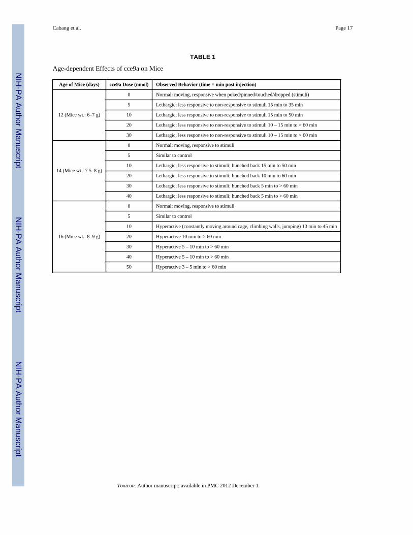
3.4. Developmentally dependent biological activityIntracranial bioassay using synthetic cce9a revealed that the behavioral phenotype elicited inthe mice was age-dependent (Table 1). In younger mice (12 and 14 days old), cce9a inducedlethargy and a delayed response to stimuli at doses of 5 to 40 nmol per mouse (6 – 7 g ofbody weight). Recovery to normal behavior took a longer period as the administered dosewas increased. Control mice exhibited non-lethargic and responsive behavior. In older mice(16 days old), the peptide elicited hyperactivity at doses of 10 to 50 nmol per mouse. The
Cabang et al. Page 6
Toxicon. Author manuscript; available in PMC 2012 December 1.
NIH
-PA Author Manuscript
NIH
-PA Author Manuscript
NIH
-PA Author Manuscript
onset of hyperactivity, which was characterized by climbing, running, and jumping, wasmore immediate at the higher injected doses.
4. DiscussionThe present study describes the purification and characterization of the first venomcomponent from a member of a biodiverse group of toxoglossate molluscs, the crassispirids.It is likely that the crassispirids will be comparable in their biodiversity, and perhaps evenexceed the ~700 species of cone snails and ~400 species of terebrids. Thus, this group ofvenomous animals has the potential to yield as many bioactive venom components as thecone snails.
The species that we investigated, Crassispira cerithina, is one of >10,000 species that wereloosely referred to as “turrids”; the phylogeny and taxonomy of the species has had aconfusing history. The species was known earlier as Turridrupa cerithina (Powell, 1966);despite the earlier assignment to the genus Turridrupa (a genus in the subfamily Turrinae),our phylogenetic analysis definitively establishes that Crassispira cerithina does not belongto this subfamily. Instead, as shown in Fig. 2, Crassispira cerithina, together with species inthe genus Inquisitor, as well as the new world genus Zonulospira, comprise a differentmajor branch. These genera were assigned to the subfamily Crassispirinae in most priortaxonomic work. Another new world species, Pseudomelatoma moesta, is more distantlyrelated, but appears to be allied to these genera.
It has recently been suggested that the various families conventionally included in thesuperfamily Conoidea fall into two major groups, and that the families less closely related toConus be moved to a new superfamily, Turroidea (Tucker and Tenorio, 2009). Thissuggestion is consistent with the molecular phylogeny shown in Fig. 2; the proposedsuperfamily Turroidea would encompass 4 major branches of the phylogenetic tree shown inFig. 2: the auger snails or terebrids, (family Terebridae), the turrids (family Turridae,narrowly defined), the drillids (family Drillidae) and the crassispirids.
The “crassispirids”, as defined above, include several (but not all) genera assigned to thesubfamily Crassispirinae (i.e., Crassispira, Inquisitor and Zonulospira); this correspondswell to “Clade 2” of Puillandre et al., (2008). It should be noted that the molecularphylogeny in Fig. 2 is somewhat divergent from the recent literature on the conventionalphylogeny of the group (see Bouchet and Rocroi, 2005; Poppe, 2008; Taylor et al., 1993;Tucker and Tenorio, 2009) that generally treat crassispirids as more closely related to theturrines (subfamily Turrinae) than to the drillids (Dillinae/Drillidae). The phylogeneticanalysis that we carried out and prior molecular phylogeny indicate that the converse is true:the sister group of the crassispirids are the drillids (e.g., Drillia, Clavus, etc.). It should benoted that the taxonomy and phylogeny of the crassispirids need further elucidation, sincethere are many species that may be crassispirids, but in the absence of molecular data, theirassignment to this group is uncertain. These two major branches of venomous molluscs, thecrassispirids and the drillids, are unexplored territory for toxinologists, with the presentwork providing the very first characterization of any venom component from these species-rich lineages of venomous gastropods. We propose to call venom peptides from crassispiridsand drillids “crassipeptides” and “drillipeptides”, respectively, to distinguish them from“conopeptides”, “augerpeptides” and “turripeptides” from cone snails, terebrids and turrids,respectively.
The approach that we used to purify and characterize the first crassipeptide was one that hasbeen extraordinarily productive for conopeptides—assessing the behavioral phenotype of amouse after injection of a venom component into the central nervous system. The peptide
Cabang et al. Page 7
Toxicon. Author manuscript; available in PMC 2012 December 1.
NIH
-PA Author Manuscript
NIH
-PA Author Manuscript
NIH
-PA Author Manuscript
we purified caused lethargy in younger mice, with a muted response to external stimuli. Thisbehavioral phenotype allowed the purification and characterization of crassipeptide cce9a, apeptide with 29 amino acid residues and three disulfide bonds.
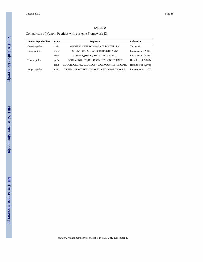
As shown in Table 2, the mature crassipeptide cce9a, whose sequence was determined usingstandard Edman methods on the purified native peptide and confirmed by sequencing acDNA clone (Fig. 4), has a striking similarity to conopeptides in the Conus P-superfamilywith Framework IX cysteine pattern (Lirazan et al., 2000) with respect to the arrangementand spacing of cysteine residues. However, apart from the pattern of cysteine residues, thereis no other sequence similarity between cce9a and P-superfamily Conus peptides.Furthermore, the symptomatology induced by the spasmodic peptides, tx9a from Conustextile and gm9a from Conus gloriamaris, and cce9a from Crassispira cerithina arestrikingly divergent.
Other peptides with the Framework IX cysteine pattern have been purified from Gemmulaspeciosa, which belongs to the family Turridae (ss) (Heralde et al., 2008), and Hastulahectica, which belongs to the family Terebridae (Imperial et al., 2007). The sequences of thepeptides purified from the venom of G. speciosa and H. hectica are compared to theconopeptides and to cce9a in Table 2; it is notable that although in the phylogenetic tree, thecrassispirids are more closely related to turrids and terebrids than to Conus, the peptidesfrom the turrid and terebrid appear to be more divergent from cce9a than are thecorresponding conopeptides, considering the size of the intercysteine regions.
Crassipeptide cce9a was further characterized after the successful chemical synthesis of thebiologically active peptide. As shown in Table 1, the behavioral phenotype elicited by thepeptide is dependent upon the age of the mice injected; the peptide causes a lethargic state inyoung mice (12 and 14 days old), which is refractory to external stimuli; however, in oldermice (16 days old) the peptide elicited hyperactivity, which is characterized by climbing,running and jumping. The switch in behavioral phenotype as a function of age is reminiscentof conopeptides that belong to the conantokin family. The first conantokin characterized,Conantokin G from Conus geographus (Olivera et al., 1985), caused a sleep-like state inyoung mice, but at approximately the same age interval, there was a transition tohyperactivity. Conantokin G is a well-established NMDA receptor antagonist; given theparallel developmental switch in behavioral phenotype, we tested cce9a for activity onNMDA receptors—these assays were negative (results not shown). Thus, the basis for thechange in behavioral phenotype as a function of age is apparently due to a molecularmechanism different from the one for the conantokins; however, it remains possible that thebehavioral shift indicates that cce9a interacts with at least 2 types of molecular target withthe effects of one type predominating over the other differentially with age (Rivier et al.,1987).
Thus, the characterization of the venom component described in this work, Crassipeptidecce9a, is noteworthy in several respects: it is the first biochemical characterization of anytoxin from the venom of a crassispirid species, and it has a novel biological activity inmammals: the behavioral symptomatology elicited by intracranial injections into mice isdevelopmentally dependent. Thus, it is reasonable to expect that many crassipeptides withnovel pharmacology will be discovered and characterized that should prove useful forinvestigating molecular mechanisms in nervous systems.
Supplementary MaterialRefer to Web version on PubMed Central for supplementary material.
Cabang et al. Page 8
Toxicon. Author manuscript; available in PMC 2012 December 1.
NIH
-PA Author Manuscript
NIH
-PA Author Manuscript
NIH
-PA Author Manuscript
AcknowledgmentsThis work was carried out through the Philippine PharmaSeas Drug Discovery Program, supported by thePhilippine Council for Aquatic and Marine Research and Development, Department of Science and Technology(DOST), Philippines. This work was supported in part by a Program Project grant from the National Institutes ofHealth, GM 48677. The field collection was done in collaboration with a team supported by the Philippine MolluskSymbiont International Cooperative Biodiversity Grant (NIH 1U01TW008163-01) from the Fogarty InternationalCenter, National Institutes of Health. JSI thanks the DOST for visits to the University of the Philippines MarineScience Institute through the Balik Scientist Program. We thank Vernon Twede for carrying out the NMDA assays,and Greg Bulaj and Pradip Bandyopadhyay for advice.
ReferencesAguilar MB, de la Rosa RA, Falcon A, Olivera BM, Heimer de la Cotera EP. Peptide pal9a from the
venom of the turrid snail Polystira albida from the Gulf of Mexico: Purification, characterization,and comparison with P-conotoxin-like (framework IX) conoidean peptides. Peptides. 2009; 30:467–476. [PubMed: 18948154]
Bouchet P, Lozouet P, Maestrati P, Heros V. Assessing the magnitude of species richness in tropicalmarine environments: high numbers of molluscs at a New Caledonia site. Biological Journal of theLinnean Society. 2002; 75:421–436.
Bouchet P, Rocroi JP. Malacologia: International Journal of Malacology, Classification andNomenclator of Gastropod Families. Malacologia - International Journal of Malacology,ConchBooks. 2005
Craig AG, Zafaralla G, Cruz LJ, Santos AD, Hillyard DR, Dykert J, Rivier JE, Gray WR, Imperial J,DelaCruz RG, Sporning A, Terlau H, West PJ, Yoshikami D, Olivera BM. An O-glycosylatedneuroexcitatory Conus peptide. Biochemistry. 1998; 37:16019–16025. [PubMed: 9819194]
Edgar RC. MUSCLE: a multiple sequence alignment method with reduced time and space complexity.BMC Bioinformatics. 2004; 5:113. [PubMed: 15318951]
Folmer O, Black M, Hoeh W, Lutz R, Vrijenhoek R. DNA primers for amplification of mitochondrialcytochrome c oxidase subunit I from diverse metazoan invertebrates. Molecular Biology andBiotechnology. 1994; 3:294 – 299.
Fuller E, Green BR, Catlin P, Buczek O, Nielsen JS, Olivera BM, Bulaj G. Oxidative folding ofconotoxins sharing an identical disulfide bridging framework. Federation of European BiochemicalSocieties Journal. 2005; 272:1727–1738. [PubMed: 15794759]
Guindon S, Gascuel O. A simple, fast, and accurate algorithm to estimate large phylogenies bymaximum likelihood. Systematic Biology. 2003; 52:696–704. [PubMed: 14530136]
Heralde FM 3rd, Imperial J, Bandyopadhyay PK, Olivera BM, Concepcion GP, Santos AD. A rapidlydiverging superfamily of peptide toxins in venomous Gemmula species. Toxicon. 2008; 51:890–897. [PubMed: 18272193]
Huelsenbeck JP, Ronquist F, Nielsen R, Bollback JP. Bayesian inference of phylogeny and its impacton evolutionary biology. Science. 2001; 294:2310–2314. [PubMed: 11743192]
Imperial JS, Chen P, Sporning A, Terlau H, Daly NL, Craik DJ, Alewood PF, Olivera BM. Tyrosine-rich conopeptides affect voltage-gated K+ channels. J Biol Chem. 2008; 283:23026–23032.[PubMed: 18505731]
Imperial JS, Kantor Y, Watkins M, Heralde FM 3rd, Stevenson B, Chen P, Hansson K, Stenflo J,Ownby JP, Bouchet P, Olivera BM. Venomous auger snail Hastula (Impages) hectica (Linnaeus,1758): molecular phylogeny, foregut anatomy and comparative toxinology. Journal ofExperimental Zoology Part B: Molecular and Developmental Evolution. 2007; 308:744–756.
Lirazan MB, Hooper D, Corpuz GP, Ramilo CA, Bandyopadhyay P, Cruz LJ, Olivera BM. Thespasmodic peptide defines a new conotoxin superfamily. Biochemistry. 2000; 39:1583–1588.[PubMed: 10677206]
López-Vera E, Heimer de la Cotera EP, Maillo M, Riesgo-Escovar JR, Olivera BM, Aguilar MB. Anovel structural class of toxins: the methionine-rich peptides from the venoms of turrid marinesnails (Mollusca, Conoidea). Toxicon. 2004; 43:365–374. [PubMed: 15051399]
Maddison, DR.; Maddison, WP. MacClade 4.08. 2005.
Cabang et al. Page 9
Toxicon. Author manuscript; available in PMC 2012 December 1.
NIH
-PA Author Manuscript
NIH
-PA Author Manuscript
NIH
-PA Author Manuscript
McIntosh JM, Yoshikami D, Mahe E, Nielsen DB, Rivier JE, Gray WR, Olivera BM. A nicotinicacetylcholine receptor ligand of unique specificity, a-conotoxin ImI. Journal of BiologicalChemistry. 1994; 269:16733–16739. [PubMed: 8206995]
Olivera BM, Cruz LJ, de Santos V, LeCheminant G, Griffin D, Zeikus R, McIntosh JM, Galyean R,Varga J, Gray WR, Rivier J. Neuronal Ca channel antagonists. Discrimination between Ca channelsubtypes using ε-conotoxin from Conus magus venom. Biochemistry. 1987; 26:2086–2090.[PubMed: 2441741]
Olivera BM, McIntosh JM, Clark C, Middlemas D, Gray WR, Cruz LJ. A sleep-inducing peptide fromConus geographus venom. Toxicon. 1985; 23:277–282. [PubMed: 4024137]
Olivera BM, Rivier J, Clark C, Ramilo CA, Corpuz GP, Abogadie FC, Mena EE, Woodward SR,Hillyard DR, Cruz LJ. Diversity of Conus neuropeptides. Science. 1990; 249:257–263. [PubMed:2165278]
Palumbi, SR. PCR. In: IM, et al., editors. Molecular Systematics. Sinauer Press; Sunderland, MA:1996.
Poppe, GT. Philippine Marine Mollusks. In: Poppe, GT., editor. Gastropoda. Vol. Part 2. ConchBooks;2008. p. 848
Powell AWB. The molluscan families Speightiidae and Turridae. Bulletin of the Auckland Instituteand Museum. 1966; 5:1–184. + 23 plates.
Powell AWB. The family Turridae in the Indo-Pacific. Part 1a. The subfamily Turrinae concluded.Indo-Pacific Moll. 1967; 1:409–432.
Puillandre N, Samadi S, Boisselier MC, Sysoev AV, Kantor YI, Cruaud C, Couloux A, Bouchet P.Starting to unravel the toxoglossan knot: molecular phylogeny of the “turrids” (Neogastropoda:Conoidea). Molecular Phylogenetic Evolution. 2008; 47:1122–1134.
Rivier J, Galyean R, Simon L, Cruz LJ, Olivera BM, Cruz LJ. Total synthesis and furthercharacterization of the γ-carboxyglutamate-containing “sleeper” peptide from Conus geographusvenom. Biochemistry. 1987; 26:8508–8512. [PubMed: 3442672]
Ronquist F, Huelsenbeck JP. MrBayes 3: Bayesian phylogenetic inference under mixed models.Bioinformatics. 2003; 19:1572–1574. [PubMed: 12912839]
Simon, C. Molecular systematics at the species boundary: exploiting conserved and variable regions ofthe mitochondrial genome of animals via direct sequencing from amplified DNA. In: Hewitt, GM.;Johnston, AWB.; Young, JPW., editors. Molecular techniques in taxonomy, NATO ASI Series.Vol. H57. 1991. p. 33-71.
Stamatakis A, Hoover P, Rougemont J. A rapid bootstrap algorithm for the RAxML Web servers. SystBiol. 2008; 57:758–71. [PubMed: 18853362]
Tavaré S. Some Probablsistic and Statistical Problems in the Analysis of DNA Sequences. AmericanMathematical Society: Lectures on Mathematics in the Lif Sciences. 1986; 17:57–86.
Taylor JD, Kantor Y, Sysoev AV. Foregut anatomy, feeding mechanisms, relationships andclassification of the Conoidea (=Toxoglossa) (Gastropoda). Bulletin, Natural History Museum,London Zoology. 1993; 59:125–170.
Teichert RW, Jimenez EC, Twede V, Watkins M, Hollmann M, Bulaj G, Olivera BM. Novelconantokins from Conus parius venom are specific antagonists of N-methyl-D-aspartate receptors.J Biol Chem. 2007; 282:36905–36913. [PubMed: 17962189]
Teichert RW, Olivera BM. Natural products and ion channel pharmacology. Future Med Chem. 2010;2(5):731–744. [PubMed: 21426200]
Terlau H, Olivera BM. Conus venoms: a rich source of novel ion channel-targeted peptides.Physiological Reviews. 2004; 84:41–68. [PubMed: 14715910]
Tucker, JK.; Tenorio, MJ. Systematic classification of Recent and fossil conoidean gastropods.Conchbooks; 2009.
Cabang et al. Page 10
Toxicon. Author manuscript; available in PMC 2012 December 1.
NIH
-PA Author Manuscript
NIH
-PA Author Manuscript
NIH
-PA Author Manuscript
Highlights
• Crassispira cerithina belongs to a new major clade, the “crassispirines”.
• Crassipeptide cce9a is 29 amino acids long and has the Framework IX cysteinepattern.
• Crassipeptide cce9a is active by intracranial injection in mice.
Cabang et al. Page 11
Toxicon. Author manuscript; available in PMC 2012 December 1.
NIH
-PA Author Manuscript
NIH
-PA Author Manuscript
NIH
-PA Author Manuscript
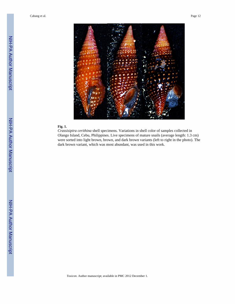
Fig. 1.Crassisipira cerithina shell specimens. Variations in shell color of samples collected inOlango Island, Cebu, Philippines. Live specimens of mature snails (average length: 1.3 cm)were sorted into light brown, brown, and dark brown variants (left to right in the photo). Thedark brown variant, which was most abundant, was used in this work.
Cabang et al. Page 12
Toxicon. Author manuscript; available in PMC 2012 December 1.
NIH
-PA Author Manuscript
NIH
-PA Author Manuscript
NIH
-PA Author Manuscript
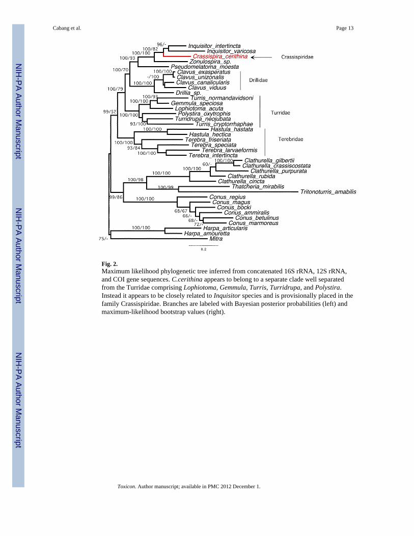
Fig. 2.Maximum likelihood phylogenetic tree inferred from concatenated 16S rRNA, 12S rRNA,and COI gene sequences. C.cerithina appears to belong to a separate clade well separatedfrom the Turridae comprising Lophiotoma, Gemmula, Turris, Turridrupa, and Polystira.Instead it appears to be closely related to Inquisitor species and is provisionally placed in thefamily Crassispiridae. Branches are labeled with Bayesian posterior probabilities (left) andmaximum-likelihood bootstrap values (right).
Cabang et al. Page 13
Toxicon. Author manuscript; available in PMC 2012 December 1.
NIH
-PA Author Manuscript
NIH
-PA Author Manuscript
NIH
-PA Author Manuscript
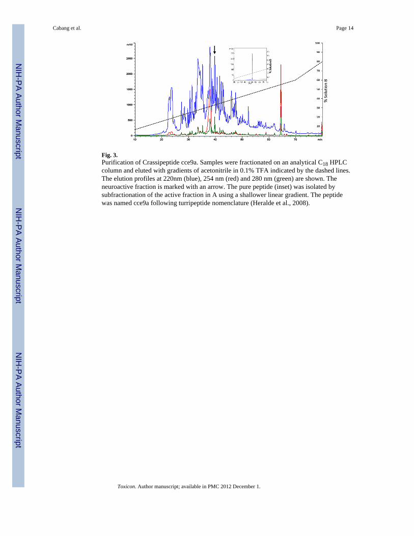
Fig. 3.Purification of Crassipeptide cce9a. Samples were fractionated on an analytical C18 HPLCcolumn and eluted with gradients of acetonitrile in 0.1% TFA indicated by the dashed lines.The elution profiles at 220nm (blue), 254 nm (red) and 280 nm (green) are shown. Theneuroactive fraction is marked with an arrow. The pure peptide (inset) was isolated bysubfractionation of the active fraction in A using a shallower linear gradient. The peptidewas named cce9a following turripeptide nomenclature (Heralde et al., 2008).
Cabang et al. Page 14
Toxicon. Author manuscript; available in PMC 2012 December 1.
NIH
-PA Author Manuscript
NIH
-PA Author Manuscript
NIH
-PA Author Manuscript
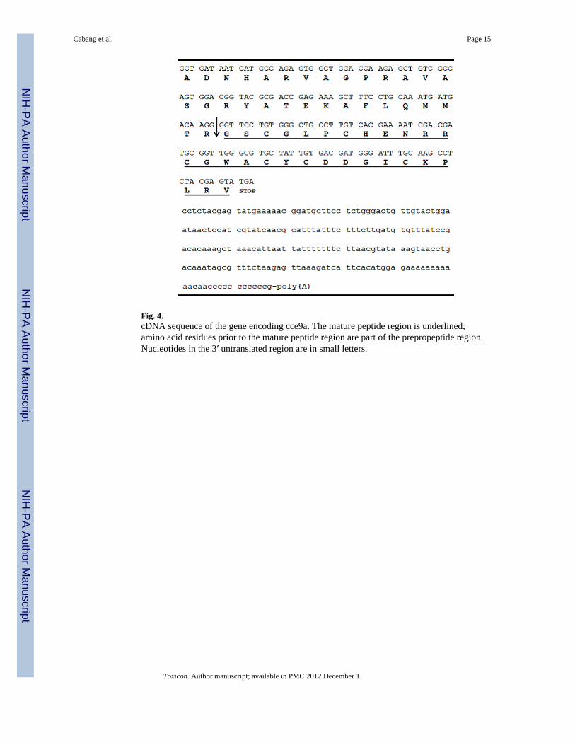
Fig. 4.cDNA sequence of the gene encoding cce9a. The mature peptide region is underlined;amino acid residues prior to the mature peptide region are part of the prepropeptide region.Nucleotides in the 3′ untranslated region are in small letters.
Cabang et al. Page 15
Toxicon. Author manuscript; available in PMC 2012 December 1.
NIH
-PA Author Manuscript
NIH
-PA Author Manuscript
NIH
-PA Author Manuscript
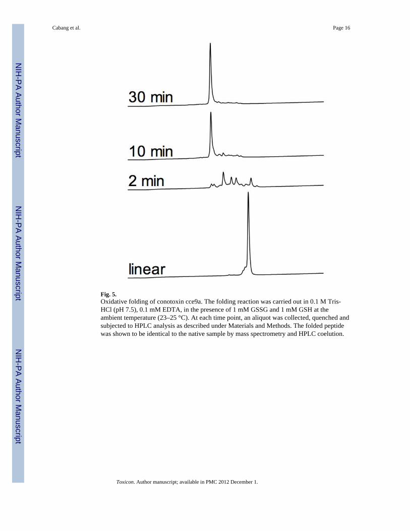
Fig. 5.Oxidative folding of conotoxin cce9a. The folding reaction was carried out in 0.1 M Tris-HCl (pH 7.5), 0.1 mM EDTA, in the presence of 1 mM GSSG and 1 mM GSH at theambient temperature (23–25 °C). At each time point, an aliquot was collected, quenched andsubjected to HPLC analysis as described under Materials and Methods. The folded peptidewas shown to be identical to the native sample by mass spectrometry and HPLC coelution.
Cabang et al. Page 16
Toxicon. Author manuscript; available in PMC 2012 December 1.
NIH
-PA Author Manuscript
NIH
-PA Author Manuscript
NIH
-PA Author Manuscript
NIH
-PA Author Manuscript
NIH
-PA Author Manuscript
NIH
-PA Author Manuscript
Cabang et al. Page 17
TABLE 1
Age-dependent Effects of cce9a on Mice
Age of Mice (days) cce9a Dose (nmol) Observed Behavior (time = min post injection)
12 (Mice wt.: 6–7 g)
0 Normal: moving, responsive when poked/pinned/touched/dropped (stimuli)
5 Lethargic; less responsive to non-responsive to stimuli 15 min to 35 min
10 Lethargic; less responsive to non-responsive to stimuli 15 min to 50 min
20 Lethargic; less responsive to non-responsive to stimuli 10 – 15 min to > 60 min
30 Lethargic; less responsive to non-responsive to stimuli 10 – 15 min to > 60 min
14 (Mice wt.: 7.5–8 g)
0 Normal: moving, responsive to stimuli
5 Similar to control
10 Lethargic; less responsive to stimuli; hunched back 15 min to 50 min
20 Lethargic; less responsive to stimuli; hunched back 10 min to 60 min
30 Lethargic; less responsive to stimuli; hunched back 5 min to > 60 min
40 Lethargic; less responsive to stimuli; hunched back 5 min to > 60 min
16 (Mice wt.: 8–9 g)
0 Normal: moving, responsive to stimuli
5 Similar to control
10 Hyperactive (constantly moving around cage, climbing walls, jumping) 10 min to 45 min
20 Hyperactive 10 min to > 60 min
30 Hyperactive 5 – 10 min to > 60 min
40 Hyperactive 5 – 10 min to > 60 min
50 Hyperactive 3 – 5 min to > 60 min
Toxicon. Author manuscript; available in PMC 2012 December 1.
NIH
-PA Author Manuscript
NIH
-PA Author Manuscript
NIH
-PA Author Manuscript
Cabang et al. Page 18
TABLE 2
Comparison of Venom Peptides with cysteine Framework IX
Venom Peptide Class Name Sequence Reference
Crassipeptides: cce9a GSCGLPCHENRRCGWACYCDDGICKPLRV This work
Conopeptides: gm9a -SCNNSCQSHSDCASHCICTFRGCGAVN* Lirazan et al. (2000)
tx9a GCNNSCQγHSDCγ SHCICTFRGCGAVN* Lirazan et al. (2000)
Turripeptides: gsp9a IDOORYCNHIICYγDSγ CSQWCTAGCNSITSKCDT Heralde et al. (2008)
gsp9b GDOORFCRDKLCSGDGDCSV WCTAGCNHDMGKCDTL Heralde et al. (2008)
Augerpeptides: hhe9a YEENCGTEYCTSKIGCPGRCVCKEYNYNGEITRRCRA Imperial et al. (2007)
Toxicon. Author manuscript; available in PMC 2012 December 1.