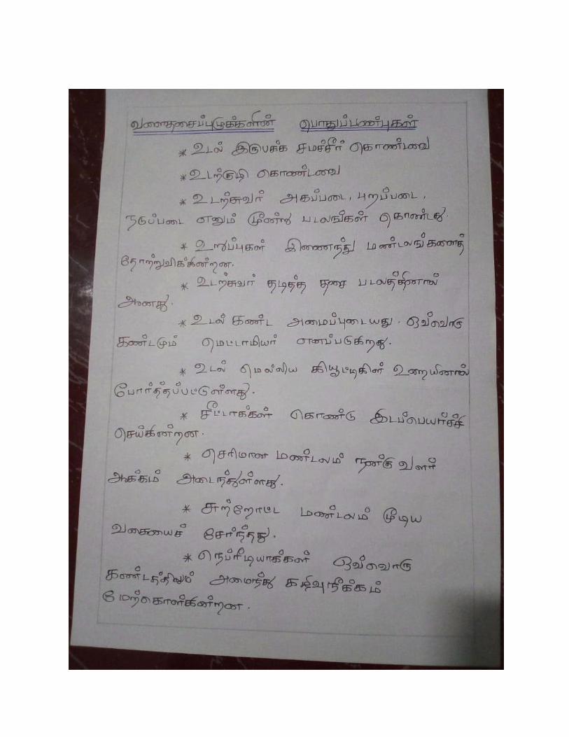
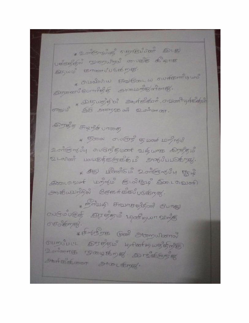
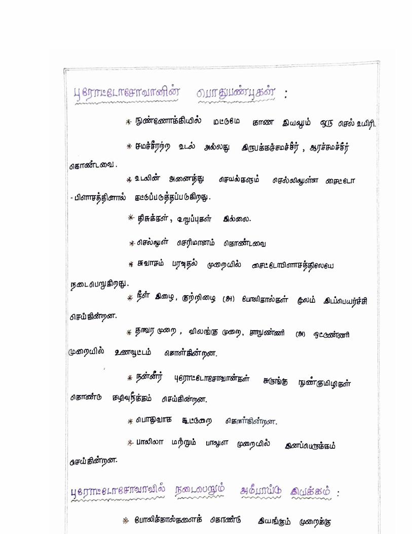
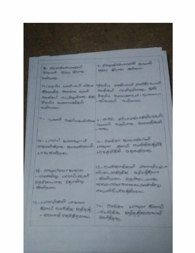
characteristics of invertebrates introduction
TRANSCRIPT
I B.Sc ZOOLOGY
PAPER : INVERTEBRATA Sub Code: 18K1Z01
UNIT-I
Characteristics of Invertebrates
Introduction
• More than 90% of the animals are invertebrates among the estimated 15-30 million
animal species.
• Invertebrates exist about anywhere.
• All invertebrates do not have a spinal cord or vertebral column, instead, most of them
possess an exoskeleton that encompasses the entire body.
• Normally, these are tiny and don’t grow very large.
• Do not possess lungs since they respire through their skin.
• Since they cannot produce their own food, Invertebrates are heterotrophic.
• Reproduction occurs through fission of gametes.
Habitat
• They are found in seas, freshwater, air, land from snow to desert.
• 80% are found in terrestrial habitat.
• Most successful invertebrates of the lands are Arthropods.
• Protozoans are free-living, parasites or commensals.
• Sponges and Coelenterates are aquatic animals.
Shape
• Varied shape.
• Amoeba are irregular every changing bodies.
• Sponges and Coelenterates are plant-like.
• Flatworms are leaf-like and ribbon-shaped.
• Annelids, Nemerteans, and Nematodes are vermiform.
• Starfishes are star-shaped.
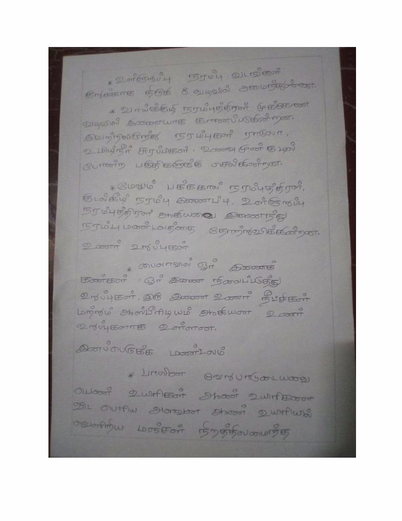
Size
• Great variation in size.
• Ranges from microscopic protozoans to large-sized cephalopods.
• Malaria parasites (Plasmodium) are the smallest one (one-fifth of the human
RBC) while the largest one is giant squids, Architeuthis with a body length of 16.5 meters
including tentacles.
Symmetry
The trait that is common to all invertebrates is the absence of a vertebral column (backbone): this
creates a distinction between invertebrates and vertebrates.
Symmetry.
• All types of symmetry.
• Protozoans are bilateral or radial or asymmetrical.
• Sponges are asymmetrical or radially symmetrical.
• Coelenterates are radially symmetrical.
• Ctenophores are biradial symmetry.
• Some are spherical symmetry (Heliozoan and Radiolaria).
Grades of organization
• All grades of the organization.
• Protoplasmic grade- Protozoa.
• Cellular grade- Sponges.
• Cell-tissue grade- Coelenterates.
• Tissue-organ grade- Flatworms.
Germ Layers
• Absent in Protozoans.
• Some are Diploblastic (derived from 2 germ layers), while others are triploblastic (3 germ
layers).
• Diploblastic- Sponges, Coelenterates.
• Triploblastic- Other Invertebrates than Sponges & Coelenterates.
Simple Integuments
• Body covering is simple.
• Protozoa- Plasma membrane
• Other posses an outer protective layer called the epidermis.
• Some have non-cellular cuticle or chitinous covering secreted by the epidermis.
Locomotion
• Sessile- Sponges, Corals.
• Pseudopodia, Cilia, Flagella- Protozoans.
• Tentacular movements- Coelenterates, Molluscs.
• Setae, Parapodia, Suckers- Annelids.
• Jointed Legs- Arthropods.
• Arms- Echinoderms.
Segmentation
• Flatworms exhibit pseudo-segmentation.
• True segmentation is found in Annelida and Arthropoda.
Living endoskeleton
• Do not possess a rigid internal skeleton.
• Some like arthropods and molluscs possess hard exoskeleton for supporting and
protecting the body.
Coelom
• Sponges and Coelenterates- Body is a double-layered sac surrounding a single cavity
(Acoelomate- No Coelom).
•
• Pseudocoelom- possess a cavity in between body wall and the gut (Nematodes).
• Some possess true coelom.
Dorsal Gut
• The alimentary canal is either absent or partially formed or complete.
• If present, lies dorsal to the nerve cord, runs anterior terminal mouth up to the posterior
terminal anus.
• Gill-slits are never formed in the pharyngeal wall.
Digestive System
• Digestion takes place within the cell (intracellular digestion)- Protozoans, Sponges.
• Digestion also takes place outside the cell (extracellular digestion).
• Coelenterates exhibits both intracellular and extracellular digestion.
Circulatory System
• A blood vascular system is well developed.
• Open or lacunar circulatory system- Arthropods, Molluscs.
• Closed circulatory system are also present.
• The heart is dorsal to the gut.
• The hepatic portal system is absent.
Respiratory System
• Protozoans, Sponges, Coelenterates and many worms have a direct diffusion of gases.
• Annelids exchange gases through moist skin.
• Gills are present in higher invertebrates.
• Echinoderms possess branchiae and tube feet for respiration.
• In insects, the tracheal system is adapted for aerial respiration.
Excretory Mechanism
• Direct diffusion through cell membranes- Protozoans, Sponges, Coelenterates.
• Flame cells- Flatworms.
• True nephridia- Annelids and Molluscs.
• Malpighian tubules- Insects.
• Amoeboid Cells- Echinoderms.
Nervous System
• Coelenterates (radially symmetry)- Head is absent, CNS is represented by a ring of nerve-
tissue encircling the body.
• In bilaterally symmetrical invertebrates- CNS is represented by a pair of nerve cord
running along the mid-ventral line of the body.
• In higher invertebrates- head ganglia form the brain.
• Solid nerves, not hollow within.
Sense Organs
• Protozoans- protoplast acts as a receptor.
• Flagellates- stigma or eyespot acts as a photoreceptor.
• Coelenterates- long sensory cells.
• Flatworms- Eyespot, chemoreceptors.
• Annelids- simple eyes.
• Arthropods- compound eyes.
• Arthropods and Molluscs- Statocyst (equilibrium), Tactile receptors, Chemoreceptors.
Reproduction
• Asexual binary fission.
• Sexual reproduction- Coelenterates, Platyhelminthes, Annelids, Crustaceans.
• Fertilization may be internal as well as external.
• Development is direct or indirect.
• Parthenogenesis- Rotifers, Bees.
Cold – Blooded animals
• All invertebrates are cold-blooded.
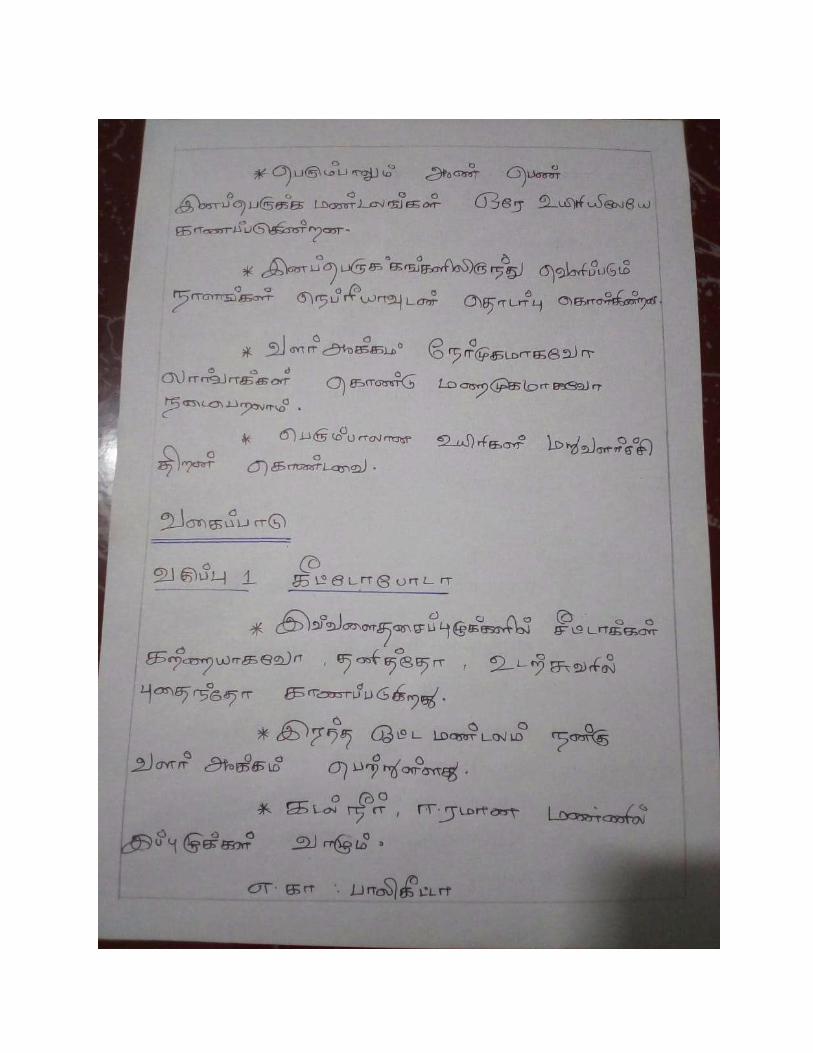
Classification of invertebrates
Invertebrates can be classified into several main categories, some of which
are taxonomically obsolescent or debatable, but still used as terms of convenience. Each however
appears in its own article at the following links
The most familiar invertebrates include the
• Protozoa,
• Porifera,
• Coelenterata,
• Platyhelminthes,
• Nematoda
• Annelida,
• Arthropoda.( Arthropoda include insects, crustaceans and arachnids)
• Mollusca
• Echinodermata,
Why is a Coelom important?
• A coelom allows compartmentalization to separate biological systems that carry out
differing major functions.
• For instance, animals without coelom (acoelomates) would have to rely on diffusion
to transport nutrients around the body.
• Coelom also allows organs to grow and change in position or shape.
• The coelom is the main body cavity in most animals and is positioned inside the
body to surround and contain the digestive tract and other organs.
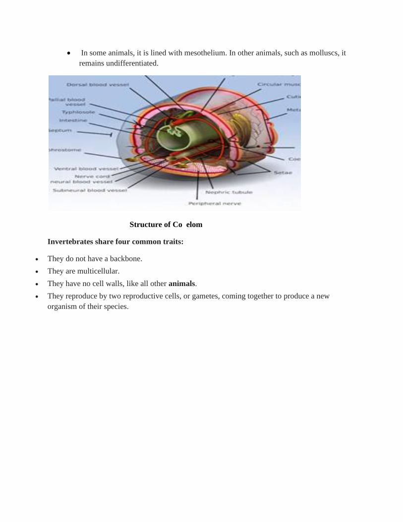
• In some animals, it is lined with mesothelium. In other animals, such as molluscs, it
remains undifferentiated.
Structure of Co elom
Invertebrates share four common traits:
• They do not have a backbone.
• They are multicellular.
• They have no cell walls, like all other animals.
• They reproduce by two reproductive cells, or gametes, coming together to produce a new
organism of their species.
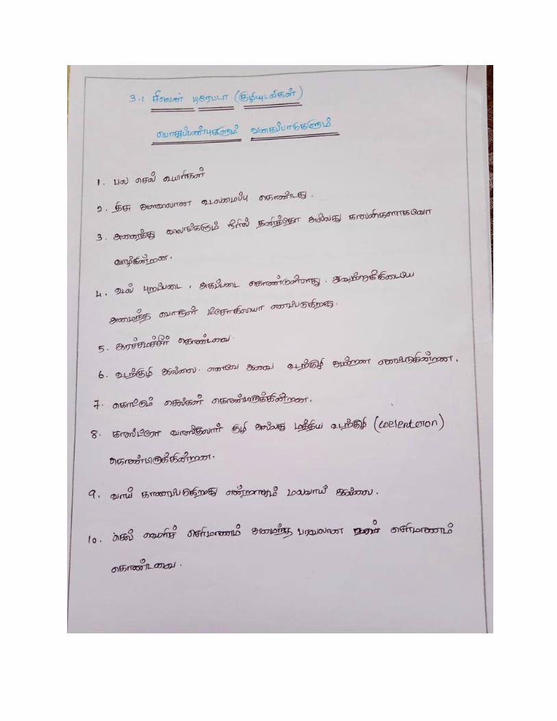
Generl Charateristics of Protozoa
(Gr., protos = first; zoon = animal)
General Characteristics
1. Microscopic and acellular animals
2. Solitary or colonial
3. Body naked or covered by pellicle and provided with internal skeleton
4. Great variety of shape : oval, spherical elongated and some are flattened
5. Cytoplasm : outer ectoplasm and inner endoplasm
6. Locomotary organs : Pseudopodia (in Rhizopoda), Flagella ( in Flagellata), Cilia (in
Ciliata), and absent in Sporozoa
7. Nutrition : Holophytic (plant-like), Holozoic (animal like), Saprozoic or Parasitic
8. Respiration : Diffusion by general body surface
9. Excretion through general body surface
10. Reproduction : Sexual (copulaion/syngamy and conjugation) , Asexual ( Fission, Budding,
Plasmotomy)
11. Lifecycle exhibits alternation of generation
12. No physiological division of Labour
Structure of Plasmodium
Taxonomy.
➢ Plasmodium belongs to the phylum Apicomplexa, a taxonomic group of single-celled
parasites with characteristic secretory organelles at one end of the cell.
➢ Within Apicomplexa, Plasmodium is within the order Haemosporida, a group that
includes all apicomplexans that live within blood cells.
Pathology of Plasmodium
➢ Malaria continues to be the most important tropical disease affecting humans. The
condition is caused by protozoa of the genus Plasmodium. Infection is transmitted to
humans by the female anopheline mosquito.
➢ The genus Plasmodium includes > 170 different species that infect mammals, reptiles,
birds, and amphibians.
➢ Malaria, serious relapsing infection in humans, characterized by periodic attacks of chills
and fever, anemia, splenomegaly (enlargement of the spleen), and often fatal
complications.
➢ It is caused by one-celled parasites of the genus Plasmodium that are transmitted to
humans by the bite of Anopheles mosquitoes
➢ Clinical manifestations of Plasmodium falciparum infection are induced by the asexual
stages of the parasite that develop inside red blood cells (RBCs).
➢ Because splenic microcirculatory beds filter out altered RBCs, the spleen can innately
clear subpopulations of infected or uninfected RBC modified during falciparum malaria.
➢ The spleen appears more protective against severe manifestations of malaria in naïve than
in immune subjects.
➢ The spleen-specific pitting function accounts for a large fraction of parasite clearance in
artemisinin-treated patients.
➢ RBC loss contributes to malarial anemia, a clinical form associated with subacute
progression, frequent splenomegaly, and relatively low parasitemia. Stringent splenic
clearance of ring-infected RBCs and uninfected, but parasite-altered, RBCs, may
altogether exacerbate anemia and reduce the risks of severe complications associated
with high parasite loads, such as cerebral malaria.
➢ The age of the patient directly influences the risk of severe manifestations. We
hypothesize that coevolution resulting in increased splenic clearance of P. falciparum–
altered RBCs in children favors the survival of the host and, ultimately, sustained parasite
transmission.
➢ This analysis of the RBC–spleen dynamic interactions during P falciparum infection
reflects both data and hypotheses, and provides a framework on which a more complete
immunologic understanding of malaria pathogenesis may be elaborated.
➢ At the same time she takes a blood meal to nourish her eggs, the
female Anopheles mosquito injects sporozoites into the blood stream of malaria’s next
victim.
➢ The sporozoites are rapidly taken up by the liver cells.
➢ In all species of Plasmodium, these parasites develop to form schizonts (the multinucleate
stage of the cell during asexual reproduction), from which several thousand merozoites
develop.
➢ In Plasmodium vivax and Plasmodium ovale only, a proportion of the liver-stage
parasites (known as hypnozoites) remain dormant in the hepatocytes.
➢ In this stage the parasite can remain dormant for months or several years. These two
species of parasite can therefore initiate a cycle of asexual reproduction causing clinical
symptoms in the absence of a new mosquito bite, giving P. vivax infection the name
relapsing malaria.
➢ When the liver cells rupture, the merozoites are released into the bloodstream where they
rapidly invade the red blood cells. These blood-stage parasites replicate asexually –
rapidly attaining a high parasite burden and destroying each red blood cell they infect,
leading to the clinical symptoms of malaria.
➢ The trigger is as yet unknown, but a small percentage of merozoites, differentiate into
male and female gametocytes, which are taken up by the mosquito in her blood meal. It is
these gametocytes that cause the cycle of transmission to continue back to the mosquito.
➢ Male and female gametocytes fuse within the mosquito forming diploid zygotes, which in
turn become ookinetes.
➢ These ookinetes migrate to the midgut of the insect, pass through the gut wall and form
the oocysts.
➢ Meiotic division of the oocysts occur and sporozoites are formed, which then migrate to
the salivary glands of the female Anopheles mosquito ready to continue the cycle of
transmission back to man.
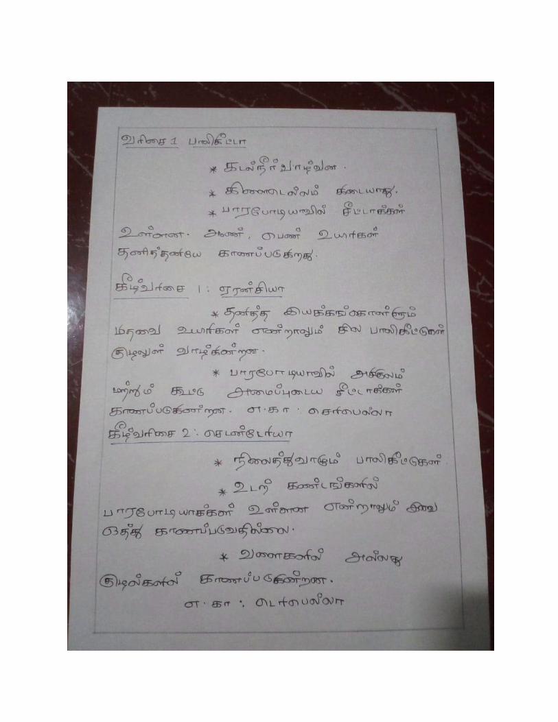
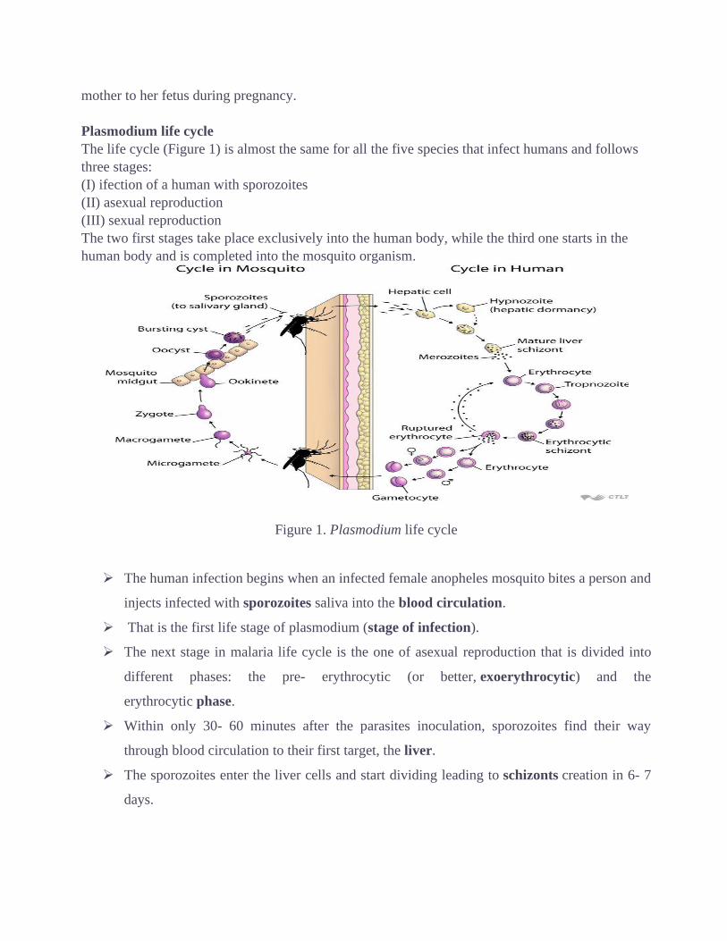
MALARIA
Plasmodium Life cycle
Plasmodium species that infect humans
Until recently, there were four plasmodium species that were considered responsible for malaria
disease in humans: P. vivax, P. falciparum, P. ovale and P. malariae. In 2008, P. knowlesi, a
species that used to infect exclusively apes of the genous Macaque, was recognised by WHO as the
fifth plasmodium species that infect humans.
Transmission routes
The main mode of transmission of the disease is by bites from infected Anopheles mosquitoes that
have previously had a blood meal from an individual with parasitemia. Less common routes of
transmission are via infected blood transfusion, transplantation, infected needles, and from a
mother to her fetus during pregnancy.
Plasmodium life cycle
The life cycle (Figure 1) is almost the same for all the five species that infect humans and follows
three stages:
(I) ifection of a human with sporozoites
(II) asexual reproduction
(III) sexual reproduction
The two first stages take place exclusively into the human body, while the third one starts in the
human body and is completed into the mosquito organism.
Figure 1. Plasmodium life cycle
➢ The human infection begins when an infected female anopheles mosquito bites a person and
injects infected with sporozoites saliva into the blood circulation.
➢ That is the first life stage of plasmodium (stage of infection).
➢ The next stage in malaria life cycle is the one of asexual reproduction that is divided into
different phases: the pre- erythrocytic (or better, exoerythrocytic) and the
erythrocytic phase.
➢ Within only 30- 60 minutes after the parasites inoculation, sporozoites find their way
through blood circulation to their first target, the liver.
➢ The sporozoites enter the liver cells and start dividing leading to schizonts creation in 6- 7
days.
➢ Each schizont gives birth to thousands of merozoites (exoerythrocytic schizogony) that
are then released into the blood stream marking the end of the exoerythrocytic phase of the
asexualreproductivestage.
➢ It is worth mentioning that, concerning P. vivax and P. ovale, sporozoites may not follow
the reproduction step and stay dormant (hypnozoites) in the liver; they may be activated
after a long time leading to relapses entering the blood stream (as merozoites) after weeks,
months or even years.
➢ The exoerythrocytic phase is not pathogenic and does not produce symptoms or signs of the
disease. Its duration is not the same for all parasite species.
Merozoites released into the blood stream, are directed towards their second target, the red
blood cells (RBCs).
➢ As they invade into the cells, they mark the beginning of the erythrocytic phase. The first
stage after invasion is a ring stage that evolves into a trophozoite.
➢ The trophozoites are not able to digest the haem so they convert it in haemozoine and digest
the globin that is used as a source of aminoacids for their reproduction.
➢ The next cellular stage is the erythrocytic schizont (initially immature and then mature
schizont).
➢ Each mature schizont gives birth to new generation merozoites (erythrocytic schizogony)
that, after RBCs rupture, are released in the blood stream in order to invade other RBCs.
This is when parasitaemia occurs and cinical manifestations appear.
➢ The liver phase occurs only once while the erythrocytic phase undergoes multiple cycles;
the merozoites release after each cycle creates the febrile waves.
A second scenario into the RBCs is the parasite differentiation into male and
female gametocytes that is a non pathogenic form of parasite.
➢ When a female anopheles mosquito bites an infected person, it takes up these gametocytes
with the blood meal (mosquitoes can be infected only if they have a meal during the period
that gametocytes circulate in the human’s blood).
➢ The gametocytes, then, mature and become microgametes (male)
and macrogametes (female) during a process known as gametogenesis. The time needed
for the gametocytes to mature differs for each plasmodium species: 3- 4 days for P.
vivax and P. ovale, 6- 8 days for P. malariae and 8- 10 days for P. falciparum.
➢ In the mosquito gut, the microgamete nucleus divides three times producing eight nuclei;
each nucleus fertilizes a macrogamete forming a zygote. The zygote, after the fusion of
nuclei and the fertilization, becomes the so- called ookinete.
➢ The ookinete, then, penetrates the midgut wall of the mosquito, where it encysts into a
formation called oocyst. Inside the oocyst, the ookinete nucleus divides to produce
thousands of sporozoites (sporogony).
➢ That is the end of the third stage (stage of sexual reproduction/ sporogony). Sporogony lasts
8- 15 days.
➢ The oocyst ruptures and the sporozoites are released inside the mosquito cavity and find
their way to its salivary glands but only few hundreds of sporozoites manage to enter.
➢ Thus, when the above mentioned infected mosquito takes a blood meal, it injects its
infected saliva into the next victim marking the beginning of a new cycle
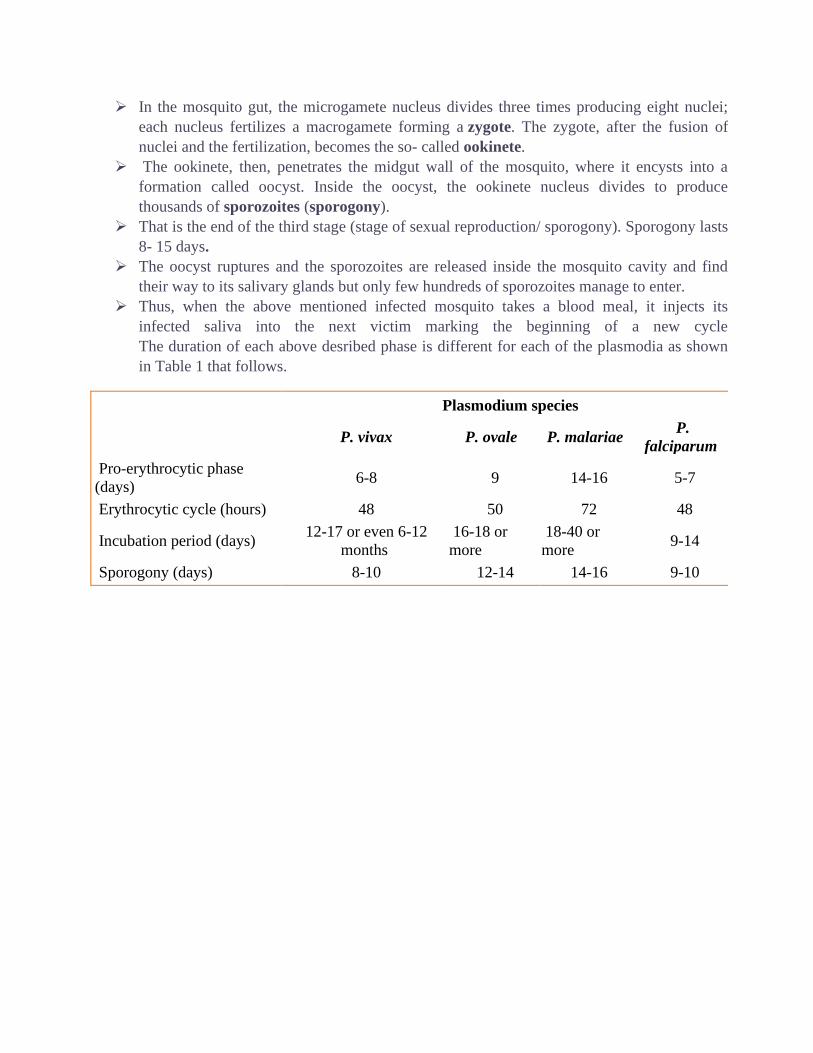
The duration of each above desribed phase is different for each of the plasmodia as shown
in Table 1 that follows.
Plasmodium species
P. vivax P. ovale P. malariae P.
falciparum
Pro-erythrocytic phase
(days) 6-8 9 14-16 5-7
Erythrocytic cycle (hours) 48 50 72 48
Incubation period (days) 12-17 or even 6-12
months
16-18 or
more
18-40 or
more 9-14
Sporogony (days) 8-10 12-14 14-16 9-10
UNIT : II
General Characteristics of Porifera
1. Porifera are all aquatic, mostly marine except one family Spongillidae which lives in
freshwater.
2. They are sessile and sedentary and grow like plants.
3. The body shape is vase or cylinder-like, asymmetrical or radially symmetrical.
4. The body surface is perforated by numerous pores, the Ostia through which water enters
the body and one or more large openings, the oscula by which the water exists.
5. The multicellular organism with the cellular level of body organization. No distinct
tissues or organs.
6. They consist of outer ectoderm and inner endoderm with an intermediate layer of
mesenchyme, therefore, diploblastic
7. The interior space of the body is either hollow or permeated by numerous canals lined
with choanocytes. The interior space of the sponge body is called spongocoel.
8. Characteristic skeleton consisting of either fine flexible spongin fibers, siliceous
spicules or calcareous spicules.
9. Mouth absent, digestion intracellular.
10. Excretory and respiratory organs are absent.
11. Contractile vacuoles are present in some freshwater forms.
12. The nervous and sensory cells are probably not differentiated.
13. The primitive nervous system of neuron arranged in a definite network of bipolar or
multipolar cells in some, but is of doubtful status.
14. The sponges are monoecious.
15. Reproduction occurs by both sexual and asexual methods.
16. Asexual reproduction occurs by buds and gemmules.
17. The sponge possesses a high power of regeneration.
18. Sexual reproduction occurs via ova and sperms.
19. All sponges are hermaphrodite.
20. Fertilization is internal but cross-fertilization can occur.
21. Cleavage holoblastic.
22. Development is indirect through a free-swimming ciliated larva
called amphiblastula or parenchymula.
23. The organization of sponges are grouped into three types which
are ascon type, sycon type, and leuconoid type, due to simple and complex forms.
24. Examples: Clathrina, Sycon, Grantia, Euplectella, Hyalonema, Oscarella, Plakina,
Thenea, Cliona, Halichondria, Cladorhiza, Spongilla, Euspondia, etc.
Ascon Sponge
Introduction
• There are three types of sponge body forms: ascon, sycon, and leucon.
• Ascon sponges are the simplest and least common sponge body from.
• Leucon sponges are the most complex of the sponge body forms and also most common.
• These sponges have multiple dermal pores and can have more than one osculum.
Structure of Ascon sponge
• Ascon sponges These sponges have multiple ostia, this is where water enters the
sponge, and only have one osculum, where water exits the sponge.
• The openings lead to the sponge's one central chamber called a spongocoel that is
lined with choanocytes. They are vaselike in shape.
• Leucosoleniais a small colonial sponge of the ascon type.
• This and othersponges of this type exhibit similar features.
• A system of horizontal tubes that bear numerous upright branches.
• The upright branches represent individual sponges of the colony
• Buds form on the sides of the individual sponges
• The terminal opening, orosculum, at the upper end of each sponge.Water passes out of
the sponge through this opening.
• Thespongocoelis a large central cavity within the sponge. This cavity islined by the
specialized, flagellated collar cells (choanocytes) whichcreate water currents within the
sponge.
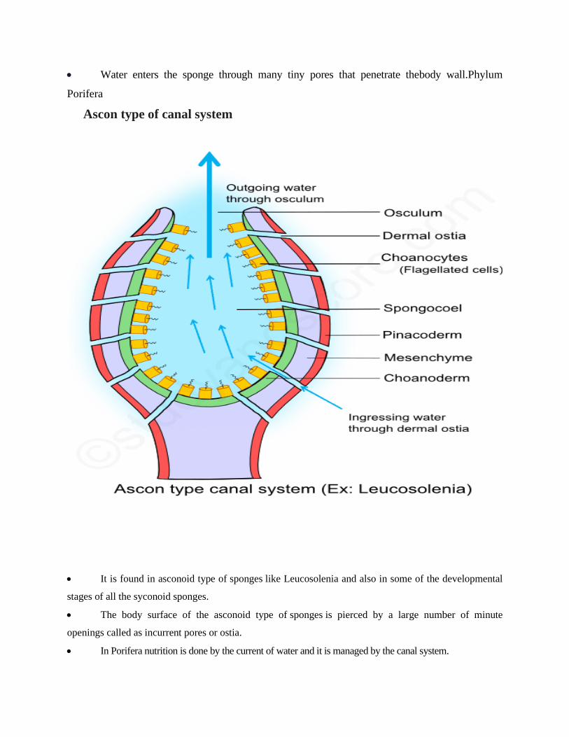
• Water enters the sponge through many tiny pores that penetrate thebody wall.Phylum
Porifera
Ascon type of canal system
• It is found in asconoid type of sponges like Leucosolenia and also in some of the developmental
stages of all the syconoid sponges.
• The body surface of the asconoid type of sponges is pierced by a large number of minute
openings called as incurrent pores or ostia.
• In Porifera nutrition is done by the current of water and it is managed by the canal system.
In porifers, four type of canal system is found
(1) Ascon type- this is the simplest type of canal system in that ostia are presented on the outer surface
of the body and directly flows to spongocoel .that are line by flagellated choanocytes cells and water
goes out through osculum
That type is found in Leucosolonia
(2) Sycon type- It is characteristic of syconoid sponge. in this body, wall is secondary folded to form
in current and radial canals that open into spongocoel by an opening called apopyle and further both
types of canals are interconnected by prosopyle . Ostia is presented on outer surface Ex- Gratia
(3) leucon type - In this type canals further divided into rounded and oval flagellated chambers Ex-
Leucosolonia
(4) Ragon type - It is found in the larval form of spongilla.
FunctionsofCanalSystem:
• The canal system helps the sponges in nutrition, respiration, excretion and reproduction.
The current of water which flows through the canal system brings the food and oxygen
and takes away the carbon dioxide, nitrogenous wastes and faeces.
Role of Canal System:
• The canal system which draws water current inside the sponge’s body and maintains a
continuous uninterrupted flow of water, plays a vital role in the physiology of sponges,
because it serves the various kinds of functions.
(i) Nutrition:
• The sponges are holozoic and bring various kinds of microscopic organisms as food such
as bacteria, diatoms, protozoans and other organic particles with the water current inside
the body. The selected food are digested by choanocytes of the flagellated chambers and
serves the purpose of nutrition.
(ii) Respiration:
A continuous flow of water brings oxygen inside, and exchange of gases takes place between the
dissolved oxygen of the flowing water and the cells of the sponge along the course of water flow.
(iii) Excretion:
The outgoing flow of water current removes the various kinds of metabolic waste materials such as
ammonia, urea, uric acid and other nitrogenous excretory products.
(iv) Reproduction:

The incoming water current brings sperms which are captured by the choanocytes and help in
fertilization.
Evolutionary Significance of Ascon Type Canal System:
1. Simplest type.
2. Hollow and gastrula-like construction.
3. Thin body wall.
4. Central large (spacious) spongocoel lined by a cell layer of choanocytes.
5. Unfolded layer of choanoderm.
6. Canals run straight through the body wall.
7. Straight entry of water into the spongocoel through the tubular porocytes
8. With the increase of the size of spongocoel, the surface area of choanocyte layer is not
increased sufficiently for the movement of water from the spongocoel and water is not pushed
out readily through an apical narrow opening, the osculum.
9. These structures impose very definite size limitations.
10. Thus asconoid sponges become always small vase-like and grow in groups attached to the
rocks in shallow seas.
Characteristic Features of Phylum Coelenterate
• Coelenterates are Metazoa or multicellular animals with tissue grade of organisation. They
are aquatic, mostly marine except few freshwater forms like Hydra.
• They are sedentary or free-swimming and solitary or colonial.
• Individuals are radially or bi-radially symmetrical with a central gastro vascular cavity
communicating to the exterior by the mouth.
• They are multicellular organisms, exhibiting tissue grade of the organisation.
• They are diploblastic, with two layers of cells, an outer layer called the ectoderm and the
inner layer called the endoderm.
• There is a non-cellular layer that is the mesoglea in between the ectoderm and the endoderm.
• They show radial symmetry.
• They have a single opening in the body through which food is taken in and also waste is
expelled out.
• The opening in the body is surrounded by tentacles.
• Digestion takes place in the body cavity which is the coelenteron.
• They can live in marine or freshwater habitats.
• They can be solitary or live in colonies. Each individual is a zooid.
• These organisms show two morphological forms – Polyps and Medusa.
• Polyps contain exoskeleton and endoskeleton.
• The skeletons are composed of calcium carbonate.
• Most if the coelenterates are carnivorous in nature with a few exceptions such as the s corals.
They get their food from other animals that live symbiotically within them.
• Digestion is both intracellular and extracellular.
• Tentacles have special structures known as the nematocysts which help in capturing and
paralyzing prey. Coelenterates simply wave their tentacles and when a prey comes in contact,
the nematocysts inject the toxin that paralyses or kills the prey. Nematocysts are the most
distinguishing feature of this phylum.
• Coelenterates do not have sensory organs.
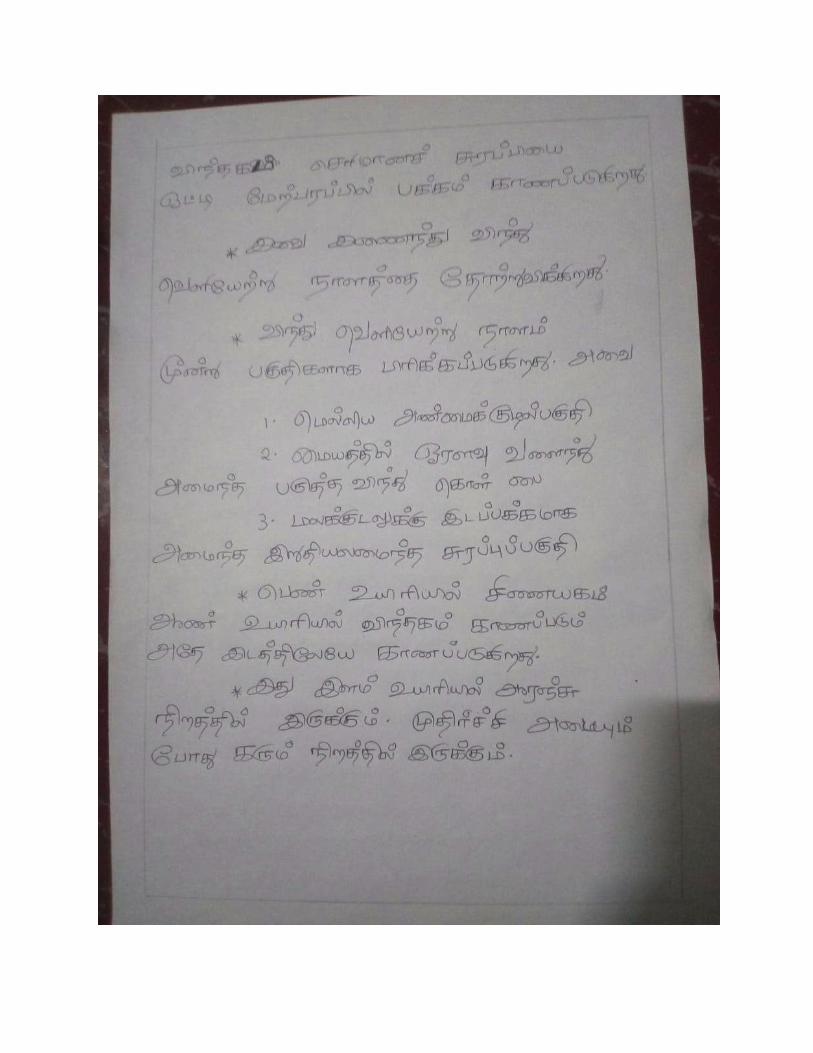
OBELIA GENERAL CHARACTERS
Distribution
It is cosmopolitan in distribution in other words, worldwide distribution except the high -
arctic and Antarctic seas. The medusa stage of Obelia species is common in coastal and
offshore plankton around the world.
Habit and Habitat
Obelia is sedentary, marine and colonial form. It is found up to the depth of 80 meters. It
occurs in both asexual and sexual forms. It grows in intertidal rock pools and at the
extreme low water of spring tides.
External Morphology
Structure of Obelia
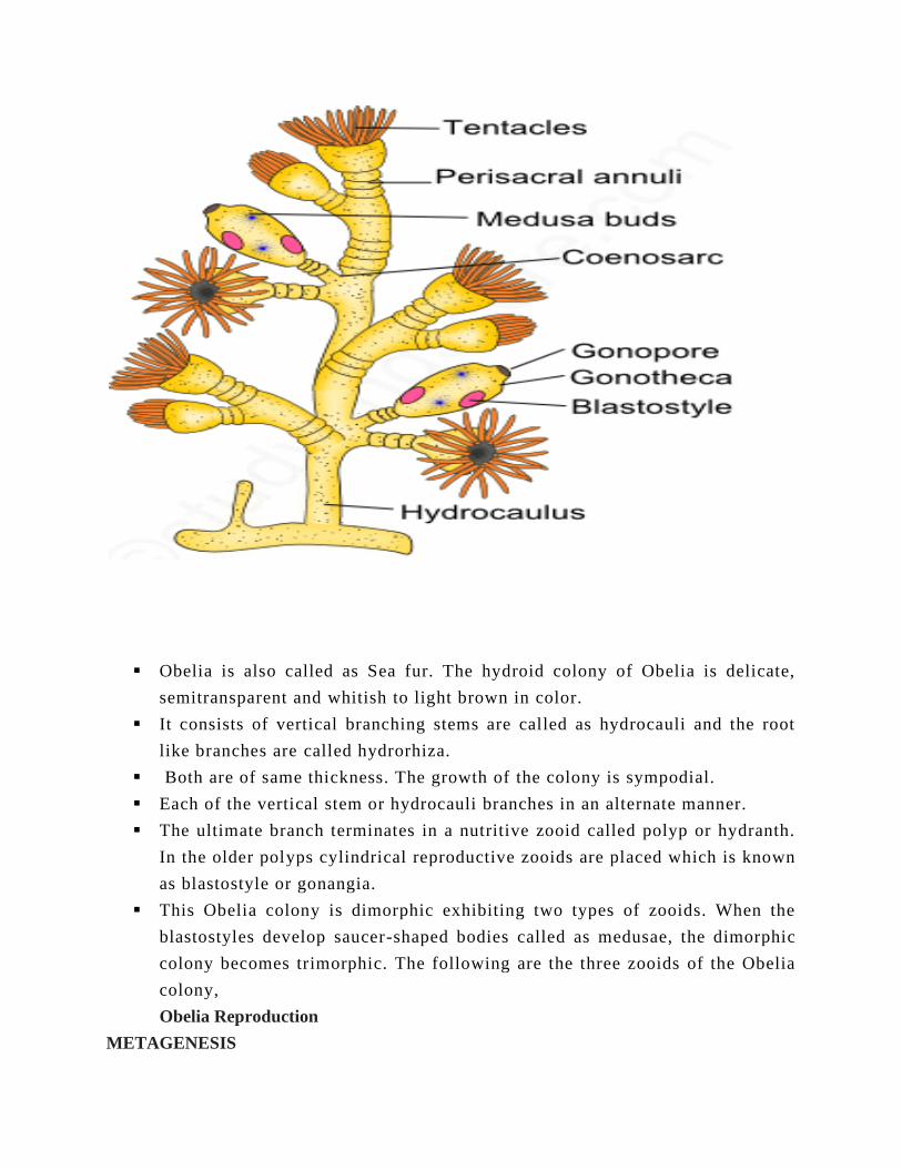
▪ Obelia is also called as Sea fur. The hydroid colony of Obelia is delicate,
semitransparent and whitish to light brown in color.
▪ It consists of vertical branching stems are called as hydrocauli and the root
like branches are called hydrorhiza.
▪ Both are of same thickness. The growth of the colony is sympodial.
▪ Each of the vertical stem or hydrocauli branches in an alternate manner.
▪ The ultimate branch terminates in a nutritive zooid called polyp or hydranth.
In the older polyps cylindrical reproductive zooids are placed which is known
as blastostyle or gonangia.
▪ This Obelia colony is dimorphic exhibiting two types of zooids. When the
blastostyles develop saucer-shaped bodies called as medusae, the dimorphic
colony becomes trimorphic. The following are the three zooids of the Obelia
colony,
Obelia Reproduction
METAGENESIS
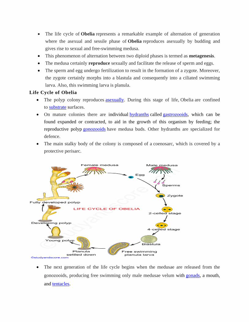
• The life cycle of Obelia represents a remarkable example of alternation of generation
where the asexual and sessile phase of Obelia reproduces asexually by budding and
gives rise to sexual and free-swimming medusa.
• This phenomenon of alternation between two diploid phases is termed as metagenesis.
• The medusa certainly reproduce sexually and facilitate the release of sperm and eggs.
• The sperm and egg undergo fertilization to result in the formation of a zygote. Moreover,
the zygote certainly morphs into a blastula and consequently into a ciliated swimming
larva. Also, this swimming larva is planula.
Life Cycle of Obelia
• The polyp colony reproduces asexually. During this stage of life, Obelia are confined
to substrate surfaces.
• On mature colonies there are individual hydranths called gastrozooids, which can be
found expanded or contracted, to aid in the growth of this organism by feeding; the
reproductive polyp gonozooids have medusa buds. Other hydranths are specialized for
defence.
• The main stalky body of the colony is composed of a coenosarc, which is covered by a
protective perisarc.
• The next generation of the life cycle begins when the medusae are released from the
gonozooids, producing free swimming only male medusae velum with gonads, a mouth,
and tentacles.
• The physical appearance of the male and female medusae velum, including their gonads,
are indistinguishable, and the sex can only be determined by observing the inside of the
gonads, which will either contain sperm or eggs.
• The medusae reproduce sexually, releasing sperm and eggs that fertilize to form a zygote,
which later morphs into a blastula, then a ciliated swimming larva called a planula.
• The planulae are free-swimming for a while but eventually attach themselves to some
solid surface, where they begin their reproductive phase of life.
• Once attached to a substrate, a planula quickly develops into one feeding polyp. As the
polyp grows, it begins developing branches of other feeding individuals, thus forming a
new generation of polyps by asexual budding.
Polymorphism in Hytrozoa
▪ The phenomenon of occurrence of an individual in two or more distinct morphological and
functional forms.
▪ It occurs in the same species of an individual.
▪ It is also known as genetic polymorphism. For example different individuals of a species
may remain separate as represented by various castes in termites, ants and Cuban snails.
▪ Polymorphism is an important feature of phylum coelenteratea.
▪ Each individual member of Coelenterates is known as Zooid and they often units to form a
colony which acts as a single unit (individual).
1. Polyp:
• In Hydrozoa, polyps has a tubular body with a mouth surrounded by tentacles at one end.
Other end is blind and usually attached to pedal disc to the substratum.
• Polyps are generally sessile
• They reproduce asexually
2. Medusa:
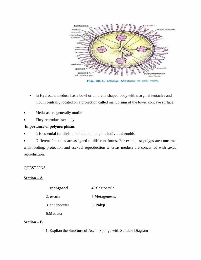
• In Hydrozoa, medusa has a bowl or umbrella shaped body with marginal tentacles and
mouth centrally located on a projection called manubrium of the lower concave surface.
• Medusas are generally motile
• They reproduce sexually
Importance of polymorphism:
• It is essential for division of labor among the individual zooids.
• Different functions are assigned to different forms. For examples; polyps are concerned
with feeding, protection and asexual reproduction whereas medusa are concerned with sexual
reproduction.
QUESTIONS
Section – A
1. spongocoel 4.Blastostyle
2. oscula 5.Metagenesis.
3. choanocytes 6. Polyp
6.Medusa
Section – B
1. Explian the Structure of Ascon Sponge with Suitable Diagram
2. Describe the Structure of Ascon Sponge with Suitable Diagram
3.Write about the Structure of Obelia with Suitable Diagram
4.Write a short note on Medusa
5. What is Metagenesis ? Explain it.
6. Describe Polymorphism in Hytrozoa.
Section – C
1. Explain the General characteristics of Porifera.
2. List out the General characteristics of Coelenterata.
3, Explain the Structural importance of Ascon Sponge
4. Describe the Stucture of Obelia with its Life cycle.

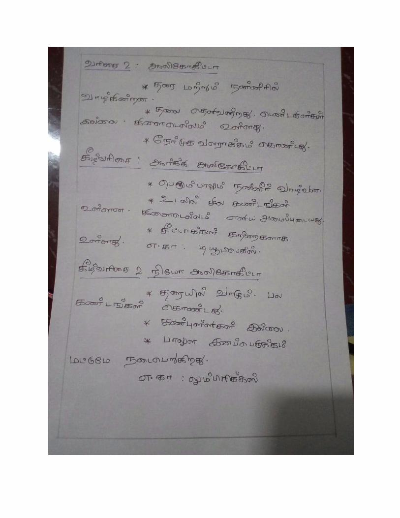
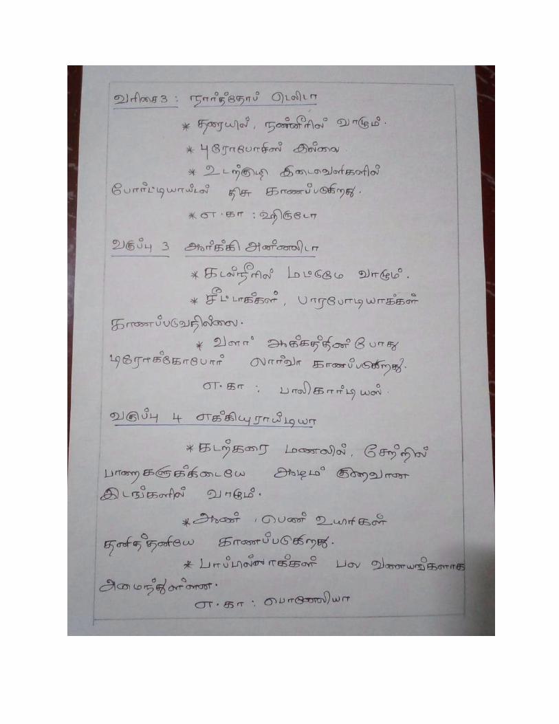
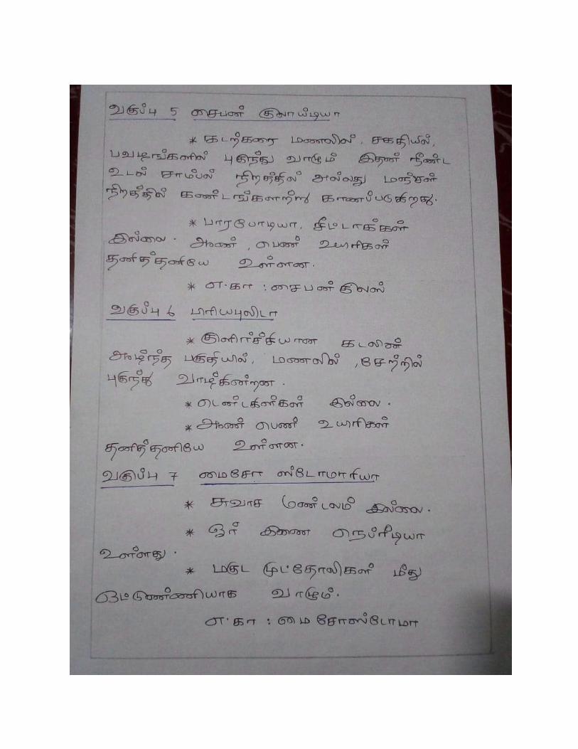
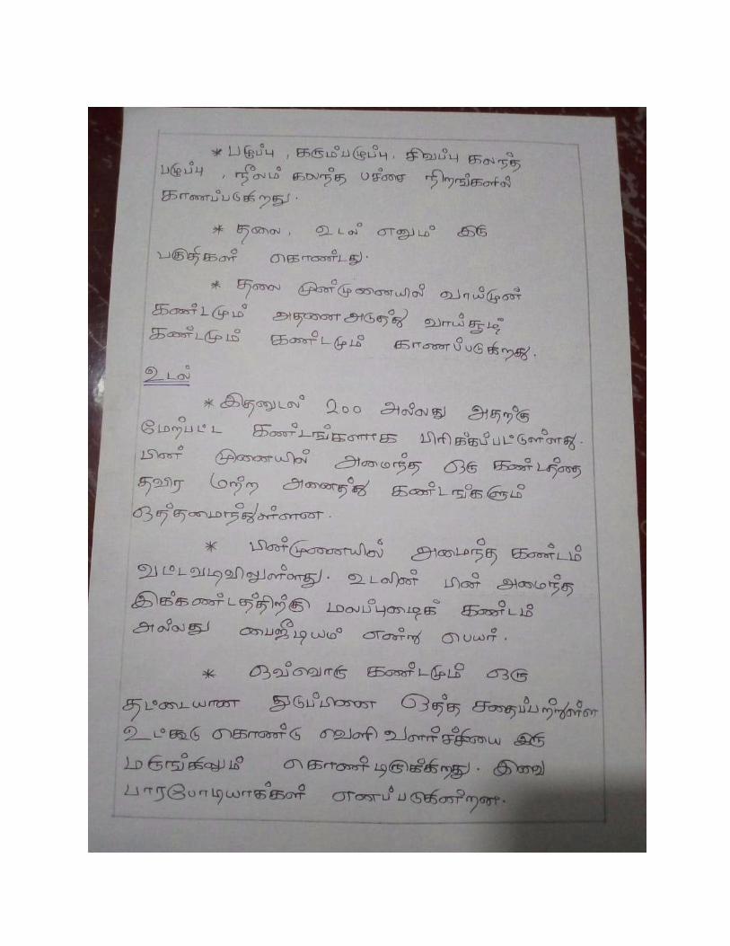
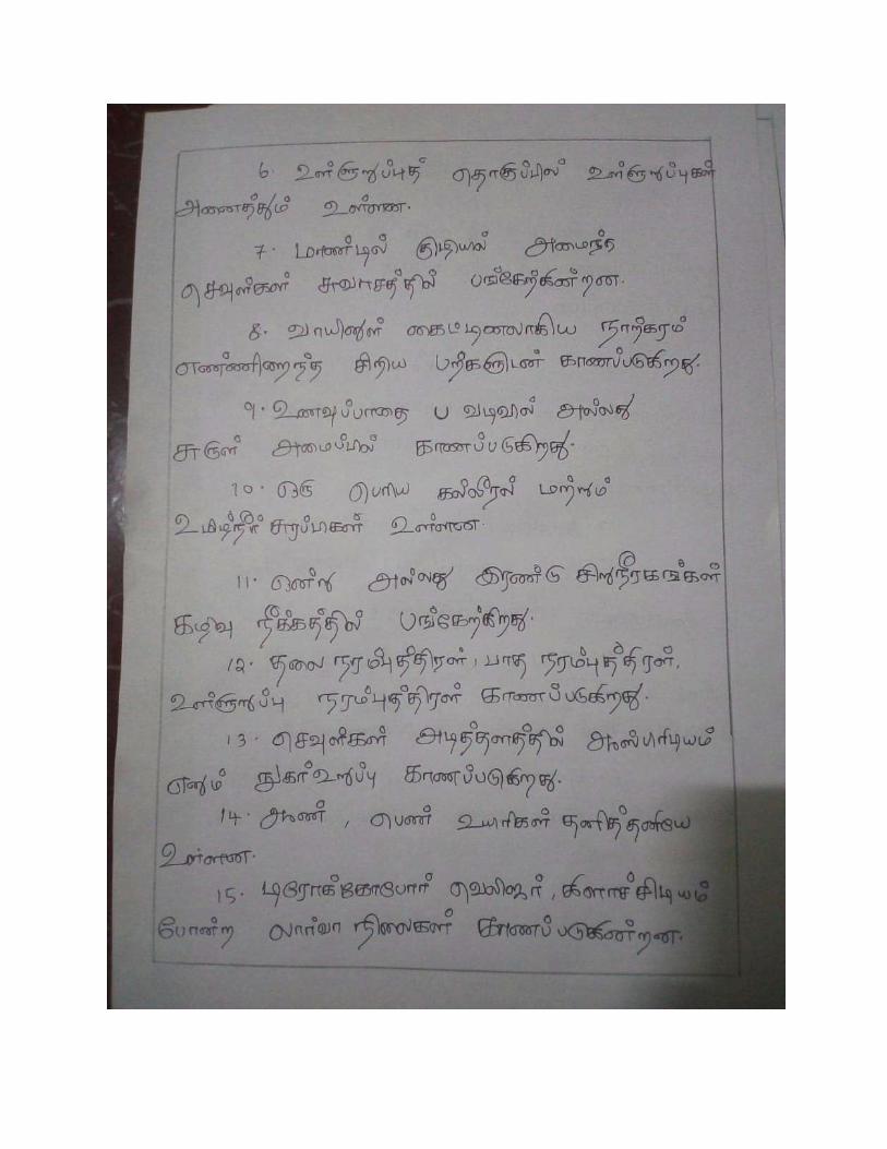
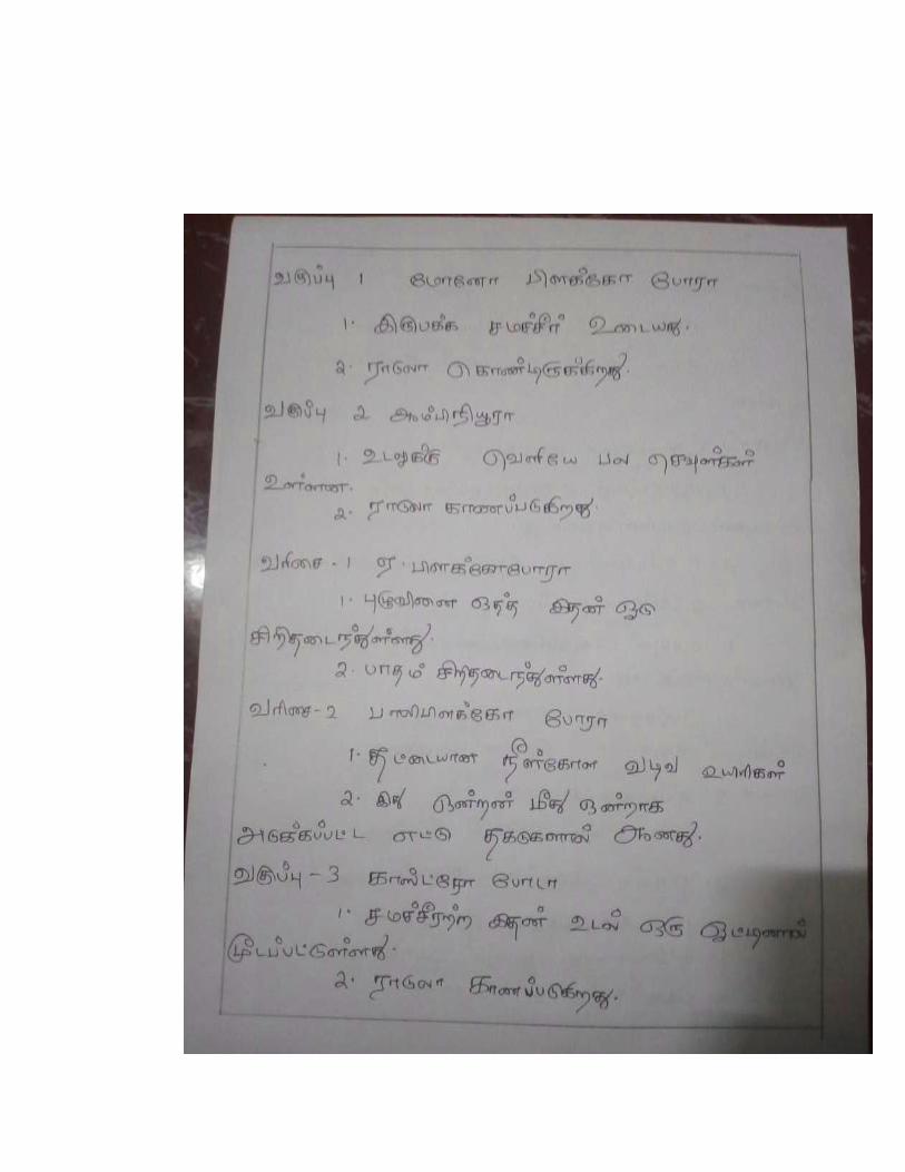
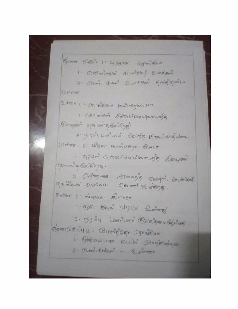
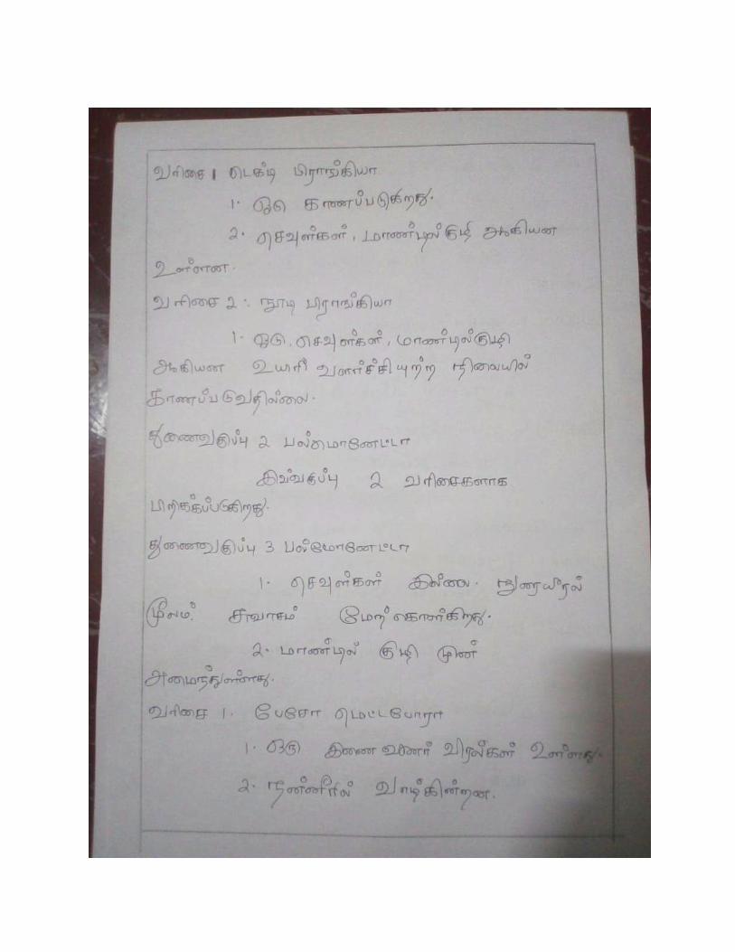
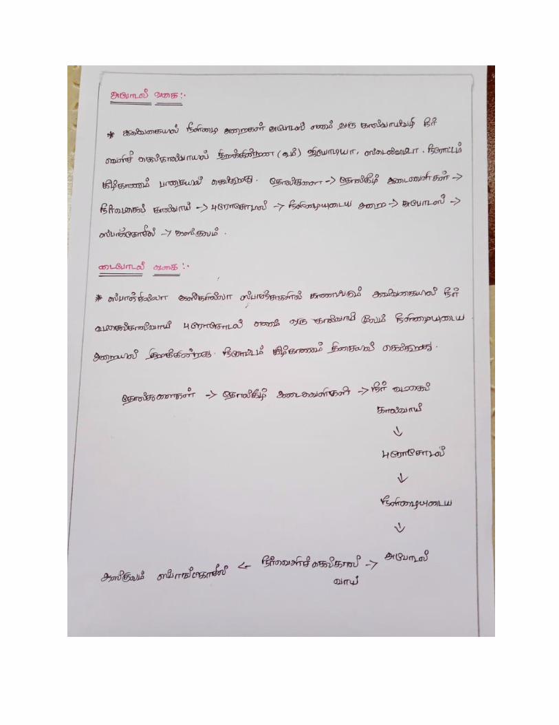
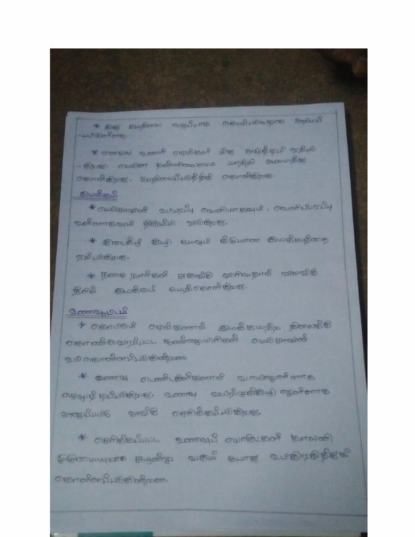
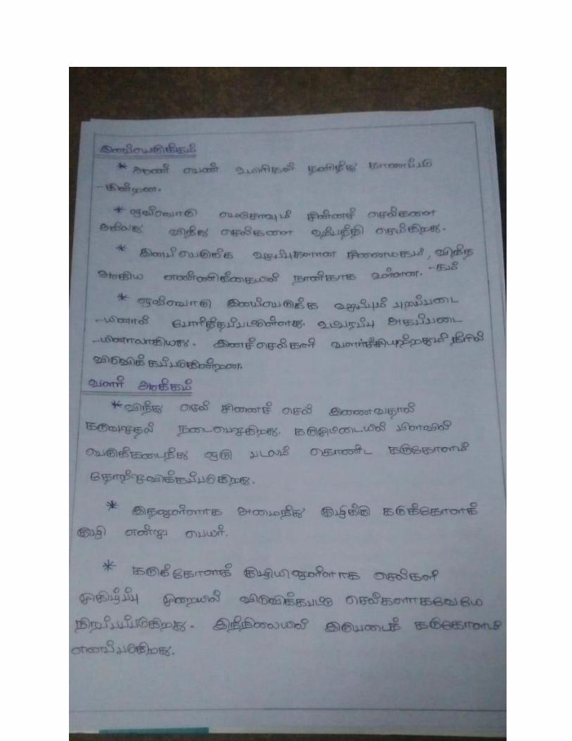
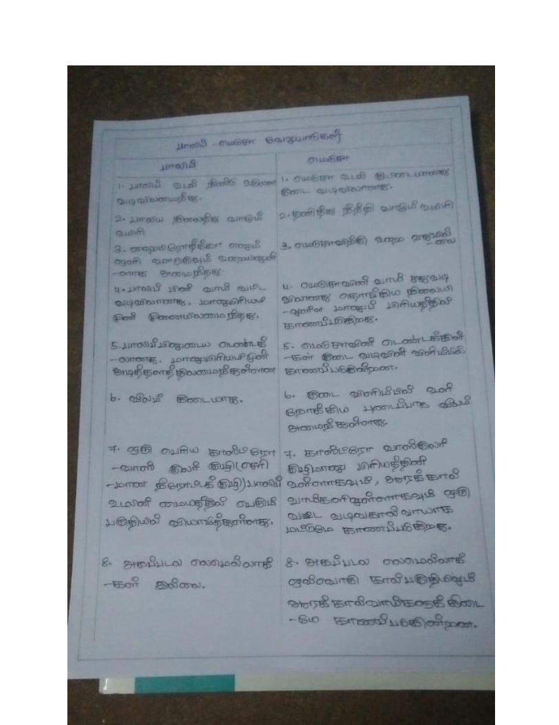
I. B.Sc ZOOLOGY I- INVERTEBRATA
UNIT : I
Subject Code: 18KIZ01 Material in Tamil


UNIT: II


I B.Sc ZOOLOGY
PAPER : INVERTEBRATA Sub Code: 18K1Z01
UNIT-III
UNIT- III
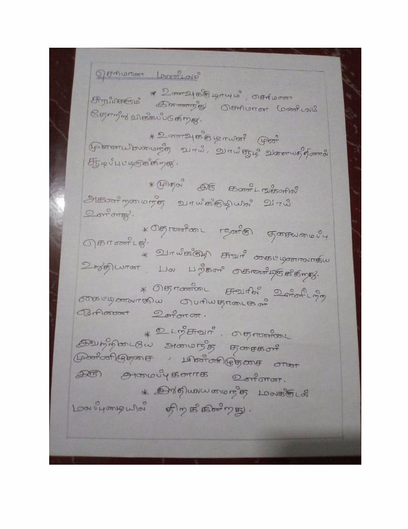
Phylum Platyhelminthes belongs to kingdom Animalia. This phylum includes 13,000
species. The organisms are also known as flatworms. These are acoelomates and they include
many free-living and parasitic life forms.
Members of this phylum range in size from a single-celled organism to around 2-3 feet long.
Characteristics of Platyhelminthes
Platyhelminthes have the following important characteristics:
1. They are triploblastic, acoelomate, and bilaterally symmetrical.
2. They may be free-living or parasites.
3. The body has a soft covering with or without cilia.
4. Their body is dorsoventrally flattened without any segments and appears like a leaf.
5. They are devoid of the anus and circulatory system but has a mouth.
6. They respire by simple diffusion through the body surface.
7. They have an organ system level of organization.
8. They do not have a digestive tract.
9. The space between the body wall and organs is filled with connective tissue parenchyma
which helps in transporting the food material.
10. They are hermaphrodites, i.e., both male and female organs are present in the same body.
11. They reproduce sexually by fusion of gametes and asexually by regeneration by fission
and regeneration. Fertilization is internal.
12. The life cycle is complicated with one or more larval stages.
13. They possess the quality of regeneration.
14. The flame cells help in excretion and osmoregulation.
15. The nervous system comprises the brain and two longitudinal nerve cords arranged in a
ladder-like fashion.
External Features of Taenia Solium:
A fully-developed Taenia Solium may attain a length of 3-5 metres. Its anteroposterior
ends are clearly distinguishable but it is difficult to differentiate the dorsal from the ventral
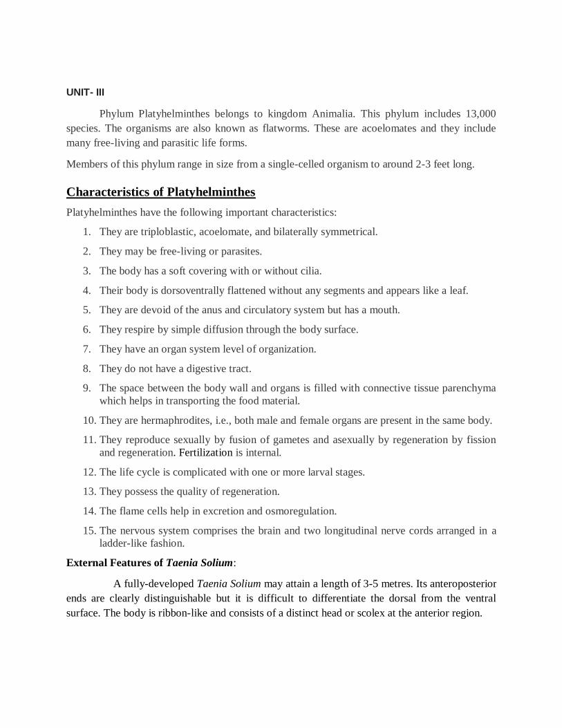
surface. The body is ribbon-like and consists of a distinct head or scolex at the anterior region.
The tip of the head bears a conical elevation—the rostellum which can be retracted or extended.
The rostellum bears 28 to 33 hooks arte of two types- larger and smaller and they alternate with
each other. Each hook parts has three a base or guard, a conical blade at the tip and a handle
projected from the middle.
The hooks are arranged in two rows. When the contractile rostellum is withdrawn the
hooks become anteriorly directed and get fixed into the host tissue. The head bears in the middle
four cup-like suckers or Acetabulum. Rostellum and suckers act as organs of attachment to the
intestine of the host. Behind the head there is a narrow and small tubular region—the neck or the
zone of proliferation. The rest of the body or tape is called strobila. The strobila is segmented in
appearance.
The chain like strobila is made up of numerous segments or sexual units called
proglottids. The proglottids progressively increase in size and mature towards the posterior
extremity. The youngest or newly formed proglottid occupies a position just beneath the neck
while the oldest one is at the posterior end. The number of proglottids varies from 800-850 in a
full-grown worm.
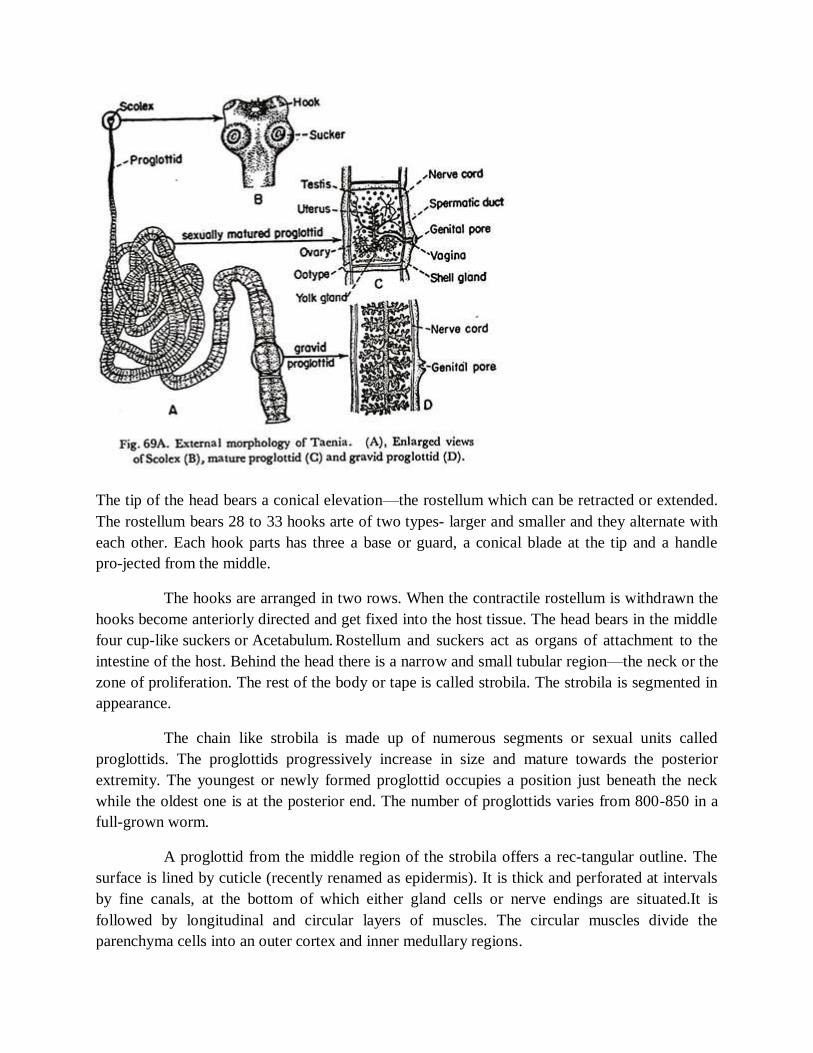
A proglottid from the middle region of the strobila offers a rectangular outline. The
surface is lined by cuticle (recently renamed as epidermis). It is thick and perforated at intervals
by fine canals, at the bottom of which either gland cells or nerve endings are situated.It is
followed by longitudinal and circular layers of muscles. The circular muscles divide the
parenchyma cells into an outer cortex and inner medullary regions.
Towards each lateral margin is found the longitudinal nerve and just median to them lies the
longitudinal pair of excretory vessels. A transverse excretory canal is situated at a posterior
position of the proglottid.
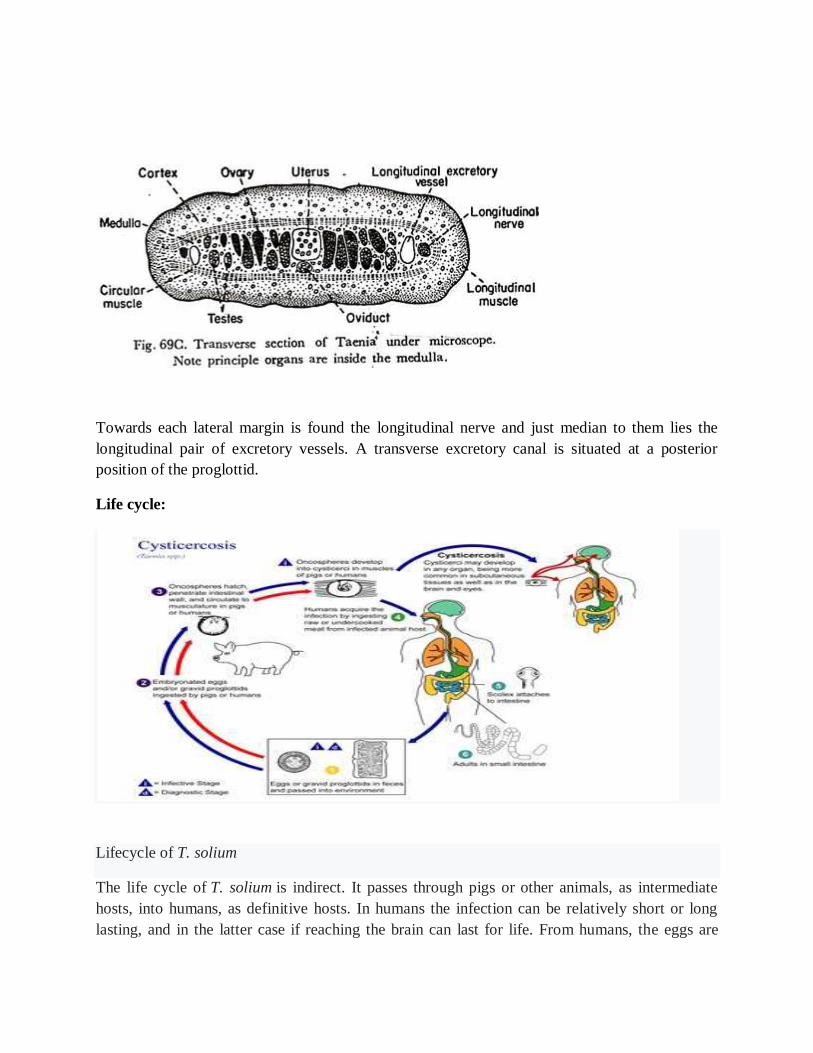
Life cycle:
Lifecycle of T. solium
The life cycle of T. solium is indirect. It passes through pigs or other animals, as intermediate
hosts, into humans, as definitive hosts. In humans the infection can be relatively short or long
lasting, and in the latter case if reaching the brain can last for life. From humans, the eggs are
released in the environment where they await ingestion by another host. In the secondary host,
the eggs develop into oncospheres which bore through the intestinal wall and migrate to other
parts of the body where the cysticerci form. The cysticerci can survive for several years in the
animal.
Definitive host
Humans are colonised by the larval stage, the cysticercus, from undercooked pork or
other meat. Each microscopic cysticercus is oval in shape, containing an inverted scolex
(specifically "protoscolex"), which everts once the organism is inside the small intestine. This
process of evagination is stimulated by bile juice and digestive enzymes (of the host). Then, the
T. Solium lodges in the host’s upper intestine by using its crowned hooks and 4 suckers to enter
the intestinal mucosa. Then, the scolex is fixed into the intestine by having the suckers attached
to the villi and hooks extended. It grows in size using nutrients from the surroundings. Its strobila
lengthens as new proglottids are formed at the foot of the neck. In 10–12 weeks after initial
colonization, it is an adult worm.The exact life span of an adult worm is not determined;
however, evidences from an outbreak among British military in the 1930s indicate that they can
survive 2 to 5 years in humans.
As a hermaphrodite, it reproduces by self-fertilisation, or cross-fertilisation if gametes are
exchanged between two different proglottids. Spermatozoa fuse with the ova in the fertilisation
duct, where the zygotes are produced. The zygote undergoes holoblastic and
unequal cleavage resulting in three cell types, small, medium and large (micromeres, mesomeres,
megameres). Megameres develop into a syncytial layer, the outer embryonic membrane;
mesomeres into the radially striated inner embryonic membrane or embryophore; micromeres
become the morula. The morula transforms into a six-hooked embryo known as an oncosphere,
or hexacanth ("six hooked") larva. A gravid proglottid can contain more than 50,000
embryonated eggs. Gravid proglottids often rupture in the intestine, liberating the oncospheres in
faeces. Intact gravid proglottids are shed off in groups of four or five. The free eggs and detached
proglottids are spread through the host's defecation (peristalsis). Oncospheres can survive in the
environment for up to two months.
Intermediate host
Pigs are the most common host who ingest such eggs in traces of human faeces, mainly
from vegetation contaminated with it such as from water bearing traces of it. The embryonated
eggs enter intestine where they hatch into motile oncospheres. The embryonic and basement
membranes are removed by the host's digestive enzymes (particularly pepsin). Then the free
oncospheres attach on the intestinal wall using their hooks. With the help of digestive enzymes
from the penetration glands, they penetrate the intestinal mucosa to enter blood and lymphatic
vessels. They move along the general circulatory system to various organs, and large numbers
are cleared in the liver. The surviving oncospheres preferentially migrate to striated muscles, as
well as the brain, liver, and other tissues, where they settle to form cysts — cysticerci. A single
cysticercus is spherical, measuring 1–2 cm in diameter, and contains an invaginated protoscolex.

The central space is filled with fluid like a bladder, hence it is also called bladder worm.
Cysticerci are usually formed within 70 days and may continue to grow for a year.
Humans are also accidental secondary hosts when they are colonised by embryonated eggs,
either by auto-colonisation or ingestion of contaminated food. As in pigs, the oncospheres hatch
and enter blood circulation. When they settle to form cysts, clinical symptoms
of cysticercosis appear. The cysticercus is often called the metacestode.
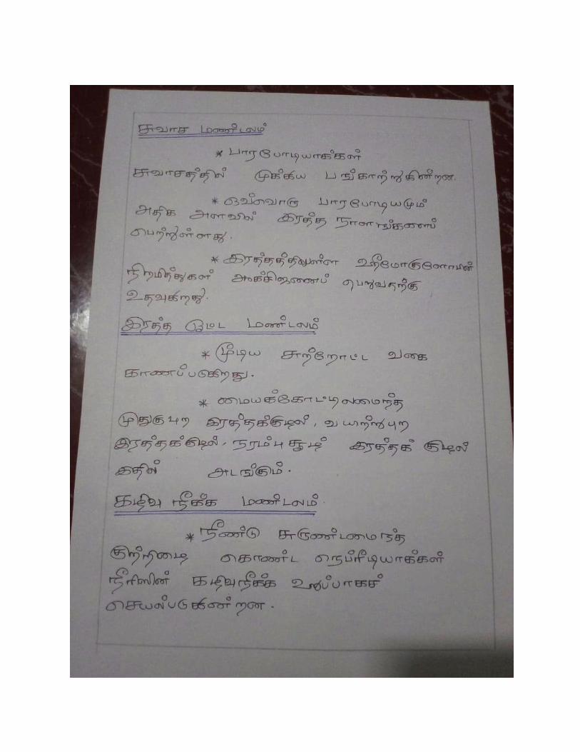
Phylum Nematoda Characteristics
They are widely distributed, aquatic or terrestrial, parasitic or free-living.
Their body is elongated, cylindrical, unsegmented, worm-like, bilaterally symmetrical and
tapering at both ends.
They are triploblastic animals with perivisceral cavity more extensive than that of
platyhelminths.
The body is of organ -system grade organization.
The body is generally covered with thick, flexible multi-layered collagenous cuticle and often
bears cuticle setae (hairs), spines or annulations.
Cuticle moulted periodically.
They lack true coelom. The body cavity is pseudocoel or blastocoel not lined by mesoderm and
filled with parenchyma in most cases.
They lack cilia. circulatory and respiratory systems are absent. i.e. respiration occurs through
general body surface and aerobic in free-living form and anaerobic in parasitic form.
The digestive system is complete with a distinct mouth and anus. Muscular pharynx and the
inner surface of the gut usually not lined by cilia.
Extracellular digestion occurs in them.
The mouth is surrounded by six lips.Excretory without flame cell and nephridia. In the class
Adenophorea glandular renette cells with the duct. Sexes are separate (gonochoristic). the male is
smaller than females.Tubular gonad is present in them. Male genital duct leads into the cloaca.
Female genital ducts with a separate opening.No asexual reproduction.Fertilization is internal or
maybe cross or self.Development may be direct, with or without an intermediate host or indirect.
Various lateral lines and pores are present on the surface of the body.
Ancylostoma duodenale :
Ancylostoma duodenale is a species of the roundworm genus Ancylostoma. It is a
parasitic nematode worm and commonly known as the Old World hookworm. It lives in the
small intestine of hosts such as humans, cats and dogs, where it is able to mate and mature.
Lifecycle
After a filariform "infective" larva penetrates the intact skin – most commonly through
the feet – the larva enters the blood circulation. It is then carried to the lungs, breaks into alveoli,
ascends the bronchi and trachea, and is coughed up and swallowed back into the small intestine,
where it matures. The larva later matures into an adult in the small intestine (jejunum mainly),
where they attach to the villi and female worms can lay 25,000 eggs per day. The eggs are
released into the feces and reside on soil; when deposited on warm, moist soil, a larva rapidly
develops in the egg and hatches after 1 to 2 days. This rhabditiform larva moults twice in the soil
and becomes a skin-penetrating third-stage infective larva within 5–10 days. The infective
rhabditiform larvae are able to sense vibrations in the soil, heat, or carbon dioxide, and are able
to use dendritic processes similar to cilia. They use these processes as thermosensory,
chemosensory, and mechanosensory receptors to migrate towards a host for infection.The
rhabditiform larvae can then penetrate the exposed skin of another organism and begin a new
cycle of infection.
Pathogenenecity and control measures
A light hookworm infection causes abdominal pain, loss of appetite, and geophagy.
Heavy infection causes severe protein deficiency or iron-deficiency anemia. Protein deficiency
may lead to dry skin, edema, and abdominal extension from edema (potbelly), while iron-
deficiency anemia might result in mental dullness and heart failure. Women who are pregnant
and infected should be aware that this parasite is able to infect the fetus and can cause
complications such as low birth weight, maternal anemia, and infant mortality.
The eggs of A. duodenale and Necator americanus cannot be distinguished. Larvae
cannot be found in stool specimens unless they are left at ambient temperature for a day or
more.Education, improved sanitation, and controlled disposal of human feces are important.
Wearing shoes in endemic areas can reduce the prevalence of infection, as well.A. duodenale can
be treated with albendazole, mebendazole, and benzimidazoles. Pyrantel pamoate is an
alternative. In severe cases of anemia, blood transfusion may be necessary.
Wuchereria bancrofti:
Lifecycle
W. bancrofti carries out its lifecycle in two hosts. Humans serve as the definitive host and
mosquitos as the intermediate host. The adult parasites reside in the lymphatics of the human
host. They are found mostly in the afferent lymphatic channels of the lymph glands in the lower
part of the body. The first-stage larvae, known as microfilariae, are present in the circulation.
The microfilariae have a membrane "sheath". This sheath, along with the area in which the
worms reside, makes identification of the species of microfilariae in humans easier to determine.
The microfilariae are found mainly in the peripheral blood and can be found at peak amounts
from 10 pm to 4 am. They migrate between the deep and the peripheral, circulation exhibiting
unique diurnal periodicity. During the day, they are present in the deep veins, and during the
night, they migrate to the peripheral circulation. The cause of this periodicity remains unknown,
but the advantages of the microfilariae being in the peripheral blood during these hours may
ensure the vector, the nighttime mosquito, will have a higher chance of transmitting them
elsewhere. Physiological changes also are associated with sleeping, such as lowered body
temperature, oxygen tension, and adrenal activity, and an increased carbon dioxide tension,
among other physical alterations, any of which could be the signals for the rhythmic behavior of
microfilarial parasites. If the hosts sleep by day and are awake at night, their periodicity is
reversed. In the South Pacific, where W. bancrofti shows diurnal periodicity, it is known as
periodic.
The microfilariae are transferred into a vector, which are most commonly mosquito
species of the genera Culex, Anopheles, Mansonia, and Aedes. Inside the mosquito, the
microfilariae mature into motile larvae called juveniles; these migrate to the labium after a period
around 10 days. When the infected mosquito has its next blood meal, W. bancrofti larvae are
deposited from the mouthparts onto the skin of the prospective host and migrate through
microcuts in the dermis or the tract created by the proboscis into the bloodstream of the new
human host. The larvae move through the lymphatic system to regional lymph nodes,
predominantly in the legs and genital area. The larvae develop into adult worms over the course
of a year, and reach sexual maturity in the afferent lymphatic vessels. After mating, the adult
female worm can produce thousands of microfilariae that migrate into the bloodstream. A
mosquito vector can bite the infected human host, ingest the microfilariae, and thus repeat the
lifecycle. The organism notably does not multiply within its intermediate host, the mosquito.
Diagnosis
A blood smear is a simple and fairly accurate diagnostic tool, provided the blood sample is taken
during the period in the day when the juveniles are in the peripheral circulation. Technicians
analyzing the blood smear must be able to distinguish between W. bancrofti and other parasites
potentially present.
A polymerase chain reaction test can also be performed to detect a minute fraction, as little as 1
pg, of filarial DNA.Some infected people do not have microfilariae in their blood. As a result,
tests aimed to detect antigens from adult worms can be used.Ultrasonography can also be used to
detect the movements and noises caused by the movement of adult worms.Dead, calcified worms
can be detected by X-ray examinations.
Prevention
Prevention focuses on protecting against mosquito bites in endemic regions. Insect repellents and
mosquito nets are useful to protect against mosquito bites. Public education efforts must also be
made within the endemic areas of the world to successfully lower the prevalence of W. bancrofti
infections.
Treatment
The severe symptoms caused by the parasite can be avoided by cleansing the skin, surgery, or the
use of anthelmintic drugs, such as diethylcarbamazine, ivermectin, or albendazole. The drug of
choice is diethylcarbamazine, which can eliminate the microfilariae from the blood and also kill
the adult worms with a dose of 6 mg/kg/day for 12 days, semiannually or annually. A
polytherapy treatment that includes ivermectin with diethylcarbamazine or albendazole is more
effective than either drug alone.Protection is similar to that of other mosquito-spread illnesses;
one can use barriers both physical (a mosquito net), chemical (insect repellent), or mass
chemotherapy as a method to control the spread of the disease.
Parasitic adaptations in nematodes
i The body wall is covered with tough, thick and resistant cuticle, shields against the digestive
enzymes of the host and antitoxins.ii. Ingested food of this parasite is pre- digested, so that there
are no elaborate digestive glands.
siii. The respiration is almost entirely anaerobic. Extremely low metabolic rate and anaerobic
respiration enable the worm to live inside the host’s intestine, where the free oxygen is
negligible.
iv. Reproductive system of is well- developed and numerous eggs are produced to make up for
the poor chances of the right host being reached.
v. The eggs are covered with resistant covering or chitinous shell which provide safety to the
zygote and embryonated eggs from unfavourable environmental factors.
vi. The minute size and resistant nature of eggs make them to withstand prolonged dryness and
cold. The minute size eggs afford far and wide dispersal of the parasite.
Questions
5 marks
1. Write about the external structure of tape worm with diagram
2. Give a note on general characters of nematodes
3. Briefly describe about elephantiasis
4. Comment on onchosphere
10 marks
1. Explain the reproductive system of Taenia solium
2. Describe the life history of Ancyclostoma duodenale
UNIT-IV
Characteristics of Phylum Annelida
They are mostly aquatic; marine or freshwater some terrestrial, burrowing or tubicolous, sedentary
or free-living, some commensal and parasitic.
The body is elongated, triploblastic, bilaterally symmetrical, truly coelomate and vermiform.
The body is metamerically segmented; externally by transverse grooves and internally by septa
into a number of divisions; each division is called a segment, metamere or somite.
Body organization is of organ grade system.
The epidermis is of a single layer of columnar epithelial cells, covered by thin cuticle not made of
chitin.
The body wall is contractile or dermo-muscular consisting of outer muscle fiber circular and inner
longitudinal.
Appendages are jointed when present.
Locomotory organs are segmentally repeated chitinous bristles called setae or chaetae, embedded
in the skin. It may be bored by lateral fleshy appendages or parapodia.
The presence of true schizocoelous coelom usually divided into compartments by transverse
septa. Mostly well-developed in leeches. Coelomic fluid with cells or corpuscles.
The alimentary canal is straight tube-like, complete, extending from mouth to anus. Digestion is
entirely extracellular.
Respiration occurs through moist skin or gills of parapodia and head.
The blood vascular system is a closed type. Blood is red due to the presence of hemoglobin or
erythromycin dissolved in plasma.
Excretion is by metamerically disposed coiled tubes; nephridia which communicate the coelom to
the exterior.
The nervous system consists of a pair of cerebral ganglia; brain and double ventral nerve cord
having segmentally arranged ganglia and lateral nerves in each segment.
Receptor organs include tactile organs, taste buds, statocysts, photoreceptor cells and sometimes
eyes with lenses in some.
They are monoecious i.e. hermaphroditic or sexes separate cleavage spiral and determinate;
dioecious or unisexual form also present.
Their development is direct in monoecious form but indirect in dioecious form.
Larva, when present is a trochophore is characteristics in case of indirect development, while in
others this stage is passed through development.
Regeneration is common.
Asexual reproduction occurs in some.
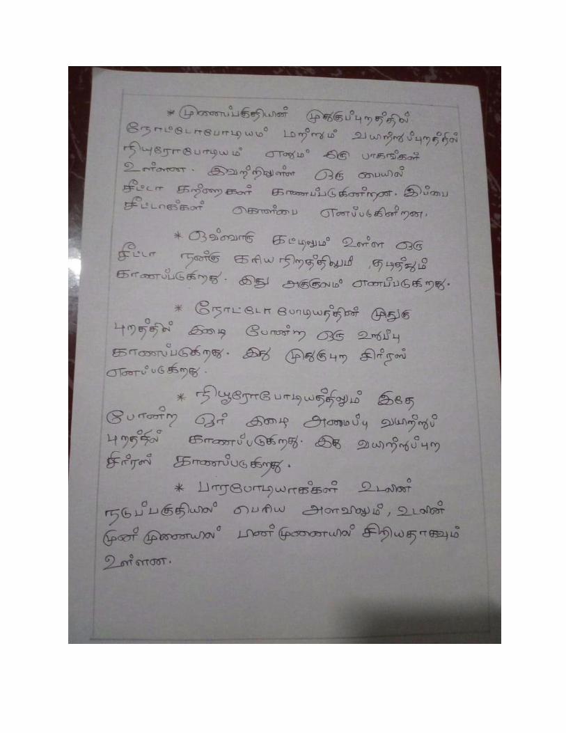
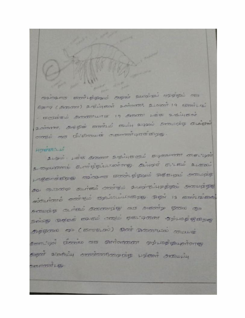
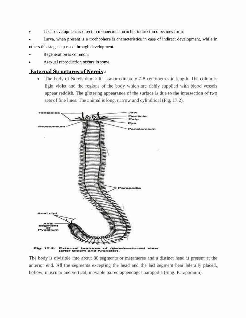
External Structures of Nereis :
The body of Nereis dumerilii is approximately 7-8 centimetres in length. The colour is
light violet and the regions of the body which are richly supplied with blood vessels
appear reddish. The glittering appearance of the surface is due to the intersection of two
sets of fine lines. The animal is long, narrow and cylindrical (Fig. 17.2).
The body is divisible into about 80 segments or metameres and a distinct head is present at the
anterior end. All the segments excepting the head and the last segment bear laterally placed,
hollow, muscular and vertical, movable paired appendages parapodia (Sing. Parapodium).
The terminal segment is termed as the anal segment or pygidium and it bears at its posterior end
a small round opening, the anus. Anal segment bears a pair of elongated anal cirri. On the ventral
surface and near the base of the parapodium lies a nephridial aperture. Thus a pair of
nephridiopores is present in each parapodial segment.
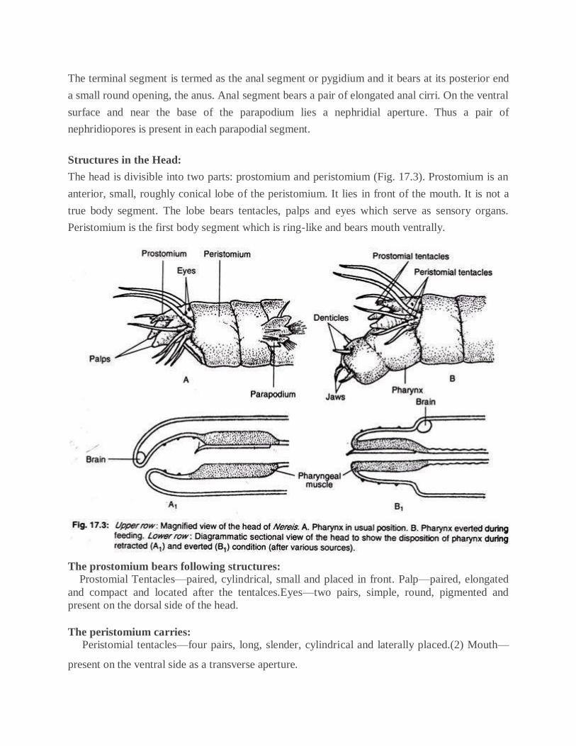
Structures in the Head:
The head is divisible into two parts: prostomium and peristomium (Fig. 17.3). Prostomium is an
anterior, small, roughly conical lobe of the peristomium. It lies in front of the mouth. It is not a
true body segment. The lobe bears tentacles, palps and eyes which serve as sensory organs.
Peristomium is the first body segment which is ring-like and bears mouth ventrally.
The prostomium bears following structures: Prostomial Tentacles—paired, cylindrical, small and placed in front. Palp—paired, elongated
and compact and located after the tentalces.Eyes—two pairs, simple, round, pigmented and
present on the dorsal side of the head.
The peristomium carries: Peristomial tentacles—four pairs, long, slender, cylindrical and laterally placed.(2) Mouth—
present on the ventral side as a transverse aperture.
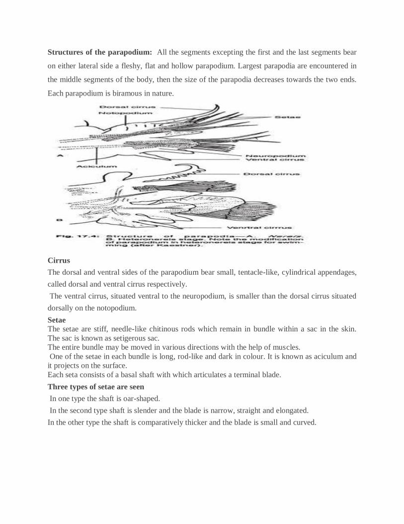
Structures of the parapodium: All the segments excepting the first and the last segments bear
on either lateral side a fleshy, flat and hollow parapodium. Largest parapodia are encountered in
the middle segments of the body, then the size of the parapodia decreases towards the two ends.
Each parapodium is biramous in nature.
Cirrus
The dorsal and ventral sides of the parapodium bear small, tentacle-like, cylindrical appendages,
called dorsal and ventral cirrus respectively.
The ventral cirrus, situated ventral to the neuropodium, is smaller than the dorsal cirrus situated
dorsally on the notopodium.
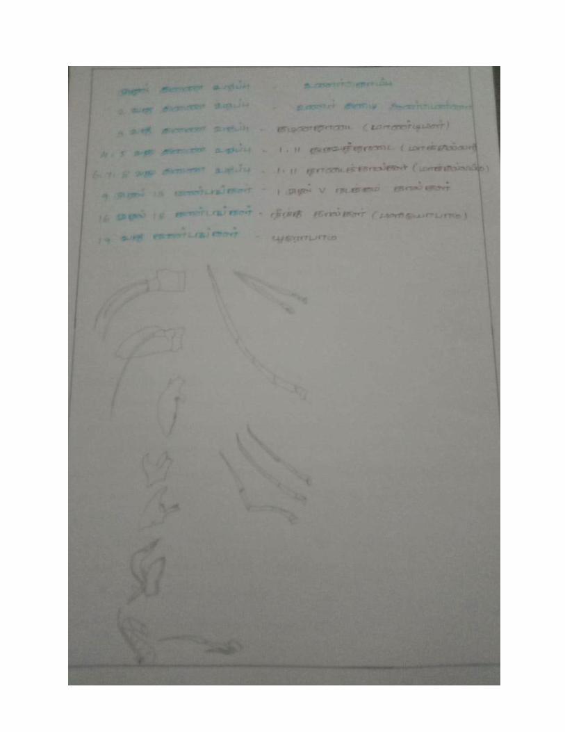
Setae The setae are stiff, needle-like chitinous rods which remain in bundle within a sac in the skin.
The sac is known as setigerous sac.
The entire bundle may be moved in various directions with the help of muscles.
One of the setae in each bundle is long, rod-like and dark in colour. It is known as aciculum and
it projects on the surface.
Each seta consists of a basal shaft with which articulates a terminal blade.
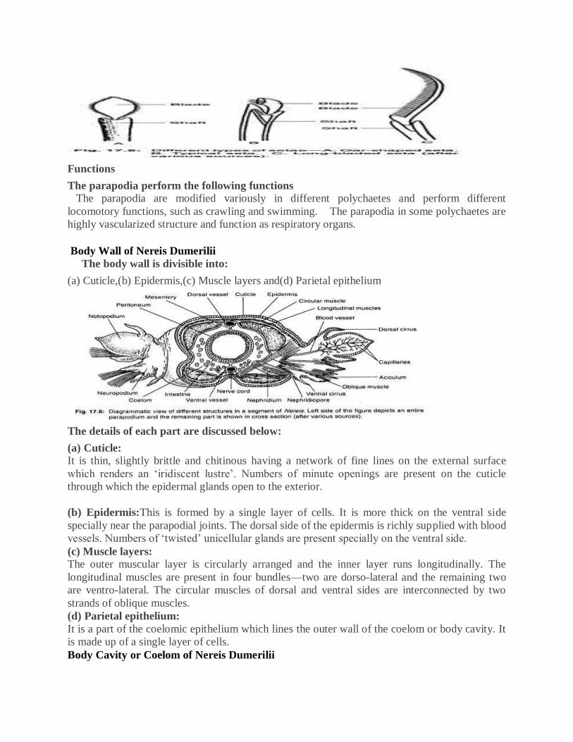
Three types of setae are seen
In one type the shaft is oar-shaped.
In the second type shaft is slender and the blade is narrow, straight and elongated.
In the other type the shaft is comparatively thicker and the blade is small and curved.
Functions
The parapodia perform the following functions The parapodia are modified variously in different polychaetes and perform different
locomotory functions, such as crawling and swimming. The parapodia in some polychaetes are
highly vascularized structure and function as respiratory organs.
Body Wall of Nereis Dumerilii
The body wall is divisible into:
(a) Cuticle,(b) Epidermis,(c) Muscle layers and(d) Parietal epithelium
The details of each part are discussed below:
(a) Cuticle: It is thin, slightly brittle and chitinous having a network of fine lines on the external surface
which renders an ‘iridiscent lustre’. Numbers of minute openings are present on the cuticle
through which the epidermal glands open to the exterior.
(b) Epidermis:This is formed by a single layer of cells. It is more thick on the ventral side
specially near the parapodial joints. The dorsal side of the epidermis is richly supplied with blood
vessels. Numbers of ‘twisted’ unicellular glands are present specially on the ventral side.
(c) Muscle layers: The outer muscular layer is circularly arranged and the inner layer runs longitudinally. The
longitudinal muscles are present in four bundles—two are dorso-lateral and the remaining two
are ventro-lateral. The circular muscles of dorsal and ventral sides are interconnected by two
strands of oblique muscles.
(d) Parietal epithelium:
It is a part of the coelomic epithelium which lines the outer wall of the coelom or body cavity. It
is made up of a single layer of cells.
Body Cavity or Coelom of Nereis Dumerilii
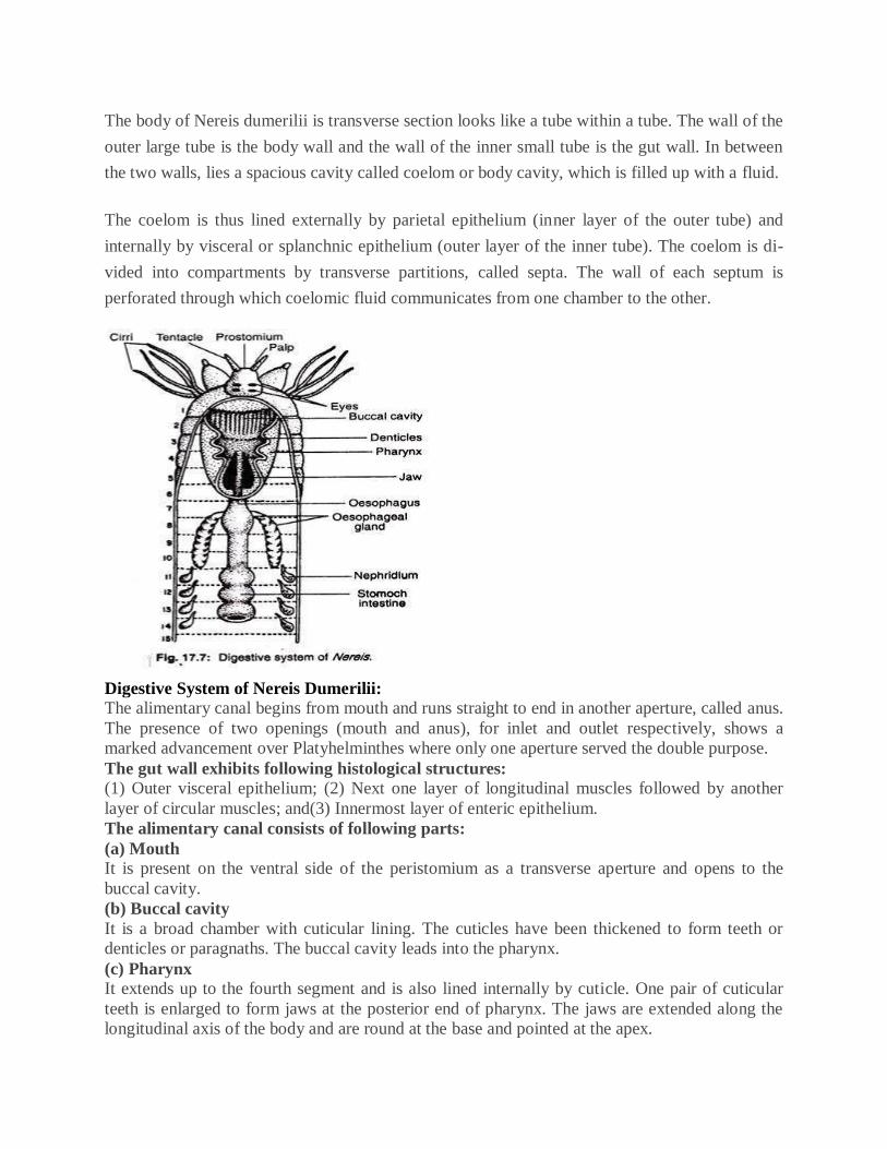
The body of Nereis dumerilii is transverse section looks like a tube within a tube. The wall of the
outer large tube is the body wall and the wall of the inner small tube is the gut wall. In between
the two walls, lies a spacious cavity called coelom or body cavity, which is filled up with a fluid.
The coelom is thus lined externally by parietal epithelium (inner layer of the outer tube) and
internally by visceral or splanchnic epithelium (outer layer of the inner tube). The coelom is di-
vided into compartments by transverse partitions, called septa. The wall of each septum is
perforated through which coelomic fluid communicates from one chamber to the other.
Digestive System of Nereis Dumerilii:
The alimentary canal begins from mouth and runs straight to end in another aperture, called anus.
The presence of two openings (mouth and anus), for inlet and outlet respectively, shows a
marked advancement over Platyhelminthes where only one aperture served the double purpose.
The gut wall exhibits following histological structures: (1) Outer visceral epithelium; (2) Next one layer of longitudinal muscles followed by another
layer of circular muscles; and(3) Innermost layer of enteric epithelium.
The alimentary canal consists of following parts:
(a) Mouth It is present on the ventral side of the peristomium as a transverse aperture and opens to the
buccal cavity.
(b) Buccal cavity
It is a broad chamber with cuticular lining. The cuticles have been thickened to form teeth or
denticles or paragnaths. The buccal cavity leads into the pharynx.
(c) Pharynx It extends up to the fourth segment and is also lined internally by cuticle. One pair of cuticular
teeth is enlarged to form jaws at the posterior end of pharynx. The jaws are extended along the
longitudinal axis of the body and are round at the base and pointed at the apex.
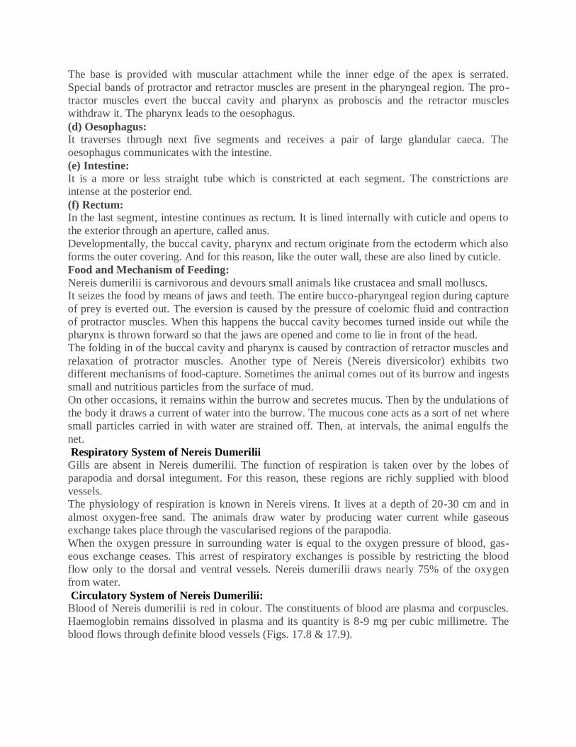
The base is provided with muscular attachment while the inner edge of the apex is serrated.
Special bands of protractor and retractor muscles are present in the pharyngeal region. The pro-
tractor muscles evert the buccal cavity and pharynx as proboscis and the retractor muscles
withdraw it. The pharynx leads to the oesophagus.
(d) Oesophagus: It traverses through next five segments and receives a pair of large glandular caeca. The
oesophagus communicates with the intestine.
(e) Intestine:
It is a more or less straight tube which is constricted at each segment. The constrictions are
intense at the posterior end.
(f) Rectum: In the last segment, intestine continues as rectum. It is lined internally with cuticle and opens to
the exterior through an aperture, called anus.
Developmentally, the buccal cavity, pharynx and rectum originate from the ectoderm which also
forms the outer covering. And for this reason, like the outer wall, these are also lined by cuticle.
Food and Mechanism of Feeding:
Nereis dumerilii is carnivorous and devours small animals like crustacea and small molluscs.
It seizes the food by means of jaws and teeth. The entire bucco-pharyngeal region during capture
of prey is everted out. The eversion is caused by the pressure of coelomic fluid and contraction
of protractor muscles. When this happens the buccal cavity becomes turned inside out while the
pharynx is thrown forward so that the jaws are opened and come to lie in front of the head.
The folding in of the buccal cavity and pharynx is caused by contraction of retractor muscles and
relaxation of protractor muscles. Another type of Nereis (Nereis diversicolor) exhibits two
different mechanisms of food-capture. Sometimes the animal comes out of its burrow and ingests
small and nutritious particles from the surface of mud.
On other occasions, it remains within the burrow and secretes mucus. Then by the undulations of
the body it draws a current of water into the burrow. The mucous cone acts as a sort of net where
small particles carried in with water are strained off. Then, at intervals, the animal engulfs the
net.
Respiratory System of Nereis Dumerilii
Gills are absent in Nereis dumerilii. The function of respiration is taken over by the lobes of
parapodia and dorsal integument. For this reason, these regions are richly supplied with blood
vessels.
The physiology of respiration is known in Nereis virens. It lives at a depth of 20-30 cm and in
almost oxygen-free sand. The animals draw water by producing water current while gaseous
exchange takes place through the vascularised regions of the parapodia.
When the oxygen pressure in surrounding water is equal to the oxygen pressure of blood, gas-
eous exchange ceases. This arrest of respiratory exchanges is possible by restricting the blood
flow only to the dorsal and ventral vessels. Nereis dumerilii draws nearly 75% of the oxygen
from water.
Circulatory System of Nereis Dumerilii:
Blood of Nereis dumerilii is red in colour. The constituents of blood are plasma and corpuscles.
Haemoglobin remains dissolved in plasma and its quantity is 8-9 mg per cubic millimetre. The
blood flows through definite blood vessels (Figs. 17.8 & 17.9).
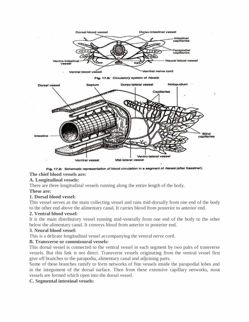
The chief blood vessels are:
A. Longitudinal vessels: There are three longitudinal vessels running along the entire length of the body.
These are:
1. Dorsal blood vessel:
This vessel serves as the main collecting vessel and runs mid-dorsally from one end of the body
to the other end above the alimentary canal. It carries blood from posterior to anterior end.
2. Ventral blood vessel: It is the main distributory vessel running mid-ventrally from one end of the body to the other
below the alimentary canal. It conveys blood from anterior to posterier end.
3. Neural blood vessel:
This is a delicate longitudinal vessel accompanying the ventral nerve cord.
B. Transverse or commissural vessels:
This dorsal vessel is connected to the ventral vessel in each segment by two pairs of transverse
vessels. But this link is not direct. Transverse vessels originating from the ventral vessel first
give off branches to the parapodia, alimentary canal and adjoining parts.
Some of these branches ramify to form networks of fine vessels inside the parapodial lobes and
in the integument of the dorsal surface. Then from these extensive capillary networks, stout
vessels are formed which open into the dorsal vessel.
C. Segmental intestinal vessels:
The ventral vessel gives off two pairs of intestinal vessels in each segment to form capillary
network in the gut wall. From there blood is returned to the dorsal vessel by another two pairs of
intestinal vessels.
Mechanism of Blood Circulation:
Blood remains in constant circulation through the vessels by means of contractions which are
peristaltic in nature. Waves of contractions transmit along the walls of the vessels to drive the
blood. A series of ring-like muscle fibres round the walls of the blood vessels at short intervals
aid in contraction.
The contractions of the dorsal vessel are the most powerful. Dorsal vessel is the main collecting
vessel and blood flows through it from posterior to anterior end. Whereas the flow of blood is in
opposite direction through ventral vessel and by transverse and intestinal vessels it sends blood
to the different parts of the body.
Excretory System of Nereis Dumerilii
The excretory system consists of series of metamerically arranged paired tubes, called nephridia
or segmental organs. They are absent in the anterior and posterior segments.
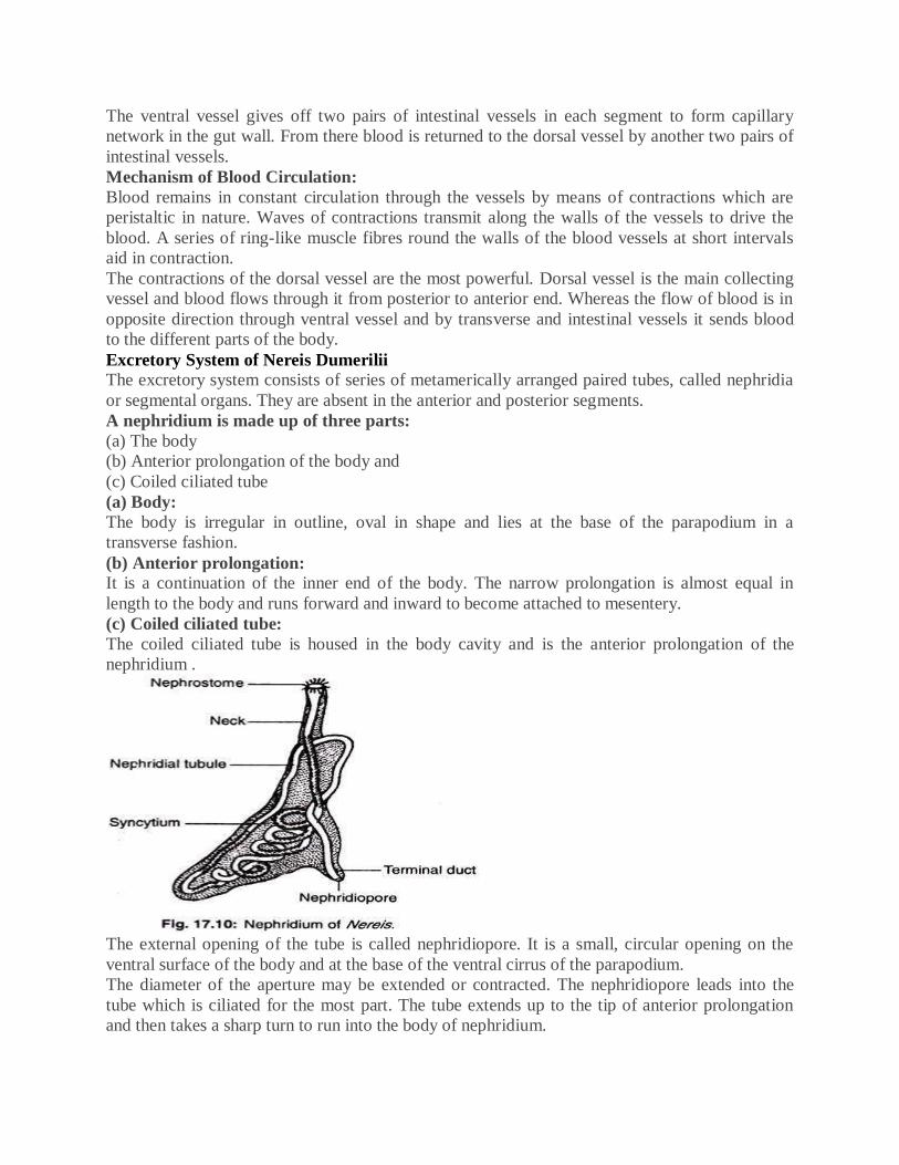
A nephridium is made up of three parts:
(a) The body
(b) Anterior prolongation of the body and
(c) Coiled ciliated tube
(a) Body:
The body is irregular in outline, oval in shape and lies at the base of the parapodium in a
transverse fashion.
(b) Anterior prolongation: It is a continuation of the inner end of the body. The narrow prolongation is almost equal in
length to the body and runs forward and inward to become attached to mesentery.
(c) Coiled ciliated tube:
The coiled ciliated tube is housed in the body cavity and is the anterior prolongation of the
nephridium .
The external opening of the tube is called nephridiopore. It is a small, circular opening on the
ventral surface of the body and at the base of the ventral cirrus of the parapodium.
The diameter of the aperture may be extended or contracted. The nephridiopore leads into the
tube which is ciliated for the most part. The tube extends up to the tip of anterior prolongation
and then takes a sharp turn to run into the body of nephridium.
Inside the body, the tube follows a zigzag course and ultimately passes through the anterior
prolongation to open as the nephhrostome into the preceding segment. The nephrostome is
funnel-shaped and its border is beset with a number of narrow ciliated processes.
Dorsal Ciliated Organs:
Each segment bears on the dorsal surface specially developed ciliated tract of coelomic
epithelium in the form of short funnels without external aperture. These are called dorsal ciliated
organs. The specific roles of these structures are not clearly understood. Some believe that they
are excretory in function while others consider them as genital ducts of temporary nature.
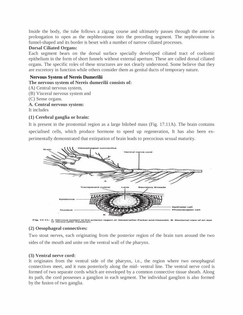
Nervous System of Nereis Dumerilii
The nervous system of Nereis dumerilii consists of: (A) Central nervous system,
(B) Visceral nervous system and
(C) Sense organs.
A. Central nervous system: It includes
(1) Cerebral ganglia or brain:
It is present in the prostomial region as a large bilobed mass (Fig. 17.11A). The brain contains
specialised cells, which produce hormone to speed up regeneration, It has also been ex-
perimentally demonstrated that extirpation of brain leads to precocious sexual maturity.
(2) Oesophageal connectives:
Two stout nerves, each originating from the posterior region of the brain turn around the two
sides of the mouth and unite on the ventral wall of the pharynx.
(3) Ventral nerve cord: It originates from the ventral side of the pharynx, i.e., the region where two oesophageal
connectives meet, and it runs posteriorly along the mid- ventral line. The ventral nerve cord is
formed of two separate cords which are enveloped by a common connective tissue sheath. Along
its path, the cord possesses a ganglion in each segment. The individual ganglion is also formed
by the fusion of two ganglia.
(4) Peripheral nerves: These are nerves given off by brain, oesophageal connectives and ganglia of the ventral nerve
cord. From brain, nerves are supplied to the tentacles, palpi and eyes. The oesophageal
connectives supply branches to innervate peristomeal tentacles. The ganglion on the ventral
nerve cord sends nerves to the various parts of the corresponding segment.
B. Visceral nervous system:
In addition to the nerves belonging to the central nervous system, another set of nerves is given
off from the brain. These fine nerves with ganglia innervate the anterior part of the alimentary
system. It is known as stomatogastric or visceral nervous system.
C. Sense organs.
Following sense organs are present in Nereis dumerilii:
(a) Eyes:
There are two pairs of eyes. Each eye is a cup-shaped and darkly pigmented structure. The
concave side bears the retina, a circular aperture, pupil and a lens of gelatinous consistency
Many elongated and slender cells which are arranged parallelly form the wall of the cup. These
cells through the union of their outer ends form the optic nerve and their inner ends extend
towards the lens as clear and hyaline rods. The region of the cuticle which covers the eyes, acts
as the cornea.
(b) Olfactory organs: The olfactory organs are known as nuchal organs. These paired organs are present on the
posterior and dorsal side of the prostomium and remain in close contact with the hinder part of
the brain. Each nuchal organ has two pits lined with ciliated epithelium.
(c) Tactile organs: The tentacles, palpi and cirri are regarded as specialised tactile sense organs. With the help of
specialised sensory cells they can discriminate the changes in the environment.
Reproductive System of Nereis Dumerilii:
Sexes are separate in Nereis dumerilii but well- formed gonads in the form of testes or ovaries
are not regularly recognised. The gonads develop by the proliferation of coelomic epithelial cells
of the body cavity. The gonads are temporary structures and appear only in the breeding season.
The males develop only a pair of testes which are present in any one of the segments between
nineteenth and twenty-fifth. The number of testes may be more in other species. During breeding
season groups of cells pinch off from the testes into the coelomic fluid. These cells undergo
division and each daughter cell develops into a sperm. The sperms have rod-shaped heads and
vibratile tails.
Spherical ovaries in females appear along the entire length of the body. They are metamerically
arranged and occur one pair in each segment. The ova (when young) become detached from the
ovaries into the coelomic fluid where they attain maturity. Both ovaries and testes degenerate
after the liberation of sex cells.
Mature reproductive cells are liberated probably through temporary apertures formed by the
rupture of the body wall. Fertilization is external and occurs in sea water.
Structural changes during gonad formation: The formation of gametes induces changes in the posterior half of the body. Such changes are
noted in the critical appearance of lobes of the parapodia and in the number of setae in bundles.
In addition, the prostomial eyes become enlarged and the terminal segment produces sensory
papillae. The worms after such transformations are known as Heteronereis.
The heteronereis forms are free-swimming. The body is divisible into two distinct parts. The
anterior or asexual part is called ‘Atoke’ and the posterior or sexual part is called ‘Epitoke’.
The changes of the parapodia in the posterior half of the body are, first, increase in size and
secondly, the formation of leaf-like outgrowths on the lobes. Bristles which replace setae remain
inserted into the parapodium and assume fan-like appearance (Fig. 17.4B).
The transformation to the heteronereis form is due to the impact of hormones, released into the
blood plexus from certain specialised cells, which remain very close to the brain. Some authors,
however, consider Nereis and Heteronereis as two distinct species.
Development: The matured egg has two enveloping membranes. The outer cover is thin and the inner one is
broad with radial striations. With these membranes the egg remains within a covering of
gelatinous consistency. Oil droplets and yolk bodies remain scattered throughout the cytoplasm
of the egg.
The fertilization or the entry of sperm cell brings following changes in the egg:
(a) Inner radiated layer dissolves,
(b) Egg completes the maturation phase by liberating two polar bodies,
(c) Egg exhibits irregular amoeboid movement and
(d) Considerable rearrangement of cytoplasmic particles occurs.
This results into the shifting of oil droplets and yolk spherules towards the centre, thus leaving a
side with granular cytoplasm and nucleus.
The zygote finally assumes spherical shape and starts to divide. This is called cleavage. First two
divisions produce four cells. These cells are called macromeres. One macromere becomes larger
than the other three. These macromeres divide unequally in three sets and thus give rise to twelve
micromeres.
At the end of first unequal division of the macromeres, four micromeres of equal sizes are
produced. The second unequal division of the macromeres produces four more micromeres, but
this time three are of same sizes and one is large. The third unequal division of the macromeres
again results three more micromeres of same sizes and one large micromere.
During these unequal divisions of the macromeres, the micromeres are not produced at their tops.
On the contrary, after first division the micromeres are pushed towards right, and then during
next division they shift to the left and again to the right. Such arrangement gives rise to a spiral
pattern. The two large micromeres are known as somatoblasts or mesentomeres.
The micromeres give rise to ectoderm, somatoblasts or mesentomeres result into mesoderm and
macromeres produce endoderm layer. The micromeres spread over the macromeres and push
them and somatoblasts inside. Further development involves transformation of these cells into
the various structures of larva. The larva is known as trochop
It consists of a basal part and two distal pats:
Dorsally placed notopodium and Ventrally placed neuropodium Both these parts are subdivided
into lobes and both of them carry pack of needle-like structures, called setae (sing, seta) which
project beyond the lobes.
Adaptive radiation Among Annelids.
Adaptive radiation is the rapid evolution of different species from a single common ancestor.
... For example, a natural disaster occurs or a species becomes extinct. This changed contains
many available niches for organisms to occupy.
Adaptive Radiation in.Annelida Phylum Annelida includes three classes; Polychaeta,
Oligochaeta and Hirudinea. Of these, the Polychaeta do not h?ve clitellum; the Oligochaeta and
Hirudinaria are clitellate. Now you should recollect the classification and characters of Annelida
which you have studied in Unit 4, Block 1 of this course. The early annelids are supposed to I
have been marine worms burrowing in the'bottom, in sand and mud on the shore.
MODE OF LIFE IN POLYCHAETA (ANNELIDA)
Class Polychaeta shows the greatest diversity in Phylum Annelida. Majority of the 5000
species of polychaetes are marine and exhibit a variety of habits and habitats for which they
show anatomical and physiological adaptations.
PLANKTONIC POLYCHAETES
Planktonic polychaetes swim near the surface of the sea where the danger of predators and
solar radiation is excessive. They generally have semitransparent body that imparts them near
invisibility. Some have large eyes while others have none. Parapodia are small and
locomotion is by lateral undulation of body. Cirri are generally longer as they carry
tangoreceptors and help in locating food. Ex. Vanadis and Tomopteris.
CRAWLING POLYCHAETES
Crawling polychaetes belong to order Errantia which includes marine, freely moving animals
that crawl on sea bottom. Head bears sense organs such as eyes, tentacles and palps.
Locomotory organs or parapodia bear setae which can be retracted and protruded out in
various directions for crawling among rocks and stones. Sense organs on prostomium and peristomium are well developed due to the free swimming and crawling habit.
Errant forms are carnivorous that feed on small molluscs, crustaceans, sponges etc. The
raptorial feeding is performed by eversible pharynx which is provided with chitinous jaws and
dentacles on the internal lining. Ex. Nereis, Aphrodite, Polynoe, Eunice etc. In Aphrodite the
body is short and broad with arched dorsal side and a flat ventral side to permit creeping
under the stones and crevices. Body is covered with stiff and iridescent setae for protection.
BURROWING POLYCHAETES Many polychaetes, such as Nereis crawl on the seabed in night in search of food and burrow
in the soil in day time. But polychaetes, such as Arenicola, Glycera,
Amphitrite and Terebella spend most of the time in burrows and come out only to capture
pray or for ingesting detritus. In these animals, for burrowing mode of life the external surface
of body has become smooth and parapodia have become small to give minimum friction
during burrowing. Their setae have become hook-like to anchor in the burrow.
In Arenicola posterior part of body is without parapodia and tentacles and palps are also
reduced because of mud-ingesting habit. However, in Amphitrite and Terebella there are long
feeding tentacles on the anterior end and bunches of gills for respiration.
TUBICOLOUS POLYCHAETES
Tubicolous polychaetes live permanently inside the tubes made of mud, shell or sand grains,
parchment or calcium carbonate. Tubicolous annelids are capable of secreting mucous or
other adhesive substance to which sand and shell particles get attached and make the tube
strong, e.g. Pectinaria, Owenia, Diopatra, Clymenella, Sabella.
In these animals water current carries micro-organisms inside the burrow and then tentacles
move the food towards the mouth. Sometimes a mucous cone is produced from mouth to trap
food particles.In Chaetopterus the parapodial setae become peg-like or hook-like for
anchoring the animal to the wall of the tube and water current is maintained by three fan-like
appendages. Feeding is done by the peristomial funnel, mouth, ciliated groove and three fans
on segments 14-16. A cone of mucous is secreted on the anterior side for trapping food
particles.
Sabella or the peacock worm lives in vertical parchment tube and projects the fan-like
tentacles which are covered with sticky mucous to which the minute food particles get
attached and are carried to mouth through the ciliated groove.
In Myxicola, the gill filaments are joined by a thin sheet and form a semicircle at the apex, in
which food is captured. Parapodia are lost or reduced or modified for anchoring in the tube.
Sense organs are also reduced.
Serpula and Spirorbis possess glands that secrete Calcium carbonate for making a hard tube
inside which the worm lives. Its anterior part of body and tentacles can be projected out to
collect planktonic food particles on which it feeds.
COMMENSAL POLYCHAETES
Myzostoma is a commensal that lives in the tubes of other polychaetes or with sea cucumbers
and sea stars. Its small body is oval in shape and parapodia are modified for clinging onto the
host. Mouth has eversible pharynx.
PARASITIC POLYCHAETES
Histriobdella lives as ectoparasite in the gill chamber of crustaceans and sucks
blood. Ichthyotomus is an ectoparasite on the fins of marine eels. Its parapodia are modified
for clinging and mouth parts transform into piercing stylets for sucking blood.
General characters
1. Cosmopolitan in distribution found in aquatic, terrestrial and aerial forms.
Some are ectoparasitic and vectors of disease.
2. Body have jointed appendages or legs (which are modified to different structures to
perform different functions like jaws, gills, walking legs, paddle) . There may be 3 pairs,
4 pairs, 5 pairs, many pairs.
3. Body is triploblastic.
4. Bilaterally symmetrical.
5. Organ system level of organization.
6. Body is divisible into head, thorax and abdomen.
NOTE: In some (crustacean and arachnida) body is divisible into cephalothorax
(head and thorax is fused) and abdomen. 7. This is the first group to develop a true head, which contains sense organs and feeding organs
specialized for their particular habitats.
8. Body is covered with chitinous exoskeleton.
9. They are haemocoelomate. Coelom i.e. body cavity is filled with blood or fluid.
10. Head bears a pair of compound eyes and antenna.
11. Locomotion takes place by jointed appendages.
12. Digestive system is complete, straight and well developed.
The mouth bears mouth parts for ingestion of foods. Mouths are modified fo r chewing,
biting, sponging, piercing, siphoning.
13. Respiration takes place by general body surface or gills (in Crustaceans) or trachea ( in insects,
diplopoda and chilopoda) or booklungs (Arachnida) and book gills (in king cobra).
14. Circulatory system is of open type i.e. do not have blood vessels and enters directly into
the body chambers. The blood is colorless.
15. Excretion takes place through Malphigian tubules (in terrestrial form) or green glands or coxal
glands (in aquatic forms).
NOTE: Aquatic forms are ammonotelic, terrestrial forms are uricotelic.
16. Nervous system is of annelidian type, which consists of brain and ventral nerve cord.
17. Unisexual i.e. sexes are separate.
18. Fertilization is internal or external.
19. They are either oviparous or ovoviviparous.
20. Development may be direct or indirect.
21. Sensory organ include antennae, sensory hairs for touch and chemoreceptor, simple and
compound eyes, auditory organs (in insects) and statocysts (in crustacean).
22. External features of palaemon or prawn of phylum arthropoda
23. Phylum Arthropoda is the largest phylum of the animal kingdom and crustacea is a large and
important class of the phylum Arthropoda. It includes prawns, crabs, lobesters, shrimps,
barnacles, water fleas and related forms.
Habits and habitat :
Palaemon or prawn, found in fresh water ponds, rivers and lakes. It is a nocturnal animal. It
comes out at the water surface in the night in search of food.
It is an omnivorous. It is an active swimmer and swims with the help of pleopods. It breeds from
May to July.
Shape and Size:
Palaemon has elongated, spindle shaped, bilaterally symmetrical body. Different species have
different sizes. P. malcomsonii (Macrobrachium malcomsonii) the giant prawn found in
Tamilnadu, Bengal and Central India is about 30 cm in size.
Colouration:
Young stages are transiuscent and white but the adults are usually dull pale-blue or greenish with
orange-red patches.
Segmentation:
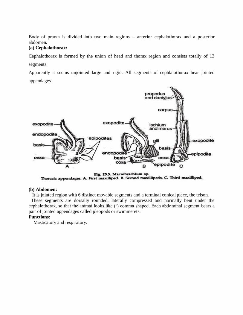
Body of prawn is divided into two main regions – anterior cephalothorax and a posterior
abdomen.
(a) Cephalothorax:
Cephalothorax is formed by the union of head and thorax region and consists totally of 13
segments.
Apparently it seems unjointed large and rigid. All segments of cephlalothorax bear jointed
appendages.
(b) Abdomen:
It is jointed region with 6 distinct movable segments and a terminal conical piece, the telson.
These segments are dorsally rounded, laterally compressed and normally bent under the
cephalothorax, so that the animai looks like (‘) comma shaped. Each abdominal segment bears a
pair of jointed appendages called pleopods or swimmerets.
Functions:
Masticatory and respiratory.
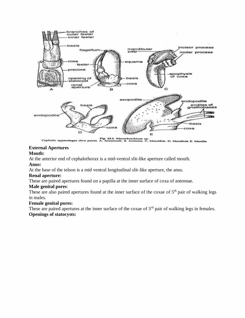
External Apertures
Mouth: At the anterior end of cephalothorax is a mid-ventral slit-like aperture called mouth.
Anus: At the base of the telson is a mid ventral longitudinal slit-like aperture, the anus.
Renal aperture: These are paired apertures found on a papilla at the inner surface of coxa of antennae.
Male genital pores:
These are also paired apertures found at the inner surface of the coxae of 5th pair of walking legs
in males.
Female genital pores:
These are paired apertures at the inner surface of the coxae of 3rd pair of walking legs in females.
Openings of statocysts:

At the dorsal surface of the precoxae of antennule paired statocysts open outside.
Palaemon's digestive system contains a long alimentary canal and large hepatopancreatic gland.
Locomotion in Prawn:
Change of place in prawn takes place in three ways:
i. Swimming:
The body is kept straight in a horizontal position. The pleopods act like oars. The forward
movement of the pleopods is slow, while the back stroke is fast and the animal moves forward.
The uropod helps in guiding and the antennae move constantly, presumably feeling the
surroundings.
ii. Walking: The straightened body is supported by all the five pairs of pereopods. The walking legs move in
harmony during walking.
iii. Darting:
This is resorted under emergency. With stretched pleopods and uropod the abdomen suddenly
moves forward towards the cephalothorax and the animal swiftly moves backward with a jerk
due to the sudden thrust.
I. Alimentary canal:
. The alimentary canal is a long tube.
It starts at the mouth and ends with anus
It shows buccal cavity, oesophagus, stomach intestine and rectum.
The buccal cavity, oesophagus and stomach are lined with euncle.
It is called stomodaeum or fore-gut. Intestine is lined by endoderm and is called
mesenteron or mid-gut.
The rectum is lined by cuticle and is called the proctodeam or hind gut.
a) Mouth:
The mouth is a longitudinal slit on the ventral side of the head.
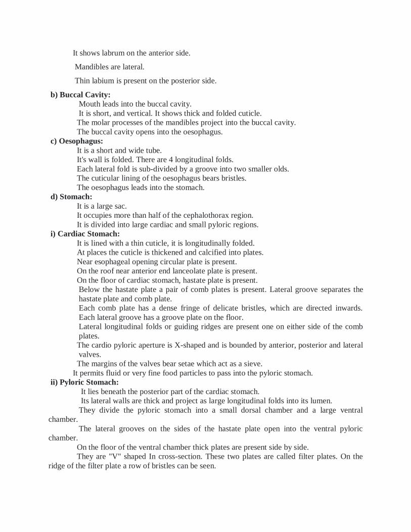
It shows labrum on the anterior side.
Mandibles are lateral.
Thin labium is present on the posterior side.
b) Buccal Cavity:
Mouth leads into the buccal cavity.
It is short, and vertical. It shows thick and folded cuticle.
The molar processes of the mandibles project into the buccal cavity.
The buccal cavity opens into the oesophagus.
c) Oesophagus:
It is a short and wide tube.
It's wall is folded. There are 4 longitudinal folds.
Each lateral fold is sub-divided by a groove into two smaller olds.
The cuticular lining of the oesophagus bears bristles.
The oesophagus leads into the stomach.
d) Stomach:
It is a large sac.
It occupies more than half of the cephalothorax region.
It is divided into large cardiac and small pyloric regions.
i) Cardiac Stomach:
It is lined with a thin cuticle, it is longitudinally folded.
At places the cuticle is thickened and calcified into plates.
Near esophageal opening circular plate is present.
On the roof near anterior end lanceolate plate is present.
On the floor of cardiac stomach, hastate plate is present.
Below the hastate plate a pair of comb plates is present. Lateral groove separates the
hastate plate and comb plate.
Each comb plate has a dense fringe of delicate bristles, which are directed inwards.
Each lateral groove has a groove plate on the floor.
Lateral longitudinal folds or guiding ridges are present one on either side of the comb
plates.
The cardio pyloric aperture is X-shaped and is bounded by anterior, posterior and lateral
valves.
The margins of the valves bear setae which act as a sieve.
It permits fluid or very fine food particles to pass into the pyloric stomach.
ii) Pyloric Stomach:
It lies beneath the posterior part of the cardiac stomach.
Its lateral walls are thick and project as large longitudinal folds into its lumen.
They divide the pyloric stomach into a small dorsal chamber and a large ventral
chamber.
The lateral grooves on the sides of the hastate plate open into the ventral pyloric
chamber.
On the floor of the ventral chamber thick plates are present side by side.
They are "V" shaped In cross-section. These two plates are called filter plates. On the
ridge of the filter plate a row of bristles can be seen.
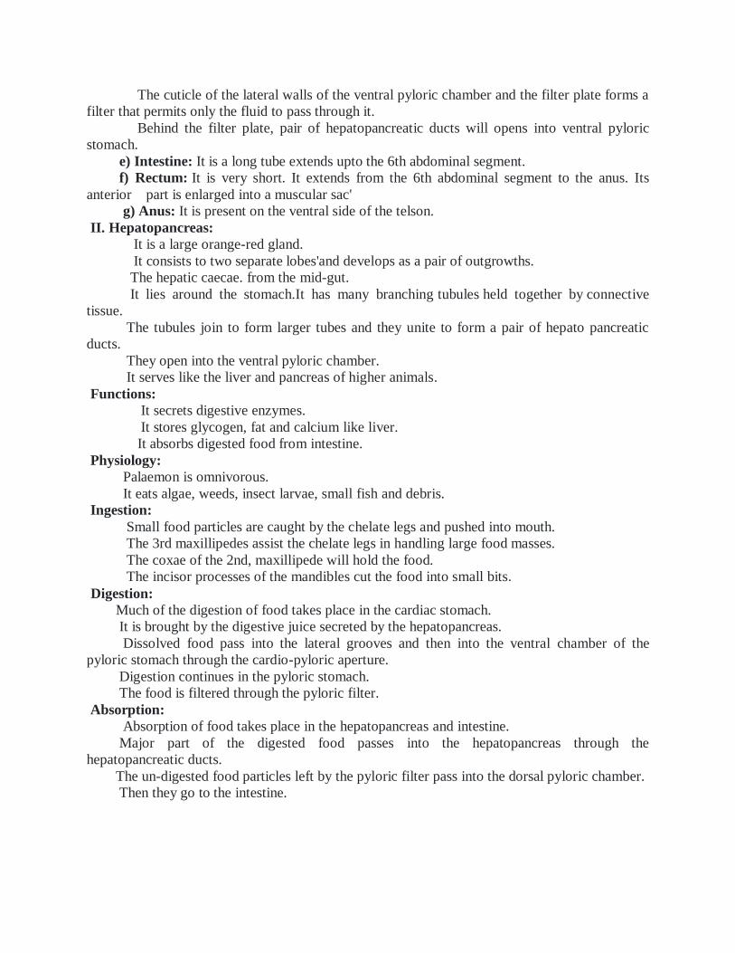
The cuticle of the lateral walls of the ventral pyloric chamber and the filter plate forms a
filter that permits only the fluid to pass through it.
Behind the filter plate, pair of hepatopancreatic ducts will opens into ventral pyloric
stomach.
e) Intestine: It is a long tube extends upto the 6th abdominal segment.
f) Rectum: It is very short. It extends from the 6th abdominal segment to the anus. Its
anterior part is enlarged into a muscular sac'
g) Anus: It is present on the ventral side of the telson.
II. Hepatopancreas:
It is a large orange-red gland.
It consists to two separate lobes'and develops as a pair of outgrowths.
The hepatic caecae. from the mid-gut.
It lies around the stomach.It has many branching tubules held together by connective
tissue.
The tubules join to form larger tubes and they unite to form a pair of hepato pancreatic
ducts.
They open into the ventral pyloric chamber.
It serves like the liver and pancreas of higher animals.
Functions:
It secrets digestive enzymes.
It stores glycogen, fat and calcium like liver.
It absorbs digested food from intestine.
Physiology:
Palaemon is omnivorous.
It eats algae, weeds, insect larvae, small fish and debris.
Ingestion:
Small food particles are caught by the chelate legs and pushed into mouth.
The 3rd maxillipedes assist the chelate legs in handling large food masses.
The coxae of the 2nd, maxillipede will hold the food.
The incisor processes of the mandibles cut the food into small bits.
Digestion:
Much of the digestion of food takes place in the cardiac stomach.
It is brought by the digestive juice secreted by the hepatopancreas.
Dissolved food pass into the lateral grooves and then into the ventral chamber of the
pyloric stomach through the cardio-pyloric aperture.
Digestion continues in the pyloric stomach.
The food is filtered through the pyloric filter.
Absorption:
Absorption of food takes place in the hepatopancreas and intestine.
Major part of the digested food passes into the hepatopancreas through the
hepatopancreatic ducts.
The un-digested food particles left by the pyloric filter pass into the dorsal pyloric chamber.
Then they go to the intestine.
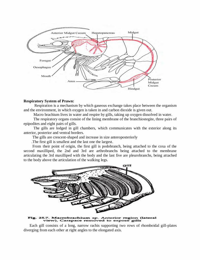
Respiratory System of Prawn: Respiration is a mechanism by which gaseous exchange takes place between the organism
and the environment, in which oxygen is taken in and carbon dioxide is given out.
Macro brachium lives in water and respire by gills, taking up oxygen dissolved in water.
The respiratory organs consist of the lining membrane of the branchiostegite, three pairs of
epipodites and eight pairs of gills.
The gills are lodged in gill chambers, which communicates with the exterior along its
anterior, posterior and ventral borders.
The gills are crescent-shaped and increase in size anteroposteriorly
.The first gill is smallest and the last one the largest.
From their point of origin, the first gill is podobranch, being attached to the coxa of the
second maxilliped, the 2nd and 3rd are arthrobranchs being attached to the membrane articulating the 3rd maxilliped with the body and the last five are pleurobranchs, being attached
to the body above the articulation of the walking legs.
Each gill consists of a long, narrow rachis supporting two rows of rhomboidal gill-plates
diverging from each other at right angles to the elongated axis.
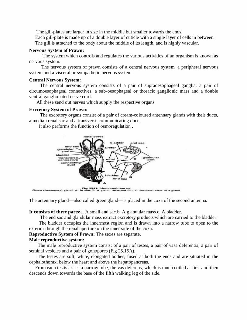
The gill-plates are larger in size in the middle but smaller towards the ends.
Each gill-plate is made up of a double layer of cuticle with a single layer of cells in between.
The gill is attached to the body about the middle of its length, and is highly vascular.
Nervous System of Prawn: The system which controls and regulates the various activities of an organism is known as
nervous system.
The nervous system of prawn consists of a central nervous system, a peripheral nervous
system and a visceral or sympathetic nervous system.
Central Nervous System: The central nervous system consists of a pair of supraoesophageal ganglia, a pair of
circumoesophageal connectives, a sub-oesophageal or thoracic ganglionic mass and a double
ventral ganglionated nerve cord.
All these send out nerves which supply the respective organs
Excretory System of Prawn: The excretory organs consist of a pair of cream-coloured antennary glands with their ducts,
a median renal sac and a transverse communicating duct.
It also performs the function of osmoregulation .
The antennary gland—also called green gland—is placed in the coxa of the second antenna.
It consists of three parts:a. A small end sac.b. A glandular mass.c. A bladder.
The end sac and glandular mass extract excretory products which are carried to the bladder.
The bladder occupies the innermost region and is drawn into a narrow tube to open to the
exterior through the renal aperture on the inner side of the coxa.
Reproductive System of Prawn: The sexes are separate.
Male reproductive system:
The male reproductive system consist of a pair of testes, a pair of vasa deferentia, a pair of
seminal vesicles and a pair of gonopores (Fig 25.15A).
The testes are soft, white, elongated bodies, fused at both the ends and are situated in the
cephalothorax, below the heart and above the hepatopancreas.
From each testis arises a narrow tube, the vas deferens, which is much coiled at first and then
descends down towards the base of the fifth walking leg of the side.
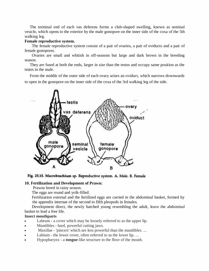
The terminal end of each vas deferens forms a club-shaped swelling, known as seminal
vesicle, which opens to the exterior by the male gonopore on the inner side of the coxa of the 5th
walking leg.
Female reproductive system.
The female reproductive system consist of a pair of ovaries, a pair of oviducts and a pair of
female gonopores.
Ovaries are small and whitish in off-seasons but large and dark brown in the breeding
season.
They are fused at both the ends, larger in size than the testes and occupy same position as the
testes in the male.
From the middle of the outer side of each ovary arises an oviduct, which narrows downwards
to open in the gonopore on the inner side of the coxa of the 3rd walking leg of the side.
10. Fertilization and Development of Prawn: Prawns breed in rainy season.
The eggs are round and yolk-filled.
Fertilization external and the fertilized eggs are carried in the abdominal basket, formed by
the appendix internae of the second to fifth pleopods in females.
Development direct, the newly hatched young resembling the adult, leave the abdominal
basket to lead a free life.
Insect mouthparts Labrum - a cover which may be loosely referred to as the upper lip.
Mandibles - hard, powerful cutting jaws.
Maxillae - 'pincers' which are less powerful than the mandibles. ...
Labium - the lower cover, often referred to as the lower lip. ...
Hypopharynx - a tongue-like structure in the floor of the mouth.
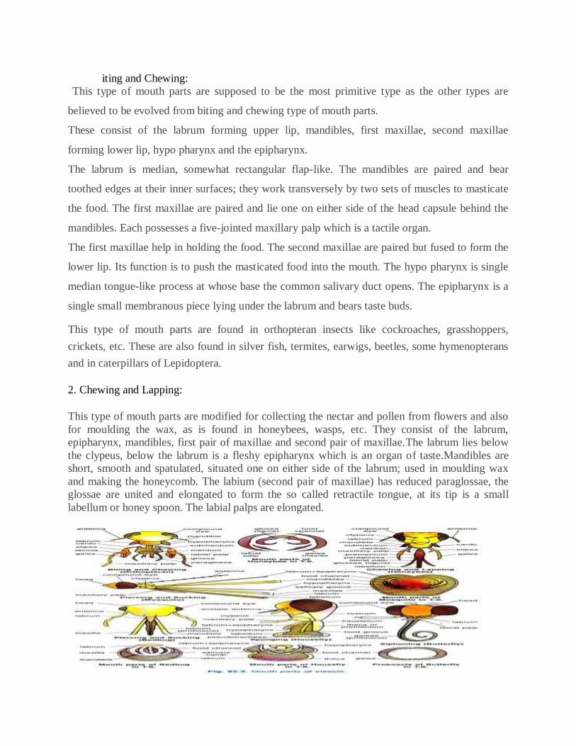
iting and Chewing:
This type of mouth parts are supposed to be the most primitive type as the other types are
believed to be evolved from biting and chewing type of mouth parts.
These consist of the labrum forming upper lip, mandibles, first maxillae, second maxillae
forming lower lip, hypo pharynx and the epipharynx.
The labrum is median, somewhat rectangular flap-like. The mandibles are paired and bear
toothed edges at their inner surfaces; they work transversely by two sets of muscles to masticate
the food. The first maxillae are paired and lie one on either side of the head capsule behind the
mandibles. Each possesses a five-jointed maxillary palp which is a tactile organ.
The first maxillae help in holding the food. The second maxillae are paired but fused to form the
lower lip. Its function is to push the masticated food into the mouth. The hypo pharynx is single
median tongue-like process at whose base the common salivary duct opens. The epipharynx is a
single small membranous piece lying under the labrum and bears taste buds.
This type of mouth parts are found in orthopteran insects like cockroaches, grasshoppers,
crickets, etc. These are also found in silver fish, termites, earwigs, beetles, some hymenopterans
and in caterpillars of Lepidoptera.
2. Chewing and Lapping:
This type of mouth parts are modified for collecting the nectar and pollen from flowers and also
for moulding the wax, as is found in honeybees, wasps, etc. They consist of the labrum,
epipharynx, mandibles, first pair of maxillae and second pair of maxillae.The labrum lies below
the clypeus, below the labrum is a fleshy epipharynx which is an organ of taste.Mandibles are
short, smooth and spatulated, situated one on either side of the labrum; used in moulding wax
and making the honeycomb. The labium (second pair of maxillae) has reduced paraglossae, the
glossae are united and elongated to form the so called retractile tongue, at its tip is a small
labellum or honey spoon. The labial palps are elongated.
Piercing and Sucking:
This type of mouth parts are adapted for piercing the tissues of animals and plants to suck blood
and plant juice, and found in dipteran insects like mosquitoes and hemipteran insects like bugs,
aphids, etc. They usually consist of labium, labrum and epipharynx, mandibles, maxillae (1st
pair) and hypo pharynx.
Peripatus resembles the arthropods in the following characteristic features:Peripatus is
special interest because its body shows certain structural characteristic of annelids and other
structures found in arthropods. They are regarded as an intermediate stage/connection between
Annelida and Arthropoda.Peripatus is a terrestrial animal found in moist places and in crevices
of rocks under bark, stones, logs and underneath fallen leaves and dark and damp places. Its
generally confined to humid habitats.
Presence of antennae.
Body is covered with chitinous cuticle like that of arthropods.
The appendages are provided with claws.
Jaws are modified appendages.
Locomotion is not annelid-like but by legs having definite musculature.
UNIT V
Phylum Mollusca (Mollusks) Characteristics
They are essentially aquatic mostly marine, few freshwater and some terrestrial form.
They may be found as hidden parasites in the interior of other animals.
They vary in size from giant squids and clams to little snails, a millimeter long.
They have at least two characters radula and mantle not found elsewhere.
The body is soft, unsegmented (except in Monoplacophora), bilaterally symmetrical,
coelomate, triploblastic.
They have tissue-system grade of body organization
The body consists of head, foot, mantle, and the visceral mass.
The body is clothed with one-layered often ciliated epidermis.
The body is commonly protected by an exoskeleton calcareous shell of one or more piece
secreted by the mantle.
Head is distinct, bearing mouth, eyes, tentacles and other sense organs except in
pelecypoda and scaphodoa.
The ventral body is modified into a muscular plough-like surface, the foot which is
variously modified for creeping, burrowing and swimming.
Mantle or pallium is a fold of a body wall that leaves between itself the main body,
mantle cavity.
The visceral mass contains the vital organs of the body in the compact form taking the
form of dorsal humps or dome.
The body cavity is hemocoel. The coelom is reduced and represented mainly by the
pericardial cavity, gonadial cavity, and nephridia.
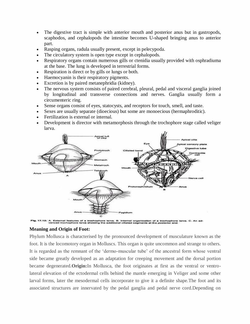
The digestive tract is simple with anterior mouth and posterior anus but in gastropods,
scaphodos, and cephalopods the intestine becomes U-shaped bringing anus to anterior
part.
Rasping organs, radula usually present, except in pelecypoda.
The circulatory system is open type except in cephalopods.
Respiratory organs contain numerous gills or ctenidia usually provided with osphradiuma
at the base. The lung is developed in terrestrial forms.
Respiration is direct or by gills or lungs or both.
Haemocyanin is their respiratory pigments.
Excretion is by paired metanephridia (kidney).
The nervous system consists of paired cerebral, pleural, pedal and visceral ganglia joined
by longitudinal and transverse connections and nerves. Ganglia usually form a
circumenteric ring.
Sense organs consist of eyes, statocysts, and receptors for touch, smell, and taste.
Sexes are usually separate (dioecious) but some are monoecious (hermaphroditic).
Fertilization is external or internal.
Development is director with metamorphosis through the trochophore stage called veliger
larva.
Meaning and Origin of Foot:
Phylum Mollusca is characterised by the pronounced development of musculature known as the
foot. It is the locomotory organ in Molluscs. This organ is quite uncommon and strange to others.
It is regarded as the remnant of the ‘dermo-muscular tube’ of the ancestral form whose ventral
side became greatly developed as an adaptation for creeping movement and the dorsal portion
became degenerated.Origin:In Mollusca, the foot originates at first as the ventral or ventro-
lateral elevation of the ectodermal cells behind the mantle emerging in Veliger and some other
larval forms, later the mesodermal cells incorporate to give it a definite shape.The foot and its
associated structures are innervated by the pedal ganglia and pedal nerve cord.Depending on
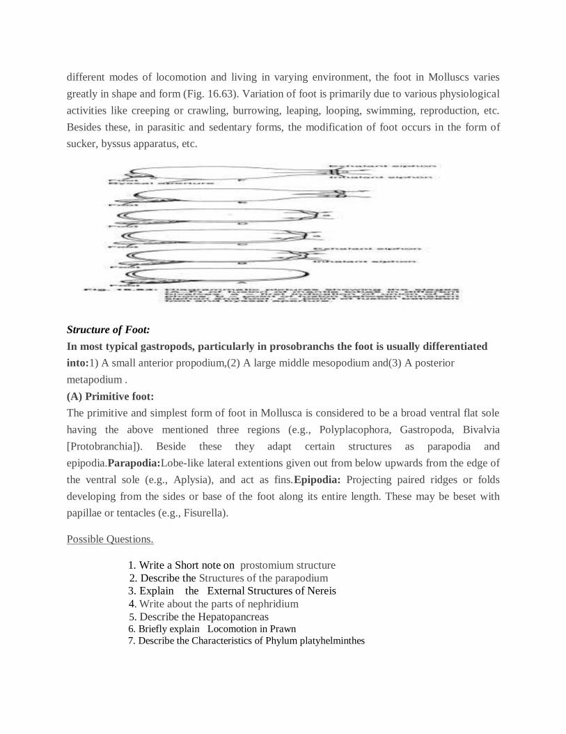
different modes of locomotion and living in varying environment, the foot in Molluscs varies
greatly in shape and form (Fig. 16.63). Variation of foot is primarily due to various physiological
activities like creeping or crawling, burrowing, leaping, looping, swimming, reproduction, etc.
Besides these, in parasitic and sedentary forms, the modification of foot occurs in the form of
sucker, byssus apparatus, etc.
Structure of Foot:
In most typical gastropods, particularly in prosobranchs the foot is usually differentiated
into:1) A small anterior propodium,(2) A large middle mesopodium and(3) A posterior
metapodium .
(A) Primitive foot:
The primitive and simplest form of foot in Mollusca is considered to be a broad ventral flat sole
having the above mentioned three regions (e.g., Polyplacophora, Gastropoda, Bivalvia
[Protobranchia]). Beside these they adapt certain structures as parapodia and
epipodia.Parapodia:Lobe-like lateral extentions given out from below upwards from the edge of
the ventral sole (e.g., Aplysia), and act as fins.Epipodia: Projecting paired ridges or folds
developing from the sides or base of the foot along its entire length. These may be beset with
papillae or tentacles (e.g., Fisurella).
Possible Questions.
1. Write a Short note on prostomium structure
2. Describe the Structures of the parapodium
3. Explain the External Structures of Nereis
4. Write about the parts of nephridium
5. Describe the Hepatopancreas
6. Briefly explain Locomotion in Prawn
7. Describe the Characteristics of Phylum platyhelminthes

8. Explain the Locomotion of Prawn
UNIT-V
General characteristics of Phylum Echinodermata
1. (Echinos: Spines; derma: Skin)
2. Kingdom: Animalia
3. Habitat: These are exclusively marine
4. Grade of organization: organ system grade
5. Germ layer: triploblastic
6. Symmetry: Adults are radially symmetrical while the larvae are bilaterally symmetrical.
7. Coelom: present ( coelomate)
8. Body without segmentation
9. The shape of the body is flat, star like, spherical or elongated.
10. Head is absent
11. Presence of tube feet
12. Presence of water vascular system
13. Mouth is present on ventral side while anus is present on dorsal side
14. Respiration: by papule, gills or clocal respiratory tree
15. Nervous system: absent, they are brainless organism.
16. Circulatory system: is reduced, heart is absent
17. Blood has no pigment.
18. Digestive system: complete
19. Excretory system: absent
20. Sexes: mostly dioecious, rarely monocious

UNIT : III
UNIT-III
தடடடபபுழுககள
தடடடபபுழுககள (Flat worms) முதுகு -
வயிறறுபபுறமாகததடடடயாககபபடடபுழுஉடலடமபடபஉடடயவிலங
குகளாகும . இவறறிலசுயாதனவாழிகளும , ஒடடுணணிகளுமஉணடு .
Planaria
உதாரணஅஙகியாகககறகபபடுமஒருசுயாதனவாழிஅஙகததவராகும .
ஈரறதடடடயன ,
நாடாபபுழுஎனபனமனிதடனதததாறறுகினறமுககியஒடடுணணிகளாக
ம . இவறறிலசுவாசதததாகுதியயா ,
சுறயறாடடதததாகுதியயாஇலலாததாலஉடலயமறபரபபினூடானவாயுப
பரவலவததடதஅதிகரிககஅதிகயமறபரபபுகயகறறவாறாகததடடட
யானஉடடலகதகாணடுளளன. இவறறிலபிரதானமாக 4
வகுபபுககளஉளளன . அடவடரதபலயலரியா (Turbelleria),
டயரமயடாடா(Trematoda), தசஸயடாடா(Cestoda), யமாயனாஜனியா(Monogenea)
,எனபடவயாகும .
இவறறிலடரபயலரியாடவததவிரஏடனயமூனறுவகுபபுககடளசசார
நதஅஙகிகளஅடனததுமஒடடுணணிகளாகும .
இவறறினஉடலடமபபுஏடனயமுபபடடவிலஙகுகடளககாடடிலுமஎளிய
தாகக காணபபடடாலும ,
இடவஒபபடடளவிலஅணடமயிலகூரபபடடநதவிலஙகுககணமாகும .
சுயாதனவாழிகளநனனரிலும , ஈரபபதனானமணணிலுமவாழகினறன .
சுயாதனவாழிகளதபாதுவாகஊனுணணிகளாகஉளளன. உதாரணமான Bipalium
எனுமடரபயலரியாவகுபடபசசாரநததடடடபபுழுமணபுழுடவபப
ிடிததுஉணணககூடி யதாகும .
இயலபுகள
முபபடடயுடடயடவ, இருபககசசமசசரானடவ .
உடறகுழிகாணபபடுவதிலடல . திணமஉடடலகதகாணடடவ.
சுயாதனவாழிகளிலதடலஉணடு . ஒடடுணணிகளிலதடலஇலடல .
தமயயானஅனுபாததுததுணடுபடலஅறறஉடல . சில
Cestodaககளிலயபாலிததுணடுபடலகாணபபடும .
யமறயறாலினூடாகபரவல முடறயிலசுவாசமநடடதபறும .
சுறயறாடடதததாகுதிகாணபபடாது .
எவவிதவனகூடடுககடடடமபபுககளுமகாணபபடாது .
கழிவகறறலசுவாடலககலஙகள (Flame cells) மூலமநிகழும .
இசசுவாடலககலததிலமுதலுருதவளிநடடஙகளஉளளன .
இவறறாலகலததிடடபபாயதபாருளிலஉளளஅனுயசபககழிவுகளும ,
நருமஅகததுறஞசபபடடுபபிசிரடிபபினாலகழிவகறறலகான
ககுளயசரககபபடடுதவளியயறறபபடும .


UNIT-IV