characterising root attachment in rhizobium-legume symbioses
TRANSCRIPT

Vio
Jack Parsons
New College
University of Oxford
A thesis submitted for the degree of Doctor of Philosophy
Trinity Term 2019
Characterising root attachment in Rhizobium-legume symbioses

1

2
Abstract
Characterising root attachment in Rhizobium-legume symbioses
Jack Parsons, New College
Submitted for the degree of DPhil., Trinity Term 2019
In Rhizobium-legume symbioses the earliest stage of physical contact between bacteria and
the plant root is primary root attachment. This is crucial for nitrogen-fixing symbiosis
development and underpins many plant growth-promoting relationships. Rhizobia use a
variety of factors for primary attachment including pH-dependent adhesins (such as
glucomannan and the hypothesised rhicadhesin), surface proteins and extracellular
polymeric substances. However, primary attachment remains an understudied area of
symbiosis development.
In this work I use a range of techniques including luminescence-based attachment assays,
mariner insertion sequencing (INSeq) and real-time imaging to investigate the factors
governing primary attachment to plant roots with the model organism Rhizobium
leguminosarum biovar viciae 3841. These techniques demonstrate that bacterial cell surface
and extracellular factors are crucial and show extensive pH-condition and plant host
specificity. Exopolysaccharide, lipopolysaccharide and peptidoglycan all show different
profiles of modification in attachment to pea roots at different pHs and attachment to barley
and soybean roots. The glycolytic enzyme TpiA is likely to be surface localized and is an
attachment factor required under all conditions. Further, outer membrane protein and
Flp/Tad pilus usage in attachment to soybean and barley roots shows that Rlv3841 can use
primary attachment mechanisms demonstrated in other bacterial species. Another novel
insight is that a filamentous hemagglutinin adhesin factor is also a previously unknown
primary attachment factor. Proteomics, attachment assays, INSeq and confocal imaging
were used to investigate rhicadhesin, demonstrating that there are multiple attachment
factors matching the criteria for this protein. As glucomannan-independent root hair
attachment is shown to be both polar and non-polar, these factors are likely distributed
across the cell surface. Results from INSeq showed that control of cyclic-di-GMP levels is
another important parameter in root attachment. It seems likely that the regulator RL4145
(required for attachment to all plants tested) functions via repression of a cyclic-di-GMP
degrading factor to promote attachment. This work also builds on root-microbe interaction
imaging technologies by developing a system suitable for Rhizobium and legume plants.
Results reinforced the idea that the root elongation zone is a crucial region for early stage
interactions, and that bacterial cell motility is important for this. Overall, this work
significantly enhances our understanding of primary attachment mechanisms in Rhizobium-
legume symbioses, demonstrating a previously unknown mechanistic complexity.

3

4
Acknowledgments
In the first instance, a debt of gratitude is owed to my supervisor Professor Philip Poole
for his remarkable support throughout, and for imparting much wisdom in the process,
both science and non-science related. Similarly, my appreciation goes to Professor Gail
Preston who, in her role as second supervisor, provided invaluable guidance. Thirdly, to
Dr Alison East. Your role was not bestowed with the title of supervisor, but without you I
have no doubt this project would be a shadow of its present self. Although I will refrain
from listing you all here, my thanks also extend to each and every member of the Poole
Lab and Department of Plant Sciences who have steered this project and provided an
engaging research environment in which to study over the last four years.
The funding sources for this project were twofold; I am particularly grateful to the
Bellingburn Trust for their generous support through the Yeotown Scholarship of New
College, and to the BBSRC for their financing through the Interdisciplinary Biosciences
Doctoral Training Partnership. Receipt of this funding was crucial to my ability to
undertake doctoral studies, and it is important that funders understand how critical their
support is, both in terms of shaping scientific research and impacting student’s lives.
New College, my ‘home base’ for the last four years in Oxford, deserves a special mention
of its own here. The warmth, friendship and intellect of the College community has
strengthened my resolve in times of difficulty, challenged me to broaden my horizons and
been a thoroughly formative influence. Manners Makyth Man.
In the same vein, I am indebted to Cumberland Lodge and all the donors who made
visiting scholarships at this charitable foundation possible. My thanks to Canon Dr
Edmund Newell, Dr Owen Gower and Dr Rachel Smillie for believing I had something to
offer, and for upholding the Lodge’s mantra by encouraging a ‘safe space for unsafe
discussion’ throughout.
Third on the institutional appreciation list is Campion Hall. The generosity of the
community is commendable. In particular, I am eternally grateful to Rev’d Dr James
Hanvey SJ and to Dr Philip Kennedy. Your wisdom, humanity and, most importantly,
friendship is and always will be a pillar of strength. I hope many more can encounter the
peace I was able to find inside the beautiful walls of the Hall.
To my friends: I wish I saw you all more often, as you are a crucial piece of this jigsaw
puzzle. For Leah Lazar (one of life’s great listeners), Izzy Gordon (you can always make
me laugh), Hamish Dustagheer (providing Mediterranean escapes and more), Mark
Blandford-Baker (being truly excellent company) and Dr Mark Byford (inspiring me on
alpine retreats), a special mention is undoubtedly deserved.
Finally, to those who, with their love and support, have enabled all I have achieved in the
last 27 years; I dedicate this thesis to my parents.

5

6
Abbreviations
AD Advantaged
ADP Adenosine diphosphate
AMP Adenosine monophosphate
Amp Ampicillin
Ampr Ampicillin resistant
Apra Apramycin
Aprar Apramycin resistant
ATP Adenosine triphosphate
AU Arbitrary units
BCA Bicinchoninic acid
BLAST Basic local alignment search tool
BNF Biological nitrogen fixation
CA Crude adhesin
c-di-GMP Cyclic di-GMP
cDNA Complementary DNA
CFU Colony forming units
CRISPR Clustered regularly interspaced short palindromic repeat
CPS Capsular polysaccharide
DE Defective
DNA Deoxyribonucleic acid
DUF Domain of unknown function
DWA Distilled water agar
EDTA Ethylenediaminetetraacetic acid
EPS Exopolysaccharide
ES Essential
FHA Filamentous hemagglutinin adhesin
FP Fahräeus plant
FV Fitness value
gDNA Genomic DNA
Gent Gentamycin
Gentr Gentamycin resistant
GFP Green fluorescent protein
GTP Guanosine triphosphate
HAD Dehalogenase-hydrolase
HEPES 4-(2-hydroxyethyl)-1-piperazineethanesulfonic acid
HITS High throughput insertion tracking by deep sequencing
7
HMM Hidden Markov model
HTH Helix-turn-helix
INSeq Insertion sequencing
IRLC Inverted repeat-lacking clade
IT Infection thread
Kan Kanamycin
Kanr Kanamycin resistant
Kdo 3-deoxy-D-manno-octulosonate
LB Lysogeny broth
LC-MS/MS Liquid chromatography-mass spectrometry
LPS Lipopolysaccharide
MOPS 3-(N-morpholino)propanesulfonic acid
NAD Nicotinamide adenine dinucleotide
NAT N-acetyl transferase
NCR Nodule-specific cysteine rich
NDK Nucleoside diphosphate kinase
NE Neutral
Neo Neomycin
Neor Neomycin resistant
NFR Nod factor receptor
NO Nitric oxide
OML Outer membrane lectin
OMP Outer membrane protein
PCR Polymerase chain reaction
PDMS Polydimethylsiloxane
PE Phosphatidylethanolamine
PEG Polyethylene glycol
PGPR Plant growth promoting rhizobacteria
PHB Polyhydroxybutarate
POTRA Polypeptide transport associated
PTS Phosphotransferase systems
REZ Root elongation zone
RNA Ribonucleic acid
RNR Ribonucleotide reductase
SDS-PAGE sodium dodecyl sulfate-polyacrylamide gel electrophoresis
SEM Standard error of the mean
SILAC Stable isotope labelling with amino acids in cell culture
SOC Super optimal broth with catabolite repression
Spec Spectinomycin
Specr Spectinomycin resistant
Str Streptomycin
8
Strr Streptomycin resistant
TA Thymine-adenine
TAE Tris-acetate EDTA
TCA Tricarboxylic acid cycle
Tet Tetracycline
Tetr Tetracycline resistant
TPS Two partner secretion
TraDIS Transposon directed insertion site sequencing
TraSH Transposon site hybridization
TRIS Tracking root interactions system
TY Tryptone yeast
UDP Uridine diphosphate
UMA Universal minimal agar
UMS Universal minimal salts
UPP Unipolar polysaccharide
UV Ultraviolet
VMM Vincents minimal media
VWA Von Willebrand factor type A

9

10
Table of Contents
Abstract .................................................................................................................. 2
Acknowledgments .................................................................................................. 4
Abbreviations ......................................................................................................... 6
Introduction ..................................................................................................................... 20
1.1 Planetary health: climate change and the nitrogen crisis .......................... 21
1.1.2 Biogeochemical flows and the nitrogen cycle ............................... 22
1.2 Evolutionary answers: the nitrogen fixers .................................................. 24
1.2.1 Biological nitrogen fixation ............................................................ 24
1.3 Trading places: lifestyle switching in the Rhizobium-legume symbioses .. 25
1.3.1 Signaling and the initiation of symbiosis ...................................... 25
1.3.2 Primary root attachment ............................................................... 26
1.3.3 Secondary root attachment ............................................................ 30
1.3.4 Infection thread formation ............................................................ 32
1.3.5 Nodule development and bacteroid formation ............................ 33
1.4 Nitrogenase biochemistry .............................................................................. 36
1.5 Can we fix it? Harnessing nitrogen fixing symbioses ................................. 38
1.5.1 Enhancing existing symbioses ........................................................ 38
1.5.2 Synthetic symbiosis approaches .................................................... 40
1.6 The Rhizobium genus ..................................................................................... 41
1.6.1 Rhizobial taxonomy ........................................................................ 41
1.6.2 Rlv3841 ............................................................................................ 42

11
1.7 ‘Omics approaches to understanding gene function in rhizobia ............... 44
1.7.1 Genetic and genomic approaches .................................................. 45
1.7.2 Transcriptomic approaches ........................................................... 47
1.7.3 Proteomic approaches .................................................................... 48
1.8 High throughput whole-genome screening with insertion sequencing ..... 49
1.8.1 Mariner insertion sequencing ........................................................ 51
1.8.2 Principles and methodology of insertion sequencing .................. 53
1.8.3 Statistical approaches to analyzing insertion sequencing data .. 56
1.9 Imaging early-stage root-microbe interactions ........................................... 61
1.10 Research objectives ...................................................................................... 63
Materials and Methods ................................................................................................... 66
2.1 Bacterial strains, plasmids and primers ...................................................... 67
2.2 Media and Antibiotics ................................................................................... 84
2.2.1 Media ............................................................................................... 84
2.2.2 Antibiotics ........................................................................................ 85
2.3 DNA techniques ............................................................................................. 86
2.3.1 Isolation of genomic DNA .............................................................. 86
2.3.2 PCR amplification .......................................................................... 87
2.3.3 Gel electrophoresis ......................................................................... 87
2.3.4 Restriction digests and DNA ligation ............................................ 88
2.4 Cloning techniques ........................................................................................ 88
2.4.1 Transformation ............................................................................... 88
2.4.2 Conjugation to transfer a plasmid from E. coli to R.
leguminosarum ......................................................................................... 88

12
2.4.3 Mutagenesis by pK19mob integration .......................................... 89
2.3.5 Transduction of R. leguminosarum ............................................... 90
2.5 Proteomics with mass spectrometry ............................................................ 90
2.5.1 Crude adhesin isolation .................................................................. 90
2.5.2 LC-MS/MS ...................................................................................... 91
2.6 Root attachment assays ................................................................................. 92
2.6.1 Buffering capacity of vermiculite .................................................. 92
2.6.2 Growth of Rlv3841 strains for Lux and insertion sequencing
attachment assays .................................................................................... 93
2.6.3 Root section attachment assays ..................................................... 93
2.6.4 Sterilisation and germination for whole root attachment assays94
2.6.5 Colony count whole root attachment assays ................................ 94
2.6.6 Lux whole root attachment assays ................................................ 95
2.7 Insertion sequencing ...................................................................................... 95
2.7.1 Mariner library construction ......................................................... 96
2.7.2 Mariner library inoculation for insertion sequencing ................. 96
2.7.3 Library preparation and sequencing ............................................ 97
2.7.4 Transposon insertion analysis using a four-state hidden Markov
model ......................................................................................................... 98
2.7.5 Transposon insertion analysis with gene fitness value calculation
................................................................................................................... 98
2.8 R. leguminosarum root interaction imaging ................................................ 99
2.8.1 Preparation of tracking root interactions systems chambers ..... 99

13
2.8.2 Seed sterilization and germination for Lux reporter testing and
tracking root interactions system ......................................................... 100
2.8.3 Lux reporter testing on roots ....................................................... 100
2.8.4 Bacterial growth and preparation for tracking root interactions
systems and chamber imaging and interaction profiling systems ..... 101
2.8.5 Tracking root interactions systems setup and confocal imaging
................................................................................................................. 101
2.8.6 Seed sterilization and germination for Chamber Imaging and
Interaction Profiling Systems (ChIIPS) ............................................... 102
2.8.7 Chamber imaging and interaction profiling systems setup and
confocal imaging (including for polarity experiments) ...................... 102
2.9 Bioinformatics, data handling and statistical methods ............................ 103
Investigating novel root attachment factors in Rhizobium using a new luminescence-
based root-attachment assay ......................................................................................... 105
3.1 Introduction ................................................................................................. 106
3.2 Results and discussion ................................................................................. 110
3.2.1 A crude adhesin fraction isolated from Rlv3841 inhibits bacterial
attachment to pea root sections ............................................................ 110
3.2.2 The 14 kDa crude adhesin band is made up of at least 15 protein
components ............................................................................................. 113
3.2.3 Evaluating the suitability of vermiculite for attachment studies at
a range of pHs ........................................................................................ 118
3.2.4 Validating Lux for measuring attachment of bacteria to whole
roots ......................................................................................................... 120
14
3.2.5 Validation of Lux-based attachment assay under different pH
conditions using a range of Rlv3841 mutants ..................................... 122
3.2.5.1 Wild-type attachment is the same at pH 6.5, 7.0 and 7.5
..................................................................................................... 125
3.2.5.2 A nifH mutant in unchanged in attachment relative to
wild-type ..................................................................................... 125
3.2.5.3 Mutants in pssA, flgE and motA are impaired in
attachment at all pHs relative to wild-type ............................. 126
3.2.5.4 A gmsA mutant is impaired in attachment at pH 6.5 and
7.0 relative to wild-type ............................................................. 126
3.2.6 praR regulation of attachment is highly dependent on pH
conditions ................................................................................................ 127
3.2.7 Attempted mutation of possible rhicadhesin genes ................... 131
3.2.8 Bioinformatic identification of possible novel root attachment
factors ..................................................................................................... 132
3.2.9 Testing possible novel Rlv3841 adhesin factor mutants in Lux
whole-root attachment assays ............................................................... 135
3.3 Conclusion .................................................................................................... 140
Genome-scale characterisation of the primary attachment determinants in the R.
leguminosarum symbiosis under acid, neutral and alkaline pH conditions ............. 145
4.1 Introduction ................................................................................................. 146
4.2 Results and discussion ................................................................................. 151
4.2.1 Root attachment assays – determining inoculum density and
bacterial recovery method for INSeq ................................................... 151

15
4.2.2 INSeq experimental design .......................................................... 154
4.2.3 HMM analysis of INSeq data ...................................................... 155
4.2.4 INSeq gene classifications ............................................................ 157
4.2.5 Validation of INSeq predictions .................................................. 158
4.2.6 Primary attachment gene requirements and functional
classifications .......................................................................................... 161
4.2.7 Genomic localization of genes required for primary root
attachment .............................................................................................. 164
4.2.8 Mapping gene requirements at different symbiosis stages from
INSeq data .............................................................................................. 165
4.2.9 Comparison of INSeq predictions and Lux attachment assays 168
4.2.10 Increasing the specificity for identification of primary root
attachment factors from INSeq results - pleiotropy filtering ............ 175
4.2.11 Primary attachment determinants required under different pH
conditions ................................................................................................ 177
4.2.12 Regulators required for primary attachment under all pH
conditions ................................................................................................ 207
4.2.13 Using INSeq to investigate rhicadhesin .................................... 209
4.3 Conclusion .................................................................................................... 213
Genome-scale characterisation of the primary attachment determinants of R.
leguminosarum to roots of a non-host legume and non-legume ................................ 223
5.1 Introduction ................................................................................................. 224
5.2 Results and discussion ................................................................................. 229

16
5.2.1 Attachment assays of Rlv3841 – determining inoculum density
and bacterial recovery method for INSeq ........................................... 229
5.2.2 INSeq experimental design .......................................................... 231
5.2.3 HMM genome analysis ................................................................. 232
5.2.4 INSeq gene classifications ............................................................ 232
5.2.5 Literature validation of INSeq predictions ................................ 234
5.2.6 Primary attachment gene requirements and functional
classifications .......................................................................................... 234
5.2.7 Genomic localization of genes required for primary root
attachment to pea, soybean and barley ............................................... 237
5.2.8 Increasing specificity of primary root attachment factor
identification from INSeq - pleiotropy filtering .................................. 241
5.2.9 Specificity in Rlv3841 primary attachment factor requirements is
highly plant-dependent .......................................................................... 242
5.2.10 Regulatory requirements for Rlv3841 primary attachment to
pea, soybean and barley roots .............................................................. 244
5.2.11 Primary attachment determinants required for interaction with
different plants ....................................................................................... 252
5.2.12 Mutation of some Rlv3841 genes leads to an increase in primary
attachment to different plants .............................................................. 277
5.3 Conclusion .................................................................................................... 282
Using real-time imaging to track early-stage interaction dynamics of R.
leguminosarum with plant roots ................................................................................... 293
6.1 Introduction ................................................................................................. 294

17
6.2 Results and discussion ................................................................................. 299
6.2.1 Establishing growth conditions for motile Rlv3841 cultures .... 299
6.2.2 Evaluating root diameter for TRIS compatibility ..................... 300
6.2.3 Preliminary reporter gene testing using a luminescence promoter
fusion ....................................................................................................... 301
6.2.4 Reporter gene testing using TRIS ............................................... 303
6.2.5 Developing Chamber Imaging and Interaction Profiling Systems
(ChIIPS) .................................................................................................. 305
6.2.6 Rlv3841 interaction dynamics with legume roots in ChIIPS .... 307
6.2.7 The role of motility in early-stage interaction dynamics .......... 310
6.2.8 Using ChIIPS to investigate root hair attachment polarity ...... 312
6.2.9 ChIIPS2 design for future work .................................................. 315
6.3 Conclusion .................................................................................................... 317
General discussion ......................................................................................................... 322
7.1 Overview ....................................................................................................... 323
7.2 Extracellular/surface localized primary attachment factor requirements
............................................................................................................................. 325
7.2.1 EPS and peptidoglycan ................................................................ 325
7.2.2 Surface enzymes ............................................................................ 327
7.2.3 Flp/Tad pili, outer membrane proteins and LPS ....................... 327
7.2.4 FHA ................................................................................................ 328
7.2.5 Motility .......................................................................................... 329
7.2.6 Rhicadhesin ................................................................................... 331
7.3 Intracellular primary attachment factors ................................................. 333

18
7.3.1 Regulators - PraR ......................................................................... 333
7.3.2 c-di-GMP regulation and regulators – RL4145 ......................... 334
7.3.3 ClpS-modulated ClpA protease ................................................... 336
7.4 Uncharacterised primary attachment factors ........................................... 336
7.5 Experimental techniques and future research directions ........................ 337
7.5.1 Lux whole-root attachment assay ............................................... 337
7.5.2 INSeq .............................................................................................. 338
7.5.3 ChIIPS and ChIIPS2 .................................................................... 339
7.5.4 The plant perspective of primary root attachment ................... 341
7.6 Concluding remarks .................................................................................... 342
Bibliography ....................................................................................................... 345
Appendix 1. Supplementary material for Chapters 3, 4 and 5 ...................... 373
Appendix 2. Supplementary material for Chapters 4, 5 and 6 ...................... 422

19

20
Chapter 1
Introduction

21
1.1 Planetary health: climate change and the nitrogen crisis
1.1.1 Climate change and the Anthropocene
Global health (human population health worldwide) and planetary health (encompassing
the state of earth’s natural systems) are inextricably linked; humanity is dependent on the
health of multiple planetary systems for survival [1]. In the Holocene era, modern human
civilisations developed and planetary changes (such as temperature fluctuations) were
effectively buffered. The industrial revolution (characterised by fossil fuel use and
industrial agriculture) enabled a new age, known as the Anthropocene, where humanity’s
impact became such that the planet began to move away from the stability of the Holocene
[1–3].
Present day global health is considered better than at any time beforehand. Reductions in
extreme poverty, higher life expectancy, lower child mortality and rapid healthcare and
technology advances evidence this [4, 5]. This rapid development is underpinned by earth
systems (oceans, forests, wetlands etc.) which provide both direct (e.g. food, fuel, water)
and indirect (e.g. nutrient cycling) goods and services [3]. However, rapid population
growth (see [6]) and continued unsustainable resource use risks irreversible earth system
alteration, which may undermine human societal development and species survival [1].
In a comprehensive evaluation of planetary health, Rockström et al (2009) developed the
planetary boundaries (PBs) framework and defined the current status of each boundary
based on risk of large negative planetary perturbation [2, 7]. The PBs are shown in Figure
1-1, below. Note that climate change and land system change sit in the zone of
uncertainty, whilst biosphere integrity and biogeochemical flows (especially nitrogen)
have moved into the high-risk categories.

22
Figure 1-1. The current status of the nine planetary boundaries. The green zone represents
the safe operating space. The yellow zone represents the zone of uncertainty and the red
zone is the high-risk zone. Processes for which global-level boundaries cannot yet be
quantified are represented by question marks. Reproduced from Steffen et al. (2015), [7].
The further these PBs are transgressed beyond the safe operating space the more likely
dramatic planetary changes become. This, in turn, impacts on the security of humanity.
Climate change itself (encapsulating all PBs) represents the greatest threat to humanity in
the 21st century and is largely driven by fossil fuel combustion and expansion of
agriculture [2]. Over 30% of non-ice or desert land globally has been converted for
agricultural purposes, and this conversion continues, causing soil degradation, forest loss
and water pollution [8–10].
1.1.2 Biogeochemical flows and the nitrogen cycle
Disruptions to the nitrogen and phosphorous cycle are major drivers of ecosystem change
and decreases in planetary health. The nitrogen cycle is the biogeochemical flows of

23
different forms of nitrogen through the atmosphere (including terrestrial ecosystems) [11]
(Figure 1-2).
Figure 1-2. The Nitrogen cycle. Red arrows indicate reduction of atmospheric dinitrogen
to ammonia (through biological or industrial processes), which provides fertilizer for
plants (green arrows). Excess ammonia is processed by soil microorganisms in
nitrification (light blue arrows) and denitrification (dark blue arrows), which, along with
leaching, converts nitrogen-containing fertilizers into pollutants. Reproduced from
Lehnert et al. (2018), [12].
Whereas biological processes for the conversion of atmospheric dinitrogen into reactive
forms are largely driven by microorganisms and lightning strikes, anthropogenic forms are
mostly due to the Haber-Bosch process. This process demands high temperatures and
pressures for the conversion of nitrogen to ammonia and is responsible for up to 2 % of
global energy use per year [13, 14].

24
Plants require biologically active forms of nitrogen (including ammonia) for growth [15];
indeed, production of nitrogen fertilizers is a large contributing factor to the increases in
global food output over the last 10 years [16]. However, only 30-50 % of reactive nitrogen
applied to fields ever reaches the intended crops. The rest leaches into terrestrial and
aquatic ecosystems, with myriad negative consequences [17]. These include biodiversity
loss, eutrophication of water systems and death of aquatic life [17, 18]. Oxides of nitrogen
are particularly damaging given their potent greenhouse gas activity and direct negative
impact on human respiratory health [3, 19].
1.2 Evolutionary answers: the nitrogen fixers
1.2.1 Biological nitrogen fixation
Biological nitrogen fixation (BNF) carried out by bacteria is responsible for ~65 % of the
available nitrogen in the biosphere [20]. Although BNF can be carried out by various
diazotrophic bacteria [21], the largest contribution to BNF comes from symbioses between
Rhizobium (soil bacteria of the Rhizobiaceae family) and legume plants [22–24].
Within this symbiosis, rhizobia reduce atmospheric dinitrogen to ammonia (NH4+) via the
nitrogenase enzyme complex. This ammonia is provided to the plant (acting as a bio-
fertilizer) and bacteria receive carbon sources (mostly as dicarboxylates) in return [25,
26]. Annually, the nitrogen fixing symbiosis provide ~40 million tonnes of bio-available
nitrogen into agricultural systems [27]. As fixed nitrogen is delivered directly from the
bacteria into plant tissues, BNF reduces the detrimental side effects of fertilizer
application, including eutrophication and greenhouse gas release [21, 24]. As these
symbioses only occur with legume crops (such as pea, soybean and alfalfa), crop rotation

25
has traditionally been used (alternating planting of cereal crops with that of legumes) to
increase soil nitrogen content, boosting crop yields. However, this technique is rarely
employed in intensive agricultural settings [18, 28].
1.3 Trading places: lifestyle switching in the Rhizobium-legume symbioses
1.3.1 Signaling and the initiation of symbiosis
Prior to the initiation of symbiosis, rhizobia live a motile, saprophytic lifestyle in the soil
in competition with a large microbial community. A single gram of soil contains up to 104
bacterial species and 109 bacterial cells [25]. The rhizosphere is an interesting zone of soil
immediately surrounding plant roots, which is influenced heavily by exudates [29, 30]. It
is in the rhizosphere that signaling processes leading to the formation of Rhizobium-
legume nitrogen fixing symbioses begin.
The process of symbiosis formation is initiated by a two-way molecular dialogue between
legume roots and symbiont Rhizobium. When soil nitrogen conditions are low
(iso)flavonoids are secreted from legume roots which act both as rhizobial
chemoattractants and inducers of Nod genes [25] (see Figure 1-3 and 1-4). Flavonoids
themselves are functional secondary plant metabolites with different chemical moieties
that convey signaling specificity with cognate symbiont rhizobia. Flavonoids are
perceived by the NodD transcription factor, which acts as a positive inducer of the nod
gene cluster in rhizobia [31]. These genes encode Nod factor, a recognition molecule
specific to each Rhizobium-legume symbiosis which, when perceived by the plant,
activates the symbiosis signaling pathway [32, 33]. The Nod factors themselves are
lipochitooligosaccharides (LCOs) with an N-acetylglucosamine oligosaccharide backbone

26
and differential molecular decoration (including sulphonation, glycosylation, methylation,
fucosylation and acetylation). They are crucial symbiosis specificity factors and determine
recognition between different legumes, which perceive them using Nod factor receptors
(NFRs). NFR activation elicits a calcium-based signaling response pathway in planta
which triggers the development of root nodules and symbiosis formation [26, 33].
1.3.2 Primary root attachment
In the first physical interaction of nitrogen fixing symbiosis, rhizobia attach to root hairs
(see Figure 1-3 and 1-4). Attachment also occurs to bulk root epidermis in a further type
of non-nitrogen fixing symbiosis [34]. In this symbiosis, plant roots exude up to 20 % of
their photosynthate through roots, and these exudates can serve as preferential growth
substrates for rhizobia [34, 35]. Host plants also derive benefits of bulk root attachment.
For example, the production of indole acetic acid and siderophores by root colonizing
Rhizobium leguminosarum has been shown to promote the growth of tomato, peppers,
maize and lettuce, among others [36–38]. These plant growth promoting rhizobacteria
(PGPR) can also protect plant roots from pathogens, likely as a result of plant immune
modulation (indirect mechanism) or pathogen exclusion (direct mechanism, see 5.1 for
further discussion) [39–43].
Recently, a common biphasic model of root attachment has been proposed which exists
across agriculturally relevant microbial species including Rhizobium, Agrobacterium,
Pseudomonas, Azospirillum and Salmonella [34]. Following migration to the root
(typically via flagella or pili mediated motility [44, 45]), universal, non-specific binding
forces mediate interaction with the root surface. These include Van der Waals forces,
electrostatic and hydrophobic interactions [34, 44, 46]. Motility assists in overcoming
27
repulsive electrostatic forces caused by bacterial cell envelope charge [44]. These initial,
reversible interactions are followed by the first (primary) stage of root attachment, which
relies on microbe specific factors.
Primary attachment is characterised by root interactions stronger than those mediated by
universal forces, but which are still reversible and often involve surface proteins,
polysaccharides and flagella [34]. Primary attachment mechanisms used can be affected
by a range of different factors, including nutrient availability and soil pH.
A good example of this is the proposed model for pH dependent primary root attachment
in R. leguminosarum, the cognate nitrogen-fixing symbiont of legumes including pea and
vetch. Under acidic to neutral soil conditions, R. leguminosarum uses the polarly located
surface polysaccharide glucomannan to bind root hair lectin [47]. Lectins themselves are
widespread carbohydrate binding proteins that act as recognition molecules in cell-cell
interactions and mediate specific yet reversible binding interactions [48, 49]. It is thought
that acidic pH lectin-mediated attachment is also used in other Rhizobium-legume
symbioses, as Bradyrhizobium japonicum can attach to soybean roots in this way [50, 51],
and legume species often show high lectin content [48].
Under alkaline pH conditions, root lectins are solubilised, and glucomannan no longer
mediates attachment [47]. It has been proposed that an extracellular rhizobial protein
termed rhicadhesin mediates rhizobial attachment to an unknown plant receptor under
these conditions. In this model rhicadhesin is bound to the bacterial cell wall by a calcium
(Ca2+) ion and may disassociate under acidic pH conditions [52]. Rhicadhesin is thought
to be a 14 kDa protein and could inhibit rhizobial root attachment to pea roots when used
as a pre-treatment [53], but neither the protein nor its encoding gene has yet been
identified. Agrobacterium is also thought to make use of rhicadhesin under alkaline

28
conditions [53, 54]. However, the definition of rhicadhesin based on its ability to inhibit
attachment may not necessarily show that it has a direct role in attachment, and it is also
possible that multiple copies of the gene could contribute to difficulties in identifying this
factor [55].
Multiple further genes are implicated in primary root attachment in R. leguminosarum,
though to what extent these are required for root hair vs. bulk root attachment is not
always clear. The importance of EPS was highlighted by a pssA (acidic EPS biosynthesis
gene) mutation in R. leguminosarum biovar viciae 3841 (Rlv3841), strongly defective in
root attachment at acidic or alkaline pH [56]. Rhizobium adhering proteins, the PlyB EPS
glycanase, RosR cell surface component regulator and predicted cadherin attachment
proteins were also implicated [57–59].
In Agrobacterium, surface proteins, molecular adhesins, pili and capsular polysaccharides
(CPS) are involved in primary attachment [60]. In Azospirillum, primary attachment is
also called adsorbtion, and is mediated by the polar flagellum [60–62]. Glycosylation of
flagellin (the flagellar subunit) is known to be important, and occurs via the same genes
involved in lipopolysaccharide (LPS) biosynthesis; mutation of these genes inhibits
primary attachment [63]. Various outer membrane proteins (OMPs), which function both
in root attachment and cellular aggregation, are also required [64]. It is additionally
proposed that the LPS O-antigen of Azospirillum directly binds maize root lectin to
mediate primary attachment [65].
Pseudomonas fluorescens utilises pili for primary attachment along with flagella and
various surface polysaccharides, although for this bacteria the process is not particularly
well characterised [66]. Outer membrane porin F (OprF) from P. fluorescens shows
adhesive properties to wheat, barley, maize and tomato roots, among others [67, 68] and

29
implicates this as a primary attachment factor. In Salmonella, flagella, fimbrae and pili
have all been shown to be important in attachment to Arabidopsis roots [69, 70]. A
summary of these different factors is shown in 1-3, below.
Overall, this highlights the wide variety in primary attachment mechanisms used across
different bacterial species. Despite this knowledge, it remains the case that primary
attachment mechanisms in Rhizobium-legume symbioses (nitrogen fixing or bulk root) are
vastly under-characterised in relation to other symbiotic stages [25].
Figure 1-3. Bacterial root attachment mechanisms. In the first stage, chemoattractants
exuded from roots cause bacteria to migrate to the root surface. Primary attachment of
single cells to the root surface follows this and can be polar, with a variety of different
factors involved depending on bacteria and environmental conditions. Secondary
attachment follows, with bacterial aggregates becoming tightly bound to the root surface.
Secondary attachment factors differ depending on bacteria. EPS – exopolysaccharide,
Raps – Rhizobium adhering proteins, CPS – capsular polysaccharide, LPS –

30
lipopolysaccharide, UPP – unipolar polysaccharide. Adapted from Wheatley et al. (2018),
[34].
1.3.3 Secondary root attachment
Secondary attachment follows primary attachment (often being dependent on it as a
precursor stage, [60]) and is characterised by tight binding to roots, which typically
involves extracellular fibril synthesis [71]. Secondary attachment secures microbial
association with the root and can be a precursor to endophyte colonization [34]. For
nitrogen-fixing rhizobia, cellulose fibrils, polysaccharides and secreted proteins permit the
accumulation of bacteria at the site of initial primary attachment [34, 72]. Secreted
proteins involved often contain Cadherin-like domains, and this has led Rap proteins to be
classed as secondary attachment factors [73, 74]. However, the requirement for RapA2
and RapC proteins in a two hour attachment assay gives weight to the idea that they are
likely involved in both primary and secondary attachment [59]. RapA1 (the most well
characterised of the Raps) is a 30 kDa polar surface protein which functions in cell
agglutination through binding EPS or capsular polysaccharide (CPS) [58], and
overexpression of Raps enhances both stages of root attachment [59, 75].
Polysaccharides also fall into this dual-role category, particularly EPS. This major cell
surface carbohydrate polymer is known as acidic EPS in R. leguminosarum and is an
octasaccharide repeating unit of glucose, glucuronic acid and galactose in a 5:2:1 molar
ratio [76]. On both abiotic and biotic surfaces, EPS deficient mutant strains show
defective attachment and colonization; root attachment itself is compromised in both
primary and secondary attachment stages [77].
31
In Agrobacterium, unipolar polysaccharide (UPP) is a polarly located polysaccharide
analogous to rhizobial glucomannan [47], although the attachment it mediates is
irreversible [55]. UPP production is thought to be stimulated by increased cellular cyclic
di-GMP (c-di-GMP), which has a key role in attachment and biofilm formation [78, 79].
Cellulose fibrils are also utilised and facilitate bacterial aggregate formation on the plant
surface; this mechanism of biofilm formation is also under c-di-GMP regulation [55].
The correlation between high c-di-GMP concentration in cells and biofilm formation (as
well as low c-di-GMP levels and motility) has been demonstrated in multiple bacteria,
including E. coli, P. aeruginosa and S. enterica [80]. Specific attachment and biofilm
formation factors under the regulation of c-di-GMP signalling include type IV pili, EPS
production and surface adhesin production, among others [81]. Intracellular c-di-GMP
concentrations are modified by altering its rate of synthesis or degradation. Diguanylate
cyclases synthesise c-di-GMP from two GTP molecules, and phosphodiesterases degrade
it [79]. In terms of protein domains, a GGDEF active site motif typically represents
diguanylate cyclase activity, whilst an EAL or HD-GYP active site motif represents
phosphodiesterase activity [82, 83]. Whilst proteins can carry both GGDEF and EAL/HD-
GYP domains, it is usually the case that only one is functional, or that a third regulatory
domain manages protein activity [84, 85]. Recently, Little et al. (2019) highlighted the
importance of c-di-GMP signalling in competitive wheat rhizosphere colonization of P.
fluorescens [86]. The relationship between high c-di-GMP levels and positive regulation
of attachment and biofilm formation has also been described for Agrobacterium and S.
meliloti [87, 88].
In Azospirillum the biosynthesis of polysaccharides and extracellular fibrils is similarly
associated with secondary attachment, including EPS and CPS, as well as LPS, OMPs and

32
outer membrane lectins (OMLs) [34]. Burdman et al. (2000) demonstrated that EPS
concentration and composition directly affects cell aggregation, with higher arabinose
content having a positive correlation with the levels of attachment and colonization [89].
A 67 kDa OML in A. brasilense specifically recognises EPS, and is thought to mediate
adhesion between attaching cells through the formation of EPS bridges, promoting biofilm
formation [62].
In Pseudomonas, cellulose fibrils contribute heavily to secondary attachment, alongside
two large adhesin proteins, LapA and LapF. LapA is thought to act in both initial polar
attachment and the initiation of biofilm formation. Initiation of LapF production in the
latter attachment stage mediates cell-cell interactions to secure biofilms [90, 91].
Cellulose fibrils are also crucial to Salmonella secondary attachment and surface
aggregate fimbrae nucleator proteins also aid in microcolony and biofilm formation [69].
Figure 1-3 (above) provides a summary of these different factors.
1.3.4 Infection thread formation
Following attachment of rhizobia to root hairs, Nod factor signaling induces the influx of
calcium (Ca2+) at the root hair tip [27]. This disrupts the apical growth profile of root hairs
and leads to curling, which entraps the bacteria in the shepherd’s crook structure [26] (see
Figure 1-4). From here they derive entry to the plant through an infection thread structure
[92]. As the infection thread is formed by an invagination of the plant cell wall, bacteria
remain topologically external to the plant, even whilst traversing toward the inner root
cortical cells [26, 72].
The infection threads are long passages which allow bacteria to migrate toward the base of
the root hair cell. Division of infecting rhizobia at the leading edge of the infection thread
33
is thought to provide the force for infection thread progression, whilst plant cytoskeletal
rearrangement in underlying cells directs the path of infection threads [93, 94].
An infection thread will travel the length of a cell, with bacteria released in the
extracellular space. From here, further infection threads form to transport rhizobia toward
the developing nodule, through epidermal and cortical cells [26, 95].
Infection thread formation is usually clonal, meaning that one initial Rhizobium cell
multiplies within the thread and populates the nodule, free from competitor bacteria [72].
Given the intimate nature of the symbiosis and the resources invested by the plant in
nodulation, checkpoints aside from Nod factor specificity exist to protect against non-
symbiont or pathogen infection. It is thought that specific rhizobial surface polysaccharide
recognition may play a role here, with cognate symbiont EPS suppressing plant defense
responses. An example of this is the interaction of EPS receptor 3 (EPR3) of Lotus
japonicus with the EPS of Mesorhizobium loti. The extracellular LysM domains of EPR3
selectively recognize cognate symbiont Rhizobium EPS, and an intracellular kinase
domain transduces a signaling cascade that is required to sustain the infection thread in L.
japonicus [96]
1.3.5 Nodule development and bacteroid formation
Within the root cortex itself, symbiosis signaling pathways leads to the concomitant
cellular differentiation and formation of root nodules (specialized structures in which BNF
takes place) during the growth of the bacterial infection thread [25]. Upon reaching the
nodule in an infection thread structure, bacteria bud off from the tip of the thread and enter
developing nodules as membrane enclosed symbiotic bacterial cells [72].
34
Two types of cortical nodule exist: indeterminate and determinate [72, 97]. Indeterminate
nodules are most commonly formed on legumes found in temperate and tropical regions,
are elongated and have different zones of development and persistent meristematic activity
[72]. Determinate nodules, however, show a loss of meristem function after development,
and are spherical, with no distinct zones [97]. The symbiosis between Rlv3841 and Pisum
sativum results in the formation of indeterminate nodules, whereas in the M. loti and L.
japonicus symbiosis determinate nodules are formed [72, 97].
In the process of becoming bacteroids, rhizobial cells undergo a differentiation process
which results in the production of nitrogenase and accessory components. These permit
the fixation of atmospheric dinitrogen into ammonia (see 1.4) [27]. Bacteroids can be
viewed as nitrogen-fixing organelles; they are enclosed by a plant-derived symbiosome
membrane across which ammonium is provided for the plant in return for carbon sources
(mostly C4 dicarboxylates), which provide an energy source for this process [25, 26].
The terminal differentiation that rhizobia undergo to become bacteroids in indeterminate
nodules involves genome endoreduplication, loss of cell division and large morphological
changes [98]. Plant derived nodule specific cysteine rich peptides (NCRs) are thought to
drive these changes in bacterial physiology. NCRs show homology to antimicrobial
peptides, and therefore represent an adaptation of plant immunity in controlling
endosymbionts [99, 100]. The presence of NCR peptides in inverted repeat lacking clade
(IRLC) legumes as well as the older Dalbergioid lineage likely demonstrates convergent
evolution; the independent evolution of terminal bacteroid differentiation in two plant
lineages has developed to occur by similar mechanisms [101, 102].
In both determinate and indeterminate nodules, bacteria become metabolically dependent
on their hosts in the nodule, which provides a protective niche for the bacteria. The

35
activation of many nitrogen-fixation related genes (including nif and fix genes) in the
bacteria is governed by low oxygen conditions, maintained by leghaemoglobin. This high
oxygen affinity protein buffers nodule oxygen concentration (important for nitrogenase
function, see 1.4) and causes pink coloration of the nodule due to the presence of the
protein heme group [103, 104].
Upon nodule senescence, bacteria from determinate and indeterminate nodules recolonize
the soil. In the former this is via de-differentiation back to their free-living state, whilst in
the latter this is via the release of undifferentiated bacteria from the infection thread [56,
105, 106].
An overview of the process from attachment to nodule formation, as well as a diagram of
an indeterminate nodule, is shown in Figure 1-4, below.
Figure 1-4. The formation of nitrogen-fixing symbiosis and structure of indeterminate
nodules. A – symbiosis development, from free-living soil bacteria to nitrogen-fixing
bacteroid. 1 – Rhizobium perceive flavonoid signals from host legume roots via the
transcriptional regulator NodD, which induces Nod factor transcription. 2 – rhizobia attach
A B

36
to roots and plant Nod factor perception induces root hair curling, trapping the bacteria in
a shepherd’s crook. 3 – infection thread formation begins with the invagination of the
plant cell wall, which expands inwards toward the root cortex and allows bacteria to
proceed toward the developing nodule structure. 4 – nodulation is completed as bacteria
bud off from the infection thread and enter the nodule, differentiating to become nitrogen-
fixing bacteroids. Fixation occurs via the nitrogenase enzyme complex in the reaction
shown. Adapted from Laranjo et al. (2014), [107]. B – A longitudinal section from a 10-
day old indeterminate alfalfa nodule infected with GFP labelled S. meliloti with the
meristem, infection and fixation zones indicated. Plant tissue is stained red with propidium
iodide. Adapted from Gage et al. (2004), [72].
1.4 Nitrogenase biochemistry
Nitrogenase is the metalloenzyme complex which is the catalyst for BNF. It catalyses the
conversion of atmospheric dinitrogen to ammonia in the following reaction [108]:
N2 + 16 ATP + 8 H+ + 8 e- → 2 NH3 + 16 ADP + 16 Pi + H2
The molybdenum-nitrogenase is the best characterised, being composed of one iron-
molybdenum protein heterodimer and two iron protein dimers (each containing an iron-
sulphur cluster and an ATP binding site) [109] (Figure 1-5). The iron-molybdenum
protein is an α2β2 tetramer with two metalloclusters in each αβ subunit pair. These
metalloclusters are the M cluster (MoFe7S9C-homocitrate) and the P cluster (Fe8S7) [110].
The iron protein dimer acts as the electron donor for N2 substrate reduction in the M
cluster [110].

37
Figure 1-5. Molybdenum nitrogenase. The two iron (Fe) protein dimers are shown at
either end of the complex with their iron-sulphur (Fe4S4) clusters. The central iron-
molybdenum protein heterodimer is composed of two α and two β subunits, with two
metalloclusters (one P - Fe8S7 – and one M - MoFe7S9C-homocitrate) in each αβ pair.
Electrons for N2 fixation are donated from the ATP binding site (where ADP is shown) of
the Fe protein dimers inwards through the iron-sulphur cluster, to the P cluster and then to
the M cluster. Figure adapted from Hu and Ribbe, 2013, [110]
Bacteroids are unable to synthesise their own homocitrate (a key M cluster component)
and rely on plant provision for nitrogen fixation. Iron and sulphur are also provided by the
plant across the symbiosome membrane [111].
Fe protein dimer
Fe protein dimer
iron-molybdenum protein heterodimer
Iron-sulphur cluster
Iron-sulphur cluster
α subunit
α subunit
β subunit
β subunit
P cluster
M cluster
P cluster
ADP
ADP
38
Nodule leghaemoglobin is crucial for nitrogenase function, as oxidation of the enzyme
iron-sulphur clusters blocks catalysis [27, 112]. The outer nodule layers form a physical
barrier to oxygen diffusion and plant mitochondrial oxygen consumption also lowers
nodule oxygen concentration [27]. Note, however, that leghaemoglobin also functions to
deliver oxygen to respiring bacteroids which must meet the high ATP demands for
nitrogen fixation [113, 114].
Of the dicarboxylic acid carbon sources provided in exchange for fixed nitrogen, malate
and succinate are the most abundant, being imported into bacteroids via the high affinity
dicarboxylic acid transport system [115–117]. These dicarboxylates enter the tricarboxylic
acid (TCA) cycle, from which ATP and reductants are produced [118].
Ammonia is thought to be provided to the plant by diffusion out of the bacteroid across
the lipid bilayer or through protein channels non-specific to ammonia, though these have
not been identified [27, 109]. Outside the bacteroid, ammonia is protonated in the
symbiosome space separating the bacteroid and the plant before transport across the
symbiosome membrane through protein channels such as AmtB [109, 111, 119].
Following transport to the plant cell, ammonia is assimilated into organic forms before
transport throughout the plant tissue [111].
1.5 Can we fix it? Harnessing nitrogen fixing symbioses
1.5.1 Enhancing existing symbioses
Given the environmental problems caused by overuse of nitrogenous fertilizers, as well as
the need to produce more food for a growing population (see 1.1), naturally evolved
nitrogen-fixing symbioses present significant opportunities for increasing the

39
sustainability and output of agricultural systems. For the legumes, this could be achieved
via enhancement of existing symbioses. Various factors affect the efficiency of nitrogen
fixing symbioses. These include phosphate availability, temperature, moisture, light, soil
acidity and soil salinity [120]. The diversity of these abiotic factors highlights the
importance of good management of agricultural soils to optimize these parameters.
Optimizing soil parameters would also mean that crop planting could be managed more
effectively.
Further, identification of the most effective rhizobial nodulating strains for different plant
species will be important for optimizing agricultural inoculants. This is because legumes
are not nodulated by the strains most effective in nitrogen fixation, but by those strains
most competitive in nodulation, which are not always the same [120]. A longer-term goal
(due to the complexity of research) is to fully characterise how plant microbiome
composition affects symbiotic efficiency. Different microbiome components can prove
positive or detrimental for nitrogen fixing symbiosis efficiency, and characterising these
effects more fully could allow tailoring of the microbiome to maximize symbiotic benefits
[120, 121].
A further possible avenue for enhancing symbioses is genetic engineering of the plant or
endosymbiont. Currently, the focus of this research area is to develop a more holistic
understanding of both legume and bacterial genetics, and their complex interplay in
symbiosis development. Emerging new technologies in genetic screening and
manipulation, particularly with CRISPR/Cas genome editing technology, show large
promise for agricultural application, including in nitrogen fixing symbiosis enhancement
[122, 123].

40
1.5.2 Synthetic symbiosis approaches
Despite the importance of nitrogen-fixing symbioses for legume crops, cereals (the highest
contributors to global calorie intake, providing almost half of all calories consumed, [124,
125]) do not possess this ability and must rely on biologically available nitrogen in the soil
or the addition of nitrogenous fertilizers [35]. Thus, the introduction of nitrogen fixation
abilities to cereals has long been a goal of agricultural biotechnology research.
There are three avenues which form the main bulk of research into developing synthetic
symbioses for non-legume crops. The first of these is to introduce the entire symbiosis
pathway from legumes into cereals and engineer rhizobia for compatibility with cereal
nodulation. This could be achieved in part by modification of the ubiquitous plant-
arbuscular mycorrhizal symbiosis signaling path, which shows extensive overlap with
nitrogen fixing symbiosis signaling [126–128]. The second approach is to engineer in
planta nitrogenase expression. This necessitates not only correct expression and assembly
of the multiple enzyme subunits, but also engineering of a suitable low oxygen
environment for enzyme function (although plant organelles, notably the chloroplast,
could prove suitable for this application, [128]). However, the complexity associated with
these two approaches means that it may be another 15 years before genuine solutions can
be delivered [128].
The most promising approach to synthetic symbiosis is the re-engineering of pre-existing
cereal-endophyte or root colonizer associations, such that bacteria are provided with
nitrogen fixation ability. This was reportedly achieved in 2013 with the transfer of
nitrogenase biosynthesis genes to Psuedomonas protegens, which was able to release
ammonium and promote growth of alfalfa and maize [129]. The shortcoming of this
strategy is the high metabolic cost of nitrogen fixation to bacteria, meaning engineered

41
bacteria are likely to be rapidly outcompeted by other soil microbes under field conditions
[35, 130]. One approach to overcoming this issue is to engineer specificity signaling
between plants and bacteria which could bias the rhizosphere in their favor and act as a
signal to express nitrogenase genes. Rhizosphere biasing has been demonstrated in
transgenic plants producing opines, with the rhizosphere community remodeled in favor of
opine catabolizing bacteria [131]. Placing the expression of nitrogen fixation genes under
the control of a specific plant signal which can act as a carbon source for a ‘synthetic
symbiont’ could help overcome limitations to this approach [35, 130, 132].
1.6 The Rhizobium genus
1.6.1 Rhizobial taxonomy
The Rhizobiaceae family are gram negative soil proteobacteria, of which ~100 species
have been described in five main genera: Rhizobium, Bradyrhizobium, Azorhizobium,
Mesorhizobium and Sinorhizobium [133, 134]. The rhizobial genomes are large and
consist of a circular chromosome and a series of plasmids [135, 136]. The complex
rhizobial genomes reflect the challenging soil environment in which they are found, where
microbial resource competition can be high and energy/nutrient sources take many forms
[137, 138]. The nodulation (nod) and nitrogen fixation (nif and fix) genes are either
encoded on a symbiosis plasmid or colocalised as a ‘symbiotic island’ in the chromosome
[135].
R. leguminosarum is a species with distinct symbiovars and has been extensively
researched [138]. The symbiovar viciae nodulates Viciae legumes including pea (Pisum
sativum) and vetch (Vicia cracca and hirsuta, among others) [139, 140]. Rlv3841 was

42
used throughout this work because of its fully sequenced genome, variety of
transcriptomic and physiological data available, well characterised growth conditions and
symbiotic engagement with agriculturally important legumes [138, 139, 141–143].
1.6.2 Rlv3841
The genome of Rlv3841 is 7.75 Mb and consists of a circular chromosome (5.08 Mb) and
six plasmids: pRL7 (0.15 Mb), pRL8 (0.15 Mb), pRL9 (0.35 Mb), pRL10 (0.49 Mb),
pRL11 (0.68 Mb) and pRL12 (0.87 Mb) (Figure 1-6). Sequencing of this genome enabled
prediction of 7263 protein coding genes, 65% of which are chromosomally encoded [138].

43
Figure 1-6. The chromosome and plasmids or Rlv3841. Plasmids are shown at relative
scale, the chromosome as 25% relative scale. Gene classifications are indicated by color:
membrane proteins (bright green), conserved and non-conserved hypotheticals (brown and
pale green, respectively), phage and transposons (pink), DNA
transcription/restriction/helicases (red, shown for chromosome only) and transcriptional
regulators (blue, shown for chromosome only). Inner circles describe deviations in GC
content (black) and GC skew (olive/maroon). Figure reproduced from Young et al.
(2006), [138].

44
Most genes essential for cellular function (e.g. ribosomal subunits) are chromosomally
encoded. Plasmids replicate and partition to daughter cells via the repABC system [144].
pRL10 is known as the symbiosis plasmid, and encodes the nod genes, as well as
nitrogenase enzyme components (the nifHDKEN cluster) and electron transfer proteins
(fixABCX) [138]. pRL8 has been classified as a rhizosphere specific plasmid, with genes
selectively expressed in this environment, although many remain of unknown function
[141]. pRL11 and 12 are considered the most ‘chromosomal’ of the plasmids, showing the
most chromosome-like gene and nucleotide composition, and have thus been designated
as ‘chromids’ [145]. pRL7 is the least well characterised of all the plasmids, with the
majority of genes having no known function [138].
Around ~35% of the Rlv3841 genome is considered ‘core’, indicating evolutionary
conservation. The remaining ~65% is considered ‘accessory’ (generally encoded on
plasmids or chromosomal islands) and is not widely conserved [138, 140]. The high
proportion of accessory genes likely reflects the environmental adaptations of Rlv3841.
Interestingly, ~35% of the annotated Rlv3841 genes remain uncharacterised, and
assigning function to these will help develop a more holistic understanding of organism
function [138].
1.7 ‘Omics approaches to understanding gene function in rhizobia
Improving our understanding of gene function’s relation to phenotype in symbioses
between Rhizobium and legumes is important on multiple fronts. For nitrogen fixing
symbiosis development, a more complete characterisation of mechanisms could aid with
both enhancing existing symbioses and developing synthetic ones (see 1.5). The symbiosis
that occurs when rhizobia attach to roots is also important. Here, carbon-rich exudates are

45
gained whilst rhizobia can shape a beneficial microbiome, produce plant growth
promoting hormones, provide nutrients and protect plants from pathogens [36–38, 41–43].
A better understanding of these characteristics has clear applications for improving plant
health and yields. Primary attachment is a particularly important, and under-characterised
stage of both these symbioses [25, 34].
Relevant to the improved characterisation of primary attachment mechanisms are genetic,
transcriptomic and proteomic approaches which can link gene function to phenotype. The
availability of sequenced genomes for Rlv3841 and other rhizobia is critical in allowing
such ‘omics based experimental approaches. Functional ‘omics approaches are now
particularly important as, whilst gene discovery and sequenced genome availability has
increased rapidly, knowledge of gene function has lagged behind [146].
Genetic approaches are often based on mutagenesis strategies, whereby gene function is
disrupted, and subsequent phenotype observed. Transcriptomic approaches quantify gene
transcripts (as RNA or cDNA) to show how gene expression changes under different
conditions, which can also point to gene function and/or regulatory networks. Proteomics
generally focusses on large scale protein identification (which shows functional protein,
rather than transcript, expression changes) but can also be used for investigating protein
localization, interaction partners and structure [147].
1.7.1 Genetic and genomic approaches
As discussed above, genetic approaches often use mutagenesis to assign gene function
based on phenotypic effects. Multiple studies demonstrate the usefulness of such an
approach in Rhizobium. Williams et al. (2008) used a Tn5 transposon cassette to disrupt
the Rlv3841 glucomannan gene. Using the resulting strain in root attachment and

46
nodulation competitiveness assays at different pHs allowed them to better define the role
of glucomannan in primary attachment and downstream symbiosis formation [56]. Hosie
et al, (2002) used similar mutagenesis approaches (as well as various transporter activity
assays) to define the role of MctP as the first permease of a new transporter subfamily,
important for monocarboxylate transport in Rlv3841 [148]. White et al. (2009) used
mutagenesis to disrupt the gtsR gene in Rlv3841, demonstrating (in combination with
transcriptomic approaches) that this gene regulates a γ-aminobutyric acid uptake operon
[142]. Whilst there are many more examples from single gene mutagenesis studies, such
approaches are time and labour intensive, making them more suited for targeted studies
than genome screens for entirely new functions.
This limitation can be overcome by isolating and screening large mutant libraries under
test conditions, with identification of mutations leading to phenotypes of interest [149].
Multiple methods exist for this, including chemical and UV mutagenesis, as well as
transposon insertion. These techniques are often faster and cheaper than targeted
mutagenesis approaches, and are particularly useful when genomes are not well
characterised [150]. In genome-wide transposon insertion studies, the site of mutagenesis
can be identified by high throughput sequencing of the transposon and its neighbouring
gDNA, and this principle has proven key to unlocking large amounts of knowledge on
gene function in different organisms [149].
Of the available transposon mutagenesis tools, Tn5 is particularly notable for studies in
Rhizobium [150, 151]. Tn5 transposon mutagenesis is random and can be highly
saturating, with almost no insertion bias, allowing the whole genome to be reliably
queried under test conditions [152–154]. Prell et al. (2012) used a Tn5 mutant library to
isolate colonies with a dry morphology on TY media. By cloning and sequencing the
47
flanking gDNA from these transposon mutants it was shown that the ptsP gene had been
disrupted. This genotype-phenotype linkage, combined with subsequent transporter
assays, identified a key role for the PTSNtr system in the global regulation of Rlv3841 ATP
transporter activity [155].
Whilst Tn5 mutagenesis screens are powerful tools, there are limitations: random
saturating mutagenesis does not allow targeting of gene subsets, library composition can
be biased to exclude low fitness mutants and compromised strains face a selective pressure
to develop second site suppressor mutations, which can mask the effects of transposon
insertion [143, 146, 151].
1.7.2 Transcriptomic approaches
Transcriptome level techniques aim to use measures of RNA abundance to quantify the
expression of genes in absolute or relative terms [147]. Identifying the conditions under
which a gene is transcribed can be useful in inferring its function, but also in determining
its regulatory relationships with other genes. The techniques reported most widely are
microarrays and RNA sequencing (RNASeq) [156, 157]. Microarray technology relies on
the reverse transcription of cellular RNA to cDNA, which is labelled with a fluorescent
probe. Genomic probe sequences are printed on a glass microarray slide and used as
hybridization targets for labelled cDNAs. Fluorescent signal from each probe spot is used
as a proxy for transcript abundance at the probe site [156]. One of the major drawbacks of
microarrays is that quantification of gene expression is limited to genomic regions of
known sequence. Microarrays have been successfully applied to the studying Rlv3841
gene expression, including in metabolic studies with glucose, pyruvate, succinate or

48
acetate as carbon sources, and to investigate rhizobial adaptations in the rhizosphere of
host and non-host plants [139, 141].
RNASeq has largely supplanted microarrays and enables unbiased, high-throughput and
direct quantitation of cellular transcripts via sequencing and bioinformatic analysis [157].
By applying RNASeq to a wild-type and rosR mutant strain of R. leguminosarum biovar
trifolii, it was shown that the rosR gene functions as a transcriptional repressor with a role
in regulating polysaccharide production, motility and aspects of metabolism [158]. More
recently, RNASeq was used to compare bacteroid gene expression profiles in determinate
and indeterminate nodules using two R. leguminosarum strains isogenic apart from their
sym plasmid. This shed light on the different conditions bacteria face in each nodule type,
including higher levels of metabolite detoxification activity required in determinate
(Phaseolus bean) nodules and increased expression of DNA replication genes in
indeterminate (pea) nodules, consistent with endoreduplication [159].
1.7.3 Proteomic approaches
An advantage of proteomic studies in relation to transcriptomics is their ability to show
changes in gene expression at the protein level, which is often the functional unit of the
gene. This is an important difference, as protein production can be modified by many
factors additional to expression of the encoding gene (see [160] as an example). Further,
proteomic studies can shed light on protein localization in the cell, interaction partners and
posttranslational modifications. The latter are difficult to predict from transcriptional or
genomic data, but can have a large effect on protein function [161].
Often, proteins in biological samples have been studied with 2D gel electrophoresis,
which separates and purifies them based on molecular weight and isoelectric focusing

49
point. Relative protein abundance can then be compared using peptide staining in Western
blots. Mass spectrometric peptide sequencing can also be used to characterise proteins and
their modifications [147]. Using peptide sequencing and the known codon preferences of
an organism can allow encoding genes to be identified from their protein products, and
such an approach could be of use in better characterising rhicadhesin (see section 1.3.2).
Liquid chromatography protein separation techniques and tandem mass spectrometry (LC-
MS/MS) is a more sensitive method of protein separation and analysis and can be applied
to protein bands partially purified by gel electrophoresis. Several studies have
demonstrated the application of proteomic approaches to characterising nitrogen-fixing
symbioses. Comparative proteomics studies of S. meliloti in free living state or in
symbiosis with alfalfa demonstrated the cellular remodelling that occurs, with nitrogen
fixation proteins, amino acid ABC transporters and stress related proteins all upregulated
[162–164].
1.8 High throughput whole-genome screening with insertion sequencing
A key aim of this work was to better characterise the range of primary attachment
mechanisms displayed by Rlv3841 under different environmental conditions. This is
important from the perspective of both basic research and subsequent symbiosis
development (be it nitrogen fixing, or bulk root-attached). In the development of this
project, it became clear that a high throughput genome wide screen would be appropriate
to address this aim. High throughput screening techniques can provide large mutant
libraries and allow screening of both characterised and uncharacterised genes for function
under test conditions. Four methods using this approach in bacteria were published in

50
2009: transposon sequencing (Tn-seq, [165]), insertion sequencing (INSeq, [166]), high
throughput insertion tracking by deep sequencing (HITS, [167]) and transposon directed
insertion site sequencing (TraDIS, [168]).
Both Tn-seq and INSeq use a transposon cassette with a type IIS restriction enzyme site,
such as MmeI for the mariner transposon used in INSeq [165, 166]. As type IIS restriction
enzymes cleave target DNA a defined number of bases downstream of their recognition
site (which is, in this case, in the mariner transposon) a gDNA sequence of 16 bp adjacent
to the transposon insertion is captured. Sequencing this tag and aligning it to the genome
reveals the location of mariner insertion, and therefore which gene is mutated in different
cells [169]. HITS and TraDIS differ in that the transposons used (himar1 and a derivative
of EZ-Tn5, respectively) do not have type IIS restriction sites. Instead, random shearing of
DNA by sonication yields gDNA tags for mapping [167, 168]. Whilst the sonication
method makes HITS and TraDIS compatible with any transposable element, Tn-seq and
INSeq require the type IIS restriction site modification. However, the mapping tag isolated
remains at 16 bp, ensuring that all reads generated are mappable [146].
Fundamentally, these techniques harness the power of high-throughput short-read
sequencing platforms to allow millions of diagnostic DNA fragments to be sequenced
simultaneously. These approaches have now replaced previous methods, some of which
used tag arrays. An example of this is transposon site hybridization (TraSH) and
derivatives, which used microarrays to detect insertions. By comparing the hybridization
signal intensity of insertion locations before and after selection in test conditions, relative
gene importance could be assessed. However, different hybridization characteristics for
each microarray spot and missing of smaller genes are key disadvantages [170, 171].
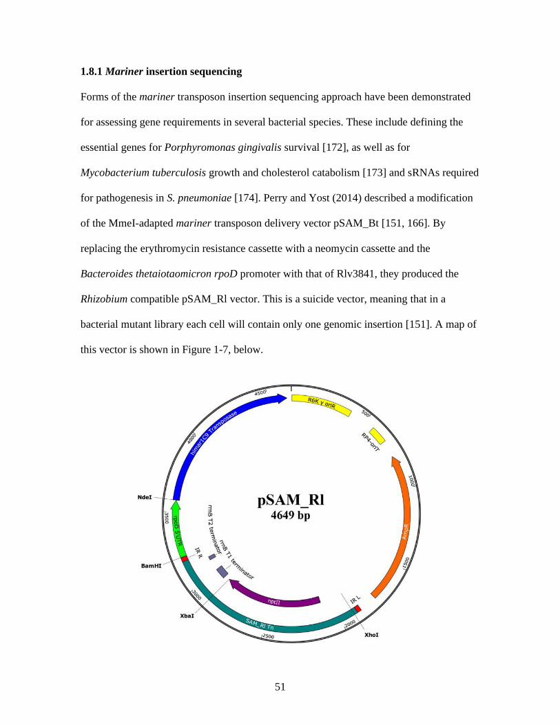
51
1.8.1 Mariner insertion sequencing
Forms of the mariner transposon insertion sequencing approach have been demonstrated
for assessing gene requirements in several bacterial species. These include defining the
essential genes for Porphyromonas gingivalis survival [172], as well as for
Mycobacterium tuberculosis growth and cholesterol catabolism [173] and sRNAs required
for pathogenesis in S. pneumoniae [174]. Perry and Yost (2014) described a modification
of the MmeI-adapted mariner transposon delivery vector pSAM_Bt [151, 166]. By
replacing the erythromycin resistance cassette with a neomycin cassette and the
Bacteroides thetaiotaomicron rpoD promoter with that of Rlv3841, they produced the
Rhizobium compatible pSAM_Rl vector. This is a suicide vector, meaning that in a
bacterial mutant library each cell will contain only one genomic insertion [151]. A map of
this vector is shown in Figure 1-7, below.

52
Figure 1-7. Plasmid map of pSAM_Rl. Antibiotic markers (ampicillin, AmpR,
neomycin/kanamycin, nptII) are shown, as well as origin of replication (R6K y oriR),
transposase (himar1C9), transposase promoter (rpoD 5’UTR), MmeI-adapted mariner
inverse repeats (IR_R, IR_L), and the transposon borne Rho-independent terminator (rrnB
T1 and rrnB T2). Reproduced from Perry and Yost (2014), [151].
Since this time, pSAM_Rl has been used to describe gene requirements for Rlv3841
growth on TY media [151], Vincent’s minimal media (VMM) [175] and under 21% and
1% oxygen conditions with glucose or succinate as carbon sources (relevant to
understanding metabolism under bacteroid conditions) [143].
The key benefit of mariner transposons is their specific insertion at thymine-adenine (TA)
motifs [176]. Unlike the use of Tn5 (with no insertion preference), the use of marniner
elements allows transposition events to be comprehensively modelled in silico prior to
mutant library analysis [151]. This allows for robust statistical analysis of gene
essentiality, as well as analysis of specific intra-gene disruption effects at different TA
sites and focusing of analysis on defined regions of the genome [151]. The overall
advantage of this is that defining gene requirements for specific test conditions does not
just rely on input and output mutant pool comparison. Rather, it can use statistical
inference based on the known number and location of insertion sites in the genome to
infer under or over represented insertions in gene regions when determining gene
essentiality [143, 151].
Barquist et al. (2013) argued that the specific TA site requirement for mariner insertion
provides a limit on potential insertion sites that can bias screen results, particularly for GC
rich genome regions. This may result in genes with very few or no insertions, and

53
therefore unreliable classifications in downstream statistical analysis [150]. However, they
also note the advantages of defined site insertion for statistical analysis; insertions specific
to TA sites allow more accurate predictions of gene essentiality in near-saturated libraries
using statistical methods (see 1.8.3), given that every potential integration site is known
and the probability of insertion at any given site can usually be assumed to be almost
equal.
The genome of Rlv3841 is particularly suitable for mariner transposon studies, with
140,056 TA sites in total, an average of 15 per gene, and only 21 genes lacking TA sites
[151, 175].
1.8.2 Principles and methodology of insertion sequencing
As reviewed by Barquist et al. (2013), all transposon mutagenesis and sequencing
methods rely on similar principles. Large, highly saturated libraries of insertion mutants
(the input library) are subject to a selection pressure, with transposon tag DNA isolated
from both the input and subsequent output library. Sequencing transposon tags and
comparing gene insertion frequency before and after selection pressure allows genes to be
classified, as deleterious insertions will be reduced in the output [150]. Figure 1-8
provides an overview of this workflow, whilst Figure 1-9 provides a schematic of the
process of library DNA preparation, which is carried out before sequencing of input and
output samples for INSeq.

54
Figure 1-8. A simplified workflow for transposon insertion mutagenesis studies. 1 –
transposon cassettes are conjugated into target organism cells for mutagenesis. Here, a
filter conjugation is depicted on a rich media plate. 2 – From the conjugation a library of
highly saturated transposon mutants is isolated. Shown in blue are cells carrying mutations
that produce no phenotype under selection conditions (step 3), whilst red cells are those
with deleterious insertions. DNA is isolated directly from a sample of this input library
(step 4). 3 – Expose transposon mutants to selection conditions. Here, selection on a
media type is shown, and the deleterious mutants (red cells) disappear from the
population. 4 – Recover DNA from both input and output library and prepare for
sequencing (see also Figure 1-9, below). 5 – Sequence prepared transposon tags (an Ion
Proton sequencing system is shown here as an example). 6 – Map reads to genome and
compare input and output library insertion frequencies. Loss of insertions in the output
library indicates genes deleterious when mutated under selection conditions and allows
gene classification.

55
Figure 1-9. A workflow of library DNA (transposon tag) preparation for INSeq. Genomic
DNA is extracted from input and output mutant libraries. A – linear PCR amplifies out
from the mariner transposon using a BioSAM 5’ biotinylated primer. B – Linear PCR
products are purified with streptavidin beads, which bind the biotin tag. C –
Hexanucleotide primers and Klenow polymerase are used for second strand synthesis. D –
DNA bound to streptavidin beads is digested with the type IIS restriction enzyme MmeI.
This cuts 16 bp downstream of the mariner insertion, forming the genomic ‘transposon
tag’ that is used for mapping. A two nucleotide ‘NN’ overhang is left after cleavage. E –
Adaptor sequences are ligated to the NN overhang (shown in orange). F – Sample specific
sequencing barcodes (shown in green) are ligated to adaptors, allowing identification of

56
which library sequencing reads originated from. A final limited PCR removes samples
from streptavidin beads and yields ~187 bp products which include the transposon tag for
sequencing (G). For simplicity, only one side of the transposon is shown in B-G, but both
sides are amplified and sequenced using this method. Figure adapted from Goodman et al.
(2011) [169].
A highly saturated mutant library is desirable in order to allow accurate assessment of
gene insertion effects before and after exposure to selection conditions. To achieve this,
multiple independent transposon conjugations (isolated following in vivo conjugation of
the transposon construct to cells of the target organism, see Figure 1-8) can be pooled
together to generate a ‘master’ input library. In addition, isolation of libraries under rich
media conditions can minimize the number of insertions deleterious in library
construction, again permitting higher input saturation [143].
1.8.3 Statistical approaches to analyzing insertion sequencing data
Using transposon mutagenesis screens, the effect of disrupting a gene under test
conditions can be deduced by comparing the frequency of transposon insertions present
before and after exposure of mutants to selective conditions. The central rationale is that
insertion events deleterious under the selection condition will reduce in frequency
compared to the input, allowing a gene to be classified. Using the TraSH approach, probes
generated from insertion sequences in the output library (after exposure to selection
conditions) were hybridized to microarrays with randomly labelled chromosomal DNA
probes, which hybridize to every microarray spot. Spots hybridizing with the genomic, but
not output, probe indicated genes required under selection conditions [171].

57
Newer technologies relying on quantitative, high-throughput sequencing of input and
output libraries make it possible to apply far more nuanced and precise statistical analyses
to categorize gene disruption effects. It is important to note that all these statistical
methods rely on three central assumptions [177]:
• abundance of insertions in the input / output library indicates gene fitness changes
under test conditions
• transposon insertion in unbiased (meaning, in INSeq, that all TA sites have equal
likelihood of being targeted by mariner)
• reads obtained from sequencing are representative of the real insertion profiles of
the input and output libraries.
These statistical models vary in complexity. ‘ESSENTIALS’ was one of the first
developed as a simple, automated tool for transposon insertion sequencing data analysis.
The software predicts gene essentiality by mapping and comparing the sequenced reads
per gene with the expected number of sequenced reads. This is calculated based on library
sequencing depth, size and number of insertion sites per gene. The cutoff for gene
essentiality is calculated based on the extent of gene underrepresentation in the output
library. The model is benchmarked on Tn-seq data from S. pneumoniae with 49 known
essential and 49 known non-essential genes [178]. ESSENTIALS thereby classifies genes
into binary essential / non-essential categories under test conditions.
Bayesian models offer more nuance than ESSENTIALS and rely on the principle that the
probability of gene essentiality can be refined continually as higher numbers of insertion
sites in each gene are sequentially included in the analysis. This method identifies the

58
largest consecutive stretch of TA sites in a gene and estimates the likelihood that such an
insertion-lacking region could occur by chance. Each individual TA site is treated as
independent and classed as essential or non-essential based on defined background
insertion frequency in non-essential genes [179]. This approach allows the statistical
significance of runs of TA sites lacking insertions in libraries to be assessed, and gene
essentiality defined. Using this method allows genes to be classed as essential even if
insertions are present at the gene termini. This is important, as previous work has shown
that transposon insertions in the extreme N and C termini of essential genes can
sometimes be tolerated, and this could confound transposon insertion sequencing results
[179–182].
Both ESSENTIALS and Bayesian approaches categorize genes as essential or non-
essential. Whilst useful for inferring function, these models do not reflect the more
nuanced ways in which gene disruption can affect bacteria under different conditions. A
hidden Markov model (HMM) can be applied to transposon insertion sequencing data to
classify genes into one of four categories, indicating how their mutation affects bacterium
fitness: essential, defective, neutral or advantaged [183].
HMMs are applicable to datasets where a sequence of observed values (such as read
counts at TA sites) is explained by an underlying state sequence (the state call of each
gene, which is hidden, in that it is not directly observable). A genome is viewed by an
HMM as an alternating sequence of different state call regions. The HMM of DeJesus et
al. (2013) uses read count information from individual TA sites to infer the probability
distribution of state calls over a string of TA sites in a gene. A Viterbi algorithm (a
dynamic programming algorithm in which the probability of each TA site state call is
based on the state probability distribution from the preceding string of TA sites) then

59
infers the most likely state call for each TA site, and for a gene. The sequential
dependence of state calls afforded by the HMM (defined by the Viterbi algorithm and
dependent on previous sites) enables a ‘smoothing’ of read count data. This allows TA
sites with no insertions which are in non-essential genes to be classed as neutral because
neighboring sites have insertions. This helps prevent erroneous labelling of TA sites as
essential just because they are, purely by chance, absent from the sequencing library. It
also helps hedge against other sources of variability such as spikes in the data, which
could be generated by PCR bias producing an artificial overrepresentation of reads at one
site, or insertions in extreme N and C termini which may be tolerated in essential genes.
Only if a consecutive sequence of TA sites with no or low insertions is long enough will
the Viterbi algorithm label sites as growth essential or defective. [183]
For the HMM to assign state calls accurately, the probability of read counts in non-
essential regions as well as the transition probabilities (the point at which the Viterbi
algorithm designates a TA as different in classification from previous sites) must be well
defined. A key strength of the HMM described by DeJesus et al. (2013) is that it is
calibrated such that it generates reasonable and robust state calls across datasets with high
or low insertion density and high or low read counts. Further, incorporating the four-state
model allows regions with low (but not no) or higher than average read counts to be
differentially classified, as defective or advantaged in this case. This allows identification
of genes where insertion impaired fitness under test conditions, as well as those that have
some ‘cost’ under test conditions, and therefore enhance organism fitness when mutated
[183].
To increase model robustness when applied to different datasets, geometric distributions
are used with data smoothing to define likelihood functions for non-neutral states. For an

60
essential state call, site read count is set very near to 1 (meaning sites with 0 reads are
likely to be essential, but that sites with 1 or 2 reads can be tolerated as neutral if
immediately preceding sites show higher read counts). For defective calls, read counts
must be approximately 100 times lower than the mean for a dataset (which is the neutral
mark), whilst for advantaged states, read counts must be 5 times the mean read count or
higher for a string of sites [183].
Combining this principle with the Viterbi algorithm generates the Markov chain, the
system in which transitions from one state call to another are dependent on probabilistic
rules which define the state calls in the previous TA sites. The geometric likelihood
functions of each state call at a given TA site, as well as the Markov chain for this model
and an example of read count data and state calls are given in Figure 1-10, below.
C
A B

61
Figure 1-10. Principles and examples of the HMM. A – Log plot of the geometric
likelihood function for each state call at a given TA site, independent of the Viterbi
algorithm. Overlapping densities of the four likelihood functions allow four distinct
classifications with one dominating individually at the boundary of each read count
parameter. B – The Markov chain for the four-state HMM. From left to right, state calls
represent read counts of increasing magnitude. The interlinked nature of all nodes means
that state transition can occur from one node to any other node in the model. Longer
arrows indicate a larger change in read count number that must occur at a TA site for the
state call to change from those at preceding sites. C – Example read counts for three genes
in the M. tuberculosis genome obtained from transposon insertion sequencing. Whilst the
left and right genes are classified as neutral, the 19 consecutive TA sites in the central
gene with zero read counts lead to an essential classification. The ‘smoothing’ of the data
afforded by the Viterbi algorithm of the HMM allows the extreme C terminal insertion
mapping read of this gene to be tolerated and maintains the essential classification.
Figures reproduced from DeJesus et al. (2013), [183].
1.9 Imaging early-stage root-microbe interactions
Prior to attachment, rhizobia must position themselves in proximity to the host plant root
[44]. Root exudates can act as chemotactic signals as well as carbon sources for rhizobia
[34, 35]. Root exudates are not uniformly exuded along root length. Rather, there seems to
be spatial distribution of exudation, which is also dependent on developmental time point
[184–187]. Given the importance of root exudates as rhizosphere community influencing
chemoattractants, and their differential presence along root length, it is possible that

62
interesting spatio-temporal interaction dynamics exist between host legume roots and
rhizobia, and that characterising these could shed more light on symbiosis stages
immediately preceding attachment.
Massalha et al. (2017) reported root-microbe interaction imaging of Arabidopsis thaliana
with Bacillus subtilis and/or E. coli using a novel microfluidics imaging system [188].
This system used polydimethylsiloxane to cast microfluidics chambers from a master
mould featuring channels for plant roots to grow through and inlet/outlet holes for
addition of labelled bacteria. By combining the microfluidics device with confocal
imaging, the preferential interaction of B. subtilis with a specific root zone (termed the
elongation zone) of Arabidopsis was demonstrated. Further, exclusion of E. coli from the
root surface by B. subtilis was shown, demonstrating that there may be a spatial aspect to
pathogen exclusion from roots [188].
Multiple further microfluidics imaging systems for plant roots have been reported (see
6.1), the most enlarged version of which (RMI-chip) was used to image root-microbe
interactions between P. fluorescens and Populus tremuloides (aspen tree) roots [189].
RMI-chip allowed root-microbe interaction imaging over a period of one month and
showed that different B. subtilis biosensors could demonstrate root exudate composition
changes over time [189].
Whilst microfluidics imaging platforms have demonstrated potential in characterising
root-microbe interactions, there have been no reports of such imaging in Rhizobium-
legume symbioses. An appropriate imaging system applied in this context could examine
root interaction dynamics as well as strain competition for root colonization and
interaction between PGPR and pathogens. The final experimental chapter of this work
aimed to develop an imaging system that could be applied to these questions.

63
1.10 Research objectives
To date the process of primary root attachment, important both for nitrogen fixing
symbioses and bulk root interaction, remains one of the least well characterised of all the
symbiosis stages [25]. In this study, I aimed to better characterise primary attachment
mechanisms of rhizobia under different environmental conditions using the model
organism Rlv3841.
Proteomics work (chapter 3) was used to investigate rhicadhesin, a factor hypothesized to
be important for root hair attachment to legumes at alkaline pHs [34]. To better
characterise root attachment, a whole-root luminescence-based (Lux) one-hour attachment
assay was developed with Rlv3841 and pea and validated at different pHs and with mutant
rhizobial strains of known attachment phenotype. A bioinformatic screen was used to
identify putative novel primary attachment factors, and mutants in these were tested in
Lux attachment assays.
Chapter 4 expanded on the findings of chapter 3 by taking a high-throughput and genome-
wide approach to characterising primary attachment determinants using an INSeq screen
at different pHs. Appropriate experimental parameters were determined and INSeq results
compared with published literature and Lux attachment assay results. The screen
identified the attachment factors required for Rlv3841 interacting with pea roots at three
pHs and was also used to further investigate rhicadhesin and the regulatory requirements
for attachment.
Whilst attachment to cognate symbiont partners is important for nitrogen fixation, rhizobia
can interact with many different plant roots in the soil [36–38], and evidence from the
literature suggests that mechanisms may be, at least in part, plant specific [34, 96].
Therefore, chapter 5 applied the principles of an INSeq attachment assay to Rlv3841 with

64
a non-host legume (soybean) and a non-legume (barley). This screen comprehensively
identified the genes that are universally needed for attachment to different plant roots and
shed new light on regulatory mechanisms underpinning Rlv3841 transition from a free-
living to root-attached state.
Given the recent advances in imaging technologies and largely uncharacterised spatio-
temporal interaction dynamics of rhizobia with legume roots (see 6.1), chapter 6 aimed to
develop a real-time imaging system that could examine these processes. Growth
conditions for highly motile bacterial cultures were established and spatio-temporal
dynamics of reporter gene activation were demonstrated in an existing [188] and newly
developed imaging platform. The role of motility in early interaction dynamics and root
hair attachment polarity under different conditions were examined with the new system,
and all these results were used to inform the design for an updated imaging system. This
system will be applicable for future plant-PGPR-pathogen studies and examining root
preference between different biovars of rhizobia.
Overall, this data has provided a comprehensive characterisation of primary attachment
determinants in Rlv3841 under different environmental conditions and in interaction with
different host plants.

65

66
Chapter 2
Materials and Methods

67
2.1 Bacterial strains, plasmids and primers
Bacterial strains used are listed in Table 2-1. All strains were stocked in 15% glycerol and
flash frozen in liquid nitrogen before being stored at -80 oC. Plasmids used are listed in
Table 2-2 and all primers used are listed in Table 2-3.

68
Table 2-1. Rhizobium and E. coli strains used in this work
Strain Description Reference
Rhizobium leguminosarum
Rlv3841 Rhizobium leguminosarum biovar viciae 3841 (Rlv3841); Strr derivative of
strain 300. Nodulates Viciae family legumes such as pea (Pisum sativum) and
Vetch (Vicia cracca)
[190]
A1045
Mutant of Rlv3841, gmsA::Tn5ΩKan/Neo. Strr, Kan/Neor
[56]
A1480 Rhizobium leguminosarum biovar viciae 300 rapA2ΩSpec rapC::Tn5ΩApra.
Specr, Aprar
[59]
A963 Rlv3841 praR::Tn5ΩKan/Neo. Strr, Kan/Neor
[191]
D5250 Rlv3841[pIJ11282]
[59]
LMB310 Rlv3841 pssA::TnΩSpec. Strr, Specr
[155]
LMB349 pLMB211 integration into Rlv3841 disrupting RL3273. Strr, Kan/Neor
[192]
LMB487 Rlv3841[pLMB579] (375bp promoter of lppE - RL3234) cloned into
pIJ11268. Strr, Tetr
[193]

69
OPS0111 A963[pIJ11282]. Strr, Kan/Neor, Tetr
Dr Vinoy Ramachandran,
postdoctoral researcher, Poole
Group, unpublished
OPS0167
Rlv3841[pOPS0065] (lppE GFP promoter fusion, pTac mCherry). Strr, Gentr
Dr Vinoy Ramachandran,
postdoctoral researcher, Poole
Group, unpublished
OPS0296
A1480[pIJ11282]. Specr, Aprar, Tetr
Dr Vinoy Ramachandran,
postdoctoral researcher, Poole
Group, unpublished
OPS0804
LMB310[pIJ11282]. Strr, Specr, Tetr
Dr Carmen Sanchez-Canizares,
postdoctoral researcher, Poole
group, unpublished
OPS0914
A1045[pIJ11282]. Strr, Kan/Neor, Tetr
This work
OPS1131
OPS2051[pIJ11282]. Strr, Kan/Neor, Tetr This work
OPS1235
OPS1783[pIJ11282]. Strr, Kan/Neor, Tetr This work
OPS1236 OPS1782[pIJ11282]. Strr, Kan/Neor, Tetr This work
OPS1237 LMB349[pIJ11282]. Strr, Kan/Neor, Tetr This work
OPS1238 OPS2054[pIJ11282]. Strr, Kan/Neor, Tetr This work

70
OPS1239
A1480::A963 phage transduction; Rhizobium leguminosarum biovar viciae
300::rapA2ΩSpec rapC::Tn5ΩApra praR::Tn5ΩKan/Neo. Specr, Aprar,
Kan/Neor
This work
OPS1266
OPS1239[pIJ11282]. Specr, Aprar, Kan/Neor, Tetr
This work
OPS1290
OPS2052[pIJ11282]. Strr, Kan/Neor, Tetr This work
OPS1291
OPS2053[pIJ11282]. Strr, Kan/Neor, Tetr This work
OPS1709
Rlv3831 RL0703::pK19mob (pOPS1095), Strr, Kan/Neor. RL0703: chemotaxis
motility protein MotA
This work
OPS1710
Rlv3831 RL0728::pK19mob (pOPS1096), Strr, Kan/Neor. RL0728: flagellar
hook protein FlgE
This work
OPS1730
A1045[pLMB449]. Strr, Kan/Neor, Gentr This work
OPS1734
Rlv3841[pLMB449]. Strr, Gentr This work
OPS1736
OPS170[pLMB449]. Strr, Kan/Neor, Gentr This work
OPS1738 OPS1710[pIJ11282]. Strr, Kan/Neor, Tetr
This work
OPS1739
OPS1709[pIJ11282]. Strr, Kan/Neor, Tetr
This work

71
OPS1782
Rlv3831 RL4382::pK19mob (pOPS0489), Strr, Kan/Neor. RL4382: putative
filamentous hemagglutinin adherence factor
This work
OPS1783
Rlv3831 RL2969::pK19mob (pOPS0487), Strr, Kan/Neor. RL2969: putative
transmembrane protein
This work
OPS1878
RU4062[pIJ11282]. Strr, Kan/Neor, Tetr This work
OPS1907
Rlv3831 RL3453::pK19mob (pOPS1294), Strr, Kan/Neor. RL3453: putative
two-component sensor histidine kinase transcriptional regulatory protein
This work
OPS1908
Rlv3831 RL4145::pK19mob (pOPS1295), Strr, Kan/Neor. RL4145: putative
LacI family HTH-type transcriptional repressor
This work
OPS1909
Rlv3831 pRL100406::pK19mob (pOPS1296), Strr, Kan/Neor. pRLl100406:
transcriptional regulatory protein MctR
This work
OPS1967
OPS1907[pIJ11282]. Strr, Kan/Neor, Tetr This work
OPS1968
OPS1908[pIJ11282]. Strr, Kan/Neor, Tetr This work
OPS1969
OPS1909[pIJ11282]. Strr, Kan/Neor, Tetr This work
OPS2051
Rlv3831 pRL110543::pK19mob (pOPS0480), Strr, Kan/Neor. pRL110543:
conserved hypothetical protein
This work

72
OPS2052 Rlv3831 pRL110071::pK19mob (pOPS0479), Strr, Kan/Neor. pRL110071:
conserved hypothetical protein
This work
OPS2053 Rlv3831 pRL100053::pK19mob (pOPS0478), Strr, Kan/Neor. pRL100053:
putative transmembrane protein
This work
OPS2054
Rlv3831 RL0109::pK19mob (pOPS0483), Strr, Kan/Neor. RL0109: conserved
hypothetical protein
This work
RU4062
pRL100162 (nifH) mutant of Rlv3841 (pRU2056 integrated). Strr, Kan/Neor [139]
E. coli
DH5α Competent E. coli strain for use in transformations, carrying the following
mutations; F-deoR endA1 recA1 relA1 gyrA96 hsdR17(rk-mk+) supE44 thi-1 -
phoA Δ(lacZYA-argF) U169 Φ80lacZΔM15 λ.
Bioline
pSAM_Rl SM10λpir carrying pSAM_Rl Mariner transposon vector; Kan/Neor, Ampr. [151]
Strr: Streptomycin resistance, Kan/Neor: Kanamycin and Neomycin resistance (conferred by a neomycin phosphotransferase II gene,
nptII), Tetr: Tetracycline resistance, Gentr: Gentamycin resistance, Aprar: Apramycin resistance, Specr: Spectinomycin resistance,
Ampr: Ampicillin resistance. Plasmid numbers are included for pK19mob strains isolated in Rlv3841 (see Table 2-2).

73
Table 2-2. Plasmids used in this work
Plasmid Description Reference
pIJ11268 Vector with promoterless reporter luciferase genes. Tetr [59]
pIJ11282 pIJ11268 with luciferase genes under control of the nptII promoter. Tetr [59]
pK19mob Mobilizable vector used for integration mutagenesis in Rhizobium. Kan/Neor
[194]
pLMB449
Reporter vector containing a gfp gene under control of pTac. Gentr Karunakaran Ramakrishnan,
postdoctoral researcher,
Poole group, unpublished
pLMB579
375bp promoter of lppE (RL3234) cloned into pIJ11268. Tetr [193]
pOPS0065 Reporter vector containing a gfp gene under control of the 375bp promoter of
lppE (RL3234) with an mCherry gene under control of pTac. Gentr
Karunakaran Ramakrishnan /
Alison East, postdoctoral
researchers, Poole group,
unpublished
pOPS0478 opx1751/oxp1752 PCR product of Rlv3841 pRL100053 BD ligated into HindIII
digested pK19mob. Kan/Neor
This work
pOPS0479 oxp1753/oxp1754 PCR product of Rlv3841 pRL110071 BD ligated into HindIII
digested pK19mob. Kan/Neor
This work

74
pOPS0480 oxp1755/oxp1756 PCR product of Rlv3841 pRL110543 BD ligated into HindIII
digested pK19mob. Kan/Neor
This work
pOPS0483 oxp1785/oxp1786 PCR product of Rlv3841 Rl0109 BD ligated into HindIII
digested pK19mob. Kan/Neor
This work
pOPS0487 oxp1767/oxp1768 PCR product of Rlv3841 Rl2969 BD ligated into HindIII
digested pK19mob. Kan/Neor
This work
pOPS0489 oxp1777/oxp1778 PCR product of Rlv3841 Rl4382 BD ligated into HindIII
digested pK19mob. Kan/Neor
This work
pOPS1095 oxp2817/oxp2818 PCR product of Rlv3841 Rl0703 BD ligated into HindIII
digested pK19mob. Kan/Neor
This work
pOPS1096 oxp2819/oxp2820 PCR product of Rlv3841 Rl0728 BD ligated into HindIII
digested pK19mob. Kan/Neor
This work
pOPS1294 oxp3235/oxp3236 PCR product of Rlv3841 Rl3453 BD ligated into HindIII
digested pK19mob. Kan/Neor
This work
pOPS1295 oxp3231/oxp3232 PCR product of Rlv3841 Rl4145 BD ligated into HindIII
digested pK19mob. Kan/Neor
This work
pOPS1296 oxp3239/oxp3240 PCR product of Rlv3841 pRL100406 BD ligated into HindIII
digested pK19mob. Kan/Neor
This work

75
pRK2013 Helper plasmid for tripartental conjugation. Kan/Neor
[195]
pSAM_Rl Mariner transposon vector. Ampr, Kan/Neor [151]
Kan/Neor: Kanamycin and Neomycin resistance (conferred by a neomycin phosphotransferase II gene, nptII), Tetr: Tetracycline
resistance, Gentr: Gentamycin resistance, Ampr: Ampicillin resistance. Primer numbers are included for PCR amplified gene products
ligated into pK19mob (see Table 2-3).

76
Table 2-3. Primers used in this work
Primer Name Sequence 5’ → 3’ Description
hah-2
AAACGGGAAAGGTTCCGTCCA Universal mapping primer for Tn5
insertions
INSeq_Adpt_Top AGATCGGAAGAGCGTCGTGTAGGGAA
INSeq Adapter (Top) [169]
INSeq_Adpt_Bottom TTCCCTACACGACGCTCTTCCGATCTNN
INSeq Adapter (Bottom) [169]
Ion Torrent BioSAM /BiotinTEG/CGGTTCGCTTGCTGTCCATAAAACC Ion Torrent BioSAM with 5' Biotin TEG
[151]
IT_A_FP_1 CCATCTCATCCCTGCGTGTCTCCGACTCAGCTAAGGT
AACGATATAAAACCGCCCAGTCTACTCGAGGG
Ion express forwards barcode primer 1
[151]. Barcode sequence is underlined
IT_A_FP_2 CCATCTCATCCCTGCGTGTCTCCGACTCAGTAAGGA
GAACGATATAAAACCGCCCAGTCTACTCGAGGG
Ion express forwards barcode primer 2
[151]
IT_A_FP_3 CCATCTCATCCCTGCGTGTCTCCGACTCAGAAGAGG
ATTCGATATAAAACCGCCCAGTCTACTCGAGGG
Ion express forwards barcode primer 3
[151]
IT_A_FP_4 CCATCTCATCCCTGCGTGTCTCCGACTCAGTACCAAG
ATCGATATAAAACCGCCCAGTCTACTCGAGGG
Ion express forwards barcode primer 4
IT_A_FP_5 CCATCTCATCCCTGCGTGTCTCCGACTCAGCAGAAG
GAACGATATAAAACCGCCCAGTCTACTCGAGGG
Ion express forwards barcode primer 5

77
IT_A_FP_6 CCATCTCATCCCTGCGTGTCTCCGACTCAGCTGCAAG
TTCGATATAAAACCGCCCAGTCTACTCGAGGG
Ion express forwards barcode primer 6
IT_A_FP_7 CCATCTCATCCCTGCGTGTCTCCGACTCAGTTCGTGA
TTCGATATAAAACCGCCCAGTCTACTCGAGGG
Ion express forwards barcode primer 7
IT_A_FP_8 CCATCTCATCCCTGCGTGTCTCCGACTCAGTTCCGAT
AACGATATAAAACCGCCCAGTCTACTCGAGGG
Ion express forwards barcode primer 8
IT_A_FP_9 CCATCTCATCCCTGCGTGTCTCCGACTCAGTGAGCG
GAACGATATAAAACCGCCCAGTCTACTCGAGGG
Ion express forwards barcode primer 9
IT_A_FP_10 CCATCTCATCCCTGCGTGTCTCCGACTCAGCTGACCG
AACGATATAAAACCGCCCAGTCTACTCGAGGG
Ion express forwards barcode primer 10
IT_A_FP_11 CCATCTCATCCCTGCGTGTCTCCGACTCAGTCCTCGA
ATCGATATAAAACCGCCCAGTCTACTCGAGGG
Ion express forwards barcode primer 11
IT_A_FP_12 CCATCTCATCCCTGCGTGTCTCCGACTCAGTAGGTGG
TTCGATATAAAACCGCCCAGTCTACTCGAGGG
Ion express forwards barcode primer 12
IT_trP1_FP CCTCTCTATGGGCAGTCGGTGATTTCCCTACACGAC
GCTCTTCCGATCT
Universal reverse barcode primer [151]
M12 Top CTGTCCGTTCCGACTACCCTCCCGAC
M12 adapter [169]

78
M12 Bottom GTCGGGAGGGTAGTCGGAACGGACAG
M12 adapter [169]
M13 uni (-21) TGTAAAACGACGGCCAGT
Sequencing/mapping primer for pK19mob
containing vectors
M13 rev (-29) CAGGAAACAGCTATGACC
Sequencing/mapping primer for pK19mob
containing vectors
oxp1751 TGATTACGCCAAGCTATGGTTGCCATCAAGC
Amplification primer (forward) for
pRL100053::pK19mob
oxp1752 GCAGGCATGCAAGCTTCTTTGAAGCGATCACGGGC
Amplification primer (reverse) for
pRL100053::pK19mob
oxp1753 TGATTACGCCAAGCTTGCGGGAAGGGGCGT
Amplification primer (forward) for
pRL110071::pK19mob
oxp1754 GCAGGCATGCAAGCTCGAAATTCGCTGCGGAAAAC
Amplification primer (reverse) for
pRL110071::pK19mob
oxp1755 TGATTACGCCAAGCTCTGCCGAAGCCAAGG
Amplification primer (forward) for
pRL110543::pK19mob
oxp1756 GCAGGCATGCAAGCTCGTTCGGCCTTTCGGG
Amplification primer (reverse) for
pRLl110543::pK19mob

79
oxp1767 TGATTACGCCAAGCTGTCCAGTCGGCGCCG
Amplification primer (forward) for
RL2969::pK19mob
oxp1768 GCAGGCATGCAAGCTAGCGGCTGTAGCAATAGTCG
Amplification primer (reverse) for
RL2969::pK19mob
oxp1777 TGATTACGCCAAGCTGATCTCCGGAACCATTGCC
Amplification primer (forward) for
RL4382::pK19mob
oxp1778 GCAGGCATGCAAGCTGGTGCGGCGGGTGTG
Amplification primer (reverse) for
RL4382::pK19mob
oxp1785 TGATTACGCCAAGCTACCGGCAAGAAGTTTTATGAT
CT
Amplification primer (forward) for
RL0109::pK19mob
oxp1786 GCAGGCATGCAAGCTATGATGTCGCTCATGTCGTCA
TC
Amplification primer (reverse) for
RL0109::pK19mob
oxp2052 TATCTCCCCCGCCGCGTTAT
RL2969::pK19mob mapping primer
(forward)
oxp2053 GCGAGGCTTGCTCCGAT
RL2969::pK19mob mapping primer
(reverse)
oxp2058 TTCGCTCGATTTTACCAAGC
RL4382::pK19mob mapping primer
(forward)
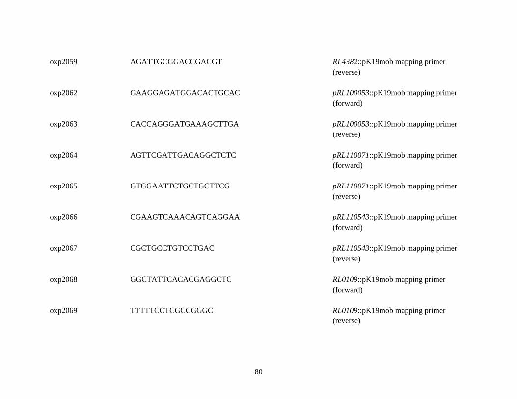
80
oxp2059 AGATTGCGGACCGACGT
RL4382::pK19mob mapping primer
(reverse)
oxp2062 GAAGGAGATGGACACTGCAC
pRL100053::pK19mob mapping primer
(forward)
oxp2063 CACCAGGGATGAAAGCTTGA
pRL100053::pK19mob mapping primer
(reverse)
oxp2064 AGTTCGATTGACAGGCTCTC
pRL110071::pK19mob mapping primer
(forward)
oxp2065 GTGGAATTCTGCTGCTTCG
pRL110071::pK19mob mapping primer
(reverse)
oxp2066 CGAAGTCAAACAGTCAGGAA
pRL110543::pK19mob mapping primer
(forward)
oxp2067 CGCTGCCTGTCCTGAC
pRL110543::pK19mob mapping primer
(reverse)
oxp2068 GGCTATTCACACGAGGCTC
RL0109::pK19mob mapping primer
(forward)
oxp2069 TTTTTCCTCGCCGGGC
RL0109::pK19mob mapping primer
(reverse)

81
oxp2423
CGGATATAGGGCTCGACGAC praR (RL0390)::Tn5 mapping primer
(forward)
oxp2424
GATCTTCGAGACGCATCTGA praR (RL0390)::Tn5 mapping primer
(reverse)
oxp2817 TGATTACGCCAAGCTTGCTTGTTGCGCACGAC
Amplification primer (forward) for
RL0703::pK19mob
oxp2818 GCAGGCATGCAAGCTATCGACAATCCGGCCGAATC
Amplification primer (reverse) for
RL0703::pK19mob
oxp2819 TGATTACGCCAAGCTCAATCAGGAAGGGCTGACCG
Amplification primer (forward) for
RL0728::pK19mob
oxp2820 GCAGGCATGCAAGCTTCCATGTCGATCATCTGGCC
Amplification primer (reverse) for
RL0728::pK19mob
oxp2821 GCCGACCATCAGCTTCGATA
RL0703::pK19mob mapping primer
(forward)
oxp2822 GCGAAATCGAGCATGTTGCC
RL0703::pK19mob mapping primer
(reverse)
oxp2823 GCGATCACCAACTACACCGA
RL0728::pK19mob mapping primer
(forward)

82
oxp2824 GGCATTGTTCAGCGAGTTGG
RL0728::pK19mob mapping primer
(reverse)
oxp3231 TGATTACGCCAAGCTATTGACATAGCCCTGGTAGC
Amplification primer (forward) for
RL4145::pK19mob
oxp3232 GCAGGCATGCAAGCTCGGGAGAAGATCAAGGAACA
Amplification primer (reverse) for
RL4145::pK19mob
oxp3233 CTCATCGTTCAGCACATCAG
RL4145::pK19mob mapping primer
(forward)
oxp3234 CCTGAGCAATGTCATATCGC
RL4145::pK19mob mapping primer
(reverse)
oxp3235 TGATTACGCCAAGCTGAAATCGGATTCCAGCTTCG
Amplification primer (forward) for
RL3453::pK19mob
oxp3236 GCAGGCATGCAAGCTTGCCTCGAACACACATATCT
Amplification primer (reverse) for
RL3453::pK19mob
oxp3237 GAAAGAAGGTACCCAGACGA
RL3453::pK19mob mapping primer
(forward)
oxp3238 CAGATATTTCCGACGATGCG
RL3453::pK19mob mapping primer
(reverse)

83
oxp3239 TGATTACGCCAAGCTATGCCGAGCTTCTTTTTGAG
Amplification primer (forward) for
pRL100406::pK19mob
oxp3240 GCAGGCATGCAAGCTGACCATATCGAAGTTGCCG
Amplification primer (reverse) for
pRL100406::pK19mob
oxp3241 CCAGACCCCATTCATCGATA
pRL100406::pK19mob mapping primer
(forward)
oxp3242 CATCAAGGAAATCAGACGGC
pRL100406::pK19mob mapping primer
(reverse)
Table 2-3. Primers used in this work. Barcode sequences in Ion Express Barcodes are shown underlined

84
2.2 Media and Antibiotics
2.2.1 Media
E. coli strains were grown at 37°C in Luria Bertani broth (LB) (10 g L-1 tryptone, 5 g L-1
yeast extract, 5 g L-1 NaCl). Cultures were shaken at 200 rpm unless otherwise stated. For
growth on solid media, agar was added 1.4 % w/v prior to autoclaving.
R. leguminosarum strains were grown on tryptone yeast (TY) media at 28oC (5 g L-1
tryptone, 3 g L-1 yeast extract, 6 mM CaCl2). For growth on solid media, agar was added
1.75 % w/v before autoclaving.
Where a minimal growth media was required, R. leguminosarum strains were grown in
Universal Minimal Salts (UMS) at 28oC. UMS was prepared as follows: 0.5 g L-1
MgSO4•7H2O, 0.2 g L-1 NaCl, 4.19 g L-1 MOPS, 0.5 mM K2HPO4, and 1 mL of trace
elements (0.375 g L-1 EDTA-Na2, 016 g L-1 ZnSO4•7H2O, 0.2 g L-1 NaMoO4, 0.25 g L-1
H3BO3, 0.2 g L-1 MnSO4•4H2O, 0.02 g L-1 CuSO4•5H2O, 1 g L-1 CoCl2•6H2O). pH was
adjusted to 7.0 before autoclaving. Media was supplemented after autoclaving with 1 mL
of calcium stock solution (75 g L-1 CaCl2•2H2O), 1 mL of iron stock solution (12 g L-1
FeSO4•7H2O dissolved in 1M HCl), and 1 mL of vitamin stock solution (1 g L-1 Thiamine
hydrochloride, 2 g L-1 D-Pantothenic acid calcium salt, 100 mg L-1 Biotin). UMS was then
supplemented with 30 mM pyruvate (or 3 mM pyruvate where low-carbon availability
conditions were required) and 10 mM ammonium chloride. Cultures were shaken at 200

85
rpm unless otherwise stated. For growth on solid media, agar was added 1.75 % w/v
before autoclaving. Where a different media pH was required, pH was adjusted before use.
For experiments examining interactions of Rlv3841 strains with plant roots, liquid FP
media was used for strain resuspension. FP media was prepared per litre as follows: 2.5
mL 0.27 M CaCl2•2H2O, 3 mL 0.16 M MgSO4•7H2O, 3.33 mL 0.22 M KH2PO4, 3.33
mL 0.32 M Na2HPO4, 2 mL 0.01 M FeC6H5O7 and 1 mL Gibson’s trace (2.86 g H3BO3,
2.03g MnSO4•4H2O, 220 mg ZnSO4•7H2O, 80 mg CuSO4•5H2O and 80 mg H2MoO4).
pH was adjusted to 7.0 before autoclaving. For plant growth on solid media, agar was
added 1.0 % w/v before autoclaving.
2.2.2 Antibiotics
For a selection of bacterial strains, antibiotics were added to the media at the
concentrations shown in Table 2-4.
Table 2-4. Concentrations (in µg mL-1) of antibiotics used in liquid and solid media.
Antibiotic Antibiotic
suspended in
Rhizobium
leguminosarum
E. coli
Ampicillin (Amp) Water
- 100
Apramycin
(Apra)
Water 50 -

86
Gentamycin
(Gent)
HEPES 20 10
Kanamycin (Kan)
Water 50 20
Neomycin (Neo)
HEPES 80/240a 20
Spectinomycin
(Spec)
HEPES 100 50
Streptomycin
(Str)
HEPES 500 25
Tetracycline (Tet) Water 5 10
a: higher concentration of neomycin used for selection of interposon mutants.
2.3 DNA techniques
2.3.1 Isolation of genomic DNA
Genomic DNA was extracted from Rlv3841 strains using a Qiagen DNeasy Blood and
Tissue kit according to the manufacturer’s instructions. 1 x 10⁹ cells were used per
isolation. Genomic DNA was diluted to 1/100 with sterile water for use in PCR
amplifications. Plasmid DNA was extracted using a Thermo Fisher GeneJET Plasmid
Miniprep kit according to manufacturer’s instructions.
87
2.3.2 PCR amplification
Primers for PCR amplification (Table 2-3) were designed using Geneious R10 [196] and
synthesized by Eurofins MWG Operon. Phusion ® High-fidelity PCR Master Mix
(Thermo Fisher) was used for amplification of DNA products for downstream cloning
reactions. GoTaq ® Green Master Mix (Promega) was used for mapping reactions.
Primer annealing temperatures were determined by Geneious R10. A Verti® thermocycler
(Applied Biosystems) was used for all reactions with conditions determined by
manufacturer’s guidelines for polymerases. PCR products were purified using a Thermo
Fisher GeneJET PCR purification kit according to manufacturer’s instructions.
Colony PCR was used for mapping reactions with E. coli strains; a single colony was
transferred into a PCR reaction using a sterile pipette tip. For mapping reactions with
Rlv3841 strains, alkaline polyethylene glycol (PEG) was used for DNA extraction [197].
A single colony was resuspended in 15 μL alkaline PEG and heated at 60oC for 10
minutes (min). 3 μL of this extract was used per 10 μL total PCR reaction volume.
2.3.3 Gel electrophoresis
Gel electrophoresis was used to separate PCR products and restriction digest fragments in
0.9 % agarose (Sigma Aldrich) with added Sybr ® Safe DNA dye (Invitrogen, according to
manufacturer’s instructions), in TAE buffer (400 mM TRIS acetate, 1 mM EDTA). Gels
were run at 120 mV for 20-30 min. Gels were visualized using a GelDoc EZ system
(BioRad).

88
2.3.4 Restriction digests and DNA ligation
Restriction digests were carried out using NEB, Roche or Thermo Fisher restriction
endonucleases and their respective buffers according to manufacturer’s instructions. DNA
ligation was performed with T4 DNA ligase and 10X T4 DNA ligase buffer (Thermo
Fisher) according to manufacturer’s instructions.
2.4 Cloning techniques
2.4.1 Transformation
E. coli DH5α cells were thawed on ice for 15 min and transformation DNA added in a
1:10 v/v ratio. Cells were incubated on ice for 30 min then heat-shocked at 42 oC for 45
seconds (sec) then incubated on ice for 2 min. SOC medium was added at 9-fold the
original transformation volume and cells shaken at 37 oC for 1 hour (hr) at 200 rpm before
plating on LB agar with appropriate antibiotics. Colonies were grown overnight at 37o C
before PCR screening and sequencing to confirm the expected construct.
2.4.2 Conjugation to transfer a plasmid from E. coli to R. leguminosarum
Triparental mating was used for conjugation of chromosomal integration plasmids from E.
coli into Rlv3841 using helper plasmid pRK2013. Three days (d) before conjugation the
recipient Rlv3841 strain was grown on a TY agar slope containing relevant antibiotics.
One day before conjugation the conjugative donor and pRK2013 containing E. coli strains
were inoculated in 10 mL LB with relevant antibiotics and grown overnight at 37oC with
shaking at 200 rpm. On the day of conjugation, overnight E. coli cultures were sub-

89
cultured (1:10 v/v) into 5 mL fresh LB with antibiotics and grown with shaking until
reaching OD600 0.4-0.6. E. coli strains were pelleted at 1,000 x g for 5 min and washed
with TY 3x to remove traces of antibiotic. The Rlv3841 slope was resuspended in 3-5 mL
TY and 400 μL recipient Rlv3841 mixed with 400 μL donor E. coli and 200 μL pRK2013.
This mixture was pelleted at 2,500 x g for 5 min and the pellet resuspended in 30 μL TY
and pipetted onto a sterile nitrocellulose filter on a TY plate and incubated at 28o C
overnight. Bacteria were then streaked from the filter onto a TY plate containing the
appropriate transformant selection antibiotics and incubated for 3-4 d at 28oC.
2.4.3 Mutagenesis by pK19mob integration
pK19mob (Table 2-2) was used to generate integration mutants. Purified pK19mob vector
was digested with HindIII. An internal fragment (~500 bp) of the target gene was
amplified with PCR primers including 15 bp of homology to the digested pK19mob vector
at the 3’- end of each primer. The internal gene fragment was cloned into the digested
pK19mob vector using InFusion ® HD cloning according to the manufacturer’s
instructions. The resulting vector was transformed into chemically competent DH5α cells
and plated on LB kanamycin. Restriction digest and sequencing with M13 uni (-21) and
M13 rev (-29) primers was used to confirm correct vector ligation. The correct pK19mob
construct was conjugated into Rlv3841 and correct conjugants selected on TY str and neo.
Genomic DNA was isolated from conjugants and used for mapping and sequencing with
relevant mapping primers (Table 2-3) and M13 uni (-21) and M13 rev (-29) to confirm
correct insertion.

90
2.3.5 Transduction of R. leguminosarum
Phage transduction was used to generate a triple mutant strain (OPS1239) from two parent
mutant strains: A1480 (R. leguminosarum biovar viciae
300 rapA2::Tn5ΩSpec rapC::Tn5ΩApra) and A963 (Rlv3841 praR::Tn5ΩKan/Neo). The
donor strain (A963) was grown for 3 d on a TY slope at 28oC before being resuspended in
3 mL sterile distilled water. 100 μL A963 resuspension was added to 900 μL serial
dilutions (10-2 – 10-6) of RL38 phage stock [198]. 4 mL of half-agar TY was added to each
dilution and these were plated on TY for 2-3 d incubation at 28oC. The plate with a
bacterial lawn nearing confluence was taken and 10 mL sterile distilled water added. After
rocking for 2 hr the supernatant (now the phage lysate) was taken and stored at 4oC after
addition of 3 drops of chloroform.
A1480 (the recipient strain) was grown for 3 d on a TY slope at 28oC and resuspended in
3 mL TY. 200 μL A1480 resuspension was mixed with a serial dilution (1-0.001 μL) of
phage lysate and incubated for 1 hr at 28oC. 100 μL of each dilution was plated on TY
spec, apra and neo plates before incubation at 28oC for 4 days. Single colony transductants
were carefully removed and purified on selective plates before genomic DNA was isolated
and used for mapping and sequencing with relevant primers (hah-2, targeting 9 base pair
repeats at the Tn5 cassette termini and oxp2363 and 2364, see Table 2-3).
2.5 Proteomics with mass spectrometry
2.5.1 Crude adhesin isolation
A crude adhesin protein fraction was isolated from Rlv3841 cells as described by Smit et
al. (1989) [53] for use in root section attachment assays. Briefly, Rlv3841 was grown in

91
TY media and cells harvested by centrifugation at OD600 0.7. The cell pellet was washed
with and resuspended in 25 mM phosphate buffer. Cells were sheared for 5 min with a
sonic dismembrator 705 (Fisher Scientific) at an amplitude of 5. The suspension was
centrifuged at 12,000 x g for 10 min at 4oC and the supernatant then centrifuged at
100,000 x g (Beckmann TL-100 ultracentrifuge) for 2 hr at 4oC. The supernatant obtained
from this step was termed the crude adhesin fraction. SDS-PAGE gels were used for
protein visualization by molecular weight according to the protocol of Smith (1994) [199].
Gels were run at 180 mV for 45 min and stained with SYPRO® Ruby (Thermo Fisher)
according to manufacturer’s instructions. Gels were imaged using a GelDoc EZ system
(BioRad). Crude adhesin protein concentration was measured using a Pierce® BCA
protein assay kit (Thermo Fisher) according to manufacturer’s instructions.
2.5.2 LC-MS/MS
A 14 kDa band from the crude adhesin protein fraction was sent for liquid
chromatography – mass spectrometry (LC-MS) at the Analytics Core Facility at the
University of Duisburg-Essen. Experiments were performed on an Orbitrap Elite
instrument (Thermo Fisher) that was coupled to an EASY-nLC 1000 liquid
chromatography (LC) system (Thermo Fisher) as described in [200] with the following
adjustments: MS/MS spectra data were searched against the UniProt reference database
UP000006575_216596.fasta (Rlv3841, 7091 entries). The MS/MS target protein range
was set to 12-17 kDa. For data analysis, only protein groups with at least three identified
unique peptides over all runs were considered for further analysis. Non-Rlv3841 proteins
and hits to the decoy database were removed.
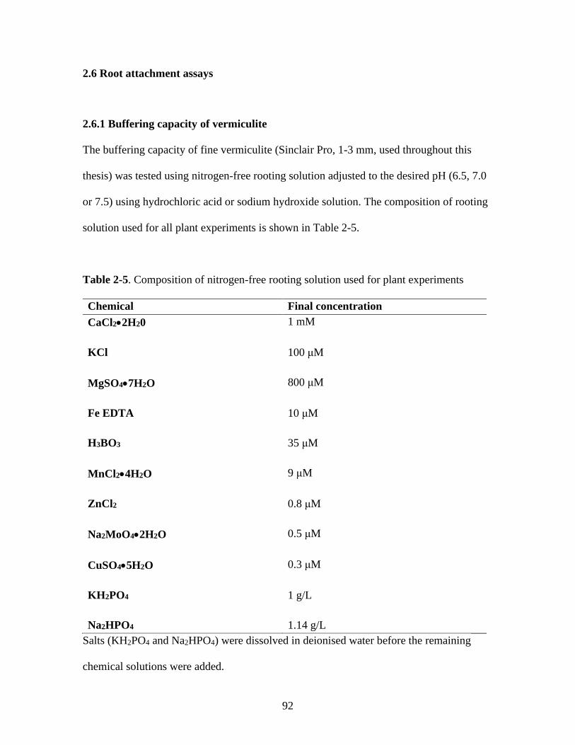
92
2.6 Root attachment assays
2.6.1 Buffering capacity of vermiculite
The buffering capacity of fine vermiculite (Sinclair Pro, 1-3 mm, used throughout this
thesis) was tested using nitrogen-free rooting solution adjusted to the desired pH (6.5, 7.0
or 7.5) using hydrochloric acid or sodium hydroxide solution. The composition of rooting
solution used for all plant experiments is shown in Table 2-5.
Table 2-5. Composition of nitrogen-free rooting solution used for plant experiments
Chemical Final concentration
CaCl2•2H20
1 mM
KCl
100 μM
MgSO4•7H2O
800 μM
Fe EDTA
10 μM
H3BO3
35 μM
MnCl2•4H2O
9 μM
ZnCl2
0.8 μM
Na2MoO4•2H2O
0.5 μM
CuSO4•5H2O
0.3 μM
KH2PO4
1 g/L
Na2HPO4 1.14 g/L
Salts (KH2PO4 and Na2HPO4) were dissolved in deionised water before the remaining
chemical solutions were added.

93
25 mL rooting solution at the desired starting pH was added to 10 g fine vermiculite in
triplicate in 50 mL Falcon tubes. Tubes were rotated at 20 rpm and the pH of rooting
solution measured at defined intervals from 1 min to 72 hr using a pH meter (Hanna).
2.6.2 Growth of Rlv3841 strains for Lux and insertion sequencing attachment assays
Rlv3841 strains were grown on UMA slopes adjusted to the desired pH for Lux root
attachment assays. For insertion sequencing assays, the Rlv3841 mariner transposon
mutant library (see 2.7.1) was grown on UMA slopes at pH 7.0.
2.6.3 Root section attachment assays
Pea seeds (Pisum sativum variety Avola, used throughout this thesis) were sterilized by
immersion in 95 % ethanol in a sterile flask for 30 sec before rinsing with sterile distilled
water and immersing in 2 % sodium hypochlorite solution for 5 min. Seeds were rinsed
10x in sterile distilled water, before transferring seeds onto 1 % distilled water agar
(DWA) plates and incubating in the dark at room temperature for 3 d. Root section
attachment assays were carried out as described in [201]. Briefly, root sections were
incubated for 1 hr in phosphate buffer (pH 7.0) with or without 600 μg total crude adhesin
protein before washing in fresh phosphate buffer. D5250 (Rlv3841[pIJ11268]) grown on
TY slopes for 3 days at 28oC was resuspended in liquid TY. 20 mL of the resuspension
(OD600 = 0.1) was added to pea roots of each group and roots incubated on a rocking
platform for 1 hr. D5250 luminescence was assayed in triplicate using a GloMax ® Multi+
detection system (Promega). Roots were washed and weighed before luminescence was
measured using a GloMax ®-Multi Jr single-tube multimode reader (Promega). Data was

94
normalized for the weight of roots and luminescence of starting culture, which was used to
calculate the relative luminescence of a single bacterial cell. Roots were also imaged using
a NightOWL II LB 983 imaging system (Berthold).
2.6.4 Sterilisation and germination for whole root attachment assays
Seeds of pea (P. sativum), soybean (Glycine max) and barley (Hordeum vulgare) were
sterilized as described in 2.6.3. Boiling tubes (200 mL size, DURAN ®) were filled ¾ full
with vermiculite before 25 mL rooting solution (see Table 2-5) adjusted to the desired pH
was added. After sealing with foam bungs, boiling tubes were autoclaved. Sterile seeds
were planted 1 per tube and germinated under foil for 4 d in a controlled growth chamber
at 23oC. Plants were grown for a further 3d with a 16:8 (light:dark) photoperiod.
2.6.5 Colony count whole root attachment assays
To quantify attachment of Rlv3841 strains by colony count, seeds were germinated and
grown as described in 2.6.4. Rlv3841 strains were grown on UMA slopes (see 2.2.1) at pH
7.0. Plants were removed from vermiculite and Rlv3841 strains resuspended to OD600 0.1
in 15 mM MES/HEPES buffer (adjusted to pH 7.0). Strain luminescence was tested as
described in 2.6.3. Plant roots were washed by dipping in 15 mM MES/HEPES buffer and
placed in fresh boiling tubes. 50 mL Rlv3841 strain resuspension was added, with plants
secured such that only roots were immersed. Control plants had 50 mL 15 mM
MES/HEPES added. After a 1 hr incubation at room temperature with gentle shaking (20
rpm), plants were removed and washed by dipping in MES/HEPES buffer. Shoots were
excised and roots weighed before placing in 50 mL Falcon tubes with 20 mL fresh
MES/HEPES buffer and vortexing (Heidolph Multi Reax shaker) at the maximum speed

95
for 10 min. For attachment assays in which roots were also ground, a pestle and mortar
were used after vortexing to grind roots in 10 mL fresh MES/HEPES buffer, which was
then pooled with the buffer from vortexing. The suspension was centrifuged at 2,500 x g
for 5 min to pellet the Rlv3841 cells released from the roots. Each pellet was resuspended
in 1 mL TY, with 100 μL volumes of serial dilutions plated onto TY agar containing strep.
Plates were incubated at 28oC for 3-4 d before the colonies were counted.
2.6.6 Lux whole root attachment assays
For whole-root attachment assays of Rlv3841 strains using luminescence (Lux assays),
seeds were germinated and grown as described in 2.6.4 and plant roots inoculated as
described in 2.6.5. Strain luminescence was tested as described in 2.6.3. After 1 hr
incubation at room temperature with gentle shaking (20 rpm), plants were removed from
inoculum and washed by dipping in MES/HEPES buffer. Shoots were excised and roots
imaged using a NightOWL II LB 983 imaging system (Berthold). Lux assays were
conducted with plants grown with rooting solution at pH 6.5, 7.0 or 7.5. For these pH
groups, Rlv3841 strains were grown on UMA slopes of matching pH, and 15 mM
MES/HEPES buffer was also of matching pH. Data was normalized as described in 2.6.3.
2.7 Insertion sequencing
Insertion sequencing protocols were used from Goodman et al. (2011) [169] and Perry and
Yost (2014) [151] with adjustments listed below.
96
2.7.1 Mariner library construction
Plasmid pSAM_Rl carrying E. coli donor cells (Table 2-1) were grown in LB with kan
and amp overnight and Rlv3841 recipient cells were grown on a TY strep agar slope and
resuspended in TY. Cultures were pelleted at 2,500 x g for 5 min and washed 3x in TY.
Donor and recipient cells were pooled in equal ratios, pelleted, resuspended in 30 μL TY
and plated on nitrocellulose filters on TY agar for conjugation. After overnight incubation
at 28 oC, bacteria from conjugations were resuspended in 15 % glycerol UMS and serial
dilutions plated on TY str, neo to quantify Rlv3841 transposon insertion mutant
concentration. Approximately 300,000 colony forming units (CFU) from each conjugation
was plated in triplicate on 245 x 245 x 25 mm TY str, neo plates and grown at 28 oC for
12-18 hours until pinprick colonies formed. Colonies were scraped off plates and
resuspended in 15% glycerol UMS. Three master mariner transposon Rlv3841 input
libraries were formed from the pooling of two independent pSAM_Rl/Rlv3841
conjugations each.
2.7.2 Mariner library inoculation for insertion sequencing
Mariner libraries (isolated as described in 2.7.1) were grown from -80 oC stocks on UMA
slopes at pH 7.0. Plants were grown as described in 2.6.4 and inoculated as described in
2.6.5. The input library DNA was extracted at this point. Output libraries (Rlv3841
mariner transposon mutants after 1 hr root attachment assay) were recovered from roots
by vortexing as described in 2.6.5. To decrease the contamination with plant DNA and
increase bacterial DNA for extraction, output libraries were grown in TY for 12 hr at 28oC
with shaking at 200 rpm. DNA was extracted from output libraries using a Qiagen
DNeasy Blood and Tissue kit according to the manufacturer’s instructions with the

97
following adjustments: 2 μL RNase A (100 mg/mL) was added alongside proteinase K,
and DNA was eluted in 100 μL MilliQ water. DNA was quantified using a NanoDrop
spectrophotometer.
2.7.3 Library preparation and sequencing
Library preparation was carried out separately for each of the input and output libraries.
Transposon-tags were prepared for DNA sequencing using the method described in
Wheatley et al. (2017) [143] to make the process compatible with the Ion Proton
sequencing platform (Thermo Fisher). Briefly, linear PCR products were amplified using
BioSAM primers with 500 ng template DNA. Biotinylated PCR products were purified
(GeneJet PCR purification kit) and bound to Pierce streptavidin magnetic beads (Thermo
Fisher). Klenow fragment (3’→5’ exo-, NEB) was used for second strand synthesis before
digestion with MmeI (using an M12 fragment in trans for efficient DNA cleavage) and
ligation of an Ion Proton sequencing adapter. A final round of PCR amplification was
performed with a unique barcoded sequencing primer used for each sample. This
barcoding (using twelve different IonXpress barcoding sequences, Thermo Fisher)
enabled automated sequencing read separation by experimental sample. An Ion Torrent
system (Thermo Fisher) reverse sequencing target ‘trP1’ was used for all samples in
conjunction with the unique forward barcoded primer. All primers are listed in Table 2-3.
The end sequencing template was 187 bp and was gel purified from the PCR reactions
using E-Gel SizeSelect II agarose gels (Thermo Fisher). DNA for sequencing from each
library was analysed on a Bioanalyzer high sensitivity DNA chip (Agilent Technologies),
diluted to 100 pM and pooled in equimolar ratios. An Ion Chef and Ion PI chip kit V3
98
(Thermo Fisher) was used for template preparation of pooled barcoded libraries, and
sequencing was performed on an IonProton system (Thermo Fisher).
2.7.4 Transposon insertion analysis using a four-state hidden Markov model
Sequencing reads were analyzed on a Linux server as previously described [151, 169].
Cutadapt [202] was used for quality trimming and adapter sequence removal and resulting
transposon tags were checked for a leading TA motif with a custom Perl script. Tags were
mapped to the Rlv3841 reference genome (downloaded from Rhizobase,
http://genome.annotation.jp/Rhizobase) using Bowtie [203] and split by replicon. Files
were converted to .wig format using a custom Perl script and analyzed further with the Tn-
HMM (Hidden Markov Model) Python module [183]. This module calculates the HMM
state of each thymine-adenine (TA) site (at which the Mariner transposon can insert,
[151]) and then determines the state of all TA sites in a gene to assign a gene to one of
four possible classifications, indicating how mutation affects bacterium fitness: essential,
defective, neutral or advantaged. Gene annotations were obtained from UniProt and from
a lab-curated annotation file.
2.7.5 Transposon insertion analysis with gene fitness value calculation
Fitness values were calculated for genes (reflecting the impact of mutation on bacterium
fitness) as described in [143], allowing quantification of mutants retrieved for any gene in
the library as well as numerical comparison of gene fitness across different test conditions
(after standardising fitness values per million library reads). The equation for fitness value
is given by:

99
Fitness value = (potential insertion sites x insertion density) x mean read count
Potential insertion sites is the number of TA sites within the annotated length of a gene.
Insertion density is the proportion of TA sites in a gene with one or more insertions. Mean
read count is the mean number of reads for TA sites with at least one insertion. Fitness
values can be standardised per million library reads
2.8 R. leguminosarum root interaction imaging
Tracking root interactions system (TRIS) experiments were conducted as described in
Massalha et al (2017) [188], with adjustments described below.
2.8.1 Preparation of tracking root interactions systems chambers
TRIS chambers were prepared as described in [188]. Briefly, photolithography was used
to pattern microchannels onto a layer of SU-8 2100 negative tone resist (Microchem) on a
silicon wafer (Agar Scientific). A mask aligner was used to expose the photoresist layer
through a custom mask designed by AutoCAD. The crosslinked photoresist formed the
master mould. Polydimethylsiloxane (PDMS) and crosslinker (Sylgard 184 silicon
elastomer kit, Dow-Corning) were mixed according to manufacturer’s instructions and
poured into the master mould. PDMS was cured by heating at 60 oC overnight and slabs
(now referred to as TRIS chambers) were cut from the master mould and inlet/outlet/root
holes added using a 1 mm biopsy punch (Agar Scientific). TRIS chambers were mounted
on a glass slide using plasma bonding. A diagram of a completed TRIS chamber can be
seen in Figure 6-1 A, page 297.

100
2.8.2 Seed sterilization and germination for Lux reporter testing and tracking root
interactions system
For Lux reporter testing, seeds of vetch (Vicia cracca and Vicia hirsuta) and clover
(Trifolium repens and Trifolium pratense) were gently scarified with sandpaper before
immersion in 2% sodium hypochlorite solution for five min. Seeds were rinsed 10x in
sterile distilled water and immersed in sterile distilled water for 5 hr to imbibe. 200 μL
pipette tips with the ~5 mm of the thinnest end removed were filled with FP agar and
seeds placed using sterile forceps one per tip at the widest end. 100 mL rooting solution
was added to a pipette tip box and tips with agar and seeds placed in the box tip holder.
Boxes were sealed with parafilm and plants germinated in the dark for 4 d, and with a 16:8
(light:dark) photoperiod for 2 d.
For TRIS, seeds were sterilized as above and ‘planted’ in pipette tips. Tips were
embedded vertically in a sterile magenta filled 1 cm deep with 1% distilled water agar.
Seeds were vernalized in the dark at 4oC for 24 hr and germinated for ~60 hr at room
temperature before transplanting to TRIS chambers.
2.8.3 Lux reporter testing on roots
Strain LMB487 was grown for 3 d on UMA slopes at pH 7.0 and resuspended to OD600 =
0.1 in 15 mM MES/HEPES buffer. Pipette tips containing germinated plants (2.8.2) were
removed from boxes and the roots immersed in the LMB487 suspension. Control roots
were immersed in buffer alone. After 2 hr roots were removed, washed by dipping in fresh
buffer and imaged using a NightOWL II LB 983 camera (Berthold).

101
2.8.4 Bacterial growth and preparation for tracking root interactions systems and
chamber imaging and interaction profiling systems
Bacterial strains were grown from loop inoculation taken from a TY plate into 100 mL
UMS media with 3 mM pyruvate (see 2.2.1) with shaking at 200 rpm for 24 hr. Before
use, bacteria were filtered through a single layer of Whatman filter paper and 20 mL
culture centrifuged at 100 x g for five min. The upper 16 mL of media was gently
removed by pipetting and 16 mL filter sterilized FP media added. This process was
repeated with 8 mL fresh FP media added the second time. Bacterial motility was
observed using an inverted optical microscope with a 10 x dark-field objective (Olympus)
and data evaluated with CellSens (Olympus).
2.8.5 Tracking root interactions systems setup and confocal imaging
Plants in pipette tips were transplanted into TRIS chambers (2.8.2) and grown on a 20 o
slant to promote gravitropic root growth toward the chamber outlet in a 16:8 (light:dark)
photoperiod for 36 hr. For imaging, 1.2 mm polypropylene tubes filled with FP media
were connected to the inlet and outlet holes. 1 mL plastic syringes filled with FP media
were connected to outlet tubes using 18-gauge needles. Chambers were mounted on a
motorized microscope stage (inverted laser scanning confocal Ti-eclipse, Nikon) and
covered with a custom-made transparent lid. Bacterial cells were introduced into the
chambers by immersing the inlet tube in prepared culture (2.8.4) and drawing back gently
on the outlet syringe.
Images were acquired at ± 40 μM from the root mid-plane using a 10 x objective lens.
Laser excitation at 488 nm with an emission window of 500-550 nm was used for GFP

102
fluorescence and excitation of 561 nm with an emission window of 595-650 nm was used
for mCherry.
2.8.6 Seed sterilization and germination for Chamber Imaging and Interaction
Profiling Systems (ChIIPS)
Seeds for chamber imaging and interaction profiling systems (ChIIPS) were sterilized as
described in 2.8.2. For alfalfa (Medicago sativa) and clover (T. repens), seeds were placed
on sterile filter paper on a layer of 1% DWA in a square plate, sealed with parafilm. Plates
were placed upright to promote gravitropic root growth and plants were germinated at
room temperature in the dark for 24 hr and in a 16:8 (light:dark) photoperiod for a further
24 hr (M. sativa) or 3 d (T. repens). For vetch (V. villosa) and lotus (L. japonicus), seeds
were lightly embedded in 1% DWA in a square plate with a layer of sterile cellophane
directly below the seed. This allowed root growth over the layer of cellophane and not into
the agar. Seeds were germinated in the dark for 24 hr and in a 16:8 (light:dark)
photoperiod for a further 3 d (L. japonicus) or 5 d (V. villosa) before use.
2.8.7 Chamber imaging and interaction profiling systems setup and confocal imaging
(including for polarity experiments)
Sterile ChIIPS chambers (adapted from an SPL Life Sciences single well cell culture slide
#31301) were filled halfway with 2% distilled water agar to form an artificial base. Plants
were transferred to the chamber so they lay horizontal with the root perpendicular to the
chamber sides. Plants were secured by embedding a sterile metal hook into the agar over
the shoot, and the hook secured with a small spot of water agar. For imaging, the chamber
was filled with bacterial suspension and the lid placed on such that the inner lid surface

103
was in contact with the liquid, allowing clear imaging. The lid and chamber were sealed
with parafilm and transferred to the motorized confocal stage. Imaging took place on a
Leica TCS SP5 laser scanning confocal microscope. Images were acquired at ± 20 μM
from the root mid-plane using a 10 x objective lens. Laser excitation at 488 nm with an
emission window of 500-530 nm was used for GFP fluorescence and excitation of 543 nm
with an emission window of 600-630 nm was used for mCherry.
2.9 Bioinformatics, data handling and statistical methods
Geneious R10 was used for primer design and local sequence alignment [196]. Global
nucleotide and protein sequence alignments were carried out using BLASTn and BLASTp
(NCBI) [204]. Protein-protein interaction networks were predicted and visualized using
STRING [205]. Cellular protein localization was predicted using pSORTb v 3.0.2 [206].
In bacterial attachment assays, luminescence was evaluated using IndiGO software
(Berthold) and subject to statistical testing in GraphPad Prism 8. Unpaired t-tests were
used with the following p values: * = p <0.05, ** = p <0.001, *** = p <0.0005, **** = p
<0.0001. Imaging data from tracking root interactions systems and chamber imaging and
interaction profiling systems was evaluated with FIJI [207] and LAS AF (Leica). Data
handling was largely in MS Excel and all graphs were generated using GraphPad Prism 8.
Analysis of sequencing reads from INSeq transposon insertion mutagenesis was carried
out using a four-state HMM as described in 2.7.4.

104

105
Chapter 3
Investigating novel root attachment factors in Rhizobium
using a new luminescence-based root-attachment assay

106
3.1 Introduction
Soil bacteria of the order Rhizobiales are gram negative α-proteobacteria able to form
nitrogen-fixing symbioses with their host legumes. Within this symbiosis, rhizobia fix
atmospheric dinitrogen (N2) into ammonia (NH3), which can be used by the host plant for
amino acid synthesis, in return for carbon sources in the form of dicarboxylic acids [26].
One of the early stages of nitrogen fixing symbiosis formation is attachment of rhizobia to
the plant root hairs; attachment to bulk epidermal root surface also occurs, but by different
mechanisms [34]. Attachment to root hairs is thought to be governed by a pH-dependent
two-component system conserved among rhizobia. Under acidic soil conditions, a plant
lectin localised at root hair tips is bound by glucomannan, polarly located on rhizobia cells
[47, 56]. Under alkaline conditions, the plant lectin disassociates from roots and instead it
has been proposed that rhizobia use an extracellular rhicadhesin protein, bound to the
bacterial surface by calcium ions, to attach to an unknown plant receptor [53, 71, 208–
211]. Following attachment, infection thread formation allows rhizobia to infect nodules,
in which they form bacteroids and fix nitrogen [26].
Successful attachment to root hairs is important for subsequent symbiosis formation; a
glucomannan mutant in Rlv3841 could still nodulate pea, but when co-inoculated with
wild-type was almost completely outcompeted for nodule occupancy [56]. Attachment to
the bulk root epidermis of various plants is also likely to be important for persistence of
rhizobia in the soil; root exudates can serve as preferential growth substrates for rhizobia
[34] and as much as 20 % of photosynthate can be exuded from roots [35].
Rhicadhesin has been reported to be important for attachment in Rhizobium-legume
symbioses in multiple studies [52–54, 209–211]. Smit et al. (1987) first hypothesised the
existence of a calcium-dependent adhesin when they showed that low calcium growth

107
conditions reduced attachment to glass and root hair cap formation for multiple rhizobia
species under pH 7.5 attachment conditions [52]. A candidate 14 kDa calcium-binding
rhicadhesin protein was later isolated from the cell surface of Rhizobium leguminosarum
biovar viciae 248 (Rlv248) and found to inhibit root attachment at pH 7.5 when roots were
pre-treated with adhesin. Surface preparations from other rhizobia, but not non-rhizobia,
demonstrated the same properties, and were able to inhibit attachment to both legume and
non-legume plants. This suggests conservation of rhicadhesin across rhizobia and a
common mechanism of attachment to root hairs under alkaline conditions [53]. Calcium
ions were shown to be required for anchoring of rhicadhesin to the rhizobia cell surface
[211] and purified rhicadhesin rescued the attachment negative phenotype of a chvG
Agrobacterium tumefaciens mutant, deficient in β-1,2 glucan production [54]. A putative
plant receptor for rhicadhesin was also identified based on its ability to prevent
rhicadhesin-mediated inhibition of Rlv248 attachment to root hairs at pH 7.5 [209].
Despite the reported importance of rhicadhesin, its identity remains unknown at the gene
or protein level [34].
A two-component model has been proposed for the primary attachment to root hairs [34].
Primary attachment is defined as the initial, reversible binding of bacteria, whilst
secondary attachment is defined as tight, irreversible binding [34]. Secondary attachment
to root hairs occurs in rhizobia through cellulose microfibril, polysaccharide and secreted
protein synthesis [34, 71].
Multiple other factors have been implicated in the primary and secondary attachment of
rhizobia to the epidermis of plant roots (summarised in Figure 1-3). Motility is important
for chemotactic attraction and root exudates (including flavonoids) induce positive
chemotaxis in strains of Rhizobium [212, 213]. Flagella (the proteinaceous protruding

108
filaments, driven by a membrane-embedded motor and responsible for motility in many
bacteria) provide propulsion and hence allow rhizobia to reach root surfaces for
attachment [44]. The flagellum itself may also possess adhesin properties [45, 61, 214,
215], though to what extent flagella contribute as adhesins in Rlv3841 is unclear. EPS is
also known to play a role in attachment and a mutant in pssA (a key acidic EPS
biosynthesis gene) of Rlv3841 is strongly defective in attachment to pea roots after 90 min
at pH 6.5 or 7.5, especially to root hairs [56]. Ausmees et al. (2001) also identified the
Rhizobium adhering proteins (Raps) [57] which are negatively regulated by praR in
Rlv3841 [59]. Mutation of praR in Rlv3841 leads to a phenotype of hyper-attachment to
pea roots, and only combinations of mutations in Rap genes could abolish the increased
attachment of a praR mutant [59]. PraR was also shown to negatively regulate plyB, a
glycanase important for correct EPS processing to facilitate attachment [58], rosR, a
regulator of cell surface components and polysaccharides (mutation of which inhibits
Rhizobium-clover symbiosis [216]), and predicted ‘cadherin’ attachment proteins [59].
There is a comparative lack of characterisation of attachment factors in Rhizobium-legume
symbioses relative to other symbiotic stages [25]. This is possibly due to the difficulty of
assessing root attachment with a method that is both simple and has reasonable
throughput, where larger numbers of experiments can be performed to give statistical
significance. Previously reported root attachment assays have relied on a handful of
methods, each of which have limitations. High magnification microscopy for the counting
of root-attached bacteria [52, 53, 210, 217–219] relies on extrapolation of data from a
small count area and is very time consuming. The same limitations apply to assaying by
confocal microscopy [56]. Whilst vortexing can be used to recover root-attached bacteria
for plating and enumeration, this method does not allow observation of the dynamics of

109
spatial attachment. Frederix et al. (2014) used a scintillation-based attachment assay,
radiolabelling rhizobia [59]. Whilst quantitative, radiolabelling presents obvious
limitations. The same group then developed a Lux luminescence-based attachment assay,
labelling rhizobia with bioluminescence genes [59]. This method, combined with
appropriate imaging techniques, allows light emission from plant roots to be used as a
proxy for enumerating the bacterial attached. However, this study used an attachment
assay which examined root sections excised from pea seedlings. This excision creates an
unnatural exudation source from the wounding site, which is likely to affect attachment
dynamics and rapidly alter root physiology.
In this chapter, the rhicadhesin protein fraction from Rlv3841 is characterised using root
section attachment assays and proteomic analysis. A major aim of this work was to
establish new tools for the investigation of primary root attachment in Rhizobium-legume
symbioses. To this end, the development of a new whole-root luminescence-based
attachment assay, which is compatible with studying bacterial attachment under
conditions of different pH, is presented. This method was validated using a variety of
Rlv3841 strains mutated in genes encoding known attachment factors. A further key aim
was to generate a more holistic overview of primary attachment determinants and their pH
dependency. A bioinformatic approach was therefore taken to identifying potential novel
attachment factors in Rlv3841 and mutants in a subset of these tested experimentally using
the Lux attachment assay. A better understanding of primary attachment determinants is of
relevance both to Rhizobium-legume symbiosis development and the mechanisms of
persistence of rhizobia in the soil.

110
3.2 Results and discussion
3.2.1 A crude adhesin fraction isolated from Rlv3841 inhibits bacterial attachment to
pea root sections
A crude adhesin fraction was isolated from Rlv3841 cells (2.5.1) using the method
described in [53] and visualized on an SDS-PAGE gel (Figure 3-1). A 14 kDa band was
seen in both the soluble and crude adhesin fractions and was absent from the membrane
fraction, in agreement with the results of Smit et al. (1989) [53]. The banding pattern of
the crude adhesin fraction SDS-PAGE gel showed a high level of similarity to the banding
pattern seen in the crude adhesin fraction SDS-PAGE gel of Smit et al. (1989) [53] (data
not shown).

111
Figure 3-1. Sypro Ruby stained SDS-PAGE gel of the soluble, membrane and crude
adhesin fractions isolated from Rlv3841. Smit et al. (1989) [53] described a 14 kDa band
of interest and bands of this size are circled in blue. Molecular weight markers are shown
at either edge of the gel (Ladder). Numbers indicate weight markers (kDa).
This crude adhesin fraction was used in a pea root section attachment assay (2.6.3) at pH
7.0 to determine whether pretreatment of roots with 600 ug crude adhesin protein for 1 hr
affected subsequent 1 hr attachment of Rlv3841. Data normalization was carried out as
described in Frederix et al. (2014) [59]. Preincubation of roots with crude adhesin fraction
led to a 72 % reduction in the average number of attached Rlv3841 cells (Figure 3-2).

112
Figure 3-2. Attachment of bacteria to pea root sections, without and with crude adhesin
pretreatment. A – Number of bacteria - Rlv3841[pIJ11282] (D5250) - attached to pea root
sections after 1 hr without (D5250) and with (D5250 + CA) pretreatment with crude
adhesin (CA) for 1 hr. B – Aerial and lateral view of luminescence signal from a
representative root of the D5250 group. C - Aerial and lateral view of luminescence signal
from a representative root of the D5250 + CA group. Imaging was using a NightOWL II
LB 983 camera. White bars are for scale; each represents 1 cm. Data is displayed as mean
± SEM, n=10 roots. An unpaired t-test was used to compare groups. **** = p <0.0001
The ability of a crude adhesin fraction to inhibit the attachment of Rlv3841 to pea roots
agrees with the results of Smit et al. (1989) [53] although, due to their use of an
‘attachment class’ metric (where observed levels of attachment were ranked into one of
four qualitative categories), it is difficult to make a direct comparison of the data. This
result, using a crude adhesin preparation from Rlv3841, also seems to indicate that the
adhesin activity is also conserved in this strain of Rhizobium. This is important as,
A B C

113
although conservation of rhicadhesin across multiple strains of rhizobia was demonstrated
[53], Rlv3841 was not tested. Although the original attachment assays in [53] were carried
out at pH 7.5, the ability of the crude adhesin fraction to inhibit Rlv3841 attachment at
pH 7.0 is not unexpected, as both the rhicadhesin and glucomannan mechanisms of
attachment are thought to operate at neutral pH [34, 71].
3.2.2 The 14 kDa crude adhesin band is made up of at least 15 protein components
The 14 kDa band shown in Figure 3-1 was sent for protein identification by LC-MS/MS at
the analytics core facility, University of Duisberg-Essen (see 2.5.2). A total of fifteen
protein hits were identified from the isolated SDS-PAGE gel band (Table 3-1).

114
Table 3-1. Gene designations for the proteins identified by LC-MS/MS from the 14 kDa band of Rlv3841 crude adhesin preparation,
ordered by relative abundance.
Gene Protein description Protein size
(kDa)
Predicted
localization
Relative
abundance
RL4733
Conserved hypothetical protein 17 Un 100
ndk (R1580)
Nucleoside diphosphate kinase 15 Ex 83
pal (RL3968)
Putative OmpA family peptidoglycan associated lipoprotein
19 OM 42
RL0770
Putative phasin, phasin-2 superfamily 16 Un 10
rpIQ (RL1799)
50 S ribosomal protein L17 15 Cyt 8
RL1635
Putative outer membrane protein 19 Un 7
omp19 (RL4441)
Outer membrane lipoprotein Omp19 18 OM 5
atpF1 (RL0928) ATP synthase subunit b 1 19 CytM 5
rpsH (RL1788)
30 S ribosomal protein S8 15 Cyt 3
rplT (RL0268)
50 S ribosomal protein L20 15 Cyt 3
rpsP (RL4549) 30 S ribosomal protein S16 14 Cyt 3

115
rpsI (RL1672)
30 S ribosomal protein S9 17 Cyt 3
rpIU (RL4676)
50 S ribosomal protein L21 12 Cyt 2
rpiR (RL1790)
50 S ribosomal protein L18 13 Cyt 2
rosR (RL1379)
Putative nodulation competitiveness transcriptional
regulator
16 Un 2
Predicted localization data is from pSORTb [206] and coded as follows: Cty = cytoplasmic, Un = unknown, CytM = cytoplasmic
membrane, Ex = extracellular, OM = outer membrane. Relative abundance reflects intensity of protein hits detected in LC-MS/MS and
is indexed to 100 for RL4733, the most abundant of the detected proteins.

116
Whilst rhicadhesin was reported to be a 14 kDa protein, this was determined by running
both crude and purified protein on SDS-PAGE gels against protein standards of known
size [53]. However, this method can lead to inaccuracies, especially for small proteins
[220]. This is further evidenced by the fact that the protein band running at 14 kDa on an
SDS-PAGE gel (Figure 3-1) which was analysed here contained proteins of 12-19 kDa
(Table 3-1). Therefore, none of these proteins should be ruled out as a rhicadhesin based
on size alone.
This list contains 8 proteins which are predicted by pSORTb to be localized in the
cytoplasm or cytoplasmic membrane (RpiQ, AtpF1, RpsH, RpsT, RpsP, RpsI, RpIU and
RpiR). These include ribosomal subunit proteins. This would indicate that at least some
Rlv3841 cell lysis had occurred during the preparation of crude adhesin fraction. This is in
contrast to a previous report [53], where it was claimed that cell viability was not affected,
although no data or methodology for testing this was shown. However, given the high
sensitivity of MS/MS protein detection methods, high abundance of ribosomal proteins in
bacterial cells [221], and the low relative abundance of ribosomal subunit proteins in the
sample, this indicates that any cell lysis was likely to be minor. These cytoplasm or
cytoplasmic membrane localized proteins are unlikely candidates for rhicadhesin given
their described functions and predicted subcellular localization.
Of the remaining candidates, RL0770 (putative phasin) is also an unlikely candidate.
Phasins play a role in the accumulation and stabilization of poly-β-hydroxybutyrate
granules, an intracellular carbon storage mechanism [222, 223]. Interestingly, phasin-like
proteins were highly up-regulated in a rosR mutant of R. leguminosarum biovar trifolii.
However, as a rosR mutant is known to be defective in symbiosis [216], this provides
further evidence against RL0770 being a rhicadhesin.

117
RosR itself (RL1379) was also identified as the least abundant protein in the sample in
LC-MS/MS but, although known to be involved in transcriptional regulation of cell
surface properties [216], there is no evidence for the direct involvement of this protein in
attachment.
RL1580 encodes a nucleoside diphosphate kinase (NDK); these are generally cytoplasmic
nucleotide metabolism enzymes, although may also be involved in the supply of
nucleotide-linked sugars for mycobacteria cell wall polysaccharide synthesis [224]. NDK
has been shown to be secreted by the PrsDE type I system in Rlv3841, and proteins
secreted through this system (including RapA2 and RapC) have previously been shown to
be important for attachment [57, 59, 225]. However, as mutants lacking secreted NDK
have been reported to show no symbiosis defect it has not been implicated in attachment
[225].
RL3968 is annotated as the Pal component of the characterised Tol-Pal system. This
system is critical for outer membrane integrity in Gram-negative bacteria and in cell
division [226], but has no characterised role in attachment.
Omp19 (RL4441) is an outer membrane lipoprotein; a lipid anchored protein exported
through the localization of lipoproteins (lol) pathway. Although the role of Omp19 in
Rlv3841 is unknown, outer membrane lipoproteins have been implicated in iron uptake
[227], cell surface modifications [228, 229] and adhesion [230, 231] in different bacteria.
Whilst Omp19 could therefore represent a rhicadhesin, its lipid (rather than calcium ion)
anchored nature argues against this. However, given the known involvement of Omp
proteins in bacterial adhesion [230, 231] this should be investigated further.
RL1635 is a putative outer membrane protein (UniProt annotation, pSORTb was unable to
predict localization) of 19kDa, and may represent a rhicadhesin.

118
Finally, RL4733 is a conserved hypothetical protein of unknown localization and the most
likely rhicadhesin candidate, given it was the most abundant protein in the sample and is
close to 14 kDa in size.
In order to investigate these candidates (Omp19, RL1635 and RL4733) further an
attachment assay capable of profiling attachment factor importance at different pHs (given
the pH dependent nature of rhicadhesin mediated attachment, [34, 71]) was developed to
investigate this and other possible novel attachment factors further.
3.2.3 Evaluating the suitability of vermiculite for attachment studies at a range of
pHs
For the development of an attachment assay capable of determining the pH dependency
profile of attachment factors, plants and bacterial strains were buffered during growth at
the same pH. Many previous studies featured inconsistencies in pH range attachment
assays, such as growing bacteria and plants at neutral pH before resuspending bacteria in a
pH 6.5 or 7.5 buffer for the assay [56]. This is unlikely to be representative of more
realistic attachment conditions, where plants and bacteria may be exposed to a more
consistent pH for time during growth before attachment. It has previously been reported
that vermiculite is inappropriate for pH studies due to its high buffering capacity [232],
although the type of vermiculite used was unspecified. The buffering capacity of Sinclair
Pro fine vermiculite was tested as described in 2.6.1, with results shown in Figure 3-3.

119
Figure 3-3. pH changes in rooting solution incubated with fine vermiculite over time.
25mL of rooting solution at pH 6.5 (red), 7.0 (green) or 7.5 (blue) was added to 10 g fine
vermiculite and the pH measured over time, up to 72 hours. n=3 per group.
In contrast to the results of [232], large changes in the pH of rooting solution were not
seen. This could be to do with the brand of vermiculite (not specified in [232]), cleanliness
(vermiculite was washed thoroughly and allowed to dry before all experiments in this
work) or different concentrations of rooting solution components [232]. Whilst small
upward pH changes were observed for all test groups, due to its low buffering capacity the
Sinclair Pro vermiculite was deemed appropriate for use in pH attachment studies.
pH 6.5
pH 7.0
pH 7.5

120
3.2.4 Validating Lux for measuring attachment of bacteria to whole roots
A major aim of this work was to develop a new whole-root luminescence-based
attachment assay which could be used for pH attachment studies. Such a tool would be
useful given the limitations of previously reported techniques (3.2). Luminescence was
previously reported to be a reliable method for measuring Rlv3841 attachment to root
sections [59], but this was not validated with whole roots. In order to use luminescence, a
plasmid (pIJ11282) which constitutively expresses Lux (using a pNeo promoter) needs to
be introduced into each strain assessed. Root attachment measured by vortexing pea roots
and counting the colonies released (2.6.5) was compared to assessment of attachment
using luminescence data (2.6.6) (Figure 3-4).
A B
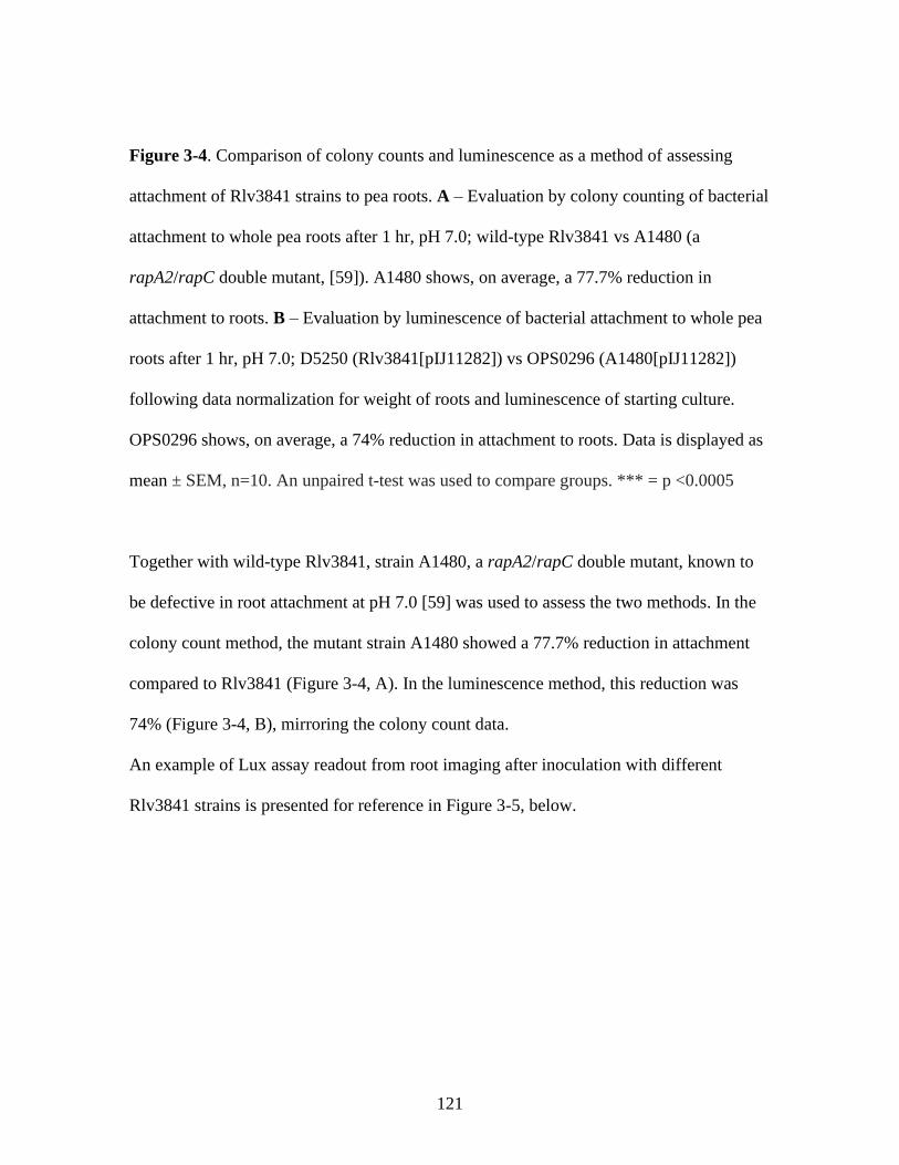
121
Figure 3-4. Comparison of colony counts and luminescence as a method of assessing
attachment of Rlv3841 strains to pea roots. A – Evaluation by colony counting of bacterial
attachment to whole pea roots after 1 hr, pH 7.0; wild-type Rlv3841 vs A1480 (a
rapA2/rapC double mutant, [59]). A1480 shows, on average, a 77.7% reduction in
attachment to roots. B – Evaluation by luminescence of bacterial attachment to whole pea
roots after 1 hr, pH 7.0; D5250 (Rlv3841[pIJ11282]) vs OPS0296 (A1480[pIJ11282])
following data normalization for weight of roots and luminescence of starting culture.
OPS0296 shows, on average, a 74% reduction in attachment to roots. Data is displayed as
mean ± SEM, n=10. An unpaired t-test was used to compare groups. *** = p <0.0005
Together with wild-type Rlv3841, strain A1480, a rapA2/rapC double mutant, known to
be defective in root attachment at pH 7.0 [59] was used to assess the two methods. In the
colony count method, the mutant strain A1480 showed a 77.7% reduction in attachment
compared to Rlv3841 (Figure 3-4, A). In the luminescence method, this reduction was
74% (Figure 3-4, B), mirroring the colony count data.
An example of Lux assay readout from root imaging after inoculation with different
Rlv3841 strains is presented for reference in Figure 3-5, below.

122
Figure 3-5. A representative example of Lux assay root imaging data following 1 hr
attachment assays. A - luminescence signal from a pea root inoculated with D5250
(Rlv3841[pIJ11282]) for 1 hr under pH 7.0 test conditions. B – with OPS0804 (a pssA
mutant). C – with OPS0111 (a praR mutant). Here, luminescence signal indicates reduced
attachment of a pssA mutant and increased attachment of a praR mutant in relation to
D5250 even before data normalisation (see 3.2.5 and 3.2.6). All images are scaled to a
counts per second cut-off of 500 (scale bar shown right). White bars are for scale; each
represents 1 cm. n = 10 per group, representative image provided
3.2.5 Validation of Lux-based attachment assay under different pH conditions using
a range of Rlv3841 mutants
As normalised luminescence enabled accurate quantification of bacterial attachment,
multiple further strains with characterised attachment phenotypes were tested under
different pH conditions (Figure 3-6). Most strains used with this assay are pK19mob
integration mutants. To isolate these mutants, an internal fragment of the target gene was
A B C

123
cloned into the pK19mob vector and conjugated into wild-type Rlv3841. Homologous
recombination causes single crossover integration mutagenesis and disruption of the target
gene. See also 2.4.3.

124
B
C D
E F
A

125
Figure 3-6. Comparison of wild-type Rlv3841 and known attachment deficient strains
under different pH conditions in a Lux whole root attachment assay. A – Root attachment
of D5250 (Rlv3841[pIJ11282]) under pH 6.5, 7.0 and 7.5 conditions. Data from A is
presented for D5250 for all further comparisons. B – D5250 vs OPS1878 (nifH mutant). C
– D5250 vs OPS0804 (pssA mutant). D – D5250 vs OPS1738 (flgE mutant). E – D5250 vs
OPS1739 (motA mutant). F - D5250 vs OPS0914 (gmsA mutant). Strains and pH
conditions are given on the x axes. Data is displayed as mean ± SEM, n=10. An unpaired
t-test was used to compare groups. *** = p <0.0005, **** = p <0.0001
3.2.5.1 Wild-type attachment is the same at pH 6.5, 7.0 and 7.5
No statistically significant difference was seen in the ability of D5250
(Rlv3841[pIJ11282]) to attach to pea roots under any of the pH conditions tested (Figure -
6, A). It is important to note that the pH conditions used here (6.5, 7.0 and 7.5) were
chosen because root attachment assays reported in the literature (e.g. [53, 56, 59]) were
performed within this pH range.
3.2.5.2 A nifH mutant is unchanged in attachment relative to wild-type
A nifH mutant was tested in this attachment assay and compared with wild-type Rlv3841
(Figure 3-6, B) at the three pHs. NifH is a nitrogenase iron protein essential for nitrogen
fixation. A nifH mutant is a non-nitrogen fixing strain but otherwise identical to Rlv3841
[233]. No statistically significant difference was seen in the ability of a nifH mutant to
attach under any pH conditions in comparison to wild-type. This result is important as it
demonstrates that pK19 insertion mutagenesis in itself does not affect the attachment of
Rlv3841 strains to pea roots.
126
3.2.5.3 Mutants in pssA, flgE and motA are impaired in attachment at all pHs relative
to wild-type
OPS0804, a pssA mutant, and OPS1738, a flgE mutant (Figure 3-6, C-D) show significant
reductions in attachment under all pH conditions when compared to wild-type, in
agreement with the expected phenotypes for these strains given previous literature reports
(see section 3.2, [44, 56]). OPS1739 (a mutant in motA, a component of the membrane
embedded flagellar motor, [234]) is also defective in attachment under all pH conditions
(Figure 3-5, E). When comparing Figure 3-6 panels D and E, there is no significant
difference between the OPS1738 (flgE mutant) and OPS1739 (motA mutant) attachment
results at each pH condition. This is an interesting result, as it implies that it is lack of
motility, rather than loss of flagellar adhesin action, which causes the defective attachment
of the OPS1738 (flgE) mutant. This assumes that the motA mutant strain remains
flagellated, as is the case for motA mutant Salmonella strains [235]. As discussed in [45,
61, 214, 215], the flagellum itself may possess adhesin properties, but it seems that this is
either not the case in Rlv3841, or that adhesin properties cannot be attributed to the FlgE
flagellar hook subunit, or that flagellar adhesin properties do not contribute to primary
root attachment.
3.2.5.4 A gmsA mutant is impaired in attachment at pH 6.5 and 7.0 relative to wild-
type
A very important result in validating the suitability of the whole-root Lux assay for pH
studies was the comparison of D5250 and OPS0914 (a gmsA mutant, Figure 3-5, F). gmsA
encodes glucomannan which is the only fully characterised primary attachment factor of
known pH dependency. The results gained here, whereby a glucomannan mutant is
impaired in attachment under pH 6.5 and 7.0 conditions, but not at pH 7.5, are in

127
agreement with the described model of primary root hair attachment in Rlv3841 [25, 34,
71].
The results presented in Figure 3-6 provide strong validation of the whole-root Lux assay
as a method for characterising primary attachment factors under different pH conditions.
3.2.6 praR regulation of attachment is highly dependent on pH conditions
Two further strains, OPS0296 (rapA2/rapC double mutant) and OPS0111 (praR mutant)
were also used to validate the Lux attachment assay under different pH conditions, but the
results revealed an interesting pH dependency in praR regulation of attachment (Figure 3-
7, Figure 3-8).
Figure 3-7. Comparison of Rlv3841and mutant strains under different pH conditions in a
Lux whole root attachment assay. A – Root attachment of D5250 (Rlv3841[pIJ11282])
under pH 6.5, 7.0 and 7.5 conditions vs OPS0296 (rapA2/rapC mutant). B – D5250 vs
A B

128
OPS0111 (praR mutant). Data is displayed as mean ± SEM, n=10. An unpaired t-test was
used to compare groups. * = p <0.05, *** = p <0.0005, **** = p <0.0001
A rapA2/rapC double mutant strain in Rlv3841 has previously been characterised as
deficient in attachment to pea roots [59]. Whilst this is seen here at pH 7.0 (Figure 3-7, A),
the mutant strain shows a large increase in attachment at pH 6.5 and 7.5. This result was
unexpected given the importance of these Raps at neutral pH, though the attachment
phenotype of this strain under different pH conditions has not previously been reported. It
suggests that RapA2 and RapC proteins interfere with binding promoted by other adhesins
at acid and alkaline pH. New hypothetical protein adhesin RapX(s) might result in very
strong binding at acid and alkaline pH, while RapA2 and RapC are most important for
binding at neutral pH. Given the reported negative regulatory function of praR on the
rapA2 and rapC genes [59], a praR mutant was tested in the Lux attachment assay (Figure
3-7, B) to investigate this further. A praR mutant demonstrates the large expected increase
in attachment relative to wild-type under pH 7.0 conditions (due to a loss of repression of
rapA2 and rapC genes, among others, [59]). However, under pH 6.5 and 7.5 mutation of
praR results in a significant inhibition of binding, although this inhibitory effect is not as
strong as its positive effect at neutral pH. This is intriguing, as it implies that, under pH
6.5 and 7.5 conditions, PraR promotes attachment via RapX(s). A triple mutant strain (a
praR/rapA2/rapC mutant) would therefore show defective attachment at pH 7.0 due to the
absence of RapA2/C. However, it may also have reduced attachment at pH 6.5 and 7.5 due
to the loss of positive regulation of RapX(s). A triple mutant strain (OPS1239) was
isolated (see 2.3.5) and conjugated with pIJ11282 to isolate OPS1266, which was tested in
a Lux whole-root attachment assay (Figure 3-8).

129
Figure 3-8. Comparison of D5250 (Rlv3841[pIJ11282]) and OPS1266 (praR/rapA2/rapC
triple mutant) under different pH conditions in a Lux whole root attachment assay. Data is
displayed as mean ± SEM, n=10. An unpaired t-test was used to compare groups. **** =
p <0.0001
As shown in Figure 3-8, a praR/rapA2/rapC triple mutant strain is strongly impaired in
attachment under all conditions compared to wild-type (D5250). This result is consistent
with the multifaceted positive and negative regulatory activity of PraR [59].
praR itself is repressed in a population density dependent fashion by quorum sensing
regulators. As population density increases, cinR, a regulator, induces the cinIS operon.
CinS (an antirepressor) binds and inhibits PraR, thereby relieving PraR mediated
repression of rapA2 and rapC [59, 191, 236] and allowing attachment and biofilm

130
formation under neutral conditions. However, studies of PraR regulation have not been
reported under different pH conditions. Based on the results presented in Figures 3-7 and
3-8 it may be that, at pH 6.5 and 7.5, PraR activity is not repressed, permitting the
expression of unknown RapX factors, which are important for attachment at pH 6.5 and
7.5. Frederix et al. (2014) demonstrated that PraR acts as a positive as well as negative
regulator [59], and it may be the case that some positively regulated targets of PraR (all of
which are uncharacterised, [59]) may act as novel adhesin factors important for attachment
at pH 6.5 or 7.5. Based on my results, and those of [59, 191, 236], a simple model of pH
dependent PraR regulation of some aspects of primary attachment machinery can be
hypothesised (Figure 3-9).
Figure 3-9. A simple model for PraR pH dependent regulation of some aspects of primary
attachment machinery in Rlv3841. At pH 7.0, CinS (induced by CinR, not shown) inhibits
PraR. This prevents repression of rapA2 and rapC, which are important for attachment
under pH 7.0 conditions. However, at pH 6.5 and 7.5, PraR remains active and activates
the transcription of unknown and possibly plural ‘rapX’ adherence factors which are
important for attachment under non-neutral pH conditions.

131
Whilst this model provides a possible explanation for the pH dependencies of strain
attachment (Figures 3-7, 3-8 and 3-9) it is likely that the regulatory mechanisms
underlying these results are more complex. PraR is known to be a direct regulator of
rapA2 and rapC [59], but it is unknown whether other genes up or downregulated in a
praR mutant (including possible ‘rapX’ genes) are directly or indirectly regulated by
PraR. In addition to rapA2 and rapC, Rlv3841 has two further annotated rap genes: rapA1
(RL3660) and rapB (RL3911). A rapB mutant strain shows no root attachment defects at
neutral pH [59], but has not been tested in acid or alkaline conditions. Given the role of
PraR in regulating rap genes, rapB represents a rapX candidate, and testing a rapB mutant
for attachment at pH 6.5 and 7.5 could shed further light on PraR pH-dependent regulation
of primary attachment. Additionally, there may be other uncharacterised regulatory
aspects of quorum sensing and PraR regulation which act as relays of external conditions
and permit the activation of different attachment mechanisms at different pHs.
3.2.7 Attempted mutation of possible rhicadhesin genes
The three most promising rhicadhesin candidates from the proteins identified from LC-
MS/MS of a crude adhesin band were Omp19 (RL4441, 18 kDa), RL1635 (putative outer
membrane protein, 19 kDa), and RL4733 (conserved hypothetical protein, 17 kDa).
Unfortunately, despite several attempts, it did not prove possible in this work to isolate
pK19mob mutants in the genes encoding these proteins. This could be because genes are
essential for membrane stability or other important, uncharacterised functions, meaning
that mutation is lethal. A second possibility is that small gene size may present a problem
for interposon mutagenesis. Gene sizes are as follows: omp19 (RL4441): 513 bp, RL1635:
132
531 bp, RL4733: 471 bp. It is important that an internal fragment of the target gene is
cloned into the pK19mob vector, rather than the gene in its entirety, to ensure that
interposon mutagenesis leads to a functional disruption of the gene. For this reason, it is
normal to exclude ~100 bp from each end of the target gene when cloning an internal
fragment. For these genes, the resulting internal fragment sizes were therefore in the range
of 271-331 bp in length. Efficiency of interposon mutagenesis drops rapidly when the
target gene internal fragment used for interposon mutagenesis is below 350 bp (Poole lab,
internal communication). Such findings have also been reported elsewhere [237]. Note
that these genes were successfully targeted by the mariner transposon for mutagenesis in
INSeq, and attachment classifications are presented in Chapter 4, Table 4-17.
Having validated a Lux whole-root attachment assay suitable for pH studies and
demonstrated the involvement of a broad range of factors in primary root attachment, it
was therefore decided to undertake a bioinformatic screen for possible novel Rlv3841
attachment factors in order to continue this work.
3.2.8 Bioinformatic identification of possible novel root attachment factors
Genome scale datasets were analysed to identify likely novel root attachment factors. Data
from one RNASeq experiment, six microarrays and two INSeq conditions (Table 3-2)
were analysed by the two approaches described in Figure 3-10, resulting in the
identification of 136 putative novel root attachment genes.

133
Table 3-2. RNASeq, microarray and INSeq experiments used in a bioinformatic screen for potential novel root attachment factors in
Rlv3841.
For microarrays, Array Express accession numbers for datasets are given in square brackets. Where two or more accession numbers
are given, they form part of different time courses
Experiment Source
1) RNASeq 7-day pea rhizosphere, 7-day post-inoculation Unpublished data, this lab
2) 7-day pea rhizosphere, 1-day post-inoculation microarray [E-MEXP-2845, E-MEXP-2848] [141]
3) 7-day pea rhizosphere, 3-day post-inoculation microarray [E-MEXP-2848] [141]
4) 7-day pea rhizosphere, 7-day post-inoculation microarray [E-MEXP-2848, E-MEXP-2852, E-
MEXP-2854]
[141]
5) 14-day pea rhizosphere, 1-day post-inoculation microarray [E-MEXP-2845] [141]
6) 21-day pea rhizosphere, 1-day post-inoculation microarray [E-MEXP-2845] [141]
7) 7-day pea rhizosphere inoculation, 21-day post-inoculation bacteroid microarray [E-MEXP-1918] [139]
8) INSeq rhizosphere, 7-day pea rhizosphere inoculation, 5-day post-inoculation [238]
9) INSeq root colonization, 7-day pea rhizosphere inoculation, 5-day post-inoculation [238]

134
Figure 3-10. Flow charts of the two approaches (A and B) taken to identify
bioinformatically novel root attachment factor genes in Rlv3841. Experiment numbers
refer to those listed in Table 3-2.
The rationale behind approach A (Figure 3-10) was that genes involved in early stage root
attachment should be consistently upregulated in the rhizosphere but subsequently
downregulated in the bacteroid stage of symbiosis. The rationale behind approach B was
that genes involved in early stage root attachment and/or colonization exclusively should
have a neutral phenotype when mutated in INSeq input and rhizosphere libraries. One
gene defective in the rhizosphere (pRL110071) and one defective in the rhizosphere and

135
input (RL0109) was added to the list of genes generated by approach B (Step 3). Testing
mutants in these genes in a Lux assay will determine whether fitness reductions in input
and rhizosphere libraries translate into reduced root attachment, as would be expected.
All genes identified by approaches A and B (Figure 3-10) can be found in Tables A1 and
A2 (Appendix 1) respectively.
3.2.9 Testing possible novel Rlv3841 adhesin factor mutants in Lux whole-root
attachment assays
pK19mob interposon mutants were isolated in seven of the genes identified in section
3.3.8 and tested in Lux whole-root attachment assays. Details of these seven genes and the
rationale for their choice are summarized in Table 3-3, and attachment assay results are
shown in Figure 3-11.

136
Table 3-3. Summary of bioinformatically identified genes tested in Lux whole-root attachment assay
Approach refers to the two approaches (A and B) shown in Figure 3-10. Experiment numbers refer to experiments listed in Table 3-2.
Strain Gene Protein description Approach Rationale for choice
OPS1131 pRL110543 conserved hypothetical B Essential for colonization, neutral in input and rhizosphere
(experiments 8 and 9) – likely root attachment factor.
OPS1235 RL2969 putative transmembrane A Upregulated in the rhizosphere in experiments 1-6, downregulated in
experiment 7 – rhizosphere induction indicates possible attachment
role.
OPS1236 RL4382 putative filamentous
hemagglutinin adherence
factor precursor
B Defective for colonization, neutral in input and rhizosphere
(experiments 8 and 9) – likely root attachment factor.
OPS1237 RL3273 putative von Willebrand factor
type A
A Upregulated in the rhizosphere in experiments 1-6, downregulated in
experiment 7 (Table 3-2). Rhizosphere induction indicates possible
attachment role and annotation suggestive of role in attachment.
OPS1238 RL0109 Conserved hypothetical B Defective in input, rhizosphere and colonization, experiments 8 and 9.
Testing will determine whether input and rhizosphere defects decrease
primary root attachment.
OPS1290 pRL110071 Conserved hypothetical B Defective in rhizosphere and colonization, experiments 8 and 9.
Testing will determine whether rhizosphere and colonization defects
decrease primary root attachment.
OPS1291 pRL100053 Putative transmembrane B Essential for colonization, neutral in input and rhizosphere
(experiments 8 and 9) - likely root attachment factor.

137
A
B C
D

138
Figure 3-11. Comparison of Rlv3841 and novel interposon mutant strains under different
pH conditions in a Lux whole root attachment assay. A – Root attachment of D5250
(Rlv3841[pIJ11282]) under pH 6.5, 7.0 and 7.5 conditions vs OPS1236 (RL4382 mutant),
OPS1238 (RL0109 mutant), OPS1290 (pRL110071 mutant) and OPS1291 (pRL100053
mutant). B – D5250 vs OPS1235 (RL2969 mutant). C – D5250 vs OPS1131 (pRL110543
mutant. D – D5250 vs OPS1237 (RL3273 mutant). Strains and pH conditions are given on
the x axes. Data is displayed as mean ± SEM, n=10. An unpaired t-test was used to
compare groups. * = p < 0.05, ** = p < 0.001, *** = p < 0.0005
OPS1236 (RL4382 mutant, Figure 3-11, A) is defective in root attachment under all pH
conditions relative to D5250. RL4382 encodes a putative filamentous hemagglutinin
(FHA) adherence factor precursor. FHA is well described in Bordetella pertussis where it
has been heavily used in acellular component vaccines [239]. In Bordetella, FHA can
either be cell surface associated, or extracellular. It is translocated across the bacterial
outer membrane by FhaC, a polypeptide transport associated (POTRA) domain containing
protein and member of the two-partner secretion (TPS) pathway, common in gram-
negative bacteria. From there it can be proteolytically processed and released, and is
important for adherence to host cells and cell agglutination [239]. Intriguingly, it has been
demonstrated that the plant pathogen Xanthomonas axonopodis pathovar citri requires a
similar FHA for host plant tissue colonization [240]. Further, the rhizosphere colonising
P. putida is known to use related proteins to colonise maize roots [241]. In Rlv3841, a
neighbouring gene of FHA is RL4381, a putative outer membrane POTRA domain
containing protein. These POTRA domains are often found in beta-barrel transporters
[242]. The results presented in Figure 3-11, as well as the presence of co-localised gene

139
encoding a putative POTRA domain transporter, indicate that Rlv3841 also makes use of
an FHA system in primary attachment to pea roots, which has not previously been
documented.
Rlv3841 strains carrying mutations in RL0109 (OPS1238), pRL110071 (OPS1290),
pRL100053 (OPS1291) (Figure 3-11, A) and RL2969 (OPS1235, Figure 3-11, B) were
also defective in attachment under all pH conditions. Little is known about these genes;
they encode a putative TIGRO2300 family protein, a beta-lactamase domain containing
protein and two putative transmembrane domain containing proteins respectively, all of
unknown function. A pRL110071 mutant was shown to be defective in the rhizosphere
(Table 3-2, experiments 8 and 9), whilst an RL0109 mutant was defective in the
rhizosphere and input in the same experiments. The defects in primary attachment shown
for OPS1290 (pRL110071 mutant strain) and OPS1238 (RL0109 mutant strain) here could
therefore result either from a reduced strain fitness in terms of growth, or from loss of a
primary attachment factor which also plays a separate role in input and/or rhizosphere
conditions. Overall, these results indicate that the bioinformatic approaches taken in 3.3.8
to identifying putative novel Rlv3841 adhesins is very successful.
OPS1131, a mutant in pRL110543 (encoding a conserved uncharacterised coiled-coil
domain containing protein), shows no statistical difference in attachment ability under any
conditions compared to D5250 (Figure 3-11, C). Given this gene was essential for
colonization but neutral in input and rhizosphere libraries in INSeq [238], it is most likely
to represent a secondary attachment determinant.
Finally, OPS1237 (RL3273 mutant, Figure 3-11, D) was defective in attachment under pH
7 and 7.5 conditions. This gene encodes a putative von Willebrand factor type A (VWA)
domain containing protein. VWA domain proteins have been widely implicated in cell
140
adhesion and intracellular enzyme activity in eukaryotes, and are widely conserved [243].
Within rhizobia, R. loti has 12 VWA domain proteins (one magnesium chelatase and 11
uncharacterised) but R. meililoti has just three. Both Vanderlinde (2011) and Neudorf
(2015) have linked mutations in RL3501, a VWA domain protein, to morphology defects
and impaired cell envelope function in Rlv3841 [244, 245]. Thus, it may be that mutation
in RL3273 produces its negative effect on root attachment by direct disruption of an
adhesin factor, or indirectly through cell envelope disruption, or by a further unknown
mechanism, and that these effects are dependent on pH conditions.
Testing of mutants in putative novel adhesin factors has revealed the involvement of
several in primary root attachment which had not previously been identified. This
illustrates the usefulness of the Lux technique combined with genomic scale datasets for
investigating such factors.
3.3 Conclusion
In summary, a crude adhesin fraction isolated from Rlv3841 was able to inhibit
attachment when preincubated with root sections. The 14 kDa protein band thought to
represent this adhesin was found to be made up of 15 robustly identified protein
components, although these were unable to be characterised further at this stage due to
difficulties in isolating mutant strains in the encoding genes. Particularly for the three
most likely rhicadhesin candidates (RL4733, omp19 – RL4441 and RL1635) an in-frame
mutagenesis strategy should be applied (such as the double recombination approach
described by Link et al. (1997)) to investigate these further [246]. This is not limited by
gene size, unlike the pK19mob insertion technique used in this study.

141
However, regarding the rhicadhesin hypothesis, the definition of rhicadhesin based on
ability to inhibit attachment [53] may be a flaw in its identification as an adhesin. The
ability of a protein mixture to block attachment does not necessarily imply a ‘lock and
key’ type interaction where rhicadhesin occupies plant root binding sites. Rather,
interference with the plant root or bacterial surface (surface protein aggregation, for
example) or some other indirect effect could be responsible. As an example of this, in one
case it was demonstrated that added rhicadhesin could rescue attachment of an A.
tumefaciens chvB mutant to pea root hairs [54]. However, as discussed by Matthysse
(2014), it is likely that the effects of chvA or chvB mutations are indirect and result from
multiple defects caused by the absence of cyclic-β-1,2-D-glucan from the periplasmic
space rather than the absence of rhicadhesin [55].
Although proteomics results do not conclusively identify rhicadhesin, the candidate root
attachment factors identified bioinformatically (Appendix 1 Tables 1 and 2) are
informative. Two approaches (Figure 3-10) led to the identification of 136 possible novel
root attachment genes. Testing mutants in seven of these revealed that six showed defects
in attachment compared to wild-type Rlv3841. Given how many root attachment factors
there are likely to be in Rlv3841, preparation of a membrane fraction, even if it is focussed
around 14 kDa proteins, may isolate one or more proteins capable of inhibiting root
attachment when pre-incubated with roots. However, this does not indicate that a protein
with this property is an important adhesin. Although it may have adhesin properties and be
directly responsible for attachment of bacterial cells to root surfaces, it may also have an
indirect mode of action. In this case, it could alter the bacterial cell surface in such a way
that it alters the presentation, structure or activity of another factor which is a direct

142
adhesin. This distinction was never thoroughly investigated in the literature reports of
rhicadhesin [53, 54, 208, 211].
Whilst a singular rhicadhesin may exist, it is also possible that there are multiple
functional genes encoding rhicadhesin, or that it is expressed as a larger precursor and
subsequently processed. These would be confounding factors in the identification of a
rhicadhesin gene. Combined with the caveats of previous research, the identification of
rhicadhesin is extremely difficult. A further complication is that Smit et al. (1989) never
demonstrated conclusively that their purified rhicadhesin was a homogenous single
protein [53]. Although their purified rhicadhesin ran as a ~14 kDa band on an SDS-PAGE
gel, it could still have been a mixture of similar-sized proteins, further confounding the
definition of a rhicadhesin. Therefore, whilst a rhicadhesin with the properties described
by Smit et al. (1989) may exist, there are also multiple alternative explanations as to how
an isolated protein fraction could inhibit bacterial root attachment, and there are likely to
be many different factors that could be rhicadhesin candidates.
To investigate bacterial root attachment further, a luminescence-based whole root
attachment assay was developed and validated through comparison with a bacterial colony
counting method and by testing with known attachment factor mutants. Crucially, the
assay was able to demonstrate the known pH dependent profile of a glucomannan mutant
in attachment. This work also highlighted the importance of motility in primary root
attachment, and indicated that it is motility of strains, rather than flagellar adhesin
properties, that is important for this process.
Whilst mirroring the characterised attachment phenotype of a rapA2/rapC double mutant
and a praR mutant, this assay also shed new light on the complexities and pH dependency
of PraR regulation. By investigating the attachment phenotype of a rapA2/rapC/praR

143
triple mutant strain, additions to the known PraR regulatory circuit were able to be
proposed which may account for the unexpected attachment phenotypes seen.
Using a bioinformatic approach to investigate genome-scale RNASeq, microarray and
INSeq datasets, 136 putative novel root attachment factor genes were identified in
Rlv3841. Testing of pK19mob mutants in seven of these genes in a Lux assay revealed
that six showed reduced attachment under two or more pH conditions in comparison to
wild-type. This indicates that the bioinformatic approaches taken to identifying putative
novel Rlv3841 attachment factors was very successful. Further, this testing implicated
genes and factors previously unreported in the primary root attachment process, including
FHA, a VWA domain protein and multiple uncharacterised proteins.
Given these findings, and the low-throughput nature of individual pK19mob mutant
isolation and testing, this chapter provides justification for taking a genome-scale
approach to characterising primary Rlv3841 attachment determinants at different pHs.
INSeq (Chapters 4 and 5) is a powerful method for such investigation and provides a
further experimental avenue by which to investigate the possible identity of rhicadhesin.

144

145
Chapter 4
Genome-scale characterisation of the primary
attachment determinants in the R. leguminosarum
symbiosis under acid, neutral and alkaline pH
conditions

146
4.1 Introduction
To attach to the roots of host legume plants rhizobia use multiple mechanisms. The
earliest stages of physical interactions between bacteria and plant roots rely on universal,
non-specific binding forces [34]. These include Van der Waals forces (caused by
momentary changes in molecular electron density [247]), electrostatic interactions (caused
by differences in molecular electric charge [44]) and hydrophobic interactions (the
strongest of these universal interactions [46, 248, 249]) [34].
Following this, soil bacteria exhibit a range of different specific molecular mechanisms of
primary root attachment. These include adhesive pili (Agrobacterium tumefaciens, [250]),
flagella and outer membrane proteins (Azospirillum spp., [60, 89]), pili and outer
membrane porins (Pseudomonas spp., [67, 214]) and flagella and proteinaceous fimbriae
(S. enterica, [69]).
For rhizobia, primary root attachment factors include not only the glucomannan system,
important for attachment to root hairs at acidic pHs [56, 71], but also the proposed
rhicadhesin system [53, 210], thought to be required for root hair attachment at alkaline
pHs. Rlv3841 uses additional attachment factors important for primary attachment to root
hairs and/or bulk root. Characterised examples include Rhizobium adhering proteins
(Raps) [57, 59], acidic exopolysaccharides [56] and PraR regulated genes [59]. The work
presented in Chapter 3 developed a new method for assaying primary attachment and
provided evidence for the involvement of many factors that have yet to be included in
primary attachment models (see 3.2.9). Successful primary root attachment is important
for both nitrogen-fixing symbiosis formation with legumes [56], but also accessing root
exudates, which can serve as growth substrates [34]. Therefore, a detailed understanding
of primary root attachment will improve understanding of the early nitrogen-fixing

147
symbioses stages and persistence of rhizobia in the soil. Increased understanding of these
processes is timely. Soil degradation as result of intensive agriculture [1] and climate
change induced acidification [251] may alter the balance of primary root attachment
factors relied on by rhizobia. This could be important for agricultural inoculant formation.
Further, development of synthetic symbioses seeking to engineer root-associative
interactions with nitrogen-fixing bacteria to boost crop yields [35, 109] could leverage the
genetic circuitry of primary root attachment mechanisms to increase effectiveness.
The presence of diverse primary root attachment mechanisms in rhizobia is not surprising
considering the heterogeneity of the soil environment and the diversity of plant roots
encountered. A single gram of bulk soil can contain 108 bacterial cells [252] and varies
widely in soil particle size, nutrient concentration and pH [253–256]. Competition for
plant root attachment is intense and can involve the interaction of plant beneficial rhizobia
and other soil bacteria, pathogenic bacteria, mycorrhizal fungi and many more [257–260].
Additionally, plant roots may also undergo physical changes in different soil conditions
which necessitate adaptation by attaching bacteria. One example of this is the alkaline pH-
induced dissociation of plant root hair lectin, thought to prevent glucomannan-mediated
attachment of Rlv3841 [25, 47, 60].
The complexity of the soil growth environment is reflected in the genome architecture of
Rlv3841. The 7.75 Mb genome consists of a circular chromosome (4800 genes) and six
plasmids: pRL7 (186 genes), pRL8 (142 genes), pRL9 (313 genes), pRL10 (471 genes),
pRL11 (644 genes) and pRL12 (790 genes) [138, 151]. This is considerably larger than
the typical bacterial genome, which is around 5 Mb and encodes ~5,000 proteins [261].
Various ‘omics studies have indicated a plethora of gene functions in Rlv3841 ([139, 141,
143] as examples). However, despite this, ~25% of Rlv3841 genes remain

148
uncharacterised. Although some gene functions have been inferred from regulatory
profiles in transcriptomics [139, 141], this does not identify the importance or role of
genes under specific conditions, such as primary root attachment.
Mariner transposon insertion sequencing (INSeq) is a technique that overcomes these
limitations. This method allows the effect of mutation on individual genes to be analyzed
under conditions of interest using libraries of single insertion bacterial mutants coupled
with high-throughput sequencing [169]. By combining sequencing data with a hidden
Markov model (HMM), the importance of individual genes in tested conditions can be
ascertained.
A benefit of this technique in relation to Tn5 transposon insertion screens is the specific
targeting by the mariner transposon of thymine-adenine (TA) motifs [176]. This defined
insertion site requirement allows in silico prediction of insertion site density in any
genome sequence and robust statistical analysis based on a defined number of insertion
sites [151]. The HMM used here expands on the binary essential/non-essential
classifications of other statistical methods by assigning each gene to one of four
classification states. These are based on the inferred fitness effect on a bacterium of
mutation to a given gene and are as follows: essential (ES, the bacterium is unable to
tolerate insertion), defective (DE, insertion impairs fitness of the bacterium), neutral (NE,
insertion has no fitness effect) and advantaged (AD, insertion increases fitness of the
bacterium). Successful application of an HMM to transposon insertion mutagenesis
screens has been reported in a variety of bacterial species [183, 262, 263]. Figure 4-1
summarizes how an INSeq experiment and HMM gene assignments can give information
on the bacterial genes involved in primary attachment to roots.
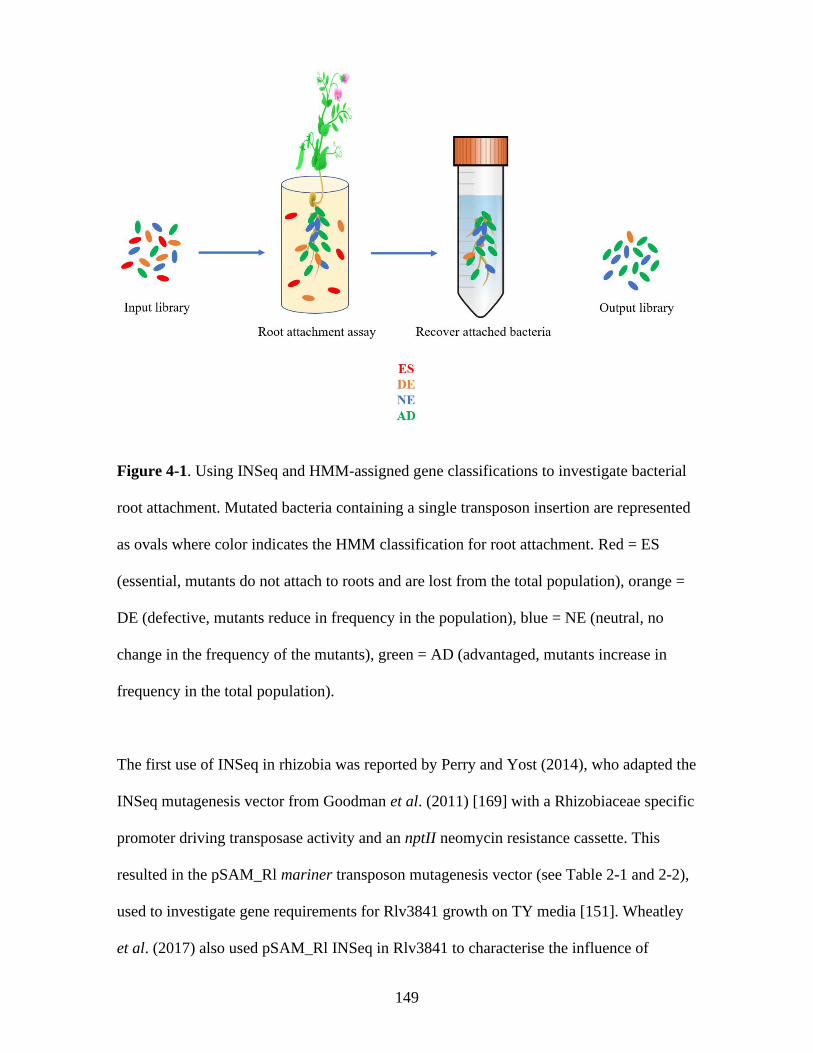
149
Figure 4-1. Using INSeq and HMM-assigned gene classifications to investigate bacterial
root attachment. Mutated bacteria containing a single transposon insertion are represented
as ovals where color indicates the HMM classification for root attachment. Red = ES
(essential, mutants do not attach to roots and are lost from the total population), orange =
DE (defective, mutants reduce in frequency in the population), blue = NE (neutral, no
change in the frequency of the mutants), green = AD (advantaged, mutants increase in
frequency in the total population).
The first use of INSeq in rhizobia was reported by Perry and Yost (2014), who adapted the
INSeq mutagenesis vector from Goodman et al. (2011) [169] with a Rhizobiaceae specific
promoter driving transposase activity and an nptII neomycin resistance cassette. This
resulted in the pSAM_Rl mariner transposon mutagenesis vector (see Table 2-1 and 2-2),
used to investigate gene requirements for Rlv3841 growth on TY media [151]. Wheatley
et al. (2017) also used pSAM_Rl INSeq in Rlv3841 to characterise the influence of

150
atmospheric and 1 % oxygen concentrations on growth with glucose or succinate as a
carbon source [143].
Here, pSAM_Rl is used in an INSeq experiment to characterise the primary attachment
determinants of Rlv3841 to pea roots at three different pHs: 6.5, 7.0 and 7.5. This
demonstrates that this process is far more complex than previously reported and uncovers
multiple novel mechanisms involved in attachment.
The raw data for this INSeq experiment can be found in Appendix 2, Table 1.

151
4.2 Results and discussion
4.2.1 Root attachment assays – determining inoculum density and bacterial recovery
method for INSeq
Root attachment assays with Rlv3841 (2.6.5) were carried out to determine the optimum
starting inoculum density for INSeq analysis after recovery of root-attached bacteria
(Figure 4-2).
Figure 4-2. Recovered Rlv3841 as a function of starting inoculum density after 1 hr
attachment to pea roots at pH 7.0. Inoculum density is given as OD600. n = 5 for all groups.
As demonstrated in Figure 4-2, the higher the starting inoculum density of Rlv3841, the
greater the number of Rlv3841 cells recovered from roots after 1 hr attachment. For
statistically robust HMM state assignments for genes in an INSeq experiment, it is
desirable to have at least 500-750-fold coverage of the genome represented in the output

152
library. The number of plants required to achieve this for a given starting inoculum
density can be calculated using the data shown in Figure 4-2 and the following equation:
𝑷𝒏 = 𝑭𝒄 ÷ (�̅�
𝒏)
where Pn is the number of plants needed for the desired fold coverage, Fc is the desired
fold coverage, x̅ is the mean of the number of root-attached bacteria and n is the number of
genes in the genome.
For the starting inoculum densities shown in Figure 4-2, Table 4-1 provides the number of
plants needed for 500 or 750-fold coverage using the equation above.
Table 4-1. Number of plants needed for 500- or 750-fold coverage of 7,300 genes in the
genome of Rlv3841 based on input inoculum density (OD600)
Input inoculum
(OD600)
Plants needed for 500-fold
coverage of 7,300 genes
Plants needed for 750-fold
coverage of 7,300 genes
0.001 107 161
0.01 12 18
0.1 2 3
Note that, for Rlv3841, an OD600 of 0.1 = 108 cfu/mL (data not shown). Given that 500-
750-fold coverage of a genome is considered a minimum requirement for robust statistical
analysis, and that Lux attachment assays were conducted with an inoculum of OD600 0.1, it
was decided to use a starting inoculum of OD600 0.1 with 10 plants per test condition.

153
Each test condition was performed in triplicate (see 4.2.2). This starting inoculum density
and plant number should allow for > 3,000 fold coverage of the ~7,300 Rlv3841 genes
(and >165-fold coverage of the 140,056 TA sites).
Primary attachment is defined by relatively weak and reversible interactions between
bacteria and plant roots. Secondary attachment is defined by a more ‘irreversible’
interaction [34, 264, 265], meaning that it is difficult to remove bacteria from roots. Any
INSeq assay seeking to define gene roles in root attachment must recover as much of the
root-attached bacterial population as possible or risk mis-classifying genes. To this end,
the numbers of Rlv3841 that could be recovered from roots after a 1 hr attachment assay
by either vortexing alone, or by vortexing and grinding roots with a pestle and mortar,
were compared (see 2.6.5, Figure 4-3).

154
Figure 4-3. Rlv3841 recovered by vortexing alone or vortexing and grinding using a
starting inoculum density of OD600 0.1 in a 1 hr attachment assay to pea roots at pH 7.0. n
= 5 for all groups
No statistically significant difference was seen between the number of bacteria recovered
by vortexing alone compared to vortexing and grinding of roots (Figure 4-3). It can
therefore be concluded that vortexing alone is sufficient to recover attached Rlv3841 cells
in this INSeq root attachment assay.
4.2.2 INSeq experimental design
To characterise primary attachment determinants at different pH conditions, an INSeq
experiment was designed (Figure 4-4). A population of insertion mutants (library 1-3, 500
mL of an OD600 0.1 population) was used to inoculate 10 pea plants for each test condition
(pH 6.5, 7.0 and 7.5), resulting in each being performed in triplicate.
Insertion mutants were recovered from roots after 1 hr and grown in liquid TY for 12 hr
before the 12 sample libraries (3x3 experimental conditions + 3 input libraries) underwent
barcoding, mariner library preparation and sequencing (2.7.3, Figure 4-4).
HMM gene classifications were averaged across the three replicates to increase robustness
of analysis.
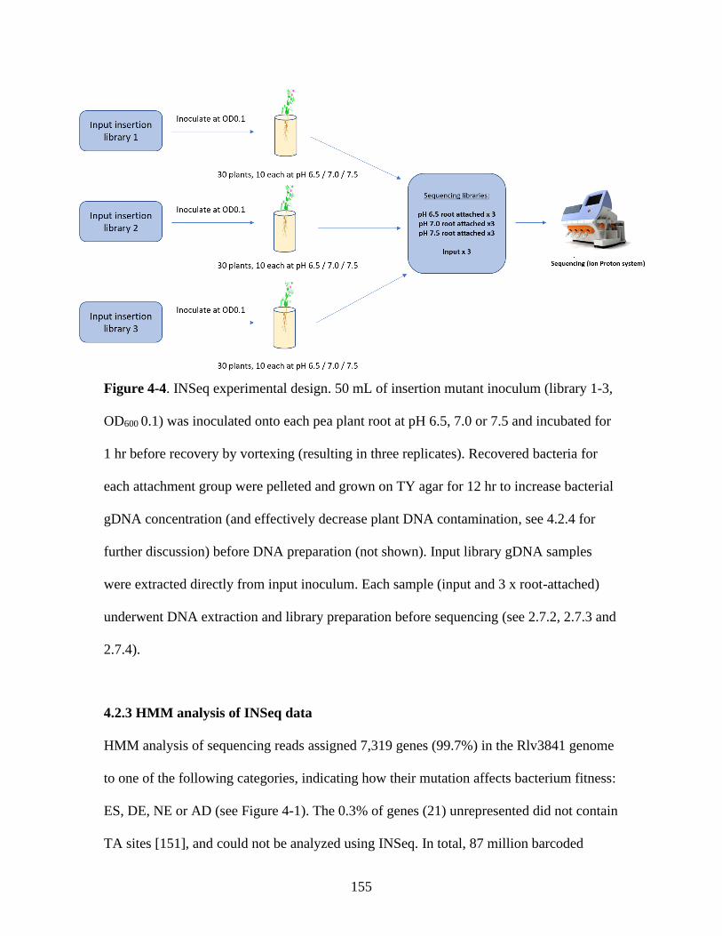
155
Figure 4-4. INSeq experimental design. 50 mL of insertion mutant inoculum (library 1-3,
OD600 0.1) was inoculated onto each pea plant root at pH 6.5, 7.0 or 7.5 and incubated for
1 hr before recovery by vortexing (resulting in three replicates). Recovered bacteria for
each attachment group were pelleted and grown on TY agar for 12 hr to increase bacterial
gDNA concentration (and effectively decrease plant DNA contamination, see 4.2.4 for
further discussion) before DNA preparation (not shown). Input library gDNA samples
were extracted directly from input inoculum. Each sample (input and 3 x root-attached)
underwent DNA extraction and library preparation before sequencing (see 2.7.2, 2.7.3 and
2.7.4).
4.2.3 HMM analysis of INSeq data
HMM analysis of sequencing reads assigned 7,319 genes (99.7%) in the Rlv3841 genome
to one of the following categories, indicating how their mutation affects bacterium fitness:
ES, DE, NE or AD (see Figure 4-1). The 0.3% of genes (21) unrepresented did not contain
TA sites [151], and could not be analyzed using INSeq. In total, 87 million barcoded

156
sequencing reads were obtained from a total of twelve samples (nine from plants and three
input libraries).
Isolation of mariner libraries produced highly saturated mutant pools. The input libraries
were shown by sequencing to have an average insertion density (percentage of TA motif
insertion sites that carry one or more insertions) of 82% (Table 4-2). The greater the
insertion density in the input library, the higher the coverage of TA motif insertion sites.
Insertion densities <25% are considered insufficient for robust statistical analysis [146,
150, 169, 179]. Note that for the chromosome or any plasmids individually the insertion
density obtained did not differ more than ± 5% from the average input figure given in
Table 4-2, indicating unbiased mariner insertions across the genome. In this INSeq
experiment, it was important that sufficiently large mutant libraries could be recovered
from roots so that HMM classifications of genes could be compared between the input and
root-attached samples. The insertion densities obtained for the root-attached libraries
(Table 4-2) provides early validation of the inoculum density parameters chosen for this
experiment.
Table 4-2. Percentage insertion density in the input and root-attached (pH 6.5, 7.0 and
7.5) libraries.
Library Insertion density
Input 82 %
pH 6.5 48 %
pH 7.0 50 %
pH 7.5 50%

157
Insertion density is defined as the proportion of total TA motifs that contain at least one
insertion in each sequenced library.
4.2.4 INSeq gene classifications
The distribution of HMM gene classifications across the four different states (ES, DE, NE
and AD) in input and root-attached libraries is shown in Table 4-3, below
Table 4-3. Distribution of HMM assignment of Rlv3841 genes in input and root-attached
(pH 6.5, 7.0 and 7.5) libraries.
HMM
assignment
Library
Input pH 6.5 pH 7.0 pH 7.5
ES 1 % 6 % 7 % 5 %
DE 10 % 7 % 6 % 7 %
NE 86 % 86 % 86 % 87 %
AD 3 % 1 % 1 % 1 %
Values are given as percentage of the 7,319 genes which contain TA sites.
Additional processing of the input library data was necessary to allow valid comparison
between the input and root-attached libraries because of the 12 hr TY growth step for the
bacteria recovered from roots. This step increases bacterial gDNA concentration and
dilutes contaminating plant DNA before extraction (see 2.7.2). TY was chosen for this as
it has been shown, following bacterial centrifugation and resuspension, to promote rapid
growth of Rlv3841 (Poole Lab, data not shown). Regrowth in TY, even though it was for

158
a relatively short time (12 hr, ~ 4 generations for Rlv3841) could lead to an inadvertent
selection pressure on mutants and affect downstream HMM classifications.
To prevent this, input library data was curated post-HMM processing to remove any
mutations that result in a non-NE gene classification in the HMM when strains are grown
on TY. At the outstet, a total of 954 genes were classified as non-NE (meaning ES, DE or
AD) in the input library (UMA media) (2.2.1). Perry and Yost (2014) reported 759 genes
which were non-NE for growth on TY. Of these, 540 shared a non-NE classification in the
input library and 219 were uniquely non-NE on TY. Taking these 219 genes into account
(where HMM classification could be skewed due to the TY growth step), 14% of the
genome was classified as non-NE in the input library.
When using INSeq to identify primary root-attachment determinants, it is genes that are
NE in the input but non-NE in attachment which are of interest.
4.2.5 Validation of INSeq predictions
Initial validation of these INSeq results was provided by comparing the HMM
classifications of well-characterised genes with their known or predicted phenotypes from
previous relevant literature (Table 4-4)

159
Table 4-4. Summary of HMM classifications of well-characterised genes with known
or expected phenotypes.
Gene(s) INSeq
classification
Description and references
murB, -C, -G, -D, -F, -E
(RL3305, -06, -07, -09,
-11, -12, respectively)
DE or ES - all
conditions
Peptidoglycan biosynthesis genes; key
structural role in the cell wall, of critical
importance to cell viability [266]
RL1769-99 (50S and
30S ribosomal subunits)
DE or ES - all
conditions
Ribosomal subunit proteins are essential for
protein synthesis and cellular viability [267]
repABC modules of all
plasmids
DE or ES – all
conditions
RepABC systems maintain rhizobial plasmids
in daughter cells; previous work has identified
ES genes on all plasmids; plasmid
maintenance should be essential for cell
function [151]
kdtA (RL0902) DE – all
conditions
KdtA catalyzes the transfer of 3-deoxy-D-
manno-octulosonate (Kdo) to the precursor of
Lipid A, the anchor of LPS. The Lipid A Kdo
domain is required for growth [268]
dnaC (RL1551) and
dnaX (RL0134)
DE or ES – all
conditions
DnaC is a putative DNA helicase and DnaX a
putative polymerase, essential for DNA
replication [269]
160
sufS (RL2578), sufC
(RL2580), sufB
(RL2582)
DE or ES – all
conditions
Iron sulphur biogenesis and transport related
proteins, shown to have a lethal phenotype on
mutation [192]
aapJ (RL2204) NE – all
conditions
Putative solute binding protein of PAAT L-
amino acid transporter, mutation has non-
lethal phenotype [270]
mntH (RL0940) NE – all
conditions
Putative manganese transporter, mutant has
comparable growth rate to Rlv3841 in non-
manganese limited conditions [271]
All conditions = input, attachment at pH 6.5, 7.0 and 7.5
Various cellular components are critical for viability, including peptidoglycan (the main
shape-maintaining element of the bacterial cell wall) [266] and ribosomal subunits
(required for protein synthesis) [267]. In addition, RepABC for each plasmid, KdtA and
DNA separation and replication proteins are also required [151, 268, 269]. Fe-S clusters
are used as cofactors by various proteins, with Suf proteins involved in oxidative stress
responses. Rlv3841 has only one Suf system, so any mutation is deleterious [192]. Given
the importance of these cell components, it would be expected that mutants in any of these
factors would show reduced fitness in INSeq, which is indeed the case (Table 4-4).
Conversely, mutations in the transporters aapJ and mntH (involved in the transport of
polar amino acids and manganese ions, respectively) are not deleterious under free-living
conditions, and would be expected to be NE in INSeq, which is what was found in these
experiments (Table 4-4).

161
Further to this literature validation, HMM classifications of known secondary attachment
factors were also compared with literature. In this case it was hoped that established
secondary attachment factors would not be affected in this INSeq experiment to discover
primary attachment factors. This would show that the two stages can be separated
experimentally. Cellulose fibril deposition is a characterised secondary attachment process
in R. leguminosarum, enabling root colonization [71]. The genes encoding cellulose
synthesis regulator PleD (RL1730, also known as CelR) as well as cellulose synthase
CelABC (RL1646-48) were classified as NE in INSeq assessing primary attachment at all
pHs, as were RhiABC genes (pRL100169-71) (Appendix 2, Table 1). The rhi genes,
named for being strongly rhizosphere-induced, have been implicated in secondary root
attachment and are required for successful five-day colonization of pea roots [141, 238].
The NE classification of these known secondary attachment factors provides validation of
the sampling (1 hr post-inoculation) as suitable for investigating primary attachment.
4.2.6 Primary attachment gene requirements and functional classifications
After filtering genes which were assigned a non-NE HMM classification in the input
library, 6146 genes remained. Some genes were required (ES/DE) for attachment at
discrete pH conditions with others required at multiple pHs. In total, 292 genes classified
NE in the curated input library were classified as ES/DE in one or more of the root-
attached libraries; the distribution of these is shown in Figure 4-5. These genes and their
HMM classifications from experiments listed in Table 4-5 and 4-7 are given in Appendix
1 Table A3 (all pHs), Table A4 (pH 6.5), Table A5 (pH 7.0), Table A6 (pH 7.5), Table A7
(pH 6.5 and 7.0), Table A8 (pH 7.0 and 7.5) and Table A9 (pH 6.5 and 7.5).

162
Figure 4-5. Genes classified as NE in the input library and required (ES/DE) in one or
more of the root-attached libraries. Total genes = 292. Venn diagram circle color indicates
pH; red = pH 6.5, green = pH 7.0, blue = pH 7.5.
Thus far, the only described primary attachment determinants with a pH-dependent profile
of activity are glucomannan [56] and the hypothesized rhicadhesin [34, 53, 56, 71, 208,
210, 211]. Given that there are large numbers of genes specifically required at different
pHs (50, 44 and 58 at pH 6.5, 7.0 and 7.5, respectively, Figure 4-5), this is likely to be a
vast underrepresentation of the complexity of primary root attachment. Differences in
plant root surfaces at different pHs, exemplified by the dissociation of root hair lectins at
alkaline pH, [25, 47, 60], may at least partially account for this. However, it appears that
there are many unexplored factors involved.
The functional classifications of these 292 genes (Figure 4-4) were investigated, and
comparison made with those genes required (ES/DE) in the input library. Riley codes
(classifying genes into one of seven broad functional categories based on their putative
function [138, 272]) are useful for this purpose. Figure 4-6 shows the Riley classification
pH 6.5
pH 7.0
pH 7.5

163
of the 1,173 genes required (ES/DE) in the input library compared with that of the 65
genes required (ES/DE) for attachment at all pHs.
Figure 4-6. Functional classification (using Riley codes) of the 1,173 genes required
(ES/DE) in the input library compared with the 65 genes required (ES/DE) for primary
root attachment at pH 6.5, 7.0 and 7.5. Data is presented as percentage of total genes in
each category.
Functional classifications (Figure 4-6) reveal that the most abundant ES/DE gene class in
the input library is intermediary metabolism, implying that metabolic pathway functions
are the most important determinants in ‘free-living’ growth. In contrast, the 65 genes
ES/DE under all root attachment conditions show higher representation of cell process,
cell membrane/envelope and macromolecule metabolism, as well as uncharacterised

164
genes, indicating these classes are the most importance in successful primary root
attachment.
4.2.7 Genomic localization of genes required for primary root attachment
Further insight can be gained by comparing the genetic loci of genes required (ES/DE) for
primary attachment (this work) and secondary attachment (5 d colonization of pea roots,
[238]). The experiments of Wheatley (2018) [238] were at neutral pH, so comparison was
made to 162 genes required (ES/DE) for primary attachment at pH 7.0 (shown in Figure 4-
7).
Figure 4-7. Genomic localization of genes (as a proportion of the total). A – 7,340
Rlv3841 genes. B – 162 primary attachment genes required (ES/DE) at pH 7.0. C – 125
secondary attachment genes required (ES/DE) at pH 7.0 determined from 5 d colonization
INSeq [238].
A B C
Gene distribution of primary attachment ES/DE genes at pH 7.0
Gene distribution of 5-day colonization ES/DE genes at pH 7.0
Gene distribution in the Rlv3841 genome

165
In the Rlv3841 genome, 65.4 % of annotated genes are located on the chromosome, with
the remainder divided between the six plasmids (Figure 4-7, A). At pH 7.0, the
distribution of the genes required for primary attachment (Figure 4-7, B) shows that,
compared the genome, the location is skewed towards the chromosome (~81%) and away
from the plasmids, with no pRL8 representation. In contrast, genes required for secondary
attachment (Figure 4-7, C) show a distribution more like that of the genome, with ~65%
encoded on the chromosome and representation of all plasmids, although there is a clear
skew to pRL10 (~15% compared to ~6% in the genome).
This demonstrates that different factors are involved in primary and secondary attachment
and that genes required for the latter are likely to be induced during the attachment
process. The fact that most of the nodulation and nitrogen fixation genes are encoded on
pRL10 [141] as well as the rhi genes, important root colonization determinants [238] may
explain the skew to pRL10 in 5 d colonization. This agrees with a model in which
secondary attachment factors (e.g. extracellular fibrils) are synthesized in response to
successful primary attachment [60, 71], and provides further validation of the
experimental separation of attachment stages.
4.2.8 Mapping gene requirements at different symbiosis stages from INSeq data
INSeq datasets (detailed in Table 4-5) are available for multiple stages of the Rlv3841/pea
symbiosis and development of bacteroids. It is possible to map gene requirements for
primary attachment with those of other symbiosis stages to see if they are primary
attachment-specific or also needed for later symbiosis stages (Figure 4-8). Of those genes
required (ES/DE) only in primary attachment, 104 of 292 genes (Figure 4-5) are likely to
166
represent factors important for attachment to the bulk root epidermis. Those 49 genes
which are ‘progressive’ through infection threads and nodulation (i.e. needed for primary
root attachment and all subsequent symbiosis stages, Figure 4-8) are likely to be involved
in attachment to root hairs since these are the site of infection thread development [26].
The remaining 139 of 292 genes needed in one or more of the root attached libraries
(Figure 4-5) are required (ES/DE) at non-contiguous stages of the symbiosis, and not
shown in Figure 4-8. These genes are likely to have discrete functions at different stages
of symbiosis development. For example, they may be required in primary attachment and
in nodulation. INSeq HMM designation for the genes discussed can be found in Appendix
1, Tables A3-A9.
Table 4-5. Details of the five INSeq libraries used to map the gene requirements to stages
of Rlv3841 symbiosis with pea plants.
Library Condition Genes required
(ES/DE)
Source
Rhizosphere
Pea rhizosphere
904 [238]
Primary root
attachment
1 hr root
attachment, pH
6.5, 7.0, 7.5
292 This work
Colonization
(secondary root
attachment)
5 d root
colonization
911 [238]
Infection thread Infection thread 1131 Unpublished data, Poole
Lab
Nodulation Nodule bacteroids 1043 Unpublished data, Poole
Lab
Conditions refer to the environment the output libraries were extracted from prior to DNA
isolation and library preparation/sequencing.

167
Figure 4-8. Genes required (ES/DE) at each stage of Rlv3841/pea symbiosis. Light boxes
show genes required (ES/DE) in primary root attachment (and other stages where
applicable) at different combinations of pHs. Color code: brown - pH 6.5, 7.0 and 7.5. Red
– pH 6.5 only. Green – pH 7.0 only. Blue – pH 7.5 only. Purple – pH 6.5 and 7.5. Dark
orange boxes show genes required in other conditions (Table 4-5). All genes were NE in
the input library.

168
4.2.9 Comparison of INSeq predictions and Lux attachment assays
To compare results from Lux attachment assays and HMM classifications, attachment
assay data from Chapter 3 (Figure 3-5, 3-7 and 3-11) was compared to the HMM gene
classifications for primary root attachment obtained in this chapter.
INSeq investigates the effect of single-insertion mariner transposon mutants, so
meaningful comparison with the double and triple mutant strains tested in Lux assays was
not possible. It is also difficult to compare the HMM classifications (ES, DE, NE, AD)
directly with the simpler statistical testing of results (as a percentage of wild-type
attachment) in Lux assays. To enable comparison, the following assumptions were made:
genes classified as NE in INSeq should show no statistical difference in attachment in a
Lux assay from wild-type (Rlv3841[pIJ11282], D5250), while genes required (ES/DE)
should show a statistically significant reduction from wild-type in Lux attachment assays.
Significant or non-significant differences in the case of Lux assays are based on unpaired
t-test results with the upper threshold for p value significance set at p <0.05. Table 4-6
gives a comparison of the 13 single mutant strains tested in Lux whole-root attachment
assays in Chapter 3 with the gene classification from primary attachment INSeq data.

169
Table 4-6. Comparison of INSeq and Lux attachment assay data.
Gene Strain Luminescence from mutant as a
percentage of wild-type a
INSeq classification
pH 6.5 pH 7.0 pH 7.5 pH 6.5 pH 7.0 pH 7.5
praR (RL0390)
OPS0111 61% 467% 67% NE NE NE
pssA (RL3752)
OPS0804 21% 37% 38% DE DE DE
gmsA (RL1661) OPS0914 1% 16% 70% *
DE DE NE
pRL110543
OPS1131 105% * 133% * 114% * NE NE NE
RL2969
OPS1235 8% 16% 67% NE NE NE
RL4382
OPS1236 3% 20% 8% DE DE DE
RL3273
OPS1237 138% * 45% 42% NE NE NE
RL0109
OPS1238 8% 23% 18% DE ES ES
pRL110071
OPS1290 28% 43% 21% DE DE DE
pRL100053
OPS1291 4% 7% 19% DE DE DE
flgE (RL0728)
OPS1738 19% 12% 36% NE NE NE
170
motA (RL0703)
OPS1739 16% 13% 12% NE NE NE
nifH (pRL100162) OPS1878 102% * 107% * 99% * NE NE NE a Wild-type in Lux attachment assay is Rlv3841[pIJ11282] (D5250). Results are expressed as average root attachment of test strain in
percent, indexed to 100% attachment for D5250 under each condition. Lux attachment results in black show agreement with INSeq
data, while those in red do not when using the criteria defined in 4.2.9. * indicates that in Lux attachment assay the strain does not
show a statistically significant difference from D5250 using an unpaired t-test with the upper threshold for p value significance set at p
<0.05.

171
Comparing results from the Lux attachment assay to the HMM classifications for primary
attachment reveals that, using the criteria defined, there is close agreement in the results
from the two techniques for 25 out of 39 comparisons (13 strains tested under three
attachment conditions, Table 4-6). Whilst this is a reasonable degree of agreement
(~66%), there are various reasons why the two would not completely agree since they are
measuring slightly different things. This is due to the design of each experimental
technique, each with limitations that need to be considered.
The conflict in results for OPS0111 praR (RL0390, mutant strain OPS0111), could be due
to the low gene TA site number (only four in this case) which affects the degree of
certainty in HMM gene classification. Although the HMM incorporates neighboring TA
site data to smooth read counts, low TA site number may still reduce the certainty of
classification [183]. Using TnSeq, Griffin et al. (2011) and Zhang et al. (2012) reported
uncertainty in assigning state call to bacterial genes with fewer than six TA sites [173,
273]. Therefore, despite the relatively high saturation of the input library (Table 4-2), low
TA site number may present an issue for HMM classification, and care should be taken for
genes with under six TA sites. 83% of the annotated Rlv3841 genes have >six TA sites.
The use of a richer media for mariner transposon library construction, or pooling more
independent conjugations (as described by DeJesus et al. in [274]) would increase input
saturation and reduce this problem. For the remaining lack of agreement between these
two techniques (genes RL2969, RL3273, RL0728 and RL0703), TA site number is unlikely
the issue, each gene having >20 TA sites.
Another possible reason for results disagreement is the different nature of the two assays.
In a Lux assay, inoculum is an OD600 0.1 resuspension of a single strain. In INSeq,
inoculum is the mariner transposon input library, a heterogeneous population of ~115,000

172
different mutant strains (140,845 genomic TA sites and 82% insertion density). This raises
the possibility of in trans complementation, where bacteria possessing a functional copy
of a gene may act to ‘rescue’ bacteria with a non-functional copy of the same gene
(providing them with a secreted factor, as described for lethal factor in Bacillus anthracis,
for example [275]). To test if in trans complementation could rescue strains OPS1235
(mutated in RL2969), OPS1237 (mutated in RL3273), OPS1738 (mutated in flgE) or
OPS1739 (mutated in motA) in a Lux attachment assay, further assays were performed as
described in 2.6.6. However, inoculation was at 1:1 or 1:100 mixture of pIJ11282-labelled
test strain (mutant) and Rlv3841 (unlabeled), thereby providing wild-type Rlv3841 cells
with functional copies of all genes which would be able to rescue mutant cells reduced
attachment phenotypes if in trans complementation is occuring. Complementation was
seen for OPS1235 at a 1:100 inoculum, returning attachment to wild-type levels at all pHs
tested (Figure 4-9). As RL2969 encodes a putative transmembrane protein, it seems that
either this protein could be released from wild-type cells surfaces and bind cell surfaces of
mutant strains to increase attachment ability by acting as an attachment factor.
Alternatively, this protein could act to aggregate wild-type cells with mutants on the root
surface, and thereby yield a complementation phenotype in attachment. Precisely how this
in trans complementation is occurring remains unclear; more detailed characterisation of
RL2969 could shed further light on this. However, this result shows that in trans
complementation may skew INSeq results for a subset of genes and mask their role in
primary attachment.

173
Figure 4-9. Comparison of D5250 (Rlv3841[pIJ11282]) and OPS1235 (RL2969 mutant)
in single inoculum, or in 1:1 or 1:100 mixed inoculum with unlabelled Rlv3841 in a Lux
whole root attachment assay. Strains, inoculum ratios and pH conditions are given on the
x-axis. Data is displayed as mean ± SEM, n=10. An unpaired t-test was used to compare
groups.
For the remaining three strains (RL3273, flgE and motA mutants) complementation was
not seen under any tested conditions (data not shown). Although the reasons for this are
unclear, it may be that a lower inoculum ratio would be needed to see effects. In INSeq,
mutants in a given TA site will be present at a frequency of (in this case) ~1:115,000.

174
However, Lux assay sensitivity precludes testing at such low ratios of labelled cells. Note
that a method such bacterial recovery from roots and plating on antibiotics could be used
to investigate rescuing of attachment in mutants at extremely low inoculum ratios.
However, this approach was not pursued here.
Particularly intriguing are the results for OPS1738 (flgE mutant) and OPS1739 (motA
mutant). These non-motile strains are classified as NE in INSeq but are strongly defective
in a Lux attachment assay (Table 4-6, Figure 3-5). Lux results mirror the characterised
motility requirement for migration towards roots and positioning for primary attachment
[34, 44, 45, 276]. In a mixed inoculum it may be possible that the presence of a majority-
motile population could result in a biased Brownian motion or ‘aided motility’. This could
occur through cell-cell collisions between chemotaxing cells and non-motile cells and lead
to positioning of non-motile cells in proximity to the root, permitting primary attachment.
In combination with the above, motile bacteria could disrupt a boundary layer around the
plant root (caused by replusive hydrodynamic forces at the root surface, [44]). Rotating
flagella are known to act as an active propeller to overcome these boundaries [44], and
non-motile cells may be able to benefit from this boundary disruption. Therefore, ‘aided
motility’ could be thought of as a combination of biased Brownian motion and root
boundary effect disruption in a mixed motile/non-motile bacterial cell population, which
can allow non-motile cells to attach to roots by reaching the root surface. If this happens
for enough non-motile cells, TA site representation may be high enough in the output
library for NE classification. Here, the fitness values (see 2.7.5) for flgE and motA did
decrease (from 248 in the input to an average of 144 in the outputs for flgE, and from 756
to 231 for motA). Although the HMM uses more sophisticated statistics for gene
classifications [183], a decrease in fitness value does indicate a decrease in strain presence

175
in the output library. This may support the notion of some ‘aided motility’ occurring. A
mathematical modelling exercise could shed more light on the extent to which this is
possible in a mixed motile/non-motile bacterial population undergoing chemotaxis.
4.2.10 Increasing the specificity for identification of primary root attachment factors
from INSeq results – pleiotropy filtering
Many of the factors identified in INSeq show pleiotropic effects on other stages of
symbiosis (188 of 292 genes, Figure 4-5). This could indicate that they have discrete roles
at other stages of symbiosis, or that loss of primary attachment function leads to loss of
mutant strains when assaying at later symbiosis stages using INSeq.
However, if a mariner mutant shows pleiotropic effects in growth on different media or
under different metabolic conditions, this may be an indicator that a gene may not be a
specific primary attachment determinant. Rather, that mutation impairs cellular function in
a way that reduces attachment in a general, non-specific way. To increase the specificity
of primary attachment factor identification, HMM classifications were cross-referenced
against other INSeq datasets (listed in Tables 4-5 and 4-7). Results were used to assign a
‘pleiotropy filter’, where genes showing a non-NE classification in any of the experiments
listed in Table 4-7 were considered non-specific primary attachment determinants. This
filter can be seen in Tables A3-A9. The distribution of the updated list of116 post-filtering
genes is shown in Figure 4-10.

176
Table 4-7. Details of INSeq libraries used to increase the specificity of primary root
attachment factor identification
Library Condition Genes required
(ES/DE)
Source
VMM
Vincents
media
845 [175]
21% oxygen
10mM glucose
UMS/glucose/
NH4, 21% O2
1168 [238]
1% oxygen
10mM glucose
UMS/glucose/
NH4, 1% O2
1100 [238]
21% oxygen
20mM succinate
UMS/succinate
/NH4, 21% O2
1166 [238]
1% oxygen
20mM succinate
UMS/succinate
/NH4, 1% O2
1192 [238]
Selection conditions refer to the environment the output libraries were extracted from
prior to DNA isolation and library preparation/sequencing.

177
Figure 4-10. Specific primary attachment determinants. These genes have been classified
as NE in the input library, required (ES/DE) in one or more of the root-attached libraries
(pH 6.5, 7.0 or 7.5) and have been classified as NE in the following INSeq datasets:
VMM, 21% oxygen 10mM glucose, 1% oxygen 10mM glucose, 21% oxygen 20mM
succinate and 1% oxygen 20mM succinate (see Table 4-7). Total genes = 116. Venn
diagram circle color indicates the pH at which the attachment assay was performed; red =
pH 6.5, green = pH 7.0, blue = pH 7.5
4.2.11 Primary attachment determinants required under different pH conditions
Filtering increased the specificity of primary attachment determinant identification
(4.2.10). Remaining genes (Figure 4-10) were investigated in more detail to determine
what roles they play and how pH changes influence Rlv3841 primary attachment
mechanisms. Table 4-8 provides a summary of the tables and figures in which this data is
shown. Note that only two genes were classified as AD and are discussed in the next
chapter (5.2.12, Table 5-13 and Table A17).
pH 6.5
pH 7.0
pH 7.5

178
Table 4-8. Summary of where results are presented for genes required (ES/DE) for
primary attachment at pH 6.5, 7.0 and 7.5 and combinations thereof.
Primary attachment
pHs
Genes required
(ES/DE)
Reference Tables and Figures
pH 6.5, 7.0 and 7.5
22 Table 4-9, Appendix Table A3, Figure 4-11
pH 6.5 only 22 Table 4-10, Appendix Table A4, Figure 4-12
pH 7.0 only
14 Table 4-11, Appendix Table A5, Figure 4-13
pH 7.5 only
31 Table 4-12, Appendix Table A6, Figure 4-14
pH 6.5 and 7
11 Table 4-13, Appendix Table A7, Figure 4-12
and 4-13
pH 7.0 and 7.5
7 Table 4-14, Appendix Table A8, Figure 4-13
and 4-14
pH 6.5 and 7.5 9 Table 4-15, Appendix Table A9, Figure 4-12
and 4-14

179
Table 4-9. 22 genes identified as required (ES/DE) for primary attachment at pH 6.5, 7.0 and 7.5
Gene(s) Symbiosis
defects
Description
pRL100053 PA, Col, IT,
Nod
Putative transmembrane domain containing protein, helix-turn-helix 37 domain. See Figure 3-11 and section
3.2.9. Unknown protein localization.
pRL100174 RA Hypothetical protein, no known conserved domains. Unknown protein localization.
hslO (RL0551) Rhi, PA, Col Putative Hsp33-like chaperonin. Redox regulated molecular chaperone protecting thermally unfolding and
oxidised proteins from aggregation – defence against oxidative stress [277, 278]. May assist in coping with a
diverse rhizosphere environment (see 4.1)
RL0876 RA Conserved hypothetical protein, no known conserved domains. Cytoplasmic protein localization.
RL1381 RA Uncharacterised protein. Unknown protein localization.
amn (RL1478) PA, IT, Nod AMP nucleosidase; catalyses hydrolysis of AMP to form adenine and ribose 5-phosphate. Changes in AMP
levels allow rapid adjustments to changing metabolic conditions [279].
RL2400 RA, Nod Putative MarC family transmembrane protein, not involved in antibiotic resistance [280], function unknown.
Cytoplasmic membrane protein localization.
tpiA (RL2513) Rhi, PA, Col Putative triosephosphate isomerase. Upregulated in Staphylococcus aureus biofilm, possibly due to oxygen
limitation [281]. Glycolytic enzymes play additional roles when localised on the cell surface (e.g. α-enolase
plasminogen binding in streptococci and GAPDH transferrin binding activity in S. aureus) [282, 283].
Surface localised glycolytic enzymes are multifaceted and can be involved in substrate binding. TpiA has
been shown to be surface localised and have a direct role in attachment to host cells in Mycoplasma
gallisepticum [284].

180
recA (RL2637) Rhi, PA, Col,
IT, Nod
RecA is needed for DNA repair and the SOS response. The major activity of RecA in DNA metabolism is the
promotion of DNA strand exchange [285]. Requirements for all symbiosis stages suggest that RecA may
assist in coping with a diverse rhizosphere environment (see 4.1). However, recA disruption has been shown
to reduce adherence and colonization of host cells by Vibrio cholerae, although the mechanism underlying
this remains unknown [286].
pfp (RL3322) PA, IT Putative pyrophosphate-fructose 6-phosphate 1-phosphotransferase. Catalyses the first committed step in
glycolysis, the phosphorylation of D-fructose-6-phosphate [287]. Like RL2513 (see above), could have
multifaceted role
pssA (RL3752)
PA Glycosyl transferase involved in EPS biosynthesis [288, 289]. Mutants are deficient in EPS production and
form biofilms slowly compared to Rlv3841. Does not attach to root hairs [56]. Biofilms are flat and
unstructured [58]. See also Figure 3-5 and section 3.2.5.
rpoH (RL3766) Rhi, PA, Col Putative RNA polymerase sigma-32-factor, heat shock. Involved in the regulation of expression of heat shock
genes and stress response; may also confer pH change and osmotic stress tolerance [290]. May assist in
coping with a diverse rhizosphere environment (see 4.1)
RL3987-90 Rhi, PA, Col,
IT, Nod
RL3987 – uncharacterised, SpoVT-AbrB domain. RL3988 – uncharacterised, PINc domain. PIN domains
function as single stranded RNA nucleases [291]. In prokaryotes they are usually the toxin of toxin-antitoxin
operons, helping free-living prokaryotes cope with nutritional stress [292]. RL3989, RL3990 – Holliday
junction ATP dependent DNA helicases RuvA and RuvB; DNA damage repair mechanism. May be required
for osmotic shock responses [293, 294]
RL4065 Rhi, Col, RA Conserved hypothetical protein, no known conserved domains. Cytoplasmic protein localization.
RL4145 RA Putative conserved LacI type transcriptional regulator (repressor). Regulatory targets unknown. Cytoplasmic
protein localization.

181
RL4362/ dacC
(RL4363)
PA, IT, Nod RL4362 – putative cobalamin (vitamin B12) synthesis protein, CobW domain [295]. Required by S. meliloti
for symbiosis with M. sativa [296]. Only one cobalamin dependent enzyme (nrdJ, S.meliloti cobalamin
dependent ribonucleotide reductase, RNR) affects symbiosis. Removal of ndrJ impairs symbiosis; rhizobia
are lysed in the plant cytoplasm [297]. Loss of RL4362 may reduce fitness for competitive primary
attachment. Note that cobalamin synthesis genes may be misclassified due to high homology with glutamine
amidotransferases, which are involved in peptidoglycan amidation [298].
RL4363 – dacC, putative penicillin binding protein, peptidase S11 domain. In E. coli dacC processes sugar-
peptide cell wall precursors; involved in peptidoglycan biosynthesis [299].
RL4381/RL4382 PA/PA, Col Putative POTRA domain transporter (RL4381) and filamentous hemagglutinin adhesin (RL4382). See Figure
3-11 and section 3.2.9
Genes are listed together where co-localised. Symbiosis defects describe the test conditions under which these genes are classified as
ES/DE. Rhi = rhizosphere, RA = (primary) root attachment, Col = 5 d colonization, IT = infection thread, Nod = nodulation. Protein
localization was predicted using pSORTb v 3.0.2 [206].

182
The genes described in Table 4-9 constitute the core primary attachment factors for
Rlv3841 in symbiosis with pea, being required at all pHs. The requirements for RL4381
and RL4382 were previously unknown, but this result (along with those in 3.2.9, where a
mutant in RL4382 is impaired in attachment in a Lux assay) shows that FHA is crucial to
root attachment. The requirement for pRL100053 (Table 4-6) was demonstrated in 3.2.9,
although its exact function remains unknown.
The requirement for pssA is unsurprising. This glycosyltransferase catalyses the transfer of
glucose-1-phosphate from UDP-glucose to the isoprenylphosphate lipid carrier, the first
stage in EPS biosynthesis [288]. This again (in addition to Lux attachment assay results
Table 4-6 and 3.2.9) highlights the central role of EPS in primary attachment.
RL4362 is annotated as a putative cobalamin synthesis protein, but may actually represent
a peptidoglycan amidation factor [298]. This seems likely given the co-involvement of
RL4363, a peptidoglycan biosynthesis factor in E. coli [299], and points to the importance
of peptidoglycan function and modification in primary attachment.
Of interest is RL2513, encoding the glycolytic enzyme TpiA. The demonstrated
moonlighting function of this enzyme [282, 283], particularly in Mycoplasma
gallisepticum attachment [284], indicates a novel function in Rlv3841/pea interactions,
where it may be surface localised. This may also be the case for pfp (RL3322), though this
is less clear.
Further genes - RL0051, rpoH - RL3766, amn - RL1478, RL3987-90 and recA - RL2637 -
demonstrate a requirement for stress responses, metabolic changes and DNA repair in
primary attachment. Of these, recA has been shown to play a role in Vibrio cholerae cell
adherence, although it is unclear as to whether this is due to the role of RecA in stress
responses or its ability to alter the expression of colonization factors [286]. For others,

183
their requirement may reflect stresses encountered in assay conditions where inoculum is
resuspended in buffer before root inoculation, which could induce nutritional and
oxidative stress. However, considering the broad functions of these genes, the diversity of
stresses in a real rhizosphere environment (including oxidative, nutritional and
temperature-related), and that many are required for subsequent symbiosis stages
(particularly colonization, where experiments were conducted in vermiculite [238]), it
seems possible that they may also be important under field conditions, although this
cannot be definitively concluded at this stage. The remaining genes involved (Table 4-9)
are largely of unknown function.
In addition to the genes described in Table 4-9, several genes known to be important for
primary attachment were also required (ES/DE) at all pHs but were also classified as
ES/DE in the input libraries. This is presumably due to their importance for normal cell
functioning. These factors include exoR (RL2037) (involved in succinoglycan synthesis,
flagellar gene expression and biofilm formation [300, 301]), chvA (RL4640) (cyclic-β-1,2-
D-glucan synthase; one of the first known attachment mutants, also hypersensitive to
osmotic stress [55, 302–304]) and rosR (RL1379) (nodulation competitiveness and
polysaccharide transcriptional regulator, [216]).

184
Figure 4-11. Diagram of a stylized gram-negative Rlv3841 cell showing the primary attachment determinants at all pH 6.5, 7.0 and 7.5
(see Table 4-9), grouped by function. Cell outer membrane, peptidoglycan layer, inner membrane, plasmids, nucleoid and other
cellular factors are shown, not to scale.

185
Table 4-10. 22 genes identified as required (ES/DE) for primary attachment at pH 6.5 only.
Gene(s) Symbiosis
defects
Description
pRL100176 Rhi, RA Pseudogene – generally considered non-functional, but can encode partial activity or affect mRNA stability
of homologues genes [305]. Unknown protein localization.
pRL100177 RA Putative homologue of eukaryotic tubulin. Unknown protein localization.
fucA
(pRL100274)
RA Putative α-L-fucosidase. Cleaves fucosidic bonds in glycans (particularly in peptidoglycan structures)[306];
likely involved in remodelling of the cell surface. Fucose rich EPS has been documented in R. sullae [307].
pRL110283 RA, IT, Nod Putative DNA binding protein. Cytoplasmic protein localization.
pRL120021 RA, Col Uncharacterised protein. Unknown protein localization.
impA
(pRL120475)
RA Inner membrane protein ImpA. Mutation of impA causes outer membrane disruption in Actinobacillus
actinomycetemcomitans [308]. Expressed from the pathogenic injection type VI secretion system operon in
V. cholerae [309]. Nicolsamide reduces Xanthomonas oryzae leaf blight disease in rice by downregulating
xanthan, EPS and impA expression [310]
npr (RL0032) /
manX (RL0033)
Rhi, RA, IT,
Nod
Components of the phosphenolpyruvate phosphotransferase (PTS) system regulate lifestyle switches [155].
Phosphorelay between Npr and ManX is believed to regulate carbon metabolism [311]. Npr and ManX are
co-localised with the ChvI operon [312] and globally regulate ATP-dependent ABC transporter activity in a
pos-translational fashion [155, 313]. Mutation of PTS system components causes dry colony morphology due
to reduced EPS secretion [313].
RL0398 RA, IT, Nod Putative N-acetyltransferase (NAT). STRING database indicates functional interaction with RL0399
(putative endopeptidase, interaction score 0.91) and MurE (peptidoglycan biosynthesis factor, interaction
score 0.71) – possibly related to alterations in peptidoglycan structure

186
RL0614 RA Unknown function. Cytoplasmic protein localization.
RL0726 RA, IT, Nod Conserved hypothetical exported protein, transglycosylase Slt domain. Degrades peptidoglycan via β 1-4
glycosidic bond cleavage. Linked to biofilm formation in S. enterica, E. coli and Acinetobacter baumannii
[314–316]. Lytic transglycosylases participate extensively in cell wall remodelling, recycling of
peptidoglycan and space-making for insertion of cell-envelope spanning structures [317]. A lytic
transglycosylase (RL4716) was characterised in Rlv3841 as required for cell envelope function and biofilm
formation [318].
ctaE (RL1026) RA Putative cytochrome c oxidase polypeptide III. Biofilm formation is promoted by some cytochrome c
oxidases under anoxic conditions in P. aeruginosa, possibly due to nitric oxide (NO) accumulation [319]
RL2211 RA, Nod Uncharacterised protein. Phasin 2 domain commonly found in phasin proteins which stabilise
polyhydroxybutarate storage granules [222]. Unknown protein localization.
hflX (RL2285) RA GTPase HflX. Heat-shock induced ribosome splitting factor; rescues translationally stalled ribosomes under
heat shock [320].
ccdA (RL2303) Rhi, RA,
Col, IT, Nod
Putative cytochrome c-type biogenesis protein, DsbD transmembrane domain, disulphide bond formation and
redox condition maintenance [321]
RL2316 RA Putative guanylate cyclase. Catalyses formation of cyclic di-GMP (c-di-GMP), key second messenger in
biofilm formation / motile to sessile lifestyle switch [79]. NO can stimulate biofilm formation by regulating
c-di-GMP levels in Shewanella oneidensis [322]
nnrE (RL2394) Rhi, RA Multifunctional fusion protein NnrE. Repairs epimers of NAD(P)HX, a damaged form of NAD(P)H
RL2564 RA Hypothetical exported protein. Unknown protein localization.
RL2595 RA Putative MutT/nudix family protein. Mutagenesis of nudix proteins in P. syringae str DC3000 and P.
aeruginosa display defects in motility and biofilm formation [323]

187
dgkA (RL2780) RA Diacylglycerol kinase DgkA. In E.coli, DgkA mutants are defective in biofilm formation [316], and DgkA
function has been linked to phospholipid recycling and LPS modifications [324]. In B. subtillis it is important
for lipoteichoic acid synthesis [325]
RL3179 RA Putative cobalamin (vitamin B12) synthesis protein, CobW domain, may also be involved in peptidoglycan
amidation. See Table 4-9, RL4362
gelA (RL4404) RA Gel forming EPS production protein GelA. Regulated by RosR [158]. Mutation has no effect on 2-9 day biofilm
formation or 90 minute pH 6.5 or 7.5 attachment to root hairs [56]. Appears to have role in primary bulk root
attachment at pH 6.5 based on this data.
Genes are listed and described together where co-localised. Rhi = rhizosphere, RA = (primary) root attachment, Col = 5 d colonization,
IT = infection thread, Nod = nodulation. Protein localization was predicted using pSORTb v 3.0.2 [206].

188
Genes required for primary attachment at pH 6.5 suggest specific Rlv3841 adaptations
under these conditions. Cell wall and surface modification requirements are demonstrated,
including EPS, peptidoglycan and LPS modifications (fucA -pRL100274, RL3179 and
dgkA - RL2780, respectively) and cell wall remodelling (RL0726), possibly for the
insertion of cell envelope spanning structures, which can include flagella and secretion
systems [317]. This could theoretically be linked to impA (pRL120475) requirement, as
this is known to be involved in membrane function and attachment [308, 310]. However,
RL2564 (hypothetical exported protein) or one of the other uncharacterised proteins could
also be involved.
The gelA (RL4404) requirement is of particular interest as, whilst annotation and RosR
regulation is suggestive of a role in attachment, no role had been previously defined for
this EPS production protein [56, 158].
Biofilm formation factors (ctaE - RL1026 and RL2595) required only at pH 6.5 may
reflect adaptation to changing root surface profiles (as hypothesised in the
glucomannan/rhicadhesin system [34, 56]), or bacterial pH adaptations.
The ES/DE classifications of npr (RL0032) and manX (RL0033) are of interest, as
mutation reduces EPS secretion and ATP dependent ABC transporter activity [313], and it
is unclear why these PTS system components should be required for attachment only at
pH 6.5.
RL2316 was also required for attachment at pH 6.5 and is interesting given its role as a
likely catalyst of c-di-GMP formation. This is a key second messenger in biofilm
formation and the motile to sessile lifestyle switch in many bacterial species [79] (c-di-
GMP in attachment is discussed in detail in 5.2.12 and 7.8). Further genes in Table 4-10
are also important, though their precise role remains unknown.

189
Table 4-11. 14 genes identified as required (ES/DE) for primary attachment at pH 7.0 only.
Gene(s) Symbiosis
defects
Description
pRL100112 Rhi, RA, Col Putative dehalogenase-hydrolase (HAD), member of a large superfamily of phosphohydrolases. HADs
display activity against various phosphorylated metabolites [326]. A mutant in a Xanthomonas axonopodis
pv. citri HAD is defective in biofilm formation [327]
pRL120518 RA Putative TetR family transcriptional regulator
RL0052 RA Uncharacterised protein. Unknown protein localization.
RL1013 Rhi, RA,
Col, IT
Uncharacterised protein, 17kDa Anti 2 motif. These motifs are common membrane proteins and include
surface antigens in Rickettsia [328]. Unknown protein localization.
RL1052 RA Uncharacterised protein. Unknown protein localization.
pspA (RL1106) Rhi, RA Putative PspA family regulator, phage shock protein A. Involved in antibiotic resistance and biofilm
formation in E.coli and pathogenesis in S. typhimurium [329, 330]
RL1371 Rhi, RA, Col Putative transmembrane protein
scpA (RL2044) RA Segregation and condensation protein A, participates in chromosomal division during cell partition
anmK (RL2587) RA Anhydro-N-acetylmuramic acid kinase AnmK. Catalyses the phosphorylation of 1,6-anhydro-N-
acetylmuramic acid (anhMurNAc), cleaving the 1,6 anhydro ring and generating MurNAc-6-P. Required for
cell wall recycling [331, 332]
tyrS (RL2588) Rhi, IT, Nod Tyrosine-tRNA ligase TyrS. Catalyses attachment of tyrosine to tRNA
gor (RL2694)
/RL2695
RA, IT, Nod
/ RA
RL2694 – Glutathione reductase (Gor). RL2695 – uncharacterised protein. Unknown protein localization.
ahpD (RL3226) RA Alkyl hydroperoxide reductase AhpD

190
RL4704 RA Putative glyoxylase family protein, member of the VOC superfamily. Members of this family are known to
detoxify methylglyoxal, formed as a by-product of lipid metabolism [333]
Genes are listed and described together where co-localised. Rhi = rhizosphere, RA = (primary) root attachment, Col = 5 d colonization,
IT = infection thread, Nod = nodulation. Protein localization was predicted using pSORTb v 3.0.2 [206].

191
At pH 7.0 primary attachment factors include the cell wall recycling AnmK protein
(RL2587), which cleaves peptidoglycan structures [331, 332]. This is indicative of further
cell wall and surface modifications required for attachment competence at this pH. In line
with this, RL4704 requirements (detoxifying by products of lipid metabolism) may
indicate LPS modification and turnover, though this is speculative.
Further factors important in biofilm formation (pspA and pRL100112) and a possible
surface antigen-like adhesin (RL1013) are also necessary specifically at pH 7.0, with the
function of remaining factors given in Table 4-11 being unclear.

192
Table 4-12. 31 genes identified as required (ES/DE) for primary attachment at pH 7.5 only.
Gene(s)
Symbiosis
defects
Description
fhuA2
(pRL120322)
RA, Col Outer membrane siderophore receptor FhuA2
pRL120795 RA Uncharacterised protein. Unknown protein localization.
miaB (RL0395) RA tRNA-2-methylthio-N(6)-dimethylallyladenosine synthase
RL0401 RA, Nod Putative universal stress protein, UspA family.
RL0561 RA, Col Putative AraC family transcriptional regulator
rpiI (RL1552) Rhi, RA,
Col, IT, Nod
50S ribosomal protein L9 RpiI
RL1164 /
RL1165
RA RL1164 – uncharacterised protein, unknown protein localization. RL1165 – uncharacterised protein, 93%
identity to gene RLV_3555 from R. leguminosarum biovar viciae, PepSY domain containing. These domains
are likely to have a protease inhibitory function and may be cell wall associated [334]. Biofilm
metalloprotease 1 (BmpI) from Pseudoalteromonas contains a PepSY domain required for biofilm formation
[335]
pmtA (RL1338) /
RL1339
RA rl1338 – putative phosphatidylethanolamine (PE) N-methyltransferase PmtA. Significantly upregulated in a
praR mutant of Rlv3841, suggesting attachment role [59]. Phosphatidylcholine is found in the membranes of
Rhizobiaceae and is synthesized from PE by PmtA. pmtA mutation in Bradyrhizobium japonicum disrupts
symbiosis formation [336]. rl1339- uncharacterised protein
rpoZ (RL1505) Rhi, RA DNA directed RNA polymerase subunit omega, RpoZ. The smallest RNA polymerase subunit. However,
additional roles have been identified: deletion of RpoZ impairs biofilm formation and sliding motility in M.

193
smegmatis [337] and biofilm formation in E. coli [338] and S. aureus [339] through transcriptional changes.
In S. aureus, rpoZ deletion impairs production of two tributyrin lipases. Loss of these lipases impairs biofilm
formation; this may partly explain the rpoZ mutant phenotype [340].
RL2080 /
RL2081 /
RL2083
RA / RA, IT,
Nod / Rhi,
RA, Col, IT
rl2080 – putative acetyltransferase, RL2081 – putative transmembrane protein, RL2083 – putative
acetyltransferase
RL2094
/RL2095
RA RL2094 – putative polyhydroxybutarate (PHB) synthase. RL2095 – uncharacterised protein. Unknown
protein localization.
RL2489A RA Uncharacterised, 100% identity to transglycosylase associated protein Rleg_2013 from R. leguminosarum
biovar trifolii WSM1325. Transglycosylation is a key step in bacterial peptidoglycan synthesis, catalysing
glycan chain polymerisation [341, 342]
RL2491 RA Conserved hypothetical exported protein
sixA (RL2644) RA Phosphohistidine phosphatase SixA, conserved. SixA is the only known bacterial phosphohistidine
phosphatase, and dephosphorylates Npr in E.coli [343]. Implicated in biofilm formation in E. coli [344]. See
also Table 4-10, RL0032 / RL0033
RL2777 /
RL2778
RA RL2777 – uncharacterised protein. Cytoplasmic protein localization. RL2778 - Putative exopolysaccharide
biosynthesis protein
RL2857 /
RL2858
RA RL2857 – putative LysR family transcriptional regulator. RL2858 – conserved hypothetical exported protein,
coiled-coil domain
hflC (RL3253),
hflK (RL3254)
RA / RA, IT Putative transmembrane serine proteases HflC and HflK. Functional HflC and HflK also modulate HflB
activity. HflB, an AAA metalloprotease, is involved in membrane protein regulation, LPS biosynthesis and
biofilm formation in E. coli, B. subtillis and others (where it is often called FtsH) [345, 346]. indicating a role

194
in membrane regulation and biofilm formation [347–349]. HflB itself (encoded by RL3965) is ES/DE under
all conditions in this work, including input (Appendix 2 Table 1).
RL4018 RA, IT, Nod Putative ATP binding component of ABC transporter. 96% identity to Lipid A ABC exporter from R.
leguminosarum biovar trifolii WSM2304, gene Rleg2_3249. Mutants with reduced lipid A show a delay in
nodulation onset and impaired bacteroid shape [350]. Further, defects in lipid A production reduce surface
attachment and motility [351]
RL4062 RA, IT, Nod Putative amidohydrolase
RL4063 RA Uncharacterised protein. Unknown protein localization.
RL4075 DE Putative 5’-nucleotidase
RL4383 RA Putative AsnC family transcriptional regulator
RL4497 Rhi, RA,
Nod
Putative transmembrane protein, coiled-coil domain
Genes are listed and described together where co-localised. Rhi = rhizosphere, RA = (primary) root attachment, Col = 5 d colonization,
IT = infection thread, Nod = nodulation. Percentage identities describe similarity at the protein level (see 2.9). Protein localization was
predicted using pSORTb v 3.0.2 [206].
195
Cell wall and surface modifying factors also feature heavily in the primary attachment
requirements at pH 7.5. RL1165 (encoding a possible cell wall protease modulator) and
likely phosphatidylcholine production pmtA (RL1378) genes fit this category. The
upregulation of pmtA in a praR mutant (PraR is a known attachment factor regulator, [59])
supports its role in primary attachment. RL2489A (encoding a likely transglycosylase),
RL4018 (encoding a likely lipid A exporter) and hflC (RL3253) and hflK (RL3254) –
transmembrane serine proteases, also involved in FtsH/HflB regulation, see Table 4-12 –
also fit into this category.
Interestingly, the smallest DNA polymerase subunit rpoZ (RL1505) is also required for
attachment at pH 7.5. Aside from DNA synthesis, additional roles in biofilm formation
(and therefore attachment) have been identified for RpoZ [337–339]. Based on the
published literature, this is the first time RpoZ has been implicated in attachment in
rhizobia.
The requirement for sixA (RL2644) is particularly intriguing. SixA is a phosphohistidine
phosphatase that dephosphorylates Npr in E. coli [343]. Both Npr and ManX were
required for primary attachment at pH 6.5, and an activating phospho-relay exists between
Npr and ManX [311]. In S. meliloti loss of ManX increases succinoglycan accumulation
[352] and it is known that succinoglycan production affects EPS I composition and host
colonization in this Rhizobium [353–356]. Theoretically, dephosphorylating Npr would
deactivate phosphorelay signaling, altering EPS I composition [357]. However, EPS
structures display high strain-specificity and, whilst succinoglycan EPS I is well
characterised in S. meliloti, EPS in R. leguminosarum is markedly different (an
octasaccharide repeating unit of glucose, glucuronic acid and galactose in a 5:2:1 molar
ratio, [76]). It has been shown that mutating components of the PTSNtr system in Rlv3841

196
globally reduces ATP-dependent ABC transporter activity in a post-translational manner,
and that these mutants displayed dry colony morphology [155]. The ‘dry’ colony
morphology is most likely as a result of reduced EPS secretion through an unknown ABC
exporter [313]. Thus, although the effects of PTSNtr signaling are complex, it seems that
regulatory control of this switch is important in facilitating primary attachment to pea
roots under pH 6.5 or 7.5 conditions, and this may be linked to levels of EPS secretion
and/or modification in a pH dependent manner.
The remaining genes required for attachment at pH 7.5 are largely uncharacterised and of
unknown function in attachment.

197
Table 4-13. 11 genes identified as required (ES/DE) for primary attachment at pH 6.5 and 7.0 only.
Gene(s) Symbiosis
defects
Description
pRL100220 Rhi, RA, IT Uncharacterised protein
pRL100242 Rhi, RA, Col Uncharacterised protein. Cytoplasmic protein localization
pRL110043 RA Putative transmembrane transporter protein. 99% similarity to arabinose efflux permease family protein in R.
leguminosarum biovar trifolii WSM597. EPS arabinose content regulates cell aggregation in Azospirillum
[358]
ppx (RL1600) RA, Col, IT,
Nod
Putative exopolyphosphatase Ppx; can function in the hydrolysis of EPS [359]
gmsA (RL1661) RA Glucomannan biosynthesis protein GmsA. Characterised factor important for polar primary root attachment
at acidic but not alkaline pH [56]. See also Figure 3-5
ecfE (RL2227) Rhi, RA,
Col, IT
Zinc metalloprotease EcfE. STRING database indicates functional interaction with CdsA (RL2266,
phospholipid metabolism, score 0.92) and BamA (RL2228, outer membrane assembly factor, score 0.87)
suggesting a role in membrane lipid processing
hfq (RL2284) RA, Col RNA binding protein Hfq. Global post transcriptional regulator. Loss of Hfq in S. meliloti delays nodulation
and reduces competitiveness for attachment to alfalfa roots [360]
RL2520 RA Putative transmembrane protein, ABC transporter permease. Many ABC transporters are involved in lipid
transport to the outer membrane [361]
RL3277 RA Putative transmembrane protein

198
RL4309 RA, IT, Nod Putative transmembrane protein. 94% identity to R. hidalgonense DedA family protein (CO674_30990).
DedA proteins appear to function in membrane homeostasis; mutants show altered membrane lipid
composition in multiple bacterial species [362].
RL4335 RA Uncharacterised protein. Unknown protein localization.
Genes are listed and described together where co-localised. Rhi = rhizosphere, RA = (primary) root attachment, Col = 5 d colonization,
IT = infection thread, Nod = nodulation. Percentage identities describe similarity at the protein level (see 2.9). Protein localization was
predicted using pSORTb v 3.0.2 [206].

199
Among the genes required for primary attachment at both pH 6.5 and 7.0 is pRL110043, a
likely arabinose efflux protein which increases EPS arabinose content. Exactly how this
influences attachment is not clear, but the change to EPS composition is clearly important,
and similar EPS profiles are important for Azospirillum cell aggregation [358].
RL4309, ppx (RL1600), RL2520 and RL2227 are all likely involved in EPS or membrane
lipid modifications and stress the importance of cell surface and secreted factor changes
for attachment competence at different pHs.
The involvement of glucomannan (gmsA, RL1661) fits the characterised profile for this
attachment factor [56] and indicates that INSeq was able to accurately classify the gene in
this case. Well characterised genes giving rise to known phenotypes when mutated were
analysed in terms of INSeq classification (Table 4-4) and gave further confidence in
INSeq’s ability accurately classify genes using the HMM.
The remaining genes in Table 4-13 are uncharacterised.

200
Table 4-14. Seven genes identified as required (ES/DE) for primary attachment at pH 7.0 and 7.5 only.
Gene(s) Symbiosis
defects
Description
cycM (RL0141) RA, IT, Nod Membrane-bound cytochrome c CycM
RL0617 Rhi, RA, Col Putative dTTP/UTP pyrophosphatase
RL1504 RA, Col Uncharacterised protein, NYN domain. Possibly novel RNAse with regulatory role [363]. Cytoplasmic
protein localization.
clpS (RL2212) RA, IT, Nod ATP dependent Clp protease adaptor protein ClpS. ClpS modifies ClpA substrate specificity [364]. A clpS
mutant of P. aeruginosa showed 70% reduction in biofilm formation and significant impairment in
attachment to abiotic surfaces after one hour [365]
RL2642 / dksA
(RL2643)
RA RL2642 – uncharacterised protein. RL2643 – putative DnaK suppressor protein DksA. Studies from various
bacteria indicate that these chaperones can be secreted and bind ligands, contributing to cell adherence and
biofilm formation [366–369]. Cytoplasmic protein localization.
RL4083 RA Uncharacterised protein, SGHN family esterase domain. Unknown protein localization.
Genes are listed and described together where co-localised. Rhi = rhizosphere, RA = (primary) root attachment, Col = 5 d colonization,
IT = infection thread, Nod = nodulation. Protein localization was predicted using pSORTb v 3.0.2 [206].

201
Only 7 genes are needed for primary attachment exclusively at both pH 7.0 and 7.5.
Although most are of uncharacterised or unknown function, two are better defined. One of
these, dksA (RL2643), encodes a cellular chaperone factor which can be secreted,
potentially contributing to attachment via ligand binding [366–369], and may also play
this role in Rlv3841. As this seems to be the first indication of chaperone secretion and
ligand binding involved in primary attachment in rhizobia, it would be of interest to fuse
this gene to a fluorescent protein and confirm its export with microscopy before further
characterisation.
The second gene is clpS (RL2212), a modifier of the substrate preference of the ClpA
protease [364]. ClpA (RL2213) itself is also required for attachment at pH 7.0 and 7.5 (but
also DE in 1% oxygen 20mM succinate INSeq and therefore filtered from analysis, see
Table A8). The proteolytic targets of ClpS modulated ClpA remain unclear [364, 365], but
are of interest in understanding pH 7.0/7.5 primary attachment further.

202
Table 4-15. Nine genes identified as required (ES/DE) for primary attachment at pH 6.5 and 7.5
Gene(s) Symbiosis
defects
Description
pRL100162A /
pRL100163
Rhi, RA, Col pRL100162A – hypothetical protein. Unknown protein localization. pRL100163 – uncharacterised protein,
asparagine synthetase family domain. Cytoplasmic protein localization.
pRL100470 RA, Rhi, IT,
Col
Uncharacterised protein. Uncharacterised protein localization.
pRL110045 /
pRL110046
RA, Rhi, Col
/ RA, Rhi,
Col, IT, Nod
pRL110045 – uncharacterised protein. Unknown protein localization. pRL110046 – putative transcriptional
regulator. Cytoplasmic protein localization.
RL1805 / degQ
(RL1806)
RA RL1805 – putative transmembrane protein. RL1806 – Periplasmic serine endoprotease DegQ. In B. subtilis
DegQ stimulates phosphotransfer to a transcriptional regulator affecting biofilm formation, promoting
transition from a motile to sessile attached state [370]
RL2098 RA Putative transmembrane protein
dacF (RL2477) RA Putative penicillin-binding protein DacF. Functions as a D-alanyl-D-alanine carboxypeptidase in E. coli and
is thought to be involved in cell wall synthesis and modifications [371]
Genes are listed and described together where co-localised. Rhi = rhizosphere, RA = (primary) root attachment, Col = 5 d colonization,
IT = infection thread, Nod = nodulation. Protein localization was predicted using pSORTb v 3.0.2 [206].

203
A small number of genes are important for attachment at pH 6.5 and 7.5, though the
reasons for these non-linear pH requirements are unclear. Despite most of the genes in this
category being uncharacterised or of unknown function, degQ (RL1805) seems to promote
biofilm formation and dacF (RL2477) is involved in cell wall modification. It may be the
case that DacF promotes different cell wall modifications at pH 6.5 and 7.5 which are
required for primary attachment, or that the same modifications promote attachment under
both conditions.

204
Figure 4-12. Diagram of a stylized gram-negative Rlv3841 cell showing the primary attachment determinants identified as ES/DE at
pH 6.5 (red, see Table 4-10), 6.5 and 7 (orange, see Table 4-13) and 6.5 and 7.5 (purple, see Table 4-15), grouped by function. Cell
outer membrane, peptidoglycan layer, inner membrane, plasmids, nucleoid and other cellular factors are shown, not to scale.

205
Figure 4-13. Diagram of a stylized gram-negative Rlv3841 cell showing the primary attachment determinants identified as ES/DE at
pH 7.0 (green, see Table 4-11), 7 and 7.5 (blue, see Table 4-14) and 6.5 and 7 (orange, see Table 4-13), grouped by function. Cell
outer membrane, peptidoglycan layer, inner membrane, plasmids, nucleoid and other cellular factors are shown, not to scale.

206
Figure 4-14. Diagram of a stylized gram-negative Rlv3841 cell showing the primary attachment determinants identified as ES/DE at
pH 7.5 (dark blue, see Table 4-12), 7 and 7.5 (blue, see Table 4-14) and 6.5 and 7.5 (purple, see Table 4-15), grouped by function. Cell
outer membrane, peptidoglycan layer, inner membrane, plasmids, nucleoid and other cellular factors are shown, not to scale.

207
4.2.12 Regulators required for primary attachment under all pH conditions
Three transcriptional regulators were required (ES/DE) for primary attachment at all pHs
in INSeq (see Table 4-9 and Table A3). These were: RL4145 (putative conserved LacI
type transcriptional regulator), RL3453 (putative two-component sensor-regulator;
histidine kinase) and mctR (pRL100406, putative two component sensor/regulator,
transcriptional regulator). Note that RL3453 was classified DE in a 21% oxygen 20mM
succinate INSeq (Poole Lab, unpublished data) and mctR (pRL100406) was classified AD
in a VMM INSeq [175] and so neither are featured in the post-filtering list of genes given
in Table 4-9. However, due to the low number of transcriptional regulators involved in
root attachment at all pHs, all three were examined here. Only MctR (pRL100406) has
any known function, as part of a two-component sensor-regulator system activating mctP
expression (MctP is a transporter of alanine and other monocarboxlates) [148]. What role
this may play in primary attachment is unclear.
Transcriptional regulators are often located adjacent to their regulatory targets. Genes in
the vicinity of the two uncharacterised regulators were screened to see if they might
represent regulated primary attachment determinants. Genes had to be in close proximity
to the regulator, have an ES/DE HMM classification at one or more pHs and have
annotation suggestive of possible role in primary attachment (see Table 4-16).
Although the putative regulatory targets of RL3453 and RL4145 are mostly conserved
hypothetical, hypothetical exported or putative transmembrane (making precise
assignment of function difficult), one gene, RL3463 (a possible regulatory target of
RL3453), has a role in lipoprotein biosynthesis.

208
Table 4-16. Putative targets of the transcriptional regulators RL3453 and RL4145
identified as required for primary root attachment.
Transcriptional
regulator
Putative regulatory targets HMM classification
from INSeq
Locus Description Input pH
6.5
pH
7.0
pH
7.5
RL3453 RL3461 Conserved hypothetical protein DE DE DE DE
RL3462 Conserved hypothetical protein NE DE DE DE
RL3463 Putative prolipoprotein
diacylglyceryl transferase
DE DE ES ES
RL3464 Conserved hypothetical protein
DE DE ES ES
RL4145 RL4147 Conserved hypothetical protein DE DE ES DE
RL4158 Conserved hypothetical exported
protein
DE ES DE DE
RL4159 Putative transmembrane protein DE ES DE DE
ES = essential, DE = defective, NE = neutral
To investigate transcriptional regulatory requirements further, pK19 interposon
mutagenesis was used to disrupt RL3453 and RL4145 (see 2.4.3), and each mutant strain
was labelled with the Lux plasmid (by conjugating with pIJ11282), giving OPS1967 and
1968, respectively. OPS1967 and OPS1968 were tested in a Lux root attachment assay
(Figure 4-15). Both strains showed a statistically significant reduction in attachment under
all conditions compared to wild-type (D5250, Rlv3841[pIJ11282]), a result which agrees
with those of INSeq (Figure 4-15).

209
Figure 4-15. Comparison of Rlv3841and mutant strains at pH 6.5, 7.0 and 7.5 in a Lux
root attachment assay. A – Root attachment of D5250 (Rlv3841[pIJ11282]) vs OPS1967
(RL3453 mutant). B – D5250 (Rlv3841[pIJ11282]) vs OPS1968 (RL4145 mutant). Data is
displayed as mean ± SEM, n=10. An unpaired t-test was used to compare groups. ** = p
<0.001, *** = p <0.0005, **** = p <0.0001
Whilst a Lux attachment assay confirms the importance of RL3453 and RL4145 for
primary root attachment, RNASeq of these mutant strains combined with protein-protein
and/or protein-DNA interaction studies, among other techniques, would be required to
comprehensively elucidate their regulatory targets.
4.2.13 Using INSeq to investigate rhicadhesin
The proposed rhicadhesin primary attachment factor mediates attachment under alkaline
conditions and is a protein of ~14 kDa [34, 53, 71, 210]. Following the inconclusive
attempts to identify rhicadhesin using proteomic approaches (see 3.2.2), INSeq primary
A B

210
attachment results were screened to identify rhicadhesin candidates. As glucomannan
(gmsA) (the proposed ‘partner’ of rhicadhesin, mediating attachment under acidic
conditions [56, 71]) was required for root attachment both at pH 6.5 and 7.0 in INSeq (see
Table 4-13), it follows that rhicadhesin should be required at one or both of pH 7.0 and
7.5. A size filter of 12-16 kDa was applied. Based on these criteria the candidates in Table
4-17 were identified (first seven entries, above red line). The INSeq HMM classifications
for the eight rhicadhesin candidates identified in LC-MS/MS (in 3.2.2, Table 3-1) that
were not predicted to be in the cytoplasm or cytoplasmic membrane are also included
(Table 4-17, below red line).
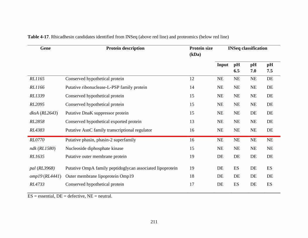
211
Table 4-17. Rhicadhesin candidates identified from INSeq (above red line) and proteomics (below red line)
ES = essential, DE = defective, NE = neutral.
Gene Protein description Protein size
(kDa)
INSeq classification
Input pH
6.5
pH
7.0
pH
7.5
RL1165 Conserved hypothetical protein 12 NE NE NE DE
RL1166 Putative ribonuclease-L-PSP family protein 14 NE NE NE DE
RL1339 Conserved hypothetical protein 15 NE NE NE DE
RL2095 Conserved hypothetical protein 15 NE NE NE DE
dksA (RL2643) Putative DnaK suppressor protein 15 NE NE DE DE
RL2858 Conserved hypothetical exported protein 13 NE NE NE DE
RL4383 Putative AsnC family transcriptional regulator 16 NE NE NE DE
RL0770 Putative phasin, phasin-2 superfamily 16 NE NE NE NE
ndk (RL1580) Nucleoside diphosphate kinase 15 NE NE NE NE
RL1635 Putative outer membrane protein 19 DE DE DE DE
pal (RL3968) Putative OmpA family peptidoglycan associated lipoprotein 19 DE ES DE ES
omp19 (RL4441) Outer membrane lipoprotein Omp19 18 DE DE DE DE
RL4733 Conserved hypothetical protein 17 DE ES DE ES

212
There was no overlap in the rhicadhesin candidates identified from INSeq and proteomic
analysis using the criteria described (Table 4-17, 3.3.2), showing that none seem to fit the
properties required for a rhicadhesin (required for attachment only under alkaline or
neutral-alkaline conditions [34, 53, 71]). However, this result may be misleading, as
rhicadhesin is hypothesised to be a root hair specific attachment factor. Given that the
INSeq experiment examined attachment to the entire root (and root hairs likely constitute
a relatively small part of this in terms of surface area), genes required only for attachment
to root hairs, or root hairs in specific root zones, may be misclassified and therefore
overlooked as rhicadhesin candidates. It is unclear as to why some rhicadhesin candidate
genes identified in proteomics should be classified DE in the input (Table 4-17), as
rhicadhesin is not thought to be an integral component of the bacterial cell [53]
Although proteomics results do not identify rhicadhesin, the candidates extracted from the
INSeq data (Table 4-17, above red line) are informative. These demonstrate that multiple
factors in the 12-16 kDa range display the pattern of defects expected for rhicadhesin and
contribute to attachment under alkaline conditions. They may not all be direct adhesins in
themselves; effects may be indirect, but this seems less likely for candidates such as dksA
(RL2643– see Table 4-14) and RL2858 (encoding a conserved hypothetical exported
protein). As none of these were identified via proteomic approaches, it is impossible to say
whether any of these represent the rhicadhesin(s) first putatively isolated by Smit et al.
(1989) [53]. As rhicadhesin is thought to be important for attachment to root hairs, more
detailed attachment studies (using high magnification microscopy) would be needed to
characterise the spatial attachment characteristics of mutants in these genes at alkaline
pHs.

213
INSeq could be applied to identifying primary attachment factors directly involved in root
hair attachment at different pHs. Ramos et al. (2003) and Qiao et al. (2013) describe the
isolation of purified root hairs from M. truncatula and G. max using liquid nitrogen [372,
373]. A primary attachment INSeq followed by isolation of root hairs and recovery of
bacteria would identify attachment factors required for root hair attachment. However,
there are technical challenges. Ramos et al. (2003) reported 40 μg root hair yield from-100
M. truncatula root tips, whilst this was 800-1000 mg from 60 whole soybean roots for
Qiao et al. (2013) [372, 373]. The yield of root hairs, number of bacteria attached, and
whether bacteria remain attached during root hair isolation all represent significant
challenges to experimental design.
Whilst this approach could be used to identify rhicadhesin, the caveats of previous
research and the technical challenges associated with root-hair INSeq meant that the
characterisation of rhicadhesin was not pursued further in this work. However, pursuing
the characterisation of rhicadhesin as part of this work has enabled far greater insights into
the vast array of root attachment factors utilized by Rlv3841 in attachment to pea roots at
different pHs.
4.3 Conclusion
In summary, experimental parameters for a primary root attachment INSeq experiment at
different pHs were determined. Approximately 87 million barcoded sequencing reads
were obtained from twelve sample libraries with high (82%) insertion density. HMM
classifications were obtained for 7,319 (99.7%) of Rlv3841 genes. Input library data was
curated using a published dataset [151] to account for a TY bacterial recovery step

214
following isolation from plant roots; this left 86% of the genome classified as NE in the
input library.
INSeq results were compared with the phenotypes of mutants characterised in the
literature. Lux data for mutant strain attachment from Chapter 3 was also compared to the
INSeq gene classifications obtained in this chapter. There was a broad overlap between
the INSeq and Lux attachment assay results, although there were some discrepancies. In
one case (praR), this was likely due to low TA site number reducing HMM classification
accuracy. The disagreement of Lux and INSeq results for two motility mutant strains
(OPS1738, flgE and OPS1739, motA) was surprising, but could possibly be explained by
the differing nature of the experimental assays. Lux assays use a homogenous single
inoculum while INSeq involves inoculation with a mixed population of mutants. This was
clearly a factor in the case of an RL2969 mutant. An RL2969 mutant (strain OPS1235)
was identified as attachment-deficient in a single inoculum Lux assay but was NE in the
INSeq experiment. Rescue by in trans complementation was suggested to be occurring in
INSeq, as the OPS1235 mutant strain was similarly able to be rescued in a Lux attachment
assay with 1:100 (mutant:wild-type) inoculation. After accounting for this (and with the
regulator mutants tested in Lux assays in 4.2.12), Lux and INSeq showed a ~75%
agreement rate using the defined criteria (see 4.2.9).
In total, 292 genes were identified as required (ES/DE) for primary attachment at one or
more pHs, with over half of these required specifically at one pH (50 at pH 6.5, 44 at pH
7.0 and 58 at pH 7.5). Examining functional classifications revealed that genes encoding
proteins involved in cell processes, cell membrane/envelope and macromolecule
metabolism, as well as a high proportion of uncharacterised genes, were involved in
primary attachment. Comparison of the genomic localization of primary attachment vs 5-

215
day colonization genes that seemed to be important included some primary attachment
factors that may be constitutively expressed, as opposed to secondary attachment
determinants which are largely inducible.
Using data from INSeq experiments across symbiosis development stages, it was possible
to determine the genes required only for primary attachment (104) or for primary
attachment and subsequent symbiosis stages (49). This is useful in identifying which
factors may be involved in attachment to bulk root epidermis (generalist attachment
factors) as oppose to root hairs, from which infection threads form. The latter can be
considered symbiosis determining primary attachment factors.
INSeq data from experiments with different growth media or metabolic conditions was
leveraged to increase the specificity of primary attachment factor identification. This
filtering reduced the 292 identified factors to 116, with over half specific to one
attachment pH. Examining these in more detail shed light on a range of different factors
used by Rlv3841 for primary attachment to pea roots in a pH dependent fashion. A
summary of the different gene functions needed at different pHs for the primary
attachment of Rlv3841 to pea roots is shown in Figure 4-16.

216
Figure 4-16. Gene functions needed for primary attachment of Rlv3841 to pea roots at pH
6.5, 7.0, 7.5 and combinations thereof. Circle color indicates pH: red = pH 6.5, green = pH
7.0, dark blue = pH 7.5. Text color indicates pH requirement of indicated gene function
for primary attachment: red = pH 6.5, orange = pH 6.5 and 7.0, green = pH 7.0, blue = pH
7.0 and 7.5, dark blue = pH 7.5, purple = pH 6.5 and 7.5, black = pH 6.5, 7.0 and 7.5.
Gene functions are drawn from Tables 4-9 to 4-15, see also Figures 4-11 to 4-14.
pH 7.0
pH 7.0
pH 7.5
pH 7.0
pH 6.5

217
For attachment at all pHs, the requirement for PssA highlighted the core importance of
EPS. The demonstrated requirement for FHA (RL4382) is novel and should be built into
models of primary attachment after further corroborating evidence is obtained. Similarly,
moonlighting glycolytic enzymes (particularly TpiA) also seem to be important, as do
likely peptidoglycan modification factors (RL4362/RL4363). Further genes required with
functions in cell stress responses (hslO – RL0551) and DNA repair (recA – RL2637) may
reflect stresses encountered in the assay, and similar stresses may also be encountered
under field conditions. Transcription factors (notably RL4145) are also likely required,
though regulatory targets need to be identified in future transcriptomic studies.
At pH 6.5, EPS, LPS and peptidoglycan modifications were required for attachment,
notably glycan fucosidic bond cleavage (fucA, pRL100274), diacylglycerol kinase activity
(dgkA, RL2780, linked to phospholipid recycling and LPS modifications [324]) and
transglycosylase mediated cell wall alterations (RL0726). Of importance was the
demonstration of GelA (RL4404) function as a primary attachment factor at pH 6.5, likely
involved in bulk root epidermis, as oppose to root hair attachment.
At both pH 6.5 and 7.0, pRL110043 (an arabinose efflux protein) indicated a specific EPS
structural change needed for attachment, whilst other EPS and membrane lipid
modifications were also necessary. These included ppx (RL1600), a putative
exopolyphosphatase, and RL4309, a likely DedA family protein which functions in
membrane homeostasis [362]. The RNA binding protein Hfq (encoded by RL2284) is also
required. This is of interest, as loss of this factor has been shown to reduce
competitiveness for attachment to alfalfa roots in S. meliloti, likely through metabolic
pathway perturbation and loss of stress-associated chaperone regulation [360]. INSeq

218
highlighted the pH dependent profile of glucomannan activity reported previously [56],
demonstrating the resolving power of this technique.
AnmK (RL2587, which cleaves peptidoglycan structures, [331, 332]) was required at pH
7.0. Also needed were biofilm promoting factors such as pspA (RL1106), which functions
to promote biofilm formation by stabilizing the cytoplasmic membrane [329], and
membrane proteins including RL1013, which features a protein motif also present in
surface antigens of other bacterial species [328].
The ClpS/ClpA protease system was needed at pH 7.0 and 7.5, although the proteolytic
targets remain unclear. As with TpiA, the DnaK suppressor protein DksA seems to have a
moonlighting role, being secreted to bind ligands and contribute to cell adherence at these
pHs.
At pH 7.5, higher membrane phosphatidylcholine requirements were highlighted (PmtA),
along with various protease-driven cell wall changes (Table 4-12). These include those
induced by hflC (RL3253) and hflK (RL3254), transmembrane serine proteases. As well as
their own proteolytic activity, HflC and HflK also modulate HflB, a metalloprotease
known to be involved in regulation of membrane proteins and biofilm formation in E. coli
and other bacteria [345, 346]. HflB itself (encoded by RL3965) is classified ES/DE in the
input library and at all attachment pHs. As HflC and HflK are only needed pH 7.5 (whilst
HflB is needed at all pHs) it seems that HflC and HlfK proteolytic activity and/or
modulation of HflB activity is occurring to promote pH 7.5 attachment. Another novel
finding was the requirement for RpoZ. A DNA polymerase subunit, RpoZ has been linked
to transcriptional regulation of attachment factors [337–339] and is likely to be
functioning similarly in Rlv3841.

219
The requirement for SixA at pH 7.5 and Npr/ManX at pH 6.5 seems to indicate an
opposing signalling system. The PTSNtr system (of which these components are part)
controls the complex metabolic phenotypes. At pH 6.5, Npr/ManX requirements indicate
EPS secretion through ATP dependent ABC transporters [313]. At pH 6.5, SixA likely
deactivates this system, reducing transporter activity [343]. This may be compensated for
by other factors required at pH 7.5, such as PmtA, RL2489A and RL4018 (likely lipid A
exporter). A summary of the different gene functions needed at different pHs for the
primary attachment of Rlv3841 to pea roots is shown in Figure 4-16.
These results largely indicate the disparate cell wall, EPS and LPS requirements for
primary attachment to pea roots at different pHs and demonstrate a greatly increased
complexity of primary attachment mechanisms compared to the published literature.
Two uncharacterised regulators, encoded by RL3453 and RL4145, were needed for
attachment at all pHs. Using genomic proximity and HMM classifications to infer
regulatory targets indicated that exported, transmembrane and lipoprotein biosynthesis
proteins were likely to be regulated by these proteins. Mutants in RL3453 and RL4145
(OPS1967 and OPS1968) were confirmed to be deficient in root attachment using Lux
whole-root attachment assays with a single inoculum. These mutants will be useful for
RNASeq to investigate the transcriptional networks important in primary attachment
further.
Using the INSeq dataset to investigate possible rhicadhesin genes identified seven
candidates encoding proteins between 12-16 kDa which were required for attachment only
at pH 7.0 and/or 7.5. There was no overlap between these genes and those previously
identified from proteomic analysis of a protein preparation band thought to contain
rhicadhesin. Whilst this does not necessarily mean proteins identified by proteomics

220
cannot be a rhicadhesin, it does highlight the numerous caveats of previously published
[53, 54, 208–211] rhicadhesin research:
I) defining an adhesin based on ability of a protein preparation to inhibit attachment does
not conclusively demonstrate it is an adhesin
II) Given the high number of factors involved in Rlv3841 root attachment, preparation of
a membrane fraction may isolate root attachment factors, but this does not indicate they
are more important for primary attachment than any other factor
III) There may be multiple functional genes encoding rhicadhesin, or multiple
rhicadhesins
IV) Smit et al. (1989) never conclusively demonstrated they were working with a single
purified protein; their ‘purified adhesin’ could have been a protein mixture [53]
V) There are multiple Rlv3841 factors of 12-16 kDa that contribute to attachment at pH
7.0 and/or pH 7.5, all of which could be considered rhicadhesins
Whilst pursuing the characterisation of rhicadhesin as part of a wider strategy to identify
root attachment factors has been very informative, the identity of the protein(s) isolated by
Smit et al. (1989) [53] remain unclear. Given the number and diversity of Rlv3841
primary attachment factors, they may well have isolated a protein that plays a role in this
process, but whether in a direct or indirect capacity is unknown. Pending further, in depth
characterisation of small surface localized root attachment factors (particularly for calcium
binding activity and involvement in attachment specifically to root hairs, postulated
characteristics of rhicadhesin - Smit et al. (1989) [53]) it seems unhelpful to describe
primary root attachment in Rlv3841 as a two component, pH-dependent
glucomannan/rhicadhesin system. More holistically, it should be thought of as a process

221
employing diverse cellular factors, with a focus on EPS, LPS and peptidoglycan
biosynthesis and modification, as well as direct adhesins (such as FHA), membrane
proteins and uncharacterised factors, all of which show different pH dependencies. Whilst
glucomannan is clearly important for attachment at acidic and neutral pHs [56] (also seen
here in a Lux attachment assay – Figure 3-6 – and INSeq), there is likely to be a multitude
of other factors that contribute to root hair attachment at alkaline pHs.
This chapter highlights the power of INSeq for investigating early stage symbiosis
determinants on a genome-wide scale and has revealed far greater complexity than
previously known in primary attachment mechanisms.

222
223
Chapter 5
Genome-scale characterisation of the primary
attachment determinants of R. leguminosarum to roots
of a non-host legume and non-legume

224
5.1 Introduction
The first physical interaction in Rhizobium-legume symbioses occurs when bacteria attach
to their hosts roots [26]. A comparative lack of research into this interaction [25] means
that relatively few factors involved in primary root attachment have been fully
characterised. The use of INSeq to investigate primary root attachment determinants at
different pHs in Rlv3841/pea symbiosis (described in Chapter 4 of this thesis) revealed far
greater complexity than previously thought. By enabling genome-wide characterisation of
attachment determinants in a single screen, it provided a powerful tool to unearth novel
biological roles in symbiosis formation for >100 genes.
It is not just attachment leading to nitrogen-fixing symbioses (in which rhizobia provide
fixed nitrogen in return for carbon sources [25]) which can bring benefits to rhizobia. The
ability to attach to bulk root epidermis, or the roots of non-host plants, neither of which
results in nodule formation and nitrogen-fixing symbiosis development, is also likely to
give nutritional benefits as exudates from roots can serve as preferential growth substrates
for rhizobia [34]. This interaction could be considered a symbiosis in itself, as the host
plant may also derive benefits. For example, R. leguminosarum is able to colonise the
roots of tomato and pepper plants and promote plant growth, likely through indole acetic
acid and siderophore production [38]. Further studies have reported similar effects of R.
leguminosarum in maize, lettuce, rice, canola and more [36–38]. Plant growth promoting
rhizobacteria (PGPR) may also confer benefits by protecting plants from pathogens. R.
leguminosarum biovar viciae inoculation of pea gives protective effects against Didymella
pinodes pathogenesis [43]. The protective effects against D. pinodes-induced wilt disease
are in addition to the plant growth promotion resulting from nitrogen-fixing symbiosis
formation [43]. Sistani et al. (2017) demonstrated that inoculation of pea seeds with R.

225
leguminosarum biovar viciae induced changes in the seed proteome and metabolome. By
inducing higher ROS responses, plant cell wall structural changes and production of
antimicrobial seed flavonoids and triterpenoids (notably soyasapogenol), significant
reductions in D. pinodes infection were observed [43]. In addition, different rhizobial
isolates have been reported to protect chickpea against Fusarium wilt [374]. In this case
Arfaoui et al. (2006) screened 21 field isolates of R. leguminosarum biovar ciceri. 17 were
able to directly inhibit Fusarium oxysporum growth and protect plants from disease via
production of volatiles and/or cyanide and some also displayed phosphate solubilising
capabilities, meaning plant growth promotion could also occur via nutrient provision
[374]. Recent imaging work has even demonstrated that, at least in the short term (up to
12 hours post-inoculation), B. subtilis (acting as a PGPR) was able to physically exclude
E. coli from interacting with Arabidopsis roots, suggesting a possible physical protection
against plant pathogens is possible in some cases [188]. Such physical exclusion has been
documented for B. subtilis acting against the plant pathogen P. syringae pv tomato
DC3000, which is able to infect Arabidopsis roots. Bais et al. (2004) demonstrated that
root protection was effected by B. subtilis forming extensive biofilms on the root surface
(thus physically preventing pathogen-root contact) and secreting surfactin, a lipopeptide
antimicrobial which is toxic to P. syringae [375].
Intriguingly, recent microbiome research has demonstrated that the presence of a nitrogen
fixing symbiosis, in this case between L. japonicus and M. loti, is able to influence the
wider microbiome structure of organisms interacting with roots in the rhizosphere. In
plants deficient in symbiosis formation the microbiome structure is dramatically altered,
implying that a legume host selects a broad taxonomic range of root associated bacteria
that could contribute to plant growth [121].
226
Attachment of different bacteria to roots is a diverse process with different factors thought
to be involved, which show considerable variation [34]. For example, whilst
Agrobacterium rely on adhesive pili and capsular polysaccharide (among other factors) for
root attachment, Pseudomonas is able to use outer membrane porins as well as pili and
flagella, and LPS and outer membrane proteins plays a role in Azospirillum [34] (see also
Figure 1-3).
As shown in Chapter 4, the factors required for attachment of Rlv3841 to the roots of one
plant (pea) also show considerable diversity under different conditions (in this case, pH
changes). A good example of this is the variation seen in EPS. EPS is known to be of
crucial importance in attachment of Rlv3841 to roots [58, 77]; in this thesis a mutant in
pssA (RL3752), a glycosyltransferase involved in the first step of EPS biosynthesis [288]
was seen to be deficient in root attachment both in a Lux whole-root attachment assay
(Figure 3-5) and using INSeq (Table 4-9). However, EPS requirements are more nuanced
than this. Several genes involved in EPS processing were required for primary root
attachment at different pHs. These included gelA (RL4404, gel forming EPS production
protein important in attachment at pH 6.5), ppx (RL1600, putative exopolyphosphatase
functioning in EPS hydrolysis [359], important at pH 6.5 and 7) and RL2778 (a putative
EPS biosynthesis protein, important for attachment at pH 7.5). In addition to processing of
EPS, multiple genes were identified as required for attachment at different pHs with likely
roles in LPS and peptidoglycan modification, as well as functional roles in the membrane,
highlighting the importance of polymeric substances and the bacterial cell membrane and
wall structures in primary attachment. There is considerable diversity in EPS structures
between different Rhizobium (R. leguminosarum biovars triofolii and viciae and S. meliloti

227
all demonstrate distinct EPS structures [376]), and this can influence the specificity of the
nitrogen fixing symbiosis [96].
Given that so many different factors are required to attach to the roots of pea at different
pHs (Chapter 4), and that different bacteria use different factors for primary attachment to
different plant roots [34], it may also be the case that different Rlv3841 factors are
required to attach to the roots of different plants.
Given the broad role of PGPR-root interactions in increasing crop yields in diverse plants
and eliminating pathogens (see also [39–42]), improving our understanding of root-
bacteria interactions is of major interest to both basic and applied plant research.
Very little is known about the mechanistic determinants of interactions of rhizobia with
non-host plants in terms of primary attachment factor requirements. Albareda et al (2008)
examined the effects of salt concentration, culture age, pH and inoculum composition on
total numbers of rhizobia colonising the roots of non-host plants [377]. Further,
Villacieros et al (2002) investigated the colonization of P. fluorescens in the alfalfa
rhizosphere and how this was influenced by the presence of S. meliloti [378]. This
demonstrated that, whilst both strains colonize the alfalfa rhizosphere efficiently alone, in
co-inoculation S. meliloti came to dominate the rhizosphere and reduced the deleterious
effects of P. fluorescens single inoculation on alfalfa growth. 16S rRNA sequencing has
been used to investigate the rhizosphere community influence of rhizobial strain co-
inoculation on Dalbergia odorifera roots (demonstrating that different rhizobial
inoculums cause different shifts in rhizosphere strain abundance [379]). Further, the
influence of Bradyrhizobium on arbuscular mycorrhizal fungi colonization of soybean
roots has also been characterised, showing that bacterial co-inoculation could significantly
enhance fungal colonization and increase plant dry weight [380]. However, whilst

228
providing overviews of changes in community structure and levels of attachment to roots
under different conditions, among other parameters, these studies provide no mechanistic
information in terms of how bacteria physically attach to the roots of different plants.
The large diversity seen in Rhizobium EPS structures (given as an example above) hint
that interactions with different plant species may involve very different primary
attachment determinants. This idea is reinforced by the different primary attachment
determinants used by different rhizobacteria when attaching to plant roots [34] (see also
4.1). In this chapter, the use of INSeq to investigate the primary attachment determinants
of Rlv3841 to soybean (a non-host legume) and barley (a non-legume) roots at pH 7.0 is
described. The major aim of this work was to investigate whether primary attachment
determinants of Rlv3841 are unique to different plants, or whether mechanistic overlap is
seen, and to improve our understanding of root-bacteria interactions. By combining this
data with a subset of the pea primary root attachment data from Chapter 4, the core
determinants required for primary attachment to multiple plant species and the variable
determinants, which show specificity depending on the plant species, are defined. To the
best of my knowledge, this represents the first study to define the determinants of primary
attachment to roots of different plants for a given rhizobial species.
The raw data for this INSeq experiment can be found in Appendix 2, Table 2.

229
5.2 Results and discussion
5.2.1 Attachment assays of Rlv3841 – determining inoculum density and bacterial
recovery method for INSeq
Attachment assays with Rlv3841 (2.6.5) were carried out to determine the optimum
starting inoculum density for INSeq after recovery of root-attached bacteria for both
soybean and barley (Appendix 1, Figures A1 and A2, see also Figure 4-2 for pea). As was
the case for pea (where recovered bacteria rose with increasing inoculum density, leading
to the recovery of ~ 2 x 10⁶ cells/root after 1 hr with 50 mL of an OD600 0.1 inoculum),
higher starting inoculum density gave greater numbers of Rlv3841 cells recovered from
roots after one hour.
As discussed in 4.2.1, there is a minimum fold-coverage requirement (500-750) of genes
in INSeq for robust downstream HMM classification. The numbers of plants required to
achieve this 750-fold coverage or higher for soybean and barley is calculated using the
equation from 4.2.1 and the data in Appendix 1, Figures A1 and A2. For a given starting
inoculum density, Table 5-1 provides the number of plants needed for 750-fold coverage
for both soybean and barley.

230
Table 5-1. Number of plants needed for 750-fold coverage of 7,300 genes in the genome
of Rlv3841 based on input inoculum density (OD600) for soybean and barley
Input inoculum (OD600) Plants needed for 750-fold coverage of 7,300 genes
Soybean Barley
0.001 30 18
0.01 16 10
0.1 1 2
Given the results presented in Table 5-1 and the setup of Lux attachment assays and
INSeq work presented in Chapter 4, it was decided to use a starting inoculum of OD600 0.1
with 10 plants per test condition. Each test condition was repeated in triplicate (see 4.2.2).
This starting inoculum density and plant number should allow for > 3,000 fold coverage
of the ~7,300 Rlv3841 genes for each plant.
As in Chapter 4, no statistically significant difference was seen between the number of
Rlv3841 recovered by vortexing compared to vortexing and grinding of roots for either
plant (for soybean shown in Appendix 1, Figure A3 and barley in Appendix 1, Figure A4),
meaning that vortexing alone should be sufficient to recover Rlv3841 cells in INSeq
primary root attachment assays.

231
5.2.2 INSeq experimental design
To characterise Rlv3841 primary attachment determinants with barley and soybean roots,
an INSeq experiment was designed (Figure 5-1). Results were compared with primary
attachment determinants identified using INSeq for pea roots at pH 7.0 (Chapter 4). An
OD600 0.1 (108 cfu/mL) population of insertion mutants (library 1-3, 500 mL) was used to
inoculate 10 soybean or barley plants in triplicate. Insertion mutants were recovered from
roots after 1 hr and grown in liquid TY for 12 hr before the 9 sample libraries (2x3
experimental conditions + 3 input libraries) underwent barcoding, mariner library
preparation and sequencing (2.7.3) (Figure 5-1). HMM gene assignments were averaged
across three replicates for each plant as described in 4.2.2.
Figure 5-1. INSeq experimental design, 50mL of insertion mutant inoculum (library 1-3,
OD600 0.1) was inoculated onto each soybean or barley root at pH 7.0 and incubated for 1
hr before recovery by vortexing (resulting in three replicates). Recovered bacteria for each
attachment group were pelleted and grown on TY for 12 hours to increase bacterial gDNA
concentration (and effectively decrease plant DNA contamination) before DNA

232
preparation (not shown). Input library gDNA samples were extracted directly from input
inoculum. Each sample (input and 3 x root-attached) underwent DNA extraction and
library preparation before sequencing (see 2.7.2, 2.7.3 and 2.7.4).
5.2.3 HMM genome analysis
For primary attachment to roots of soybean or barley, HMM analysis of sequencing reads
assigned each of the 7,319 genes in the Rlv3841 genome to one of the following
categories, based on how gene mutation affects bacterium fitness: ES, DE, NE or AD (see
Figure 4-1), as in the previous chapter for pea plants. In total, 127 million barcoded
sequencing reads were obtained from a total of nine samples (six from plants and three
input libraries). The insertion densities (proportion of total TA motifs that contain at least
one insertion in each sequenced library) for libraries were as follows: input – 87 %,
soybean pH 7 – 54%, barley pH 7 – 56%. These insertion densities are more than
sufficient, based on multiple reviews of Tn-Seq and INSeq data analyzed using a HMM
approach and their respective library insertion densities, for robust statistical analysis and
assignment of HMM gene classification [146, 150, 169, 179].
5.2.4 INSeq gene classifications
The distribution of HMM gene classifications across the four different states (ES, DE, NE
and AD) in input and root-attached libraries for the two plants is shown in Table 5-2. Note
that for the chromosome or any plasmids individually the insertion density obtained did
not differ more than ± 3% from the average input figure given in Table 5-2, indicating
unbiased mariner insertions across the genome.

233
Table 5-2. Distribution of HMM assignment of Rlv3841 genes in the input and root-
attached (pea, soybean and barley, pH 7.0) libraries.
HMM gene
assignment
Library condition
Input pH 7.0 pea pH 7.0 soybean pH 7.0 barley
ES 1 % 6 % 7 % 8 %
DE 10 % 7 % 8 % 6 %
NE 86 % 86 % 84 % 85 %
AD 3 % 1 % 1 % 1 %
Data for pea plants grown at pH 7.0 is from Chapter 4 (see Table 4-3, section 4.2.4).
Values are given as percentage of the 7,319 genes which contain TA sites.
As explained in the previous chapter, due to the nature of the experimental design, the
input library data was curated using the published dataset from Perry and Yost (2014)
[151] to account for the 12 hour TY growth step for recovered root-attached populations.
This 12 hr growth step was used for bacteria recovered from roots to increase bacterial
gDNA concentration and dilute contaminating plant DNA before DNA extraction.
Curation removed any inadvertent selection on genes from this step which could affect
downstream HMM classification (see also 4.2.4). When using INSeq to identify primary
root-attachment determinants, it is genes that are NE in the input but non-NE in
attachment which are of interest.

234
5.2.5 Literature validation of INSeq predictions
In order to provide preliminary validation of the HMM gene classifications for soybean
and barley root attachment, the INSeq classifications of well-characterised genes (see
4.2.5) were compared with their known or predicted phenotypes according to previous
publications. It was not expected that HMM classifications should be plant dependent.
These genes are listed in Table 4-4, and include genes encoding peptidoglycan
biosynthesis factors, ribosomal subunits, DNA helicases and polymerases, all critical for
cell viability. Mutation in these genes leads to an ES or DE HMM classification in
attachment to soybean and barley, as is the case for pea. Similarly, genes encoding non-
essential factors, such as the putative manganese transporter mntH (RL0940) are classified
as NE in attachment to soybean and barley, as well as pea. Classifications for these genes
in attachment to soybean and barley were the same as for attachment to pea (Table 4-4),
providing both initial validation of this dataset and an indication of reproducibility in the
HMM classifications.
5.2.6 Primary attachment gene requirements and functional classifications
In total, 464 genes classified as NE in the curated input libraries were classified as ES/DE
in one or more of the pH 7.0 root-attached libraries (input library data from chapter 4 was
used for pea, and input library data from this chapter used for soybean and barley). The
distribution of these is shown in Figure 5-2, below. These genes and their HMM
classifications from experiments listed in Table 4-5 (pea rhizosphere [238], 5 d root
colonization [238], infection thread and nodule bacteroids - unpublished data, Poole Lab)
and Table 4-7 (Vincents media [175], 21% or 1% oxygen with 10mM glucose or 20mM
succinate, [238]) are listed in the following Appendix 1 tables: Table A10: all plants,

235
Table A11: pea only, Table A12: soybean only, Table A13: barley only, Table A14: pea
and soybean, Table A15: pea and barley, Table A16: barley and soybean.
Figure 5-2. Genes required (ES/DE) for primary attachment to roots of pea, soybean and
barley at pH 7.0. Total genes = 464. Color indicates plant; green = pea, yellow = soybean,
brown = barley.
The distribution of genes required for attachment to pea, soybean and barley roots (Figure
5-2) is interesting because it indicates that, whilst there are a large number of genes (68)
required for attachment to all plants, there are also a large number of genes required
exclusively for attachment to one or two of the three plants. This demonstrates both the
conservation of some primary attachment mechanisms for Rlv3841 across plant species,
but also some mechanistic specificity in attachment, which is dependent on plant host.
Functional classifications were examined for genes required for attachment to all plants
(68 genes) or to pea (40 genes), soybean (201 genes) or barley (52 genes) exclusively
using Riley codes [138, 272] as described in 4.2.6 (Figure 5-3).

236
Figure 5-3. Functional classification (using Riley codes) of the 68 genes required
(ES/DE) for attachment to pea, soybean and barley roots at pH 7.0 compared with the 40,
201 and 52 genes required for attachment only to pea, soybean or barley roots respectively
at pH 7.0. Data is presented as percentage of total genes in each category.
Functional classifications (Figure 5-3) reveal that the most abundant ES/DE gene classes
for attachment to all plants are uncharacterised genes and macromolecule metabolism.
However, differences in the functional classifications of genes required for attachment to
individual plants are visible. Like the finding from pea (Figure 4-6), it seems that
uncharacterised genes play an important role in determining successful primary root
attachment in all cases, although this is especially the case for barley.

237
5.2.7 Genomic localization of genes required for primary root attachment to pea,
soybean and barley
Further insight can be gained by comparing the loci of genes required (ES/DE) for
attachment to the roots of these three plants (Figure 5-4). As for the functional
classifications above, comparisons were made for the genes required for attachment to all
plants (68 genes) with the genes required for attachment only to one of the three plants (40
genes for pea, 201 genes for soybean and 52 genes for barley, Figure 5-2).
A B
C D E
Distribution of genes in the Rlv3841 genome
Distribution of primary attachment ES/DE genes for all plants
Distribution of primary attachment ES/DE genes for pea
Distribution of primary attachment ES/DE genes for soybean
Distribution of primary attachment ES/DE genes for barley

238
Figure 5-4. Genomic localization of Rlv3841 genes (as a proportion of the total). A –
genome of Rlv3841 (7,340 genes). Genes required (ES/DE) for attachment to roots at pH
7.0 to B – all plants (68 genes), C – pea (40 genes), D – soybean (201 genes), E – barley
(52 genes).
As discussed previously, 65.4% of the annotated encoded genes in the Rlv3841 genome
are located on the chromosome, with the remainder divided between six plasmids (Figure
5-4, A). The distribution of primary attachment genes required for attachment to all plants
tested (pea, soybean and barley, Figure 5-4, B) indicates that, compared to the genome the
location of these genes is skewed toward the chromosome (91%) and away from the
plasmids, with no representation for pRL12 or 8. This most closely mirrors the
distribution seen for barley (Figure 5-4, E), where the chromosomally encoded factor
requirement is 94%. Figure 5-4 C demonstrates that, for pea, chromosomally encoded
factors form the bulk of primary attachment determinants (as they do in all cases), but
plasmid representation is also the highest of all plant groups. Plasmid pRL10 is the most
well represented; this is of interest as pRL10 is referred to as the symbiosis plasmid,
encoding symbiotic functions in the nod, nif and fix genes [381].
Gene localization of soybean only attachment determinants is particularly interesting, as it
demonstrates a large overrepresentation of pRL11 genes (28 genes in total) and is the only
attachment condition featured in this work to require any genes encoded on pRL8 (three
genes) (Table 5-3, Figure 5-4, D). These three genes are presented in Table 5-3, below,
along with six genes from pRL11 which are sequentially encoded and may play a
complementary role in promoting attachment based on annotation functionally linking
them to the pRL8 genes.

239
The three pRL8 genes required for attachment to soybean roots encode conjugal transfer
or pilus stabilising/secretion machinery and the six pRL11 genes cluster functionally
around Flp pilus function (Table 5-3). The Flp pilus is part of the Flp/Tad system, which is
known to be important for initial adhesion and biofilm formation in many Gram positive
and negative bacteria [382]. S. meliloti mutants in the Flp pili cluster are impaired in
nodulation with M. sativa, which is likely to be due to attachment and colonization defects
[383]. A. tumefaciens with mutations in the ctp locus (which encodes the Flp/Tad system
in this bacterium) shows reduced adherence and biofilm formation on abiotic surfaces
[44]. Deletions in the Flp/Tad genes in many other bacteria, including Pectobacterium (a
potato pathogen), Vibrio vulnificus, Aggregatibacter actinomycetemcomitans and
Caulobacter crescentus, have also been shown to reduce surface adhesion [44, 384, 385].
Studies such as these have led to the general conclusion that Flp/Tad (here the pRL11
genes in Table 5-3) and Flp/Tad related genes (here putatively the pRL8 genes in Table 5-
3, which may function in pilus stabilisation and/or secretion) are involved in attachment
and biofilm formation [87]. It therefore seems that Rlv3841 requires pilus formation and
type IV secretion system components for primary attachment to soybean roots at pH 7.0.
Although this has not been documented in the Rlv3841 interaction with pea,
Agrobacterium and Pseudomonas are known to use pili in primary attachment to plant
roots [34].

240
Table 5-3. Genes from pRL8 and pRL11 required (ES/DE) for Rlv3841 attachment to soybean roots at pH 7.0 which may play a
complementary role in promoting attachment.
Gene Description
trbLp8
(pRL80133)
P-type conjugative transfer protein TrbL. Found in the trb locus of Agrobacterium Ti plasmids, involved in the type IV secretion
system for conjugative transfer. A homologue of VirB9, a type VI secretion system protein evolved from the conjugative
apparatus which exports virulence proteins in pathogenic bacteria. Essential for virulence in Agrobacterium, also stabilises the
pilin conjugative structure [386–389].
trbHp8
(pRL80134)
Putative conjugal transfer protein TrbH. Contains a putative membrane lipoprotein lipid attachment site.
trbGp8
(pRL80135)
Putative conjugal transfer protein TrbG. A homologue of VirB6, an essential component of type IV secretion machinery for pilus
formation in Agrobacterium [388].
pRL110569 Putative transmembrane protein. 95% identity to a pilus assembly protein (gene EHH54_1685) from R. leguminosarum.
pRL110570 Hypothetical exported protein.
pRL110571 Uncharacterised protein. 96% identity to Flp pilus assembly protein CpaB from R. leguminosarum biovar viciae. RcpC family
protein expressed from the tight-adherence Tad locus in gram negative bacteria.; likely has a role in modifying and assembling the
Flp pilin [390].
pRL110572 Putative Flp pilus assembly protein. T2SP-E domain, containing components of the type II secretion system, type 4 pili and type
IV secretion system.
pRL110573 Putative transmembrane pilus component protein. T2SSF domain, found in the type II secretion system and type 4 pili.
pRL110574 Putative transmembrane pilus component protein. 100% identity to Flp pilus assembly protein TadB from R. leguminosarum
biovar viciae WSM1455, predicted to form part of a large molecular structure for Flp pili assembly and secretion [391].

241
5.2.8 Increasing specificity of primary root attachment factor identification from
INSeq – pleiotropy filtering
As discussed in 4.2.10, factors identified as primary attachment determinants which also
show pleiotropic effects under different media or metabolic test conditions may be less
likely to represent specific primary attachment factors. To increase the specificity of
primary attachment factor identification and provide a starting point for in-depth analysis,
results from this INSeq experiment were cross-referenced against the INSeq datasets listed
in Table 4-7 (Vincents media [175], 21% or 1% oxygen with 10mM glucose or 20mM
succinate, [238]). Results were used to assign a ‘pleiotropy filter’ (see 4.2.10) where genes
showing a non-NE classification in any of the experiments listed in Table 4-7 were
considered as non-specific primary attachment determinants. Use of this filter can be seen
in Appendix 1, Tables A10-A16. Filtering resulted in 312 genes which are required
specifically for root attachment. Their distribution over the three plants is shown in Figure
5-5.
242
Figure 5-5. Genes specific for primary attachment to roots of pea, soybean and barley.
These 312 genes have been classified as NE in the respective input library, required
(ES/DE) in one or more of the root attached libraries (pea, soybean or barley, all at pH
7.0) and have been classified as NE in the following INSeq datasets: VMM [175], 21%
oxygen 10mM glucose, 1% oxygen 10mM glucose, 21% oxygen 20mM succinate and 1%
oxygen 20mM succinate, [238] (see Table 4-7). Color indicates plant; green = pea, yellow
= soybean, brown = barley.
5.2.9 Specificity in Rlv3841 primary attachment factor requirements is highly plant-
dependent
While there are clear differences in the genes specific for primary attachment to different
plants, there is also substantial overlap between the plant species (Figure 5-5).
In order to visualize the specificity of the interaction for each plant species, the number of
genes involved only in attachment to one plant’s roots can be expressed as a percentage of
the total number of genes involved in attachment to those roots. For example, after
filtering a total of 55 genes are required (ES/DE) for attachment to pea roots (14 + 6 + 11
+ 24, see Figure 5-5). Of these, 14 are specific for attachment to pea roots alone (31%).
69% are therefore also involved in attachment to barley and/or soybean and can be
considered non-specific for pea. This calculation illustrating the specificity for attachment
to pea, soybean and barley is summarized in Figure 5-6.

243
Figure 5-6. Specificity of primary attachment factors for Rlv3841 attaching to pea,
soybean and barley roots. Data are expressed as percentage of genes required (ES/DE) for
primary attachment to plant roots post-filtering (5.2.8, Figure 5-5) which are specific (i.e.
required for attachment to one plant type only, blue) or non-specific (i.e. required for
attachment to more than one plant type, orange). Gene numbers are given within the bars
for each plant type. Total n = 55 genes for pea, 240 genes for soybean and 113 genes for
barley.
Dividing the data in this way (Figure 5-6) demonstrates that Rlv3841 displays the highest
unique primary attachment factor requirements (~86%) in interaction with the roots of
soybean (non-host legume). The roots of pea plants (host legume) and barley (non-
legume) show a similar proportion (~35% in each case), of specific primary attachment
factors. This is surprising, given that primary attachment factor requirements for legume
roots might be expected to be more similar than for non-legume roots. However, given the
far higher unique primary attachment factor requirements, and the demonstrated
14
41
172
68
40
73

244
requirement for pilus formation and type IV secretion system components for primary
attachment to soybean roots at pH 7.0 (see 5.2.7), it seems that this is not necessarily the
case. Primary attachment factor requirement may therefore be governed by features of the
root surface which are not conserved along legume/non-legume lines. This could also be
influenced by differential gene induction in response to different plant exudation profiles,
although to what extent this is occurring remains unclear.
5.2.10 Regulatory requirements for Rlv3841 primary attachment to pea, soybean and
barley roots
Given the large differences in gene specificity requirements for primary attachment
(described in 5.2.9 and Figure 5-6), it seems logical that genes with differing regulatory
functions play a role in attachment to different plant roots. Given that soybean shows the
highest requirement for specific genes (~86%, Figure 5-6), it may be the case that soybean
has the highest number of specific regulators involved. Using Riley codes [138, 272] to
identify regulators showed that, post-filtering (5.2.8), there is one regulator required for
primary attachment to all plants (RL4145, Table 5-4), three required only for pea
(pRL120518, RL1105 and pspA – RL1106, Table 5-4), 22 required only for soybean
(Table 5-4), and six required only for barley (Table 5-4). These figures may support the
idea that higher specificity in primary attachment factor requirements is coupled to
increased regulatory requirements as the numbers for soybean are higher than those of
other plants. Only six have functional annotation. These are pspA (RL1106), putatively
required for attachment to pea, ecfM (pRL100385), mur (RL0397), cheY1 (RL0687) and
aldR (RL1965), required for attachment to soybean and msiR (RL2857), required for
attachment to barley.

245
Table 5-4. Genes with regulatory function (based on Riley codes) that are required (ES/DE) in primary attachment to all plants, or to
pea, soybean or barley roots exclusively, at pH 7.0.
Gene Plant Description
RL4145 All putative LacI family transcriptional regulator (repressor) (see also Figure 4-15)
pRL120518 Pea putative TetR family transcriptional regulator
RL1105 Pea putative TetR family transcriptional regulator
pspA (RL1106) Pea putative (phage shock protein A) PspA family regulator by protein-protein interactions (see Table 4-
11)
ecfM
(pRL100385)
Soybean putative RNA polymerase ECF sigma factor, family ECF20/ECF01. As a member of the clade VI
sigma factors, EcfM may play a role in stress response or host interactions [392]
pRL110046 Soybean putative FNR/CRP family transcriptional regulator
pRL110283 Soybean putative ArsR family transcriptional regulator
RL0229 Soybean putative LysR family transcriptional regulator
mur (RL0397) Soybean putative FUR-like transcriptional regulator, manganese uptake regulator [393]
cheY1 (RL0687) Soybean putative two-component sensor/regulator; chemotaxis transcriptional regulator CheY
RL0762 Soybean putative XRE family (HipB) transcriptional regulator
RL1040 Soybean putative LysR family transcriptional regulator

246
RL1162 Soybean putative two-component sensor/regulator; transcriptional regulator
RL1163 Soybean putative two-component sensor/regulator; histidine kinase
aldR (RL1965) Soybean putative AsnC family positive transcriptional regulator of alanine dehydrogenase [394]
RL2486 Soybean putative LysR family transcriptional regulator
RL2766 Soybean putative ArsR family transcriptional regulator
RL2779 Soybean putative two-component sensor/regulator; histidine kinase
RL3146 Soybean putative LysR family transcriptional regulator
RL3148 Soybean putative ArsR family transcriptional regulator
RL3196 Soybean putative MarR family transcriptional regulator
RL3263 Soybean putative LysR family transcriptional regulator
RL3265 Soybean putative AraC family transcriptional regulator (activator)
RL4189 Soybean putative LysR family transcriptional regulator
RL4219 Soybean putative DeoR family transcriptional regulator (repressor) of sorbitol/mannitol operon
RL4604 Soybean putative GntR family transcriptional regulator
RL0159 Barley putative MarR family transcriptional regulator
RL0561 Barley putative AraC family transcriptional regulator (activator)
RL2657 Barley putative GGDEF/GAF sensory box protein

247
msiR (RL2857) Barley putative ArsR family transcriptional regulator MsiR, regulates MsiA canavanine (found in seed
exudate) exporter
RL3455 Barley putative MarR family transcriptional regulator
RL3595 Barley putative LacI family transcriptional regulator (repressor)
Where available, gene annotation is given in brackets next to gene number.

248
RL4145 (the regulator important for Rlv3841 primary root attachment to all plants, Table
5-4), has been investigated using a Lux attachment assay (Figure 4-15 and Table 4-9) and
was required for attachment to pea roots under all pH conditions. Although the regulatory
targets of RL4145 remain unknown (the only predicted functional interaction partner from
the STRING database with a confidence score >0.6 is RL4144, a putative
oxidoreductase/dehydrogenase) this gene seems to be a very important regulator of
attachment to pea, soybean and barley roots, including at different pHs.
One regulator involved in attachment only to pea is annotated: pspA (RL1106). This phage
shock protein A gene functions to stabilise the cytoplasmic membrane in E. coli and
promote biofilm formation [329]. It is also upregulated under membrane stress conditions
in R. leguminosarum biovar trifolii, where it may improve desiccation tolerance [395].
Although the link with root attachment in this case remains unclear, desiccation tolerance
has previously been linked in Rlv3841 with higher EPS accumulation [396]. Given the
importance of EPS in root attachment [34, 71, 77], if loss of pspA affects EPS production,
this could corroborate the importance of this factor for primary attachment to pea roots.
Of the 21 regulators required for primary attachment to soybean only four are annotated:
ecfM (pRL100385), mur (RL0397), cheY1 (RL0687) and aldR (RL1965) (Table 5-4). Of
interest is the requirement for cheY1 (RL0687), a response regulator controlling flagellar
rotation in chemotaxis. In the previous chapter, motility and chemotaxis components were
not required for Rlv3841 attachment to pea roots in INSeq, even though they were
required in the Lux root attachment assay (see Figure 3-5). Possible reasons for this,
mainly concerning the mixed inoculum setup of INSeq compared with the homogenous
single inoculum for Lux assays, were discussed in 4.2.9. CheY1 is a minor chemotaxis
regulator, whilst CheY2 is of greater importance; deletion of cheY1 reduces swimming

249
ability by 30%, whereas this figure is 70% in the case of cheY2 deletion [397]. If
chemotaxis is necessary for primary attachment to soybean roots, cheY2 would also be
expected to be required. However, cheY2 has only a single TA site (cheY1 has three). As
discussed in 4.2.9, low TA site number (< six) can confound HMM classification.
To investigate this further, several other chemotaxis genes shown to be required by this
INSeq experiment in the soybean library and with more TA sites were compared to the
same genes in the input, pea and barley libraries using fitness values (FV). FVs allow
numerical comparison of the fitness of bacteria carrying mutations in particular genes
under different test conditions, providing a simplified visualization of the data underlying
HMM classifications [143] (see 2.7.5). Comparisons of gene TA site number and HMM
classifications are shown in Table 5-5 and comparison of FVs for the same genes are
shown in Figure 5-7.
Table 5-5. Comparison of TA site number and HMM classifications for five chemotaxis-
related genes under different INSeq conditions
FV (HMM classification)
Gene #TA I 1 Pea I 2 Bar Soy
mcpR (pRL120056) – putative methylation accepting
chemotaxis protein
29 NE NE NE NE DE
icpA (RL0685) – putative chemoreceptor protein
15 NE NE NE NE DE
cheX1 (RL0686) – putative chemotaxis related CheX
protein
3 NE NE NE NE DE
cheY1 (RL0687) – putative two component
sensor/regulator; chemotaxis regulator CheY
3 NE NE NE NE DE
cheW3 (RL4030) – putative chemotaxis protein 9 NE NE NE NE DE
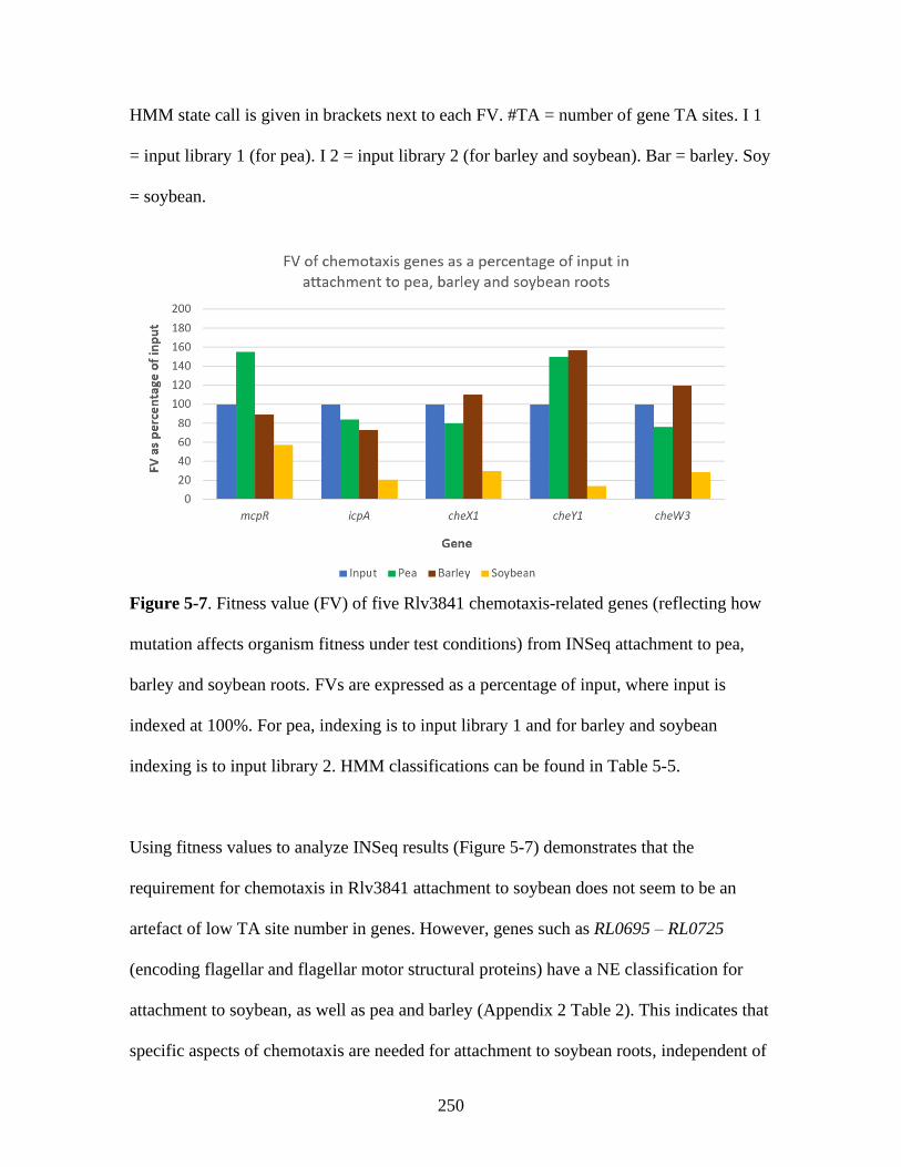
250
HMM state call is given in brackets next to each FV. #TA = number of gene TA sites. I 1
= input library 1 (for pea). I 2 = input library 2 (for barley and soybean). Bar = barley. Soy
= soybean.
Figure 5-7. Fitness value (FV) of five Rlv3841 chemotaxis-related genes (reflecting how
mutation affects organism fitness under test conditions) from INSeq attachment to pea,
barley and soybean roots. FVs are expressed as a percentage of input, where input is
indexed at 100%. For pea, indexing is to input library 1 and for barley and soybean
indexing is to input library 2. HMM classifications can be found in Table 5-5.
Using fitness values to analyze INSeq results (Figure 5-7) demonstrates that the
requirement for chemotaxis in Rlv3841 attachment to soybean does not seem to be an
artefact of low TA site number in genes. However, genes such as RL0695 – RL0725
(encoding flagellar and flagellar motor structural proteins) have a NE classification for
attachment to soybean, as well as pea and barley (Appendix 2 Table 2). This indicates that
specific aspects of chemotaxis are needed for attachment to soybean roots, independent of

251
non-chemotactic motility. For motile bacteria with mutations in chemotaxis related genes,
a negative attachment phenotype may arise due to aberrant swimming, whereby bacteria
swim randomly and are therefore not correctly positioned in proximity to the root surface
for attachment. Non-motile bacteria may still be able to reach the root via the ‘aided
motility’ mechanism (biased Brownian motion and overcoming of repulsive root boundary
forces) hypothesized in 4.2.9, accounting for the NE classification of flagellar genes such
as RL0695 – RL0725. However, the question of why chemotaxis requirements should be
seen only for attachment to soybean, and not pea or barley roots, remains. This could be
explained by differential exudation between plants. Plants including pea, soybean and
barley exude carbon-rich molecules from their roots which act as chemoattractants,
chemorepellents and carbon sources for soil bacteria, and likely play a role in preferential
recruitment of certain bacteria to form the root microbiome [34, 212, 213, 398]. In this
study, soybean roots are by far the largest of the three root types and may therefore exude
higher levels of all compounds, including any chemoattractants. This in turn could provide
a stronger advantage to those bacteria able to sense these chemoattractants in this INSeq
experiment, making them more likely to reach roots than for plants with lower levels of
root exudation. A stronger selection for root attachment on chemo-sensing bacteria
combined with the aberrant swimming of motile but non-chemotactic bacteria could
potentially result in under-representation of chemotaxis mutants in the soybean root-
attached library.
Of the six regulators specific for barley primary root attachment only one is annotated:
msiR (RL2857, Table 5-4). MsiR regulates the MsiA-mediated export of canavanine, a
toxin found in seeds and exudates of some plants, including many legumes [141]. MsiA
(RL2856) is also required (DE) specifically for primary attachment to barley roots.

252
Canavanine export is important for root attachment and rhizosphere persistence for
Rhizobium interacting with canavanine-producing legumes [141, 399]. Previous work has
shown that soil extracts from barley-cropped areas contain unidentified phytotoxins which
can inhibit plant root growth. It may be that barley roots produce a phytotoxic canavanine
analogue, accounting for the requirement for the canavanine export system in Rlv3841.
5.2.11 Primary attachment determinants required for interaction with different
plants
Filtering increased the specificity of primary attachment determinant identification (5.2.8).
There remains a distribution of gene requirements for attachment to different plant roots.
These were investigated in more detail to determine what roles they may play and how
different host roots influence Rlv3841’s mechanisms of primary attachment. For brevity,
only factors with roles discernible from their annotation are discussed below. The full lists
of factors (including uncharacterised and hypothetical proteins) can be found in Appendix
1, Tables A10-A16.
Table 5-6 provides a summary of the tables in which the results are stored.
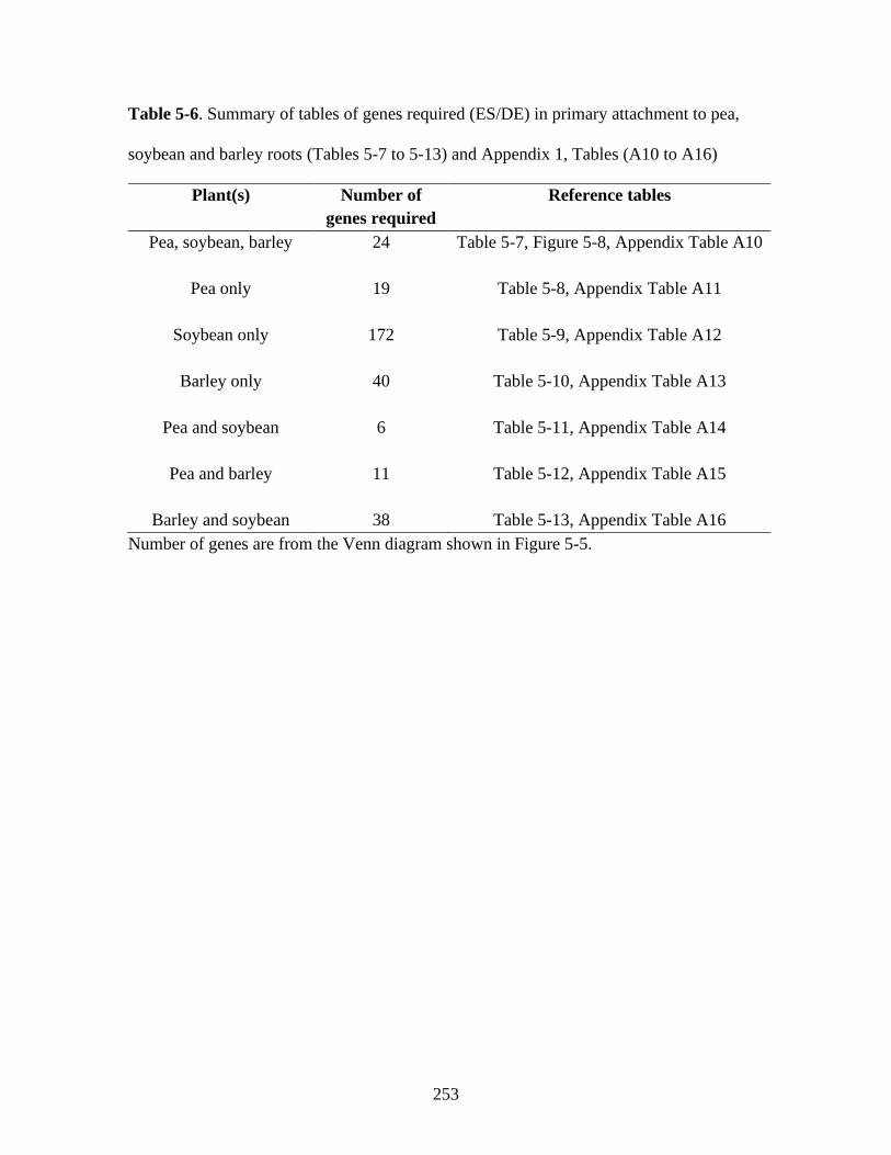
253
Table 5-6. Summary of tables of genes required (ES/DE) in primary attachment to pea,
soybean and barley roots (Tables 5-7 to 5-13) and Appendix 1, Tables (A10 to A16)
Plant(s) Number of
genes required
Reference tables
Pea, soybean, barley
24 Table 5-7, Figure 5-8, Appendix Table A10
Pea only 19 Table 5-8, Appendix Table A11
Soybean only
172 Table 5-9, Appendix Table A12
Barley only
40 Table 5-10, Appendix Table A13
Pea and soybean
6 Table 5-11, Appendix Table A14
Pea and barley
11 Table 5-12, Appendix Table A15
Barley and soybean 38 Table 5-13, Appendix Table A16
Number of genes are from the Venn diagram shown in Figure 5-5.

254
Table 5-7. Twenty-four genesa identified as required (ES/DE) for primary attachment to pea, soybean and barley roots.
Gene(s) Description
pRL110043 Putative transmembrane transporter protein. 99% similarity to arabinose efflux permease family protein in R. leguminosarum
biovar trifolii WSM597. EPS arabinose content regulates cell aggregation in Azospirillum [358]. See also Table 4-13 and
Figures 4-12 and 4-13
pRL100112 Putative dehalogenase-hydrolase (HAD), member of a large superfamily of phosphohydrolases. HADs display activity
against various phosphorylated metabolites [326]. A mutant in a Xanthomonas axonopodis pv. citri HAD is defective in
biofilm formation [327]. See also Table 4-11 and Figure 4-13
hslO (RL0551) Putative Hsp33-like chaperonin. Redox regulated molecular chaperone protecting thermally unfolding and oxidised proteins
from aggregation – defence against oxidative stress [277, 278]. May assist in coping with a diverse rhizosphere environment
(see 4.1). See also Table 4-9 and Figure 4-11
RL0617 Putative dTTP/UTP pyrophosphatase. See also Table 4-14 and Figures 4-13 and 4-14
RL1013 Uncharacterised protein, 17kDa Anti 2 motif. These motifs are common membrane proteins and include surface antigens in
Rickettsia [328]. Unknown protein localization. See also Table 4-11 and Figure 4-13
amn (RL1478) AMP nucleosidase; catalyses hydrolysis of AMP to form adenine and ribose 5-phosphate. Changes in AMP levels allow rapid
adjustments to changing metabolic conditions [279]. See also Table 4-9 and Figure 4-11
ppx (RL1600) Putative exopolyphosphatase Ppx; can function in the hydrolysis of EPS [359]. See also Table 4-13 and Figures 4-12 and 4-13
clpS (RL2212) ATP dependent Clp protease adaptor protein ClpS. ClpS modifies ClpA substrate specificity [364]. A clpS mutant of P.
aeruginosa showed 70% reduction in biofilm formation and significant impairment in attachment to abiotic surfaces after one
hour [365]. See also Table 4-14 and Figures 4-13 and 4-14.
hfq (RL2284) RNA binding protein Hfq. Global post transcriptional regulator. Loss of Hfq in S. meliloti delays nodulation and reduces
competitiveness for attachment to alfalfa roots [360]. See also Table 4-13 and Figures 4-12 and 4-13

255
tpiA (RL2513) Putative triosephosphate isomerase. Upregulated in Staphylococcus aureus biofilm, possibly due to oxygen limitation [281].
Glycolytic enzymes play additional roles when localised on the cell surface (e.g. α-enolase plasminogen binding in
streptococci and GAPDH transferrin binding activity in S. aureus) [282, 283]. Surface localised glycolytic enzymes are
multifaceted and can be involved in substrate binding. TpiA has been shown to be surface localised and have a direct role in
attachment to host cells in Mycoplasma gallisepticum [284]. See also Table 4-9 and Figure 4-11.
recA (RL2637) RecA is needed for DNA repair and the SOS response. The major activity of RecA in DNA metabolism is the promotion of
DNA strand exchange [285]. Requirements for all symbiosis stages suggest that RecA may assist in coping with a diverse
rhizosphere environment (see 4.1). However, recA disruption has been shown to reduce adherence and colonization of host
cells by Vibrio cholerae, although the mechanism underlying this remains unknown [286]. See also Table 4-9 and Figure 4-
11.
dksA (RL2643) RL2643 – putative DnaK suppressor protein DksA. Studies from various bacteria indicate that these chaperones can be
secreted and bind ligands, contributing to cell adherence and biofilm formation [366–369]. Cytoplasmic protein localization.
See also Table 4-14 and Figures 4-13 and 4-14
gor (RL2694) RL2694 – Glutathione reductase (Gor). See also Table 4-11 and Figure 4-13
pfp (RL3322) Putative pyrophosphate-fructose 6-phosphate 1-phosphotransferase. Catalyses the first committed step in glycolysis, the
phosphorylation of D-fructose-6-phosphate [287]. Like tpiA (RL2513) (see Table 4-9), could have multifaceted role. See also
Table 4-9 and Figure 4-11
RL3987-90 RL3987 – uncharacterised, SpoVT-AbrB domain. RL3988 – uncharacterised, PINc domain. PIN domains function as single
stranded RNA nucleases [291]. In prokaryotes they are usually the toxin of toxin-antitoxin operons, helping free-living
prokaryotes cope with nutritional stress [292]. RL3989, RL3990 – Holliday junction ATP dependent DNA helicases RuvA
and RuvB; DNA damage repair mechanism. May be required for osmotic shock responses [293, 294]. See also Table 4-9 and
Figure 4-11

256
RL4065 Conserved hypothetical protein, no known conserved domains. Cytoplasmic protein localization. See also Table 4-9 and
Figure 4-11
RL4145 Putative conserved LacI type transcriptional regulator (repressor). Regulatory targets unknown. Cytoplasmic protein
localization. See also Table 4-9 and Figure 4-11
RL4362/ dacC
(RL4363)
RL4362 – putative cobalamin (vitamin B12) synthesis protein, CobW domain [295]. Required by S. meliloti for symbiosis
with M. sativa [296]. Only one cobalamin dependent enzyme (nrdJ, S.meliloti cobalamin dependent ribonucleotide reductase,
RNR) affects symbiosis. Removal of ndrJ impairs symbiosis; rhizobia are lysed in the plant cytoplasm [297]. Loss of RL4362
may reduce fitness for competitive primary attachment. Note that cobalamin synthesis genes may be misclassified due to high
homology with glutamine amidotransferases, which are involved in peptidoglycan amidation [298]. RL4363 – dacC, putative
penicillin binding protein, peptidase S11 domain. In E. coli dacC processes sugar-peptide cell wall precursors; involved in
peptidoglycan biosynthesis [299]. See also Table 4-9 and Figure 4-11.
RL4381 Putative POTRA domain transporter. See also Table 4-9, Figures 3-11 and 4-11 and section 3.2.9
aNumber of genes are from the Venn diagram shown in Figure 5-5. Red = Genes also required (ES/DE) in attachment to pea roots at
all pHs tested in Chapter 4.

257
These genes, being ES/DE in attachment to pea, are discussed in the relevant tables and
figures in Chapter 4, which are referenced in Table 5-7. The genes listed in Table 5-7 are
‘core’ determinants of primary attachment in Rlv3841, as they are required not only for
attachment to pea, but also to soybean and barley roots. Of these 25 genes, 14 (highlighted
in red) are required for attachment to pea roots under all pH conditions and are therefore
some of the most important primary attachment determinants in the Rlv3841 genome.
These are shown, along with brief functional descriptions, in Figure 5-8, below.

258
Figure 5-8. Diagram of a stylized gram-negative Rlv3841cell showing the primary attachment determinants whose genes were
identified as required (ES/DE) in attachment to pea (pH 6.5, 7.0 and 7.5), soybean and barley (see Table 4-9). Cell outer membrane,
peptidoglycan layer, inner membrane, plasmids, nucleoid and other cellular factors are shown, not to scale.

259
Table 5-8. Fourteen genesa identified as required (ES/DE) for primary attachment to pea roots only (pH 7.0).
Gene Description
pRL100053 Putative transmembrane domain containing protein, helix-turn-helix 37 domain. Unknown protein localization. See Table 4-9
and Figures 3-11 and 4-11 and section 3.2.9.
pRL100174 Hypothetical protein, no known conserved domains. Unknown protein localization. See also Table 4-9 and Figure 4-11
pRL100242 Uncharacterised protein. Cytoplasmic protein localization. See also Table 4-13 and Figures 4-12 and 4-13
pRL120518 Putative TetR family transcriptional regulator. See also Table 4-11 and Figure 4-13.
cycM (RL0141) Membrane-bound cytochrome c CycM. See also Table 4-14 and Figures 4-13 and 4-14
RL1105 putative TetR family transcriptional regulator. See also Table 4-11 and Figure 4-13
pspA (RL1106) Putative PspA family regulator, phage shock protein A. Involved in antibiotic resistance and biofilm formation in E.coli and
pathogenesis in S. typhimurium [329, 330]. See also Table 4-11 and Figure 4-13
RL1504 Uncharacterised protein, NYN domain. Possibly novel RNAse with regulatory role [363]. Cytoplasmic protein localization.
See also Table 4-14 and Figures 4-13 and 4-14
gmsA (RL1661) Glucomannan biosynthesis protein GmsA. Characterised factor important for polar primary root attachment at acidic but not
alkaline pH [56]. See also Tables 4-13 and Figures 3-5, 4-12 and 4-13
scpA (RL2044) Segregation and condensation protein A, participates in chromosomal division during cell partition. See also Table 4-11 and
Figure 4-13
anmK (RL2587) Anhydro-N-acetylmuramic acid kinase AnmK. Catalyses the phosphorylation of 1,6-anhydro-N-acetylmuramic acid
(anhMurNAc), cleaving the 1,6 anhydro ring and generating MurNAc-6-P. Required for cell wall recycling [331, 332]. See
also Table 4-11 and Figure 4-13
ahpD (RL3226) Alkyl hydroperoxide reductase AhpD. See also Table 4-11 and Figure 4-13
260
pssA (RL3752)
Glycosyl transferase involved in EPS biosynthesis [288, 289]. Mutants are deficient in EPS production and form biofilms
slowly compared to Rlv3841. Does not attach to root hairs [56]. Biofilms are flat and unstructured [58]. See also Table 4-9
and Figures 3-5 and 4-11 and section 3.2.5
RL4704 Putative glyoxylase family protein, member of the VOC superfamily. Members of this family are known to detoxify
methylglyoxal, formed as a by-product of lipid metabolism [333]. See also Table 4-11 and Figure 4-13
aNumber of genes are from the Venn diagram shown in Figure 5-5.

261
These genes, being defective in attachment to pea when mutated, are discussed in the
relevant reference tables and figures in Chapter 4, which are referenced in Table 5-8.
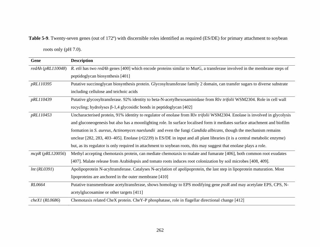
262
Table 5-9. Twenty-seven genes (out of 172a) with discernible roles identified as required (ES/DE) for primary attachment to soybean
roots only (pH 7.0).
Gene Description
redAh (pRL110048) R. etli has two redAh genes [400] which encode proteins similar to MurG, a transferase involved in the membrane steps of
peptidoglycan biosynthesis [401]
pRL110395 Putative succinoglycan biosynthesis protein. Glycosyltransferase family 2 domain, can transfer sugars to diverse substrate
including cellulose and teichoic acids
pRL110439 Putative glycosyltransferase. 92% identity to beta-N-acetylhexosaminidase from Rlv trifolii WSM2304. Role in cell wall
recycling; hydrolyses β-1,4 glycosidic bonds in peptidoglycan [402]
pRL110453 Uncharacterised protein, 91% identity to regulator of enolase from Rlv trifolii WSM2304. Enolase is involved in glycolysis
and gluconeogenesis but also has a moonlighting role. In surface localised form it mediates surface attachment and biofilm
formation in S. aureus, Actinomyces naeslundii and even the fungi Candida albicans, though the mechanism remains
unclear [282, 283, 403–405]. Enolase (rl2239) is ES/DE in input and all plant libraries (it is a central metabolic enzyme)
but, as its regulator is only required in attachment to soybean roots, this may suggest that enolase plays a role.
mcpR (pRL120056) Methyl accepting chemotaxis protein, can mediate chemotaxis to malate and fumarate [406], both common root exudates
[407]. Malate release from Arabidopsis and tomato roots induces root colonization by soil microbes [408, 409].
lnt (RL0391) Apolipoprotein N-acyltransferase. Catalyses N-acylation of apolipoprotein, the last step in lipoprotein maturation. Most
lipoproteins are anchored in the outer membrane [410]
RL0664 Putative transmembrane acetyltransferase, shows homology to EPS modifying gene pssR and may acetylate EPS, CPS, N-
acetylglucosamine or other targets [411]
cheX1 (RL0686) Chemotaxis related CheX protein. CheY-P phosphatase, role in flagellar directional change [412]

263
cheY1 (RL0687) Chemotaxis response regulator, minor role in flagellar reorientation [397]
icpA (RL0865) Chemotaxis protein with a broad attractant spectrum [413, 414]
RL0963 Putative transmembrane/surface protein – BA14K family protein, likely with a role in LPS biosynthesis [415]
RL1015 Putative polysaccharide deacetylase protein. 95% identity to xylanase/chitin deacetylase from Rlv WSM1455.
RL1016 Putative D-alanyl-D-alanine carboxypeptidase. Carboxypeptidases are extensively involved in peptidoglycan biosynthesis
and modification [416]
RL1090 Putative transmembrane protein, DUF1499 family. Domain of unknown function (DUF) family proteins are often conserved
(indicating an important role) and function often only becomes apparent under particular conditions [417]
RL1108 Putative transmembrane AMP-binding acyltransferase, PlsC (phosphate acetyltransferase) domain. Functions in membrane
biogenesis and modification, using fatty acid chains to form membrane phospholipids [418]
RL1155 Putative glycosyltransferase. 96% identity to glycosyltransferase 25 (LPS biosynthesis protein) from R. leguminosarum.
LPS strongly influences adhesive properties in this bacteria [419]
RL1549 Putative transmembrane efflux protein, 92% identity to LysE family translocator from R. hidalgonenese, which excretes
excess l-Lysine as a result of natural flux imbalance or peptide hydrolysis [420]
dgkA (RL2780) Diacylglycerol kinase DgkA. In E.coli, DgkA mutants are defective in biofilm formation [316], and DgkA function has
been linked to phospholipid recycling and LPS modifications [324]. In B. subtillis it is important for lipoteichoic acid
synthesis [325]
RL3149 Putative adenylate cyclase/guanylate cyclase
flaH (RL3268) Flagellin, flagella subunit. Rlv3841 flaH mutants show shorter flagella and reduced motility [421]; loss of motility and/or
flagella adhesin function may lead to ES/DE classification
RL4030 (cheW3) Mcp type chemoreceptor

264
RL4075 Uncharacterised LysM domain containing protein. A highly conserved carbohydrate binding module, LysM domains often
bind peptidoglycan in prokaryotes [422]
nodN2 (RL4120) NodN2, an additional copy of NodN (pRL100179), which participates in Nod factor biogenesis. The role of NodN remains
unclear [423]. However, core Nod biosynthesis genes (producing the chitin oligomer backbone of Nod factor) are important
for biofilm formation in S. meliloti [424]. The protein encoded by this gene may also possess enoyl-CoA hydratase activity.
cysZ (RL4210) Putative cysteine biosynthesis protein. Mutation of cysZ in V. fischeri were defective in biofilm formation which could be
complemented by the cysK gene but not by cysteine, suggesting unknown additional roles for CysK [425]
gelA (RL4404) Gel forming EPS production protein GelA. Regulated by RosR [158]. Also impaired in attachment to pea roots under pH
6.5 conditions. (Table 4-10)
exoD (RL4420) Putative EPS biosynthesis protein ExoD. This protein has been shown to be involved in EPS biosynthesis, but its exact
function remains unknown [426]
nagA (RL4602) Putative N-acetylglucosamine-6-phosphate deacetylase NagA. Required for cell wall peptidoglycan and teichoic acid
biosynthesis in L. monocytogenes [427].
aNumber of genes are from the Venn diagram shown in Figure 5-5.
265
The genes with a role in chemotaxis/motility (mcpR – pRL120056, cheX1 – RL0686,
cheY1 – RL0687, icpA – RL0856, flaH – RL3268 and cheW3 – RL4030) required for
attachment to soybean roots are discussed in 5.2.10.
Various genes required indicate modification of EPS for attachment to soybean roots.
pRL110395 is a putative succinoglycan biosynthesis protein, and succinoglycan
production is known to affect EPS composition in S. meliloti [353–356]. RL0064 has high
homology to the EPS modifying pssR gene, loss of which reduces EPS acetylation in R.
leguminosarum biovar viciae VF39 and decreases the efficiency of nitrogen-fixing
symbiosis formation with alfalfa, possibly through a reduction in root attachment [411].
Conversely, RL1015 is a polysaccharide deacetylase, indicating that, for attachment to
soybean roots, a balance of EPS acetylation level may be required that is different from
the EPS acetylation levels required for attachment to pea or barley roots, likely reflecting
differences in root surfaces which require different or modified bacterial factors for
successful attachment. RL0064 and RL1015 may interact to fulfil this role, although this
has not been demonstrated. exoD (RL4420) is an EPS biosynthesis protein of unknown
function [426]. Also required is the gel-forming EPS production protein GelA (RL4404).
Previously, the role of this gene in Rlv3841 attachment to vetch root hairs was
investigated. A mutant displayed normal attachment, meaning a role for this gene was not
assigned, meaning its function remained unknown [158]. The requirement for GelA in
Rlv3841 attachment to pea at pH 6.5 seen in Chapter 4 (Table 4-10), combined with the
results of Williams et al. (2008) [158], suggested a role for this gene in attachment to bulk
epidermal pea root surface (as opposed to root hairs) at pH 6.5. The requirement for this
gene in attachment to soybean roots at pH 7.0 is interesting, although the similarities

266
between pea root surfaces at pH 6.5 and soybean root surfaces at pH 7.0 that would
necessitate GelA for primary attachment are unclear.
Genes likely involved in peptidoglycan modification include redAh (pRL110048), a
homologue of murG which transfers N-acetylglucosamine to a lipid intermediate in the
biosynthesis of peptidoglycan [401]. pRL110439 may play a role in cell wall recycling by
hydrolysing β-1,4 glycosidic bonds in peptidoglycan [402]. RL1016 encodes a
carboxypeptidase; these proteases have been heavily implicated in peptidoglycan
biosynthesis and modification in many bacterial species [416]. nagA (RL4062) has been
shown to influence both peptidoglycan biosynthesis in L. monocytogenes [427], and may
also be playing this role in Rlv3841. RL4075 likely encodes a peptidoglycan binding
factor, though its exact role remains unclear [422].
A further subset of genes is involved in LPS biosynthesis and modification. RL0963 is a
BA14K family protein, named after homology to a 14 kDa protein from Brucella abortus.
In B. abortus, this protein shows lectin-like carbohydrate binding activity and is essential
for virulence, most likely due to its direct or indirect effects on LPS biosynthesis [415].
Whilst it is unclear what role RL0963 plays in Rlv3841, BA14K homology is suggestive
of a function in LPS production. RL1155 encodes a glycosyltransferase likely involved in
LPS biosynthesis, whilst dgkA (RL2780) has been linked to LPS modifications in E. coli
[324], with mutants defective in biofilm formation in a 24 hr assay [316].
Two genes are likely involved in membrane function: lnt (RL0391), involved in the
production of outer membrane associated lipoproteins [410] and RL1108, functioning in
membrane phospholipid production [418].
Similar to TpiA (a glycolytic enzyme which can be cell surface localized, function in
substrate binding [284] and is required for attachment to pea roots at all pHs – Table 4-9)

267
enolase may also be involved in a surface localised capacity in attachment to soybean
roots. It has been shown to play this role in various other organisms [282, 283, 403–405]
and, unless pRL110453 (putative regulator of enolase) has other regulatory targets, it is
likely to also be playing this role here.
As a possible guanylate cyclase, RL3149 may be linked to cellular c-di-GMP levels, a
well-known signalling and regulatory mechanism governing bacterial motile-sessile
lifestyle changes [79].
The requirement for nodN2 (RL4120) could indicate that a core Nod biosynthetic gene
product is involved in attachment. Although NodN function is unclear, such a finding is
not unprecedented as it has been shown for S. meliloti that the core Nod chitin backbone
has a second role of stabilising biofilms [424]. Alternatively, this gene could represent a
fatty acid metabolising enoyl-CoA hydratase, as also suggested by UniProt annotation.
The role of RL1549 (transmembrane efflux protein), RL1090 (DUF family protein) and
the cysteine biosynthesis protein CysZ (RL4210) in root attachment is unknown.

268
Table 5-10. Ten genes (out of 40a) with discernible roles identified as required (ES/DE) for attachment to barley roots only (pH 7.0).
Gene(s) Description
RL0726 Conserved hypothetical exported protein, transglycosylase Slt domain. Degrades peptidoglycan via β 1-4 glycosidic bond
cleavage. Linked to biofilm formation in S. enterica, E. coli and Acinetobacter baumannii [314–316]. Lytic
transglycosylases participate extensively in cell wall remodelling, recycling of peptidoglycan and space-making for
insertion of cell-envelope spanning structures [317]. A lytic transglycosylase (RL4716) was characterised in Rlv3841 as
required for cell envelope function and biofilm formation [318]. Also impaired in attachment to pea roots under pH 6.5
conditions
celB (RL1647) Cyclic di-GMP binding protein, cellulose synthesis protein. Binds the cellulose synthase activator and is involved in
cellulose biosynthesis. Also has a role in the synthesis of lipid-linked intermediates, although the exact function in this role
is unknown [428]
degQ (RL1806) Periplasmic serine endoprotease DegQ. In B. subtilis DegQ stimulates phosphotransfer to a transcriptional regulator
affecting biofilm formation, promoting transition from a motile to sessile attached state [370]. Also impaired in attachment
to pea roots under pH 6.5 and 7.5 conditions
RL2039 Putative HesB family protein, Fe/S biosynthesis. E. coli show a link between Fe/S biogenesis mutants and cell surface
properties, which may be impaired in biofilm formation and/or motility, although the mechanisms underpinning this remain
unknown [429]
sixA (RL2644) Phosphohistidine phosphatase SixA, conserved. SixA is the only known bacterial phosphohistidine phosphatase, and
dephosphorylates Npr in E.coli [343]. Implicated in biofilm formation in E. coli [344]. See also Table 4-10, rl0032 / rl0033.
Also impaired in attachment to pea under pH 7.5 conditions
RL2656 Putative D-alanyl-D-alanine carboxypeptidase. Carboxypeptidases are extensively involved in peptidoglycan biosynthesis
and modification [416]

269
RL2657 Putative GGDEF/GAF domain sensory box protein, downstream of RL2656 (above). Likely catalyses synthesis and/or
hydrolysis of cyclic di-GMP (c-di-GMP), key second messenger in biofilm formation / motile to sessile lifestyle switch [79]
RL3252 Periplasmic serine endoprotease, DegP-like. In V. cholerae DegP associates with the outer membrane and is an important
determinant of the biofilm matrix structure [430], facilitating secretion of proteases which may help biofilms adhere to
surfaces by processing extracellular components [431]
hflC (RL3253), hflK
(RL3254)
Putative transmembrane serine proteases HflC and HflK. Functional HflC and HflK also modulate HflB activity. HflB, an
AAA metalloprotease, is involved in membrane protein regulation, LPS biosynthesis and biofilm formation in E. coli, B.
subtillis and others (where it is often called FtsH) [345, 346], indicating a role in membrane regulation and biofilm
formation [347–349]. HflB itself (RL3965) is ES/DE under all conditions in this work, including input (Appendix 2 Table
2). Also impaired in attachment to pea under pH 7.5 conditions
aNumber of genes are from the Venn diagram shown in Figure 5-5.

270
Amongst the list of genes required for attachment to barley roots (Table 5-10),
requirement of celB (RL1647, encoding a cellulose synthesis protein) was interesting as
cellulosic fibrils are normally considered a secondary attachment factor [34]. It may be
that cellulosic fibril deposition is important in primary root attachment in barley, or that
this gene is playing an unidentified role in synthesis of an attachment-required lipoprotein
[428] in attachment to these roots.
RL2656 (carboxypeptidase) is likely to be involved in altering peptidoglycan structures.
Two serine endoproteases (encoded by RL3252 and degQ - RL1806) are also needed. The
former seems to process extracellular components for attachment competence, while the
latter promotes activity of transcriptional regulators involved in attachment [370, 431].
The requirement for RL0726 (transglycosylase) as well as hflC (RL3253) and hflK
(RL3254) indicate membrane and cell wall alterations are involved in primary attachment
to barley roots. The phosphohistidine phosphatase SixA (RL2644), whose function is to
dephosphorylate Npr [343], is required for attachment to barley and also for attachment to
pea at pH 7.5 (Table 4-12). This is likely to indicate the involvement of down-regulation
of ATP-dependent ABC transporters and export of certain EPS structures and is discussed
more fully in 4.2.11.
The role of RL2039 (an Fe/S biosynthesis protein) remains unclear, though it has been
linked previously to impaired attachment in E. coli [429].

271
Table 5-11. Sixa genes identified as required (ES/DE) for attachment to pea and soybean roots at pH 7.0.
Gene Description
pRL100220 Uncharacterised protein
RL1052 Uncharacterised protein
RL1371 Putative transmembrane protein
RL2400 Putative MarC family transmembrane protein, not involved in antibiotic resistance [280], function unknown
RL2520 Putative transmembrane protein, ABC transporter permease. Many ABC transporters are involved in lipid transport to the
outer membrane [361]
RL4083 Uncharacterised protein, SGHN family esterase domain
aNumber of genes are from the Venn diagram shown in Figure 5-5.
272
After pleiotropy filtering (section 5.2.8) only six genes are required (ES/DE) for
attachment to both legumes (pea and soybean) used in this study. Their functions are
largely unknown, although RL2520 may be involved in outer membrane lipid transport
[361]. Given the importance of outer membrane characteristics, including lipid profiles,
for surface attachment [44], mutation in this gene may reduce attachment via alterations to
the outer membrane lipid profile.

273
Table 5-11. Two (out of 11a) genes with discernible roles required (ES/DE) for attachment to pea and barley roots (pH 7.0).
Gene(s) Description
RL4309 Putative transmembrane protein. 94% identity to R. hidalgonense DedA family protein (CO674_30990). DedA proteins
appear to function in membrane homeostasis; mutants show altered membrane lipid composition in multiple bacterial
species [362].
RL4382 Putative filamentous hemagglutinin adhesin (rl4382). See Figure 3-11 and section 3.2.9
aNumber of genes are from the Venn diagram shown in Figure 5-5.

274
For attachment to pea and barley roots, only two genes had discernible function. RL4382
encodes FHA, which is a newly defined important factor for attachment to pea roots at pH
6.5, 7.0 and 7.5 (discussed in detail in 3.2.9). The second gene, RL4309, is likely to
encode a DedA protein which is linked to membrane lipid composition [362].

275
Table 5-12. Ten genes (out of 38a) with discernible roles required (ES/DE) for attachment to barley and soybean roots (pH 7.0).
Gene Description
RL1165 rl1165 – uncharacterised protein, 93% identity to gene RLV_3555 from R. leguminosarum biovar viciae, PepSY domain
containing. These domains are likely to have a protease inhibitory function and may be cell wall associated [334]. Biofilm
metalloprotease 1 (BmpI) from Pseudoalteromonas contains a PepSY domain required for biofilm formation [335]. Also
impaired in attachment to pea under pH 7.5 conditions
RL1440 Serine endoprotease, DegP-like. In V. cholerae DegP associates with the outer membrane and is an important determinant of
the biofilm matrix structure [430], facilitating secretion of proteases which may help biofilms adhere to surfaces by
processing extracellular components [431]
dacF (RL2477) Putative penicillin binding protein DacF. 92% identity to Rlv trifolii WSM2297 D-alanyl-D-alanine carboxypeptidase.
Carboxypeptidases are extensively involved in peptidoglycan biosynthesis and modification [416]
RL2595 Putative MutT/nudix family protein. Mutagenesis of nudix proteins in Pseudomonas syringae str DC3000 and P.
aeruginosa display defects in motility and biofilm formation [323]. Also impaired in attachment to pea under pH 6.5
conditions
RL2778 Putative exopolysaccharide biosynthesis protein. Also impaired in attachment to pea under pH 7.5 conditions
RL3267 Putative OmpA family outer membrane protein. OmpA family proteins have diverse roles, including in signal transduction,
primary surface adhesion and biofilm formation [265, 432, 433]
RL3320 Putative signalling and peptidoglycan binding protein. N-acetylmuramoyl-L-alanine amidase domain; cleaves the amide
bond between N-acetylmuramoyl and L-amine acids in bacterial cell walls [434]
RL4018 Putative ATP binding component of ABC transporter. 96% identity to Lipid A ABC exporter from R. leguminosarum
biovar trifolii WSM2304, gene Rleg2_3249. Mutants with reduced lipid A show a delay in nodulation onset and impaired
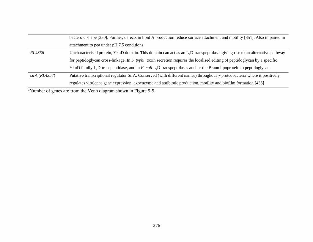
276
bacteroid shape [350]. Further, defects in lipid A production reduce surface attachment and motility [351]. Also impaired in
attachment to pea under pH 7.5 conditions
RL4356 Uncharacterised protein, YkuD domain. This domain can act as an L,D-transpeptidase, giving rise to an alternative pathway
for peptidoglycan cross-linkage. In S. typhi, toxin secretion requires the localised editing of peptidoglycan by a specific
YkuD family L,D-transpeptidase, and in E. coli L,D-transpeptidases anchor the Braun lipoprotein to peptidoglycan.
sirA (RL4357) Putative transcriptional regulator SirA. Conserved (with different names) throughout γ-proteobacteria where it positively
regulates virulence gene expression, exoenzyme and antibiotic production, motility and biofilm formation [435]
aNumber of genes are from the Venn diagram shown in Figure 5-5.
277
From this list of requirements for attachment to soybean and barley roots (Table 5-12),
membrane, cell wall and EPS modifications are clearly important. Genes required include
sirA (RL4357), RL1165 (encoding PepSY-domain protein), RL1440 (encoding a DegP-
like serine endoprotease), RL2477 (dacF, encoding a penicillin binding protein), RL2778
(encoding a putative EPS biosynthesis protein), RL3320, RL4018 (likely encoding a lipid
A exporter) and RL4356 (encoding a transpeptidase). Also involved are RL3267, encoding
an OmpA family outer membrane protein of which many have adhesin roles [265, 432,
433] and RL2595, encoding a MutX/Nudix family protein.
5.2.12 Mutation of some Rlv3841 genes leads to an increase in primary attachment to
different plants
In INSeq experiments, genes are classified as AD when insertion into a gene increases
bacterium fitness in, in this case, attachment to roots. This indicates that gene disruption is
beneficial to bacterial root attachment and that an AD gene, when functional, somehow
inhibits/reduces attachment. After classification and filtering of genes for pleiotropic
effecs (see 5.2.6 and 5.2.8), 17 genes remained with an AD classification under one or
more conditions, represented in Figure 5-9 (full list of genes in Appendix 1, Table A17).
For brevity, only factors with putative functions are listed (Table 5-13) and discussed
below.

278
Figure 5-9. Genes classified as NE in the input library and AD in one or more of the root-
attached libraries which are also classified as NE in the following INSeq datasets: VMM,
21% oxygen 10mM glucose, 1% oxygen 10mM glucose, 21% oxygen 20mM succinate
and 1% oxygen 20mM succinate (see Table 4-7).Venn diagram circle color indicates
attachment condition; yellow = soybean roots (pH 7.0), green = pea roots (pH 7.0), brown
= barley roots (pH 7.0).

279
Table 5-13. Three (out of 17a) genes with discernible roles identified where mutation is advantageous (AD) for primary attachment to
pea, soybean and barley roots (pH 7.0).
Gene Plants Description
pRL70156 Pea Putative conjugative DNA transfer protein TrbGp7. A homologue of VirB9, a type VI secretion system
protein in Agrobacterium tumefaciens [225]
RL3927 Soybean Soluble lytic murein transglycosylase protein, Slt domain. Degrades peptidoglycan via β 1-4 glycosidic
bond cleavage. Lytic transglycosylases participate extensively in cell wall remodelling, recycling of
peptidoglycan and space-making for insertion of cell-envelope spanning structures [317].
RL4139 Pea, Soybean,
Barley
Putative transmembrane sensory box GGDEF/EAL protein. The GGDEF domain is likely to catalyze
synthesis and/or hydrolysis of c-di-GMP, whereas EAL may function as a diguanylate phosphodiesterase
[79]. c-di-GMP is a known positive regulator of biofilm formation [436–438]. However, an EAL domain
protein in Pseudomonas putida strongly inhibited biofilm formation and could also lead to rapid biofilm
dispersal, indicating these proteins also have a role in transitioning cells from a sessile to motile state
[439]
a Number of genes is from the Venn diagram shown in Figure 5-9.

280
The three genes in which mutation leads to increased attachment with discernable roles
(Table 5-13) include, specifically for pea roots, pRL70156 (putative TrbGp7), which
encodes a putative DNA transfer protein and is homologous to the type IV secretion
system component VirB9 in A. tumefaciens [225]. Other Trb proteins (involved in pilus
formation and type IV secretion system function) are required for attachment to soybean
roots (5.2.7 and Table 5-3), but this gene impairs Rlv3841 attachment to pea.
Showing an increased attachment only in soybean is mutation of RL3927. RL3927 seems
to have a role in cell wall remodelling. Although another similar Slt domain
transglycosylase protein (RL0726) was required (ES/DE) for attachment to roots of pea
(pH 6.5, Table 4-10) and barley (pH 7.0, Table 5-10), the cell wall remodelling promoted
by this gene is acting in the opposite way, to impair/reduce Rlv3841 attachment to
soybean (and is thus classified AD when mutated), though the reason remains unclear.
What is clear, is that factors affecting cell surface, such as cell walls and pili, influence
primary attachment and that disruption of these structures perturbs, either to decrease or
increase, the ability of Rlv3841 to attach to roots.
Of interest is RL4139 (putative transmembrane sensory box GGDEF/EAL protein), AD
when mutated in attachment to all plant roots at pH 7.0. Levels of the second messenger c-
di-GMP have a role in biofilm formation [436–438] and lifestyle changes from a motile to
sessile state [87, 88, 439]. In the cell, c-di-GMP is formed by GGDEF domain proteins
and broken down by EAL domain proteins [82, 83]. Some proteins (including RL4139)
carry both GGDEF and EAL domains. For these proteins, it is usually the case that only
one of the two domains is functional, or that a third regulatory domain manages protein
activity [84, 85]. An EAL domain protein was shown to inhibit biofilm formation and
promote single celled, motile lifestyle in P. putida, likely through decreases in
281
intracellular c-di-GMP [439]. EAL protein mediated decreases in c-di-GMP have also
been demonstrated in P. aeruginosa, Yersinia pestis and S. enterica [80, 440, 441].
The regulator RL4145 (UniProt annotation: putative LacI type transcriptional regulator,
repressor) is the only regulator required for attachment to all plant roots (Table 5-7, figure
5-8). Putative targets of this regulator were discussed in 4.2.12. However, if this regulator
is a repressor then HMM classifications and genomic proximity link it to RL4139. Given
the data presented, the following model is possible: under neutral pH conditions functional
RL4145 represses RL4139 expression, enabling intracellular c-di-GMP levels to increase,
facilitating successful primary root attachment. When RL4145 is mutated an ES/DE
classification results, as RL4139 expression is not repressed and intracellular c-di-GMP
decreases, impairing attachment. When RL4139 is mutated an AD classification results, as
c-di-GMP levels increase without the action of the protein’s EAL domain. This model
would account for the opposing HMM classifications of RL4145 and RL4139. If correct,
this interplay is extremely important in governing pH 7.0 primary root attachment (the
HMM classification of RL4139 at pH 6.5 and 7.5 in pea root attachment is NE), and an
RNASeq study with Rlv3841 and an RL4145 mutant as well as promoter binding assays
could investigate this further. Alternative signalling pathways are likely to be operating
under non-neutral conditions, especially considering the differential regulatory
requirements and involvement of other putative c-di-GMP signalling factors (see 4.2.11)
Overall, this reinforces the notion that high levels of intracellular c-di-GMP are very
important for root attachment (as seen with RL2316 – pH 6.5 attachment to pea – and
RL2657 – pH 7.0 attachment to barley) and that reduction of c-di-GMP signalling inhibits
this process.

282
5.3 Conclusion
In summary, appropriate experimental parameters were established to screen in an INSeq
study for bacterial primary root attachment to soybean and barley roots at pH 7.0. 127
million barcoded sequencing reads were obtained from nine sample libraries, achieving
HMM classifications for 99.7% of the Rlv3841 genes. Validation of the results from
INSeq by comparison with the literature indicated that the HMM method returned the
expected classification for genes of known function (such as ribosomal subunits and
plasmid replication systems) under the experimental conditions used here.
Prior to filtering based on pleiotropy effects under media/metabolic conditions, 464 genes
classified as NE in the input libraries were classified as required (ES/DE) for attachment
to the roots of one or more plants. Their distribution (Figure 5-2) indicated that there were
multiple different attachment requirements unique to each plant. Examining the functional
classification of these genes indicated that, whilst there were different functional
classifications based on plant root attachment conditions, there were also significant
requirements for uncharacterised genes in all cases.
The genomic localization of these genes revealed that, whilst chromosomal factors were
the largest constituent of genes required for attachment to different plant’s roots, the
plasmid with the highest number of required genes in pea was pRL10. Plasmid pRL10 is
referred to as the symbiosis plasmid, containing nod, nif and fix genes [381]. After
pleiotropy filtering to remove any genes affected in growth under different conditions or
substrates, the only pRL10 genes remaining were transmembrane proteins (and
uncharacterised proteins). This work suggests a role for the symbiosis plasmid in primary
root attachment to peas, although the precise function of these genes (pRL100053,
pRL100174 and pRL100242) remains unknown.

283
Soybean attachment factors showed the largest plasmid requirement for pRL11, along
with a requirement for pRL8, which was not seen under any other condition tested.
Analysis of the likely function of pRL8 and pRL11 genes indicates a requirement for
Flp/Tad pili and type IV secretion system-mediated attachment to soybean roots. This
mechanism is not obviously used by Rlv3841 in attachment to other plant roots, but is
known to be used for attachment by different rhizobia (including S. meliloti and A.
tumefaciens) [44, 383].
After filtering of genes based on pleiotropy phenotype under media/metabolic conditions
(see 5.2.8), 312 genes remained ES/DE in attachment to plant roots, showing a mixture of
plant specific and non-specific requirements. Visualizing the ‘specificity’ of genes
involved in Rlv3841 attachment to different plant roots revealed that a high percentage of
Rlv3841 genes required (ES/DE) for soybean primary root attachment are not required for
attachment to pea or barley. 86% of the genes required for attachment to soybean were
involved only in attachment to soybean whereas, for pea and barley, this figure was ~30%.
This suggests that legume and non-legume plants do not necessarily cluster together in
terms of similarity of primary attachment determinants.
The high specificity of genes required for soybean root attachment was reflected in the
high number of regulators involved (22 in total). This figure was three for pea and six for
barley. Only one regulator (RL4145) was required for attachment to all plant roots.
Analyzing the possible regulatory targets of this gene based on genomic proximity and
HMM classification (see section 4.2.12) revealed that this regulator may control
expression of nearby hypothetical proteins and a putative transmembrane protein. Of these
three possible regulatory targets, only RL4147 (a conserved hypothetical protein) was
required for attachment to all plant roots. RL4145 can be thought of as an important

284
regulator involved in primary root attachment and represents a good target for future
RNASeq-based investigation of transcriptional networks underpinning attachment.
Interestingly, one of the regulators required for soybean root attachment was CheY1, a
chemotaxis response regulator. Analysis of further chemotaxis genes with higher TA site
number (for which HMM classifications are likely to be more accurate) by both HMM
classification and fitness values indicated a requirement for chemotactic motility in
Rlv3841 attachment to soybean, not seen with other plant roots under these INSeq
conditions. One possible explanation for this is a higher level of chemoattractant
exudation from soybean roots (due to large root size) providing a stronger advantage, or
selection, for those bacteria able to sense them, which may not be seen in experiments
with smaller rooting systems. The requirement for the MsiR regulator (RL2857) in
attachment to barley roots indicates that phytotoxin export is likely necessary for
attachment and colonization of this root system. A summary of the different gene
functions needed for the primary attachment of Rlv3841 to the roots of pea, soybean and
barley is shown in Figure 5-10.

285
Figure 5-10. Gene functions needed for primary attachment of Rlv3841 to pea, soybean
or barley roots and combinations thereof at pH 7.0. Circle color indicates plant: yellow =
soybean, green = pea, brown = barley. Text color indicates plant requirement of indicated
gene function for primary attachment: yellow = soybean, mauve = soybean and pea, green
= pea, blue = pea and barley, brown = barley, red = barley and soybean, black = pea,
soybean and barley. OM = outer membrane. Gene functions are drawn from Tables 5-7 to
5-13. See also Figure 5-8.
Pea
Barley Soybean

286
Twenty-four genes were classed as ES/DE in attachment to all plant roots, and of these 14
were required for attachment to pea under all pH conditions. These can be thought of as
core attachment determinants. Among the functions indicated by these genes is
peptidoglycan modification. Specifically, RL4362, a putative cobalamin synthesis protein
which may actually represent a peptidoglycan amidation factor, and dacC (RL4363), a
penicillin binding protein likely involved in peptidoglycan biosynthesis via sugar-peptide
cell wall precursor processing [299]. EPS modification, particularly an increased
arabinose content due to pRL110043 (an arabinose efflux permease), is also required. This
is of interest as arabinose content in known to be an important regulator of cell
aggregation in Azospirillum [358] and is also important in Rlv3841 primary root
attachment. Modulation of the ClpA protease (RL2213) by ClpS (RL2212) is necessary
for attachment to pea, soybean and barley, though the targets of ClpA (with or without
ClpS modulation) remain unclear [364, 365]. Glycolytic enzymes (such as TpiA, which
may have a surface localized ‘moonlighting’ role in attachment [282–284]) and
uncharacterised proteins were also implicated. Additionally, DNA repair factors which,
although they may be required for coping with cellular stresses in the assay setup, may
also play an uncharacterised role in attachment [286], are also required. Interestingly,
RL4381 (a POTRA domain transporter) but not RL4382 (FHA, filamentous
hemagglutinin adhesin) which it is thought to export, was required for attachment to all
plants. This indicates that, whilst the role of FHA is important (being required for
attachment to pea and barley roots), RL4381 may additionally export other primary
attachment factor determinant(s).

287
As well as chemotaxis and Tad/Flp pili, attachment to soybean roots also required various
sugar transferases and EPS/LPS/peptidoglycan biosynthesis and modification genes.
RL0064 (pssR) promotes EPS acetylation whilst RL1015 (a polysaccharide deacetylase) is
also required, indicating a possible balancing of EPS acetylation levels required to
facilitate soybean root attachment. RL0963 (a BA14K family protein) and RL1155 (a
glycosyltransferase) are involved in LPS biosynthesis whilst dgkA (RL2780) is an LPS
modifier, with E. coli dgkA mutants defective in biofilm formation [316]. redAh
(pRL110048) is a transferase involved in the membrane steps of peptidoglycan
biosynthesis [401], whilst pRL110439 and RL1016 are peptidoglycan modifying factors.
The involvement of GelA defines another role for this previously uncharacterised EPS
biosynthesis protein [158] which is needed both for attachment to pea roots at pH 6.5 and
soybean roots at pH 7.0. The requirement for pRL110453 indicated that exported enolase
may play a role in attachment and NodN2 (RL4120) indicated that a Nod biosynthetic
gene product is likely also to be involved.
In attachment to barley roots, various genes involved in c-di-GMP synthesis as well as
proteases were required. A Deg-P like serine endoprotease (RL3252) may process
extracellular components to facilitate adherence, a Slt domain transglycosylase likely
participates in cell wall remodelling and the serine proteases DegQ, HflC and HflK (all
with roles in membrane regulation/biofilm formation) are also required. RL2656 (a
carboxypeptidase) seems to be involved in peptidoglycan biosynthesis and modification
[416]. The involvement of CelB (RL1647), a cellulose synthesis protein, is of interest as,
whilst cellulosic fibrils are more often considered as a secondary attachment factor [34],
they may also to play a role in barley primary root attachment, although celB could be
playing a role in unknown lipoprotein synthesis [428]. The phosphohistidine phosphatase

288
SixA (discussed in 4.3), with a putative role in Npr dephosphorylation and levels of EPS
secretion and/or modification also seems to be involved.
A putative transmembrane DedA protein (RL4309) with roles in regulatory membrane
lipid composition was required for attachment to pea and barley (but not soybean) roots,
indicating differential membrane requirements for interactions between these plants.
Additionally, RL4382 (encoding FHA) was also required. This result means that, whilst
FHA is a key factor at all pHs for pea root attachment (4.2.11), it also plays a role in
Rlv3841/barley interactions.
Genes required for attachment to soybean and barley roots were also mainly of the cell
wall/extracellular protein/peptidoglycan synthesis and modification classes. These
included RL1165, a cell wall associated PepSY domain protein likely to modulate
proteases and promote biofilm formation [334, 335] and RL1440, a DegP-like serine
endoprotease. In V. cholerae DegP facilitates processing of extracellular components to
encourage biofilm formation [431]. Further, dacF (RL2477, a likely carboxypeptidase)
and RL3320 (a peptidoglycan binding and cleavage factor [434]) are also necessary. The
SirA virulence and biofilm regulator (RL4357), OmpA adhesin factor (RL3267), and a
lipid A exporter (RL4018) were also involved.
As well as the Rlv3841 factors required for attachment to different plant roots, it is also
informative to examine the factors required for attachment to pea roots (the host legume)
which are not required for attachment to soybean (a non-host legume) and barley (a non-
legume). One of these is glucomannan (gmsA, RL1661), the cell surface factor important
for primary root attachment at acidic but not alkaline conditions [56]. This factor binds
root hair lectins, which are thought to disassociate from roots under alkaline conditions
[34]. A glucomannan mutant was demonstrated to be deficient in root attachment at pH

289
6.5 and 7.0, but not 7.5, in a Lux attachment assay (Figure 5-3). gmsA was classified as
NE in attachment to soybean and barley roots at pH 7.0 (Appendix 2 Table 2). As gmsA is
not required for attachment to these plants, it can be inferred that root lectins present are
not recognized by Rlv3841 glucomannan. As lectins act as recognition molecules in cell-
cell interactions [48, 49], this suggests that there is specificity in the Rlv3841
glucomannan/lectin interaction for attachment to the host legume, pea. B. japonicum
attaches to the root hairs of its host legume (soybean) with a lectin-mediated system at
acidic to neutral pHs [50, 51], but must use a factor different from Rlv3841 glucomannan
to allow soybean lectin recognition and binding. This indicates that there is some
specificity in attachment to host legume root hairs.
PssA (encoded by RL3752), a glycosyltransferase involved in the first steps of EPS
biosynthesis, is also required for attachment to pea roots only. PssA is responsible for the
addition of glucose-1-phosphate to a polyprenyl phosphate carrier, one of the first steps in
EPS synthesis [442]. An R. leguminosarum biovar trifolii Rt270 strain carrying a pssA
mutation was deficient in EPS production, and led to host clover plants forming fewer
nodules [442]. Various EPS production and modifying factors are important for Rlv3841
attachment to soybean and barley. This, combined with the NE classification of pssA in
attachment of Rlv3841 to these plants, indicates that enough EPS must be produced by
alternative pathways to facilitate root attachment. In R. leguminosarum biovar trifolii
5599, pssA mutants still produce capsular polysaccharide (structurally very similar to
EPS) at wild-type levels [443], indicating that production of closely related
polysaccharides remains possible.
A further Rlv3841 factor involved only in attachment to pea is pRL100053, a putative
transmembrane domain containing protein. Testing in a Lux assay confirmed a mutant in
290
this gene to be defective in attachment to pea roots at pH 6.5, 7.0 and 7.5 (Figure 3-11).
Given that the INSeq classification for this gene is NE in the rhizosphere but ES/DE in
pea root attachment, colonization, infection thread and nodule experiments (see Table 4-
5), the product of this gene likely represents a novel root hair attachment factor. As
pRL100053 is not required for attachment to soybean or barley, this reinforces the idea of
specificity in mechanisms of attachment to host legume root hairs.
After pleiotropy filtering, 17 genes were classified as AD, indicating that mutation was
beneficial to primary root attachment. Of these, only three had discernible roles (see
5.2.12). The most important of these was RL4139, classified as AD in attachment to all
plants when mutated. This is likely because of a c-di-GMP depleting role of this EAL
domain protein when functional. Considering the genomic proximity and opposing HMM
state calls of RL4139 and the regulator RL4145, it seems possible that, when both are
functional, the latter represses expression of the former. If correct, this is an important
transcriptional regulatory insight into the process of primary root attachment and warrants
further investigation.
This chapter has demonstrated that primary attachment determinants show a high degree
of plant root specificity and has identified the ‘core’ factors that are important for
attachment to all plant roots tested. These findings indicate that different host plants likely
present very different root surfaces for attachment by Rlv3841 and, therefore, have
differential attachment requirements. Intriguingly, it does not seem to hold that primary
attachment mechanisms to legumes are more similar than to non-legumes, as soybean had
the most differential requirements in comparison to pea and barley.
Especially considering the relatively small number of characterised primary attachment
determinants in the literature [25], this work highlights the astonishing diversity of

291
primary attachment mechanisms that exist under different conditions and in interaction
with different plants.

292

293
Chapter 6
Using real-time imaging to track early-stage interaction
dynamics of R. leguminosarum with plant roots

294
6.1 Introduction
Plant roots are one of the most productive ecosystem environments in the topsoil and also
have a dominant role in shaping the rhizosphere [188]. Multiple inter-species interactions
between bacteria, fungi and plants are found in the rhizosphere and these are often shaped
by differential plant root exudation, which has a strong selective pressure on
microorganisms [444, 445]. Especially in agricultural crops, the constituents of the
rhizosphere are important determinants of overall crop productivity [36–38].
The rhizosphere is a heterogeneous environment (even between plants of the same
species) and this is often due to root exudate diversity and differential availability in
spatial root zones. As an example, A. thaliana exudate profiling demonstrated the
presence of many different chemical classes (amino acids, secondary metabolites and
organic acids among them [446]). Further, Moussaieff et al. (2013) demonstrated that
Arabidopsis root exudation varies widely by tissue [447]. This provides weight to the
notion (also proposed by others) that root exudation varies spatially along roots as well as
according to developmental timepoint [185–187]. Spatial variation in root exudation has
been demonstrated in the annual grass Avena barbata, with significantly higher sugar
exudation nearing the root tip and tryptophan exudation increasing toward lateral regions
[184]. More recently, Pini et al. (2017) used 14 different luminescent biosensor-carrying
strains of Rlv3841 to demonstrate spatial and temporal characteristics of exudation for
compounds including sugars, amino acids and flavonoids from pea roots [201].
Given the importance of root exudates in shaping the rhizosphere community and acting
as chemoattractants for soil microorganisms [34, 212, 213, 398], it would be expected that
these exudates might play a key role in establishing root-microbe spatio-temporal
interaction dynamics. Not only this, but differential exudation gradients may also govern

295
root site preference for microbial attachment and colonization. Imaging techniques have
the potential to enable examination not only of physical root microbe interaction
dynamics, but also patterns of bacterial gene expression if appropriate reporters are used.
This would be of interest for further dissecting the primary/secondary root attachment
processes (secondary root attachment typically involves upregulation of multiple inducible
factors, [34]) and examining any spatio-temporal dynamics in bacterial gene expression
which may occur during bacterial root attachment or colonization.
Recently, microfluidic approaches have emerged as powerful imaging tools to investigate
these questions. Such systems usually rely on miniaturized and transparent chambers into
which plant roots grow before imaging, which can be with or without interacting
microorganisms. The principal advantages of microfluidics systems for root imaging
include the tightly controlled microenvironment of the imaging chamber (allowing precise
control of nutrient parameters and establishment of laminar fluid flows, for example,
[448]), and the miniaturization of the experimental setup, permitting relatively easy
microscopic imaging of the plant roots.
An early example of this is RootChip [449]. This microfluidic device featured an array of
small channels through which Arabidopsis roots could grow linked to control pipes which
allowed rapid delivery of different medias to different roots. Using transgenic plants with
fluorescent glucose and galactose biosensors, plant cytosolic sugar levels could be
monitored at the subcellular level as environmental conditions changed [449].
A development of this technology came from Busch et al. (2012), who developed
RootArray. In the microfluidics setup, 64 Arabidopsis seedlings were grown
simultaneously, and roots imaged with confocal microscopy. By using transgenic reporter
lines and imaging regions of interest at high magnification, data from hundreds of roots

296
could be leveraged to investigate expression patterns of genes during plant root
development [450].
In addition to this, Jiang et al. (2014) demonstrated TipChip. This device, also using
Arabidopsis, is a vertical microfluidics device in which gravitropic root growth could be
characterised in wild-type and immutans mutant lines. However, this study went further in
that interactions of Arabidopsis with a fungal plant pathogen (Phytophthora sojae) were
observed, with zoospores visible on roots at 31 hours [451].
Massalha et al. (2017) became the first to report the imaging of bacterial interaction with
plant roots in a microfluidics setup termed tracking root interactions system (TRIS). This
used a transparent polydimethylsiloxane (PDMS) device cast from a silicon master mould
featuring 160 μM channels for roots to grow through, as well as inlets and outlets for
addition of fluorescently labelled bacteria. Each TRIS device contained nine independent
channels, allowing imaging of multiple roots in parallel with confocal microscopy. By
inoculating roots with labelled Bacillus subtilis (a well-known PGPR, [452–454]),
directed attraction of bacteria toward the root elongation zone (REZ) was observed, likely
driven by chemotaxis toward a high exudate concentration in this region. It was further
observed that B. subtilis excluded E. coli from the root surface (a possible physical
protection against plant pathogens) and that a dual-channel TRIS setup could be used to
investigate root-microbe interactions in the presence of different root genotypes [188]. A
schematic of the TRIS setup as well as an example image of the root-microbe interactions
observed, are shown in Figure 6-1.

297
Figure 6-1. TRIS and example root-microbe interactions. A – illustration of the TRIS
device on a microscope stage with imaging area and objective also indicated. Inset -
section of a TRIS channel with an Arabidopsis root and Bacillus cells (red, not to scale)
with the inlet and outlet channels also visible at either end of the channel. B – Interaction
of an Arabidopsis root with B. subtilis (mKate labelled; red) and E. coli (GFP labelled;
green) cells 12 hours post-inoculation demonstrating B. subtilis interaction with the REZ,
and an exclusion of E. coli from the root surface. Images adapted from [188].
Since this time, further studies have implemented similar microfluidics setups to
investigate Arabidopsis-PGPR interactions [455], Arabidopsis root development [456],
bacterial communication between separated microenvironments [457] and nematode
behaviors in soil microenvironments [458]. An enlarged version of the TRIS system has
even been used to study the interaction of P. fluorescens with Populus tremuloides (aspen
tree) roots [189]. Despite the extraordinary insights provided by these imaging
technologies, there remains no reported application of these techniques to the study of
root-microbe interactions in Rhizobium-legume symbioses. Instead, techniques such as
A B

298
confocal or scanning electron microscope imaging of Rhizobium on roots have been used,
sometimes in conjunction with fluorescence in situ hybridization of probes to bacteria to
visualize colonization patterns [459]. These techniques usually result in partial imaging of
plant roots and do not provide ‘live’ continuous imaging. Therefore, only a snapshot in
time of root-microbe interactions is gained [459].
Such interactions are of great importance to symbiosis formation, as positioning of
bacteria in physical proximity to the root is required for attachment [34]. Little is known
about which region of the root might be important for this, how interaction dynamics
change in the presence of different bacterial species or strains, and what spatio-temporal
patterns of gene expression may guide attachment to different root zones.
In this chapter the applicability of microfluidics devices and real time imaging for the
visualization of spatio-temporal reporter gene expression in Rlv3841 interactions with
legume roots is demonstrated. The development of a new imaging platform and
investigation of the role of bacterial motility in establishing early root interaction
dynamics is also undertaken. This work constitutes an exploratory investigation of
applying imaging systems to Rhizobium-legume interactions and provides a basis for
further development and testing of such systems.
The root-microbe interaction video data referenced in this Chapter can be found in the
Oxford Research Archive at the following link (also given in Appendix 2):
https://doi.org/10.5287/bodleian:w4AxBzN4k
299
6.2 Results and discussion
6.2.1 Establishing growth conditions for motile Rlv3841 cultures
Massalha et al (2017) reported that bacterial culture motility was important for observing
root-microbe interaction dynamics between Bacillus and Arabidopsis roots [188]. Prior to
investigating root-microbe interaction dynamics with Rlv3841, it was also important to
ensure bacterial culture conditions were appropriate for TRIS inoculum preparation.
Bacterial motility is strongly linked to growth phase and carbon source/nutrient
availability. Later growth phases and lower nutrient availability can act as triggers for cell
motility, enabling them to seek out more favorable environments [421, 460]. Therefore,
low-carbon liquid minimal media culture conditions were used (see 2.2.1 and 2.8.4). In
TRIS, it is also important that bacterial inoculum is washed such that remaining nutrients
from growth are as dilute as possible, minimizing interference with chemotactic signals
from roots [188]. Here, bacterial cultures were washed as described in 2.8.4. A similar
washing protocol was used by Massalha et al. (2017), and remaining trace media
components were not seen to interfere with root interaction dynamics [188]. Bacterial
motility was visualized using a dark field microscope (2.8.4), and an example processed
image demonstrating high culture motility is shown in Figure 6-2, below.

300
Figure 6-2. Tracking motility of Rlv3841 culture grown in low-carbon conditions.
Bacteria were washed and prepared for TRIS (2.8.4) and observed using 10 x
magnification with dark field imaging. CellSens software (Olympus) processed images for
cell tracking. This image shows a representative culture sample where individual cell
movements have been tracked for 5 seconds. Dashed trails (in white) overlaid on the
image by CellSens show bacterial motility.
6.2.2 Evaluating root diameter for TRIS compatibility
The channels for root growth in TRIS are 160 μM in diameter. This is large enough to
accommodate small Arabidopsis roots (which had a reported diameter of ~100 μM when
germinated through pipette tips for the TRIS setup [188]) but legume plant roots
(particularly vetch, also a cognate symbiont for Rlv3841) are typically larger in diameter.
To evaluate root suitability for use in TRIS, seeds of four plants; two vetches (Vicia
cracca and Vicia hirsute) and two clovers (Trifolium repens and Trifolium pratense) were
germinated through pipette tips as described in [188] (see also 2.8.2). This method allows
tips with growing roots to be connected to TRIS chambers without disturbing roots. Once
20 μM

301
roots had emerged to ~10 mm below the tip, they were imaged using a Leica M165 FC
microscope with LAS AF software for measuring root diameter in images. The results of
these measurements are given in Table 6-1.
Table 6-1. Average root diameter of legume plants germinated through pipette tips.
Plant Average root diameter
V. cracca
166 ± 40 μM
V. hirsuta
250 ± 60 μM
T. repens
104 ± 15 μM
T. pratense 125 ± 30 μM
Data is provided as mean ± SEM, n=5.
Based on these measurements, the roots of both vetch species were too large for TRIS as
originally developed meaning that, of plants tested here, only the clovers were suitable.
6.2.3 Preliminary reporter gene testing using a luminescence promoter fusion
Microfluidics imaging systems examining root-microbe interactions could be used to
examine spatio-temporal profiles of reporter gene expression, which would be useful in
characterising gene expression patterns underpinning attachment and colonization in
different root zones. lppE (RL3234, encoding a putative lipoprotein) has been reported to
be significantly upregulated in the pea rhizosphere [141]. Further, the lppE promoter has
been shown to be active specifically near the REZ, likely in response to specific exudate
compounds (Poole lab, unpublished data). The REZ is where newly derived cells from the
root meristem elongate before becoming part of mature root tissue [461]. Although the
role of lppE in attachment and colonization remains unclear (lppE was classified as NE

302
under all INSeq conditions tested in this work), its expression does seem to play a role in
early interactions with the REZ. To validate this expression pattern, LMB487
(Rlv3841[pLMB579] - a 375bp promoter of lppE cloned upstream of the luxCDABE
operon in pIJ11268, a luminescence reporter plasmid) was prepared and inoculated onto
roots of T. repens and T. pratense as described in 2.8.3. The results of root imaging using
a NightOWL II LB 983 camera are shown in Figure 6-3.
Figure 6-3. Imaging LMB487 on clover roots. Activation of the lppE reporter construct
can be seen in the REZ of both T. repens (left) and T.pratense (right) at 2 hr post-
inoculation. Representative images from 5 replicate experiments with each plant.
lppE reporter activation was seen localized to the REZ in both clover plant species tested.
A larger signal was detected from T. repens (Figure 6-3, left), though this may be due to a
larger root having higher exudation of promoter activating signals as opposed to a genuine
stronger activation of lppE in T. repens. The activation of lppE in response to non-cognate
0.5 cm

303
host roots is interesting, as it suggests a role for lppE in interaction with legume roots
more broadly. Although the REZ boundaries can be defined by in planta reporter genes
(as in [188]), such reporter lines were not available for use here.
Therefore, the more general definition of the REZ as given above was used to designate it
in this work.
6.2.4 Reporter gene testing using TRIS
Having validated the spatio-temporal expression pattern of lppE in interaction with T.
repens roots using a luminescence method, a GFP reporter strain (OPS0167) was used to
investigate lppE activation using TRIS. T. repens were germinated and grown into TRIS
channels, and roots inoculated with motile OPS0167 culture for imaging (see section 2.8).
Although lppE expression was seen in the REZ after approximately 1 hr (data not shown),
signal from the GFP reporter was at its strongest after 39 hrs, as shown in Figure 6-4 (see
also Appendix 2 Video 1).

304
Figure 6-4. Imaging of OPS0167 with a T. repens root in the TRIS system. The Rlv3841
strain (OPS0167) is marked with a constitutively expressed mCherry gene, with GFP
expression driven by the lppE promoter. The activation of the lppE reporter construct can
be seen in the REZ, here at 39 hrs post-inoculation. See also Appendix 2 Video 1.
Expression of lppE in the REZ of T. repens could be seen clearly using TRIS. This result
highlights the potential of such microfluidics-based imaging systems to characterise the
spatio-temporal expression patterns of genes responsive to interactions with roots. If
applied to the study of primary and secondary attachment factor expression patterns, this
could generate insights into how attachment and colonization occurs in different root
1 mm

305
regions. However, investigations of this sort would warrant a larger system than TRIS,
where only ~6-8 mm of root length can be imaged due to the design of the chamber.
6.2.5 Developing Chamber Imaging and Interaction Profiling Systems (ChIIPS)
Work with TRIS demonstrated the potential for investigating root-microbe interaction
dynamics between Rhizobium and legumes using microfluidics imaging platforms.
However, the roots of some plants are too large to fit into the narrow TRIS channels
(Table 6-1). Additionally, the short length imaging window (~6-8mm) means that, for
larger roots, it may be difficult to image across different root zones. Further, if imaging is
to take place with an upright confocal microscope (rather than an inverted microscope, as
was used for TRIS), then the chamber structure must be more compact so as not to collide
with the objective lens.
Given these considerations (as well as time constraints on this project which prevented a
comprehensive reengineering of TRIS) a potential new alternative to TRIS was trialed
which is lower cost, has reduced setup time, enables imaging of larger roots and is
compatible with upright confocal microscopes. Named ChIIPS (chamber imaging and
interaction profiling system), this method makes use of commercially available SPL Life
Sciences cell culturing chamber slides as containers for plants and labelled bacteria. Plants
are germinated in sterile conditions outside of the chamber before being aseptically
transferred to a bed of 3% water agar solidified inside the chamber to 1cm depth. This
post-germination transfer ensures that only plants with straight roots that can be more
easily imaged are used. Bacterial cultures are prepared by filtering, washing and
resuspending in FP media (see 2.8.4) before adding directly to fill the chamber, which is
then sealed with parafilm for imaging. Such a simple design with commercially available

306
components ensures rapid and inexpensive experimental setup. The length of the chamber
(~ 5 cm) and vastly increased depth compared to TRIS allows for imaging of much larger
roots. A diagram of the ChIIPS chamber and a photo of a plant in the chamber are shown
in Figure 6-5.
Figure 6-5. A diagram of the ChIIPS chamber and a photographic example. A – Design of
the SPL Life Sciences cell culturing slide used as the basis for ChIIPS. The lid, chamber,
base and holder are indicated. Measurements are given in millimeters. Image adapted from
the SPL Life Sciences product catalogue. B – a photographic example of a clover plant in
ChIIPS with bacterial inoculum, ready for imaging. White bar is for scale and represents 1
cm.
Although not strictly speaking a ‘microfluidics’ device, as there is no fluid flow once
imaging begins, this was also the case for TRIS, where fluid flow was only on initial
Lid
Chamber
Base
Holder
A
B

307
addition of bacteria [188]. Unlike in TRIS, when using ChIIPS the aerial plant parts sit
within the bacterial inoculum. Although this likely alters plant physiology and
photosynthetic function, the direct transfer of pre-grown plants to the chambers allowed
far quicker experimental setup in this preliminary work. Despite the limitations of plant
leaf submersion (which will, if photosynthetic output is reduced, likely also reduce
exudation [35, 462]), distinct root-microbe interaction patterns were still observable (see
below), and this work provided a useful platform from which to develop Rhizobium-
legume imaging technology further.
6.2.6 Rlv3841 interaction dynamics with legume roots in ChIIPS
Having developed a preliminary test system to allow the imaging of larger roots over a
greater area, the interaction dynamics of OPS1734 (Rlv3841[pLMB449] – a reporter
plasmid containing a gfp gene under control of the pTac promoter) with various legume
roots were examined as described in 2.8.7. Interaction dynamics with L. japonicus, Vicia
villosa and M. sativa revealed a strong preference for bacterial accumulation in the
putative REZ. In all cases this was visible after 2.5 hrs (and often before) and generally
peaked around 7.5 hours. Confocal images as well as plots of bacterial fluorescence
intensity along the root axis for lotus, vetch and alfalfa are presented in Figure 6-6. See
also Appendix 2 Videos 2, 3 and 4 respectively.

308
Figure 6-6. Rlv3841 interaction dynamics with legume roots in ChIIPS. A – upper panel:
GFP signal from L. japonicus root (outlined in dashed white) imaged with OPS1734
inoculum at 7.5 hours post-inoculation. Lower panel: GFP fluorescence intensity (in
arbitrary units, AU) along the edge of the root (shown below the x axis for reference) from
mature zone to growing tip (Appendix 2 Video 2). B – as for A, but with V. villosa
(Appendix 2 Video 3), C – as for A, but with M. sativa (Appendix 2 Video 4). White bars
are for scale; each represents 3 mm.
As shown in Figure 6-6, a peak of bacterial fluorescence intensity (indicating higher
density of cells) is seen ~ 3 mm back from the growing tip of the root, indicating a
preference for interaction with the REZ. This was not unexpected; the REZ is a hotspot for
sugar and other chemoattractant exudation [184, 461], and a similar interaction dynamic

309
was seen between B. subtilis and Arabidopsis [188]. However, this result indicates that
this interaction pattern is likely to be widely conserved, particularly between soil bacteria
and uncolonized roots. This interaction peaked at ~ 7.5 hrs post-inoculation in all cases,
and generally decreased slightly thereafter (Appendix 2 Videos 2-4). This may have been
due to reduced root exudation as a result of leaf submersion (discussed above) and/or loss
of bacterial cell viability due to extended containment in the air-tight chamber, or laser
irradiation from extended confocal imaging. Differences in peak signal intensity at 7.5 hrs
between the three plants are likely due to differences in levels of exudation or different
exudate components having higher or lower chemotactic potential with Rlv3841.
Despite the unfavourable physiological conditions that submersion may expose plants to,
this experimental setup was still able to demonstrate root-microbe interaction dynamics
(Figure 6-6). A higher magnification image of OPS1734 interacting with an M. sativa root
at 6 hrs is shown below (Figure 6-7) and illustrates this point further. However, for future
work (which may wish to examine interactions over longer periods of time) it would be
recommended to incorporate a design enabling the aerial plant parts to remain external
from the chamber, and a future possible ‘ChIIPS2’ design is given in 6.2.9.

310
Figure 6-7. Imaging M. sativa root with OPS1734 6 hours post inoculation, GFP signal
overlaid onto brightfield image. White bar is for scale and represents 3 mm.
6.2.7 The role of motility in early-stage interaction dynamics
Section 6.2.6 demonstrated the Rhizobium-legume interaction dynamics between Rlv3841
and various legume roots, with strong attraction to the REZ seen in all cases (Figure 6-6,
6-7). If chemoattraction toward higher root exudation from this zone is driving this
interaction dynamic, then a non-motile inoculum should not show this attraction profile
when interacting with legume roots. To test this hypothesis, a non-motile GFP marked
strain, OPS1736 (OPS1709 (motA::pK19)[pLMB449] – gfp driven by a pTac promoter)
was tested in the ChIIPS system with V. villosa and the results compared to interaction
dynamics seen with OPS1734 (the motile Rlv3841 GFP strain). The results of confocal
imaging and plotting bacterial fluorescence intensity along the root at 7.5 hrs are shown in
Figure 6-8.
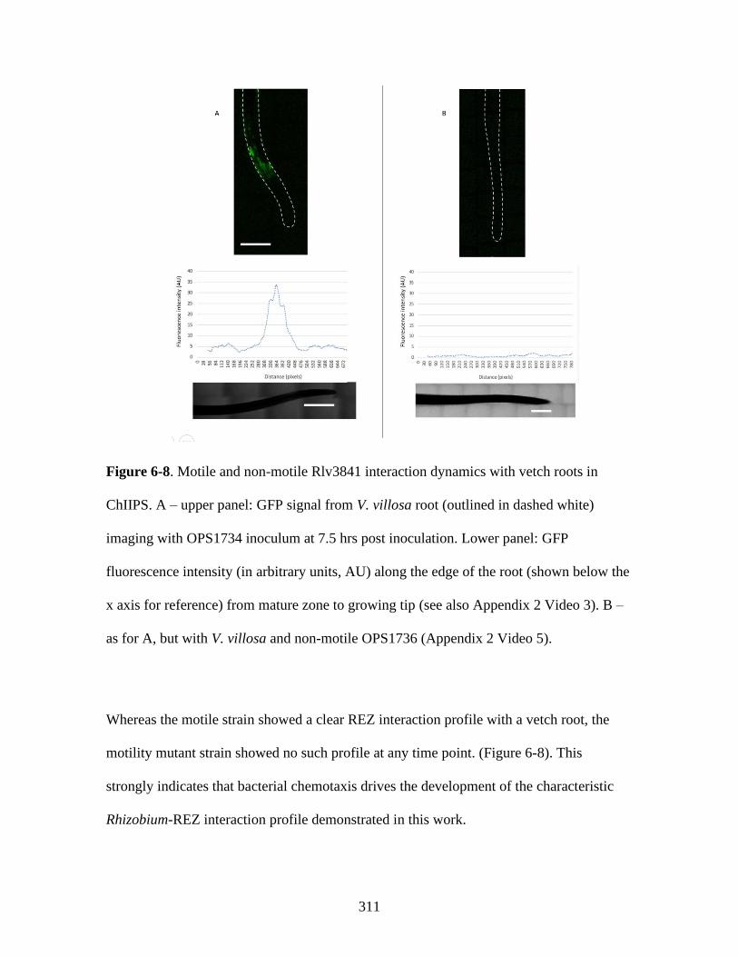
311
Figure 6-8. Motile and non-motile Rlv3841 interaction dynamics with vetch roots in
ChIIPS. A – upper panel: GFP signal from V. villosa root (outlined in dashed white)
imaging with OPS1734 inoculum at 7.5 hrs post inoculation. Lower panel: GFP
fluorescence intensity (in arbitrary units, AU) along the edge of the root (shown below the
x axis for reference) from mature zone to growing tip (see also Appendix 2 Video 3). B –
as for A, but with V. villosa and non-motile OPS1736 (Appendix 2 Video 5).
Whereas the motile strain showed a clear REZ interaction profile with a vetch root, the
motility mutant strain showed no such profile at any time point. (Figure 6-8). This
strongly indicates that bacterial chemotaxis drives the development of the characteristic
Rhizobium-REZ interaction profile demonstrated in this work.

312
6.2.8 Using ChIIPS to investigate root hair attachment polarity
The existing literature model of Rlv3841 attachment to legume root hairs indicates that,
under acidic to neutral conditions, polar glucomannan on the bacterial cell surface binds
root hair lectin. Under alkaline conditions these lectins disassociate, and Rlv3841 uses
rhicadhesin to bind root hairs [34, 71]. This work has investigated rhicadhesin extensively
(see section 4.2.13). Despite uncertainty around the identity of factor(s) promoting
alkaline pH root hair attachment, basic physiology of this process (such as whether
attachment to root hairs is also polar, as with glucomannan) remains uncharacterised.
To investigate this, ChIIPS was used to image the attachment of both Rlv3841 and a gmsA
(glucomannan) mutant strain to vetch root hairs at high magnification (see 2.8.7; here a
64x objective lens was used). Representative images and graphed percentage attachment
classification (polar or non-polar) for Rlv3841 and the glucomannan mutant strain are
shown in Figure 6-9.

313
A
B

314
Figure 6-9. Attachment polarity of OPS1734 (Rlv3841[pLMB449] – pTac gfp) and
OPS1730 (A1045- gmsA::Tn5ΩKan/Neo[pLMB449] – pTac gfp) to V. villosa root hairs
after two hours. A – Representative images of OPS1734 attached to root hairs. Root hairs
are outlined in dashed white and attached bacteria are indicated with arrow. Yellow arrow
colour indicates polar attachment; white arrow colour indicates non-polar attachment. B –
as for A, but with OPS1730. C – Percentage attachment classification of both strains
(lateral vs polar) for all counted bacteria. n = 35 for OPS1734 and 42 for OPS1730.
These results demonstrate that, under neutral conditions, all Rlv3841 cells observed with
functioning glucomannan attached to root hairs in an end-on, polar fashion. However, in a
glucomannan mutant strain, this figure was ~ 30%, with the remainder of observed cells
being attached in a non-polar (i.e. lateral) fashion. This indicates that, whilst glucomannan
mediated attachment is polar, when Rlv3841 cells lack glucomannan they can attach to
root hairs in a polar or non-polar fashion. There are two possible conclusions that can be
drawn from this: rhicadhesin is not exclusively located at cell poles and can therefore
mediate root hair attachment in a polar or non-polar fashion, or that there are multiple,
OPS1734 OPS1730
C

315
differentially localized cell surface components which govern attachment in the absence of
glucomannan. This is a possibility given the diversity of attachment factors required for
Rlv3841 attachment to pea roots under alkaline conditions (when glucomannan will not be
functioning) seen in this work (see chapter 4 and 4.2.11).
These findings are comparable to those described by Matthysse (2014), reviewing work
with A. tumefaciens. In this bacteria, unipolar polysaccharide (UPP, a glucomannan
analogue) mediates polar binding of bacteria to root hairs under low calcium, low
phosphate or acidic pH conditions. When UPP is not produced, attachment is mediated by
unknown molecules and is both polar and lateral [55]. It was noted that, in UPP-
independent attachment, the total number of bacterial cells attaching was also lower than
with UPP-producing bacteria [55]. This fits with the results of a Lux assay and INSeq for
a glucomannan mutant, where attachment is reduced in comparison to Rlv3841 under
neutral pH conditions (see Figure 5-3, Table 4-6).
Therefore, although factors governing glucomannan or UPP-independent root hair
attachment remain to be identified, it seems both Rlv3841 and A. tumefaciens can attach in
a polar or lateral fashion in the absence of these factors.
6.2.9 ChIIPS2 design for future work
The ChIIPS chamber used in this work proved suitable for imaging early-stage
Rhizobium-legume root interaction dynamics. However, for imaging over longer
timescales and for more realistic test conditions, it would be desirable for the aerial plant
parts to remain external to the chamber. A schematic for a new chamber design (ChIIPS2)
which would overcome the limitations of ChIIPS whilst maintaining its advantages is
shown in Figure 6-10.

316
Figure 6-10. ChIIPS2 design. A – Schematic of the proposed ChIIPS2 shown from above.
Plants are germinated upright in 1000 µL pipette tips to promote gravitropic root growth.
These tips are transferred to the ChIIPS2 chamber (before the root emerges from the tip)
through a hole in the chamber wall and held in place by the rubber seal. Roots continue to
grow into the chamber and can then be imaged. B – longitudinal section of the proposed
ChIIPS2 showing the plant root inside the chamber with bacterial inoculum (brown ovals,
not to scale).
A
B

317
6.3 Conclusion
Here, the applicability of microfluidics systems to the study of root interaction dynamics
and spatio-temporal mapping of interactions between bacteria and roots (the first step in
Rhizobium-legume symbioses) is demonstrated.
For mapping the spatio-temporal activation of the lppE gene (RL3234) on roots, clover
plants (T. repens and T. pratense) were used with Rlv3841 reporter strains. Having
validated the specific activation of lppE (RL3234) gene expression in the REZ using a
luminescence method, spatiotemporal gene expression visualization was also tested in the
TRIS setup. Strong activation of the lppE promoter was seen in the REZ from 1 hr
onward, and this peaked at ~39 hrs post inoculation. Although the role of lppE in root
interaction remains unknown, its expression is clearly related to REZ signaling molecules.
Particularly in secondary root attachment, different factors are expressed and upregulated
in different rhizobia to facilitate the colonization process. These include extracellular
fibrils, cellulose, cadherin-like proteins, fimbrae and outer membrane proteins, among
others [34]. However, very little is known about either the precise timings of gene
expression in root interactions, or whether genes show specific spatial requirements for
attachment to different root zones. As the data for lppE shows, a specific spatial
expression does exist. By using microfluidics setups with a suite of reporter genes, the
process of primary and secondary attachment could be more specifically defined.
The REZ is a zone of interest for root-microbe interactions. This is not just because of its
high exudation and identity as a preferential microbe interaction site in several systems,
but also because of the role it may play in primary attachment. In a soil system with large
microbial load, it seems likely that the REZ, emerging directly behind the meristematic

318
growing tip, will represent one of the only uncolonized root zones. The high level of
exudation from this site may serve to rapidly recruit new root microbiota preferentially
and act as a method to protect against pathogen colonization. If this hypothesis is correct,
the elongation zone presents an interesting area of focus for characterising root-microbe
interaction profiles, and spatio-temporal gene expression patterns of attaching bacteria in
this region could provide novel mechanistic insights.
Given the limitations of TRIS (where narrow channels with a short imaging window
prevent imaging of roots much larger than Arabidopsis, and chamber design precludes use
with an upright confocal microscope), a preliminary new test setup called ChIIPS was
trialed. Based on commercially available cell culturing slides, ChIIPS is lower cost than
TRIS, compatible with much larger root systems and easier to use with upright confocal
microscopes. Despite the limitations of this preliminary system, preferential interaction of
Rlv3841 with the REZs of lotus, vetch and alfalfa were clearly visible. This result is
important in confirming the REZ as a hotspot for interaction across multiple plant species.
A motility mutant of Rlv3841 did not show these preferential interaction dynamics with
vetch, confirming that chemotactic motility is required on the part of interacting bacteria.
This result is interesting when compared with data from Lux root attachment assays
(Chapter 3) and INSeq experiments (Chapters 4 and 5). In a Lux root attachment assay,
loss of motility lead to a large reduction in attachment to pea roots at all pHs in a motA
mutant strain (Figure 3-6). However, INSeq classified motA (and other motility genes) as
NE (not required for root attachment) in attachment to pea roots at all pHs (see 4.2.9) and
to barley roots at pH 7.0, although chemotaxis was needed for attachment to soybean roots
(see 5.2.10). The hypotheses explaining these results are as follows: in a homogenous
inoculum of non-motile cells (as in a Lux attachment assay with a motA mutant), bacteria

319
cannot move toward the root surface for attachment. However, in a mixed INSeq
inoculum (where most of the cells will be motile), ‘aided motility’ occurs as cell-cell
collisions between chemotaxing and non-motile cells (as well as disruption of the
repulsive hydrodynamic boundary layer) leads to positioning of non-motile cells in
proximity to the root which can permit primary attachment. For soybean, the large root
size and higher exudation could result in a stronger selection for root attachment on
chemo-sensing bacteria, resulting in under-representation of chemotaxis mutants in the
soybean root-attached library and classifying chemotaxis genes as ES/DE. A ChIIPS
system could be used to investigate these hypotheses further, especially with regards to
‘aided motility’. A simple experiment using single and mixed (differentially labelled)
wild-type and non-motile Rlv3841 inoculums with different roots could quantify how
many non-motile cells migrate to within close proximity of the root over time. This would
enable quantification of any ‘aided motility’ effect and could provide additional evidence
to explain disparities between Lux attachment assay and INSeq results.
As a final test of ChIIPS, high magnification confocal microscopy was used to investigate
how Rlv3841 and a glucomannan mutant attach to root hairs at neutral pH. Interestingly,
whereas Rlv3841 showed only polar, end-on root hair attachment, a glucomannan mutant
showed a mixture of lateral (~65 %) and polar (~ 35 %) attachment. This mirrors the
characterised attachment profiles of UPP proficient and deficient strains of
Agrobacterium, respectively. This suggests that, whilst the identity of any ‘rhicadhesin(s)’
mediating UPP or glucomannan independent attachment remains unknown, there is likely
to be mechanistic similarities between the two species. If there is a single rhicadhesin, it is
not only polarly located but also permits lateral cell attachment to root hairs.

320
To conclude this chapter, an improved ChIIPS2 design was suggested, which would
overcome ChIIPS limitations. The main new feature of this design is the provision for
aerial plant parts to remain separate from the bacterial inoculum. This is likely to enable
longer term imaging of root-microbe interactions within a less physiologically disruptive
environment for the plant.
The use of ChIIPS2 would enable many experimental questions to be addressed.
Alongside spatiotemporal gene expression patterns, interactions between Rhizobium, roots
and pathogens could be examined. Also of interest would be to use a dual channel setup
(not dissimilar to that seen in TRIS, [188]), whereby two different roots can be imaged
side by side. This would enable inoculum preferences to be examined; for example, is root
preference seen in a differentially labelled inoculum of R. leguminosarum biovar trifolii
and Rlv3841 interacting with vetch and clover roots? Does one strain outcompete another
for root interaction when its cognate symbiont root is present? Such questions remain
unexplored.
One limitation of all systems mentioned in this work is the long-term immersion of roots
in liquid, which is not particularly representative of soil conditions. An exciting prospect
is the combination of ChIIPS2 with transparent soil. This is a form of highly treated
fluoropolymer which has very similar water and nutrient retention characteristics to real
soil, and is suitable for sustaining plant growth and imaging root-microbe interactions
[463]. However, for the moment this transparent soil remains expensive and technically
challenging to produce, particularly in large quantities.
This work highlights the huge potential of novel imaging platforms in the study of
Rhizobium-legume root microbe interactions. Further work in this area is likely to yield
novel insights into these inter-kingdom relationships.

321

322
Chapter 7
General discussion

323
7.1 Overview
In this work a range of techniques, including proteomic and bioinformatic analysis, newly
developed Lux whole-root attachment assays and INSeq were used to define the primary
root attachment determinants needed for Rlv3841 interaction with pea at different pHs and
with those of a non-host legume and non-legume at pH 7.0. These approaches revealed a
far greater diversity of primary attachment mechanisms than previously recognized for
Rlv3841, which demonstrate considerable pH and plant-host dependency. A summary of
the most important findings regarding attachment factors from all the experimental
chapters of this work is presented in Figure 7-1. These findings are discussed and
evaluated in the subsequent sections of this chapter, starting with extracellular/surface-
localized factors and moving to intracellular and uncharacterised factors. A critique of the
experimental techniques employed is followed by an outline of further research directions.
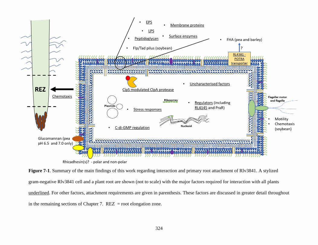
324
Figure 7-1. Summary of the main findings of this work regarding interaction and primary root attachment of Rlv3841. A stylized
gram-negative Rlv3841 cell and a plant root are shown (not to scale) with the major factors required for interaction with all plants
underlined. For other factors, attachment requirements are given in parenthesis. These factors are discussed in greater detail throughout
in the remaining sections of Chapter 7. REZ = root elongation zone.

325
7.2 Extracellular/surface localized primary attachment factor requirements
7.2.1 EPS and peptidoglycan
Chapters 4 and 5 demonstrated that Rlv3841 primary root attachment at different pHs and
to different plant roots is vastly more complex than previously reported. Whilst there are
multiple follow up points to this work, several target genes stand out for further
investigation. In terms of cell surface and exported factors, gene classifications from
INSeq suggested that activation of the Npr/ManX system is needed for pea root
attachment at pH 6.5, but not at pH 7.5, where they are likely deactivated by SixA. The
most likely effect of this at pH 7.5 is a reduced EPS secretion through an unknown ABC
exporter [313]. However, this could be confirmed by quantification of EPS in Rlv3841
cell cultures at pH 6.5 and 7.5. Methods such as liquid chromatography-organic carbon
detection–organic nitrogen detection and colorimetric assays of protein, sugar and
phenolic content (among others) [464, 465] can also be applied and demonstrate at a
molecular level the EPS changes needed for pea root attachment at different pHs. A large
diversity of different factors involved in EPS biosynthesis and modification was shown to
be required for attachment to pea roots at different pHs and to different plants roots using
both Lux and INSeq approaches. Therefore, the EPS category encompasses crucial cell
surface alterations (Figure 7-1) promoting attachment under different conditions.
Similarly, INSeq experiments highlighted a ubiquitous requirement for peptidoglycan
biosynthesis and modification factors for primary attachment under all plant attachment
conditions tested. Of particular note was the dacC (RL4363) gene, encoding a putative
penicillin binding protein, required for attachment to all plant roots and at all pH

326
conditions. In E. coli, DacC processes sugar-peptide cell wall precursors and is involved
in peptidoglycan biosynthesis [299]. What is clear is that peptidoglycan composition is
another crucial factor promoting attachment under different conditions. Ramstedt et al.
(2011) have described a method of cryo-x-ray photoelectron spectroscopy analysis
combined with a curve resolution analysis of the carbon spectra. This technique is able to
closely monitor the chemical composition of bacterial cell walls and predict changes in
peptidoglycan structures [466]. Such a technique could be useful in characterising the
peptidoglycan changes required for root attachment under different conditions and with
different plant hosts in Rlv3841.
Not discussed so far in this work is the role of cell shape, which can play a crucial role in
regulating bacterial attachment [467]. For example, C. crescentus has a crescent cell shape
determined by the production of a protein called crescentin [468]. Being an organism
largely residing in freshwater lakes and streams, this crescent shape specifically enhances
surface colonization under fluid-flow conditions. This is due to favourable cellular
orientation caused by shape relative to the target surface under these conditions [468].
Peptidoglycan is the main stress-bearing structure that dictates bacterial cell shape [467].
It would be interesting (using microscopy) to investigate the cellular morphology in
Rlv3841 peptidoglycan mutants shown to be deficient in attachment under certain
conditions to determine if any common altered morphologies are observed. These would
likely disrupt attachment by interfering with bacterial cell-surface factor interactions with
plant roots.

327
7.2.2 Surface enzymes
Several enzymes (such as TpiA, a glycolytic enzyme with a role in central carbon
metabolism) were hypothesised to play an important bacterial surface-associated
moonlighting role in primary attachment to all plant roots based on INSeq results. TpiA
was very interesting, being required for attachment to all plant roots at all pHs tested
(Figure 7-1, included in ‘surface enzymes’). If correct, this would represent a further novel
primary attachment mechanism for Rlv3841. Surface localised glycolytic enzymes are
multifaceted and can be involved in substrate binding. TpiA has been shown to be surface
localised and have a direct role in adherence to host cells in Mycoplasma gallisepticum
[284]. An Rlv3841 mutant in TpiA carrying pIJ11282 (luminescence cassette) could be
used to validate this in a Lux attachment assay, whilst fluorescent protein tagging could
investigate the hypothesised secretion of TpiA.
7.2.3 Flp/Tad pili, outer membrane proteins and LPS
Further to the novel attachment factor requirements described above, INSeq robustly
demonstrated that surface localised primary attachment factors previously identified in
other bacterial species can be used by Rlv3841 in a context dependent manner. Primary
attachment to soybean roots showed a Flp/Tad pilus requirement (Figure 7-1) which was
demonstrated by the need for encoding genes found on plasmids pRL11 and pRL8. This
shows an overlap whereby Rlv3841 can make use of attachment mechanisms (namely
adhesive Flp/Tad pili) reported in Agrobacterium, Pectobacterium (a potato pathogen), V.
vulnificus, A. actinomycetemcomi and C. crescentus, among others [44, 384, 385].
A second example of this is the requirement of the OmpA protein RL3267 for attachment
to barley and soybean roots (included under ‘outer membrane proteins, Figure 7-1). Such

328
outer membrane proteins acting as primary attachment factors are well characterised in
Azospirillum, where they function both in root attachment and cellular aggregation [64].
The third example is the requirement for various LPS biosynthesis and modification
factors for attachment to different plant roots under different conditions (Figure 7-1). As
an example, dgkA (RL2780) was needed for attachment to pea roots at pH 6.5 and soybean
roots at pH 7.0. In E.coli, dgkA mutants are defective in biofilm formation [316], and
DgkA function has been linked to phospholipid recycling and LPS modifications [324]. In
B. subtillis it is important for lipoteichoic acid synthesis [325]. However, LPS has not
previously been implicated in primary attachment in Rlv3841, instead being more
associated with Azospirillum. Indeed, it has been proposed that the LPS O-antigen of
Azospirillum directly binds maize root lectin to mediate primary attachment [65].
A powerful conclusion of this work overall is therefore that Rlv3841 can make use of
primary attachment mechanisms which, until now, have only been associated with other
bacterial species. It does so in an environmental condition and plant host-dependent
manner. Given this finding, it seems possible that primary attachment mechanisms are
more conserved across soil bacteria than realised but demonstrate greater context
dependency than previous experimental work has been able to investigate. Characterising
this further would require inverse experiments to be conducted, for example using INSeq
to examine the primary attachment mechanisms of Azospirillum to different plant roots
under different conditions.
7.2.4 FHA
RL4382 (filamentous hemagglutinin adhesin - FHA) was important for pea root
attachment at all pHs and to barley roots (Figure 7-1). This was also demonstrated in a

329
Lux assay in Chapter 3. Previous work in other bacteria suggests that FHA plays a direct
attachment role and agglutinates cells, leading to higher numbers of cells attached [239–
241]. This has not previously been documented in Rhizobium, making this a novel
attachment factor. Various scanning-electron microscopy techniques are available for
high-resolution imaging of biofilm structures [469], and applying such techniques to wild-
type Rlv3841 and an RL4382 mutant could shed more light on the mechanisms behind
FHA mediated attachment. RL4381 (putative POTRA domain exporter of FHA) was
needed for attachment to all plant roots whilst FHA was not (Figure 7-1), suggesting this
exporter my interact with other attachment factors. Application of TRANSIT analysis (a
method combining multiple different statistical approaches to analysing INSeq data into
one platform [470], see also 7.9.2) could reinforce or challenge this idea by enabling
comparison of gene classifications with different methods of data analysis. If reinforced, a
bioinformatic screen of other Rlv3841 genes encoding TPS domains (required for
interaction with POTRA domain exporters, [239]) could indicate the identity of other
exported factors. Further, RL4381 should be confirmed as the transporter for FHA. One
approach to this would be to express RL4382 fused to a fluorescent protein in both wild-
type and RL4381 mutant Rlv3841 strains. Changes in fluorophore location
(intracellular/extracellular) would show whether the transporter encoded by RL4381 is
necessary and sufficient for FHA export.
7.2.5 Motility
In a Lux attachment assay, flgE and motA mutant strains were defective in attachment to
pea roots at pHs 6.5, 7.0 and 7.5 (see Figure 7-1, motility). These mutant strains showed
no significant differences to one another in terms of attachment level. It was therefore

330
hypothesised that it was motility itself (rather than flagellar adhesin properties)
contributing to attachment in Rlv3841/pea symbiosis. This suggests that flagellar adhesin
action is not contributing directly to attachment, as is seen in some other rhizobacteria
[34]. No flagellar subunits were classified as required (ES/DE) in pea root attachment at
any pHs in INSeq (Chapter 4). However, the disagreement (possibly caused by ‘aided
motility’, 4.2.9) between Lux and INSeq means that a role for flagellar subunits in
adhesion cannot be completely ruled out (indeed flaH, a flagellar subunit, was needed for
soybean root attachment, see Table 5-9). The bacterial flagellum is made up of more than
20 different proteins, of which FliC is the major subunit. FliC has been reported to have
adhesin properties in E. coli and P. aeruginosa [235]. To investigate possible flagellar
adhesin properties in Rlv3841, further single flagellar subunit mutants could be tested in
Lux attachment assays and compared to a motA mutant strain. Mutants showing reduced
attachment versus the motA mutant would indicate both loss of motility and loss of
flagellar adhesin function.
Motility was not required for Rlv3841 attachment to pea or barley roots in INSeq, but
aspects of chemotaxis were needed for soybean root attachment (Figure 7-1). The reasons
for this (and the disagreement with Lux attachment assay data showing motility as
required for effective pea root attachment) remain unclear. A form of ‘aided motility’ in
INSeq (biased Brownian motion as well as disruption of the repulsive hydrodynamic
boundary layer around roots, see 4.2.9), along with aberrant swimming of chemotaxis
mutants and higher levels of exudation from soybean roots (providing a stronger selection
for chemotaxis-competent bacteria than for other plant roots), was hypothesized to explain
these results. Exudation levels from soybean, pea and barley roots could potentially be
examined using Rlv3841 luminescence biosensor strains, such as those developed by Pini

331
et al. (2017), with levels of light emission acting as a proxy for exudate levels [201].
Further, a mathematical modelling exercise of bacterial motion in mixed population of
chemotaxing/non-chemotaxing cells could examine the plausibility of the ‘aided motility’
hypothesis. Using differentially labelled motile and non-motile Rlv3841 strains in a
ChIIPS setup could also help resolve this question, with confocal imaging able to
determine how many non-motile cells were able to reach the plant root surface.
Inoculating a motile wild-type culture of Rlv3841 labelled with GFP with lotus, vetch or
alfalfa roots showed preferential bacterial interaction with the REZ (Figure 7-1),
indicating that the REZ exudes high levels of chemoattractants. This result was also
demonstrated for B. subtilis interacting with Arabidopsis roots in TRIS [188]. This
interaction profile was not seen with a motA mutant of Rlv3841. Whilst this further
suggests that motility is important for establishing early-stage root microbe interaction
dynamics, this further highlights the need to clarify INSeq results and determine cell
movement dynamics where non-motile strains are a minority cell type in a mixed
population interacting with roots.
7.2.6 Rhicadhesin
Rhicadhesin is a proposed 14 kDa protein which facilitates attachment of Rhizobium to
legume root hairs at alkaline pHs [34], but has never been identified at the gene level. A
proteomics approach was taken to identifying rhicadhesin. Using a 14 kDa protein band
(thought to contain rhicadhesin) for LC-MS/MS identified 15 proteins, of which three
(RL4733, omp19 – RL4441 and RL1635) were the most likely rhicadhesin candidates. Due
to gene size limitations, pK19mob mutagenesis was unsuccessful in targeting rhicadhesin
candidate genes identified. An in-frame mutagenesis approach could be used to overcome

332
these issues, as very small genes (such as the hdeA gene in E. coli, 110 bp) have been
mutated by this approach in the past [246].
The preparation of a crude adhesin fraction from Rlv3841 cells relied on isolating cell
surface proteins, as described by Smit et al (1989) [53]. However, here the 14kDa band
from the crude adhesin fraction also contained ribosomal subunit and other predicted
intracellular proteins at low levels, suggesting that some minor cell lysis had occurred.
Siciliano et al. (2019) describe several innovative proteomics approaches to investigating
bacterial surface-exposed proteins (the proteosurfaceome) which could be more
appropriate to the study of cell surface adhesins. These include cell shaving, in which
surface exposed protein fragments are released by proteolytic digestion using trypsin with
intact bacterial cells. This approach allows comprehensive analysis of the
proteosurfaceome as membrane and cell wall embedded proteins can be released, further
digested with trypsin and subjected to LC-MS/MS. This approach has been applied to both
Gram-positive and negative bacteria [221]. However, tryptic digest is a limitation of this
technique as is it dependent on trypsin binding sites being accessible in target proteins to
generate peptides for mass spectrometry. Further, functional proteins will not be
recovered. A more advanced approach uses biotin tag-containing reagents to target surface
proteins before lysing cells and isolating surface proteins using streptavidin affinity
chromatography. This method is similarly suitable for both Gram-positive and negative
bacteria [221]. Combining these methods could allow the identification at the gene level
of ~14kDa Rlv3841 surface proteins as well as the recovery of intact surface proteins for
functional assays.
Regarding rhicadhesin, the original research defining its role (Smit et al. (1989), [53]) was
certainly not conclusive. Defining an adhesin based on its ability to inhibit attachment is

333
not definitive. Further, given the number of root attachment factors identified
bioinformatically in Chapter 3, it is likely that membrane preparations may isolate
proteins that can inhibit attachment when pre-incubated with roots, but this does not imply
these proteins are important adhesins. Multiple genes encoding rhicadhesin or use of an
impure protein preparation when defining its activity also confound definitive
identification further. When combined with INSeq results from Chapter 4 (where multiple
12-16 kDa factors were important for root attachment at neutral and/or alkaline pHs) it is
apparent that there are multiple attachment factors with rhicadhesin-like properties.
Further, work in Chapter 6 demonstrated that a glucomannan mutant of Rlv3841 can
attach to vetch roots in both a polar and non-polar fashion (whilst glucomannan is required
for attachment at acidic to neutral pHs in a polar fashion, demonstrated with Lux assays,
INSeq and ChIIPS, see also Figure 7-1). Therefore, whilst the definitive identification of a
rhicadhesin as originally proposed by Smit et al. (1989) [53] was not possible, this work
demonstrates there are multiple small proteins with rhicadhesin-like profiles of attachment
activity. Hence, in Figure 7-1, rhicadhesin(s) are shown on both the polar and lateral
surfaces of the Rlv3841 cell.
7.3 Intracellular primary attachment factors
7.3.1 Regulators - PraR
Intracellular factors important for root attachment include a variety of regulators required
at different pHs and with different roots (Chapters 3, 4 and 5). A mutant in the important
praR regulator demonstrated a previously uncharacterised pH dependency in Lux assays.
Attachment phenotypes for praR and rapA2/rapC mutants were as previously described at

334
pH 7.0 [59] in a Lux assay with pea roots. However, these strains showed very different
attachment profiles at pH 6.5 and 7.5, with the praR mutant defective in attachment and
the rapA2/rapC mutant a hyper-attaching strain (hence the PraR regulator is shown in
Figure 7-1). Testing a praR/rapA2/rapC triple mutant in a Lux assay (defective in
attachment at all pHs) allowed an updated model of regulation to be proposed. At pH 7.0,
CinS inhibits PraR which relieves inhibition on rapA2 and rapC, permitting increased
attachment. However, at pH 6.5 and 7.5 PraR is not inhibited, and induces expression of
unknown ‘rapX’ factor(s), possible given the positive and negative regulatory activity of
PraR [59]. Whilst this model has not been validated, mutation of praR leads to hyper-
attachment at pH 7.0 and reduced attachment at pH 6.5 and 7.5. RNASeq has previously
been used to investigate the transcriptome profile of a praR mutant [59], and expanding
this work to pH 6.5 and 7.5 conditions could refine the model of PraR/Rap interactions
and primary root attachment at different pHs.
7.3.2 c-di-GMP regulation and regulators – RL4145
Various c-di-GMP synthesis proteins were required for root attachment at different pHs
and to different plant roots (Figure 7-1, ‘c-di-GMP regulation’). Whist c-di-GMP
signalling is well characterised, particularly in biofilm formation [78, 79], how the
different c-di-GMP synthesis proteins in Rlv3841 are contributing to attachment under
different conditions and to different roots is unknown, and warrants further investigation.
c-di-GMP regulated biofilm determinants range from flagella rotation to EPS and surface
adhesin production and type IV pili retraction [81]. Reconciling the global effect of
cellular c-di-GMP concentration on biofilm formation with the discrete actions of c-di-
GMP is difficult, but various hypotheses exist. One is that diguanylate cyclases (DGCs)

335
and phosphodiesterases (PDEs) show differential expression and enzyme activity and so
have discrete impacts on the cellular pool of c-di-GMP. Another is that different effectors
show different c-di-GMP binding affinities and are differentially responsive to
intracellular c-di-GMP levels. One of the most intriguing theories is that different c-di-
GMP pools are sequestered in multi-protein complexes at distinct cellular sites [79]. This
has been observed for the LapA adhesin of P. fluorescens, where LapA activity is
regulated post-translationally by the LapD/LapG c-di-GMP effector system. This system
activates LapA in the outer membrane on receipt of a specifically localized c-di-GMP
signal. This signal in turn is likely generated by activation of DGCs co-localized with
LapD/LapG [471].
What is clear is that understanding c-di-GMP effects is not as simple as measuring cellular
c-di-GMP content. Tightly regulated signalling systems, localized protein complexes and
distinct subcellular changes may all be involved in producing different outcomes in terms
of primary root attachment.
One regulator (RL4145) was required for attachment to all plants (Figure 7-1), whilst
many were required for attachment to pea at different pHs. In the case of the protein
encoded by RL4145, a likely regulatory target is RL4139 (a putative c-di-GMP degrading
EAL domain protein). If it is the case that RL4145 represses RL4139 (as was suggested by
their inverse HMM classifications), then the requirement for RL4145 in attachment to all
plants can be explained by the maintenance of high intracellular c-di-GMP levels by this
protein. Investigating RL4145 regulation further could be approached using RNASeq to
infer RL4145 regulatory targets as well as protein-protein interaction studies or promoter
binding assays to see if there is direct activity between RL4145 and RL4139. An RNASeq

336
approach would also be well suited to investigating transcriptional regulatory networks
underpinning attachment in mutant Rlv3841 strains of other important regulators.
7.3.3 ClpS-modulated ClpA protease
INSeq demonstrated a requirement for the ClpS/ClpA protease system for attachment to
pea roots at pH 7.0 and 7.5, as well as to soybean and barley roots (Figure 7-1). The
targets of ClpA (with or without modulation by ClpS) are unclear and understanding these
would aid in defining the cellular changes induced by this system. Multiple experimental
approaches could be taken to characterising ClpS/ClpA targets. These include
experimental methods such as gel separation of control and ClpS/ClpA proteolyzed
bacterial protein preparations, where proteolytic targets are identified by gel band shifts
between the two samples. Further, radiolabelling approaches such as SILAC in wild-type
and ClpS/ClpA mutant Rlv3841 strain, followed by MS-based peptide quantitation, could
also be applied [472]. There are also multiple bioinformatic approaches which could be
applied to predicting protease targets [473].
7.4 Uncharacterised primary attachment factors
Amongst the ‘uncharacterised factors’ is pRL100053 encoding a putative transmembrane
protein of unknown function (Figure 7-1), which is defective in attachment in a Lux assay
at all pHs and required for primary attachment (all pHs), colonization, infection thread and
nodulation in INSeq experiments (Table 4-9). This data strongly suggests that pRL100053
encodes a novel root hair attachment factor which is critical for subsequent nitrogen fixing
symbiosis development.
337
For the 292 genes required for primary attachment to pea roots (INSeq, Chapter 4),
multiple datasets from different stages of Rlv3841/pea symbiosis were used to map gene
requirements and infer which primary attachment factors could be involved in attachment
to root hairs (the entry-point for nitrogen-fixing symbioses [25]) or bulk root epidermis.
This approach uses the rationale that factors required for root hair attachment will also be
required in subsequent symbiosis stages. However, INSeq data alone does not directly
demonstrate this, and the picture is complicated by factors which are classified as required
in non-contiguous symbiotic stages. A potentially high-impact way of resolving this
further would be to follow up on INSeq results with in-depth studies of gene function, and
pRL100053 represents a good candidate for this.
Expressing a vector-borne pRL100053 construct whereby pRL100053 is fused to a
fluorescent protein in Rlv3841 could visualize protein localization. Using biotin-tagged
pRL100053 as a bait protein for pea root protein extracts could identify its plant root
binding partners, and purification studies could also define protein structure.
7.5 Experimental techniques and future research directions
7.5.1 Lux whole-root attachment assay
The Lux whole-root attachment assay developed here allowed the primary attachment
abilities of Rlv3841 strains to be tested at different pHs. This assay represents a
considerable advance on previously described techniques which relied on high
magnification microscopy and counting bacteria [52, 53, 210, 217–219], confocal
microscopy [56], bacterial recovery and plating or radiolabeling approaches [59]. In
comparison, Lux is higher throughput, preserves spatial attachment information, is safer

338
than radiolabelling and is more appropriately established for studies at different pHs. The
activation of the lppE promoter selectively in the REZ along with the REZ attraction
profile of bacteria (Chapter 6) suggests spatio-temporal interaction dynamics between
Rlv3841 and legume roots, which may extend to attachment. Although not pursued here,
Lux assays at different time points could evaluate the spatio-temporal attachment profile
of different Rlv3841 strains to roots. This would increase our understanding not just of
attachment factors, but also root zone importance in the attachment and colonization
process. However, it is also worth noting that interpretation of Lux data over longer time
periods may become more complex than in a one-hour attachment assay as bacterial
replication and metabolic activity may affect Lux signal intensity [201].
7.5.2 INSeq
Chapters 4 and 5 used INSeq with HMM analysis to classify gene requirements in primary
root attachment. Although there are various advantages to using an HMM for analysis
over other published methods (see 1.8.3), low TA site number in a gene can still be a
confounding factor. DeJesus et al. (2015) developed a software package for analyzing
TnSeq data (which can also be applied to INSeq) called TRANSIT [470]. TRANSIT
provides a graphical interface with three different methods for analyzing INSeq data,
allowing combinations of methods or direct comparisons and simpler manual curation of
results that differ between analysis methods. A Bayesian/Gumbel method for data analysis
is complemented by a HMM for assigning gene classifications in individual test
conditions, and a resampling (permutation) test allows comparative analysis of gene
classifications between conditions [470]. The latter would be particularly applicable in the
case of attachment at different pHs, as the model creates a distribution of read count

339
differences that could occur by chance between conditions before analyzing data with the
null hypothesis that gene classifications are not different. Genes with truly different
classifications between conditions fall out of the bounds of the resampling distribution and
are classified as such. This offers a different and complimentary approach to the HMM for
INSeq data analysis. Using this method with the INSeq data collected in Chapters 4 and 5
could allow comparison of results between analysis techniques for low TA site genes, with
a consensus result being assigned to increase classification certainty.
7.5.3 ChIIPS and ChIIPS2
A preliminary new imaging system called ChIIPS was developed, better suited to imaging
work with larger legume roots and upright confocal microscopes compared to TRIS. The
specific REZ activation of the lppE promoter illustrated that devices such as ChIIPS and
TRIS can be used to investigate spatio-temporal activation of genes on the root surface.
To understand root-microbe interactions and root attachment better, spatio-temporal
activation dynamics of genes upregulated in the rhizosphere and primary/secondary
attachment factors should be investigated further. Plant roots display multiple different
zones with very different properties [461]. However, how attachment factor requirements
vary by root zone is largely unknown. Using reporter fusion strains could investigate not
only this, but also timing of gene expression, and thus help determine the primary-
secondary attachment switch more precisely.
Results in Chapter 6 indicate that the REZ exudes high levels of chemoattractants.
However, the composition of these chemoattractants and how their exudation varies along
the root length is not understood. An interesting further application of the biosensors
developed by Pini et al. (2014) [201] would be for study of spatial root exudate
340
composition in ChIIPS. This could be achieved by inoculating roots with fluorescent
protein exudate biosensor strains and observing the location and intensity of reporter
induction along the plant root and over time. Ideally this experimental approach would use
ratiometric imaging, with strains carrying a constitutive gene and a biosensor gene
present, allowing a better quantitation of reporter expression relative to a background gene
expression level.
An improved ChIIPS2 design was presented to enable future Rhizobium-legume
interaction studies. ChIIPS2 enables the aerial plant parts to remain external to the
inoculum-containing imaging chamber. Based on the work in Chapter 6, ChIIPS2 should
be applicable to more complex questions than examining chemotactic responses to root
zones. The question of root choice remains unexplored. For example, are a vetch and a
clover root in the same chamber preferentially ‘chosen’ for interaction by R.
leguminosarum biovars viciae and trifolii, respectively? Competition between strains
could also be examined, for example by inoculating a vetch root with a mixed R.
leguminosarum biovars viciae and trifolii population and observing whether one
outcompetes the other for root interaction. From the plant perspective, use of transgenic
reporter gene-carrying or mutant plant lines (e.g. Massalha et al. (2017, [188]) in ChIIPS2
could help define the gene-level responses of plants to different bacterial inoculums and
the pathways needed for production of exudates that drive REZ interaction profiles.
Rhizobium-pathogen-root interactions could also be investigated. Massalha et al. (2017)
demonstrated the (likely surfactin-mediated) exclusion of E. coli from the roots of
Arabidopsis by B. subtilis [188], but no such interactions have been investigated with
Rhizobium and plant or human pathogens. In order to conduct such experiments

341
effectively, motility inducing culture conditions (such as the low carbon supply conditions
used in this work) would have to be established for each bacterial strain and species.
A particularly innovative experimental setup could be gained by combining ChIIPS2 with
transparent soil [463] when asking the questions posed above. This would vastly increase
the physiological relevance of the environment under study by providing a heterogeneous,
porous and transparent substrate for plant root growth which is directly suitable for
imaging root-microbe interactions and has soil-like properties in terms of nutrient content
and root growth morphology.
7.5.4 The plant perspective of primary root attachment
The work in this thesis has examined root attachment from the bacterial perspective. It is
likely that plant root surface changes at different pHs (such as lectin disassociation, [71])
are one of the major drivers of bacterial attachment factor requirements, and that roots of
different plants show wide variation in surface architecture. A holistic understanding of
rhizobial primary root attachment would therefore also include plant root surface
structures and their interaction with attachment factors. A lack of well-characterised
mutant libraries for many plant species hampers this work and conducting bacterial
attachment assays with a plant mutant library would be extremely time intensive. Other
approaches that could be taken to investigating primary attachment from the plant
perspective include labelling bacterial attachment factor proteins of interest and using
them as bait in a plant root protein preparation, followed by analysis of binding partners.
Ausmees et al. (2001) used a phage display approach to identify rhizobial proteins binding
bacterial cell surface receptors [57]. Here, bacterial peptides of interest are displayed on
the surface of a phage carrying the peptide gene. As the peptide and its coding DNA are in

342
the same phage particle, with a library of phages it is possible to enrich for particles
interacting with an affinity substrate. For this research, isolated plant root proteins (such
as lectins) could be used as affinity substrates to test for specific bacterial protein
interacting partners, although the exact experimental setup would have to be carefully
designed.
7.6 Concluding remarks
In this work proteomic and bioinformatic approaches have been combined with a newly
developed Lux whole-root attachment assay and INSeq to define the primary root
attachment determinants needed for Rlv3841 interaction with pea at different pHs and
with a non-host legume and non-legume. An important contribution of this work is to
suggest a new approach to defining primary root attachment for Rlv3841 that does not
revolve around a glucomannan/rhicadhesin hypothesis. More holistically, primary
attachment of Rlv3841 to plant roots should be thought of as a process employing diverse
cellular factors, with a focus on EPS, LPS and peptidoglycan biosynthesis and
modification, as well as direct adhesins, membrane proteins and uncharacterised factors,
all of which can show different pH as well as plant host dependencies. Additionally,
Rlv3841 is also able to use primary attachment mechanisms previously demonstrated in
other bacterial species, such as Flp/Tad pili (used by Agrobacterium, Pectobacterium, V.
vulnificus, A. actinomycetemcomi and C. crescentus, among others [44, 384, 385]), outer
membrane proteins and LPS (used by Azospirillum [64, 65]). These are used in both an
environment and plant host-dependent manner.
Further, the applicability of real-time imaging setups, notably the newly developed
ChIIPS system, to investigating spatio-temporal root interaction and gene expression

343
dynamics has been demonstrated. By showing conserved preference for microbe
interaction with the REZ, spatio-temporal reporter gene activation on roots, and providing
a design for the ChIIPS2 system, this sets the stage for further high-impact developments
in both imaging capabilities and our understanding of root-microbe interactions.
This work provides clear future avenues for further research. In the first instance, using
RNASeq, protein-protein interaction and promoter binding assays would define regulatory
networks underpinning primary attachment and provide new insights into PraR regulation
and the activity of the important attachment regulator RL4145, among others. Secondly,
an in-depth study of pRL100053 function would likely represent a high-impact
contribution to our understanding of symbiosis formation, given that it may function as a
novel pea root hair attachment factor which also impacts downstream colonisation,
infection thread formation and nodulation. Further, luminescence and confocal imaging
assays at different time points could be used to shed light on spatio-temporal attachment
and colonization dynamics. These and INSeq could be used in conjunction with reporter
genes to better define the primary-secondary temporal attachment shift, which remains
loosely defined at present. Combining new imaging technologies such as ChIIPS2 with
experiments using mixed bacterial inoculums, different plant roots, plant pathogens,
bioreporters and transparent soil would provide detailed insights into how and when root-
microbe interactions are established and regulated under different conditions. Finally,
studying both bacterial and plant root surface proteins could be used to define modes of
action for attachment factors and explain differential pH/plant root attachment
requirements.

344

345
Bibliography 1. Parsons J (2018) Global and Planetary Health. Good Health and Well-Being. Encyclopedia
of the UN Sustainable Development Goals. In Leal W, Wall T, Azeiteuro U, Azul A,
Brandi L, Oezuyar P (eds) Good Health and Well-Being, pp 1–12, Springer, Cham.
2. Rockström J, Steffen W, Noone K, Persson Å, Chapin FS, Lambin E, Lenton TM, Scheffer
M, Folke C, Schellnhuber HJ, Nykvist B, de Wit CA, Hughes T, van der Leeuw S, Rodhe
H, Sörlin S, Snyder PK, Costanza R, Svedin U, et al. (2009) Planetary boundaries:
Exploring the safe operating space for humanity. Ecology and Society 14 (2): 474-475.
3. Whitmee S, Haines A, Beyrer C, Boltz F, Capon AG, De Souza Dias BF, Ezeh A, Frumkin
H, Gong P, Head P, Horton R, Mace GM, Marten R, Myers SS, Nishtar S, Osofsky SA,
Pattanayak SK, Pongsiri MJ, Romanelli C, et al. (2015) Safeguarding human health in the
Anthropocene epoch: Report of the Rockefeller Foundation-Lancet Commission on
planetary health. The Lancet 386 (10007): 1973–2028.
4. You D, Hug L, Chen Y, Wardlaw T, Newby H (2014) Levels and trends in child mortality.
New York: United Nations Inter-agency Group for Child Mortality Report 2014.
5. Olinto P, Beegle K, Sobrado C, Uematsu H (2013) The State of the Poor: Where Are The
Poor, Where Is Extreme Poverty Harder to End, and What Is the Current Profile of the
World’s Poor? The World Bank 125: 1–8.
6. Gerland P, Raftery AE, Ševčíková H, Li N, Gu D, Spoorenberg T, Alkema L, Fosdick BK,
Chunn J, Lalic N, Bay G, Buettner T, Heilig GK, Wilmoth J (2014) World population
stabilization unlikely this century. Science 346 (6206): 234–237.
7. Steffen W, Richardson K, Rockström J, Cornell SE, Fetzer I, Bennett EM, Biggs R,
Carpenter SR, De Vries W, De Wit CA, Folke C, Gerten D, Heinke J, Mace GM, Persson
LM, Ramanathan V, Reyers B, Sörlin S (2015) Planetary boundaries: Guiding human
development on a changing planet. Science 347 (6223): 736-747.
8. Foley JA, Monfreda C, Ramankutty N, Zaks D (2007) Our share of the planetary pie.
Proceedings of the National Academy of Sciences 104 (31): 12585–12586.
9. Lambin EF, Meyfroidt P (2011) Global land use change, economic globalization, and the
looming land scarcity. Proceedings of the National Academy of Sciences 108 (9): 3465–
3472.
10. Suweis S, Rinaldo A, Maritan A, D’Odorico P (2013) Water-controlled wealth of nations.
Proceedings of the National Academy of Sciences 110 (11): 4230-4233.
11. Gruber N, Galloway JN (2008) An Earth-system perspective of the global nitrogen cycle.
Nature 451 (7176): 293–296.
12. Lehnert N, Dong HT, Harland JB, Hunt AP, White CJ (2018) Reversing nitrogen fixation.
Nature Reviews Chemistry 2 (10): 278–289.
13. Smil V (2011) Global Energy: The Latest Infatuations. American Scientist 99 (3): 212–219.
14. Scheibel MG, Schneider S (2012) New insights into the biological and synthetic fixation of
nitrogen. Angewandte Chemie - International Edition 51 (19): 4529–4531.
15. Hayatsu M, Tago K, Saito M (2008) Various players in the nitrogen cycle: Diversity and
functions of the microorganisms involved in nitrification and denitrification. Soil Science
and Plant Nutrition 54 (1): 33–45.
16. Erisman JW, Sutton MA, Galloway J, Klimont Z, Winiwarter W (2008) How a century of
ammonia synthesis changed the world. Nature Geoscience 1 (10): 636–639.
17. Tilman D, Cassman KG, Matson PA, Naylor R, Polasky S (2002) Agricultural
sustainability and intensive production practices. Nature 418 (6898): 671–677.
18. Stagnari F, Maggio A, Galieni A, Pisante M (2017) Multiple benefits of legumes for

346
agriculture sustainability: an overview. Chemical and Biological Technologies in
Agriculture 4 (2): 1–13.
19. Gillespie-Bennett J, Pierse N, Wickens K, Crane J, Howden-Chapman P, Shields H,
Viggers H, Free S, Phipps R, Fjallstrom P, Boulic M, Cunningham M, Lloyd B,
Cunningham C, Chapman R, Bullen C, Woodward A (2011) The respiratory health effects
of nitrogen dioxide in children with asthma. European Respiratory Journal 38 (2): 303–
309.
20. E. M. Lodwig, A. H. F. Hosie, A. Bourde`s, K. Findlay, D. Allaway, R. Karunakaran, J. A.
Downie PSP (2003) Amino-acid cycling drives nitrogen fixation in the legume-Rhizobium
symbiosis. Nature 422: 722-726.
21. Santi C, Bogusz D, Franche C (2013) Biological nitrogen fixation in non-legume plants.
Annals of Botany 111 (5): 743–767.
22. Scott Fields (2004) Global nitrogen cycle: Cycling out of Control. Environmental Health
Perspectives 112 (10): 556–563.
23. Galloway JN, Townsend AR, Erisman JW, Bekunda M, Cai Z, Freney JR, Martinelli LA,
Seitzinger SP, Sutton MA (2008) Transformation of the nitrogen cycle: recent trends,
questions, and potential solutions. Science 320: 889–892.
24. Fowler D, Coyle M, Skiba U, Sutton M, Cape JN, Reis S, Sheppard L, Jenkins A, Grizetti
B, Galloway JN, Vitousek P, Leach A, Bouwman L, Butterbach-Bahl K, Dentener F,
Stevenson D, Amann M, Voss M (2013) The global nitrogen cycle in the 21th century.
Philisophical Transactions of the Royal Society of London, B Biological Sciences 368
(1621): 1–13.
25. Poole P, Ramachandran V, Terpolilli J (2018) Rhizobia : from saprophytes to
endosymbionts. Nature Publishing Group 16 (5): 291–303.
26. Oldroyd GED, Murray JD, Poole PS, Downie JA (2011) The Rules of Engagement in the
Legume-Rhizobial Symbiosis. Annual Review of Genetics 45 (1): 119–144.
27. Udvardi M, Poole PS (2013) Transport and metabolism in legume-rhizobia symbioses.
Annual review of plant biology 64: 781–805.
28. Gan Y, Hamel C, O’Donovan JT, Cutforth H, Zentner RP, Campbell CA, Niu Y, Poppy L
(2015) Diversifying crop rotations with pulses enhances system productivity. Scientific
Reports 5 (14625): 1–14.
29. Walker TS, Bais HP, Grotewold E, Vivanco JM (2003) Update on Root Exudation and
Rhizosphere Biology Root Exudation and Rhizosphere Biology 1. Plant Physiology 132:
44–51.
30. Estabrook EM, Yoder JI (1998) Update on Plant-Plant Communication Plant-Plant
Communications : Rhizosphere Signaling between Parasitic Angiosperms and Their Hosts.
Plant Physiology 116: 1–7.
31. Theunis M, Kobayashi H, Broughton WJ, Prinsen E (2007) Flavonoids, NodD1, NodD2,
and Nod -Box NB15 Modulate Expression of the y4wEFG Locus That Is Required for
Indole-3-Acetic Acid Synthesis in Rhizobium sp. strain NGR234. Molecular Plant-Microbe
Interactions 17 (10): 1153–1161.
32. Brencic A, Winans SC, Colonization P (2005) Detection of and Response to Signals
Involved in Host-Microbe Interactions by Plant-Associated Bacteria. Microbiology and
molecular biology reviews 69 (1): 155–194.
33. Oldroyd GED (2013) Speak, friend, and enter: signalling systems that promote beneficial
symbiotic associations in plants. Nature 11 (4): 252–263.
34. Wheatley RM, Poole PS (2018) Mechanisms of bacterial attachment to roots. FEMS
microbiology reviews 42: 448–461.
35. Geddes BA, Ryu MH, Mus F, Garcia Costas A, Peters JW, Voigt CA, Poole P (2015) Use
of plant colonizing bacteria as chassis for transfer of N2-fixation to cereals. Current
Opinion in Biotechnology 32: 216–222.

347
36. Chabot R, Antoun H, Cescas MP (1996) Growth promotion of maize and lettuce by
phosphate-solubilizing Rhizobium leguminosarum biovar. phaseoli. Plant and Soil 184 (2):
311–321.
37. Mishra RPN, Singh RK, Jaiswal HK, Kumar V, Maurya S (2006) Rhizobium-mediated
induction of phenolics and plant growth promotion in rice (Oryza sativa L.). Current
Microbiology 52 (5): 383–389.
38. García-Fraile P, Carro L, Robledo M, Ramírez-Bahena MH, Flores-Félix JD, Fernández
MT, Mateos PF, Rivas R, Igual JM, Martínez-Molina E, Peix Á, Velázquez E (2012)
Rhizobium promotes non-legumes growth and quality in several production steps: Towards
a biofertilization of edible raw vegetables healthy for humans. PLoS ONE 7 (5): 1–7.
39. Weyens N, van der Lelie D, Taghavi S, Vangronsveld J (2009) Phytoremediation: plant-
endophyte partnerships take the challenge. Current Opinion in Biotechnology 20 (2): 248–
254.
40. Mei C, Flinn B (2010) The use of beneficial microbial endophytes for plant biomass and
stress tolerance improvement. Recent patents on biotechnology 4 (1): 81–95.
41. Hardoim PR, van Overbeek LS, Elsas JD van (2008) Properties of bacterial endophytes and
their proposed role in plant growth. Trends in Microbiology 16 (10): 463–471.
42. Santhanam R, Luu VT, Weinhold A, Goldberg J, Oh Y, Baldwin IT (2015) Native root-
associated bacteria rescue a plant from a sudden-wilt disease that emerged during
continuous cropping. Proceedings of the National Academy of Sciences 112 (36): 5013–
5020.
43. Sistani N, Kaul H-P, Desalegn G, Wienkoop S (2017) Rhizobium Impacts on Seed
Productivity, Quality, and Protection of Pisum sativum upon Disease Stress Caused by
Didymella pinodes: Phenotypic, Proteomic, and Metabolomic Traits. Frontiers in Plant
Science 8 (1961): 1–15.
44. Cécile Berne et al., Berne C, Ducret A, Hardy GG, Brun Y V., Hardy GG (2015) Adhesins
Involved in Attachment to Abiotic Surfaces by Gram-Negative Bacteria. Microbiology
Spectrum 3 (4): 1–27.
45. Zheng H, Mao Y, Teng J, Zhu Q, Ling J, Zhong Z (2015) Flagellar-dependent motility in
Mesorhizobium tianshanense is involved in the early stage of plant host interaction: Study
of an flgE mutant. Current Microbiology 70 (2): 219–227.
46. Kochkodan V, Tsarenko S, Potapchenko N, Kosinova V, Goncharuk V (2008) Adhesion of
microorganisms to polymer membranes: a photobactericidal effect of surface treatment
with TiO2. Desalination 220: 380–385.
47. Laus MC, Logman TJ, Lamers GE, Van Brussel AAN, Carlson RW, Kijne JW (2006) A
novel polar surface polysaccharide from Rhizobium leguminosarum binds host plant lectin.
Molecular Microbiology 59 (6): 1704–1713.
48. Lagarda-Diaz I, Guzman-Partida AM, Vazquez-Moreno L (2017) Legume lectins: Proteins
with diverse applications. International Journal of Molecular Sciences 18 (6): 1–18.
49. Sharon N, Lis H (2004) History of lectins: From hemagglutinins to biological recognition
molecules. Glycobiology 14 (11): 53–62.
50. Lodeiro AR, López-García SL, Vázquez TEE, Favelukes G (2000) Stimulation of
adhesiveness, infectivity, and competitiveness for nodulation of Bradyrhizobium japonicum
by its pretreatment with soybean seed lectin. FEMS Microbiology Letters 188 (2): 177–
184.
51. Lodeiro AR, Favelukes G (1999) Early interactions of Bradyrhizobium japonicum and
soybean roots: specificity in the process of adsorption. Soil Biology and Biochemistry 31:
1405–1411.
52. Smit G, Kijne JW, Lugtenberg BJ (1987) Involvement of both cellulose fibrils and a Ca2+-
dependent adhesin in the attachment of Rhizobium leguminosarum to pea root hair tips.
Journal of Bacteriology 169 (9): 4294–4301.
348
53. Smit G, Logman TJ, Boerrigter ME, Kijne JW, Lugtenberg BJ (1989) Purification and
partial characterization of the Rhizobium leguminosarum biovar viciae Ca2+-dependent
adhesin, which mediates the first step in attachment of cells of the family Rhizobiaceae to
plant root hair tips. Journal of Bacteriology 171 (7): 4054–4062.
54. Swart S, Smit G, Lugtenberg BJJ, Kijne JW (1993) Restoration of attachment, virulence
and nodulation of Agrobacterium tumefaciens chvB mutants by rhicadhesin. Molecular
Microbiology 10 (3): 597–605.
55. Matthysse AG (2014) Attachment of Agrobacterium to plant surfaces. Fronteirs in Plant
Science 5: 1–8.
56. Williams A, Wilkinson A, Krehenbrink M, Russo DM, Zorreguieta A, Downie JA (2008)
Glucomannan-mediated attachment of Rhizobium leguminosarum to pea root hairs is
required for competitive nodule infection. Journal of Bacteriology 190 (13): 4706–4715.
57. Ausmees N, Jacobsson K, Lindberg M (2001) A unipolarly located, cell-surface-associated
agglutinin, RapA, belongs to a family of Rhizobium-adhering proteins (Rap) in Rhizobium
leguminosarum bv. trifolii. Microbiology 147 (3): 549–559.
58. Russo DM, Williams A, Edwards A, Posadas DM, Finnie C, Dankert M, Downie JA,
Zorreguieta A (2006) Proteins exported via the PrsD-PrsE type I secretion system and the
acidic exopolysaccharide are involved in biofilm formation by Rhizobium leguminosarum.
Journal of Bacteriology 188 (12): 4474–4486.
59. Frederix M, Edwards A, Swiderska A, Stanger A, Karunakaran R, Williams A, Abbruscato
P, Sanchez-Contreras M, Poole PS, Downie JA (2014) Mutation of praR in Rhizobium
leguminosarum enhances root biofilms, improving nodulation competitiveness by increased
expression of attachment proteins. Molecular Microbiology 93 (3): 464–478.
60. Rodriguez-Navarro DN, Dardanelli M, Ruiz-Sainz JE (2007) Attachment of bacteria to the
roots of higher plants. FEMS Microbiology Letters 272: 127-136
61. Croes C, Moens S, Bastelaere E, Vanderleyden J, Michiels W (1993) The polar flagellum
mediates Azospirillum brasilense adsorption to wheat roots. Journal of general
microbiology 139 (1993): 2261–2269.
62. Mora P, Rosconi F, Franco Fraguas L, Castro-Sowinski S (2008) Azospirillum brasilense
Sp7 produces an outer-membrane lectin that specifically binds to surface-exposed
extracellular polysaccharide produced by the bacterium. Archives of Microbiology 189 (5):
519–524.
63. Rossi FA, Medeot DB, Liaudat JP, Pistorio M, Jofré E (2016) In Azospirillum brasilense,
mutations in flmA or flmB genes affect polar flagellum assembly, surface polysaccharides,
and attachment to maize roots. Microbiological Research 190: 55–62.
64. Burdman S, Dulguerova G, Okon Y, Jurkevitch E (2001) Purification of the Major Outer
Membrane Protein of Azospirillum brasilense, Its Affinity to Plant Roots, and Its
Involvement in Cell Aggregation. Molecular Plant-Microbe Interactions 14 (4): 555–561.
65. Balsanelli E, Tuleski TR, de Baura VA, Yates MG, Chubatsu LS, de Oliveira Pedrosa F, de
Souza EM, Monteiro RA (2013) Maize Root Lectins Mediate the Interaction with
Herbaspirillum seropedicae via N-Acetyl Glucosamine Residues of Lipopolysaccharides.
PLoS ONE 8 (10): 1–9.
66. Sauer K, Camper AK (2001) Characterization of phenotypic changes in Pseudomonas
putida in response to surface-associated growth. Journal of Bacteriology 183 (22): 6579–
6589.
67. Alvarez Crespo MC, Valverde C (2009) A single mutation in the oprF mRNA leader
confers strict translational control by the Gac/Rsm system in Pseudomonas fluorescens
CHA0. Current Microbiology 58 (2): 182–188.
68. De Mot R, Vanderleydenl J, Schoofs G, Roelandt A, Declerck P, Proost P, Van Damme J,
Vanderleyden J (1994) Molecular characterization of the major outer- membrane protein
OprF from plant root- colonizing Pseudornonas fluorescens. Microbiology 140: 1377–

349
1387.
69. Barak JD, Gorski L, Naraghi-Arani P, Charkowski AO (2005) Salmonella enterica
virulence genes are required for bacterial attachment to plant tissue. Applied and
environmental microbiology 71 (10): 5685–91.
70. Cooley MB, Miller WG, Mandrell RE (2003) Colonization of Arabidopsis thaliana with
Salmonella enterica and Enterohemorrhagic Escherichia coli O157 : H7 and Competition
by Enterobacter asburiae. Applied and Environmental Microbiology 69 (8): 4915–4926.
71. Downie JA (2010) The roles of extracellular proteins, polysaccharides and signals in the
interactions of rhizobia with legume roots. FEMS Microbiology Reviews 34 (2): 150–170.
72. Gage DJ (2004) Infection and invasion of roots by symbiotic, nitrogen-fixing rhizobia
during nodulation of temperate legumes. Microbiology and molecular biology reviews 68
(2): 280–300.
73. Poole P (2017) Shining a light on the dark world of plant root–microbe interactions.
Proceedings of the National Academy of Sciences 114 (17): 4281-4283.
74. Abdian PL, Caramelo JJ, Ausmees N, Zorreguieta A (2013) RapA2 Is a Calcium-binding
Lectin Composed of Two Highly Conserved Cadherin-like Domains That Specifically
Recognize Rhizobium leguminosarum Acidic Exopolysaccharides. Journal of Biological
Chemistry 288 (4): 2893–2904.
75. Mongiardini EJ, Ausmees N, Pérez-Giménez J, Julia Althabegoiti M, Ignacio Quelas J,
López-García SL, Lodeiro AR (2008) The rhizobial adhesion protein RapA1 is involved in
adsorption of rhizobia to plant roots but not in nodulation. FEMS Microbiology Ecology 65
(2): 279–288.
76. Skorupska A, Janczarek M, Marczak M, Mazur A, Król J (2006) Rhizobial
exopolysaccharides: Genetic control and symbiotic functions. Microbial Cell Factories 5
(7): 1–19.
77. Janczarek M, Rachwał K, Cieśla J, Ginalska G, Bieganowski A (2015) Production of
exopolysaccharide by Rhizobium leguminosarum bv. trifolii and its role in bacterial
attachment and surface properties. Plant and Soil 388 (1–2): 211–227.
78. Xu J, Kim J, Koestler BJ, Choi JH, Waters CM, Fuqua C (2013) Genetic analysis of
agrobacterium tumefaciens unipolar polysaccharide production reveals complex integrated
control of the motile-to-sessile switch. Molecular Microbiology 89 (5): 929–948.
79. Valentini M, Filloux A (2016) Biofilms and Cyclic di-GMP (c-di-GMP) signaling: Lessons
from Pseudomonas aeruginosa and other bacteria. Journal of Biological Chemistry 291
(24): 12547–12555.
80. Simm R, Morr M, Kader A, Nimtz M, Römling U (2004) GGDEF and EAL domains
inversely regulate cyclic di-GMP levels and transition from sessibility to motility.
Molecular Microbiology 53 (4): 1123–1134.
81. Romling U, Galperin MY, Gomelsky M (2013) Cyclic di-GMP: the First 25 Years of a
Universal Bacterial Second Messenger. Microbiology and Molecular Biology Reviews 77
(1): 1–52.
82. Ryjenkov DA, Tarutina M, Moskvin O V, Gomelsky M (2005) Cyclic diguanylate is a
ubiquitous signaling molecule in bacteria: Insights into biochemistry of the GGDEF protein
domain. Journal of Bacteriology 187 (5): 1792–1798.
83. Schmidt AJ, Ryjenkov DA, Gomelsky M (2005) The ubiquitous protein domain EAL is a
cyclic diguanylate-specific phosphodiesterase: Enzymatically active and inactive EAL
domains. Journal of Bacteriology 187 (14): 4774–4781.
84. Chou S-H, Galperin MY (2016) Diversity of Cyclic Di-GMP-Binding Proteins and
Mechanisms. Journal of Bacteriology 198 (1): 32–46.
85. Galperin MY (2010) Diversity of structure and function of response regulator output
domains. Current Opinion in Microbiology 13 (2): 150–159.
86. Little RH, Woodcock SD, Campilongo R, Fung RKY, Heal R, Humphries L, Pacheco-

350
Moreno A, Paulusch S, Stigliano E, Vikeli E, Ward D, Malone JG (2019) Differential
Regulation of Genes for Cyclic-di-GMP Metabolism Orchestrates Adaptive Changes
During Rhizosphere Colonization by Pseudomonas fluorescens. Frontiers in Microbiology
10: 1–14.
87. Heindl JE, Wang Y, Heckel BC, Mohari B, Feirer N, Fuqua C (2014) Mechanisms and
regulation of surface interactions and biofilm formation in Agrobacterium. 5: 1–21.
88. Schäper S, Krol E, Skotnicka D, Kaever V, Hilker R, Søgaard-Andersen L, Becker A
(2016) Cyclic Di-GMP Regulates Multiple Cellular Functions in the Symbiotic
Alphaproteobacterium Sinorhizobium meliloti. Journal of bacteriology 198 (3): 521–35.
89. Burdman S, Okon Y, Jurkevitch E (2000) Surface characteristics of Azospirillum
brasilense in relation to cell aggregation and attachment to plant roots. Critical Reviews in
Microbiology 26 (2): 91–110.
90. Fuqua C (2010) Passing the baton between laps: Adhesion and cohesion in Pseudomonas
putida biofilms: MicroCommentary. Molecular Microbiology 77 (3): 533–536.
91. Martínez-Gil M, Yousef-Coronado F, Espinosa-Urgel M (2010) LapF, the second largest
Pseudomonas putida protein, contributes to plant root colonization and determines biofilm
architecture. Molecular Microbiology 77 (3): 549–561.
92. Robledo M, Jiménez-Zurdo JI, Velázquez E, Trujillo ME, Zurdo-Piñeiro JL, Ramírez-
Bahena MH, Ramos B, Díaz-Mínguez JM, Dazzo F, Martínez-Molina E, Mateos PF (2008)
Rhizobium cellulase CelC2 is essential for primary symbiotic infection of legume host
roots. Proceedings of the National Academy of Sciences 105 (19): 7064 – 7069.
93. Fournier J, Timmers ACJ, Sieberer BJ, Jauneau A, Chabaud M, Barker DG (2008)
Mechanism of Infection Thread Elongation in Root Hairs of Medicago truncatula and
Dynamic Interplay with Associated Rhizobial Colonization. Plant Physiology 148 (4):
1985–1995.
94. Brewin NJ (2004) Plant cell wall remodelling in the Rhizobium-legume symbiosis. Critical
Reviews in Plant Sciences 23 (4): 293–316.
95. Gage DJ, Margolin W (2000) Hanging by a thread: Invasion of legume plants by rhizobia.
Current Opinion in Microbiology 3 (6): 613–617.
96. Kawaharada Y, Kelly S, Nielsen MW, Hjuler CT, Gysel K, Muszyński A, Carlson RW,
Thygesen MB, Sandal N, Asmussen MH, Vinther M, Andersen SU, Krusell L, Thirup S,
Jensen KJ, Ronson CW, Blaise M, Radutoiu S, Stougaard J (2015) Receptor-mediated
exopolysaccharide perception controls bacterial infection. Nature 523 (7560): 308–312.
97. Oke V, Long SR (1999) Bacteroid formation in the Rhizobium–legume symbiosis. Current
Opinion in Microbiology 2 (6): 641–646.
98. Kondorosi E, Roudier F, Gendreau E (2000) Plant cell-size control: Growing by ploidy?
Current Opinion in Plant Biology 3 (6): 488–492.
99. Maunoury N, Redondo-Nieto M, Bourcy M, Van De Velde W, Alunni B, Laporte P,
Durand P, Agier N, Marisa L, Vaubert D, Delacroix H, Duc G, Ratet P, Aggerbeck L,
Kondorosi E, Mergaert P (2010) Differentiation of symbiotic cells and endosymbionts in
Medicago truncatula nodulation are coupled to two transcriptome-switches. PLoS ONE 5
(3): 1–14.
100. Van De Velde W, Zehirov G, Szatmari A, Debreczeny M, Ishihara H, Kevei Z, Farkas A,
Mikulass K, Nagy A, Tiricz H, Satiat-Jeunemaître B, Alunni B, Bourge M, Kucho KI, Abe
M, Kereszt A, Maroti G, Uchiumi T, Kondorosi E, et al. (2010) Plant peptides govern
terminal differentiation of bacteria in symbiosis. Science 327 (5969): 1122–1126.
101. Montiel J, Downie JA, Farkas A, Bihari P, Herczeg R, Bálint B, Mergaert P, Kereszt A,
Kondorosi É (2017) Morphotype of bacteroids in different legumes correlates with the
number and type of symbiotic NCR peptides. Proceedings of the National Academy of
Sciences 114 (19): 5041–5046.
102. Czernic P, Gully D, Cartieaux F, Moulin L, Guefrachi I, Patrel D, Pierre O, Fardoux J,

351
Chaintreuil C, Nguyen P, Gressent F, Da Silva C, Poulain J, Wincker P, Rofidal V, Hem S,
Barrière Q, Arrighi J-F, Mergaert P, et al. (2015) Convergent Evolution of Endosymbiont
Differentiation in Dalbergioid and Inverted Repeat-Lacking Clade Legumes Mediated by
Nodule-Specific Cysteine-Rich Peptides. Plant Physiology 169 (2): 1254–1265.
103. Roponen I (1970) The Effect of Darkness on the Leghemoglobin Content and Amino Acid
Levels in the Root Nodules of Pea Plants. Physiologia Plantarum 23 (3): 452–460.
104. Downie JA (2014) Legume nodulation. Current Biology 24 (5): R184–R190.
105. Paau AS, Bloch CB, Brill WJ (1980) Developmental fate of Rhizobium meliloti bacteroids
in alfalfa nodules. Journal of Bacteriology 143 (3): 1480–1490.
106. Müller J, Boller T, Wiemken A (2001) Trehalose becomes the most abundant non-
structural carbohydrate during senescence of soybean nodules. Journal of Experimental
Botany 52 (358): 943–947.
107. Laranjo M, Alexandre A, Oliveira S (2014) Legume growth-promoting rhizobia: An
overview on the Mesorhizobium genus. Microbiological Research 169 (1): 2–17.
108. Hoffman BM, Lukoyanov D, Yang Z-Y, Dean DR, Seefeldt LC (2014) Mechanism of
Nitrogen Fixation by Nitrogenase: The Next Stage. Chemical Reviews 114 (8): 4041–4062.
109. Mus F, Crook MB, Garcia K, Costas AG, Geddes BA, Kouri ED, Paramasivan P, Ryu MH,
Oldroyd GED, Poole PS, Udvardi MK, Voigt CA, Ane JM, Peters JW (2016) Symbiotic
nitrogen fixation and the challenges to its extension to nonlegumes. Applied and
Environmental Microbiology 82 (13): 3698–3710.
110. Hu Y, Ribbe MW (2013) Nitrogenase assembly. Biochimica et Biophysica Acta -
Bioenergetics 1827 (8–9): 1112–1122.
111. Clarke VC, Loughlin PC, Day DA, Smith PMC (2014) Transport processes of the legume
symbiosome membrane. Frontiers in Plant Science 5 (15): 1–9.
112. Ott T, Van Dongen JT, Günther C, Krusell L, Desbrosses G, Vigeolas H, Bock V,
Czechowski T, Geigenberger P, Udvardi MK (2005) Symbiotic leghemoglobins are crucial
for nitrogen fixation in legume root nodules but not for general plant growth and
development. Current Biology 15 (6): 531–535.
113. Kundu S, Hargrove MS (2003) Distal heme pocket regulation of ligand binding and
stability in soybean leghemoglobin. Proteins: Structure, Function and Genetics 50 (2):
239–248.
114. Kundu S, Trent JT, Hargrove MS (2003) Plants, humans and hemoglobins. Trends in Plant
Science 8 (8): 387–393.
115. Glenn AR, Brewin NJ (1981) Succinate-resistant Mutants of Rhizobium leguminosarum.
Journal of general microbiology 126 : 237–241.
116. Finan TM, Wood JM, Jordan DC (1983) Symbiotic properties of C4-dicarboxylic acid
transport mutants of Rhizobium leguminosarum. Journal of Bacteriology 154 (3): 1403–
1413.
117. Ronson CW, Astwood PM, Downie JA (1984) Molecular cloning and genetic organization
of C4-dicarboxylate transport genes from Rhizobium leguminosarum. Journal of
Bacteriology 160 (3): 903–909.
118. Lodwig E, Poole P (2003) Metabolism of Rhizobium Bacteroids. Critical Reviews in Plant
Sciences 22 (1): 37–78.
119. Niemietz CM, Tyerman SD (2000) Channel-mediated permeation of ammonia gas through
the peribacteroid membrane of soybean nodules. FEBS Letters 465 (2–3): 110–114.
120. Al-Falih AMK (2002) Factors affecting the efficiency of symbiotic nitrogen fixation by
Rhizobium. Pakistan journal of biological sciences 5 (11): 1277–1293.
121. Zgadzaj R, Garrido-Oter R, Jensen DB, Koprivova A, Schulze-Lefert P, Radutoiu S (2016)
Root nodule symbiosis in Lotus japonicus drives the establishment of distinctive
rhizosphere, root, and nodule bacterial communities. Proceedings of the National Academy
of Sciences of the United States of America 113 (49): 7996–8005.
352
122. Karkute SG, Singh AK, Gupta OP, Singh PM, Singh B (2017) CRISPR/Cas9 Mediated
Genome Engineering for Improvement of Horticultural Crops. Frontiers in Plant Science 8
: 1–6.
123. Parsons J (2018) The theory and applications of CRISPR in plant and tree improvement.
CAB Reviews: Perspectives in Agriculture, Veterinary Science, Nutrition and Natural
Resources 13 (7): 1–12.
124. FAO (2009) How to Feed the World in 2050. Insights from an expert meeting at FAO: 1–
35.
125. Webb J, Misselbrook TH, Tscharntke T, Clough Y, Wanger TC, Jackson L, Motzke I,
Perfecto I, Vandermeer J, Whitbread A, Tilman D, Cassman KG, Matson P a, Naylor R,
Polasky S, Balzer C, Hill J, Befort BL, Thévenot A, et al. (2011) The Future of Food and
Farming: Challenges and choices for global sustainability. The Government Office for
Science, London 149: 193–208.
126. Gutjahr C, Banba M, Croset V, An K, Miyao A, An G, Hirochika H, Imaizumi-Anraku H,
Paszkowski U (2008) Arbuscular Mycorrhiza-Specific Signaling in Rice Transcends the
Common Symbiosis Signaling Pathway. The Plant Cell 20 (11): 2989–3005.
127. Maillet F, Poinsot V, André O, Puech-Pagés V, Haouy A, Gueunier M, Cromer L, Giraudet
D, Formey D, Niebel A, Martinez EA, Driguez H, Bécard G, Dénarié J (2011) Fungal
lipochitooligosaccharide symbiotic signals in arbuscular mycorrhiza. Nature 469 (7328):
58–64.
128. Oldroyd GED, Dixon R (2014) Biotechnological solutions to the nitrogen problem.
Current Opinion in Biotechnology 26: 19–24.
129. Setten L, Soto G, Mozzicafreddo M, Fox AR, Lisi C, Cuccioloni M, Angeletti M, Pagano
E, Díaz-Paleo A, Ayub ND (2013) Engineering Pseudomonas protegens Pf-5 for Nitrogen
Fixation and its Application to Improve Plant Growth under Nitrogen-Deficient Conditions.
PLoS ONE 8 (5): 1–14.
130. Parsons J (2017) Can we fix it? Biotechnology and nitrogen fixing symbioses. The
Biologist 64 (4): 12–16.
131. Mondy S, Lenglet A, Beury-Cirou A, Libanga C, Ratet P, Faure D, Dessaux Y (2014) An
increasing opine carbon bias in artificial exudation systems and genetically modified plant
rhizospheres leads to an increasing reshaping of bacterial populations. Molecular Ecology
23 (19): 4846–4861.
132. Geddes B (2014) Engineering synthetic symbiosis : characterizing rhizopines and
developing tools. Proceedings of Synthetic Symbioses 2: 1-17
133. Ormeno-Orrillo E, Servin-Garciduenas LE, Rogal MA, Gonzalez V, Peralta H, Mora J,
Martinez-Romero J, Martinez-Romero E (2015) Taxonomy of rhizobia and agrobacteria
from the Rhizobiaceae family in light of genomics. Systematic and Applied Microbiology
197: 51–57.
134. Berrada H (2014) Taxonomy of the Rhizobia: Current Perspectives. British Microbiology
Research Journal 4 (6): 616–639.
135. Mazur A, Stasiak G, Wielbo J, Kubik-Komar A, Marek-Kozaczuk M, Skorupska A (2011)
Intragenomic diversity of Rhizobium leguminosarum bv. trifolii clover nodule isolates.
BMC Microbiology 11 (123): 1–15.
136. Pérez Carrascal OM, VanInsberghe D, Juárez S, Polz MF, Vinuesa P, González V (2016)
Population genomics of the symbiotic plasmids of sympatric nitrogen-fixing Rhizobium
species associated with Phaseolus vulgaris. Environmental Microbiology 18 (8): 2660–
2676.
137. González V, Acosta JL, Santamaría RI, Fernández PBJL, Hernández González IL, Díaz R,
Flores M, Palacios R, Mora J, Dávila G (2010) Conserved symbiotic plasmid DNA
sequences in the multireplicon pangenomic structure of Rhizobium etli. Applied and
Environmental Microbiology 76 (5): 1604–1614.

353
138. Young JPW, Crossman LC, Johnston AWB, Thomson NR, Ghazoui ZF, Hull KH, Wexler
M, Curson ARJ, Todd JD, Poole PS, Mauchline TH, East AK, Quail MA, Churcher C,
Arrowsmith C, Cherevach I, Chillingworth T, Clarke K, Cronin A, et al. (2006) The
genome of Rhizobium leguminosarum has recognizable core and accessory components.
Genome Biology 7 (4): 1–20.
139. Karunakaran R, Ramachandran VK, Seaman JC, East AK, Mouhsine B, Mauchline TH,
Prell J, Skeffington A, Poole PS (2009) Transcriptomic Analysis of Rhizobium
leguminosarum biovar viciae in Symbiosis with Host Plants Pisum sativum and Vicia
cracca. Journal of Bacteriology 191 (12): 4002–4014.
140. Kumar N, Lad G, Giuntini E, Kaye ME, Udomwong P, Shamsani NJ, Young PW, Bailly X
(2015) Bacterial genospecies that are not ecologically coherent: population genomics of
Rhizobium leguminosarum. Open Biology 5 (1): 140133–140133.
141. Ramachandran VK, East AK, Karunakaran R, Downie JA, Poole PS (2011) Adaptation of
Rhizobium leguminosarum to pea, alfalfa and sugar beet rhizospheres investigated by
comparative transcriptomics. Genome Biology 12 (10): 1:12.
142. White JP, Prell J, Ramachandran VK, Poole PS (2009) Characterization of a γ-
aminobutyric acid transport system of Rhizobium leguminosarum bv. viciae 3841. Journal
of Bacteriology 191 (5): 1547–1555.
143. Wheatley R, Ramachandran VK, Geddes BA, Perry BJ, Yost CK, Poole PS (2017) Role of
O2 in the Growth of Rhizobium leguminosarum bv. viciae 3841 on Glucose and Succinate.
Journal of Bacteriology 199 (1): 1–17.
144. Ramírez-Romero MA, Soberón N, Pérez-Oseguera A, Téllez-Sosa J, Cevallos MA (2000)
Structural elements required for replication and incompatibility of the Rhizobium etli
symbiotic plasmid. Journal of Bacteriology 182 (11): 3117–3124.
145. Harrison PW, Lower RPJ, Kim NKD, Young JPW (2010) Introducing the bacterial
“chromid”: Not a chromosome, not a plasmid. Trends in Microbiology 18 (4): 141–148.
146. van Opijnen T, Camilli A (2013) Transposon insertion sequencing: a new tool for systems-
level analysis of microorganisms. Nature Reviews Microbiology 11 (7): 435–442.
147. Lardi M, Pessi G (2018) Functional Genomics Approaches to Studying Symbioses between
Legumes and Nitrogen-Fixing Rhizobia. High-Throughput 7 (2): 1–20.
148. Hosie AHF, Allaway D, Poole PS (2002) A Monocarboxylate Permease of Rhizobium
leguminosarum Is the First Member of a New Subfamily of Transporters. Journal of
Bacteriology 184 (19): 5436–5448.
149. Gray AN, Koo B, Shiver AL, Peters JM, Osadnik H, Gross CA (2015) High throughput
bacterial functional genomics in the sequencing era. Current Opinion in Microbiology 27 :
86–95.
150. Barquist L, Boinett CJ, Cain AK (2013) Approaches to querying bacterial genomes with
transposon-insertion sequencing. RNA biology 10 (7): 1161–1169.
151. Perry BJ, Yost CK (2014) Construction of a mariner-based transposon vector for use in
insertion sequence mutagenesis in selected members of the Rhizobiaceae. BMC
Microbiology 14 (1): 1–11.
152. Johnston AWB, Beynon JL, Buchanan-Wollaston A V., Setchell SM, Hirsch PR, Beringer
JE (1978) High frequency transfer of nodulating ability between strains and species of
Rhizobium [29]. Nature 276 (5688): 634–636.
153. Reeve WG, Tiwari RP, Worsley PS, Dilworth MJ, Glenn AR, Howieson JG (1999)
Constructs for insertional mutagenesis, transcriptional signal localization and gene
regulation studies in root nodule and other bacteria. Microbiology 145 (6): 1307–1316.
154. Wilson KJ (1995) Molecular techniques for the study of rhizobial ecology in the field. Soil
Biology and Biochemistry 27 (4–5): 501–514.
155. Prell J, Poole P, Untiet V, Karunakaran R (2012) The PTSNtr System Globally Regulates
ATP-Dependent Transporters in Rhizobium leguminosarum. Biological Nitrogen Fixation

354
84 (1): 117-129.
156. Schena M, Shalon D, Davis RW, Brown PO (1995) Quantitative monitoring of gene
expression patterns with a complementary DNA microarray. Science 270 (5235): 467-470.
157. Wang Z, Gerstein M, Snyder M (2009) RNA-Seq: a revolutionary tool for transcriptomics
Zhong. Nature Reviews Genetics 10 (1): 57–63.
158. Rachwal K, Matczyńska E, Janczarek M (2015) Transcriptome profiling of a Rhizobium
leguminosarum bv. trifolii rosR mutant reveals the role of the transcriptional regulator
RosR in motility, synthesis of cell-surface components, and other cellular processes. BMC
Genomics 16 (1111): 1–18.
159. Green RT, East AK, Karunakaran R, Downie JA, Poole PS (2019) Transcriptomic analysis
of Rhizobium leguminosarum bacteroids in determinate and indeterminate nodules.
Microbial Genomics 5 (2): 1–16.
160. Kozak M (2005) Regulation of translation via mRNA structure in prokaryotes and
eukaryotes. Gene 361 (1–2): 13–37.
161. Graves PR, Haystead TAJ (2002) Molecular Biologist’s Guide to Proteomics.
Microbiology and Molecular Biology Reviews 66 (1): 39–63.
162. Natera SHA, Guerreiro N, Djordjevic MA (2000) Proteome Analysis of Differentially
Displayed Proteins As a Tool for the Investigation of Symbiosis. Molecular Plant-Microbe
Interactions 13 (9): 995–1009.
163. Djordjevic MA (2004) Sinorhizobium meliloti metabolism in the root nodule: A proteomic
perspective. Proteomics 4 (7): 1859–1872.
164. Djordjevic MA, Chen HC, Natera S, Van Noorden G, Menzel C, Taylor S, Renard C,
Geiger O, Weiller GF (2003) A Global Analysis of Protein Expression Profiles in
Sinorhizobium meliloti : Discovery of New Genes for Nodule Occupancy and Stress
Adaptation. Molecular Plant-Microbe Interactions 16 (6): 508–524.
165. van Opijnen T, Bodi KL, Camilli A (2009) Tn-seq: High-throughput parallel sequencing
for fitness and genetic interaction studies in microorganisms. Nature Methods 6 (10): 767–
772.
166. Goodman AL, McNulty NP, Zhao Y, Leip D, Mitra RD, Lozupone CA, Knight R, Gordon
JI (2009) Identifying Genetic Determinants Needed to Establish a Human Gut Symbiont in
Its Habitat. Cell Host and Microbe 6 (3): 279–289.
167. Gawronski JD, Wong SMS, Giannoukos G, Ward D V, Akerley BJ (2009) Tracking
insertion mutants within libraries by deep sequencing and a genome-wide screen for
Haemophilus genes required in the lung. Proceedings of the National Academy of Sciences
106 (38): 16422–16427.
168. Langridge GC, Phan MD, Turner DJ, Perkins TT, Parts L, Haase J, Charles I, Maskell DJ,
Peters SE, Dougan G, Wain J, Parkhill J, Turner AK (2009) Simultaneous assay of every
Salmonella Typhi gene using one million transposon mutants. Genome Research 19 (12):
2308–2316.
169. Goodman AL, Wu M, Gordon JI (2011) Identifying microbial fitness determinants by
insertion sequencing using genome-wide transposon mutant libraries. Nature Protocols 6
(12): 1969–1980.
170. Chaudhuri RR, Allen AG, Owen PJ, Shalom G, Stone K, Harrison M, Burgis TA, Lockyer
M, Garcia-Lara J, Foster SJ, Pleasance SJ, Peters SE, Maskell DJ, Charles IG (2009)
Comprehensive identification of essential Staphylococcus aureus genes using Transposon-
Mediated Differential Hybridisation (TMDH). BMC Genomics 10 (291): 1–18.
171. Sassetti CM, Boyd DH, Rubin EJ (2003) Genes required for mycobacterial growth defined
by high density mutagenesis. Molecular Microbiology 48 (1): 77–84.
172. Klein BA, Tenorio EL, Lazinski DW, Camilli A, Duncan MJ, Hu LT (2012) Identification
of essential genes of the periodontal pathogen Porphyromonas gingivalis. BMC Genomics
13 (578): 1–17.

355
173. Griffin JE, Gawronski JD, DeJesus MA, Ioerger TR, Akerley BJ, Sassetti CM (2011) High-
resolution phenotypic profiling defines genes essential for mycobacterial growth and
cholesterol catabolism. PLoS Pathogens 7 (9): 1–9.
174. Mann B, van Opijnen T, Wang J, Obert C, Wang YD, Carter R, McGoldrick DJ, Ridout G,
Camilli A, Tuomanen EI, Rosch JW (2012) Control of virulence by small RNAs in
Streptococcus pneumoniae. PLoS Pathogens 8 (7): 34.
175. Perry BJ, Akter MS, Yost CK (2016) The use of transposon insertion sequencing to
interrogate the core functional genome of the legume symbiont Rhizobium leguminosarum.
Frontiers in Microbiology 7: 1–21.
176. Plasterk RHA, Izsvak Z, Ivics Z (1999) Resident Aliens: The Tc1/marniner superfamily of
transposable elements. Trends in Genetics 15 (8): 326–332.
177. DeJesus MA, Nambi S, Smith CM, Baker RE, Sassetti CM, Ioerger TR (2017) Statistical
analysis of genetic interactions in Tn-Seq data. Nucleic Acids Research 45 (11): 1–11.
178. Zomer A, Burghout P, Bootsma HJ, Hermans PWM, van Hijum SAFT (2012) Essentials:
Software for rapid analysis of high throughput transposon insertion sequencing data. PLoS
ONE 7 (8): 1–9.
179. DeJesus MA, Zhang YJ, Sassetti CM, Rubin EJ, Sacchettini JC, Ioerger TR (2013)
Bayesian analysis of gene essentiality based on sequencing of transposon insertion
libraries. Bioinformatics 29 (6): 695–703.
180. Christen B, Abeliuk E, Collier JM, Kalogeraki VS, Passarelli B, Coller JA, Fero MJ,
McAdams HH, Shapiro L (2011) The essential genome of a bacterium. Molecular Systems
Biology 7 (528): 1–7.
181. Akerley BJ, Rubin EJ, Camilli A, Lampe DJ, Robertson HM, Mekalanos JJ (1998)
Systematic identification of essential genes by in vitro mariner mutagenesis. Proceedings
of the National Academy of Sciences 95 (15): 8927–8932.
182. Smith V, Chou KN, Lashkari D, Botstein D, Brown PO (1996) Functional analysis of the
genes of yeast chromosome V by genetic footprinting. Science 274 (5295): 2069–2074.
183. DeJesus MA, Ioerger TR (2013) A Hidden Markov Model for identifying essential and
growth-defect regions in bacterial genomes from transposon insertion sequencing data.
BMC Bioinformatics 14 (1): 1-12.
184. Jaeger III CH, Lindow SE, Miller W, Clark E, Firestone MK (1999) Mapping sugar and
aminon acid exudation from roots in soil using bacterial sensors of sucrose and tryptophan.
Applied an Environmental Microbiology 65 (6): 2685–2690.
185. Ofek M, Hadar Y, Minz D (2011) Colonization of cucumber seeds by bacteria during
germination. Environmental Microbiology 13 (10): 2794–2807.
186. Aulakh MS, Wassmann R, Bueno C, Kreuzwieser J, Rennenberg H (2001) Characterization
of root exudates at different growth stages of ten rice (Oryza sativa L.) cultivars. Plant
Biology 3 (2): 139–148.
187. Chaparro JM, Badri D V., Vivanco JM (2014) Rhizosphere microbiome assemblage is
affected by plant development. ISME Journal 8 (4): 790–803.
188. Massalha H, Korenblum E, Malitsky S, Shapiro OH, Aharoni A (2017) Live imaging of
root–bacteria interactions in a microfluidics setup. Proceedings of the National Academy of
Sciences 114 (17) 4549-4554 .
189. Gros M-FS, Shinde S, Akins C, Johnson JL, Zerbs S, Wilton R, Kemner KM, Noirot PH,
Babnigg G (2018) RMI-chip: a microfluidics setup for functional imaging of microbial
interactions with tree roots. BioRxiv: (pre-print).
190. Johnston AW, Beringer JE (1975) Identification of the Rhizobium strains in pea root
nodules using genetic markers. Journal of General Microbiology 87 (2): 343–50.
191. Frederix M, Edwards A, Mcanulla C, Downie JA (2011) Co-ordination of quorum-sensing
regulation in Rhizobium leguminosarum by induction of an anti-repressor. Molecular
Microbiology 81 (4): 994–1007.

356
192. Hood GA (2013) Physiological response of Rhizobium leguminosarum during bacteroid
development. Doctoral Thesis submitted to the Department of Molecular Microbiology,
John Innes Centre (University of East Anglia), Norwich.
193. Mcnally A (2012) Rhizosphere Colonization : Assigning genes induced in the rhizosphere
to regulatory networks. Masters Thesis submitted to the Department of Molecular
Microbiology, John Innes Centre (University of East Anglia), Norwich.
194. Schafer A, Tauch A, Jager W, Kalinowski J, Thierbachb G, Puhler A (1994) Small
mobilizable multi-purpose cloning vectors derived from the Escherichia coli plasmids
pK18 and pK19: selection of defined deletions in the chromosome of Corynebacterium
glutamicum. Gene 145: 69–73.
195. Ditta G, Stanfield S, Corbin D, Helinski DR, Donald Helinskl CR (1980) Broad host range
DNA cloning system for Gram-negative bacteria: Construction of a gene bank of
Rhizobium meliloti (plasmid RK2/plasmid vehicle/conjugal transfer/nif genes). Genetics 77
(12): 7347–735.
196. Kearse M, Moir R, Wilson A, Stones-Havas S, Cheung M, Sturrock S, Buxton S, Cooper
A, Markowitz S, Duran C, Thierer T, Ashton B, Meintjes P, Drummond A (2012)
Geneious Basic: An integrated and extendable desktop software platform for the
organization and analysis of sequence data. Bioinformatics 28 (12): 1647–1649.
197. Chomczynski P, Rymaszewski M (2006) Alkaline polyethylene glycol-based method for
direct PCR from bacteria, eukaryotic tissue samples, and whole blood. BioTechniques 40
(4): 454–458.
198. Buchanan-Wollaston V (1979) Generalized Transduction in Rhizobium leguminosarum.
Journal of General Microbiology 112: 135–142.
199. Keen J (1994) SDS Polyacrylamide Gele Electrophoresis of Proteins. In Walker JM (eds)
Basic Protein and Peptide Protocols, Methods in Molecular Biology, Humana Press.
200. Buscaill P, Chandrasekar B, Sanguankiattichai N, Kourelis J, Kaschani F, Thomas EL,
Morimoto K, Kaiser M, Preston GM, Ichinose Y, van der Hoorn RAL (2019) Glycosidase
and glycan polymorphism control hydrolytic release of immunogenic flagellin peptides.
Science 364 (6436): 1-11
201. Pini F, East AK, Appia-Ayme C, Tomek J, Karunakaran R, Mendoza-Suárez M, Edwards
A, Terpolilli JJ, Roworth J, Downie JA, Poole PS (2017) Bacterial Biosensors for in Vivo
Spatiotemporal Mapping of Root Secretion. Plant Physiology 174 (3): 1289–1306.
202. Martin. M (2011) Cutadapt removes adaptor sequences from high-throughput sequencing
reads. EMBnet 17 (1): 10–12.
203. Langmead B, Trapnell C, Pop M, Salzberg SL (2009) Ultrafast and memory-efficient
alignment of short DNA sequences to the human genome. Genome Biology 10 (3): 1-10
204. Gish W, States DJ (1993) Identification of protein coding regions by database similarity
search. Natural Genetics 3 : 266–272.
205. Szklarczyk D, Franceschini A, Wyder S, Forslund K, Heller D, Huerta-Cepas J, Simonovic
M, Roth A, Santos A, Tsafou KP, Kuhn M, Bork P, Jensen LJ, Von Mering C (2015)
STRING v10: Protein-protein interaction networks, integrated over the tree of life. Nucleic
Acids Research 43 (1): 447-D452.
206. Yu NY, Wagner JR, Laird MR, Melli G, Rey S, Lo R, Dao P, Cenk Sahinalp S, Ester M,
Foster LJ, Brinkman FSL (2010) PSORTb 3.0: Improved protein subcellular localization
prediction with refined localization subcategories and predictive capabilities for all
prokaryotes. Bioinformatics 26 (13): 1608–1615.
207. Schindelin J, Arganda-Carreras I, Frise E, Kaynig V, Longair M, Pietzsch T, Preibisch S,
Rueden C, Saalfeld S, Schmid B, Tinevez J-Y, White DJ, Hartenstein V, Eliceiri K,
Tomancak P, Cardona A (2012) Fiji: an open-source platform for biological-image
analysis. Nature Methods 9 (7): 676–82.
208. Smit G, Swart S, Lugtenberg BJJ, Kijne JW (1992) Molecular mechanisms of attachment

357
of Rhizobium bacteria to plant roots. Molecular Microbiology 6 (20): 2897–2903.
209. Swart S, Logman TJJ, Smit G, Lugtenberg BJJ, Kijne JW (1994) Purification and partial
characterization of a glycoprotein from pea (Pisum sativum) with receptor activity for
rhicadhesin, an attachment protein of Rhizobiaceae. Plant Molecular Biology 24 (1): 171–
183.
210. Dardanelli M, Angelini J, Fabra A (2003) A calcium-dependent bacterial surface protein is
involved in the attachment of rhizobia to peanut roots. Canadian Journal of Microbiology
49 (6): 399–405.
211. Smit G, Tubbing DMJ, Kijne JW, Lugtenbert JJ (1991) Role of Ca 2 + in the activity of
rhicadhesin from Rhizobium leguminosarum biovar viciae, which mediates the first step in
attachment of Rhizobiaceae cells to plant root hair tips. Archives of Microbiology 155:
278–283.
212. Aguilar MM, Ashby AM, Richards GJL, Watson MD, Shaw CH (1988) Chemotaxis of
Rhizobium leguminosarum biovar phaseoli towards Flavonoid Inducers of the Symbiotic
Nodulation Genes. Journal of General Microbiology 134: 2741–2746.
213. Dharmatilake AJ, Bauer WD (1992) Chemotaxis of Rhizobium meliloti towards Alfalfa
Roots. Applied and Environmental Microbiology 58 (4): 1153–118.
214. Vesper SJ (1987) Production of Pili (Fimbriae) by Pseudomonas fluorescens and
Correlation with Attachment to Corn Roots. Applied and environmental microbiology 53
(7): 1397–405.
215. Pratt LA, Kolter R (1998) Genetic analysis of Escherichia coli biofilm formation: Roles of
flagella, motility, chemotaxis and type I pili. Molecular Microbiology 30 (2): 285–293.
216. Rachwal K, Boguszewska A, Kopcinska J, Karas M, Tchórzewski M, Janczarek M (2016)
The regulatory protein RosR affects Rhizobium leguminosarum bv. trifolii protein profiles,
cell surface properties, and symbiosis with clover. Frontiers in Microbiology 7: 1–21.
217. Dazzo FB, Napoli CA, Hubbell DH (1976) Adsorption of bacteria to roots as related to host
specificity in the Rhizobium-clover symbiosis. Applied and Environmental Microbiology
32 (1): 166–171.
218. Dazzo FB, Truchet GL, Sherwood JE, Hrabak EM, Abe M, Pankratz SH (1984) Specific
phases of root hair attachment in the Rhizobium trifolii-clover symbiosis. Applied and
Environmental Microbiology 48 (6): 1140–1150.
219. Smit G, Kijne JW, Lugtenberg BJJ (1986) Correlation between extracellular fibrils and
attachment of Rhizobium leguminosarum to pea root hair tips. Journal of Bacteriology 168
(2): 821–827.
220. Chait BT, Kent SBH (1992) Weighing Naked Proteins : Practical , High-Accuracy Mass
Measurement of Peptides and Proteins. Science 257 (5078): 1885–1894.
221. Siciliano RA, Lippolis R, Mazzeo MF (2019) Proteomics for the Investigation of Surface-
Exposed Proteins in Probiotics. Frontiers in Nutrition 6 (52): 1–8.
222. Yoshida KI, Takemoto Y, Sotsuka T, Tanaka K, Takenaka S (2013) PhaP phasins play a
principal role in poly-β-hydroxybutyrate accumulation in free-living Bradyrhizobium
japonicum. BMC Microbiology 13 (290): 1–10.
223. Quelas JI, Mesa S, Mongiardini EJ, Jendrossek D, Lodeiro AR (2016) Regulation of
Polyhydroxybutyrate Synthesis in the Soil Bacterium Bradyrhizobium diazoefficiens.
Applied and Environmental Microbiology 82 (14): 4299–4308.
224. Chakrabarty AM (1998) Nucleoside diphosphate kinase: role in bacterial growth, virulence,
cell signalling and polysaccharide synthesis. Molecular Microbiology 28 (5): 875–882.
225. Krehenbrink M, Allan JA (2008) Identification of protein secretion systems and novel
secreted proteins in Rhizobium leguminosarum bv. viciae. BMC Genomics 9 (55): 1–19.
226. Dubuisson JF, Vianney A, Hugouvieux-Cotte-Pattat N, Lazzaroni JC (2005) Tol-Pal
proteins are critical cell envelope components of Erwinia chrysanthemi affecting cell
morphology and virulence. Microbiology 151 (10): 3337–3347.

358
227. Dashper SG, Hendtlass A, Slakeski N, Jackson C, Cross KJ, Brownfield L, Hamilton R,
Barr I, Reynolds EC (2000) Characterization of a Novel Outer Membrane Hemin-Binding
Protein of. Journal of Bacteriology 182 (22): 6456–6462.
228. Pride AC, Guan Z, Trent MS (2014) Characterization of the Vibrio cholerae volA surface-
exposed lipoprotein lysophospholipase. Journal of Bacteriology 196 (8): 1619–1626.
229. d’Enfert C, Ryter A, Pugsley AP (1987) Cloning and expression in Escherichia coli of the
Klebsiella pneumoniae genes for production, surface localization and secretion of the
lipoprotein pullulanase. The EMBO Journal 6 (11): 3531–3538.
230. Jin S, Joe A, Lynett J, Hani EK, Sherman P, Chan VL (2001) JlpA, a novel surface-
exposed lipoprotein specific to Campylobacter jejuni, mediates adherence to host epithelial
cells. Molecular Microbiology 39 (5): 1225–1236.
231. Fuchs H, Wallicht R, Simont MM, Kramer MD (1994) The outer surface protein A of the
spirochete Borrelia burgdorferi. Microbiology 91: 12594–12598.
232. Indrasumunar A, Gresshoff PM (2013) Vermiculite’s strong buffer capacity renders it
unsuitable for studies of acidity on soybean (Glycine max L.) nodulation and growth. BMC
Research Notes 6 (1): 1-8.
233. Westhoek A, Field E, Rehling F, Mulley G, Webb I, Poole PS, Turnbull LA (2017)
Policing the legume-Rhizobium symbiosis: a critical test of partner choice. Scientific
Reports 7 (1): 1419.
234. Blair DF, Berg HC (1990) The MotA Protein of E. coli Is a Proton-Conducting Component
of the Flagellar Motor. 60: 439–449.
235. Haiko J, Westerlund-Wikström B (2013) The Role of the Bacterial Flagellum in Adhesion
and Virulence. Biology 2 (4): 1242–1267.
236. Edwards A, Frederix M, Wisniewski-Dyé F, Jones J, Zorreguieta A, Allan Downie J (2009)
The cin and rai quorum-sensing regulatory systems in Rhizobium leguminosarum are
coordinated by ExpR and CinS, a small regulatory protein coexpressed with CinI. Journal
of Bacteriology 191 (9): 3059–3067.
237. Nelson RT, Pryor BA, Lodge JK (2003) Sequence length required for homologous
recombination in Cryptococcus neoformans. Fungal Genetics and Biology 38 (1): 1–9.
238. Wheatley R (2018) Genome-scale characterisation of symbiotic fitness determinants of
Rhizobium leguminosarum using INSeq. Doctoral Thesis submitted to the University of
Oxford.
239. Scheller E V., Cotter PA (2015) Bordetella filamentous hemagglutinin and fimbriae:
critical adhesins with unrealized vaccine potential. Pathogens and disease 73 (8): 1–9.
240. Gottig N, Garavaglia BS, Garofalo CG, Orellano EG, Ottado J (2009) A filamentous
hemagglutinin-like protein of Xanthomonas axonopodis pv. citri, the phytopathogen
responsible for citrus canker, is involved in bacterial virulence. PLoS ONE 4 (2): 1-13.
241. Molina MA, Ramos JL, Espinosa-Urgel M (2006) A two-partner secretion system is
involved in seed and root colonization and iron uptake by Pseudomonas putida KT2440.
Environmental Microbiology 8 (4): 639–647.
242. Pavlova O, Peterson JH, Ieva R, Bernstein HD (2013) Mechanistic link between barrel
assembly and the initiation of autotransporter secretion. Proceedings of the National
Academy of Sciences 110 (10): 938–947.
243. Whittaker CA, Hynes R (2002) Distribution and Evolution of von Willebrand/Integrin A
Domains: Widely Dispersed Domains with Roles in Cell Adhesion and Elsewhere.
Molecular Biology of the Cell 13: 3369–3387.
244. Vanderlinde EM, Magnus SA, Tambalo DD, Koval SF, Yost CK (2011) Mutation of a
broadly conserved operon (RL3499-RL3502) from Rhizobium leguminosarum biovar
viciae causes defects in cell morphology and envelope integrity. Journal of Bacteriology
193 (11): 2684–2694.
245. Neudorf KD, Vanderlinde EM, Tambalo DD, Yost CK (2015) A previously

359
uncharacterized tetratricopeptide-repeat-containing protein is involved in cell envelope
function in Rhizobium leguminosarum. Microbiology 161 (1): 148–157.
246. Link AJ, Church GM (1997) Methods for Generating Precise Deletions and Insertions in
the Genome of Wild-Type. Microbiology 179 (20): 6228–6237.
247. Kendall K, Roberts AD (2015) Van der Waals forces influencing adhesion of cells.
Philosophical Transactions of the Royal Society B: Biological Sciences 370: 1–5.
248. Giaouris E, Chapot-Chartier MP, Briandet R (2009) Surface physicochemical analysis of
natural Lactococcus lactis strains reveals the existence of hydrophobic and low charged
strains with altered adhesive properties. International Journal of Food Microbiology 131
(1): 2–9.
249. Krasowska A, Sigler K (2014) How microorganisms use hydrophobicity and what does this
mean for human needs? Frontiers in Cellular and Infection Microbiology 4 (112): 1–7.
250. Backert S, Fronzes R, Waksman G (2008) VirB2 and VirB5 proteins: specialized adhesins
in bacterial type-IV secretion systems? Trends in Microbiology 16 (9): 409–413.
251. Karmakar R, Das I, Dutta D, Rakshit A (2016) Potential Effects of Climate Change on Soil
Properties: A Review. Science International 4 (2): 51–73.
252. Raynaud X, Nunan N (2014) Spatial ecology of bacteria at the microscale in soil. PLoS
ONE 9 (1): 1-9.
253. García-Palacios P, Maestre FT, Gallardo A (2011) Soil nutrient heterogeneity modulates
ecosystem responses to changes in the identity and richness of plant functional groups.
Journal of Ecology 99 (2): 551–562.
254. García-palacios P, Maestre FT, Bardgett RD, de Kroon H, De H, de Kroon H (2012)
Europe PMC Funders Group Plant responses to soil heterogeneity and global
environmental change. The Journal of Ecology 100 (6): 1303–1314.
255. Xue W, Huang L, Yu FH (2016) Spatial heterogeneity in soil particle size: Does it affect
the yield of plant communities with different species richness? Journal of Plant Ecology 9
(5): 608–615.
256. Zhang Z, Hu B, Hu G (2014) Spatial heterogeneity of soil chemical properties in a
subtropical karst forest, Southwest China. The Scientific World Journal 2014: 1-9.
257. Liu L, Yu S, Xie ZP, Staehelin C (2016) Distance-dependent effects of pathogenic fungi on
seedlings of a legume tree: impaired nodule formation and identification of antagonistic
rhizosphere bacteria. Journal of Ecology 104 (4): 1009–1019.
258. De Boer W, Folman LB, Summerbell RC, Boddy L (2005) Living in a fungal world:
Impact of fungi on soil bacterial niche development. FEMS Microbiology Reviews 29 (4):
795–811.
259. Whipps JM (2001) Microbial interactions and biocontrol in the rhizosphere. Journal of
Experimental Botany 52: 487–511.
260. Frey-Klett P, Burlinson P, Deveau A, Barret M, Tarkka M, Sarniguet A (2011) Bacterial-
Fungal Interactions: Hyphens between Agricultural, Clinical, Environmental, and Food
Microbiologists. Microbiology and Molecular Biology Reviews 75 (4): 583–609.
261. Land M, Hauser L, Jun SR, Nookaew I, Leuze MR, Ahn TH, Karpinets T, Lund O, Kora
G, Wassenaar T, Poudel S, Ussery DW (2015) Insights from 20 years of bacterial genome
sequencing. Functional and Integrative Genomics 15 (2): 141–161.
262. Long JE, DeJesus M, Ward D, Baker RE, Loerger T, Sassetti CM (2015) Identifying
Essential Genes in Mycobacterium tuberculosis by Global Phenotypic Profiling. Methods
in Molecular Biology 1279: 79–95.
263. Chao MC, Pritchard JR, Zhang YJ, Rubin EJ, Livny J, Davis BM, Waldor MK (2013)
High-resolution definition of the Vibrio cholerae essential gene set with hidden Markov
model-based analyses of transposon-insertion sequencing data. Nucleic Acids Research 41
(19): 9033–9048.
264. Ellison CK (2018) Bacterial adhesion at the single-cell level. Nature Reviews Microbiology

360
16: 614-627.
265. Petrova OE, Sauer K (2012) Sticky situations: Key components that control bacterial
surface attachment. Journal of Bacteriology 194 (10): 2413–2425.
266. Scheffers DJ, Pinho MG (2005) Bacterial Cell Wall Synthesis: New Insights from
Localization Studies Dirk-Jan. Microbiology and Molecular Biology Reviews 69 (4): 585–
607.
267. Frazier AD, Champney WS (2013) Inhibition of Ribosomal Subunit Synthesis in
Escherichia coli by the Vanadyl Ribonucleoside Complex Ashley. Current Microbiology
67 (2): 226–233.
268. Raetz CRH, Reynolds CM, Trent SM, Bishop RE (2007) Lipid A modification systems in
gram-negative bacteria. Annual Reviews of Biochemistry 76: 295–329.
269. Noirot-Gros M-F, Dervyn E, Wu LJ, Mervelet P, Errington J, Ehrlich SD, Noirot P (2002)
An expanded view of bacterial DNA replication. Proceedings of the National Academy of
Sciences 99 (12): 8342–7.
270. Walshaw DL, Poole PS (1996) The general L-amino acid permease of Rhizobium
leguminosarum is an ABC uptake system that also influences efflux of solutes. Molecular
Microbiology 21 (6): 1239–1252.
271. Hood G, Ramachandran V, East AK, Downie JA, Poole PS (2017) Manganese transport is
essential for N2-fixation by Rhizobium leguminosarum in bacteroids from galegoid but not
phaseoloid nodules. Environmental Microbiology 19 (7): 2715–2726.
272. Riley M (1993) Function of the gene products in Escherichia coli. Microbiology Reviews
57 (4): 862–952.
273. Zhang YJ, Ioerger TR, Huttenhower C, Long JE, Sassetti CM, Sacchettini JC, Rubin EJ
(2012) Global Assessment of Genomic Regions Required for Growth in Mycobacterium
tuberculosis. PLoS Pathogens 8 (9): 1–9.
274. DeJesus MA, Gerrick ER, Xu W, Park SW, Long JE, Boutte CC, Rubin EJ, Schnappinger
D, Ehrt S, Fortune SM, Sassetti CM, Ioerger TR (2017) Comprehensive Essentiality
Analysis of the Mycobacterium tuberculosis Genome via Saturating Transposon
Mutagenesis . mBio 8 (1): 1–17.
275. Lowe DE, Ya J, Glomski IJ (2014) In trans complementation of lethal factor reveal roles in
colonization and dissemination in a murine mouse model. PLoS ONE 9 (4): 1–12.
276. Caetano-Anolles G, Wall LG, De Micheli AT, Macchi EM, Bauer WD, Favelukes G
(1988) Role of Motility and Chemotaxis in Efficiency of Nodulation by Rhizobium meliloti.
Plant Physiology 86 (4): 1228–1235.
277. Hoffmann JH, Linke K, Graf PCF, Lilie H, Jakob U (2004) Identification of a redox-
regulated chaperone network. EMBO Journal 23 (1): 160–168.
278. Kumar CMS, Mande SC, Mahajan G (2015) Multiple chaperonins in bacteria—novel
functions and non-canonical behaviors. Cell Stress and Chaperones 20 (4): 555–574.
279. Zhang Y, Cottet SE, Ealick SE (2004) Structure of Escherichia coli AMP nucleosidase
reveals similarity to nucleoside phosphorylases. Structure 12 (8): 1383–1394.
280. McDermott PF, McMurry LM, Podglajen I, Dzink-Fox JL, Schneiders T, Draper MP, Levy
SB (2008) The marC gene of Escherichia coli is not involved in multiple antibiotic
resistance. Antimicrobial Agents and Chemotherapy 52 (1): 382–383.
281. Becker P, Hufnagle W, Peters G, Herrmann M, Rda DN a, Atpase C (2001) Detection of
Differential Gene Expression in Biofilm-Forming versus Planktonic Populations of
Staphylococcus aureus Using Micro-Representational-Difference Analysis. Applied and
Environmental Microbiology 67 (7): 2958–2965.
282. Pancholi V, Fischetti V a (1998) α -Enolase, a Novel Strong Plasmin(ogen) Binding
Protein on the Surface of Pathogenic Streptococci. The Journal of Biological Chemistry
273 (23): 14503–14515.
283. Modun B, Williams P (1999) The staphylococcal transferrin-binding protein is a cell wall

361
glyceraldehyde-3-phosphate dehydrogenase. Infection and Immunity 67 (3): 1086–1092.
284. Bao S, Chen D, Yu S, Chen H, Tan L, Hu M, Qiu X, Song C, Ding C (2015)
Characterization of triosephosphate isomerase from Mycoplasma gallisepticum. FEMS
Microbiology Letters 362 (17): 1–9.
285. Cox MM (2003) The Bacterial RecA Protein as a Motor Protein. Annual Review of
Microbiology 57 (1): 551–577.
286. Kumar KK, Srivastava R, Sinha VB, Michalski J, Kaper JB, Srivastava BS (1994) recA
mutations reduce adherence and colonization by classical and El Tor strains of Vibrio
cholerae. Microbiology 140 (5): 1217–1222.
287. Duan E, Wang Y, Liu L, Zhu J, Zhong M, Zhang H, Li S, Ding B, Zhang X, Guo X, Jiang
L, Wan J (2016) Pyrophosphate: fructose-6-phosphate 1-phosphotransferase (PFP)
regulates carbon metabolism during grain filling in rice. Plant Cell Reports 35 (6): 1321–
1331.
288. Janczarek M, Skorupska A (2004) Regulation of pssA and pssB gene expression in
Rhizobium leguminosarum bv. trifolii in response to environmental factors. International
Journal of General and Molecular Microbiology 85 (3): 217–227.
289. Ivashina T V., Khmelnitsky MI, Shiyapnikov MG, Kanapin AA, Ksenzenko VN (1994)
The pss4 gene from Rhizobium leguminosarum bv viciae VF39: cloning, sequence and the
possible role in polysaccharide production and nodule formation. Gene 150 (1): 111–116.
290. Oke V, Rushing BG, Fisher EJ, Moghadam-Tabrizi M, Long SR (2001) Identification of
the heat-shock sigma factor RpoH and a second RpoH-like protein in Sinorhizobium
meliloti. Microbiology 147 (9): 2399–2408.
291. Makarova KS, Aravind L, Galperin MY, Grishin N V, Tatusov RL, Wolf YI, Koonin E V
(1999) Comparative Genomics of the Archaea (Euryarchaeota): Evolution of Conserved
Protein Families, the Stable Core, and the Variable Shell. Genome research 9 : 608–628.
292. Clissold PM, Ponting CP (2000) PIN domains in nonsense-mediated mRNA decay and
RNAi. Current Biology 10 (24): 888–890.
293. Domínguez-Ferreras A, Pérez-Arnedo R, Becker A, Olivares J, Soto MJ, Sanjuán J (2006)
Transcriptome profiling reveals the importance of plasmid pSymB for osmoadaptation of
Sinorhizobium meliloti. Journal of Bacteriology 188 (21): 7617–7625.
294. Pittman JR, Buntyn JO, Posadas G, Nanduri B, Pendarvis K, Donaldson D (2014)
Proteomic Analysis of Cross Protection Provided between Cold and Osmotic Stress in
Listeria monocytogenes. Journal of Proteome Research 13 (4): 1896–1904.
295. Rodionov DA, Vitreschak AG, Mironov AA, Gelfand MS (2003) Comparative Genomics
of the Vitamin B12 Metabolism and Regulation in Prokaryotes. Journal of Biological
Chemistry 278 (42): 41148–41159.
296. Campbell GRO, Taga ME, Mistry K, Lloret J, Anderson PJ, Roth JR, Walker GC (2006)
Sinorhizobium meliloti bluB is necessary for production of 5,6-dimethylbenzimidazole, the
lower ligand of B12. Proceedings of the National Academy of Sciences 103 (12): 4634–
4639.
297. Taga ME, Walker GC (2010) Sinorhizobium meliloti Requires a Cobalamin-Dependent
Ribonucleotide Reductase for Symbiosis With Its Plant Host. Molecular Plant-Microbe
Interactions 23 (12): 1643–1654.
298. Leisico F, Vieira D V., Figueiredo TA, Silva M, Cabrita EJ, Sobral RG, Ludovice AM,
Trincão J, Romão MJ, De Lencastre H, Santos-Silva T (2018) First insights of
peptidoglycan amidation in Gram-positive bacteria-The high-resolution crystal structure of
Staphylococcus aureus glutamine amidotransferase GatD. Scientific Reports 8 (1): 1–13.
299. Pedersen LB, Murray T, Popham DL, Setlow P (1998) Characterization of dacC, which
encodes a new low-molecular- weight penicillin-binding protein in Bacillus subtilis.
Journal of Bacteriology 180 (18): 4967–4973.
300. Tomlinson AD, Ramey-Hartung B, Day TW, Merritt PM, Fuqua C (2010) Agrobacterium

362
tumefaciens ExoR represses succinoglycan biosynthesis and is required for biofilm
formation and motility. Microbiology 156 (9): 2670–2681.
301. Ghosh PK, Maiti TK (2016) Structure of Extracellular Polysaccharides (EPS) Produced by
Rhizobia and their Functions in Legume–Bacteria Symbiosis: — A Review. Achievements
in the Life Sciences 10 (2): 136–143.
302. Douglas CJ, Halperin W, Nester EW (1982) Agrobacterium tumefaciens mutants affected
in attachment to plant cells. Journal of Bacteriology 152 (3): 1265–1275.
303. Douglas CJ, Staneloni RJ, Rubin RA, Nester EW (1985) Identification and genetic analysis
of an Agrobacterium tumefaciens chromosomal virulence region. Journal of Bacteriology
161 (3): 850–860.
304. Puvanesarajah V, Schell FM, Stacey G, Douglas CJ, Nester EW (1985) Role for 2-linked-
β-D-glucan in the virulence of Agrobacterium tumefaciens. Journal of Bacteriology 164
(1): 102–106.
305. Pei B, Sisu C, Frankish A, Howald C, Habegger L, Mu XJ, Harte R, Balasubramanian S,
Tanzer A, Diekhans M, Reymond A, Hubbard TJ, Harrow J, Gerstein MB (2012) The
GENCODE pseudogene resource. Genome Biology 13 (R51): 1–26.
306. Tsai TI, Li ST, Liu CP, Chen KY, Shivatare SS, Lin CW, Liao SF, Lin CW, Hsu TL, Wu
YT, Tsai MH, Lai MY, Lin NH, Wu CY, Wong CH (2017) An effective bacterial
fucosidase for glycoprotein remodeling. ACS Chemical Biology 12 (1): 63–72.
307. Gharzouli R, Carpéné MA, Couderc F, Benguedouar A, Poinsot V (2013) Relevance of
fucose-rich extracellular polysaccharides produced by Rhizobium sullae strains nodulating
Hedysarum coronarium L. legumes. Applied and Environmental Microbiology 79 (6):
1764–1776.
308. Mintz KP, Fives-Taylor PM (2000) impA, a gene coding for an inner membrane protein,
influences colonial morphology of Actinobacillus actinomycetemcomitans. Infection and
Immunity 68 (12): 6580–6586.
309. Pukatzki S, Ma AT, Sturtevant D, Krastins B, Sarracino D, Nelson WC, Heidelberg JF,
Mekalanos JJ (2005) Identification of a conserved bacterial protein secretion system in
Vibrio cholerae using the Dictyostelium host model system. Proceedings of the National
Academy of Sciences 103 (5): 1528–1533.
310. Sahu SK, Zheng P, Yao N (2018) Niclosamide Blocks Rice Leaf Blight by Inhibiting
Biofilm Formation of Xanthomonas oryzae. Frontiers in Plant Science 9 : 1–16.
311. Dozot M, Poncet S, Nicolas C, Copin R, Bouraoui H, Mazé A, Deutscher J, de Bolle X,
Letesson JJ (2010) Functional characterization of the incomplete phosphotransferase
system (PTS) of the intracellular pathogen brucella melitensis. PLoS ONE 5 (9): 1–16.
312. Pflüger-Grau K, Görke B (2010) Regulatory roles of the bacterial nitrogen-related
phosphotransferase system. Trends in Microbiology 18 (5): 205–214.
313. Untiet V, Karunakaran R, Krämer M, Poole P, Priefer U, Prell J (2013) ABC Transport Is
Inactivated by the PTSNtr under Potassium Limitation in Rhizobium leguminosarum 3841.
PLoS ONE 8 (5): 2–10.
314. Crépin S, Ottosen EN, Peters K, Smith SN, Himpsl SD, Vollmer W, Mobley HLT (2018)
The lytic transglycosylase MltB connects membrane homeostasis and in vivo fitness of
Acinetobacter baumannii. Molecular Microbiology 109 (6): 745–762.
315. Monteiro C, Fang X, Ahmad I, Gomelsky M, Römling U (2011) Regulation of biofilm
components in Salmonella enterica serovar typhimurium by lytic transglycosylases
involved in cell wall turnover. Journal of Bacteriology 193 (23): 6443–6451.
316. Niba ETE, Naka Y, Nagase M, Mori H, Kitakawa M (2007) A Genome-wide Approach to
Identify the Genes Involved in Biofilm Formation in E. coli. DNA Research 14 (6): 237–
246.
317. Scheurwater E, Reid CW, Clarke AJ (2008) Lytic transglycosylases: Bacterial space-
making autolysins. International Journal of Biochemistry and Cell Biology 40 (4): 586–

363
591.
318. Neudorf KD, Yost CK (2017) An uncharacterized gene coding a conserved lytic
transglycosylase domain (RL4716) is required for proper cell envelope function in
Rhizobium leguminosarum. FEMS Microbiology Letters 364 (5): 1–7.
319. Hamada M, Toyofuku M, Miyano T, Nomura N (2014) cbb3-type cytochrome c oxidases,
aerobic respiratory enzymes, impact the anaerobic life of Pseudomonas aeruginosa PAO1.
Journal of Bacteriology 196 (22): 3881–3889.
320. Dey S, Biswas C, Sengupta J (2018) The universally conserved GTPase HflX is an RNA
helicase that restores heat-damaged Escherichia coli ribosomes. Journal of Cell Biology
217 (7): 2519–2529.
321. Missiakas D, Schwager F, Raina S (1995) Identification and characterization of a new
disulfide isomerase-like protein (DsbD) in Escherichia coli. The EMBO Journal 14 (14):
3415–3424.
322. Plate L, Marletta MA (2012) Nitric Oxide Modulates Bacterial Biofilm Formation through
a Multicomponent Cyclic-di-GMP Signaling Network. Molecular Cell 46 (4): 449–460.
323. Modzelan M, Kujawa M, Głabski K, Jagura-Burdzy G, Kraszewska E (2014) NudC Nudix
hydrolase from Pseudomonas syringae, but not its counterpart from Pseudomonas
aeruginosa, is a novel regulator of intracellular redox balance required for growth, motility
and biofilm formation. Molecular Microbiology 93 (5): 867–882.
324. Wahl A, My L, Dumoulin R, Sturgis JN, Bouveret E (2011) Antagonistic regulation of
dgkA and plsB genes of phospholipid synthesis by multiple stress responses in Escherichia
coli. Molecular Microbiology 80 (5): 1260–1275.
325. Jerga A, Lu Y-J, Schujman GE, De Mendoza D, Rock CO (2007) Identification of a
Soluble Diacylglycerol Kinase Required for Lipoteichoic Acid Production in Bacillus
subtilis. The Journal of Biological Chemistry 282 (30): 21738–21745.
326. Kuznetsova E, Nocek B, Brown G, Makarova KS, Flick R, Wolf YI, Khusnutdinova A,
Evdokimova E, Jin K, Tan K, Hanson AD, Hasnain G, Zallot R, De Crécy-Lagard V, Babu
M, Savchenko A, Joachimiak A, Edwards AM, Koonin E V., et al. (2015) Functional
diversity of haloacid dehalogenase superfamily phosphatases from Saccharomyces
cerevisiae: Biochemical, structural, and evolutionary insights. Journal of Biological
Chemistry 290 (30): 18678–18698.
327. Li J, Wang N (2011) Genome-wide mutagenesis of xanthomonas axonopodis pv. citri
reveals novel genetic determinants and regulation mechanisms of biofilm formation. PLoS
ONE 6 (7): 1-16.
328. Anderson BE, Baumstark BR, Bellini WJ (1988) Expression of the gene encoding the 17-
kilodalton antigen from Rickettsia rickettsii: Transcription and posttranslational
modification. Journal of Bacteriology 170 (10): 4493–4500.
329. Darwin AJ (2013) Stress Relief during Host Infection: The Phage Shock Protein Response
Supports Bacterial Virulence in Various Ways. PLoS Pathogens 9 (7): 1-4.
330. Wang Q, Frye JG, McClelland M, Harshey RM (2004) Gene expression patterns during
swarming in Salmonella typhimurium : genes specific to surface growth and putative new
motility and pathogenicity genes. Molecular Microbiology 52 (1): 169–187.
331. Uehara T, Suefuji K, Valbuena N, Donegan M, Park JT (2005) Recycling of the Anhydro-
N-Acetylmuramic Acid Derived from Cell Wall Murein Involves a Two-Step Conversion
to N-Acetylglucosamine-Phosphate. Journal of Bacteriology 187 (11): 3643–3649.
332. Uehara T, Suefuji K, Jaeger T, Mayer C, Park JT (2006) MurQ Etherase Is Required by
Escherichia coli in Order To Metabolize Anhydro-N-Acetylmuramic Acid Obtained either
from the Environment or from Its Own Cell Wall. Journal of Bacteriology 188 (4): 1660–
1662.
333. Singla-Pareek SL, Reddy MK, Sopory SK (2003) Genetic engineering of the glyoxalase
pathway in tobacco leads to enhanced salinity tolerance. Proceedings of the National

364
Academy of Sciences 100 (25): 14672–14677.
334. Yeats C, Rawlings ND, Bateman A (2004) The PepSY domain: A regulator of peptidase
activity in the microbial environment? Trends in Biochemical Sciences 29 (4): 169–172.
335. Iijima S, Washio K, Okahara R, Morikawa M (2009) Biofilm formation and proteolytic
activities of Pseudoalteromonas bacteria that were isolated from fish farm sediments.
Microbial Biotechnology 2 (3): 361–369.
336. Minder AC, Rudder KEE De, Narberhaus F, Fischer H, Hennecke H, Geiger O (2001)
Phosphatidylcholine levels in Bradyrhizobium japonicum membranes are critical for an
efficient symbiosis with the soybean host plant. Molecular Microbiology 39 (5): 1186–
1198.
337. Mathew R, Mukherjee R, Balachandar R, Chatterji D (2006) Deletion of the rpoZ gene ,
encoding the v subunit of RNA polymerase, results in pleiotropic surface-related
phenotypes in Mycobacterium smegmatis. Microbiology 152: 1741–1750.
338. Bhardwaj N, Syal K, Chatterji D (2018) The role of ω- subunit of Escherichia coli RNA
polymerase in stress response. Genes to Cells 23 : 357–369.
339. Weiss A, Moore BD, Tremblay MHJ, Chaput D, Kremer A, Shaw LN (2017) The Omega
Subunit Governs RNA Polymerase Stability and Transcriptional Specificity in
Staphylococcus aureus. Journal of Bacteriology 199 (2): 1–16.
340. Hu C, Xiong N, Zhang Y, Rayner S, Chen S (2012) Functional characterization of lipase in
the pathogenesis of Staphylococcus aureus. Biochemical and Biophysical Research
Communications 419 (4): 617–620.
341. Heijenoort J Van (2001) Formation of the glycan chains in the synthesis of bacterial
peptidoglycan 1. Glycobiology 11 (3): 25–36.
342. Takahashi Y, Takechi K, Takio S, Takano H (2016) Both the transglycosylase and
transpeptidase functions in plastid penicillin-binding protein are essential for plastid
division in Physcomitrella patens. Proceedings of the Japanese Academy 92 (10): 499–508.
343. Schulte JE, Goulian M (2018) The Phosphohistidine Phosphatase SixA Targets a
Phosphotransferase System. mBio 9 (6): 1–13.
344. Beloin C, Valle J, Latour-lambert P, Faure P, Kzreminski M, Balestrino D, Haagensen JAJ,
Molin S, Prensier G, Arbeille B, Ghigo J (2004) Global impact of mature biofilm lifestyle
on Escherichia coli K-12 gene expression. Molecular Microbiology 51 (3): 659–674.
345. Yepes A, Schneider J, Mielich B, Koch G, Ramamurthi KS, Vlamakis H, López D (2012)
The biofilm formation defect of a Bacillus subtilis flotillin- defective mutant involves the
protease FtsH. Molecular Microbiology 86 (2): 457–471.
346. Skagia A, Zografou C, Vezyri E, Venieraki A, Katinakis P, Dimou M (2016) Cyclophilin
PpiB is involved in motility and biofilm formation via its functional association with
certain proteins. Genes to Cells 21 : 833–851.
347. Schumann W (1999) FtsH - a single-chain charonin? FEMS Microbiology Reviews 23: 1–
11.
348. Kihara A, Akiyama Y, Ito K (1996) A protease complex in the Escherichia coli plasma
membrane : HfIKC ( HfIA ) forms a complex with FtsH ( HfIB ), regulating its proteolytic
activity against SecY. The EMBO journal 15 (22): 6122–6131.
349. Hinderhofer M, Walker CA, Friemel A, Stuermer CAO, Möller HM, Reuter A (2009)
Evolution of prokaryotic SPFH proteins. BMC Evolutionary Biology 9 (10): 1–18.
350. Vedam V, Haynes JG, Kannenberg EL, Carlson RW, Sherrier DJ (2004) A Rhizobium
leguminosarum Lipopolysaccharide Lipid-A Mutant Induces Nitrogen-Fixing Nodules with
Delayed and Defective Bacteroid Formation. Molecular Plant-Microbe Interactions 17 (3):
283–291.
351. Harrison JJ, Vanderlinde EM, Muszyn A, Koval SF, Foreman DL, Ceri H, Kannenberg EL,
Carlson RW, Yost CK (2009) Rhizobium leguminosarum biovar viciae 3841, deficient in
27-hydroxyoctacosanoate-modified lipopolysaccharide, is impaired in desiccation tolerance

365
, biofilm formation and motility. Microbiology 155 : 3055–3069.
352. Pinedo CA, Bringhurst RM, Gage DJ (2008) Sinorhizobium meliloti mutants lacking
phosphotransferase system enzyme HPr or EIIA are altered in diverse processes, including
carbon metabolism, cobalt requirements, and succinoglycan production. Journal of
Bacteriology 190 (8): 2947–2956.
353. Cheng HP, Walker GC (1998) Succinoglycan Is Required for Initiation and Elongation of
Infection Threads during Nodulation of Alfalfa by Rhizobium meliloti. Journal of
Bacteriology 180 (19): 5183–5191.
354. González JE, Reuhs BL, Walker GC (1996) Low molecular weight EPS II of Rhizobium
meliloti allows nodule invasion in Medicago sativa. Proceedings of the National Academy
of Sciences 93 (16): 8636–41.
355. Leigh JA, Signer ER, Walker GC (1985) Exopolysaccharide-deficient mutants of
Rhizobium meliloti that form ineffective nodules. Proceedings of the National Academy of
Sciences 82 (18): 6231–6235.
356. Yao SY, Luo L, Har KJ, Becker A, Rüberg S, Yu GQ, Zhu JB, Cheng HP (2004)
Sinorhizobium meliloti ExoR and ExoS proteins regulate both succinoglycan and flagellum
production. Journal of Bacteriology 186 (18): 6042–6049.
357. Niemeyer D, Becker A (2001) The Molecular Weight Distribution of Succinoglycan
Produced by Sinorhizobium meliloti is Influenced by Specific tyrosine phosphorylation and
ATPase activity of the cytoplasmic domain of the ExoP protein. Journal of Bacteriology
183 (17): 5163–5170.
358. Bahat-Samet E, Castro-sowinski S, Okon Y (2004) Arabinose content of extracellular
polysaccharide plays a role in cell aggregation of Azospirillum brasilense. FEMS
Microbiology Letters 237 (237): 195–203.
359. Zeng F, Jin W, Zhao Q (2019) Temperature effect on extracellular polymeric substances
(EPS) and phosphorus accumulating organisms (PAOs) for phosphorus release of
anaerobic sludge. RSC Advances 9 : 2162–2171.
360. Torres-Quesada O, Oruezabal RI, Peregrina A, Jofré E, Lloret J, Rivilla R, Toro N,
Jiménez-zurdo JI (2010) The Sinorhizobium meliloti RNA chaperone Hfq influences
central carbon metabolism and the symbiotic interaction with alfalfa. BMC Microbiology
10 (71): 1–20.
361. Narita S (2011) ABC Transporters Involved in the Biogenesis of the Outer Membrane in
Gram-Negative Bacteria. Bioscience, Biotechnology and Biochemistry 75 (6): 1044–1054.
362. Doerrler WT, Sikdar R, Kumar S, Boughner LA (2013) New functions for the ancient
DedA membrane protein family. Journal of Bacteriology 195 (1): 3–11.
363. Anantharaman V, Aravind L (2006) The NYN domains: Novel predicted RNAses with a
PIN domain-like fold. RNA Biology 3 (1): 18–27.
364. Dougan DA, Reid BG, Horwich AL, Bukau B (2002) ClpS, a substrate modulator of the
ClpAP machine. Molecular Cell 9 (3): 673–683.
365. Fernández L, Breidenstein EBM, Song D, Hancock REW (2012) Role of intracellular
proteases in the antibiotic resistance, motility, and biofilm formation of Pseudomonas
aeruginosa. Antimicrobial Agents and Chemotherapy 56 (2): 1128–1132.
366. Henderson B, Fares MA, Lund PA (2013) Chaperonin 60: A paradoxical, evolutionarily
conserved protein family with multiple moonlighting functions. Biological Reviews 88 (4):
955–987.
367. Ojha A, Anand M, Bhatt A, Kremer L, Jacobs WR, Hatfull GF (2005) GroEL1: A
dedicated chaperone involved in mycolic acid biosynthesis during biofilm formation in
mycobacteria. Cell 123 (5): 861–873.
368. Castanié-Cornet MP, Bruel N, Genevaux P (2014) Chaperone networking facilitates
protein targeting to the bacterial cytoplasmic membrane. Biochimica et Biophysica Acta -
Molecular Cell Research 1843 (8): 1442–1456.

366
369. Yang CK, Ewis HE, Zhang XZ, Lu CD, Hu HJ, Pan Y, Abdelal AT, Tai PC (2011)
Nonclassical protein secretion by Bacillus subtilis in the stationary phase is not due to cell
lysis. Journal of Bacteriology 193 (20): 5607–5615.
370. Kobayashi K (2007) Gradual activation of the response regulator DegU controls serial
expression of genes for flagellum formation and biofilm formation in Bacillus subtilis.
Molecular Microbiology 66 (2): 395–409.
371. Davies C, White SW, Nicholas RA (2001) Crystal structure of a deacylation-defective
mutant of penicillin-binding protein 5 at 2.3-Å resolution. Journal of Biological Chemistry
276 (1): 616–623.
372. Ramos J, Bisseling T (2003) A method for the isolation of root hairs from the model
legume Medicago truncatula. Journal of Experimental Botany 54 (391): 2245–2250.
373. Qiao Z, Libault M (2013) Unleashing the potential of the root hair cell as a single plant cell
type model in root systems biology. Frontiers in Plant Science 4: 1–8.
374. Arfaoui A, Sifi B, Boudabous A, Hadrami I, Cherif M (2006) Identification of Rhizobium
isolates possessing antagonistic activity against Fusarium oxysporum F.S.P ciceris, the
causal agent of Fusarium wilt in chickpea. Journal of Plant Pathology 88 (1): 67–75.
375. Bais HP, Fall R, Vivanco JM (2004) Biocontrol of Bacillus subtilis against Infection of
Arabidopsis Roots by Pseudomonas syringae Is Facilitated by Biofilm Formation and
Surfactin Production. Plant Physiology 134: 307–319.
376. Marczak M, Mazur A, Koper P, Żebracki K, Skorupska A (2017) Synthesis of rhizobial
exopolysaccharides and their importance for symbiosis with legume plants. Genes 8 (360):
1–24.
377. Albareda M, Dardanelli MS, Sousa C, Megías M, Temprano F, Rodríguez-Navarro DN
(2006) Factors affecting the attachment of rhizospheric bacteria to bean and soybean roots.
FEMS Microbiology Letters 259 (1): 67–73.
378. Villacieros M, Power B, Sánchez-Contreras M, Lloret J, Oruezabal RI, Martín M,
Fernández-Piñas F, Bonilla I, Whelan C, Dowling DN, Rivilla R (2003) Colonization
behaviour of Pseudomonas fluorescens and Sinorhizobium meliloti in the alfalfa (Medicago
sativa) rhizosphere. Plant and Soil 251 (1): 47–54.
379. Lu J, Yang F, Wang S, Ma H, Liang J, Chen Y (2017) Co-existence of rhizobia and diverse
non-rhizobial bacteria in the rhizosphere and nodules of Dalbergia odorifera seedlings
inoculated with Bradyrhizobium elkanii, Rhizobium multihospitium-Like and Burkholderia
pyrrocinia-like strains. Frontiers in Microbiology 8 (2255): 1–11.
380. Hemmat Jou MH, Besalatpour AA (2018) Interactive effects of co-inoculation of
Bradyrhizobium japonicum strains and mycorrhiza species on soybean growth and nutrient
contents in plant. Journal of Plant Nutrition 41 (1): 10–18.
381. Crossman LC, Castillo-Ramirez S, McAnnula C, Lozano L, Vernikos GS, Acosta JL,
Ghazoui ZF, Hernández-González I, Meakin G, Walker AW, Hynes MF, Young JPW,
Downie JA, Romero D, Johnston AWB, Dávila G, Parkhill J, González V (2008) A
common genomic framework for a diverse assembly of plasmids in the symbiotic nitrogen
fixing bacteria. PLoS ONE 3 (7): 1–9.
382. Tomich M, Planet PJ, Figurski DH (2007) The tad locus: Postcards from the widespread
colonization island. Nature Reviews Microbiology 5 (5): 363–375.
383. Zatakia HM, Nelson CE, Syed UJ, Scharf BE (2014) ExpR Coordinates the Expression of
Symbiotically Important, Bundle-Forming Flp Pili with Quorum Sensing in Sinorhizobium
meliloti. Applied and Environmental Microbiology 80 (8): 2429–2439.
384. Nykyri J, Mattinen L, Niemi O, Adhikari S, Kõiv V, Somervuo P, Fang X, Auvinen P, Mäe
A, Palva ET, Pirhonen M (2013) Role and Regulation of the Flp/Tad Pilus in the Virulence
of Pectobacterium atrosepticum SCRI1043 and Pectobacterium wasabiae SCC3193. PLoS
ONE 8 (9): 1–11.
385. Pu M, Rowe-Magnus DA (2018) A Tad pilus promotes the establishment and resistance of

367
Vibrio vulnificus biofilms to mechanical clearance. npj Biofilms and Microbiomes 4 (1): 1–
8.
386. Christie PJ, Whitaker N, González-Rivera C (2014) Mechanism and structure of the
bacterial type IV secretion systems. Biochimica et Biophysica Acta 1843 (8): 1578–1591.
387. Haase J, Lurz R, Grahn AM, Bamford, DH, Lanka E (1995) Bacterial conjugation
mediated by plasmid RP4: RSF1010 mobilization, donor-specific phage propagation, and
pilus production require the same Tra2 core components of a proposed DNA transport
complex. Journal of bacteriology 177 (16): 4779–4791.
388. Aguilar J, Cameron TA, Zupan J, Zambryski P (2011) Membrane and core periplasmic
Agrobacterium tumefaciens virulence type IV secretion system components localize to
multiple sites around the bacterial perimeter during lateral attachment to plant cells. mBio 2
(6): 1–8.
389. Jakubowski SJ, Cascales E, Krishnamoorthy V, Christie PJ (2005) Component of a Type
IV Secretion System , Regulates Substrate Selection and T-Pilus Biogenesis. Journal of
bacteriology 187 (10): 3486–3495.
390. Bernard CS, Bordi C, Termine E, Filloux A, De Bentzmann S (2009) Organization and
PprB-dependent control of the Pseudomonas aeruginosa tad locus, involved in Flp Pilus
Biology. Journal of Bacteriology 191 (6): 1961–1973.
391. Clock SA, Planet PJ, Perez BA, Figurski DH (2008) Outer membrane components of the
tad (tight adherence) secreton of Aggregatibacter actinomycetemcomitans. Journal of
Bacteriology 190 (3): 980–990.
392. Stanger A (2008) Analysis of the ECF sigma factors of Rhizobium leguminosarum biovar
viciae 3941. Doctoral Thesis submitted to the Department of Molecular Microbiology, John
Innes Centre (University of East Anglia), Norwich.
393. Díaz-Mireles E, Wexler M, Sawers G, Bellini D, Todd JD, Johnston AWB (2004) The Fur-
like protein Mur of Rhizobium leguminosarum is a Mn2+ - responsive transcriptional
regulator. Microbiology 150 (5): 1447–1456.
394. Lodwig E, Kumar S, Allaway D, Bourdes A, Prell J, Priefer U, Poole P (2004) Regulation
of L-Alanine Dehydrogenase in Rhizobium leguminosarum bv. viciae and Its Role in Pea
Nodules. Journal of Bacteriology 186 (3): 842–849.
395. Casteriano A, Wilkes MA, Deaker R (2013) Physiological Changes in Rhizobia after
Growth in Peat Extract May Be Related to Improved Desiccation Tolerance. Applied and
Environmental Microbiology 79 (13): 3998–4007.
396. Vanderlinde EM, Harrison JJ, Muszyński A, Carlson RW, Turner RJ, Yost CK (2010)
Identification of a novel ABC transporter required for desiccation tolerance, and biofilm
formation in Rhizobium leguminosarum bv. viciae 3841. FEMS Microbiology Ecology 71
(3): 327–340.
397. Sourjik V, Schmitt R (1996) Different roles of CheY1 and CheY2 in the chemotaxis of
Rhizobium meliloti. Molecular Microbiology 22 (3): 427–436.
398. Steinauer K, Chatzinotas A, Eisenhauer N (2016) Root exudate cocktails: the link between
plant diversity and soil microorganisms? Ecology and Evolution 6 (20): 7387–7396.
399. Cai T, Cai W, Zhang J, Zheng H, Tsou AM, Xiao L, Zhong Z, Zhu J (2009) Host legume-
exuded antimetabolites optimize the symbiotic rhizosphere. Molecular Microbiology 73
(3): 507–517.
400. Vos K, Braeken K, Fauvart M, Ndayizeye M, Verhaert J, Zachurzok S, Lambrichts I,
Michiels J (2007) The Rhizobium etli opt operon is required for symbiosis and stress
resistance. Environmental Microbiology 9 (7): 1665–1674.
401. Mengin-Lecreulx D, Texier L, Rousseau M, van Heijenoort J (1991) The murG gene of
Escherichia coli codes for the UDP-N-acetylglucosamine: N-acetylmuramyl-(pentapeptide)
pyrophosphoryl-undecaprenol N-acetylglucosamine transferase involved in the membrane
steps of peptidoglycan synthesis. Journal of bacteriology 173 (15): 4625–36.

368
402. Slámová K, Bojarová P, Petrásková L, Křen V (2010) Β-N-Acetylhexosaminidase: What’s
in a name...? Biotechnology Advances 28 (6): 682–693.
403. Paddick JS, Brailsford SR, Rao S, Soares RF, Kidd EAM, Beighton D, Homer KA (2006)
Effect of biofilm growth on expression of surface proteins of Actinomyces naeslundii
genospecies 2. Applied and Environmental Microbiology 72 (5): 3774–3779.
404. Arrecubieta C, Matsunaga I, Asai T, Naka Y, Deng MC, Lowy FD (2008) Vaccination
with Clumping Factor A and Fibronectin Binding Protein A to Prevent Staphylococcus
aureus Infection of an Aortic Patch in Mice . The Journal of Infectious Diseases 198 (4):
571–575.
405. Silva RC, Padovan ACB, Pimenta DC, Ferreira RC, da Silva C V., Briones MRS (2014)
Extracellular enolase of Candida albicans is involved in colonization of mammalian
intestinal epithelium. Frontiers in Cellular and Infection Microbiology 4: 1–9.
406. Parales RE, Luu RA, Chen GY, Liu X, Wu V, Lin P, Hughes JG, Nesteryuk V, Parales J
V., Ditty JL (2013) Pseudomonas putida F1 has multiple chemoreceptors with overlapping
specificity for organic acids. Microbiology 159: 1086–1096.
407. Rengel Z, Marschner P (2005) Nutrient availability and management in the rhizosphere:
Exploiting genotypic differences. New Phytologist 168 (2): 305–312.
408. Rudrappa T, Czymmek KJ, Pare PW, Bais HP (2008) Root-Secreted Malic Acid Recruits
Beneficial Soil Bacteria. Plant Physiology 148 (3): 1547–1556.
409. Chen Y, Shugeng C, Yunrong C, Jon C, Roberto K, Jian-hua G, Losick R (2012) A
Bacillus subtilis Sensor Kinase Involved in Triggering Biofilm Formation on the Roots of
Tomato Plants. Molecular Microbiology 85 (3): 418–430.
410. Robichon C, Vidal-Ingigliardi D, Pugsley AP (2005) Depletion of apolipoprotein N-
acyltransferase causes mislocalization of outer membrane lipoproteins in Escherichia coli.
Journal of Biological Chemistry 280 (2): 974–983.
411. Ivashina T V., Ksenzenko VN (2012) Exopolysaccharide Biosynthesis in Rhizobium
leguminosarum: From Genes to Functions. In The Complex World of Polysaccharides,
Karunaratne, DN (eds), Intech, pp 99–126.
412. Muff TJ, Foster RM, Liu PJY, Ordal GW (2007) CheX in the three-phosphatase system of
bacterial chemotaxis. Journal of Bacteriology 189 (19): 7007–7013.
413. Miller LD (2007) Characterization of the Chemotaxis System of the Endosymbiotic
Bacterium Rhizobium leguminosarum bv . viciae Strain 3841. Doctoral thesis submitted to
the Georgia Institute of Technology.
414. Meier VM, Muschler P, Scharf BE (2007) Functional analysis of nine putative
chemoreceptor proteins in Sinorhizobium meliloti. Journal of Bacteriology 189 (5): 1816–
1826.
415. Vemulapalli TH, Vemulapalli R, Schurig GG, Boyle SM, Sriranganathan N (2006) Role in
virulence of a Brucella abortus protein exhibiting lectin-like activity. Infection and
Immunity 74 (1): 183–191.
416. Sauvage E, Kerff F, Terrak M, Ayala JA, Charlier P (2008) The penicillin-binding proteins:
Structure and role in peptidoglycan biosynthesis. FEMS Microbiology Reviews 32 (2):
234–258.
417. Rorbach J, Gammage PA, Minczuk M (2012) C7orf30 is necessary for biogenesis of the
large subunit of the mitochondrial ribosome. Nucleic Acids Research 40 (9): 4097–4109.
418. Zhang Y-M, Rock CO (2008) Thematic Review Series: Glycerolipids. Acyltransferases in
bacterial glycerophospholipid synthesis. Journal of Lipid Research 49 (9): 1867–1874.
419. Russo DM, Abdian PL, Posadas DM, Williams A, Vozza N, Giordano W, Kannenberg E,
Allan Downie J, Zorreguietaa A (2015) Lipopolysaccharide O-chain core region required
for cellular cohesion and compaction of in vitro and root biofilms developed by Rhizobium
leguminosarum. Applied and Environmental Microbiology 81 (3): 1013–1023.
420. Vrljic M, Sahm H, Eggeling L (1996) A new type of transporter with a new type of cellular

369
function: L-lysine export from Corynebacterium glutamicum. Molecular Microbiology 22
(5): 815–826.
421. Tambalo DD, Bustard DE, Del Bel KL, Koval SF, Khan MF, Hynes MF (2010)
Characterization and functional analysis of seven flagellin genes in Rhizobium
leguminosarum bv. viciae. Characterization of R. leguminosarum flagellins. BMC
Microbiology 10 (219): 1–16.
422. Mesnage S, Dellarole M, Baxter NJ, Rouget JB, Dimitrov JD, Wang N, Fujimoto Y,
Hounslow AM, Lacroix-Desmazes S, Fukase K, Foster SJ, Williamson MP (2014)
Molecular basis for bacterial peptidoglycan recognition by LysM domains. Nature
Communications 5 (4269): 1–11.
423. Baev N, Schultze M, Barlier I, Dang Cam Ha, Virelizier H, Kondorosi E, Kondorosi A
(1992) Rhizobium nodM and nodN genes are common nod genes: nodM encodes functions
for efficiency of nod signal production and bacteroid maturation. Journal of Bacteriology
174 (23): 7555–7565.
424. Fujishige NA, Lum MR, De Hoff PL, Whitelegge JP, Faull KF, Hirsch AM (2008)
Rhizobium common nod genes are required for biofilm formation. Molecular Microbiology
67 (3): 504–515.
425. Singh P, Brooks JF, Ray VA, Mandel MJ, Visick KL (2015) CysK Plays a Role in Biofilm
Formation and Colonization by Vibrio fischeri. Applied and Environmental Microbiology
81 (15): 5223–5234.
426. Pereira SB, Mota R, Vieira CP, Vieira J, Tamagnini P (2015) Phylum-wide analysis of
genes/proteins related to the last steps of assembly and export of extracellular polymeric
substances (EPS) in cyanobacteria. Scientific Reports 5: 1–16.
427. Popowska M, Osińska M, Rzeczkowska M (2012) N-acetylglucosamine-6-phosphate
deacetylase (NagA) of Listeria monocytogenes EGD, an essential enzyme for the
metabolism and recycling of amino sugars. Archives of Microbiology 194 (4): 255–268.
428. Ausmees N, Jonsson H, Höglund S, Ljunggren H, Lindberg M (1999) Structural and
putative regulatory genes involved in cellulose synthesis in Rhizobium leguminosarum bv.
trifolii. Microbiology 145 (5): 1253–1262.
429. Roche B, Aussel L, Ezraty B, Mandin P, Py B, Barras F (2013) Iron/sulfur proteins
biogenesis in prokaryotes: Formation, regulation and diversity. Biochimica et Biophysica
Acta - Bioenergetics 1827 (8–9): 923–937.
430. Altindis E, Fu Y, Mekalanos JJ (2014) Proteomic analysis of Vibrio cholerae outer
membrane vesicles. Proceedings of the National Academy of Sciences 111 (15): 1548–
1556.
431. Berk V, Fong JCN, Dempsey GT, Develioglu ON, Zhuang X, Liphardt J, Yildiz FH, Chu S
(2012) Molecular Architecture and Assembly Principles of Vibrio cholerae Biofilms.
Science 337 (6091): 236–239.
432. Zimaro T, Thomas L, Marondedze C, Garavaglia BS, Gehring C, Ottado J, Gottig N (2013)
Insights into Xanthomonas axonopodis pv. citri biofilm through proteomics. BMC
Microbiology 13 (186): 1–14.
433. Smith SGJ, Mahon V, Lambert MA, Fagan RP (2007) A molecular Swiss army knife:
OmpA structure, function and expression. FEMS Microbiology Letters 273 (1): 1–11.
434. Loessner MJ, Wendlinger G, Scherer S (1995) Heterogeneous endolysins in Listeria
monocytogenes bacteriophages: a new class of enzymes and evidence for conserved holin
genes within the siphoviral lysis cassettes. Molecular Microbiology 16 (6): 1231–1241.
435. Teplitski M, Goodier RI, Ahmer BMM (2003) Pathways Leading from BarA/SirA to
Motility and Virulence Gene Expression in Salmonella. Journal of Bacteriology 185 (24):
7257–7265.
436. Whiteley CG, Lee DJ (2015) Bacterial diguanylate cyclases: Structure, function and
mechanism in exopolysaccharide biofilm development. Biotechnology Advances 33 (1):

370
124–141.
437. Barnhart DM, Su S, Farrand SK (2014) A signaling pathway involving the diguanylate
cyclase CelR and the response regulator DivK controls cellulose synthesis in
Agrobacterium tumefaciens. Journal of Bacteriology 196 (6): 1257–1274.
438. Pérez-Mendoza D, Aragón IM, Prada-Ramírez HA, Romero-Jiménez L, Ramos C,
Gallegos MT, Sanjuán J (2014) Responses to elevated c-di-GMP levels in mutualistic and
pathogenic plant-interacting bacteria. PLoS ONE 9 (3): 1–13.
439. Gjermansen M, Ragas P, Tolker-Nielsen T (2006) Proteins with GGDEF and EAL domains
regulate Pseudomonas putida biofilm formation and dispersal. FEMS Microbiology Letters
265 (2): 215–224.
440. Bobrov AG, Kirillina O, Perry RD (2005) The phosphodiesterase activity of the HmsP
EAL domain is required for negative regulation of biofilm formation in Yersinia pestis.
FEMS Microbiology Letters 247 (2): 123–130.
441. Hickman JW, Tifrea DF, Harwood CS (2005) A chemosensory system that regulates
biofilm formation through modulation of cyclic diguanylate levels. Proceedings of the
National Academy of Sciences 102 (40): 14422–14427.
442. Janczarek M, Rachwał K (2013) Mutation in the pssA gene involved in exopolysaccharide
synthesis leads to several physiological and symbiotic defects in Rhizobium leguminosarum
bv. trifolii. International Journal of Molecular Sciences 14 (12): 23711–23735.
443. van Workum WAT, Cremers HCJC, Wijfjes AHM, van der Kolk C, Wijffelman CA, Kijne
JW (1997) Cloning and Characterization of Four Genes of Rhizobium leguminosarum bv.
trifolii Involved in Exopolysaccharide Production and Nodulation. Molecular Plant-
Microbe Interactions 10 (2): 290–301.
444. Bulgarelli D, Rott M, Schlaeppi K, Ver Loren van Themaat E, Ahmadinejad N, Assenza F,
Rauf P, Huettel B, Reinhardt R, Schmelzer E, Peplies J, Gloeckner FO, Amann R,
Eickhorst T, Schulze-Lefert P (2012) Revealing structure and assembly cues for
Arabidopsis root-inhabiting bacterial microbiota. Nature 488 (7409): 91–5.
445. Lebeis SL, Parades SH, Lundberg DS, Breakfield N, Gehring J, McDonald M, Malfatti S,
Glavina del Rio T, Jones CD, Tringe SG, Dangl JL (2015) Salicylic acid modulates
colonization of the root microbiome by specific bacterial taxa. Science 349 (6250): 860–
864.
446. Strehmel N, Böttcher C, Schmidt S, Scheel D (2014) Profiling of secondary metabolites in
root exudates of Arabidopsis thaliana. Phytochemistry 108: 35–46.
447. Moussaieff A, Rogachev I, Brodsky L, Malitsky S, Toal TW, Belcher H, Yativ M, Brady
SM, Benfey PN, Aharoni A (2013) High-resolution metabolic mapping of cell types in
plant roots. Proceedings of the National Academy of Sciences 110 (13): 1232–1241.
448. Meier M, Lucchetta EM, Ismagilov RF (2010) Chemical stimulation of the Arabidopsis
thaliana root using multi-laminar flow on a microfluidic chip. Lab on a Chip 10 (16):
2147–2153.
449. Grossmann G, Guo W-J, Ehrhardt DW, Frommer WB, Sit R V., Quake SR, Meier M
(2011) The RootChip: An Integrated Microfluidic Chip for Plant Science. The Plant Cell
23 (12): 4234–4240.
450. Busch W, Moore BT, Martsberger B, MacE DL, Twigg RW, Jung J, Pruteanu-Malinici I,
Kennedy SJ, Fricke GK, Clark RL, Ohler U, Benfey PN (2012) A microfluidic device and
computational platform for high-throughput live imaging of gene expression. Nature
Methods 9 (11): 1101–1106.
451. Jiang H, Xu Z, Aluru MR, Dong L (2014) Plant-Chip for High-Throughput Phenotyping of
Arabidopsis. Lab Chip 14 (7): 1281–1293.
452. Vullo DL, Coto CE, Sineriz F (1991) Characteristics of an Inulinase Produced by Bacillus
subtilis 430A, a Strain Isolated from the Rhizosphere of Vernonia herbacea (Vell Rusby).
Applied an Environmental Microbiology 57 (8): 2392–2394.

371
453. Pandey A, Palni LMS (1997) Bacillus species: the dominant bacteria of the rhizosphere of
established tea bushes. Microbiological Research 152 : 359–365.
454. Cazorla FM, Romero D, Pérez-García A, Lugtenberg BJJ, Vicente A De, Bloemberg G
(2007) Isolation and characterization of antagonistic Bacillus subtilis strains from the
avocado rhizoplane displaying biocontrol activity. Journal of Applied Microbiology 103
(5): 1950–1959.
455. Aufrecht JA, Timm CM, Bible A, Morrell-Falvey JL, Pelletier DA, Doktycz MJ, Retterer
ST (2018) Microfluidics: Quantifying the Spatiotemporal Dynamics of Plant Root
Colonization by Beneficial Bacteria in a Microfluidic Habitat (Adv. Biosys. 6/2018).
Advanced Biosystems 2 (6): 1–12.
456. Stanley C, Shrivastava J, Brugman R, Heinzelmann E, Frajs V, Bühler A, van Swaay D,
Grossmann G (2018) Fabrication and Use of the Dual-Flow-RootChip for the Imaging of
Arabidopsis Roots in Asymmetric Microenvironments. Bio-Protocol 8 (18): 1–19.
457. Osmekhina E, Jonkergouw C, Schmidt G, Jahangiri F, Jokinen V, Franssila S, Linder MB
(2018) Controlled communication between physically separated bacterial populations in a
microfluidic device. Communications Biology 1 (97): 1–7.
458. Aleklett K, Kiers ET, Ohlsson P, Shimizu TS, Caldas VE, Hammer EC (2018) Build your
own soil: Exploring microfluidics to create microbial habitat structures. ISME Journal 12
(2): 312–319.
459. Cardinale M (2014) Scanning a microhabitat: Plant-microbe interactions revealed by
confocal laser microscopy. Frontiers in Microbiology 5 (94): 1–10.
460. Wei X, Bauer WD (1998) Starvation-induced changes in motility, chemotaxis, and
flagellation of Rhizobium meliloti. Applied and Environmental Microbiology 64 (5): 1708–
1714.
461. Kong X, Liu G, Liu J, Ding Z (2018) The Root Transition Zone: A Hot Spot for Signal
Crosstalk. Trends in Plant Science 23 (5): 403–409.
462. Martin BC, Gleeson D, Statton J, Siebers AR, Grierson P, Ryan MH, Kendrick GA (2018)
Low light availability alters root exudation and reduces putative beneficial microorganisms
in seagrass roots. Frontiers in Microbiology 8 : 1–16.
463. Downie H, Holden N, Otten W, Spiers AJ, Valentine TA, Dupuy LX (2012) Transparent
Soil for Imaging the Rhizosphere. PLoS ONE 7 (9): 1–6.
464. Felz S, Vermeulen P, van Loosdrecht MCM, Lin YM (2019) Chemical characterization
methods for the analysis of structural extracellular polymeric substances (EPS). Water
Research 157: 201–208.
465. Stewart TJ, Traber J, Kroll A, Behra R, Sigg L (2013) Characterization of extracellular
polymeric substances (EPS) from periphyton using liquid chromatography-organic carbon
detection-organic nitrogen detection (LC-OCD-OND). Environmental Science and
Pollution Research 20 (5): 3214–3223.
466. Ramstedt M, Nakao R, Wai SN, Uhlin BE, Boily J-F (2011) Monitoring Surface Chemical
Changes in the Bacterial Cell Wall. Journal of Biological Chemistry 286 (14): 12389–
12396.
467. Huang KC, Mukhopadhyay R, Wen B, Gitai Z, Wingreen NS (2008) Cell shape and cell-
wall organization in Gram-negative bacteria. Proceedings of the National Academy of
Sciences of the United States of America 105 (49): 19282–19287.
468. Yang DC, Blair KM, Salama NR (2016) Staying in Shape: the Impact of Cell Shape on
Bacterial Survival in Diverse Environments. Microbiology and molecular biology reviews
80 (1): 187–203.
469. Alhede M, Qvortrup K, Liebrechts R, Høiby N, Givskov M, Bjarnsholt T (2012)
Combination of microscopic techniques reveals a comprehensive visual impression of
biofilm structure and composition. FEMS Immunology and Medical Microbiology 65 (2):
335–342.

372
470. DeJesus MA, Ambadipudi C, Baker R, Sassetti C, Ioerger TR (2015) TRANSIT - A
Software Tool for Himar1 TnSeq Analysis. PLoS Computational Biology 11 (10): 1–17.
471. Newell PD, Yoshioka S, Hvorecny KL, Monds RD, O’Toole GA (2011) Systematic
analysis of diguanylate cyclases that promote biofilm formation by Pseudomonas
fluorescens Pf0-1. Journal of Bacteriology 193 (18): 4685–4698.
472. Agard NJ, Wells JA (2009) Methods for proteomic identification of protease substrates.
Current Opinion in Chemical Biology 13 (5–6): 503–509.
473. Marini S, Vitali F, Rampazzi S, Demartini A, Akutsu T (2019) Protease target prediction
via matrix factorization. Bioinformatics 35 (6): 923–929.

373
Appendix 1. Supplementary material for Chapters 3, 4 and 5
Overview
Chapter 3 – Tables A1 – A2
Chapter 4 – Tables A3 – A9
Chapter 5 – Figures A1 – A4 and Tables A10 – A17

374
Table A1 – Genes identified by approach 1 (Figure 3-10) in Rlv3841 putative novel adhesin identification
Rhizo = rhizosphere, dpi = days post inoculation. For INSeq, DE = defective, NE = neutral. Array Express accession numbers for microarrays
can be found in Table 3-2.
Fold expression changes vs free living controls
RNA
sequencing Microarrays
Gene Protein description
INSeq root
colonisation
RNA seq 7d
rhizo 7dpi
7d rhizo
1dpi
7d rhizo
3dpi
7d rhizo
7dpi
14d rhizo
1dpi
21d rhizo
1dpi
21d
bacteroid
pRL80020 Conserved hypothetical protein DE 242.38 23.04 65.74 34.52 12.3 31.94 1.69
RL2307 Conserved hypothetical protein DE
154.64 31.29 27.39 22.56 14.71 21.9 1.55
RL0506 Conserved hypothetical protein DE 138.66 56.13 45.78 42.89 28.5 40.9 1.54
RL1165 Conserved hypothetical protein DE
97.87 68.52 41.33 24.31 39.44 55.42 1.91
RL1172 Putative transmembrane protein NE 67.68 35.66 28.52 31.92 17.25 20.85 2.14
RL3186 Putative transmembrane protein DE
64 42.93 84.33 56.5 37.88 45.19 2.05
RL3982 Conserved hypothetical protein DE 52.31 32.04 21.59 27.33 23.72 31.45 2.57
RL2969 putative transmembrane protein NE
50.78 8.78 5.37 10.77 9.6 12.03 0.93
RL3702 putative transmembrane protein NE 21.37 2.94 1.84 3.02 2.42 3.09 1.13
pRL120724 Putative transmembrane protein DE
18.18 18.08 17.61 16.88 10.94 19.56 1.77
pRL110268 Conserved hypothetical protein DE 11.8 149.22 93.33 66.34 39.65 42.52 2.04
RL2554 Hypothetical exported protein DE
11.56 19.18 37.37 87.24 37.26 123.98 1.59
RL3273 putative von Willebrand factor type A DE 7.25 8.75 4.42 3.05 4.32 9.38 2.39
RL3384
Conserved hypothetical exported
protein
NE
6.29 22.64 49.48 32.6 54.98 75.06 0.44

375
Table A2– Genes identified by approach 2 (Figure 3-10) in Rlv3841 putative novel adhesin
identification
Gene Description
Input
library
Rhizosphere
library
Root
colonisation
library
pRL70001
(repA) putative replication protein RepA NE NE DE
pRL80032 putative LysR family transcriptional regulator NE NE DE
pRL90009 conserved hypothetical protein with CBS domain NE NE ES
pRL90149 putative LuxR/GerE family transcriptional regulator NE NE DE
pRL90204 putative amidase NE NE DE
pRL90243 putative acyl-CoA dehydrogenase NE NE DE
pRL90298 conserved hypothetical protein NE NE DE
pRL100011 hypothetical protein NE NE DE
pRL100012 conserved hypothetical protein NE NE DE
pRL100026 conserved hypothetical protein NE NE DE
pRL100027 putative restriction modification methylase NE NE DE
pRL100053 putative transmembrane protein NE NE ES
pRL100056 putative aromatic amino acid aminotransferase NE NE DE
pRL100057 putative AsnC family transcriptional regulator NE NE DE
pRL100127
putative ATP-binding component of ABC transporter
Unclass NE NE DE
pRL100128
putative ATP-binding component of ABC transporter
Unclass NE NE DE
pRL100129
putative permease component of ABC transporter
Unclass NE NE DE
pRL100164
putative enzyme to synthesise (autoinducer) AHL,
RhiI NE NE DE
pRL100170
(rhiB) rhizosphere-induced protein RhiB NE NE DE
pRL100171
(rhiC) rhizosphere-induced protein RhiC NE NE DE
pRL100172
(rhiR)
putative transcriptional regulatory protein controlling
rhi gene expression RhiR NE NE DE
pRL100179
(nodN) nodulation protein NodN NE NE DE
pRL100309
cadherin domain-containing calcium-binding
glycoprotein NE NE DE
pRL100387 putative gluconolactonase precursor NE NE DE
pRL100428
putative permease component of ABC transporter
Unclass NE NE DE
pRL100429 putative short-chain dehydrogenase NE NE DE
pRL110142 hypothetical protein NE NE DE
pRL110169 putative SBP of ABC transporter CUT2 NE NE DE

376
pRL110353 conserved hypothetical protein NE NE DE
pRL110543 conserved hypothetical protein NE NE ES
pRL110544
(minE) putative cell division topological specificity factor NE NE ES
pRL110545
(minD) putative septum site-determining protein MinD NE NE ES
pRL110560
(soxB) putative sarcosine oxidase beta subunit NE NE DE
pRL110561
(soxD) putative sarcosine oxidase delta subunit NE NE DE
pRL110564 putative tight adherence protein NE NE ES
pRL120021 hypothetical protein NE NE ES
pRL120058 hypothetical protein NE NE DE
pRL120160
putative DeoR family transcriptional regulator
(repressor) NE NE ES
pRL120306
putative ATP-binding component of ABC transporter
NitT NE NE DE
pRL100325
(fhuA) outer membrane siderophore receptor precursor NE NE DE
pRL120579 putative transmembrane protein NE NE ES
pRL120584 putative epimerase NE NE DE
pRL120656 hypothetical protein NE NE ES
pRL120692 putative ROK family transcriptional regulator NE NE DE
RL0133 conserved hypothetical protein NE NE DE
RL0256
putative XRE family (HipB) transcriptional regulator
with cupin2 domian NE NE DE
RL0561
putative AraC family transcriptional regulator
(activator) NE NE DE
RL0682 putative transmembrane ion efflux system protein NE NE DE
RL0799 putative hexapeptide repeat transferase NE NE DE
RL0802 putative deoxygenase NE NE DE
RL0817 putative transmembrane protein NE NE DE
RL0929 hypothetical protein NE NE DE
RL0930
(rnhB) putative ribonuclease HII NE NE DE
RL0934
(moaB) putative molybdenum cofactor biosynthesis protein B NE NE DE
RL0963
putative transmembrane/surface protein BA14K-like
immuno reactive protein NE NE DE
RL1001 conserved hypothetical protein NE NE DE
RL1032
(rnhA) putative ribonuclease HI NE NE DE
RL1040 putative LysR family transcriptional regulator NE NE DE
RL1041 putative oxidoreductase/monooxygenase NE NE DE
RL1047
putative ATP-binding component of ABC transporter
MZT NE NE DE
RL1048
putative permease component of ABC transporter
MZT NE NE DE

377
RL1084
(smc)
putative structural maintenance of chromosomes
protein NE NE DE
RL1085 hypothetical protein NE NE DE
RL1091 conserved hypothetical protein NE NE DE
RL1092 conserved hypothetical protein NE NE DE
RL1093 putative beta-lactamase family protein NE NE DE
RL1101 hypothetical protein NE NE DE
RL1340
(sodB) putative superoxide dismutase NE NE DE
RL1504 conserved hypothetical protein NE NE DE
RL1600
(ppx) putative exopolyphosphatase NE NE DE
RL1628 hypothetical protein NE NE ES
RL1631
putative cytochrome c type nitrate reductase NapC
exported to periplasm NE NE DE
RL1632
(ribH) putative 6,7-dimethyl-8-ribityllumazine synthase NE NE DE
RL1633
(nusB) putative N utilization substance protein B NE NE DE
RL1937 hypothetical protein NE NE DE
RL1938 putative phosphatase NE NE DE
RL1939 hypothetical protein NE NE DE
RL2109 conserved hypothetical protein NE NE DE
RL2153 conserved hypothetical protein NE NE DE
RL2284
(hfq) putative host factor protein NE NE ES
RL2291 conserved hypothetical protein NE NE DE
RL2304 conserved hypothetical protein NE NE DE
RL2305 hypothetical protein NE NE DE
RL2307 conserved hypothetical protein NE NE DE
RL2368 putative GntR family transcriptional regulator NE NE DE
RL2507 conserved hypothetical exported protein NE NE DE
RL2508
(gltA) putative citrate synthase II NE NE DE
RL2662 putative racemase/isomerase NE NE DE
RL2663
putative permease component of ABC transporter
Unclass NE NE DE
RL2664
(lpxH) putative UDP-2,3-diacylglucosamine hydrolase NE NE DE
RL2692 putative DAHP synthetase protein NE NE DE
RL2702
(rapD) putative rhizobium adhering-like protein NE NE DE
RL2776 putative AsnC family transcriptional regulator NE NE DE
RL2828
putative XRE family (HipB) family transcriptional
regulator NE NE DE
RL2837
putative SBP of ABC transporter QAT? orphan
proline/glycine betaine NE NE DE
RL2930 conserved hypothetical protein NE NE DE

378
NE = neutral, DE = defective, ES = essential.
RL2931 putative methyl-accepting chemotaxis protein McpN NE NE DE
RL2932 putative acyl-CoA hydrolase NE NE DE
RL2933 putative transmembrane protein NE NE DE
RL2934 conserved hypothetical exported protein NE NE DE
RL3198 conserved hypothetical protein NE NE DE
RL3199 putative aminotransferase NE NE DE
RL3213 putative tetracycline resistance protein NE NE DE
RL3320 putative signalling and peptidoglycan binding protein NE NE DE
RL3321 putative DnaJ family chaperone NE NE DE
RL3460
(proC) putative pyrroline-5-carboxylate reductase NE NE DE
RL3560
(map) putative methionine aminopeptidase NE NE DE
RL3561 putative bacterial luciferase family protein NE NE DE
RL3591
putative two-component sensor/regulator; histidine
kinase NE NE DE
RL3677
(IspL) putative UDP-glucuronate 5'-epimerase NE NE DE
RL3828 putative FNR/CRP family transcriptional regulator NE NE DE
RL4087 putative LysR family transcriptional regulator NE NE DE
RL4183 putative transmembrane protein NE NE DE
RL4382
putative filamentous hemagglutinin adherence factor
precursor NE NE DE
RL4632
putative DeoR family transcriptional regulator
(repressor) NE NE DE
RL4669 hypothetical protein NE NE DE
RL4670 conserved hypothetical protein NE NE DE
RL4671
(rci) putative shufflon-specific DNA recombinase NE NE DE
RL4689 conserved hypothetical protein NE NE DE
RL4690 conserved hypothetical protein NE NE DE
pRL110071 conserved hypothetical protein NE DE ES
RL0109 conserved hypothetical protein DE DE DE

379
Table A3 – Genes ES/DE under all pHs in primary root attachment which were also NE in the input library (not shown).
Gene Description G21 S21 G1 S1 VMM TY Rhi
RA
6.5
RA
7.0
RA
7.5 Col IT Nod
RL0876 conserved hypothetical protein NE NE NE NE NE NE NE DE DE DE NE NE NE
pRL100174 hypothetical protein NE NE NE NE NE NE NE DE DE DE NE NE NE
RL1381 conserved hypothetical protein NE NE NE NE NE NE DE DE DE DE DE NE NE
RL3752
(pssA)
putative transferase involved in exopolysaccharide
biosynthesis NE NE NE NE NE NE NE DE DE DE NE NE NE
RL4145
putative LacI family transcriptional regulator
(repressor) NE NE NE NE NE NE NE DE DE DE NE NE NE
RL4381 putative cell surface protein NE NE NE NE NE NE NE DE DE DE NE NE NE
RL4382
putative filamentous hemagglutinin adherence factor
precursor NE NE NE NE NE NE NE DE DE DE DE NE NE
RL0551 (hslO) putative Hsp33-like chaperonin NE NE NE NE NE NE DE DE DE DE DE NE NE
RL2513 (tpiA) putative triosephosphate isomerase NE NE NE NE NE NE DE DE DE DE DE NE NE
RL4065 conserved hypothetical protein NE NE NE NE NE NE DE DE DE DE DE NE NE
RL3766
(rpoH1)
putative RNA polymerase sigma-32 factor (heat
shock) NE NE NE NE NE NE DE DE DE DE DE NE NE
pRL100053 putative transmembrane protein NE NE NE NE NE NE NE DE DE DE ES DE DE
RL1478 (amn) putative AMP nucleosidase NE NE NE NE NE NE NE DE DE DE NE ES ES
RL2400
putative MarC (multiple antibiotic resistance) family
transmembrane protein NE NE NE NE NE NE NE DE DE DE NE DE AD
RL3322 (pfp)
putative pyrophosphate--fructose 6-phosphate 1-
phosphotransferase NE NE NE NE NE NE NE DE DE DE NE NE DE
RL3987 conserved hypothetical protein NE NE NE NE NE NE DE DE DE DE DE DE DE
RL3988 conserved hypothetical protein NE NE NE NE NE NE DE DE DE DE DE DE DE
RL3989
(ruvA) putative Holliday junction DNA helicase RuvA NE NE NE NE NE NE DE DE DE DE DE DE DE
RL3990
(ruvB) putative Holliday junction DNA helicase RuvB NE NE NE NE NE NE DE DE DE DE DE DE DE
RL4362 putative cobalamin synthesis protein NE NE NE NE NE NE NE DE DE ES NE DE DE
RL4363
(dacC) putative penicillin-binding protein precursor NE NE NE NE NE NE NE DE DE ES NE DE DE

380
RL2637 (recA) putative recombinase NE NE NE NE NE NE DE DE DE DE DE ES ES
pRL100405 putative transmembrane protein NE NE NE NE AD NE NE DE DE DE NE NE NE
pRL100406
(mctR)
putative two-component sensor/regulator;
transcriptional regulator NE NE NE NE AD NE NE DE DE DE NE NE NE
pRL110044 conserved hypothetical protein NE NE NE NE DE NE DE DE DE DE DE NE NE
pRL100404
(mctP) putative transmembrane sodium-solute symporter NE NE NE NE AD NE AD DE DE DE NE NE NE
pRL110071 conserved hypothetical protein DE DE DE DE ES NE DE DE DE DE ES NE DE
pRL70055 hypothetical protein DE DE DE DE NE NE DE DE DE DE DE DE ES
pRL70100 hypothetical protein ES DE ES DE NE NE DE DE DE DE NE NE NE
RL0160
(polA) putative DNA polymerase I DE DE NE DE NE NE NE DE DE DE NE DE NE
RL0187
putative permease component of ABC transporter
PepT DE DE DE DE NE NE NE DE DE DE NE ES ES
RL0430 (def1)
putative peptide deformylase (polypeptide
deformylase) DE DE DE DE DE NE DE DE DE DE DE DE ES
RL0431 putative plasmid stability protein DE DE DE DE NE NE DE DE DE DE DE DE ES
RL0432 putative plasmid stability protein DE DE DE DE NE NE DE DE DE DE DE DE ES
RL0855
(gshA)
putative ADmma-glutamylcysteine synthetase
precursor DE NE NE NE NE NE DE DE DE DE DE ES ES
RL0921 putative cationic transport protein, CorA family DE DE NE NE NE NE DE DE DE DE DE DE DE
RL1389 conserved hypothetical protein DE DE NE DE NE NE NE DE DE DE NE NE ES
RL1390 hypothetical protein NE DE NE DE NE NE NE DE DE DE NE NE NE
RL1391 putative transmembrane protein NE DE NE DE NE NE NE DE DE DE NE NE NE
RL1392 putative transmembrane protein NE DE NE DE NE NE NE DE DE DE NE NE NE
RL1506 (relA) putative stringent response protein NE NE DE DE NE NE DE DE DE DE DE ES ES
RL1507 conserved hypothetical protein NE NE NE DE NE NE DE DE DE DE DE DE NE
RL1508 putative transmembrane protein NE NE NE DE NE NE DE DE DE DE NE ES ES
RL1572 putative DNA polymerase III subunit DE DE DE DE NE NE DE DE DE DE DE ES ES
RL2062 (tig) putative chaperone trigger factor DE DE NE DE NE NE DE DE DE DE NE NE NE
RL2406
(queA)
putative S-adenosylmethionine:tRNA
ribosyltransferase-isomerase DE NE NE NE NE NE NE DE DE DE NE DE ES
RL2407 (tgt) putative queuine tRNA-ribosyltransferase DE NE NE NE NE NE NE DE DE DE NE DE ES

381
RL2473
(metG) putative methionyl-tRNA synthetase DE DE DE NE NE NE NE DE DE DE NE NE ES
RL2474 putative transmembrane protein DE DE DE DE NE NE DE DE DE DE NE NE NE
RL2514 conserved hypothetical protein DE NE NE NE NE NE DE DE DE DE DE NE NE
RL2638
(rbsK3) putative ribokinase ES ES NE DE NE NE DE DE DE DE DE NE NE
RL3257 conserved hypothetical protein DE DE DE DE NE NE DE DE DE DE DE ES DE
RL3453
putative two-component sensor/regulator; histidine
kinase NE DE NE NE NE NE NE DE DE DE NE ES DE
RL3462 conserved hypothetical protein DE DE DE DE NE NE DE DE DE DE DE DE NE
RL3596 conserved hypothetical protein DE DE NE NE NE NE DE DE DE DE NE NE DE
RL3597
putative DEAD-box ATP-dependent RNA helicase
protein DE DE NE NE NE NE DE DE DE DE DE NE DE
RL3677 (IspL) putative UDP-glucuronate 5'-epimerase NE NE NE DE NE NE NE DE DE DE DE ES ES
RL3763 putative rRNA methyltransferase DE DE DE DE NE NE DE DE DE DE DE NE NE
RL3872
(purC2)
putative phosphoribosylamidoimidazole-
succinocarboxamide synthase DE DE DE DE DE NE NE DE DE DE NE NE NE
RL4040
(thiE2) putative thiamine-phosphate pyrophosphorylase ES DE DE DE NE NE DE DE DE ES DE ES ES
RL4066 conserved hypothetical protein DE DE DE DE DE NE DE DE DE DE DE ES ES
RL4073 (hss) putative homospermidine synthase DE DE NE DE NE NE DE DE DE DE NE ES ES
RL4582
(fbpC)
putative ATP-binding component of ABC transporter
Unclass ferric cations transporter DE DE DE DE NE NE DE DE DE DE DE DE DE
RL4583
(fbpB)
putative permease component of ABC transporter
Unclass ferric cations transporter DE DE DE DE NE NE DE DE DE DE DE DE DE
RL4584
(fbpA)
putative SBP of ABC transporter Unclass ferric
cations transporter DE DE DE DE NE NE DE DE DE DE NE NE NE
INSeq experiments are coded as follows: G21 = 21% oxygen, 10 mM glucose. S21 = 21 % oxygen, 20 mM succinate. G1 = 1% oxygen, 10 mM
glucose. S1 = 1% oxygen, 20 mM succinate. VMM = Vincents media. TY = Tryptone yeast media. Rhizo = rhizosphere. RA 6.5 = root
attachment, pH 6.5. RA 7.0 = root attachment, pH 7.0. RA 7.5 = root attachment, pH 7.5 Colon = 5 day root colonisation. IT = infection thread.
Nod = nodulation. Thick red line = pleiotropy filter cutoff, as described in 4.2.8

382
Table A4 – Genes ES/DE under pH 6.5 conditions only in primary root attachment (data for primary root attachment pH 7.0 and 7.5 conditions
not shown) which were also NE in the input library (not shown).
Gene Description G21 S21 G1 S1 VMM TY Rhi
RA
6.5 Col IT Nod
pRL100177 putative homologue of eukaryotic tubulin NE NE NE NE NE NE NE DE NE NE NE
pRL100274
(fucA) putative alpha-L-fucosidase NE NE NE NE NE NE NE DE NE NE NE
RL0614 conserved hypothetical protein NE NE NE NE NE NE NE DE NE NE NE
RL1026 (ctaE1)
putative cytochrome c oxidase polypeptide III (cytochrome aa3
subunit 3) NE NE NE NE NE NE NE DE NE NE NE
RL2285 (hflX) putative GTP-binding protein NE NE NE NE NE NE NE DE NE NE NE
RL2316 putative cyclase/kinase NE NE NE NE NE NE NE DE NE NE NE
RL2564 conserved hypothetical exported protein NE NE NE NE NE NE NE DE NE NE NE
RL2595 putative MutT/Nudix family protein (phosphohydrolases) NE NE NE NE NE NE NE DE NE NE NE
RL2780 (dgkA) putative transmembrane diacylglycerol kinase NE NE NE NE NE NE NE DE NE NE NE
RL3179 putative cobalamin synthesis protein NE NE NE NE NE NE NE DE NE NE NE
pRL120475
(impA) inner membrane protein ImpA, component of T6SS NE NE NE NE NE NE NE DE NE NE NE
RL4404 (pssA3) putative exopolysaccharide production protein NE NE NE NE NE NE NE DE NE NE NE
pRL120021 hypothetical protein NE NE NE NE NE NE NE DE ES NE NE
pRL100176 pseudogene, incomplete ATP-binding protein NE NE NE NE NE NE DE DE NE 0 0
RL2394 putative carbohydrate kinase protein NE NE NE NE NE NE DE DE NE NE NE
pRL110283 putative ArsR family transcriptional regulator NE NE NE NE NE NE NE DE NE ES DE
RL0032 putative phosphocarrier protein HPr for mannose NE NE NE NE NE NE DE ES NE ES ES
RL0033
putative phosphotransferase system component, mannose PTS
component IIA NE NE NE NE NE NE DE ES NE ES ES
RL0398 putative acetyltransferase NE NE NE NE NE NE NE DE NE DE DE
RL0726 conserved hypothetical exported protein NE NE NE NE NE NE NE DE NE DE DE
RL2211 conserved hypothetical protein NE NE NE NE NE NE NE DE NE NE DE
RL2303 (ccdA) putative cytochrome c-type biogenesis protein NE NE NE NE NE NE ES DE DE ES ES
pRL100158 (nifN)
putative nitrogenase iron-molybdenum cofactor biosynthesis
protein NifN DE DE DE DE NE NE DE DE NE DE DE
RL0019 conserved hypothetical protein DE DE DE DE NE NE NE DE NE DE ES
RL0226 putative permease component of ABC transporter PepT DE DE DE DE NE NE DE DE NE ES ES
RL0227 putative permease component of ABC transporter PepT DE DE DE DE NE NE DE DE NE ES ES
RL0228 putative SBP of ABC transporter PepT DE DE DE DE NE NE DE DE NE ES ES
RL0501 putative orotate phosphoribosyltransferase DE NE NE NE DE NE NE ES NE NE DE

383
RL0570 conserved hypothetical exported protein DE NE NE DE NE NE NE DE NE NE NE
RL0856 conserved hypothetical protein DE NE NE NE NE NE AD DE NE AD NE
RL1020 putative invasion associated protein DE DE NE DE NE NE DE DE NE NE NE
RL1025 (ctaG) putative cytochrome c oxidase assembly protein DE DE DE DE NE NE NE DE NE NE NE
RL1548 (radA) putative DNA repair protein RadA homologue DE DE NE NE NE NE NE DE NE NE NE
RL2032 hypothetical protein DE DE NE DE NE NE NE DE NE NE NE
RL2045 (scpB) putative chromosome segreADtion and condensation protein B DE DE DE DE NE NE DE ES NE NE NE
RL2117A hypothetical protein DE DE NE DE ES NE NE DE NE ES DE
RL2152 hypothetical protein DE DE ES DE NE NE NE DE NE ES ES
RL2324 putative ROK family transcriptional regulator DE NE NE NE NE NE DE DE NE DE NE
RL2404 putative peptidyl-prolyl cis-trans isomerase (cyclophilin) DE NE NE NE NE NE NE DE NE DE DE
RL2526 putative oxidoreductase DE NE DE DE NE NE NE DE NE DE NE
RL3180 hypothetical protein DE DE NE NE NE NE NE DE NE NE NE
RL3181 conserved hypothetical protein DE DE NE NE NE NE DE DE NE NE NE
RL3295 (recN) putative DNA repair protein DE DE DE DE NE NE DE DE DE NE NE
RL3334 (rnsA) putative ribonuclease I DE DE NE DE NE NE DE DE NE NE NE
RL3335 putative lysophospholipase DE DE NE DE NE NE NE DE NE NE NE
RL3465 conserved hypothetical protein NE DE NE DE NE NE NE DE NE DE DE
RL3667 putative UDP-glucose 6-dehydrogenase DE DE DE DE NE NE DE DE DE ES DE
RL3669 conserved hypothetical protein NE DE NE NE NE NE DE DE NE NE NE
RL3678 hypothetical protein NE NE NE DE NE NE NE DE NE NE NE
RL4011 (pgk) putative phosphoglycerate kinase NE NE NE NE DE NE NE ES NE NE DE
INSeq experiments are coded as follows: G21 = 21% oxygen, 10 mM glucose. S21 = 21 % oxygen, 20 mM succinate. G1 = 1% oxygen, 10 mM
glucose. S1 = 1% oxygen, 20 mM succinate. VMM = Vincents media. TY = Tryptone yeast media. Rhizo = rhizosphere. RA 6.5 = root
attachment, pH 6.5. Colon = 5 day root colonisation. IT = infection thread. Nod = nodulation. 0 = data not available. Thick red line = pleiotropy
filter cutoff, as described in 4.2.8

384
Table A5 – Genes ES/DE under pH 7.0 conditions only in primary root attachment (data for primary root attachment pH 6.5 and 7.5 conditions
not shown) which were also NE in the input library (not shown).
Gene Description G21 S21 G1 S1 VMM TY Rhi
RA
7.0 Col IT Nod
pRL120518 putative TetR family transcriptional regulator NE NE NE NE NE NE NE DE NE NE NE
RL0552 hypothetical protein NE NE NE NE NE NE NE DE NE NE NE
RL1052 hypothetical protein NE NE NE NE NE NE NE DE NE NE NE
RL2044 (scpA) putative chromosome segreADtion and condensation protein A NE NE NE NE NE NE NE DE NE NE NE
RL2587 conserved hypothetical protein NE NE NE NE NE NE NE DE NE NE NE
RL2695 hypothetical protein NE NE NE NE NE NE NE DE NE NE NE
RL3226 conserved hypothetical protein NE NE NE NE NE NE NE DE NE NE NE
RL4704 putative glyoxalase NE NE NE NE NE NE NE DE NE NE NE
RL1106 (pspA)
putative (phage shock protein A) PspA family regulator by protein-
protein interactions NE NE NE NE NE NE DE DE NE NE NE
pRL100112 putative dehalogenase/hydrolase NE NE NE NE NE NE DE DE DE NE NE
RL1371 putative transmembrane protein NE NE NE NE NE NE DE DE DE NE NE
RL1013 conserved hypothetical protein NE NE NE NE NE NE DE DE DE ES NE
RL2588 (tyrS1) putative tyrosyl-tRNA synthetase NE NE NE NE NE NE NE DE NE ES ES
RL2694 (gor) putative glutathione reductase NE NE NE NE NE NE NE DE NE ES ES
pRL100149 conserved hypothetical protein NE NE NE NE ES NE NE ES NE NE NE
pRL100388 putative LacI family transcriptional regulator (repressor) NE DE NE DE NE NE NE DE NE NE NE
pRL110073 putative GntR family transcriptional regulator DE DE DE DE NE NE NE DE NE NE NE
pRL110389 putative exopolysaccharide production protein DE DE DE DE NE NE NE ES NE ES ES
pRL90040 pseudogene, conserved hypothetical protein DE DE DE DE NE NE DE DE DE 0 0
pRL90280 conserved hypothetical protein ES ES DE DE ES NE NE ES NE NE NE
RL0417 (ihfB) putative integration host factor beta subunit NE NE NE NE ES NE ES DE NE NE NE
RL0563 conserved hypothetical protein DE DE DE DE NE NE NE DE NE NE NE
RL0564 (mcpE) putative methyl-accepting chemotaxis protein DE DE DE DE NE NE NE DE NE NE NE
RL1001 conserved hypothetical protein DE DE DE DE NE NE NE DE DE NE NE
RL1002 (bioY) putative transmembrane biotin biosynthesis protein DE DE DE DE NE NE DE DE DE ES ES

385
RL1003 putative permease component of ABC transporter Unclass DE DE DE DE NE NE DE DE DE ES ES
RL1004 putative ATP-binding component of ABC transporter Unclass DE DE DE DE NE NE DE DE DE ES ES
RL1060 putative ribosomal-protein-alanine acetyltransferase DE DE NE DE ES NE NE ES NE ES ES
RL1105 putative TetR family transcriptional regulator NE NE NE NE ES NE DE DE NE NE ES
RL1450 hypothetical protein DE DE DE ES NE NE DE ES DE NE NE
RL1451 putative uracil-DNA glycosylase DE DE DE ES NE NE DE ES DE NE NE
RL1585 (moaD) putative molybdopterin converting factor subunit D NE NE NE NE ES NE NE DE NE NE NE
RL1618 putative MarR family transcriptional regulator DE DE DE DE NE NE DE DE NE DE DE
RL2071 putative transmembrane protein DE NE NE NE NE NE NE ES NE NE NE
RL2072 putative transmembrane protein DE NE NE NE NE NE NE ES NE NE NE
RL2073 (ruvX) putative Holliday junction resolvase RuvX DE NE NE NE NE NE NE ES NE NE NE
RL2478 putative outer membrane protein NE DE NE DE NE NE NE DE NE NE NE
RL2652 (fdxB2) putative ferredoxin, 2Fe-2S DE DE DE DE NE NE NE DE NE NE NE
RL2653
putative HPt (Histidine Phosphotransfer) domain protein of two-
component system (multistage phosphorelay can result) DE DE DE DE NE NE NE DE NE NE NE
RL2696 putative acylphosphatase ES DE ES ES NE NE NE DE NE NE NE
RL2953 putative adenylate/guanylate cyclase DE NE NE NE NE NE NE DE NE NE NE
RL4304 putative transmembrane transporter protein NE ES DE NE NE NE NE DE NE NE NE
RL4546 conserved hypothetical protein ES DE DE NE NE NE NE DE NE NE NE
INSeq experiments are coded as follows: G21 = 21% oxygen, 10 mM glucose. S21 = 21 % oxygen, 20 mM succinate. G1 = 1% oxygen, 10 mM
glucose. S1 = 1% oxygen, 20 mM succinate. VMM = Vincents media. TY = Tryptone yeast media. Rhizo = rhizosphere. RA 7.0 = root
attachment, pH 7.0. Colon = 5 day root colonisation. IT = infection thread. Nod = nodulation. 0 = data not available. Thick red line = pleiotropy
filter cutoff, as described in 4.2.8

386
Table A6 – Genes ES/DE under pH 7.5 conditions only in primary root attachment (data for primary root attachment pH 6.5 and 7.0 conditions
not shown) which were also NE in the input library (not shown).
Gene Description G21 S21 G1 S1 VMM TY Rhi RA
7.5
Col IT Nod
pRL120795 hypothetical protein NE NE NE NE NE NE NE DE NE NE NE
RL0395 (miaB) putative MiaB protein (methylthiolation of isopentenylated A37
derivatives in rRNA)
NE NE NE NE NE NE NE DE NE NE NE
RL1164 conserved hypothetical protein NE NE NE NE NE NE NE DE NE NE NE
RL1165 conserved hypothetical protein NE NE NE NE NE NE NE DE NE NE NE
RL1338 (pmtA) putative phosphatidylethanolamine N-methyltransferase NE NE NE NE NE NE NE DE NE NE NE
RL1339 conserved hypothetical protein NE NE NE NE NE NE NE DE NE NE NE
RL2094 (phaC) putative poly(3-hydroxyalkanoate) polymerase (PHA synthase) NE NE NE NE NE NE NE DE NE NE NE
RL2095 conserved hypothetical protein NE NE NE NE NE NE NE DE NE NE NE
RL2489A conserved hypothetical protein NE NE NE NE NE NE NE DE NE NE NE
RL2491 conserved hypothetical exported protein NE NE NE NE NE NE NE DE NE NE NE
RL2644 conserved hypothetical protein NE NE NE NE NE NE NE DE NE NE NE
RL2777 conserved hypothetical protein NE NE NE NE NE NE NE DE NE NE NE
RL2778 putative exopolysaccharide biosynthesis protein NE NE NE NE NE NE NE DE NE NE NE
RL2857 (msiR) putative ArsR family transcriptional regulator MsiR, regulates
MsiA canavanine (found in seed exudate) exporter
NE NE NE NE NE NE NE DE NE NE NE
RL2858 conserved hypothetical exported protein NE NE NE NE NE NE NE DE NE NE NE
RL3253 (hflC) putative transmembrane serine protease NE NE NE NE NE NE NE DE NE NE NE
RL4063 conserved hypothetical protein NE NE NE NE NE NE NE DE NE NE NE
RL4075 putative 5'-nucleotidase NE NE NE NE NE NE NE DE NE NE NE
RL4383 putative AsnC family transcriptional regulator NE NE NE NE NE NE NE DE NE NE NE
pRL120322 (fhuA2) outer membrane siderophore receptor precursor NE NE NE NE NE NE NE DE DE NE NE
RL0561 putative AraC family transcriptional regulator (activator) NE NE NE NE NE NE NE ES DE NE NE
RL1505 putative polymerase NE NE NE NE NE NE DE DE NE NE NE
RL0401 putative universal stress protein NE NE NE NE NE NE NE DE NE NE DE
RL1552 (rplI) putative 50S ribosomal protein L9 NE NE NE NE NE NE DE ES DE DE DE
RL2080 putative acetyltransferase NE NE NE NE NE NE AD DE AD NE NE

387
RL2081 putative transmembrane protein NE NE NE NE NE NE NE DE AD ES ES
RL2083 putative acetyltransferase NE NE NE NE NE NE DE DE DE DE NE
RL3254 (hflK) putative transmembrane serine protease NE NE NE NE NE NE NE DE NE DE NE
RL4018 putative ATP-binding:permease (ABC:IMP) component of ABC
transporter Export
NE NE NE NE NE NE NE DE NE DE DE
RL4062 putative amidohydrolase NE NE NE NE NE NE NE DE NE ES ES
RL4497 putative transmembrane protein NE NE NE NE NE NE DE DE NE NE DE
pRL100037 hypothetical protein DE DE DE DE NE NE NE DE NE NE NE
pRL100199 (fixB) electron transfer protein FixB DE DE DE DE NE NE NE DE NE DE DE
pRL100200 (fixA) electron transfer protein FixA DE DE DE DE NE NE NE DE NE DE DE
pRL120319 (rpol) putative RNA polymerase ECF sigma factor involved in iron
uptake, ECF09 gene arrangment (no asf) induced in MA of iron
limitation in Rlv3841
DE DE DE DE DE NE NE DE DE NE NE
pRL120796 hypothetical exported protein DE DE NE NE NE NE NE DE NE NE NE
pRL90144 putative exopolysaccharide biosynthesis-related protein DE DE DE DE NE NE NE DE NE NE NE
RL0344 putative AsnC family transcriptional regulator NE DE DE ES NE NE DE DE NE ES ES
RL0345 conserved hypothetical protein NE DE DE ES NE NE NE DE NE ES NE
RL0346 (dut) putative deoxyuridine 5'triphosphate nucleotidohydrolase DE DE DE DE NE NE DE DE DE DE DE
RL0546 (phoU) putative phosphate uptake regulator PhoU, unknown mechanism
to regulate expression of high-affinity ABC systems
NE NE DE DE NE NE NE DE NE ES ES
RL0547 (phoB) putative two-component sensor/regulator; phosphate regulon
transcriptional regulator PhoB
DE NE DE NE NE NE NE DE NE NE NE
RL0903 (gph) putative phosphoglycolate phosphatase DE DE DE DE NE NE NE DE NE ES ES
RL0922 (kup1) putative potassium uptake transport system protein DE DE NE NE NE NE DE DE DE DE NE
RL1166 putative ribonuclease-L-PSP family protein DE DE DE DE NE NE NE DE NE DE NE
RL1167 putative TetR family transcriptional regulator DE DE DE DE NE NE NE DE NE DE NE
RL1393 (pbpF) putative peptidoglycan biosynthesis/penicillin binding protein NE DE NE DE NE NE NE DE NE NE NE
RL1564 (ksAD) putative dimethyladenosine transferase DE DE DE DE NE NE NE DE NE NE NE
RL2236 (lexA) putative LexA repressor (SOS regulatory protein) NE DE NE NE NE NE NE DE NE NE NE
RL2237 conserved hypothetical protein DE DE DE DE NE NE NE DE NE NE NE
RL2496 (moeA) putative molybdopterin synthesis protein MoeA ES ES DE ES ES NE NE DE NE NE NE
RL2818 (fnrN) putative FNR/CRP family transcriptional regulator, 100% id to
VF39 FnrN
DE DE DE DE NE NE NE DE NE AD NE

388
RL3549 (glnII) putative glutamine synthetase II DE DE DE DE NE NE NE DE NE DE ES
RL4297 putative foldase/peptidyl-prolyl cis-trans isomerase DE DE DE DE NE NE DE DE DE ES ES
RL4430 putative 3-oxoacyl-[acyl-carrier-protein] reductase DE DE DE DE NE NE DE DE DE DE NE
RL4431 conserved hypothetical exported protein DE DE DE DE NE NE DE DE DE DE NE
RL4432 conserved hypothetical protein DE DE DE DE NE NE DE DE NE ES NE
RL4493 (gpsA) putative glycerol-3-phosphate dehydrogenase [NAD(P)+] NE DE NE NE ES NE NE DE NE ES DE
INSeq experiments are coded as follows: G21 = 21% oxygen, 10 mM glucose. S21 = 21 % oxygen, 20 mM succinate. G1 = 1% oxygen, 10 mM
glucose. S1 = 1% oxygen, 20 mM succinate. VMM = Vincents media. TY = Tryptone yeast media. Rhizo = rhizosphere. RA 7.5 = root
attachment, pH 7.5. Colon = 5 day root colonisation. IT = infection thread. Nod = nodulation. Thick red line = pleiotropy filter cutoff, as
described in 4.2.8

389
Table A7 – Genes ES/DE under pH 6.5 and 7.0 conditions only in primary root attachment (data for primary root attachment pH 7.5 conditions
not shown) which were also NE in the input library (no shown).
Gene Description G21 S21 G1 S1 VMM TY Rhi RA
6.5
RA
7.0
Col IT Nod
pRL110043 putative transmembrane transporter protein NE NE NE NE NE NE NE DE DE NE NE NE
RL1661 (pssA2) putative transmembrane suADr transferase protein NE NE NE NE NE NE NE DE DE NE NE NE
RL2520 putative transmembrane protein NE NE NE NE NE NE NE ES DE NE NE NE
RL3277 putative transmembrane protein NE NE NE NE NE NE NE DE DE NE NE NE
RL4335 conserved hypothetical protein NE NE NE NE NE NE NE DE DE NE NE NE
RL2284 (hfq) putative host factor protein NE NE NE NE NE NE NE DE DE ES NE NE
pRL100242 conserved hypothetical protein NE NE NE NE NE NE DE DE DE DE NE NE
pRL100220 conserved hypothetical protein NE NE NE NE NE NE DE ES DE NE DE NE
RL2227 putative transmembrane protease NE NE NE NE NE NE DE DE DE DE DE NE
RL4309 putative transmembrane protein NE NE NE NE NE NE NE DE DE NE ES ES
RL1600 (ppx) putative exopolyphosphatase NE NE NE NE NE NE NE DE DE DE ES ES
pRL100161 (nifD) nitrogenase molybdenum-iron protein alpha chain NifD DE DE DE DE NE NE DE DE DE DE ES DE
pRL110072 putative GntR family transcriptional regulator NE NE DE DE NE NE NE DE DE NE NE NE
pRL110565 putative transmembrane protein ES NE ES NE NE NE NE DE DE NE NE NE
pRL70037 conserved hypothetical protein DE DE DE DE NE NE DE DE DE DE NE NE
pRL70166 conserved hypothetical protein DE DE DE DE ES NE NE DE DE NE DE DE
RL0123 (truB) putative tRNA pseudouridine synthase B (tRNA pseudouridine 55
synthase) (Psi55 synthase) (pseudouridylate synthase) (uracil
hydrolyase)
DE DE NE DE NE NE NE DE DE NE DE ES
RL0186 putative permease component of ABC transporter PepT (S. mel
SBP homologue SMc02832 induced by taurine, valine, isoleucine)
DE DE DE DE NE NE NE DE DE NE ES ES
RL0188 putative ATP-binding:ATP-binding (ABC:ABC) componentof
ABC transporter PepT (S. mel SBP homologue SMc02832
induced by taurine, valine, isoleucine)
DE DE DE DE NE NE NE DE DE NE NE NE
RL0885 putative hydrolase DE DE DE DE NE NE DE DE DE DE ES DE
RL1628 hypothetical protein NE NE NE NE ES NE NE DE DE ES ES NE
RL2405 putative peptidyl-prolyl cis-trans isomerase B (cyclophilin-related
protein)
DE NE NE NE NE NE NE DE DE NE ES DE

390
RL2514 conserved hypothetical protein DE NE NE NE NE NE DE DE DE DE NE NE
RL2638 (rbsK3) putative ribokinase ES ES NE DE NE NE DE DE DE DE NE NE
RL3294 conserved hypothetical protein DE DE DE DE NE NE DE DE DE DE NE NE
RL3462 conserved hypothetical protein DE DE DE DE NE NE DE DE DE DE NE DE
RL3677 (IspL) putative UDP-glucuronate 5'-epimerase NE NE NE DE NE NE NE DE DE DE ES ES
RL4322 (tlpA) putative thiol:disulfide interchange protein NE NE NE NE ES NE NE DE ES ES NE NE
RL4675 hypothetical protein ES ES ES ES NE NE NE DE ES NE ES ES
INSeq experiments are coded as follows: G21 = 21% oxygen, 10 mM glucose. S21 = 21 % oxygen, 20 mM succinate. G1 = 1% oxygen, 10 mM
glucose. S1 = 1% oxygen, 20 mM succinate. VMM = Vincents media. TY = Tryptone yeast media. Rhizo = rhizosphere. RA 6.5 = root
attachment, pH 6.5. RA 7.0 = root attachment, pH 7.0. Colon = 5 day root colonisation. IT = infection thread. Nod = nodulation. Thick red line =
pleiotropy filter cutoff, as described in 4.2.8

391
Table A8 – Genes ES/DE under pH 7.0 and 7.5 conditions only in primary root attachment (data for primary root attachment pH 6.5 conditions
not shown) which were also NE in the input library (not shown).
Gene Description G21 S21 G1 S1 VMM TY Rhi
RA
7.0
RA
7.5 Col IT Nod
RL2642 conserved hypothetical protein NE NE NE NE NE NE NE DE DE NE NE NE
RL2643 (dksA2) putative DnaK suppressor protein NE NE NE NE NE NE NE DE DE NE NE NE
RL4083 conserved hypothetical protein NE NE NE NE NE NE NE DE DE NE NE NE
RL1504 conserved hypothetical protein NE NE NE NE NE NE NE DE DE DE NE NE
RL0617 putative Maf septum inhibitor protein NE NE NE NE NE NE DE DE DE DE NE NE
RL2212 (clpS1) putative ATP-dependent Clp protease adaptor protein NE NE NE NE NE NE NE DE DE NE DE DE
RL0141 (cycM) putative cytochrome c NE NE NE NE NE NE NE DE ES NE ES ES
pRL110615 (nadC)
putative nicotinate-nucleotide pyrophosphorylase
[carboxylating] DE DE NE DE NE NE NE DE DE NE NE NE
pRL110616 (nadB) putative L-aspartate oxidase DE DE NE DE NE NE NE DE DE NE NE NE
pRL110617 (nadA) quinolinate synthetase A DE DE NE DE NE NE NE DE DE NE NE NE
pRL110618 conserved hypothetical protein DE DE NE DE NE NE NE DE DE NE NE NE
pRL90143 putative transposase DE DE DE DE NE NE DE DE DE DE DE DE
RL0945 (aroA2) putative 3-phosphoshikimate 1-carboxyvinyltransferase ES DE DE NE ES NE NE ES DE NE ES NE
RL0956 putative para-hydroxybenzoate--polyprenyltransferase NE DE NE DE NE NE AD DE DE AD NE NE
RL0957 conserved hypothetical protein NE DE NE DE NE NE DE DE DE ES ES ES
RL1340 (sodB) putative superoxide dismutase NE NE NE NE ES NE NE ES DE DE ES NE
RL1617 putative transmembrane protein ES ES ES ES NE NE NE DE DE NE DE DE
RL2213 (clpA) putative ATP-dependent Clp protease ATP-binding subunit NE NE NE DE NE NE NE DE DE NE ES NE
RL2087 putative leucyl/phenylalanyl-tRNA--protein transferase DE DE DE DE NE NE DE DE DE DE DE NE
RL2495 (moaC) putative molybdenum cofactor biosynthesis protein C ES ES DE ES ES NE NE DE DE NE NE NE
RL2776 putative AsnC family transcriptional regulator NE NE NE NE ES NE NE DE DE DE NE NE
RL4017 (rpmE) putative 50S ribosomal protein L31 DE DE DE DE NE NE NE DE DE NE DE DE
RL4074 conserved hypothetical protein DE DE NE DE NE NE DE DE DE NE NE NE
RL4295 putative acetyltransferase DE DE DE DE NE NE NE DE DE NE ES ES
RL4296 (argJ) putative arginine biosynthesis bifunctional protein DE DE DE DE NE NE DE DE DE DE ES ES

392
INSeq experiments are coded as follows: G21 = 21% oxygen, 10 mM glucose. S21 = 21 % oxygen, 20 mM succinate. G1 = 1% oxygen, 10 mM
glucose. S1 = 1% oxygen, 20 mM succinate. VMM = Vincents media. TY = Tryptone yeast media. Rhizo = rhizosphere. RA 7.0 = root
attachment, pH 7.0. RA 7.5 = root attachment, pH 7.5. Colon = 5 day root colonisation. IT = infection thread. Nod = nodulation. Thick red line =
pleiotropy filter cutoff, as described in 4.2.8

393
Table A9 – Genes ES/DE under pH 6.5 and 7.5 conditions only in primary root attachment (data for primary root attachment pH 7.0 conditions
not shown) which were also NE in the input library (not shown).
Gene Description G21 S21 G1 S1 VMM TY Rhi
RA
6.5
RA
7.5 Col IT Nod
RL1805 putative transmembrane protein NE NE NE NE NE NE NE DE DE NE NE NE
RL1806 (degQ) putative protease DegQ precursor NE NE NE NE NE NE NE DE DE NE NE NE
RL2477 (dacF) putative penicillin-binding protein NE NE NE NE NE NE NE DE DE NE NE NE
pRL100162A hypothetical protein NE NE NE NE NE NE DE DE DE DE 0 0
pRL100163
hypothetical protein with homology to asparagine synthase
(glutamine hyrdrolysing ) at C-term NE NE NE NE NE NE DE DE DE DE NE NE
pRL110045 conserved hypothetical protein NE NE NE NE NE NE DE DE DE DE NE NE
pRL100470 hypothetical protein NE NE NE NE NE NE DE DE DE NE ES ES
pRL110046 putative FNR/CRP family transcriptional regulator NE NE NE NE NE NE DE DE DE DE ES ES
RL2098 putative transmembrane protein NE NE NE NE NE NE NE DE DE NE AD NE
pRL100275 putative LacI family transcriptional regulator (repressor) DE DE DE DE DE NE DE DE DE DE NE NE
pRL100404 (mctP) putative transmembrane sodium-solute symporter NE NE NE NE AD NE AD DE DE NE NE NE
pRL70099 putative phage-derived invertase/resolvase ES DE ES DE NE NE NE DE DE NE NE NE
RL0618 conserved hypothetical protein DE DE DE DE NE NE DE DE DE DE NE DE
RL0921 putative cationic transport protein, CorA family DE DE NE NE NE NE DE DE DE DE DE DE
RL1731 (rpmG) putative 50S ribosomal protein L33 DE DE DE DE NE NE NE DE DE NE DE DE
RL2208 putative hydrolase NE DE NE NE NE NE DE DE DE NE AD AD
RL2209 (cysE1) putative serine acetyltransferase DE DE DE ES DE NE DE DE DE DE ES DE
RL2210 conserved hypothetical protein DE DE DE ES DE NE DE DE DE DE NE DE
RL2389 conserved hypothetical exported protein DE DE DE DE NE NE DE DE DE NE NE NE
RL4040 (thiE2) putative thiamine-phosphate pyrophosphorylase ES DE DE DE NE NE DE DE ES DE ES ES
RL4117 (glAD) putative glycogen synthase DE NE DE NE DE NE NE DE DE NE ES ES
INSeq experiments are coded as follows: G21 = 21% oxygen, 10 mM glucose. S21 = 21 % oxygen, 20 mM succinate. G1 = 1% oxygen, 10 mM
glucose. S1 = 1% oxygen, 20 mM succinate. VMM = Vincents media. TY = Tryptone yeast media. Rhizo = rhizosphere. RA 6.5 = root
attachment, pH 6.5. RA 7.5 = root attachment, pH 7.5 Colon = 5 day root colonisation. IT = infection thread. Nod = nodulation. 0 = data not
available. Thick red line = pleiotropy filter cutoff, as described in 4.2.8
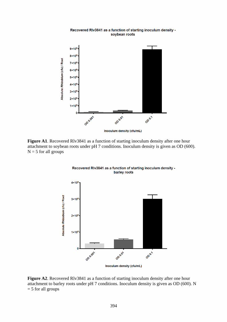
394
Figure A1. Recovered Rlv3841 as a function of starting inoculum density after one hour
attachment to soybean roots under pH 7 conditions. Inoculum density is given as OD (600).
N = 5 for all groups
Figure A2. Recovered Rlv3841 as a function of starting inoculum density after one hour
attachment to barley roots under pH 7 conditions. Inoculum density is given as OD (600). N
= 5 for all groups
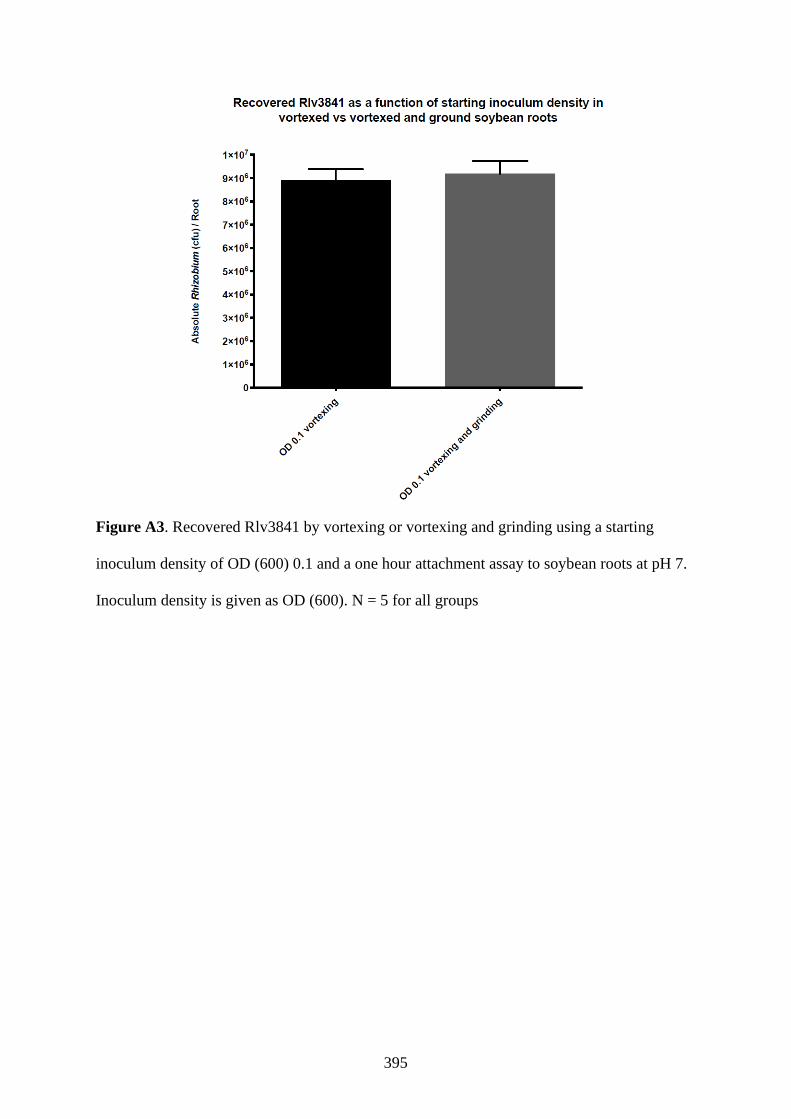
395
Figure A3. Recovered Rlv3841 by vortexing or vortexing and grinding using a starting
inoculum density of OD (600) 0.1 and a one hour attachment assay to soybean roots at pH 7.
Inoculum density is given as OD (600). N = 5 for all groups

396
Figure A4. Recovered Rlv3841 by vortexing or vortexing and grinding using a starting
inoculum density of OD (600) 0.1 and a one hour attachment assay to barley roots at pH 7.
Inoculum density is given as OD (600). N = 5 for all groups

397
Table A10 – Genes ES/DE in primary root attachment to pea, soybean and barley roots which were also NE in the input library (not
shown).
Gene Description G21 S21 G1 S1 VMM TY Rhi Pea
7.0
Soy
7.0
Bar
7.0
Col IT Nod
pRL110043 putative transmembrane transporter protein NE NE NE NE NE NE NE DE DE DE NE NE NE
RL2643
(dksA2)
putative DnaK suppressor protein NE NE NE NE NE NE NE DE DE DE NE NE NE
RL4145 putative LacI family transcriptional regulator
(repressor)
NE NE NE NE NE NE NE DE DE DE NE NE NE
RL4381 putative cell surface protein NE NE NE NE NE NE NE DE DE DE NE NE NE
RL0551 (hslO) putative Hsp33-like chaperonin NE NE NE NE NE NE DE DE DE DE DE NE NE
pRL100112 putative dehalogenase/hydrolase NE NE NE NE NE NE DE DE DE DE DE NE NE
RL0617 putative Maf septum inhibitor protein NE NE NE NE NE NE DE DE DE DE DE NE NE
RL4362 putative cobalamin synthesis protein NE NE NE NE NE NE NE DE DE DE NE DE DE
RL4363 (dacC) putative penicillin-binding protein precursor NE NE NE NE NE NE NE DE DE DE NE DE DE
RL2694 (gor) putative glutathione reductase NE NE NE NE NE NE NE DE DE DE NE ES ES
RL2212 (clpS1) putative ATP-dependent Clp protease adaptor protein NE NE NE NE NE NE NE DE DE DE NE DE DE
RL3322 (pfp) putative pyrophosphate--fructose 6-phosphate 1-
phosphotransferase
NE NE NE NE NE NE NE DE DE DE NE DE NE
RL1013 conserved hypothetical protein NE NE NE NE NE NE DE DE DE DE DE ES NE
RL1478 (amn) putative AMP nucleosidase NE NE NE NE NE NE NE DE DE DE NE ES ES
RL1600 (ppx) putative exopolyphosphatase NE NE NE NE NE NE NE DE DE DE DE ES ES
RL2284 (hfq) putative host factor protein NE NE NE NE NE NE NE DE DE DE ES NE NE
RL2513 (tpiA) putative triosephosphate isomerase NE NE NE NE NE NE DE DE DE DE DE NE NE
RL2637 (recA) putative recombinase NE NE NE NE NE NE DE DE DE DE DE ES ES
RL3987 conserved hypothetical protein NE NE NE NE NE NE DE DE DE DE DE DE DE
RL3988 conserved hypothetical protein NE NE NE NE NE NE DE DE DE DE DE DE DE
RL3989 (ruvA) putative Holliday junction DNA helicase RuvA NE NE NE NE NE NE DE DE DE DE DE DE DE
RL3990 (ruvB) putative Holliday junction DNA helicase RuvB NE NE NE NE NE NE DE DE DE DE DE DE DE

398
RL4065
conserved hypothetical protein
NE
NE
NE
NE
NE
NE
DE
DE
DE
DE
DE
NE
NE
RL2776 putative AsnC family transcriptional regulator NE NE NE NE ES NE NE DE DE DE DE NE NE
pRL110044 conserved hypothetical protein NE NE NE NE DE NE DE DE DE DE DE NE NE
pRL110617
(nadA)
quinolinate synthetase A DE DE NE DE NE NE NE DE DE DE NE NE NE
pRL110618 conserved hypothetical protein DE DE NE DE NE NE NE DE DE DE NE NE NE
pRL70055 hypothetical protein DE DE DE DE NE NE DE DE DE DE DE ES DE
pRL90143 putative transposase DE DE DE DE NE NE DE DE DE DE DE DE DE
RL0160 (polA) putative DNA polymerase I DE DE NE DE NE NE NE DE DE DE NE NE DE
RL0187 putative permease component of ABC transporter PepT DE DE DE DE NE NE NE DE DE DE NE ES ES
RL0430 (def1) putative peptide deformylase (polypeptide
deformylase)
DE DE DE DE DE NE DE DE DE DE DE ES DE
RL0431 putative plasmid stability protein DE DE DE DE NE NE DE DE DE DE DE ES DE
RL0432 putative plasmid stability protein DE DE DE DE NE NE DE DE DE DE DE ES DE
RL4546 conserved hypothetical protein ES DE DE NE NE NE NE DE DE DE NE NE NE
RL4582 (fbpC) putative ATP-binding component of ABC transporter
Unclass ferric cations transporter
DE DE DE DE NE NE DE DE DE DE DE DE DE
RL4583 (fbpB) putative permease component of ABC transporter
Unclass ferric cations transporter
DE DE DE DE NE NE DE DE DE DE DE DE DE
RL4584 (fbpA) putative SBP of ABC transporter Unclass ferric cations
transporter
DE DE DE DE NE NE DE DE DE DE NE NE NE
RL4675 hypothetical protein ES ES ES ES NE NE NE ES ES ES NE ES ES
RL0563 conserved hypothetical protein DE DE DE DE NE NE NE DE DE DE NE NE NE
RL0564 (mcpE) putative methyl-accepting chemotaxis protein DE DE DE DE NE NE NE DE DE DE NE NE NE
RL0855 (gshA) putative ADmma-glutamylcysteine synthetase
precursor
DE NE NE NE NE NE DE DE DE DE DE ES ES
RL0956 putative para-hydroxybenzoate--polyprenyltransferase NE DE NE DE NE NE AD DE DE DE AD NE NE
RL0957 conserved hypothetical protein NE DE NE DE NE NE DE DE DE DE ES ES ES
RL1507 conserved hypothetical protein NE NE NE DE NE NE DE DE DE DE DE NE DE
RL1508 putative transmembrane protein NE NE NE DE NE NE DE DE DE DE NE ES ES
RL1572 putative DNA polymerase III subunit DE DE DE DE NE NE DE DE DE ES DE ES ES

399
RL1617 putative transmembrane protein ES ES ES ES NE NE NE DE DE DE NE DE DE
RL1618 putative MarR family transcriptional regulator DE DE DE DE NE NE DE DE DE DE NE DE DE
RL2087 putative leucyl/phenylalanyl-tRNA--protein transferase DE DE DE DE NE NE DE DE DE DE DE DE NE
RL2213 (clpA) putative ATP-dependent Clp protease ATP-binding
subunit
NE NE NE DE NE NE NE DE DE DE NE ES NE
RL2405 putative peptidyl-prolyl cis-trans isomerase B
(cyclophilin-related protein)
DE NE NE NE NE NE NE DE DE DE NE ES DE
RL2406 (queA) putative S-adenosylmethionine:tRNA
ribosyltransferase-isomerase
DE NE NE NE NE NE NE DE DE DE NE ES DE
RL2407 (tgt) putative queuine tRNA-ribosyltransferase DE NE NE NE NE NE NE DE DE DE NE ES DE
RL2473 (metG) putative methionyl-tRNA synthetase DE DE DE NE NE NE NE DE DE DE NE ES NE
RL2474 putative transmembrane protein DE DE DE DE NE NE DE DE DE DE NE NE NE
RL2478 putative outer membrane protein NE DE NE DE NE NE NE DE DE DE NE NE NE
RL2514 conserved hypothetical protein DE NE NE NE NE NE DE DE DE DE DE NE NE
RL2638 (rbsK3) putative ribokinase ES ES NE DE NE NE DE DE DE DE DE NE NE
RL2652 (fdxB2) putative ferredoxin, 2Fe-2S DE DE DE DE NE NE NE DE DE DE NE NE NE
RL2653 putative HPt (Histidine Phosphotransfer) domain
protein of two-component system (multistage
phosphorelay can result)
DE DE DE DE NE NE NE DE DE DE NE NE NE
RL3294 conserved hypothetical protein DE DE DE DE NE NE DE DE DE DE DE NE NE
RL3596 conserved hypothetical protein DE DE NE NE NE NE DE DE DE DE NE DE NE
RL3597 putative DEAD-box ATP-dependent RNA helicase
protein
DE DE NE NE NE NE DE DE DE DE DE DE NE
RL4017 (rpmE) putative 50S ribosomal protein L31 DE DE DE DE NE NE NE DE DE DE NE DE DE
RL4073 (hss) putative homospermidine synthase DE DE NE DE NE NE DE DE DE ES NE ES ES
RL4296 (argJ) putative arginine biosynthesis bifunctional protein DE DE DE DE NE NE DE DE DE DE DE ES ES
RL3677 (lspL) putative UDP-glucuronate 5'-epimerase NE NE NE DE NE NE NE DE DE DE DE ES ES
INSeq experiments are coded as follows: G21 = 21% oxygen, 10 mM glucose. S21 = 21 % oxygen, 20 mM succinate. G1 = 1%
oxygen, 10 mM glucose. S1 = 1% oxygen, 20 mM succinate. VMM = Vincents media. TY = Tryptone yeast media. Rhizo =
rhizosphere. Pea 7.0 = pea root attachment, pH 7.0. Soy 7.0 = soybean root attachment, pH 7.0. Bar 7.0 = barley root attachment, pH
7.0. Colon = 5 day pea root colonisation. IT = pea infection thread. Nod = pea nodulation. Thick red line = pleiotropy filter cutoff, as
described in 5.2.8

400
Table A11 – Genes ES/DE in primary root attachment to pea only (data for primary root attachment to soybean and barley not shown)
which were also NE in the input library (not shown).
Gene Description G21 S21 G1 S1 VMM TY Rhi Pea
7.0
Col IT Nod
RL4704 putative glyoxalase NE NE NE NE NE NE NE DE NE NE NE
RL3752 (pssA) putative transferase involved in exopolysaccharide
biosynthesis
NE NE NE NE NE NE NE DE NE NE NE
RL3226 conserved hypothetical protein NE NE NE NE NE NE NE DE NE NE NE
RL2587 conserved hypothetical protein NE NE NE NE NE NE NE DE NE NE NE
RL1661 (pssA2) putative transmembrane suADr transferase protein NE NE NE NE NE NE NE DE NE NE NE
RL2044 (scpA) putative chromosome segreADtion and condensation protein
A
NE NE NE NE NE NE NE DE NE NE NE
pRL120518 putative TetR family transcriptional regulator NE NE NE NE NE NE NE DE NE NE NE
pRL100174 hypothetical protein, no known homology NE NE NE NE NE NE NE DE NE NE NE
RL1504 conserved hypothetical protein NE NE NE NE NE NE NE DE DE NE NE
RL1106 (pspA) putative (phage shock protein A) PspA family regulator by
protein-protein interactions
NE NE NE NE NE NE DE DE NE NE NE
pRL100242 conserved hypothetical protein NE NE NE NE NE NE DE DE DE NE NE
pRL100053 putative transmembrane protein NE NE NE NE NE NE NE DE ES DE DE
RL0141 (cycM) putative cytochrome c NE NE NE NE NE NE NE DE NE ES ES
RL2588 (tyrS1) putative tyrosyl-tRNA synthetase NE NE NE NE NE NE NE DE NE ES ES
RL0417 (ihfB) putative integration host factor beta subunit NE NE NE NE ES NE ES DE NE NE NE
RL1105 putative TetR family transcriptional regulator NE NE NE NE ES NE DE DE NE NE ES
RL1585 (moaD) putative molybdopterin converting factor subunit D NE NE NE NE ES NE NE DE NE NE NE
RL1628 hypothetical protein NE NE NE NE ES NE NE DE ES ES NE
pRL100161
(nifD)
nitrogenase molybdenum-iron protein alpha chain NifD DE DE DE DE NE NE DE DE DE ES DE
pRL100388 putative LacI family transcriptional regulator (repressor) NE DE NE DE NE NE NE DE NE NE NE
pRL110072 putative GntR family transcriptional regulator NE NE DE DE NE NE NE DE NE NE NE
pRL110073 putative GntR family transcriptional regulator DE DE DE DE NE NE NE DE NE NE NE

401
pRL110389 putative exopolysaccharide production protein DE DE DE DE NE NE NE ES NE ES ES
pRL70037 conserved hypothetical protein DE DE DE DE NE NE DE DE DE NE NE
pRL70166 conserved hypothetical protein DE DE DE DE ES NE NE DE NE DE DE
pRL90040 pseudogene, conserved hypothetical protein DE DE DE DE NE NE DE DE DE 0 0
pRL90280 conserved hypothetical protein ES ES DE DE ES NE NE ES NE NE NE
RL1001 conserved hypothetical protein DE DE DE DE NE NE NE DE DE NE NE
RL1002 (bioY) putative transmembrane biotin biosynthesis protein DE DE DE DE NE NE DE DE DE ES ES
RL1003 putative permease component of ABC transporter Unclass DE DE DE DE NE NE DE DE DE ES ES
RL1004 putative ATP-binding component of ABC transporter Unclass DE DE DE DE NE NE DE DE DE ES ES
RL1389 conserved hypothetical protein DE DE NE DE NE NE NE DE NE ES NE
RL1390 hypothetical protein NE DE NE DE NE NE NE DE NE NE NE
RL2495 (moaC) putative molybdenum cofactor biosynthesis protein C ES ES DE ES ES NE NE DE NE NE NE
RL2696 putative acylphosphatase ES DE ES ES NE NE NE DE NE NE NE
RL2953 putative adenylate/guanylate cyclase DE NE NE NE NE NE NE DE NE NE NE
RL3872 (purC2) putative phosphoribosylamidoimidazole-succinocarboxamide
synthase
DE DE DE DE DE NE NE DE NE NE NE
RL4295 putative acetyltransferase DE DE DE DE NE NE NE DE NE ES ES
RL4304 putative transmembrane transporter protein NE ES DE NE NE NE NE DE NE NE NE
INSeq experiments are coded as follows: G21 = 21% oxygen, 10 mM glucose. S21 = 21 % oxygen, 20 mM succinate. G1 = 1%
oxygen, 10 mM glucose. S1 = 1% oxygen, 20 mM succinate. VMM = Vincents media. TY = Tryptone yeast media. Rhizo =
rhizosphere. Pea 7.0 = pea root attachment, pH 7.0. Colon = 5 day pea root colonisation. IT = pea infection thread. Nod = pea
nodulation. 0 = data not available. Thick red line = pleiotropy filter cutoff, as described in 5.2.8

402
Table A12 – Genes ES/DE in primary root attachment to soybean only (data for primary root attachment to pea and barley not shown)
which were also NE in the input library (not shown).
Gene Description G21 S21 G1 S1 VMM TY Rhi Soy
7.0
Col IT Nod
pRL110197 pseudogene NE NE NE NE NE NE NE DE NE 0 0
pRL110205
(hutH1)
putative histidine ammonia-lyase NE NE NE NE NE NE NE DE NE NE NE
pRL110206 (hutI) putative imidazolonepropionase NE NE NE NE NE NE NE DE NE NE NE
pRL110207 putative histidine degradation related amidohydrolase NE NE NE NE NE NE NE DE NE NE NE
pRL110213 putative permease component of ABC transporter
PAAT
NE NE NE NE NE NE NE DE NE NE NE
pRL110214 putative ATP-binding component of ABC transporter
PAAT
NE NE NE NE NE NE NE DE NE NE NE
pRL110393 hypothetical protein NE NE NE NE NE NE NE DE NE NE NE
pRL110394 putative transmembrane protein NE NE NE NE NE NE NE DE NE NE NE
pRL110395 putative succinoglycan biosynthesis protein NE NE NE NE NE NE NE DE NE NE NE
pRL110439 putative glycosyl transferase NE NE NE NE NE NE NE DE NE NE NE
pRL110440 putative acetyltransferase NE NE NE NE NE NE NE DE NE NE NE
pRL110453 conserved hypothetical protein NE NE NE NE NE NE NE DE NE NE NE
pRL110541 putative glycine degradation aminomethyltransferase NE NE NE NE NE NE NE DE NE NE NE
pRL110569 putative transmembrane protein NE NE NE NE NE NE NE DE NE NE NE
pRL110572 putative flp pilus assembly protein NE NE NE NE NE NE NE DE NE NE NE
pRL110573 putative transmembrane pilus component protein NE NE NE NE NE NE NE DE NE NE NE
pRL110574 putative transmembrane pilus component protein NE NE NE NE NE NE NE DE NE NE NE
pRL110575 putative transmembrane protein NE NE NE NE NE NE NE DE NE NE NE
pRL120056 (mcpR) putative methylation accepting chemotaxis protein NE NE NE NE NE NE NE DE NE NE NE
pRL120074 putative ATP-binding component of ABC transporter
PAAT (S. mel SBP homologue SMb21135 induced by
ADlactosamine, glucosamine)
NE NE NE NE NE NE NE DE NE NE NE
pRL120075 (stbC) putative plasmid stability protein StbC NE NE NE NE NE NE NE DE NE NE NE
pRL120487 putative salicylaldehyde dehydrogenase NE NE NE NE NE NE NE DE NE NE NE

403
pRL120488 putative 3-alpha-hydroxysteroid dehydrogenase NE NE NE NE NE NE NE DE NE NE NE
pRL120489 putative permease component of ABC transporter
HAAT
NE NE NE NE NE NE NE DE NE NE NE
pRL120490 putative permease component of ABC transporter
HAAT
NE NE NE NE NE NE NE DE NE NE NE
pRL120491 putative ATP-binding component of ABC transporter
HAAT
NE NE NE NE NE NE NE DE NE NE NE
pRL90242 putative nitrilotriacetate monooxygenase NE NE NE NE NE NE NE DE NE NE NE
RL0229 putative LysR family transcriptional regulator NE NE NE NE NE NE NE DE NE NE NE
RL0230 putative peptidase NE NE NE NE NE NE NE DE NE NE NE
RL0376 putative L-lysine dehydrogenase NE NE NE NE NE NE NE DE NE NE NE
RL0391 (int) putative transmembrane acyltransferase NE NE NE NE NE NE NE DE NE NE NE
RL0589 conserved hypothetical protien NE NE NE NE NE NE NE DE NE NE NE
RL0590 putative short-chain dehydrogenase/reductase NE NE NE NE NE NE NE DE NE NE NE
RL0664 putative transmembrane acyltransferase (lipid
metabolism)
NE NE NE NE NE NE NE DE NE NE NE
RL0762 putative XRE family (HipB) transcriptional regulator NE NE NE NE NE NE NE DE NE NE NE
RL0909 conserved hypothetical protein NE NE NE NE NE NE NE DE NE NE NE
RL0910 (mutL) putative DNA mismatch repair protein NE NE NE NE NE NE NE DE NE NE NE
RL0964 putative Mg2+ transporter protein (uptake and/or
export, other ions can be transported), CorA family
NE NE NE NE NE NE NE DE NE NE NE
RL1015 putative polysaccharide deacetylase NE NE NE NE NE NE NE DE NE NE NE
RL1016 putative D-alanyl-D-alanine carboxypeptidase
(penicillin-binding protein)
NE NE NE NE NE NE NE DE NE NE NE
RL1033 conserved hypothetical protein NE NE NE NE NE NE NE DE NE NE NE
RL1034 conserved hypothetical protein NE NE NE NE NE NE NE DE NE NE NE
RL1038 conserved hypothetical protein NE NE NE NE NE NE NE DE NE NE NE
RL1039 conserved hypothetical protein NE NE NE NE NE NE NE DE NE NE NE
RL1108 putative transmembrane AMP-binding acyltransferase
family protein
NE NE NE NE NE NE NE DE NE NE NE
RL1154 conserved hypothetical exported protein NE NE NE NE NE NE NE DE NE NE NE
RL1155 putative glycosyl transferase NE NE NE NE NE NE NE DE NE NE NE

404
RL1162 putative two-component sensor/regulator;
transcriptional regulator
NE NE NE NE NE NE NE DE NE NE NE
RL1163 putative two-component sensor/regulator; histidine
kinase
NE NE NE NE NE NE NE DE NE NE NE
RL1549 putative transmembrane efflux protein NE NE NE NE NE NE NE DE NE NE NE
RL1644 conserved hypothetical exported protein NE NE NE NE NE NE NE DE NE NE NE
RL1918 putative exported aryl-sulfatase protein NE NE NE NE NE NE NE DE NE NE NE
RL1919 putative protein with alpha/beta hydrolase fold
(DUF900)
NE NE NE NE NE NE NE DE NE NE NE
RL2485 putative transmembrane transporter protein NE NE NE NE NE NE NE DE NE NE NE
RL2486 putative LysR family transcriptional regulator NE NE NE NE NE NE NE DE NE NE NE
RL2505 (deaD) putative cold-shock DEAD-box protein A (ATP-
dependent RNA helicase DEAD)
NE NE NE NE NE NE NE DE NE NE NE
RL2506 (cyaA12) putative adenylate cyclase 1 (ATP pyrophosphate-lyase
1) (adenylyl cyclase 1)
NE NE NE NE NE NE NE DE NE NE NE
RL2521 putative IAA acetyltransferase NE NE NE NE NE NE NE DE NE NE NE
RL2562 putative DoxD family transmembrane protein NE NE NE NE NE NE NE DE NE NE NE
RL2563 (pccB) putative propionyl-CoA carboxylase beta subunit NE NE NE NE NE NE NE DE NE NE NE
RL2570 conserved hypothetical exported protein NE NE NE NE NE NE NE DE NE NE NE
RL2571 conserved hypothetical protein NE NE NE NE NE NE NE DE NE NE NE
RL2572 (gcvT) putative aminomethyltransferase (glycine cleavage
system t protein)
NE NE NE NE NE NE NE DE NE NE NE
RL2764 putative transmembrane protein NE NE NE NE NE NE NE DE NE NE NE
RL2765 conserved hypothetical protein NE NE NE NE NE NE NE DE NE NE NE
RL2766 putative ArsR family transcriptional regulator NE NE NE NE NE NE NE DE NE NE NE
RL2767 (ssuB) putative ATP-binding component of ABC transporter
NitT aliphatic sulfonates
NE NE NE NE NE NE NE DE NE NE NE
RL2768 (ssuC) putative permease component of ABC transporter NitT
aliphatic sulfonates
NE NE NE NE NE NE NE DE NE NE NE
RL2779 putative two-component sensor/regulator; histidine
kinase
NE NE NE NE NE NE NE DE NE NE NE
RL2875 putative ion efflux protein NE NE NE NE NE NE NE DE NE NE NE
RL2876 putative transmembrane protein NE NE NE NE NE NE NE DE NE NE NE

405
RL2877 putative transmembrane transporter protein NE NE NE NE NE NE NE DE NE NE NE
RL2900 (dhaK1) putative dihydroxyacetone kinase NE NE NE NE NE NE NE DE NE NE NE
RL2901 (dhaL1) putative PTS-dependent dihydroxyacetone kinase,
ADP-binding subunit
NE NE NE NE NE NE NE DE NE NE NE
RL2902 putative PTS-dependent dihydroxyacetone kinase,
phosphotransferase subunit
NE NE NE NE NE NE NE DE NE NE NE
RL2903 (ptsH) putative multiphosphoryl transfer/phosphocarrier
protein
NE NE NE NE NE NE NE DE NE NE NE
RL2904 (ptsI) putative phosphoenolpyruvate-protein
phosphotransferase
NE NE NE NE NE NE NE DE NE NE NE
RL2999 putative glyoxylase NE NE NE NE NE NE NE DE NE NE NE
RL3000 conserved hypothetical protein NE NE NE NE NE NE NE DE NE NE NE
RL3006 (rhbB) putative L-2,4-diaminobutyrate decarboxylase NE NE NE NE NE NE NE DE NE NE NE
RL3146 putative LysR family transcriptional regulator NE NE NE NE NE NE NE DE NE NE NE
RL3147 putative transmembrane transporter protein NE NE NE NE NE NE NE DE NE NE NE
RL3149 putative adenylate cyclase NE NE NE NE NE NE NE DE NE NE NE
RL3192 putative ATP-binding component of ABC transporter
Unclass
NE NE NE NE NE NE NE DE NE NE NE
RL3193 putative permease component of ABC transporter
Unclass
NE NE NE NE NE NE NE DE NE NE NE
RL3262 (mocA) putative MocA family oxidoreductase NE NE NE NE NE NE NE DE NE NE NE
RL3263 putative LysR family transcriptional regulator NE NE NE NE NE NE NE DE NE NE NE
RL3264 putative transmembrane protein NE NE NE NE NE NE NE DE NE NE NE
RL3265 putative AraC family transcriptional regulator
(activator)
NE NE NE NE NE NE NE DE NE NE NE
RL3268 (flaH) putative flagellin protein NE NE NE NE NE NE NE DE NE NE NE
RL4120 (nodN2) putative nodulation protein NodN2 NE NE NE NE NE NE NE DE NE NE NE
RL4189 putative LysR family transcriptional regulator NE NE NE NE NE NE NE DE NE NE NE
RL4219 putative DeoR family transcriptional regulator
(repressor) of sorbitol/mannitol operon
NE NE NE NE NE NE NE DE NE NE NE
RL4220 (exoD) putative exopolysaccharide biosynthesis protein NE NE NE NE NE NE NE DE NE NE NE
RL4604 putative GntR family transcriptional regulator NE NE NE NE NE NE NE DE NE NE NE

406
pRL100385 (ecfM) putative RNA polymerase ECF sigma factor, family
ECF20/ECF01
NE NE NE NE NE NE DE DE DE NE NE
pRL100183 (nodF) nodulation protein NodF acyl carrier protein (ACP)
used in Nod factor synthesis
NE NE NE NE NE NE NE DE NE DE DE
pRL110045 conserved hypothetical protein NE NE NE NE NE NE DE DE DE NE NE
pRL110046 putative FNR/CRP family transcriptional regulator NE NE NE NE NE NE DE DE DE ES ES
pRL110048
(redAh)
UDP-N-acetylglucosamine-N-acetylmuramyl-
(pentapeptide) pyrophosphoryl-undecaprenol N-
acetylglucosamine transferase
NE NE NE NE NE NE DE DE NE NE DE
pRL110049 (redB) putative glycosyltransferase NE NE NE NE NE NE DE DE NE ES DE
pRL110283 putative ArsR family transcriptional regulator NE NE NE NE NE NE NE DE NE ES DE
pRL110441 (thiD) putative phosphomethylpyrimidine kinase NE NE NE NE NE NE DE DE NE NE NE
pRL110560
(soxB2)
putative sarcosine oxidase beta subunit NE NE NE NE NE NE NE DE DE NE NE
pRL110561
(soxD2)
putative sarcosine oxidase delta subunit NE NE NE NE NE NE NE DE DE NE NE
pRL110570 hypothetical exported protein NE NE NE NE NE NE DE DE NE NE NE
pRL110571 conserved hypothetical protein NE NE NE NE NE NE DE DE NE NE NE
pRL120057 putative plasmid stability protein NE NE NE NE NE NE ES DE DE NE NE
pRL120058 hypothetical protein NE NE NE NE NE NE NE DE DE NE NE
pRL120306 putative ATP-binding component of ABC transporter
NitT
NE NE NE NE NE NE NE DE DE NE NE
pRL80133 (trbLp8) putative conjuADl transfer protein TrbL NE NE NE NE NE NE ES DE ES NE NE
pRL80134 (trbHp8) putative conjuADl transfer protein TrbH NE NE NE NE NE NE ES DE ES NE NE
pRL80135 (trbGp8) putative conjuADl transfer protein TrbG NE NE NE NE NE NE ES DE ES NE NE
pRL90243 putative acyl-CoA dehydrogenase NE NE NE NE NE NE NE DE DE NE NE
pRL90298 conserved hypothetical protein NE NE NE NE NE NE NE DE DE AD NE
RL0338 (gshB) putative glutathione synthetase NE NE NE NE NE NE ES DE NE ES ES
RL0397 (mur) putative FUR-like transcriptional regulator, iron
response regulator
NE NE NE NE NE NE NE DE NE DE DE
RL0482A putative short-chain dehydrogenase/oxidoreductase NE NE NE NE NE NE DE DE NE NE NE
RL0685 (hemAT) putative chemoreceptor protein NE NE NE NE NE NE NE DE NE ES ES
RL0686 (cheX1) putative chemotaxis related CheX protein NE NE NE NE NE NE NE DE NE ES ES

407
RL0687 (cheY1) putative two-component sensor/regulator; chemotaxis
transcriptional regulator CheY
NE NE NE NE NE NE NE DE NE ES ES
RL0962 putative ring hydroxylating dioxygenase subunit NE NE NE NE NE NE NE DE NE ES ES
RL0963 putative transmembrane/surface protein BA14K-like
immuno reactive protein
NE NE NE NE NE NE NE DE DE NE NE
RL1040 putative LysR family transcriptional regulator NE NE NE NE NE NE NE DE DE NE NE
RL1089 conserved hypothetical protein NE NE NE NE NE NE NE DE NE AD NE
RL1090 putative transmembrane protein NE NE NE NE NE NE NE DE NE AD NE
RL1215 putative transmembrane protein NE NE NE NE AD NE NE DE NE NE NE
RL1438 (cycK) putative cytochrome c-type biogenesis protein NE NE NE NE NE NE NE DE NE ES ES
RL1965 (aldR) putative AsnC family transcriptional regulator NE NE NE NE NE NE DE DE NE NE NE
RL1966 (aldA) putative alanine dehydrogenase NE NE NE NE NE NE DE DE NE NE NE
RL2051 (pcm) putative protein-L-isoaspartate O-methyltransferase NE NE NE NE NE NE NE DE NE NE AD
RL2102 (cspA5) putative cold shock protein NE NE NE NE NE NE NE DE NE DE NE
RL2303 (ccdA) putative cytochrome c-type biogenesis protein NE NE NE NE NE NE ES DE DE ES ES
RL2305 hypothetical protein NE NE NE NE NE NE NE DE DE NE NE
RL2394 putative carbohydrate kinase protein NE NE NE NE NE NE DE DE NE NE NE
RL2507 conserved hypothetical exported protein NE NE NE NE NE NE NE DE DE NE NE
RL2508 (gltA2) putative citrate synthase II NE NE NE NE NE NE NE DE DE NE NE
RL2564 conserved hypothetical exported protein NE NE NE NE NE NE NE DE NE NE NE
RL2569 conserved hypothetical protein NE NE NE NE NE NE NE DE NE DE NE
RL2608 (purQ) putative phosphoribosylformylglycinamidine synthase
I
NE NE NE NE ES NE NE DE NE DE NE
RL2609 conserved hypothetical exported protein NE NE NE NE NE NE NE DE NE DE NE
RL2662 putative racemase/isomerase NE NE NE NE NE NE NE DE DE NE NE
RL2663 putative permease component of ABC transporter
Unclass
NE NE NE NE NE NE NE DE DE NE NE
RL2664 (lpxH) putative UDP-2,3-diacylglucosamine hydrolase NE NE NE NE NE NE NE DE DE NE NE
RL2780 (dgkA) putative transmembrane diacylglycerol kinase NE NE NE NE NE NE NE DE NE NE NE
RL2819 hypothetical protein NE NE NE NE NE NE DE DE NE AD NE
RL2820 hypothetical protein NE NE NE NE NE NE DE DE DE NE NE

408
RL2827 conserved hypothetical protein NE NE NE NE NE NE NE DE NE AD NE
RL2933 putative transmembrane protein NE NE NE NE NE NE NE DE DE NE NE
RL2934 conserved hypothetical exported protein NE NE NE NE NE NE NE DE DE NE NE
RL3148 putative ArsR family transcriptional regulator NE NE NE NE NE NE NE DE NE NE AD
RL3196 putative MarR family transcriptional regulator NE NE NE NE NE NE NE DE NE NE NE
RL3198 conserved hypothetical protein NE NE NE NE NE NE NE DE DE NE NE
RL3560 (map2) putative methionine aminopeptidase NE NE NE NE NE NE NE DE DE NE NE
RL3561 putative bacterial luciferase family protein NE NE NE NE NE NE NE DE DE NE NE
RL3592 conserved hypothetical exported protein NE NE NE NE NE NE DE DE DE NE NE
RL4025 (suhB) putative inositol-1-monophosphatase NE NE NE NE ES NE NE DE NE ES NE
RL4030 (cheW3) putative chemotaxis protein NE NE NE NE NE NE NE DE NE AD NE
RL4062 putative amidohydrolase NE NE NE NE NE NE NE DE NE ES ES
RL4063 conserved hypothetical protein NE NE NE NE NE NE NE DE NE NE NE
RL4075 putative 5'-nucleotidase NE NE NE NE NE NE NE DE NE NE NE
RL4162 (eda) putative 2-dehydro-3-deoxyphosphogluconate aldolase NE NE NE NE ES NE NE ES NE NE NE
RL4210 (cysZ) putative cysteine biosynthesis protein NE NE NE NE NE NE NE DE NE DE NE
RL4333 putative phospholipid/glycerol acyltransferase NE NE NE NE NE NE DE DE NE ES ES
RL4404 (pssA3) putative exopolysaccharide production protein NE NE NE NE NE NE NE DE NE NE NE
RL4497 putative transmembrane protein NE NE NE NE NE NE DE DE NE NE DE
RL4538 (ccmB) putative permease component of ABC transporter
Export cytochrome c binding export protein
NE NE NE NE NE NE DE DE DE ES ES
RL4539 (cycZ) putative permease component of ABC transporter
Export heme exporter protein c (cytochrome c-type
biogenesis protein)
NE NE NE NE NE NE DE DE DE ES ES
RL4597 conserved hypothetical protein NE NE NE NE NE NE NE DE NE NE NE
RL4602 (naAD) putative N-acetylglucosamine-6-phosphate deacetylase NE NE NE NE NE NE NE DE NE ES ES
RL4603 putative aminotransferase NE NE NE NE NE NE NE DE NE ES ES
RL4618 conserved hypothetical protein NE NE NE NE NE NE DE DE DE NE NE
RL4690 conserved hypothetical protein DE ES DE ES NE NE NE DE DE NE NE
RL4411 putative transmembrane protein DE DE DE DE NE NE DE DE DE AD NE

409
RL4413 putative ATP-binding component of ABC transporter
PepT
DE DE DE DE NE NE AD DE AD NE NE
RL4493 (gpsA) putative glycerol-3-phosphate dehydrogenase
[NAD(P)+]
NE DE NE NE ES NE NE DE NE ES DE
pRL100005 conserved hypothetical protein with PIN domain ES NE ES DE NE NE NE DE NE NE NE
pRL100006 conserved hypothetical protein ES NE ES DE NE NE NE DE NE NE NE
pRL110012 putative penicillin-binding family protein DE NE NE DE NE NE NE DE NE NE NE
pRL120584 putative epimerase DE DE DE DE NE NE NE DE DE NE NE
pRL90042 pseudogene, conserved hypothetical protein DE DE NE NE NE NE NE DE NE 0 0
pRL90043 putative permease component of ABC transporter
Export
DE DE DE DE NE NE NE DE NE NE NE
pRL90066 putative transmembrane protein DE DE DE DE NE NE ES DE NE NE NE
pRL90067 conserved hypothetical exported protein DE DE DE DE NE NE AD DE NE NE NE
pRL90068 putative transmembrane protein DE DE DE DE NE NE DE DE NE NE NE
pRL90274 putative transmembrane hydrogenase-related protein NE NE NE DE NE NE NE DE NE NE NE
pRL90276 putative ATP-binding :permease (ABC:IMP)
component of ABC transporter Export cytochrome bd-
related
NE ES ES ES NE NE NE DE NE NE NE
pRL90277 putative ATP-binding :permease (ABC:IMP)
component of ABC transporter Export cytochrome bd-
related
ES ES ES ES NE NE ES DE NE NE NE
RL0501 putative orotate phosphoribosyltransferase DE NE NE NE DE NE NE ES NE NE DE
RL0887 putative plasmid stability protein DE DE DE DE NE NE DE DE DE NE NE
RL0888 conserved hypothetical protein DE DE DE DE NE NE DE DE DE NE NE
RL2208 putative hydrolase NE DE NE NE NE NE DE DE NE AD AD
RL2509 (citZ) putative citrate synthase I NE DE NE NE NE NE NE DE NE NE NE
RL2538 (pip2) putative proline iminopeptidase DE DE DE DE NE NE NE DE NE NE NE
RL2614 conserved hypothetical exported protein DE DE DE DE NE NE NE DE NE NE NE
RL2639 putative indigoidine A related protein ES NE NE DE NE NE NE DE AD NE NE
RL3005 hypothetical protein ES NE ES NE NE NE NE DE NE NE NE
RL3295 (recN) putative DNA repair protein DE DE DE DE NE NE DE DE DE NE NE

410
RL4031 (mcrA) putative sensory transducer methyl-accepting
chemotaxis protein
DE NE NE DE NE NE NE DE NE AD NE
RL4032 (mcrB) putative sensory transducer methyl-acccepting
chemotaxis protein
DE NE NE DE NE NE NE DE NE AD NE
RL4117 (glAD) putative glycogen synthase DE NE DE NE DE NE NE DE NE ES ES
INSeq experiments are coded as follows: G21 = 21% oxygen, 10 mM glucose. S21 = 21 % oxygen, 20 mM succinate. G1 = 1%
oxygen, 10 mM glucose. S1 = 1% oxygen, 20 mM succinate. VMM = Vincents media. TY = Tryptone yeast media. Rhizo =
rhizosphere. Soy 7.0 = soybean root attachment, pH 7.0. Colon = 5 day pea root colonisation. IT = pea infection thread. Nod = pea
nodulation. 0 = data not available. Thick red line = pleiotropy filter cutoff, as described in 5.2.8

411
Table A13 – Genes ES/DE in primary root attachment to barley only (data for primary root attachment to pea and soybean not shown)
which were also NE in the input library (not shown).
Gene Description G21 S21 G1 S1 VMM TY Rhi Bar
7.0
Col IT Nod
RL0159 putative MarR family transcriptional regulator NE NE NE NE NE NE NE DE NE NE NE
RL0562 conserved hypothetical protein NE NE NE NE NE NE NE DE NE NE NE
RL0592 putative thiolase NE NE NE NE NE NE NE DE NE NE NE
RL0593 conserved hypothetical protein NE NE NE NE NE NE NE DE NE NE NE
RL0597 conserved hypothetical protein NE NE NE NE NE NE NE DE NE NE NE
RL1647 (celB) putative cellulose synthase protein NE NE NE NE NE NE NE DE NE NE NE
RL1859 conserved hypothetical protein NE NE NE NE NE NE NE DE NE NE NE
RL1941 hypothetical protein NE NE NE NE NE NE NE DE NE NE NE
RL2038 (xthA) putative exodeoxyribonuclease III NE NE NE NE NE NE NE DE NE NE NE
RL2039 putative HesB family protein NE NE NE NE NE NE NE DE NE NE NE
RL2525 conserved hypothetical protein NE NE NE NE NE NE NE DE NE NE NE
RL2655 hypothetical protein NE NE NE NE NE NE NE DE NE NE NE
RL2656 putative D-alanyl-D-alanine carboxypeptidase NE NE NE NE NE NE NE DE NE NE NE
RL2657 putative DEEEF/ADF sensory box protein NE NE NE NE NE NE NE DE NE NE NE
RL2856 (msiA) putative transmembrane lysine/arginine export protein
family 97% id to MsiA of Mesorhizobium tianshanense
induced by canavanine from seed exudates. Exports
canavanine (an anti-metabolite) out of cell
NE NE NE NE NE NE NE DE NE NE NE
RL2863 conserved hypothetical protein NE NE NE NE NE NE NE DE NE NE NE
RL2864 hypothetical protein NE NE NE NE NE NE NE DE NE NE NE
RL2865 conserved hypothetical protein NE NE NE NE NE NE NE DE NE NE NE
RL3252 putative serine protease NE NE NE NE NE NE NE DE NE NE NE
RL3274 (prkA) putative PrkA family kinase NE NE NE NE NE NE NE DE NE NE NE
RL3278 hypothetical protein NE NE NE NE NE NE NE DE NE NE NE
RL3279 (pncA) putative pyrazinamidase/nicotinamidase NE NE NE NE NE NE NE DE NE NE NE
RL0401 putative universal stress protein NE NE NE NE NE NE NE DE NE NE DE

412
RL0561 putative AraC family transcriptional regulator (activator) NE NE NE NE NE NE NE DE DE NE NE
RL0726 conserved hypothetical exported protein NE NE NE NE NE NE NE DE NE DE DE
RL0816 conserved hypothetical protein NE NE NE NE NE NE DE DE DE ES ES
RL1805 putative transmembrane protein NE NE NE NE NE NE NE DE NE NE NE
RL1806 (degQ) putative protease DegQ precursor NE NE NE NE NE NE NE DE NE NE NE
RL2644 conserved hypothetical protein NE NE NE NE NE NE NE DE NE NE NE
RL2645 conserved hypothetical protein NE NE NE NE NE NE DE DE NE NE NE
RL2646 putative transmembrane protein NE NE NE NE NE NE DE DE NE NE NE
RL2647 conserved hypothetical exported protein NE NE NE NE NE NE DE DE NE NE NE
RL2857 (msiR) putative ArsR family transcriptional regulator MsiR,
regulates MsiA canavanine (found in seed exudate)
exporter
NE NE NE NE NE NE NE DE NE NE NE
RL2858 conserved hypothetical exported protein NE NE NE NE NE NE NE DE NE NE NE
RL3253 (hflC) putative transmembrane serine protease NE NE NE NE NE NE NE DE NE NE NE
RL3254 (hflK) putative transmembrane serine protease NE NE NE NE NE NE NE DE NE DE NE
RL3455 putative MarR family transcriptional regulator NE NE NE NE NE NE NE DE NE DE NE
RL3595 putative LacI family transcriptional regulator (repressor) NE NE NE NE NE NE NE DE NE AD NE
RL3950 putative phage-related protein NE NE NE NE NE NE AD DE NE NE NE
RL4599 putative lysyl-tRNA synthetase homolog NE NE NE DE NE NE DE DE NE ES ES
pRL120796 hypothetical exported protein DE DE NE NE NE NE NE DE NE NE NE
pRL70099 putative phage-derived invertase/resolvase ES DE ES DE NE NE NE DE NE NE NE
pRL90149 putative LuxR/GerE family transcriptional regulator, part
of two component response regulator?
ES DE ES ES NE NE NE DE DE NE NE
RL0566 conserved hypothetical protein DE DE DE DE NE NE NE DE NE NE NE
RL0821 putative O-antigen transporter NE DE NE NE NE NE DE DE DE ES ES
RL1167 putative TetR family transcriptional regulator DE DE DE DE NE NE NE DE NE DE NE
RL2036 putative outer membrane transport protein DE DE DE DE NE NE NE DE NE NE AD
RL2152 hypothetical protein DE DE ES DE NE NE NE DE NE ES ES
RL2526 putative oxidoreductase DE NE DE DE NE NE NE DE NE DE NE
RL2553 conserved hypothetical protein ES ES ES ES NE NE NE DE NE NE NE

413
RL2837 putative SBP of ABC transporter QAT? orphan
proline/glycine betaine
DE DE DE DE NE NE NE DE DE NE NE
RL3498 conserved hypothetical protein DE DE DE DE NE NE NE DE NE AD NE
INSeq experiments are coded as follows: G21 = 21% oxygen, 10 mM glucose. S21 = 21 % oxygen, 20 mM succinate. G1 = 1%
oxygen, 10 mM glucose. S1 = 1% oxygen, 20 mM succinate. VMM = Vincents media. TY = Tryptone yeast media. Rhizo =
rhizosphere. Bar 7.0 = barley root attachment, pH 7.0. Colon = 5 day pea root colonisation. IT = pea infection thread. Nod = pea
nodulation. Thick red line = pleiotropy filter cutoff, as described in 5.2.8

414
Table A14 – Genes ES/DE in primary root attachment to pea and soybean roots (data for primary root attachment to barley not shown)
which were also NE in the input library (not shown).
Gene Description G21 S21 G1 S1 VMM TY Rhi Pea
7.0
Soy
7.0
Col IT Nod
RL4083 conserved hypothetical protein NE NE NE NE NE NE NE DE DE NE NE NE
pRL100220 conserved hypothetical protein NE NE NE NE NE NE DE DE DE NE DE NE
RL1052 hypothetical protein NE NE NE NE NE NE NE DE DE NE NE NE
RL1371 putative transmembrane protein NE NE NE NE NE NE DE DE ES DE NE NE
RL2400 putative MarC (multiple antibiotic resistance) family
transmembrane protein, they may be transporters
NE NE NE NE NE NE NE DE DE NE AD DE
RL2520 putative transmembrane protein NE NE NE NE NE NE NE DE DE NE NE NE
pRL100149 conserved hypothetical protein NE NE NE NE ES NE NE ES ES NE NE NE
pRL110615 (nadC) putative nicotinate-nucleotide pyrophosphorylase
[carboxylating]
DE DE NE DE NE NE NE DE DE NE NE NE
pRL110616 (nadB) putative L-aspartate oxidase DE DE NE DE NE NE NE DE DE NE NE NE
RL0188 putative ATP-binding:ATP-binding (ABC:ABC)
componentof ABC transporter PepT (S. mel SBP
homologue SMc02832 induced by taurine, valine,
isoleucine)
DE DE DE DE NE NE NE DE DE NE NE NE
RL1060 putative ribosomal-protein-alanine acetyltransferase DE DE NE DE ES NE NE ES ES NE ES ES
RL1392 putative transmembrane protein NE DE NE DE NE NE NE DE DE NE NE NE
RL3462 conserved hypothetical protein DE DE DE DE NE NE DE DE DE DE NE DE
RL3763 putative rRNA methyltransferase DE DE DE DE NE NE DE DE DE DE NE NE
RL4074 conserved hypothetical protein DE DE NE DE NE NE DE DE DE NE NE NE
INSeq experiments are coded as follows: G21 = 21% oxygen, 10 mM glucose. S21 = 21 % oxygen, 20 mM succinate. G1 = 1%
oxygen, 10 mM glucose. S1 = 1% oxygen, 20 mM succinate. VMM = Vincents media. TY = Tryptone yeast media. Rhizo =
rhizosphere. Pea 7.0 = pea root attachment, pH 7.0. Soy 7.0 = soybean root attachment, pH 7.0. Colon = 5 day pea root colonisation. IT
= pea infection thread. Nod = pea nodulation. Thick red line = pleiotropy filter cutoff, as described in 5.2.8

415
Table A15 – Genes ES/DE in primary root attachment to pea and barley roots (data for primary root attachment to soybean not shown)
which were also NE in the input library (not shown).
Gene Description G21 S21 G1 S1 VMM TY Rhi Pea
7.0
Bar
7.0
Col IT Nod
RL2642 conserved hypothetical protein NE NE NE NE NE NE NE DE DE NE NE NE
RL2695 hypothetical protein NE NE NE NE NE NE NE DE DE NE NE NE
RL3277 putative transmembrane protein NE NE NE NE NE NE NE DE DE NE NE NE
pRL100405 putative transmembrane protein NE NE NE NE AD NE NE DE DE NE NE NE
pRL100406 (mctR) putative two-component sensor/regulator;
transcriptional regulator
NE NE NE NE AD NE NE DE DE NE NE NE
RL1340 (sodB) putative superoxide dismutase NE NE NE NE ES NE NE ES ES DE ES NE
RL1381 conserved hypothetical protein NE NE NE NE NE NE DE DE DE DE NE NE
RL2227 putative transmembrane protease NE NE NE NE NE NE DE DE DE DE DE NE
RL3453 putative two-component sensor/regulator;
histidine kinase
NE DE NE NE NE NE NE DE DE NE DE ES
RL4309 putative transmembrane protein NE NE NE NE NE NE NE DE DE NE ES ES
RL4382 putative filamentous hemagglutinin adherence
factor precursor
NE NE NE NE NE NE NE DE DE DE NE NE
pRL70100 hypothetical protein ES DE ES DE NE NE DE DE DE NE NE NE
RL0186 putative permease component of ABC
transporter PepT (S. mel SBP homologue
SMc02832 induced by taurine, valine,
isoleucine)
DE DE DE DE NE NE NE DE DE NE ES ES
RL4379 (hemA1) putative 5-aminolevulinate synthase NE NE NE NE ES ES DE DE DE DE ES ES
INSeq experiments are coded as follows: G21 = 21% oxygen, 10 mM glucose. S21 = 21 % oxygen, 20 mM succinate. G1 = 1%
oxygen, 10 mM glucose. S1 = 1% oxygen, 20 mM succinate. VMM = Vincents media. TY = Tryptone yeast media. Rhizo =
rhizosphere. Pea 7.0 = pea root attachment, pH 7.0. Soy 7.0 = soybean root attachment, pH 7.0. Colon = 5 day pea root colonisation. IT
= pea infection thread. Nod = pea nodulation. Thick red line = pleiotropy filter cutoff, as described in 5.2.8

416
Table A16 – Genes ES/DE in primary root attachment to soybean and barley roots (data for primary root attachment to pea not shown)
which were also NE in the input library (not shown).
Gene Description G21 S21 G1 S1 VMM TY Rhi Soy
7.0
Bar
7.0
Col IT Nod
RL0162 conserved hypothetical protein NE NE NE NE NE NE NE DE DE NE NE NE
RL0163 (phnN) putative phosphonate metabolism protein N, ATP-
binding
NE NE NE NE NE NE NE DE DE NE NE NE
RL0594 putative fatty oxidation complex subunit NE NE NE NE NE NE NE DE DE NE NE NE
RL0595 (cspA1) putative cold shock protein NE NE NE NE NE NE NE DE DE NE NE NE
RL0596 putative MarR family transcriptional regulator NE NE NE NE NE NE NE DE DE NE NE NE
RL1111 conserevd hypothetical protein NE NE NE NE NE NE NE DE DE NE NE NE
RL1645 conserved hypothetical protein NE NE NE NE NE NE NE DE DE NE NE NE
RL2118 putative transmembrane protein NE NE NE NE NE NE NE DE DE NE NE NE
RL2287 putative guanine/cytosine deaminase NE NE NE NE NE NE NE DE DE NE NE NE
RL2774 putative LysR family transcriptional regulator NE NE NE NE NE NE NE DE DE NE NE NE
RL2862 conserved hypothetical exported protein NE NE NE NE NE NE NE DE DE NE NE NE
RL3261 putative MocA family oxidoreductase NE NE NE NE NE NE NE DE DE NE NE NE
RL3266 conserevd hypothetical protein NE NE NE NE NE NE NE DE DE NE NE NE
RL3267 putative OmpA family outer membrane protein NE NE NE NE NE NE NE DE DE NE NE NE
RL4090 putative lysophospholipase NE NE NE NE NE NE NE DE DE NE NE NE
RL4357 (sirA) putative two-component sensor/regulator;
transcriptional regulator
NE NE NE NE NE NE NE DE DE NE NE NE
RL4380 conserved hypothetical protein NE NE NE NE NE NE NE DE DE NE NE NE
RL0032 putative phosphocarrier protein HPr for mannose NE NE NE NE NE NE DE ES DE NE ES ES
RL0398 putative acetyltransferase NE NE NE NE NE NE NE DE DE NE DE DE
RL0892 putative ribosomal large subunit pseudouridine
synthase B
NE NE NE NE NE NE DE DE DE DE DE DE
RL1012 (cbpA) putative curved DNA-binding protein NE NE NE NE NE NE DE DE DE DE NE NE
RL1026 (ctaE1) putative cytochrome c oxidase polypeptide III
(cytochrome aa3 subunit 3)
NE NE NE NE NE NE NE DE DE NE NE NE
RL1164 conserved hypothetical protein NE NE NE NE NE NE NE DE DE NE NE NE

417
RL1165 conserved hypothetical protein NE NE NE NE NE NE NE DE DE NE NE NE
RL1439 (cycL) putative cytochrome c-type biogenesis protein NE NE NE NE NE NE NE DE DE NE ES ES
RL1440 (degP1) putative serine protease NE NE NE NE NE NE NE DE DE NE ES ES
RL2285 (hflX) putative GTP-binding protein NE NE NE NE NE NE NE DE DE NE NE NE
RL2477 (dacF) putative penicillin-binding protein NE NE NE NE NE NE NE DE DE NE NE NE
RL2595 putative MutT/Nudix family protein
(phosphohydrolases)
NE NE NE NE NE NE NE DE DE NE NE NE
RL2777 conserved hypothetical protein NE NE NE NE NE NE NE DE DE NE NE NE
RL2778 putative exopolysaccharide biosynthesis protein NE NE NE NE NE NE NE DE DE NE NE NE
RL2828 putative XRE family (HipB) family transcriptional
regulator
NE NE NE NE NE NE NE ES ES DE ES ES
RL3320 putative signalling and peptidoglycan binding
protein
NE NE NE NE NE NE NE DE DE DE NE NE
RL3321 putative DnaJ family chaperone NE NE NE NE NE NE NE DE DE DE NE NE
RL3986 (ruvC) putative Holliday junction endodeoxyribonuclease
RuvC
NE NE NE NE NE NE NE DE DE NE DE NE
RL4018 putative ATP-binding:permease (ABC:IMP)
component of ABC transporter Export
NE NE NE NE NE NE NE DE DE NE DE DE
RL4354 (xerD) putative tyrosine recombinase NE NE NE NE NE NE NE DE DE NE DE ES
RL4356 conserved hypothetical protein NE NE NE NE NE NE NE DE DE NE DE DE
RL1091 conserved hypothetical protein DE NE NE NE NE NE NE DE DE DE NE NE
RL1092 conserved hypothetical protein DE NE NE NE NE NE NE DE DE DE NE NE
RL1093 putative beta-lactamase family protein DE NE NE NE NE NE NE DE DE DE NE NE
pRL100010 conserved hypothetical protein DE DE DE DE DE NE NE DE DE NE DE NE
pRL100011 hypothetical protein DE DE DE DE NE NE NE DE DE DE NE NE
pRL100275 putative LacI family transcriptional regulator
(repressor)
DE DE DE DE DE NE DE DE DE DE NE NE
pRL90144 putative exopolysaccharide biosynthesis-related
protein
DE DE DE DE NE NE NE DE DE NE NE NE
RL0153 putative penicillin-binding
transpeptidase/transglycosylase protein
DE NE NE NE NE NE DE DE DE DE NE NE
RL0161 putative cell division DNA translocase protein DE DE DE DE NE NE NE DE DE NE NE NE

418
RL0226 putative permease component of ABC transporter
PepT
DE DE DE DE NE NE DE DE DE NE ES ES
RL0227 putative permease component of ABC transporter
PepT
DE DE DE DE NE NE DE DE DE NE ES ES
RL0228 putative SBP of ABC transporter PepT DE DE DE DE NE NE DE DE DE NE ES ES
RL0423 putative transmembrane protein NE DE NE DE NE NE DE DE DE DE NE AD
RL0546 (phoU) putative phosphate uptake regulator PhoU,
unknown mechanism to regulate expression of
high-affinity ABC systems
NE NE DE DE NE NE NE DE DE NE ES ES
RL0547 (phoB) putative two-component sensor/regulator;
phosphate regulon transcriptional regulator PhoB
DE NE DE NE NE NE NE DE DE NE NE NE
RL0565 putative SBP of ABC transporter PAAT (S. mel
SBP homologue SMc02219 induced by valine,
homoserine, isoleucine)
DE DE DE DE NE NE NE DE DE NE NE NE
RL0618 conserved hypothetical protein DE DE DE DE NE NE DE DE DE DE NE DE
RL0818 putative lipopolysaccharide biosynthesis protein NE DE NE NE NE NE DE DE DE DE ES ES
RL0819 putative imidazole glycerol phosphate synthase
subunit
NE DE NE NE NE NE DE DE DE DE ES ES
RL0820 (hisH2) putative imidazole glycerol phosphate synthase
subunit (igp synthase glutamine amidotransferase
subunit)
NE DE NE NE NE NE DE DE DE DE ES ES
RL1024 (coxF) putative cox locus protein DE DE DE DE NE NE DE DE DE DE ES ES
RL1025 (ctaG) putative cytochrome c oxidase assembly protein DE DE DE DE NE NE NE DE DE NE NE NE
RL1166 putative ribonuclease-L-PSP family protein DE DE DE DE NE NE NE DE DE NE DE NE
RL1388 conserved hypothetical protein NE DE NE DE NE NE NE DE ES NE NE NE
RL1548 (radA) putative DNA repair protein RadA homologue DE DE NE NE NE NE NE DE DE NE NE NE
RL1642 putative two-component sensor/regulator;
transcriptional regulator
NE DE NE DE NE NE NE DE ES NE NE NE
RL1731 (rpmG) putative 50S ribosomal protein L33 DE DE DE DE NE NE NE DE DE NE DE DE
RL4430 putative 3-oxoacyl-[acyl-carrier-protein] reductase DE DE DE DE NE NE DE DE DE DE DE NE
RL2209 (cysE1) putative serine acetyltransferase DE DE DE ES DE NE DE DE DE DE ES DE
RL2210 conserved hypothetical protein DE DE DE ES DE NE DE DE DE DE NE DE
RL2236 (lexA) putative LexA repressor (SOS regulatory protein) NE DE NE NE NE NE NE DE DE NE NE NE
RL2237 conserved hypothetical protein DE DE DE DE NE NE NE DE DE NE NE NE

419
RL2510 conserved hypothetical protein ES DE DE DE NE NE DE DE ES DE NE NE
RL2818 (fnrN) putative FNR/CRP family transcriptional regulator,
100% id to VF39 FnrN
DE DE DE DE NE NE NE DE DE NE AD NE
RL3260 putative FAD-dependent
dehydrogenase/oxidoreductase
DE DE DE NE NE NE DE DE DE DE ES ES
RL3667 putative UDP-glucose 6-dehydrogenase DE DE DE DE NE NE DE DE DE DE ES DE
INSeq experiments are coded as follows: G21 = 21% oxygen, 10 mM glucose. S21 = 21 % oxygen, 20 mM succinate. G1 = 1%
oxygen, 10 mM glucose. S1 = 1% oxygen, 20 mM succinate. VMM = Vincents media. TY = Tryptone yeast media. Rhizo =
rhizosphere. Soy 7.0 = soybean root attachment, pH 7.0. Bar 7.0 = barley root attachment, pH 7.0. Colon = 5 day pea root colonisation.
IT = pea infection thread. Nod = pea nodulation. Thick red line = pleiotropy filter cutoff, as described in 5.2.8

420
Table A17 – Genes AD in primary root attachment to pea, soybean and barley roots which were also NE in the input library (not
shown).
Gene AD
classification
conditions
Description G21 G1 S21 S1 VMM TY Rhi Col IT Nod
RL4139 Pea, Bar, Soy putative transmembrane GGDEF/EAL sensory box
protein
NE NE NE NE NE NE NE NE GA GA
pRL70156
(trbGp7)
Pea putative conjugal transfer protein TrbG NE NE NE NE NE NE NE NE NE GD
RL3927 Soy conserved hypothetical protein NE NE NE NE NE NE NE NE NE NE
RL3928 Soy conserved hypothetical protein NE NE NE NE NE NE NE NE NE NE
RL3929 Soy conserved hypothetical protein NE NE NE NE NE NE NE NE NE NE
RL3930 Soy conserved hypothetical protein NE NE NE NE NE NE NE NE NE NE
RL4464 Soy putative lyase/mutase NE NE NE NE NE NE NE NE NE NE
pRL70057 Soy putative transposase NE NE NE NE NE NE NE NE NE NE
RL0645 Soy putative short-chain dehydrogenase/reductase NE NE NE NE NE NE NE NE NE NE
RL3092 Soy conserved hypothetical protein NE NE NE NE NE NE NE NE NE NE
RL3093 Soy conserved hypothetical protein NE NE NE NE NE NE NE NE NE NE
RL3861 Soy putative permease component of ABC transporter CUT1 NE NE NE NE NE NE NE NE NE NE
RL3885
(sitB)
Soy putative ATP-binding component of ABC transporter
MZT (S. mel SBP homologue SMc02509 induced by
manganese limitation)
NE NE NE NE NE NE NE NE NE NE
RL4003 Bar conserved hypothetical exported protein NE NE NE NE NE NE NE NE NE NE
RL3091 Soy, Bar putative transmembrane protein NE NE NE NE NE NE NE NE NE NE
RL4724
(rsmB)
Soy, Bar putative ribosomal RNA small subunit methyltransferase
B
NE NE NE NE NE NE NE NE NE NE
RL3090 Soy, Bar putative methyltransferase NE NE NE NE NE NE GA GA NE NE
pRL100223 Soy conserved hypothetical protein NE NE NE NE GA NE NE NE NE NE
pRL100362 Soy putative LysR family transcriptional regulator NE NE NE NE NE NE NE NE NE NE
pRL120012 Soy putative permease component of ABC transporter CUT1 NE NE NE NE NE NE NE NE NE NE
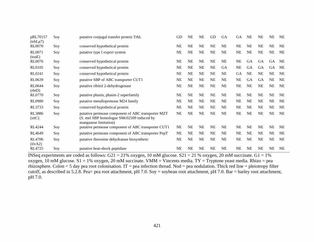
421
pRL70157
(trbLp7)
Soy putative conjugal transfer protein TrbL GD NE NE GD GA GA NE NE NE NE
RL0070 Soy conserved hypothetical protein NE NE NE NE NE NE NE NE NE NE
RL0071
(toaE)
Soy putative type I export system NE NE NE NE NE NE NE NE NE NE
RL0076 Soy conserved hypothetical protein NE NE NE NE NE NE GA GA GA NE
RL0105 Soy conserved hypothetical protein NE NE NE NE GA NE GA GA GA NE
RL0241 Soy conserved hypothetical protein NE NE NE NE NE GA NE NE NE NE
RL0639 Soy putative SBP of ABC transporter CUT1 NE NE NE NE NE NE GA GA NE NE
RL0644
(rbtD)
Soy putative ribitol 2-dehydrogenase NE NE NE NE NE NE NE NE NE NE
RL0770 Soy putative phasin, phasin-2 superfamily NE NE NE NE NE NE NE NE NE NE
RL0980 Soy putative metalloprotease M24 family NE NE NE NE NE NE NE NE NE NE
RL3733 Soy conserved hypothetical protein NE NE NE NE NE NE NE NE NE NE
RL3886
(sitC)
Soy putative permease component of ABC transporter MZT
(S. mel SBP homologue SMc02509 induced by
manganese limitation)
NE NE NE NE NE NE NE NE NE NE
RL4244 Soy putative permease component of ABC transporter CUT1 NE NE NE NE NE NE NE NE NE NE
RL4649 Soy putative permease component of ABC transporter PepT NE NE NE NE NE NE NE NE NE NE
RL4706
(ilvA2)
Soy putative threonine dehydratase biosynthetic NE NE NE NE NE NE NE NE NE NE
RL4725 Soy putative heat-shock peptidase NE NE NE NE NE NE NE NE NE NE
INSeq experiments are coded as follows: G21 = 21% oxygen, 10 mM glucose. S21 = 21 % oxygen, 20 mM succinate. G1 = 1%
oxygen, 10 mM glucose. S1 = 1% oxygen, 20 mM succinate. VMM = Vincents media. TY = Tryptone yeast media. Rhizo = pea
rhizosphere. Colon = 5 day pea root colonisation. IT = pea infection thread. Nod = pea nodulation. Thick red line = pleiotropy filter
cutoff, as described in 5.2.8. Pea= pea root attachment, pH 7.0. Soy = soybean root attachment, pH 7.0. Bar = barley root attachment,
pH 7.0.

422
Appendix 2. Supplementary material for Chapters 4, 5 and 6
Overview
Chapter 4 – Raw INSeq data: Appendix 2, Table 1
Chapter 5 – Raw INSeq data: Appendix 2, Table 2
These datasets have been deposited electronically in the Oxford University Research
Archive (ORA) and may be accessed in the repository using the following link:
https://doi.org/10.5287/bodleian:kZYnK2vQ2
Chapter 6 – Videos 1-5
These videos have been deposited electronically in ORA and may be accessed in the
repository using the following link:
https://doi.org/10.5287/bodleian:w4AxBzN4k