chapter one - covenant university repository
TRANSCRIPT
CHAPTER ONE
INTRODUCTION
1.1 Background
The term malaria originated from Medieval Italian: mala aria meaning "bad air"; and the
disease was formerly called ague or marsh fever due to its association with swamps. Malaria
is the most prevalent tropical disease in the world today. It has infected humans for over fifty
thousand years, and may have been a human pathogen for the entire history of our species. In
the tropical and subtropical regions, during the first half of the twentieth century, malaria
affected every walk of life so much so that one of the major problems in development of economy
was the problem of malaria (Sacks and Malaney, 2002).
Malaria is a vector-borne infectious disease caused by protozoan parasites of the genus
Plasmodium and is presently endemic in a broad band around the equator, in areas of the
Americas, many parts of Asia and much of Africa, however, it is in sub-Saharan Africa that
85– 90% of malaria fatalities occur (Hay et al., 2004). It is estimated that up to 124 million
people in Africa live in areas at risk of seasonal epidemic malaria, and many more in areas
outside Africa where transmission is less intense (Hay and Snow, 2006). Each year, it is
estimated to cause disease in approximately 650 million people and kills between one and
three million, mostly young children in Sub-Saharan Africa (Hay et al., 2004). It is also a
cause of poverty and a major hindrance to economic development (Sachs and Malaney, 2002).
The economic impact includes costs of health care, working days lost due to sickness, days
lost in education, decreased productivity due to brain damage from cerebral malaria, and loss
of investment and tourism (WHO, 2001). Moreover, it remains one of the leading causes of
death in Sub-Sahara regions where Human Immunodeficient Virus (HIV) infection is endemic
(Korenromp et al., 2005).
Malaria causes about 500 million clinical cases each year (10% of the world population), and
more than 1 million, mostly children, die as a result of this disease (Breman, 2001). This
translates into a death from malaria every 30 seconds, rendering it an eminent disease in
tropical countries and ranking it the third killer among communicable diseases behind
HIV/AIDS and tuberculosis (Greenwood and Mutabingwa, 2002). Malaria has been a
1
common disease and it continues to be one of the most widely spread health hazards in
tropical and subtropical regions. More than half of the world's population lives in the areas
where they remain at risk of malarial infection. The vast majority of cases occur in children
under the age of five years and pregnant women (Greenwood et al., 2005; Adefioye et. al.,
2007).
Despite efforts to reduce transmission and increase treatment, there has been little change in
the areas that are at risk of this disease since 1992 (Hay et al., 2004). Indeed, if the prevalence
of malaria stays on its present upwards course, death rate could double in the next twenty
years (Sacks and Malaney, 2002). Precise statistics of morbidity and mortality are unknown
because many cases occur in rural areas where people do not have access to hospitals or the
means to afford health care. Consequently, the majority of cases of malaria are undocumented
(Breman, 2001; Desai et al., 2007). The main cause of the worsened malaria situation
recorded in recent years has been the spread of drug-resistant parasites, which has led to rising
malaria-associated mortality, even though overall child mortality has fallen (Snow et al.,
2001).
Until recently, there has been a reliance on the cheap antimalarial drugs like Chloroquine and
Sulphadoxine-Pyrimethamine. In 2001, the World Health Organization (WHO) recommended
Artemisinin combination Therapies (ACTs) as the first line of treatment for uncomplicated
malaria (WHO, 2001). The ACTs which include Artemether-lumefantrine (AL) and
Amodiaquine (AQ) plus artesunate (AS) have been adopted for treatment of P. falciparum
malaria in many African countries. To protect drugs from resistance, there is now clear
evidence that combining them can improve their efficacy without increasing their toxicity
(Olliaro and Taylor, 2002) and with the development of highly effective artemisinin
derivatives, there is renewed hope for the treatment of malaria in the form of Artemisinin-
Based Combination therapy (ACT).
The resistance of human malaria parasites to antimalarial compounds has become of
considerable concern, particularly in view of the fast speed of emergence of resistant
parasites, the fast spread of resistant parasites, and the shortage of novel classes of
antimalarial drugs. Antimalarial drug resistance has emerged as one of the greatest challenges
2
facing malaria control today and has also been implicated in the spread of malaria to new
areas and re-emergence of malaria in areas where the disease had been eradicated (Bloland,
2001). Drug resistance has also played a significant role in the occurrence and severity of
epidemics in some parts of the world. Population movement has introduced resistant parasites
to areas previously free of drug resistance. The emergence and spread of P. falciparum
resistance to antimalarial drugs is now one of the greatest challenges facing the global effort
to control malaria in Africa (WHO, 2003). Moreover, in recent years the situation has
worsened due to malaria parasite becoming resistant to several antimalarial drugs. This
resistance concerns numerous drugs, but is thought to be most serious with Chloroquine, the
cheapest and most widely used drug to treat malaria (Sucharit et al., 1977). There have also
been reports of resistance against new drugs such as Mefloquine-Lumefantrine (Riamet®),
Mefloquine, Atovaquone-proguanil (Malarone®) and Cotrifazid Doxycycline and
Mefloquine (Lariam®) (Mccollum et al., Hyde, 2002; 2006; Noedl et al., 2008).
Several medicinal plants have also been used locally to treat malaria infection. Some of such
plants are Enantia chloranta, Nauclea natifolia, Salacia nitida (Ogbonna et al., 2008),
Acalypha fruticosa, Azadirachta indica, Cissus rotundifolia, Echium rauwalfii, Dendrosicyos
socotrana and Boswellia elongate (Merlin, 2004; Clarkson et al., 2004; Alshwash et al.,
2007), Cymbopogon giganteus and Morinda lucida (Awe and Makinde, 1997; Azas et al.,
2002).The use of these local herbs for the treatment of malaria has helped to reduce mortality
and morbidity rates especially in the rural areas of the developing world where antimalarial
drugs are not readily available. One way to prevent drug resistance of pathogenic species is by
using new compounds that are not based on existing synthetic antimicrobial agents (Azas et
al., 2002). Traditional healers claim that some medicinal plants are more efficient to treat
infectious diseases than synthetic antibiotics. Medicinal plants might represent an alternative
treatment in non-severe cases of infectious diseases. They can also be a possible source for
new potent antibiotics to which pathogenenic strains are not resistant (Elujoba et al., 2005;
Ogbonnaa et al., 2008).
Malaria remains uncontrolled and requires newer drugs and vaccines. Untill the malaria
vaccine and newer class of antimalarial drugs become available, the existing drugs need to be
used cautiously. This is because the irrational use of antimalarial drugs can cause the drug
resistant malaria. Effective usage of existing antimalarial drugs for malaria control strategies
3
requires continuous input of the drug resistance pattern in the field. Resistance to antimalarial
drugs can be assessed in vivo and also in vitro by parasite susceptibility assays or by the use
of molecular techniques including Polymerase Chain Reaction (PCR) methods to detect
genetic markers. In vivo tests are traditionally the “gold standard” method for detecting drug
resistance (WHO, 1996). This involves the assessment of clinical and parasitological
outcomes of treatment over a certain period following the start of treatment, to check for the
re-appearance of parasites in the blood. In vitro assays are based on the inhibition of the
growth and development of malaria parasites by different concentrations of a given drug, in
relation to drug-free controls (WHO, 1996). The WHO in vitro micro-test is based on
counting the parasites developing into schizonts, while the isotopic micro-test is based on
measurement of the quantity of radio-labeled hypoxanthine, a DNA precursor, incorporated
into the parasites (Childs et al., 1988). Molecular methods are now being used for the
detection of malaria infection in both clinical and research laboratories using PCR method
(Mens et al., 2006).
PCR is the most important new scientific technology to come along in the last hundred years
(Beck, 1999; de Monbrison et al., 2003). This technique is more accurate than microscopy
and its value lies in its sensitivity with the ability to detect ≤5 parasites/µl of blood with 100%
sensitivity and equal specificity. PCR methods are particularly useful for studies on strain
variation, mutations and studies of parasite genes involved in drug resistance. Rapid real-time
assays (Real time PCR, Quantitative Nucleic Acid Sequence Based Amplification, QT-
NASBA) based on the polymerase chain reaction are emerging as high-throughput genotyping
platforms (Ojurongbe et al., 2007). Molecular studies using various markers can provide the
advance information on the emergence of drug resistance pattern in the field and such can be
used to design malaria control strategies. Using molecular studies, point mutations on the P.
falciparum Chloroquine Resistant Transporter (PfCRT) and P. falciparum Multi-drug
Resistant1 (PfMDR1) genes have been reported to play an additional role for the chloroquine
resistance in P. falciparum isolates while dhfr and dhps were associated with resistance to
sulfadoxine-pyrimethamine (Ittarat et al., 1994; Adagu and Warhurst, 1999).
A major breakthrough in the search for the genetic basis of CQR in P. falciparum was the
identification of PfCRT gene, which encodes a putative transporter or channel protein (Fidock
4
et al., 2000). A K76T change on the PfCRT gene appears necessary for the resistance
phenotype, and is the most reliable molecular marker of resistance among the various fifteen
polymorphic amino acid positions in PfCRT gene associated with CQR in field isolates
(Djimde et al., 2001; Plowe, 2003; Ojurongbe et al., 2007). PfMDR1, is a homologue of the
mammalian multiple drug resistance gene encoding a P-glycoprotein on the chromosome 5 of
the P. falciparum. Several field studies indicated the positive association between the
asparagine to tyrosine change at position 86 (N86Y) and the chloroquine resistance both in
vitro (Adagu and Warhurst, 1999; Basco and Ringwald, 2001; Pickard et al., 2003) and in
vivo (Ringwald and Basco, 1999; Basco and Ringwald, 2001; Dorsey et al., 2001). However,
other studies have cast doubts on this association (Pillai et al., 2001; Ojurongbe et al., 2007).
In vitro resistance of P. falciparum to pyrimethamine and to chlorcycloguanil is due to
specific point mutations in P. falciparum Dihydrofolate reductase (DHFR), which is encoded
by a bi-functional gene (dhfr-ts) also encoding thymidylate synthase (Kublin et al., 2002;
Marks et al., 2005). A single point mutation causing a Serine (Ser) to Asparagine (Asn)
change at codon 108 causes pyrimethamine resistance with only a moderate loss of
susceptibility to chlorcycloguanil. The addition of Asn to Isoleucine (Ile) at position 51 and/or
Cysteine (Cys) to Arginine (Arg) at position 59 confers higher levels of pyrimethamine
resistance. Ile to Leucine (Leu) at position 164, when combined with Asn108 and Ile51 and/or
Arg59, confers high-level resistance to both drugs. Point mutations in the gene encoding
DHPS have similarly been associated with in vitro resistance to the sulfa drugs and sulfones.
Mutations associated with decreased susceptibility to sulfas include Ser to Alanine (Ala) at
position 436, Ala to Glycine (Gly) at position 437, Ala to Gly at position 581, and Ser to
Phenylalanine (Phe) at position 436 coupled with Ala to Threonine (Thr)/Ser at position 613
(Plowe et al., 1998; Marks et al., 2005; McCollum et al., 2006). Both DHFR and DHPS
mutations occurs in a progressive, step-wise fashion, with higher levels of in vitro resistance
occurring in the presence of multiple mutations (Plowe et al., 1998; Marks et al., 2005)
In the absence of effective and practical preventive measures, the only current options for
reducing the morbidity and mortality of malaria especially in Africa are chemoprophylaxis
and chemotherapy. For this reason, the increasing prevalence of strains of P. falciparum
resistant to antimalarial drugs poses a serious problem for the control of malaria.
5
In Nigeria, malaria accounts for high infant and childhood mortality and it imposes great
burden on the country in terms of pains and trauma suffered by its victims as well as loss in
outputs and cost of treatments. In addition to the use of orthodox medicine for treatment,
personal communication has shown that malaria is often treated in Nigeria with local herbs
and the services of spiritualists/traditional priests. Similarly, common prevention measures
include use of drugs (prophylaxis), insecticides (coils and sprays), ordinary mosquito nets,
Insecticide-Treated Nets (ITNs) and window/door nets (Ojo, 2005).
1.2 Justification/Rationale of the Study
Emerging drug-resistant P. falciparum strains are making malaria a resurging infectious
disease. Moreover, in recent years the situation has worsened in many ways mainly due to the
malaria parasite becoming resistant to almost all available antimalarial drugs.
Newer drug therapies, unfortunately, have not eluded drug resistant strains of the malaria
parasite as there have also been case reports of resistance against new drugs such as
Atovaquone-proguanil, (malarone®) cotrifazid, doxycycline, mefloquine (Lariam®).
Effective usage of existing antimalarial drugs requires continuous input of the drug resistance
pattern in the field. In-Vitro drug sensitivity assays provide information on the quantitative
drug response of P. falciparum; they are therefore important tools for monitoring the drug
response of P. falciparum and they provide background information for development and
evaluation of drug policies.
In addition, in-vitro drug sensitivity assays serve as epidemiological tools to assess baseline
sensitivity which can be an indicator of future therapeutic failure. They can also guide on the
partner drug in Artemisinin Combination Therapies (ACTs).
This study was therefore designed to assess the in vitro sensitivity pattern and the molecular
characteristics of P. falciparum that are likely to influence the observed resistance to these
new antimalarial drugs; and also to test the efficacy of some selected Nigerian herbs used as
local antimalarial drugs. This will contribute to the development of novel antimalarial drugs
thus circumventing the resistance potentials of this parasite.
6
1.3 Objectives of the Study
The objectives of this study are to:
1. establish the incidence of malaria (Plasmodium infection) in Ogun State,
Southwestern Nigeria
2. assess the resistance patterns of P. falciparum to the current antimalarial drugs
3. determine the prevalence of molecular markers of resistance to different classes of
antimalarial drugs
4. establish the factors that may contribute to the development of antimalarial drug
resistance in Southwestern Nigeria.
5. determine the efficacy of some Nigerian herbs used as local antimalarial drugs/drinks
in vitro.
1.4 Scientific Hypotheses
1. There is a wide spread of multidrug drug resistant P. falciparum
2. In vitro susceptibility testing can provide information on drug resistance pattern of P.
falciparum to the new drugs that are currently in use.
3. Molecular studies using various markers can provide advance information on the
emergence of drug resistance pattern in the field.
4. The misuse/abuse of antimalarial drugs results in fast development of resistance of P.
falciparum to antimalarial drugs.
5. There is efficacy in the local herbs used as antimalarial drugs
CHAPTER TWO
LITERATURE REVIEW
2.1. Disease Incidence and Trends
Malaria remains an important public health concern in countries where transmission occurs
regularly, as well as in areas where transmission has been largely controlled or eliminated. In
particular, young children, pregnant women, and non-immune visitors to malarious areas are
7
at greatest risk of severe or fatal illness. Ninety percent of the global burden of malaria occurs
in Africa, south of Sahara and in spite of the considerable efforts in the campaign against
malaria, the number of cases and deaths associated with the disease remain almost unvaried
(WHO, 2001). Reasons for this include the emergence of parasite resistance to drugs,
resistance of their vectors to insecticides, demographic growth with ensuing deterioration of
living and infrastructure standards in endemic areas, environmental degradation, armed
conflicts leading to large movement of refugees, uncontrolled movement through endemic
areas and natural disasters (Bourdy et al., 2008)
2.1.1. Geographical Distribution and Populations at Risk
Malaria occurs in over 90 countries worldwide (Bloland, 2001). WHO estimated that 36% of
the global population live in areas where there is risk of malaria transmission, 7% reside in
areas where malaria has never been under meaningful control and 29% live in areas where
malaria was once transmitted at low levels or not at all, but where significant transmission has
been re-established (WHO, 1996a). The development and spread of drug-resistant strains of
malaria parasites has been identified as a key factor in this resurgence (Bloland, 2001) and is
one of the greatest challenges to malaria control today. Malaria transmission occurs primarily
in tropical and subtropical regions in sub-Saharan Africa, Central and South America, the
Caribbean island of Hispaniola, the Middle East, the Indian subcontinent, South-East Asia,
and Oceania (Hay and Snow, 2006). In areas where malaria occurs, however, there is
considerable variation in the intensity of transmission and risk of malaria infection. Highlands
(greater than 1500 m) and arid areas with less than 1000 mm rainfall per annum typically
have less malaria, although they are also prone to epidemic malaria when parasitaemic
individuals provide a source of infection and climate conditions are favourable to mosquito
development (WHO 1996). Although urban areas have typically been at lower risk, explosive,
unplanned population growth has contributed to the growing problem of urban malaria
transmission (Knudsen and Slooff, 1992).
Each year an estimated 300 to 650 million clinical cases of malaria occur, making it one of
the most common infectious diseases worldwide (Hay et al., 2004). Malaria can be, in certain
epidemiological circumstances, a devastating disease with high morbidity and mortality,
demanding a rapid, comprehensive response. In other settings, it can be a more pernicious
8
public health threat. In many malarious areas of the world, especially sub-Saharan Africa,
malaria is ranked among the most frequent causes of morbidity and mortality among children
(Greenwood et al., 2005). It has been estimated that more than 90% of the 1.5 to 3.0 million
deaths attributed to malaria each year occur in African children (Hay et al., 2004). Other
estimates based on a more rigorous attempt to calculate the burden of disease in Africa
support this level of mortality (Snow et al., 1999). In addition to its burden in terms of
morbidity and mortality, the economic effects of malaria infection can be tremendous. These
include direct costs for treatment and prevention, as well as indirect costs such as lost
productivity from morbidity and mortality; time spent seeking treatment, and diversion of
household resources (Sachs and Malaney, 2002). The annual economic burden of malaria
infection in 1995 was estimated at US$ 0.8 billion, for Africa alone (Bloland 2001). This
heavy toll can hinder economic and community development activities throughout the region.
More than ever before malaria is both a disease of poverty and a cause of poverty (Bourdy et
al., 2008).
Nigeria is known for high prevalence of malaria and it is a leading cause of morbidity and
mortality in the country (Ademowo et al., 2006). Available records show that at least 50 per
cent of the population of Nigeria suffers from at least one episode of malaria each year and
that malaria accounts for over 45% of all out patient visits. It was reported that malaria
prevalence (notified cases) in year 2000 was about 2.4 million (Sowunmi et al., 2004). The
disease accounts for 25 per cent of infant mortality and 30 per cent of childhood mortality in
Nigeria. Therefore, it imposes great burden on the country in terms of pains and trauma
suffered by its victims as well as loss in outputs and cost of treatments (Ogungbamigbe et al.,
2005).
2.2. Causative Agents
Malaria is caused by intracellular protozoan parasites of the genus Plasmodium. The parasite
belongs to Kingdom Protista, Phylum Apicomplexa, Class Aconoidasida, Order
Haemosporida, Family Plasmodiidae, Genus Plasmodium and Species falciparum The most
serious forms of the disease are caused by P. falciparum and P. malariae, but other related
species (P. ovale, P. vivax) can also infect humans. This group of human-pathogenic
Plasmodium species is usually referred to as malaria parasites (Trampuz et al., 2003). P.
9
falciparum, P. vivax, P. ovale, and P. malariae differ in geographical distribution,
microscopic appearance, clinical features, periodicity of infection, potential for severe
disease, ability to cause relapses, and potential for development of resistance to antimalarial
drugs. To date, drug resistance has only been documented in two of the four species, P.
falciparum and P. vivax (Bloland, 2001).
2.3 Transmission and Biology of P. falciparum
Malaria can be transmitted by several species of female anopheline mosquitoes that differ in
behavior (Greenwood et al., 2005). There are about 460 species of the Anopheles mosquito,
but only 68 transmit malaria. Anopheles gambiae, found in Africa is one of the best malaria
vectors since it is long-lived, prefers feeding on humans, and lives in areas near human
habitation (Cowman, 2006).
Prior to transmission, P. falciparum resides within the salivary gland of the mosquito. The
parasite is in its sporozoite stage at this point. As the mosquito takes its blood meal, it injects
a small amount of saliva into the skin wound. The saliva contains antihemostatic and anti-
inflammatory enzymes that disrupt the clotting process and inhibit the pain reaction (Bruce
Chwatt, 1985). Typically, each infected bite contains 5-200 sporozoites which proceed to
infect the human (Gilles et al., 1993). Once in the human bloodstream, the sporozoites
circulate for a few minutes before infecting liver cells.
Liver Stage
After circulating in the bloodstream, the P. falciparum sporozoites enter hepatocytes to
initiate the exoerythrocytic stage. At this point, the parasite loses its apical complex and
surface coat, and transforms into a trophozoite. Within the parasitophorous vacuole of the
hepatocyte, P. falciparum undergoes schizogonic development. In this stage, the nucleus
divides multiple times with a concomitant increase in cell size, but without cell segmentation.
This exoerythrocytic schizogony stage of P. falciparum has a minimum duration of roughly
5.5 days. After segmentation, the parasite cells are differentiated into merozoites (Miller et
al., 1994). After maturation, the merozoites are released from the hepatocytes and enter the
erythrocytic portion of their life-cycle. The released merozoites do not reinfect hepatocytes.
10
Erythrocytic Stage
After release from the hepatocytes, the merozoites enter the bloodstream prior to infecting
red blood cells. At this point, the merozoites are roughly 1.5μm in length and 1 μm in
diameter, and use the apicomplexan invasion organelles (apical complex, pellicle and
surface coat) to recognize and enter the host erythrocyte (Bruce Chwatt 1985).The
parasite first binds to the erythrocyte in a random orientation. It then reorients such
that the apical complex is in proximity to the erythrocyte membrane. A tight junction
is formed between the parasite and erythrocyte. As it enters the red blood cell, the
parasite forms a parasitophorous vesicle, to allow for its development inside the
erythrocyte (Gilles et al., 1993).
After invading the erythrocyte, the parasite loses its specific invasion organelles (apical
complex and surface coat) and differentiates into a round trophozoite located within a
parasitophorous vacuole in the red blood cell cytoplasm. The young trophozoite (or "ring"
stage, because of its morphology on stained blood films) grows substantially before
undergoing schizogonic division (Arora and Arora, 2005). The growing parasite replicates its
DNA multiple times without cellular segmentation to form a schizont. These schizonts then
undergo cellular segmentation and differentiation to form roughly 16-18 merozoite cells in the
erythrocyte (Gills et al., 1993). The merozoites burst from the red blood cell, and proceed to
infect other erythrocytes. The parasite then stays in the bloodstream for roughly 60 seconds
before invading another erythrocyte. This infection cycle occurs in a highly synchronous
fashion, with roughly all of the parasites throughout the blood in the same stage of
development. This precise clocking mechanism has been shown to be dependent on the
human host's own circadian rhythm. Specifically, human body temperature changes, as a
result of the circadian rhythm, seem to play a role in the development of P. falciparum within
the erythrocytic stage (Bruce Chwatt, 1985).
Within the red blood cell, the parasite metabolism depends greatly on the digestion of
hemoglobin. Infected erythrocytes are often sequestered in various human tissues or organs,
such as the heart, liver and brain. This is caused by parasite-derived cell surface proteins
being present on the red blood cell membrane and it is these proteins that bind to receptors on
human cells. Sequestration in the brain causes cerebral malaria, a very severe form of the
disease, which increases the victim's likelihood of death. The parasite can also alter the
11
morphology of the red blood cell, causing knobs on the erythrocyte membrane (Miller et al.,
1994).
Gametocyte Differentiation
During the erythrocytic stage, some merozoites develop into male and female gametocytes in
a process called gametocytogenesis (Billker et al., 1998). These gametocytes take roughly 8-
10 days to reach full maturity and remain within the erythrocytes until taken up by the
mosquito host.
Mosquito Stage
The gametocytes of P. falciparum are taken up by the female Anopheles mosquito as it takes
its blood meal from an infected human. Upon being taken up by the mosquito, they leave the
erythrocyte shell and differentiate into gametes. The female gamete maturation process entails
slight morphological changes, as it becomes enlarged and spherical. On the other hand, the
male gamete maturation involves significant morphological development. The male gamete's
DNA divides three times to form eight nuclei and concurrently, eight flagella are formed.
Each flagella pairs with a nucleus to form a microgamete, which then separates from the
parasite cell in a process referred to as exflagellation (Gilles et al., 1993).Gametogenesis
(formation of gametes) has been shown to be caused by: 1) a sudden drop in temperature upon
leaving the human host, 2) a rise in pH within the mosquito, and 3) xanthurenic acid within
the mosquito (Billker et al., 1998).
Fertilization of the female gamete by the male gamete occurs rapidly after gametogenesis.
The fertilization event produces a zygote. The zygote then develops into an ookinete. The
zygote and ookinete are the only diploid stages of P. falciparum. The diploid ookinete is an
invasive form of P. falciparum within the mosquito. It traverses the peritrophic membrane of
the mosquito midgut and crosses the midgut epithelium. Once through the epithelium, the
ookinete enters the basil lamina, and forms an oocyst. During the ookinete stage, genetic
recombination can occur. This takes place if the ookinete was formed from male and female
gametes derived from different populations. This can occur if the human host contained
multiple populations of the parasite, or if the mosquito fed on multiple infected individuals
within a short time-frame (Bruce Chwatt, 1985).
12
Over the period of 1-3 weeks, the oocyst grows to a size of tens to hundreds of micrometres.
During this time, multiple nuclear divisions occur. After maturation, it divides to form
multiple haploid sporozoites in a process referred to as sporogony. Immature sporozoites
break through the oocyst wall into the haemolymph, then migrate to the salivary glands and
complete their differentiation. Once mature, the sporozoites can proceed to infect a human
host during a subsequent mosquito bite (Gilles et al., 1993).
13
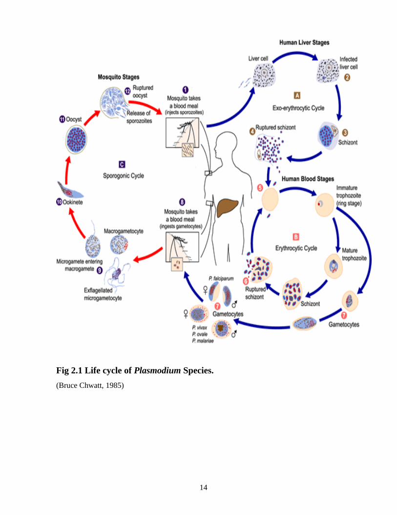
Fig 2.1 Life cycle of Plasmodium Species.
(Bruce Chwatt, 1985)
14
2.4. Symptoms
Malaria is a complex disease that varies widely in epidemiology and clinical manifestation in
different parts of the world. This variability is due to factors such as the species of malaria
parasites that occur in a given area, their susceptibility to commonly used or available
antimalarial drugs, the distribution and efficiency of mosquito vectors, climate and other
environmental conditions and the behavior and level of acquired immunity of the exposed
human populations (Greenwood et al., 1991; Mockenhaupt et al., 2000). The parasites
multiply within the red blood cells, causing symptoms that include symptoms of anemia (light
headedness, shortness of breath, tachycardia etc.), as well as other general symptoms such as
fever, chills, nausea, flu-like illness, arthralgia (joint pain), vomiting, anemia caused by
hemolysis, hemoglobinuria, and convulsions and in severe cases, coma and death (WHO
1991).
The classical symptom of malaria is cyclical occurrence of sudden coldness followed by rigor
and then fever and sweating lasting four to six hours, occurring every two days in P. vivax and
P. ovale infections, while it occurs every three days in P. malariae (Boivin, 2002). P.
falciparum can have recurrent fever every 36-48 hours or a less pronounced and almost
continuous fever (Trampuz et al., 2003). For reasons that are poorly understood, but which
may be related to high intracranial pressure, children with severe malaria frequently exhibit
abnormal posturing, a sign indicating severe brain damage (Idro et al., 2007). Malaria has
been found to cause cognitive impairments, especially in children. It causes widespread
anemia during a period of rapid brain development and also direct brain damage. This
neurologic damage results from cerebral malaria to which children are more vulnerable
(Boivin, 2002).
Consequences of severe malaria include coma and death if untreated, young children and
pregnant women are especially vulnerable. Splenomegaly (enlarged spleen), severe headache,
cerebral ischemia, hepatomegaly (enlarged liver), hypoglycemia, and hemoglobinuria with
renal failure may occur (Trampuz et al., 2003). Renal failure may cause blackwater fever,
where hemoglobin from lysed red blood cells leaks into the urine (Idro et al., 2007). Severe
malaria can progress extremely rapidly and cause death within hours or days. In the most
severe cases of the disease fatality rates can exceed 20%, even with intensive care and
15
treatment (Kain et al., 1998). In endemic areas, treatment is often less satisfactory and the
overall fatality rate for all cases of malaria can be as high as one in ten (Mockenhaupt et al.,
2004). Over the longer term, developmental impairments have been documented in children
who have suffered episodes of severe malaria (Trampuz et al., 2003). Chronic malaria is seen
in both P. vivax and P. ovale, but not in P. falciparum. Here, the disease can relapse months
or years after exposure, due to the presence of latent parasites (hypnozoites) in the liver (Kain
et al., 1998). Severe malaria is almost exclusively caused by P. falciparum infection and
usually arises 6-14 days after infection.
2.5. Diagnosis
Direct microscopic examination of intracellular parasites on stained blood films is the gold
standard for definitive diagnosis in nearly all settings. However, several other approaches
exist or are in development, some of which are discussed here.
2.5.1. Microscopy
Simple light microscopic examination of Giemsa stained blood films is the most widely
practiced and useful method for definitive malaria diagnosis. Advantages include
differentiation between species, quantification of the parasite density and ability to distinguish
clinically important asexual parasite stages from gametocytes which may persist without
causing symptoms (WHO, 1991). These advantages can be critical for proper case-
management and evaluating parasitological response to treatment. Specific disadvantages are
that slide collection, staining, and reading can be time-consuming and microscopists need to
be trained and supervised to ensure consistent reliability. While availability of microscopic
diagnosis has been shown to reduce drug use in some trial settings (Chanda et al., 2009). Any
programme aimed at improving the availability of reliable microscopy should also retrain
clinicians in the use and interpretation of microscopic diagnosis.
Another method is a modification of light microscopy called the Quantitative Buffy Coat
Method (QBCTM, Becton-Dickinson). Originally developed to screen large numbers of
specimens for complete blood cell counts, this method has been adapted for malaria diagnosis
(Levine et al., 1989). The technique uses microhaematocrit tubes precoated with fluorescent
acridine orange stain to highlight malaria parasites. With centrifugation, parasites are
16
concentrated at a predictable location. Advantages to QBC are that less training is required to
operate the system than for reading Giemsa-stained blood films and the test is typically
quicker to perform than normal light microscopy. Disadvantages are that electricity is always
required, special equipment and supplies are needed, the per-test cost is higher than simple
light microscopy, and species-specific diagnosis is not reliable. Field trials have shown that
the QBC system may be marginally more sensitive than conventional microscopy under ideal
conditions (Levine et al., 1989; Tharavanij, 1990).
2.5.2. Clinical (presumptive) Diagnosis
Although reliable diagnosis cannot be made on the basis of signs and symptoms alone
because of the non-specific nature of clinical malaria, clinical diagnosis of malaria is common
in many malarious areas (WHO, 1997). In much of the malaria-endemic world, resources and
trained health personnel are so scarce that presumptive clinical diagnosis is the only realistic
option (Smith et al., 1994). Clinical diagnosis offers the advantages of ease, speed, and low
cost. In areas where malaria is prevalent, clinical diagnosis usually results in all patients with
fever and no apparent other cause being treated for malaria. This approach can identify most
patients who truly need antimalarial treatment, but it is also likely to misclassify many who do
not (Olivar et al., 1991). Over-diagnosis contributes considerably to misuse of antimalarial
drugs (Ogungbamigbe et al., 2008). Considerable overlap exists between the signs and
symptoms of malaria and other frequent diseases, especially acute lower respiratory tract
infection and can greatly increase the frequency of misdiagnosis and mistreatment (Redd et
al., 1992).
Attempts to improve the specificity of clinical diagnosis for malaria by including signs and
symptoms other than fever or history of fever have met with only minimal success (Smith et
al., 1994). The Integrated Management of Childhood Illnesses (IMCI) programme defined an
algorithm that has been developed in order to improve diagnosis and treatment of the most
common childhood illnesses in areas relying upon relatively unskilled health care workers
working without access to laboratories or special equipment. With this algorithm, every
febrile child living in a “high-risk” area for malaria should be considered to have, and be
treated for, malaria. “High risk” has been defined in IMCI Adaptation Guides as being any
situation where as little as 5% of febrile children between the ages of 2 and 59 months are
17
parasitaemic (WHO, 1997), a definition that will likely lead to significant over-diagnosis of
malaria in areas with low to moderate malaria transmission.
2.5.3. Antigen detection tests (rapid or “dipstick” diagnostic tests)
A third diagnostic approach involves the rapid detection of parasite antigens using rapid
immunochromatographic techniques. Multiple experimental tests have been developed
targeting a variety of parasite antigens (WHO, 1996; Bloland, 2001). A number of
commercially available kits (e.g. ParaSight-F®, Becton-Dickinson; Malaquick®, ICT,
Sydney, New South Wales, Australia) are based on the detection of the histidine-rich protein
2 (HRP-II) of P. falciparum. Compared with light microscopy and QBC, this test yielded
rapid and highly sensitive diagnosis of P. falciparum infection (WHO, 1996; Craig and Sharp,
1997). Advantages to this technology are that no special equipment is required, minimal
training is needed, the test and reagents are stable at ambient temperatures and no electricity is
needed. The principal disadvantages are a currently high per-test cost and an inability to
quantify the density of infection. Furthermore, for tests based on HRP-II, detectable antigen
can persist for days after adequate treatment and cure; therefore, the test cannot adequately
distinguish a resolving infection from treatment failure due to drug resistance, especially early
after treatment (WHO, 1996).
Additionally, a test based on detection of a specific parasite enzyme (lactate dehydrogenase or
pLDH) has been developed (OptiMAL®, Flow Inc. Portland, OR, USA) and reportedly only
detects viable parasites, which if true, eliminates prolonged periods of false positivity post-
treatment (Makler et al., 1998; Palmer et al., 1999). Newer generation antigen detection tests
are able to distinguish between falciparum and non-falciparum infections, greatly expanding
their usefulness in areas where non-falciparum malaria is transmitted frequently (Bloland,
2001).
2.5.4. Molecular tests
Detection of parasite genetic material through polymerase-chain reaction (PCR) techniques
has become a more frequently used tool in the diagnosis of malaria, as well as the diagnosis
and surveillance of drug resistance in malaria. Specific primers have been developed for each
of the four species of human malaria. One important use of this new technology is in detecting
18
mixed infections or differentiating between infecting species when microscopic examination
is inconclusive (Beck, 1999). In addition, improved PCR techniques could prove useful for
conducting molecular epidemiological investigations of malaria clusters or epidemics
(Purfield et al., 2004). Primary disadvantages to these methods are overall high cost, high
degree of training required, need for special equipment, absolute requirement for electricity,
and potential for cross-contamination between samples (Berzins and Anders, 1999).
2.5.5 Serology
Techniques also exist for detecting anti-malaria antibodies in serum specimens. Specific
serological markers have been identified for each of the four species of human malaria. A
positive test generally indicates a past infection. Serology is not useful for diagnosing acute
infections because detectable levels of anti-malaria antibodies do not appear until weeks into
infection and persist long after parasitaemia has resolved. Moreover, the test is relatively
expensive, and not widely available (Bloland, 2001).
2.6. Antimalarial Drugs
For the past 50 years, there have been two main classes of antimalarial agents in use, the
antifolates and the cinchona alkaloids or the quinoline-containing drugs (Philips, 2001). The
quinoline-containing drugs include the cinchona alkaloids, quinine and quinidine, and the
aminoalcohol quinine analogues mefloquine (a 4-quinoline methanol) and halofantrine (a 9-
phenathrene methanol), which are recent introductions (ter Kuile 1993; Philips 2001). There
are also the 8-aminoquinoline primaquine, which is used for its gametocidal effect and its
action on the liver stage of P. Vivax, and the 4-aminoquinolines, chloroquine and its relative
amodiaquine (White, 1997). The antifolates include the diaminopyrimidines, such as
pyrimethamine and trimethoprim; the biguanides, represented by proguanil (cycloguanil) and
chlorproguanil; and the sulfa drugs, including the sulfonamides and the sulfones.
2.6.1. Quinine and related compounds
Quinine is the oldest and most famous anti-malarial (Dorvault, 1982). It has a long history
stretching from Peru, to the discovery of the Cinchona tree and the potential uses of its bark,
to the current day and a collection of derivatives that are still frequently used in the prevention
and treatment of malaria (Segurado et al., 1997). Quinine is an alkaloid that acts as a blood
19
schizonticidal and weak gametocide against Plasmodium species. As an alkaloid, it is
accumulated in the food vacuoles of Plasmodium, especially P. falciparum. It acts by
inhibiting the hemozoin biocrystallization, thus facilitating an aggregation of cytotoxic heme.
Quinine is less effective and more toxic as a blood schizonticidal agent than Chloroquine;
however it is still very effective and widely used in the treatment of acute cases of severe P.
falciparum (Foley and Tilley, 1998). It is especially useful in areas where there is known to be
a high level of resistance to Chloroquine, Mefloquine and sulfa drug combinations with
pyrimethamine (Foley and Tilley, 1998). Quinine is also used in post-exposure treatment of
individuals returning from an area where malaria is endemic (Foley and Tilley, 1997).
Quinine, along with its dextroisomer, Quinidine, has been the drug of last resort for the
treatment of malaria, especially severe disease.
Quinimax and Quinidine are the two most commonly used alkaloids related to Quinine, in the
treatment or prevention of Malaria. Quinimax is a combination of four alkaloids (namely
Quinine, Quinidine, Cinchoine and Cinchonidine) (Mills and Bone, 2000). This combination
has been shown in several studies to be more effective than Quinine, supposedly due to a
synergistic action between the four Cinchona derivatives. Quinidine is a direct derivative of
Quinine. It is a distereoisomer, thus having similar anti-malarial properties to the parent
compound. Quinidine is recommended only for the treatment or severe cases of malaria
(Foley and Tilley, 1998).
Chloroquine
Chloroquine is a 4-aminoquinolone derivative of quinine, first synthesized in 1934 and has
since been the most widely used antimalarial drug until recently (WHO, 2001). Historically, it
has been the drug of choice for the treatment of non-severe or uncomplicated malaria and for
chemoprophylaxis. It is believed to reach high concentrations in the vacuoles of the parasite,
which, due to its alkaline nature, raises the internal pH. It controls the conversion of toxic
heme to hemozoin by inhibiting the biocrystallization of hemozoin (Lobel and Campbell,
1986) thus poisoning the parasite through excess levels of toxicity. Other potential
mechanisms through which it may act include interfering with the biosynthesis of parasitic
nucleic acids, the formation of a chloroquine-haem or chloroquine-DNA complex. It was the
original prototype from which most other methods of treatment are derived (Foley and Tilley,
20
1997). It is also the least expensive, best tested and safest of all available drugs. The
emergence of drug resistant parasitic strains is rapidly decreasing its effectiveness
(Rieckmann et al., 1978; Wellems and Plowe, 2001); however it is still the first-line drug of
choice in most sub-Saharan African countries. It is now suggested that it is used in
combination with other antimalarial drugs to extend its effective usage.
The most significant level of activity found is against all forms of the schizonts (with the
obvious exception of chloroquine-resistant P. falciparum and P. vivax strains) and the
gametocytes of P. vivax, P. malariae, P. ovale as well as the immature gametocytes of P.
falciparum. Chloroquine also has a significant anti-pyretic and anti-inflammatory effect when
used to treat P. vivax infections, thus it may still remain useful even when resistance is more
widespread (Alene and Bennett, 1996). A slightly different drug called nivaquine or
chloroquine phosphate was also invented (Bloland 2001).
Amodiaquine
Amodiaquine is a 4-aminoquinolone anti-malarial drug similar in structure and mechanism of
action to Chloroquine (Winstanley et al., 1987). It is thought to be more effective in clearing
parasites in uncomplicated malarial than Chloroquine, thus leading to a faster rate of recovery
(Olliaro and Taylor. 2002). However, some fatal adverse effects of the drug were noted during
the 1980’s, thus reducing its usage in chemoprophylaxis. The WHO’s advice on the subject
maintained that the drug should be used when the potential risk of not treating an infection
outweighs the risk of developing side effects (Basco, 1991). It has also been suggested that it
is particularly effective and less toxic than other combination treatments in HIV positive
patients (Parise et al., 1998). Adverse reactions are generally similar in severity and type to
that seen in Chloroquine treatment. In addition, bradycardia, itching, nausea, vomiting and
some abdominal pain have been recorded. Some blood and hepatic disorders have also been
seen in a small number of patients (Olliaro et al., 1996).
Mefloquine
Mefloquine was developed during the Vietnam War and is chemically related to quinine. It
was developed to protect American troops against multi-drug resistant P. falciparum. It is a
very potent blood schizonticide with a long half-life (Mockenhaupt, 1995). It is thought to act
21
by forming toxic heme complexes that damage parasitic food vacuoles. Mefloquine
(Lariam®) is effective in prophylaxis and for acute therapy (Palmer et al., 1993). It is now
strictly used for resistant strains (and is usually combined with Artesunate) (van Vugt et al.,
1998). It has been linked with an increased number of stillbirths (Palmer et al., 1993) and is
not recommended for use during the first trimester, although considered safe during the
second and third trimesters. Mefloquine frequently produces side effects, including nausea,
vomiting, diarrhea, abdominal pain and dizziness. Several associations with neurological
events have been made, namely affective and anxiety disorders, hallucinations, sleep
disturbances, psychosis, toxic encephalopathy, convulsions and delirium. Moreover
cardiovascular effects have been recorded with bradycardia and sinus arrhythmia (Ridley,
1997)
Atovaquone
Atovaquone is a hydroxynapthoquinone that is currently being used most widely for the
treatment of opportunistic infections in immunosuppressed patients. It is effective against
chloroquine-resistant P. falciparum, but because when used alone, resistance develops
rapidly, atovaquone is usually given in combination with proguanil (Looareesuwan, 1996). A
fixed dose antimalarial combination of 250mg atovaquone and 100mg proguanil
(MalaroneTM) was introduced to market worldwide and was additionally being distributed
through a donation programme (Foege, 1997). Two drugs originally synthesized in China
were recommended for field trials. Pyronaridine was reported to be 100% effective in one trial
in Cameroon (Ringwald et al., 1996); however, it was only between 63% and 88% effective
in Thailand (Looareesuwan, 1996). Lumefantrinel, a fluoromethanol compound, was also
produced as a fixed combination. Atovaquone produces no side-effects such as the
cardiovascular effect with mefloquine which can trigger heart rhythm problems.
Primaquine
Primaquine is a highly active 8-aminoquinolone that was used in treating all types of malaria
infection (Olliaro and Trigg, 1995). It was most effective against gametocytes but also acts on
hypnozoites, blood schizonticytes and the dormant plasmodia in P. vivax and P. ovale. It is
the only known drug to cure both relapsing malaria infections and acute cases (Looareesuwan,
1996). The mechanism of action is thought to mediate some effect through creating oxygen
22
free radicals that interfere with the plasmodial electron transport chain during respiration
(Bloland et al., 1997). There are few significant side effects such as anorexia, nausea,
vomiting, cramps, chest weakness, anaemia, some suppression of myeloid activity and
abdominal pains; In cases of over-dosage granulocytopenia may occur (Bruce-Chwatt,, 1985).
Halofantrine
Halofantrine was developed by the Walter Reed Army Institute of Research in the 1960s
(Mills and Bone, 2000). It is a phenanthrene methanol, chemically related to Quinine and acts
as a blood schizonticide effective against all Plasmodium parasites (ter Kuile et al., 1993). Its
mechanism of action is similar to other anti-malarials. Cytotoxic complexes are formed with
ferritoporphyrin XI that cause plasmodial membrane damage (Bloland, 2001). Despite being
effective against drug resistant parasites, Halofantrine was not commonly used in the
treatment (prophylactic or therapeutic) of malaria due to its high cost, very variable
bioavailability and most importantly it has been shown to have potentially high levels of
cardiotoxicity (Nosten et al., 1993). A popular drug based on halofantrine is Halfan.
2.6.2 Antifolate drugs
This class of drugs includes effective casual antimalarial prophylactic and therapeutic agents,
some of which act synergistically when used in combination. They are of various
combinations of dihydrofolate-reductase inhibitors (proguanil, chlorproguanil, pyrimethamine
and trimethoprim) and sulfa drugs (dapsone, sulfalene, sulfamethoxazole, sulfadoxine, and
others). Although these drugs have antimalarial activity when used alone, parasitological
resistance can develop rapidly (Kupferschmidt et al., 1988). When combined, they produce a
synergistic effect on the parasite and can be effective even in the presence of resistance to the
individual components (Kublin et al., 2002). Typical combinations include
sulfadoxine/pyrimethamine (SP or Fansidar1), sulfalene-pyrimethamine (metakelfin) and
sulfamethoxazole-trimethoprim (co-trimoxazole). A newer antifolate combination drug which
is a combination of chlorproguanil and dapsone, also known as Lapdap, has a much more
potent synergistic effect on malaria than existing drugs such as SP. Benefits of this
combination include a greater cure rate, even in areas currently experiencing some level of SP
resistance, a lower likelihood of resistance developing because of a more advantageous
pharmacokinetic and pharmacodynamic profile and probable low cost (Kublin et al., 2002).
23
Sulphadoxine Pyrimethamine
Sulfadoxine-pyrimethamine (SP) has been widely used as first-line therapy for uncomplicated
P. falciparum malaria throughout sub-Saharan Africa, because of its affordability, its ease of
administration and until recently, its effectiveness. It is currently the only option for
intermittent treatment of malaria during pregnancy (McCollum et al., 2006). SP, which has
potent efficacy against chloroquine-resistant and pyrimethamine-resistant P. falciparum,
became available in 1971 and became the standard second-line therapy against chloroquine-
resistant falciparum malaria. Pyrimethamine acts by inhibiting dihydrofolate reductase in the
parasite, thus preventing the biosynthesis of purines and pyrimidines and therefore halting the
processes of DNA synthesis, cell division and reproduction. It acts primarily on the schizonts
during the hepatic and erythrocytic phases (Kuznetsov et al., 1984). The action of
sulphadoxine is focused on inhibiting the use of para-aminobenzoic acid during the synthesis
of dihydropteroic acid. When combined the two key stages in DNA synthesis in the plasmodia
are prevented.
Proguanil
Proguanil (Chloroguanadine) is a biguanide; a synthetic derivative of pyrimidine. It was
developed in 1945 by a British Antimalarial research group (Mills and Bone, 2000). It has
many mechanisms of action but primarily is mediated through conversion to the active
metabolite cycloguanil pamoate. This inhibits the malarial dihydrofolate reductase enzyme.
Its most prominent effect is on the primary tissue stages of P. falciparum, P. vivax and P.
ovale. It has no known effect against hypnozoites therefore is not used in the prevention of
relapse. It has a weak blood schizonticidal activity when combined with Atovaquone (a
hydroxynaphthoquinone). Proguanil is used as a prophylactic treatment in combination with
another drug, most frequently Chloroquine (Kublin et al., 2002). Proguanil has been used in
combination with dapsone for prophylaxis and treatment (Shanks et al., 1992; Mutabingwa et
al., 2005) and recently, proguanil/dapsone has been combined with artesunate (Nzila et al.,
2002). There are very few side effects to Proguanil, with slight hair loss and mouth ulcers
being occasionally reported following prophylactic use.
24
2.6.3 Antibiotics
Tetracycline and derivatives such as doxycycline are very potent antimalarials and are used
for both treatment and prophylaxis (Kremsner et al., 1994). In areas where response to
quinine has deteriorated, tetracyclines are often used in combination with quinine to improve
cure rates. Clindamycin has been used but offers only limited advantage when compared to
other available antimalarial drugs as parasitological response is slow and recrudescence rates
are high (Kremsner et al., 1994).
Doxycycline
Doxycycline is a Tetracycline compound derived from Oxytetracycline. The tetracyclines
were one of the earliest groups of antibiotics to be developed and are still used widely in
many types of infection. It is a bacteriostatic agent that acts to inhibit the process of protein
synthesis by binding to the 30S ribosomal subunit thus preventing the 50s and 30s units from
bonding (Kremsner et al., 1989). Doxycycline is used primarily for chemoprophylaxis in
areas where quinine resistance exists. It can be used in resistant cases of uncomplicated P.
falciparum but has a very slow action in acute malaria. The most commonly experienced side
effects are permanent enamel hypoplasia, transient depression of bone growth, gastrointestinal
disturbances and some increased levels of photosensitivity. Due to its effect on bone and tooth
growth it is not used in children under 8, pregnant or lactating women and those with a known
hepatic dysfunction (Kremsner et al., 1994). Tetracycline is only used in combination for the
treatment of acute cases of P. falciparum infections due to its slow onset. Unlike Doxycycline
it is not used in chemoprophylaxis. Oesophageal ulceration, gastrointestinal upset and
interferences with the process of ossification and depression of bone growth are known to
occur. The majority of side effects associated with Doxycycline are also experienced.
Clindamycin
Clindamycin is a derivative of Lincomycin, with a slow action against blood schizonticides. It
is only used in combination with Quinine in the treatment of acute cases of resistant P.
falciparum infections and not as a prophylactic (Kremsner et al., 1989; 1994)
25
2.6.4. Artemisinin compounds
Artemisinin is a Chinese herb (Qinghaosu) that has been used in the treatment of fevers for
over 1,000 years (WHO, 1996), thus predating the use of Quinine in the western world. It is
derived from the plant Artemisia annua, with the first documentation as a successful
therapeutic agent in the treatment of malaria in 340 AD (Mueller et al. 2000; 2004). The
active compound was isolated first in 1971 and named Artemsinin (Mills and Bone, 2000). It
is a sesquiterpene lactone with a chemically rare peroxide bridge linkage, which is thought to
be responsible for the majority of its anti-malarial action (Hien and White, 1993). It has
proven to be effective against all forms of multi-drug resistant P. falciparum, thus every care
is taken to ensure compliance and adherence together with other behaviours associated with
the development of resistance. It is also only given in combination with other anti-malarials.
Artemesinin has a very rapid action and the vast majority of acute patients treated show
significant improvement within 1-3 days of receiving treatment. It has demonstrated the
fastest clearance of all anti-malarials currently used and acts primarily on the trophozoite
phase, thus preventing progression of the disease. It is converted to active metabolite
dihydroartemesinin that then inhibits the Sarcoplasmic/Endoplasmic Reticulum Calcium
ATPase (SERCA) encoded by P. falciparum (Eckstein-Ludwig et al., 2003). It was one of
many candidates then tested by Chinese scientists from a list of nearly 200 traditional Chinese
medicines for treating malaria. It was the only one that was effective, but it was found that it
cleared malaria parasites from their bodies faster than any other drug in history. Artemisia
annua is a common herb and has been found in many parts of the world, including along the
Potomac River, in Washington, D.C (Hien and White, 1993).
A number of sesquiterpine lactone compounds have been synthesized from the plant
Artemisia annua (artesunate, artemether, arteether) (Mills and Bone, 2000). These compounds
are used for treatment of severe malaria and have shown very rapid parasite clearance times
and faster fever resolution than occurs with quinine. Artemisinin drugs first introduced in
South-East Asia have proven to be well tolerated and the most potent of antimalarials (White,
1999). They exhibit the following properties: rapid significant reduction of parasite
biomass, rapid resolution of clinical symptoms, effective against multidrug resistant P.
falciparum and reduction of gametocyte carriage, which may reduce transmission. However,
26
artemisinin drugs have a very short half-life and thus a multiple dose regimen of seven days is
required to achieve an acceptable cure rate (WHO, 2003).
In some areas of South-East Asia, combinations of artemisinins and mefloquine offer the only
reliable treatment for uncomplicated malaria, due to the development and prevalence of
multidrug resistant falciparum malaria (White, 1999). Combination therapy (an artemisinin
compound given in combination with another antimalarial), typically a long half-life drug like
mefloquine) has reportedly been responsible for inhibiting intensification of drug resistance
and for decreased malaria transmission levels in South-East Asia (Price et al., 1996; White,
1999). Artesunate (AS) plus amodiaquine (AQ) is one artemisinin-based combination (ACT)
recommended by the WHO for treating P. falciparum malaria in Africa (Sodiomon et al.,
2009).
Artemether is a methyl ether derivative of dihydroartemesinin. It is similar to artemesinin in
mode of action but demonstrates a reduced ability as a hypnozoiticidal compound, instead
acting more significantly to decrease gametocyte carriage. Similar restrictions are in place, as
with artemesinin, to prevent the development of resistance, therefore it is only used in
combination therapy for severe acute cases of drug-resistant P. falciparum. It was discovered
in 1982 (Dorvault, 1982). Artesunate, discovered in 1983 (Mills and Bone, 2000) is a
hemisuccinate derivative of the active metabolite dihydroartemisin. Currently it is the most
frequently used of all the artemesinin-type drugs. Its only effect is mediated through a
reduction in the gametocyte transmission. It is used in combination therapy and is effective in
cases of uncomplicated P. falciparum.
Dihydroartemisinin is the active metabolite to which Artemisinin is reduced. It is the most
effective Artemisinin compound and the least stable (Krettli, 2001). It has a strong blood
schizonticidal action and reduces gametocyte transmission. It is used for therapeutic treatment
of cases of resistant and uncomplicated P. falciparum. As with artesunate, no side effects to
treatment have thus far been recorded.
27
Arteether is an ethyl ether derivative of dihydroartemisinin. It is used in combination therapy
for cases of uncomplicated resistant P. falciparum (Krettli, 2001). No side effects have been
recorded.
2.7 COMBINATION THERAPY WITH ANTIMALARIALS
The problem of the development of malaria resistance must be weighed against the essential
goal of anti-malarial care; that is to reduce morbidity and mortality. Thus a balance must be
reached that attempts to achieve both goals whilst not compromising either too much by doing
so. The most successful attempts so far have been in the administration of combination
therapy. The key driver for combination antimalarial therapy is the need to slow development
of acquired drug resistance to a New Chemical Entity (NCE) and so maintain high levels of
efficacy for a longer period of time. This is best achieved by combining molecules which
individually have high levels of efficacy (WHO, 2001)
Combination therapy can be defined as, ‘the simultaneous use of two or more blood
schizonticidal drugs with independent modes of action and different biochemical targets in the
parasite’ (Price et al., 1999). Much evidence has supported the use of combination therapies.
The use of two antimalarial drugs simultaneously, especially when the antimalarials have
different mechanisms of action, has the potential for inhibiting the development of resistance
to either of the components. The efficacy of a combination of a 4-aminoquinoline drug
(chloroquine or amodiaquine) with Sulfadoxine/Pyrimethamine (SP) has been reviewed
(McIntosh and Greenwood, 1998). It was found that the addition of either chloroquine or
amodiaquine to SP marginally improved parasitological clearance (compared with SP alone).
The studies reviewed were mostly done in areas and at times when both SP and
Chloroquine/Amodiaquine retained a fair amount of efficacy, and it is not clear from these
studies how well such a combination would act in areas where one of the components was
significantly compromised. However several problems prevent the wide use in the areas
where its use is most advisable. These include: problems identifying the most suitable drug
for different epidemiological situations, the expense of combined therapy (it is over 10 times
more expensive than traditional mono-therapy), how soon the programmes should be
introduced and problems linked with policy implementation and issues of compliance (WHO,
28
2001). The combinations of drugs currently prescribed can be divided into two categories:
Non-artemesinin and quinine-based combinations and artemesinin based combinations.
2.7.1 Non-Artemesinin based combinations
Sulfadoxine-pyrimethamine plus Amodiaquine: This is a combination that has been shown
to produce a faster rate of clinical recovery than SP and Chloroquine (McIntosh and
Greenwood 1998). However there are serious adverse reactions associated which have limited
its distribution. It is thought to have a longer therapeutic lifetime than other combinations and
may be a more cost-effective option to introduce in areas where resistance is likely to
develop.
Sulfadoxine-Pyrimethamine plus Mefloquine: This is produced as a single dose pill
(Fansimef®) and has obvious advantages over some of the more complex regimes. This
combination of drugs has very different pharmokinetic properties with no synergistic action.
This characteristic is potentially thought to delay the development of resistance; however it is
counteracted by the very long half life of Mefloquine which could exert a high selection
pressure in areas where intensive malaria transmission occurs. It is also an expensive
combination.
Tetracycline or Doxycycline plus Quinine: Despite the increasing levels of resistance to
Quinine this combination have proven to be particularly efficacious (Kremsner et al., 1994).
The longer half-life of the Tetracycline component ensures a high cure rate. Problems with
this regime include the relatively complicated drug regimen, where Quinine must be taken
every 8 hours for 7 days. Additionally, there are severe side effects to both drugs (Cinchonism
in Quinine) and Tetracyclines are contraindicated in children and pregnant women. For these
reasons this combination is not recommended as first-line therapy but can be used for non-
responders who remain able to take oral medication.
2.7.2 Artemesinin-based combinations
Artemesinin has a very different mode of action from conventional anti-malarials. This makes
it particularly useful in the treatment of resistant infections (Mueller et al., 2000). However, in
order to prevent the development of resistance to this drug it is only recommended in
combination with another non-artemesinin based therapy. It produces a very rapid reduction
29
in the parasite biomass with an associated reduction in clinical symptoms and is known to
cause a reduction in the transmission of gametocytes thus decreasing the potential for the
spread of resistant alleles. Artemisinin combination therapy (ACT) has been widely adopted
as first-line treatment for uncomplicated falciparum malaria (Ashley et al., 2007; Nosten and
White, 2007). Although these drug combinations appear to be safe and well-tolerated,
experience with their use in Africa is still limited (Talisuna et al., 2006; Staedke et al., 2008).
Artesunate and chloroquine combination has been thoroughly tested in randomized controlled
trials and has demonstrated that it is well tolerated with few side effects (Nosten and White,
2007). However, in one study there was less than 85% cure in areas where Chloroquine
resistance was known. It is not approved for use in combination therapy and is unadvised in
areas of high P. falciparum resistance. Artesunate and Amodiaquine combination has also
been tested and proved to be more efficacious and similarly well tolerated than the
Chloroquine combination. The cure rate was greater than 90%, potentially providing a viable
alternative where levels of Chloroquine resistance are high (Sodiomon et al., 2009). The main
disadvantage is a suggested link with neutropenia (Mutabingwa et al., 2005).
Artesunate and mefloquine have been used as an efficacious first-line treatment regimen in
areas of Thailand for many years (Adjuik et al., 2004). Mefloquine is known to cause
vomiting in children and it induces some neuropsychiatric and cardiotoxic effects,
interestingly these adverse reactions seem to be reduced when the drug is combined with
Artesunate, it is suggested that this is due to a delayed onset of action of Mefloquine. This is
not considered a viable option to be introduced in Africa due to the long half-life of
Mefloquine, which potentially could exert a high selection pressure on parasites (Bloland,
2001).
Artemether and Lumefantrine (Coartem®, Riamet®, and Lonart®) is a combination that has
been extensively tested in 16 clinical trials, proving effective in children less than 5years and
has been shown to be better tolerated than Artesunate plus Mefloquine combinations
(Mutabingwa et al., 2005). There are no serious side effects documented but the drug is not
recommended in pregnant or lactating women due to limited safety testing in these groups.
This is the most viable option for widespread use and is available in fixed-dose formulae thus
increasing compliance and adherence (Lefevre et al., 2001).
30
Artesunate and Sulfadoxine-Pyrimethamine is a well tolerated combination but the overall
level of efficacy still depends on the level of resistance to Sulfadoxine and Pyrimethamine
thus limiting its usage (WHO, 2001). Piperaquine-dihydroartemisinin-trimethoprim
(Artecom®) alone and in combination with Primaquine has been studied in resistant areas of
China and Vietnam (Yeka et al., 2008). The drug has been shown to be highly efficacious
(greater than 90%) even to strains resistant to Primaquine. More information is required on
safety and tolerability in pregnant women and children and toxicology data. Pyronaridine and
Artesunate has been tested and was shown to have a clinical response rate of 100% in one trial
in Hainan (an area with high levels of P. falciparum resistance to Pyronaridine) (Nosten and
White, 2007). Chlorproguanil-Dapsone and Artesunate (Lapdap plus) is the most tested drug
currently under development and could be introduced in African countries imminently. It is
not recommended as a monotherapy due to concerns of resistance developing, thus
threatening the future use of related compounds (Nosten and White, 2007).
2.7.3. Traditional Antimalarial Herbs
The use of plants for therapeutic purposes dates back to the human history (Ogbonna et al.,
2008). Medicinal plants, since time immemorial, have been used in virtually all cultures as a
source of medicine (Hoareau and Dasilva, 1999) and for a long time, natural products were
the only sources of medication (Bourdy et al., 2008). Several medicinal plants have been used
locally to treat malaria infection. Some of such plants are Enantia chloranta, Nauclea
natifolia, Salacia Nitida (Ogbonna et al., 2008), Acalypha fruticosa, Azadirachta indica,
Cissus rotundifolia, Echium rauwalfii, Dendrosicyos socotrana and Boswellia elongate
(Merlin, 2004; Clarkson et al., 2004; Alshwash et al., 2007), Cymbopogon giganteus and
Morinda lucida etc .Medicinal plants such as Momordica charantia (Ejirin wewe), M
balsamina (Ejirin), Ageratum conyzoides (Imi Eshu), Ocimum gratissimum Cardiospermum
grandiofiorum (Ako Ejirin), Diospyros monbuttensis (Egun Eja) etc have been used to treat
one ailment or the other in Africa, especially Nigeria (Awe and Makinde, 1997; Azas et al.,
2002 Otimenyin et al., 2008).
The urgency generated by drug-resistant strains of malaria parasites has accelerated
antimalarial drug research over the last two decades. While synthetic pharmaceutical agents
31
continue to dominate research, attention has increasingly been directed to natural products
(Etkin, 2003). The success of artemisinin, isolated from Artemisia annua and its derivatives
for the treatment of resistant malaria has focused attention on the plants as a source of
antimalarial drugs (Tan et al., 1998). Moreover, plants have been the basic source of
sophisticated traditional medicine systems for thousands of years and were instrumental to
early pharmaceutical drug discovery and industry (Elujoba et al., 2005). The world's poorest
are the worst affected, and many treat themselves with traditional herbal medicines. These are
often more available and affordable, and sometimes are perceived as more effective than
conventional antimalarial drugs (Merlin, 2004).
Ethnobotanical information about antimalarial plants used in traditional herbal medicine, is
essential for further evaluation of the efficacy of plant antimalarial remedies and efforts are
now being directed towards discovery and development of new chemically diverse
antimalarial agents (Clarkson et al., 2004). Several rural dwellers depend on traditional herbal
medicine for treatment of many infectious diseases including malaria (Ali et al., 2004). The
reputed efficacies of these plants have been recognized and passed on from one generation to
the other.
About 75% of the population in Africa does not have direct access to chemical treatment,
such as chloroquine, but they have access to traditional medicine for treating fevers.
Treatment with these remedies has suffered a number of deficiencies; diagnosis is often a
problem, identification of plant extracts may be insecure and the chemical content of extracts
may vary considerably (Azas et al., 2002). Natural products isolated from plants used in
traditional medicine, which have potent antiplasmodial action in vitro, represent potential
sources of new antimalarial drugs (Wright et al,. 1994; Gasquet et al., 1993). It had been
advocated that direct crude drug formulation of the herbs following toxicological absolution
may not only produce dosage forms faster but will also lead to cheaper and more affordable
drugs for the communities that need them (Elujoba, 1998). Also, there is a belief that these
medicines are safe because they are natural and have been used traditionally over a period of
time (Sofowora, 1993; Willcox et al., 2003).
32
Plant materials remain an important resource to combat serious diseases in the world
(Tshibangu et al., 2002) and pharmacognostic investigations of plants are carried out to find
novel drugs or templates for development of new therapeutic agents (König, 1992). Moreover
herbs can be highly effective for treating malaria if government can educate those involved in
the practice regarding the normal dose to be taken before getting well. Therefore, government
should provide subvention for the Ministry of Health incorporating National Agency for Food
and Drug Administration and Control (NAFDAC) to go into more Malaria research in local
herb just to develop new and more effective drug for prevention and control, particularly in
view of the rapid spread of drug resistance. Nevertheless, much work need to be done to
educate the community and the producers of indigenous herbal products to strictly adhere to
environmental hygiene
2.8 Antimalarial Drug Resistance
Resistance to antimalarial drugs has been described for two of the four species of malaria
parasite that naturally infect humans, P. falciparum and P. vivax (Nguyen-Dinh et al., 1981;
Singh, 2000). P. falciparum has developed resistance to nearly all antimalarials in current use,
although the geographical distribution of resistance to any single antimalarial drug varies
greatly (Wongsrichanalai et al., 2002; Talisuna et al., 2004). Chloroquine-resistant
falciparum malaria has been described everywhere that P. falciparum malaria is transmitted
(Verdrager, 1986; Wernsdorfer, 1991; Wellems and Plowe, 2001; Sidhu et al., 2002; White
2004; ) except for malarious areas of Central America (north-west of the Panama Canal), the
island of Hispaniola, and limited areas of the Middle East and Central Asia (Bloland, 2001).
Verdrager (1995) has described Localized permanent epidemics as the genesis of chloroquine
resistance in P. falciparum.
Sulfadoxine/Pyrimethamine (SP) resistance occurs frequently in South-East Asia and South
America and is more prevalent in Africa as the drug is increasingly being used as a
replacement for chloroquine (Price et al., 1999; Purfield et al., 2004). Mefloquine resistance
is frequent in some areas of South-East Asia and has been reported in the Amazon region of
South America and sporadically in Africa (Mockenhaupt, 1995). Cross-resistance between
halofantrine and mefloquine is suggested by reduced response to halofantrine when used to
treat mefloquine failures (ter Kuile et al., 1993). Noedl and others (2008) in a recent study
33
have shown that artemisinin is losing its potency in Cambodia and increased efforts are
required to prevent artemisinin-resistant malaria from spreading across the globe.
2.8.1 Definition of antimalarial drug resistance
Antimalarial drug resistance has been defined as the “ability of a parasite strain to survive
and/or multiply despite the administration and absorption of a drug given in doses equal to or
higher than those usually recommended but within tolerance of the subject” (WHO, 1972).
This definition was later modified to specify that the drug in question must “gain access to the
parasite or the infected red blood cell for the duration of the time necessary for its normal
action” (Wernsdorfer and Wernsdorfer, 1995). Most researchers interpret this as referring
only to persistence of parasites after treatment doses of an antimalarial rather than prophylaxis
failure, although the latter is a useful tool for early warning of the presence of drug resistance
(Lobel and Campbell, 1986).
This definition of resistance requires demonstration of malaria parasitaemia in a patient who
has received an observed treatment dose of an antimalarial drug and simultaneous
demonstration of adequate blood drug and metabolite concentrations using established
laboratory methods (such as high performance liquid chromatography) or in vitro tests (Basco
and Ringwald, 2000). In practice, this is rarely done with in vivo studies. In vivo studies of
drugs for which true resistance is well known (such as chloroquine) infrequently include
confirmation of drug absorption and metabolism. Demonstration of persistence of parasites in
a patient receiving directly observed therapy is usually considered sufficient. Some drugs,
such as mefloquine, are known to produce widely varying blood levels after appropriate
dosing and apparent resistance can often be explained by inadequate blood levels (Slutsker,
1990; Basco, 1991).
2.8.2 Malaria treatment failure
Drug resistant parasites are often used to explain malaria treatment failure. However, they are
two different clinical scenarios. These are failure to clear parasitaemia and recover from an
acute clinical episode when a suitable treatment has been given and anti-malarial resistance in
its true form. A distinction must be made between a failure to clear malarial parasitaemia or
resolve clinical disease following a treatment with an antimalarial drug and true antimalarial
34
drug resistance. While drug resistance can cause treatment failure, not all treatment failures
are due to drug resistance (Plowe, 2003). Many factors can contribute to treatment failure
including incorrect dosing, non-compliance with duration of dosing regimen, poor drug
quality, drug interactions, poor or erratic absorption, and misdiagnosis. Probably all of these
factors, while causing treatment failure (or apparent treatment failure) in the individual, may
also contribute to the development and intensification of true drug resistance through
increasing the likelihood of exposure of parasites to suboptimal drug levels (Ariey and
Robert, 2003).
2.8.3 Mechanisms of antimalarial resistance
In general, resistance appears to occur through spontaneous mutations that confer reduced
sensitivity to a given drug or class of drugs. For some drugs, only a single point mutation is
required to confer resistance, while for other drugs, multiple mutations appear to be required
(Lobel and Campbell, 1986). Provided the mutations are not deleterious to the survival or
reproduction of the parasite, drug pressure will remove susceptible parasites while resistant
parasites survive (Watkins and Mosobo, 1993). Single malaria cases have been found to be
made up of heterogeneous populations of parasites that can have widely varying drug
response characteristics, from highly resistant to completely sensitive (Thaithong, 1983).
Similarly, within a geographical area, malaria infections demonstrate a range of drug
susceptibility. Over time, resistance becomes established in the population and can be very
stable; persisting long after specific drug pressure is removed. The biochemical mechanism of
resistance has been well described for chloroquine, the antifolate combination drugs, and
atovaquone (Looareesuwan, 1996; Alene and Bennett, 1996; Bloland, 2001; Alifrangis et al.,
2003).
2.8.3.1 Chloroquine resistance
The first type of resistance to be acknowledged was to Chloroquine in Thailand in 1957 (Mills
and Bone, 2000). As the malaria parasite digests haemoglobin, large amounts of a toxic by-
product are formed. The parasite polymerizes this by-product in its food vacuole, producing
non-toxic haemozoin (malaria pigment). It is believed that resistance of P. falciparum to
chloroquine is related to an increased capacity for the parasite to expel chloroquine at a rate
that does not allow chloroquine to reach levels required for inhibition of haem polymerization
35
(Foley and Tilley, 1997). This chloroquine efflux occurs at a rate of 40 to 50 times faster
among resistant parasites than sensitive ones (Martin et al., 1987).
Further evidence supporting this mechanism is provided by the fact that chloroquine
resistance can be reversed by drugs which interfere with this efflux system (Martin et al.,
1987). It is unclear whether parasite resistance to other quinoline antimalarials (amodiaquine,
mefloquine, halofantrine, and quinine) occurs via similar mechanisms but resistances are
thought to have occurred by similar mechanisms (Foley and Tilley, 1997).
2.8.3.2 Antifolate combination drugs
Antifolate combination drugs, such as sulfadoxine plus pyrimethamine, act through sequential
and synergistic blockade of two key enzymes involved with folate synthesis. Pyrimethamine
and related compounds inhibit the step mediated by dihydrofolate reductase (DHFR) while
sulfones and sulfonamides inhibit the step mediated by dihydropteroate synthetase (DHPS)
(Bruce-Chwatt, 1985; Kublin et al., 2002). Specific gene mutations encoding for resistance
mutations have been associated with varying degrees of resistance to antifolate combination
drugs (Plowe et al., 1998; Marks et al., 2005). Talisuna et al in Uganda [Talisuna et al.,,
2004] have shown higher levels of resistance to chloroquine and SP in zones of higher
transmission intensity. On the other hand, history shows that chloroquine resistance
emerged first in low transmission zones and that antifolate resistance has increased more
rapidly in low transmission areas (White 2004).
2.9 Spread of resistance
There is no single factor that confers the greatest degree of influence on the spread of drug
resistance, but a number of plausible causes associated with an increase have been
acknowledged. These include aspects of economics, human behaviors, pharmacokinetics, and
the biology of vectors and parasites. The most influential causes are examined below:
2.9.1 Biological influences on resistance
The biological influences are based on the parasite’s ability to survive the presence of an anti-
malarial thus enabling the persistence of resistance and the potential for further transmission
36
despite treatment. In normal circumstances parasites that persist after treatment are destroyed
by the host’s immune system, therefore factors that act to reduce the elimination of parasites
could facilitate the development of resistance (Bloland, 2001). This explains the poorer
response associated with immunocompromised individuals, pregnant women and young
children.
Based on data on the response of sensitive parasites to antimalarial drugs in vitro and the
pharmacokinetic profiles of common antimalarial drugs, there is thought to always be a
residuum of parasites that are able to survive treatment (Wernsdorfer, 1991). Under normal
circumstances, these parasites are removed by the immune system. Factors that decrease the
effectiveness of the immune system in clearing parasite residuum after treatment also appear
to increase survivorship of parasites and facilitate development and intensification of
resistance. This mechanism has been suggested as a significant contributor to resistance in
South-East Asia, where parasites are repeatedly cycled through populations of non-immune
individuals (Verdrager, 1986, 1995). The non-specific immune response of non-immune
individuals is less effective at clearing parasite residuum than the specific immune response of
semi-immune individuals (White, 1997). The same mechanism may also explain poorer
treatment response among young children and pregnant women (White, 1997). Among
refugee children in the former Zaire, those who were malnourished (low weight for height)
had significantly poorer parasitological response to both chloroquine and SP treatment
(Wolday et al., 1995). Similarly, evidence from prevention of malaria during pregnancy
suggests that parasitological response to treatment among individuals infected with the human
immunodeficiency virus (HIV) may also be poor. HIV-seropositive women require more
frequent treatment with SP during pregnancy in order to have the same risk of placental
malaria as is seen among HIV-seronegative women (Parise et al., 1998). Some characteristics
of recrudescent or drug resistant infections appear to provide a survival advantage or to
facilitate the spread of resistance conferring genes in a population (White., 1999). In one
study, patients experiencing chloroquine treatment failure had recrudescent infections that
tended to be less severe or even asymptomatic (Handunnetti et al., 1996). Schizont maturation
may also be more efficient among resistant parasites (Lobel and Campbell, 1986; Warsame et
al., 1991).
37
There has been evidence to suggest that certain parasite-vector combinations can alternatively
enhance or inhibit the transmission of resistant parasites, causing ‘pocket-like’ areas of
resistance. In South-East Asia, two important vectors, Anopheles stephensi and A. dirus,
appear to be more susceptible to drug-resistant malaria parasites than to drug sensitive malaria
parasites (Wilkinson et al., 1976; Sucharit et al., 1977). In Sri Lanka, researchers found that
patients with chloroquine-resistant malaria infections were more likely to have
gametocytaemia than those with sensitive infections and that the gametocytes from resistant
infections were more infective to mosquitoes (Handunnetti et al., 1996). The reverse is also
true; some malaria vectors may be somewhat refractory to drug-resistant malaria, which may
partially explain the pockets of chloroquine sensitivity that remain in the world in spite of
very similar human populations and drug pressure as found in Haiti (Bloland et al., 1997).
Many antimalarial drugs in current usage are closely related chemically and development of
resistance to one can facilitate development of resistance to others. Chloroquine and
Amodiaquine are both 4-aminoquinolines and cross-resistance between these two drugs is
well known (Hall, 1975; Basco, 1991). Development of resistance to mefloquine may also
lead to resistance to halofantrine and quinine (Foley and Tilley, 1998). Antifolate combination
drugs have similar action and widespread use of SP for the treatment of malaria may lead to
increased parasitological resistance to other antifolate combination drugs (Watkins and
Mosobo, 1993). Development of high levels of SP resistance through continued accumulation
of DHFR mutations may compromise the useful life span of newer antifolate combination
drugs such as chlorproguanil/dapsone (LapDap) even before they are brought into use. This
increased risk of resistance due to SP use may even affect non malarial pathogens as the use
of SP for treatment of malaria had been found to result in increased resistance to
trimethoprim/sulfamethoxazole among respiratory pathogens (Hastings, 2003).
There is an interesting theory that development of resistance to a number of antimalarial drugs
among some falciparum parasites produces a level of genetic plasticity that allows the parasite
to rapidly adapt to a new drug, even when the new drug is not chemically related to drugs
previously experienced (Rathod et al., 1997). The underlying mechanism of this plasticity is
currently unknown, but this capacity may help explain the rapidity with which strains of
falciparum develop resistance to new antimalarial drugs. The choice of using a long half-life
38
drug (SP, Mefloquine) in reference to one with a short half-life (Chloroquine, LapDap,
Quinine) has the benefit of simpler, single dose regimens which can greatly improve
compliance or make directly observed therapy feasible (Lefevre et al., 2001). Unfortunately,
that same property may increase the likelihood of resistance developing, due to prolonged
elimination periods (Newton et al., 2006). Parasites from new infections or recrudescent
parasites from infections that did not fully clear will be exposed to drug blood levels that are
high enough to exert selective pressure but are insufficient to provide prophylactic or
suppressive protection (Wernsdorfer, 1991). When blood levels drop below the minimum
inhibitory concentration (the level of drug that fully inhibits parasite growth), but remain
above the concentration of drug that produces 5% inhibition of parasite growth (EC5),
selection of resistant parasites occurs. This selection was illustrated in one study in Kenya
that monitored drug sensitivity of parasites reappearing after SP treatment (Watkins and
Mosobo, 1993). Parasites reappearing during a period when blood levels were below the point
required to clear pyrimethamine-resistant parasites, but still above that level required to clear
pyrimethamine-sensitive parasites, were more likely to be pyrimethmine-resistant than those
reappearing after levels had dropped below the level required to clear pyrimethamine-
sensitive parasites (Watkins and Mosobo, 1993). This period of selective pressure lasts for
approximately one month for mefloquine, whereas it is only 48 hours for quinine. In areas of
high malaria transmission, the probability of exposure of parasites to drug during this period
of selective pressure is high (Trape and Rogier, 1996). In Africa, for instance, people can be
exposed to as many as 300 infective bites per year; in rare cases, even as much as 1000
infective bites per year and during peak transmission, as many as five infective bites per night
(Trape and Rogier, 1996).
Mismatched pharmacokinetics can also play a role in facilitating the development of
resistance. For instance the elimination half-life of pyrimethamine is between 80 and 100
hours and is between 100 and 200 hours for sulfadoxine, leaving an extended period when
sulfadoxine is “unprotected” by synergy with pyrimethamine (Watkins and Mosobo, 1993).
This sort of mismatched pharmacokinetics is even more apparent in the mefloquine plus
sulfadoxine-pyrimethamine combination used in Thailand where mefloquine has an
elimination half-life of approximately 336 to 432 hours (Palmer et al., 1993; McIntosh and
Greenwood, 1998). It is apparent that there are more genetically distinct clones per person in
39
areas of more intense transmission than in areas of lower transmission (Babiker and Walliker,
1997). However, the interpretation of this and its implications for development of resistance
has variously been described as resistance being more likely in low-transmission
environments (Paul and Day, 1998), high-transmission environments (Mackinnon, 1997;
Mackinnon and Hastings, 1998; Babiker and Walliker, 1997), or either low- or high- but not
intermediate-transmission environments (Hastings 1997; Hastings and Mckinnon, 1998).
The relationship between transmission intensity and parasite genetic structure is obviously
complex and subject to other confounding/contributing factors (Hastings and Mckinnon,
1998). What is clear is that the rate at which resistance develops in a given area is sensitive to
a number of factors beyond mere intensity of transmission. Such factors are initial prevalence
of mutations, intensity of drug pressure, population movement between areas, the nature of
acquired immunity to the parasite or its strains, etc. However, reducing the intensity of
transmission will likely facilitate prolonging the useful life span of drugs (Hastings and
Mckinnon, 1998).
2.9.2 Programmatic influences on resistance
Programmatic influences on development of antimalarial drug resistance include overall drug
pressure, inadequate drug intake (poor compliance or inappropriate dosing regimens),
pharmacokinetic and pharmacodynamic properties of the drug or drug combination, and drug
interactions (Wernsdorfer and Wernsdorfer, 1995). Additionally, reliance on presumptive
treatment can facilitate the development of antimalarial drug resistance. Overall drug pressure
especially that exerted by programmes utilizing mass drug administration (e.g Eko free
malaria programme, Federal government Malaria intervention programme using Coartem)
probably has the greatest impact on development of resistance (Payne, 1988; Wernsdorfer,
1999). Studies have suggested that resistance rates are higher in urban and periurban areas
than rural communities, where access to and use of drug is greater (Ettling et al., 1995).
Confusion over proper dosing regimen has been described. In Thailand the malaria control
programme recommended 2 tablets (adult dose) of SP for treating malaria based on studies
suggesting that this was effective. Within a few years, this was no longer effective and the
programme increased the regimen to 3 tablets (Price et al., 1996). Although unproven, this
40
may have contributed to the rapid loss of SP efficacy there. Similar confusion over the proper
SP dosing regimen exists in Africa (Salako, 1998). To simplify treatment, many programmes
dose children based on age rather than weight and depending on the regimen being
recommended, this has been shown to produce systematic under dosing among children of
certain weight and age groups (Plowe, 2003).
The use of presumptive treatment for malaria has the potential for facilitating resistance by
greatly increasing the number of people who are treated unnecessarily but will still be
exerting selective pressure on the circulating parasite population (Wernsdorfer et al, 1994). In
some areas and at some times of the year, the number of patients being treated unnecessarily
for malaria can be very large (Persidis, 2000). Concurrent treatment with other drugs can
increase the likelihood of treatment failure and may contribute to development of drug
resistance. For instance, folate administration for treatment of anaemia and possibly when
used as a routine supplement during pregnancy can increase treatment failure rates (Ballereau,
1997). Similarly, concurrent illness and malnourishment may have an influence on drug
resistance (Payne, 1988).
Drug quality has also been implicated in ineffective treatment and possibly drug resistance
(Shakoor et al., 1997). Either through poor manufacturing practices, intentional
counterfeiting, or deterioration due to inadequate handling and storage, drugs may not contain
sufficient quantities of the active ingredients. In an analysis of chloroquine and antibiotics
available in Nigeria and Thailand, between 37% and 40% of samples assayed had substandard
content of active ingredients, mostly from poor manufacturing practices (Shakoor et al.,
1997). Another study in Africa found chloroquine stored under realistic tropical conditions
lost at least 10% of its activity in a little over a year (Ballereau, 1997).
2.10 Detection of resistance
In general, four basic methods have been routinely used to study or measure antimalarial drug
resistance: in vivo, in vitro, animal model studies, and molecular characterization (Basco,
2007). Additionally, less rigorous methods have been used, such as case reports, case series,
41
or passive surveillance. Much discussion has occurred regarding the relative merits of one test
over another, with the implication always being that one type of test should be used
preferentially. Careful consideration of the types of information each yields indicates,
however, that these are complementary, rather than competing, sources of information about
resistance. Recognition of drug resistance (or, treatment failure) in individual patients is made
difficult in many settings by operational issues such as availability and quality of microscopy.
Especially in Africa, where presumptive diagnosis and treatment for malaria is most common,
detection of treatment failures also tends to be presumptive (persistence or reappearance of
clinical symptoms in a patient recently receiving malaria treatment). Because of the non-
specific nature of clinical signs and symptoms of malaria and the many other causes of febrile
disease, this can lead to a false sense that a particular drug is not working when it is, or,
potentially, that an ineffective drug is working when it is not. In cases where microscopy is
used, presence of parasitaemia in a supposedly fully treated patient may indicate treatment
failure, but is not necessarily evidence of drug resistance.
2.10.1 In vivo tests
An in vivo test consists of the treatment of a group of symptomatic and parasitaemic
individuals with known doses of drug and the subsequent monitoring of the parasitological
and/or clinical response over time (Basco, 2007). One of the key characteristics of in vivo
tests is the interplay between host and parasite. Diminished therapeutic efficacy of a drug can
be masked by immune clearance of parasites among patients with a high degree of acquired
immunity (White, 1997). Of the available tests, in vivo tests most closely reflect actual clinical
or epidemiological situations i.e. the therapeutic response of currently circulating parasites
infecting the actual population in which the drug will be used. Because of the influence of
external factors (host immunity, variations of drug absorption and metabolism, and potential
misclassification of reinfections as recrudescences), the results of in vivo tests do not
necessarily reflect the true level of pure antimalarial drug resistance. However, this test offers
the best information on the efficacy of antimalarial treatment under close to actual operational
conditions. This should be expected to occur among clinic patients if provider and patient
compliance is high. The original methods for in vivo tests required prolonged periods of
follow-up (minimum of 28 days) and seclusion of patients in screened rooms to prevent the
possibility of re-infection. These methods have since been modified extensively and the most
42
widely used methods now involve shorter periods of follow-up (7 to 14 days) without
seclusion, under the assumption that reappearance of parasites within 14 days of treatment is
more likely due to recrudescence rather than re-infection (WHO, 1996b). Additional
modifications reflect the increased emphasis on clinical response in addition to parasitological
response. Traditionally, response to treatment was categorized purely on parasitological
grounds as sensitive, resistance I (RI), RII, and RIII (WHO, 1996). Later modifications have
combined, to varying extent, parasitological and clinical indicators (WHO, 1996).
Since anaemia can be a major component of malaria illness, in vivo methodologies allow
investigation of haematological recovery after malaria therapy (Boland, 2001). Failure of
complete parasitological clearance, even in situations where recurrence of fever is rare, can be
associated with lack of optimal haematological recovery among anaemic patients.
Standardization is however a limitation in this methodology. Major differences in sample size,
enrolment criteria, exclusion criteria, length and intensity of follow-up, loss-to-follow-up
rates, and interpretation and reporting of results are apparent in published papers on in vivo
trials (Ringwald and Basco, 1999). These differences make it difficult, if not impossible, to
compare results from one study to another with any level of confidence (Bloland, 2001). The
methodology currently being used and promoted, especially in sub-Saharan Africa, is a
system that emphasizes clinical response over parasitological response (WHO, 1996). Close
adherence to this protocol does provide comparable data; however, these data are not readily
comparable to data collected using other in vivo methods. Although not called for in the
protocol, categorization of the parasitological response using the standard WHO definitions
(WHO 1996) would allow some ability to compare to historical levels and provide useful
parasitological results that would aid in interpreting the clinical results.
2.10.2 In vitro tests
From the point of view of a researcher interested in pure drug resistance, in vitro tests avoid
many of the confounding factors which influence in vivo tests by removing parasites from the
host and placing them into a controlled experimental environment. In the most frequently
used procedure, the micro-technique, parasites obtained from a finger-prick blood sample are
exposed in microtitre plates to precisely known quantities of drug and observed for inhibition
of maturation into schizonts (Rieckmann et al., 1978; Jenson, 1988; Basco, 2007). This test
more accurately reflects “pure” antimalarial drug resistance. Multiple tests can be performed
43
on isolates, several drugs can be assessed simultaneously, and experimental drugs can be
tested. In addition, falciparum erythrocytic parasites can be evaluated in-vitro. However, the
test has certain significant disadvantages. The correlation of in vitro response with clinical
response in patients is neither clear nor consistent, and the correlation appears to depend on
the level of acquired immunity within the population being tested. Prodrugs, such as
proguanil, which require host conversion into active metabolites, cannot be tested. Neither can
drugs that require some level of synergism with the host’s immune system (Golenda and
Rosenberg, 1997). These tests are technologically more demanding and relatively expensive,
which makes them potentially more difficult to adapt successfully to routine work in the field.
In vitro assays for the sensitivity of human malaria parasites to antimalarial drugs provide
information complementary to that derived from the epidemiology of drug-resistant malaria
(Basco and Ringwald, 2007). Its principles were initially based on a suboptimal short-term
culture method reported in 1912 which was applied for the WHO macrotest system in the
1960s ( Rieckmann et al., 1978) and used until the 1980s. The principles of in vitro culture
underwent a major modification in 1976, when a new method for continuous culture of P.
falciparum was reported by Trager & Jensen (1976). The essential components of the
complete blood-medium mixture are Roswell Park Memorial Institute (RPMI) 1640 medium
buffered with N-(2-hydroxyethyl) piperazine-N´-(2-ethanesulfonic acid) (HEPES) and sodium
bicarbonate, human serum (or serum substitutes, including animal sera and lipid enriched
bovine albumin) and P. falciparum-infected human erythrocytes. This technical improvement
led to the elaboration of several in vitro assay systems in the late 1970s and in the 1980s,
including the WHO microtest system (morphological assay), the 48-h test (morphological
assay) and the radioisotope microtest (Basco, 2007). Over the past few years, two novel in
vitro drug sensitivity assays have been evaluated: an enzyme-linked immunosorbent assay
(ELISA) with monoclonal antibodies directed against either plasmodial lactate dehydrogenase
(LDH) or histidine-rich protein II (HRP II) and a fluorometric assay with DNA-binding
fluorescent dyes (WHO, 1997). These two assays are non-morphological and non-radioactive
and are also based on Trager & Jensen’s (1976) culture method.
2.10.3 Animal model studies
44
This type of test is, in essence, an in vivo test conducted in a non-human animal model and
therefore, is influenced by many of the same extrinsic factors as in vivo tests (Bloland, 2001).
The influence of host immunity is minimized by using lab-reared animals or animal-parasite
combinations unlikely to occur in nature, although other host factors would still be present.
These tests allow for the testing of parasites which cannot be adapted to in vitro environments
(provided a suitable animal host is available) and the testing of experimental drugs not yet
approved for use in humans. A significant disadvantage is that only parasites that can grow in,
or are adaptable to, non-human primates can be investigated.
2.10.4 Molecular techniques
These tests offer promising advantages to the methods described above. Molecular tests use
polymerase chain reaction (PCR) to indicate the presence of mutations encoding biological
resistance to antimalarial drugs (Plowe et al., 1998). Theoretically, the frequency of
occurrence of specific gene mutations within a sample of parasites obtained from patients
from a given area could provide an indication of the frequency of drug resistance in that area
analogous to information derived from in vitro methods (Basco, 2007). Advantages include
the need for only small amounts of genetic material as opposed to live parasites, independence
from host and environmental factors and the ability to conduct large numbers of tests in a
relatively short period of time (Beck, 1999). Disadvantages include the obvious need for
sophisticated equipment and training and the fact that gene mutations that confer antimalarial
drug resistance are currently known or debated for only a limited number of drugs primarily
for dihydrofolate reductase inhibitors (pyrimethamine), dihydropteroate synthase inhibitors
(sulfadoxine), and chloroquine (Plowe et al., 1998). Confirmation of the association between
given mutations and actual drug resistance is difficult, especially when resistance involves
more than one gene locus and multiple mutations (Sidhu et al 2002).
45
2.10.5 Case reports and passive detection of treatment failure
Additional methods for identifying or monitoring antimalarial drug resistance include the use
of case reports or case series of spontaneously reported treatment failure. In general, these
methods require far less investment in time, money and personnel and can be done on an
ongoing basis by individual health care centers (Bloland, 2001). They suffer however, from
presenting a potentially biased view of drug resistance primarily because denominators are
typically unknown and rates of resistance cannot be calculated. Nonetheless, case reports can
be useful and may indicate a problem that should be confirmed using one of the other
methods. In the United States for instance, case reports, especially when occurring in clusters,
of prophylaxis failure have been used to help formulate recommendations for
chemoprophylaxis of non-immune travellers to endemic areas (White, 1999).
Another method that has been used is passive detection of treatment failure. In this system,
patients are treated following usual treatment guidelines and told to come back to the clinic or
hospital if symptoms persist or return. Those cases which do return are considered to
represent the population of treatment failures. Because this system does not ensure
compliance with treatment regimens through directly observed therapy and does not attempt
to locate and determine the outcome of patients who do not return on their own, data are
seriously biased. In a study conducted in Ethiopia and Eritrea using this method, only 1706
(4.3%) out of 39824 patients returned to the clinic (Alene and Bennett, 1996). The assumption
was that those patients who did not return did not have resistant parasites, yielding a very low
prevalence of resistance (1.8% to 4.8%, depending on region). These results contrast
dramatically with results from standard 7-days in vivo trials conducted at two sites in Eritrea
in 1994 (Bloland, 2001) and one site in Ethiopia in 1993–1994 which found between 58% and
86% RII/RIII level resistance (Tulu, 1996).
2.11 The future: prevention of drug resistance
The future of antimalarial drug resistance and efforts to combat it is defined by a number of
assumptions. First, antimalarial drugs will continue to be needed long into the future (Breman,
2001). No strategy in existence or in development, short of an unforeseen scientific
breakthrough or complete eradication, is likely to be 100% effective in preventing malaria
infection. Secondly, as long as drugs are used, the chance of resistance developing to those
46
drugs is present (Plowe, 2003). P. falciparum has developed resistance to nearly all available
antimalarial drugs and it is highly likely that the parasite will eventually develop resistance to
any drug that is used widely. Thirdly, development of new drugs appears to be taking longer
than development of parasitological resistance. The development of resistance to antimalarial
drugs in South-East Asia has been far quicker than the estimated 12 to 17 years it takes to
develop and market a new antimalarial compound (Ridley, 1997). Fourthly, affordability is an
essential consideration for any strategy to control drug-resistant malaria, especially in Africa
(Foster and Phillips 1998; Goodman et al., 1999).
The future, especially in Africa, will also be defined by how well the central tenets of malaria
control can be reconciled with the central tenets of control of drug resistance. One of the
cornerstones of the current approach to malaria control is the provision of prompt, effective
malaria treatment. In much of Africa, easy access to public sector health care is limited and
when it is accessible, health care staff are often inadequately trained, insufficiently supplied
and supported, ineffectively supervised and poorly motivated (Goodman et al., 2000). One
response to this situation has been the intentional liberalization of access to drugs; instead of
relying so heavily on the formal public sector to distribute antimalarial drugs, some people are
suggesting that the best way to reduce the time between onset of illness and first treatment
with an antimalarial drug is by making these drugs widely available on the open market, from
unofficial sources of health care and at the household level (Salako, 1998). This approach is
gaining support internationally. This approach is also in direct conflict with the primary
methods for inhibiting development of drug resistance, limited access to and judicious use of
chemotherapeutic agents. Clearly, some middle ground will need to be identified that will
improve access to antimalarial drugs for those who need to be treated while at the same time
reducing the inappropriate use of those same drugs. Prevention strategies can be divided into
those aimed specifically at preventing malaria infection and those aimed at reducing the
likelihood of development of drug resistance. Reduction of overall malaria infection rates or
transmission rates have an indirect impact on development of drug resistance by reducing the
number of infections needing to be treated (and therefore, overall drug pressure) and by
reducing the likelihood that resistant parasites are successfully transmitted to new hosts. The
transmission reduction methods include the use of insecticide-treated bednets, indoor residual
insecticide spraying, environmental control (mosquito breeding site or “source” reduction),
47
other personal protection measures (e.g. use of repellent soap or screening windows) and
chemoprophylaxis in defined populations (Ariey and Robert, 2003). An effective and
deliverable vaccine would also be greatly beneficial (Good, 2001).
Interventions aimed at preventing drug resistance, generally focus on reducing overall drug
pressure through more selective use of drugs; improving the way drugs are used and also by
improving prescribing by clinicians, follow-up practices and patient compliance; or using
drugs or drug combinations which are inherently less likely to foster resistance or have
properties that do not facilitate development or spread of resistant parasites (Plowe, 2003).
In Nigeria, the methods that are widely used to study antimalarial drug resistance are in-vivo
methods, animal model studies and molecular methods (Oduola et al., 1993; Ogbonna et al.,
2008; Ogunfowokan et al., 2009). Using in-vitro sensitivity test more accurately reflects
“pure” antimalarial drug resistance, multiple tests can be performed on isolates, several drugs
can be assessed simultaneously and experimental drugs can be tested (Basco, 2007). In
addition, falciparum erythrocytic parasites can be evaluated in-vitro. The method is also a
good epidemiological tool to assess baseline sensitivity and to monitor the drug response of
P. falciparum (Druilhe et al., 2007). Moreover, it provides background information for
development and evaluation of drug policies, can be an indicator of future therapeutic failure
and can also guide on the partner drug in ACT (Basco, 2007).
48
CHAPTER THREE
MATERIALS AND METHODS
3.1 Study area:
This study was carried out in Ogun State, located in the tropical zone of Southwestern
Nigeria. With its state capital in Abeokuta, Ogun State has a land area of 16,409.26 square
kilometers and a population of 2,759,109 (Unpublished report, Ministry of Health, 2003).
Ogun State is bounded on the West by Benin Republic, on the South by Lagos State and the
Atlantic Ocean, on the East by Ondo State and on the North by Oyo State. The State is made
up of six ethnic groups viz, Egba, Ijebu, Remo, Egbado, Awori and Egun. The language of the
majority of the people of Ogun State is Yoruba but this is however broken into scores of
dialects. Ogun State is in the transitional zone between the tropical rain forest and derived
savannah zone in the southwest, Nigeria. The area experiences two seasons, the dry season
(November to March) and the wet season (April to October). The annual temperature range is
from 22.8oC to 34.9oC and the mean annual rainfall is about 107.3mm. Malaria is present
throughout the year with a marked increase during the rainy season (Ojo, 2005).
3.2 Study subjects
Children between 1-15 years, pregnant women and other adults were included in this study.
This is because majority of malaria cases occur in children under the age of 12 years, pregnant
women are also especially vulnerable. The blood samples were collected and analyzed
between April 2008 and June 2009.
3.3 Sampling Procedure
Ogun State, which was the study site, was divided into 4 zones for sample collection. The 4
zones are
• Sango – Ota (Yewa)
• Abeokuta (Egba)
• Ijebu Ode (Ijebu) and
• Sagamu (Remo)
49

Fig 2.1 Map of Ogun State, Southwestern Nigeria
(http://ogunstate.gov.ng/eGovernment/index.php)
50
Using the population size of each zone and the rate of prevalence of malaria in the state, the
sample size for each of the four zones was calculated with a 95% CI and precision level of 5%
as follows: n = Z2 P(1-P) 2 /d2
Where n=sample size
Z=1.96 at 95%
P=Prevalence rate
d=Sampling error that can be tolerated (0.05)
State general hospitals located in the different zones were used as sample collection centers.
Both in and out patients who presented with uncomplicated malaria in the hospitals were
recruited for the research work. Four thousand and sixty six patients were recruited into this
study. The total number of subjects recruited in Sango-ota, Abeokuta, Ijebu-ode and Sagamu
were 1120, 1116, 995 and 835 respectively. The mean age was 19 years (>1 –70) with 93%
less than 25 years.
3.4 Ethical Consideration
Scientific and Ethical clearance was obtained from the Nigerian Institute of Medical Research
- Institutional Review Board (NIMR-IRB) and Covenant University Ethics Committee for this
work. The Ogun State Ministry of health (Hospitals Management Board) was also informed
and clearance obtained for this study. Written informed consent was obtained from patients
prior to recruitment into this study. Consent for the children was provided by the
parents/guardians while some of the participants provided the assents.
3.5. Sample collection
Blood samples were collected for malaria screening from both fingerprick and venepuncture .
This is to check the presence of healthy asexual parasites in the peripheral smear of patients.
Safety procedures were adopted in the collection of finger-prick blood samples by swabbing
the area to be sampled with 70% alcohol and allowing it to dry before collection. The
bleeding was done in the hospitals by clinicians and medical laboratory scientists. About 2-5
ml of blood was then drawn (venepuncture) with a sterile disposable syringe and transferred
to a heparinised centrifuge tube. The blood samples were transported to the laboratory at 40C.
Drops of peripheral blood were placed on 1.5 x 7.0-cm strips of Whatman (Brentford, United
51
Kingdom) 3MM filter paper so that the blood covered half the length of the strip. The strips
were air-dried and kept in plastic bags until use.
3.6 Cryopreservation
The infected blood was centrifuged at 3000 revolutions per minute (rpm) for 10 min in a
refrigerated centrifuge. The supernatant/plasma was removed and cells were suspended in
equal volume of cryopreservative. The suspension was distributed into cryotubes and quickly
frozen in cryofreezer at -800C and then transferred into liquid nitrogen (-1960C). The
cryopreservative was prepared by adding 28ml glycerol to 72ml of 4.2% Sorbitol in normal
saline. The solution was sterilized by filtration through a Millipore filter of 0.22µm porosity.
3.7 Processing of Sample
3.7.1 Microscopic examination
Thick and thin films stained with Giemsa were prepared for the microscopic examination of
the malaria parasite. The thin films were fixed with methanol and all films were stained with
3% Giemsa stain of pH 7.0 for 30 min as recommended by WHO (WHO, 2000). Blood films
were examined microscopically using 100X (oil immersion) objectives as described by
Cheesbrough (2000). The thick films were used to determine the parasite densities while thin
films were used to identify the parasite species and infective stages. Parasite density per
microlitre of blood (parasitemia) was estimated from the thick film, taking the number of
leucocytes per microliter of blood as 8,000 and was expressed as follows:
Parasite count x 8,000
Parasite density/µL = No of WBC counted
3.8. Antimalarial sensitivity testing
3.8.1 Revival of Cryopreserved parasites
The vial was taken out of the liquid nitrogen tank and thawed quickly in a 370C water bath.
The content was transferred to a centrifuge tube and centrifuged at 3000rpm for 10min. The
supernatant was then removed and an equal volume of 3.5% NaCl was added. The suspension
52
was centrifuged again and the supernatant removed. The pellet was washed twice with
complete medium supplemented with 15%serum.
3.8.2 In vitro microtest (Mark III Test)
Drug samples were prepared in picomole (pM) quantities according to WHO in vitro micro
test procedure (WHO, 1990; Druilhe et al., 2001). Blood collected on heparin was washed
twice in RPMI 1640 medium (Gibco BRL, Paisley, United Kingdom) and once with complete
culture medium (Appendix I). Chloroquine sulphate, amodiaquine, mefloquine, SP and
artemisinin were dosed into 96-well microplates. Stock solutions of chloroquine sulphate (600
ng/ml) and the other antimalarial drugs (ng/ml) were prepared in sterile distilled water
(chloroquine, amodiaquine, mefloquine and quinine) or ethanol (sulphadoxine-pyrimethamine
and artemisinin) and used in twofold dilutions with the culture medium in 96-well culture
microplates (Nunc, Denmark) to obtain nine final dilutions (600 to 2.34 ng/ml for
chloroquine) and (appropriate dilution factor was determined for the other drugs). Parasitized
RBCs was added to each well and to three control wells (without drug) to a final volume of
250μl/well at a 2% hematocrit. Each isolate was tested in duplicate and the plates were
incubated at 37°C for 24 – 30 hours. At the end of the assay, the plates were read by preparing
thick smear from each well and determining the IC50 for each drug using the HN-NonLinn
software package.
3.9 Antimalarial Activity Testing of Crude Organic Extracts of Medicinal
Plants: Momordica charantia (Ejirin), Diospyros monbuttensis (Eegun eja) and Morinda
lucida (Oruwo)
3.9.1 Preparation of Plant Extract
10g of dried extract was dissolved in 50ml alcohol (95%) for 7 days at room temperature. The
alcohol was allowed to evaporate at room temperature. 10ml Dimethyl sulphoxide (DMSO)
was added to 10mg of each extract to make 1mg/ml.
3.9.2 In vitro Test
The assay was performed in duplicate in a 96-well microtiter plate, according to WHO
method [in vitro micro test (Mark III)] that is based on assessing the inhibition of schizont
53
maturation. RPMI 1640 (Gibco BRL, Paisley, United Kingdom) was the culture medium used
for cultivation of P. falciparum (Flores et al., 1997). Dilution was prepared from the crude
plant extract and the final concentrations prepared by dilution were (125, 62.5, 31.25, 15.6,
7.8, 3.9 and 1.95µg/ml). Negative controls treated by solvent and positive controls
(Chloroquine phosphate) were added to each set of experiments. 50µl from the blood mixture
media was added to each well in plate and incubated in CO2 condition at 37.5°C for 24–30 h.
After incubation, contents of the wells were harvested and stained for 30 min in a 2% Giemsa
solution pH 7.2, after that the developed schizonts were counted against the total asexual
parasite count of 200. The count process was done in duplicate, and the data were analyzed by
using HN-NonLin software to estimate IC50
3.10 Molecular studies
Polymerase Chain Reaction/Restriction fragment Length Polymorphism (PCR/RFLP) was
used to determine the resistant genes and study the genetic diversity/genetic variation of
antimalarial resistant Plasmodium falciparum.
3.10.1 DNA extraction
DNA was extracted from patient blood spotted on the filter paper ("pre-culture") and cultured
parasites ("post-culture") using the QiaAmp DNA Blood Mini kit Blood and Body Fluid Spin
Protocol (Qiagen, Valencia, CA). The protocol for the extraction was carried out according to
manufacturer’s instruction.
3.10.2 PCR for detection of Pfcrt gene
The oligonucleotides primers were designed from published sequences. For amplification of
the 1.6-kb fragment of pfcrt, the lower primer was 5'-CCGTTAATAATAAATACAGGC-
3'.The upper primer was 5'-CTTTTAAAAATGGAAGGGTGT-3', (Dorsey et al., 2001). The
primary PCR components, in a final volume of 20µL, was 2.5mM MgCl2, 640µM
deoxynucleotide triphosphate (dNTPs), buffer 10x, 10pM of each primer, 1U of Taq
polymerase (Ampli Taq Gold; Applied Biosystems, Foster City, CA, USA) and 2µL of DNA
samples. The cycling protocol was as follows: 95°C for 5 min for initial denaturation; 40
cycles of 94°C for 30 s, 52°C for 40 s and 72°C for 30 s; and a final extension of 72°C for 5
min.
54
3.10.3 Nested PCR and RFLP for Pfcrt mutation-specific detection
Product from primary PCR (2µl of 10x dilution) was used in a follow-up, nested, allele-
specific PCR amplifications to detect the codons for pfcrt 76K or 76T. These diagnostic PCR
amplifications used a common inner primer pair 5'-GGCTCACGTTTAGGTGGA-3' and 5'-
TGAATTTCCCTTTTTATTTCCAAA-3' (detects the 76T codon) or 5'-GTTCTTTTAGC
AAAAATCT-3' (detects the 76K codon). The PCR stages for these diagnostic amplifications
were at 94°C for 5 minutes, followed by 40 cycles at 94°C for 30 seconds, 52°C for 40
seconds, and 72°C for 45 seconds and a final extension of 72°C for 5 minutes. Purified
genomic DNA from P. falciparum clones HB3 (chloroquine sensitive) and Dd2 (chloroquine
resistant) were used as positive controls, and water, extracted uninfected blood smears, and
uninfected blood spots on filter paper were used as negative controls. The PCR products from
the amplification reactions were evaluated by electrophoresis on 2% agarose gels containing
ethidium bromide.
10µl of the nested PCR product reaction mixture were treated directly with 3U of the
restriction enzyme Apo I for 6 to 16 hours at 50°C as recommended by the manufacturer (New
England Biolabs, Beverly, MA). The enzyme Apo I recognise and cut the 76K codon,
releasing fragment from product. It does not cut the product containing the 76T codon found
in chloroquine-resistant P. falciparum.
3.10.4 PCR and RFLP for detection of Pfmdr1 gene
Gene segments spanning codon 86 of the Pfmdr1 gene were amplified in 20µl of standard
PCR mixture containing 5µl of extracted DNA and primers MDR1 5'-ATGGGTAAA
GAGCAGAAAGA-3' and MDR2 5'-AACGCAAGTAATACATAAAGTCA-3'. The PCR
amplification stages were at 94°C for 2 minutes, followed by 35 cycles at 94°C for 20
seconds, 52°C for 10 seconds, 48°C for 10 seconds, and 60°C for 1.5 minutes. A second,
nested amplification from this segment was then performed under the same PCR conditions
using 1µl of the product solution and primers MDR3 5'-TGGTAACCTCAG-
TATCAAAGAA-3' and MDR4 5'-ATAAACCTAAAAAGGAACTGG-3'.
55
Presence of the mutant 86Y codon was detected by digestion of 8µl of the second
amplification product solution with 1.5U of Afl III according to the manufacturer’s instruction
(New England Biolabs). The products of restriction digestion were separated by
electrophoresis on a 2% agarose gel and detected by staining with ethidium bromide.
3.10.5 PCR assays for the detection of Pfdhfr and Pfdhps genes
Pfdhfr, and Pfdhps PCRs were performed as described by May and Meyer (2003a) and Marks
et a,l (2004). For the Pfdhps PCR, two primers (primer Pfdhps-F 5-ATGATTCTTTT
TCAGATG-3_ and primer Pfdhps-R 5-CCAATTGTGTGATTTGTCCAC-3 were designed to
amplify 747 bp of the region exhibiting mutations relevant to Sulphadoxine resistance. PCR
was performed with a volume of 20µl (each primer at 0.2M, dNTPs at 200M, and 1U of
Hotstar-Taq with the appropriate buffer [Qiagen, Valencia, Calif.] and MgCl2 at a final
concentration of 2.0 mM) and approximately 80ng of template genomic human DNA, with
parasite DNA concentrations corresponding to the parasite burden in the individual. After an
initial denaturation (15 min at 95°C), 31 cycles of 30s at 94°C, 40s at 53°C, and 1min at 72°C
were run. Elongation of the amplicons was completed by a final cycle of 10min at 72°C.
Subsequently, a nested PCR was performed to increase the yields of the specific amplicons
using primers primer pfdhps-F1 (5-GTTGAACCTAAACGTGCTG-3) and pfdhps-R1 (5-
ATTACAACATTTTGATCATTC-3). 3µl of the primary PCR product was used in a reaction
volume of 25µl containing 0.2M of each primer, dNTPs at 200M, reaction buffer with MgCl2
at a final concentration of 2.0 mM, and 1U of Hotstar-Taq). In the nested PCR, a high initial
annealing temperature (AT), which ensures a high level of specificity of initial primer
binding, is followed by a gradual decrease in the AT toward the pre-calculated optimal AT.
The parameters consisted of an initial denaturation step (15 min at 95°C) and 43 cycles of 30s
at 94°C, the AT for 40 s, and 72°C for 1 min, in which the ATs were 65°C (5 cycles), 60°C (5
cycles), 56°C (7 cycles), 54°C (13 cycles), and 53°C (13 cycles). Fragment elongation was
performed by use of a cycle of 10 min at 72°C. The amplicons were monitored for quality and
the expected size on 1% ethidium bromide-stained agarose gels. Statistical analysis (_χ2
tests) was performed by the use of STATA software (version 8.2; Corp.,
College Station, Tex.).
56
3.10.6 PCR and RPLP assay for (SERCA) PfATPase6
P. falciparum positive samples were amplified by PCR using PfATPase6-specific primary and
nested primer pairs. DNA extract for each sample was subjected to nested PCR amplification
with primers flanking nucleotide codon 2307of the PfATPase6 gene. Both the primary and
secondary reactions comprised 2µl template, 0.25µM primer, 1.5mM MgCl2, 200µM dNTP’s,
1x PCR buffer and 1U Taq DNA polymerase, in 25µl reactions. Amplification cycles for both
primary and secondary reactions consisted of an initial denaturation at 940C for 2 minutes,
followed by 25 cycles of denaturation at 940C for 45 seconds, annealing at 460C for 45
seconds and extension at 650C for 1 minute, final extension was run at 650C for 2
minutes. For primary amplification, the primers are 2307FW 5’-TGA GCA TGG CAC AAG
TIT 3’; 2307RV- 5’TCA ATA ATA CCT AAT CCA CCT AAA TA-3’.For nested PCR the
primers are FW-EN 5’-TGA GCA TGG TAG AAG TTT T-3’ and RV-EN 5’- TCA TCT
GTA TTC TTA ATA ATA TTT AAA TCT GTA CTA-3’ (Zhang et al., 2008).
Internal primers for the nested PCR amplification (2307FW-EN and 2307RV-EN, were
engineered to create Csp6 I restriction sites. One site, at PfATPase6 nucleotide codon 1916,
serves as the internal control for the restriction digestion assay, which is always cut by the
enzyme. Csp6 I digestion assay comprised 4µl of secondary PCR amplicon (432bp), 1x buffer
and 1.5U of Csp6 I restriction enzyme, in 30µl reactions. Digestion assays were incubated for
12 hours at 37 0C. PCR amplicon and restriction digests were analyzed by electrophoresis on
a 2% ethidium bromide-stained agarose gel and visualized under UV transillumination.
Restriction digests were loaded in 15 µl volumes per lane while undigested product was run in
5µ loading volumes. Band sizes were measured using Syngene gel imaging analysis software.
3.10.7 Molecular Genotyping of isolates using MSP1&2 and Glurp
Primers and PCR protocols were followed as previously described by Snounou et al. (1999)
for family specific allele analysis of msp-1 (block 2) and msp-2 (block 3). For PCR
amplification, the primers for each of the families in msp-1 and msp-2 are as shown in Table
3.1. and described by Snounou et al., 2007. PCR condition consisted of an initial denaturation
step (5 min at 95°C) and 35 cycles of 30s at 95°C, 72°C for 30 s, and 72°C for 5 min and a
final elongation at 72°C for 5min.
57
Polymerase Chain Reaction (PCR) amplification was performed on thermal cycler (Perkin
Elmer 9700/2400, UK) in a final volume of 20μl. The PCR products were visualized by UV
transillumination at 302nm on gel documentation system (Syngenta, USA) after
electrophoresis on 2% agarose gel (Promega/Boehringer) using 0.5 × TBE buffer at 80–100
volts. Allele sizes were calculated using Genetool programme. To understand the identity of
Nigerian isolates with respect to isolates of other regions, sequence data available in public
domains were downloaded for allelic families of msp-1&2 and details are given below;
Thailand(K1-M77730, MAD20-M77722, R033-AAA29684, 3D7- U91676), Vietnam(K1-
AF509651, MAD20-AF509653&94, FC27-AF104696, 3D7-AF104693), Tanzania(K1-
AF061134, FC27-AY532386), Brazil(K1-AF509714, MAD20-AY714585, FC27-DQ115973,
3D7-AF177389), China(MAD20-AF251345), Sudan(MAD20-AF034635), Iran(MAD20-
AY138509, R033-AY138507, FC27-DQ338451), Indonesia(K1-AF191061, R033-
AAF18431), Western Africa (R033-PFAMSA1), Kenya(R033-AAM21583), Ghana(FC27-
AF329577), PNG(FC27-AF329579), Gambia(FC27-U91668, 3D7- U91665) and
Nigeria(3D7-AF148224) (Joshi et al., 2007).
58
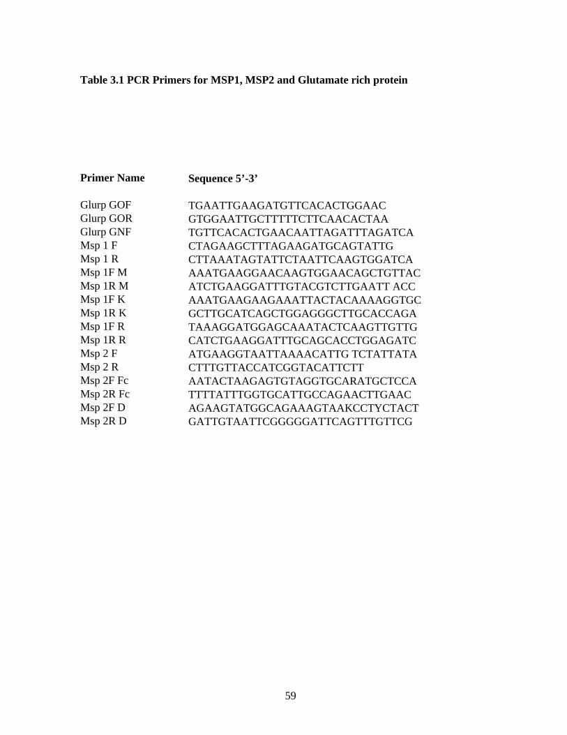
Table 3.1 PCR Primers for MSP1, MSP2 and Glutamate rich protein
Primer Name Sequence 5’-3’
Glurp GOF TGAATTGAAGATGTTCACACTGGAACGlurp GOR GTGGAATTGCTTTTTCTTCAACACTAAGlurp GNF TGTTCACACTGAACAATTAGATTTAGATCAMsp 1 F CTAGAAGCTTTAGAAGATGCAGTATTGMsp 1 R CTTAAATAGTATTCTAATTCAAGTGGATCAMsp 1F M AAATGAAGGAACAAGTGGAACAGCTGTTACMsp 1R M ATCTGAAGGATTTGTACGTCTTGAATT ACCMsp 1F K AAATGAAGAAGAAATTACTACAAAAGGTGCMsp 1R K GCTTGCATCAGCTGGAGGGCTTGCACCAGAMsp 1F R TAAAGGATGGAGCAAATACTCAAGTTGTTGMsp 1R R CATCTGAAGGATTTGCAGCACCTGGAGATCMsp 2 F ATGAAGGTAATTAAAACATTG TCTATTATAMsp 2 R CTTTGTTACCATCGGTACATTCTTMsp 2F Fc AATACTAAGAGTGTAGGTGCARATGCTCCAMsp 2R Fc TTTTATTTGGTGCATTGCCAGAACTTGAACMsp 2F D AGAAGTATGGCAGAAAGTAAKCCTYCTACTMsp 2R D GATTGTAATTCGGGGGATTCAGTTTGTTCG
59
Statistical Analysis.
Statistical analysis was performed using Stat View 5.0.1 (SAS Institute Inc., Cary, NC).
Statistical significance was based on a α level of 0.05. The data/statistical analysis for
significance were done using Microsoft excel, SPSS and HN-NonLin analysis methods. One-
way analysis of variances was used to compare the geometric mean parasites density in
patients from different age groups. The Student’s t-test was used to compare mean number of
alleles of P. falciparum MSP-1, MSP-2, and GLURP. P values < 0.05 was considered
significant for all statistical analysis. Data from all the questionnaires were coded,
entered and analysed using Epi Info 6.04 and SPSS software.
3.11 Questionnaire Administration
Structured Questionnaires were administered to patients, parents of infants and older children.
The questionnaire seeks to identify the locality, age and sex of the respondents. In addition,
the patients’ attitude to use of antimalarial drugs and Insecticide treated mosquito nets or
other means of controlling man-mosquito contact as practiced in the locality and malaria
management practices by the study population were identified. A cross sectional study was
conducted in the four zones of Ogun State, Southwestern Nigeria.
60
CHAPTER FOUR
RESULTS
4.1. Incidence of Malaria in Ogun State, Southwestern Nigeria.
4.1.1Patients Characteristics
A total of 4066 subjects comprising of 1839 males and 2227 females presenting with malaria
in four different senatorial districts of Ogun state were recruited into the study. The total
number of subjects recruited in Sango-ota, Abekuta, Ijebu-ode and Sagamu were 1120, 1116,
995 and 835 respectively. The mean age was 19 years (>1 –70) with 93% less than 25 years.
4.1.2 Incidence of Malaria
The overall incidence of falciparum malaria as determined by microscopy in the study area
was 62.7% (2250/4066). The highest incidence was observed in Sango (75.7%) while the
lowest (48.4%) was observed in Abeokuta (Table 4.2). Table 4.1 shows the incidence of P.
falciparum infection in Ogun State, Nigeria according to age and sex. Age group 1-5yrs had
the highest incidence of infection (70.8) followed by age group <1year (63.9%) while age
group 6-15years had the lowest incidence (52.8). The difference according to age was
statistically significant (p<0.0001). The highest falciparum malaria mean parasitaemia of
1080p/µl was recorded among the age group 1-5years while age group >40years recorded the
lowest mean parasitaemia of 800p/µl. Females had a higher incidence of malaria infection of
51.8% compared to males 48.2% in this study. The difference in the incidence of infection by
sex was significant (p<0.0001).
4.2. In Vitro Drug sensitivity Tests
In-Vitro antimalarial drugs and local herbal extracts sensitivity tests were carried out and the
IC50 and IC99 for each drug was determined. Sample of HN-NonLinn software statistical
package used for the data analysis is shown in Figure 4.4. Table 4.3 shows the mean IC50 and
IC99 values for chloroquine, amodiaquine, mefloquine, quinine, sulphadoxine/pyrimethamine
and artesunate. The IC50 value is defined as the concentration of an antimalarial drug that
inhibits 50 % of schizont maturation as compared with the development in drug-free control
wells. IC99 gives a result that closely approximates a reliable Minimum Inhibitory
61
Concentration (MIC). The MIC is generally defined as the lowest drug concentration that
inhibits the development of rings to schizonts. Moreover, the in vitro threshold values for
antimalarials have been defined statistically as >2 SD above the mean (Pradines et al., 1998).
IC50 implies that 50% of the parasite could not mature to schizont stage at that drug
concentration while IC99 implies that the antimalarial drugs inhibited 99% of the parasites
from maturing to schizont stage at that concentration.
All the isolates tested were sensitive to quinine, mefloquine and artesunate while in vitro
resistance was observed to chloroquine, amodiaquine and SP (Table 4.4). Highest percentage
of resistance to chloroquine (69.8%) was recorded among isolates from Yewa zone while
highest percentage resistance to amodiaquine (30%) was observed in Ijebu zone. Highest
resistance against SP (10%) was recorded in Yewa and Egba zones. A significant positive
correlation was observed between the responses to artemisinin and mefloquine (P=0.001),
artemisinin and quinine (P=0.05), quinine and mefloquine (P= 0.01), (Table 4.4). A
significant negative correlation was observed between the responses to chloroquine and
mefloquine (P=0.05).
4.3 Prevalence of drug resistant molecular markers
Prevalence of established antimalarial drug resistant markers was assayed for in 100 isolates
collected from the study area. The molecular markers assayed for in the study were Pfcrt,
Pfmdr1, PfDhfr, PfDhps and PfATpase6.Table 4.5 shows the overall and zonewise prevalence
of the molecular markers to various antimalarial drugs in P. falciparum isolates from Ogun
state, Nigeria. Sixty percent of the isolates had Pfmdr1 (Y86) resistance gene while 48% had
Pfcrt (T76) chloroquine resistance gene. None of the isolate had the PfATpase artemisinin
resistant gene. Most of the isolates that showed in-vitro resistance to the drugs also harbored
the genes coding for resistance to the drugs. Plates 4.1 – 4.5 show gel electrophoresis results
for the detection of Pfcrt, pfmdr1,dhfr, dhps andATPase6 genes/mutation respectively.
4.4 In vitro antimalarial activity of herbal extracts
The results of in vitro antimalarial activity of the three herbal extracts tested for antimalarial
activity are as shown in Table 4.6. Of the selected three herbal extracts, the highest activity
62
was obtained with extract of Diospyros monbuttensis (IC50 = 32µg/ml) while the lowest was
obtained from Morinda lucida (IC50 =250 µg/ml).
4.5 Genetic Diversity of P. falciparum
As an estimate of multiclonality of infection, the minimal number of P. falciparum strains in
each individual was determined by assessing the number of alleles of the genes encoding
MSP-1 and MSP-2 families. The MSP-1 and MSP-2 alleles were defined by gene family-
specific PCR assays and analysis of the resulting length polymorphisms of the PCR products.
One hundred P. falciparum isolates analyzed during the study
demonstrated the highly diverse nature of the field isolates in respect of
msp-1 (block 2) and msp-2 (central repeat region, block3). All the three
reported families of MSP-1(K1, MAD20 and RO33) families and two MSP-2
(FC27 and 3D7) families were observed among the isolates (Plates 4.6 -
4.10).
In MSP-1, Proportion of isolates with K1 family was 68% with 4 alleles in
the range of 100 to 300 base pairs (bp). Proportion of isolates with MAD20
family was 40% and a total of 3 alleles were observed within 100 to 300
bp. RO33 proportion was 20% and the family was observed to be
monomorphic with an allele size of 200 bp. Observed proportions,
numbers and size range of alleles among the isolates are given in Table
4.7.
In MSP-2, the reported families FC27 and 3D7 were observed among the isolates (Table 4.7).
Proportion of FC27 family was 76% and that of 3D7 was 56%. Proportional prevalence of
FC27 and 3D7 families was significantly different (χ2 = 16.5, P=0.002). Three alleles of
FC27 in the range of 300–500 bp and 3 alleles of 3D7 in the range of 450–600 bp were
observed in the study. Proportion of multiclonal isolates (multiple infection) is given in Table
4.7 and multiplicity of infection, MOI was estimated by dividing the total number of
fragments detected in the individual system by the number of samples positive in the
particular system (either MSP-1 or MSP-2) and 80% of the isolates harbour the genes that
code for glutamate rich protein with size ranging between 700 and 900.
63

The zonewise genetic diversity of P. falciparum from Ogun State is as shown on Table 4.8. A
high polymorphism was recorded in all the zones for MSP-1 (block 2) and MSP-2 (central
repeat region, block 3). Highest genetic polymorphism was recorded among the field isolates
from Yewa zone.
64
4.6 Knowledge and practice on the use of antimalarial drugs
Questionnaires were administered to respondents resident in the study sites who were either
malaria patients or parents/guardians of malaria infected children. The age of respondents
ranged between 18 and 75 (Table 4.9). A total of 946 questionnaires were administered to
assess the malaria related knowledge on issues regarding the use of antimalarial drugs and
malaria management practices. Most of the respondents were students (52.2%). The responses
of the participants are summarized in table 4.10. About 32.1% of participants have malaria
attack at least once in 3 months while only 26.3% normally complete their antimalarial drugs.
The malaria management practices showed that 24.6% attend hospitals, 12.0% use local
healers while 25.0% buy antimalarial drugs without prescription by a physician. It was also
found that some use more than one method in their management of malaria. Those who
combined antimalarial drugs with traditional medicine from local healers were found to be
17.4%, while 1.2% reported doing nothing about malaria. Table 4.10 also shows the methods
used in preventing mosquito bites by respondents. Only 18% of the sample population used
Insecticide treated mosquito nets. Majority of the people (42.3%) used only window and door
nets; 24% of the population used insecticides while 13% do not prevent mosquito bite at all.
65
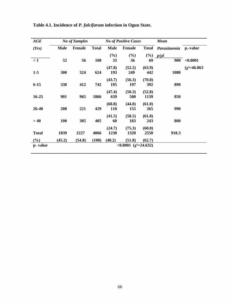
Table 4.1. Incidence of P. falcifarum infection in Ogun State.
AGE
(Yrs)
No of Samples No of Positive Cases Mean
Parasitaemia
p/µl
Male Female Total Male
(%)
Female
(%)
Total
(%)
p.-value
< 1 52 56 108 33
(47.8)
36
(52.2)
69
(63.9)
900 <0.0001
(χ²=46.8631-5 300 324 624 193
(43.7)
249
(56.3)
442
(70.8)
1080
6-15 330 412 742 195
(47.4)
197
(50.3)
392
(52.8)
890
16-25 901 965 1866 639
(60.8)
500
(44.0)
1139
(61.0)
850
26-40 208 221 429 110
(41.5)
155
(58.5)
265
(61.8)
990
> 40 100 305 405 60
(24.7)
183
(75.3)
243
(60.0)
800
Total
(%)
1839
(45.2)
2227
(54.8)
4066
(100)
1230
(48.2)
1320
(51.8)
2550
(62.7)
918.3
p- value <0.0001 (χ²=24.632)
66
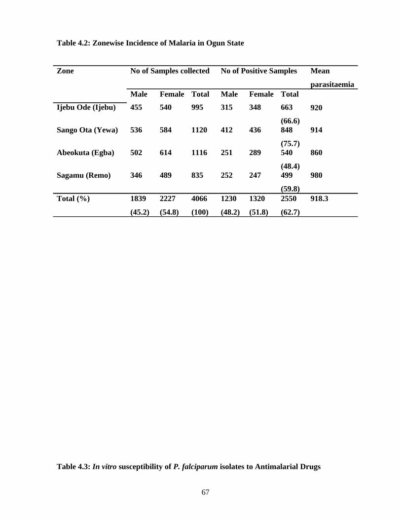
Table 4.2: Zonewise Incidence of Malaria in Ogun State
Zone No of Samples collected No of Positive Samples Mean
parasitaemiaMale Female Total Male Female Total
920Ijebu Ode (Ijebu) 455 540 995 315 348 663
(66.6)Sango Ota (Yewa) 536 584 1120 412 436 848
(75.7)
914
Abeokuta (Egba) 502 614 1116 251 289 540
(48.4)
860
Sagamu (Remo) 346 489 835 252 247 499
(59.8)
980
Total (%) 1839
(45.2)
2227
(54.8)
4066
(100)
1230
(48.2)
1320
(51.8)
2550
(62.7)
918.3
Table 4.3: In vitro susceptibility of P. falciparum isolates to Antimalarial Drugs
67
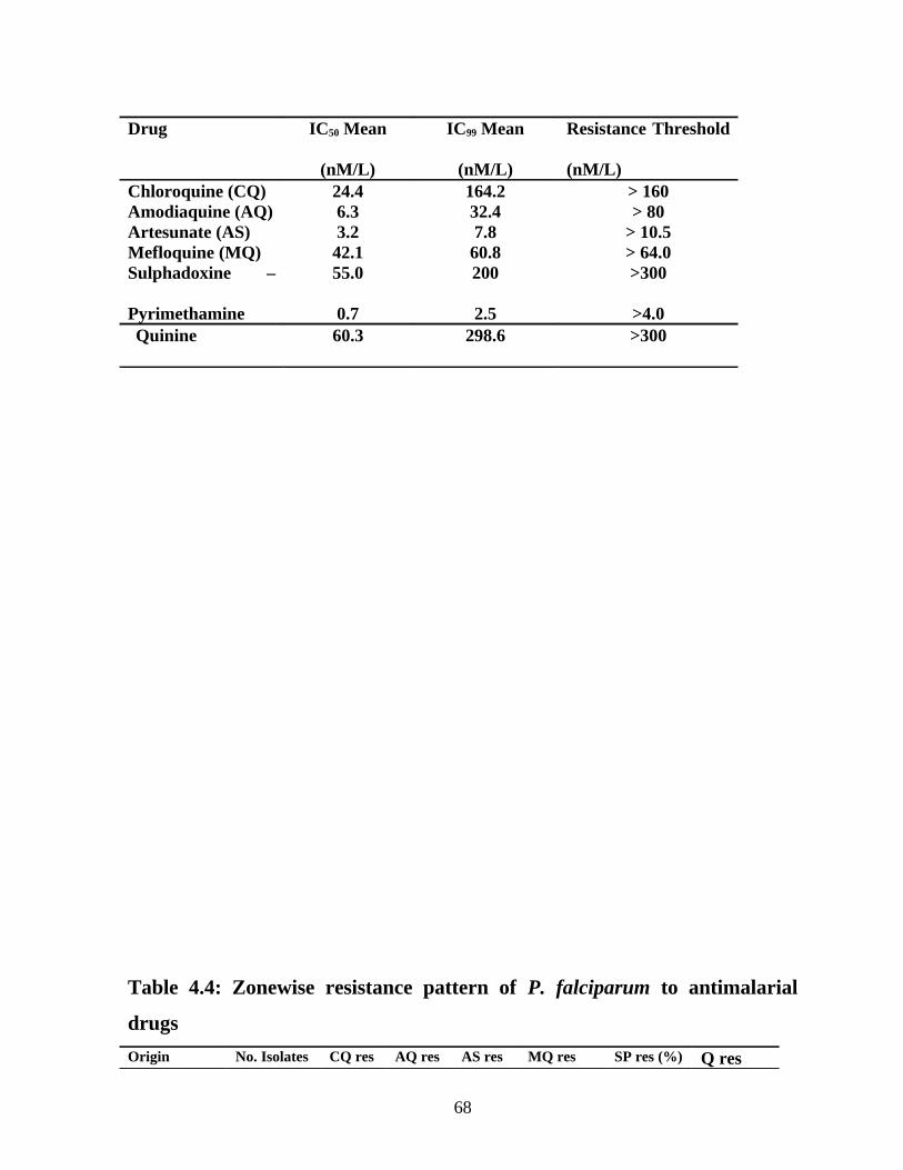
Drug IC50 Mean
(nM/L)
IC99 Mean
(nM/L)
Resistance Threshold
(nM/L)Chloroquine (CQ) 24.4 164.2 > 160Amodiaquine (AQ) 6.3 32.4 > 80Artesunate (AS) 3.2 7.8 > 10.5Mefloquine (MQ) 42.1 60.8 > 64.0Sulphadoxine –
Pyrimethamine
55.0
0.7
200
2.5
>300
>4.0Quinine 60.3 298.6 >300
Table 4.4: Zonewise resistance pattern of P. falciparum to antimalarial
drugs
Origin No. Isolates CQ res AQ res AS res MQ res SP res (%) Q res
68
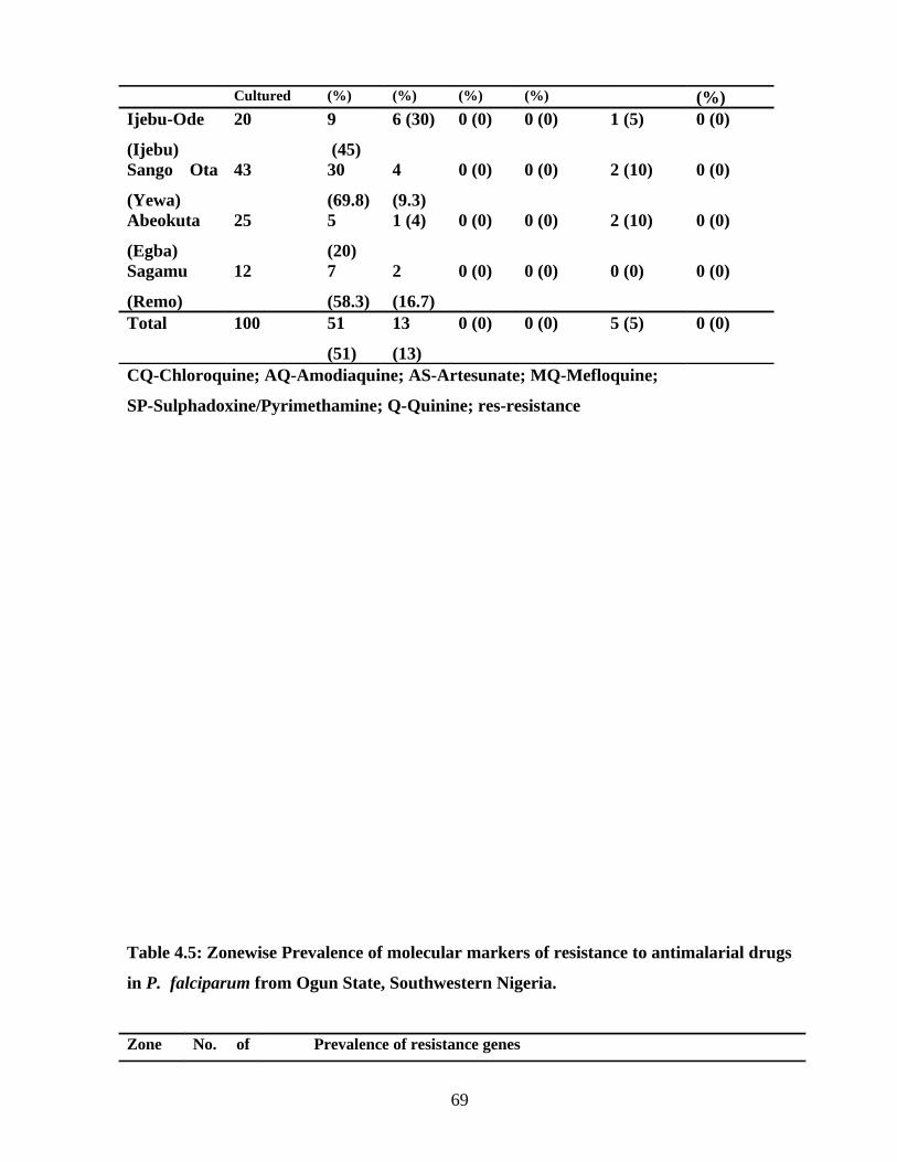
Cultured (%) (%) (%) (%) (%)Ijebu-Ode
(Ijebu)
20 9
(45)
6 (30) 0 (0) 0 (0) 1 (5) 0 (0)
Sango Ota
(Yewa)
43 30
(69.8)
4
(9.3)
0 (0) 0 (0) 2 (10) 0 (0)
Abeokuta
(Egba)
25 5
(20)
1 (4) 0 (0) 0 (0) 2 (10) 0 (0)
Sagamu
(Remo)
12 7
(58.3)
2
(16.7)
0 (0) 0 (0) 0 (0) 0 (0)
Total 100 51
(51)
13
(13)
0 (0) 0 (0) 5 (5) 0 (0)
CQ-Chloroquine; AQ-Amodiaquine; AS-Artesunate; MQ-Mefloquine;
SP-Sulphadoxine/Pyrimethamine; Q-Quinine; res-resistance
Table 4.5: Zonewise Prevalence of molecular markers of resistance to antimalarial drugs
in P. falciparum from Ogun State, Southwestern Nigeria.
Zone No. of Prevalence of resistance genes
69
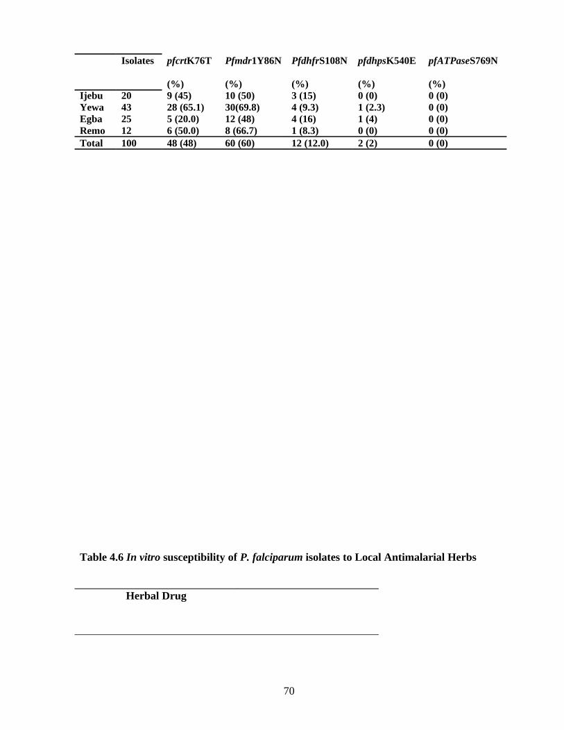
Isolates pfcrtK76T
(%)
Pfmdr1Y86N
(%)
PfdhfrS108N
(%)
pfdhpsK540E
(%)
pfATPaseS769N
(%)Ijebu 20 9 (45) 10 (50) 3 (15) 0 (0) 0 (0)Yewa 43 28 (65.1) 30(69.8) 4 (9.3) 1 (2.3) 0 (0)Egba 25 5 (20.0) 12 (48) 4 (16) 1 (4) 0 (0)Remo 12 6 (50.0) 8 (66.7) 1 (8.3) 0 (0) 0 (0)Total 100 48 (48) 60 (60) 12 (12.0) 2 (2) 0 (0)
Table 4.6 In vitro susceptibility of P. falciparum isolates to Local Antimalarial Herbs
Herbal Drug
70
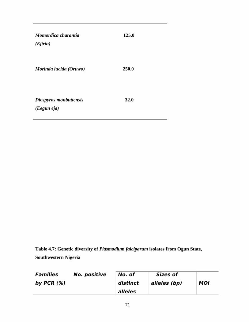
Momordica charantia 125.0
(Ejirin)
Morinda lucida (Oruwo) 250.0
Diospyros monbuttensis 32.0
(Eegun eja)
Table 4.7: Genetic diversity of Plasmodium falciparum isolates from Ogun State,
Southwestern Nigeria
Families No. positive
by PCR (%)
No. of
distinct
alleles
Sizes of
alleles (bp) MOI
71
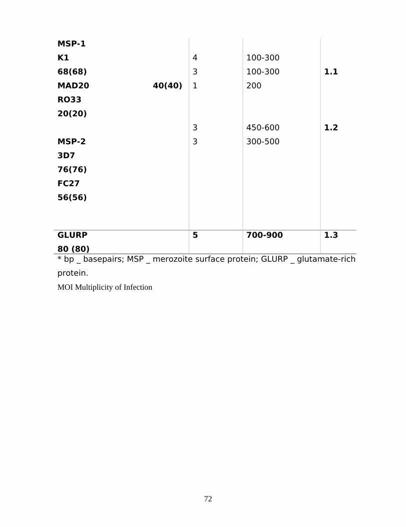
MSP-1
K1
68(68)
MAD20 40(40)
RO33
20(20)
MSP-2
3D7
76(76)
FC27
56(56)
4
3
1
3
3
100-300
100-300
200
450-600
300-500
1.1
1.2
GLURP
80 (80)
5 700-900 1.3
* bp _ basepairs; MSP _ merozoite surface protein; GLURP _ glutamate-rich
protein.
MOI Multiplicity of Infection
72
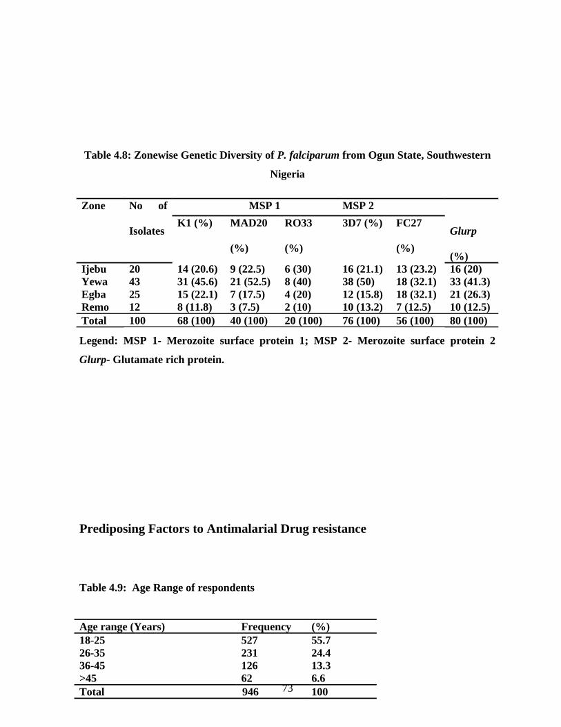
Table 4.8: Zonewise Genetic Diversity of P. falciparum from Ogun State, Southwestern
Nigeria
Legend: MSP 1- Merozoite surface protein 1; MSP 2- Merozoite surface protein 2
Glurp- Glutamate rich protein.
Prediposing Factors to Antimalarial Drug resistance
Table 4.9: Age Range of respondents
Zone No of
Isolates
MSP 1 MSP 2
Glurp
(%)
K1 (%) MAD20
(%)
RO33
(%)
3D7 (%) FC27
(%)
Ijebu 20 14 (20.6) 9 (22.5) 6 (30) 16 (21.1) 13 (23.2) 16 (20)Yewa 43 31 (45.6) 21 (52.5) 8 (40) 38 (50) 18 (32.1) 33 (41.3)Egba 25 15 (22.1) 7 (17.5) 4 (20) 12 (15.8) 18 (32.1) 21 (26.3)Remo 12 8 (11.8) 3 (7.5) 2 (10) 10 (13.2) 7 (12.5) 10 (12.5)Total 100 68 (100) 40 (100) 20 (100) 76 (100) 56 (100) 80 (100)
Age range (Years) Frequency (%)18-25 527 55.726-35 231 24.436-45 126 13.3>45 62 6.6Total 946 10073
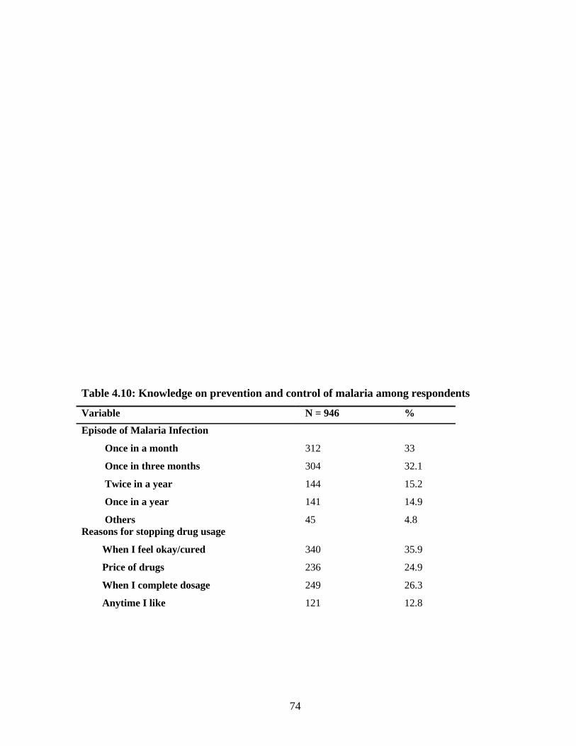
Table 4.10: Knowledge on prevention and control of malaria among respondents
Variable N = 946 %
Episode of Malaria Infection
Once in a month
Once in three months
Twice in a year
Once in a year
Others
312
304
144
141
45
33
32.1
15.2
14.9
4.8Reasons for stopping drug usage
When I feel okay/cured
Price of drugs
When I complete dosage
Anytime I like
340
236
249
121
35.9
24.9
26.3
12.8
74
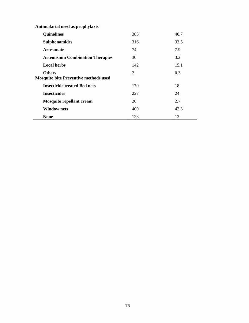
Antimalarial used as prophylaxis
Quinolines
Sulphonamides
Artesunate
Artemisinin Combination Therapies
Local herbs
Others
385
316
74
30
142
2
40.7
33.5
7.9
3.2
15.1
0.3Mosquito bite Preventive methods used
Insecticide treated Bed nets
Insecticides
Mosquito repellant cream
Window nets
None
170
227
26
400
123
18
24
2.7
42.3
13
75
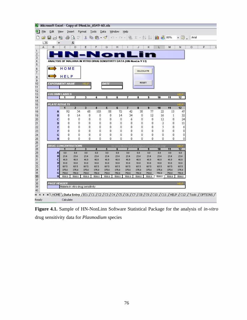
Figure 4.1. Sample of HN-NonLinn Software Statistical Package for the analysis of in-vitro
drug sensitivity data for Plasmodium species
76
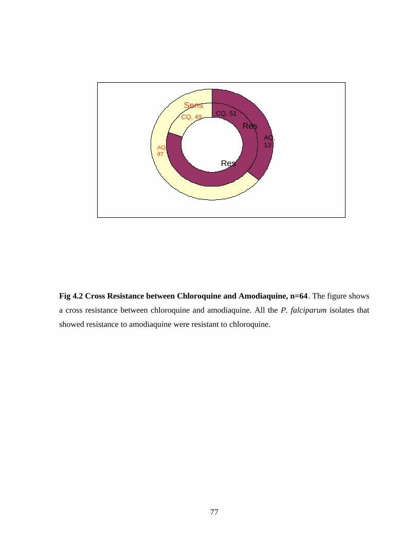
AQ,13AQ,
87
CQ, 51CQ, 49
Res
Res
Sens
Fig 4.2 Cross Resistance between Chloroquine and Amodiaquine, n=64. The figure shows
a cross resistance between chloroquine and amodiaquine. All the P. falciparum isolates that
showed resistance to amodiaquine were resistant to chloroquine.
77
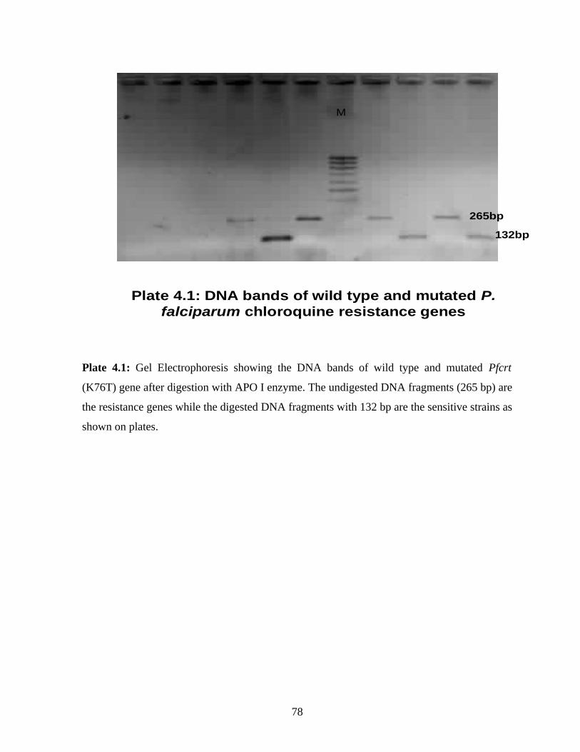
Plate 4.1: DNA bands of wild type and mutated P.falciparum chloroquine resistance genes
M
265bp
132bp
Plate 4.1: Gel Electrophoresis showing the DNA bands of wild type and mutated Pfcrt
(K76T) gene after digestion with APO I enzyme. The undigested DNA fragments (265 bp) are
the resistance genes while the digested DNA fragments with 132 bp are the sensitive strains as
shown on plates.
78
Plate Plate 4.2:4.2: P. falciparumP. falciparum Multidrug Resistance Multidrug Resistance Genes showing the wild type and mutated genesGenes showing the wild type and mutated genes
165bp
325bp
M
Plate 4.2 shows the Pfmdr1 (Y86N) genes on gel electrophoresis after digestion with Afl III
enzyme. The isolates with DNA fragments of size 325bp are the wild type i.e. sensitive strains
while the mutated strains were cut by the restriction enzyme to 165bp. M respresent the
molecular size marker
79
Plate 4.3: DNA band of Dihydrofolate reductase gene (DHFR 108)
322bp
708bp
M
Plate 4.3 shows the wild type and mutated genes of DHFR 108 for Sulphadoxine resistance.
The wild type gene has a band size of 708bp while the mutated gene has a band size of 322bp.
80
Plate 4.4: DNA band of Di hydropteroate synthase gene (DHPS 540)
M
441bp
756bp
M M
Plate 4.4 shows the DNA band of wild type and mutated genes of Dihydropteroate synthase
(DHPS 540). The band size of wild type is 756bp while that of the mutated gene is 441bp
81
Plate 4.5: DNA band of wild type Pf ATPase6
M M
The band size of the genes coding for pfATPase6 of between 430bp and 432bp is as shown on
plate 4.5. There was no digestion which means that none of the isolates harbour resistance
genes coding for artemisinin resistance.
82
M
M
180bp
300bp 200bp
Plate 4.6: DNA bands of P. falciparum MSP1 MAD20 on Gel
Legend: M-marker; bp-base pair; 180bp, 200bp 300bp - different alleles for MSP1 MAD family
The DNA bands of genes coding for P. falciparum Merozoite Surface Proteins 1 (MSP1)
MAD and K and RO33 are as shown on Plates 4.6, 4.7 and 4.8 respectively. The band size for
MAD is between 180 and 300 basepairs (bp) while that of K is 100-300bp. For RO33 the
band size in the range of 200 bp. M is the molecular size marker
83
Plate 4.7: DNA bands of P. falciparumMSP1 K1 on Gel
M
M
100bp
200bp
84

M
200bp 200bp
Plate Plate 4.8:4.8: DNA bands of DNA bands of P. falciparumP. falciparum MSP1 RO33 on GelMSP1 RO33 on Gel
Legend: M-Molecular marker; bp-basepairs; 200bp-allelic size
85
Plate 4.9: DNA bands of P. falciparum MSP2 3D7 on gel
M
M
500bp400bp
600bp
500bp
Legend: M-molecular marker; bp-basepairs; 400-600-allelic sizes
The DNA bands of genes coding for P. falciparum Merozoite Surface Proteins 2 (MSP-2)
3D7 and FC27 are as shown on Plates 4.9 and 4.10 respectively. The band size for 3D7 is
between 400 and 600bp while that of FC27 is 400-500bp. M is the molecular size marker.
86
Plate 4.10: DNA bands of P. falciparumMerozoite Surface Protein 2 FC27 on gel
M
400bp500bp
Legend: M-Marker; 400bp, 500bp- allelic sizes
87
Plate 4.11: DNA band of P. falciparum Glutarmate rich protein
900bp800bp
M
700bp
Legend: M-molecular marker; bp-basepairs; 700-900bp-allelic sizes
The DNA bands of genes coding for P. falciparum Glutamate Proteins are as shown on Plate
4.11. The band size is between 700 and 900 basepairs (bp).is 400-500bp. M is the molecular
size marker
88
CHAPTER FIVE
DISCUSSION
Plasmodium falciparum which causes the most serious type of malaria especially in sub
Saharan Africa was studied in this research work among the people of Ogun State Nigeria.
Malaria is the most prevalent tropical disease in the world today and in sub-Saharan Africa, it
is ranked among the most frequent causes of morbidity and mortality especially among
children and is often the leading identifiable cause (Greenwood et al., 2005). Four thousand
and sixty six (4066) subjects were included in the study on the incidence of P. falciparum
infection in Ogun state, South Western Nigeria. Two thousand, five hundred and fifty (62.7%)
were positive for falciparum malaria with an average parasitaemia of 918. This result is in
agreement with earlier observations that Nigeria is known for high prevalence of malaria and
it is a leading cause of morbidity and mortality in the country (Sowunmi et al., 2004;
Ademowo et al., 2006). The high incidence rate of falciparum malaria in the selected study
site is similar to the results of earlier studies in other parts of the country and other
neighboring countries. Adefioye et al. (2007) and Marielle et al. (2003) recorded a prevalence
rate of 72% and 70% among pregnant women in Oyo state, Nigeria and Gabon respectively.
A prevalence rate of 76% was also reported by Aribodor et al. (2003) in Anambra State,
South East Nigeria.
The result of this study was differs from that of Uko et al. (1998) who recorded a low
prevalence rate of (6.8%). This may be due to the fact that the study was carried out during
dry season alone when infection rate was low. Chanda et al., (2009) reported a very low
incidence of 0.7% among children less than five years attending a local health facility in
Zambia. In this study, 63.9% of children below one year and 70.8% of the children 1-5 years
were positive for falciparum malaria and highest parasitaemia was also observed in this age
group. This was followed by age range 16-25years with a prevalence of 52.8%. This findings
was in agreement with those of Aribodor et al. (2003) who also observed a high prevalence
rate among age group 10-19 years followed by age group 20-29 years old. In highly endemic
malarious area where semi-immune adults usually have substantially acquired resistance to
local strains of plasmodia, the prevalence of clinical malaria is higher and its severity greater
in pregnant women, children and young adults. The high prevalence rate in the study area
89
could result to cerebral malaria in children, maternal anaemia and low birth weight and death
as reported by other workers (Mockenhaupt et al., 2000; WHO, 2003). This is probably
because they are more exposed to malaria parasite due to bad environmental conditions and
their life styles
In-vitro method was used in this study to determine the quantitative drug response of P.
falciparum to various antimalarial drugs that are in use in Nigeria. Resistance to
antimalarial drugs can be assessed in vitro by the degree of parasite growth
inhibition exerted by particular antimalarial drugs (Basco and Ringwald, 2007; Oduola et al.,
1993). In any therapeutic strategy, establishment of the baseline sensitivity of local P.
falciparum isolates to commonly prescribed drugs that are still effective in some endemic
areas and new drugs that are not yet widely available in a country but might be employed in
the near future (e.g. artemisinin derivatives) is important. The in vitro assay is particularly
useful for fulfilling this function as it provides an objective, quantitative measure.
In-vitro resistance against chloroquine, amodiaquine and
sulphadoxine/pyrimethamine was observed among Nigerian isolates of P.
falciparum tested. In the cross sectional study, an IC50 of 24.4nM/L and IC99
of 164.2nM/L was recorded for chloroquine respectively. These results
were similar to earlier findings of Wongsrichanalai et al. (2000) where a high IC50
was also recorded. Resistance threshold of isolates against chloroquine
and amodiaquine were above 160nM/L and 80nM/L respectively which
denoted resistance against the drugs. Moreover, resistance threshold of
isolates against Sulphadoxine and pyrimethamine were 300nM/L and
4.0nM/L respectively. All the isolates tested against mefloquine, quinine
and artesunate in this study were sensitive to the drugs. The IC50 of
42.1nM/L observed in mefloquine in this study is slightly higher than that
recorded in earlier findings of Reber-Liske (1983) where IC50 of 39.7nM/L was
observed. The sensitivity ranges observed in the current study was also within the range for
sensitivity of isolates to the drug (WHO, 2001). In-vivo resistance against chloroquine,
amodiaquine, mefloquine and sulphadoxine /pyrimethamine has been reported by researchers
in Nigeria (Happi et al., 2004; Ogunfowokan et al., 2009). The in vivo response of P.
90
falciparum to antimalarial drugs is modulated by a number of factors. These include the
pharmacokinetic properties of antimalarial drugs, innate and acquired immunity in the patient,
as well as the complexity of infections in high transmission areas (Happi et al., 2004). Several
of these factors may contribute to the range of variations in the clinical expression of
chloroquine resistance and in vitro resistance patterns.
All the isolates tested in vitro against artemisinin were sensitive to the drug. However
treatment failures have been observed by other workers from Southwestern Nigeria during in
vivo studies (Happi et al., 2004). Bioavailability of the drugs in the system amongst other
factors has been attributed to treatment failures (Ibrahim et al., 2007). Although
dihydroartemisinin is the active ingredient in the artemisinin based drugs. Artesunate® which
contains artemisinin was used for the in vitro drug testing in this study. This is because it has
been reported that dihydroartemisinin is unstable on drug plates and that artemisinin is the
most appropriate drug for in vitro drug assays due to its stability in pre-dosed plates (Tanariya
et al., 2000).
High resistance against chloroquine and amodiaquine were observed among the isolates
collected from Yewa and Ijebu zones while SP resistance was not observed in isolates from
Remo zone. There was cross resistance between chloroquine and amodiaquine as some of the
isolates that showed resistance to chloroquine also showed resistance to amodiaquine in all
the zones. Cross-resistance between chloroquine and amodiaquine has been reported both in
vitro and in vivo. Pradines et al. (1999) have observed cross resistance of chloroquine and
amodiaquine in earlier studies. It has also been observed that parasites may quickly develop
resistance to amodiaquine in areas where extensive chloroquine resistance has been
documented (Winkler et al., 1994).
Antimalarial drug resistance is now generally acknowledged to be one of the greatest threats
to our ability to “Roll Back Malaria (Molta, 1995; Federal Ministry of Health 2004). The
situation is worsening, with the geographic spread of resistance widening to previously
unaffected areas and a remorseless increase both in the prevalence and degree of drug
resistance. Resistance to chloroquine and sulfadoxine-pyrimethamine (SP) is widespread in
Asia and South America and is spreading in Africa. This is evidenced in the results of this
study as there was in vitro resistance against chloroquine. The burden of disease caused by
91
malaria and its consequences has been documented in terms of childhood mortality, anemia,
maternal and infant morbidity and mortality, neurologic disability, and economic and social
costs. The burden caused specifically by antimalarial drug resistance is more difficult to
quantify. Resistance against chloroquine was observed in all the four zones where this present
study was carried out.
There is an increasing acceptance that the ideal approach to antimalarial treatment is the use of
combination of two or more drugs, rather than a single antimalarial drug, preferably with
artemisinin derivative as one of the drugs (WHO, 2001; Ashley et al., 2007). Amodiaquine in
combination with artesunate has been introduced as first-line treatment of malaria to replace
chloroquine in Nigeria and other malaria-endemic countries of Africa (Sodiomon et al., 2009).
Although the role of artesunate in this combination is to prevent the development of
amodiaquine resistance, parasites may quickly develop resistance to amodiaquine in areas
where extensive chloroquine resistance has been documented. In addition, little is known
about the mechanism or epidemiology of amodiaquine resistance. Resistant parasites may then
likely recrudesce under the selective force of the second drug in the combination and be
transmitted to mosquitoes (WHO, 2003).Therefore, we cannot rule out the possibility of
increasing selection of amodiaquine-resistant parasites with the increasing use of amodiaquine
in combination with artesunate in Nigeria.
Reduced in-vitro susceptibility is not synonymous with diminished therapeutic effectiveness,
but it is the probable first step of an alarming cascade and definitely pleads for increased
vigilance and a coordinated and rapid deployment of drug combinations. A previous study in
Nigeria assessed the quality of drugs from retail outlets and pharmacies and attributed
problems to a lack of quality control in manufacture and degradation during storage
(Onwujekwe et al., 2009). A major problem with the treatment of malaria is the high level of
treatment failures resulting in the large part from the high prevalence of counterfeit drugs
bought by the patients (Hall et al., 2006; Newton et al., 2006; Onwujekwe et al., 2009).
Anti- malarials are among the most widely consumed drugs in tropical countries that have
been particularly targeted by counterfeiters and of the anti-malarial drugs used in the world
today
92
Developing countries, where malaria is one of the most prevalent diseases, still rely on
traditional medicine as a source for the treatment of this disease. While synthetic
pharmaceutical agents continue to dominate research, increasing attention has been directed to
natural products (Alshwash et al., 2007). The success of artemisinin, isolated from Artemisia
annua, and its derivatives for the treatment of resistant malaria has focused attention on plants
as a source of antimalarial drugs (Tan et al., 1998). In this study, three crude organic extracts
obtained from medicinal plants used in Nigerian folk medicine for the treatment of fever and
malaria were tested in vitro against P. falciparum. The most active extract was obtained from
Diospyros monbuttensis that showed appreciable inhibition to the parasites at all the
concentrations used and an IC50 of 32μg/L in the study. Diospyros monbuttensis, which is
locally used for the treatment of fevers, headaches and stomach disorders, (Awe and Makinde,
1997; Azas et al., 2002) has not been widely studied. This study represents the first conducted
for antimalarial activity of crude extracts of Diospyros monbuttensis. The results confirm that
those plants which are used in traditional medicine against malaria may possess in vitro and
significant antimalarial potential and justify their use in traditional medicine. This observation
suggests that the active constituents in the extract may be cytotoxic for P. falciparum
trophozoites, thereby inhibiting their development to the schizont stage.
However, in vivo studies on these medicinal plants are necessary and should seek to determine
toxicity of the active constituents, their side effects, serum-attainable levels, pharmacokinetic
properties and diffusion in different body sites. Additional pharmacokinetic investigations are
therefore advisable to identify host-related factors, such as poor absorption, accelerated
gastrointestinal passage of the test drug, or metabolic peculiarities of some patients, which
might lead to a faster-than-normal inactivation or elimination of the test drug (Azas et al.,
2002). The use of herbs by some local people proved 100% sensitive to P. falciparum
(Merlin, 2004; Ogbonna, et al., 2008).
An IC50 observed for Momordica charantia in this study was 125μg/L. These observations
suggest that the active constituents in the extract might also be cytotoxic for P. falciparum
trophozoites, thereby inhibiting their development to the schizont stage. The antimalarial
activity of Momordica charantia has been previously reported (Alshwash et al., 2007). They
found that the aqueous extract of. M. charantia leaves showed IC50 values less than 100 µg/ml
which is in agreement with the observations in this study; the methanolic extract showed
93
moderate activity with IC50 =125 µg/ml. Morinda lucida also exhibited antimalarial activities
in this study. The IC50 of 250μg/L observed in this study is comparable with other studies.
Also for M. lucida, dose-dependent inhibitory outcomes were marked. Awe and Makinde,
(1997) reported the dose-dependent and seasonal variation in the activity of M. lucida using
both in vitro and in vivo techniques. M. lucida was reported to contain anthraquinones which
showed in vitro activity against P. falciparum and also possesses antifungal properties.
Morinda lucida is used locally in the treatment of yellow fever and jaundice (Guido et al.,
1995).
One of the factors to be considered in the prophylaxis, treatment, and control of Plasmodium
falciparum malaria is the resistance of parasite strains that may arise against virtually every
drug available. Identification of Pfcrt as the central determinant of chloroquine-resistant P.
falciparum malaria provides a molecular marker that can be used for surveillance of resistance
and to evaluate drug treatment and prophylaxis policies. The present results further support
this role of the Pfcrt gene. In the current study, 100 P. falciparum clinical isolates were
collected during 2007-2008 and 49 (49%) of these samples were found to be chloroquine-
sensitive and 51 (51%) chloroquine-resistant using the WHO in vitro microtest method.
Amplification products from all the chloroquine-sensitive samples carried the codon for Pfcrt
76K. Out of the 51 samples that were chloroquine-resistant by in vitro testing, 48 were found
to carry the 76T codon, 5 were found to contain mixed 76T/76K codons, and 3 were
discordantly found to contain the 76K codon. The results of work showed that there is a
positive correlation between in-vitro resistance pattern of isolates and prevalence of molecular
markers of resistance in the four zones. This finding corroborates with earlier findings where
molecular markers of resistance were found in samples that gave in vivo resistance/drug
failure (Djimde et al., 2001; Chen et al., 2001; Happi et al., 2004).
The high prevalence of mutations at codons 76T, that code for chloroquine resistance in
Nigerian P. falciparum isolates suggests that the population of P. falciparum that circulates in
South-Western Nigeria has been selected by the long use of CQ. The overall picture emerging
from this study is that resistance to this drug is abundant in south- Western Nigeria, and this
finding strongly supports withdrawal of CQ as the first-line drug for treatment of falciparum
malaria in Nigeria. This observation also supports the view that the Pfcrt polymorphism at
94
position 76 is in fact a significant factor of CQ resistance, as shown in previous studies from
Cameroon (Basco and Ringwald, 2001.), Mali (Djimde et al., 2001), Mozambique (Mayor et
al., 2001), Nigeria (Adagu and Warhurst, 2001), Sudan (Babiker et al., 2001), Uganda
(Kyosiimire-Lugemwa et al., 2002), Madagascar (Ariey et al., 2002), Laos (Labbe et al.,
2001), Papua New Guinea (Maguire et al., 2001), and Thailand (Chen et al., 2001; Jürgen and
Christian, 2003). CQ resistance has been attributed to a single mutation at codon 76 in the
Pfcrt gene (Djimde et al., 2001; Basco and Ringwald, 2001).
It has been observed that the Pfcrt K76 and the Pfmdr1 Y86 alleles are closely associated in
chloroquine resistant strains (Adagu and Warhurst, 1999). A similar observation was made in
this study as all isolates carrying Pfcrt mutated allele were also positive for Pfmdr1 mutated
allele. However, studies comparing the associations of the Pfmdr 1 variant and the Pfcrt K76
variant have shown that the impact of the pfcrt gene was stronger than that of the pfmdr gene
(Djimde et al., 2001; Dorsey et al., 2001; Ojurongbe et al., 2007). It has been suggested that
the degree of chloroquine resistance is further modulated by factors linked to genes other than
Pfcrt or Pfmdr (Chen et al., 2001).
Consistent with other studies, one can assume that the prevalence of Pfcrt T76 is a function of
the actual chloroquine level, age of the patient, and also influenced by acquired immunity and
natural resistance factors of the host (Wellems and Plowe 2001). Furthermore, one can assume
that chloroquine intake contributes essentially to the selection of the Pfcrt T76 allele. Based
on these observations, chloroquine appears to have an extended influence on the distribution
of the Pfcrt polymorphism in an isolate. Thus, sub-therapeutic blood levels of chloroquine
promote the emergence of drug resistance by direct selection.
Molecular methods that detect genetic markers of drug resistance are potentially powerful
tools for tracking drug-resistant malaria. In this study, the combination of Pfcrt and Pfmdr1
mutations in isolates associated with in vitro amodiaquine resistance was observed. Mutant
PfcrtT76 and Pfmdr1Y86 alleles were observed in 48% and 60% of the samples, respectively.
A previous study in Sudan (Babiker et al., 2001) found that the mutant PfcrtT76 allele is
associated with amodiaquine treatment failure. The high prevalence of the mutant PfcrtT76
allele (48%) and Pfmdr1 allele (60%) observed in Ogun State, Nigeria confirms recent reports
of the high prevalence rate of this alleles in parasites obtained from similar studies (Happi et
95
al., 2006) and is also consistent with rates ranging from 60% to 100% reported in other
malaria-endemic regions (Maguire et al., 2001; Basco and Ringwald, 2001; Djimde et al
2001). The non-significant selection of the mutant PfcrtT76 by amodiaquine may be due to
the high prevalence of this allele in the P. falciparum population from South Western Nigeria.
Selection of Pfmdr1Y86 by amodiaquine has also been reported previously in the Gambia
(Happi et al., 2003). Although the importance of point mutations in Pfcrt in producing
chloroquine resistance is beyond dispute (Basco and Ringwald, 2001; Maguire et al., 2001;
Chen et al., 2001) recent transfection studies of Pfcrt have shown that isolates expressing the
mutant PfcrtT76 allele retain sensitivity to amodiaquine while showing a reduced
susceptibility to monodesethyl amodiaquine, the active metabolite of amodiaquine (Happi et
al., 2003). The selection of the mutant PfcrtT76 and Pfmdr1Y86 alleles indicates the primary
involvement of these two genes in the mediation of amodiaquine resistance. Thus, similar to
chloroquine resistance, amodiaquine resistance in P. falciparum may depend primarily on
mutation(s) in Pfcrt and additional mutations in Pfmdr1 or other Plasmodium genes may also
have significant roles in increasing resistance to the drug.
The combination of pfcrtT76 and pfmdr1Y86 mutations was associated with amodiaquine
treatment failure. These two alleles have been shown to be in linkage disequilibrium in
chloroquine-resistant isolates of P. falciparum from The Gambia and Nigeria (Happi et al.,
2003). The similarity in the chemical structures of chloroquine and amodiaquine and their
possible likely common mode of action suggests that the molecular basis of resistance to these
two drugs may be similar.
Resistance against Sulphadoxine and Pyrimethamine was observed in this study. Among the
isolates analyzed for resistance markers, PfdhfrS108N and PfdhpsK540E genes coding for
Pyrimethamine and Sulphadoxine respectively were detected. It has been earlier observed that
Resistance to pyrimethamine is primarily conferred by a non synonymous point mutation at
codon 108 and is consecutively enhanced by mutations at codons 51 and 59 of the P.
falciparum pfdhfr gene located on chromosome 4 (Bruce-Chwatt, 1985; Kublin et al., 2002).
The enzyme is part of the folate pathway and, thus, of DNA replication. In this study genes
that code for sulphadoxine and pyrimethamine resistance were detected among the isolates
screened for markers of resistance against these drugs. PfdhfrS108N and PfdhpsK540E
alleles were detected in 12 and 2 isolates respectively.
96
On the basis of evolutionary theories, biological disadvantages are expected for parasites
carrying resistance-mediating mutations in the absence of drug pressure. The fitness deficit
conferred by the PfdhfrS108N mutation in the absence of pyrimethamine use is considered
quite low. Enduring resistance in the absence of strong drug pressure implies that the expected
decline in the prevalence of resistant parasites is balanced by mechanisms that confer
biological advantages with regard to survival fitness, replication and transmission probability,
invasion, reproduction, and vector properties that favor transmission. As Sub-Saharan African
countries are confronted with the rapid emergence of resistance against virtually every drug
that is used for the treatment of P. falciparum, malaria drug pressure is considered to
essentially promote the emergence of SP resistance, which is now widespread in East Africa,
but also well recognized in West Africa.
Selection for the Ser to Asn substitution at codon 108 of the Pfdhfr gene has been shown to be
linked to parasite survival after treatment with pyrimethamine -containing regimens (Plowe et
al., 1998; Marks et al., 2005). Accordingly, the high frequencies of resistant parasite
populations have been attributed to increased pyrimethamine consumption (Marks et al.,
2005). An Asp to Ile substitution at codon Pfdhfr51 (PfdhfrN51I) and/or a Cys to Arg
exchange at codon pfdhfr59 (pfdhfrC59R) appears to enhance pyrimethamine resistance if one
or both of these occur concurrently with PfdhfrS108N. PfdhfrS108N-N51I-C59R is the
combination of mutations most strongly associated with pyrimethamine resistance. Point
mutations at codons 437 and 540 of the Pfdhps gene located on chromosome 8 of P.
falciparum are considered responsible for sulphadoxine resistance. Pfdhps encodes a key
enzyme in the folate pathway, as does Pfdhfr. The Ala to Gly substitution at position 437
(PfdhpsA437G) is, in general, the first mutation to occur. In Africa this is followed by the Lys
to Glu substitution at position 540 (PfdhpsK540E), which confers higher levels of resistance.
It was recently shown that the presence of the three Pfdhfr mutations combined with the two
Pfdhps mutations (quintuple mutation) is strongly associated with SP resistance (Marks et al.,
2005).
Geographical clustering was reported for chloroquine-resistant Pfcrt (Wootton et al., 2002)
and anti folate resistant Pfdhfr and Pfdhps haplotypes (Cortese et al., 2003), indicating distinct
ancestral selection events in different areas. One important aspect in the possible setting
97
leading to drug resistant parasites in Western Nigeria is the genetic background of the local
parasite population. Multidrug-resistant P falciparum is common, with a high rate of Pfcrt,
Pfmdr1, Pfdhfr and Pfdhps resistance haplotypes. Thus, selection pressures were exerted on a
heavily mutated genetic background. The several PfATPase6 haplotypes harbouring the
S769N mutation indicate that selections of such mutants are therefore not impossible in the
nearest future. As chloroquine and SP are replaced by more effective artemisinin-based
combination therapies (ACTs), strategies for monitoring (and, if possible, deterring) drug-
resistant malaria must be updated and optimized. In vitro methods for measuring resistance
will be critical for confirming and characterizing resistance to ACTs. Molecular markers are
useful for tracking the emergence and dissemination of resistance and guiding treatment policy
where resistance is low or moderate.
One hundred samples that were positive for P. falciparum by microscopy were subjected to
PCR genotyping for estimating prevalence of the PfATPase codon S769N mutation. All the
100 (100%) isolates carried the artemisinin sensitive wild type allele, S769. This observation
is similar to the ones observed in several African countries where artemisinin and its
derivatives are used as first line of treatment of uncomplicated malaria (Basco and Ringwald,
2001; Djimde et al., 2001; Mayor et al., 2001; Adagu and Warhurst, 2001; Babiker et al.,
2001; Kyosiimire-Lugemwa et al., 2002). However Artemisinin resistant genes have been
detected by some workers especially in South Asia (Labbe et al., 2001; Maguire et al., 2001;
Chen et al., 2001; Jürgen and Christian, 2003). Artemisinin derivatives are an essential
component of treatment against multidrug-resistant falciparum malaria. The genes that code
for artemisinin resistance was not detected in any of the isolates screened for antimalarial
resistance genes. Widespread multidrug-resistant falciparum malaria led WHO to recommend
combination drug therapy as first-line treatment, with formulations containing an artemisinin
compound as policy standard. Artemisinin and its derivatives are the most potent and rapidly
acting antimalarials. However, artemisinin resistance has been reported in murine models of
malaria (Ferrer-Rodiriguez et al., 2004). Diligent surveillance is needed to monitor continued
susceptibility to artemisinin derivatives in endemic areas.
Numerous studies have demonstrated that children under five years and pregnant women are
at a higher risk of suffering from clinical malaria (Sachs and Malaney, 2002; Greenwood et
98
al., 2005; Adefioye et. al., 2007; Sotimehin et al., 2008), and drug prevention has been
advocated for years, using either weekly or monthly prophylaxis or, more recently in several
countries, intermittent presumptive treatment, IPT (Cot and Deloron, 2003). At a time, many
countries in the Sub Saharan region recommended a weekly chloroquine prophylaxis for each
pregnant woman from the beginning of pregnancy to delivery. One possible drawback of such
increased drug consumption is an increased drug pressure that may lead to the selection of
drug-resistant parasites. There is some evidence in favor of this hypothesis. For instance, in
countries where Artemisinin resistance has been discovered, all resistant isolates came from
areas with uncontrolled use of artemisinin derivatives (Ronan et al., 2005)
There is little existing knowledge about actual quality of drugs provided by different
providers in Nigeria and in many sub-Saharan African countries. Such information is
important for improving malaria treatment that will help in the development and
implementation of actions designed to improve the quality of treatment. A study conducted in
South-East Nigeria found that there was a high prevalence of poor quality drugs (Onwujekwe
et al., 2009). Such findings provide areas for public intervention to improve the quality of
malaria treatment services and to forestall resistance.
The Merozoite Surface Protein1 (MSP-1) and Merozoite Surface Protein2 (MSP-2) are highly
polymorphic markers and the large allelic polymorphism has been reported in the block 2 of
the msp-1 gene and the central repetitive domain (block3) of the msp-2 gene. Families
differing in nucleotide sequences and in number of repetitive sequences (length variation)
were used for genotyping purposes. This study evaluates the extent of genetic diversity in the
field isolates of P. falciparum obtained from Ogun State in South Western Nigeria. The
population structure of the isolates analyzed with the polymorphic markers MSP-1, MSP-2,
and Glutamate Rich Protein (GLURP) in this study showed extensive diversity in parasite
populations in the four zones in Ogun State, South Western Nigeria. The MSP-1, MSP-2, and
GLURP markers showed 8, 6, and 5 allelic families, respectively. This diversity of the P.
falciparum population in South Western Nigeria is reflected in the complexity of parasite
populations in the samples. A catalog of genetically distinct parasite populations co-infecting
those infected with malaria, based on PCR amplification of these markers, showed that
multiplicity of infection was common. Multiclonality of infections has been shown to be a
99
common feature in most malaria-endemic areas (Happi et al., 2004; Ntoumi et al., 1995;
Magesa et al., 2001; Nzila et al., 2006). Epidemiologic data from some study sites in Africa
suggest that the multiplicity of P. falciparum infection may be directly related to the intensity
of transmission (Arnot, 1998; Babiker et al., 1997). This multiplicity of infections may also
have important implications for the epidemiology of drug-resistant P. falciparum malaria and
the outcome of treatment in patients. The initial presence of several parasite populations with
different drug response profiles would result in elimination of drug sensitive populations and
selection of resistant parasites. The findings in this study are in agreement with those of
previous studies involving asymptomatic carriers and symptomatic patients in holoendemic
areas, both in the complexity of population structure and multiplicity of parasites in human
hosts (Ntoumi et al., 1995; Snounou et al., 1999). All the MSP 1, MSP2 and Glurp families
were represented in each of the four zones in Ogun state where samples were collected.
In considering possible strategies for the reduction of the burden of antimalarial drug
resistance, it is useful to differentiate between the current burden of drug resistance and the
potential burden in the future resulting from the continued emergence and spread of drug
resistance. The factors that are likely to contribute to the development of antimalarial
resistance were studied through administration of questionnaire to people who live in the four
zones of Ogun State, South Western Nigeria. 33% of respondents do have malaria at least
once in a month and 32.1 would have malaria at least once in three months respectively. This
means about 80.3% will have malaria at least once in a year. It was also found in this study
that the rate of exposure to antimalarial drugs including the artemisinins is very high. 40.7%
of the sample population use quinolines as prophylaxis while more than 33% use
sulphonamides and about 3.2% use artemisinins as prophylaxis. As long as drugs are used, the
chance of resistance developing to those drugs is present (Plowe, 2003). It has been observed
that the development of resistance to antimalarial drugs in South-East Asia has been far
quicker than the estimated 12 to 17 years it takes to develop and market a new antimalarial
compound (Ridley, 1997).
Affordability is an essential consideration for any strategy to control drug-resistant malaria,
especially in Africa (Foster and Phillips 1998; Goodman et al., 1999). In the current study,
about 25% of the sample population stops the use of drugs because of price or when they
100
finish the one they could afford. The future, especially in Africa, will also be defined by how
well the central tenets of malaria control can be reconciled with the central tenets of control of
drug resistance. One of the cornerstones of the current approach to malaria control is the
provision of prompt, effective malaria treatment. In much of Africa, easy access to public
sector health care is limited and when it is accessible, health care staff are often inadequately
trained, insufficiently supplied and supported, ineffectively supervised and poorly motivated
(Goodman et al., 2000). Central to achieving a reduction in both current and future burdens is
an improvement in drug usage by patients and providers so that good quality drugs are
available and taken at the correct dose and for a sufficient length of time to affect a radical
cure and reduce the likelihood that partially resistant parasites will survive. Improving drug
use is most effective where the parasite is still sensitive to the drug. Where resistance has
rendered the drug ineffective, the current burden of resistance can only be reduced by
replacing the failing drug regimen with one that is effective. The difficulty lies in deciding
which drug regimen to switch to, since the choice of drug or drug combination will determine
the subsequent development of drug resistance. Reducing the future burden of resistance
requires that effective antimalarial drugs continue to be available in the future and requires the
continuous search for and development of potential new antimalarial drugs.
However, the complete drug development process can take 10−15 years, making it imperative
that the currently available drugs are deployed in a way most likely to maximize their lifespan
by decreasing the likelihood that resistance will develop. The key strategy put forward to do
this is to use available drugs in combination to prevent the emergence and spread of
resistance. Once a drug-resistant mutant has arisen, preventing spread of resistance is difficult.
Spread is facilitated by the exposure of malarial parasites to sub-therapeutic levels of
antimalarial drugs, that kill sensitive parasites but allows parasites with a resistance mutation
to survive and reproduce. Ensuring that drugs are taken in at a sufficient dose and for a
sufficient duration reduces this risk. Drug pressure is higher where a drug with a long half-life
is taken because the drug remains in the patient’s blood at low levels for weeks, exposing any
newly introduced malarial parasites to sub-therapeutic levels (Bloland, 2001).
This is particularly likely to occur in high transmission areas where people are not only
infected more frequently, but also take antimalarial drugs frequently whether or not they are
having malaria. Theoretically,this form of drug pressure can be reduced by using drugs with a
101
shorter half-life and by restricting the use of the first line drug to patients with confirmed
malaria: i.e., only treating those with a definitive diagnosis.
CONCLUSION
This study has established the incidence of falciparum malaria in Ogun State, Southwestern
Nigeria. Moreover, a baseline for both genetic polymorphisms and drug sensitivity profiles of
P. falciparum isolated from Ogun State Nigeria has been established. This data should serve
as a baseline for future studies to monitor the changes in antimalarial drug resistance in this
part of the country. To prevent resistance to antimalarial drugs, a high degree of vigilance is
required; the level of antimalarial drug sensitivity of P. falciparum should be closely
monitored while compliance to antimalarial drug use should be encouraged.
102
CONTRIBUTIONS TO KNOWLEDGE
This research work has contributed to knowledge in the following ways.
1. The incidence of malaria (Plasmodium infection) in Ogun state, South Western
Nigeria has been established. This would help in planning prevention/control
operations, resource allocation, and upgrading medical facilities in hospitals to treat severe
and complicated cases.
2. The resistance pattern of Plasmodium falciparum isolates from Ogun State to existing
antimalarial drugs established in this study would tailor drug choices specific to
geographic regions.
3. The molecular diversity of P. falciparum strains in Ogun State, Southwestern Nigeria
has been established. This Knowledge would help to type and characterize the P.
falciparum strains in Nigeria and compare their diversity with the rest of the world.
4. Results from molecular studies on the drug resistance genes of P. falciparum obtained
in this study would provide advance information on the emergence of drug resistance
in the field. This knowledge should facilitate the ability to begin to anticipate genomic
responses to drugs yet unseen by the parasite.
5. Some of the factors which contribute to the development of resistance of Plasmodium
to antimalarial drugs have been established. This should lead to the development of
strategies and policies to control the use of existing antimalarial drugs and forestall
resistance to new drugs.
6. Antimalarial efficacy of some local herbs used as herbal drugs has been established.
This is expected to lead to the discovery of novel antimalarial drug of local origin.
103
RECOMMENDATION
There is an urgent need to find and develop alternative drugs against multidrug-resistant P.
falciparum. One group of alternative antimalarial drugs comprises artemisinin (qinghaosu)
and its derivatives. Artemisinin is a traditional Chinese medicinal herb derived from
Artemisia annua. Newer antimalarial drugs and approaches to overcome parasite resistance
are needed to deal with the expanding problem of drug resistance which continues to
challenge malaria control efforts based on early diagnosis and treatments. Only a limited
number of antimalarial drugs are currently at an advanced stage of clinical development.
There must therefore be a renewed interest in plant products. An attractive option for poor and
developing countries is the exploitation of the possible therapeutic effects of their local herbs.
Efforts must be made to reduce drug pressure which can lead to the selection of drug-resistant
parasites.
Since there was positive correlation between artesunate and mefloquine; artesunate quinine,
drugs containing these combinations should be recommended for Ogun State, the study area.
There is little existing knowledge about actual quality of drugs provided by different
providers in Nigeria and in many sub-Saharan African countries. Such information is
important for improving malaria treatment that will help in reducing the development of
resistance to the antimalarial drugs in use.
104
REFERENCES
Abu-Raddad L, Patnaik P, Kublin J (2006). "Dual infection with HIV and malaria
fuels the spread of both diseases in sub-Saharan Africa". Science 314(5805):
1603-1606.
Adagu IS, Warhurst DC (1999). Association of cg2 and pfmdr1 genotype with chloroquine
resistance in field samples of Plasmodium falciparum from Nigeria.
Parasitology 119: 343–348.
Adagu IS, Warhurst DC (2001). Plasmodium falciparum: linkage disequilibrium between
loci in chromosomes 7 and 5 and chloroquine selective pressure in northern
Nigeria. Parasitology 123: 219–224.
Adefioye OA, Adeyeba OA, Hassan WO and Oyeniran OA (2007). Prevalence of Malaria
Parasite Infection among Pregnant Women in Osogbo, Southwest, Nigeria.
American-Eurasian Journal of Scientific Research 2 (1): 43-45.
Ademowo OG, Nneji CM and Adedapo ADA (2006). In vitro antimalarial activity of
methylene blue against field isolates of Plasmodium falciparum from children in
Southwest Nigeria. Indian Journal of Medical Research 126:45-49
Adjuik M, Babiker A, Garner P, Olliaro P, Taylor W, White N (2004). International
Artemisinin Study Group: Artesunate combinations for treatment of malaria:
metaanalysis. Lancet 363:9-17.
Alene GD and Bennett S (1996). Chloroquine resistance of Plasmodium falciparum
malaria in Ethiopia and Eritrea. Tropical Medicine & International Health
1:810–815.
Ali AN, Al-rahwi AK and Lindequist U (2004). Some medicinal plants used in Yemeni
herbal medicine to treat malaria. African Journal of Traditional Medicine 1: 72–
76.
Ali AN, Attif OA and Mohammed MI (1999). Herbal medicine in two Yemeni provinces-
ethno botanical study. Yemeni Medical Journal 3:13–23.
Alifrangis M, Lemnge MM, Ronn AM, Segeja MD, Mageja SM, Khalil IF, and Bygbjerg
IC (2003). Increasing prevalence of wild types in the dihydrofolate reductase
gene of Plasmodium falciparum in an area with high levels of sulphadoxine-
pyrimethamine resistance after introduction of treated bed nets. American
Jornal of Tropical Medicine Hygiene 69:238-243.
105
Alshawsh MA, Mothana RA, Al-shamahy HA, Alsllami SF and Ulrike L (2007). Assessment
of antimalarial activity against Plasmodium falciparum and phytochemical
screening of some Yemeni medicinal plants.
eCAM, doi:10.1093/ecam/nem148
Aribodor UDN, Njoku OO, Eneanya CI and Onyali IO (2003). Studies on prevalence of
malaria and management Practices of the Azia community, Ihiala LGA, Anambra
State, Southeast Nigeria. Nigerian Journal of Parasitology 24:33-38.
Ariey F, Randrianarivelojosia M, Duchemin JB, Rakotondramarina D, Ouledi A, Robert V,
Jambou R, Jahevitra M, Andrianantenaina H, Raharimalala L and Mauclere P
(2002). Mapping of a Plasmodium falciparum pfcrt K76T mutation: a useful
strategy for controlling chloroquine resistance in Madagascar. Journal of
Infectious Diseases 185: 710–712.
Arnot D (1998). Unstable malaria in Sudan: the influence of the dry season. Clone
multiplicity of Plasmodium falciparum infections in individuals exposed to
variable levels of disease transmission. Transactions of the Royal Society of
Tropical Medicine and Hygiene 92: 580–585.
Arora DR and Arora B (2005). Protozoa. Medical Parasitology 2nd ed, CBS publishers,
India. 67-98
Ashley AA, Stepniewska K, Lindegårdh N, McGready R, Hutagalung R, Singtoroj T, Hla
G, Brockman A, Proux S, Wilahphaingern J, Singhasivanon P, White N J and
Nosten F (2007). Pharmacokinetic study of artemether-lumefantrine given once
daily for the treatment of uncomplicated multidrug-resistant falciparum
malaria. Tropical Medicine and International Health 12:201-208.
Aubouy A, Migot-Nabias F and Deloron P (2003a). Polymorphism in two merozoite surface proteins of Plasmodium falciparum isolates from Gabon. Malaria Journal 2:12
Aubouy A, Bakary M, Keundjian A, Mbomat B, Makita JF, Cot M, Le Bras J and Deloron P
(2003b). Combination of drug level measurement and parasite genotyping data
for an improved assessment of amodiaquine and sulfadoxine-pyrimethamine
efficacy in treating Plasmodium falciparum malaria in Gabonese children.
Antimicrobial Agents Chemotherapy 47:231-237
106
Awe SO and Makinde, JM (1997). Evaluation of the antimalarial activity of Morinda lucida
using both in vivo and in vitro techniques. West African Journal of Pharmacological
Drugs Research 13 (1 & 2): 39-44.
Azas N, Laurencin N, Delmas F, Di-Giorgio C, Gasquet M., Laget M and Timon-David, P
(2002). Synergistic in vitro antimalarial activity of plant extracts used as traditional
herbal remedies in Mali. Parasitology Research 88: 165-171.
Babiker HA and Walliker D (1997). Current views on the population structure of
Plasmodium falciparum: implications for control. Parasitology Today 13:262–
267.
Babiker HA, Lines J, Hill WG, Walliker D (1997). Population structure of Plasmodium
falciparum in villages with different malaria endemicity in east Africa. American
Journal of Tropical Medicine and Hygiene 56:141–147.
Babiker HA, Pringle SJ, Abdel-Muhsin A, Mackinnon M, Hunt P and Walliker D (2001).
High-level chloroquine resistance in Sudanese isolates of P. falciparum is
associated with mutations in the chloroquine resistance transporter gene pfcrt and
the multidrug resistance gene pfmdr1. Journal of Infectious Diseases 183:1535–
1538.
Ballereau F (1997). Stability of essential drugs in the field: results of a study conducted over
a two-year period in Burkina Faso. American Journal of Tropical Medicine and
Hygiene 57:31–36.
Basco LK (1991). Inefficacy of amodiaquine against chloroquine- resistant malaria.
Lancet 338:1460–1460.
Basco LK (2007). Field application of in-vitro assays for the sensitivity of human malaria
parasites to antimalarial drugs. WHO/Global Malaria Programme/WC/750. France. 5-
135.
Basco LK and Ringwald P (2000). Molecular epidemiology of malaria in Yaounde,
Cameroon. VII. Analysis of recrudescence and reinfection in patients with
uncomplicated falciparum malaria. American Journal of Tropical Medicine and
Hygiene 63: 215– 221.
107
Basco LK and Ringwald P (2001). Analysis of the key pfcrt point mutation and in vitro and
in vivo response to chloroquine in Yaounde, Cameroon. Journal of Infectious
Diseases 183: 1828–1831.
Basco LK and Ringwald P (2007). Molecular epidemiology of malaria in Cameroon. XXIV.
Trends of in vitro antimalarial drug responses in Yaounde, Cameroon. American
Journal of Tropical Medicine and Hygiene 76(1):20-26.
Bauchner H, Pelton SI and Klein JO (1999). Parents, physicians, and antibiotic use.
Pediatrics 103:395–401.
Beck HP (1999). How does molecular epidemiology help to understand malaria? Tropical
Medicine and International Health 4:1–3.
Berzins K, and Anders RF (1999). The malaria antigens in Wahlgren M, Perlmann P, eds.
Malaria: Molecular and Clinical Aspects. Amsterdam: Harwood Academic
Publishers, 181−216.
Billker O, Lindo V, Panico M, Etienne AE, Paxton T, Dell A, Rogers M, Sinden RE,
Morris (1998). "Identification of xanthurenic acid as the putative inducer of
malaria development in the mosquito." Nature 392: 289–292.
Bloland BP (2001). Drug Resistance in Malaria. WHO/CDS/CSR/DRS/2001.4:1
Bloland PB, Kazembe PN, Watkins WM, Doumbo OK, Nwanyanwu OC, Ruebush TK.
(1997). Malarone-donation programme in Africa. Lancet 350:1624–1625.
Bogreau H, Renaud F, Bouchiba H, Durand P, Assi SB, Henry MC, Garnotel E, Pradines B,
Fusai T, Wade B, Adehossi E, Parola P, Kamil MA, Puijalon O, and Rogier
C (2006). Genetic diversity and structure of African Plasmodium falciparum
populations in urban and rural areas. American Journal of Tropical Medicine
and Hygiene 74: 953-959.
Boivin MJ (2002). "Effects of early cerebral malaria on cognitive ability in Senegalese
children," Journal of Developmental and Behavioral Pediatrics 23 (5):353–364.
Bourdy M, Willcox L, Ginsburg H, Rasoanaivo P, Graz B, and Deharo E (2008).
Ethnopharmacology and malaria: New hypothetical leads or old efficient
antimalarials? International Journal for Parasitology 38: 33–41.
108
Breman J (2001). "The ears of the hippopotamus: manifestations, determinants, and
estimates of the malaria burden.” American Journal of Tropical Medicine and
Hygiene 64: 1-11.
Bruce Chwatt LJ (1985). Essential Malariology. 2nd Ed. London, Heinemanna. 61-270
Chanda P, Hamainza B, Mulenga S, Chalwe V, Msiska C and Chizema-Kawesha E
(2009).Early results of integrated malaria control and implications for the
management of fever in under-five children at a peripheral health facility: A
case study of Chongwe rural health centre in Zambia. Malaria Journal 8:49
Cheesbrough M. (2000). District laboratory practice manual in Tropical Countries pt 2.
Cambridge University press.
Chen N, Russell B, Staley J, Kotecka B, Nasveld P and Cheng Q (2001). Sequence
polymorphisms in pfcrt are strongly associated with chloroquine resistance in
Plasmodium falciparum. J Infect Dis 183: 1543–1545.
Childs GE, Wimonwattrawatee T, Pooyindee N (1988). Evaluation of an in vitro
assay system for drug susceptibility of field isolates of Plasmodium falciparum
from southern Th ailand. American Journal of Tropical Medicine and Hygiene
38:19–23.
Clarkson C, Maharaj VJ, Crouch NR, Grace OM, Pillay P, Matsabisa MG (2004). In
vitro antiplasmodial activity of medicinal plants native to or naturalised in
South Africa. Journal of Ethnopharmacology 92: 177–191.
Cortese JF, Caraballo A, Contreras CE, Plowe CV (2003). Origin and dissemination of
Plasmodium falciparum drug-resistance mutationsin South America. Journal of
Infectious Diseases 186: 999–1006.
Cot, M. and Deloron, P. (2003). Malaria prevention strategies: pregnancy-associated
malaria (PAM). British Medical Bulletin 67:137–148.
Cowman AF (2006). "Invasion of Red Blood Cells by Malaria Parasites". Cell 124:
755–766.
Craig MH and Sharp BL (1997). Comparative evaluation of four techniques for the
diagnosis of Plasmodium falciparum infections. Transactions of the Royal
Society of Tropical Medicine and Hygiene 91:279–282.
109
de Monbrison F, Raynaud D, Latour-Fondanaiche C, Staal A, Favre S, Kaiser K, Peyron F
and Picot S (2003). Real-time PCR for chloroquine sensitivity assay and for
pfmdr1-pfcrt single nucleotide polymorphisms in Plasmodium falciparum J
Microbiological Methods. 54:391–401.
Desai M, ter Kuile FO, Nosten F, McGready R, Asamoa K, Brabin B,
Newman RD (2007).
Epidemiology and burden of malaria in pregnancy. Lancet Infect Dis
7:93-104.
Djimde A, Doumbo OK, Cortese JF, Kayentao K, Doumbo S, Diourte Y, Dicko A, Su XZ,
Nomura T, Fidock DA, Wellems TE, Plowe CV, Coulibaly D, 2001. A molecular
marker for chloroquine-resistant falciparum malaria. New England Journal of
Medicine 344: 257–263.
Dorsey G, Kamya MR, Singh A, Rosenthal PJ (2001). Polymorphisms in the
Plasmodium falciparum pfcrt and pfmdr-1 genes and clinical response to
chloroquine in Kampala, Uganda. Journal of Infectious Diseases 183:1417-
1420.
Dorsey G, Staedke S, Clark TD, Njama-Meya D, Nzarubara B, Maiteki-Sebuguzi C,
Dokomajilar C, Kamya MR and Rosenthal PJ (2007). Combination therapy for
uncomplicated falciparum malaria in Ugandan children: a randomized trial. Jama
297:2210-2219.
Dorvault F (1982). L’officine, xxi ed. Vigot. Paris. Pp 110
Druilhe P, Moreno A, Blanc C, Brasseur P, and Jacquier P (2001). Colorimetric in vitro
drug sensitivity assay for P. falciparum based on a highly sensitive double- site
pLDH antigen capture ELISA assay. American Journal of Tropical Medicine
and Hygiene 64:233-241
Druilhe P, Philippe B, Catherine B, and Michael M (2007). Improved Assessment of
Plasmodium vivax Response to Antimalarial Drugs by a Colorimetric Double-
Site Plasmodium Lactate Dehydrogenase Antigen Capture Enzyme-Linked Immunosorbent
Assay. Antimicrobial Agents Chemotheapy 51(6): 2112–2116.
Eckstein_Ludwig U, Webb RJ, Van Goethem ID, East JM, Lee AG, Kimura M, O’Neill PM,
Bray PG, Ward SA, and Krishna S (2003). Artemisinins target the SERCA of
P. falciparum. Nature 21:957-961
110
Elujoba AA (1998). The role of pharmacognosy in phytotherapy, the challenges of our time.
Nigerian Journal of Natural Products and Medicine 2: 5-8.
Elujoba AA, Odeleye OM and Ogunyemi CM (2005). Traditional medicine development for
medical and dental primary health delivery systems in Africa. African Journal of
Traditional Medicine 21:46-61.
Etkin NL (2003). The co-evolution of people, plants, and parasites: biological and
cultural adaptations to malaria. Proceedings of the Nutritional Society 62:
311–317.
Ettling M, Bloland, PB, Ruebush, TK (1995). Chloroquine efficacy study in Zambia
Environmental Health Project Activity Report 15.
Federal Ministry of Health (2004). National Antimalarial Treatment report 4-5
Federal Ministry of Health, National Malaria and Vector Control Division (2001).
Malaria. 5
Ferrer-Rodiriguez I, Perez-Rosado J, Gervais GW, Peters W and Robinson BL (2004).
Plasmodium yoelii: identification and partial characterization of an mdr1
gene in an artemisinin-resistant line. Journal of Parasitology 90:152–160.
Fidock DA, Nomura T, Cooper RA, Su X, Talley AK, Wellems TE, (2000). Allelic
modifications of the cg2 and cg1 genes do not alter the chloroquine response
of drug-resistant Plasmodium falciparum. Molecular Biochemistry and
Parasitology 110: 1–10.
Flores MVC, Berger-Eiszele SM, Stuart TS (1997). Long-term cultivation of
Plasmodium falciparum in media with commercial non-serum
supplements. Parasitological Research 83: 734–6.
Foege WH (1997). Malarone-donation programme. Lancet 350:1628–1629.
Foley M and Tilley L (1997). Quinoline antimalarials: mechanisms of action and resistance.
International Journal for Parasitology 27:231–240.
Foley M and Tilley L (1998). Quinoline antimalarials: mechanisms of action and resistance and
prospects for new agents. Pharmacology and Therapeutics 79(1): 55-87.
Foster S and Phillips M (1998). Economics and its contribution to the fight against
malaria. Annals of Tropical Medicine & Parasitology 92:391–398.
Gilles HM, Warrell DA and Edward A (1993). Diagnostic Methods in Malaria. Bruce
Chwatt’s Essential Malariology 3rd ed London 78-95.
111
Golenda CF, Li J and Rosenberg R (1997). Continuous in vitro propagation of the
malaria parasite Plasmodium vivax. Proceedings of the National Academy of
Sciences of the United States of America 94:6786–6791.
Good MF (2001). Towards a blood-stage vaccine for malaria: are we following all the leads?
Nature Reviews, Immunology 1: 117-125.
Goodman CA, Coleman PG, Mills AJ (1999). Cost effectiveness of malaria control in sub-
Saharan Africa. Lancet 354:378–384.
Goodman C, Coleman P and Mills A (2000). Economic analysis of malaria control in sub-
Saharan Africa. Strategic Research Series 1. Global Forum for Health Research
(World Health Organisation, Geneva).
Greenwood B and Mutabingwa T (2002). "Malaria in 2002". Nature 415: 670–672.
Greenwood BM, Bojang K, Whitty CJ, Targett GA (2005). "Malaria". Lancet 365:
1487-1498.
Guido R, Menavanza N and Kurt H (1995). Antifungal anthaquinones from Morinda lucida.
International Journal of Pharmacology 33(2): 107-114.
Hall KA, Newton PN, Green MD, DeViej M, Vandenabeele P, Pizzanelli D,
Mayxay M, Dondorp A and Fernandez FM (2006). Characterization of
counterfeit artesunate antimalarial tablets from southeast Asia. American
Journal of Tropical Medicine and Hygiene 75:804-811.
Hall AP (1975). Amodiaquine resistant falciparum malaria in Thailand. American Journal of
Tropical Medicine & Hygiene 24:575–580.
Handunnetti, S. M., D. M. Gunewardena, P. P. Pathirana, K. Ekanayake,
S.Weerasinghe, and K. N. Mendis (1996). Features of recrudescent
chloroquine-resistant P. falciparum infections confer a survival advantage on
parasites and have implications for disease control. Transactions of the Royal
Society for Tropical Medicine and Hygiene. 90:563–567.
Happi TC, Thomas SM, Gbotosho GO, Falade CO, Akinboye DO, Gerena L, Hudson T,
Sowunmi A, Kyle DE, Milhous W, Wirth DF, Oduola AM (2003). Point
mutations in the pfcrt and pfmdr-1 genes of Plasmodium falciparum and
clinical response to chloroquine, among malaria patients from Nigeria.
Annals of Tropical Medical Parasitology 97: 439–451.
112
Happi CT, Gbotosho GO, Sowunmi A, Falade CO, Akinboye DO, Hudson T, Gerena L,
Kyle DE, Milhous W, Wirth DF, Oduola AMJ (2004). Molecular analysis of
Plasmodium falciparum recrudescent malaria infections in children
treated with chloroquine in Nigeria. American Journal of Tropical Medicine and
Hygiene 70: 20–26
Hastings IM (2003). Malaria control and evolution of drug resistance: an intriguing
link. Trends in Parasitology 19:70-73.
Hastings IM (1997). A model for the origins and spread of drug-resistant malaria.
Parasitology 115:133–141.
Hastings IM and Mckinnon MJ (1998). The emergence of drug-resistant malaria.
Parasitology 117:411–417.
Hay S, Guerra C, Tatem A, Noor A and Snow R (2004). "The global distribution and
population at risk of malaria: past, present, and future." Lancet 4(6): 327-336.
Hay SI and Snow RW (2006). "The Malaria Atlas Project: Developing Global Maps of
Malaria Risk." PLoS Medicine 3(12): e473.
Hien TT and White NJ (1993). Qinghaosu. Lancet 341:603-608.
Hoareau L and Dasilva EJ (1999). Medicinal plants: a re-emerging health aid. Electronic
Journal of Biotechnology 2(2): 56-69.
Holding PA and Snow RW (2001). "Impact of Plasmodium falciparum malaria on
performance and learning: review of the evidence." American Journal of
Tropical Medicine and Hygiene 64(1–2): 68–75.
Hyde JE (2002). Mechanisms of resistance of Plasmodium falciparum to antimalarial
drugs. Microbes Infection 4: 165-174.
Ibrahim ML, Gay-Andrieu F, Adehossi E, Lacroix V, Randrianarivelojosia M, Duchemin
JB (2007). Field-based evidence for the linkage of pfcrt and pfdhfr
drug-resistant malaria genotypes and clinical profiles for severe malaria in
Niger. Microbes Infection 9: 599-604
Idro R, Otieno G, White S, Kahindi A, Fegan G, Ogutu B, Mithwani S, Maitland K,
Neville BG and Newton CR (2007). "Decorticate, decerebrate and
opisthotonic posturing and seizures in Kenyan children with cerebral malaria".
Malaria Journal 4:57.
113
Ittarat I, Asawamahasakda W and Meshnick SR (1994). The effects of antimalarials on the
Plasmodium falciparum dihydroorotate dehydrogenase. Experimental
Parasitology 79:50–56.
Jensen JB (1988). In vitro cultivation of malaria parasites: erythrocytic stages. In:
Wernsdorfer WH, McGregor IA, eds. Malaria. Principles and practice of
malariology,
Vol 1, London, Churchill Livingstone Ltd, 307–320.
John TD and Petri WA (2006) Markell and Voge’s Medical Parasitology 9th ed. Elsevier inc.
USA. 79-106.
Joshi H, Valecha N, Verma A, KaulA, Mallick PK, Sneh S1, Prajapati SK, Sharma SK,
Vas D, Sukla B, Nutan N, Malhotra MS, Sarala K S and Dash AP (2007). Genetic
structure of Plasmodium falciparum field isolates in eastern and north-eastern
India Malaria Journal 6:60
Jürgen M. And Christian GM (2003). Association of Plasmodium Falciparum
Chloroquine Resistance Transporter Variant T76 with Age-Related Plasma
Chloroquine Levels. American Journal Tropical Medicine Hygiene 68(2):143-146
Kain K, Harrington M, Tennyson S and Keystone J (1998). "Imported malaria: prospective
analysis of problems in diagnosis and management." Clinical Infectious Diseases
27 (1): 142-149.
Kirsten M, Inger L, Hedvig P, Artur S, Mats W (2008). Methods in Malaria, 5th ed. MR4 /
ATCC Manassas, Virginia. 199-289.
Knudsen AB and Slooff R (1992). Vector-borne disease problems in rapid urbanization: new
approaches to vector control. Bulletin of the World Health Organization 70:1–6.
Konig GM (1992). Meeresorganismen als Quelle pharmazeutisch bedentsamer Naturstoffe.
Deutsche Apotheker Zeitung 132(14): 673-683.
Korenromp E, Williams B, de Vlas S, Gouws E, Gilks C, Ghys P, Nahlen B (2005).
"Malaria attributable to the HIV-1 epidemic, sub-Saharan Africa." Emerging
Infectious Diseases 11 (9): 1410-9.
Kremsner PG, Winkler GS, Brandts C, Neifer S, Bienzle U, and Graninger W. (1994).
Clindamycin in combination with chloroquine or quinine is an effective therapy for
uncomplicated Plasmodium falciparum malaria in children from Gabon. The
Journal of Infectious Diseases 169:467–470.
114
Kremsner PG, Zotter GM, Feldmeier H, Graninger W, Westerman RL, and Rocha RM
(1989). Clindamycin treatment of falciparum malaria in Brazil. Journal of
Antimicrobial Chemotherapy 23:275–281.
Krettli AU (2001). Antimalarial chemotherapy. Mechanisms of action, resistance and new
directions in drug discovery. Memorias do Instituto Oswaldo Cruz 96(8): 1185-
1186.
Krettli AU, Andrade-Neto VF, Brandao MG and Ferrari WMS (2001). The search for new
antimalarial drugs from plants used to treat fever and malaria or plants randomly
selected: a review. Memorias do Instituto Oswaldo Cruz 96(8): 1033-1042.
Kublin JG, Dzinjalamala FK, Kamwendo DD, Malkin EM, Cortese JF, Martino LM,
Mukadam RA, Rogerson SJ, Lescano AG, Molyneux ME, Winstanley PA,
Chimpeni P, Taylor TE and Plowe CV (2002). Molecular markers for failure of
sulfadoxine-pyrimethamine and Chlorproguanil-dapsone treatment of Plasmodium
falciparum malaria. Journal of Infectious Diseases 185:380-388.
Kublin G James, Padmaja Patnaik Charles S Jere, William C Miller, Irving F Hoffman,
Nelson Chimbiya , Richard Pendame, Terrie E Taylor and Malcolm
E Molyneux (2005). Effect of Plasmodium falciparum malaria on concentration of
HIV-1-RNA in the blood of adults in rural Malawi: a prospective cohort study. The
Lancet 365:233-240
Kupferschmidt HG, Schroder K, and Beltzner B (1988). Chloroquine and fansidar
resistance of Plasmodium falciparum now also in Ghana. Angew. Parasitol.
29:211–213.
Kuznetsov RL, Storey J, Kilama W and Pine D (1984). Detection of pyrimethamine
resistance in P. falciparum long after discontinuation of its use. Medica
Parazitologie (Moskow) 4:13–16.
Kyosiimire-Lugemwa J, Nalunkuma-Kazibwe AJ, Mujuzi G, Mulindwa H, Talisuna A,
Egwang TG and (2002). The Lys-76-Thr mutation in PfCRT and chloroquine
resistance in Plasmodium falciparum isolates from Uganda. Transactions of the
Royal Society of Tropical Medicine and Hygiene 96: 91–95.
Labbe AC, Bualombai P, Pillai DR, Zhong KJ, Vanisaveth V, Hongvanthong B,
Looareesuwan S, Kain KC, 2001. Molecular markers for chloroquine- resistant
115
Plasmodium falciparum malaria in Thailand and Laos. Annals of Tropical Medical
Parasitology 95: 781–788.
Lefevre G, Looareesuwan S, Treeprasertsuk S, et al. (2001). "A clinical and
pharmacokinetic trial of six doses of artemether-lumefantrine for multidrug-
resistant Plasmodium falciparum malaria in Thailand". American Journal of
Tropical Medicine and Hygiene 64 (5–6): 247–256.
Levine RA, Wardlaw SC and Patton CL (1989). Detection of haematoparasites using
Quantitative Buffy Coat analysis tubes. Parasitology Today 5:132–133.
Lobel HO and Campbell CC (1986). Malaria Prophylaxis and distribution of drug resistance.
Clinics in Tropical Medicine and Communicable Diseases 1:225– 242.
Looareesuwan S (1996). Clinical studies of atovaquone, alone or in combination with
other antimalarial drugs for the treatment of acute uncomplicated malaria in
Thailand. American Journal of Tropical Medicine and Hygiene 54:62–66.
Hygiene 92:188–195.
Magesa SM, Mdira KY, Farnert A, Simonsen PE, Bygbjerg IC, Jakobsen PH, 2001.
Distinguishing Plasmodium falciparum treatment failures from re-infections by
using polymerase chain reaction genotyping in a holoendemic area in
northeastern Tanzania. American Journal of Tropical Medicine and Hygiene 65:
477–483.
Maguire JD, Susanti AI, 2001. Krisin, Sismadi P, Fryauff DJ, Baird JK. The T76 mutation
in the pfcrt gene of Plasmodium falciparum and clinical chloroquine resistance
phenotypes in Papua, Indonesia. Annals of Tropical Medicine and
Parasitology 95: 559–572.
Marielle, KBA, Denisa EIC, Modeste MM, Eric K, Pierre BM, Elie M and Maryvome K
(2003). Prevalence of Plasmodium falciparum Infection in Pregnant women in
Gabon. Malaria Journal 2: 1-17.
Marks F, Evans J, Meyer CG, Browne EN, Flessner C, von Kalckreuth V,
Eggelte, TA, Horstmann R D, and May J (2005). High Prevalence of
Markers for Sulfadoxine and Pyrimethamine Resistance in Plasmodium falciparum in the
Absence of Drug Pressure in the Ashanti Region of Ghana. Antimicrobial
Agents and Chemotherapy 49(3):1101-1105
116
Martin SK, Oduola AM and Milhous WK (1987). Reversal of chloroquine resistance in
Plasmodium falciparum by verapamil. Science 235:899–901.
May J and Meyer CG (2003). Chemoresistance in falciparum malaria. Trends in
Parasitology 19:432–435.
Mayor AG, Gomez-Olive X, Aponte JJ, Casimiro S, Mabunda S, Dgedge M, Barreto A,
Alonso PL (2001). Prevalence of the K76T mutation in the putative nnnnnn
Plasmodium falciparum chloroquine resistance transporter (pfcrt) gene and
its relation to chloroquine resistance in Mozambique. Journal of Infectious
Disease 183: 1413–1416.
McCollum AM, Poe AC, Hamel, M, Huber C, Zhou Z, Shi PY, Ouma P, Vulule J,
Bloland P, Slutsker L,. Barnwell JW, Udhayakumar V, and Escalante AA
(2006). Antifolate Resistance in Plasmodium falciparum: Multiple rigions and
Identification of Novel dhfr Alleles. Journal of Infectious Diseases 194:189–97
McIntosh HM and Greenwood BM (1998). Chloroquine or amodiaquine combined with
sulfadoxine-pyrimethamine as a treatment for uncomplicated malaria-a systematic
review. Annals of Tropical Medicine & Parasitology 93:265–270.
Mens PF, Schoone GJ, Kager PA and Schallig HD (2006). "Detection and identification of
human Plasmodium species with real-time quantitative nucleic acid sequence-
based amplification". Malaria Journal 5 (80).
Merlin LW (2004). Traditional Medicinal Plants and Malaria. Buckingham, Gerard
Bodeker, University of Oxford England, UK: University of Oxford.
Meyer C G, May J, Arez AP, Gill J P, and do Rosario V (2002). Genetic diversity of
Plasmodium falciparum:asexual stages. Tropical Medicine and International
Health 7:395–408.
Miller CH, Good MF and Milon G (1994). Malaria pathogenesis. Science 264:1878-
1883.
Mills S and Bone K (2000). Principles and practice of phytotherapy. Churchill
Livingstone, Edinburgh, 91-108.
Mockenhaupt FP (1995). Mefloquine resistance in Plasmodium falciparum.
Parasitology Today 11:248–253.
Mockenhaupt FP, Rong B, Gunther M, Beck S, Till H, Kohn E, Thompson WN and
Bienzle U (2000). Anaemia in Pregnant Ghanaian women: Importance of
117
Malaria, iron deficiency and haemoglobinopathies. Transactions of the Royal
Society of Tropical Medicine & Hygiene 94: 477-483.
Mockenhaupt F, Ehrhardt S, Burkhardt J, Bosomtwe S, Laryea S, Anemana S,
Otchwemah R, Cramer J, Dietz E, Gellert S, Bienzle U (2004). "Manifestation and
outcome of severe malaria in children in northern Ghana." American Journal of
Tropical Medicine and Hygiene 71 (2): 167-72.
Molta NB (1995). Susceptibility of Plasmodium falciparum to malaria drugs in North-
Eastern Nigeria. Transactions of the Royal Society of Tropical Medicine and
Hygiene 89: 422–425.
Mueller MS, Runyambo N, Wagner I, Bormann S, Dietz K and Heide L (2004).
Randomized controlled trial of a traditional preparation of Artemisia annua L.
(Annual Wormwood) in the treatment malaria. Transactions of the Royal
Society of Tropical Medicine & Hygiene 98:318-321
Mueller, M.S., Karhagomba, I.B., Hirt, H.M., Wemarkor, E., Li, S.M., and Heide L
(2000). The potential of Artemisia annua L. as a locally produced remedy for
malaria in the tropics: agricultural, chemical and clinical aspects. Journal of
Ethnopharmacology 74:487-493.
Mutabingwa TK, Anthony D, Heller A (2005). "Amodiaquine alone,
amodiaquine+sulfadoxine-pyrimethamine, amodiaquine+artesunate, and
artemether-lumefantrine for outpatient treatment of malaria in Tanzanian
children: a four-arm randomised effectiveness trial". Lancet 365 (9469): 1474–
1480.
Newton CR and Krishna S (1998). Severe falciparum malaria in children: current
understanding of pathophysiology and supportive treatment. Pharmacology and
Therapeutics 79(1): 1-53
Newton PN, Green MD, Fernandez FM, Day NPJ, White NJ (2006) Counterfeit anti-
infective drugs. Infectious Diseases 6:602-613.
Nguyen-Dinh P, Hobbs JH and Campbell CC (1981). Assessment of chloroquine sensitivity
of Plasmodium falciparum in Choluteca, Honduras. Bulletin of the World
Health Organization 59:641–646.
118
Noedl H, Se Y, Schaecher K, Smith BL, Socheat D, Fukuda MM (2008). "Evidence of
artemisinin-resistant malaria in western Cambodia". New England Journal of
Medicine 359 (24): 2619–20.
Nosten F and White NJ (2007). Artemisinin-based combination treatment of falciparum
malaria. American Journal of Tropical Medicine and Hygiene 77:181-192.
Nosten F, ter Kuile FO, Luxemburger C, Woodrow C, Kyle DE, Chongsuphajaisiddhi T and
White NJ (1993). Cardiac effects of antimalarial treatment with halofantrine.
Lancet 341:1054–1056.
Ntoumi F, Contamin H, Rogier C, Bonnefoy S, Trape J and Mercereau-Puijalon O
(1995). Age-dependent carriage of multiple Plasmodium falciparum merozoite
surface antigen-2 alleles in asymptomatic malaria infections. American
Journal of Tropical Medicine and Hygiene 52:81–88.
Nzila AM, Mberu EK, Nduati E, Ross A, Watkins WM, Sibley CH, 2002. Genetic
diversity of Plasmodium falciparum parasites from Kenya is not affected by
antifolate drug selection. International Journal of Parasitol 32: 1469–1476.
Oduola AM, Moyou-Somo RS, Kyle DE, Martin SK, Gerena L, Milhous WK, 1989.
Chloroquine resistant Plasmodium falciparum in indigenous residents of
Cameroon. Transactions of the Royal Society of Tropical Medicine and Hygiene
83:308–310.
Oduola AM, Omitowoju GO, Gerena L, Kyle DE, Milhous WK, Sowunmi A, Salako LA,
1993. Reversal of mefloquine resistance with penfluridol in isolates of
Plasmodium falciparum from south-west Nigeria. Transactions of the Royal
Society of Tropical Medicine and Hygiene 87: 81–83.
Ogbonna DN, Sokari TG, and Agomuoh AA (2008). Antimalarial activities of some
selected traditional herbs from Southeastern Nigeria against Plasmodium
species. Research Journal of Parasitology 3(1):25-31
Ogunfowokan O, Dankyau M, Madaki AJ, and Thacher TD (2009). Comparison of
Chlorproguanil-Dapsone with a Combination of Sulfadoxine-Pyrimethamine and
Chloroquine in Children with Malaria in Northcentral Nigeria. American
Journal of Tropical Medicine and Hygiene 80(2): 199–201.
119
Ogungbamigbe T, Ogunro P, Elemile P, Egbewale B, Olowu O, Abiodun O (2005).
Presciption patterns of antimalarial drugs among medical practitioners in
Osogbo Metropolis, South-West Nigeria. Trop Med Health 33: 201-208.
Ogungbamigbe T O, Ojurongbe O, Ogunro P S, Okanlawon B M, Kolawole S O (2008).
Chloroquine resistant Plasmodium falciparum malaria in Osogbo Nigeria: efficacy of
amodiaquine + sulfadoxine-pyrimethamine and chloroquine + chlorpheniramine for
treatment. Mem Inst Oswaldo Cruz, Rio de Janeiro 103(1): 79-84.
Ojo DA (2005). Malaria in Endemic Area of Ogun State, Nigeria: Investigation of
Perceptions and practices among the residents and health providers. Pakistan
Journal of Social Sciences 3 (4): 564- 571.
Ojurongbe O, Ogungbamigbe TO, Fagbenro-Beyioku AF, Fendel R, Kremsner PG, Kun JF
2007. Rapid detection of Pfcrt and Pfmdr1 mutations in Plasmodium falciparum
isolates by FRET and in vivo response to chloroquine among children from
Osogbo, Nigeria. Malaria Journal 6: 41.
Olliaro P, Nevill C, LeBras J, Ringwald P, Mussano P, Garner P, Brasseur P (1996).
Systematic review of amodiaquine treatment in uncomplicated malaria. Lancet
348:1196-1201.
Olliaro P and Taylor W (2002). Amodiaquine for the treatment of uncomplicated falciparum
malaria. WHO/CDS/TDR: 16-18.
Olivar M, Develoux M, Abari A C and Loutan L (1991). Presumptive diagnosis of
malaria results in a significant risk of mistreatment of children in urban Sahel.
Transactions of the Royal Society of Tropical Medicine & Hygiene 85:729–
730.
Otimenyin SO, Uguru MO and Auta A (2008). Anti-Inflammatory and Analgesic
Activities of Cassia goratensis and Sacrocephalus esculentus Extracts. Journal of
Herbs, Spices & Medicinal Plants 13(2):59-67.
Onwujekwe O, Kaur H, Dike N, Shu E, Uzochukwu B, Hanson K, Okoye V and Okonkwo P
(2009). Quality of anti-malarial drugs provided by public and private healthcare
providers in south-east Nigeria. Malaria Journal 8:22.
120
Palmer KJ, Holliday SM and Brogden RN (1999). Mefloquine: A review of its antimalarial
activity, pharmacokinetic properties and therapeutic efficacy. Drugs 45:430–475.
Parise ME, Ayisi JG, Nahlen BL (1998). Efficacy of sulfadoxine pyrimethamine for
prevention of placental malaria in an area of Kenya with a high prevalence of
malaria and human immunodeficiency virus infection. American Journal of
Tropical Medicine and Hygiene 59:813–22.
Payne D (1988). Did medicated salt hasten the spread of chloroquine resistance in
Plasmodium falciparum? Parasitology Today 4:112–115.
Phillips R S (2001). Current Status of Malaria and Potential for Control. Clinical
Microbiology Reviews 14(1):208-226.
Pickard AL, Wongsrichanalai C, Purfield A, Kamwendo D, Emery K, Zalewski C,
Kawamoto F, Miller RS and Meshnick SR (2003). Resistance to antimalarials in
Southeast Asia and genetic polymorphisms in pfmdr1. Antimicrobial Agents and
Chemotherapy 47:2418–2423
Pillai DR, Labbe AC, Vanisaveth V, Hongvangthong B, Pomphida S, Inkathone S, Zhong
K, Kain KC (2001). Plasmodium falciparum malaria in Laos: chloroquine
treatment outcome and predictive value of molecular markers. Journal of
Infectious Diseases 183: 789–795.
Plowe CV, Kublin JG and Duombo OK (1998). Plasmodium falciparum dihydrofolate
reductase and dihydropteroate synthase mutations: epidemiology and role in
clinical resistance to antifolates. Drug Resistance Updates 1:389–396.
Plowe CV (2003). Monitoring antimalarial drug resistance: making the most of the
tools at hand. Journal of Experimental Biology. 206:3745–3752.
Pradines B, Rogier C, Fusai T, Tall A, Trape JF, and Doury JC (1998). In Vitro activity
of artemether against african isolates (senegal) of plasmodium falciparum in
comparison with standard antimalarial drugs. American Journal of Tropical
Medicine and Hygiene 58(3): 354–357.
121
Price RN, Nosten F, Luxemburger C, ter Kuile FO, Paiphun L, Chongsuphajaisiddhi T and
White NJ (1996). Effects of artemisinin derivatives on malaria transmissibility.
Lancet 347:1654-1658.
Purfield A, Nelson A, Laoboonchai A, Congpuong K, McDaniel P, Miller RS, Welch K,
Wongsrichanalai C, and Meshnick SR (2004). A new method for detection of
pfmdr1 mutations in Plasmodium falciparum DNA using real-time PCR.
Malaria Journal 3:9
Rathod PK, McErlean T and Lee PC (1997). Variations in frequencies of drug resistance
in Plasmodium falciparum. Proceedings of the National Academy of
Sciences of the United States of America. 94:9389–9393.
Redd S C , Bloland P B, Campbell C C, and Kazembe P N (1992). Usefulness of clinical
case-definitions in guiding therapy for African children with malaria or pneumonia.
Lancet 340:1140–1143.
Ridley RG (1997). Plasmodium: Drug discovery and development—an industrial
perspective. Experimental Parasitology 87:293–304.
Rieckmann KH, Campbell GH, Sax LJ and Mreme JE (1978). Drug sensitivity of
Plasmodium falciparum: an in-vitro microtechnique. Lancet 1: 22–23.
Ringwald P and Basco LK (1999). Comparison of in vivo and in vitro tests of resistance
in patients treated with chloroquine in Yaounde, Cameroon. Bulletin of World
Health Organisation. 77:34–43.
Ringwald P, Bickii J and Basco L (1996). Randomised trial of pyronaridine versus
chloroquine for acute uncomplicated falciparum malaria in Africa. Lancet
347:24–27.
Ronan J, Eric L, Makhtar N, Nimol K, Pharath L, Béatrice V, Marie T E, Christiane B,
Philippe E, Thierry F and Odile M (2005). Resistance of Plasmodium falciparum
field isolates to in-vitro artemether and point mutations of the SERCA-type
PfATPase6. Lancet 366: 1960–1963
Sachs J and Malaney P (2002). The economic and social burden of malaria. Nature 415(7):
680- 685.
Salako LA (1998). An African perspective. World Health 3:24–25.
122
Segurado AA, di Santi SM and Shiroma M (1997). In vivo and in vitro Plasmodium
falciparum resistance to chloroquine, amodiaquine and quinine in the Brazilian
Amazon. Revista do Instituto de Medicina Tropical de São Paulo 39:85–90.
Shakoor O, Taylor RB and Behrens RH (1997). Assessment of the incidence of substandard
drugs in developing countries. Tropical Medicine and International Health
2:839–845.
Sidhu AB, Verdier-Pinard D and Fidock DA (2002). Chloroquine resistance in P.
falciparum malaria parasites conferred by pfcrt mutations. Science 298:210- 213.
Singh RK (2000). Emergence of chloroquine-resistant vivax malaria in south Bihar
(India). Transactions of the Royal Society of Tropical Medicine and Hygiene
94:327.
Slutsker LM (1990). Mefloquine therapy for Plasmodium falciparum malaria in children
under 5 years of age in Malawi: in vivo/in vitro efficacy and correlation of drug
concentration with parasitological outcome. Bulletin of the World Health
Organization 68:53–59.
Smith T, Schellenberg JA & Hayes R (1994). Attributable fraction estimates and case
definitions for malaria in endemic areas. Statistics in Medicine 13:2345–2358.
Snounou G, Zhu X, Siripoon N, Jarra W, Thaithong S, Brown K, Viriyakosol S (1999).
Biased distribution of msp1 and msp2 allelic variants in Plasmodium falciparum
populations in Thailand. Transactions of the Royal Society of Tropical Medicine
and Hygiene 93: 369–374.
Snow RW, Craig M, Deichmann U and Marsh K (1999). Estimating mortality, morbidity
and disability due to malaria among Africa’s non-pregnant population. Bulletin of
the World Health Organization 77:624–640.
Snow RW, Trape J.F, Marsh K. (2001). The past, present and future of childhood malaria
mortality in Africa. Trends in Parasitology 17: 593–97.
Sodiomon BS, Alfred BT, Adama G, Amidou D, Amidou O, Amadou TK, Jean-Rene K,
Morgan CC, Olliaro PL and Walter RJT (2009). The efficacy and safety of a new
fixed-dose combination of amodiaquine and artesunate in young African children
with acute uncomplicated Plasmodium falciparum Malaria Journal 8:48
Sofowora (1993). Medicinal Plants and Traditional Medicine in Africa. 2nd ed: Spectrum
Books Ltd., Nigeria. Pp 32
123
Sowunmi A, Fehintola FA, Adedeji AA, Gbotosho GO, Falade CO, Tambo E (2004).
Open randomized study of pyrimethaminesulphadoxine vs. pyrimethamine-
sulphadoxine plus probenecid for the treatment of uncomplicated Plasmodium
falciparum malaria in children. Tropical Medicine and International Health 9:
606-19.
Staedke SG, Jagannathan P, Yeka A, Bukirwa H, Banek K, Maiteki-Sebuguzi C, Clark TD,
Nzarubara B, Njama-Meya D, Mpimbaza A et al., ( 2008) Monitoring antimalarial
safety and tolerability in clinical trials: a case study from Uganda. Malaria Journal 7:107.
Sucharit, S., K. Surathin, W. Tumrasvin, and P. Sucharit. (1977). Chloroquine resistant
Plasmodium falciparum in Thailand: susceptibility of Anopheles.Journal of
Medical Association of Thailand 60:648–654.
Suisse Zool. 101:865–874.
Takechi M, Matsuo M, Ziba C, MacHeso A, Butao D, Zungu IL, Chakanika I and Bustos MD
(2001). Therapeutic efficacy of sulphadoxine/pyrimethamine and susceptibility in
vitro of Plasmodium falciparum isolates to sulphadoxine-pyremethamine and other
antimalarial drugs in Malawian children. Tropical Medicine and International
Health. 6:429–434.
Talisuna AO, Bloland P and D'Alessandro U (2004). History, dynamics, and public
health importance of malaria parasite resistance. Clinical Microbiological Reviews
17:235–254.
Talisuna AO, Staedke SG and D'Alessandro U (2006). Pharmacovigilance of antimalarial
treatment in Africa: is it possible? Malaria Journal 5:50.
Tan RX, Zheng WF and Tang HQ (1998). Biologically active substances from the
genus. Artemisia. Planta Medica 64: 295–302.
Tan RX, Zheng WF, Tang HQ. Biologically active substances from the genus. Artemisia.
Planta Med ( 1998;) 64:: 295–302.[
Thaithong S (1983). Clones of different sensitivities in drug resistant isolates of
Plasmodium falciparum. Bulletin of the World Health Organization 61: 709– 712.
Tharavanij S (1990). New developments in malaria diagnostic techniques. Southeast
Asian Journal of Tropical Medicine and Public Health 21:3–16.
124
ter Kuile F, White NJ, Holloway P et al. (1993). Plasmodium falciparum: in vitro studies of
the pharmacodynamic properties of drugs used for the treatment of severe malaria.
Experimental Parasitology 76:85–95.
Trager, W and Jensen JB (1976). Human malaria parasites in continuous culture. Science
193:673–675.
Trape JF and Rogier C (1996). Combating malaria morbidity and mortality by reducing
transmission. Parasitology Today 12:236–240.
Tshibangu JN, Chifundera K, Kaminsky R, Wright AD and König GM (2002). Screening of
African medicinal plants for antimalarial inhibitory activity. Journal of
Ethnopharmacology 80: 25-35.
Trampuz A, Jereb M, Muzlovic I, Prabhu R (2003). "Clinical review: Severe malaria".
Critical Care 7 (4): 315–23.
Tulu AN (1996). Failure of chloroquine treatment for malaria in the highlands of Ethiopia.
Transactions of the Royal Society of Tropical Medicine & Hygiene 90:556–557.
Uko EK, Emeribe AO and Ejezie GC (1998). Malaria Infection of the placenta and
Neo-Natal Low Birth Weight in Calabar. Journal of Medical Laboratory Science 7:
7-10.
van Vugt M, Brockman A, Gemperli B, Luxemburger C, Gathmann I, Royce C,
Slight T, Looareesuwan S, White NJ and Nosten FA (1998). A randomised comparison of
artemether-benflumetol and artesunate-mefloquine in the treatment of multidrug-resistant
falciparum malaria. Antimicrobial Agents and Chemotherapy 42:135–139.
Verdrager J (1986). Epidemiology of the emergence and spread of drug-resistant falciparum
malaria in South-East Asia and Australasia. Journal of Tropical Medicine & Hygiene
89:277–289.
Verdrager J. (1995). Localized permanent epidemics: the genesis of chloroquine resistance
in Plasmodium falciparum. Southeast Asian Journal of Tropical Medicine and Public
Health 26:23–28.
Warsame M, Wernsdorfer WH, Payne D and Björkman A (1991). Susceptibility of
Plasmodium falciparum in vitro to chloroquine, mefloquine, quinine and
sulfadoxine/pyrimethamine in Somalia, relationships between the responses to
125
the different drugs. Transactions of the Royal Society of Tropical Medicine &
Hygiene 85:565–569.
Watkins WM and Mosobo M. (1993). Treatment of Plasmodium falciparum malaria with
pyrimethamine-sulfadoxine: selective pressure for resistance is a function of long
elimination half-life. Transactions of the Royal Society of Tropical Medicine &
Hygiene 87:75–78.
Wellems TE and Plowe CV (2001). Chloroquine-resistant malaria. Journal of Infectious
Diseases 184: 770–776.
Wernsdorfer WH (1991). The development and spread of drug resistant malaria.
Parasitology Today 7:297–303.
Wernsdorfer WH (1999). Epidemiology of drug resistance in malaria. Acta Tropica
56:143–156.
Wernsdorfer WH, Wernsdorfer MG (1995). The evaluation of in vitro tests for the
assessment of drug response in Plasmodium falciparum. Mitt Oesterr Ges Trop
Parasitol 17:221–228.
White NJ (1997). Assessment of the pharmacodynamic properties of antimalarial drugs in
vivo. Antimicrobial Agents and Chemotherapy 41:1413–1422.
White NJ (1999). Delaying antimalarial drug resistance with combination chemotherapy.
Parasitologia 41:301-308.
White NJ (2004). Antimalarial drug resistance. Journal of Clinical Investigations 113:
1084-1092.
WHO (1972). The problem of Plasmodium falciparum drug resistance in Africa south of the
Sahara. Bulletin of the World Health Organization 62 (Suppl.), 5562.
WHO (1990). Mefloquine therapy for Plasmodium falciparum malaria in children
under 5 years of age in Malawi: in vivo/in vitro efficacy and correlation of drug
concentration with parasitological outcome. Bulletin of the World Health
Organization 68:53–59.
WHO (1991). Basic Laboratory Methods in Medical Parasitology. World Health
Organization Geneva 12.
126
WHO (1996). World malaria situation in part I. Weekly Epidemiological Record 71:17– 22.
WHO (1996a). A rapid dipstick antigen capture assay for the diagnosis of falciparum
malaria. WHO informal consultation on recent advances in diagnostic techniques and
vaccines for malaria. Bulletin of the World Health Organization 74:47–54.
WHO (1996b). Assessment of therapeutic efficacy of antimalarial drugs for
uncomplicated falciparum malaria in areas with intense transmission.
WHO/MAL/96.1077.
WHO (1997). Integrated Management of Childhood Illnesses Adaptation Guide. Part 2. C.
Technical basis for adapting clinical guidelines, feeding recommendations, and
local terms. Working Draft Version 3. Division of Child Health and Development,
World Health Organization pp. 49–51.
WHO (1999). MMV comes of age. TDR News 60:6.
WHO (2000). Expert Committee on Malaria, 20th Report, World Health Organisation Technical
Report Series, Geneva 892.
WHO (2001). WHO recommended strategies for the prevention and control of
communicable disease. WHO/CDLS/CPE/ SMT/ 13:107-110.
W.H.O. (2001). Antimalarial drug combination therapy: Report of a WHO Technical
Consultation 35.
WHO (2001). In vitro micro-test (Mark III) for the assessment of P. falciparum to
chloroquine, mefloquine, quinine, amodiaquine, sulfadoxine/pyrimethamine and
artemisinin. CTD/MAL/9720 Rev 2. Geneva, Switzerland.
WHO (2003). Monitoring antimalarial drug resistance: a report of a consultation.
WHO/CDS/CSR/EPH/2002.17. WHO/CDS/RBM/2002.39. Geneva, Switzerland.
WHO (2003). Global defense against the infectious disease threat. WHO/CDS/2003/15.
18:178-181.
Wilkinson RN, Noeypatimanondh S and Gould DJ (1976). Infectivity of falciparum
malaria patients for anopheline mosquitoes before and after chloroquine treatment
Transactions of the Royal Society of Tropical Medicine & Hygiene 70:306–307.
Willcox M, Gamaniel SK, Matsabisa MG, Randriasamimanana JR, Wambebe CON and
Rasoanaivo P (2003). Guidelines for the pre-clinical evaluation of the safety of
traditional herbal antimalarials. In: Willcox, M.L., Bodeker, G., Rasoanaivo, P (Eds.)
Traditional Medicine, Medicinal Plants and Malaria. London: Taylor and Francis.
127
Winstanley P, Edwards G, Orme M and Breckenridge A (1987). The disposition of
amodiaquine in man after oral administration. British Journal of Clinical
Pharmacology 23:1-7.
Wolday D, Kibreab T, Bukenya D and Hodes R (1995). Sensitivity of Plasmodium
falciparum in vivo to chloroquine and pyrimethamine sulfadoxine in Rwandan
patients in a refugee camp in Zaire. Transactions of the Royal Society of Tropical
Medicine & Hygiene 89:654–656.
Wongsrichanalai C, Pickard AL, Wernsdorfer WH and Meshnick SR (2002).
Epidemiology of drug-resistant malaria. Lancet 2:209–218.
Wootton JC, Feng X, Ferdig MT, Cooper RA, Mu J, Baruch DI, Magill AJ, Su XZ, 2002.
Genetic diversity and chloroquine selective sweeps in Plasmodium falciparum.
Nature 418:320–323.
Yeka A, Dorsey G, Kamya MR, Talisuna A, Lugemwa M, Rwakimari JB, Staedke SG,
Rosenthal PJ, Wabwire-Mangen F and Bukirwa H (2008). Artemether-
lumefantrine versus dihydroartemisinin-piperaquine for treating uncomplicated
malaria: a randomized trial to guide policy in Uganda. PLoS ONE 3:2390.
Zhang G, Guan Y, Zheng B, Wu S and Tang L (2008). No PfATPaseS769N mutation
found in Plasmodium falciparum isolates from China. Malaria Journal 7:122.
Zwetyenga J, Rogier C, Tall A, Fontenille D, Snounou G, Trape JF, Mercereau-Puijalon O
(1998). No influence of age on infection complexity and allelic distribution in P.
falciparum infections in Ndiop, a Senegalese village with seasonal mesoendemic
malaria. American Journal of Tropical Medicine and Hygiene 59:726-735.
APPENDICES
APPENDIX I
Preparation of Culture Medium (Stock)
One packet of RPMI which contains 25mM of HEPES buffer was dissolved in 960ml of triple
distilled water. 2gm of glucose was added and dissolved. 40µg/ml of Gentamycin Sulphate
128
was added to prevent contamination. The solution was sterilized by passing it through a
Millipore filter of 0.22µm porousity and was stored at 40C as 96ml aliquots in glass media
bottles.
Preparation of 5% Sodium Bicarbonate
5gm of Sodium bicarbonate was dissolved in 100ml of double distilled water. The solution
was filtered through a Millipore filter of 0.22µm porosity and stored at 40C
Preparation of Incomplete Culture Medium (washing medium)
4.0 ml of 5% Sodium Bicarbonate was added to 96ml of stock RPMI 1640. This was prepared
fresh when needed.
3.8.5 Preparation of Complete Culture Medium
To prepare 100ml of the medium, 90ml of RPMI 1640 solution containing 5% NaHCO3 was
mixed with 10ml of human serum. The medium was then stored at 4oC. Complete culture
medium is a 3:1 mix (vol/vol) of RPMI 1640 medium containing 4- (2-hydroxyethyl)
-piperazine-ethanesulfonic acid i.e. HEPES (25 mM), NaHCO3 (25 mM), and Waymouth
medium (Flow Laboratories, Irvine, United Kingdom) supplemented with 12% (vol/vol)
human type AB Rhesus negative serum and hypoxanthine (10 μg/liter).
Preparation of Giemsa Stain
To prepare 400ml of Giemsa stain, 3g of Giemsa powder was dissolved in 200ml of glycerol.
200ml of methanol was added. The solution was be incubated in water bath for two hours
with gradual shaking at five minutes interval (Cheesebrough, 2001).
APPENDIX II
Questionnaire
A survey of the predisposing factors to the development of antimalarial drug resistance in Plasmodium falciparum in South Western Nigeria
129
1. Residential area: Town/City/Village …………………..2. Sex M � F �
3. Age 18 – 25 � 26 – 35 � 36 – 45 � 45 and above �
4. Educational Background:Non formal � Quranic � Primary � Secondary � Post Secondary �
5. Main Occupation:Teaching � Trading � Student � Artisan � Housewife � Farming � Civil Servant � Others � Specify
History of malaria disease/infection and management: 6. How often do you have malaria –
Once in a month � once in three months � Once a year� Twice a year � Others � Specify
8. Where do you seek Medical advice when you experience symptoms of malaria?From: Doctors/Hospitals� Nurse/midwife� Pharmacists� Chemists � Self medication � others� List them
9. Thick appropriately, any antimalarial drug that you have used before Chloroquine � Amodiaquine � Quinine � Fansider � Maloxine�
Amalar � Malamox � Halfan � Lornart � Alaxin � Artesunate � Artemether � arteplus� others � specify
10. Which of the above drugs do you take frequently (mention)……………………………… …………………………………….. …………………………
11. Do you take these drugs based on doctors’ prescription? Yes � No �
12. If yes do you take the full regimen as directed by the doctor? Yes� No.�
130
13. When do you stop medication?a) When I feel okayb) When I complete dosagec) Anytime I liked) When I finish what my money can buye) Others (specify)
14. What factors influence you to stop the medication?a) Price of drugsb) Dislike for drugsc) Fear of overdose once the fever is downd) When I feel curede) Personal/religious beliefsf) Others (specify)
15. Do you take all drugs prescribed by the doctor?Yes� No� All drugs except the vitamins�
Just the ones I feel are important�16. Do you belief there is a need for Doctors’ prescription?
Yes� No� 17. Do you take antibiotics in combination with above antimalarial drugs? Yes� No �
18. Which antibiotics do you take in combination with antimalarial drugs? (Mention)………………………
19. Do you follow doctors’ prescription strictly? Yes � No �20. Do you take any local herbal preparation to cure malaria? Yes � No �
Mention the local name of the herb………………..21. Do you get a medical Laboratory diagnosis before administration of drugs?
Yes � No� Sometimes�
22. Do you take antimalarial drugs as prophylaxis/preventive against malaria? Yes� No� If yes, mention the drugs you take to prevent
malaria…………………
23. How effective is the antimalarial drug you commonly use in case of malariaHighly ineffective 1 2 3 4 5 extremely effective (circle as appropriate)
131
24. Which of these do you consider most effective for the cure of malaria?� Local herbal preparation � Doctors’ prescribed drugs� Traditional healers � others (specify)
25. Which method do you use to prevent mosquito bite/transmission of malaria infection?
� Window/door nets � Insecticide treated bed nets � Insecticide spray (specify) � Mosquito repellent creams � mosquito coils � others (specify)
APPENDIX III
132
ASSENT FORM
Title of study: IN-VITRO AND MOLECULAR STUDIES ON THE RESISTANCE OF P.
falciparum TO ANTIMALARIAL DRUGS IN OGUN STATE, SOUTHWESTERN
NIGERIA
Date
Dear participant,
This is to seek your assent despite the explanation given to you and your parent/guardian on
the above project. You are free to decline to participate even if your parent/guardian has
agreed on your behalf. If you agree that your blood be taken for the tests required in the study
Please signify by nodding your head.
HEAD NODDED……………………………………..
HEAD NOT NODDED…………………………………….
Witness;
Signature……………………………………………….
Thank you,
Olasehinde, Grace I.Principal investigator
APPENDIX IV
CONSENT FORM
133
IN-VITRO AND MOLECULAR STUDIES ON THE RESISTANCE OF P.
falciparum TO ANTIMALARIAL DRUGS IN OGUN STATE, SOUTHWESTERN
NIGERIA
Date
Dear participant,You are invited to participate in the above research study, which has been designed to determine the incidence of malaria, the resistance patterns of the malaria parasite and the possible factors that may contribute to the development of resistance to antimalarial drugs by the parasites in South Western NigeriaYou will be subjected to malaria screening test which requires 2mls of blood from you through venepuncure; this same blood will be processed to get the DNA there from. The DNA will be screened for novel resistance genes using molecular methods. You will also be required to complete a questionnaire and it will take you about 10minutes.The information generated from this survey will be of direct benefit to you as it aims at prevent incessant use of drugs, and will probably contribute to general knowledge on how to proffer public health intervention that will help control malaria in this part of the country.Meanwhile, the study will not disclose any confidential information pertaining to you but the information generated can be used for public health intervention as regards the disease in south western Nigeria.Your decision to participate is voluntary. You are free to decline to participate in this study, or to withdraw your information, at any time.Before you complete the questionnaire or decide to participate in this study, please ask questions on any aspect of the study that is not clear to you. If you have any additional questions later, you can contact Olasehinde Grace on 08055439005.Address :
Mrs. Grace OlasehindeDepartment of Biological SciencesCollege of Science and TechnologyCovenant UniversityP.M.B. 1023,OtaOgun State, Nigeria
e-mail; [email protected]
……………………………….. ……………………..
Signature of participant Date
APPENDIX V
134
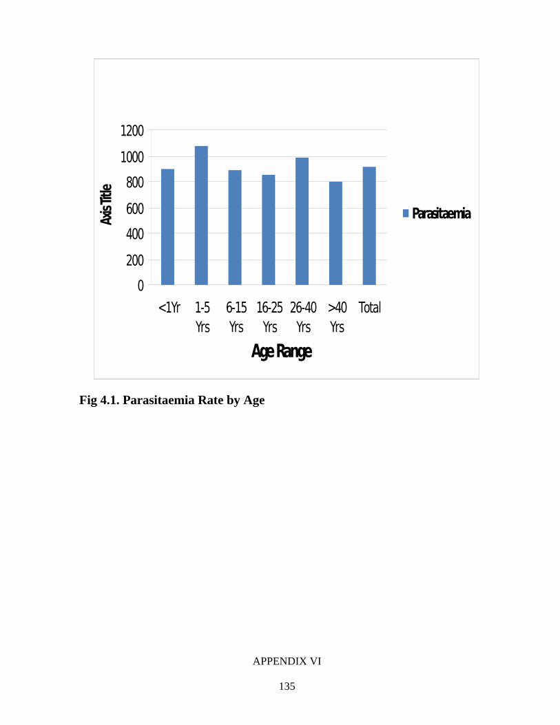
0
200
400
600
800
1000
1200
< 1Yr 1-5Yrs
6-15Yrs
16-25Yrs
26-40Yrs
> 40Yrs
Total
Age Range
Axis
Title
Parasitaemia
Fig 4.1. Parasitaemia Rate by Age
APPENDIX VI
135
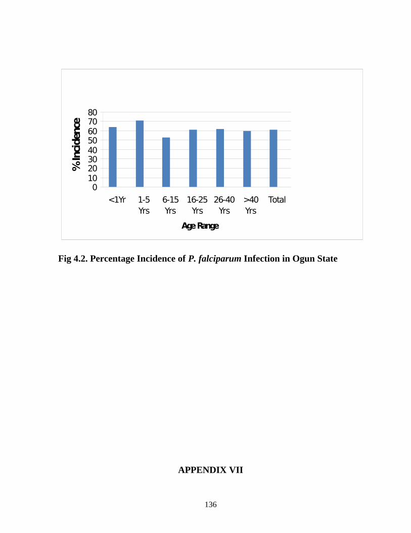
Fig 4.2. Percentage Incidence of P. falciparum Infection in Ogun State
APPENDIX VII
136
01020304050607080
< 1Yr 1-5Yrs
6-15Yrs
16-25Yrs
26-40Yrs
> 40Yrs
Total
Age Range
% In
cide
nce
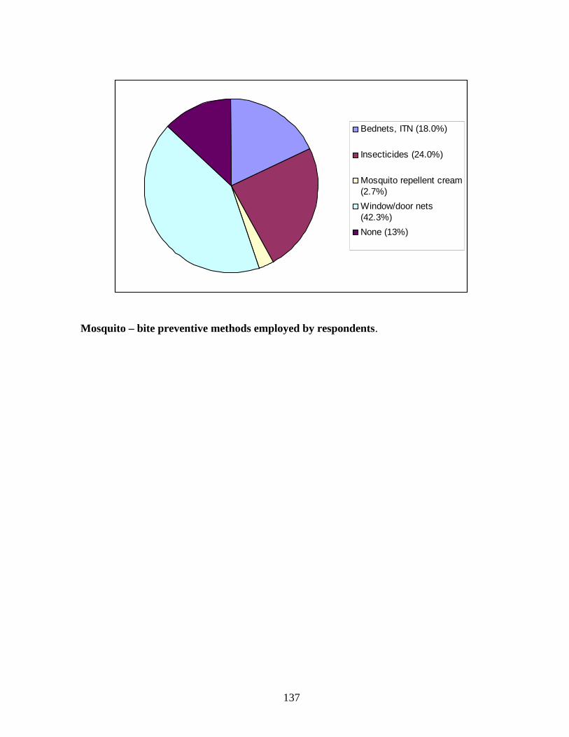
Bednets, ITN (18.0%)
Insecticides (24.0%)
Mosquito repellent cream(2.7%)
Window/door nets(42.3%)
None (13%)
Mosquito – bite preventive methods employed by respondents.
137