chapter 20 retinal insulin receptor signaling in hyperosmotic stress
TRANSCRIPT
RETINAL INSULIN RECEPTOR SIGNALING IN HYPEROSMOTICSTRESS
Raju V.S. Rajala, Ivana Ivanovic, and Ashok Kumar DillyDepartments of Ophthalmology and Cell Biology, and Dean A. McGee Eye Institute, University ofOklahoma Health Sciences Center, Oklahoma City, OK
AbstractIn the diabetic eye, the increased accumulation of sorbitol in the retina has been implicated in thepathogenesis of diabetic retinopathy. Neurodegeneration is an important component of diabeticretinopathy as demonstrated by increased neural apoptosis in the retina during experimental andhuman diabetes. Insulin receptor (IR) activation has been shown to rescue retinal neurons fromapoptosis through a phosphoinositide 3-kinase and protein kinase B (Akt) survival cascade. In thisstudy we examined the IR signaling in sorbitol-induced hyperosmotic stressed retinas.
INTRODUCTIONSorbitol is a sugar substitute often used in diet foods (including diet drinks and ice cream) andsugar-free chewing gums. It also occurs naturally in many stone fruits and berries from treesof the genus Sorbus. Ingesting large amounts of sorbtiol can lead to some abdominal pain, gasand mild to severe diarrhea. Sorbitol can also aggravate irritable bowl syndrome and fructosemalabsorption. Sorbitol can be used as a non-stimulant laxative as either an oral suspension orsuppository. The drug works by drawing water into the large intestine, thereby stimulatingbowl movements (Lederle, 1995). Even in the absence of dietary sorbitol, cells can producesorbitol naturally. When too much sorbitol is produced inside the cells, it can cause damage(Lorenzi, 2007). Diabetic retinopathy and neuropathy may be related to excess sorbitol in thecells of the eyes and nerves (Asnaghi et al., 2003;Lorenzi, 2007;Gabbay, 1973a). The sourceof this sorbitol in diabetes is excess glucose, which goes through the polyol pathway.
The polyol pathway of glucose metabolism is active when the intercellular glucose levels areelevated in the cell (Gabbay, 1973b). Aldose reductase (AR), the first and the rate limitingenzyme in the pathway reduces glucose to sorbitol using NADPH as a cofactor (Lorenzi,2007). Sorbitol is then metabolized to fructose by sorbitol dehydrogenase (SDH) that usedNAD+ as cofactor (Lorenzi, 2007). Sorbitol is an alcohol that is polyhydroxylated, and stronglyhydrophilic and does not diffuse readily through cell membranes and accumulatesintracellularly with possible osmotic consequences (Gabbay, 1973b). The fructose producedby the polyol pathway can get phosphorylated to fructose 3-phosphate (Szwergold et al.,1990), which can be further broken down to 3-deoxyglucosone, and both these compounds canbe very powerful glycosylating agents that can result in the formation of advanced glycationend products (AGEs) (Szwergold et al., 1990). The usage of NADPH by AR may result in lesscofactor becoming available to glutathione reductase, which is critical for the maintenance ofthe intracellular pools of reduced glutathione (GSH) (Lorenzi, 2007). This reduces thecapability of cells to respond to oxidative stress (Barnett et al., 1986). Compensatory increased
To whom correspondence should be addressed Raju V.S. Rajala Ph.D., University of Oklahoma Health Sciences Center, 608 Stanton L.Young Blvd., Oklahoma City, OK 73104, Telephone: 405-271-8255, Fax: 405-271-8128, E-mail: E-mail: [email protected].
NIH Public AccessAuthor ManuscriptVitam Horm. Author manuscript; available in PMC 2010 January 1.
Published in final edited form as:Vitam Horm. 2009 ; 80: 583–612. doi:10.1016/S0083-6729(08)00620-1.
NIH
-PA Author Manuscript
NIH
-PA Author Manuscript
NIH
-PA Author Manuscript
activity of the glucose monophosphate shunt, the principle supplier of cellular NADPH mayoccur (Barnett et al., 1986). The usage of NAD by SDH leads to an increased ratio of NADH/NAD+ which has been termed “pseudohypoxia” and linked to a multitude of metabolic andsignaling changes known to alter cell function (Williamson et al., 1993). Excess NADH servesas a substrate for NADH oxidase and this could be a mechanism for the generation ofintracellular oxidant species (Lorenzi, 2007). Thus activation of polyol pathway, by alteringthe intracellular homeostasis, generating AGEs, and exposing cells to oxidant stress due todecreased antioxidant defense mechanism and generation of oxidant species can initiate severalmechanisms of cellular damage.
Accumulation of sorbitol and fructose and the generation or enhancement of oxidative stresshas been reported in the whole retina of diabetic animals (Gabbay, 1975;Dagher et al.,2004;Lorenzi, 2007). The retinas of experimentally derived diabetic rats show increased lipidperoxidation (Obrosova et al., 2003), increased nitrotyrosine formation (Obrosova et al.,2005) and depletion of antioxidant enzymes (Obrosova et al., 2003). These abnormalities areprevented by drugs that inhibit AR (Dahlin et al., 1987;Tomlinson et al., 1992;Narayanan,1993;Tomlinson et al., 1994;Obrosova et al., 2003;Lorenzi, 2007). Retinas from diabeticpatients with retinopathy show more abundant AR immunoreactivity in ganglion cells, nervefibers, and Muller cells than retinas from nondiabetic individuals (Vinores et al., 1988). It hasalso been shown that human retinas from nondiabetic eye donors exposed to high glucose levelsin organ cultures accumulate sorbitol to the same extent as similarly incubated retinas ofnondiabetic rats (Dagher et al., 2004). Retinal ganglion cells, Muller glia, vascular pericytesand endothelial cells are endowed with AR in all species (Dagher et al., 2004) and these cellsare known to be damaged in diabetes (Lorenzi and Gerhardinger, 2001). The retinal vessels ofdiabetic rats treated with sorbinill, an AR inhibitor for the 9 months duration of diabetes,showed prevention of early complement activation, decreased levels of complement inhibitors,microvascular cell apoptosis and acellular capillaries (Dagher et al., 2004). Based on the datafrom the animal models, there is evidence for the concept that polyol pathway activation is asufficient mechanism for the retinal abnormalities induced by diabetes in rats.
Neurodegeneration is an important component of diabetic retinopathy as demonstrated byincreased neural apoptosis in the retina during experimental and human diabetes (Barber et al.,1998). IR activation has been shown to rescue retinal neurons from apoptosis through aphosphoinositide 3-kinase and protein kinase B (Akt) survival cascade. A significant decreaseof retinal IR kinase activity has been reported after 4 weeks of hyperglycemia in STZ treatedrats (Reiter et al., 2006). Hyperosmotic-stress responses interact with the insulin signalingpathways at several levels (Ouwens et al., 2001). Sorbitol has been previously shown to inducethe tyrosine phosphorylation of IR (Ouwens et al., 2001).
In the present study we examined the retinal insulin receptor signaling in sorbitol-treated retinasex vivo and show that sorbitol activates both the IR and IGF-1R tyrosine kinases, which resultsin activation of the receptor’s direct downstream targets. This receptor activation leads to theactivation of PI3K and Akt survival pathway in the retina. With the advent of phospho-site-specific antibody microarry, we observed either increased or decreased phosphorylation ofseveral tyrosine, serine/threonine kinases and cytoskeletal proteins which are downstreameffector molecules of IR and IGF-1R signaling pathways.
EXPERIMENTAL PROCEDURESMaterials
Human insulin R (rDNA origin) was obtained from Eli Lilly & Co. (Indianapolis, IN). Theactin antibody was obtained from Affinity BioReagents (Golden, CO). Polyclonal anti-IRß,polyclonal anti-Cbl and monoclonal anti-PY-99 antibodies were obtained from Santa Cruz
Rajala et al. Page 2
Vitam Horm. Author manuscript; available in PMC 2010 January 1.
NIH
-PA Author Manuscript
NIH
-PA Author Manuscript
NIH
-PA Author Manuscript
Biotechnology (Santa Cruz, CA). Polyclonal anti-Gab1 antibody was obtained from UpstateBiotechnology (Lake Placid, NY). Sorbitol, BAPTA and SB 203580 were obtained from Sigma(St Louis, MO). LY294002, anti-pAkt (S473), anti-Akt, anti-p38 and anti-phospho-p38antibodies were obtained from Cell Signaling (Beverly, MA). Genestin, HNMP3, PP1, PP2and PP3 were obtained from Calbiochem (San Diego, CA). Actin antibody was obtained fromAffinity BioReagents (Golden, CO). All other reagents were of analytical grade and fromSigma (St. Louis, MO).
AnimalsAll animal work was done in strict accordance with the NIH Guide for the Care and Use ofLaboratory Animals and the Association for Research in Vision and Ophthalmology on theUse of Animals in Vision Research. All protocols were approved by the IACUC at theUniversity of Oklahoma Health Sciences Center and the Dean McGee Eye Institute. In allexperiments, rats were killed by asphyxiation with carbon dioxide before the retinas wereremoved.
Retinal organ culturesRetinal organ cultures were carried out as previously described (Rajala et al., 2004;Rajala etal., 2007). Retinas were removed from Sprague-Dawley albino rats that were born and raisedin dim cyclic light (5 lux; 12 h ON: 12 h OFF), and incubated for either 5 min (insulin) or 30min (sorbitol) at 37 °C in Dulbecco’s modified Eagle’s (DMEM) medium (Gibco BRL) in thepresence of either insulin or sorbitol. Control cultures were carried out in the absence ofadditives. At the indicated times, retinas were snap-frozen in liquid nitrogen and stored at -80°C until analyzed. The retinas were lysed in lysis buffer [1% NP 40, 20 mM HEPES (pH 7.4),and 2 mM EDTA] containing phosphatase inhibitors (100 mM NaF, 10 mM Na4P2O7, 1 mMNaVO3 and 1 mM molybdate) and protease inhibitors (10 μM leupeptin, 10 μg/ml aprotinin,and 1 mM PMSF), and kept on ice for 10 min followed by centrifugation at 4 °C for 20 min.
Preparation of rat rod outer segmentsRetinas in culture were stimulated with either 1 μM insulin or 1 M sorbitol for 30 min at 37 °C. After treatment, the rod outer segments (ROS) were prepared using a discontinuous sucrosegradient as previously described (Rajala et al., 2002). Retinas were homogenized in 4.0 ml ofice-cold 47% sucrose solution containing buffer A [100 mM NaCl, 1 mM EDTA, 1 mMNaVO3, 1 mM PMSF and 10 mM Tris-HCl (pH 7.4)]. Retinal homogenates were transferredto 15-ml centrifuge tubes and sequentially overlaid with 3.0 ml of 42%, 3.0 ml of 37% and 4.0ml of 32% sucrose dissolved in buffer A. The gradients were spun in a swinging bucket rotorat 82,000 x g for 1 h at 4 °C. The 32/37% interfacial sucrose band, containing the ROSmembranes, was harvested and diluted with 10 mM Tris-HCl (pH 7.4) containing 100 mMNaCl and 1 mM EDTA. The band solution was then centrifuged at 27,000 x g for 30 min at 4°C. The ROS pellets were resuspended in 10 mM Tris-HCl (pH 7.4) containing 100 mM NaCland 1 mM EDTA, and stored at -20 °C. The non-ROS band designated Band II (37/42%) wasalso saved for comparison with the ROS fraction. All protein concentrations were determinedby BCA reagent (Pierce, Rockford, IL) following the manufacturer’s instructions.
Cloning, expression and purification of protein tyrosine phosphatase 1BRetinal PTP1B was obtained by PCR after reverse transcribing mouse retinal RNA and usingPTP1B primers (sense: GAA TTC ATG GAG ATG GAG AAG GAG TTC GAG; antisense:GTC GAC TCA GTG AAA ACA CAC CCG GTA GC). The PCR product was verified byDNA sequencing, digested with EcoR1 and Sal1, and cloned into GST fusion vector,pGEX-4T1. Site-directed mutagenesis was carried out according to methods described earlier(Rajala et al., 2004). Mutant PTP1B-D181A was amplified using primers, (sense: ACC ACA
Rajala et al. Page 3
Vitam Horm. Author manuscript; available in PMC 2010 January 1.
NIH
-PA Author Manuscript
NIH
-PA Author Manuscript
NIH
-PA Author Manuscript
TGG CCT GCC TTT GGA GTC CCC; antisense: GGG GAC TCC AAA GGC AGG CCATGT GGT). The PCR products were cloned into TOPO vector (Invitrogen) and both the WTand mutant sequences were verified by DNA sequencing. The WT and mutant cDNAs werelater excised from the sequencing vector as EcoRI/SalI fragments and cloned into GST fusionvector, pGEX-4T1. An overnight culture of E.coli BL21 (DE3) (pGEX-PTP1Bor pGEX-PTP1B-D181A) was diluted 1:10 with 100μg/ml ampicillin per ml, grown for 1 hr at 37 °C,and induced for another hour by addition of IPTG to 1 mM. Bacteria were sonicated three timesfor 20 s each time in lysis buffer containing 10 mM imidazole-HCl (pH7.2), 1 mM EDTA, 100mM NaCl, 1mM dithiothreitol, and 1% Triton X-100. Lysates were clarified by centrifugation,and the supernatants were incubated with 500 μl of 50% glutathione-coupled beads (AmershamPharmacia) for 30 min at 4 °C. The GST-PTP1B fusion proteins were washed in lysis bufferand eluted twice with 1 ml of 5 mM reduced glutathione (Sigma) in phosphatase buffer [20mM Tris (pH 7.4), 5% glycerol, 0.05% Trion X-100, 2.5 mM MgCl2, aprotinin (2 μg/ml) andleupeptin (5 μg/ml)]. Glycerol was added to a final concentration of 33% (vol/vol), and aliquotsof enzyme were stored at -20 °C.
PI3-kinase assayEnzyme assays were carried out as previously described (Rajala et al., 2007). Briefly, assayswere performed directly on either IRβ or Cbl immunoprecipitates of retinal lysates preparedfrom sorbitol treated or untreated lysates in 50 μl of reaction mixture containing 0.2 mg/mlPI-4,5-P2, 50 μM ATP, 10 μCi [γ32P]ATP, 5 mM MgCl2, and 10 mM HEPES buffer (pH 7.5).The reactions were carried out for 30 min at room temperature and stopped by the addition of100 μl of 1 N HCl followed by 200 μl of chloroform/methanol (1/1, v/v). Lipids were extractedand resolved on oxalate-coated TLC plates (silica gel 60) with a solvent system of 2-propanol/2 M acetic acid (65/35, v/v). The plates were coated in 1% (w/v) potassium oxalate in 50% (v/v) methanol and then baked in an oven at 100 °C for 1 hr prior to use. TLC plates were exposedto X-ray film overnight at -70 °C and radioactive lipids were scraped and quantified by liquidscintillation counting.
Phosphatase activity assayThe sorbitol-treated or untreated retinas were lysed in buffer containing 10 mM imidazole-HCl(pH7.2), 1 mM EDTA, 100 mM NaCl, 1mM dithiothreitol, and 1% Triton X-100. The assayswere performed (Takai and Mieskes, 1991) directly on retinal lysates in 80 μl of reactionmixture containing assay buffer [25 mM HEPES (pH 7.2), 50 mM NaCl, 5 mM dithiothritol,2.5 mM EDTA], 5 μl of 5% BSA and either test or positive control sample. The reactions werepre-incubated at 37 °C for 15 min, followed by the addition of 120 μl of pNPP (1.5 mg/ml)and incubated for 5-15 min at 37 °C. The reactions were stopped by the addition of 20 μl of13% K2HPO4 and the absorbance read at 405 nm. One unit of enzyme activity was equivalentto 1 nmol PNPP hydrolyzed per minute and the extinction coefficient for pNPP at A405 = 1.78× 10⁴ M-1 cm-1.
Dephosphorylation of tyrosine phosphorylated proteins by PTP1B in vitroSorbitol treated or untreated retinal proteins were incubated in the presence of catalyticallyactive or inactive PTP1B for 30 min at 30 °C. At the end of the incubation period, the reactionproducts were subjected to Western blot analysis with anti-PY99 antibody.
Antibody MicroarryControl and sorbitol-treated retinas in culture were lysed in lysis buffer [1% NP 40, 20 mMHEPES (pH 7.4), 2 mM EDTA and 1 mM dithiothreitol] containing phosphatase inhibitors(100 mM NaF, 10 mM Na4P2O7, 1 mM NaVO3 and 1 mM molybdate) and protease inhibitors(10 μM leupeptin, 10 μg/ml aprotinin, and 1 mM PMSF), and kept on ice for 10 min followed
Rajala et al. Page 4
Vitam Horm. Author manuscript; available in PMC 2010 January 1.
NIH
-PA Author Manuscript
NIH
-PA Author Manuscript
NIH
-PA Author Manuscript
by centrifugation at 4 °C for 20 min. The protein samples were then subjected to screeningusing antibody microarry containing 377 pan-specific and 273 phospho-site-specific antibodies(Kinexus Services, Vancouver, Canada).
ImmunoprecipitationRetinal lysates were prepared as previously described (Li et al., 2007). Insoluble material wasremoved by centrifugation at 17,000 x g for 20 min at 4 °C, and the solubilized proteins werepre-cleared by incubation with 40 μl of protein A-Sepharose for 1 h at 4 °C with mixing. Thesupernatant was incubated with primary antibodies overnight at 4 °C and subsequently with40 μl of protein A-Sepharose for 2 h at 4 °C. Following centrifugation at 17,000 x g for 1 minat 4 °C, immune complexes were washed three times with ice-cold wash buffer [50 mM HEPES(pH 7.4) 118 mM NaCl, 100 mM NaF, 2 mM NaVO3, 0.1% (w/v) SDS and 1% (v/v) TritonX-100]. The immunoprecipitates were either subjected to Western blotting analysis or used tomeasure the PI3K activity.
SDS-PAGE and Western blottingProteins were resolved by 10% SDS-PAGE and transferred onto nitrocellulose membranes.The blots were washed twice for 10 min with TTBS [20 mM Tris-HCl (pH 7.4), 100 mM NaCl,and 0.1% Tween-20] and blocked with either 5% bovine serum albumin or non-fat dry milkpowder (Bio-Rad) in TTBS for 1 h at room temperature. Blots were then incubated with anti-PY99 (1:1000), anti-Cbl (1:1000), anti-pAkt (1:1000), anti-Akt (1:1000), or anti-Akt1 (1:1000)or anti-Akt2 (1:1000) or anti-Akt3 (1:1000) or anti-IRß or anti-IGF 1R (1:1000) or anti-actin(1:1000) antibodies overnight at 4 °C. Following primary antibody incubations, immunoblotswere incubated with HRP-linked secondary antibodies (either anti-rabbit or anti-mouse) anddeveloped by ECL according to the manufacturer’s instructions.
RESULTSSorbitol-induced activation of p38MAP kinase
The p38MAP kinase is known to be activated in stress (Cheng et al., 2002). To determine ifsorbitol induces the activation of p38MAP kinase, we subjected the sorbitol-treated retinalproteins to western blot analysis with anti-phospho-p38 and total p38 antibodies. The resultsindicate a gradual increase in p38 phosphorylation between 0.2 and 1.0 M sorbitol comparedto control (Fig. 1A). The total p38 levels were unchanged in the presence of varyingconcentrations of sorbitol (Fig. 1B). These results suggested that sorbitiol induces the activationof p38MAP kinase and the organotypic cultures were mimicking the in vivo stress condition.
Sorbitol-induced activation of insulin- and insulin-like growth factor-1 receptorTo determine the sorbitol-induced activation of IR and IGF-1 receptors, weimmunoprecipitated retinal lysates from control and sorbitol-treated organotypic cultures withanti-IRß (Fig. 2B) and anti-IGF 1R (Fig. 2E) antibodies followed by Western blot analysiswith anti-PY99 antibody. The results indicated the activation of IR (Fig. 2A and 2C) and IGF1R (Fig. 2D and 2F) in response to sorbitol-induced stress.
Sorbitol-induced activation of AktTo determine if sorbitol-induced the activation of Akt, we incubated rat retinas in organotypiccultures for 30 min in the presence of varying concentrations of sorbitol (0, 0.2, 0.5, 1.0, 2.0and 3.0 M). At the end of the incubation, the retinas were lysed and subjected to Western blotanalysis with anti-pAkt (S473) and anti-total Akt antibodies. The results indicated a gradualincrease in Akt phosphorylation from 1.0 to 3.0 M sorbitol (Fig. 3A). The total Akt levels did
Rajala et al. Page 5
Vitam Horm. Author manuscript; available in PMC 2010 January 1.
NIH
-PA Author Manuscript
NIH
-PA Author Manuscript
NIH
-PA Author Manuscript
not change in response to sorbitol treatment (Fig. 3B). These results suggested that sorbitolinduced the activation of Akt.
Sorbitol-induced activation of Akt2Akt exist in three isoforms, all of which are expressed in the retina (Reiter et al., 2003). Todetermine if sorbitol stress activated a specific Akt isoform, we immunoprecipitated retinallysates from non stimulated control, insulin-stimulated and the sorbitol-treated organotypiccultures with anti-Akt1, anti-Akt2 and anti-Akt3 antibodies. The immune complexes weresubjected to Western blot analysis with anti-pAkt antibody. The results indicated that insulinactivated the Akt1 and the Akt3 isoforms, but not Akt2 (Fig. 3C). Sorbitol treatment, however,resulted in the activation of all three isoforms of Akt (Fig. 3D). These results suggested thatthe activation of Akt2 isoform may be stress-dependent.
Sorbitol induced activation of insulin receptor associated PI3K activity—Stress-inducedactivation of PI3K
We have previously reported the activation of PI3K through tyrosine phosphorylated IR in theretina (Rajala et al., 2007). To determine whether the activation of PI3K is regulated throughIR, we have immunoprecipitated IR from retinal lysates that were prepared from non stimulatedcontrol and sorbitol-treated (0-3.0 M) organotypic cultures, and measured the PI3K activity.The results indicated an increased PI3K activity with IR from sorbitol-treated retinas (Fig. 4).These results suggested that sorbitol-induced activation of PI3K occurs via activation of theIR.
PI3K-independent activation of AktTo determine whether sorbitol also induced the activation of Akt independent of PI3K, weincubated the retinas in the presence of the PI3K inhibitor LY294002 for 30 min before sorbitolor insulin treatment. Retinal proteins were subjected to Western blot analysis with anti-pAktand total Akt antibodies. The results indicated that LY294002 failed to inhibit the sorbitol-induced activation of Akt, but LY294002 inhibited the insulin-induced activation of Akt (Fig.5A-C), suggesting that Akt activation in hyperosomotic stress can also occur without PI3Kactivation.
p38MAP kinase activation is independent of PI3K activationTo determine if PI3K activation regulates the p38MAP kinase pathway, we pre-incubated theretinas in the presence of either the PI3K inhibitor LY294002 or the MAP kinase inhibitorSB203580 followed by sorbitol treatment. The retinal proteins were subjected to Western blotanalysis with anti-phospho-p38 and anti-p38 antibodies. The results indicated thatphosphorylation of p38 may be inhibited by the MAP kinase inhibitor SB203580 (Fig. 5),whereas the PI3K inhibitor LY294002 failed to inhibit the sorbitol-induced activation of p38MAP kinase (Fig. 5). These results suggested that MAP kinase activation is independent ofPI3K activation during hyperosomotic stress.
Role of calcium in sorbitol-induced Akt activationIt has been shown that hyperosomotic stress evokes an increase in the cytosolic calciumconcentration and triggers calcium signaling (Marchenko and Sage, 2000;Pritchard et al.,2002). To determine whether the sorbitol-induced activation of Akt is calcium dependent, wepre-incubated the retinas in organotypic cultures in the presence of the calcium specific chelatorBAPTA followed by 1.0 M sorbitol treatment. The retinas were lysed and the proteins weresubjected to Western blot analysis with anti-pAkt, anti-Akt, anti-actin, and anti-PY99antibodies. The results indicated that BAPTA failed to reduce the activation of Akt and thatthe levels of Akt activation were similar to the levels seen in the sorbitol treatment (Fig. 6A).
Rajala et al. Page 6
Vitam Horm. Author manuscript; available in PMC 2010 January 1.
NIH
-PA Author Manuscript
NIH
-PA Author Manuscript
NIH
-PA Author Manuscript
The blot was stripped and reprobed with total Akt (Fig. 6B) and actin (Fig. 6C) to ensure thatequal amounts of protein were loaded. These results suggested that under our experimentalconditions, calcium has no role in the activation of Akt.
Tyrosine kinase induced-activation of AktTo determine the pathway by which Akt undergoes activation in sorbitol stress, we haveincubated the retinas in organotypic cultures in the presence of tyrosine kinase inhibitors suchas genestin (inhibitor of tyrosine kinases), HNMP3 (inhibitor of IR kinase activity), PP1 (srckinase inhibitor), PP2 (src kinase inhibitor) and PP3 (a negative control for the src familyprotein tyrosine kinase inhibitor PP2) followed by sorbitol treatment. Retinas were lysed, andthe proteins were subjected to Western blot analysis with anti-pAkt, anti-Akt and anti-PY99antibodies. The results indicated that genestin blocked the activation of Akt, but all othertyrosine kinase and non-receptor tyrosine kinase inhibitors failed to block the activation of Akt(Fig. 7A). These results suggested that stress-induced activation of Akt is not under theregulation of non-receptor src family tyrosine kinase(s). Collectively these experimentssuggested that the activation of Akt in osmotic stress could be through tyrosine kinaseactivation.
Cholesterol depletion results in the sorbitol-induced activation of AktLocalization of insulin receptor in caveolae of adipocyte plasma membrane has been reportedand cholesterol depletion attenuates insulin receptor signaling (Gustavsson et al., 1999). Toexamine the role of lipid rafts on Akt activation in sorbitol induced stress, we stimulated retinasin organ cultures with either insulin or sorbitol in the presence or absence of cholesterol-sequestering agent, MCD, a treatment that disrupts cholesterol-rich DRMs. MCD treatmentresulted in a dramatic increase in sorbitol-induced activation of Akt compared to retinas treatedwith sorbitol in the absence of MCD (Fig. 8). Insulin effect on the activation of Akt issignificantly lower then either sorbitol or sorbitol in the presence of MCD (Fig. 8). These resultsclearly suggested that disruption of lipid rafts in sorbitol induced stress, which resulted in theactivation of Akt.
Sorbitol-induced activation of protein phosphatase (PA) activityTo determine whether sorbitol induced the activation of PA, we did phosphatase assays, usingp-Nitrophenyl Phosphate (pNPP) as the substrate. The results indicated a significant increasein the PA activity in 3.0 M treated retinas compared to untreated retinas (Fig. 9A). The resultssuggested that hyperosmotic stress increased the activation of PA activity. The total PA activitywe measured did not differentiate serine/threonine phosphatase activity from protein tyrosinephosphatase activity (PTP). To differentiate between the two activities, we measured PAactivity in the presence of sodium vanadate to inhibit the PTPase activity. The results indicateda significant increase in serine/threonine PA activity in sorbitol treated retinas compared tountreated retinas (Fig. 9A). Purified PTP1b, a protein tyrosine phosphatase was used as controlfor the sodium vanadate experiment. The results indicated the complete inhibition of PTP1Bactivity in the presence of sodium vanadate (Fig. 9A). The protein samples used for the PAactivity were used to examine the phosphorylation of Akt and the results indicated an increasedphosphorylation of Akt in 3.0 M sorbitol treated retinas (Fig. 9B). The blot were then strippedand reprobed with total Akt to ensure equal amounts of protein were loaded (Fig. 9B).Collectively, these results suggested that the observed activation of Akt in hyperosmotic stressis not due to the inhibition of phosphatase activity by sorbitol.
Sorbitol-induced tyrosine phosphorylation of several retinal proteinsThe PI3K-independent activation of Akt in the sorbitol-treated retinas prompted us toinvestigate the pathway by which Akt undergoes activation. Retinal proteins were either
Rajala et al. Page 7
Vitam Horm. Author manuscript; available in PMC 2010 January 1.
NIH
-PA Author Manuscript
NIH
-PA Author Manuscript
NIH
-PA Author Manuscript
stressed with sorbitol or stimulated with insulin and subjected to Western blot analysis withthe anti-PY99 antibody. The results indicated a significantly increased level of tyrosinephosphorylation in the retinal proteins in the 1.0 and 2.0 M sorbitol treatment compared to theinsulin-stimulated retinas (Fig. 10A). We observed the tyrosine phosphorylation of severalretinal proteins, with apparent molecular weights of 170, 130, 115, 79, 70 and 41 kDa.
To determine whether the stress-induced tyrosine phosphorylated proteins are localized to theROS or other retinal membranes, we probed the ROS and the non-ROS fractions with the anti-PY99 antibody. The results indicated a much greater tyrosine phosphorylation in the non-ROSfraction compared to the ROS (Fig. 10B). These results also suggested that stress responseinduces a significant tyrosine phosphorylation in inner segment and other retinal cellmembranes.
To determine the global changes in the phosphorylation (tyrosine and serine/threonine) ofretinal proteins, we examined the phosphorylation state of retinal proteins by antibodymicroarray. Of 273 phospho-site-specific antibodies 33 proteins were found to exhibit eitherincreased or decreased phosphorylation (data not shown). These proteins include serine/threonine and tyrosine kinases and mainly proteins involved in the cytoskeletal organization.These results clearly suggest that hyperosmotic stress-induces the activation of several proteinkinases which may in turn regulate the cytoskeletal reorganization.
Sorbitol-induced tyrosine phosphorylated proteins are the substrates of protein tyrosinephosphatase 1B (PTP1B) in vitro
Sorbitol treated retinal proteins were subjected to in vitro phosphatase assays in the presenceof either catalytically active or inactive PTP1B enzyme. After incubation, the reaction productswere subjected to SDS-PAGE followed by Western blot analysis with anti-PY99 antibody.The results indicated that PTP1B dephosphorylates the major 130 and 115 kDa tyrosinephosphorylated proteins in sorbitol-induced stress (Fig. 11).
Interaction of retinal Cbl with p85 subunit of PI3K under hyperosmotic conditionsTo determine the physical interaction between Cbl and p85 subunit of PI3K, we subjectedsorbitol-treated or untreated retinal lysates to GST-pull down experiments with GST-p85 fulllength fusion protein followed by Western blot analysis with anti-Cbl antibody. The resultsshowed the binding of Cbl to p85 subunit of PI3K (Fig. 12A). To further determine whetherthe binding is phosphorylation dependent, we immunoprecipitated Cbl from sorbitol-treatedor untreated retinal lysates followed by either Western blot analysis with anti-PY 99 antibody(Fig. 12B) or directly measured the Cbl associated PI3K activity (Fig. 12C). The results clearlyindicated that sorbitol-induced the phopshorylation of Cbl (Fig.12B) and that thephosphorylated Cbl was able to associate with the PI3K activity (Fig. 12C). In addition to beingan adaptor protein, Cbl has been characterized as an E3 ubiquitin ligase that interacts with PI3Kand mediates its ubiquitination and proteasome degradation (Fang et al., 2001). This interactionis phosphorylation-independent and it is established through binding of the SH3 domain ofp85 with the proline-rich region of Cbl (Fang et al., 2001). In order to identify the ubiquitinationstate of p85 in sorbitol treated and untreated retinas, immunoblots of anti-Ub IPs were probedwith anti-p85 antibody. The results indicated a decrease in p85 polyubiquitination in sorbitol-treated retinas compared to the untreated ones (Fig. 12D). This finding suggests that,phosphorylation of Cbl during hyperosmotic stress may act as a ‘switch’ changing the functionof Cbl from a E3 ubiquitin ligase to an adaptor function and thereby regulate the activity ofPI3K.
Rajala et al. Page 8
Vitam Horm. Author manuscript; available in PMC 2010 January 1.
NIH
-PA Author Manuscript
NIH
-PA Author Manuscript
NIH
-PA Author Manuscript
DISCUSSIONOur studies clearly indicate that both Akt and MAP kinase pathway are activated in responseto sorbitol stress. It has been shown previously that MAPKs acts as glucose transducers fordiabetic complications (Tomlinson, 1999). The sorbitol pathway, non-enzymatic glycation ofproteins and increased oxidative stress are known to activate protein kinase C which is aneffective activator of MAPKs (Tomlinson, 1999). These kinases phosphorylate transcriptionfactors, which in turn alter the balance of gene expression and promote the development ofdiabetic nephropathy, retinopathy and neuropathy (Tomlinson, 1999). The normal retinal IRexhibits high constitutive activity that is reduced in diabetes (Reiter et al., 2006). The diabeticrat retina further shows loss of PI3K, Akt1 and Akt-3, mTOR and p70S6K activities andincreased GSK3β activity (Reiter and Gardner, 2003). Elevated levels of sorbitol have beenshown to be implicated in the pathogenesis of diabetic retinopathy (Mizutani et al.,1998;Lorenzi and Gerhardinger, 2001;Asnaghi et al., 2003;Dagher et al., 2004;Lorenzi,2007). The rate limiting step in the pathway, aldose reductase which reduces the glucose tosorbitol is the major therapeutic target for diabetic retinopathy (Dvornik et al., 1973;Kinoshitaet al., 1979;Dahlin et al., 1987;Tomlinson et al., 1992;Chandra et al., 2002;Obrosova et al.,2003;Obrosova et al., 2005;Lorenzi, 2007). In this study, like insulin, sorbitol was found toinduce tyrosine phosphorylation of IR and IGF-1R. It was reported previously that insertionof IR into the plasma membrane is necessary for sorbitol-induced IR activation (Ouwens et al.,2001). Consistent with these observation we reported that IRs in rod outer segments of retinasare localized to plasma membrane (Rajala et al., 2007). Further studies, however, are requiredto understand how the IR kinase activity gets reduced in diabetes.
The organotypic culture system has been successfully used to study protein phosphorylationand provides access to the retina for the addition of exogenous modulators of cellular function(Rajala et al., 2004) We used this system to study the activation of IR, IGF-1, PI3K, Akt andMAP kinase activation under hyperosmotic stress. Inactivation and dephosphorylation of Akthave been reported during hyperosmotic stress (Meier et al., 1998) These studies were done incell culture with 0.5 M sorbitol and at this concentration, sorbitol activated the MAP kinasebut not Akt kinase pathway (Meier et al., 1998) Another independent study also failed todemonstrate the activation of Akt in 3T3L1 adipocytes under hyperosmotic stress using 0.6 Msorbitol (Chen et al., 1997) In agreement with these studies, we did not observe activation ofAkt at 0.5 M sorbitol in our ex vivo retinal organ cultures. However, Akt was activated between1.0 and 3.0 M sorbitol with the maximum being at 3.0 M. These concentrations are higher thanthose reported previously (Chen et al., 1997;Meier et al., 1998), but provide the same degreeof activation we found in vivo light stress (Li et al., 2007), suggesting that sorbitol-inducedhyperosmotic stress may mimic the in vivo light-stress model. Hyperosmotic-stress has beenrelated to the pathogenesis of retinal detachment in diabetic retinopathy (Quintyn and Brasseur,2004;Marmor et al., 1980;Ola et al., 2006) and the sorbitol-induced ex vivo cultures may beuseful to study the molecular signaling pathway(s) involved in diabetic retinopathy..
In our study, we observed that 3.0 M sorbitol induced the activation of Akt. Inactivation anddephosphorylation of Akt have been reported during hyperosmotic stress (Meier et al., 1998).Our observation of Akt activation suggests that 3.0 M sorbitol could inhibit the proteinphosphatase(s) which results in the activation of Akt. To address whether sorbitol could activateAkt signaling or inhibition of phosphatase(s) which indirectly block the dephosphorylation ofAkt, we measured the phosphatase activity in hyperosmotic stress. Our results indicate asignificant increase in the total phosphatase activity in hyperosomotic stress. To distinguishthe activity of serine/threonine phosphatase activity from protein tyrosine phosphatase (PTP),we used sodium vanadate to inhibit the PTPase activity and the results still indicate a significantincrease in serine/threonine phosphatase activity. These studies clearly suggest that the
Rajala et al. Page 9
Vitam Horm. Author manuscript; available in PMC 2010 January 1.
NIH
-PA Author Manuscript
NIH
-PA Author Manuscript
NIH
-PA Author Manuscript
observed activation of Akt in hyperosmotic stress is not due to the inhibition of phosphatase(s).
In the present study, in response to sorbitol, we have observed increased activation of PI3Kthrough IR activation. In some neuronal cell types, such as cerebellar granular neurons (59)and PC-12 cells (60), receptor activation of PI3K has been shown to protect these cells fromstress-induced neurodegeneration. Further, IR activation has been shown to rescue retinalneurons from apoptosis through a phosphoinositide 3-kinase (PI3K) cascade (Barber et al.,2001;Barber et al., 1998). We have previously reported that under physiological conditions,light-induced the tyrosine phosphorylation of retinal IR which leads to the activation of PI3K(Rajala et al., 2002). The earlier studies along with the results from the present study clearlysuggests that sorbitol also induces the activation of PI3K associated with the tyrosinephosphorylated IR.
In hyperosomotic stress we have observed both PI3K-dependent and independent activationof Akt. The PI3K inhibitor LY294002 was found to inhibit the insulin-induced activation ofAkt but the same inhibitor failed to inhibit the sorbitol-induced activation of Akt. Activationof PI3K/Akt pathway contributes to cell survival (Datta et al., 1999), however, it has beenshown that dopamine induced the PI3K-indepndent activation of Akt in striated neurons(Brami-Cherrier et al., 2002). These results suggest that sorbitol-induced activation of Aktmight also be regulated through PI3K-independent mechanism. Consistent with thishypothesis, we have observed an increased tyrosine phosphorylation of several proteins in theretina under hyperosmotic stress. Further, HNMP3, an inhibitor of IR kinase activity has noeffect on sorbitol-induced Akt activation. However, genestin, a global tyrosine kinase inhibitorwas found to block the sorbitol-induced activation of Akt. It appears from this data that sorbitol-induced Akt activation is mediated through a receptor tyrosine kinase(s), but not IR. Theobserved Akt activation is not mediated through Src family non-receptor tyrosine kinases dueto the fact that inhibitors of this family failed to block the sorbitol-induced Akt activation.Increased protein tyrosine phosphorylation in the retina after ischemic-reperfusion injury andgenestin, ameliorates retinal degeneration after ischemia-reperfusion injury in rats (Hayashi etal., 1997;Hayashi et al., 1996). These results further support our findings that Akt activationcould be trigged in response to stress and retinal injury.
The ability of osmotic shock to directly stimulate tyrosine phosphorylation events wasconfirmed by phosphotyrosine immunoblotting. Several discrete tyrosine-phosphorylatedproteins in the range of 115-170 kDa and 41-79 kDa were clearly induced by osmotic shocktreatment. Previous studies have also reported the activation of tyrosine phosphorylation inresponse to hyperosmotic stress (Chen et al., 1997;Hresko and Mueckler, 2000;Janez et al.,2000). We have probed the phosphotyrosine immunoblots with known tyrosine phosphorylatedproteins such as PYK2, FAK, Na+ K+ ATPase, EGFR, JAK2, TYK2 PDE6beta, HSP90, cSrcand Grb2-associated binder 1 (Gab-1). Under our experimental conditions, except Gab1, allother proteins were not tyrosine phosphorylated in sorbitol-induced stress conditions. The Grb2associated binder Gab-1 is shown to be tyrosine phosphorylated following sorbitol stimulation(Janez et al., 2000). We found that Gab1 is rapidly tyrosine phosphorylated immediately aftersorbitol treatment and after 5 min, the phosphorylation was greatly diminished (data notshown). We have also demonstrated in this study that genestin completely blocks the Aktactivation; which suggests that Akt activation may be signaled through Gab1. Gab-1 isphosphorylated on tyrosine after stimulation with insulin and several growth factors (Rocchiet al., 1998;Janez et al., 2000). It possesses 16 potential phosphotyrosine sites, some of whichcould serve as binding sites for SH2 domains of the p85 regulatory subunit of PI3K, Grb2,phospholipase C-γ, Nck, and SHP-2 (Schlessinger and Lemmon, 2003;Liu and Rohrschneider,2002;Gual et al., 2000;Rocchi et al., 1998). It has been shown previously that Gab-1 mediatesthe neurite outgrowth, DNA synthesis, and survival in PC12 cells (Korhonen et al., 1999).
Rajala et al. Page 10
Vitam Horm. Author manuscript; available in PMC 2010 January 1.
NIH
-PA Author Manuscript
NIH
-PA Author Manuscript
NIH
-PA Author Manuscript
Further, overexpression of Gab-1 has shown to inhibit apoptosis in PC12 cells (Holgado-Madruga et al., 1997). Therefore, it is tempting to speculate that decreased phosphorylation ofGab1 could be a contributory factor for diabetic retinopathy
In this study, we report the specific activation of Akt2 in response to hyperosmotic stress.Consistent with this hypothesis is our earlier finding that Akt2 knockout mice exhibit a greatersensitivity to light-induced retinal degeneration, but not Akt1 knockout mice (Li et al., 2007).Further, mice lacking Akt2 have defects in glucose metabolism that ultimately lead tohyperglycemia and hyperinsulinemia (Cho et al., 2001b; Garofalo et al., 2003). These studiesclearly suggest the importance of Akt2 in both light-induced retinal degeneration and sorbitol-induced stress.
In the current study, we observed that sorbitol-induced the tyrosine phosphorylation of Cbland this activation leads to the binding of p85 subunit of PI3K, as we observed an increasedCbl associated PI3K activity in ex vivo retinal organ cultures. Although a small fraction of Cblis constitutively phosphorylated and associated with PI3K, it appears that a more significantfraction of Cbl could potentially negatively regulate p85 by polyubiquitination and degradationas demonstrated in our sorbitol untreated control retinas.
Cbl is a ubiquitously expressed cytosolic protein characterized as both, an adaptor protein andan E3 ubiquitin ligase. Cbl protein has a tyrosine kinase binding (TKB) domain, proline-richregion and five tyrosine phosphorylation sites; Cbl is able to bind to protein tyrosine kinases(PTK), SH3 and SH2 domain containing proteins, respectively (Swaminathan et al.,2007;Swaminathan and Tsygankov, 2006;Meisner et al., 1995);Meisner, 1995 1543 /id;Fukazawa, 1995 3 /id}. Cbl also contains the RING finger domain and a ubiquitin-associateddomain which allow for ubiquitination and proteasomal degradation of activated PTK(Levkowitz et al., 1999;Meisner et al., 1997). Stress, extracellular stimuli, growth factors andhormones stimulate tyrosine phosphorylation of Cbl. Under certain cellular conditionsphosphorylated Cbl acts as an adaptor protein and binds to SH2 domain-containing proteinssuch as Vav guanine nucleotide exchange factor, p85 subunit of PI3K, and Crk proteins topropagate downstream signaling (Miyake et al., 1997;Feshchenko et al., 1998). However,previous findings showed that tyrosine phosphorylation of both Cbl and EGFR tyrosine kinasewere necessary for binding, ubiquitination and degradation of an activated receptor (Levkowitzet al., 1999).
Interaction between p85 subunit of PI3K and Cbl is phosphorylation-dependent andindependent. Phosphorylation of Tyr-731 of Cbl is essential for its binding to SH2 domains ofp85 (Feshchenko et al., 1998;Swaminathan et al., 2007). . Tyrosine phosphorylationsignificantly increase p85-Cbl binding in membrane fractions when compared to the cytosolicfractions of v-Abl-transformed 3T3 fibroblasts and thus further facilitating PI3K/Rhodownstream cytoskeletal effects (Swaminathan et al., 2007). . Another study has shown thatthe two SH2 domains of p85 were dispensable for p85 and Cbl interaction, subsequentubiquitination and proteasome degradation therefore suggesting phosphorylation-independentbinding (Fang et al., 2001). The same study proved that in Jurkat T cell line, the SH3 domainof p85 and the proline-rich region of Cbl were necessary for the binding of the two proteinsand further ubiquitination of p85 by the RING finger domain of Cbl.
Diabetic model studies have shown that chronic and increased sorbitol secretion by the cellsestablishes a hyperosmotic environment fatal to cell survival and viability. The elevatederythrocyte sorbitol levels were found in diabetic patients with developing diabetic retinopathy(DR), a leading cause of blindness (Reddy et al., 2008). Both insulin and sorbitol-inducedhyperosmotic shock increased intrinsic tyrosine kinase activity of insulin receptor (IR)followed by transient tyrosine phosphorylation of insulin-receptor substrate-1 (IRS-1) and Cbl
Rajala et al. Page 11
Vitam Horm. Author manuscript; available in PMC 2010 January 1.
NIH
-PA Author Manuscript
NIH
-PA Author Manuscript
NIH
-PA Author Manuscript
(Rajala and Anderson, 2001;Ahmed et al., 2000).. Tyrosine phosphorylated IRS-1 and Cblharbor signaling proteins involved in activation of PI3K/Akt survival pathway and F-actinpolymerization, respectively (Strawbridge et al., 2006). Previous studies have shown that type2 diabetes, obesity, metabolic syndrome X and age related insulin resistance were characterizedby elevated levels of vasoactive peptide endothelin-1 (ET-1) (Sayama et al., 1999;Ferri et al.,1995;Ferri et al., 1997).. ET-1 peptide induced defects in lipid membrane, impaired PI3Ksignaling, reduced tyrosine phosphorylation of Cbl and F-actin polymerization as well asGLUT4 trafficking when added to insulin- or sorbitol-incubation conditions; thus, ET-1challenges and overwrites protective role of Cbl (Strawbridge et al., 2006). Taken together,previous and our current findings suggest that under normal conditions Cbl negatively regulatesp85 whereby it potentially minimizes the available PI3K pool utilized for basal signaling byRTK and non-RTK. In our in vitro hyperosmolarity stress model, Cbl becomes tyrosinephosphorylated and takes on a role of an adaptor protein aiding to maximize and recruit PI3Knecessary for execution of downstream survival pathways. In addition, our laboratory findingsshow an increase IR tyrosine activity and PI3K/Akt survival signals under the same conditions.Further studies will help us elucidate the function of Cbl as a neuroprotector involved inmaintaining cell integrity and viability under the compromising hyperosmotic conditionscharacteristic of diabetic models.
ACKNOWLEDGEMNTSThe project described was supported by grants from the National Eye Institute (R01EY016507) and the NationalCenter for Research Resources (P20RR17703). The content is solely the responsibility of the authors and does notnecessarily represent the official views of the National Center for Research Resources, the National Eye Institute, orthe National Institutes of Health. The authors thank Dr. Yogita Kanan for reading this manuscript.
ABBREVIATIONSIR, insulin receptor; PI3K, phosphoinositide 3-kinase; IRβ, IR beta subunit; IGF 1R, insulin-like growth factor-1 receptor; SDS-PAGE, sodium dodecyl sulfate polyacrylamide gelelectrophoresis; ROS, rod outer segments; IPs, immunoprecipitates; Grb2, growth factorreceptor-bound protein 2; Gab-1, Grb2-assoicated binder 1.
REFERENCES1. Ahmed Z, Smith BJ, Pillay TS. The APS adapter protein couples the insulin receptor to the
phosphorylation of c-Cbl and facilitates ligand-stimulated ubiquitination of the insulin receptor. FEBSLett 2000;475:31–34. [PubMed: 10854852]
2. Asnaghi V, Gerhardinger C, Hoehn T, Adeboje A, Lorenzi M. A role for the polyol pathway in theearly neuroretinal apoptosis and glial changes induced by diabetes in the rat. Diabetes 2003;52:506–511. [PubMed: 12540628]
3. Barber AJ, Lieth E, Khin SA, Antonetti DA, Buchanan AG, Gardner TW. Neural apoptosis in theretina during experimental and human diabetes. Early onset and effect of insulin. J Clin Invest1998;102:783–791. [PubMed: 9710447]
4. Barber AJ, Nakamura M, Wolpert EB, Reiter CE, Seigel GM, Antonetti DA, Gardner TW. Insulinrescues retinal neurons from apoptosis by a phosphatidylinositol 3-kinase/Akt-mediated mechanismthat reduces the activation of caspase-3. J Biol Chem 2001;276:32814–32821. [PubMed: 11443130]
5. Barnett PA, Gonzalez RG, Chylack LT Jr. Cheng HM. The effect of oxidation on sorbitol pathwaykinetics. Diabetes 1986;35:426–432. [PubMed: 3956880]
6. Brami-Cherrier K, Valjent E, Garcia M, Pages C, Hipskind RA, Caboche J. Dopamine induces a PI3-kinase-independent activation of Akt in striatal neurons: a new route to cAMP response element-binding protein phosphorylation. J Neurosci 2002;22:8911–8921. [PubMed: 12388598]
Rajala et al. Page 12
Vitam Horm. Author manuscript; available in PMC 2010 January 1.
NIH
-PA Author Manuscript
NIH
-PA Author Manuscript
NIH
-PA Author Manuscript
7. Chandra D, Jackson EB, Ramana KV, Kelley R, Srivastava SK, Bhatnagar A. Nitric oxide preventsaldose reductase activation and sorbitol accumulation during diabetes. Diabetes 2002;51:3095–3101.[PubMed: 12351453]
8. Chen D, Elmendorf JS, Olson AL, Li X, Earp HS, Pessin JE. Osmotic shock stimulates GLUT4translocation in 3T3L1 adipocytes by a novel tyrosine kinase pathway. J Biol Chem 1997;272:27401–27410. [PubMed: 9341192]
9. Cheng H, Kartenbeck J, Kabsch K, Mao X, Marques M, Alonso A. Stress kinase p38 mediates EGFRtransactivation by hyperosmolar concentrations of sorbitol. J Cell Physiol 2002;192:234–243.[PubMed: 12115730]
10. Dagher Z, Park YS, Asnaghi V, Hoehn T, Gerhardinger C, Lorenzi M. Studies of rat and humanretinas predict a role for the polyol pathway in human diabetic retinopathy. Diabetes 2004;53:2404–2411. [PubMed: 15331552]
11. Dahlin LB, Archer DR, McLean WG. Treatment with an aldose reductase inhibitor can reduce thesusceptibility of fast axonal transport following nerve compression in the streptozotocin-diabetic rat.Diabetologia 1987;30:414–418. [PubMed: 2445613]
12. Datta SR, Brunet A, Greenberg ME. Cellular survival: a play in three Akts. Genes Dev 1999;13:2905–2927. [PubMed: 10579998]
13. Dvornik E, Simard-Duquesne N, Krami M, Sestanj K, Gabbay KH, Kinoshita JH, Varma SD, MerolaLO. Polyol accumulation in galactosemic and diabetic rats: control by an aldose reductase inhibitor.Science 1973;182:1146–1148. [PubMed: 4270794]
14. Fang D, Wang HY, Fang N, Altman Y, Elly C, Liu YC. Cbl-b, a RING-type E3 ubiquitin ligase,targets phosphatidylinositol 3-kinase for ubiquitination in T cells. J Biol Chem 2001;276:4872–4878.[PubMed: 11087752]
15. Ferri C, Bellini C, Desideri G, Baldoncini R, Properzi G, Santucci A, De Mattia G. Circulatingendothelin-1 levels in obese patients with the metabolic syndrome. Exp Clin Endocrinol Diabetes1997;105(Suppl 2):38–40. [PubMed: 9288542]
16. Ferri C, Bellini C, Desideri G, Di Francesco L, Baldoncini R, Santucci A, De Mattia G. Plasmaendothelin-1 levels in obese hypertensive and normotensive men. Diabetes 1995;44:431–436.[PubMed: 7698512]
17. Feshchenko EA, Langdon WY, Tsygankov AY. Fyn, Yes, and Syk phosphorylation sites in c-Cblmap to the same tyrosine residues that become phosphorylated in activated T cells. J Biol Chem1998;273:8323–8331. [PubMed: 9525940]
18. Gabbay KH. Role of sorbitol pathway in neuropathy. Adv Metab Disord 1973a;2(Suppl32)19. Gabbay KH. The sorbitol pathway and the complications of diabetes. N Engl J Med 1973b;288:831–
836. [PubMed: 4266466]20. Gabbay KH. Hyperglycemia, polyol metabolism, and complications of diabetes mellitus. Annu Rev
Med 1975;26:521–536. [PubMed: 238458]21. Gual P, Giordano S, Williams TA, Rocchi S, Van Obberghen E, Comoglio PM. Sustained recruitment
of phospholipase C-gamma to Gab1 is required for HGF-induced branching tubulogenesis. Oncogene2000;19:1509–1518. [PubMed: 10734310]
22. Gustavsson J, Parpal S, Karlsson M, Ramsing C, Thorn H, Borg M, Lindroth M, Peterson KH,Magnusson KE, Stralfors P. Localization of the insulin receptor in caveolae of adipocyte plasmamembrane. FASEB J 1999;13:1961–1971. [PubMed: 10544179]
23. Hayashi A, Koroma BM, Imai K, de Juan E Jr. Increase of protein tyrosine phosphorylation in ratretina after ischemia-reperfusion injury. Invest Ophthalmol Vis Sci 1996;37:2146–2156. [PubMed:8843901]
24. Hayashi A, Weinberger AW, Kim HC, de Juan E Jr. Genistein, a protein tyrosine kinase inhibitor,ameliorates retinal degeneration after ischemia-reperfusion injury in rat. Invest Ophthalmol Vis Sci1997;38:1193–1202. [PubMed: 9152239]
25. Holgado-Madruga M, Moscatello DK, Emlet DR, Dieterich R, Wong AJ. Grb2-associated binder-1mediates phosphatidylinositol 3-kinase activation and the promotion of cell survival by nerve growthfactor. Proc Natl Acad Sci U S A 1997;94:12419–12424. [PubMed: 9356464]
26. Hresko RC, Mueckler M. A novel 68-kDa adipocyte protein phosphorylated on tyrosine in responseto insulin and osmotic shock. J Biol Chem 2000;275:18114–18120. [PubMed: 10764780]
Rajala et al. Page 13
Vitam Horm. Author manuscript; available in PMC 2010 January 1.
NIH
-PA Author Manuscript
NIH
-PA Author Manuscript
NIH
-PA Author Manuscript
27. Janez A, Worrall DS, Imamura T, Sharma PM, Olefsky JM. The osmotic shock-induced glucosetransport pathway in 3T3-L1 adipocytes is mediated by gab-1 and requires Gab-1-associatedphosphatidylinositol 3-kinase activity for full activation. J Biol Chem 2000;275:26870–26876.[PubMed: 10842168]
28. Kinoshita JH, Fukushi S, Kador P, Merola LO. Aldose reductase in diabetic complications of the eye.Metabolism 1979;28:462–469. [PubMed: 45423]
29. Korhonen JM, Said FA, Wong AJ, Kaplan DR. Gab1 mediates neurite outgrowth, DNA synthesis,and survival in PC12 cells. J Biol Chem 1999;274:37307–37314. [PubMed: 10601297]
30. Lederle FA. Epidemiology of constipation in elderly patients. Drug utilisation and cost-containmentstrategies. Drugs Aging 1995;6:465–469. [PubMed: 7663066]
31. Levkowitz G, Waterman H, Ettenberg SA, Katz M, Tsygankov AY, Alroy I, Lavi S, Iwai K, ReissY, Ciechanover A, Lipkowitz S, Yarden Y. Ubiquitin ligase activity and tyrosine phosphorylationunderlie suppression of growth factor signaling by c-Cbl/Sli-1. Mol Cell 1999;4:1029–1040.[PubMed: 10635327]
32. Li G, Anderson RE, Tomita H, Adler R, Liu X, Zack DJ, Rajala RV. Nonredundant role of Akt2 forneuroprotection of rod photoreceptor cells from light-induced cell death. J Neurosci 2007;27:203–211. [PubMed: 17202487]
33. Liu Y, Rohrschneider LR. The gift of Gab. FEBS Lett 2002;515:1–7. [PubMed: 11943184]34. Lorenzi M. The polyol pathway as a mechanism for diabetic retinopathy: attractive, elusive, and
resilient. Exp Diabetes Res 2007;2007:61038. [PubMed: 18224243]35. Lorenzi M, Gerhardinger C. Early cellular and molecular changes induced by diabetes in the retina.
Diabetologia 2001;44:791–804. [PubMed: 11508263]36. Marchenko SM, Sage SO. Hyperosmotic but not hyposmotic stress evokes a rise in cytosolic Ca2+
concentration in endothelium of intact rat aorta. Exp Physiol 2000;85:151–157. [PubMed: 10751511]37. Marmor MF, Martin LJ, Tharpe S. Osmotically induced retinal detachment in the rabbit and primate.
Electron miscoscopy of the pigment epithelium. Invest Ophthalmol Vis Sci 1980;19:1016–1029.[PubMed: 7409995]
38. Meier R, Thelen M, Hemmings BA. Inactivation and dephosphorylation of protein kinase Balpha(PKBalpha) promoted by hyperosmotic stress. EMBO J 1998;17:7294–7303. [PubMed: 9857186]
39. Meisner H, Conway BR, Hartley D, Czech MP. Interactions of Cbl with Grb2 and phosphatidylinositol3′-kinase in activated Jurkat cells. Mol Cell Biol 1995;15:3571–3578. [PubMed: 7791764]
40. Meisner H, Daga A, Buxton J, Fernandez B, Chawla A, Banerjee U, Czech MP. Interactions ofDrosophila Cbl with epidermal growth factor receptors and role of Cbl in R7 photoreceptor celldevelopment. Mol Cell Biol 1997;17:2217–2225. [PubMed: 9121472]
41. Miyake S, Lupher ML Jr. Andoniou CE, Lill NL, Ota S, Douillard P, Rao N, Band H. The Cblprotooncogene product: from an enigmatic oncogene to center stage of signal transduction. Crit RevOncog 1997;8:189–218. [PubMed: 9570294]
42. Mizutani M, Gerhardinger C, Lorenzi M. Muller cell changes in human diabetic retinopathy. Diabetes1998;47:445–449. [PubMed: 9519752]
43. Narayanan S. Aldose reductase and its inhibition in the control of diabetic complications. Ann ClinLab Sci 1993;23:148–158. [PubMed: 8457142]
44. Obrosova IG, Minchenko AG, Vasupuram R, White L, Abatan OI, Kumagai AK, Frank RN, StevensMJ. Aldose reductase inhibitor fidarestat prevents retinal oxidative stress and vascular endothelialgrowth factor overexpression in streptozotocin-diabetic rats. Diabetes 2003;52:864–871. [PubMed:12606532]
45. Obrosova IG, Pacher P, Szabo C, Zsengeller Z, Hirooka H, Stevens MJ, Yorek MA. Aldose reductaseinhibition counteracts oxidative-nitrosative stress and poly(ADP-ribose) polymerase activation intissue sites for diabetes complications. Diabetes 2005;54:234–242. [PubMed: 15616034]
46. Ola MS, Berkich DA, Xu Y, King MT, Gardner TW, Simpson I, LaNoue KF. Analysis of glucosemetabolism in diabetic rat retinas. Am J Physiol Endocrinol Metab 2006;290:E1057–E1067.[PubMed: 16380392]
47. Ouwens DM, Gomes de Mesquita DS, Dekker J, Maassen JA. Hyperosmotic stress activates theinsulin receptor in CHO cells. Biochim Biophys Acta 2001;1540:97–106. [PubMed: 11513972]
Rajala et al. Page 14
Vitam Horm. Author manuscript; available in PMC 2010 January 1.
NIH
-PA Author Manuscript
NIH
-PA Author Manuscript
NIH
-PA Author Manuscript
48. Pritchard S, Erickson GR, Guilak F. Hyperosmotically induced volume change and calcium signalingin intervertebral disk cells: the role of the actin cytoskeleton. Biophys J 2002;83:2502–2510.[PubMed: 12414684]
49. Quintyn JC, Brasseur G. Subretinal fluid in primary rhegmatogenous retinal detachment:physiopathology and composition. Surv Ophthalmol 2004;49:96–108. [PubMed: 14711443]
50. Rajala A, Anderson RE, Ma JX, Lem J, Al Ubaidi MR, Rajala RV. G-protein-coupled ReceptorRhodopsin Regulates the Phosphorylation of Retinal Insulin Receptor. J Biol Chem 2007;282:9865–9873. [PubMed: 17272282]
51. Rajala RV, Anderson RE. Interaction of the insulin receptor beta-subunit with phosphatidylinositol3-kinase in bovine ROS. Invest Ophthalmol Vis Sci 2001;42:3110–3117. [PubMed: 11726610]
52. Rajala RV, McClellan ME, Ash JD, Anderson RE. In vivo regulation of phosphoinositide 3-kinasein retina through light-induced tyrosine phosphorylation of the insulin receptor beta-subunit. J BiolChem 2002;277:43319–43326. [PubMed: 12213821]
53. Rajala RV, McClellan ME, Chan MD, Tsiokas L, Anderson RE. Interaction of the Retinal InsulinReceptor beta-Subunit with the P85 Subunit of Phosphoinositide 3-Kinase. Biochemistry2004;43:5637–5650. [PubMed: 15134438]
54. Reddy GB, Satyanarayana A, Balakrishna N, Ayyagari R, Padma M, Viswanath K, Petrash JM.Erythrocyte aldose reductase activity and sorbitol levels in diabetic retinopathy. Mol Vis2008;14:593–601. [PubMed: 18385795]
55. Reiter CE, Gardner TW. Functions of insulin and insulin receptor signaling in retina: possibleimplications for diabetic retinopathy. Prog Retin Eye Res 2003;22:545–562. [PubMed: 12742394]
56. Reiter CE, Sandirasegarane L, Wolpert EB, Klinger M, Simpson IA, Barber AJ, Antonetti DA, KesterM, Gardner TW. Characterization of insulin signaling in rat retina in vivo and ex vivo. Am J PhysiolEndocrinol Metab 2003;285:E763–E774. [PubMed: 12799319]
57. Reiter CE, Wu X, Sandirasegarane L, Nakamura M, Gilbert KA, Singh RS, Fort PE, Antonetti DA,Gardner TW. Diabetes reduces basal retinal insulin receptor signaling: reversal with systemic andlocal insulin. Diabetes 2006;55:1148–1156. [PubMed: 16567541]
58. Rocchi S, Tartare-Deckert S, Murdaca J, Holgado-Madruga M, Wong AJ, Van Obberghen E.Determination of Gab1 (Grb2-associated binder-1) interaction with insulin receptor-signalingmolecules. Mol Endocrinol 1998;12:914–923. [PubMed: 9658397]
59. Sayama H, Nakamura Y, Saito N, Konoshita M. Does the plasma endothelin-1 concentration reflectatherosclerosis in the elderly? Gerontology 1999;45:312–316. [PubMed: 10559648]
60. Schlessinger J, Lemmon MA. SH2 and PTB domains in tyrosine kinase signaling. Sci STKE2003;2003:RE12. [PubMed: 12865499]
61. Strawbridge AB, Elmendorf JS, Mather KJ. Interactions of endothelin and insulin: expandingparameters of insulin resistance. Curr Diabetes Rev 2006;2:317–327. [PubMed: 18220636]
62. Swaminathan G, Feshchenko EA, Tsygankov AY. c-Cbl-facilitated cytoskeletal effects in v-Abl-transformed fibroblasts are regulated by membrane association of c-Cbl. Oncogene 2007;26:4095–4105. [PubMed: 17237826]
63. Swaminathan G, Tsygankov AY. The Cbl family proteins: ring leaders in regulation of cell signaling.J Cell Physiol 2006;209:21–43. [PubMed: 16741904]
64. Szwergold BS, Kappler F, Brown TR. Identification of fructose 3-phosphate in the lens of diabeticrats. Science 1990;247:451–454. [PubMed: 2300805]
65. Takai A, Mieskes G. Inhibitory effect of okadaic acid on the p-nitrophenyl phosphate phosphataseactivity of protein phosphatases. Biochem J 1991;275(Pt 1):233–239. [PubMed: 1850239]
66. Tomlinson DR. Mitogen-activated protein kinases as glucose transducers for diabetic complications.Diabetologia 1999;42:1271–1281. [PubMed: 10550410]
67. Tomlinson DR, Stevens EJ, Diemel LT. Aldose reductase inhibitors and their potential for thetreatment of diabetic complications. Trends Pharmacol Sci 1994;15:293–297. [PubMed: 7940997]
68. Tomlinson DR, Willars GB, Carrington AL. Aldose reductase inhibitors and diabetic complications.Pharmacol Ther 1992;54:151–194. [PubMed: 1438531]
69. Vinores SA, Campochiaro PA, Williams EH, May EE, Green WR, Sorenson RL. Aldose reductaseexpression in human diabetic retina and retinal pigment epithelium. Diabetes 1988;37:1658–1664.[PubMed: 3142801]
Rajala et al. Page 15
Vitam Horm. Author manuscript; available in PMC 2010 January 1.
NIH
-PA Author Manuscript
NIH
-PA Author Manuscript
NIH
-PA Author Manuscript
70. Williamson JR, Chang K, Frangos M, Hasan KS, Ido Y, Kawamura T, Nyengaard JR, van den EM,Kilo C, Tilton RG. Hyperglycemic pseudohypoxia and diabetic complications. Diabetes1993;42:801–813. [PubMed: 8495803]
Rajala et al. Page 16
Vitam Horm. Author manuscript; available in PMC 2010 January 1.
NIH
-PA Author Manuscript
NIH
-PA Author Manuscript
NIH
-PA Author Manuscript
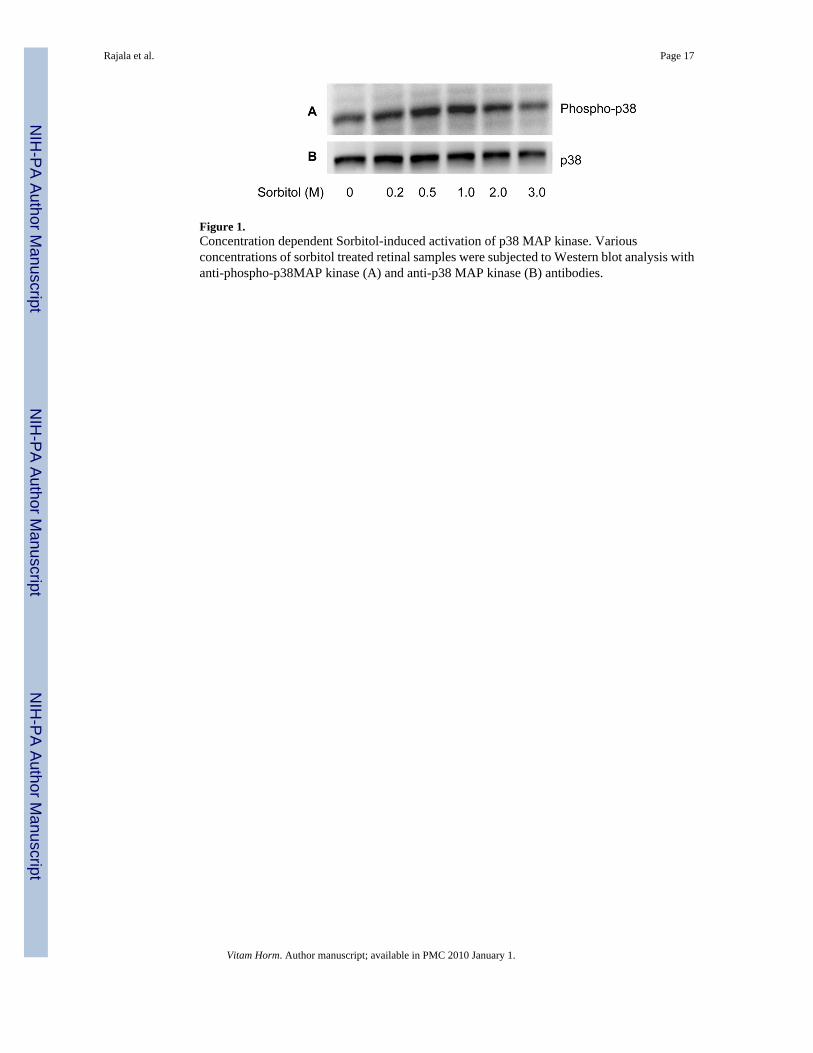
Figure 1.Concentration dependent Sorbitol-induced activation of p38 MAP kinase. Variousconcentrations of sorbitol treated retinal samples were subjected to Western blot analysis withanti-phospho-p38MAP kinase (A) and anti-p38 MAP kinase (B) antibodies.
Rajala et al. Page 17
Vitam Horm. Author manuscript; available in PMC 2010 January 1.
NIH
-PA Author Manuscript
NIH
-PA Author Manuscript
NIH
-PA Author Manuscript
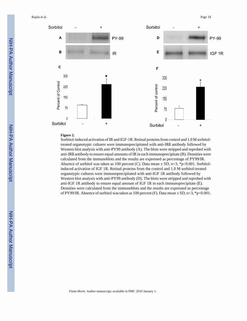
Figure 2.Sorbitol-induced activation of IR and IGF-1R. Retinal proteins from control and 1.0 M sorbitol-treated organotypic cultures were immunoprecipitated with anti-IRß antibody followed byWestern blot analysis with anti-PY99 antibody (A). The blots were stripped and reprobed withanti-IRß antibody to ensure equal amounts of IR in each immunoprecipitate (B). Densities werecalculated from the immunoblots and the results are expressed as percentage of PY99/IR.Absence of sorbitol was taken as 100 percent (C). Data mean ± SD, n=3, *p<0.001. Sorbitol-induced activation of IGF 1R. Retinal proteins from the control and 1.0 M sorbitol-treatedorganotypic cultures were immunoprecipitated with anti-IGF 1R antibody followed byWestern blot analysis with anti-PY99 antibody (D). The blots were stripped and reprobed withanti-IGF 1R antibody to ensure equal amount of IGF 1R in each immunoprecipitate (E).Densities were calculated from the immunoblots and the results are expressed as percentageof PY99/IR. Absence of sorbitol was taken as 100 percent (F). Data mean ± SD, n=3, *p<0.001.
Rajala et al. Page 18
Vitam Horm. Author manuscript; available in PMC 2010 January 1.
NIH
-PA Author Manuscript
NIH
-PA Author Manuscript
NIH
-PA Author Manuscript
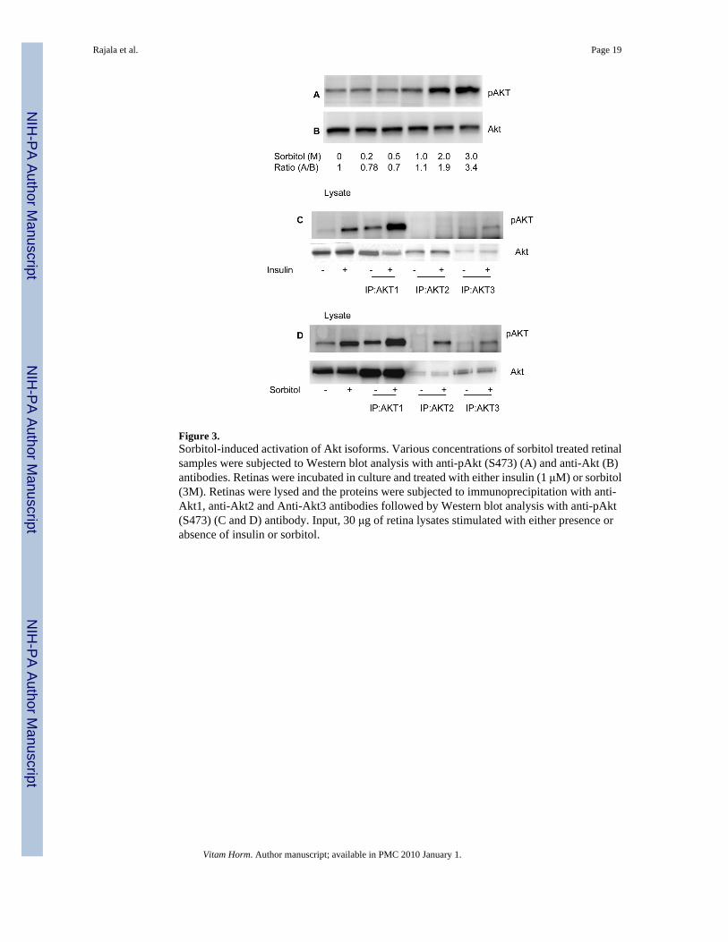
Figure 3.Sorbitol-induced activation of Akt isoforms. Various concentrations of sorbitol treated retinalsamples were subjected to Western blot analysis with anti-pAkt (S473) (A) and anti-Akt (B)antibodies. Retinas were incubated in culture and treated with either insulin (1 μM) or sorbitol(3M). Retinas were lysed and the proteins were subjected to immunoprecipitation with anti-Akt1, anti-Akt2 and Anti-Akt3 antibodies followed by Western blot analysis with anti-pAkt(S473) (C and D) antibody. Input, 30 μg of retina lysates stimulated with either presence orabsence of insulin or sorbitol.
Rajala et al. Page 19
Vitam Horm. Author manuscript; available in PMC 2010 January 1.
NIH
-PA Author Manuscript
NIH
-PA Author Manuscript
NIH
-PA Author Manuscript
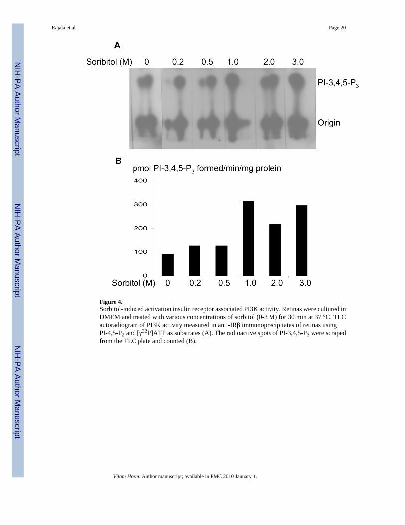
Figure 4.Sorbitol-induced activation insulin receptor associated PI3K activity. Retinas were cultured inDMEM and treated with various concentrations of sorbitol (0-3 M) for 30 min at 37 °C. TLCautoradiogram of PI3K activity measured in anti-IRβ immunoprecipitates of retinas usingPI-4,5-P2 and [γ32P]ATP as substrates (A). The radioactive spots of PI-3,4,5-P3 were scrapedfrom the TLC plate and counted (B).
Rajala et al. Page 20
Vitam Horm. Author manuscript; available in PMC 2010 January 1.
NIH
-PA Author Manuscript
NIH
-PA Author Manuscript
NIH
-PA Author Manuscript
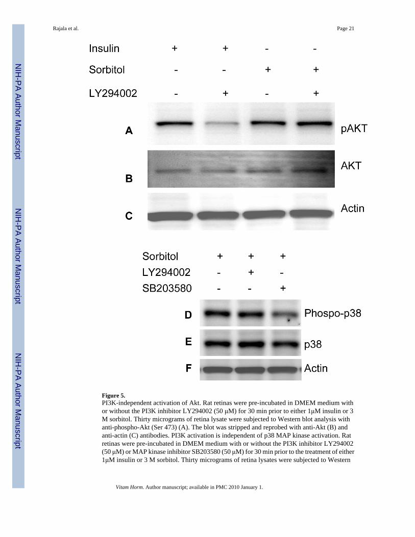
Figure 5.PI3K-independent activation of Akt. Rat retinas were pre-incubated in DMEM medium withor without the PI3K inhibitor LY294002 (50 μM) for 30 min prior to either 1μM insulin or 3M sorbitol. Thirty micrograms of retina lysate were subjected to Western blot analysis withanti-phospho-Akt (Ser 473) (A). The blot was stripped and reprobed with anti-Akt (B) andanti-actin (C) antibodies. PI3K activation is independent of p38 MAP kinase activation. Ratretinas were pre-incubated in DMEM medium with or without the PI3K inhibitor LY294002(50 μM) or MAP kinase inhibitor SB203580 (50 μM) for 30 min prior to the treatment of either1μM insulin or 3 M sorbitol. Thirty micrograms of retina lysates were subjected to Western
Rajala et al. Page 21
Vitam Horm. Author manuscript; available in PMC 2010 January 1.
NIH
-PA Author Manuscript
NIH
-PA Author Manuscript
NIH
-PA Author Manuscript
blot analysis with anti-phospho-p38 MAP kinase(D). The blot was stripped and reprobed withanti-p38 MAP kinase (E) and anti-actin (F) antibodies.
Rajala et al. Page 22
Vitam Horm. Author manuscript; available in PMC 2010 January 1.
NIH
-PA Author Manuscript
NIH
-PA Author Manuscript
NIH
-PA Author Manuscript
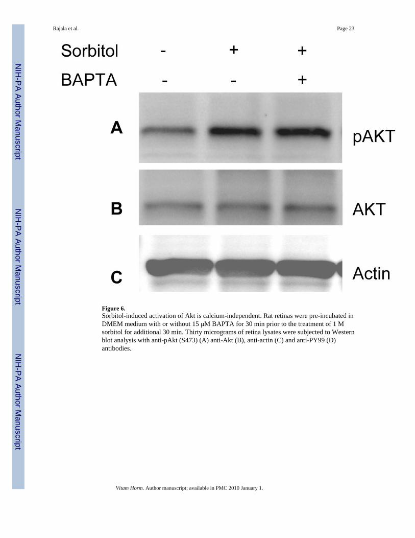
Figure 6.Sorbitol-induced activation of Akt is calcium-independent. Rat retinas were pre-incubated inDMEM medium with or without 15 μM BAPTA for 30 min prior to the treatment of 1 Msorbitol for additional 30 min. Thirty micrograms of retina lysates were subjected to Westernblot analysis with anti-pAkt (S473) (A) anti-Akt (B), anti-actin (C) and anti-PY99 (D)antibodies.
Rajala et al. Page 23
Vitam Horm. Author manuscript; available in PMC 2010 January 1.
NIH
-PA Author Manuscript
NIH
-PA Author Manuscript
NIH
-PA Author Manuscript
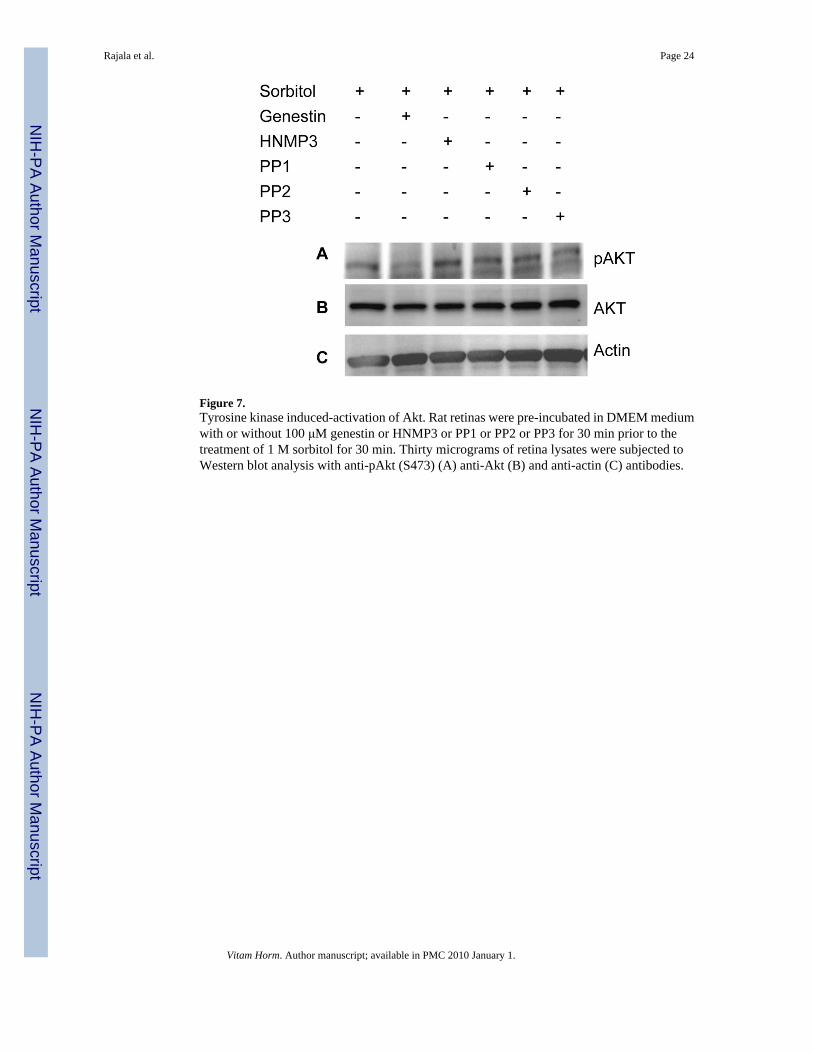
Figure 7.Tyrosine kinase induced-activation of Akt. Rat retinas were pre-incubated in DMEM mediumwith or without 100 μM genestin or HNMP3 or PP1 or PP2 or PP3 for 30 min prior to thetreatment of 1 M sorbitol for 30 min. Thirty micrograms of retina lysates were subjected toWestern blot analysis with anti-pAkt (S473) (A) anti-Akt (B) and anti-actin (C) antibodies.
Rajala et al. Page 24
Vitam Horm. Author manuscript; available in PMC 2010 January 1.
NIH
-PA Author Manuscript
NIH
-PA Author Manuscript
NIH
-PA Author Manuscript
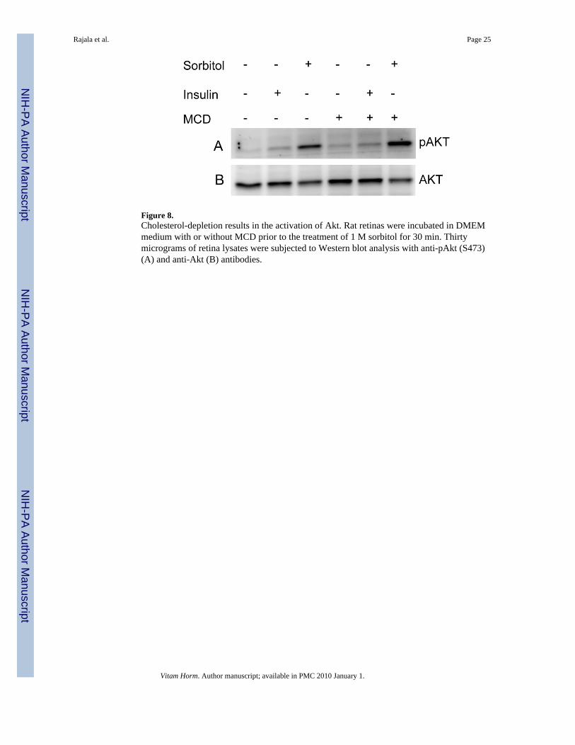
Figure 8.Cholesterol-depletion results in the activation of Akt. Rat retinas were incubated in DMEMmedium with or without MCD prior to the treatment of 1 M sorbitol for 30 min. Thirtymicrograms of retina lysates were subjected to Western blot analysis with anti-pAkt (S473)(A) and anti-Akt (B) antibodies.
Rajala et al. Page 25
Vitam Horm. Author manuscript; available in PMC 2010 January 1.
NIH
-PA Author Manuscript
NIH
-PA Author Manuscript
NIH
-PA Author Manuscript
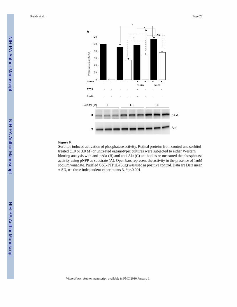
Figure 9.Sorbitol-induced activation of phosphatase activity. Retinal proteins from control and sorbitol-treated (1.0 or 3.0 M) or untreated organotypic cultures were subjected to either Westernblotting analysis with anti-pAkt (B) and anti-Akt (C) antibodies or measured the phosphataseactivity using pNPP as substrate (A). Open bars represent the activity in the presence of 1mMsodium vanadate. Purified GST-PTP1B (5μg) was used as positive control. Data are Data mean± SD, n= three independent experiments 3, *p<0.001.
Rajala et al. Page 26
Vitam Horm. Author manuscript; available in PMC 2010 January 1.
NIH
-PA Author Manuscript
NIH
-PA Author Manuscript
NIH
-PA Author Manuscript
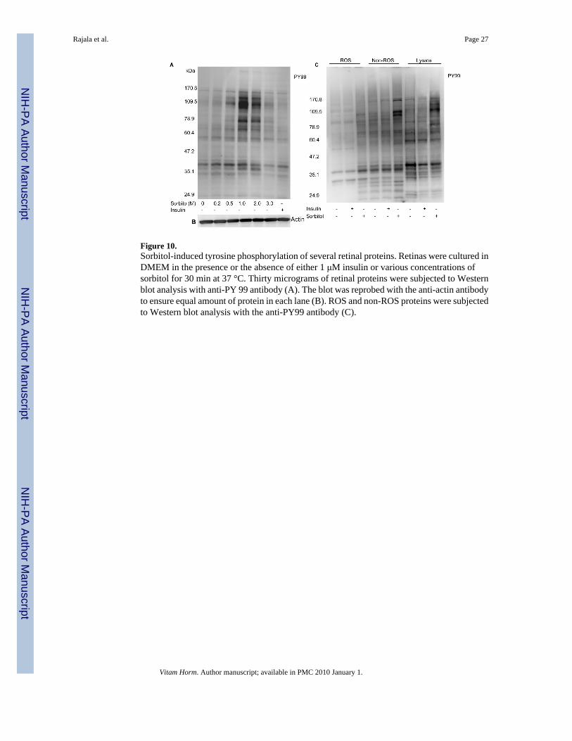
Figure 10.Sorbitol-induced tyrosine phosphorylation of several retinal proteins. Retinas were cultured inDMEM in the presence or the absence of either 1 μM insulin or various concentrations ofsorbitol for 30 min at 37 °C. Thirty micrograms of retinal proteins were subjected to Westernblot analysis with anti-PY 99 antibody (A). The blot was reprobed with the anti-actin antibodyto ensure equal amount of protein in each lane (B). ROS and non-ROS proteins were subjectedto Western blot analysis with the anti-PY99 antibody (C).
Rajala et al. Page 27
Vitam Horm. Author manuscript; available in PMC 2010 January 1.
NIH
-PA Author Manuscript
NIH
-PA Author Manuscript
NIH
-PA Author Manuscript
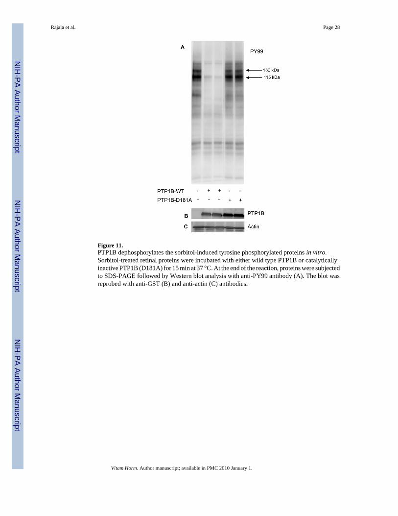
Figure 11.PTP1B dephosphorylates the sorbitol-induced tyrosine phosphorylated proteins in vitro.Sorbitol-treated retinal proteins were incubated with either wild type PTP1B or catalyticallyinactive PTP1B (D181A) for 15 min at 37 °C. At the end of the reaction, proteins were subjectedto SDS-PAGE followed by Western blot analysis with anti-PY99 antibody (A). The blot wasreprobed with anti-GST (B) and anti-actin (C) antibodies.
Rajala et al. Page 28
Vitam Horm. Author manuscript; available in PMC 2010 January 1.
NIH
-PA Author Manuscript
NIH
-PA Author Manuscript
NIH
-PA Author Manuscript
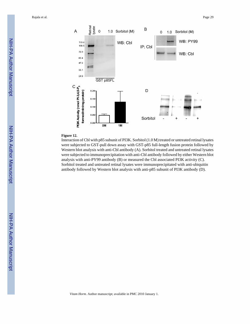
Figure 12.Interaction of Cbl with p85 subunit of PI3K. Sorbitol (1.0 M) treated or untreated retinal lysateswere subjected to GST-pull down assay with GST-p85 full-length fusion protein followed byWestern blot analysis with anti-Cbl antibody (A). Sorbitol treated and untreated retinal lysateswere subjected to immunoprecipitation with anti-Cbl antibody followed by either Western blotanalysis with anti-PY99 antibody (B) or measured the Cbl associated PI3K activity (C).Sorbitol treated and untreated retinal lysates were immunoprecipitated with anti-ubiquitinantibody followed by Western blot analysis with anti-p85 subunit of PI3K antibody (D).
Rajala et al. Page 29
Vitam Horm. Author manuscript; available in PMC 2010 January 1.
NIH
-PA Author Manuscript
NIH
-PA Author Manuscript
NIH
-PA Author Manuscript