changes in the histological architecture of hepatocytes and ovarian tissues during growth,...
TRANSCRIPT
The Journal of Zoology Studies
` Vol. 2 No. 2 2015 Journalofzoology.com
Page 12
The Journal of Zoology Studies 2015; 2(2): 12-23
ISSN 2348-5914 JOZS 2015; 2(2): 12-23 JOZS © 2015 Received: 04-03-2015 Accepted: 20-04-2015 Padmanabha Chakrabarti Fisheries Laboratory, Department of Zoology, The University of Burdwan, Golapbag, Burdwan-713 104, West Bengal, India Satinath Kundu Fisheries Laboratory, Department of Zoology, The University of Burdwan, Golapbag, Burdwan-713 104, West Bengal, India Corresponding Author:
Padmanabha Chakrabarti Department of Zoology, The University of Burdwan, Golapbag, Burdwan-713 104, West Bengal, India Tel: +91-342-2634798 Fax No.: +91-342-2657938 E-mail: [email protected]
Changes in the histological architecture of hepatocytes and ovarian tissues during growth, maturation spawning and post-spawning phases in Puntius
sarana (Hamilton, 1822)
Authors: Padmanabha Chakrabarti and Satinath Kundu
Abstract
The present investigation dealt with the cytological status of hepatocytes and correlated them with
the seasonal changes of ovarian activities in female Puntius sarana (Hamilton, 1822). The
hepatocyte of female was provided with distinct nucleus and dense basophilic cytoplasmic
granules. Different germ line cells were recognized on the basis of size and histoarchitecture of the
cells. During growth and maturation phases it was found that the density of cytoplasmic granules of
hepatocytes was increased in number as well as the nucleus became hypertrophied. These features
were well correlated with the occurrence of cortical alveolus and yolk granule stage in the ovary.
During spawning phase the sparse cytoplasmic granules were encountered in the hypertrophied
hepatic cells. This was because of the dynamic cytological activities during vitellogenesis and
occurrence of mature follicles in the ovary. It was concluded that the cytological changes in the
hepatocytes and ovarian activities correlate well during growth, maturation spawning and post-
spawning phases in female Puntius sarana.
Keywords: Histology, Hepatocytes, Ovarian tissues, Growth, Maturation, Spawning, Post-spawning, Puntius sarana
1. Introduction
The reproductive process is an essential part of the study the biology of species [1]. As such an
understanding of the gonadal development and reproductive cycles of fishes is of fundamental
importance for the conservation of natural stocks and for fish culture purposes. Various phases of
gonadal development of fishes have been studied to clarify the dynamics and regulation of oogenesis [2]. It is known that the ovarian cycle in majority of freshwater teleosts which are seasonal breeders
undergo remarkable changes during various periods of the season [3, 4, 5, 6, 7, 8, 9]. The gradual increase
in total weight and associated morphohistological changes in gonad of the fish from the pre-
spawning season is, therefore, intimately associated with the transfer of various nutrients from the
body muscles and liver as well as with the proliferation of various germ line cells formed by the
active process of gametogenesis [10, 11]. Banaee and Naderi [12] opined that an increase of
hepatosomatic index (HSI) during maturation phase may indicate the increased activity of liver
during vitellogenesis and vitellogenin synthesis.
The Journal of Zoology Studies
` Vol. 2 No. 2 2015 Journalofzoology.com
Page 13
Therefore, it would be interesting to study in details the functional aspects associated with the changes in the morphohistology of the ovary on the one hand and the variation in the histological aspects of the liver on the other in relation to growth, maturation, spawning and post-spawning phases of Puntius sarana (Hamilton, 1822). 2. Materials and Methods Adult live female specimens of P. sarana (length 21-24 cm and weight 100-150 g) were procured from khari river which was 64 km. away from Burdwan, West Bengal during the second week of every month from January to December 2013. The fishes were acclimatized for 5 days period by feeding artificial diet daily. Data on total body weight and ovarian weight of 10 fishes were taken to calculate the mean gonadosomatic index (GSI) using the formula:
GSI = [Total ovary weight/(Body weight-Weight of the ovaries)] × 100 Likewise data on total body weight and liver weight to 10 fishes were taken to calculate the mean HSI using the following formula:
HSI = [Total liver weight/(Body weight-Weight of the liver)] × 100 2.1. Histological Methods After decapitation of the fish the fragments of liver and ovary were fixed in aqueous Bouin’s fluid for 18 hours for histological studies. Paraffin sections were cut at 4µm thickness and stained with Delafield’s haematoxylin-eosin, Mallory’s triple stain and iron-alum haematoxylin for ovary and liver tissues. From the histological preparations of the ovaries, the diameter of various oogenetic cells and their nuclei were measured with the help of reticulo-micrometer and ocular micrometer respectively. 3. Results Histologically the liver of P.sarana was composed of parenchyma covered by a thin capsule of connective tissue. Hepatocytes varied from polyhedral to round shape and were irregularly arranged surrounding a central vein. Each hepatocyte contained a round central nucleus and basophilic cytoplasm (Figs.1, 3). Hepatic cells showed changes during different reproductive phases. 3.1. Oogenesis Histologically the germinal epithelium of ovarian cavity projected into ovigerous lamellae where development of new crops of oogonia took place. The sequence of oocyte maturation in P. sarana had been divided into six developmental stages based upon the cytological characteristics of the cells, viz; oogonia (stage I), early and late perinucleolus stage (stage II and stage III), yolk vesicle stage (stage IV), yolk granule stage (stage V) and mature follicle (stage VI).
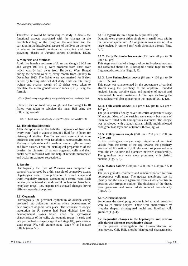
3.1.1. Oogonia (6 µm × 8 µm to 12 µm ×10 µm) Oogonia were present either singly or in small nests within the lamellar epithelium. An oogonium was made up of a large nucleus (4 µm to 5 µm) with chromatin threads (Figs. 2, 5). 3.1.2. Early Perinucleolus oocyte (23 µm × 28 µm to 50 µm × 60 µm) This stage consisted of a large oval centrally placed nucleus and contained about 8 to 10 basophilic nuclei together with fragmented chromatin (Figs. 2, 9). 3.1.3. Late Perinucleolus oocyte (84 µm × 100 µm to 90 µm × 105 µm) This stage was characterized by the appearance of cortical alveoli along the periphery of the ooplasm. Rounded nucleoli having variable sizes and number of nuclei and condensed chromatin materials. A thin layer enclosing the zona radiata was also appearing in this stage (Figs.11, 12). 3.1.4. Yolk vesicle oocyte (112 µm × 132 µm to 124 µm × 145 µm) The yolk vesicles finally cover the entire ooplasm of stage IV oocyte. Most of the vesicles were empty but some of them were filled with homogenous materials. The oocyte was enveloped with a zona radiata, middle multinucleated zona granulosa layer and outermost theca (Fig. 4). 3.1.5. Yolk granules oocyte (220 µm × 250 µm to 280 µm × 340 µm) In this vitellogenic oocyte stage migration of germinal vesicle from the centre of the egg towards the periphery was started. Formation of yolk globules took place and as a result the cell volume and diameter increased considerably. The granulosa cells were more prominent with distinct nucleus (Figs. 5, 6). 3.1.6. Mature follicle (380 µm × 400 µm to 450 µm × 500 µm) The yolk granules coalesced and remained packed to form homogenous yolk mass. The nuclear membrane lost its identity and the nucleus (germinal vesicle) was eccentric in position with irregular outline. The thickness of the theca, zona granulosa and zona radiata reduced considerably (Figs.8, 9). 3.1.7. Atretic oocytes: Sometimes the developing oocytes failed to attain maturity were called atretic oocytes. These were characterized by irregular shaped, disintegrated nuclei and liquified yolk granules (Fig. 4). 3.2. Sequential changes in the hepatocytes and ovarian cells during different reproductive phases In the present investigation the histoarchitecture of heaptocytes, GSI, HSI, morpho-histological characteristics
The Journal of Zoology Studies
` Vol. 2 No. 2 2015 Journalofzoology.com
Page 14
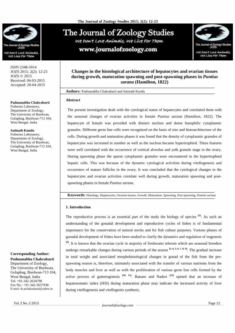
of oogenetic cells were found to undergo changes during growth, maturation, spawning and post-spawning phases. 3.2.1. Growth Phase (December to February) In this phase the cytoplasm of the liver cells accumulated dense homogenous granules. Nuclei were found in the centre of the hepatic cells. The sinusoid endothelium was present in between hepatic cells (Fig. 1). Primary oocytes (stage I and stage II) were present in the ovary (Fig. 2). However, during the end of this period percentage of stage III oocytes increased which showed cortical alveoli. During this phase slight increment of GSI was noticed and during January and February the GSI value increased gradually from 1.86 ± 0.22 to 2.19 ± 1.15. HSI also gradually increased during December (2.20 ± 0.28) and during late growth phase i.e. January and February the HSI increased to 2.78 ± 0.25 to 3.45 ± 0.28 (Table 1). 3.2.2. Maturation Phase (March to May) During this yolk deposition stage the hepatic cells were found slightly enlarged in size and the nuclei were gradually hypertrophied. The sinusoids were well vascularized (Fig. 3). The highest oogenetic activity was found to occur during this phase. Different stages of vitellogenic oocytes were present. However, majority of the developing oocytes were of stage IV and stage V respectively. At the end of this phase the yolk granules of stage V continued to coalesce. Prominent zona granulosa and zona radiata were present. The immature oocytes were found to be decreased in number. A few atretic follicles were also found at this stage (Figs. 4, 5, 6). During the onset of maturation phase in March onwards when the ovary entered into the maturation, GSI gradually increased from 3.12 ± 0.68 to 12.01 ± 2.15 in May. In this phase during the month of March the HSI reached maximum 3.86 ± 0.09 and considerably decreased thereafter in April (1.38 ± 0.16) and May (1.12 ± 0.15) respectively (Table 1).
3.2.3. Spawning phase (June to August) In the spawning phase the hepatic cells showed momentous changes than growth and maturation phases. The hepatocytes were enlarged with depleted cytoplasm and having a hypertrophic nuclei (Fig. 7). However, some of the hepatic cells still showed granulation in the cytoplasm. The vascularisation of the hepatic sinusoids were also reduced (Fig. 7). The ovaries at this stage were full of stage V oocytes and mature follicles. The mature follicles became larger and irregular in shape, the yolk globules condensed and provided with ecentric germinal vesicles (Figs. 8, 9). A few discharged follicles were also observed (Fig. 9). In June the ovary was with full of mature follicles and GSI attended the peak value (16.73 ± 1.86) but in July and August the GSI value showed a declining trend (13.44 ± 2.10 and 8.60 ± 2.32). The significant decreased of HSI was noticed during the entire spawning phase i.e. in the month of June (1.07 ± 0.08), July (0.98 ± 0.26) and August (0.56 ± 0.17) (Table 1). 3.2.4. Post-spawning phase (September to November) The hepatocytes were gradually transformed like that of growth phase having centrally placed nuclei and basophilic granules were started to accumulate in the hepatic cytoplasm. The hepatic sinusoids were present in between hepatic cells (Fig. 10). During this reproductive phase mature ova were few in numbers. The oogonia and early perinucleolar oocytes were appeared along with few late perinuclear oocytes (Figs. 11, 12) in between primary oocytes. In the post-spawning period i.e. in September onwards the yolky follicles reabsorbed and the ovaries showed a regression state. The GSI was recorded to 3.52 ± 0.27, 1.28 ± 0.34 and 0.92 ± 0.22 during September, October and November respectively. During September the HSI recorded to 1.20 ± 0.08 and gradually increased during October (1.45 ± 0.06) and November (1.52 ± 0.09) (Table 1).
Table 1: Variations in the gonadosomatic index (GSI) and hepatosomatic index (HSI) of female Puntius sarana
Stages of ovary Month Mean GSI ± SE Mean HSI ± SE
Growth Phase December January February
1.48 ± 0.25 1.86 ± 0.22 2.19 ± 1.15
2.20 ± 0.28 2.78 ± 0.25 3.45 ± 0.28
Maturation Phase March April May
3.12 ± 0.68 6.52 ± 1.60 12.01 ± 2.15
3.86 ± 0.09 1.38 ± 0.16 1.12 ± 0.15
Spawning Phase June July
August
16.73 ± 1.86 13.44 ± 2.10 8.60 ± 2.32
1.07 ± 0.08 0.98 ± 0.26 0.56 ± 0.17
Post-spawning and Resting Phase
September October
November
3.52 ± 0.27 1.28 ± 0.34 0.92 ± 0.22
1.20 ± 0.08 1.45 ± 0.06 1.52 ± 0.09
The Journal of Zoology Studies
` Vol. 2 No. 2 2015 Journalofzoology.com
Page 15
Fig 1: Hepatic cells (arrow heads) of growth phase with centrally placed nucleus and dense cytoplasm. Note hepatic
sinusoids (arrows) in between hepatic cells (HE) × 400X.
Fig 2: Oogonia (arrows), oocyte I (OI) and early perinucleolus oocyte (OII) during growth phase (HE) × 150X.
The Journal of Zoology Studies
` Vol. 2 No. 2 2015 Journalofzoology.com
Page 16
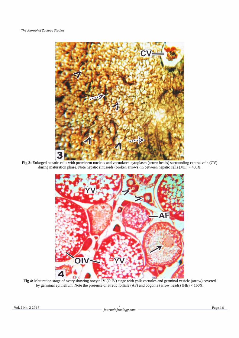
Fig 3: Enlarged hepatic cells with prominent nucleus and vacuolated cytoplasm (arrow heads) surrounding central vein (CV)
during maturation phase. Note hepatic sinusoids (broken arrows) in between hepatic cells (MT) × 400X.
Fig 4: Maturation stage of ovary showing oocyte IV (O IV) stage with yolk vacuoles and germinal vesicle (arrow) covered
by germinal epithelium. Note the presence of atretic follicle (AF) and oogonia (arrow heads) (HE) × 150X.
The Journal of Zoology Studies
` Vol. 2 No. 2 2015 Journalofzoology.com
Page 17
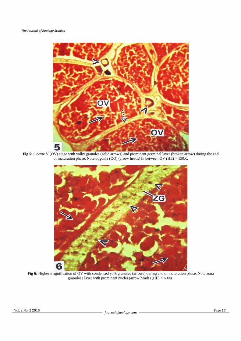
Fig 5: Oocyte V (OV) stage with yolky granules (solid arrows) and prominent germinal layer (broken arrow) during the end
of maturation phase. Note oogonia (OO) (arrow heads) in between OV (HE) × 150X.
Fig 6: Higher magnification of OV with condensed yolk granules (arrows) during end of maturation phase. Note zona
granulose layer with prominent nuclei (arrow heads) (HE) × 600X.
The Journal of Zoology Studies
` Vol. 2 No. 2 2015 Journalofzoology.com
Page 18
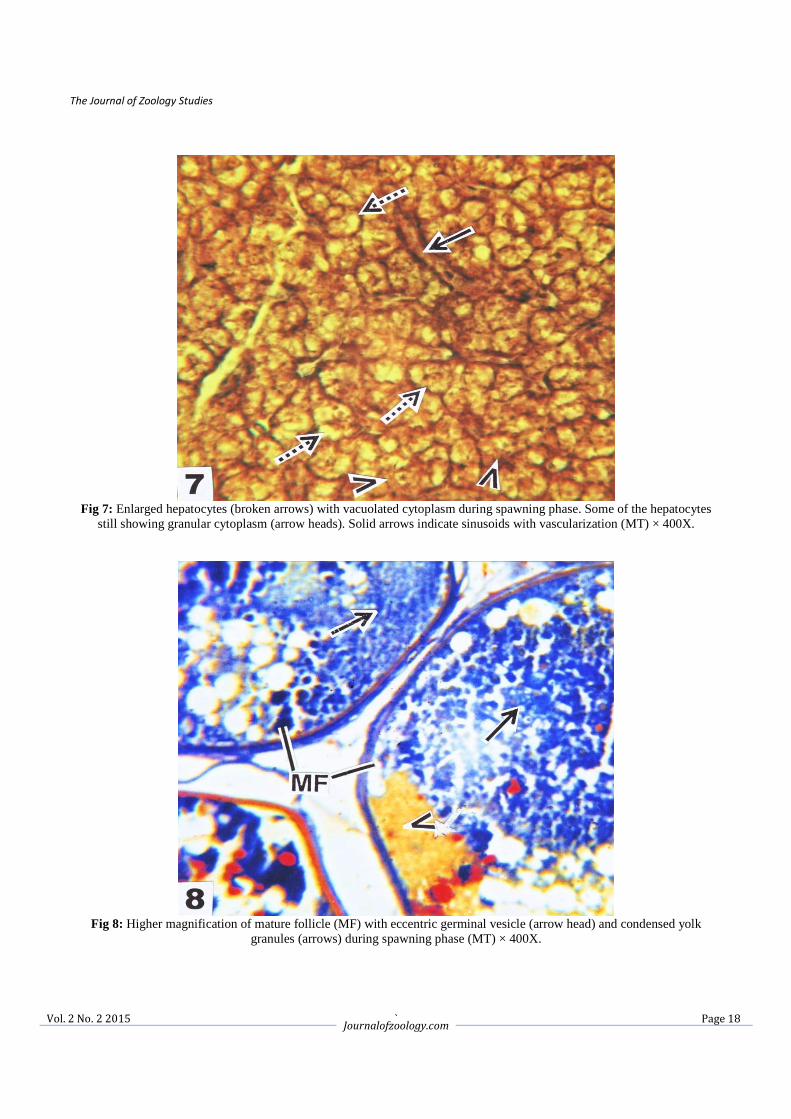
Fig 7: Enlarged hepatocytes (broken arrows) with vacuolated cytoplasm during spawning phase. Some of the hepatocytes
still showing granular cytoplasm (arrow heads). Solid arrows indicate sinusoids with vascularization (MT) × 400X.
Fig 8: Higher magnification of mature follicle (MF) with eccentric germinal vesicle (arrow head) and condensed yolk
granules (arrows) during spawning phase (MT) × 400X.
The Journal of Zoology Studies
` Vol. 2 No. 2 2015 Journalofzoology.com
Page 19
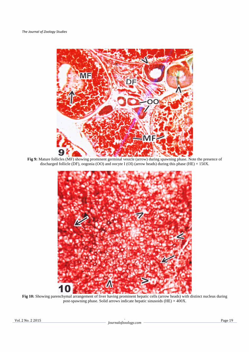
Fig 9: Mature follicles (MF) showing prominent germinal vesicle (arrow) during spawning phase. Note the presence of
discharged follicle (DF), oogonia (OO) and oocyte I (OI) (arrow heads) during this phase (HE) × 150X.
Fig 10: Showing parenchymal arrangement of liver having prominent hepatic cells (arrow heads) with distinct nucleus during
post-spawning phase. Solid arrows indicate hepatic sinusoids (HE) × 400X.
The Journal of Zoology Studies
` Vol. 2 No. 2 2015 Journalofzoology.com
Page 20
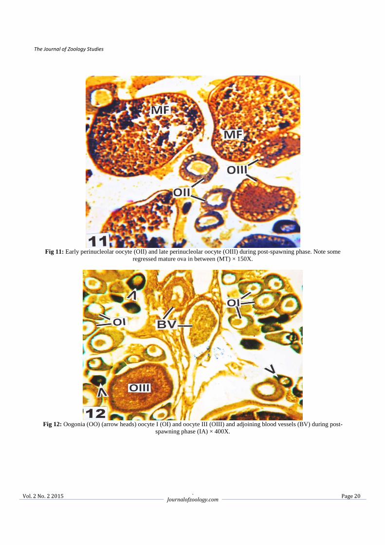
Fig 11: Early perinucleolar oocyte (OII) and late perinucleolar oocyte (OIII) during post-spawning phase. Note some
regressed mature ova in between (MT) × 150X.
Fig 12: Oogonia (OO) (arrow heads) oocyte I (OI) and oocyte III (OIII) and adjoining blood vessels (BV) during post-
spawning phase (IA) × 400X.
The Journal of Zoology Studies
` Vol. 2 No. 2 2015 Journalofzoology.com
Page 21
4. Discussion In the present study the relationship of HSI with GSI in the fish, P.sarana during growth, maturation, spawning and post-spawning phases indicate marked differences. The lowest GSI and HSI values were noticed during the end and beginning of post-spawning phase when the ovaries were found in regressed condition containing mainly primary oocytes. However, GSI increased marginally but remained almost stationary during the growth phase whereas HSI gradually increased during the post-spawning phase and sharply rose at the end of growth phase. This may be due to the gradual proliferation of late perinucleolar oocytes in the ovary and accumulation of various nutrients in the hepatic cells of liver. Sudarshan and Kulkarni [13] also reported high HSI value during beginning of preparatory phase in Notopterus notopterus. GSI increased rapidly from the end of the maturation phase and continued up to spawning phase due to slow accumulation of yolk granules and proliferation of vitellogenic oocytes. On the contrary, HSI reached maximum during early maturation to beginning of spawning phase. The highest values of HSI indicate heavier liver probably due to the synthesis and accumulation of various nutrients during vitellogenesis process. Such a rhythm of changes of HSI and GSI in N. notopterus has also been reported by Sudarshan and Kulkarni [13]. The HSI sharply declined in the spawning phase which might be due to the release of nutrients to the blood and then transported into the mature ooctyes during complete process of vitellogenesis. Mukherjee et al. [14] and Mandal [15] reported that adequate food availability helped the female fish in recruitment of vitellogenic oocytes and in maintaining the maturation process in the ovary. However, GSI value declined prominently from September onwards due to discharge or reabsorption of yolky oocytes. Singh and Singh [16] studied the relationship between HSI and GSI in the fish Heteropneustes fossilis and found that high HSI during preparatory and post-spawning and low levels during pre-spawning and spawning. Histologically the liver of P. sarana showed hepatic parenchymal arrangement consisted of hepatic cells along with sinusoids which were arranged around a central vein. This histoarchitecture of liver resembled that described for the stripped weak fish Cynoscion guatucupa [17] and for rainbow trout [18]. In the present investigation oogenesis occurred in two stages, the division of oogonia and the transformation of the resting oocytes into mature oocytes. In P. sarana it was observed that oogonia after development from the germinal epithelium of the ovigerous lamellae passed through a number of maturation stages before it become a mature ovum. This involved complex changes in the cytology of the nucleus and cytoplasm. The formation of yolk globules in the late perinucleolus oocyte in P. sarana began in the periphery of the developing ooplasm which gradually moved to the centre of mature follicles through different
stages of development. Bisht and Joshi [19] and Kapoor [20] observed similar pattern of yolk deposition in Schizothorax richardsonii and Puntius ticto. In the present study the mature follicles were enveloped by the multinucleated zona granulosa, outermost theca and inner zona radiata. In the late developing oocytes, it may be assumed that through zona radiata the essential nutrients were transported from the granulosa layer to the ooplasm for building up the yolk globules of the mature oocytes. Similar observations have also been made by Bromage and Cumaranatunga [21] and Shabanipour and Heidari [22] in the ovary of rainbow trout and Liza aurata. In the present investigation the maturational activity in the ovary reached to the highest during the late maturation and early spawning phase when oocyte diameter as well as GSI rose up. At the same period the hepatocytes were found to be enlarged along with hypertrophy of nuclei and were accompanied with the depletion of cytoplasmic granules. This feature might be due to the uptake and accumulation of various nutrients in the oocytes and yolk precursor vitellogenin from hepatocytes. Sinha and Pal [23] opined that the stored protein, lipid and carbohydrate were shifted from liver and body muscle to the ovary to ensure proper growth and development of oocytes. de Vlaming et al. [24] reported that principal events responsible for the growth of the oocytes involved sequestration of hepatically derived protein precursor, vitellogenin which took part in the formation of yolk protein. Aida et al. [25] opined that the liver cells in female ayu (Plecoglossus altivelis) showed an activated state of female specific plasma protein (FSPP) synthesis following ovarian maturation. They also stated that upon synthesis in the liver cells, this protein is probably released into the blood and then transported into the oocytes during the process of ovarian maturation in ayu. 5. Conclusion Cytological changes in the hepatocytes are correlated with the reproductive phases in P. sarana. It is found that during growth and maturation phases the density of cytoplasmic granules of hepatocytes is increased as well as the nucleus becomes hypertrophied. These features are well correlated with the occurance of cortical alveolus and yolk granule stage in the ovary.During spawning phase the cytoplasmic granules are sparse in the hypertrophied hepatic cells. This is due to the dynamic cytological activities like vitellogenesis and occurance of mature oocytes in the ovary. However, no significant alterations are noticed in the hepatocytes during post-spawning phase due to the appearance of new germ cells in the ovary. 6. Acknowledgements The authors are grateful to Dr. Sanjib Ray, Head of the Department of Zoology, The University of Burdwan, Burdwan for providing laboratory facilities.
The Journal of Zoology Studies
` Vol. 2 No. 2 2015 Journalofzoology.com
Page 22
7. References 1. Silva RMPC, Esper MLP. Observacoes sobre o
desenvolvimento citomorfologico dos ovarios de tainha, Mugil platanus (Gunther) da Baia de paranagua (Brasil). Acta Biologica Paranaense,1991; 20:15-39
2. Potts GW, Wootton RJ. Fish reproduction: Strategies avol tactics. Academic Press: London,1984; 410p
3. Sanwal R, Khanna SS. Seasonal changes in the testes of a freshwater fish, Channa gachua. Acta Anatomica, 1972; 83:139-148
4. Srivastava SS, Srivastava AK. Oogenesis in a freshwater lagre murrel, Channa striatus. Boletim de zoology, University of Sao Paulo,1984; 8:155-163
5. Manna PR, Bhattacharya S. Gonadotropin binding to the ovary of an Indian major carp, Catla catla at different stages of reproductive cycle. Journal of Bioscience,1993; 18:361-372
6. Msiska OV. The histology of mature gonads of Oreochromis (Nyasalpia) karongae (Trewavas). African Journal of Ecology,2002; 40:164-171
7. Singh A, Singh IJ, Ram RN, Kushwaha B. Ovarian development in Labeo dyocheilus (Mc Clelland) during active reproductive phase under captive and wild conditions. Journal of Environmental Biology,2008; 29:169-174
8. Saeed SS, Reza IM, Bagher AF, Saeed G. Histological study of ovarian development and sexual maturity of kutum (Rutilus frisii kutum) (Kamemskii 1901). World Applied Science Journal,2010; 8:1343-1350
9. Yin JX, Racey P, Li J, Zhang YG. The ovarian cycle of the fish Leptobotia elongata Bleeker, endemic to China. Pakistan Journal of Zoology,2012; 44:997-1005
10. Washbarn BS, Frye DJ, Hung SSO, Doroshov SI. Conte FS. Dietary effects on tissue composition, oogenesis and the reproductive performance of female rainbow trout (Onchorhynchus mykiss). Journal of Aquaculture,1990; 90:179-195
11. Anderson D, Boetius I, Larsen LO, Seidler PH. Effects of oestradiol enrich diet and of feeding with porcine testicular tissue on macroscopic gonadal sex in European eels. Journal of Fish Biology,1996; 48:484-492
12. Banaee M, Naderi M. The reproductive biology of Shirbot (Barbus grypus Hackel, 1843) in the Maroon River, Iran. International Journal of Aquatic Biology,2014; 2:43-52
13. Sudarshan S, Kulkarni RS, Determination of condition factor (k), somatic condition factor (ks), hepatic and gonadosomatic indices in the freshwater fish Notopterus notopterus.
International Journal of Scientific Research,2013; 2:524-526
14. Mukherjee J, Bhattacharya S, Nath P. Annual changes in ovary and vitellogenin content of liver, serum and ovary of murrel, Channa punctatus (Bloch). Journal of Experimental Biology,1989; 27:240-252
15. Mandal DK. Effects of artificial food on the gonadal maturation in Labeo bata and Puntius javanicus. Ph.D Thesis; The University of Burdwan, Burdwan, West Bengal, India. 2000.
16. Singh AK, Singh TP. Seasonal fluctuation in lipid and cholesterol content in ovary, liver and blood serum in relation to annual sexual cycle in Heteropneustes fossilis (Bloch).1979; 73: 47-54.
17. Diaz AA, Gonzalez Castro M, Garcia AM, Devincenti CV, Goldenberg AL. Morphological and histochemical characterization of liver from stripped weakfish, Cynoscion guatucupa (Cuvier). Biosciences,1999; 7:67-78
18. Atamanalp M, Sisman T, Geyikoglu F, Topal A. The histopathological effects of copper sulphate on rainbow trout liver (Oncorhynchus mykiss). Journal of Fisheries and Aquatic Sciences,2008; 3:291-297
19. Bisht JS, Joshi ML. Seasonal histological changes in the ovaries of a mountain stream teleost, Schizothorax richardsonii. Acta Anatomica,1975; 93:512-525
20. Kapoor, CP. Nucleolar extrusion in the oocytes pf Puntius ticto. Zoologische Beitrage, 1977; 23: 221-226
21. Bromage N, Cumaranatunga R. Egg production in the rainbow trout. In Recent Advances in Aquaculture (ed. Muir JF, Roberts RJ). London. Croom Helm,1988; 63-138
22. Shabanipour N, Heidari B. A histological study of the zona radiata during late oocyte developmental stages in the Caspian sea magilid, Liza aurata. Brazilian journal of Morphological Sciences,2004; 21:191-195
23. Sinha GM, Pal PC. Seasonal variations in protein, lipid and carbohydtate contents of ovary, liver and body muscle in relation to gonadosomatic index and oogenesis of Clarias batrachus Linnaeus. In impacts of environment on animals and aquaculture, vol:xxiii, National Symposium, ed. Manna GK, Jana BB. Kalyani; West Bengal, India.
24. de-Vlaming VL, Wiley HS, Delahunty G, Wallace RA. Goldfish (Carassius auratus) vitellogenin: induction, isolation, properties and relationship to yolk proteins. Comparative Biochemistry and Physiology,1980; 678: 613-623
25. Aida K, Hirose K, Yokote M, Hibiya T. Physiological studies on gonadal maturation of fishes II. Histological changes in liver cells of Ayu
The Journal of Zoology Studies
` Vol. 2 No. 2 2015 Journalofzoology.com
Page 23
following gonadal alteration and estrogen administration. Bulletin Society of Fisheries, 1973; 39: 1107-1115
Chakrabarti P, Kundu S. Changes in the histological architecture of hepatocytes and ovarian tissues during growth, maturation spawning and post-spawning phases in Puntius sarana (Hamilton, 1822). Journal of Zoology Studies. 2015; 2(2):00-00.
*************************************************** **************