cellular expression, trafficking, and function of two isoforms of human ulbp5/raet1g
TRANSCRIPT
Cellular Expression, Trafficking, and Function of TwoIsoforms of Human ULBP5/RAET1GRobert A. Eagle1,5*, Gillian Flack2, Anthony Warford2, Jesus Martınez-Borra1, Insiya Jafferji1, James A.
Traherne1, Maki Ohashi1, Louise H. Boyle1, Alexander D. Barrow1, Sophie Caillat-Zucman3, Neil T.
Young4, John Trowsdale1
1 Cambridge Institute for Medical Research, Wellcome Trust/MRC Building, Addenbrookes Hospital, Cambridge, United Kingdom, 2 Atlas of Protein Expression Group,
Wellcome Trust Sanger Institute, Wellcome Trust Genome Campus, Hinxton, Cambridgeshire, United Kingdom, 3 INSERM U561 AVENIR Team, Hopital St-Vincent de Paul,
Paris, France, 4 Department of Pathology, University of Cambridge, Cambridge, United Kingdom, 5 California Institute of Technology, MC170-25, Pasadena, California,
United States of America
Abstract
Background: The activating immunoreceptor NKG2D is expressed on Natural Killer (NK) cells and subsets of T cells. NKG2Dcontributes to anti-tumour and anti-viral immune responses in vitro and in vivo. The ligands for NKG2D in humans arediverse proteins of the MIC and ULBP/RAET families that are upregulated on the surface of virally infected cells and tumours.Two splicing variants of ULBP5/RAET1G have been cloned previously, but not extensively characterised.
Methodology/Principal Findings: We pursue a number of approaches to characterise the expression, trafficking, and functionof the two isoforms of ULBP5/RAET1G. We show that both transcripts are frequently expressed in cell lines derived fromepithelial cancers, and in primary breast cancers. The full-length transcript, RAET1G1, is predicted to encode a molecule withtransmembrane and cytoplasmic domains that are unique amongst NKG2D ligands. Using specific anti-RAET1G1 antiserum tostain tissue microarrays we show that RAET1G1 expression is highly restricted in normal tissues. RAET1G1 was expressed at alow level in normal gastrointestinal epithelial cells in a similar pattern to MICA. Both RAET1G1 and MICA showed increasedexpression in the gut of patients with celiac disease. In contrast to healthy tissues the RAET1G1 antiserum stained a widevariety or different primary tumour sections. Both endogenously expressed and transfected RAET1G1 was mainly found insidethe cell, with a minority of the protein reaching the cell surface. Conversely the truncated splicing variant of RAET1G2 wasshown to encode a soluble molecule that could be secreted from cells. Secreted RAET1G2 was shown to downregulate NKG2Dreceptor expression on NK cells and hence may represent a novel tumour immune evasion strategy.
Conclusions/Significance: We demonstrate that the expression patterns of ULBP5RAET1G are very similar to the well-characterised NKG2D ligand, MICA. However the two isoforms of ULBP5/RAET1G have very different cellular localisationsthat are likely to reflect unique functionality.
Citation: Eagle RA, Flack G, Warford A, Martınez-Borra J, Jafferji I, et al. (2009) Cellular Expression, Trafficking, and Function of Two Isoforms of Human ULBP5/RAET1G. PLoS ONE 4(2): e4503. doi:10.1371/journal.pone.0004503
Editor: Jacques Zimmer, Centre de Recherche Public de la Sante (CRP-Sante), Luxembourg
Received September 23, 2008; Accepted November 20, 2008; Published February 18, 2009
Copyright: � 2009 Eagle et al. This is an open-access article distributed under the terms of the Creative Commons Attribution License, which permitsunrestricted use, distribution, and reproduction in any medium, provided the original author and source are credited.
Funding: This work is funded by grants from Cancer Research UK and The Isaac Newton Trust to RAE, The Leukaemia Research Fund to NTY, and The WellcomeTrust to JT. The funders had no role in study design, data collection and analysis, decision to publish, or preparation of the manuscript.
Competing Interests: RAE and JT hold a patent on RAET1G.
* E-mail: [email protected]
Introduction
The activating immunoreceptor NKG2D has been established
as an important component of the innate immune system. In
humans NKG2D is constitutively expressed on NK cells, cd T
cells, and CD8+ T cells[1]. NKG2D is not normally found on
CD4+ T cells, however it can be expressed in these cells in patients
with rheumatoid arthritis[2], cancer[3], and when stimulated with
HCMV[4]. NKG2D can invoke powerful immune responses
against target cells expressing its ligands both in vitro and in
vivo[1,5], however cellular activation is likely to require synergy
with other signals delivered by cytokines or other receptors[6,7,8].
The ligands of NKG2D are surprisingly diverse proteins that
are structurally related to MHC class I[9,10]. In humans they
comprise two members of the MIC family and five members of the
ULBP/RAET family[11,12,13,14]. According to the paradigm of
NKG2D mediated immune recognition, ligands for the receptor
are expressed on cells subject to stress, for example cells infected
with viruses such as HCMV, or during tumourigenesis[15,16].
The importance of NKG2D in viral infection is evidenced by the
fact that both human and murine cytomegalovirus encode
immune evasion molecules designed to prevent cell surface
expression of NKG2D ligands[15].
NKG2D ligands are widely expressed on cancer cells.
Activation of DNA damage pathways during tumour develop-
ment has been reported to be a central mechanism leading to
NKG2D ligand expression [17]. There is evidence that NKG2D
participates in a process of immunosurveillance for cancer. In
mice, the ectopic expression of NKG2D ligands mediates
immune responses to transplanted tumours[18,19]. Neutralising
PLoS ONE | www.plosone.org 1 February 2009 | Volume 4 | Issue 2 | e4503
NKG2D with antibodies decreases host protection to experi-
mentally induced tumours[20] and an NKG2D knockout mouse
showed an increased susceptibility to some model cancers [21].
Therefore it is proposed that in order to progress, cancers must
evade NKG2D mediated immune responses. One evasion
mechanism may be the production of TGF-b by tumours,
which downregulates NKG2D expression[22,23]. NKG2D may
also be downregulated by soluble ligands released from the
surface of cancer cells[24], and by chronic exposure to cells
expressing its ligands[25,26,27]. These studies suggest that
NKG2D mediated immunity may become anergised during
tumour development.
The MICA molecule has restricted expression in normal tissues
but is found in normal gut epithelial cells[28]. In healthy
individuals MICA seems to be largely inside the cell, however in
celiac disease (CD) MICA expression is increased and redistributes
to the cell surface where it can provoke autoimmune attack and
villous atrophy[29,30]. Inappropriate NKG2D ligand expression
has a role in other autoimmune diseases, including Crohn’s
disease[31], autoimmune diabetes[32], and rheumatoid arthri-
tis[28].
An open question in the field is why are their so many ligands for
the same receptor? Intriguingly NKG2D ligands possess markedly
different domain structures. ULBP1-3 molecules are GPI anchored
to the cell surface, whilst MICA/B, ULBP4/RAET1E, and
ULBP5/RAET1G have transmembrane and cytoplasmic domains
of variable length and sequence[12,13,14,33]. NKG2D ligands
are also capable of being expressed independently of each
other[12,34,35]. This suggests that NKG2D ligands may not all
be functionally equivalent. The predominant driver of NKG2D
ligand diversity is proposed to be an immunological arms race
with viruses, which have in turn acquired a varied array of
immunoevasin molecules that prevent NKG2D ligands being
expressed at the cell surface[10]. However this is not the whole
story. NKG2D ligands have clearly evolved other unique properties
that do not relate to their susceptibility to viral immunoevasins. One
example is MICA, which possesses motifs that allow it to be targeted
to specific membranes in specialised epithelial cell layers[36].
Therefore it is essential to examine the expression profile and
properties of individual MIC and ULBP/RAET molecules to get a
fuller understanding of NKG2D function.
Two alternative transcripts of ULBP5/RAET1G have previ-
ously been identified (named RAET1G1 and RAET1G2)[14,37].
Here we examine the expression, trafficking and function of each
variant in detail.
Materials and Methods
Antibody generation and western blottingPolyclonal antibody to RAET1G was raised in rabbits using two
peptides corresponding to part of the CYT of the protein. The
peptides were:
CNNGAARYSEPLQVSIS and CSHGHHPQSLQPPPHPP
Peptides were manufactured and coupled to Ovalbumin by
Southampton Polypeptides. The antiserum was raised in rabbit
using a combination of both peptides, separately coupled to
ovalbumin, by Harlan Seralabs. The polyclonal antibody was
purified by the caprylic acid/ ammonium sulphate precipitation
method. For western blotting, ,5*105 cells were lysed straight into
reducing SDS-PAGE buffer, boiled and separated by SDS-PAGE.
For soluble RAET1G detection in tissue culture 40 ml tissue
culture medium was boiled with SDS-PAGE buffer and loaded
onto a gel. Western blotting was carried out according to standard
protocols. Membranes were probed with either anti-RAET1G
polyclonal, rabbit polyclonal anti-V5 (Invitrogen), or mouse
monoclonal anti-b-actin (Sigma) followed by species specific
HRP-conjugated secondary antibodies (Dako).
Cell lines, and primary cell culturesCell lines are as described previously[14]. Primary NK cells
were isolated from peripheral blood using standard Ficoll isolation
of mononuclear cells, followed by use of an NK cell negative
isolation kit (Dynal) to deplete unwanted cell populations. NK cells
were cultured in RPMI-1640 medium containing penicillin,
streptomycin, 10% human serum and 100 U/ml rIL-2. Prior to
use, isolated NK cells were shown to be CD56+, NKG2D+ and
CD32 (data not shown).
RT-PCRCell line and primary breast cancer mRNA was a gift from Dr
Cherie Blenkiron and Prof Carlos Caldas[38]. Fresh frozen
primary tumours were collected with consent from patients
between 1997–2000 by the Addenbrooke’s Hospital Histology
Tissue Bank, Cambridge, UK. All samples were used with
appropriate Local Research Ethics Committee approval, Adden-
brookes Hospital, and were collected and stored with written
consent from patients. First strand cDNA synthesis from cell line
and primary tumour mRNAs was performed using the Super-
script-III cDNA Synthesis Kit (Invitrogen). cDNA was quantitated
and adjusted so that 200 ng was used in each PCR reaction. Splice
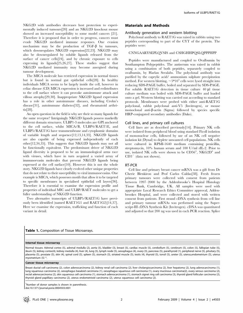
Table 1. Composition of Tissue Microarrays.
Internal tissue microarray
Normal tissues: Adrenal cortex (5), adrenal medulla (2), aorta (5), bladder (3), breast (5), cardiac muscle (5), cerebellum (5), cerebrum (5), colon (5), fallopian tube (5),ileum (5), kidney cortex(4), kidney medulla (4), liver (4), lung (5), lymph node (5), oesophagus (4), ovary (3), pancreas (5), parathyroid (1), peripheral nerve (5), pituitary (5),placenta (5), prostate (5), skin (4), spinal cord (5), spleen (5), stomach (3), striated muscle (5), testis (4), thyroid (5), tonsil (5), ureter (3), uterus,endometrium (5), uterusmyometrium (5) *.
Cancer tissue microarray
Breast ductal cell carcinoma (2), colon adenocarcinoma (2), kidney renal cell carcinoma (2), liver cholangiocarcinoma (2), liver hepatoma (2), lung adenocarcinoma (1),lung squamous carcinoma (2), oesophagus basaloid carcimona (1), oesophagus squamous cell carcinoma (1), ovary mucinous carcinoma(2), ovary serous carcinoma (2),rectal adenocarcinoma (2), skin squamous cell carcinoma (1), stomach adenocarcinoma (1), stomach signet ring cell carcinoma (3), thyroid gland follicular carcinoma (2),thyroid gland papilliary carcinoma (2), uterus endometrioid carcinoma (2), uterus squamous cell carcinoma (2).
*Number of donor samples is shown in parenthesis.doi:10.1371/journal.pone.0004503.t001
Isoforms of ULBP5/RAET1G
PLoS ONE | www.plosone.org 2 February 2009 | Volume 4 | Issue 2 | e4503
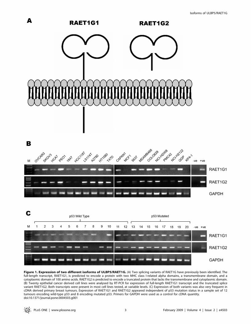
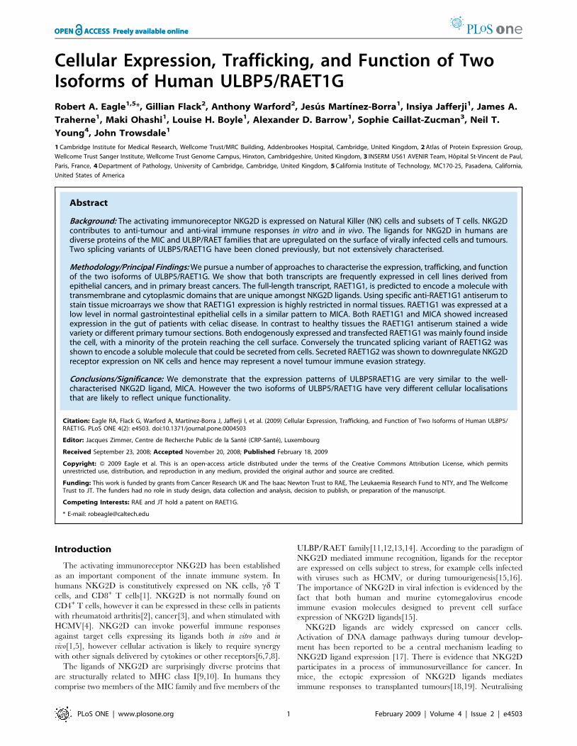
Figure 1. Expression of two different isoforms of ULBP5/RAET1G. (A) Two splicing variants of RAET1G have previously been identified. Thefull-length transcript, RAET1G1, is predicted to encode a protein with two MHC class I-related alpha domains, a transmembrane domain, and acytoplasmic domain of 100 amino acids. RAET1G2 is predicted to encode a truncated protein that lacks the transmembrane and cytoplasmic domain.(B) Twenty epithelial cancer derived cell lines were analysed by RT-PCR for expression of full-length RAET1G1 transcript and the truncated splicevariant RAET1G2. Both transcripts were present in most cell lines tested, at variable levels. (C) Expression of both variants was also very frequent incDNA derived primary breast tumours. Expression of RAET1G1 and RAET1G2 appeared independent of p53 mutation status in a sample set of 12tumours encoding wild-type p53 and 8 encoding mutated p53. Primers for GAPDH were used as a control for cDNA quantity.doi:10.1371/journal.pone.0004503.g001
Isoforms of ULBP5/RAET1G
PLoS ONE | www.plosone.org 3 February 2009 | Volume 4 | Issue 2 | e4503
form specific primers were RAET1G1Fwd: CCATGTCCT-
CAGGCACAGC; RAET1G1Rev: CAGGGAACCATCAAGA-
TATGG; RAET1G2Fwd: AGCCCCGCGTTCCTTCTA;
RAET1G2Rev: GGGTCAGACTGTGCCTCCT. PCR reac-
tions were carried out for 35 cycles with an annealing temperature
of 62uC on a Peltier Thermal Cycler.
Constructs and cDNA expression in mammalian cells andbacteria
An N-terminal GFP fusion construct of RAET1G1 and ULBP2
was created in the vector PK1 [39]. The resulting construct
contained a leader peptide followed by GFP, a Myc tag, and a
short linker peptide fused to the relevant cDNA. Stable cell lines in
HT1080 cells were created with a lentiviral exression system (kind
gift of Dr Paul Lehner, [40]). C-terminal V5 tagged constructs
were created using the pEF6/V5-His TOPO TA Expression Kit
(Invitrogen). IMAGE clones 3070730 and 2911855 were used as
templates for RAET1G1 and RAET1G2 constructs respectively.
For western blot, ,5*105 Cos7 cells were transiently transfected
with 1 mg PK1-RAET1G1 plasmid using Fugene reagent (Roche).
For expression of V5 tagged constructs 5*106 Cos7 cells were
transfected with 5 mg plasmid, again with Fugene, and cells were
harvested for western blot and supernatant collected for NK cell
assays. Soluble recombinant RAET1G extracellular domain was
Figure 2. Generation of specific anti-RAET1G1 antiserum. (A) A rabbit polyclonal antiserum was raised against the cytoplasmic domain ofRAET1G. By western blot a protein of approximately 70 kDa was recognised in Cos7 cells transfected with a construct encoding an RAET1G-GFPfusion protein, but not in untransfected Cos7 cells. (B) By confocal microscopy the anti-RAET1G antiserum specifically stains (red) Cos7 cellstransfected with RAET1G-GFP (green), but not the highly related molecule ULBP2-GFP.doi:10.1371/journal.pone.0004503.g002
Isoforms of ULBP5/RAET1G
PLoS ONE | www.plosone.org 4 February 2009 | Volume 4 | Issue 2 | e4503
produced as a six histidine N-terminal fusion protein using a
plasmid described previously[14]. The protein was expressed in
inclusion bodies in BL21 E.coli (Novagen) and solubilised in 6 M
guanidine hydrochloride. Refolding was achieved by a stepwise
dilution into a Tris and Arginine containing buffer followed by
concentration by centriprep (Amicon). Prior to use in NK cell
assays recombinant RAET1G was dialysed into PBS.
Confocal microscopyConfocal microscopy was carried out as described previoulsy33.
For staining mouse monoclonal antibodies to MHC class I (W6/
32, kind gift from Dr Adrian Kelly), GM130 (BD Transduction
Labs), EEA1 (BD Transduction Labs) were used followed by anti-
mouse Alexa Fluor 568 secondary antibody (Molecular Probes).
Rabbit polyclonal anti-RAET1G or anti-calreticulin (BD Trans-
duction Labs) was used followed by anti-rabbit Alexa Fluor 568.
For antibody internalisation experiments transfected cells were
incubated with rabbit anti-myc antibody (AbCam) for 30 min at
4uC and then warmed to 37uC for 2 hours prior to fixing and
staining with secondary antibody
Flow CytometryHT1080 cell lines stably expressing PK1-ULBP2 and PK1-
RAET1G were stained with anti-GFP monoclonal antibody
(Roche) for 30 minutes at 4uC, followed by goat anti-mouse Alexa
Fluor 633 nm conjugate (Molecular Probes). As the stable cell lines
were roughly 70% GFP positive, GFP negative cells were gated
out in analysis. Data was then collected using BD FACSCalibur.
For primary NK cell analysis cells were cultured for 2 days either
in the presence or absence of soluble RAET1G and then analysed
for NKG2D expression with a PE conjugated anti-NKG2D mAb
(Coulter).
Radiolabelling and immunoprecipitationRadiolabelling and immunoprecipitations were carried out as
described previously [41]. Briefly, 107 HT1080 stable cell lines
were starved in methionine/cysteine free tissue culture media
(Sigma) and then labelled with 1 mCi [35S] methionine and
cysteine Pro-mix (Amersham) for 10 minutes at 37uC. The cells
were then chased for 180 minutes in media containing unlabelled
methionine/cysteine media. After washing in PBS radiolabelled
cells were lysed in 1% Triton X-100 lysis buffer (150 mM NaCl,
20 mM Tris, 1 mM EDTA, 5 mM MgCl2 with protease inhibitors
from Roche). After removing nuclei and debris by centrifugation
1 mg anti-GFP monoclonal antibody (Roche) was added to
immunoprecipitate GFP fusion proteins, and protein A sepaharose
(Amersham) added to bind immune complexes. After washing the
bound proteins were eluted in reducing SDS-PAGE buffer and
half the eluate subject to digestion with Endo Hf (New England
Biolabs).
ImmunohistochemistryImmunohistochemistry was undertaken on paraffin wax tissue
microarrays[42,43]. The first was prepared using guided tissue
selection transferring 260.6 mm diameter cores from each
formalin fixed donor tissue into the recipient array. The normal
tissue microarray contained a total of 344 cores from 172 donor
samples as listed in Table 1. A similar array comprising normal
and tumour tissues was probed with purified pre-immune serum in
equal amounts as a control for staining, All samples were obtained
from Medical Solutions plc with ethical approval obtained from
LREC, Cambridge, UK. To evaluate the tissue distribution of the
RAET1G antibody in tumours, sections of commercial parafor-
maldehyde fixed tissue microarray (Petagen Inc, code A201(1))
containing 1 mm cores from 35 epithelial cancer samples (Table 1)
were also immunostained. Automated immunohistochemistry was
undertaken using a Ventana Medical Systems DiscoveryTM
system. Sections were dewaxed, pre-treated with mild cell
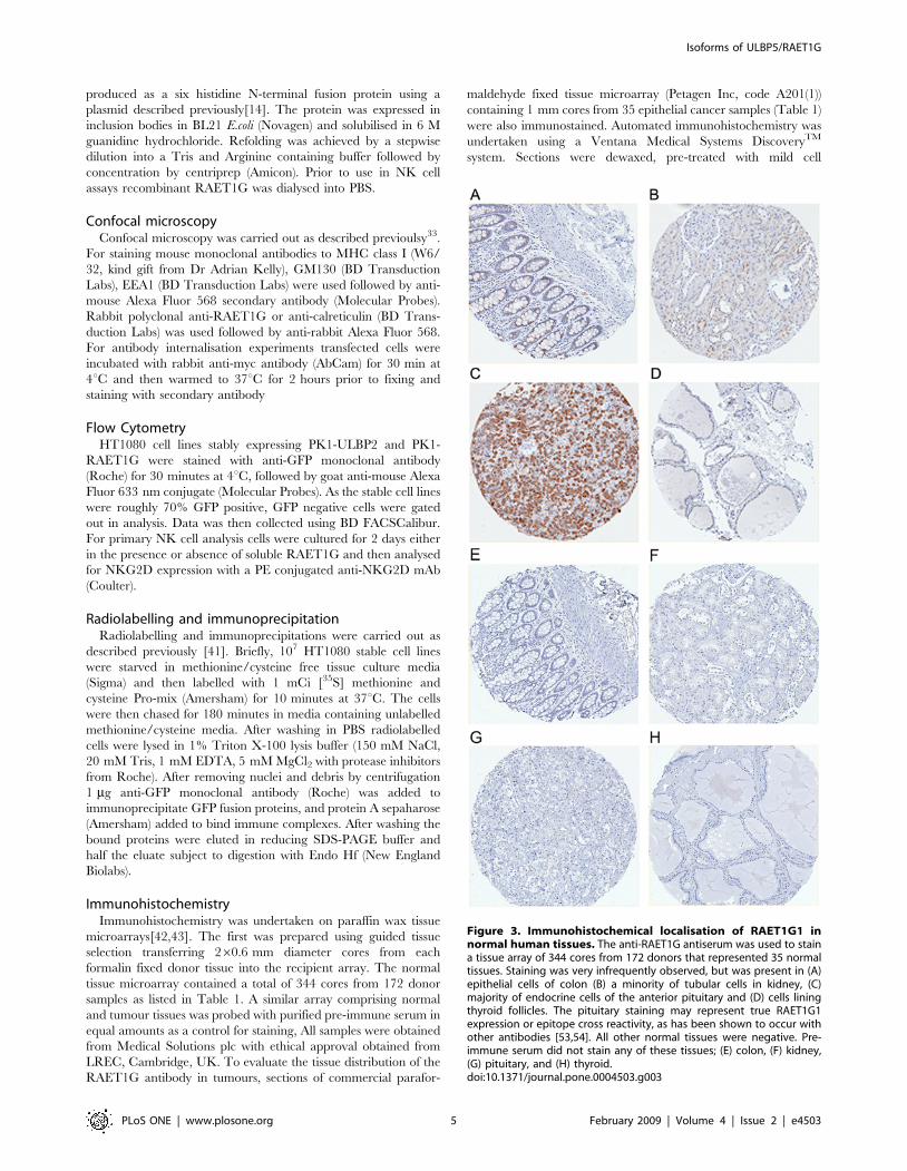
Figure 3. Immunohistochemical localisation of RAET1G1 innormal human tissues. The anti-RAET1G antiserum was used to staina tissue array of 344 cores from 172 donors that represented 35 normaltissues. Staining was very infrequently observed, but was present in (A)epithelial cells of colon (B) a minority of tubular cells in kidney, (C)majority of endocrine cells of the anterior pituitary and (D) cells liningthyroid follicles. The pituitary staining may represent true RAET1G1expression or epitope cross reactivity, as has been shown to occur withother antibodies [53,54]. All other normal tissues were negative. Pre-immune serum did not stain any of these tissues; (E) colon, (F) kidney,(G) pituitary, and (H) thyroid.doi:10.1371/journal.pone.0004503.g003
Isoforms of ULBP5/RAET1G
PLoS ONE | www.plosone.org 5 February 2009 | Volume 4 | Issue 2 | e4503
conditioner 1 (Tris borate/EDTA, pH 8.0) then incubated in the
rabbit anti RAET1G antibody at 10 mg/ml for 20 min at 37uC.
Staining was optimised by staining with a range of antibody
concentrations 10 mg/ml was selected as the optimum concentra-
tion. For detection an avidin/biotin block preceded application of
biotinylated goat anti rabbit (DakoCytomation code E0432)
diluted 1/100 for 8 min at 37uC. The biotinylated antibody was
then detected using a streptavidin/biotin/peroxidase kit (Ventana,
DAB MAPTM, code 760-124). The protocol was completed by
automated haematoxylin counterstaining followed by manual
dehydration clearing and mounting in resinous mountant.
Controls were included within each immunohistochemical runs.
Anti lysozyme and vimentin antibodies were used as positive
controls to verify the antigenic preservation of the tissue cores.
These controls provided positive staining in all tissue cores. To
establish if any staining present in the tissues was due to non-
specific interaction of the detection reagents slides were also
processed without the RAET1G antibody. No tissue staining was
observed in these preparations. Images of the stained tissue
microarray cores were automatically captured using an Ariol SL-
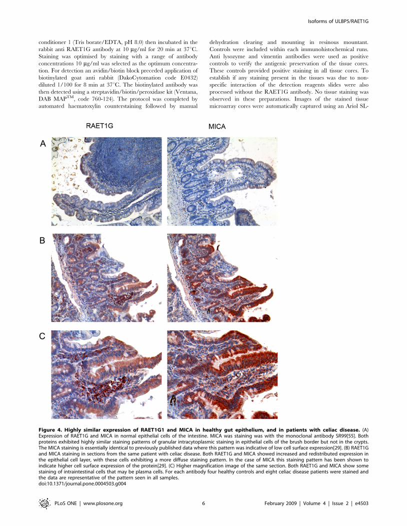
Figure 4. Highly similar expression of RAET1G1 and MICA in healthy gut epithelium, and in patients with celiac disease. (A)Expression of RAET1G and MICA in normal epithelial cells of the intestine. MICA was staining was with the monoclonal antibody SR99[55]. Bothproteins exhibited highly similar staining patterns of granular intracytoplasmic staining in epithelial cells of the brush border but not in the crypts.The MICA staining is essentially identical to previously published data where this pattern was indicative of low cell surface expression[29]. (B) RAET1Gand MICA staining in sections from the same patient with celiac disease. Both RAET1G and MICA showed increased and redistributed expression inthe epithelial cell layer, with these cells exhibiting a more diffuse staining pattern. In the case of MICA this staining pattern has been shown toindicate higher cell surface expression of the protein[29]. (C) Higher magnification image of the same section. Both RAET1G and MICA show somestaining of intraintestinal cells that may be plasma cells. For each antibody four healthy controls and eight celiac disease patients were stained andthe data are representative of the pattern seen in all samples.doi:10.1371/journal.pone.0004503.g004
Isoforms of ULBP5/RAET1G
PLoS ONE | www.plosone.org 6 February 2009 | Volume 4 | Issue 2 | e4503
Isoforms of ULBP5/RAET1G
PLoS ONE | www.plosone.org 7 February 2009 | Volume 4 | Issue 2 | e4503
50 automated image capture and system (Applied Imaging Inc)
using a 620 objective. Tissue sections and staining protocols for
celiac disease patients are as previously published [29].
Results
Expression of transcripts encoding two different isoformsof ULBP5/RAET1G
We have previously identified two splicing variants of ULBP5/
RAET1G, termed RAET1G1 and RAET1G2. The protein
topology analysis programme Phobius [44] predicts that
RAET1G1 encodes a protein with a transmembrane domain
and a cytoplasmic domain of approximately 100 amino acids
(Figure 1A). RAET1G2 has a premature stop codon before the
putative transmembrane domain due to alternative splice site
usage in exon 4 [14] and is predicted to encode a soluble molecule
(Figure 1A). Splice form specific primers were designed to amplify
each variant by RT-PCR. Specificity was tested by sequencing
PCR products, and primers were designed across intron/exon
boundaries in order to exclude the possibility that PCR products
were amplified from contaminating genomic DNA. Both
RAET1G1 and RAET1G2 were widely expressed in a panel of
20 epithelial cancer cell lines (Figure 1B). Both transcripts were
also widely expressed in a panel of cDNAs derived from primary
breast cancers (Figure 1C). RAET1G expression in breast cancer
did not correlate with p53 mutation status, as both p53 wild-type
and p53 mutated tumours frequently expressed both transcripts
(Figure 1C). GAPDH primers are a described previously [35].
Generation of anti-RAET1G antibodiesIn order to investigate the cellular expression of RAET1G1 we
raised specific polyclonal antiserum. The extracellular domain of
RAET1G is almost identical to ULBP2, therefore we chose to
generate rabbit polyclonal antiserum peptides derived from the
unique cytoplasmic domain of RAET1G1. By western blotting the
antiserum specifically recognised RAET1G1-GFP transfected into
Cos7 cells (Figure 2A). By confocal microscopy the anti-
RAET1G1 antiserum clearly stained Cos7 cells transfected with
RAET1G1-GFP, but not the highly related molecule ULBP2
(Figure 2B). ULBP2 is the most closely related gene to RAET1G in
the human genome.
RAET1G protein has restricted expression in normaltissues
Limited expression of the NKG2D ligand MICA in normal
tissues has been previously reported [28], however no systematic
study of other NKG2D ligands has been conducted. We used our
RAET1G1 specific antiserum to probe a tissue microarray,
comprised of 344 cores from 172 donors, representing 35 normal
tissues (Table 1). Purified pre-immune rabbit serum did not stain
any core. RAET1G1 antibody staining was demonstrated only in
four normal tissues (Figure 3). These include gut epithelium, that
has previously been shown to express RAET1G1 transcript [14],
and stained with the anti-RAET1G1 antiserum in two of five
individuals tested. Expression of RAET1G1 in the gut is also
noteworthy as MICA has widely been reported to be constitutively
expressed in the gut epithelium, and involved in gut pathology
[28,29,31,45].
Expression of RAET1G and MICA in normal gutepithelium and in celiac disease
Induction of MICA expression in gut epithelial cells is known to
be an important factor in the development of CD[29,45]. We
carried out a direct comparison of MICA and RAET1G
expression in both normal intestine and in biopsies from patients
with CD by immunohistochemistry. Both MICA and RAET1G1
antibodies gave punctate staining of villous epithelial cells that was
largely intracellular in normal control sections (Figure 4A). In CD
sections there was much higher intensity staining of the villous
epithelium and the staining exhibited altered distribution through-
out these cells (Figure 4B, 4C). In the case of MICA this staining
pattern has been shown previously to reflect redistribution of
protein from a largely intracellular distribution in normal gut
epithelium, to the cell surface in CD [29]. Both the MICA
monoclonal antibody and RAET1G1 antiserum showed staining
of cells in the lamina propria. Therefore RAET1G1 may have a
parallel role to MICA in the pathology of the gut and CD.
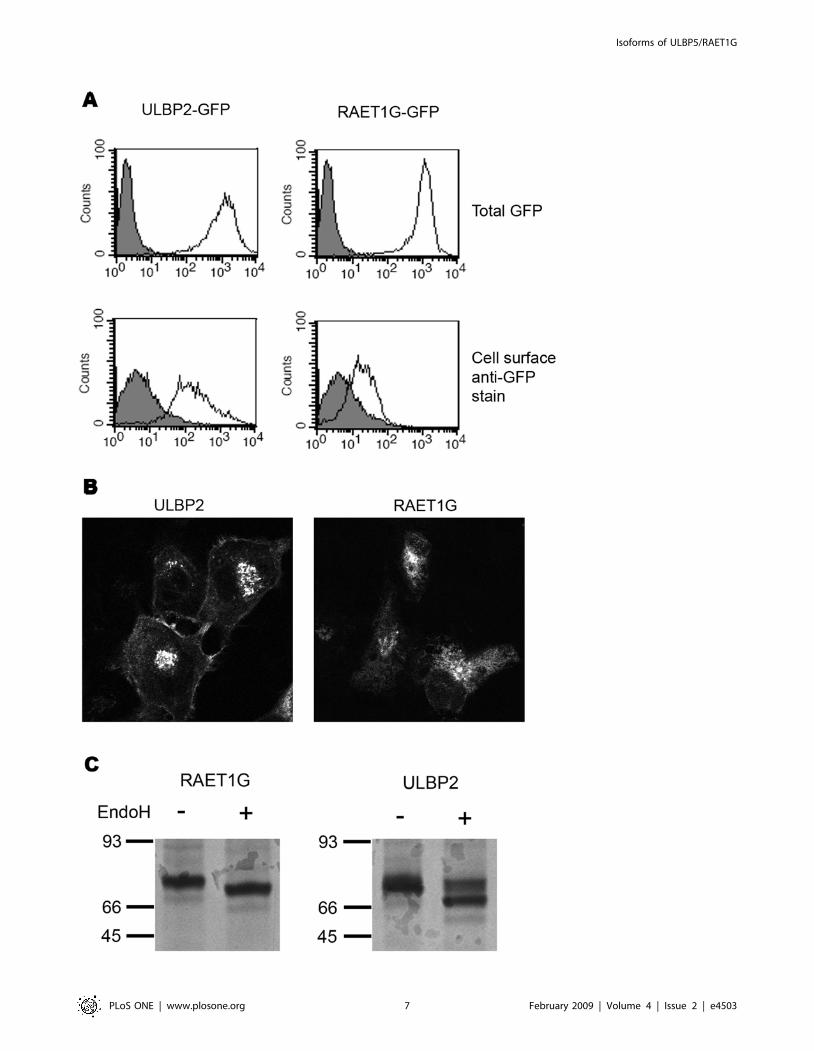
Subcellular localisation of RAET1G1There is evidence for the regulation of the trafficking and
subcellular localisation of MICA in a number of different cell types
[29,36,46]. As RAET1G1 and MICA had highly similar
expression patterns in normal and CD gut epithelial cells we
went on to examine the subcellular expression of RAET1G
further. Stable cell lines of ULBP2 and RAET1G, N-terminally
fused to GFP and a Myc-tag, were created in HT1080 cells. By
FACS the two cell lines expressed similar amounts of GFP-tagged
transgene (Figure 5A). However on staining with an anti-GFP
monoclonal antibody less cell surface expression of RAET1G
could be detected in contrast to ULBP2 (Figure 5A). Analysing
GFP expression by confocal microscopy the ULBP2-GFP
transfectants exhibited clear cell surface expression whereas
RAET1G-GFP transfectants exhibited much lower levels of
staining (Figure 5B).
HT1080 cell lines were radiolabelled, chased for 180 minutes,
lysed, and the fusion protein immunoprecipitated with anti-GFP
antibodies (Figure 5C). After 180 minutes a substantial proportion
of ULBP2-GFP had acquired Endo H resistance, indicating that it
had trafficked to the cell surface. In contrast RAET1G-GFP was
entirely Endo H sensitive (Figure 5C).
On co-staining with other cellular markers RAET1G showed a
contrasting localisation to MHC class I by confocal microscopy,
with little co-localisation at the cell surface (Figure 6A). RAET1G
mainly co-localised with calreticulin, a protein predominately
Figure 5. RAET1G1 protein is poorly expressed at the cell surface. (A) Stable cell lines expressing N-terminal GFP fusion proteins of ULBP2,and RAET1G1, were created in HT1080 cells. The transfectants expressed equivalent levels of transgene, as evident by total GFP fluorescence (openhistogram), plotted versus untransfected HT1080 cells (gray shaded histogram). Cell surface expression was assessed by staining with an anti-GFPmonoclonal antibody followed by an Alexa-Fluor 633 nm (far red) secondary antibody. The RAET1G1 transfectant had only a modest level of stainingover untransfected cells, whereas the ULBP2 transfectant stained at a high level. This indicates that a much smaller percentage of RAET1G1-GFPtransgene is present at the cell surface when compared to the closely related molecule ULBP2. (B) This observation was confirmed by confocalmicroscopy. The ULBP2-GFP transfectant showed clearly defined cell surface fluorescence in all cells, whereas cell surface RAET1G1-GFP could not beobserved. (C) Stable transfectants were radiolabelled and chased for 180 minutes. Radiolabelled RAET1G1 and ULBP2 were then immunoprecipitatedwith an anti-GFP antibody. Endo H digests reveal that a substantial proportion of ULBP2 has acquired Endo H resistance after 180 minutes, and hencehas trafficked to the cell surface. In contrast RAET1G1 remains Endo H sensitive.doi:10.1371/journal.pone.0004503.g005
Isoforms of ULBP5/RAET1G
PLoS ONE | www.plosone.org 8 February 2009 | Volume 4 | Issue 2 | e4503
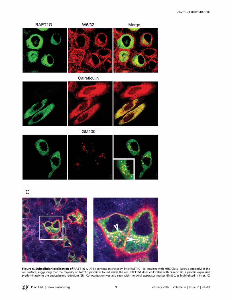
Figure 6. Subcellular localisation of RAET1G1. (A) By confocal microscopy, little RAET1G1 co-localised with MHC Class I (W6/32 antibody) at thecell surface, suggesting that the majority of RAET1G protein is found inside the cell. RAET1G1 does co-localise with calreticulin, a protein expressedpredominately in the endoplasmic reticulum (ER). Co-localisation was also seen with the golgi apparatus marker GM130, as highlighted in inset. (C)
Isoforms of ULBP5/RAET1G
PLoS ONE | www.plosone.org 9 February 2009 | Volume 4 | Issue 2 | e4503
expressed in the endoplamic reticulum (ER), and the golgi marker
GM130 (Figure 6A). An antibody internalisation experiment
showed RAET1G protein at the cell surface where it could be
internalised into endosomes (Figure 6B).
These data show that whilst the majority of RAET1G protein is
found inside the cell, a small proportion does reach the cell surface
and can enter the endocytic pathway.
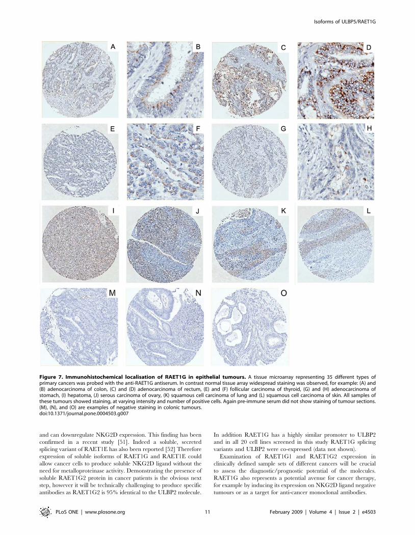
RAET1G1 protein is expressed in a range of tumoursWe next examined whether RAET1G1 expression was
amplified in tumours, using tissue microarray representing 20
different tumour types (Table 1). In contrast to the normal
tissue microarray extensive staining was observed in many
different cores and was of a higher intensity than in normal
tissues (Figure 7). The tumour types stained included; adeno-
carcinoma of colon, rectum and stomach; squamous cell
carcinoma of lung, oesophagus and skin, endometroid carcino-
ma of uterus, follicular carcinoma of thyroid, hepatoma and
serous carcinomas of ovary (Figure 7). Purified pre-immune
serum did not stain any core. Therefore RAET1G1 is a novel
tumour marker.
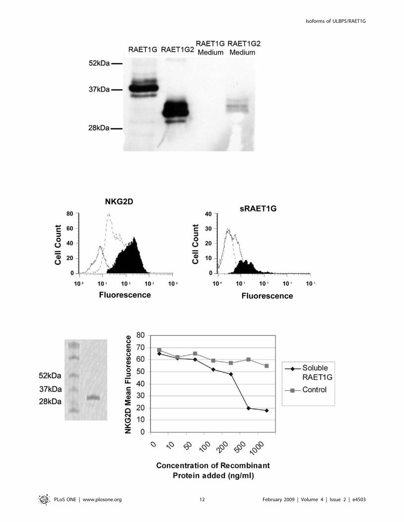
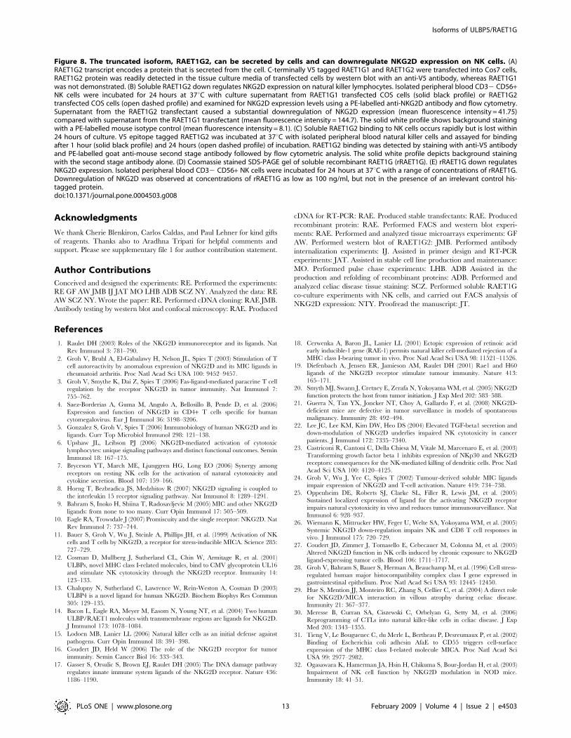
Cellular localisation and function of RAET1G2As RAET1G1 protein is widely expressed in cancer, the possible
co-expression of the putative soluble form RAET1G2 may have
relevance as a tumour immune evasion mechanism. Both
RAET1G1 and RAET1G2 were cloned into a cDNA expression
vector with a C-Teminal V5 epitope tag. When transfected into
Cos7 cells, RAET1G2, but not RAET1G1, was readily detected in
the tissue culture medium by western blot with anti-V5 antibody
(Figure 8A). Therefore RAET1G2 is a soluble molecule that can
be secreted from cells.
We took cultures of primary NK cells and added the tissue
culture medium from Cos7 cells transiently transfected with either
RAET1G1 or RAET1G2 for two days. Culturing NK cells with
media from RAET1G2 transfectants resulted in a reduction in cell
surface NKG2D staining when compared to NK cells cultured
with media from RAET1G1 transfectants (Figure 8B). To exclude
the possibility that RAET1G2 protein was simply blocking the
epitope recognised by the anti-NKG2D antibody we performed a
similar control experiment as described previously[24]. Using an
antibody to the C-terminal V5 Tag on soluble RAET1G in FACS,
NK cells cultured with RAET1G2 containing media for 1 hour
showed a slight shift, indicating presence of RAET1G2 protein
binding at the cell surface This was not seen in NK cells cultured
for 24 hours (Figure 8C). Therefore at the 24 hour time point
epitope blocking was not responsible for the observed loss of cell
surface NKG2D staining.
To obtain a more quantitative assessment of levels of RAET1G
protein required to downregulate NKG2D expression we used
recombinant bacterially expressed RAET1G extracellular domain
(rRAET1G; Figure 8D). With this reagent we observed downreg-
ulation of NKG2D with 100 ng/ml protein after 24 hours
incubation (Figure 8E).
Discussion
NKG2D ligands display considerable diversity in domain
structure, and can be expressed independently from each other
in cell lines. They are therefore highly unlikely to play functionally
identical roles in human disease processes. In order to understand
the implications of NKG2D ligand diversity it is necessary to
investigate differences, and similarities, in the properties of
individual MICA and ULBP/RAET molecules. Here we present
the first in depth characterisation of RAET1G1 and RAET1G2.
A systematic search for RAET1G1 protein expression revealed
that it has a very restricted expression pattern in healthy tissues.
The most striking finding was a staining pattern in the colon and
intestinal epithelium similar to that of MICA. Others have
previously shown that in the normal gut MICA is found largely
inside the cell, however increased expression and/or redistribution
of protein to the cell surface may occur in autoimmune conditions
such as CD and Crohn’s disease[29,31,47]. Once at the cell
surface MICA can facilitate NKG2D mediated recognition of the
gut epithelium and could be a factor in triggering autoimmune
attack. Upregulation of cell surface MICA on epithelial cells has
been described on binding of a pathogenic Escherichia coli protein,
AfaE, to CD55[31]. TLR3 signalling has also been shown to
induce expression of the mouse NKG2D ligand RAE-1 in the
intestinal epithelium[48]. Therefore it is suggested that changes in
gut flora may be a trigger for inducing cell surface expression of
NKG2D ligands on gut epithelial cells[31,48]. It has also been
shown that some MICA expressing melanoma cell lines do not
necessarily express significant levels of MICA at the cell
surface[46]. This observation was interpreted as an immune
evasion strategy by the cancer, which had switched on a
mechanism to retain MICA inside the cell.
Our data show an apparent intracellular staining of RAET1G1
in gut epithelial cells. This distribution was confirmed with a GFP-
RAET1G1 fusion protein that was largely intracellularly distrib-
uted in the ER and golgi, with only a minority of the protein at the
cell surface. It is possible that RAET1G1 could have an as yet
undefined function inside the cell. Alternatively it is possible that
stimuli exist that can trigger its mobilisation to the cell surface.
RAET1G1 expression was widespread in a range of different
epithelial tumour types and is therefore a novel tumour marker. In
order to evade immune recognition via NKG2D it has been
proposed that tumours release soluble NKG2D ligands from the
cell surface[24]. This has the effect of downregulating receptor
expression, rather than simply blocking receptor binding to
cellular ligand[24]. In the case of MICA and ULBP2 the
mechanism for shedding of protein from the cell surface is thought
to involve proteolytic cleavage by metalloproteinases[49,50]. A
truncated splicing variant of RAET1G, termed RAET1G2, was
widely expressed in primary breast cancer, was secreted from cells,
Antibody internalisation experiment to prove that some RAET1G1 protein does reach the cell surface where it can be internalised into the endocyticpathway. Live cells were incubated with an anti-myc tag antibody prior to fixing and staining. Red represents myc tag staining, purple is staining withan antibody to the early endosome marker EEA1, and green is RAET1G1. Only RAET1G1 positive cells showed staining with the myc antibody;RAET1G1 negative cells only stained with anti-EEA1. In a magnified image EEA1, myc and RAET1G1 co-localised in vesicles (white, marked by arrows)showing that RAET1G1 is being internalised into early endosomes. Also, EEA1 negative, RAET1G1 positive, myc positive vesicles could be seen(yellow), and probably represent other compartments in the endocytic pathway. These data indicate that the vast majority of RAET1G1 protein ispresent in the ER and early golgi apparatus, and hence does not acquire Endo H resistance. A small minority of RAET1G1 can reach the cell surface,where it can be internalised into the endocytic pathway.doi:10.1371/journal.pone.0004503.g006
Isoforms of ULBP5/RAET1G
PLoS ONE | www.plosone.org 10 February 2009 | Volume 4 | Issue 2 | e4503
and can downregulate NKG2D expression. This finding has been
confirmed in a recent study [51]. Indeed a soluble, secreted
splicing variant of RAET1E has also been reported [52] Therefore
expression of soluble isoforms of RAET1G and RAET1E could
allow cancer cells to produce soluble NKG2D ligand without the
need for metalloproteinase activity. Demonstrating the presence of
soluble RAET1G2 protein in cancer patients is the obvious next
step, however it will be technically challenging to produce specific
antibodies as RAET1G2 is 95% identical to the ULBP2 molecule.
In addition RAET1G has a highly similar promoter to ULBP2
and in all 20 cell lines screened in this study RAET1G splicing
variants and ULBP2 were co-expressed (data not shown).
Examination of RAET1G1 and RAET1G2 expression in
clinically defined sample sets of different cancers will be crucial
to assess the diagnostic/prognostic potential of the molecules.
RAET1G also represents a potential avenue for cancer therapy,
for example by inducing its expression on NKG2D ligand negative
tumours or as a target for anti-cancer monoclonal antibodies.
Figure 7. Immunohistochemical localisation of RAET1G in epithelial tumours. A tissue microarray representing 35 different types ofprimary cancers was probed with the anti-RAET1G antiserum. In contrast normal tissue array widespread staining was observed, for example: (A) and(B) adenocarcinoma of colon, (C) and (D) adenocarcinoma of rectum, (E) and (F) follicular carcinoma of thyroid, (G) and (H) adenocarcinoma ofstomach, (I) hepatoma, (J) serous carcinoma of ovary, (K) squamous cell carcinoma of lung and (L) squamous cell carcinoma of skin. All samples ofthese tumours showed staining, at varying intensity and number of positive cells. Again pre-immune serum did not show staining of tumour sections.(M), (N), and (O) are examples of negative staining in colonic tumours.doi:10.1371/journal.pone.0004503.g007
Isoforms of ULBP5/RAET1G
PLoS ONE | www.plosone.org 11 February 2009 | Volume 4 | Issue 2 | e4503
Isoforms of ULBP5/RAET1G
PLoS ONE | www.plosone.org 12 February 2009 | Volume 4 | Issue 2 | e4503
Acknowledgments
We thank Cherie Blenkiron, Carlos Caldas, and Paul Lehner for kind gifts
of reagents. Thanks also to Aradhna Tripati for helpful comments and
support. Please see supplementary file 1 for author contribution statement.
Author Contributions
Conceived and designed the experiments: RE. Performed the experiments:
RE GF AW JMB IJ JAT MO LHB ADB SCZ NY. Analyzed the data: RE
AW SCZ NY. Wrote the paper: RE. Performed cDNA cloning: RAE JMB.
Antibody testing by western blot and confocal microscopy: RAE. Produced
cDNA for RT-PCR: RAE. Produced stable transfectants: RAE. Produced
recombinant protein: RAE. Performed FACS and western blot experi-
ments: RAE. Performed and analyzed tissue microarrays experiments: GF
AW. Performed western blot of RAET1G2: JMB. Performed antibody
internalization experiments: IJ. Assisted in primer design and RT-PCR
experiments: JAT. Assisted in stable cell line production and maintenance:
MO. Performed pulse chase experiments: LHB. ADB Assisted in the
production and refolding of recombinant proteins: ADB. Performed and
analyzed celiac disease tissue staining: SCZ. Performed soluble RAET1G
co-culture experiments with NK cells, and carried out FACS analysis of
NKG2D expression: NTY. Proofread the manuscript: JT.
References
1. Raulet DH (2003) Roles of the NKG2D immunoreceptor and its ligands. Nat
Rev Immunol 3: 781–790.
2. Groh V, Bruhl A, El-Gabalawy H, Nelson JL, Spies T (2003) Stimulation of T
cell autoreactivity by anomalous expression of NKG2D and its MIC ligands in
rheumatoid arthritis. Proc Natl Acad Sci USA 100: 9452–9457.
3. Groh V, Smythe K, Dai Z, Spies T (2006) Fas-ligand-mediated paracrine T cell
regulation by the receptor NKG2D in tumor immunity. Nat Immunol 7:
755–762.
4. Saez-Borderias A, Guma M, Angulo A, Bellosillo B, Pende D, et al. (2006)
Expression and function of NKG2D in CD4+ T cells specific for human
cytomegalovirus. Eur J Immunol 36: 3198–3206.
5. Gonzalez S, Groh V, Spies T (2006) Immunobiology of human NKG2D and its
ligands. Curr Top Microbiol Immunol 298: 121–138.
6. Upshaw JL, Leibson PJ (2006) NKG2D-mediated activation of cytotoxic
lymphocytes: unique signaling pathways and distinct functional outcomes. Semin
Immunol 18: 167–175.
7. Bryceson YT, March ME, Ljunggren HG, Long EO (2006) Synergy among
receptors on resting NK cells for the activation of natural cytotoxicity and
cytokine secretion. Blood 107: 159–166.
8. Horng T, Bezbradica JS, Medzhitov R (2007) NKG2D signaling is coupled to
the interleukin 15 receptor signaling pathway. Nat Immunol 8: 1289–1291.
9. Bahram S, Inoko H, Shiina T, Radosavljevic M (2005) MIC and other NKG2D
ligands: from none to too many. Curr Opin Immunol 17: 505–509.
10. Eagle RA, Trowsdale J (2007) Promiscuity and the single receptor: NKG2D. Nat
Rev Immunol 7: 737–744.
11. Bauer S, Groh V, Wu J, Steinle A, Phillips JH, et al. (1999) Activation of NK
cells and T cells by NKG2D, a receptor for stress-inducible MICA. Science 285:
727–729.
12. Cosman D, Mullberg J, Sutherland CL, Chin W, Armitage R, et al. (2001)
ULBPs, novel MHC class I-related molecules, bind to CMV glycoprotein UL16
and stimulate NK cytotoxicity through the NKG2D receptor. Immunity 14:
123–133.
13. Chalupny N, Sutherland C, Lawrence W, Rein-Weston A, Cosman D (2003)
ULBP4 is a novel ligand for human NKG2D. Biochem Biophys Res Commun
305: 129–135.
14. Bacon L, Eagle RA, Meyer M, Easom N, Young NT, et al. (2004) Two human
ULBP/RAET1 molecules with transmembrane regions are ligands for NKG2D.
J Immunol 173: 1078–1084.
15. Lodoen MB, Lanier LL (2006) Natural killer cells as an initial defense against
pathogens. Curr Opin Immunol 18: 391–398.
16. Coudert JD, Held W (2006) The role of the NKG2D receptor for tumor
immunity. Semin Cancer Biol 16: 333–343.
17. Gasser S, Orsulic S, Brown EJ, Raulet DH (2005) The DNA damage pathway
regulates innate immune system ligands of the NKG2D receptor. Nature 436:
1186–1190.
18. Cerwenka A, Baron JL, Lanier LL (2001) Ectopic expression of retinoic acidearly inducible-1 gene (RAE-1) permits natural killer cell-mediated rejection of a
MHC class I-bearing tumor in vivo. Proc Natl Acad Sci USA 98: 11521–11526.
19. Diefenbach A, Jensen ER, Jamieson AM, Raulet DH (2001) Rae1 and H60
ligands of the NKG2D receptor stimulate tumour immunity. Nature 413:
165–171.
20. Smyth MJ, Swann J, Cretney E, Zerafa N, Yokoyama WM, et al. (2005) NKG2D
function protects the host from tumor initiation. J Exp Med 202: 583–588.
21. Guerra N, Tan YX, Joncker NT, Choy A, Gallardo F, et al. (2008) NKG2D-deficient mice are defective in tumor surveillance in models of spontaneous
malignancy. Immunity 28: 492–494.
22. Lee JC, Lee KM, Kim DW, Heo DS (2004) Elevated TGF-beta1 secretion and
down-modulation of NKG2D underlies impaired NK cytotoxicity in cancer
patients. J Immunol 172: 7335–7340.
23. Castriconi R, Cantoni C, Della Chiesa M, Vitale M, Marcenaro E, et al. (2003)
Transforming growth factor beta 1 inhibits expression of NKp30 and NKG2D
receptors: consequences for the NK-mediated killing of dendritic cells. Proc NatlAcad Sci USA 100: 4120–4125.
24. Groh V, Wu J, Yee C, Spies T (2002) Tumour-derived soluble MIC ligandsimpair expression of NKG2D and T-cell activation. Nature 419: 734–738.
25. Oppenheim DE, Roberts SJ, Clarke SL, Filler R, Lewis JM, et al. (2005)
Sustained localized expression of ligand for the activating NKG2D receptorimpairs natural cytotoxicity in vivo and reduces tumor immunosurveillance. Nat
Immunol 6: 928–937.
26. Wiemann K, Mittrucker HW, Feger U, Welte SA, Yokoyama WM, et al. (2005)Systemic NKG2D down-regulation impairs NK and CD8 T cell responses in
vivo. J Immunol 175: 720–729.
27. Coudert JD, Zimmer J, Tomasello E, Cebecauer M, Colonna M, et al. (2005)
Altered NKG2D function in NK cells induced by chronic exposure to NKG2D
ligand-expressing tumor cells. Blood 106: 1711–1717.
28. Groh V, Bahram S, Bauer S, Herman A, Beauchamp M, et al. (1996) Cell stress-
regulated human major histocompatibility complex class I gene expressed ingastrointestinal epithelium. Proc Natl Acad Sci USA 93: 12445–12450.
29. Hue S, Mention JJ, Monteiro RC, Zhang S, Cellier C, et al. (2004) A direct role
for NKG2D/MICA interaction in villous atrophy during celiac disease.Immunity 21: 367–377.
30. Meresse B, Curran SA, Ciszewski C, Orbelyan G, Setty M, et al. (2006)
Reprogramming of CTLs into natural killer-like cells in celiac disease. J ExpMed 203: 1343–1355.
31. Tieng V, Le Bouguenec C, du Merle L, Bertheau P, Desreumaux P, et al. (2002)
Binding of Escherichia coli adhesin AfaE to CD55 triggers cell-surfaceexpression of the MHC class I-related molecule MICA. Proc Natl Acad Sci
USA 99: 2977–2982.
32. Ogasawara K, Hamerman JA, Hsin H, Chikuma S, Bour-Jordan H, et al. (2003)
Impairment of NK cell function by NKG2D modulation in NOD mice.
Immunity 18: 41–51.
Figure 8. The truncated isoform, RAET1G2, can be secreted by cells and can downregulate NKG2D expression on NK cells. (A)RAET1G2 transcript encodes a protein that is secreted from the cell. C-terminally V5 tagged RAET1G1 and RAET1G2 were transfected into Cos7 cells,RAET1G2 protein was readily detected in the tissue culture media of transfected cells by western blot with an anti-V5 antibody, whereas RAET1G1was not demonstrated. (B) Soluble RAET1G2 down regulates NKG2D expression on natural killer lymphocytes. Isolated peripheral blood CD32 CD56+NK cells were incubated for 24 hours at 37uC with culture supernatant from RAET1G1 transfected COS cells (solid black profile) or RAET1G2transfected COS cells (open dashed profile) and examined for NKG2D expression levels using a PE-labelled anti-NKG2D antibody and flow cytometry.Supernatant from the RAET1G2 transfectant caused a substantial downregulation of NKG2D expression (mean fluorescence intensity = 41.75)compared with supernatant from the RAET1G1 transfectant (mean fluorescence intensity = 144.7). The solid white profile shows background stainingwith a PE-labelled mouse isotype control (mean fluorescence intensity = 8.1). (C) Soluble RAET1G2 binding to NK cells occurs rapidly but is lost within24 hours of culture. V5 epitope tagged RAET1G2 was incubated at 37uC with isolated peripheral blood natural killer cells and assayed for bindingafter 1 hour (solid black profile) and 24 hours (open dashed profile) of incubation. RAET1G2 binding was detected by staining with anti-V5 antibodyand PE-labelled goat anti-mouse second stage antibody followed by flow cytometric analysis. The solid white profile depicts background stainingwith the second stage antibody alone. (D) Coomassie stained SDS-PAGE gel of soluble recombinant RAET1G (rRAET1G). (E) rRAET1G down regulatesNKG2D expression. Isolated peripheral blood CD32 CD56+ NK cells were incubated for 24 hours at 37uC with a range of concentrations of rRAET1G.Downregulation of NKG2D was observed at concentrations of rRAET1G as low as 100 ng/ml, but not in the presence of an irrelevant control his-tagged protein.doi:10.1371/journal.pone.0004503.g008
Isoforms of ULBP5/RAET1G
PLoS ONE | www.plosone.org 13 February 2009 | Volume 4 | Issue 2 | e4503
33. Bahram S, Bresnahan M, Geraghty DE, Spies T (1994) A second lineage of
mammalian major histocompatibility complex class I genes. Proc Natl Acad SciUSA 91: 6259–6263.
34. Pende D, Rivera P, Marcenaro S, Chang CC, Biassoni R, et al. (2002) Major
histocompatibility complex class I-related chain A and UL16-binding proteinexpression on tumor cell lines of different histotypes: analysis of tumor
susceptibility to NKG2D-dependent natural killer cell cytotoxicity. Cancer Res62: 6178–6186.
35. Eagle RA, Traherne JA, Ashiru O, Wills MR, Trowsdale J (2006) Regulation of
NKG2D ligand gene expression. Hum Immunol 67: 159–169.36. Suemizu H, Radosavljevic M, Kimura M, Sadahiro S, Yoshimura S, et al.
(2002) A basolateral sorting motif in the MICA cytoplasmic tail. Proc Natl AcadSci USA 99: 2971–2976.
37. Radosavljevic M, Cuillerier B, Wilson MJ, Clement O, Wicker S, et al. (2002) Acluster of ten novel MHC class I related genes on human chromosome 6q24.2–
q25.3. Genomics 79: 114–123.
38. Naderi A, Teschendorff AE, Barbosa-Morais NL, Pinder SE, Green AR, et al.(2006) A gene-expression signature to predict survival in breast cancer across
independent data sets. Oncogene 26: 1507–1516.39. Boyle LH, Gillingham AK, Munro S, Trowsdale J (2006) Selective export of
HLA-F by its cytoplasmic tail. J Immunol 176: 6464–6472.
40. Thomas M, Boname JM, Field S, Nejentsev S, Salio M, et al. (2008) Down-regulation of NKG2D and NKp80 ligands by Kaposi’s sarcoma-associated
herpesvirus K5 protects against NK cell cytotoxicity. Proc Natl Acad Sci USA105: 1656–1661.
41. Boyle LH, Traherne JA, Plotnek G, Ward R, Trowsdale J (2007) Splice variationin the cytoplasmic domains of myelin oligodendrocyte glycoprotein affects its
cellular localisation and transport. J Neurochem 102: 1853–1862.
42. Kononen J, Bubendorf L, Kallioniemi A, Barlund M, Schraml P, et al. (1998)Tissue microarrays for high-throughput molecular profiling of tumor specimens.
Nat Med 4: 844–847.43. Simon R, Mirlacher M, Sauter G (2004) Tissue microarrays. Biotechniques 36:
98–105.
44. Kall L, Krogh A, Sonnhammer EL (2004) A combined transmembrane topologyand signal peptide prediction method. J Mol Biol 338: 1027–1036.
45. Meresse B, Chen Z, Ciszewski C, Tretiakova M, Bhagat G, et al. (2004)
Coordinated induction by IL15 of a TCR-independent NKG2D signaling
pathway converts CTL into lymphokine-activated killer cells in celiac disease.
Immunity 21: 357–366.
46. Fuertes MB, Girart MV, Molinero LL, Domaica CI, Rossi LE, et al. (2008)
Intracellular retention of the NKG2D ligand MHC class I chain-related gene A
in human melanomas confers immune privilege and prevents NK cell-mediated
cytotoxicity. J Immunol 180: 4606–4614.
47. Allez M, Tieng V, Nakazawa A, Treton X, Pacault V, et al. (2007)
CD4+NKG2D+ T cells in Crohn’s disease mediate inflammatory and cytotoxic
responses through MICA interactions. Gastroenterology 132: 2346–2358.
48. Zhou R, Wei H, Sun R, Zhang J, Tian Z (2007) NKG2D recognition mediates
Toll-like receptor 3 signaling-induced breakdown of epithelial homeostasis in the
small intestines of mice. Proc Natl Acad Sci USA 104: 7512–7515.
49. Salih HR, Rammensee HG, Steinle A (2002) Cutting edge: down-regulation of
MICA on human tumors by proteolytic shedding. J Immunol 169: 4098–4102.
50. Waldhauer I, Steinle A (2006) Proteolytic release of soluble UL16-binding
protein 2 from tumor cells. Cancer Res 66: 2520–2526.
51. Cao W, Xi X, Wang Z, Dong L, Hao Z, et al. (2008) Four novel ULBP splice
variants are ligands for human NKG2D. Int Immunol 20: 981–991.
52. Cao W, Xi X, Hao Z, Li W, Kong Y, et al. (2007) RAET1E2, a soluble isoform
of the UL16-binding protein RAET1E produced by tumor cells, inhibits
NKG2D-mediated NK cytotoxicity. J Biol Chem 282: 18922–18928.
53. Hale G, Rye PD, Warford A, Lauder I, Brito-Babapulle A (1993) The
glycosylphosphatidylinositol-anchored lymphocyte antigen CDw52 is associated
with the epididymal maturation of human spermatozoa. J Reprod Immunol 23:
189–205.
54. Stuart AE, Warford A (1983) Staining of human splenic sinusoids and
demonstration of unusual banded structures by monoclonal antisera. J Clin
Pathol 36: 1176–1180.
55. Hue S, Monteiro RC, Berrih-Aknin S, Caillat-Zucman S (2003) Potential role of
NKG2D/MHC class I-related chain A interaction in intrathymic maturation of
single-positive CD8 T cells. J Immunol 171: 1909–1917.
Isoforms of ULBP5/RAET1G
PLoS ONE | www.plosone.org 14 February 2009 | Volume 4 | Issue 2 | e4503