modulation of cellular protein trafficking by human immunodeficiency virus type 1 nef: role of the...
TRANSCRIPT

JOURNAL OF VIROLOGY, Feb. 2006, p. 1837–1849 Vol. 80, No. 40022-538X/06/$08.00�0 doi:10.1128/JVI.80.4.1837–1849.2006Copyright © 2006, American Society for Microbiology. All Rights Reserved.
Modulation of Cellular Protein Trafficking by Human ImmunodeficiencyVirus Type 1 Nef: Role of the Acidic Residue in the ExxxLL Motif
Scott H. Coleman,1,3 Ricardo Madrid,4 Nanette Van Damme,3 Richard S. Mitchell,1 Jerome Bouchet,4Cecile Servant,4 Satish Pillai,2,3 Serge Benichou,4 and John C. Guatelli1,3*
Department of Medicine1 and Division of Biology,2 University of California–San Diego, La Jolla, California 92093-0679;San Diego Veterans Affairs Healthcare System, San Diego, California 921213; and Institut Cochin, Department of
Infectious Diseases, INSERM U567-CNRS UMR8104, Universite Paris V, 75014 Paris, France4
Received 16 June 2005/Accepted 18 November 2005
The nef gene contributes to the replication of primate lentiviruses by altering the trafficking of cellularproteins involved in adaptive immunity (class I and II major histocompatibility complex [MHC]) and viraltransmission (CD4 and DC-SIGN). A conserved acidic leucine-based sequence (E160xxxLL) within humanimmunodeficiency virus type 1 (HIV-1) Nef binds to the cellular adaptor protein (AP) complexes, whichmediate protein sorting into endosomal vesicles. The leucine residues in this motif are required for thedown-regulation of CD4 and for the up-regulation of DC-SIGN and the invariant chain of MHC class II, butthe role of the acidic residue is unclear. Here, substitution of E160 with uncharged residues impaired the abilityof Nef to up-regulate the expression of the invariant chain and DC-SIGN at the cell surface, whereassubstitution with a basic residue was required for a similar effect on the down-regulation of CD4. Allsubstitutions of E160 relieved the Nef-mediated block to transferrin uptake. E160 was required for the efficientinteraction of Nef with AP-1 and AP-3 and for the stabilization of these complexes on endosomal membranesin living cells. Systematic mutation of the ExxxLL sequence together with correlation of binding and functionaldata leads to the hypotheses that AP-1 and AP-3 are major cofactors for the effect of Nef on the trafficking oftransferrin, are less important but contribute to the modulation of the invariant chain and DC-SIGN, and areleast critical for the modulation of CD4. The data suggest that the E160 residue plays a differential role in themodulation of leucine-dependent Nef-targets and support a model in which distinct AP complexes are used byNef to modulate different cellular proteins.
The nef gene of primate lentiviruses is required for high-levelviremia and the efficient pathogenesis of AIDS (12, 25, 44). Theseeffects are at least partly due to the effect of Nef on the cellularprotein trafficking environment. Nef alters the subcellular local-ization of a number of proteins, including CD4, DC-SIGN, trans-ferrin receptor, tumor necrosis factor, LIGHT, CD28, class Imajor histocompatibility complex (MHC), and both mature andimmature class II MHC (1, 27, 39, 40, 42, 43). These effects likelyinfluence the efficiency of viral replication. For example, thedown-regulation of the cell surface level of CD4 by Nef pre-vents the binding of the viral envelope glycoprotein (gp120) toCD4 on the surface of the virus-producing cell, preserving theinfectivity of newly formed virions and potentially enhancing theirrelease (26, 37). In contrast to CD4, Nef up-regulates the surfacelevel of DC-SIGN, a C-type lectin expressed on dendritic cellsthat both allows the uptake of mannosylated antigens and servesas an adhesion molecule, facilitating the interaction of dendriticcells with T cells during antigen presentation (17, 40). AlthoughDC-SIGN binds gp120, the human immunodeficiency virus viri-ons internalized into dendritic cells remain infectious and aresubsequently transmitted to T cells, a process that Nef may facil-itate. Finally, Nef disrupts the presentation of viral antigens bydown-regulating class I MHC and mature class II MHC from thecell surface, while up-regulating the surface expression of theinvariant chain, which normally chaperones the immature class II
complex to an endosomal compartment in which antigens derivedfrom the extracellular space are processed (42).
The down-regulation of CD4, CD28, and transferrin receptoras well as the up-regulation of tumor necrosis factor, LIGHT,invariant chain, and DC-SIGN require two leucine residueswithin a C-terminal, solvent-exposed loop of the Nef protein (4, 9,18, 27, 40, 42, 43). The leucine codons are conserved amonghuman immunodeficiency virus type 1 (HIV-1) nef alleles, andtheir mutation leads to the complete loss of Nef’s effect on thesemembrane proteins. The two leucines, together with the adjacentupstream sequence, conform to a class of intracellular sortingsignals whose consensus sequence is E/DxxxLL. These sequencesbind to the heterotetrameric adaptor protein (AP) complexes,which form part of the coat of a subset of vesicles that mediatetransport within the endosomal system (21, 31). The family ofAP complexes has four members: AP-1 mediates vesiculartransport between the trans-Golgi network and endosomalcompartments; AP-2 mediates transport between the plasmamembrane and early/sorting endosomes; AP-3 mediates trans-port from the trans-Golgi network and early endosomes to lateendosomes and lysosomes; and AP-4 may mediate transportfrom the trans-Golgi network to the basolateral plasma mem-brane of polarized cells.
The roles of these individual AP complexes in the effects ofNef on protein trafficking are unclear. The rate of internaliza-tion of CD4 from the cell surface is increased by Nef, suggest-ing involvement of AP-2 (1). In further support of a role forAP-2, Nef has been detected in clathrin-coated pits at theplasma membrane by electron microscopy (16), and a domi-
* Corresponding author. Mailing address: University of California,San Diego, 9500 Gilman Dr., La Jolla, CA 92093. Phone: (858) 552-7439. Fax: (858) 552-7445. E-mail: [email protected].
1837
by on July 18, 2007 jvi.asm
.orgD
ownloaded from

nant-negative subunit of AP-2 reportedly blocks Nef-mediateddown-regulation of CD4 (3). However, an obligate role forAP-2 in the down-regulation of CD4 by HIV-1 Nef has notbeen supported by knock-down experiments using RNA inter-ference (36). Furthermore, microscopic colocalization withAP-2 is not a consistent feature of HIV-1 Nef proteins (10, 18).Indeed, HIV-1 Nef binds very weakly to intact AP-2 in vitro(5). In contrast, a relatively robust binding of HIV-1 Nef tointact AP-1 and AP-3 has been detected in vitro, and HIV-1Nef colocalizes extensively in intact cells with AP-1 and AP-3by immunofluorescence microscopy (10, 23). In further sup-port of a role for AP-1 and AP-3, when HIV-1 Nef proteinsfrom distinct isolates were compared to determine the gener-ality of AP-binding preferences, all four interacted with hemi-complexes of AP-1, two of four interacted with AP-3, and noneinteracted with AP-2 (24).
This apparent preference of HIV-1 Nef for AP-1 (and to alesser extent AP-3) suggests that a primary action of Nef iswithin the endosomal system. In support of this hypothesis,Nef blocks the recycling of internalized cell surface proteinssuch as CD4 and transferrin receptor from endosomes to theplasma membrane (29, 33). Recently, we reported that theblock to recycling of transferrin receptor is dileucine depen-dent and is associated with the morphological distortion of aRab11-positive, endosomal recycling compartment in whichtransferrin receptor accumulates (29). These effects may relateto the ability of Nef to induce the persistent attachment ofAP-1 and AP-3 to endosomal membranes, a property revealedin cells treated with brefeldin A and which correlates with thebinding of Nef to the AP complexes via its dileucine-basedmotif (7, 23).
Although the mechanisms by which similar dileucine-basedsequences bind differentially to the distinct members of the APcomplex family are not fully understood, residues upstream ofthe two leucines have been implicated in the binding to AP-3.For example, in Saccharomyces cerevisiae, an acidic residue atposition �4 and/or �5 relative to the leucines is required forAP-3-mediated transport of the t-SNARE Vam3p to thevacuole, and a polar residue at position �2 is favored (11).Mammalian proteins show a similar preference: the leucine-dependent binding of LIMP II and the melanosomal proteintyrosinase to intact AP-3 in vitro requires an acidic residue atposition �4 and/or �5 (22). Notably, progressive substitutionsof the upstream residues in the DERAPLI sequence of LIMPII from acidic to neutral to basic amino acids causes progres-sive changes in subcellular localization, with basic residuescausing mislocalization of LIMP II to the plasma membrane(38). This result suggests that the interaction with AP-2 maytolerate neutral but not basic residues at these positions.
HIV-1 Nef has a glutamic acid residue at position �4 rela-tive to the leucines, E160. Despite the conservation of thisresidue among diverse viral isolates, alanine substitution at thisposition has been reported to have remarkably little effect onthe Nef-mediated down-regulation of CD4 or the enhance-ment of viral infectivity (9, 24). These data leave open thepossibility that this residue is more important for other Nefphenotypes.
Here, we investigated the roles of the sequence upstream ofthe two leucines both in Nef-functions and in the interaction ofNef with AP complexes. We hypothesized that this analysis
might reveal contributions of these residues to the interactionswith AP-1 and AP-3, and it might identify a more convincingphenotypic correlate for the genetic conservation of the E160residue. The data indicate that residues upstream of thedileucine are required for efficient interaction of Nef with AP-1and AP-3. They further show that the upstream acidic residue,while required for the optimal down-regulation of CD4, ap-pears to play a more dramatic role in the modulation of trans-ferrin, DC-SIGN, and the invariant chain.
MATERIALS AND METHODS
Plasmid constructs. Overlap PCR was used to mutate the ENTSLL sequence ofHIV-1NL4-3 nef, adding an EcoRI site to the 5� end and a SalI site to the 3� end ofnef to insert the sequences into pCIneo (Promega) and pGEX 4T-1 (Pharmacia).The yeast three-hybrid plasmids pBridge, encoding Nef plus �1 and Nef plus �3,and pGADT7, encoding �- or �-adaptin, were a gift from Juan Bonifacino (24).The BspEI and BlpI sites were used to subclone the mutated nef sequences intothe Nef-�1 vector and the EcoRI/SalI sites were used for the Nef-�3 vector. TheBspEI and BlpI sites were used to subclone the mutated sequences intopRcCD8-Nef (15). PCR mutagenesis was used to create substitutions in theENTSLL sequence of pCG-Nef-GFP (19). The expression vector for CD4(pCMX-CD4) was the gift of Didier Trono (1). The expression vector for DC-SIGN-1 was the gift of Nathalie Sol-Foulon (40).
Antibodies. The following antibodies were used: murine anti-�-adaptin clone100/3 (Sigma), murine anti-�-adaptin (Transduction Laboratories), murine fluores-cein isothiocyanate-conjugated anti-CD8 (Jackson Laboratories), rhodamineX-conjugated goat anti-mouse immunoglobulin G (Jackson Laboratories), phyco-erythrin (PE)-conjugated anti-mouse immunoglobulin G (Jackson Immuno Re-search), Alexa-647-conjugated murine anti-CD4 (Molecular Probes, Invitrogen; andBD Pharmingen), sheep polyclonal anti-Nef (gift of Celsa Spina), murine anti-DC-SIGN (161-PE; R&D Systems), phycoerythrin-conjugated murine anti-CD74 (in-variant chain) clone M-B741 (Ancell), murine anti-human HLA-A, -B, and -C cloneW6/32 (Dako), murine anti-CD71 (transferrin receptor) clone DF1513 (Sigma), andmurine antitubulin (Sigma).
Cells and transfections. HEK 293T cells were the gift of Ned Landau and weremaintained in Eagle’s minimal essential medium with 10% fetal bovine serumand supplemental glutamine, penicillin, and streptomycin. HeLa clone P4.R5cells were also the gift of Ned Landau and were maintained in Dulbecco’smodified Eagle’s medium (DMEM) with 10% fetal bovine serum and supple-mental glutamine, penicillin, and streptomycin. HeLa-CIITA cells, which expressclass II MHC, were the gift of Philippe Benaroch and were maintained inDMEM with 10% fetal bovine serum and supplemental glutamine, penicillin,streptomycin, nonessential amino acids, and hygromycin B (300 �g/ml). HEK293 T cells and HeLa-CIITA were transfected using cationic lipid Lipofectamine2000 (Invitrogen), according to the manufacturer. Unmodified HeLa cells weretransfected using Lipofectamine (Invitrogen) or by electroporation as describedpreviously (23). HeLa P4.R5 cells were transfected using cationic lipid FuGene6 (Roche) or Lipofectamine according to the manufacturer.
CD4 down-regulation. HEK 293T cells (106) were transfected using Lipo-fectamine 2000 (Invitrogen). For each transfection, 0.5 �g pCMX-CD4; 0.03, 0.1,or 0.3 �g pCG-Nef-GFP or the pCG-Nef-GFP variants; pCG-GFP to a total of0.3 �g of green fluorescent protein (GFP) expression construct; and pCIneo toa total of 4 �g of DNA were used. After 24 h, the cells were stained withAlexa-647-conjugated anti-CD4 at 4°C in phosphate-buffered saline (PBS) with0.1% sodium azide and 2% fetal bovine serum, fixed with 1% paraformaldehyde,and analyzed by two-color flow cytometry.
MHC class I down-regulation. HeLa cells (106) were transfected using Lipo-fectamine (Invitrogen) with 1 �g of pCG-GFP, pCG-Nef-GFP, or the pCG-Nef-GFP variants. After 24 h, the cells were stained at 4°C in PBS with a phyco-erythrin-conjugated antibody to HLAs, fixed, and analyzed by two-color flowcytometry. The mean PE fluorescence intensity of the GFP-positive cells wasused to calculate the activity of each mutant in reducing surface MHC class Irelative to wild-type Nef.
Invariant chain up-regulation. HeLa-CIITA cells (4 � 105) were cotrans-fected using Lipofectamine 2000 with plasmids expressing wild-type or mutatedNef (pCIneo-derived; 4 �g) and GFP (pCG-GFP; 0.1 �g). Alternatively, Nef-GFP was expressed from the pCG-derived vectors to allow assessment of theNefE160K mutant, which was poorly expressed as native Nef. After 48 h, analiquot of the cells was lysed for analysis of Nef expression by Western blotting;the remainder was stained at 4°C in PBS with PE-conjugated anti-CD74, fixed,
1838 COLEMAN ET AL. J. VIROL.
by on July 18, 2007 jvi.asm
.orgD
ownloaded from

and analyzed by two-color flow cytometry. For the dose-response analysis, thetotal DNA in each transfection mixture was normalized to 4 �g using pCIneo.
DC-SIGN up-regulation. HeLa cells (106) were cotransfected using Lipo-fectamine with plasmids expressing DC-SIGN-1 (0.5 �g) and Nef-GFP or theNef-GFP variants (1 �g). After 24 h, an aliquot of the cells was lysed for analysisof Nef expression by Western blotting; the remainder was stained at 4°C in PBSwith PE-conjugated anti-DC-SIGN, fixed, and analyzed by two-color flow cytom-etry as previously described (40).
Transferrin uptake. HeLa cells (107) were transfected by electroporation withplasmid vectors expressing wild-type or mutated Nef-GFP (12 �g) and plated onglass slides. After 24 h, the cells were incubated in serum-free medium (contain-ing 0.1% bovine serum albumin, 10 mM HEPES) for 2 h at 37°C, and thenincubated at 37°C for 30 min in the same medium containing 10 �g /ml Alexa-594-conjugated transferrin. The cells were washed, fixed, and analyzed by con-focal microscopy as previously described (29).
Transferrin receptor down-regulation. HeLa cells (106) were transfected usingLipofectamine with 1 �g of pCG-GFP, pCG-Nef-GFP, or the pCG-Nef-GFPvariants. After 24 h, the cells were stained at 4°C in PBS with an anti-CD71antibody, followed by a secondary antibody conjugated to PE, fixed, and analyzedby two-color flow cytometry. The mean PE fluorescence intensity of the GFP-positive cells was used to calculate the activity of each mutant in reducing surfacetransferrin receptor relative to wild-type Nef.
GST pull-down assay. Wild-type or mutated Nef proteins fused to glutathione-S-transferase (GST) were produced in Escherichia coli and purified as previouslydescribed (23). HeLa cells (107) were lysed in 50 mM Tris-HCl (pH 8), 5 mMEDTA, 150 mM NaCl, and 1% Triton X-100. The cytoplasmic lysates wereincubated overnight at 4°C with 2 �g of GST or GST-Nef proteins, immobilizedon glutathione-Sepharose beads (Amersham Biosciences). Beads were washedfive times in lysis buffer, the bound cellular proteins were analyzed by Westernblotting using anti-�- or anti-�-adaptin antibodies and chemiluminescent detec-tion, and the signals were quantified using NIH Image software as previouslydescribed (23).
Yeast three-hybrid assays. Yeast cells (strain HF7c) were cotransformed withpBridge (Clontech) containing Nef variants fused to the GAL4 DNA-bindingdomain plus �1 or �3 along with pGADT7 containing �- or �-adaptin fused tothe GAL4 activation domain and streaked on medium that selected only forcotransformation (leucine- and tryptophan-minus). Five to 10 colonies from eachplate were picked and patched in duplicate on leucine- and tryptophan-minusmedium. After growth at 30°C, the patches were replica plated onto medium thatselected for protein-protein interaction (leucine, tryptophan, and histidine mi-nus) as well as medium that selected only for cotransformation (leucine andtryptophan minus), and incubated at 30°C for 3 to 5 days.
Nef-mediated membrane association of adaptor complexes in living cells.HeLa cells (4 � 104; clone P4.R5) were plated on glass coverslips, then trans-fected with CD8-Nef expression constructs (1 �g) using FuGene 6 (Roche).After 24 h, the cells were treated with 10 �g /ml brefeldin A (Epicenter) for 5min, then fixed with 3% paraformaldehyde and permeabilized with 0.1% NP-40.The cells were incubated with either murine anti-�-adaptin or murine anti-�-adaptin, then with rhodamine X-conjugated goat anti-mouse immunoglobulin G.After extensive washing to remove unbound secondary antibody, the cells wereincubated with normal mouse serum to block exposed antigen binding sites onthe goat anti-mouse immunoglobulin G secondary antibody, then stained withfluorescein isothiocyanate-conjugated anti-CD8. The cells were imaged using aspinning disk confocal fluorescence microscope (Olympus) and the data pro-cessed using Slidebook software.
RESULTS
Conservation of the ExxxLL motif among naturally occur-ring Nef sequences. To document the conservation of residueswithin the ExxxLL motif of HIV-1 Nef, we analyzed over 600Nef sequences from group M HIV-1 (the main genetic groupresponsible for the pandemic) obtained from the Los AlamosNational Laboratory HIV Sequence Database. Table 1 indi-cates that a negatively charged residue is encoded by 99.1% ofisolates at position �4 relative to the leucines. An uncharged,polar residue is encoded by 97.4% of isolates at position �2and by 94.9% of isolates at position �1. These data support theimportance of the �4, �2, and �1 positions, which correspondto E160, T162, and S163 in the NL4-3 Nef protein studiedhere. Position �3, here N161, is also conserved: an asparagineis encoded by 87.2% of isolates and an aspartic acid by 9.5%.Each of the leucine residues is conserved in 99.5% of isolates.
Mutation of the ExxxLL sequence and expression of the Nefvariants. To investigate the roles of the residues upstream of theleucines, we mutated the HIV-1NL4-3 Nef sequence, E160NTSLL.Three different substitutions in the upstream acidic residue wereconstructed: E160A, E160G, and E160K. Glycine substitutions ofresidues N161, T162, and S163 were also constructed. The alaninesubstitution of L164 and L165 was constructed previously (9).
Initial analysis by immunoblotting after transient transfec-tion of 293T cells with pCIneo-derived plasmids indicated thatall the Nef proteins were reasonably well expressed at steadystate with the exception of E160K, which was undetectable asnative Nef (data not shown; see also Fig. 2). However, whenfused to the N terminus of the green fluorescent protein, all themutants, including E160K, were detectable (Fig. 1A). Al-though Nef-E160K, as well as Nef-T162G and Nef-S163G,appeared slightly less well expressed than wild-type Nef, thesedata on protein expression suggested that functional studieswould be interpretable, with the caveat that GFP fusions wouldbe required to incorporate the E160K substitution into theanalyses.
Role of the E160, N161, T162, and S163 residues in theNef-mediated down-regulation of CD4 from the cell surface.The effect of these mutations on the down-regulation of CD4was tested by transfection of cells with plasmids expressing theNef proteins as fusions with GFP in a dose-response formatand measurement of CD4 at the cell surface by flow cytometry(Fig. 1B). As expected, the steady-state level of CD4 on cellsexpressing wild-type Nef-GFP was reduced compared to thatof control cells that expressed only GFP, while the surface level
TABLE 1. Characteristics of Nef amino acids 160 to 165 among HIV-1 group M isolates
ParameteraPosition
160 161 162 163 164 165
Residue in NL4-3 Nef E N T S L LResidue in consensus Nef E N N C L L% NP 0.4 0.2 1.5 1.7 100.0 (99.5 L) 99.8 (99.5 L)% UP 0.3 89.0 (87.2 N) 97.4 (92.0 N, 2.7 S, 2.6 T) 94.9 (51.7 C, 41.1 S) 0.0 0.2% PC 0.2 1.2 0.8 3.5 0.0 0.0% NC 99.1 (97.7 E, 1.3 D) 9.6 (9.5 D) 0.3 0.0 0.0 0.0
a NP, nonpolar (G, A, V, L, I, P, F, W, M); UP, uncharged, polar (S, T, C, Y, N, Q); PC, positively charged (K, R, H); NC, negatively charged (D, E).
VOL. 80, 2006 REQUIREMENTS AND ROLES OF Nef/AP COMPLEX INTERACTIONS 1839
by on July 18, 2007 jvi.asm
.orgD
ownloaded from

of CD4 was not reduced in cells expressing Nef-GFP LL/AA.The impact of mutation of E160 on the down-regulation ofCD4 was dose dependent and influenced by the specific sub-stitution: Nef-GFP E160A was minimally impaired; Nef-GFPE160G was more impaired; and Nef-GFP E160K was the mostimpaired, retaining less than half the activity of wild-type Nef.These defects were most apparent at the lower doses of Nefexpression. Notably, 0.1 �g of plasmid expressing wild-typeNef-GFP achieved the largest dynamic range while just reach-ing a saturating level of activity. Using this point for compar-ison, the E160A mutant retained 70% of wild-type activity; theE160G mutant retained 50% activity, and the E160K mutantretained only 30% of the activity. In contrast to E160, muta-tions of the N, T, and S residues had little or no effect ondown-regulation of CD4. Notably, the similar expression ofNef-GFP S163G and Nef-GFP E160K argued against proteininstability as the reason for the phenotypic defect of the E160Kmutant.
Role of the E160, N161, T162, and S163 residues in theexpression of CD8-Nef chimeras at the cell surface. To directlyassess protein trafficking, the effect of these mutations on the
steady-state level of CD8-Nef chimeras at the surface of tran-siently transfected cells was measured using flow cytometry(Fig. 1C). In these chimeras, wild-type or mutated Nef wasfused to the extracellular and transmembrane domains of CD8.Compared to wild-type Nef, CD8-Nef LL/AA was up-regu-lated at the cell surface, presumably reflecting a loss of itsinternalization signal. Although all the mutants had low sur-face levels compared to Nef-LL/AA, the surface level of Nef-E160K was increased significantly compared to wild-type Nef.Substitution of the N, T, and S residues had no detectableeffect on the surface levels of the CD8-Nef chimera. These flowcytometric data agreed with the microscopic localizations ofthe chimeras shown in Fig. 7 and 8 below. These data on thesurface expression of the chimeras were also consistent withthe data on CD4 down-regulation above: both properties wereLL dependent but markedly affected by mutation of E160 onlyin the case of the lysine substitution.
Role of the E160, N161, T162, and S163 residues in theNef-mediated down-regulation of class I MHC from the cellsurface. The leucine motif in Nef is dispensable for the down-regulation of class I MHC. We analyzed the contribution of the
FIG. 1. Initial characterization of the E, N, T, and S mutants: protein expression, modulation of CD4, surface expression of CD8 chimeras, andmodulation of class I MHC. (A) Immunoblot analysis of Nef protein expression. Cells (HEK 293T) were transfected with pCG-based plasmids (0.5�g) expressing wild-type Nef-GFP (wt) or the indicated missense mutants and then analyzed for Nef expression by Western blotting; simultaneousdetection of tubulin was used as loading control. Ctrl, cells transfected with pCG-GFP. (B) Nef-mediated down-regulation of CD4. Cells (HEK293T) were transfected with 0.5 �g pCMX-CD4; 0.03, 0.1, or 0.3 �g pCG-Nef-GFP or the pCG-Nef-GFP variants; and pCG-GFP as required toa total of 0.3 �g of GFP expression construct. One day later, the cells were stained for CD4 with an Alexa 647-conjugated antibody and analyzedby two-color flow cytometry. The mean Alexa 647 (CD4) fluorescence intensity of the GFP-positive cells is graphed. (C) Expression of CD8-Nefchimeras on the cell surface. Cells (HEK 293T) were transfected with plasmids expressing CD8-Nef chimeras in which the extracellular andtransmembrane domains of the CD8 chain are fused to Nef as the cytoplasmic domain. The cells were stained for CD8 using a fluoresceinisothiocyanate-conjugated antibody at 4°C in the presence of azide, fixed, and then analyzed by flow cytometry. The mean fluorescence intensityof the cells is graphed; error bars are the standard deviation of the mean for duplicate transfections. (D) Nef-mediated modulation of class I MHC.Cells (HeLa) were transfected with the Nef-GFP expression plasmids described above (1 �g). One day later, the cells were stained with aphycoerythrin-conjugated antibody to HLA-A, -B, and -C and analyzed by two-color flow cytometry. The mean PE (class I MHC) fluorescenceintensity of the GFP-positive cells was used to calculate activity relative to wild-type Nef; error bars are the standard deviation of the mean forduplicate transfections.
1840 COLEMAN ET AL. J. VIROL.
by on July 18, 2007 jvi.asm
.orgD
ownloaded from

residues upstream of LL in Nef to the down-regulation of classI MHC by expressing transiently Nef-GFP and related mu-tants. The steady-state level of class I MHC A, B, and C at thecell surface was measured by flow cytometry (Fig. 1D). As
expected, the down-regulation of class I MHC was markedlyimpaired by alanine substitution of residues E62-E65 but wasunaffected by substitution of the LL sequence. Furthermore,the down-regulation of class I MHC was not impaired by sub-
FIG. 2. Up-regulation of the invariant chain (Ii) of class II MHC to the cell surface. (A) HeLa-CIITA cells were cotransfected with pCG-GFP(0.1 �g) and pCIneo-derived plasmids expressing either native, wild-type Nef or the indicated Nef mutants (4 �g). After 2 days, the cells werestained for Ii with a PE-conjugated antibody and analyzed by two-color flow cytometry. Neo, cells cotransfected with the empty plasmid pCIneo.Representative histograms of relative cell number versus PE (Ii) fluorescence intensity for the GFP-positive cells are shown; the percentage ofIi-positive cells is indicated. (B) Relative levels of surface Ii in the transfected cells. The mean PE (Ii) fluorescence intensities (MFI) for theGFP-positive cells are expressed relative to cells expressing wild-type Nef. Wild-type (WT) Nef was set at 100%; the mean fluorescence intensityof the Nef-negative control is the actual value relative to that of the wild type (WT) and was not normalized to zero. Values are the means of twoindependent transfections; error bars are the standard deviation from the mean. (C) Phenotype of the E160K mutant as a fusion with GFP.HeLa-CIITA cells were transfected with pCG-based plasmids (4 �g) expressing wild-type Nef-GFP, the indicated missense mutants, or no Nef(“GFP”) and then stained for Ii 2 days later with PE-conjugated antibody and analyzed by two-color flow cytometry. The mean PE (Ii) fluorescenceintensities for the GFP-positive cells are expressed relative to that of cells expressing wild-type Nef. Wild-type Nef (Nef-GFP) was set at 100%;the mean fluorescence intensity of the Nef-negative control (GFP) is the actual value relative to that of Nef-GFP and was not normalized to zero.Values are the mean of two independent transfections; error bars are the standard deviation from the mean. (D) Aliquots of the same populationsof cells analyzed in panel A were analyzed for Nef expression by Western blotting; simultaneous detection of tubulin was used as a loading control.(E) Dose-response curve of Ii up-regulation using native Nef. The experiment was performed as in panels A and B, except that the amount ofpCIneo-based plasmid expressing wild-type Nef was varied as shown; all transfections were normalized to 4 �g total of pCIneo-based plasmid usingthe empty pCIneo.
VOL. 80, 2006 REQUIREMENTS AND ROLES OF Nef/AP COMPLEX INTERACTIONS 1841
by on July 18, 2007 jvi.asm
.orgD
ownloaded from
stitution of the E, N, T, or S residue. These data argued againstany marked instability of the mutants, and they support thespecificity of the defects in the modulation of CD4 describedabove and of invariant chain, DC-SIGN, and transferrin de-scribed below.
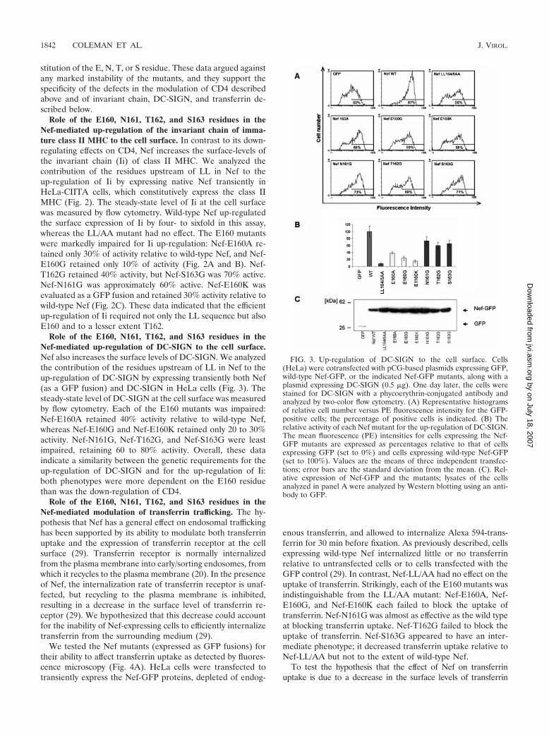
Role of the E160, N161, T162, and S163 residues in theNef-mediated up-regulation of the invariant chain of imma-ture class II MHC to the cell surface. In contrast to its down-regulating effects on CD4, Nef increases the surface-levels ofthe invariant chain (Ii) of class II MHC. We analyzed thecontribution of the residues upstream of LL in Nef to theup-regulation of Ii by expressing native Nef transiently inHeLa-CIITA cells, which constitutively express the class IIMHC (Fig. 2). The steady-state level of Ii at the cell surfacewas measured by flow cytometry. Wild-type Nef up-regulatedthe surface expression of Ii by four- to sixfold in this assay,whereas the LL/AA mutant had no effect. The E160 mutantswere markedly impaired for Ii up-regulation: Nef-E160A re-tained only 30% of activity relative to wild-type Nef, and Nef-E160G retained only 10% of activity (Fig. 2A and B). Nef-T162G retained 40% activity, but Nef-S163G was 70% active.Nef-N161G was approximately 60% active. Nef-E160K wasevaluated as a GFP fusion and retained 30% activity relative towild-type Nef (Fig. 2C). These data indicated that the efficientup-regulation of Ii required not only the LL sequence but alsoE160 and to a lesser extent T162.
Role of the E160, N161, T162, and S163 residues in theNef-mediated up-regulation of DC-SIGN to the cell surface.Nef also increases the surface levels of DC-SIGN. We analyzedthe contribution of the residues upstream of LL in Nef to theup-regulation of DC-SIGN by expressing transiently both Nef(as a GFP fusion) and DC-SIGN in HeLa cells (Fig. 3). Thesteady-state level of DC-SIGN at the cell surface was measuredby flow cytometry. Each of the E160 mutants was impaired:Nef-E160A retained 40% activity relative to wild-type Nef,whereas Nef-E160G and Nef-E160K retained only 20 to 30%activity. Nef-N161G, Nef-T162G, and Nef-S163G were leastimpaired, retaining 60 to 80% activity. Overall, these dataindicate a similarity between the genetic requirements for theup-regulation of DC-SIGN and for the up-regulation of Ii:both phenotypes were more dependent on the E160 residuethan was the down-regulation of CD4.
Role of the E160, N161, T162, and S163 residues in theNef-mediated modulation of transferrin trafficking. The hy-pothesis that Nef has a general effect on endosomal traffickinghas been supported by its ability to modulate both transferrinuptake and the expression of transferrin receptor at the cellsurface (29). Transferrin receptor is normally internalizedfrom the plasma membrane into early/sorting endosomes, fromwhich it recycles to the plasma membrane (20). In the presenceof Nef, the internalization rate of transferrin receptor is unaf-fected, but recycling to the plasma membrane is inhibited,resulting in a decrease in the surface level of transferrin re-ceptor (29). We hypothesized that this decrease could accountfor the inability of Nef-expressing cells to efficiently internalizetransferrin from the surrounding medium (29).
We tested the Nef mutants (expressed as GFP fusions) fortheir ability to affect transferrin uptake as detected by fluores-cence microscopy (Fig. 4A). HeLa cells were transfected totransiently express the Nef-GFP proteins, depleted of endog-
enous transferrin, and allowed to internalize Alexa 594-trans-ferrin for 30 min before fixation. As previously described, cellsexpressing wild-type Nef internalized little or no transferrinrelative to untransfected cells or to cells transfected with theGFP control (29). In contrast, Nef-LL/AA had no effect on theuptake of transferrin. Strikingly, each of the E160 mutants wasindistinguishable from the LL/AA mutant: Nef-E160A, Nef-E160G, and Nef-E160K each failed to block the uptake oftransferrin. Nef-N161G was almost as effective as the wild typeat blocking transferrin uptake. Nef-T162G failed to block theuptake of transferrin. Nef-S163G appeared to have an inter-mediate phenotype; it decreased transferrin uptake relative toNef-LL/AA but not to the extent of wild-type Nef.
To test the hypothesis that the effect of Nef on transferrinuptake is due to a decrease in the surface levels of transferrin
FIG. 3. Up-regulation of DC-SIGN to the cell surface. Cells(HeLa) were cotransfected with pCG-based plasmids expressing GFP,wild-type Nef-GFP, or the indicated Nef-GFP mutants, along with aplasmid expressing DC-SIGN (0.5 �g). One day later, the cells werestained for DC-SIGN with a phycoerythrin-conjugated antibody andanalyzed by two-color flow cytometry. (A) Representative histogramsof relative cell number versus PE fluorescence intensity for the GFP-positive cells; the percentage of positive cells is indicated. (B) Therelative activity of each Nef mutant for the up-regulation of DC-SIGN.The mean fluorescence (PE) intensities for cells expressing the Nef-GFP mutants are expressed as percentages relative to that of cellsexpressing GFP (set to 0%) and cells expressing wild-type Nef-GFP(set to 100%). Values are the means of three independent transfec-tions; error bars are the standard deviation from the mean. (C). Rel-ative expression of Nef-GFP and the mutants; lysates of the cellsanalyzed in panel A were analyzed by Western blotting using an anti-body to GFP.
1842 COLEMAN ET AL. J. VIROL.
by on July 18, 2007 jvi.asm
.orgD
ownloaded from
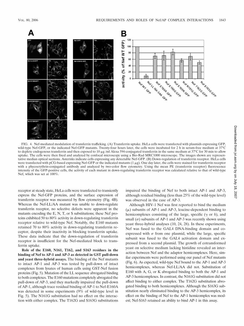
receptor at steady state, HeLa cells were transfected to transientlyexpress the Nef-GFP proteins, and the surface expression oftransferrin receptor was measured by flow cytometry (Fig. 4B).Whereas the Nef-LL/AA mutant was unable to down-regulatetransferrin receptor, no selective defects were apparent in themutants encoding the E, N, T, or S substitutions; these Nef pro-teins exhibited 50 to 80% activity in down-regulating transferrinreceptor relative to wild-type Nef. Notably, the E160 mutantsretained 70 to 80% activity in down-regulating transferrin re-ceptor, despite their inactivity in blocking transferrin uptake.These data indicate that the down-regulation of transferrinreceptor is insufficient for the Nef-mediated block to trans-ferrin uptake.
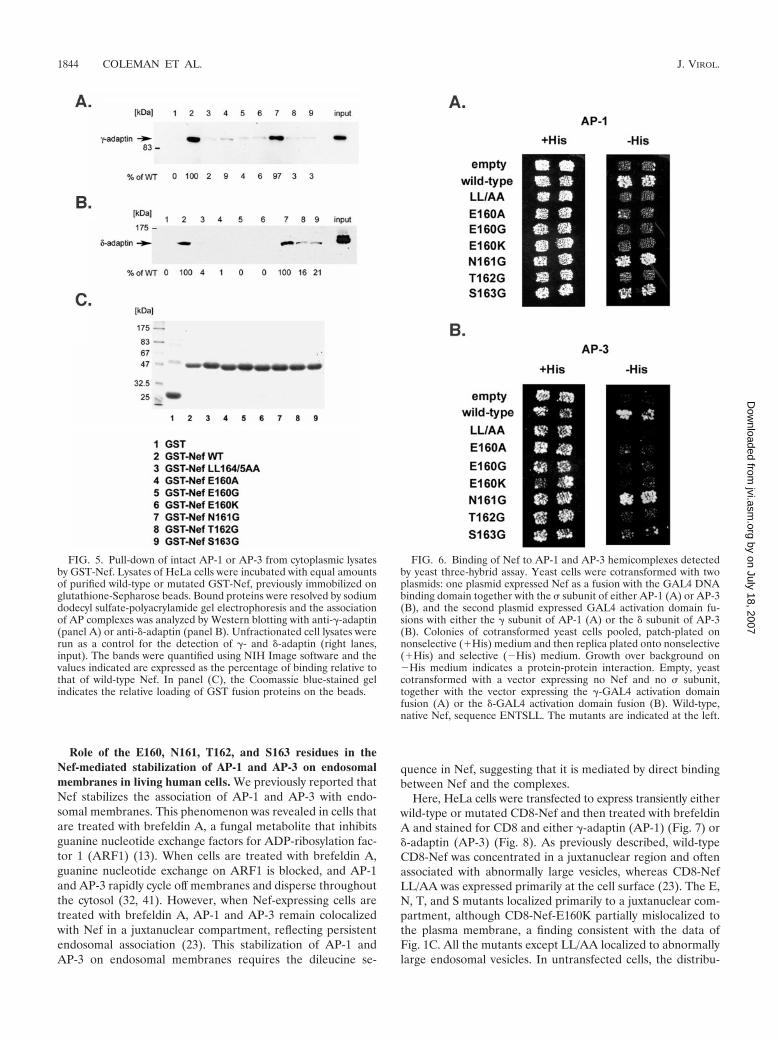
Role of the E160, N161, T162, and S163 residues in thebinding of Nef to AP-1 and AP-3 as detected in GST pull-downand yeast three-hybrid assays. The binding of the Nef mutantsto intact AP-1 and AP-3 was tested by pull-down of intactcomplexes from lysates of human cells using GST-Nef fusionproteins (Fig. 5). Mutation of the LL sequence abrogated bindingto both complexes. The E160 mutations completely abrogated thepull-down of AP-3, and they markedly impaired the pull-downof AP-1, although trace residual binding of AP-1 to Nef-E160Awas detected in some experiments (9% of wild-type level inFig. 5). The N161G substitution had no effect on the interac-tion with either complex. The T162G and S163G substitutions
impaired the binding of Nef to both intact AP-1 and AP-3,although residual binding (less than 25% of the wild-type level)was observed in the case of AP-3.
Although HIV-1 Nef was first reported to bind the medium(�) subunits of AP-1 and AP-3, leucine-dependent binding tohemicomplexes consisting of the large, specific (� or �), andsmall (�) subunits of AP-1 and AP-3 was recently shown usingyeast three-hybrid analyses (10, 24, 28). In these experiments,Nef was fused to the GAL4 DNA-binding domain and co-expressed with � from one plasmid, while the large, specificsubunit was fused to the GAL4 activation domain and ex-pressed from a second plasmid. The growth of cotransformedyeast on selective medium lacking histidine revealed an inter-action between Nef and the adaptor hemicomplexes. Here, sim-ilar experiments were performed using our panel of Nef mutants(Fig. 6). As expected, wild-type Nef bound to the AP-1 and AP-3hemicomplexes, whereas Nef-LL/AA did not. Substitution ofE160 with A, G, or K abrogated binding to both the AP-1 andAP-3 hemicomplexes. In contrast, the N161G substitution did notaffect binding to either complex. The T162G substitution abro-gated binding to both hemicomplexes. Although the S163G sub-stitution nearly eliminated binding to the AP-3 hemicomplex, itseffect on the binding of Nef to the AP-1 hemicomplex was mod-est; Nef-S163 retained an ability to bind AP-1 in this assay.
FIG. 4. Nef-mediated modulation of transferrin trafficking. (A) Transferrin uptake. HeLa cells were transfected with plasmids expressing GFP,wild-type Nef-GFP, or the indicated Nef-GFP mutants. Twenty-four hours later, the cells were incubated for 2 h in serum-free medium at 37°Cto deplete endogenous transferrin and then exposed to 10 �g /ml Alexa 594-conjugated transferrin in the same medium at 37°C for 30 min to allowuptake. The cells were then fixed and analyzed by confocal microscopy using a Bio-Rad MRC1000 microscope. The images shown are represen-tative median optical sections. Asterisks indicate cells expressing any detectable Nef-GFP. (B) Down-regulation of transferrin receptor. HeLa cellswere transfected with pCG-based expressing Nef-GFP or the indicated mutants (1 �g). One day later, the cells were stained for transferrin receptorwith a phycoerythrin-conjugated antibody and analyzed by two-color flow cytometry. Using the mean PE (transferrin receptor) fluorescenceintensity of the GFP-positive cells, the activity of each mutant in down-regulating transferrin receptor was calculated relative to that of wild-typeNef, which was set at 100%.
VOL. 80, 2006 REQUIREMENTS AND ROLES OF Nef/AP COMPLEX INTERACTIONS 1843
by on July 18, 2007 jvi.asm
.orgD
ownloaded from
Role of the E160, N161, T162, and S163 residues in theNef-mediated stabilization of AP-1 and AP-3 on endosomalmembranes in living human cells. We previously reported thatNef stabilizes the association of AP-1 and AP-3 with endo-somal membranes. This phenomenon was revealed in cells thatare treated with brefeldin A, a fungal metabolite that inhibitsguanine nucleotide exchange factors for ADP-ribosylation fac-tor 1 (ARF1) (13). When cells are treated with brefeldin A,guanine nucleotide exchange on ARF1 is blocked, and AP-1and AP-3 rapidly cycle off membranes and disperse throughoutthe cytosol (32, 41). However, when Nef-expressing cells aretreated with brefeldin A, AP-1 and AP-3 remain colocalizedwith Nef in a juxtanuclear compartment, reflecting persistentendosomal association (23). This stabilization of AP-1 andAP-3 on endosomal membranes requires the dileucine se-
quence in Nef, suggesting that it is mediated by direct bindingbetween Nef and the complexes.
Here, HeLa cells were transfected to express transiently eitherwild-type or mutated CD8-Nef and then treated with brefeldinA and stained for CD8 and either �-adaptin (AP-1) (Fig. 7) or�-adaptin (AP-3) (Fig. 8). As previously described, wild-typeCD8-Nef was concentrated in a juxtanuclear region and oftenassociated with abnormally large vesicles, whereas CD8-NefLL/AA was expressed primarily at the cell surface (23). The E,N, T, and S mutants localized primarily to a juxtanuclear com-partment, although CD8-Nef-E160K partially mislocalized tothe plasma membrane, a finding consistent with the data ofFig. 1C. All the mutants except LL/AA localized to abnormallylarge endosomal vesicles. In untransfected cells, the distribu-
FIG. 5. Pull-down of intact AP-1 or AP-3 from cytoplasmic lysatesby GST-Nef. Lysates of HeLa cells were incubated with equal amountsof purified wild-type or mutated GST-Nef, previously immobilized onglutathione-Sepharose beads. Bound proteins were resolved by sodiumdodecyl sulfate-polyacrylamide gel electrophoresis and the associationof AP complexes was analyzed by Western blotting with anti-�-adaptin(panel A) or anti-�-adaptin (panel B). Unfractionated cell lysates wererun as a control for the detection of �- and �-adaptin (right lanes,input). The bands were quantified using NIH Image software and thevalues indicated are expressed as the percentage of binding relative tothat of wild-type Nef. In panel (C), the Coomassie blue-stained gelindicates the relative loading of GST fusion proteins on the beads.
FIG. 6. Binding of Nef to AP-1 and AP-3 hemicomplexes detectedby yeast three-hybrid assay. Yeast cells were cotransformed with twoplasmids: one plasmid expressed Nef as a fusion with the GAL4 DNAbinding domain together with the � subunit of either AP-1 (A) or AP-3(B), and the second plasmid expressed GAL4 activation domain fu-sions with either the � subunit of AP-1 (A) or the � subunit of AP-3(B). Colonies of cotransformed yeast cells pooled, patch-plated onnonselective (�His) medium and then replica plated onto nonselective(�His) and selective (�His) medium. Growth over background on�His medium indicates a protein-protein interaction. Empty, yeastcotransformed with a vector expressing no Nef and no � subunit,together with the vector expressing the �-GAL4 activation domainfusion (A) or the �-GAL4 activation domain fusion (B). Wild-type,native Nef, sequence ENTSLL. The mutants are indicated at the left.
1844 COLEMAN ET AL. J. VIROL.
by on July 18, 2007 jvi.asm
.orgD
ownloaded from
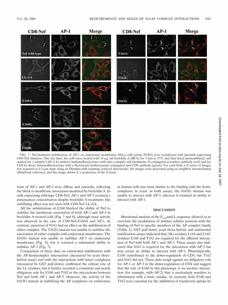
tions of AP-1 and AP-3 were diffuse and cytosolic, reflectingthe block to membrane association mediated by brefeldin A. Incells expressing wild-type CD8-Nef, AP-1 and AP-3 retained ajuxtanuclear concentration despite brefeldin A treatment; thisstabilizing effect was not seen with CD8-Nef LL/AA.
All the substitutions of E160 blocked the ability of Nef tostabilize the membrane association of both AP-1 and AP-3 inbrefeldin A-treated cells (Fig. 7 and 8), although trace activitywas observed in the case of CD8-Nef-E160A and AP-1. Incontrast, mutation of N161 had no effect on the stabilization ofeither complex. The T162G mutant was unable to stabilize theassociation of either complex with endosomal membranes. TheS163G mutant was unable to stabilize AP-3 on endosomalmembranes (Fig. 8), but it retained a substantial ability tostabilize AP-1 (Fig. 7).
Comparison of these data on endosomal stabilization withthe AP-hemicomplex interactions (measured by yeast three-hybrid assay) and with the interactions with intact complexes(measured by GST pull-down) confirmed the critical role ofthe LL residues, but it further revealed a consistent and nearlyobligatory role for E160 and T162 in the interactions betweenNef and both AP-1 and AP-3. However, the activity of theS163G mutant in stabilizing the AP complexes on endosomes
in human cells was most similar to the binding with the hemi-complexes in yeast: in both assays, the S163G mutant wasunable to interact with AP-3, whereas it retained an ability tointeract with AP-1.
DISCUSSION
Mutational analysis of the E160xxxLL sequence allowed us tocorrelate the modulation of distinct cellular proteins with thebinding of Nef to specific members of the AP complex family(Table 2). GST pull-down, yeast three-hybrid, and endosomalstabilization assays indicated that, like residues L164 and L165,residues E160 and T162 are required for the efficient interac-tion of Nef with both AP-1 and AP-3. These assays also indi-cated that S163 is required for the interaction with AP-3 butmay retain an ability to interact with AP-1. Phenotypically,E160 contributed to the down-regulation of CD4, but T162and S163 did not. These data weigh against an obligatory rolefor AP-1 or AP-3 in the down-regulation of CD4 and suggestthat the role of E160 in this phenotype is via another interac-tion (for example, with AP-2) that is particularly sensitive tosubstitution with a basic residue. In contrast, both E160 andT162 were essential for the inhibition of transferrin uptake by
FIG. 7. Nef-mediated stabilization of AP-1 on endosomal membranes. HeLa cells (clone P4.R5) were transfected with plasmids expressingCD8-Nef chimeras. One day later, the cells were treated with 10 �g /ml brefeldin A (BFA) for 5 min at 37°C and then fixed, permeabilized, andstained for �-adaptin (AP-1) by indirect immunofluorescence with anti-�-adaptin and rhodamine X-conjugated secondary antibody (red) and forCD8 by direct immunofluorescence with a fluorescein isothiocyanate-conjugated anti-CD8 antibody (green). For each field, a Z-series of imageswas acquired at 0.5-�m steps using an Olympus disk-scanning confocal microscope; the images were processed using no-neighbor deconvolution(Slidebook software); and the image shown is a projection of the Z-stack.
VOL. 80, 2006 REQUIREMENTS AND ROLES OF Nef/AP COMPLEX INTERACTIONS 1845
by on July 18, 2007 jvi.asm
.orgD
ownloaded from

Nef, indicating a major role for AP-1 and/or AP-3 in thisphenotype.
In the cases of the invariant chain and DC-SIGN, the defectsin up-regulation caused by uncharged substitutions of E160and by substitutions of T162 and S163 were greater than thedefects in the down-regulation of CD4, suggesting a moresubstantial contribution of AP-1 and/or AP-3 to these pheno-types. However, these complexes cannot be wholly responsiblefor these up-regulatory effects, because neither the T162G nor
S163 mutant was fully defective. Consequently, as in the case ofCD4, the disproportionate defects caused by the E160 muta-tions suggest a role for another interaction in the modulationof invariant chain and DC-SIGN.
Together, these data lead to two testable hypotheses.First, the E160 residue affects an interaction with an APcomplex in addition to AP-1 and AP-3 that contributes tothe modulation of CD4, invariant chain, and DC-SIGN; thismay be AP-2. Second, Nef uses multiple AP family members
FIG. 8. Nef-mediated stabilization of AP-3 on endosomal membranes. HeLa cells (clone P4.R5) were transfected with plasmids expressing theCD8-Nef chimeras, treated with brefeldin A, and imaged exactly as described in the legend to Fig. 7, except that the indirect immunofluorescencewas done using a primary antibody to the � subunit of AP-3.
TABLE 2. Summary of AP-binding and trafficking phenotypesd
Nef
Y3Ha withhemicomplexes GST pull-down Stabilizationb
CD4down-regulation
Internalizationas CD8-Nef c
Invariant chainup-reg.
DC-SIGNup-reg.
Tf uptakeblock
AP-1 AP-3 AP-1 AP-3 AP-1 AP-3
Wild type � � � � � � � � � � �LL/AA � � � � � � � � � � �E160A � � � � � � � � Int Int �E160G � � � � � � Int � � � �E160K � � � � � � � Int � � �N161G � � � � � � � � Int Int �T162G � � � � � � � � Int Int �S163G Int � � � � � � � Int Int Int
a Y3H, yeast three-hybrid.b Stabilization on endosomes in brefeldin-treated cells.c Inverse of surface expression.d �, positive; �, negative; Int, intermediate; reg, regulation.
1846 COLEMAN ET AL. J. VIROL.
by on July 18, 2007 jvi.asm
.orgD
ownloaded from

to modulate different proteins in distinct but overlappingpatterns: primarily AP-2 for CD4, AP-2 and AP-1 (and/orAP-3) for invariant chain and DC-SIGN, and AP-1 (and/orAP-3) for transferrin.
What are the implications of these data regarding the mech-anisms of Nef action on different target proteins? As noted, thedata suggest that interactions with AP-1 and AP-3 are largelydispensable for the down-regulation of CD4 from the cell sur-face (Table 2). This conclusion is supported by the persistentNef-mediated down-regulation of CD4 observed in cells inwhich the � subunit of either AP-1 or AP-3 has been depletedby RNA interference (35); it is also consistent with the findingthat replacement of the ENTSLL sequence with DKQTLLyields a Nef protein unable to bind hemicomplexes of eitherAP-1 or AP-3 but still able to down-regulate CD4 (7). Similarto the case of down-regulation of CD4, the localization ofCD8-Nef chimeras on internal membranes at steady state doesnot appear to require interactions between Nef and AP-1 orAP-3. Yet, both the down-regulation of CD4 and the internal-ization of the chimeras require the LL sequence and likelyrequire AP complexes. Consequently, by eliminating majorroles for AP-1 and AP-3, the data indirectly support the AP-2complex as the most likely cofactor for the down-regulationof CD4 by Nef.
If AP-2 is the cofactor for the down-regulation of CD4, thenthe functional data predict that this interaction has less strictsequence requirements than the interaction with AP-1 andAP-3. The putative interaction must tolerate either a nega-tively charged or neutral amino acid at position �4 in theExxxLL motif, because the E160A and E160G mutants re-tained 50% or greater activity in the down-regulation of CD4and localized to internal membranes as effectively as the wild-type when expressed as CD8 chimeras. Similarly, this interac-tion must tolerate nonpolar residues at positions �1 and �2,because glycine substitutions at these sites had no apparentimpact on the down-regulation of CD4 or the localization ofthe CD8 chimeras to internal membranes. However, if an in-teraction between Nef and AP-2 underlies the down-regulationof CD4, then it must be impaired by a positively chargedresidue at position �4: the E160K mutant inefficiently down-regulated CD4, and CD8-Nef E160K was mislocalized in partto the plasma membrane.
In support of this model, the effect of the Nef E160K sub-stitution is analogous to that of substitution of the acidic res-idues in the LIMP II leucine-based motif with arginines: mis-localization from endosomal membranes to the cell surface,presumably due to a failure of internalization via AP-2 (38).Notwithstanding these considerations, the nearly undetectabledirect interaction between HIV-1 Nef and AP-2, together withrecent knock-down experiments indicating that AP-2 is notobligatory, leaves the role of AP-2 in the down-regulation ofCD4 formally unproven (5, 36). Indeed, taken together, thepublished knock-down experiments suggest that no individualcomplex (neither AP-1, -2, or -3) is obligatory for the down-regulation of CD4 by HIV-1 Nef (35, 36); conceivably, HIV-1Nef may be able to use more than one member of the APfamily to reduce the expression of CD4 at the cell surface.
In contrast to the down-regulation of CD4, the block totransferrin uptake mediated by Nef correlated closely with thebinding to AP-1 and/or AP-3: the E160, T162, and S163 resi-
dues each played an major role in this effect, just as they did inassays of interaction with AP-1 and AP-3 (Table 2). Notably,we recently hypothesized that the block to transferrin uptake isdue to reduced expression of transferrin receptor at the cellsurface, which in turn is due to an inhibition of the recycling ofinternalized transferrin receptor to the plasma membrane (29).However, data herein indicate that mutation of residue E160,T162, or S163 has relatively little effect on the down-regulationof transferrin receptor despite markedly impairing the block totransferrin uptake. These data leave the mechanism of theuptake block open to speculation.
Interestingly, the hierarchical contribution of individual res-idues in the ExxxLL motif to the up-regulation of the invariantchain was similar to that of DC-SIGN. Although the impor-tance of the E160 residue supports a role for AP-1 and/or AP-3in both of these properties, the modest impairment cause bythe T162G and S163G substitutions (together with the detect-able impairment caused by the N161G substitution) rendersinterpretation complex. Overall, this imprecise fit between theAP-binding data and the modulation of DC-SIGN and invari-ant chain suggests the involvement of leucine-dependent co-factors in addition to AP-1 and AP-3 in these effects. Notably,recent experiments using RNA interference to knock down theexpression of AP complexes in HeLa-CIITA cells indicate thatAP-2 is predominantly responsible for targeting the invariantchain to the endosomal class II antigen processing compartment(14). Consequently, part of the Nef-mediated up-regulation ofinvariant chain could be due to an inhibition of endocytosismediated by AP-2. This hypothesis is consistent with the aboveconclusion that the disproportionate effects of the E160 sub-stitutions on the up-regulation of the invariant chain indicateinvolvement of an AP complex in addition to AP-1 and AP-3.Similar considerations apply to the up-regulation of DC-SIGN.The data are best explained by a role for both AP-2 andAP-1/AP-3 in these phenotypes; such a model allows the mod-est effects of the T162 and S163 mutations to be attributed toimpaired interactions with AP-1 and/or AP-3, while the quan-titatively greater effects of the E160 mutations are attributed toan additional effect on the putative interaction with AP-2.
Notably, the mechanism by which Nef up-regulates the sur-face expression of host proteins is unclear. One hypothesis isthat saturation of AP complexes by Nef causes the displace-ment of the target protein to the cell surface by default; weproposed this mechanism to explain the Nef-mediated increasein the surface expression of a Tac chimera containing theDKQTLL sequence (9, 30). Both the invariant chain and DC-SIGN have dileucine-based sorting signals in their cytoplasmicdomains (34, 40). Interestingly, both motifs in the invariantchain have typically spaced upstream acidic residues, but themotif in DC-SIGN has no such residue. If only similar motifscompete with each other for specific AP complexes, then mu-tation of E160 would have affected only the up-regulation ofinvariant chain and not DC-SIGN. Consequently, the findingthat E160 plays a major role in the up-regulation of bothinvariant chain and DC-SIGN weighs against a simple compe-tition for AP complexes as the mechanism of up-regulation.
The data herein also provide potential insights into howindividual residues in ExxxLL motifs contribute to interactionswith AP-1 and AP-3. Previous studies have indicated thatacidic residues at positions �4 and/or �5 relative to the
VOL. 80, 2006 REQUIREMENTS AND ROLES OF Nef/AP COMPLEX INTERACTIONS 1847
by on July 18, 2007 jvi.asm
.orgD
ownloaded from

leucines are important for binding to AP-3 (11, 22, 38). Here,three distinct binding assays (GST pull-down, yeast three-hy-brid, and endosomal stabilization assays) confirmed the role ofthe glutamic acid at the �4 position in the Nef ExxxLL se-quence for the binding to AP-3. However, this residue wasalmost equally important for the binding to AP-1, indicatingthat the role of acidic residues in ExxxLL motifs may extend tointeractions with AP-1 as well as AP-3.
The original study using the three-hybrid assay to analyzedileucine-based interactions with AP complexes reported noeffect of a T162A substitution on the interaction between Nefand hemicomplexes of AP-1 and AP-3 (24). In contrast, theT162G substitution herein markedly impaired the interactionsof Nef with both complexes as observed not only using yeast-three hybrid but also using GST pull-down and endosomalstabilization assays. Notably, the results herein are consistentwith a previous mutagenesis study in yeast that indicated arequirement for a polar residue at position �2 for the inter-action with AP-3 (11). The data herein also indicated thatposition �1 relative to the leucines is constrained with respectto binding these complexes: the S163G mutation impaired theinteraction with AP-3 in every assay, although at least partialactivity in binding AP-1 was preserved in two of three assays.Together, the data indicate that the �1, �2, and �4 positionsrelative to the leucines in ExxxLL motifs are direct contribu-tors to the interactions with the AP-1 and AP-3 complexes.
Taken together, the differential effects of the E, T, and Smutations reviewed above suggest that Nef uses different APcomplexes, or different combinations of complexes, to modu-late specific proteins. If this hypothesis is correct, then thecomplexes may be specified not only by Nef but also by thetarget protein itself. The cytoplasmic domains of CD4, trans-ferrin receptor, DC-SIGN, and the invariant chain each con-tain sorting motifs (1, 8, 34, 40). The simplest mechanism bywhich these proteins can contribute to the utilization of specificAP complexes is by the involvement of their own sorting motifsin the selection process.
Notably, the concept that the sorting motifs in target proteinsmay interact directly with AP complexes during Nef-mediatedmodulation has been supported by two recent studies, each ofwhich reported that the LL sequence in the cytoplasmic domainof CD4, while required for Nef-mediated down-regulation, doesnot contribute to the interaction between Nef and CD4 (2, 6). Inthis emerging model, the AP binding motifs in both Nef andthe target protein are functional. Instead of serving as a simpleconnector between the cytoplasmic domains of target proteinsand AP complexes (33), Nef would facilitate the interactions ofits targets with the complexes via their own AP-binding signals.This facilitation may be based in the ability of Nef to stabilizethe complexes on endosomal membranes, as shown herein.Although we can document this stabilization only in the case ofAP-1 and AP-3, it may occur with AP-2 as well, explaining theability of Nef to induce the formation of coated pits at theplasma membrane (16).
Interestingly, this “facilitator” model allows Nef to performa catalytic-like function: it may stimulate the inclusion of atarget protein in a transport vesicle without itself becomingpart of that vesicle. This scenario is supported by a heretoforeanomalous observation exemplified herein: although certainmutations in the ExxxLL motif clearly alter the subcellular
distribution of CD8-Nef chimeras in which Nef is a covalentpart of a transmembrane protein, specifically Nef-LL/AA andNef-E160K (Fig. 7 and 8) (15, 23), the same mutations have noeffect on the subcellular distribution of Nef-GFP (Fig. 4) (10),despite their ability to abrogate the effect of Nef-GFP on targetproteins.
In summary, mutagenesis of the ExxxLL motif has revealeda differential role of the acidic residue in a subset of theleucine-dependent, Nef-induced perturbations of cellular pro-tein trafficking. Binding studies implicated AP-1 and AP-3 insome of these effects but suggested by default a role for AP-2in others. Together, the data are consistent with a model inwhich Nef uses different AP complexes, or different combina-tions of complexes, to modulate specific proteins. We speculatethat the cytoplasmic domains of the proteins targeted by Nefparticipate in the selection of the complexes and that Neffacilitates sorting via a stabilizing effect on the vesicle coat.
ACKNOWLEDGMENTS
Scott Coleman was in the Biomedical Sciences program of UCSD.Richard Mitchell was supported by an AIDS Training grant from theNIH (AI07384). This work was supported by grants from the NationalInstitutes of Health (AI38201), the UCSD Center for AIDS Research(NIH AI36214), the Research Center for AIDS and HIV Infection ofthe San Diego Veterans Affairs Healthcare System, and the AgenceNationale de Recherche sur le SIDA and SIDACTION, France.
We thank Juan Bonifacino for the yeast three-hybrid vectors,Nathalie Sol-Foulon for the plasmid expressing DC-SIGN, PhilippeBenaroch for the HeLa-CIITA cells, and Colleen Noviello and JohnDay for reviewing the manuscript.
REFERENCES
1. Aiken, C., J. Konner, N. R. Landau, M. Lenburg, and D. Trono. 1994. Nefinduces CD4 endocytosis: requirement for a critical dileucine motif in themembrane-proximal CD4 cytoplasmic domain. Cell 76:853–864.
2. Bentham, M., S. Mazaleyrat, and M. Harris. 2003. The di-leucine motif inthe cytoplasmic tail of CD4 is not required for binding to human immuno-deficiency virus type 1 Nef, but is critical for CD4 down-modulation. J. Gen.Virol. 84:2705–2713.
3. Blagoveshchenskaya, A. D., L. Thomas, S. F. Feliciangeli, C.-H. Hung, andG. Thomas. 2002. HIV-1 Nef downregulates MHC-I by a PACS-1- andPI3K-regulated ARF6 endocytic pathway. Cell 111:853–866.
4. Bresnahan, P. A., W. Yonemoto, S. S. Ferrell, D. G. R. Williams-Herman,and W. C. Greene. 1998. A dileucine motif in HIV-1 Nef acts as an inter-nalization signal for CD4 downregulation and binds the AP-1 clathrin adap-tor. Curr. Biol. 8:1235–1238.
5. Bresnahan, P. A., W. Yonemoto, and W. C. Greene. 1999. Cutting edge: SIVNef protein utilizes both leucine- and tyrosine-based protein sorting path-ways for down-regulation of CD4. J. Immunol. 163:2977–2981.
6. Cluet, D., C. Bertsch, C. Beyer, L. Gloeckler, M. Erhardt, J. P. Gut, J. L.Galzi, and A. M. Aubertin. 2005. Detection of human immunodeficiencyvirus type 1 Nef and CD4 physical interaction in living human cells by usingbioluminescence resonance energy transfer. J. Virol. 79:8629–8636.
7. Coleman, S. H., N. Van Damme, J. R. Day, C. M. Noviello, D. Hitchin, R.Madrid, S. Benichou, and J. C. Guatelli. 2005. Leucine-specific, functionalinteractions between human immunodeficiency virus type 1 Nef and adaptorprotein complexes. J. Virol. 79:2066–2078.
8. Collawn, J. F., M. Stangel, L. A. Kuhn, V. Esekogwu, S. Q. Jing, I. S.Trowbridge, and J. A. Tainer. 1990. Transferrin receptor internalizationsequence YXRF implicates a tight turn as the structural recognition motiffor endocytosis. Cell 63:1061–1072.
9. Craig, H. M., M. W. Pandori, and J. C. Guatelli. 1998. Interaction of HIV-1Nef with the cellular dileucine-based sorting pathway is required for CD4down-regulation and optimal viral infectivity. Proc. Natl. Acad. Sci. USA95:11229–11234.
10. Craig, H. M., T. R. Reddy, N. L. Riggs, P. P. Dao, and J. Guatelli. 2000.Interactions of HIV-1 Nef with the � subunits of adaptor protein complexes1, 2, and 3: role of the dileucine-based sorting motif. Virology 271:9–17.
11. Darsow, T., C. G. Burd, and S. D. Emr. 1998. Acidic di-leucine motif essen-tial for AP-3-dependent sorting and restriction of the functional specificity ofthe Vam3p vacuolar t-SNARE. J. Cell Biol. 142:913–922.
12. Deacon, N. J., A. Tsykin, A. Solomon, K. Smith, M. Ludford-Menting, D. J.Hooker, D. A. McPhee, A. L. Greenway, A. Ellett, C. Chatfield, V. A. Lawson,
1848 COLEMAN ET AL. J. VIROL.
by on July 18, 2007 jvi.asm
.orgD
ownloaded from

S. Crowe, A. Maerz, S. Sonza, J. Learmont, J. S. Sullivan, A. Cunningham,D. Dwyer, D. Dowton, and J. Mills. 1995. Genomic structure of an attenuatedquasi species of HIV-1 from a blood transfusion donor and recipients.Science 270:988–991.
13. Donaldson, J. G., D. Finazzi, and R. D. Klausner. 1992. Brefeldin A inhibitsGolgi membrane-catalysed exchange of guanine nucleotide onto ARF pro-tein. Nature 360:350–352.
14. Dugast, M., H. Toussaint, C. Dousset, and P. Benaroch. 2005. AP2 clathrinadaptor complex, but not AP1, controls the access of the major histocom-patibility complex (MHC) class II to endosomes. J. Biol. Chem. 280:19656–19664.
15. Erdtmann, L., K. Janvier, G. Raposo, H. M. Craig, P. Benaroch, C. Berlioz-Torrent, J. C. Guatelli, R. Bernarous, and S. Benichou. 2000. Two indepen-dent regions of HIV-1 Nef are required for connection with the endocyticpathway through binding to the �1 chain of AP1 complex. Traffic 1:871–883.
16. Foti, M., A. Mangasarian, V. Piguet, D. P. Lew, K.-H. Krause, D. Trono, andJ.-L. Carpentier. 1997. Nef-mediated clathrin-coated pit formation. J. CellBiol. 139:37–47.
17. Geijtenbeek, T. B., D. S. Kwon, R. Torensma, S. J. van Vliet, G. C. vanDuijnhoven, J. Middel, I. L. Cornelissen, H. S. Nottet, V. N. KewalRamani,D. R. Littman, C. G. Figdor, and Y. van Kooyk. 2000. DC-SIGN, a dendriticcell-specific HIV-1-binding protein that enhances trans-infection of T cells.Cell 100:587–597.
18. Greenberg, M., L. DeTulleo, I. Rapoport, J. Skowronski, and T. Kirch-hausen. 1998. A dileucine motif in HIV-1 Nef is essential for sorting intoclathrin-coated pits and for downregulation of CD4. Curr. Biol. 8:1239–1242.
19. Greenberg, M. E., S. Bronson, M. Lock, M. Neumann, G. N. Pavlakis, andJ. Skowronski. 1997. Co-localization of HIV-1 Nef with the AP-2 adaptorprotein complex correlates with Nef-induced CD4 down-regulation. EMBOJ. 16:6964–6976.
20. Hao, M., and F. R. Maxfield. 2000. Characterization of rapid membraneinternalization and recycling. J. Biol. Chem. 275:15279–15286.
21. Hirst, J., and M. S. Robinson. 1998. Clathrin and adaptors. Biochim. Bio-phys. Acta 1404:173–193.
22. Honing, S., I. V. Sandoval, and K. von Figura. 1998. A di-leucine-based motifin the cytoplasmic tail of LIMP-II and tyrosinase mediates selective bindingof AP-3. EMBO J. 17:1304–1314.
23. Janvier, K., H. Craig, D. Hitchin, R. Madrid, N. Sol-Foulon, L. Renault,J. Cherfils, D. Cassel, S. Benichou, and J. Guatelli. 2003. HIV-1 Nef stabi-lizes the association of adaptor protein complexes with membranes. J. Biol.Chem. 275:8725–8732.
24. Janvier, K., Y. Kato, M. Boehm, J. R. Rose, J. A. Martina, B. Y. Kim, S.Venkatesan, and J. S. Bonifacino. 2003. Recognition of dileucine-basedsorting signals from HIV-1 Nef and LIMP-II by the AP-1 gamma-sigma1 andAP-3 delta-sigma3 hemicomplexes. J. Cell Biol. 163:1281–1290.
25. Kestler, H. W., III, D. J. Ringler, K. Mori, D. L. Panicali, P. K. Sehgal, M. D.Daniel, and R. C. Desrosiers. 1991. Importance of the nef gene for mainte-nance of high virus loads and for development of AIDS. Cell 65:651–662.
26. Lama, J., A. Mangasarian, and D. Trono. 1999. Cell surface expresion ofCD4 reduces HIV-1 infectivity by blocking Env incorporation in a Nef- andVpu-inhibitable manner. Curr. Biol. 9:622–631.
27. Lama, J., and C. F. Ware. 2000. Human immunodeficiency virus type 1 nefmediates sustained membrane expression of tumor necrosis factor and therelated cytokine LIGHT on activated T cells. J. Virol. 74:9396–9402.
28. Le Gall, S., L. Erdtmann, S. Benichou, C. Berlioz-Torrent, L. Liu, R.Benarous, J.-M. Heard, and O. Schwartz. 1998. Nef interacts with the �
subunit of clathrin adaptor complexes and reveals a cryptic sorting signal inMHC 1 molecules. Immunity 8:483–495.
29. Madrid, R., K. Janvier, D. Hitchin, J. Day, S. Coleman, C. Noviello, J.Bouchet, A. Benmerah, J. Guatelli, and S. Benichou. 2005. Nef-inducedalteration of the early/recycling endosomal compartment correlates withenhancement of HIV-1 infectivity. J. Biol. Chem. 280:5032–5044.
30. Marks, M. S., L. Woodruff, H. Ohno, and J. S. Bonifacino. 1996. Proteintargeting by tyrosine- and di-leucine-based signals: evidence for distinctsaturable components. J. Cell Biol. 135:341–354.
31. Mellman, I. 1996. Endocytosis and molecular sorting. Annu. Rev. Cell Dev.12:575–625.
32. Ooi, C. E., E. C. Dell’Angelica, and J. S. Bonifacino. 1998. ADP-ribosylationfactor 1 (ARF1) regulates recruitment of the AP-3 adaptor complex tomembranes. J. Cell Biol. 142:291–402.
33. Piguet, V., Y.-L. Chen, A. Mangasarian, M. Foti, J.-L. Carpentier, and D.Trono. 1998. Mechanism of Nef-induced CD4 endocytosis: Nef connectsCD4 with the � chain of adaptor complexes. EMBO J. 17:2472–2481.
34. Pond, L., L. A. Kuhn, L. Teyton, M.-P. Schutze, J. A. Tainer, M. R. Jackson,and P. A. Peterson. 1995. A role for acidic residues in di-leucine motif-basedtargeting to the endocytic pathway. J. Biol. Chem. 270:19989–19997.
35. Roeth, J. F., M. Williams, M. R. Kasper, T. M. Filzen, and K. L. Collins.2004. HIV-1 Nef disrupts MHC-I trafficking by recruiting AP-1 to theMHC-I cytoplasmic tail. J. Cell Biol. 167:903–913.
36. Rose, J. J., K. Janvier, S. Chandrasekhar, R. P. Sekaly, J. S. Bonifacino, andS. Venkatesan. 2005. CD4 down-regulation by HIV-1 and simian immuno-deficiency virus (SIV) Nef proteins involves both internalization and intra-cellular retention mechanisms. J. Biol. Chem. 280:7413–7426.
37. Ross, T. M., A. E. Oran, and B. R. Cullen. 1999. Inhibition of HIV-1 progenyvirion release by cell surface CD4 is relieved by expression of the viral Nefprotein. Curr. Biol. 9:613–621.
38. Sandoval, I. V., S. Martinez-Arca, J. Valdueza, S. Palacios, and G. D.Holman. 2000. Distinct reading of different structural determinants mod-ulates the dileucine-mediated transport steps of the lysosomal membraneprotein LIMPII and the insulin-sensitive glucose transporter GLUT4.J. Biol. Chem. 275:39874–39885.
39. Schwartz, O., V. Marechal, S. Le Gall, F. Lemonnier, and J.-M. Heard. 1996.Endocytosis of major histocompatibility complex class I molecules is inducedby the HIV-1 Nef protein. Nat. Med. 2:338–342.
40. Sol-Foulon, N., A. Moris, C. Nobile, C. Boccaccio, A. Engering, J. P.Abastado, J. M. Heard, Y. van Kooyk, and O. Schwartz. 2002. HIV-1Nef-induced upregulation of DC-SIGN in dendritic cells promotes lym-phocyte clustering and viral spread. Immunity 16:145–155.
41. Stamnes, M. A., and J. E. Rothman. 1993. The binding of AP-1 clathrinadaptor particles to Golgi membranes requires ADP-ribosylation factor, asmall GTP-binding protein. Cell 73:999–1005.
42. Stumptner-Cuvelette, P., S. Morchoisne, M. Dugast, S. Le Gall, G. Raposo,O. Schwartz, and P. Benaroch. 2001. HIV-1 Nef impairs MHC class IIantigen presentation and surface expression. Proc. Natl. Acad. Sci. USA98:12144–12149.
43. Swigut, T., N. Shohdy, and J. Skowronski. 2001. Mechanism for down-regulation of CD28 by Nef. EMBO J. 20:1593–1604.
44. Tobiume, M., M. Takahoko, T. Yamada, M. Tatsumi, A. Iwamoto, and M.Matsuda. 2002. Inefficient enhancement of viral infectivity and CD4 down-regulation by human immunodeficiency virus type 1 Nef from Japaneselong-term nonprogressors. J. Virol. 76:5959–5965.
VOL. 80, 2006 REQUIREMENTS AND ROLES OF Nef/AP COMPLEX INTERACTIONS 1849
by on July 18, 2007 jvi.asm
.orgD
ownloaded from