cell organization of the rat pars tuberalis. evidence for open communication between pars tuberalis...
TRANSCRIPT
REGULAR ARTICLE
Cell organization of the rat pars tuberalis. Evidence for opencommunication between pars tuberalis cells, cerebrospinalfluid and tanycytes
Montserrat Guerra & Juan Luís Blázquez & Bruno Peruzzo & Belén Peláez &
Sara Rodríguez & Daniel Toranzo & Francisco Pastor & Esteban M. Rodríguez
Received: 3 July 2009 /Accepted: 9 September 2009 /Published online: 25 November 2009# Springer-Verlag 2009
Abstract The pars tuberalis (PT) is the only pituitaryregion in close contact with the medial-basal hypothalamusand bathed by cerebrospinal fluid (CSF). Although PT haslong been recognized as an endocrine gland, certain aspectsof its structure remain obscure. The present investigationhas been designed to gain information concerning (1) thecellular organization of PT, (2) the PT/median eminencespatial relationship and (3) the exposure of various cellcompartments of PT to CSF. Non-endocrine cells (S100-reactive) appear as the organizer of the PT architecture. Theapical poles of these cells line large cistern-like cavities andthe processes of these cells establish a close spatialrelationship with PT-specific secretory cells, portal capil-laries and tanycytes. The cisterns are also endowed withclusters of ciliated cells and with a highly electron-denseand PAS-reactive content. The unique spatial organizationof endocrine and non-endocrine cells of the PT supports afunctional relationship between both cell populations. PTendocrine cells display a hallmark of PT-specific cells,namely, the paranuclear spot, which is a complex structure
involving the Golgi apparatus, a large pool of immaturesecretory granules and a centriole from which originates asingle 9+0 cilium projecting to the intercellular channels.Horseradish peroxidase (HRP) injected into the CSF readilyreaches the intercellular channels of PT and the innerchannel of the single cilium and is incorporated by theendocytic machinery of the secretory cells. The PTendocrine cells, through their single 9+0 cilium, may actas sensors of the CSF. HRP also reaches the lumen of thecisterns, indicating that this PT compartment is alsoexposed to CSF. PT endocrine cells establish direct cell-to-cell contacts with hypothalamic β1 tanycytes, suggestinga second means of brain-PT communication.
Keywords Pars tuberalis . Ultrastructure .
Immunocytochemistry . Endocrine cells . Non-endocrinecells . Cerebrospinal fluid . Tanycytes .
Rat (Sprague Dawley, male)
Introduction
The hypophyseal pars tuberalis (PT) is a thin layer of cellsunderlying the median eminence and surrounding theinfundibular stalk. PT is formed by secretory cells thatdisplay distinct characteristics and that have been regardedas PT-specific cells. In all mammals studied, these cellsreact with antibodies against the common α-subunit ofthyrotrophin (TSH) and the gonadotrophins (luteinizinghormone and follicle-stimulating hormone) (Gross 1984;Stoeckel et al. 1994; Kameda et al. 2000). However, PT-specific cells differ from the pars distalis (PD) gonado-trophs and thyrotrophs with respect to their morphologicalappearance, immunoreaction patterns and secretory activity(Bockers et al. 1990; Bockmann et al. 1997; Sakai et al.
This work was supported by grants from DID and MECESUP ofUniversidad Austral to M.G and a grant from Fondecyt no. 1070241,Chile, to E.M.R.
Dr. Francisco Pastor died after the present investigation had beencompleted. We wish to dedicate this publication to him.
M. Guerra : B. Peruzzo : S. Rodríguez : E. M. Rodríguez (*)Instituto de Anatomía, Histología y Patología,Facultad de Medicina, Universidad Austral de Chile,Valdivia, Chilee-mail: [email protected]
J. L. Blázquez : B. Peláez :D. Toranzo : F. PastorDepartamento de Anatomía e Histología Humana,Facultad de Medicina, Universidad de Salamanca,Salamanca, Spain
Cell Tissue Res (2010) 339:359–381DOI 10.1007/s00441-009-0885-8
1999; Wittkowski et al. 1999). Nevertheless, typical PDgonadotrophs and thyrotrophs are found in the ventrocaudalportion of the PT, a transition zone between PT and PD(Gross 1984). The PT also contains a cell type described asfollicular or interstitial cells. The relative density of PT-specific cells and follicular cells varies among species(Cameron and Foster 1972; Dellmann et al. 1974; Kurotaniet al. 1999).
Some of the factors that regulate the secretory activity ofPT-specific cells have been identified, such as melatonin,adenosine and pituitary adenylate cyclase-activating peptide(PACAP; Barret et al. 2002; von Gall et al. 2002, 2005;Schuster 2007). With the exception of the human PT, forwhich the evidence is controversial, the PT of allmammalian species studied displays melatonin receptors(Williams and Morgan 1988; Schuster 2007). Furthermore,in sheep, chicken and hamster, PT-specific cells arereported to be the melatonin-responsive cells (Morgan etal. 1991; Bockmann et al. 1996; Klosen et al. 2002;Dardente et al. 2003). In the PT of these species, melatoninregulates the expression of various genes, including thecircadian clock genes (Jilg et al. 2005; von Gall et al. 2002,2005; Wagner et al. 2007; Dupré et al. 2008). However, thepathway used by melatonin to reach the PT is stillunknown. Aguado et al. (1981) have shown that the ratPT possesses intercellular channels that are in opencommunication with the subarachnoid space. Furthermore,pineal melatonin is known to be released to both thesystemic blood and cerebrospinal fluid (CSF) in a circadianpattern and the melatonin concentration in the CSF is 20-fold higher than that in the plasma (Skinner and Malpaux1999). Although CSF can be seen as a pathway by whichmelatonin (or other factors) can reach the PT, such apossibility has not been further studied.
In some vertebrates, the PT secretes TSH in a light- ormelatonin-sensitive manner, exerting an effect on hypotha-lamic cells expressing type 2-iodothyronine deiodinase(DIO). Interestingly, DIO is selectively expressed intanycytes (Tu et al. 1997; Lechan and Fekete 2006), whichare specialized cells of glial origin lining the floor (medianeminence) and ventrolateral walls of the third ventricle(Rodríguez et al. 2005). Since the expression of DIO intanycytes is under the influence of melatonin (Yasuo et al.2007; Nakao et al. 2008; Ono et al. 2008), they might playa key role in the photoperiodic response.
Adenosine and PACAP are known as paracrine modu-lators of the endocrine and folliculostellate cells of the PD(Wong et al. 2000; Kell and Stehle 2005). In PT, melatoninacts in concert with adenosine to elicit rhythms in clockgene expression by binding to A2-adenosine receptor (vonGall et al. 2002, 2005). PACAP-binding sites have not beenfound in the PT, but in the median eminence (Barret et al.2002). Since PACAP regulates the secretory activity of PT-
specific cells, PACAP has been suggested to be released byunidentified cells of the median eminence (Barret et al.2002). Thus, the current view is that the effect of PACAPon PT activity is exerted by a paracrine factor released byhypothalamic unidentified cells (tanycytes?).
The aim of the present investigation has been three-fold:(1) to take a closer look at the cell organization of the ratPT by using immunocytochemistry and transmissionelectron microscopy; (2) to analyse the cellular andsubcellular distribution, in the PT, of horseradish peroxi-dase (HRP) administered into the CSF; (3) to obtain furtherevidence for a relationship between PT cells and hypotha-lamic tanycytes by studying the PT/median eminenceinterphase by electron microscopy and immunocytochem-istry with specific antibodies for tanycytes and PTendocrine and non-endocrine cells. The findings obtainedindicate that PT has a unique cell organization in which thesecretory cells, the non-endocrine cells (S100-reactive), theportal capillaries and tanycytes establish a distinct spatialarrangement supporting a functional relationship betweenthese compartments, that all PT cells are directly exposed toCSF and that PT-specific secretory cells have a uniquesecretory pathway design and, in addition, display a single9+0 cilium projecting into the intercellular channels,suggesting that they act as sensors of the CSF present insuch channels.
Materials and methods
Animals
Male Sprague-Dawley rats, about 3 months old, were used.Rats were maintained under a 12 h light/12 h dark cycle(light on from 8.00 a.m. to 8.00 p.m.) and constanttemperature (22°C). Water and food were provided adlibitum. Before any treatment, rats were anaesthetized eitherwith ether (early experiments) or with ketamine (40 mg/kg)and xilazine (10 mg/kg). All animals were handled andcared for in accordance with the recommendations ofEuropean Commission and Spanish laws (2007/526/ECand RD 1201/2005) and following the regulations approvedby the council of the American Physiological Society.
Light microscopy
The central nervous system of four male rats was fixed byvascular perfusion with Bouin’s fluid. Fixation was per-formed between 10.00 a.m. and noon. Tissue was embed-ded in paraffin. The hypothalamo-pituitary region wasoriented to obtain frontal (n=2 rats) or sagittal (n=2 rats)sections (8 µm thick). The whole block of tissue from eachanimal was serially cut. All sections, except for every tenth
360 Cell Tissue Res (2010) 339:359–381
section, were mounted and stained with haematoxylin-eosin. Digital pictures of most sections were obtained.Every tenth section of the series was mounted individuallyon separate slides and used for immunocytochemistry and/or the periodic acid-Schiff reaction (PAS). This protocolallowed three-dimensional views of the PT structures to beconstructed and to be identified by immunolabelling.
Immunohistochemistry
The medial basal hypothalamus of eight male rats wasdissected out between 10.00 a.m. and noon, fixed byimmersion in Bouin’s fluid for 24 h, dehydrated andembedded in Paraplast. Adjacent sections were mountedseparately on gelatine-coated slides to immunostain themwith the various antibodies. Sections were processedaccording to the immunoperoxidase method of Sternbergeret al. (1970) or by using the streptavidin-biotin method(Vectastain kit; VECTOR, SERVA, Heidelberg, Germany)with diaminobenzidine (DAB) as the electron donor. Somesections were processed for immunofluorescence, as de-scribed below. All antisera and the peroxidase-antiperoxidase complex were diluted in TRIS buffer, pH7.8, containing 0.7% non-gelling seaweed lambda carra-geenan (Sigma) and 0.5% Triton X-100 (Sigma). Thefollowing primary antibodies were used: (1) anti-tuberalinII (Ab-p21), dilution 1:500 (Guerra and Rodríguez 2001);(2) anti-rat βTSH (NIDDK, IC-1, AFP 1274789), dilution1:1000; (3) anti-rat α subunit (GSU, NIDDK- IC-1, AFP66P9986), dilution 1:1000; (4) anti-α 58K, a marker of theGolgi apparatus, monoclonal (AbCam, Cambridge, UK),dilution 1:1000; (5) anti-S100 (AbCam, Cambridge, UK),dilution 1:100; (6) anti-connexin 43 (kindly provided byJ.C. Saéz, Universidad Católica de Chile), dilution 1:750;(7) anti-caveolin-1, raised in rabbits, affinity purified (N-20,Santa Cruz Biotechnology, San Diego, Calif., USA), 1:200dilution (this antibody reacts specifically with caveolin-1;Peruzzo et al. 2004); (8) anti-βIV-tubulin, a marker of cilia,monoclonal (AbCam, Cambridge, UK), dilution 1:100; (9)anti-alpha catenin, raised in rabbits (Santa Cruz Biotech),dilution 1:250; (10) anti-P85, raised in rats in our laboratory(Blázquez et al. 2002), dilution 1:250. Use of preimmuneserum and omission of incubation in the primary antiserumduring the immunostaining procedure were used as controltests and resulted in no immunostaining. Immunoabsorptionof anti-βTSH and anti-tuberalin II with PD extracts andβTSH was performed and reported in detail in Guerra andRodríguez (2009).
Immunofluorescence microscopy
Double-immunofluorescence For double-immunofluorescence,sections were incubated overnight at room temperature with
a pair of primary antibodies (raised in rats, mice or rabbits)for 18 h. The following primary antibodies were used: (1)anti-tuberalin II (Ab-21), (2) anti-rat βTSH, (3) anti-rat αsubunit, (4) anti-alpha 58K, (5) anti-S100, (6) anti-connexin43, (7) anti-caveolin-1, (8) anti-βIV-tubulin (source anddilution of these antibodies as indicated above). After beingwashed with TRIS buffer, sections were incubated withAlexa-488- or Alexa-594-labelled anti-rabbit IgG (Invitro-gen) and Alexa-488- or Alexa-594-labelled anti-mouse IgGantibodies (Invitrogen) diluted 1:500, for 2 h. Sections weremounted in Vectashield (DAKO) and inspected with anepifluorescence microscope provided with the multidimen-sional acquisition software AxioVision Rel version 4.6(Zeiss, Germany). Use of preimmune serum and omissionof incubation in the primary antiserum in the immunostain-ing procedure were used as control tests and resulted in noimmunostaining.
Relative frequency of endocrine and non-endocrine cells Digitalpictures (n=50) obtained by double-immunofluorescencewith markers for secretory (tuberalin II) and cisternal typeB cells (S100) cells were used to estimate the relativenumber of endocrine and non-endocrine cell types in therostral, medial and caudal thirds of PT. A histogramshowing the relative frequency of both cell types in eachregion was obtained by using the software Graph PadPrism version 4.0 (Graph Pad Software, San Diego, Calif.,USA).
Transmission electron microscopy
Tissue preparation Male rats of about 3 months old (n=14)were anaesthetized and decapitated between 10.00 a.m. andnoon. The brain was quickly removed and a block of tissuecontaining the medial basal hypothalamus was dissectedout and fixed by immersion in a triple aldehyde mixture,containing 4% paraformaldehyde, 2.5% glutaraldehyde and1% acrolein, buffered to pH 7.4 with 0.1 M monosodium-disodium phosphate (Rodríguez 1969). After 20 min ofimmersion, the medial basal hypothalamus was placedunder a dissecting microscope and a frontal cut wasperformed to obtain two blocks of tissue containing therostral and the caudal half of the PT. The blocks werefurther immersed in the fixative to complete a period of 2 h,at room temperature. Postfixation was carried out in 1%OsO4 dissolved in 0.1 M phosphate buffer, for 2 h at 4°C,and the tissue was embedded in Epon. Blocks of tissuewere oriented to obtain frontal sections through the medianeminence/PT. Semithin sections were obtained and stainedwith toluidine blue. Ultrathin sections were contrasted withuranyl acetate and lead citrate. In seven of the rats, thefollowing sequential procedure was used to study the two
Cell Tissue Res (2010) 339:359–381 361
blocks containing the rostral and caudal halves of the PT:(1) ultrathin sections were obtained prior to cutting semithinsections in order to visualize the same cells under both lightand electron microscopy; (2) several thick sections wereobtained and discarded; (3) serial ultrathin and semithinsections were again obtained. This allowed the PT to bestudied at four rostro/caudal levels. In the remaining sevenrats, only one ultrathin/semithin sequence was obtainedfrom each half of the PT. Each of the 42 grids supportingthe ultrathin sections was studied under the electronmicroscope and about 20 micrographs, at various magnifi-cations, were obtained from each grid. A total of 920electron micrographs were obtained.
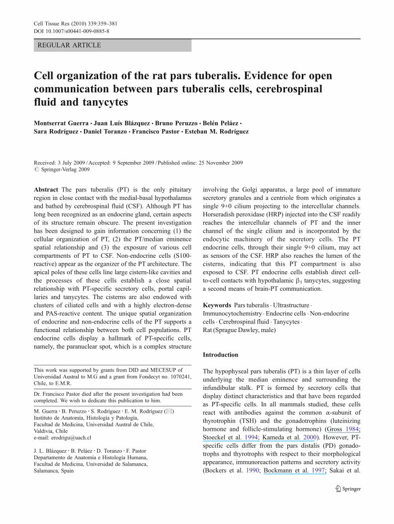
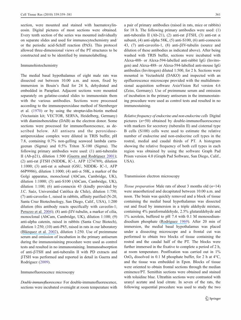
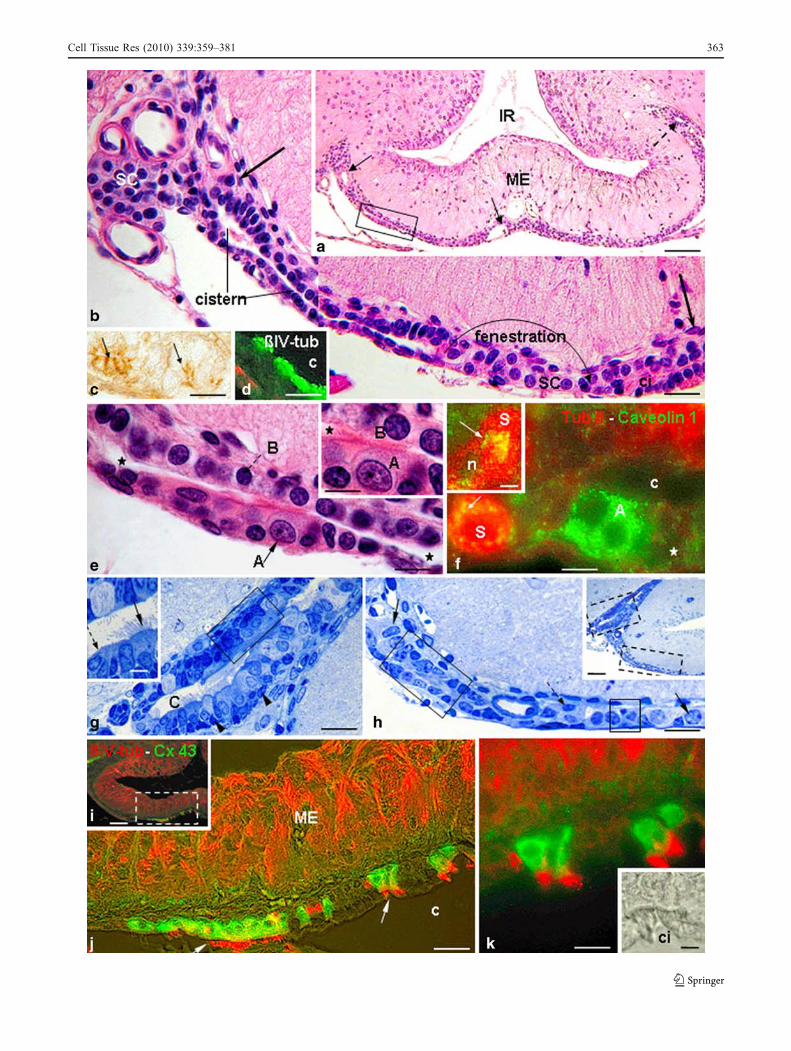
Fig. 1 a Frontal paraffin section through the medial basal hypothal-amus; haematoxylin-eosin staining (IR infundibular recess, MEmedian eminence, dashed arrow dilated edge of a cistern located inthe tuberoinfundibular sulcus). A cistern reconstructed from serialsections was found to extend from the tuberoinfundibular sulcus tomidline (region delimited by two arrows). The boxed area is shown athigher magnification in e. Bar 80 µm. b Sections adjacent to that of a.Montage of two micrographs obtained from two adjacent sectionsshowing a cistern extending from the lateral to the medial region ofthe pars tuberalis (PT; area between large arrows) interrupted at onepoint by a fenestration (curved arrow both parts of the cistern are incontinuity in adjacent sections). The latero-external edge of the cisternis dilated (SC secretory cells, ci ciliated cells). Bar 25 µm. c Rat PTimmunostained with anti-alpha catenin (arrows adherens junctions).Bar 12 µm. d Sagittal section through a cistern (c) immunostainedwith anti-βIV-tubulin antibody. The cistern is lined by cell types A(ciliated, green) and B. Bar 18 µm. e Detailed view of boxed area ina, showing a frontal section through a cistern lined by cell types A(ciliated, arrow) and B (dashed arrow) cells (stars cistern lumen) Bar12 µm. Inset Detailed view of cell types A (ciliated, A) and B (B) withthe cistern lumen (star). Bar 8 µm. f Double-immunostaining of thePT with antibodies against caveolin-1 (green) and tuberalin II (Tub II,red). Type A (A) cells and tuberalin-II-secreting cells (S, arrow)strongly express caveolin-1 (star cells weakly expressing caveolin-1(c cistern lumen). Bar 10 µm. Inset: High magnification of a tuberalin-secreting cell (n nucleus). Caveolin-1-immunoreactive particles(arrow) are concentrated in the vicinity of the paranuclear spot (S).Bar 6 µm. g Detailed magnification of upper boxed area in inset in h.Section through the dilated edge of a cistern (C) located in thetuberoinfundibular sulcus. Cell types A and B are intermingled(arrowheads undifferentiated cells). The boxed area is shown inFig. 3a. Bar 18 µm. Inset: Detailed view showing cell types A (arrow)and B (dashed arrow). Bar 7 µm. h Detailed magnification of lowerboxed area in inset. Secretory (arrows) and cistern type B cells(dashed arrow) are present. Boxed areas are shown by transmissionelectron microscopy in Figs. 3d (square), 5a (rectangle). Bar 18 µm.Inset Semithin section through the hypothalamus of a normal adult rat,stained with toluidine blue. The boxed areas are shown in g, h. Bar50 µm. i Double-immunostaining of the median eminence/PT withantibodies against connexin 43 (Cx43, green) and βIV-tubulin (βIV-tub, red). The boxed area is shown in j. Bar 63 µm. j Highermagnification of boxed area in i. Cistern type A cells (A) express bothmarkers. Median eminence tanycytes (ME) express βIV-tubulin (ccistern lumen). Bar 20 µm. k Detailed view of cistern type A cells (A)shown in j. Cilia are strongly reactive with anti-βIV-tubulin antibody.Bar 11 µm. Inset: Phase-contrast of the same two type A cells asshown in k (ci cilia). Bar 2.5 µm
bTab
le1
Ultrastructuralcharacteristicsof
thesecretorycells
oftheratpars
tuberalis
(m.a.major
axis,PNSparanu
clearspot,RERroug
hendo
plasmic
reticulum
)
Character
Type1
Type2
Type3
Type4
Type
5
Shape,size
Elongated,12
µm
m.a.
Bipolar,12–
18µm
m.a.
Ovoid,10
µm
m.a.
Ovoid,15
µm
m.a.
Elongated,11
µm
m.a.
RER
Few
cisternae
Cisternae
atsynthesispole
Few
cisternae
Highlydeveloped
Poorlydeveloped
Golgi
apparatus
Com
plex
andmuch
developedas
PNS
Com
plex
andmuch
developedas
PNS
Poorlydeveloped
Welldeveloped
Welldeveloped
Secretory
granules
Sizedistributio
ncurve
65–1
00nm
80–1
30nm
Two(?):
80–1
40nm
110–280
nm80
–100
nm
100–
160
nm
Sizepeak
80/90
nm95/100
nm90
nm90/120
nm180
nm
Num
berpersection
20–5
060
–80
60–1
00100–
120
200
Distribution
Vascularpole
mainly
Vascularpole
mainly
Throughoutcytoplasm
Clustersam
ongRER
Throughout
cytoplasm
Nem
atosom
ePresent
Present
Not
found
Not
found
Not
found
Endocytic
machinery
Vesicles
Frequent
Frequent
Scarce
300–
500
nmdensebodies
300–
500
nmdensebodies
Endosom
es,lysosomes
Abundantin
PNS
Abundantin
PNS
Scarce
Singleciliu
mPresent
Present
Not
found
Not
found
Not
found
Distributionof
secretorycells
inthegland
Throughout
Throughout
Tuberoinfundibular
sulcus
mainly
Tuberoinfundibular
sulcus
mainly
Caudalthirdof
PT
Relativefrequencyof
secretorycells
High
High
Low
Low
Low
362 Cell Tissue Res (2010) 339:359–381
Cell Tissue Res (2010) 339:359–381 363
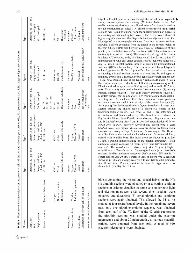
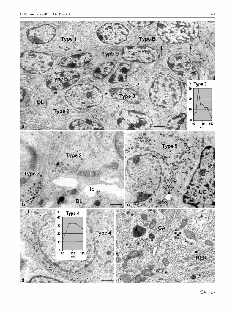
Quantitative analysis Micrographs obtained at ×5000to ×15,000 magnification were used to measure the sizeof the secretory granules. By using the program ImageSP-viewer-exe (version 1.0.22.85), the diameter of eachsecretory granule present in individual secretory cells wasrecorded. A size distribution curve for the population ofgranules present in each cell was obtained by using thesoftware Graph Pad Prism version4.0 (Graph Pad Soft-ware). The size distribution curves of 200 cells wereobtained and compared. The cells displaying secretorygranules with a similar size distribution curve wereconsidered to correspond to the same cell type. Thecharacterization of a given cell type was further establishedby other ultrastructural features (see Table 1). Once a celltype was fully characterized, its frequency, distribution andcell-to-cell relationships were studied in the whole collec-tion of electron micrographs.
Peroxidase injection
The tissue samples studied in the present investigationcorresponded to an early experiment carried out in ourlaboratory (Aguado et al. 1981). New sections wereobtained and studied under the electron microscope, payingspecial attention to the subcellular distribution of the tracerin the PT cells. In brief, rats were injected with 100 µl of a3% solution of HRP (Sigma, Grade VI) into the cisternamagna and killed 10 min later. The PT was processed tovisualize HRP under the electron microscope (Aguado et al.1981).
Results
General organization of PT
The various types of secretory and non-endocrine cellsforming the rat PT were unevenly distributed; in the middlethird, the cells were arranged into a thin layer, whereaslaterally, they clustered into a cell mass filling the tubero-infundibular sulcus (Fig. 1a). The PT was surrounded by abasal lamina that separated the gland from the subarachnoidspace and the perivascular space of the portal capillaries.The mass of PT cells occupying the tuberoinfundibularsulcus had capillaries and arterioles distributed throughoutthe gland (Fig. 1a). However, the thin layer of PT cellsunderlying the middle third of the median eminence wastraversed by the portal vein and was devoid of capillaries(Fig. 1b). At this level, the nerve terminals and tanycytes ofthe median eminence, on the one side, and the PT cells onthe other, shared the same capillaries of the primary plexusof the portal system.
The PT was formed by endocrine cells intermingled withnon-endocrine cells that lined a complex cistern-likestructure.
Non-endocrine cells of PT
The light and electron microscopy findings indicated thatthe PT had a unique and complex cell organization (see alsobelow). The analysis of the full series of frontal and sagittalsections through the whole PT revealed the presence, oneither side of the gland, of cistern-like structures extendingfrom the lateral (tuberoinfundibular sulcus) to the midlineregion of PT (Fig. 1a, b) and through the two caudal thirdsof the gland (Fig. 2a). For descriptive purposes, suchstructures will be referred to as cisterns. Cisterns wereabout 400 µm wide (latero-medial axis), 800 µm long(rostro-caudal axis) and 20–50 µm thick (dorso-ventralaxis). The cistern thickness was uneven because the size of thecells lining the cavity, and the cistern lumen varied from zoneto zone (Fig. 1b, e). Throughout most of the cistern, its lumenwas extremely slender (less than 2 µm; Figs. 1b, e, 2a, b). Atthe latero-external edge, the cistern lumen expanded to reach10–15 µm (Fig. 1b). When cut frontally, the cistern appearedas a duct running latero-medially (Fig. 1b). The cistern hadnumerous fenestrations (Figs. 1a, b, 2a–h). The whole
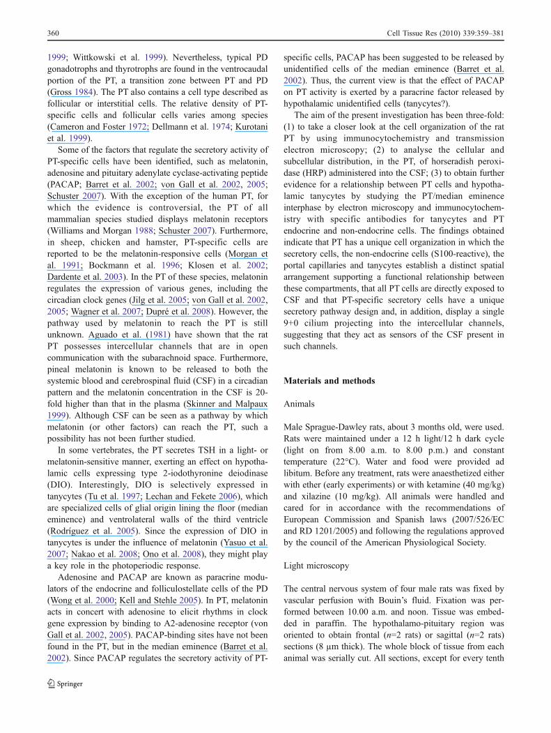
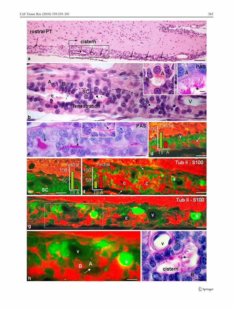
Fig. 2 a Sagittal section through the PT located at the tubero-infundibular sulcus; haematoxylin-eosin staining. Sagittal sectionthrough a cistern (arrows) displaying fenestrations (as reconstructedfrom serial sections). The boxed area is shown in b. Bar 60 µm. bDetailed view of boxed area in a showing a cistern (c) interrupted byfenestrations (curved arrows the lumen of the cistern is continuous atneighbouring planes of sections). Cistern type A (A) and B (B) cellsline the cistern (V blood vessel, SC secretory cells). Bar 12 µm. Leftinset: Detailed view of type A cells (ciliated, A) and type B cells (B).Bar 4.2 µm. Right inset: Section adjacent to that of previous insetstained with periodic acid Schiff (PAS). Masses of PAS-positivematerial appear on top of cilia (star). Bar 3 µm. c Sagittal sectionthrough the PT stained with PAS. Sagittal view of a cistern with anobvious lumen. The lumen is readily visualized because it has a PAS-positive content (arrows). Bar 12 µm. Inset: A slender lumen at anotherpoint. Bar 6 µm. d–h Sagittal sections through the PT located at thetuberoinfundibular sulcus. Double-immunostaining with antibodiesagainst tuberalin II (Tub II, green) and S100 (red). d Caudal third ofPT (arrow cistern, v portal capillaries). Bar 30 µm. e Rostral third of PTmainly formed by secretory cells (SC). Bar 30 µm. f Middle third of PTwith numerous S100-immunoreactive cells lining a cistern (c) andintermingling with secretory cells (S). Cells lining the cistern sendprocesses to the basal lamina (arrows). Bar 25 µm. Histogram insets ind–f Percentages of immunoreactive cell types (TII tuberalin-II-immunoreactive, A S100) in the designated regions. g Same section asin f but at a more caudal level. The boxed area is shown in h (c cisternlumen, V blood vessel, S secretory cell. Bar 18 µm. h Highermagnification of boxed area in g. A cistern is lined by S100-immunoreactive (B) and ciliated (A) cells (arrow cilia, V capillary, Ssecretory cells). Bar 10 µm. i Section neighbouring that in h but stainedwith PAS. Cistern type A cells (A) and type B cells (B). PAS-positivematerial (arrow) appears on top of cilia (V blood vessel). Bar 8 µm
b
364 Cell Tissue Res (2010) 339:359–381
Cell Tissue Res (2010) 339:359–381 365
structure resembled a Golgi cistern with its fenestrations. Thesecretory cells surrounded the cistern and squeezed throughits fenestrations. In some rats, the lumen of the cisternappeared expanded (Figs. 1j; see also below). Apparently,the cisterns were closed cavities, since they were never seento open into adjacent structures. Two types of cells lined thecisterns.
In those animals processed for serial sagittal sectioning,the hypophysis remained in situ (see below). No structuresresembling the PT cisterns were seen in the PD.
Cistern type A cell This type of cell was tall, 12–15 µmhigh, and projected numerous 9+2 cilia into the cisternlumen (Figs. 1e, g, 2b, i, 3b). They were unevenlydistributed throughout the cistern wall. In tissue sections,they appeared isolated or as clusters formed by a few orseveral cells (Figs. 1b–k, 2b, i). They were readily visualizedby immunocytochemistry with the anti-βIV-tubulin antibody,which strongly stained the cilia (Fig. 1d, j, k). Type A cellsexpressed caveolin-1 strongly, the immunoreaction appearingas numerous small particles distributed throughout thecytoplasm (Fig. 1f). Type A cells were the only cell type ofthe PT that expressed connexin 43 (Fig. 1j, k). The cellnucleus contained abundant heterochromatin and thecytoplasm was endowed with a well-developed Golgiapparatus, numerous elongated mitochondria and clustersof glycogen particles, whereas rough endoplasmic reticu-lum (RER) was sparse (Fig. 3a, b). The supranuclearcytoplasm contained several endocytic vesicles and earlyand late (multivesicular bodies) endosomes. Apically,these cells were joined together by zonula adherens, gapjunctions and numerous desmosomes; tight junctions weremissing. The basal pole established contact with thesecretory cells and with the basement membrane surround-ing the PT.
Cistern type B cell These cells were strongly immunoreac-tive with anti-S100 antibody, the immunoreactive proteinbeing distributed throughout the cell allowing the wholecell profile to be visualized. The number, shape andorganization of these cells varied throughout the gland.Double-immunolabelling for S100 and tuberalin II, asecretory product of PT-specific cells, was used to estimatethe relative concentration of both cell types in the rostral,medial and caudal thirds of the gland. As seen inhistograms (Fig. 2d–f), type B cells were scarce at therostral third (5%), abundant in the medial (60%) and caudal(33%) thirds. This agreed with the finding that the cisternwas not present in the rostral third of the gland. The cellbody of the S100-immunoreactive cells lined most of thewall of the cisterns and displayed basal processes embrac-ing the secretory cells and ending on the basementmembrane of the gland or on blood vessels (Fig. 2d–h,
see also below). Some immunoreactive cells were foundaway from the cistern lumen; whether they corresponded totrue non-cisternal cells or to cisternal cells tangentially cutcould not be established (Fig. 2d, e, g, h).
Type B cells were devoid of cilia and had a variableshape. When they lined the dilated region of the cistern,they had a cylindrical shape but, when they lined aflattened region of the cistern, they adapted to the spatialorganization of the secretory cells and became flattened,elongated, bipolar or stellate (Figs. 3a, d, 5a, see alsobelow). They displayed an elongated nucleus, 6–8 µmlong, with distinct patches of condensed chromatin(Fig. 3a, d). Near the lumen of the cistern, the cells werejoined together by zonula adherens, macula adherens anddesmosomes; tight and gap junctions were not seen(Fig. 3c, f). Most of these cells contacted the PT basallamina, frequently via a slender process squeezing inbetween the secretory cells (Fig. 3d; see also below).Conversely, the PT basal lamina sent processes into thegland and were contacted by type B cells. At certainregions of the cistern, the cells projected short globularmicrovilli to the cistern lumen (Fig. 3c), whereas in others,the apical plasma membrane was smooth (Fig. 3e). Thecytoplasm contained abundant polyribosomes, scarce RERcisternae, 3–5 stacks of Golgi cisternae and numeroussmooth-surfaced tubular structures, probably correspondingto the trans-Golgi network (Fig. 3e, f). The endocyticmachinery was well developed.
Cistern lumen At certain regions of the cistern, the lumenwas partially or fully occupied by PAS-positive contents(Fig. 2c). Masses of PAS-positive material were seen on topof the cilia of type A cells (Fig. 2b, inset, i). In PTprocessed for electron microscopy, the cistern had a lumenof about 150 nm; at discrete sites, the lumen expanded toseveral hundred nanometres. Microvilli, cilia and/or an
Fig. 3 a–f Transmission electron microscopy of the PT of normal rats.a Area similar to that boxed in Fig. 1g (UC undifferentiated cells, BLbasal lamina, IC intercellular channels). Cell types A and B line acavity filled with an electron-dense material (large arrows). Cisterncells are joined by adherens junctions (small arrows). Bar 1.9 µm. bCistern cell type A. Bar 700 nm. Left inset: Cross section of a 9+2cilium. Right inset: Cluster of glycogen particles. c Cross sectionthrough a slender segment of a cistern lined by type B cells projectingmicrovilli to the lumen (ZA zonula adherens, D desmosome). Bar500 nm. d Area similar to boxed area (square) in Fig. 1h. A Type Bcell projects a process (arrow) between two secretory cells to end onthe basal lamina (BL). Bar 900 nm. e Cross section through a slendersegment of a cistern lined by type B cells (SC secretory cell, Ddesmosome, ZA zonula adherens). The slender lumen is filled with anelectron-dense material (stars). Bar 760 nm. Inset: Adherens (ZA) anddesmosome (D) junctions between type B cells. Bar 500 nm. fSupranuclear cytoplasm of type B cell with a well-developed Golgiapparatus (star canaliculus between two B cells, SC secretory cell).Bar 447 nm
b
366 Cell Tissue Res (2010) 339:359–381
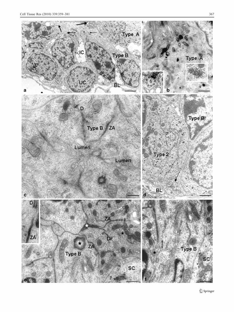
Cell Tissue Res (2010) 339:359–381 367
electron-dense material occupied the expanded region ofthe lumen (Fig. 3a, c).
Undifferentiated cells Cells with an undifferentiated ap-pearance were located around the wall of the dilated edgeof the cistern (tuberoinfundibular sulcus). The nucleus wasstrongly basophilic. Polyribosomes were abundant, butother organelles were poorly developed (Figs. 1g, 3a).
Secretory cells of PT
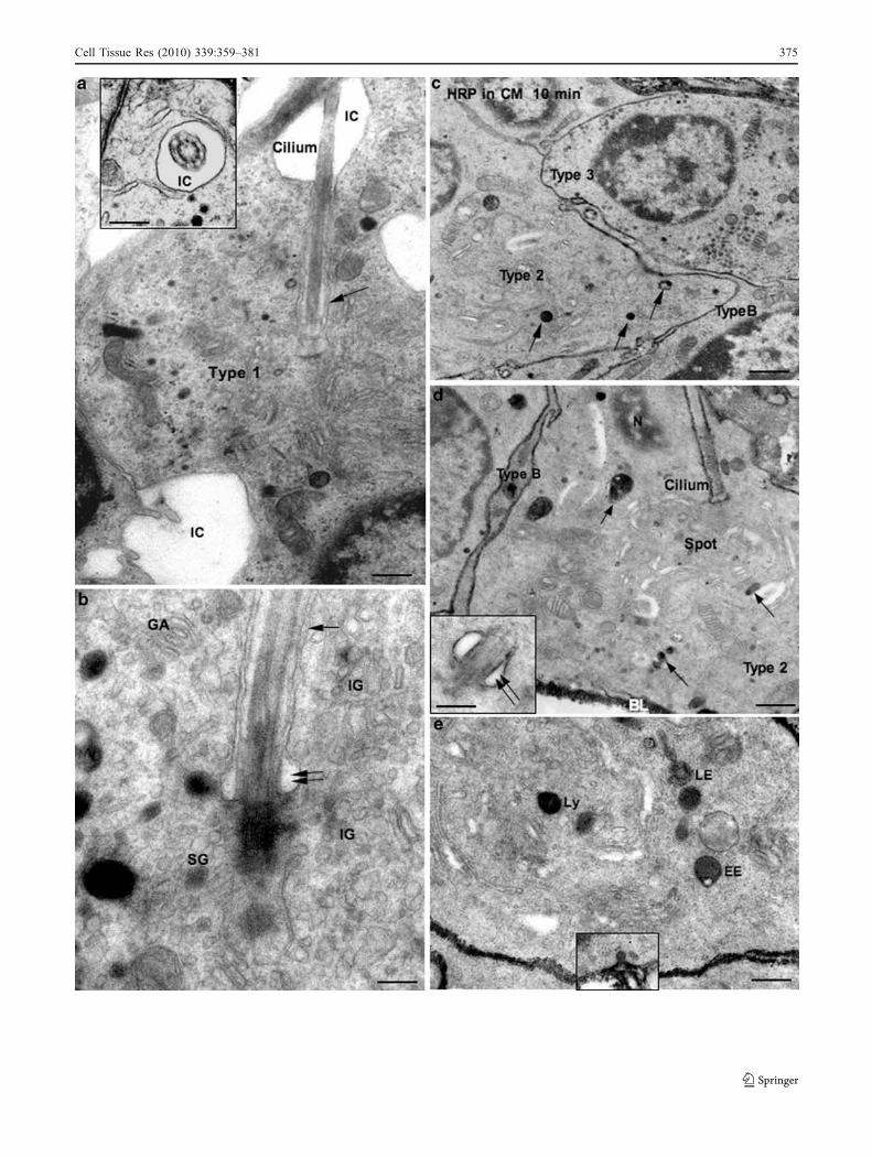
Based on their ultrastructure, five types of secretory cellscould be found in the PT (Figs. 4, 5, 6, 7, Table 1). Themost distinct features of type 1 and 2 cells were: (1) a well-developed and complex Golgi apparatus displaying specificultrastructural and immunocytochemical peculiarities that willbe described in detail as the “paranuclear spot” (Figs. 4, 6) and(2) a single 9+0 cilium that originated in the core of theparanuclear spot and projected to the intercellular channels(Fig. 7a, b). These cells were elongated, with a major axisranging between 12 µm and 18 µm (Figs. 4, 5). Thecytoplasm was zoned. The synthesis pole contained thestructures involved in protein/peptide synthesis, namely,the Golgi apparatus, several elongated and slendercisternae of RER, few secretory granules, numerousmitochondria and nematosomes (Fig. 4). The release polecontacted the basal lamina and contained most of thesecretory granules (Fig. 4). Clusters of granules undergo-ing exocytosis were frequently seen (Fig. 4c).
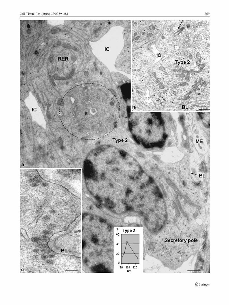
The distinct feature of type 3 cells was their largenumber of secretory granules, 60–100 per section, fillingmost of the relatively sparse cytoplasm. The size distribu-tion curve of the granule diameter ranged between 80–160 nm (Fig. 5a, b). The curve profile was compatible withthe existence of two subpopulations of granules, oneranging between 80 and 100 nm, with a peak at 90 nm,and a second ranging between 100 and 140 nm. Type 4cells were distinguished by the large development of RER,whose cisternae were elongated with a dilated lumen filledwith a filamentous material. Packages of interconnectedcisternae were distributed throughout the cytoplasm(Fig. 5d, e). The feature characterizing type 5 cells wasthe large number of secretory granules of 110–280 nm indiameter; a section through the cell displayed about 200such secretory granules (Fig. 5c). Dense bodies, 300–500 nm in diameter, were consistently found throughout thecytoplasm of types 4 and 5 cells (Fig. 5e).
Secretory cells established contact with the basallamina and with cisternal cells. They were never exposedto the cistern lumen; a thin cytoplasmic process ofadjacent cistern cells segregated the secretory cells fromthe cistern lumen.
Types 1 and 2 largely outnumbered the other cell types.The various cell types were unevenly distributed through-out the PT. In the vicinity of the midline of PT, only types 1and 2 were present. However, all types of secretory cellswere present in the mass of PT occupying the tuber-oinfundibular sulcus (Fig. 5a). Type 5 cells were morenumerous in the caudal region of the PT.
Paranuclear spot, a cell marker of PT-specific cells, displaysunique immunochemical and ultrastructural characteristics
Immunocytochemistry Most secretory cells of the PT werereactive with antibodies against the alpha subunit ofglycoprotein hormones (GSU); these cells also reacted withanti-βTSH and/or anti-tuberalin II (cf. Guerra and Rodríguez2009; Fig. 6a, e, f). The immunoreactive cells exhibited aspecific cytoplasmic region that lay close to the nucleus,was about 4 µm in diameter and was strongly reactive withthe three antibodies (Fig. 6c, f). Double-immunolabellingshowed that the same spot reacted with these antibodies andwith anti-α58K, a marker of the Golgi apparatus (Fig. 6g,inset). A similar structure has been described by otherauthors, being designated as the “paranuclear spot” andregarded as a marker of PT-specific cells (Gross 1984;Bergmann et al. 1989; Sakai et al. 1992). Indeed, sectionsof the PD immunostained with anti-GSU showed a largepopulation of immunoreactive cells, but none of themdisplayed a paranuclear spot (Fig. 6a–c).
Ultrastructure The paranuclear spot had a spherical shapewith a diameter of 3–4 µm (Figs. 4a, 6d). A clearperipheral-central zonation could be distinguished(Fig. 6g). Eight to ten stacks of Golgi cisternae, many ofwhich appeared interconnected by tubular structures,formed the peripheral zone. Each Golgi stack containedabout five ordered cisternae or saccules; each saccule had acentral plate-like region and a peripheral dilated edge. Thisstructure characterized the central and trans saccules.However, the cis saccules had a wide lumen of 50–60 nmthroughout and a fenestrated appearance. The intermediatecompartment between endoplasmic reticulum and the cisGolgi apparatus contained smooth-surfaced tubules andvesicles with an electron-lucent content (Fig. 6h). Direct
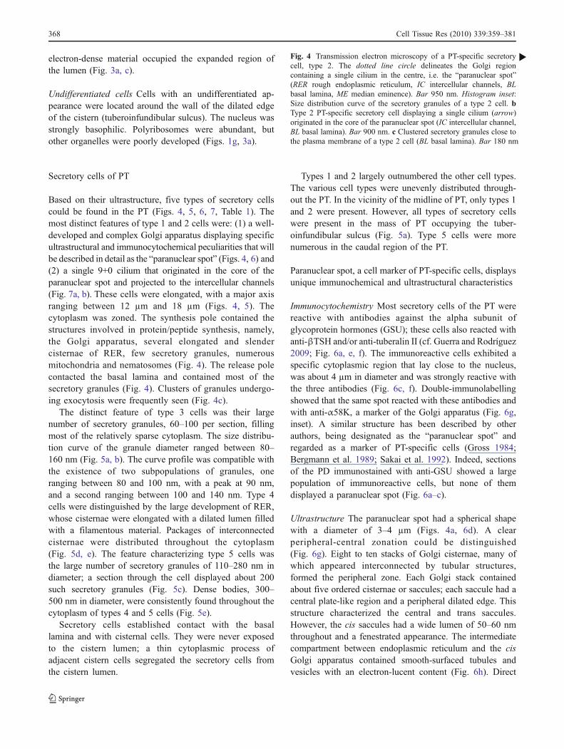
Fig. 4 Transmission electron microscopy of a PT-specific secretorycell, type 2. The dotted line circle delineates the Golgi regioncontaining a single cilium in the centre, i.e. the “paranuclear spot”(RER rough endoplasmic reticulum, IC intercellular channels, BLbasal lamina, ME median eminence). Bar 950 nm. Histogram inset:Size distribution curve of the secretory granules of a type 2 cell. bType 2 PT-specific secretory cell displaying a single cilium (arrow)originated in the core of the paranuclear spot (IC intercellular channel,BL basal lamina). Bar 900 nm. c Clustered secretory granules close tothe plasma membrane of a type 2 cell (BL basal lamina). Bar 180 nm
b
368 Cell Tissue Res (2010) 339:359–381
Cell Tissue Res (2010) 339:359–381 369
endoplasmic reticulum to Golgi apparatus connections werefrequent (Fig. 6h). Vesicles of 80–100 nm, with a content ofmoderate electron density, were seen connected with theedge of trans cisternae and lying free nearby (Fig. 6h).
The central zone of the paranuclear spot containedstructures corresponding to the secretory and endocytic path-ways: (1) numerous vesicles ranging in size between 80 and120 nm in diameter, with a content whose electron density wasinversely related to their size, most likely corresponding tocondensing vesicles becoming immature secretory granules(Fig. 6g); (2) small and isolated RER cisternae withribosomes (Fig. 6h); (3) a complex and highly developedtrans-Golgi network (TGN), with tubules of 60–100 nm inwidth, coated vesicles and coated pits; (4) the plasmamembrane-endosome-TGN pathway represented by coatedpits opening to the intercellular channels, coated and smooth-surfaced vesicles, early vesicular endosomes, several multi-vesicular bodies (late endosomes) and lysosome-like bodies,the last two types of bodies being frequent in the central zoneof the paranuclear spot (Fig. 7); (5) an exceptional componentof the paranuclear spot, i.e. a single cilium, whose basalcorpuscle was located in the core of the spot (see below).
Endocrine and non-endocrine cells of PT are in opencommunication with CSF
Intercellular channels Maculae adherentes were seen be-tween secretory cells and between the latter and cisternalcells. The intercellular space between PT cells was 15 nm.At various points, this space became dilated reaching 400–1000 nm in width and up to 3 µm in length (Figs. 4, 5, 7;cf. Aguado et al. 1981). These intercellular channels formeda network distributed throughout the PT (Figs. 3a, 4a, 5a, b,7a, 8d, e) and were in open communication with the basallamina surrounding the gland and the capillaries.
Type 1 and 2 secretory cells have a single 9+0 cilium Inmost type 1 and 2 cells sectioned through the paranuclearspot, a single basal corpuscle located in the core of the spotwas the origin of a cilium (Figs. 4, 6, 7). Cells displayingtwo or more cilia were never seen, supporting theassumption that each cell had a single cilium. Paraffinsections double-immunostained with anti-GSU and anti-βIV-tubulin showed secretory cells projecting only onecilium (Fig. 6d, inset). This cilium lacked the central pair ofmicrotubules (Fig. 7a). The inner structure of the singlecilium was variable; in some cases, it presented nine pairsof microtubules orderly arranged at the periphery, whereasin others, microtubules were not orderly arranged, withonly some of the microtubules being paired.
The cilium originated in the core of the paranuclear spotand projected to a neighbouring intercellular channel
(Fig. 7a). The cilium occupied the lumen of a deepinvagination of the plasma membrane (Fig. 7a, b) and hada diameter of about 280 nm. The plasma membraneinvagination had a lumen of about 340 nm; thus, the spacearound the cilium was 30 nm wide. At the bottom of theplasma membrane invagination, this space became expand-ed, forming a cuff about 480 nm in diameter (Fig. 7b). HRPadministered into the cisterna magna reached the intercel-lular channels of the PT and filled the invaginationsurrounding the cilium (Fig. 7d).
Intracisternal administration of HRP By 10 min after itsadministration into the cisterna magna, HRP was visualizedin the basal lamina surrounding the PT, in the perivascularspace of the PT capillaries and in the intercellular space ofthe gland (Fig. 7c–e). In secretory cells type 1 and 2, HRPwas incorporated into pinocytic vesicles, early endosomes,late endosomes and lysosomes (Fig. 7c–e). The late endo-somes and lysosomes were located in the periphery and thecore of the paranuclear spot. Trans-Golgi network structureswere also labelled. HRP was present in the deep plasmamembrane invagination occupied by the single cilium, and inthe cuff-like enlargement of such an invagination located inthe paranuclear spot (Fig. 7d). Double-immunolabellingshowed that tuberalin-II-secreting cells had a large numberof caveolin-1-labelled vesicles mostly restricted to thevicinity of the paranuclear spot (Fig. 1f).
Intracisternally injected HRP also reached the intercel-lular space between cisternal cells and the cistern lumen(Fig. 9e). Secretory cells types 3–5, type B cisternal cellsand undifferentiated cells incorporated little or no HRP,despite the label being available in the intercellularchannels (Fig. 7c).
Median eminence/PT relationship: β1 tanycytes establishcontact with PT-specific cells located at the tuberoinfundibularsulcus
The use of anti-p85, a specific marker of tanycytes(Blázquez et al. 2002), anti-βIV-tubulin and anti-S100,
Fig. 5 Transmission electron microscopy of a zone of the PT similarto that boxed (rectangle) in Fig. 1h. a Type B cells line a cisternregion with a slender lumen (arrows). PT-specific secretory cell types1, 2 and 3 are present (stars intercellular channels, BL basal lamina).Bar 2 µm. Histogram inset: Size distribution curve of the secretorygranules of a type 3 cell. b Secretory cells types 2 and 3 (BL basallamina, ic intercellular channel). Bar 900 nm. c Secretory cell type 5(UC undifferentiated cell). Bar 1.8 µm. d Secretory cell type 4.Histogram inset: Size distribution curve of the secretory granules of atype 4 cell. Bar 1200 nm. e Higher magnification of the cytoplasm ofcell shown in d (GA Golgi apparatus, RER rough endoplasmicreticulum). Bar 600 nm
b
370 Cell Tissue Res (2010) 339:359–381
Cell Tissue Res (2010) 339:359–381 371
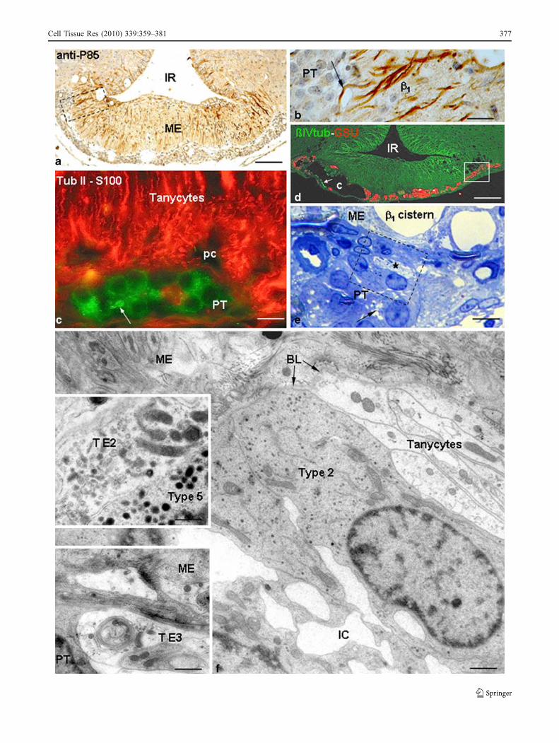
both of which also label tanycytes, revealed the closespatial relationship between the terminals of β1 tany-cytes, the portal capillaries and the PT secretory cells(Fig. 8a–d). A few bundles of processes of β1 tanycytestraversed the brain-limiting membrane and reached PT cells(Fig. 8a, b).
Transmission electron microscopy of the lateral region ofthe median eminence showed that bundles of 5–10processes of β1 tanycytes entered the PT and ended onPT-specific secretory cells located in the tuberoinfundibularsulcus (Fig. 8f). The ultrastructure of the tanycyte processeswas identical to that of β1 tanycyte processes describedearlier for the median eminence (Rodríguez et al. 1979,2005). Branching of the processes into the PT wasobserved. Tanycyte type 2 and 3 terminals, as describedfor the median eminence (Rodríguez et al. 1979, 2005)were found (Fig. 8f, insets). Type 2 endings had numerouselectron-dense granules of 100–130 nm in diameter andshort tubular formations of 100–130 nm in diameter with anelectron-dense content. Smooth-surfaced cisternae, fila-ments, mitochondria and microtubules were abundant inthe preterminal region (Fig. 8f, upper inset). Type 3terminals contained granules of various sizes filled with afilamentous material and several smooth-surfaced cisternaethat were concentrically arranged (Fig. 8f, lower inset). Nomembrane specializations were seen at the site of contactbetween tanycytes and PT cells.
Discussion
Rat PT: a discrete region of the adenohypophysisdisplaying a unique cell organization
The present study has revealed the presence, in the PT, ofcistern-like structures extending from the lateral (tubero-infundibular sulcus) to the midline region and through thetwo caudal thirds of the gland. Since these cisterns arefenestrated, their cavity in tissue sections can appear asducts or follicles. Indeed, early studies have described thepresence of follicles and follicular cells in the PT(Dellmann et al. 1974; Fitzgerald 1979; Gross 1984;Kameda 1990). This unique cisternal structure is linedby ciliated (type A) and non-ciliated (type B, S100-positive) cells arranged as clusters throughout the cisternwall. Type B cells, through their apical pole, line thecistern and, through their processes, establish a closespatial relationship with (1) PT-specific secretory cells, (2)portal capillaries and (3) tanycytes (Fig. 9). What is thefunctional meaning of such an organization? Do type Bcells secrete into the cistern lumen? What is the nature ofthe cistern content? The presence of electron-densematerial in the cistern lumen indicates the proteinaceous
nature of such a material and its reaction with PASsuggests that such a material is glycosylated. In the PD,cells with ultrastructural features and S100 immunoreac-tivity similar to those of type B cells, the so-calledfolliculostellate cells, have been reported to producevarious bioactive compounds, such as basic fibroblastgrowth factor (FGF-2), vascular endothelial growthfactor, follistatin and interleukin-6 (Ferrara et al. 1987;Gospodarowicz and Lau 1989; Inoue et al. 1999). Whethertype B cells of the PT cisterns secrete some of thesecompounds remains to be investigated.
What is the fate of the cistern content? Apparently,cisterns do not open into any other compartment. Thus, itscontent as a bulk, although moving by the beating of themulticiliated (type A) cells, would not be drained. Howev-er, molecules present in the cistern lumen may be absorbedby type A cells (they express caveolin-1 strongly), by typeB cells or, alternatively, move through the intercellularspace of the cistern wall and reach the intercellular channelsof PT or even the CSF. Indeed, HRP injected into thecisternal CSF follows, in a reverse way, this pathway(Fig. 9b) and reaches the cistern lumen. This agrees withthe absence of tight junctions between the cells lining thecistern.
Do type B cells and PT-specific secretory cells engage incross talk, as suggested by their close and peculiar spatialorganization? The PD follicular cells are electronicallycoupled through gap junctions and are thought to coordi-nate the activity of hormonal cells (Fauquier et al. 2002;
Fig. 6 a Sagittal paraffin section through the pars tuberalis (PT) andpars distalis (PD) of a normal adult rat immunostained with anti-alphasubunit of glycoprotein hormone (GSU). Bar 140 µm. Left inset: MostPT cells display an immunoreactive paranuclear spot (arrow). Bar14 µm. Right inset: The cytoplasm of PD immunoreactive cells isstrongly and evenly immunostained (arrow). Bar 17 µm. b Highermagnification of the PD cell indicated (arrow) in right inset in a. Bar3.4 µm. c Higher magnification of the PT cell indicated (arrow) in leftinset of a. Bar 3.9 µm. d Transmission electron microscopy of a type2 PT-specific secretory cell (dotted circle paranuclear spot, N nucleus).Bar 800 nm. Inset: Double-immunostaining of a PT-specific secretorycell with antibodies against GSU (red) and the cilium marker βIV-tubulin (green). Note the single cilium (arrow). Bar 1.5 µm. e Double-immunostaining of the PT with antibodies against thyrotrophin (TSH,green) and tuberalin II (Tub II, red). Bar 80 µm. f PT-specificsecretory cells react with anti-TSH (II, green) and anti-tuberalin II (I,red) or both (III). Bar 15 µm. Inset: In type III cells, the sameparanuclear spot reacts with both antibodies. Bar 7 µm. g Highermagnification of the ultrastructure of a paranuclear spot (TGN trans-Golgi network, CV condensing vacuoles, IG immature secretorygranules, ci cilium, star cilium channel). Bar 220 nm. Insets: Double-immunostaining of a PT-specific secretory cell using antibodiesagainst GSU (green) and the Golgi marker α-58K (red). The sameparanuclear spot reacts with both antibodies (arrows). Bar 5 µm.h Tubular structures (arrow) connect the cisternae of the roughendoplasmic reticulum (RER) with cis cisternae of the Golgi apparatus(GA). Note the immature secretory granules (IG), secretory granule(SG), cilium (ci). Bar 190 nm
b
372 Cell Tissue Res (2010) 339:359–381
Cell Tissue Res (2010) 339:359–381 373
Denef 2008). This mechanism is missing in type B cells ofPT. With the exception of type A cells (multiciliated),neither type B cells nor PT-endocrine cells expressconnexin 43 or show gap junctions at the ultrastructurallevel, indicating that coupling among these cells does notoccur and that a putative functional relationship betweentype B cells and PT-endocrine cells is mediated byparacrine factors. In the PD, the production of growthfactors, such as FGF-2, by folliculostellate cells is regulatedby oestradiol (Oomizu et al. 2004) and follistatin secretedby folliculostellate cells in a paracrine way regulatesfollicle-stimulating hormone output from neighbouringgonadotrophs (Denef 2008). The functional relationshipbetween type B cells and PT-endocrine cells is thus fullyopen to future investigations.
A second target of projection of type B cell processes isthe median eminence/PT interphase at which they establishclose contact with the portal capillaries and tanycyteterminals (Fig. 9). Evidence for a functional relationshipbetween tanycytes and PT will be discussed below.
Endocrine activity of PT
Our ultrastructural evidence suggests that five secretory celltypes occur in the rat PT. Cell types 1 and 2 display uniquefeatures indicating that they correspond to PT-specificsecretory cells. They carry the so-called paranuclear spot,a cytoplasmic mass that has high affinity for wheat germagglutinin and antibodies against GSU, βTSH and tuberalinII (Gross 1984; Wittkowski et al. 1999; Guerra andRodríguez 2001). The paranuclear spot has been regardedas a marker of PT-specific cells (Gross 1984; Bergmann etal. 1989; Wittkowski et al. 1999; Guerra and Rodríguez2001). The present investigation has shown that the para-nuclear spot is a complex zoned structure involving theGolgi apparatus. Distinct features of the spot are thenumerous interconnected stacks of Golgi cisternae formingthe cortex of the spot, a well-developed trans-Golginetwork that produces a large number of condensingvacuoles and immature secretory granules that accumulatein the core of the spot. A unique feature is the presence, inthe core of the spot, of a centriole as the origin of a 9+0cilium. According to the present investigation, the samespot immunoreacts with anti-α58K, a well-known markerof the Golgi apparatus (Bloom and Brashear 1989; Gao etal. 1998), and with anti-GSU, anti-TSH and/or anti-tuberalin II. In all probability, these immunoreactivesecretory proteins are contained in the numerous condens-ing vacuoles and immature secretory granules located in thespot. Whereas the paranuclear spot is strongly reactive withantibodies against GSU, TSH and tuberalin II, the othercytoplasmic regions are weakly reactive (present report;Guerra and Rodríguez 2009) suggesting that the main pool
of hormones is restricted to the spot. This is in agreementwith the relatively scarce mature secretory granules distrib-uted throughout the cytoplasm of these cells, many ofwhich concentrate in the vascular cell pole. However, TSHcells of the PD immunostained with the same antibodies donot display a paranuclear spot and the cytoplasm appearsevenly immunostained.
What is the functional meaning of the exceptionalorganization of the secretory pathway in the PT-specificcells (types 1, 2)? Professional protein-secreting cellsaccumulate mature secretory granules. However, PT-specific cells might accumulate immature secretory gran-ules. Immature granules are known to represent the firststimulable compartment of the regulated secretory pathway(Arvan and Castle 1998; Borgonovo et al. 2006). Do theimmature secretory granules concentrated in the para-nuclear spot contain a releasable pool of hormone or dothey store a pool of readily processable precursor forms?Is there a functional relationship between the pool ofimmature granules and the 9+0 cilium seated in themiddle of such a pool? How is this whole arrangementrelated to the cellular physiology of the PT-specific cells,in particular with the reception of external regulatorysignals? The mechanisms underlying the secretory processin this exceptional endocrine cell are unknown, althoughthey appear essential for understanding the physiology ofthe PT.
We have shown the existence, in the rat PT, of threepopulations of PT-specific cells carrying a paranuclear spotand expressing tuberalin II and GSU (type I), βTSH andGSU (type II) and tuberalin II, βTSH and GSU (type III;Guerra and Rodríguez 2009; present report). The densityand distribution of these cell types in PT and the presenceof the paranuclear spot in them support the possibility thatthese cell types correspond to types 1 and 2 reported in thepresent ultrastructural study. The existence of cells express-
Fig. 7 Transmission electron microscopy of the PT of a normal adultrat. a Type 1 PT-specific secretory cell displaying a single cilium(arrow) originating in the core of the paranuclear spot (IC intercellularchannels). Bar 770 nm. Inset: Cross section of 9+0 cilium lying in anintercellular channel. Bar 360 nm. b Higher magnification of thesingle cilium emerging from the core of the spot (GA Golgi apparatus,IG immature granules, SG secretory granule). The deep infolding ofthe plasma membrane containing the cilium (single arrow) dilates atits base (double arrow). Bar 220 nm. c–e Transmission electronmicroscopy of the rat PT 10 min after the injection of horseradishperoxidase (HRP) into the cisterna magna (CM). c HRP fills theintercellular space and is incorporated by type 2 secretory cells(arrows). Bar 1100 nm. d HRP labels the basal lamina (BL), fills theintercellular space and the cilium channel and is incorporated by atype 2 secretory cell (arrows). Bar 900 nm. Inset: HRP reaches thebottom of the cilium channel (double arrow). e HRP is incorporatedfrom the intercellular channels into pinocytotic vesicles, early (EE)and late (LE) endosomes and lysosomes (Ly). Bar 900 nm
b
374 Cell Tissue Res (2010) 339:359–381
Cell Tissue Res (2010) 339:359–381 375
ing GSU, βTSH and tuberalin II raises the question ofwhether the last two mentioned secretory proteins are storedin the same or separate granules. Ultrastructural immuno-cytochemistry is required to solve this problem. Threepopulations of TSH cells displaying granules of varioussizes have been described in the rat PD by use ofultrastructural immunocytochemistry. The relative frequen-cy of these populations of TSH cells varies with age(Ozawa and Kurosumi 1989) or functional state (Yoshimuraet al. 1982). Thus, the possibility that PT-cells types 1 and2, both bearing a paranuclear spot and a cilium, correspondto the same cell type but under different functional statesshould be kept in mind. However, the various immunocy-tochemical properties of PT-cells carrying the paranuclearspot (present report; Guerra and Rodríguez 2009) do notsupport this possibility.
Secretory cells type 3, 4 and 5 display all the characteristicof endocrine cells but do not carry the paranuclear spot. Thenature of their secretory products is unknown.
Our ultrastructural study suggests that PT-endocrinecells can secrete into the intercellular channels and theperivascular space of the portal capillaries (Fig. 9b). Sinceboth compartments are open to the subarachnoid space(present report; Aguado et al. 1981), compounds secretedby PT-cells could reach targets in the central nervoussystem, via CSF, or in the PD, via the portal circulation.The recent demonstration that TSH secreted by PTregulates the expression of type 2 deiodinase (DIO2) bytanycytes (Nakao et al. 2008) supports the possibility thatPT-originating compounds reach targets in the centralnervous system via the CSF. On the other hand, evidencehas been presented that compounds released by PT (intothe portal circulation?) regulate the secretion of prolactinby the PD (Hazlerigg et al. 1996a, 1996b; Graham et al.2002).
Polypeptides secreted by PT-specific cells have beenstudied by biosynthetic labelling by exposing primarycultures of PT to [35S]methionine and analysing theconditioned medium by fluorography; several compoundsin the range of 14–100 kDa have been identified (Morganet al. 1994). The major protein detected is a 72-kDaprotein designated as p72, whose secretion is regulated bymelatonin (Morgan et al. 1994). We have identified twoproteins of 21-kDa and 72-kDa in the conditionedmedium of bovine PT explants and have raised anti-bodies against them (Guerra and Rodríguez 2001). Anti-p72 and anti-p21 antibodies immunoreact with variouspopulations of the bovine PT. At the ultrastructural level,both antibodies exclusively react with secretory granules(Guerra and Rodríguez 2001). These findings stronglysuggest that both compounds are stored in secretorygranules of PT-specific cells and released into the culturemedium.
PT-endocrine cells are designed as sensorsof the composition of CSF
A distinct feature of PT-specific cells is the presence of asingle 9+0 cilium projecting from the core of the para-nuclear spot to a nearby intercellular channel. To ourknowledge, this is a unique characteristic for anendocrine cell and its functional significance is unclear.The 9+0 cilia are known to perform receptive functionsrather than a motile activity (Yokoyama 2004; Praetoriusand Spring 2005). Various types of receptors, ion channels,effector proteins and transcription factors have beendescribed in 9+0 cilia (Pazour et al. 2005; Satir andChristensen 2008). In PT-specific cells, the 9+0 ciliummight act as a sensor of the composition of the fluid presentin the intercellular channels and, through unknown mech-anisms, transfer the input stimulus to the biosyntheticmachinery of the cell. The finding that HRP injected intothe CSF readily reaches the PT intercellular channels(present report; Aguado et al. 1981) and, most importantly,the inner channel surrounding the 9+0 cilium, is a goodindication that substances carried by the CSF can reach thePT-endocrine cells. Consequently, all compounds present inthe CSF become candidates for influencing PT functions.Melatonin appears as the best candidate. Indeed, PT-cellsexpress melatonin receptors (Williams and Morgan 1988;Schuster 2007), the CSF concentration of melatonin is 20-fold higher than that of plasma (Skinner and Malpaux1999) and CSF melatonin undergoes circadian variationssimilar to those in plasma (Reppert et al. 1979; Kanematsu
Fig. 8 a Paraffin section through the medial basal hypothalamus ofthe rat immunostained with anti-P85, a marker of tanycytes. Theboxed area is shown in b (IR infundibular recess, ME medianeminence). Bar 90 µm. b Detailed view of boxed area in a. A bundleof processes of β1tanycytes (β1) enters the PT (arrow). Bar 24 µm. cRostral PT double-immunostained with antibodies against tuberalin II(Tub II, green) and S100 (red). Note the close spatial relationshipbetween tanycytes, portal capillaries (pc) and tuberalin-II-secretingcells (arrow paranuclear spot). Bar 15 µm. d Paraffin section throughthe hypothalamus of a normal rat (IR infundibular recess). Double-immunostaining with anti-βIV tubulin antibody (βIVtub, green) andthe anti-GSU antibody (red). Tanycytes and cilia (arrow) appeargreen, whereas PT secretory cells appear red. An expanded cistern (c)extends from the lateral to the medial region of PT. An area similar tothat boxed was studied by electron microscopy (see below). Bar100 µm. e Semithin section of an area similar to that boxed in d. Abundle of tanycytes (star) is seen within the PT (arrow intercellularchannels). The boxed area is imaged by transmission electronmicroscopy in f (ME median eminence). Bar 7 µm. f Ultrathin sectionof the same boxed area and cells as in e. A bundle of tanycytes isshown in close contact with a type 2 secretory cell (IC intercellularchannels, BL basal lamina, ME median eminence). Bar 1100 nm. Topinset: Tanycyte ending type 2 (TE2) in close contact with secretorycell type 5. Bar 660 nm. Bottom inset: Tanycyte ending type 3 (TE3)in close contact with a PT cell (PT). Bar 400 nm
b
376 Cell Tissue Res (2010) 339:359–381
Cell Tissue Res (2010) 339:359–381 377
et al. 1989). Thus, the possibility that the regulatoryfunction of melatonin on the PT is exerted via the CSFmust be seriously considered. The effect of melatonin onPT is mediated by the activation of high-affinity membranereceptors (Williams and Morgan 1988; Schuster 2007) and,because of its highly lipophilic nature, by binding tonuclear receptors expressed by PT-specific cells (Hazlerigget al. 1996a, 1996b; Klosen et al. 2002).
PT-specific type 1 and 2 cells internalize HRP injectedinto the CSF and transport it throughout the endocyticpathway. The high expression of caveolin-1 in the PT-specific secretory cells indicates that caveolar endocytosisoccurs in these cells. Several membrane receptors andmembrane transporters have been localized in caveolae(Nichols and Lippincott-Schwartz 2001). Potential candi-dates for internalization by PT-specific cells via caveolaeare those compounds known to be endocytosed through anon-clathrin mechanism, such as adenosine (Nichols andLippincott-Schwartz 2001).
Are tanycytes involved in PT function?
A good body of evidence indicates the presence, in themedio-basal hypothalamus, of four types of tanycytes (α1,α2, β1, and β2; Rodríguez et al. 2005). The basal processesof β1 and β2 tanycytes terminate at the basal laminaseparating the median eminence from PT. A subpopulationof β1 tanycytes crosses the basal lamina and establishescell-to-cell contact with PT-specific secretory cells. Asimilar finding has been reported in an early study carriedout in the rhesus monkey by Knowles and Kumar (1969). βTanycytes have the capacity to transport compounds fromthe ventricular CSF to the perivascular space of the portalcapillaries (Peruzzo et al. 2004; Rodríguez et al. 2005) and,consequently, to the intercellular channels of the PT; βtanycytes are also known to secrete, through their basalprocesses, biologically active compounds such as β-transforming growth factor, prostaglandin E2 or the specificprotein p85 (Rodríguez et al. 2005; Blázquez et al. 2002;Ojeda et al. 2008).
Some information about a functional relationship be-tween tanycytes and PT has recently been obtained.Hypothalamic tanycytes express DIO2, the enzyme con-verting the prohormone T4 into bioactive T3 (Lechan andFekete 2006); this expression is under the influence ofmelatonin (Yasuo et al. 2007). Furthermore, the secretion ofTSH by PT of the Japanese quail triggers the photoperiodicendocrine response by a rapid induction of the geneexpression of DIO2 by tanycytes, as shown by Nakao etal. (2008) who have suggested that TSH might be trans-ported to the third ventricle by tanycytes. This seemsunlikely since tanycytes are endowed with polarizedendocytosis allowing them to absorb compounds from the
third ventricle but not through their terminals (Peruzzo etal. 2004). The TSH receptors that are expressed bytanycytes (Nakao et al. 2008) might be located in thosetanycytes entering the PT. Alternatively, the effect of PTTSH on the arcuate nucleus and tanycytes, reported byNakao et al. (2008) and Hanon et al. (2008), might beexerted via the CSF, which is in open communication withthe PT-specific secretory cells, including the TSH-secretingcells.
The present findings have thus contributed to unfold-ing the complex cell organization of the PT and mayhelp with the interpretation of the ongoing experimentsin several laboratories and aid the design of newinvestigations.
Acknowledgements We are grateful to Genaro Alvial for technicalsupport.
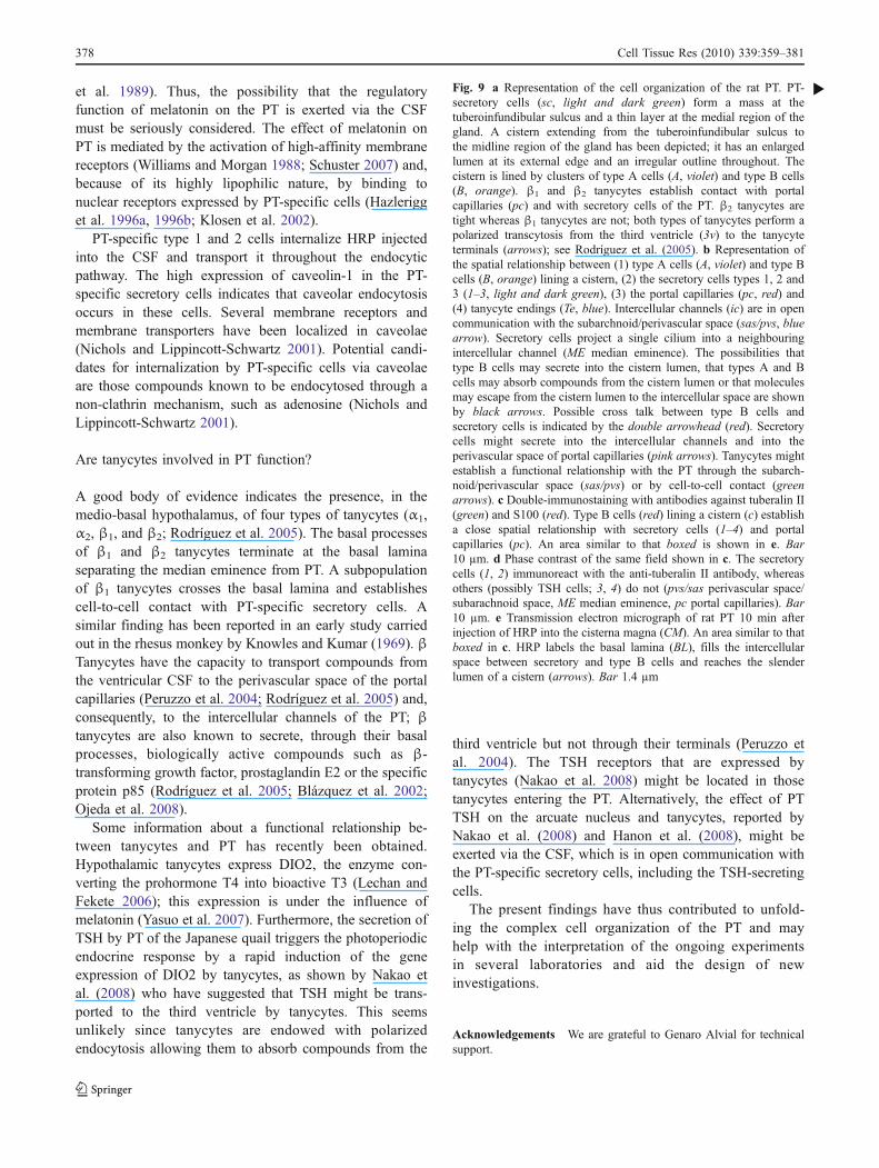
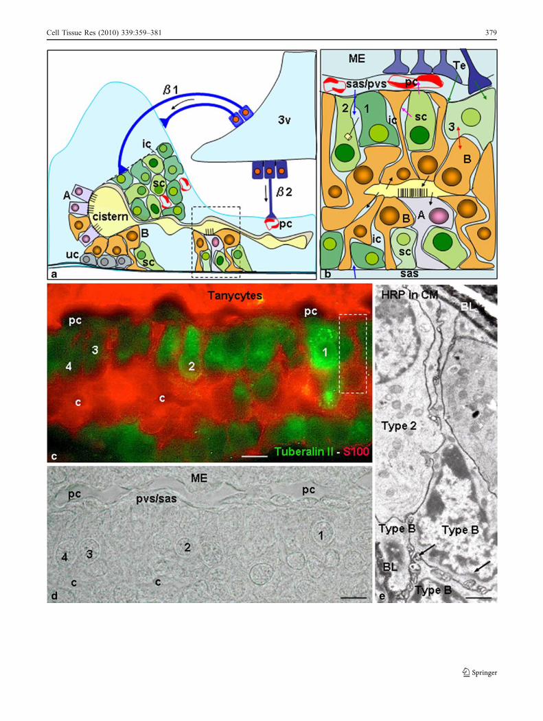
Fig. 9 a Representation of the cell organization of the rat PT. PT-secretory cells (sc, light and dark green) form a mass at thetuberoinfundibular sulcus and a thin layer at the medial region of thegland. A cistern extending from the tuberoinfundibular sulcus tothe midline region of the gland has been depicted; it has an enlargedlumen at its external edge and an irregular outline throughout. Thecistern is lined by clusters of type A cells (A, violet) and type B cells(B, orange). β1 and β2 tanycytes establish contact with portalcapillaries (pc) and with secretory cells of the PT. β2 tanycytes aretight whereas β1 tanycytes are not; both types of tanycytes perform apolarized transcytosis from the third ventricle (3v) to the tanycyteterminals (arrows); see Rodríguez et al. (2005). b Representation ofthe spatial relationship between (1) type A cells (A, violet) and type Bcells (B, orange) lining a cistern, (2) the secretory cells types 1, 2 and3 (1–3, light and dark green), (3) the portal capillaries (pc, red) and(4) tanycyte endings (Te, blue). Intercellular channels (ic) are in opencommunication with the subarchnoid/perivascular space (sas/pvs, bluearrow). Secretory cells project a single cilium into a neighbouringintercellular channel (ME median eminence). The possibilities thattype B cells may secrete into the cistern lumen, that types A and Bcells may absorb compounds from the cistern lumen or that moleculesmay escape from the cistern lumen to the intercellular space are shownby black arrows. Possible cross talk between type B cells andsecretory cells is indicated by the double arrowhead (red). Secretorycells might secrete into the intercellular channels and into theperivascular space of portal capillaries (pink arrows). Tanycytes mightestablish a functional relationship with the PT through the subarch-noid/perivascular space (sas/pvs) or by cell-to-cell contact (greenarrows). c Double-immunostaining with antibodies against tuberalin II(green) and S100 (red). Type B cells (red) lining a cistern (c) establisha close spatial relationship with secretory cells (1–4) and portalcapillaries (pc). An area similar to that boxed is shown in e. Bar10 µm. d Phase contrast of the same field shown in c. The secretorycells (1, 2) immunoreact with the anti-tuberalin II antibody, whereasothers (possibly TSH cells; 3, 4) do not (pvs/sas perivascular space/subarachnoid space, ME median eminence, pc portal capillaries). Bar10 µm. e Transmission electron micrograph of rat PT 10 min afterinjection of HRP into the cisterna magna (CM). An area similar to thatboxed in c. HRP labels the basal lamina (BL), fills the intercellularspace between secretory and type B cells and reaches the slenderlumen of a cistern (arrows). Bar 1.4 µm
b
378 Cell Tissue Res (2010) 339:359–381
Cell Tissue Res (2010) 339:359–381 379
References
Aguado LI, Schoebitz K, Rodríguez EM (1981) Intercellular channelsin the pars tuberalis and their relationship to the subarachnoidspace. Cell Tissue Res 218:345–354
Arvan P, Castle D (1998) Sorting and storage during secretory granulebiogenesis: looking backward and looking forward. Biochem J332:593–610
Barret P, Messager S, Schuster C, Moar KM, Mercer JG, Morgan PJ(2002) Pituitary adenylate cyclase-activating polypeptide acts asa paracrine regulator of melatonin-responsive cells of the ovinepars tuberalis. Endocrinology 143:2366–2375
Bergmann M, Wittkowski W, Hoffmann K (1989) Ultrastructurallocalization of the thyrotropin (TSH)-like immunoreactivity inspecific secretory cells of the hypophyseal pars tuberalis of theDjungarian hamster, Phodopus sungorus. Cell Tissue Res256:649–652
Blázquez JL, Guerra M, Pastor F, Peruzzo B, Amat P, Rodríguez EM(2002) Antibodies obtained by xenotransplantation of organcul-tured median eminence specifically recognize hypothalamictanycytes. Cell Tissue Res 308:241–253
Bloom GS, Brashear TA (1989) A novel 58-kDa protein associateswith the Golgi apparatus and microtubules. J Biol Chem264:16083–16092
Bockers TM, Sourgens H, Wittkowski W, Jekat A, Pera F (1990)Changes in TSH-immunoreactivity in the pars tuberalis and parsdistalis of the fetal rat hypophysis following maternal adminis-tration of propylthiouracil and thyroxin. Cell Tissue Res260:403–410
Bockmann J, Bockers TM, Vennemann B, Niklowitz P, Muller J,Wittkowski W, Sabel B, Kreutz MR (1996) Short photoperiod-dependent down-regulation of thyrotropin-alpha and -beta inhamster pars tuberalis-specific cells is prevented by pinealecto-my. Endocrinology 137:1804–1813
Bockmann J, Böckers TM, Winter C, Wittkowski W, Winterhoff H,Deufel TH, Kreuts R (1997) Thyrotropin expression in hypo-physeal pars tuberalis-specific cells is 3, 5, 3″-triiodothyronine,thyrotropin-releasing hormone, and Pit-1 independent. Endocri-nology 138:1019–1928
Borgonovo B, Ouwendijk J, Solimena M (2006) Biogenesis ofsecretory granules. Curr Opin Cell Biol 18:365–370
Cameron E, Foster CL (1972) Some light and electron microscopicalobservations on the pars tuberalis of the pituitary gland of therabbit. J Endocrinol 54:505–511
Dardente H, Klosen P, Pévet P, Masson-Pévet M (2003) MT1melatonin receptor mRNA expressing cells in the pars tuberalisof the European hamster: effect of photoperiod. J Neuroendoc-rinol 15:778–786
Dellmann HD, Stoeckel ME, Hindelang-Gertner C, Porte A, StutinskyF (1974) A comparative ultrastructural study of the pars tuberalisof various mammals, the chicken and the newt. Cell Tissue Res148:313–329
Denef C (2008) Paracrinicity: the story of 30 years of cellular pituitarycrosstalk. J Neuroendocrinol 20:1–70
Dupré SM, Burt DW, Talbot R, Downing A, Mouzaki D, WaddingtonD, Malpaux B, Davis JR, Lincoln GA, Loudon AS (2008)Identification of melatonin-regulated genes in the ovine pituitarypars tuberalis, a target site for seasonal hormone control.Endocrinology 149:5527–5539
Fauquier T, Lacampagne A, Travo P, Bauer K, Mollard P (2002)Hidden face of the anterior pituitary. Trends Endocrinol Metab13:304–309
Ferrara N, Schweigerer L, Neufeld G, Mitchell R, Gospodarowicz D(1987) Pituitary follicular cells produce basic fibroblast growthfactor. Proc Natl Acad Sci USA 84:5773–5777
Fitzgerald KT (1979) The structure and function of the pars tuberalisof the vertebrate adenohypophysis. Gen Comp Endocrinol37:383–399
Gall C von, Garabette ML, Kell CA, Frenzel S, Dehghani F, Schumm-Draeger PM,Weaver DR, Korf HW, HastingsMH, Stehle JH (2002)Rhythmic gene expression in pituitary depends on heterologoussensitization by the neurohormone melatonin. Nat Neurosci 5:234–238
Gall C von, Weaver DR, Moek J, Jilg A, Stehle JH, Korf HW (2005)Melatonin plays a crucial role in the regulation of rhythmic clockgene expression in the mouse pars tuberalis. Ann NY Acad Sci1040:508–511
Gao YS, Alvarez C, Nelson DS, Sztul E (1998) Molecular cloning,characterization, and dynamics of rat formiminotransferasecyclodeaminase, a Golgi-associated 58-kDa protein. J Biol Chem273:33825–33834
Gospodarowicz D, Lau K (1989) Pituitary follicular cells secrete bothvascular endothelial growth factor and follistatin. BiochemBiophys Res Commun 165:292–298
Graham ES, Webster CA, Hazlerigg DG, Morgan PJ (2002) Evidencefor the biosynthesis of a prolactin-releasing factor from the ovinepars tuberalis, which is distinct from thyrotropin-releasinghormone. J Neuroendocrinol 14:945–954
Gross DS (1984) The mammalian hypophyseal pars tuberalis: acomparative immunocytochemical study. Gen Comp Endocrinol56:283–298
Guerra M, Rodríguez EM (2001) Identification, cellular and subcel-lular distribution of 21 and 72 kDa proteins (tuberalins?) secretedby specific cells of the pars tuberalis. J Endocrinol 168:363–379
Guerra M, Rodríguez EM (2009) Expression of tuberalin II, α-subunit(GSU) and β-TSH in the pars tuberalis of the rat. Immunocyto-chemical evidence for PT-specific cell types. Neuroendocrinolo-gy (in press)
Hanon EA, Lincoln GA, Fustin JM, Dardente H, Masson-Pévet M,Morgan PJ, Hazlerigg DG (2008) Ancestral TSH mechanismsignals summer in a photoperiodic mammal. Curr Biol 18:1147–1152
Hazlerigg DG, Hastings MH, Morgan PJ (1996a) Production of aprolactin releasing factor by the ovine pars tuberalis. J Neuro-endocrinol 8:489–492
Hazlerigg DG, Barrett P, Hastings MH, Morgan PJ (1996b) Arenuclear receptors involved in pituitary responsiveness to melato-nin? Mol Cell Endocrinol 123:53–59
Inoue K, Couch EF, Takano K, Ogawa S (1999) The structure andfunction of folliculo-stellate cells in the anterior pituitary gland.Arch Histol Cytol 62:205–218
Jilg A, Moek J, Weaver DR, Korf HW, Stehle JH, Gall C von (2005)Rhythms in clock proteins in the mouse pars tuberalis depend onMT1 melatonin receptor signalling. Eur J NeuroSci 22:2845–2854
Kameda Y (1990) Occurrence of colloid-containing follicles andciliated cysts in the hypophysial pars tuberalis from guinea pigsof various ages. Am J Anat 188:185–198
Kameda Y, Miura M, Ohno S (2000) Expression of the commonalpha-subunit mRNA of glycoprotein hormones during the chickpituitary organogenesis, with special reference to the parstuberalis. Cell Tissue Res 299:71–80
Kanematsu N, Mori Y, Hayashi S, Hoshino K (1989) Presence of adistinct 24-hour melatonin rhythm in the ventricular cerebrospi-nal fluid of the goat. J Pineal Res 7:143–152
Kell CA, Stehle JH (2005) Just the two of us: melatonin andadenosine in rodent pituitary function. Ann Med 37:105–120
Klosen P, Bienvenu C, Demarteau O, Dardente H, Guerrero H, Pévet P,Masson-Pévet M (2002) The mt1 melatonin receptor and RORbetareceptor are co-localized in specific TSH-immunoreactive cells inthe pars tuberalis of the rat pituitary. J Histochem Cytochem50:1647–1657
380 Cell Tissue Res (2010) 339:359–381
Knowles FRS, Kumar A (1969) Structural changes related toreproduction in the hypothalamus and in the pars tuberalis ofthe rhesus monkey. Part I: the hypothalamus. Part II: the parstuberalis. Philos Trans R Soc Lond Biol 256:357–375
Kurotani R, Tahara S, Sanno N, Teramoto A, Mellon PL, Inoue K,Yoshimura S, Osamura RY (1999) Expression of Ptx1 in the adultrat pituitary glands and pituitary cell lines: hormone-secreting cellsand folliculo-stellate cells. Cell Tissue Res 298:55–61
Lechan RM, Fekete C (2006) The TRH neuron: a hypothalamicintegrator of energy metabolism. Prog Brain Res 153:209–235
Morgan PJ, King TP, Lawson W, Davidson G (1991) Ultrastructure ofmelatonin-responsive cells in the ovine pars tuberalis. Cell TissueRes 263:529–534
Morgan PJ, Barrett P, Davidson G, Lawson W, Hazlerigg D (1994)p72, a marker protein for melatonin action in ovine pars tuberaliscells: its regulation by protein kinase A and protein kinase C anddifferential secretion relative to prolactin. Neuroendocrinology59:325–335
Nakao N, Ono H, Yamamura T, Anraku T, Takagi T, Higashi K, Yasuo S,Katou Y, Kageyama S, Uno Y, Kasukawa T, Ligo M, Sharp PJ,Iwasawa A, Suzuki Y, Sugano S, Niimi T, Mizutani M, Namikawa T,Ebihara S, Ueda HR, Yoshimura T (2008) Thyrotrophin in the parstuberalis triggers photoperiodic response. Nature 452:317–322
Nichols BJ, Lippincott-Schwartz J (2001) Endocytosis withoutclathrin coats. Trends Cell Biol 11:406–412
Ojeda SR, Lomniczi A, Sandau US (2008) Glial-gonadotrophinhormone (GnRH) neurone interactions in the median eminenceand the control of GnRH secretion. J Neuroendocrinol 20:732–742
Ono H, Hoshino Y, Yasuo S, Watanabe M, Nakane Y, Murai A,Ebihara S, Korf HW, Yoshimura T (2008) Involvement ofthyrotropin in photoperiodic signal transduction in mice. ProcNatl Acad Sci USA 105:18238–18242
Oomizu S, Chaturvedi K, Sarkar DK (2004) Folliculostellate cellsdetermine the susceptibility of lactotropes to estradiol’s mitogen-ic action. Endocrinology 145:1473–1480
Ozawa H, Kurosumi K (1989) Postnatal development of thyrotrophsin the rat anterior pituitary as studied by immunogold electronmicroscopy. Anat Embryol (Berl) 180:207–212
Pazour GJ, Agrin N, Leszyk J, Witman GB (2005) Proteomic analysisof a eukaryotic cilium. J Cell Biol 170:103–113
Peruzzo B, Pastor FE, Blázquez JL, Amat P, Rodríguez EM (2004)Polarized endocytosis and transcytosis in the hypothalamictanycytes of the rat. Cell Tissue Res 317:147–164
Praetorius HA, Spring KR (2005) A physiological view of the primarycilium. Annu Rev Physiol 67:515–529
Reppert SM, Perlow MJ, Tamarkin L, Klein DC (1979) A diurnalmelatonin rhythm in primate cerebrospinal fluid. Endocrinology104:295–301
Rodríguez EM (1969) Fixation of the central nervous system byperfusion of the cerebral ventricles with a threefold aldehydemixture. Brain Res 15:395–412
Rodríguez EM, González CB, Delannoy L (1979) Cellular organiza-tion of the lateral and postinfundibular regions of the medianeminence in the rat. Cell Tissue Res 201:377–408
Rodríguez EM, Blázquez JL, Pastor F, Pelaez B, Peña P, Peruzzo B,Amat P (2005) Hypothalamic tanycytes: a key component ofbrain-endocrine interaction. Int Rev Cytol 247:89–164
Sakai T, Inoue K, Kurosumi K (1992) Light and electron microscopicimmunocytochemistry of TSK-like cells occurring in the parstuberalis of the adult male rat pituitary. Arch Histol Cytol55:151–157
Sakai T, Sakamoto S, Ijima K, Matsubara K, Kato Y, Inoue K (1999)Characterization of TSH-positive cells in foetal rat pars tuberalisthat fail to express Pit-1 factor and thyroid hormone beta2receptors. J Neuroendocrinol 11:187–193
Satir P, Christensen S (2008) Structure and function of mammaliancilia. Histochem Cell Biol 129:687–693
Schuster C (2007) Sites and mechanisms of action of melatonin inmammals: the MT1 and MT2 receptors. J Soc Biol 201:85–96
Skinner DC, Malpaux B (1999) High melatonin concentrations inthird ventricular cerebrospinal fluid are not due to Galen veinblood recirculating through the choroid plexus. Endocrinology140:4399–4405
Sternberger LA, Hardy PH, Cuculis JJ, Meyer HG (1970) Theunlabelled antibody-enzyme method of immunohistochemistry:preparation and properties of a soluble antigen-antibody complex(horseradish-peroxidase-antiperoxidase) and its use in identifica-tion of spirochetes. J Histochem Cytochem 18:315–345
Stoeckel ME, Hindelang C, Klein MJ, Poissonnier M, Felix JM(1994) Expression of the alpha-subunit of glycoprotein hormonesin the pars tuberalis-specific glandular cells in rat, mouse andguinea pig. Cell Tissue Res 278:617–624
Tu HM, Kim SW, Salvatore D, Bartha T, Legradi G, Larsen PR,Lechan RM (1997) Regional distribution of type 2 thyroxinedeiodinase messenger ribonucleic acid in rat hypothalamus andpituitary and its regulation by thyroid hormone. Endocrinology138:3359–3368
Wagner GC, Johnston JD, Tournier BB, Ebling FJ, Hazlerigg DG(2007) Melatonin induces gene-specific effects on rhythmicmRNA expression in the pars tuberalis of the Siberian hamster(Phodopus sungorus). Eur J Neurosci 25:485–490
Williams LM, Morgan PJ (1988) Demonstration of melatonin bindingsites on the pars tuberalis of the rat. J Endocrinol 119:R1–R3
Wittkowski W, Bockmann J, Kreutz MR, Böckers T (1999) Cell andmolecular biology of the pars tuberalis of the pituitary. Int RevCytol 185:157–194
Wong AO, Li WS, Lee EK, Leung MY, Tse LY, Chow BK, Lin HR,Chang JP (2000) Pituitary adenylate cyclase activating polypep-tide as a novel hypophysiotropic factor in fish. Biochem CellBiol 78:329–343
Yasuo S, Yoshimura T, Ebihara S, Korf HW (2007) Temporaldynamics of type 2 deiodinase expression after melatonininjections in Syrian hamsters. Endocrinology 148:4385–4392
Yokoyama T (2004) Motor or sensor: a new aspect of primary ciliafunction. Anat Sci Int 79:47–54
Yoshimura F, Nogami H, Yashiro T (1982) Fine structural criteria forpituitary thyrotrophs in immature and mature rats. Anat Rec204:255–263
Cell Tissue Res (2010) 339:359–381 381