carbon monoxide prevents ventilator-induced lung injury via caveolin-1*
TRANSCRIPT

Carbon monoxide prevents ventilator induced lung injury viacaveolin-1
Alexander Hoetzel, MD, Rene Schmidt, MD, Simone Vallbracht, PhD, Ulrich Goebel, M.D.,Tamas Dolinay, MD, Hong Pyo Kim, PhD, Emeka Ifedigbo, Stefan W. Ryter, PhD, andAugustine M. K. Choi, MDDepartment of Medicine, Division of Pulmonary, Allergy, and Critical Care Medicine, University ofPittsburgh Medical Center, Pittsburgh, PA, USA (AH, TD, HPK, EI, SWR), Department ofAnaesthesiology and Critical Care Medicine, University Hospital Freiburg, Freiburg, Germany(AH, RS, SV, UG), and Pulmonary and Critical Care Medicine, Brigham and Women's Hospital,Harvard Medical School, 75 Francis Street, Boston, MA 02115, USA (TD, HPK, EI, SWR, AMKC)
AbstractObjectives—Carbon monoxide (CO) can confer anti-inflammatory protection in rodent modelsof ventilator-induced lung injury (VILI). Caveolin-1 exerts a critical role in cellular responses tomechanical stress, and has been shown to mediate cytoprotective effects of CO in vitro. We soughtto determine the role of caveolin-1 in lung susceptibility to VILI in mice. Furthermore, weassessed the role of caveolin-1 in the tissue protective effects of CO in the VILI model.
Design—Prospective experimental study
Setting—University laboratory
Subjects—Wild type (wt) and caveolin-1 deficient (cav-−/−) mice
Interventions—Mice were subjected to tracheostomy and arterial cannulation. Wt and cav-1−/−
mice were ventilated with a tidal volume of 12 ml/kg body weight and a frequency of 80/min for 5min as control, or for 8h with air in the absence or presence of CO (250 parts per million).Bronchoalveolar lavage (BAL) and histology were used to determine lung injury. Lung sections orhomogenates were analyzed for caveolin-1 expression by immunohistochemical staining orWestern Blotting, respectively.
Measurements and Main Results—Ventilation led to an increase in BAL proteinconcentration, cell count, neutrophil recruitment, and edema formation that was prevented in thepresence of CO. While ventilation alone slightly induced caveolin-1 expression in epithelial cells,the application of CO during the ventilation significantly increased the expression of caveolin-1.In comparison to wt mice, mechanical ventilation of cav-1−/− mice led to a significantly higherdegree of lung injury as compared to wt mice. In contrast to its effectiveness in wt mice, CO-administration failed to reduce lung injury markers in cav-1−/− mice.
Conclusions—Caveolin-1 null mice are more susceptible to VILI. Carbon monoxide executeslung protective effects during mechanical ventilation that are dependent in part, on caveolin-1expression.
Correspondence and requests for reprints should be addressed to: Augustine M.K. Choi, M.D., Pulmonary and Critical CareMedicine, Brigham and Women's Hospital, Harvard Medical School, 75 Francis Street, Boston, MA 02115, USA, phone: (617)732-7599, Fax: (617) 732-7421, [email protected] remaining authors have not disclosed any potential conflicts of interest.
NIH Public AccessAuthor ManuscriptCrit Care Med. Author manuscript; available in PMC 2011 May 3.
Published in final edited form as:Crit Care Med. 2009 May ; 37(5): 1708–1715. doi:10.1097/CCM.0b013e31819efa31.
NIH
-PA Author Manuscript
NIH
-PA Author Manuscript
NIH
-PA Author Manuscript

Keywordsventilator induced lung injury; mechanical ventilation; carbon monoxide; caveolin-1;mechanotransduction; acute lung injury
INTRODUCTIONMechanical ventilation is commonly used in the clinical management of respiratory failure.Despite recent progress in reducing mechanical ventilation-associated mortality byemploying lower tidal volumes in patients (1), ventilator induced lung injury (VILI) remainsa major problem in the intensive care unit. Ventilator associated lung injury arises fromcyclic stretch to the lung imposed during mechanical ventilation, which disrupts cellularmembranes, increases oxidative stress, induces pro-inflammatory cascades, and maysubsequently lead to multiple organ failure (2). Therefore, supplementary therapeuticoptions, other than modulation of the ventilator setting, need to be developed in order tominimize VILI.
Over the last decade, carbon monoxide (CO) has been explored as a potential therapeuticagent in many disease models. Despite its noxious properties at elevated concentration,application of low dose CO exerts anti-inflammatory, anti-proliferative, anti-apoptotic, andanti-oxidative effects in preclinical models of inflammation, sepsis, ischemia/reperfusion (I/R)-injury, transplantation, among others (reviewed in (3, 4)). The organ protective action ofinhaled CO is particularly effective in animal models of lung injury and disease. Forinstance, hyperoxic lung injury or bleomycin-induced pulmonary fibrosis were diminishedin response to CO treatment in rodents (5, 6). Our laboratory previously demonstrated potentanti-inflammatory effects of inhaled CO in VILI using the combination of high-tidal volumeventilation and bacterial lipopolysaccharide (LPS) treatment in a rat model (7). However,the mechanism(s) by which CO prevents VILI remain incompletely understood.
CO potentially acts by modulating intracellular signal transduction pathways depending onthe injury model. For example, guanylate cyclase-dependent production of cyclic-guanosinemonophosphate has been implicated in the vascular effects of CO, whereas mitogenactivated protein kinases (MAPK), and peroxisome proliferator-activated receptor-γ, amongothers, have been implicated in the anti-inflammatory effects of this gas (8, 9). Recently, wefound that the anti-proliferative effect of CO in smooth muscle cells was mediated bycaveolin-1 (10).
Caveolin-1, a 22 kDa protein, is the major structural protein of caveolae, small 50–100 nmflask-shaped invaginations of the plasma membrane. Caveolae, which occur abundantly inlung endothelial cells, type I pneumocytes, murine alveolar macrophages, and fibroblasts,facilitate endocytosis (11, 12). This mechanism is of major importance for the uptake ofproteins into cells and the first step of transporting proteins through cellular barriers(transcytosis). For instance, caveolae-mediated transcytosis is thought to be the primarymechanism of carrying albumin across continuous endothelial cell barriers (13). Impairedclearance of proteins plays a pivotal role in lung edema and lung injury, and worsensoutcome (14). In addition to inducing the biogenesis of caveolae, caveolin-1 plays a pivotalrole in mediating signaling pathways involved in inflammation (12, 15). Interestingly,caveolin-1 has also been shown to mediate cell stretch-related effects in endothelial cells(16). To date, this has not been studied in lung epithelial cells which are compromised bycyclic stretch during mechanical ventilation.
Hoetzel et al. Page 2
Crit Care Med. Author manuscript; available in PMC 2011 May 3.
NIH
-PA Author Manuscript
NIH
-PA Author Manuscript
NIH
-PA Author Manuscript
To explore the significance of caveolin-1 in the lung during mechanical ventilation and todefine its role in the protective effects of CO on VILI, we have employed a mouse model ofVILI at physiologically relevant tidal volumes. In this study we demonstrate that COreduces lung injury after mechanical ventilation, and that this protective effect depended oninduction of caveolin-1. Furthermore, we demonstrate that caveolin-1 null mice are moresusceptible to VILI. Our results implicate that caveolae represent essential cellular structuresthat limit stretch-induced injury.
MATERIALS and METHODSAnimals
All animals were housed in accordance with guidelines from the American Association forLaboratory Animal Care. The Animal Care and Use Committee of the University ofPittsburgh approved the protocols. Male C57/BL6 mice (wild type, wt) (JacksonLaboratories, Bar Harbor, ME) or caveolin-1 deficient (cav-1−/−) mice (Taconic, Hudson,NY) were anesthetized with ketamine (80 mg/kg, i.p.) and acepromazine (1 mg/kg, i.p.) andplaced on a heating pad. A polyethylene catheter was inserted into the left carotid artery fordirect blood pressure monitoring as well as for blood gas sampling, and a tracheostomy wasestablished using a 20 G catheter. A rodent ventilator (Voltekenterprises, Toronto, Canada)was set to a tidal volume of 12 ml/kg body weight, frequency of 80/min, positive end-expiratory pressure (PEEP) of 2 cm H2O and connected to the tracheal canula. Muscularrelaxation was achieved by applying pancuronium 2 mg/kg i.p. A 0.7 ml saline bolus wasinjected i.p. to compensate for evaporation during ventilation. Animals were randomized toreceive ventilation with room air, or room air supplemented with carbon monoxide at aconcentration of 250 parts per million (ppm) for 8h. Control animals were sham-operatedand ventilated shortly for 5 minutes. Blood samples were withdrawn from control animalsafter 5 minutes and from ventilated animals at 1 h after onset of mechanical ventilation andblood gases were measured using an automated blood gas analyzer (ABL5, Radiometer,Bronshoi, Denmark). Blood gases revealed a mean pH 7.31+/−0.004, pO2 82+/−1.5 mmHg,and 34 +/−0.4 mmHg without differences between groups. After 8h of mechanicalventilation, the animals were sacrificed. Tissue and blood samples were snap frozen forsubsequent analysis. To investigate the time course of cav-1 expression, additional animals(n=2/group) were ventilated with or without CO for 1 or 4 hours.
Bronchoalveolar lavageAt the end of each experiment, a bronchoalveolar lavage (BAL) was performed using 0.8 mlphosphate buffered saline (PBS). The recovered volume was centrifuged and the supernatantanalyzed for protein concentration (Biorad Assay, Biorad, Hercules, CA). Furthermore, thepellet was re-dissolved in PBS and subsequently the number of total cells as well as thefraction of neutrophils were determined.
Polymerase chain reaction (PCR)The right lower lung lobe was homogenized in TRIZOL in order to extract total RNA.Caveolin-1 mRNA was determined with a forward primer 5'-TTCAGGGAGGGGTGTCA-3'and a reverse primer 5'-AAAGTAGGTAGCAGGTTGGTAAAG-3'. GAPDH was used asthe standard gene with a forward primer 5'-ACCACAGTCCATGCCATCAC-3' and areverse primer 5'-TCCACCCTGTTGCTGTA-3'. In order to analyze the linear range ofamplification, 23 cycles were used. PCR samples were run on a 1% agarose gel containingethidium bromide and photographed. Densitometric analyses were performed using ImageJsoftware (National Institute of Health, www.rsb.info.nih.gov).
Hoetzel et al. Page 3
Crit Care Med. Author manuscript; available in PMC 2011 May 3.
NIH
-PA Author Manuscript
NIH
-PA Author Manuscript
NIH
-PA Author Manuscript

ImmunoblottingThe right upper lung lobe was homogenized in 30 mM Tris Base including completeprotease inhibitors (Roche Diagnostics, Mannheim, Germany). After determination of theprotein content, equal amounts were loaded onto a 4–12% SDS page (Invitrogen, Carlsbad,CA, USA) and blotted onto a nitrocellulose membrane. The membranes were incubated withcaveolin-1 (1:2000, sc-894, Santa Cruz, Santa Cruz, CA) overnight at 4°. After severalwashing steps, membranes were incubated for 1 h with the appropriate secondary antibody,detected (Supersignal, Peirce, Rockford, IL) and exposed to radiographic films. Re-blottingthe membranes with β-actin (Sigma, Saint Louis, MO, USA) served to control for equalloading and transfer.
ImmunohistochemistryImmunostaining of lung tissue was performed in the left lung lobe that was infused withOCT (Sakura, Torrance, CA) and stored at –80°C. Cryosections were subjected tohematoxylin & eosin staining (H&E). Caveolin-1 staining and confocal single cellmicroscopy were conducted using the above mentioned antibody (1:50). Secondary antibodyand immunofluorescence staining were conducted according to the Center of BiologicalImaging protocol (CBI, Pittsburgh, PA, www.cbi.pitt.edu). The color green indicatescaveolin-1, red indicates f-actin / phalloidin, and blue (dapi) indicates nuclei.
Cytokine/Chemokine measurementsBAL aliquots were analyzed using interleukin-6 (IL-6) and macrophage inflammatoryprotein-2 (MIP-2) ELISA kits (R&D Systems, Minneapolis, MN) according to themanufacturer's instructions.
Statistical analysisData represent means +/− standard error of means (SEM) for n=6/group. Statistical analyseswere performed using the analysis of variances (ANOVA) for multiple group comparisonand the Student-Newman-Keuls posthoc test (Sigmastat statistical software, Systat Inc.,Erkrath, Germany). For two group comparison, the Student-t-test was used to compareparametric, and the Mann-Whitney Rank Sum test to compare non-parametric data. p<0.05was considered significant.
RESULTSEffect of ventilation and carbon monoxide on lung injury
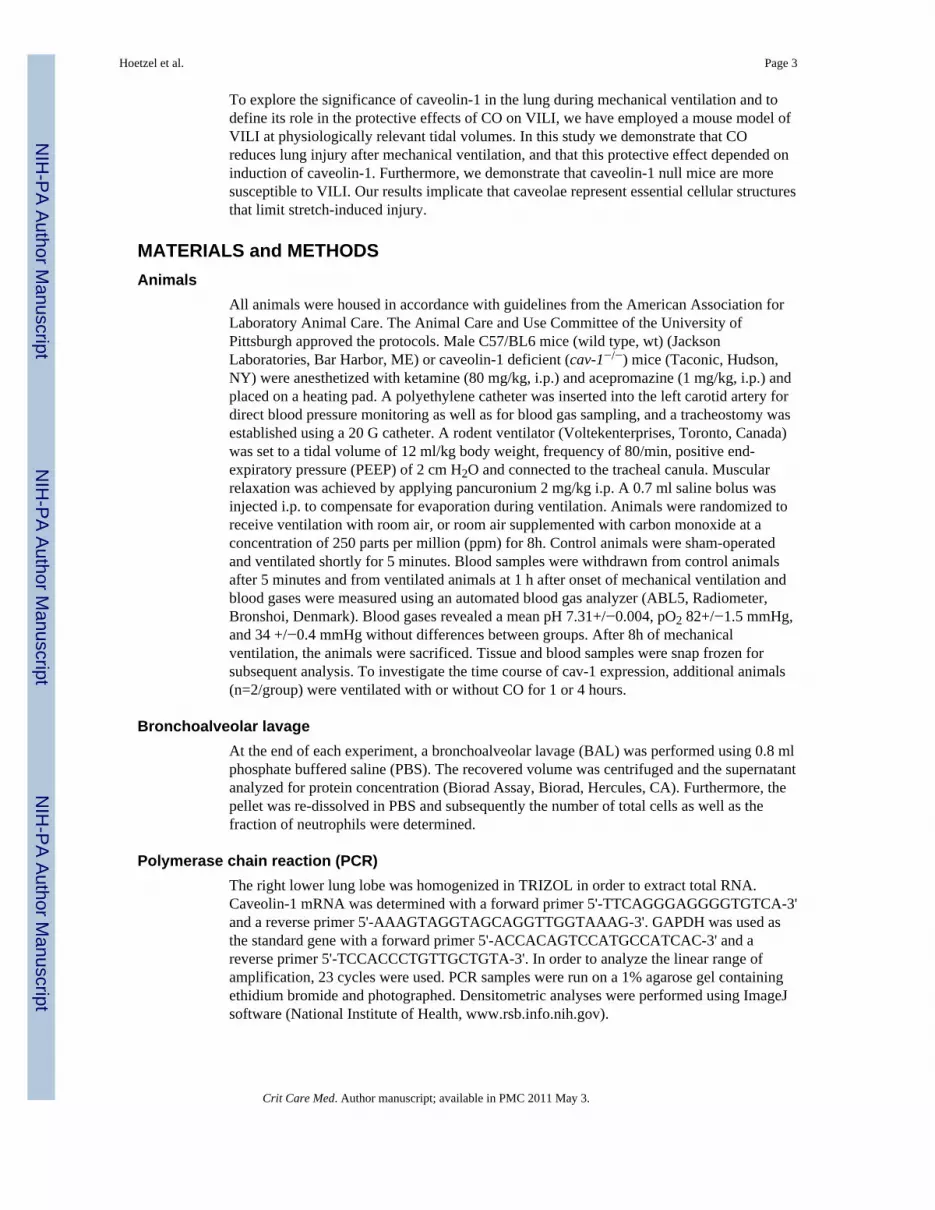
We used a total of 62 male C57/BL6 mice for the current study. Ventilation of wild-type(wt) mice with 12 ml/kg led to a significant increase in BAL protein concentrations after 8h,relative to control animals (Fig. 1a). Moreover, BAL cell count and the percentage ofneutrophils were notably elevated as a result of ventilation (Fig. 1b and c). In sharp contrast,the presence of carbon monoxide (CO, 250 ppm) during ventilation substantially diminishedthese injury markers (Fig. 1a–c) indicating that CO protected the lung from VILI.
Effect of ventilation and carbon monoxide on caveolin-1 expressionNext, we investigated the underlying mechanism(s) that mediate the protective effects of COapplication during mechanical ventilation. Recent in vitro experiments by our group havedemonstrated that certain protective effects of CO are transduced by caveolin-1 (10).Therefore, we analyzed the effect of ventilation in the absence and presence of CO on theregulation of caveolin-1. To quantify the modulation of caveolin-1 by ventilation or COadministration, PCR as well as immunoblots for caveolin-1 were performed using lungtissue homogenates. Figure 2 demonstrates increased accumulation of caveolin-1 mRNA in
Hoetzel et al. Page 4
Crit Care Med. Author manuscript; available in PMC 2011 May 3.
NIH
-PA Author Manuscript
NIH
-PA Author Manuscript
NIH
-PA Author Manuscript

response to mechanical ventilation that was even further upregulated in CO treated animals(Fig. 2a–b). The transcriptional upregulation of cav-1 was paralleled by increased proteinsynthesis that was detected by Western Blot analysis (Fig. 3a–b). To investigate the timecourse of cav-1 expression by ventilation and CO-treatment, additional animals wereventilated for 1 and 4 hours. Our results demonstrated an increased cav-1 protein synthesisas early as 4 hours after the onset of CO inhalation (Fig. 3c).
As shown in Figure 4 with immunofluorescence microscopy and confocal single cellmicroscopy, caveolin-1 (green immunofluorescence) was abundantly expressed in epithelialcells of untreated animals (Fig. 4a,d, arrow). Ventilation of these animals with 12 ml/kg for8h further increased caveolin-1 staining (Fig. 4b,e). Most interestingly, the administration ofCO clearly upregulated caveolin-1 in lung parenchymal cells (Fig. 4c,f).
Effect of caveolin-1 deficiency on lung injuryFirst, we confirmed cav-1 gene depletion in the knock out animals (Fig. 5a). Next, wedetermined the degree of lung injury in cav-1−/− mice as a result of 8h mechanicalventilation (Fig. 5b–c). Under control conditions, a slight increase in BAL proteinconcentrations was present in cav-1−/− animals. However, in these mice neither proteinconcentration nor total cell count were statistically different from wt mice. Theses results onlung injury parameters exclude the possibility that cav-1−/− mice might present severe pre-existing lung tissue damage (17). After mechanical ventilation, caveolin-1 deficiency led toa substantial increase of BAL protein and cell counts as compared to wt animals. While COexerted protective effects in wt mice and decreased the protein leakage and cellularinfiltration, this protection was absent in cav-1−/− mice (Fig. 5b–c). The protective effects ofCO inhalation in wild type and the deleterious consequences of cav-1 deficiency wereevident by H&E staining of lung sections (Fig. 6). In wt, H&E staining of control lungsshowed no obvious signs of lung injury (Fig. 6a). However, ventilation led to significantlung damage reflected by thickening of alveolar septae and infiltration of pro-inflammatorycells (Fig. 6b). In the presence of applied CO, both edema formation as well as the cellularinfiltration were absent despite ventilation (Fig. 6c). Interestingly, under control conditions,the lung histology of cav-1−/− mice (Fig. 6d) was comparable to that of wt mice (Fig. 6a).Mechanical ventilation led to massive lung injury in cav-1−/− mice (Fig. 5e) that was morepronounced than in wt mice (Fig. 6b). While CO exerted protective effects in wt mice (Fig.6c), these effects were absent in cav-1−/− mice (Fig. 6f).
Effect of caveolin-1 deficiency on neutrophils and cytokine releaseMost interestingly and contrary to our expectations, mechanical ventilation did not furtherrecruit neutrophils in cav-1−/− mice as compared to wt mice (Fig. 7a). However, COsignificantly reduced neutrophil influx in ventilated wt mice, but did not impact neutrophilinflux in cav-1−/− mice. To validate this observation, BAL cytokines and chemokines weremeasured (Fig. 7b–c). Interleukin-6 (IL-6) and macrophage inflammatory protein-2 (MIP-2)were elevated in ventilated wt mice as compared to control animals. The presence of COduring ventilation diminished both IL-6 as well as MIP-2 levels in wt mice. In cav-1−/−
mice, IL-6 levels were further increased in response to mechanical ventilation, relative to wtmice. CO application significantly reduced ventilation-induced IL-6 levels in both wt andcav-1−/− mice. In contrast, MIP-2 levels in response to ventilation were comparable betweenwt and cav-1−/− mice, and were not significantly reduced by CO in cav-1−/− mice.
DISCUSSIONIn the current study, we have demonstrated that mechanical ventilation with 12 ml/kg for 8hinduced severe lung injury as reflected by histological evaluation of lung sections, as well as
Hoetzel et al. Page 5
Crit Care Med. Author manuscript; available in PMC 2011 May 3.
NIH
-PA Author Manuscript
NIH
-PA Author Manuscript
NIH
-PA Author Manuscript

increased protein concentrations in the BAL, and infiltration of macrophages andneutrophils. CO administration at low concentration (250 ppm) significantly reduced alllung injury parameters.
The effectiveness of applying low dose CO in order to employ its organ protective effectshas been established in many in vitro and in vivo studies over the last decade (reviewed in(3, 4)). Presently, several ongoing clinical studies address the effectiveness of CO-treatmentin humans (NIH, www.clinicaltrials.gov). In animal experiments, CO confers tissueprotection through anti-proliferative, anti-oxidative, vasodilatory, anti-inflammatory, andmembrane stabilizing characteristics (18, 19). The latter two are most important regardingthe development of VILI. For instance, our laboratory has recently demonstrated thebeneficial effects of CO-administration against LPS-induced lung injury in overventilatedrats (7). The data in the present study shows that in a mouse model using moderate tidalvolumes (12 ml/kg), mechanical ventilation led to a significant degree of lung injury thatwas abolished in the presence of CO. Given these protective properties of CO duringmechanical ventilation, we attempted to identify the underlying mechanism. Signaling ofCO might be transduced by several pathways including cGMP (8), MAPK (18), orcaveolin-1, as recently described in vitro (10).
Our results demonstrate that mechanical ventilation induced the expression of caveolin-1mRNA and protein in the lung in vivo, which was dramatically further upregulated by theapplication of CO during ventilation. Caveolin-1 orchestrates membrane trafficking,endocytosis, regulation of cholesterol, and signal transduction in cellular growth andapoptosis (reviewed in (20)). Interestingly, in endothelial cells, caveolin-1 appears to play animportant role in mechanotransduction (16). Previous in vitro data on caveolin-1 expressionupon flow mediated shear stress in lung endothelial cells increased caveolin-1 density at theluminal plasma membrane (21). Lung caveolin-1 expression is not restricted to endothelialcells but includes type I epithelial cells (11). Here we show for the first time that mechanicalstress by the means of mechanical ventilation induced caveolin-1 in lung epithelial cells.Immunohistochemistry and confocal microscopy of epithelial cells demonstrated basalexpression of caveolin-1 in the alveolar luminal surface that was further enhanced byventilation. These cells cover more than 95% of the internal surface area, which is consistentwith our finding that increased expression of caveolin-1 at the luminal surface is paralleledby upregulation of caveolin-1 protein in whole lung homogenates.
We hypothesized that caveolin-1 upregulation by ventilation alone might reflect an adaptivemechanism in order to reduce stretch-induced lung injury. Therefore, we employed themouse VILI model in caveolin-1 deficient mice. Our results show clearly that caveolin-1deficiency aggravated lung injury in response to mechanical ventilation, most pronouncedwith respect to protein leakage, edema formation, and macrophage infiltration. It has beendescribed that cav-1−/− mice exert increased microvascular permeability (22) and pre-existing lung injury (17, 22–24) which may contribute to the susceptibility to VILI in thesemice. However, our results did not reveal significant alterations in lung morphology undercontrol conditions between wild type and cav-1−/−, with the exception of a trend in elevatedBAL protein concentrations in cav-1−/− controls without ventilation. The latter is mostlikely based on impaired caveolae-mediated transcytosis that is thought to be the primarytransport path for albumin at physiological concentration (13). Thus, the clearance ofalveolar proteins in the intact lung depends on caveolae (14). Most interestingly, caveolaemediated transcytosis at high protein concentrations, seems to play a minor role (14). In thisrespect, an increased nitric oxide dependent paracellular protein transport may compensatefor the lack of caveolae in cav-1−/− animals (22).
Hoetzel et al. Page 6
Crit Care Med. Author manuscript; available in PMC 2011 May 3.
NIH
-PA Author Manuscript
NIH
-PA Author Manuscript
NIH
-PA Author Manuscript

The precise mechanism responsible for the higher susceptibility to mechanical ventilation incav-1−/− mice remains unclear. Cav-1 serves as a major signaling protein, which interactswith and modulates the activation state of several major cellular signaling pathways, e.g.mitogen activated protein kinases, integrins, and nitric oxide synthases (NOS). For instance,it has been reported that cav-1 deficiency leads to activation of the endothelial isoform ofNOS thus overproducing nitric oxide (15). As a possible consequence, increased NO levelsmay have aggravated lung injury in response to ventilation in our model. However, whetherNO promotes or inhibits the development of VILI is still controversial (25–27). Studieswhich systematically investigate the influence of NO in a long term model of VILI withmoderate tidal volumes, as we applied, are not available.
We chose to measure neutrophil fraction, IL-6, and MIP-2 as indicators of a potentiallyaltered immunoresponse in cav-1 deficiency. Neutrophil infiltration is thought to play anessential role in the development of lung injury after mechanical ventilation (28). It isimportant to note that in this study, neutrophils were measured as a fraction of the sum ofmacrophages and neutrophils in the BAL. As mentioned earlier in this paper and as reportedpreviously by our laboratory, neutrophils are not detectable in control lungs and migrate intothe lungs upon mechanical ventilation (29). Intriguingly, neutrophil recruitment into the lungwas not further increased in cav-1−/− mice, and comparable to wt animals. These immunecells migrate upon stimulation from the endovascular to the alveolar space passing theendothelial and the epithelial cell barrier. It has been demonstrated by others that cav-1deficiency inhibits the endothelial transmigration of neutrophils upon pro-inflammatorystimuli (15, 30). In this regard, neutrophil migration depends at least in part on transcytosisthat is facilitated by a number of mediators (i.e. vascular endothelial growth factor-receptor,platelet endothelial adhesion molecule-1, or intercellular adhesion molecule-1). Thesemolecules are either located within or translocate to caveolae upon pro-inflammatory stimuli(31–33). Furthermore, it has been demonstrated, that endo- and transcytosis processes areimpaired in cav-1 deficiency (22). Also, neutrophils express caveolin-1 (34). The attachmentof neutrophils in cav-1−/− mice is severely inhibited and their ability to transmigrate overcellular barriers impaired (34). Therefore, inhibition of neutrophil migration would mostlikely explain the lack of a further neutrophil infiltration in cav-1−/− mice in our model.
The cytokine IL-6 and the small chemokine MIP-2 are known to be upregulated in responseto mechanical ventilation with high tidal volumes and represent major regulators ofneutrophil recruitment into the lung. In pilot experiments, we observed these cytokines andchemokines to be present at 8 h of mechanical ventilation (data not shown). Mostinterestingly, the release of the neutrophil chemoattractant MIP-2 upon mechanicalventilation in cav-1−/− animals was not further increased as compared to wt and paralleledthe migration pattern of neutrophils. These results are surprising, since MIP-2 is producedby macrophages which are outnumbered in cav-1 deficiency as compared to wt afterventilation. Nevertheless, cytokines and chemokines seem to be differentially regulated incav-1 deficiency upon pro-inflammatory stimuli (35). Regarding the influence of caveolin-1on MIP-2 release, we provide evidence that MIP-2 regulation is not dependent on cav-1expression and furthermore, an increase in MIP-2 production that would representaggravated lung injury seems to be prevented in cav-1 deficiency. Due to the lack of data onMIP-2 in cav-1 deficiency, we can only speculate on possible explanations for thisobservation. The release of IL-8, a functional human homologue of mouse MIP-2, inresponse to proinflammatory stimuli has been shown to be inhibited in vascular endothelialand renal epithelial cell lines with disrupted caveolae (36–38). Here, altered pathways suchas the MAPK, activation of sphingomyelinase, or toll like receptors might play a role.However, the role of caveolin-1 in the regulation of IL-8 is far from understood and itsimpact on MIP-2 in the lung in response to mechanical stress literally unknown.
Hoetzel et al. Page 7
Crit Care Med. Author manuscript; available in PMC 2011 May 3.
NIH
-PA Author Manuscript
NIH
-PA Author Manuscript
NIH
-PA Author Manuscript

In contrast to neutrophils and MIP-2, IL-6 production was substantially upregulated inventilated cav-1−/− mice as compared to wt. IL-6 is secreted by macrophages and might onone hand reflect the tremendous increase of macrophages in the BAL. Even though total cellcount in the BAL includes all cells containing nuclei, macrophages represent the majority ofthese cells. On the other hand, previous reports from us and others demonstrated elevatedIL-6 levels in cav-1−/− macrophage cell lines (12, 35). As reported earlier, LPS challenge ofa macrophage cell line resulted in significant IL-6 production that was even furtherincreased when cav-1 was silenced (12). Conversely, overexpression of cav-1 in these cellsled to a significant decline of IL-6 protein providing evidence for anti-inflammatory role ofcav-1. This assumption from in vitro data is in agreement with our in vivo results showingthe first time that IL-6 release after mechanical ventilation is pronounced in the absence ofcaveolin-1.
Since CO further upregulated caveolin-1 which correlated with increased protection fromVILI, we speculated that CO-protection requires caveolin-1. In fact, while CO exerted majorprotective effects in wild type mice, this was no longer the case in caveolin-1 deficiency.Protein leakage, edema formation as well as macrophage and neutrophil infiltration, whichwere prevented in wt mice by CO-administration, were not reduced by CO treatment incav-1−/− animals. However, cytokine upregulation (i.e., IL-6) was partially inhibited by COin both strains with comparable effectiveness. Thus, the inhibitory effect of CO on IL-6production appears to be independent of caveolin-1 in this model.
Previous studies from this laboratory have suggested a relationship between caveolin-1 andCO-mediated tissue protection. For example, CO application induced caveolin-1 inneointimal lesions of injured rat aorta coincident with a tissue protection (10). In addition,depletion of cav-1 using siRNA abolished the anti-proliferative effect of CO in pulmonarysmooth muscle cells. In agreement with previous studies, the present investigation indicatesthat CO requires caveolin-1 to execute its protective effects during mechanical ventilation.Furthermore, these studies identify caveolin-1 as a novel therapeutic target for modulatingthe outcome of VILI.
CONCLUSIONSIn summary, mechanical ventilation with moderate tidal volume led to a substantial degreeof lung injury that was prevented in the presence of carbon monoxide. Caveolin-1, the majorstructural protein of caveolae, plays a crucial role in limiting lung injury during mechanicalventilation and mediates the protective effects of carbon monoxide application.
AcknowledgmentsThis work was supported by a grant from the Deutsche Forschungsgemeinschaft to Alexander Hoetzel (DFG HO2464/1-1); by awards from the American Heart Association to S. W. Ryter, (AHA #0335035N), and H. P. Kim(AHA #0525552U) and NIH grants R01-HL60234, R01-HL55330, R01-HL079904, and P01-HL70807 awarded toA. M. K. Choi.
REFERENCES1. The Acute Respiratory Distress Syndrome Network: Ventilation with lower tidal volumes as
compared with traditional tidal volumes for acute lung injury and the acute respiratory distresssyndrome. N Engl J Med. 2000; 342:1301–1308. [PubMed: 10793162]
2. Belperio JA, Keane MP, Burdick MD, et al. Critical role for CXCR2 and CXCR2 ligands during thepathogenesis of ventilator-induced lung injury. J Clin Invest. 2002; 110:1703–1716. [PubMed:12464676]
Hoetzel et al. Page 8
Crit Care Med. Author manuscript; available in PMC 2011 May 3.
NIH
-PA Author Manuscript
NIH
-PA Author Manuscript
NIH
-PA Author Manuscript

3. Ryter SW, Alam J, Choi AM. Heme oxygenase-1/carbon monoxide: from basic science totherapeutic applications. Physiol Rev. 2006; 86:583–650. [PubMed: 16601269]
4. Hoetzel A, Dolinay T, Schmidt R, et al. Carbon Monoxide in Sepsis. Antioxid Redox Signal. 20075. Otterbein LE, Mantell LL, Choi AM. Carbon monoxide provides protection against hyperoxic lung
injury. Am J Physiol. 1999; 276:L688–L694. [PubMed: 10198367]6. Zhou Z, Song R, Fattman CL, et al. Carbon monoxide suppresses bleomycin-induced lung fibrosis.
Am J Pathol. 2005; 166:27–37. [PubMed: 15631997]7. Dolinay T, Szilasi M, Liu M, et al. Inhaled carbon monoxide confers antiinflammatory effects
against ventilator-induced lung injury. Am J Respir Crit Care Med. 2004; 170:613–620. [PubMed:15142867]
8. Morse D, Sethi J, Choi AM. Carbon monoxide-dependent signaling. Crit Care Med. 2001; 30:S12–S17. [PubMed: 11839940]
9. Bilban M, Bach FH, Otterbein SL, et al. Carbon monoxide orchestrates a protective responsethrough PPARgamma. Immunity. 2006; 24:601–610. [PubMed: 16713977]
10. Kim HP, Wang X, Nakao A, et al. Caveolin-1 expression by means of p38beta mitogen-activatedprotein kinase mediates the antiproliferative effect of carbon monoxide. Proc Natl Acad Sci U S A.2005; 102:11319–11324. [PubMed: 16051704]
11. Dahlin K, Mager EM, Allen L, et al. Identification of genes differentially expressed in rat alveolartype I cells. Am J Respir Cell Mol Biol. 2004; 31:309–316. [PubMed: 15205179]
12. Wang XM, Kim HP, Song R, et al. Caveolin-1 confers antiinflammatory effects in murinemacrophages via the MKK3/p38 MAPK pathway. Am J Respir Cell Mol Biol. 2006; 34:434–442.[PubMed: 16357362]
13. Mehta D, Malik AB. Signaling mechanisms regulating endothelial permeability. Physiol Rev.2006; 86:279–367. [PubMed: 16371600]
14. Hastings RH, Folkesson HG, Matthay MA. Mechanisms of alveolar protein clearance in the intactlung. Am J Physiol. 2004; 286:L679–L689.
15. Garrean S, Gao XP, Brovkovych V, et al. Caveolin-1 regulates NF-kappaB activation and lunginflammatory response to sepsis induced by lipopolysaccharide. J Immunol. 2006; 177:4853–4860. [PubMed: 16982927]
16. Yu J, Bergaya S, Murata T, et al. Direct evidence for the role of caveolin-1 and caveolae inmechanotransduction and remodeling of blood vessels. J Clin Invest. 2006; 116:1284–1291.[PubMed: 16670769]
17. Drab M, Verkade P, Elger M, et al. Loss of caveolae, vascular dysfunction, and pulmonary defectsin caveolin-1 gene-disrupted mice. Science. 2001; 293:2449–2452. [PubMed: 11498544]
18. Otterbein LE, Bach FH, Alam J, et al. Carbon monoxide has anti-inflammatory effects involvingthe mitogen-activated protein kinase pathway. Nat Med. 2000; 6:422–428. [PubMed: 10742149]
19. Farrugia G, Lei S, Lin X, et al. A major role for carbon monoxide as an endogenoushyperpolarizing factor in the gastrointestinal tract. Proc Natl Acad Sci U S A. 2003; 100:8567–8570. [PubMed: 12832617]
20. Cohen AW, Hnasko R, Schubert W, et al. Role of caveolae and caveolins in health and disease.Physiol Rev. 2004; 84:1341–1379. [PubMed: 15383654]
21. Rizzo V, Morton C, DePaola N, et al. Recruitment of endothelial caveolae intomechanotransduction pathways by flow conditioning in vitro. Am J Physiol. 2003; 285:H1720–H1729.
22. Schubert W, Frank PG, Woodman SE, et al. Microvascular hyperpermeability in caveolin-1 (−/−)knock-out mice. Treatment with a specific nitric-oxide synthase inhibitor, L-name, restores normalmicrovascular permeability in Cav-1 null mice. J Biol Chem. 2002; 277:40091–40098. [PubMed:12167625]
23. Razani B, Engelman JA, Wang XB, et al. Caveolin-1 null mice are viable but show evidence ofhyperproliferative and vascular abnormalities. J Biol Chem. 2001; 276:38121–38138. [PubMed:11457855]
24. Murata T, Lin MI, Huang Y, et al. Reexpression of caveolin-1 in endothelium rescues the vascular,cardiac, and pulmonary defects in global caveolin-1 knockout mice. J Exp Med. 2007; 204:2373–2382. [PubMed: 17893196]
Hoetzel et al. Page 9
Crit Care Med. Author manuscript; available in PMC 2011 May 3.
NIH
-PA Author Manuscript
NIH
-PA Author Manuscript
NIH
-PA Author Manuscript

25. Broccard AF, Feihl F, Vannay C, et al. Effects of L-NAME and inhaled nitric oxide on ventilator-induced lung injury in isolated, perfused rabbit lungs. Crit Care Med. 2004; 32:1872–1878.[PubMed: 15343015]
26. Choi WI, Quinn DA, Park KM, et al. Systemic microvascular leak in an in vivo rat model ofventilator-induced lung injury. Am J Respir Crit Care Med. 2003; 167:1627–1632. [PubMed:12663326]
27. Takenaka K, Nishimura Y, Nishiuma T, et al. Ventilator-induced lung injury is reduced intransgenic mice that overexpress endothelial nitric oxide synthase. Am J Physiol. 2006;290:L1078–L1086.
28. Karzai W, Cui X, Heinicke N, et al. Neutrophil stimulation with granulocyte colony-stimulatingfactor worsens ventilator-induced lung injury and mortality in rats. Anesthesiology. 2005;103:996–1005. [PubMed: 16249674]
29. Hoetzel A, Dolinay T, Vallbracht S, et al. Carbon monoxide protects against ventilator-inducedlung injury via PPAR-gamma and inhibition of Egr-1. Am J Respir Crit Care Med. 2008;177:1223–1232. [PubMed: 18356564]
30. Dejana E. The transcellular railway: insights into leukocyte diapedesis. Nat Cell Biol. 2006; 8:105–107. [PubMed: 16450004]
31. Burns AR, Smith CW, Walker DC. Unique structural features that influence neutrophil emigrationinto the lung. Physiol Rev. 2003; 83:309–336. [PubMed: 12663861]
32. Mathew R, Huang J, Shah M, et al. Disruption of endothelial-cell caveolin-1alpha/raft scaffoldingduring development of monocrotaline-induced pulmonary hypertension. Circulation. 2004;110:1499–1506. [PubMed: 15353500]
33. Millan J, Hewlett L, Glyn M, et al. Lymphocyte transcellular migration occurs through recruitmentof endothelial ICAM-1 to caveola- and F-actin-rich domains. Nat Cell Biol. 2006; 8:113–123.[PubMed: 16429128]
34. Hu G, Ye RD, Dinauer MC, et al. Neutrophil caveolin-1 expression contributes to mechanism oflung inflammation and injury. Am J Physiol. 2008; 294:L178–L186.
35. Medina FA, de Almeida CJ, Dew E, et al. Caveolin-1-deficient mice show defects in innateimmunity and inflammatory immune response during Salmonella enterica serovar Typhimuriuminfection. Infect Immun. 2006; 74:6665–6674. [PubMed: 16982844]
36. Walton KA, Gugiu BG, Thomas M, et al. A role for neutral sphingomyelinase activation in theinhibition of LPS action by phospholipid oxidation products. J Lipid Res. 2006; 47:1967–1974.[PubMed: 16775254]
37. Walton KA, Cole AL, Yeh M, et al. Specific phospholipid oxidation products inhibit ligandactivation of toll-like receptors 4 and 2. Arterioscler Thromb Vasc Biol. 2003; 23:1197–1203.[PubMed: 12775576]
38. Li H, Nord EP. Functional caveolae are a prerequisite for CD40 signaling in human renal proximaltubule cells. Am J Physiol. 2004; 286:F711–F719.
Hoetzel et al. Page 10
Crit Care Med. Author manuscript; available in PMC 2011 May 3.
NIH
-PA Author Manuscript
NIH
-PA Author Manuscript
NIH
-PA Author Manuscript
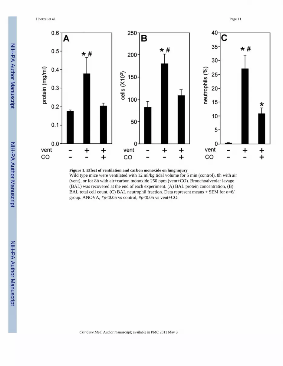
Figure 1. Effect of ventilation and carbon monoxide on lung injuryWild type mice were ventilated with 12 ml/kg tidal volume for 5 min (control), 8h with air(vent), or for 8h with air+carbon monoxide 250 ppm (vent+CO). Bronchoalveolar lavage(BAL) was recovered at the end of each experiment. (A) BAL protein concentration, (B)BAL total cell count, (C) BAL neutrophil fraction. Data represent means + SEM for n=6/group. ANOVA, *p<0.05 vs control, #p<0.05 vs vent+CO.
Hoetzel et al. Page 11
Crit Care Med. Author manuscript; available in PMC 2011 May 3.
NIH
-PA Author Manuscript
NIH
-PA Author Manuscript
NIH
-PA Author Manuscript

Figure 2. Effect of ventilation and carbon monoxide on caveolin-1 transcriptional regulationWild type mice were ventilated with 12 ml/kg tidal volume for 5 min (control), 8h with air(vent), or for 8h with air+carbon monoxide 250 ppm (vent+CO). RNA was extracted fromlung tissue at the end of the experiments and subjected to polymerase chain reaction. (A)Representative PCR for caveolin-1. (B) Densitometric analysis of caveolin-1 / GAPDH.Data represent means + SEM for n=6/group. ANOVA, *p<0.05 vs control, #p<0.05 vs vent+CO.
Hoetzel et al. Page 12
Crit Care Med. Author manuscript; available in PMC 2011 May 3.
NIH
-PA Author Manuscript
NIH
-PA Author Manuscript
NIH
-PA Author Manuscript

Figure 3. Effect of ventilation and carbon monoxide on caveolin-1 protein expressionWild type mice were ventilated with 12 ml/kg tidal volume for 5 min (control), 8h with air(vent), or for 8h with air+carbon monoxide 250 ppm (vent+CO). Lung tissue washomogenized at the end of the experiments and subjected to Western Blot analysis. (A)Representative Western blot of total caveolin-1 protein accumulation (upper panel). β-actindetection of the same membrane for normalization (lower panel). (B) Densitometric analysisof caveolin-1 / β-actin. Data represent means + SEM for n=6/group. ANOVA, *p<0.05 vscontrol , #p<0.05 vs vent+CO. (C) Time course of caveolin-1 regulation. RepresentativeWestern Blot of caveolin-1 expression at indicated time points.
Hoetzel et al. Page 13
Crit Care Med. Author manuscript; available in PMC 2011 May 3.
NIH
-PA Author Manuscript
NIH
-PA Author Manuscript
NIH
-PA Author Manuscript
Figure 4. Effect of ventilation and carbon monoxide on the localisation of caveolin-1Wild type mice were ventilated with 12 ml/kg tidal volume for 5 min (A, control), 8h withair (B, vent), or for 8h with air+carbon monoxide 250 ppm (C, vent+CO). Lung sectionswere taken at the end of each experiment and representative slides wereimmunofluorescence stained for caveolin-1 protein. (A–C) 200 X magnification, (D–F)confocal single cell microscopy, 2000 X magnification. Green color: caveolin-1 (arrow), redcolor: f-actin / phalloidin, blue color: dapi (nuclei, n).
Hoetzel et al. Page 14
Crit Care Med. Author manuscript; available in PMC 2011 May 3.
NIH
-PA Author Manuscript
NIH
-PA Author Manuscript
NIH
-PA Author Manuscript

Figure 5. Effects of ventilation and carbon monoxide on lung injury in caveolin-1 deficiencyWild type (wt, black bars) and caveolin-1−/− mice (cav-1−/−, white bars) were ventilatedwith 12 ml/kg tidal volume for 5 min (control), 8h with air (vent), or for 8h with air+carbonmonoxide 250 ppm (vent+CO). (A) Representative Western Blot for caveolin-1 (upperpanels) and β-actin (lower panels) in wt and cav-1−/− mice. (B) BAL protein concentration,(C) BAL total cell count. Data represent means + SEM for n=6/group. ANOVA forcomparison within wild type or caveolin-1−/− groups, *p<0.05 vs control, #p<0.05 vs vent+CO. p-value for comparison between wild type and caveolin-1−/− groups as indicated.
Hoetzel et al. Page 15
Crit Care Med. Author manuscript; available in PMC 2011 May 3.
NIH
-PA Author Manuscript
NIH
-PA Author Manuscript
NIH
-PA Author Manuscript

Figure 6. Effect of ventilation and carbon monoxide on lung histology in wild type and caveolin-1deficient miceWild type (A-C, wt) and caveolin-1−/− (D-F, cav-1−/−) mice were ventilated with 12 ml/kgtidal volume for 5 min (A, D, control), 8h with air (B, E, vent), or for 8h with air+carbonmonoxide 250 ppm (C, F, vent+CO). Representative lung sections were taken at the end ofeach experiment and stained with H&E (400 X magnification).
Hoetzel et al. Page 16
Crit Care Med. Author manuscript; available in PMC 2011 May 3.
NIH
-PA Author Manuscript
NIH
-PA Author Manuscript
NIH
-PA Author Manuscript
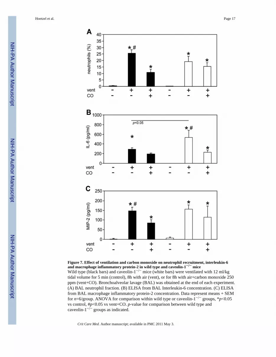
Figure 7. Effect of ventilation and carbon monoxide on neutrophil recruitment, interleukin-6and macrophage inflammatory protein-2 in wild type and caveolin-1−/− miceWild type (black bars) and caveolin-1−/− mice (white bars) were ventilated with 12 ml/kgtidal volume for 5 min (control), 8h with air (vent), or for 8h with air+carbon monoxide 250ppm (vent+CO). Bronchoalveolar lavage (BAL) was obtained at the end of each experiment.(A) BAL neutrophil fraction. (B) ELISA from BAL Interleukin-6 concentration. (C) ELISAfrom BAL macrophage inflammatory protein-2 concentration. Data represent means + SEMfor n=6/group. ANOVA for comparison within wild type or caveolin-1−/− groups, *p<0.05vs control, #p<0.05 vs vent+CO. p-value for comparison between wild type andcaveolin-1−/− groups as indicated.
Hoetzel et al. Page 17
Crit Care Med. Author manuscript; available in PMC 2011 May 3.
NIH
-PA Author Manuscript
NIH
-PA Author Manuscript
NIH
-PA Author Manuscript