cannabinoid receptors cb1 and cb2 form functional heteromers in brain
TRANSCRIPT
Cannabinoid Receptors CB1 and CB2 Form FunctionalHeteromers in Brain*
Received for publication, December 24, 2011, and in revised form, April 15, 2012 Published, JBC Papers in Press, April 24, 2012, DOI 10.1074/jbc.M111.335273
Lucía Callén‡§1, Estefanía Moreno‡§, Pedro Barroso-Chinea§¶, David Moreno-Delgado‡§, Antoni Cortés‡§,Josefa Mallol‡§, Vicent Casadó‡§, José Luis Lanciego§¶, Rafael Franco¶, Carmen Lluis‡§, Enric I. Canela‡§2,and Peter J. McCormick‡§2,3
From the ‡Department of Biochemistry and Molecular Biology, Faculty of Biology, University of Barcelona, 08028 Barcelona, Spainand the §Centro de Investigación Biomédica en Red sobre Enfermedades Neurodegenerativas and ¶Centro de Investigación MédicaAplicada, Universidad de Navarra, 31008 Pamplona, Spain
Background: Although CB1, the most abundant neuronal receptors, and CB2 receptors are co-expressed in neurons, theCB1-CB2 relationship is unknown.Results: CB1 and CB2 receptors form heteromers in neuronal cells and in the brain.Conclusion: Activation of either receptor leads to negative modulation of the partner receptor via heteromers.Significance: These heteromers may explain previous conflicting results and serve as therapeutic targets.
Exploring the role of cannabinoid CB2 receptors in the brain,we present evidence of CB2 receptor molecular and functionalinteraction with cannabinoid CB1 receptors. Using biophysicaland biochemical approaches, we discovered that CB2 receptorscan form heteromers with CB1 receptors in transfected neuro-nal cells and in rat brain pineal gland, nucleus accumbens,and globus pallidus. Within CB1-CB2 receptor heteromersexpressed in a neuronal cell model, agonist co-activation of CB1and CB2 receptors resulted in a negative cross-talk in Akt phos-phorylation and neurite outgrowth. Moreover, one specificcharacteristic of CB1-CB2 receptor heteromers consists of boththe ability of CB1 receptor antagonists to block the effect of CB2receptor agonists and, conversely, the ability of CB2 receptorantagonists to block the effect of CB1 receptor agonists, showinga bidirectional cross-antagonismphenomenon. Taken together,these data illuminate the mechanism by which CB2 receptorscan negatively modulate CB1 receptor function.
The endocannabinoid system is known to have a broadimpact on a variety of tissues. It has been shown to heavilyinfluence cardiovascular and immune systems as well as to con-trol progenitor cell proliferation (1–3). More recently, theendocannabinoid system has emerged as a major player in thecomplex web of neuromodulators, constituting a new intercel-lular communication networkmainly involved in the control ofneurotransmitter release (4–7). By acting as retrograde mes-sengers at various synapses, these endogenous arachidonic acid
derivatives participate in controlling processes, such as motoractivity, memory and learning, appetite, emesis, nociception,and somemotivational responses (8–14). There are two knowncannabinoid receptors, CB1 and CB2, with strong evidence of athird in the form of GPR55, and all have been considered astherapeutic targets for basal ganglia disorders (15, 16). CB1receptors mediate psychoactivity, whereas CB2 receptor-selec-tive agonists lack psychoactivity but are implicated in the con-trol of fundamental neural cell processes, such as proliferationand survival, and their pharmacological manipulationmight beuseful for both delaying the progression of neurodegenerativedisorders and inhibiting the growth of glial tumors (17, 18). CB1is the most abundant receptor in the central nervous system,and, besides its classical influence on mood and emotion, it hasbeen demonstrated to play a role in the modulation of memoryprocessing and in metabolism (19). The ubiquitous expressionpattern of CB1 receptors reflects the complexity and the varietyof functions the endocannabinoid system impacts in neuronalactivity. CB1 receptors are often localized presynaptically,where their stimulation usually inhibits neurotransmitterrelease (4, 20, 21). In the striatal spine module, CB1 receptorsare localized both pre- and postsynaptically (22). Presynapti-cally, CB1 receptors are localized in GABAergic terminals ofinterneurons or collaterals from medium spiny neurons andalso in glutamatergic but not in dopaminergic terminals (23–27). Postsynaptically, CB1 receptors are localized in the soma-todendritic area of medium spiny neurons (23–25), and bothenkephalinergic and dynorphinergic medium spiny neuronsexpress CB1 receptors (28). The related receptor, CB2, tradi-tionally was thought to be expressed in peripheral tissue, whereit can help control inflammation and various immunologicalresponses (1), but recent reports have suggested that it too canbe found in the brain (albeit at a lower expression level thanCB1receptors) (29) and can impact a variety of neuronal processes.Multiple studies have demonstrated the expression of CB2receptors in different non-neuronal (30–33) and neuronal pop-ulations (31, 34–38) where CB2 receptors show a preferredpostsynaptic localization (31, 35–37).
* This work was supported by Spanish Ministerio de Economia and Competi-tividad Grants SAF2010-18472, SAF2008-03229-E, and SAF2008-03118-Ewithin the framework of the Era-NET Neuron program) and a grant forcollaborative projects (Grant PI2011/02-7) from the Centro de Investi-gación Biomédica en Red sobre Enfermedades Neurodegenerativas(CIBERNED).
1 A Formación de Profesorado Univesitario Fellow (Grant AP2007-00808).2 Both authors contributed equally to this work.3 A Ramón y Cajal Fellow. To whom correspondence should be addressed:
Dept. of Biochemistry and Molecular Biology, Faculty of Biology, Universityof Barcelona, 643 Avenida Diagonal, planta-2, Barcelona 08028, Spain. Tel.:934039280; Fax: 934021559; E-mail: [email protected].
THE JOURNAL OF BIOLOGICAL CHEMISTRY VOL. 287, NO. 25, pp. 20851–20865, June 15, 2012© 2012 by The American Society for Biochemistry and Molecular Biology, Inc. Published in the U.S.A.
JUNE 15, 2012 • VOLUME 287 • NUMBER 25 JOURNAL OF BIOLOGICAL CHEMISTRY 20851
at Universidad de N
avarra, on June 18, 2012w
ww
.jbc.orgD
ownloaded from

BothCB1 andCB2 receptors aremembers of theGPCR4 fam-ily and are Gi/Go-protein-coupled receptors (39). Agonist acti-vation triggers inhibition of adenylyl cyclase and voltage-gatedcalcium channels, activation of potassium channels, mitogen-activated protein kinase (MAPK), and phosphoinositide-3kinase (PI3K)/Akt signaling pathways (40, 41). Initially, CB1and CB2 receptors, like other GPCRs, were thought to haveacted as single signaling receptors, but studies in the last decadehave convincingly shown that certain GPCRs in a variety ofdifferent tissues can also form homodimers and even hetero-mers (42). For the cannabinoid receptors, heteromers havebeen shown to exist between CB1 and the dopamine and aden-osine receptors (43–47) as well as with angiotensin (AT1) (48),opioid �1 (49), and orexin OX1 receptors (50). However, todate, no studies have examined the possible interactionsbetween CB1 and CB2 receptors despite the fact that they haveoverlapping expression and that the two receptors have beenshown to impact similar cellular processes. Here we report thatCB1 and CB2 receptors can form functional heteromers intransfected cells and in a variety of brain tissues. Heteromerformation leads to a negative cross-talk between receptor ago-nists and antagonists, suggesting an additional level of molecu-lar regulation between the two receptors.
MATERIALS AND METHODS
Fusion Proteins and Expression Vectors—The human cDNAfor the CB1, CB2, and dopamine D4.4 receptors cloned inpcDNA3.1 were amplified without their stop codons usingsense and antisense primers harboring either unique EcoRI andBamHI sites (CB1R, CB2R) orXhoI and EcoRI (D4.4R). The frag-ments were then subcloned to be in frame with Rluc into theEcoRI and BamHI restriction sites of an Rluc-expressing vector(pRluc-N1, PerkinElmer) or with enhanced YFP into the EcoRIand BamHI (CB1R, CB2R) or XhoI and EcoRI restriction sites ofan enhanced YFP-expressing vector (EYFP-N1; enhanced yel-low variant of GFP; Clontech) to give the plasmids that expressCB1, CB2, or D4.4 receptors fused to Rluc or YFP on the C-ter-minal end of the receptor (CB2R-Rluc, CB2R-YFP, CB1R-YFP,or D4R-YFP). Expression of constructs was tested by confocalmicroscopy, and the receptor functionality was tested by theERK1/2 activation pathway (see “Results”).Cell Line Cultures and Transfection—Human embryonic
kidney (HEK-293T), human neuroblastoma SH-SY5Y, andneuroblastoma and glioma hybrid NG108-15 cell lines weregrown in Dulbecco’s modified Eagle’s medium (DMEM) sup-plemented with 100 units/ml penicillin/streptomycin and 10%(v/v) heat-inactivated fetal bovine serum (FBS). Other supple-ments were 2 mM L-glutamine for HEK-293T and SH-SY5Ycells, 1 mM sodium pyruvate for SH-SY5Y cells, and 100 �M
hypoxanthine, 0.02 �M aminopterin, 16 �M thymidine (HATsupplement) for NG108-15 cells. The human neuroblastomaSK-N-MC cells were grown in minimum essential mediumsupplemented with 2 mM L-glutamine, 100 IU/ml penicillin/streptomycin, 1 mM sodium pyruvate, and 10% (v/v) heat-inac-
tivated FBS. All supplements were from Invitrogen. Cells weremaintained at 37 °C in a humidified atmosphere of 5% CO2 andwere passaged when they were 80–90% confluent (i.e. approx-imately twice a week).HEK-293T or SH-SY5Y cells were transiently transfected
with the corresponding fusion protein cDNA by the ramifiedPEI (Sigma) method. Cells were incubated (4 h) with the corre-sponding cDNA together with ramified PEI (5 ml of 10 mM PEIfor each mg of cDNA) and 150 mM NaCl in a serum-starvedmedium. After 4 h, the medium was changed to a fresh com-plete culturemedium. 72 h after transfection, cells werewashedtwice in quick succession in Hanks’ balanced salt solution (137mM NaCl, 5 mM KCl, 0.34 mM Na2HPO4�12H2O, 0.44 mM
KH2PO4, 1.26 mM CaCl2�2H2O, 0.4 mM MgSO4�7H2O, 0.5 mM
MgCl2, 10mMHEPES, pH7.4) supplementedwith 0.1% glucose(w/v), detached by gently pipetting, and resuspended in thesame buffer. To control the cell number, sample protein con-centration was determined using a Bradford assay kit (Bio-Rad)using bovine serum albumin dilutions as standards.Primary Cultures of Rat Pinealocytes—Male Sprague-Dawley
rats (3month old,�350 g), receivingwater and food ad libitum,were obtained from the animal facility of the Faculty of Biology(University of Barcelona). 4% isoflurane (2-chloro-2-(difluoro-methoxy)-1,1,1-trifluoroethane)-anesthetized animals werekilled by decapitation at 20:00 h (after the light period), andpineal glands were immediately dissected. All procedures wereapproved by the Catalan Ethical Committee for Animal Use(CEAA/DMAH 4049 and 5664). Pinealocytes were preparedfrom rat pineal glands as described previously by da SilveiraCruz-Machado et al. (51). Briefly, pinealocytes were obtainedby trypsinization (0.25%, 37 °C, 15min) followed bymechanicaldispersion in the presence of fetal bovine serum. Cells werepelleted and resuspended in defined culture medium BGJb(Invitrogen) supplemented with 10% (v/v) fetal bovine serum(heat-inactivated), 100 units/ml penicillin/streptomycin (pH7.4). The total number of cells and fractional survival was esti-mated by trypan blue exclusion. Cells (200,000 cells/well) wereplated on polylysine-coated 6-well chamber plates and main-tained at 37 °C, 5% CO2 for 48 h prior to use.Rat Brain Slice Preparation—Rats were decapitated with a
guillotine, and the brains were rapidly removed and placed inice-cold oxygenated (O2/CO2, 95%/5%) Krebs-HCO3
� buffer(124 mM NaCl, 4 mM KCl, 1.25 mM NaH2PO4, 1.5 mM MgCl2,1.5 mM CaCl2, 10 mM glucose, and 26 mM NaHCO3, pH 7.4).The brains were sliced at 4 °C in a brain matrix (Zivic Instru-ments, Pittsburgh, PA) into 0.5-mm coronal slices. Slices werekept at 4 °C in Krebs-HCO3
� buffer during the dissection of thenucleus accumbens and the globus pallidus. For signalingexperiments, each slice was transferred into an incubation tubecontaining 1 ml of ice-cold Krebs-HCO3
� buffer, and theERK1/2 phosphorylation was determined as described below.For proximity ligation assays, slices were fixed with 4% parafor-maldehyde solution for 1 h at room temperature with gentleagitation. The sliceswere thenwashed inTBS (50mMTris-HCl,0.9% NaCl, pH 7.8) and treated for 5 min with 1% Na2BH4dissolved in TBS, followed by successive TBS washes until allNa2BH4 was eliminated. Finally, the slices were cryopreservedin a 30% sucrose solution overnight at 4 °C and stored at�20 °C
4 The abbreviations used are: GPCR, G protein-coupled receptor; PLA, prox-imity ligation assay(s); BRET, bioluminescence resonance energy transfer;ANOVA, analysis of variance; ACEA, arachidonyl-2-chloroethylamide.
CB1 and CB2 Receptors Form Heteromers
20852 JOURNAL OF BIOLOGICAL CHEMISTRY VOLUME 287 • NUMBER 25 • JUNE 15, 2012
at Universidad de N
avarra, on June 18, 2012w
ww
.jbc.orgD
ownloaded from
until sectioning. 15-�m-thick slices were cut on a freezing cryo-stat (Leica Jung CM-3000) and mounted on slide glass. Sliceswere thawed at 4 °C, washed in TBS, and rockedwith the block-ing solution (Olink Bioscience, Uppsala, Sweden) for 1 h at37 °C in a humidified atmosphere.In Situ Proximity Ligation Assay (PLA)—Primary cultures of
pinealocytes or SH-SY5Y cells transfected or not with 3 �g ofcDNA corresponding to CB2R-HA (Missouri S&T ResourceCenter), were fixed in 4% paraformaldehyde for 15 min andwashed with phosphate-buffered saline (PBS) containing 20mM glycine to quench the aldehyde groups. After permeabiliza-tionwith PBS-glycine containing 0.05%TritonX-100 for 5min,cells were incubated for 1 h at room temperature with PBScontaining 1% bovine serum albumin. Rat nucleus accumbensslices were obtained as described above. The receptor-receptormolecular interaction in these samples was detected using theDuolink II in situ PLA detection kit (Olink Bioscience). Todetect heteromers in pinealocytes or in nucleus accumbensslices, the direct PLA-linked primary antibodies were used. Therabbit anti-CB1 receptor antibody (Thermo Scientific) waslinked to a plus PLA probe, and the rabbit anti-CB2 receptorantibody (Cayman Chemical, Ann Arbor, MI) was linked to aminus PLA probe following the instructions of the supplier.After a 1-h incubation at 37 °C with the blocking solution in apreheated humidity chamber, pinealocytes or slices were incu-bated overnight with these PLA probe-linked antibodies(1:1000) at 4 °C. After washing with wash buffer at room tem-perature, samples were processed for ligation, amplification,and detection as described by the manufacturer. As negativecontrols, pinealocytes or slices were incubated overnight withthe plus PLA probe-linked rabbit anti-CB1 receptor antibodyand a goat anti-D4 receptor primary antibody (1:500; SantaCruz Biotechnology) at 4 °C, followed by an incubation (2 h,37 °C) in a preheated humidity chamber with Duolink II minusPLA probe anti-goat diluted in the antibody diluent. To detectheteromers in SH-SY5Y cells, transfected or non-transfected(as negative controls) cells were incubated (1 h, 37 °C) with theblocking solution in a preheated humidity chamber and thenincubated overnight with the primary antibodies: rabbit anti-CB1 receptor antibody (1:1000; Thermo Scientific) and mousemonoclonal anti-HA tag antibody (1:1000; Abcam, Cambridge,UK) in the antibody diluent medium. SH-SY5Y cells werewashed with washing buffer at room temperature and incu-bated (2 h, 37 °C) in a preheated humidity chamber with PLAprobes detecting rabbit or mouse antibodies (Duolink II plusPLA probe anti-rabbit and Duolink II minus PLA probe anti-mouse) and processed as described above. Samples weremounted using themountingmediumwithDAPI and observedin a Leica SP2 confocal microscope (Leica Microsystems,Mannheim, Germany).Immunostaining—After 72 h of transfection,HEK-292T cells
were fixed in 4% paraformaldehyde for 15min andwashedwithPBS containing 20 mM glycine to quench the aldehyde groups.After permeabilization with PBS-glycine containing 0.05% Tri-ton X-100 for 5 min, cells were incubated with PBS containing1% bovine serum albumin. After 1 h at room temperature, pro-tein-Rlucwas labeledwith the primarymousemonoclonal anti-Rluc antibody (1:100; Chemicon, Billerica,MA) for 1 h, washed,
and stained with the secondary antibody Cy3 donkey anti-mouse (1:200; Jackson Immunoresearch Laboratories, WestGrove, PA). Protein-YFPwas detected by its fluorescence prop-erties. The sampleswere rinsed several times andmountedwitha medium suitable for immunofluorescence (30%Mowiol, Cal-biochem). The samples were observed in a Leica SP2 confocalmicroscope.RT-PCR—Total cellular RNA was isolated from HEK-293T,
SH-SY5Y, SK-N-MC, or NG108-15 cells using the QuickPreptotal RNA extraction kit (Amersham Biosciences) or fromMacaca fascicularis spleen usingTRIzol reagent (Invitrogen) asdescribed previously (38). Total RNA (1 �g) was reverse-tran-scribed by random priming using Moloney murine leukemiavirus reverse transcriptase, RNase H minus, and point mutant,following the protocol of two-step RT-PCR provided by Pro-mega (Madison, WI). The resulting single-stranded cDNA wasused to perform PCR amplification for the CB1 and CB2 recep-tors and GAPDH as an internal control of PCR technique usingTaqDNA polymerase (Promega). Common primers to amplifyhuman and rat cDNAwere used. Primers to amplify CB1Rwere5�-TGGGCAGCCTGTTCCTCAC-3� (forward) and 5�-CAT-GCGGGCTTGGTC-3� (reverse). Primers to amplify CB2Rwere 5�- CGTGGCTGTGCTCTATCTGA-3� (forward) and5�-AGCCAGCTCAGCAGGTAGTC-3� (reverse). To amplifyGAPDH, the primers used were 5�-CATCCTGCACCAC-CAACTGCTTAG-3� (forward) and 5�-GCCTGCTTCAC-CACCTTCTTGATG-3� (reverse). RNA without reverse tran-scriptions did not yield any amplicons, indicating that therewasno genomic DNA contamination.Bioluminescence Resonance Energy Transfer (BRET) Assays—
HEK-293T cells were transiently co-transfected with the indi-cated amounts of plasmid cDNAs corresponding to the indi-cated fusion proteins (see Fig. 1 legend). To quantify receptorfluorescence expression, cells (20 �g of protein) were distrib-uted in 96-well microplates (black plates with a transparentbottom; Porvair (King’s Lynn, UK)), and fluorescence was readin a FluoStar Optima fluorimeter (BMG Labtechnologies,Offenburg, Germany) equipped with a high energy xenon flashlamp, using a 10-nm bandwidth excitation filter at a reading of400 nm. Receptor fluorescence expression was determined asthe fluorescence of the sample minus the fluorescence of cellsexpressing protein-Rluc alone. For BRET measurements, theequivalent of 20�g of protein was distributed in 96-well micro-plates (Corning 3600, white plates; Sigma), and 5 �M coelen-terazine H (Molecular Probes, Inc., Eugene, OR) was added.After 1 min of adding coelenterazine H, the readings were col-lected using a Mithras LB 940 (Berthold, Bad Wildbad, Ger-many), which allows the integration of the signals detected inthe short wavelength filter at 485 nm (440–500 nm) and thelong wavelength filter at 530 nm (510–590 nm). To quantifyreceptor-Rluc expression luminescence, readings were per-formed 10 min after the addition of 5 �M coelenterazine H.Cells expressing BRET donors alone were used to determinebackground.Thenet BRET is defined as (longwavelength emis-sion/short wavelength emission)�Cf, whereCf corresponds tolong wavelength emission/short wavelength emission for theRluc construct expressed alone in the same experiment. BRETcurves were fitted by using a non-linear regression equation,
CB1 and CB2 Receptors Form Heteromers
JUNE 15, 2012 • VOLUME 287 • NUMBER 25 JOURNAL OF BIOLOGICAL CHEMISTRY 20853
at Universidad de N
avarra, on June 18, 2012w
ww
.jbc.orgD
ownloaded from
assuming a single phase with GraphPad Prism software (SanDiego, CA)ERK1/2 and Akt/PKB Phosphorylation Assays—Each globus
pallidus slice, obtained as described above, was transferred intoan incubation tube containing 1 ml of ice-cold Krebs-HCO3
�
buffer, and the temperature was raised to 23 °C. After 30 min,the medium was replaced by 2 ml of Krebs-HCO3
� buffer(23 °C) and was incubated under constant oxygenation (O2/CO2, 95%/5%) at 30 °C for 4–5h in anEppendorf Thermomixer(5 Prime, Inc., Boulder, CO). The medium was replaced by 200�l of freshKrebs-HCO3
� buffer and incubated for 30min beforethe addition of the desired concentrations of ligands. After theindicated incubation period, the solution was discarded, andslices were frozen on dry ice and stored at �80 °C until use.Transfected or non-transfected SH-SY5Y cells were cultured inserum-free medium for 16 h before the addition of any agent.Cells were treated or not with the indicated agonists for theindicated time. At the end of the incubation period, cells wererinsed with ice-cold phosphate-buffered saline. Cells or sliceswere lysed by the addition of 500 �l of ice-cold lysis buffer (50mMTris-HCl, pH 7.4, 50mMNaF, 150mMNaCl, 45mM �-glyc-erophosphate, 1%TritonX-100, 20�Mphenyl-arsine oxide, 0.4mM NaVO4, and protease inhibitor mixture). The cellulardebris was removed by centrifugation at 13,000� g for 5min at4 °C, and the protein was quantified by the bicinchoninic acidmethod using bovine serum albumin dilutions as a standard.Equivalent amounts of protein (10 �g) were separated by elec-trophoresis on a denaturing 7.5% SDS-polyacrylamide gel andtransferred onto a PVDF-FL membrane. Odyssey blockingbuffer (LI-COR Biosciences, Lincoln, NE) was then added, andthemembrane was rocked for 90min. Themembrane was thenincubated overnight with a mixture of a rabbit anti-phospho-Ser473 Akt antibody (1:2500; Signalway Antibody) to test theAkt phosphorylation ormouse anti-phospho-ERK1/2 antibody(1:2500; Sigma) to test ERK1/2 phosphorylation. As a control ofthe amount of protein loaded, rabbit anti-ERK1/2 antibody thatrecognizes both phosphorylated and non-phosphorylatedERK1/2 (1:40,000; Sigma) was used. Bands were visualized bythe addition of IRDye 680 (anti-rabbit) antibody (1:10,000;Sigma) or IRDye 800 (anti-mouse) antibody (1:10,000; Sigma)or amixture of both for 1 h and scanned by theOdyssey infraredscanner (LI-COR Biosciences). Band densities were quantifiedusing the scanner software and exported to Excel (Microsoft,Redmond,WA). The level of phosphorylated proteins was nor-malized for differences in loading using the total ERK1/2 pro-tein band intensities.Evaluation of Neurite Outgrowth—SH-SY5Y cells seeded in
10-mm coated glass coverslips were transfected or not with 3�g of cDNA corresponding to CB2R-YFP. 48 h post-transfec-tion, cells were incubated for 24 h in serum-free growingmedium in the absence or presence of 10 �M retinoic acid, 100nM ACEA, or 50 nM JWH 133 (all from Tocris, Bristol, UK)alone or in combination. Cells were washed three times withPBS, fixed in 4% paraformaldehyde for 15 min, washed withPBS containing 20 mM glycine, permeabilized for 5 minwith PBS-glycine buffer containing 0.05% Triton X-100, andblocked with PBS containing 1% BSA for 1 h at room tempera-ture. Cells were labeled for 1 h with the primary mouse anti-
MAP2 antibody (1:200; Calbiochem). Subsequently, cells werewashed and stained with the secondary antibody, Cy3-conju-gated affinity-purified donkey anti-mouse IgG (1:200 dilution;Jackson ImmunoResearch Laboratories), and nuclear stainingwas performed with Hoechst (1:1000, 1 mg/ml; Sigma). Cover-slips were rinsed for 5 min in PBS containing 1% BSA and for 5min in PBS-glycine buffer andmountedwithMowiolmountingmedium. Confocal microscope observations were made with aLeica TCS SP2 microscope with a �40 objective. Cell bodiesand neurites present in 8–12 randomly selected fields weremeasured in each experiment using ImageJ software. Cells wereconsidered to be differentiated if they had at least one processlonger than the cell body, whichwould be regarded as a neurite.The results are expressed as the percentage of differentiatedcells versus the total cell number in non-transfected cells orversus CB2R-YFP-expressing cells (detected by its own fluores-cence) in transfected cells. At least three independent experi-ments were conducted for each treatment.
RESULTS
CB2Receptors FormHeteromers withCB1 Receptors in Trans-fectedCells—Avariety ofGPCRs, includingCB1 receptors, havebeen reported to be expressed as homomers and form hetero-mers with other GPCRs (43, 45, 49, 52), but it is not knownwhether CB2 and CB1 receptors can form heteromers. To testthis, the BRET technique was used. The BRET techniquerequires the use of fusion proteins consisting of CB2R-Rluc,CB1R-YFP, and CB2R-YFP. Prior to the BRET experiments, wefirst confirmed that the fusion of Rluc or YFP to CB2 or CB1receptors did not modify receptor function, as determined byERK1/2 phosphorylation assays (Fig. 1A). In addition, we con-firmed that the subcellular localization of the fusion proteinswas indeed in the cell membrane, showing a high degree ofcolocalization when CB2R-Rluc and CB2R-YFP or CB2R-RlucandCB1R-YFPwere co-expressed (Fig. 1B). To test the ability ofCB1 and CB2 receptors to form heteromers, BRET measure-ments were performed in transiently co-transfectedHEK-293Tcells using a constant amount of cDNAcorresponding toCB2R-Rluc and increasing amounts of cDNA corresponding to CB1R-YFP. As can be seen in Fig. 1C, the BRET signal increased as ahyperbolic function of the amount of the CB1R-YFP expressed,reaching an asymptote. From the saturation curve, a BRETmaxof 33 � 1 milli BRET units (mBU) and a BRET50 of 8 � 2 werecalculated. The specificity of this interaction was demonstratedby comparing the BRET saturation curve with the low and lin-ear BRET obtained for the negative control constituted byCB1R-Rluc and D4R-YFP (Fig. 1C). These results indicate thatCB1 andCB2 receptors formheteromers in co-transfected cells.CB1 and CB2 Receptors FormHeteromers in SH-SY5YNeuro-
blastoma Cells—Knowing that CB1 and CB2 receptors formheteromers in HEK-293T transfected cells, we investigatedwhether they can formheteromers in a neuronal cellmodel.Wefirst determined the endogenous receptor expression in differ-ent neuroblastoma cell lines. As shown in Fig. 2A, all neuroblas-toma cells tested expressed the mRNA corresponding to CB1receptor, but they did not express the mRNA corresponding toCB2 receptor. From these results, human SH-SY5Y neuroblas-toma cells were selected. Non-transfected cells were used as
CB1 and CB2 Receptors Form Heteromers
20854 JOURNAL OF BIOLOGICAL CHEMISTRY VOLUME 287 • NUMBER 25 • JUNE 15, 2012
at Universidad de N
avarra, on June 18, 2012w
ww
.jbc.orgD
ownloaded from

CB1 receptor-expressing neuroblastoma cells, whereasSH-SY5Y cells transfected with HA tagged CB2 receptor wereused as CB1 receptor- and CB2 receptor-expressing cells. TheBRET technique is a powerful approach for looking at receptorinteractions in co-transfected cells, but BRET cannot be easilyapplied in native cells endogenously expressing one or bothreceptors. To solve this, other direct and indirect methods canbe used. Here we sought to determine if the endogenousexpressed CB1 and transfected CB2 receptors could also formheteromers in a neuronal cell model. To do this, we employedthe PLA, which is used to detect protein interactions. Thisdirect method requires that both receptors be close enough toallow the two different antibody-DNA probes to be able toligate (�17 nm) (53, 54). If the receptors are within sufficient
proximity, a punctate fluorescent signal can be detected by con-focal microscopy diagrammed in Fig. 2B. For these experi-ments, two different and specific primary antibodies directedagainst each of the two receptors were used. One was the wellknown commercial mouse anti-HA antibody to detect the HA-labeledCB2 receptor, and the other was a rabbit anti CB1 recep-tor antibody. The specificity of this last antibody was tested inCB1R-YFP- or CB2R-YFP-transfected HEK-293T cells (Fig.2C). Colocalization of fluorescence due to YFP with the anti-CB1 receptor antibody staining was detected in CB1R-YFP-transfected cells but not in CB2R-YFP-transfected cells, and alack of antibody-promoted staining was observed in non-trans-fected cells (cells that do not show fluorescence) (Fig. 2C).Using these antibodies in PLA experiments, the CB1-CB2
FIGURE 1. CB2 receptors form heteromers with CB1 receptors in transfected cells. A, the functionality of fusion proteins in HEK-293T cells transfected with1.5 �g of cDNA corresponding to CB1 receptor, CB2 receptor, CB2R-YFP, CB1R-YFP, or CB2R-Rluc. 72 h post-transfected cells expressing CB2R, CB2R-YFP, orCB2R-Rluc were treated for 7 min with vehicle (basal) or with JWH 133 (100 nM), and cells expressing CB1R or CB1R-YFP were treated for 7 min with vehicle (basal)or with CP 55940 (500 nM), and ERK1/2 phosphorylation was determined. Results (means � S.E. (error bars) of four different experiments performed induplicate) represent -fold over basal. Significant differences were analyzed by one-way ANOVA followed by Dunnett’s multiple comparison post hoc test (*, p �0.05; **, p � 0.01; ***, p � 0.001 compared with basal). Above is a representative Western blot. B, confocal microscopy images of cells transfected with theplasmid corresponding to CB2R-YFP (1.5 �g), CB2R-Rluc (0.5 �g), or CB1R-YFP (1.5 �g) alone (top panels) or in combination (middle panels for CB2R-Rluc andCB2R-YFP and lower panels for CB2R-Rluc and CB1R-YFP). Proteins were identified by fluorescence or by immunocytochemistry as indicated under “Materialsand Methods.” Colocalization is shown in yellow in merge panels. Scale bars, 10 �m. C, BRET saturation experiments showing CB1-CB2 receptor heteromerizationwere performed as described under “Materials and Methods” using cells transfected with 0.2 �g of cDNA corresponding to CB2R-Rluc and increasing amountsof cDNA (0 – 4 �g of cDNA) corresponding to CB1R-YFP (circles). As a negative control, cells were also transfected with cDNA corresponding to CB1R-Rluc (0.2 �g)and dopamine D4R-YFP (0 – 4 �g of cDNA) (triangles). Both fluorescence and luminescence for each sample were measured before every experiment to confirmsimilar donor expressions (�100,000 bioluminescence units) while monitoring the increase in acceptor expression (100 –20,000 net fluorescence units). Therelative amount of BRET is given as the ratio between the net fluorescence of the acceptor (YFP-YFP0), and the luciferase activity of the donor (Rluc). BRET dataare expressed as means � S.E. of four different experiments grouped as a function of the amount of BRET acceptor. mBU, milli BRET units.
CB1 and CB2 Receptors Form Heteromers
JUNE 15, 2012 • VOLUME 287 • NUMBER 25 JOURNAL OF BIOLOGICAL CHEMISTRY 20855
at Universidad de N
avarra, on June 18, 2012w
ww
.jbc.orgD
ownloaded from

receptor heteromer expression in SH-SY5Y cells was demon-strated by punctuate fluorescent signal detected by confocalmicroscopy after excitation at 624 nm. This pattern wasobserved in CB2 receptor-expressing cells, whereas no signalwas detected in non-transfected cells used as a negative control(Fig. 2D).CB1-CB2 Receptor Heteromers Are Expressed in Rat Brain—
To gain more insight about the physiological relevance of CB1-CB2 receptor heteromers, we next investigated the expressionof these heteromers in the rat brain taking advantage of the PLAapproach used above. CB1 is the most abundant GPCR in thebrain (55). Although CB2 receptor expression is much lower,sometimes even undetectable, it is also known to be present indifferent brain areas, impacting endocannabinoid signaling and
colocalizing with CB1 receptors (32, 34, 36, 37, 56–59).Recently, it has been shown that both CB1 and CB2 receptorsare co-expressed in the pineal gland, where they may beinvolved in the control of pineal physiology (57), and in thenucleus accumbens, where CB2 receptor controls cocaineintake (59). Despite evidence of their co-expression, nothing isknown about CB1 and CB2 receptor molecular interactions inthe brain. Here we determined the expression of CB1-CB2receptor heteromers in rat pinealocytes and nucleus accum-bens by PLA. We used the rabbit anti-CB1 receptor antibodydescribed above and a rabbit anti-CB2 receptor antibody that isspecific for the CB2 receptor, as previously demonstrated (57).Both antibodies were used directly linked to the PLA DNAprobes, as described under “Materials and Methods.” PLA
FIGURE 2. CB1-CB2 receptor heteromers in neuroblastoma cells. A, CB1 and CB2 receptor mRNA expression in different neuroblastoma cells was analyzed byRT-PCR using total RNA from SH-SY5Y, SK-N-MC, or NG108-15 neuroblastoma cells and specific common primers for the human and rat CB2 and CB1 receptorsor GAPDH as internal control of mRNA expression. As positive controls for the CB2 receptor expression, total RNAs from human spleen or HEK-293T cells stablyexpressing human CB2 receptors (HEK CB2) were used, and RNAs from HEK-293T cells (HEK-293T) or primers without RNA (control) were included as negativecontrols. B, a schematic representation of the PLA technology is shown. Receptors were recognized by primary antibodies and secondary antibodies linked todifferent DNA chains, one plus and one minus. If the two receptors are close enough, the two different antibody-DNA probes are able to ligate. Following anamplification process, the presence of fluorescently tagged nucleotides allows detection of a punctuate fluorescent signal by confocal microscopy. C, to testthe specificity of rabbit anti-CB1 receptor primary antibody, HEK-293T cells were transfected with CB1R-YFP (1.5 �g of plasmid) (top panels) or CB2R-YFP (1.5 �gof plasmid) (bottom panels), and immunocytochemistry was performed with rabbit anti-CB1R, as indicated under “Materials and Methods.” CB1 receptor- or CB2receptor-expressing cells were identified by their own fluorescence, and cell nuclei were stained with Hoechst (blue). Colocalization in merged images is shownin yellow. Scale bars, 10 �m. D, PLA in SH-SY5Y cells transfected with CB2R-HA (3 �g of plasmid) (top panels) or without transfection (vehicle; bottom panel). Redspots in three different fields from independent experiments with CB2 receptor-expressing cells (top panels) but not in non-transfected cells (bottom panel)indicate the CB1-CB2 receptor heteromer expression. Scale bars, 20 �m.
CB1 and CB2 Receptors Form Heteromers
20856 JOURNAL OF BIOLOGICAL CHEMISTRY VOLUME 287 • NUMBER 25 • JUNE 15, 2012
at Universidad de N
avarra, on June 18, 2012w
ww
.jbc.orgD
ownloaded from

experiments were performed with pinealocytes obtained fromrat pineal glands extracted from a rat sacrificed at 20 h (after thelight period), when the expression of CB1 and CB2 receptors ismore equilibrated (57), and with rat nucleus accumbens slicesobtained as indicated under “Materials andMethods.” CB1-CB2receptor heteromers in the primary cultures or in the sliceswere visualized as red spots in pinealocytes (Fig. 3A) or neurons(Fig. 3B) stained with Hoechst. Because CB1 and dopamine D4receptors do not form heteromers in BRET experiments (seeFig. 1C) we considered this pair as the negative control andperformed the same assay using the PLA DNA probe-linkedanti-CB1 receptor antibody described above and a goat anti-body against dopamine D4 receptor plus a goat secondary anti-body linked to the correspondingPLADNAprobe (Fig. 3,C andD). A quantification of cells containing one or more red spotsversus total cells (blue nucleus) and the ratio r (number of redspots/total cells) were determined in eight fields from threedifferent experiments. 84% of pinealocytes expressed CB1-CB2receptor heteromers with r � 1.48, in contrast with the 24% ofpositive cells with r � 0.3 detected in the negative control (Fig.3E). Analogously 51% of nucleus accumbens cells (r � 2.11)expressed CB1-CB2 receptor heteromers, in contrast with 3%cells (r� 0.04) detected for the negative control (Fig. 3F). Theseresults indicate that CB1-CB2 receptor heteromers areexpressed in rat pinealocytes and in rat nucleus accumbens.
Characterization of CB1-CB2 Receptor Heteromer Signaling—Acommon and often essential attribute of receptor heteromersis the ability to modify the downstream signaling versus thesingle constituent receptors. This type of receptor-receptorinteraction has been observed for several receptor heteromers(60, 61). Because cannabinoid receptors have been previouslydescribed to be coupled to Akt/PKB protein activation in dif-ferent cell types (40, 41, 62), we first investigated whether therewere changes in Akt/PKB (Ser-473 Akt phosphorylation) sig-naling when heteromers were co-stimulated with both agonistsor blocked with antagonists in SH-SY5Y neuronal cells. Non-transfected cells were used as CB1 receptor-expressing neuro-blastoma cells, and cells transfected with CB2R-YFP were usedas CB1- and CB2 receptor-expressing cells. Treatment with 100nM CB1 receptor agonist ACEA induced Akt/PKB phosphory-lation in both non-transfected and transfected cells (Fig. 4, Aand B), and the CB2 receptor agonist JWH 133 induced Akt/PKB phosphorylation only in transfected cells (Fig. 4, A and B).Furthermore, JWH 133-induced Akt/PKB signaling was signif-icantly diminished when cells expressing both receptors wereco-stimulatedwithACEA and JWH133 (Fig. 4,B andD). Theseresults indicate that a negative cross-talk exists between CB1and CB2 receptors in Akt/PKB phosphorylation signaling.These results are not due to a change in the time in which thesignaling peaks because differences were not observed in time-
FIGURE 3. CB1-CB2 receptor heteromers in rat brain. The PLA was performed using primary cultures of rat pinealocytes (A and C) or rat nucleus accumbensslices (B and D). Pinealocytes (A) or slices (B) were treated with rabbit CB1 receptor and CB2 receptor primary antibodies directly linked to plus and minus PLAprobes (see “Materials and Methods”). CB1-CB2 receptor heteromers were visualized as red spots around Hoechst-stained nuclei (A and B). Negative controlswere performed with pinealocytes (C) or slices (D) treated with plus PLA probe-linked rabbit anti-CB1 receptor and with primary goat anti-D4 receptor detectedwith a minus PLA probe-linked secondary goat antibody. Scale bars, 20 �m. The percentage of pinealocytes (E) or nucleus accumbens neurons (F) containingone or more red spots versus total cells (blue nucleus) is given as well as r (number of red spots/total cells) values (E and F) from eight fields in three differentexperiments. Error bars, S.E.
CB1 and CB2 Receptors Form Heteromers
JUNE 15, 2012 • VOLUME 287 • NUMBER 25 JOURNAL OF BIOLOGICAL CHEMISTRY 20857
at Universidad de N
avarra, on June 18, 2012w
ww
.jbc.orgD
ownloaded from

response curves when cells were activated with one or bothagonists (Fig. 4C). It is important to point out that all ligandswere first chosen based on theirKI value (KI � 1.4 nM for ACEA�1400-fold selective for CB1 receptor; KI � 3.4 nM for JWH133, �200-fold selective for CB2 receptor) and following adose-response curve for Akt activation, where the lowest con-centration that provided specific receptor signaling was chosenas the working concentration.
Looking at the effect of CB1 and CB2 receptor antagonists,the 100 nM ACEA-induced Akt/PKB phosphorylation was notsignificantly modified when non-transfected cells were pre-treated with 500 nM AM630, a 165-fold selective CB2 receptorantagonist over CB1 receptor (63) (Fig. 4A), but it was com-pletely counteracted when non-transfected and transfectedcells were pretreated with 200 nM AM251, a 306-fold selectiveCB1 receptor antagonist over CB2 receptor (64), as expected for
FIGURE 4. Agonist and antagonist interactions between CB1 and CB2 receptors on Akt/PKB phosphorylation (P-AKT) in SH-SY5Y neuroblastoma cells.SH-SY5Y cells (A) or SH-SY5Y cells transfected with 3 �g of cDNA corresponding to CB2R-YFP (B–D) were used. In A and B, cells were treated for 20 min. with theagonists ACEA (100 nM) or JWH 133 (50 nM) alone or in combination or pretreated for 30 min with the antagonist AM630 (500 nM) or AM251 (200 nM) prior toagonist treatment. Akt/PKB phosphorylation was measured as indicated under “Materials and Methods.” Results (means � S.E. (error bars) of four differentexperiments performed in duplicate) are expressed as -fold over basal (non-stimulated cells). Significant differences were analyzed by a one-way ANOVAfollowed by post-hoc Bonferroni’s tests (*, p � 0.05; **, p � 0.01; ***, p � 0.001 compared with basal. #, p � 0.05; ##, p � 0.01; ###, p � 0.001 compared withtreated with ACEA alone. &, p � 0.05; &&, p � 0.01; &&&, p � 0.001 compared with treated with JWH 133 alone). C, time-response curves of SH-SY5Y cells treatedfor the indicated times with 100 nM ACEA (white bars) or 50 nM JWH 133 (black bars) alone or in combination (gray bars). Results (means � S.E. of four differentexperiments performed in duplicate) are expressed as -fold over basal (non-stimulated cells). Significant differences were analyzed by a one-way ANOVAfollowed by post hoc Bonferroni’s tests (***, p � 0.001 compared with treated only with JWH 133. ##, p � 0.01; ###, p � 0.001 compared with treated only withACEA alone). D, effect of 30 min 500 nM AM630 or 200 nM AM251 antagonist pretreatment in SH-SY5Y cells co-stimulated for 20 min with 100 nM ACEA and 50nM JWH 133. Results (means � S.E. of four different experiments performed in duplicate) are expressed as -fold over basal (non-stimulated cells). No significantdifferences were detected in D.
CB1 and CB2 Receptors Form Heteromers
20858 JOURNAL OF BIOLOGICAL CHEMISTRY VOLUME 287 • NUMBER 25 • JUNE 15, 2012
at Universidad de N
avarra, on June 18, 2012w
ww
.jbc.orgD
ownloaded from

specific CB1-agonistic interaction (Fig. 4, A and B). However,interestingly, ACEA-inducedAkt/PKBphosphorylation in cellsexpressing both receptors was also significantly prevented bypretreatment with AM630 (Fig. 4B). Moreover, treatment with50 nM JWH 133, a 200-fold selective CB2 receptor agonist overCB1 receptor (65), did not induce Akt/PKB phosphorylation innon-transfected SH-SY5Y cells due to the lack of CB2 receptorexpression in these cells (Fig. 4A) but was able to induce Akt/PKBphosphorylation in cells expressingCB1 andCB2 receptors(Fig. 4B). JWH133-inducedphosphorylationwas blockedwhencells expressing both cannabinoid receptors were pretreatedwith AM630, as expected for a specific CB2 receptor agonist,but was also prevented when cells expressing both receptorswere pretreated with the CB1 receptor antagonist AM251.These results indicate that a bidirectional cross-antagonismexists between CB1 and CB2 receptors in Akt/PKB phosphory-lation signaling. We used this heteromer characteristic to fur-ther test for the expression of CB1-CB2 receptor heteromers inrat brains. We selected globus pallidus slices for these experi-ments because it has been described that globus pallidusexpresses a high amount of CB2 receptor (38). The Akt/PKBand ERK1/2 phosphorylation was determined in slices as indi-cated under “Materials and Methods.” CB1 and CB2 receptorswere poorly coupled to Akt/PKB phosphorylation in the globuspallidus (results not shown), but the activation of both recep-tors increased ERK1/2 phosphorylation (Fig. 5). The JWH 133agonist-induced ERK1/2 phosphorylation was blocked byAM630, as expected for a specific CB2 receptor antagonist, butthis antagonist was also able to block the ERK1/2 phosphory-lation induced by the CB1 receptor agonist ACEA, showing across-antagonism. These results strongly suggest that CB1-CB2receptor heteromers may likely be expressed in the globuspallidus.Functional Characterization of CB1-CB2 Receptor Hetero-
mers on Neurite Outgrowth—Because the endocannabinoidsignaling pathway is involved in brain development and neuralcell differentiation (66–68) and because activation of PI3K/Aktsignaling is involved in neural differentiation of SH-SY5Y cells(69), we investigated the role of CB1-CB2 receptor heteromerson neuritogenesis in our SH-SY5Y neuronal cell model. Again,non-transfected cells were used as CB1 receptor-expressingneuroblastoma cells, and cells transfected with CB2R-YFPwereused as CB1 and CB2 receptor-expressing cells. The humanneuroblastoma cell line SH-SY5Y is a well characterized modelsystem to study neuronal differentiation in vitro. These cellsdevelop long extensions and express several neuronal markerswhen treated with different agents, including retinoic acid orphorbol esters (70). SH-SY5Y cells reduce their rate of growthand initialize differentiation, adopting a neuronal phenotypewhen exposed to 10 �M retinoic acid (71, 72). This reagent wastherefore used as a control treatment for neuritogenesis (Fig. 6).The effect of CB1 and CB2 receptor agonists on neuritogenesiswas analyzed by confocal microscopy, by quantifying neuriticprocesses in cells stained with an antibody against themicrotu-bule-associated proteinMAP-2 (anti-MAP-2). Treatment withACEA induced, in both non-transfected (Fig. 6A) and trans-fected (Fig. 6B) SH-SY5Y cells, the appearance of neurites to amoderate extent compared with the effect exerted by retinoic
acid. Treatment with JWH 133, a selective CB2 receptor ago-nist, did not induce neuritogenesis in non-transfectedSH-SY5Y cells because they do not express CB2 receptor (Fig.6A), but it induced neuritogenesis in cells expressing CB1 andCB2 receptors (Fig. 6B). When cells expressing CB1 and CB2receptors were co-stimulated with JWH 133 and ACEA, theJWH 133-induced neuritogenesis was diminished (Fig. 6, B andC), indicating a negative cross-talk betweenCB1 andCB2 recep-tors in neuroblastoma cell differentiation, a phenomenon thatis similar to the negative cross-talk observed at the Akt/PKBsignaling.Finally, we evaluated whether the bidirectional cross-antag-
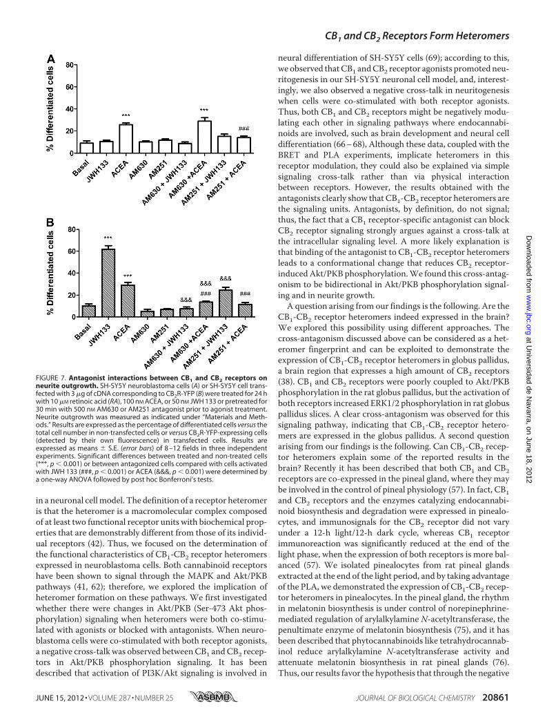
onism observed between CB1 and CB2 receptors in signalingcan also be observed in the cannabinoid receptor-mediatedneuritogenesis. As expected, the CB1 receptor antagonistAM251, but not the CB2 receptor antagonist AM630, com-pletely blocked the CB1 receptor agonist ACEA-inducedneuritogenesis in non-transfected SH-SY5Y cells (Fig. 7A).Interestingly, bothAM251 andAM630were able to prevent theACEA-induced or the CB2 receptor agonist JWH 133-induced
FIGURE 5. CB1-CB2 receptor heteromer fingerprint was found in rat glo-bus pallidus. Globus pallidus slices were obtained as described under “Mate-rials and Methods” and treated for 10 min with 500 nM JWH 133 or 1 �M ACEAor pretreated for 20 min with 5 �M AM630 antagonist prior to agonist treat-ment. ERK1/2 phosphorylation (P-ERK 1/2) was determined as describedunder “Materials and Methods.” Results (means � S.E. (error bars) of two dif-ferent experiments performed in triplicate) are expressed as -fold over basal(non-stimulated cells). Significant differences were analyzed by a one-wayANOVA followed by post hoc Bonferroni’s tests (*, p � 0.05 compared withbasal; #, p � 0.05 compared with the respective agonist-treated cells). Aboveis a representative Western blot.
CB1 and CB2 Receptors Form Heteromers
JUNE 15, 2012 • VOLUME 287 • NUMBER 25 JOURNAL OF BIOLOGICAL CHEMISTRY 20859
at Universidad de N
avarra, on June 18, 2012w
ww
.jbc.orgD
ownloaded from
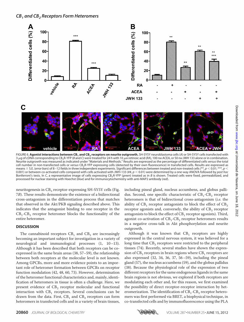
neuritogenesis in CB2 receptor-expressing SH-SY5Y cells (Fig.7B). These results demonstrate the existence of a bidirectionalcross-antagonism in the differentiation process that matchesthat observed in the Akt/PKB signaling described above. Thisindicates that the antagonist binding to one receptor in theCB1-CB2 receptor heteromer blocks the functionality of theentire heteromer.
DISCUSSION
The cannabinoid receptors CB1 and CB2 are increasinglybecoming an important subject for investigation in a variety ofneurological and immunological processes (1, 10–13).Although it has been described that both receptors can be co-expressed in the same brain areas (38, 57–59), the relationshipbetween both receptors at the molecular level is not known.Among GPCRs, more and more evidence points to an impor-tant role of heteromer formation between GPCRs on receptorfunction modulation (42, 48, 60, 73). However, determinationof the heteromer functional characteristics and, mainly, identi-fication of heteromers in tissue is often a challenge. Here, wepresent evidence of CB2 receptor molecular and functionalinteraction with CB1 receptors. Several conclusions can bedrawn from the data. First, CB1 and CB2 receptors can formheteromers in transfected cells and in a variety of brain tissues,
including pineal gland, nucleus accumbens, and globus palli-dus. Second, one specific characteristic of CB1-CB2 receptorheteromers is that of bidirectional cross-antagonism (i.e. theability of CB1 receptor antagonists to block the effect of CB2receptor agonists and, conversely, the ability of CB2 receptorantagonists to block the effect of CB1 receptor agonists). Third,agonist co-activation of CB1-CB2 receptor heteromers resultsin a negative cross-talk in Akt phosphorylation and neuriteoutgrowth.Although it was known that CB1 receptors are highly
expressed in the central nervous system, it was believed for along time that CB2 receptors were restricted to the peripheraltissues (74). Recently, several studies have shown the expres-sion of CB2 receptors in brain regions where CB1 receptors arealso expressed (32, 34, 36, 37, 56–59), including the pinealgland (57), the nucleus accumbens (59), and the globus pallidus(38). Because the physiological role of the expression of twodifferent receptors for the same endogenous ligands in the samebrain regions is not obvious, we explored if both receptors aremodulating each other and, for this reason, we first examinedthe possibility of direct receptor-receptor interaction by het-eromerization. The identification of CB1-CB2 receptor hetero-mers was first performed via BRET, a biophysical technique, inco-transfected cells and by immunofluorescence using the PLA
FIGURE 6. Agonist interactions between CB1 and CB2 receptors on neurite outgrowth. SH-SY5Y neuroblastoma cells (A) or SH-SY5Y cells transfected with3 �g of cDNA corresponding to CB2R-YFP (B and C) were treated for 24 h with 10 �M retinoic acid (RA), 100 nM ACEA, or 50 nM JWH 133 alone or in combination.Neurite outgrowth was measured as indicated under “Materials and Methods.” Results are expressed as the percentage of differentiated cells versus the totalcell number in non-transfected cells or versus CB2R-YFP expressing cells (detected by their own fluorescence) in transfected cells. Results are expressed asmeans � S.E. (error bars) of 8 –12 fields in three independent experiments. Significant differences between treated and non-treated cells (**, p � 0.01; ***, p �0.001) or between co-activated cells compared with cells activated with JWH-133 (##, p � 0.01) were determined by a one-way ANOVA followed by post hocBonferroni’s tests. In C, a representative image of cells expressing CB2R-YFP (green) treated as in B is shown. Treated cells were fixed, permeabilized, andprocessed for nuclear staining with Hoechst (blue) and for immunocytochemistry with anti-MAP2 antibody (red).
CB1 and CB2 Receptors Form Heteromers
20860 JOURNAL OF BIOLOGICAL CHEMISTRY VOLUME 287 • NUMBER 25 • JUNE 15, 2012
at Universidad de N
avarra, on June 18, 2012w
ww
.jbc.orgD
ownloaded from
in a neuronal cellmodel. The definition of a receptor heteromeris that the heteromer is a macromolecular complex composedof at least two functional receptor units with biochemical prop-erties that are demonstrably different from those of its individ-ual receptors (42). Thus, we focused on the determination ofthe functional characteristics of CB1-CB2 receptor heteromersexpressed in neuroblastoma cells. Both cannabinoid receptorshave been shown to signal through the MAPK and Akt/PKBpathways (41, 62); therefore, we explored the implication ofheteromer formation on these pathways. We first investigatedwhether there were changes in Akt/PKB (Ser-473 Akt phos-phorylation) signaling when heteromers were both co-stimu-lated with agonists or blocked with antagonists. When neuro-blastoma cells were co-stimulated with both receptor agonists,a negative cross-talk was observed betweenCB1 andCB2 recep-tors in Akt/PKB phosphorylation signaling. It has beendescribed that activation of PI3K/Akt signaling is involved in
neural differentiation of SH-SY5Y cells (69); according to this,we observed thatCB1 andCB2 receptor agonists promotedneu-ritogenesis in our SH-SY5Y neuronal cell model, and, interest-ingly, we also observed a negative cross-talk in neuritogenesiswhen cells were co-stimulated with both receptor agonists.Thus, both CB1 and CB2 receptors might be negatively modu-lating each other in signaling pathways where endocannabi-noids are involved, such as brain development and neural celldifferentiation (66–68), Although these data, coupled with theBRET and PLA experiments, implicate heteromers in thisreceptor modulation, they could also be explained via simplesignaling cross-talk rather than via physical interactionbetween receptors. However, the results obtained with theantagonists clearly show that CB1-CB2 receptor heteromers arethe signaling units. Antagonists, by definition, do not signal;thus, the fact that a CB1 receptor-specific antagonist can blockCB2 receptor signaling strongly argues against a cross-talk atthe intracellular signaling level. A more likely explanation isthat binding of the antagonist to CB1-CB2 receptor heteromersleads to a conformational change that reduces CB2 receptor-inducedAkt/PKB phosphorylation.We found this cross-antag-onism to be bidirectional in Akt/PKB phosphorylation signal-ing and in neurite growth.A question arising from our findings is the following. Are the
CB1-CB2 receptor heteromers indeed expressed in the brain?We explored this possibility using different approaches. Thecross-antagonism discussed above can be considered as a het-eromer fingerprint and can be exploited to demonstrate theexpression of CB1-CB2 receptor heteromers in globus pallidus,a brain region that expresses a high amount of CB2 receptors(38). CB1 and CB2 receptors were poorly coupled to Akt/PKBphosphorylation in the rat globus pallidus, but the activation ofboth receptors increased ERK1/2 phosphorylation in rat globuspallidus slices. A clear cross-antagonism was observed for thissignaling pathway, indicating that CB1-CB2 receptor hetero-mers are expressed in the globus pallidus. A second questionarising from our findings is the following. Can CB1-CB2 recep-tor heteromers explain some of the reported results in thebrain? Recently it has been described that both CB1 and CB2receptors are co-expressed in the pineal gland, where they maybe involved in the control of pineal physiology (57). In fact, CB1and CB2 receptors and the enzymes catalyzing endocannabi-noid biosynthesis and degradation were expressed in pinealo-cytes, and immunosignals for the CB2 receptor did not varyunder a 12-h light/12-h dark cycle, whereas CB1 receptorimmunoreaction was significantly reduced at the end of thelight phase, when the expression of both receptors is more bal-anced (57). We isolated pinealocytes from rat pineal glandsextracted at the end of the light period, and by taking advantageof the PLA, we demonstrated the expression of CB1-CB2 recep-tor heteromers in pinealocytes. In the pineal gland, the rhythmin melatonin biosynthesis is under control of norepinephrine-mediated regulation of arylalkylamine N-acetyltransferase, thepenultimate enzyme of melatonin biosynthesis (75), and it hasbeen described that phytocannabinoids like tetrahydrocannab-inol reduce arylalkylamine N-acetyltransferase activity andattenuate melatonin biosynthesis in rat pineal glands (76).Thus, our results favor the hypothesis that through the negative
FIGURE 7. Antagonist interactions between CB1 and CB2 receptors onneurite outgrowth. SH-SY5Y neuroblastoma cells (A) or SH-SY5Y cell trans-fected with 3 �g of cDNA corresponding to CB2R-YFP (B) were treated for 24 hwith 10 �M retinoic acid (RA), 100 nM ACEA, or 50 nM JWH 133 or pretreated for30 min with 500 nM AM630 or AM251 antagonist prior to agonist treatment.Neurite outgrowth was measured as indicated under “Materials and Meth-ods.” Results are expressed as the percentage of differentiated cells versus thetotal cell number in non-transfected cells or versus CB2R-YFP-expressing cells(detected by their own fluorescence) in transfected cells. Results areexpressed as means � S.E. (error bars) of 8 –12 fields in three independentexperiments. Significant differences between treated and non-treated cells(***, p � 0.001) or between antagonized cells compared with cells activatedwith JWH 133 (###, p � 0.001) or ACEA (&&&, p � 0.001) were determined bya one-way ANOVA followed by post hoc Bonferroni’s tests.
CB1 and CB2 Receptors Form Heteromers
JUNE 15, 2012 • VOLUME 287 • NUMBER 25 JOURNAL OF BIOLOGICAL CHEMISTRY 20861
at Universidad de N
avarra, on June 18, 2012w
ww
.jbc.orgD
ownloaded from
cross-talk in CB1-CB2 receptor heteromers, CB2 receptor,mainly at the end of the light period, can negatively modulatetheCB1 receptor-mediated tetrahydrocannabinol effect on ary-lalkylamine N-acetyltransferase activity and thus modulatemelatonin synthesis. Another brain region where the CB2receptor expression was reported is the nucleus accumbens,where CB2 receptor may be directly involved in many of theneurochemical and motivational properties of cocaine that areresponsible for addiction (59). In fact, Xi et al. (59) found thatCB2 receptor agonists directly infused into the nucleus accum-bens decreased the cocaine intake, CB2 receptors regulate thepsychomotor stimulant properties of cocaine, and CB2 recep-tors are endogenously activated by endocannabinoids innucleus accumbens, where they control locomotor activity.Because CB1 receptors are also expressed in the striatum (24,28) and are reported to be involved in the locomotor activitycontrol (28), we determined the expression of CB1-CB2 recep-tor heteromers in rat nucleus accumbens slices by PLA. Wefound CB1-CB2 receptor heteromer expression in this brainregion. Although the CB2 receptor-mediated effects on loco-motor activity are also seen in CB1
�/� mice (59), our resultsprovide a new perspective by which CB1 and CB2 receptorsmight bemodulating each other to control locomotion throughCB1-CB2 receptor heteromers, using one partner as a “brake”for the other partner’s actionwhen both are co-expressed in thesame neuron. CB1 receptors are extremely abundant, but CB2receptors, at least in neurons, are thought to bemuch less abun-dant. Altering the expression of CB2 receptors may provide apossible level of regulation of CB1 receptor function by chang-ing the amount of CB1-CB2 receptor heteromers. Further stud-ies will have to be performed to investigate this possibility.Finally, our data may provide explanations for several previ-
ously controversial points concerning CB1 and CB2 receptors.A confounding problem with the cannabinoid receptors hasbeen the expression levels of the two receptors in the brain.Specifically, there have been varying reports as to the amount ofCB2 receptor in the brain and whether those levels change inpathological settings as well as speculation on the role of theneuronal CB2 receptor (18, 31, 34, 35, 37, 38, 77–82). Ourresults suggest that, even at low expression levels, CB2 recep-tors could have a significant effect on signaling fromCB1 recep-tor by reducing the cellular response through CB1 receptor.This modulation could be up-regulated upon injury or disease,supporting the thesis proposed recently byOnaivi (83) that CB1and CB2 receptors can work independently and/or coopera-tively in differing neuronal populations. This interdependencein neurons expressing both receptors could be through hetero-mers. At the ligand level, low and high levels of cannabinoidreceptor ligands have given different results (84, 85), whichwere interpreted as affecting different populations of neurons,or by the presence of another cannabinoid ligand-mediatedreceptor, such as TRPV1 orGPR55, in the same neurons, but analternative explanation could also be the presence of receptorheteromers. Conflicting results have also been observed on theserotonin system at high and low doses of the nonspecific can-nabinoid receptor agonistWIN55212,2. At lowdoses, therewasan increase in neuronal excitation that decreased at higher con-centrations. The authors argued these effects must be through
CB1 receptor because they could be blocked by the CB1 recep-tor-specific antagonist, rimonabant. However, we show herethat a CB1 receptor antagonist could also block CB2 receptor-mediated signaling via receptor heteromers. It would be inter-esting to revisit these experiments using aCB2 receptor-specificantagonist as well. Heteromers could also help explain seem-ingly opposite effects seenwith the ligands 2-arachidonoylglyc-erol and anandamide, which have been reported to have seem-ingly opposite effects on striatal spiny neurons and sensoryneurons of the dorsal root ganglion (reviewed by diMarzo (86)).Part of these effects could also be through differential signalingvia CB1-CB2 receptor heteromers. More studies will berequired to elucidate how these heteromers behave in differenttissues at different concentrations of these two endogenousagonists. Another complication has been the discovery thatmultiple isoforms of the CB2 receptors are expressed. Perhapsthe different isoforms of the CB2 receptor can differentiallymodulate CB1 receptor and/or vice versa, depending on thetissue environment. Further studies may provide a clue as tohow, at the mechanistic level, CB2 receptor is altering CB1receptor signaling or vice versa and help clarify if any differ-ences in isoforms exist. Finally, a third family of the cannabi-noid receptors has recently been proposed, theGPR55 receptor(87, 88), with which a functional interaction with CB2 receptorhas also been described (89, 90). It will be interesting to furthercharacterize its role, if any, in the brain and whether it too canform heteromers with the other members of the family. In con-clusion, we report the presence of CB1-CB2 receptor hetero-mers in a variety of brain regions. These heteromersmay have aprofound impact on CNS function in a variety of neurologicaland immunological systems, and our data suggest that theseheteromers must be taken into account when designing thera-peutic approaches toward alterations involving the endocan-nabinoid system.
Acknowledgment—We thank Jasmina Jiménez (University of Barce-lona) for technical help.
Note Added in Proof—An article by Kleyer et al. (91) showing similarfindings has recently been published.
REFERENCES1. Klein, T. W., Newton, C., Larsen, K., Lu, L., Perkins, I., Nong, L., and
Friedman, H. (2003) The cannabinoid system and immune modulation.J. Leukoc. Biol. 74, 486–496
2. Aguado, T., Monory, K., Palazuelos, J., Stella, N., Cravatt, B., Lutz, B.,Marsicano, G., Kokaia, Z., Guzmán, M., and Galve-Roperh, I. (2005) Theendocannabinoid system drives neural progenitor proliferation. FASEB J.19, 1704–1706
3. Mackie, K., and Stella, N. (2006) Cannabinoid receptors and endocannabi-noids. Evidence for new players. AAPS J. 8, E298–E306
4. Piomelli, D. (2003) The molecular logic of endocannabinoid signaling.Nat. Rev. Neurosci. 4, 873–884
5. Katona, I., Urbán, G.M.,Wallace,M., Ledent, C., Jung, K.M., Piomelli, D.,Mackie, K., and Freund, T. F. (2006) Molecular composition of the endo-cannabinoid system at glutamatergic synapses. J. Neurosci. 26, 5628–5637
6. Freund, T. F., Katona, I., and Piomelli, D. (2003) Role of endogenous can-nabinoids in synaptic signaling. Physiol. Rev. 83, 1017–1066
7. Marsicano, G., Goodenough, S., Monory, K., Hermann, H., Eder,M., Can-nich, A., Azad, S. C., Cascio, M. G., Gutiérrez, S. O., van der Stelt, M.,López-Rodriguez, M. L., Casanova, E., Schütz, G., Zieglgänsberger,W., Di
CB1 and CB2 Receptors Form Heteromers
20862 JOURNAL OF BIOLOGICAL CHEMISTRY VOLUME 287 • NUMBER 25 • JUNE 15, 2012
at Universidad de N
avarra, on June 18, 2012w
ww
.jbc.orgD
ownloaded from
Marzo, V., Behl, C., and Lutz, B. (2003) CB1 cannabinoid receptors andon-demand defense against excitotoxicity. Science 302, 84–88
8. Ameri, A. (1999)The effects of cannabinoids on the brain.Prog.Neurobiol.58, 315–348
9. Howlett, A. C. (2002) The cannabinoid receptors. Prostaglandins OtherLipid Mediat. 68, 619–631
10. Venance, L., Maldonado, R., and Manzoni, O. (2004) [Endocannabinoidsin the central nervous system]Med. Sci. 20, 45–53
11. Schneider, U., Seifert, J., Karst, M., Schlimme, J., Cimander, K., and Mül-ler-Vahl, K. R. (2005) [The endogenous cannabinoid system. Therapeuticimplications for neurologic and psychiatric disorders] Nervenarzt 76,1062, 1065–1066, 1068–1072 passim
12. Ashton, J. C. (2007) Cannabinoids for the treatment of inflammation.Curr. Opin. Investig. Drugs 8, 373–384
13. Cabral, G. A., Raborn, E. S., Griffin, L., Dennis, J., andMarciano-Cabral, F.(2008) CB2 receptors in the brain. Role in central immune function. Br. J.Pharmacol. 153, 240–251
14. Katona, I., and Freund, T. F. (2008) Endocannabinoid signaling as a syn-aptic circuit breaker in neurological disease. Nat. Med. 14, 923–930
15. Romero, J., Lastres-Becker, I., de Miguel, R., Berrendero, F., Ramos, J. A.,and Fernández-Ruiz, J. (2002) The endogenous cannabinoid system andthe basal ganglia. biochemical, pharmacological, and therapeutic aspects.Pharmacol. Ther. 95, 137–152
16. Mackie, K. (2006) Cannabinoid receptors as therapeutic targets. Annu.Rev. Pharmacol. Toxicol. 46, 101–122
17. Palazuelos, J., Aguado, T., Egia, A., Mechoulam, R., Guzmán, M., andGalve-Roperh, I. (2006) Non-psychoactive CB2 cannabinoid agonistsstimulate neural progenitor proliferation. FASEB J. 20, 2405–2407
18. Fernández-Ruiz, J., Romero, J., Velasco, G., Tolón, R.M., Ramos, J. A., andGuzmán, M. (2007) Cannabinoid CB2 receptor. A new target for control-ling neural cell survival? Trends Pharmacol. Sci. 28, 39–45
19. Zanettini, C., Panlilio, L. V., Alicki, M., Goldberg, S. R., Haller, J., andYasar, S. (2011) Effects of endocannabinoid systemmodulation on cogni-tive and emotional behavior. Front. Behav. Neurosci. 5, 57
20. Di Marzo, V., Melck, D., Bisogno, T., and De Petrocellis, L. (1998) Endo-cannabinoids. Endogenous cannabinoid receptor ligands with neuro-modulatory action. Trends Neurosci. 21, 521–528
21. Maldonado, R., Valverde,O., andBerrendero, F. (2006) Involvement of theendocannabinoid system in drug addiction.Trends Neurosci. 29, 225–232
22. Ferré, S., Goldberg, S. R., Lluis, C., and Franco, R. (2009) Looking for therole of cannabinoid receptor heteromers in striatal function. Neurophar-macology 56, Suppl. 1, 226–234
23. Pickel, V. M., Chan, J., Kash, T. L., Rodríguez, J. J., and MacKie, K. (2004)Compartment-specific localization of cannabinoid 1 (CB1) and �-opioidreceptors in rat nucleus accumbens. Neuroscience 127, 101–112
24. Pickel, V. M., Chan, J., Kearn, C. S., and Mackie, K. (2006) Targetingdopamine D2 and cannabinoid-1 (CB1) receptors in rat nucleus accum-bens. J. Comp. Neurol. 495, 299–313
25. Köfalvi, A., Rodrigues, R. J., Ledent, C.,Mackie, K., Vizi, E. S., Cunha, R. A.,and Sperlágh, B. (2005) Involvement of cannabinoid receptors in the reg-ulation of neurotransmitter release in the rodent striatum. A combinedimmunochemical and pharmacological analysis. J. Neurosci. 25,2874–2884
26. Mátyás, F., Yanovsky, Y., Mackie, K., Kelsch,W., Misgeld, U., and Freund,T. F. (2006) Subcellular localization of type 1 cannabinoid receptors in therat basal ganglia. Neuroscience 137, 337–361
27. Uchigashima, M., Narushima, M., Fukaya, M., Katona, I., Kano, M., andWatanabe, M. (2007) Subcellular arrangement of molecules for 2-arachi-donoyl-glycerol-mediated retrograde signaling and its physiological con-tribution to synaptic modulation in the striatum. J. Neurosci. 27,3663–3676
28. Martín, A. B., Fernandez-Espejo, E., Ferrer, B., Gorriti, M. A., Bilbao, A.,Navarro, M., Rodriguez de Fonseca, F., and Moratalla, R. (2008) Expres-sion and function of CB1 receptor in the rat striatum. Localization andeffects onD1 andD2 dopamine receptor-mediatedmotor behaviors.Neu-ropsychopharmacology 33, 1667–1679
29. Svízenská, I., Dubový, P., and Sulcová, A. (2008) Cannabinoid receptors 1and 2 (CB1 and CB2), their distribution, ligands, and functional involve-
ment in nervous system structures. A short review. Pharmacol. Biochem.Behav. 90, 501–511
30. Ashton, J. C., Friberg, D., Darlington, C. L., and Smith, P. F. (2006) Expres-sion of the cannabinoid CB2 receptor in the rat cerebellum. An immuno-histochemical study. Neurosci. Lett. 396, 113–116
31. Brusco, A., Tagliaferro, P. A., Saez, T., and Onaivi, E. S. (2008) Ultrastruc-tural localization of neuronal brain CB2 cannabinoid receptors.Ann. N.Y.Acad. Sci. 1139, 450–457
32. Golech, S. A.,McCarron, R.M., Chen, Y., Bembry, J., Lenz, F.,Mechoulam,R., Shohami, E., and Spatz, M. (2004) Human brain endothelium. Coex-pression and function of vanilloid and endocannabinoid receptors. BrainRes. Mol. Brain Res. 132, 87–92
33. Maresz, K., Carrier, E. J., Ponomarev, E. D., Hillard, C. J., and Dittel, B. N.(2005) Modulation of the cannabinoid CB2 receptor in microglial cells inresponse to inflammatory stimuli. J. Neurochem. 95, 437–445
34. Van Sickle, M. D., Duncan, M., Kingsley, P. J., Mouihate, A., Urbani, P.,Mackie, K., Stella, N.,Makriyannis, A., Piomelli, D., Davison, J. S.,Marnett,L. J., Di Marzo, V., Pittman, Q. J., Patel, K. D., and Sharkey, K. A. (2005)Identification and functional characterization of brainstem cannabinoidCB2 receptors. Science 310, 329–332
35. Gong, J. P., Onaivi, E. S., Ishiguro, H., Liu, Q. R., Tagliaferro, P. A., Brusco,A., andUhl, G. R. (2006) CannabinoidCB2 receptors. Immunohistochem-ical localization in rat brain. Brain Res. 1071, 10–23
36. Onaivi, E. S., Ishiguro, H., Gong, J. P., Patel, S., Perchuk, A., Meozzi, P. A.,Myers, L., Mora, Z., Tagliaferro, P., Gardner, E., Brusco, A., Akinshola,B. E., Liu, Q. R., Hope, B., Iwasaki, S., Arinami, T., Teasenfitz, L., and Uhl,G. R. (2006) Discovery of the presence and functional expression of can-nabinoid CB2 receptors in brain. Ann. N.Y. Acad. Sci. 1074, 514–536
37. Brusco, A., Tagliaferro, P., Saez, T., and Onaivi, E. S. (2008) Postsynapticlocalization of CB2 cannabinoid receptors in the rat hippocampus. Syn-apse 62, 944–949
38. Lanciego, J. L., Barroso-Chinea, P., Rico, A. J., Conte-Perales, L., Callén, L.,Roda, E., Gómez-Bautista, V., López, I. P., Lluis, C., Labandeira-García,J. L., and Franco, R. (2011) Expression of the mRNA coding the cannabi-noid receptor 2 in the pallidal complex of Macaca fascicularis. J. Psycho-pharmacol. 25, 97–104
39. Pertwee, R. G. (1997) Pharmacology of cannabinoid CB1 and CB2 recep-tors. Pharmacol. Ther. 74, 129–180
40. Gómez del Pulgar, T., Velasco, G., and Guzmán, M. (2000) The CB1 can-nabinoid receptor is coupled to the activation of protein kinase B/Akt.Biochem. J. 347, 369–373
41. Molina-Holgado, E., Vela, J.M., Arévalo-Martín, A., Almazán, G.,Molina-Holgado, F., Borrell, J., and Guaza, C. (2002) Cannabinoids promoteoligodendrocyte progenitor survival. Involvement of cannabinoid recep-tors and phosphatidylinositol-3 kinase/Akt signaling. J. Neurosci. 22,9742–9753
42. Ferré, S., Baler, R., Bouvier, M., Caron, M. G., Devi, L. A., Durroux, T.,Fuxe, K., George, S. R., Javitch, J. A., Lohse, M. J., Mackie, K., Milligan, G.,Pfleger, K. D., Pin, J. P., Volkow, N. D., Waldhoer, M., Woods, A. S., andFranco, R. (2009) Building a new conceptual framework for receptor het-eromers. Nat. Chem. Biol. 5, 131–134
43. Carriba, P., Ortiz, O., Patkar, K., Justinova, Z., Stroik, J., Themann, A.,Müller, C., Woods, A. S., Hope, B. T., Ciruela, F., Casadó, V., Canela, E. I.,Lluis, C., Goldberg, S. R., Moratalla, R., Franco, R., and Ferré, S. (2007)Striatal adenosine A2A and cannabinoid CB1 receptors form functionalheteromeric complexes that mediate the motor effects of cannabinoids.Neuropsychopharmacology 32, 2249–2259
44. Carriba, P., Navarro, G., Ciruela, F., Ferré, S., Casadó, V., Agnati, L., Cor-tés, A., Mallol, J., Fuxe, K., Canela, E. I., Lluís, C., and Franco, R. (2008)Detection of heteromerization of more than two proteins by sequentialBRET-FRET. Nat. Methods 5, 727–733
45. Navarro, G., Carriba, P., Gandía, J., Ciruela, F., Casadó, V., Cortés, A.,Mallol, J., Canela, E. I., Lluis, C., and Franco, R. (2008) Detection of het-eromers formed by cannabinoid CB1, dopamine D2, and adenosine A2AG-protein-coupled receptors by combining bimolecular fluorescencecomplementation and bioluminescence energy transfer. ScientificWorld-Journal 8, 1088–1097
46. Tebano, M. T., Martire, A., Chiodi, V., Pepponi, R., Ferrante, A., Do-
CB1 and CB2 Receptors Form Heteromers
JUNE 15, 2012 • VOLUME 287 • NUMBER 25 JOURNAL OF BIOLOGICAL CHEMISTRY 20863
at Universidad de N
avarra, on June 18, 2012w
ww
.jbc.orgD
ownloaded from
menici, M. R., Frank, C., Chen, J. F., Ledent, C., and Popoli, P. (2009)Adenosine A2A receptors enable the synaptic effects of cannabinoid CB1receptors in the rodent striatum. J. Neurochem. 110, 1921–1930
47. Akirav, I., and Fattore, L. (2011) Cannabinoid CB1 and dopamine D1receptors partnership in the modulation of emotional neural processing.Front. Behav. Neurosci. 5, 67
48. Rozenfeld, R., Gupta, A., Gagnidze, K., Lim, M. P., Gomes, I., Lee-Ramos,D., Nieto, N., and Devi, L. A. (2011) AT1R-CBR heteromerization revealsa new mechanism for the pathogenic properties of angiotensin II. EMBOJ. 30, 2350–2363
49. Hojo, M., Sudo, Y., Ando, Y., Minami, K., Takada, M., Matsubara, T.,Kanaide, M., Taniyama, K., Sumikawa, K., and Uezono, Y. (2008) �-Opi-oid receptor forms a functional heterodimerwith cannabinoid CB1 recep-tor. Electrophysiological and FRET assay analysis. J. Pharmacol. Sci. 108,308–319
50. Ward, R. J., Pediani, J. D., and Milligan, G. (2011) Heteromultimerizationof cannabinoid CB1 receptor and orexin OX1 receptor generates a uniquecomplex in which both protomers are regulated by orexin A. J. Biol. Chem.286, 37414–37428
51. da Silveira Cruz-Machado, S., Carvalho-Sousa, C. E., Tamura, E. K., Pi-nato, L., Cecon, E., Fernandes, P. A., de Avellar, M. C., Ferreira, Z. S., andMarkus, R. P. (2010) TLR4 and CD14 receptors expressed in rat pinealgland trigger NFKB pathway. J. Pineal Res. 49, 183–192
52. Mackie, K. (2005) Cannabinoid receptor homo- and heterodimerization.Life Sci. 77, 1667–1673
53. Söderberg, O., Leuchowius, K. J., Gullberg, M., Jarvius, M., Weibrecht, I.,Larsson, L. G., and Landegren, U. (2008) Characterizing proteins and theirinteractions in cells and tissues using the in situ proximity ligation assay.Methods 45, 227–232
54. Trifilieff, P., Rives, M. L., Urizar, E., Piskorowski, R. A., Vishwasrao, H. D.,Castrillon, J., Schmauss, C., Slättman, M., Gullberg, M., and Javitch, J. A.(2011) Detection of antigen interactions ex vivo by proximity ligation as-say. Endogenous dopamine D2-adenosine A2A receptor complexes in thestriatum. BioTechniques 51, 111–118
55. Howlett, A. C., Bidaut-Russell, M., Devane, W. A., Melvin, L. S., Johnson,M. R., andHerkenham,M. (1990)The cannabinoid receptor. Biochemical,anatomical, and behavioral characterization. Trends Neurosci. 13,420–423
56. Núñez, E., Benito, C., Pazos, M. R., Barbachano, A., Fajardo, O., González,S., Tolón, R. M., and Romero, J. (2004) Cannabinoid CB2 receptors areexpressed by perivascular microglial cells in the human brain. An immu-nohistochemical study. Synapse 53, 208–213
57. Koch,M.,Habazettl, I., Dehghani, F., andKorf, H.W. (2008) The rat pinealgland comprises an endocannabinoid system. J. Pineal Res. 45, 351–360
58. Suárez, J., Llorente, R., Romero-Zerbo, S. Y., Mateos, B., Bermúdez-Silva,F. J., de Fonseca, F. R., andViveros,M. P. (2009) Earlymaternal deprivationinduces gender-dependent changes on the expression of hippocampalCB1 and CB2 cannabinoid receptors of neonatal rats. Hippocampus 19,623–632
59. Xi, Z. X., Peng, X. Q., Li, X., Song, R., Zhang, H. Y., Liu, Q. R., Yang, H. J.,Bi, G. H., Li, J., and Gardner, E. L. (2011) Brain cannabinoid CB2 receptorsmodulate cocaine’s actions in mice. Nat. Neurosci. 14, 1160–1166
60. Moreno, E., Vaz, S. H., Cai, N. S., Ferrada, C., Quiroz, C., Barodia, S. K.,Kabbani, N., Canela, E. I., McCormick, P. J., Lluis, C., Franco, R., Ribeiro,J. A., Sebastião, A. M., and Ferré, S. (2011) Dopamine-galanin receptorheteromers modulate cholinergic neurotransmission in the rat ventralhippocampus. J. Neurosci. 31, 7412–7423
61. González, S., Rangel-Barajas, C., Peper, M., Lorenzo, R., Moreno, E., Ciru-ela, F., Borycz, J., Ortiz, J., Lluís, C., Franco, R., McCormick, P. J., Volkow,N. D., Rubinstein, M., Floran, B., and Ferré, S. (2012) Dopamine D4 recep-tor, but not the ADHD-associated D4.7 variant, forms functional hetero-mers with the dopamine D2S receptor in the brain. Mol. Psychiatry 17,650–682
62. Sánchez, M. G., Ruiz-Llorente, L., Sánchez, A. M., and Díaz-Laviada, I.(2003) Activation of phosphoinositide 3-kinase/PKB pathway by CB1 andCB2 cannabinoid receptors expressed in prostate PC-3 cells. Involvementin Raf-1 stimulation and NGF induction. Cell. Signal. 15, 851–859
63. Hosohata, Y., Quock, R. M., Hosohata, K., Makriyannis, A., Consroe, P.,
Roeske, W. R., and Yamamura, H. I. (1997) AM630 antagonism of canna-binoid-stimulated [35S]GTP�S binding in the mouse brain. Eur. J. Phar-macol. 321, R1–R3
64. Pertwee, R. G. (2005) Pharmacological actions of cannabinoids. Handb.Exp. Pharmacol. 168, 1–51
65. Huffman, J. W., Liddle, J., Yu, S., Aung, M. M., Abood, M. E., Wiley, J. L.,and Martin, B. R. (1999) 3-(1�,1�-Dimethylbutyl)-1-deoxy-�8-THC andrelated compounds. Synthesis of selective ligands for the CB2 receptor.Bioorg. Med. Chem. 7, 2905–2914
66. Fernández-Ruiz, J., Gómez,M., Hernández,M., deMiguel, R., and Ramos,J. A. (2004) Cannabinoids and gene expression during brain development.Neurotox. Res. 6, 389–401
67. Harkany, T., Guzmán, M., Galve-Roperh, I., Berghuis, P., Devi, L. A., andMackie, K. (2007) The emerging functions of endocannabinoid signalingduring CNS development. Trends Pharmacol. Sci. 28, 83–92
68. Mulder, J., Aguado, T., Keimpema, E., Barabás, K., Ballester Rosado, C. J.,Nguyen, L., Monory, K., Marsicano, G., Di Marzo, V., Hurd, Y. L., Guil-lemot, F., Mackie, K., Lutz, B., Guzmán, M., Lu, H. C., Galve-Roperh, I.,and Harkany, T. (2008) Endocannabinoid signaling controls pyramidalcell specification and long-range axon patterning. Proc. Natl. Acad. Sci.U.S.A. 105, 8760–8765
69. López-Carballo, G., Moreno, L., Masiá, S., Pérez, P., and Barettino, D.(2002)Activation of the phosphatidylinositol 3-kinase/Akt signaling path-way by retinoic acid is required for neural differentiation of SH-SY5Yhuman neuroblastoma cells. J. Biol. Chem. 277, 25297–25304
70. Påhlman, S., Hoehner, J. C., Nånberg, E., Hedborg, F., Fagerström, S.,Gestblom, C., Johansson, I., Larsson, U., Lavenius, E., andOrtoft, E. (1995)Differentiation and survival influences of growth factors in human neuro-blastoma. Eur. J. Cancer 31A, 453–458
71. Påhlman, S., Ruusala, A. I., Abrahamsson, L.,Mattsson,M. E., and Esscher,T. (1984) Retinoic acid-induced differentiation of cultured human neuro-blastoma cells. A comparison with phorbolester-induced differentiation.Cell Differ. 14, 135–144
72. Encinas, M., Iglesias, M., Liu, Y., Wang, H., Muhaisen, A., Ceña, V., Gal-lego, C., and Comella, J. X. (2000) Sequential treatment of SH-SY5Y cellswith retinoic acid and brain-derived neurotrophic factor gives rise to fullydifferentiated, neurotrophic factor-dependent, human neuron-like cells.J. Neurochem. 75, 991–1003
73. Fiorentini, C., Busi, C., Spano, P., and Missale, C. (2010) Dimerization ofdopamine D1 andD3 receptors in the regulation of striatal function.Curr.Opin. Pharmacol. 10, 87–92
74. Munro, S., Thomas, K. L., and Abu-Shaar, M. (1993) Molecular charac-terization of a peripheral receptor for cannabinoids. Nature 365, 61–65
75. Klein, D. C. (2007) Arylalkylamine N-acetyltransferase. “The Timezyme”.J. Biol. Chem. 282, 4233–4237
76. Koch, M., Dehghani, F., Habazettl, I., Schomerus, C., and Korf, H. W.(2006) Cannabinoids attenuate norepinephrine-induced melatonin bio-synthesis in the rat pineal gland by reducing arylalkylamineN-acetyltrans-ferase activity without involvement of cannabinoid receptors. J. Neuro-chem. 98, 267–278
77. Benito, C., Núñez, E., Tolón, R. M., Carrier, E. J., Rábano, A., Hillard, C. J.,and Romero, J. (2003) Cannabinoid CB2 receptors and fatty acid amidehydrolase are selectively overexpressed in neuritic plaque-associated gliain Alzheimer’s disease brains. J. Neurosci. 23, 11136–11141
78. Ehrhart, J., Obregon, D., Mori, T., Hou, H., Sun, N., Bai, Y., Klein, T.,Fernandez, F., Tan, J., and Shytle, R. D. (2005) Stimulation of cannabinoidreceptor 2 (CB2) suppresses microglial activation. J. Neuroinflammation2, 29
79. Onaivi, E. S., Ishiguro, H., Gong, J. P., Patel, S., Meozzi, P. A., Myers, L.,Perchuk, A., Mora, Z., Tagliaferro, P. A., Gardner, E., Brusco, A., Akin-shola, B. E., Liu, Q. R., Chirwa, S. S., Hope, B., Lujilde, J., Inada, T., Iwasaki,S., Macharia, D., Teasenfitz, L., Arinami, T., and Uhl, G. R. (2008) Func-tional expression of brain neuronal CB2 cannabinoid receptors are in-volved in the effects of drugs of abuse and in depression. Ann. N.Y. Acad.Sci. 1139, 434–449
80. Sagredo, O., González, S., Aroyo, I., Pazos, M. R., Benito, C., Lastres-Becker, I., Romero, J. P., Tolón, R. M., Mechoulam, R., Brouillet, E., Ro-mero, J., and Fernández-Ruiz, J. (2009) Cannabinoid CB2 receptor ago-
CB1 and CB2 Receptors Form Heteromers
20864 JOURNAL OF BIOLOGICAL CHEMISTRY VOLUME 287 • NUMBER 25 • JUNE 15, 2012
at Universidad de N
avarra, on June 18, 2012w
ww
.jbc.orgD
ownloaded from
nists protect the striatum against malonate toxicity. Relevance forHuntington’s disease. Glia 57, 1154–1167
81. García-Gutiérrez, M. S., and Manzanares, J. (2011) Overexpression ofCB2 cannabinoid receptors decreased vulnerability to anxiety and im-paired anxiolytic action of alprazolam in mice. J. Psychopharmacol. 25,111–120
82. Onaivi, E. S., Ishiguro, H., Gu, S., and Liu, Q. R. (2012) CNS effects of CB2cannabinoid receptors. Beyond neuro-immuno-cannabinoid activity.J. Psychopharmacol. 26, 92–103
83. Onaivi, E. S. (2011) Commentary. Functional neuronal CB2 cannabinoidreceptors in the CNS. Curr. Neuropharmacol. 9, 205–208
84. Bambico, F. R., Katz, N., Debonnel, G., andGobbi, G. (2007) Cannabinoidselicit antidepressant-like behavior and activate serotonergic neuronsthrough the medial prefrontal cortex. J. Neurosci. 27, 11700–11711
85. Bellocchio, L., Lafenêtre, P., Cannich, A., Cota, D., Puente, N., Grandes, P.,Chaouloff, F., Piazza, P. V., and Marsicano, G. (2010) Bimodal control ofstimulated food intake by the endocannabinoid system.Nat. Neurosci. 13,281–283
86. Di Marzo, V. (2011) Endocannabinoid signaling in the brain. Biosyntheticmechanisms in the limelight. Nat. Neurosci. 14, 9–15
87. Pertwee, R. G. (2007) GPR55. A newmember of the cannabinoid receptorclan? Br. J. Pharmacol. 152, 984–986
88. Ryberg, E., Larsson, N., Sjögren, S., Hjorth, S., Hermansson, N. O., Le-onova, J., Elebring, T., Nilsson, K., Drmota, T., and Greasley, P. J. (2007)The orphan receptor GPR55 is a novel cannabinoid receptor. Br. J. Phar-macol. 152, 1092–1101
89. Balenga, N. A., Aflaki, E., Kargl, J., Platzer, W., Schröder, R., Blättermann,S., Kostenis, E., Brown, A. J., Heinemann, A., and Waldhoer, M. (2011)GPR55 regulates cannabinoid 2 receptor-mediated responses in humanneutrophils. Cell Res. 21, 1452–1469
90. Irving, A. (2011) New blood brothers. The GPR55 and CB2 partnership.Cell Res. 21, 1391–1392
91. Kleyer, J., Nicolussi, S., Taylor, P., Simonelli, D., Furger, E., Anderle, P., andGertsch, J. (2012) Cannabinoid receptor trafficking in peripheral cells isdynamically regulated by a binary biochemical switch. Biochem. Pharma-col. 83, 1393–1412
CB1 and CB2 Receptors Form Heteromers
JUNE 15, 2012 • VOLUME 287 • NUMBER 25 JOURNAL OF BIOLOGICAL CHEMISTRY 20865
at Universidad de N
avarra, on June 18, 2012w
ww
.jbc.orgD
ownloaded from