calreticulin contributes to c1q-dependent recruitment of microglia in the leech hirudo medicinalis...
TRANSCRIPT
Received: 2013.11.26Accepted: 2013.12.06
Published: 2014.XX.XX
4198 — 5 72
Calreticulin contributes to C1q-dependent recruitment of microglia in the leech Hirudo medicinalis following a CNS injury
DEF Françoise Le Marrec-Croq BCD Annelise Bocquet-Garcon BCD Jacopo Vizioli AB Christelle Vancamp AB Francesco Drago BC Julien Franck BC Maxence Wisztorski FG Michel Salzet DEF Pierre-Eric Sautiere* DEFG Christophe Lefebvre*
* The authors contributed equally to this work Corresponding Author: Christophe Lefebvre. e-mail: [email protected] Source of support: This work was supported by grants from Le Ministère de l’Education Nationale, de l’Enseignement Supérieur et de la Recherche
(MENESR, France) and from L’Agence Nationale de la Recherche (MIMIC, ANR-11-JSV4-005)
Background: The medicinal leech is considered as a complementary and appropriate model to study immune functions in the central nervous system (CNS). In a context in which an injured leech’s CNS can naturally restore normal synaptic connections, the accumulation of microglia (immune cells of the CNS that are exclusively resident in leeches) has been shown to be essential at the lesion to engage the axonal sprouting. HmC1q (Hm for Hirudo medicinalis) possesses chemotactic properties that are important in the microglial cell recruitment by recog-nizing at least a C1q binding protein (HmC1qBP alias gC1qR).
Material/Methods: Recombinant forms of C1q were used in affinity purification and in vitro chemotaxis assays. Anti-calreticulin antibodies were used to neutralize C1q-mediated chemotaxis and locate the production of calreticulin in leech CNS.
Results: A newly characterized leech calreticulin (HmCalR) has been shown to interact with C1q and participate to the HmC1q-dependent microglia accumulation. HmCalR, which has been detected in only some microglial cells, is consequently a second binding protein for HmC1q, allowing the chemoattraction of resident microglia in the nerve repair process.
Conclusions: These data give new insight into calreticulin/C1q interaction in an immune function of neuroprotection, sug-gesting another molecular target to use in investigation of microglia reactivity in a model of CNS injury.
Keywords: C1qandcalreticulininteraction•microgliarecruitment•CNSinjury
Full-text PDF: http://www.medscimonit.com/download/index/idArt/890091
Authors’ Contribution: Study Design A
Data Collection B Statistical Analysis CData Interpretation D
Manuscript Preparation E Literature Search FFunds Collection G
Laboratoire de Spectrométrie de Masse Biologique Fondamentale et Appliquée – EA4550, Université Lille Nord de France, Université Lille 1, Villeneuve d’Ascq, France
e-ISSN 1643-3750© Med Sci Monit, 2014; 20:
DOI: 10.12659/MSM.890091
1Indexed in: [Current Contents/Clinical Medicine] [SCI Expanded] [ISI Alerting System] [ISI Journals Master List] [Index Medicus/MEDLINE] [EMBASE/Excerpta Medica] [Chemical Abstracts/CAS] [Index Copernicus]
This work is licensed under a Creative CommonsAttribution-NonCommercial-NoDerivs 3.0 Unported License
LAB/IN VITRO RESEARCHPR
OO
F ©
INTE
RNAT
ION
AL S
CIEN
TIFI
C IN
FORM
ATIO
N
Background
Microglial cells constitute the resident immune cells, maintain-ing the integrity of the nervous system by constantly surveying the environment [1,2]. Under pathological conditions, they rap-idly change and turn to amoeboid activated microglia in order to migrate over long distances and accumulate at lesions [3].
The microglia in the leech Hirudo medicinalis were the first cells to be named “microglia” by del Rio-Hortega using his silver car-bonate method [4,5] and are considered as the first microglia model [6]. Leech microglial cells represent the only cell popu-lation capable of moving through the nerve cord. Otherwise, they cannot be mistaken with other glial cells that have a larger size and a different shape. Because individual sensory cells regenerate new synaptic connections with a high degree of specificity after a section [7,8], the involvement of microg-lia was investigated in that natural nerve repair process. The leech nerve cord is tubular and is not vascularized within the blood sinus [9,10]. In this context, by using an isolated and injured segment of nerve cord maintained in tissue culture, some authors reported that synapse regeneration is possible following the accumulation of resident microglia at the crush site and without any blood cell [11]. Even in the presence of blood sinus, a very low infiltration of blood cells is observed in injured CNS, which highlights the importance of the resident microglia at the lesions. Their accumulation has been shown to be essential for the usual sprouting of injured axons [12]. The mobility of activated resident microglia has been associated with morphological changes from stellate to rounded shape, comparable to those of activated microglia in mammals [13], and is dependent on specific chemotactic signals [10,14–16]. Among these chemotactic factors, we demonstrated the role of HmC1q (for Hirudo medicinalis) in the microglial cell accu-mulation after leech CNS injury [17].
In mammals, it is well established that C1q recognizes the C1qR(P) receptor to contribute to the phagocytosis of debris by microglia [18,19]. Some authors showed the importance of C1q in the microglial activation [20] and a neuroprotective role for C1q in early inflammatory response [21], but the C1q-dependent recruitment of mammalian microglia at lesions is still poorly understood. The chemotactic properties of C1q have been shown towards blood immune cells [22–24] and some authors specified that such chemotactic activity occurs through the recognition of both gC1qR (alias C1qBP) and cal-reticulin (also known as cC1qR, CalR, or CRT) receptors on pe-ripheral dendritic cells [25].
In the leech CNS, HmC1q contributes to the microglial recruit-ment by interacting with a recently characterized receptor, named HmC1qBP, which is homologous to mammalian gC1qR [26]. The present report shows the involvement of a second
receptor in the C1q-dependent microglial activation. A mole-cule homologous to known calreticulins was characterized and shown to interact with HmC1q. This report establishes that the presence of HmCalR in the leech microglia represents a part of the C1q-dependent reactivity. It also contributes to new in-sight into C1q/calreticulin functions in the CNS.
Material and Methods
Leech CNS and microglial cell preparation
H. medicinalis adult leeches were obtained from Ricarimpex (Eysines, France). The leech nerve cord (CNS) is constituted by the head ganglion, 21 body ganglia, and 7 fused tail gan-glia. The ganglia are joined by structures, called connectives, containing the axonal processes and glial cells [9]. After anes-thesia in 10% ethanol at 4°C for 15 min, animal CNS were dis-sected out in a sterile Ringer solution (115 mM NaCl, 1.8 mM CaCl2, 4 mM KCl, 10 mM Tris maleate, pH 7.4) under a laminar flow hood. After isolation, samples were placed in 3 succes-sive baths of antibiotics (10 UI/ml penicillin, 10 µg/ml strep-tomycin, and 10 µg/ml gentamicin) for 15 min and further in-cubated in Leibovitz L-15 medium (Invitrogen, USA) containing 2 mM L-glutamin, 0.6% glucose, and 10 mM Hepes (complete medium). The experimental injury was performed by crush-ing the connectives between the third and fourth ganglia of an isolated fragment. Nerve cords were used for fluorescence in situ hybridization, whole mount immunohistochemistry, or nerve cell preparation.
For microglial cell isolations, nerve cords treated as indicated above were placed in 35-mm Petri dishes with 200 µl of com-plete L-15 medium. Each ganglion was carefully decapsulated by removing, with micro-scissors, the collagen layer envelop-ing the nerve cord. Nerve cells, neurons, and microglial cells were mechanically dissociated by gentle scraping (total nerve cells). After a filtration through 7-µm nylon mesh as described [13,26], the enriched microglial cell population was then col-lected and centrifuged at 1000 × g for 10 min at RT. The cell pellet was resuspended in L-15 medium (100 µl per nerve cord) for migration assays.
Molecular characterization of HmCalR
The analysis of Hirudo medicinalis genome allowed in sili-co prediction of mRNA databases according to intro-ex-on boundaries. Based on the candidate sequence detect-ed, forward and reverse primers were designed to frame the complete sequence of predicted mRNA. From total RNA ex-tracted from leech nerve cord using TRIzol® reagent and ac-cording to the manufacturer’s procedure (Invitrogen, USA), cDNA were synthesized using an oligo(dT) priming. The
2Indexed in: [Current Contents/Clinical Medicine] [SCI Expanded] [ISI Alerting System] [ISI Journals Master List] [Index Medicus/MEDLINE] [EMBASE/Excerpta Medica] [Chemical Abstracts/CAS] [Index Copernicus]
Le Marrec-Croq F. et al.: Calreticulin contributes to C1q-dependent recruitment of microglia…
© Med Sci Monit, 2014; 20:
This work is licensed under a Creative CommonsAttribution-NonCommercial-NoDerivs 3.0 Unported License
LAB/IN VITRO RESEARCHPR
OO
F ©
INTE
RNAT
ION
AL S
CIEN
TIFI
C IN
FORM
ATIO
N
1
5
10
15
20
25
30
35
40
45
50
53
1
5
10
15
20
25
30
35
40
45
50
53
calreticulin-related molecule was amplified by PCR using the specific forward (5’GGTAGCAATACGTGCAGTTTG3’) and reverse (5’GCAACCAAGAGTAGGCAACC3’) primers and the Platinum® Taq DNA Polymerase according to the manufacturer’s instruc-tions (Invitrogen, USA). Selected PCR products were ligated into pGEM T-easy vector and cloned into JM109 cells accord-ing to the manufacturer’s instructions (Promega, USA). Finally, products were sequenced using BigDye Terminator v3.0 po-lymerization kit before detection on Genetic Analyzer (Applied Biosystems, USA). BLAST programs were used for sequence analysis in databases and comparison with initial predicted mRNA sequence [27,28]. Phylogenetic analysis was carried out by Geneious® Basic v5.6 software [29].
Fluorescent in situ hybridization (FISH)
Nerve cords were fixed for one hour at 4°C in 4% parafor-maldehyde just after dissection. The 5’ biotin-labeled specif-ic antisense probe and sense probe (negative control) were generated from the 842-1479 nucleotides sequence of hmc-alr mRNA (Genbank Accession Number KF709537). After PCR amplification and the insertion of the product in pGEM-T easy vector system (Promega, USA), the RNA sequence of interest was obtained by in vitro transcription using DIG/Biotin RNA-labeling kit according to the manufacturer’s instructions (Roche, Switzerland). The hybridization protocol was performed as previously described [30]. Nerve cords were incubated with a secondary anti-biotin antibody conjugated to Alexa Fluor 488 (dilution 1: 5000 in PBS) (Invitrogen, USA). Samples were rinsed with PBS. Prior to mounting with Glycergel (Sigma Life Science, USA), the cell nuclei were counterstained by Hoechst 33342 fluorescent dye (1:1,000, Invitrogen USA) for 10 min. Slides were kept at 4°C in the dark until observation with a Zeiss LSM700 confocal microscope.
Immunohistochemistry
In experiments with anti-human calreticulin antibody, analy-ses were performed on nerve cords dissected out as described above. They were fixed for 1 hour at 4°C – immediately after dissection (T=0) or 24 h (T24h) after incubation in complete L-15 medium – in 4% paraformaldehyde, washed in PBS, per-meabilized by a 24-h incubation at RT in 1% Triton X100 in PBS and pre-incubated for 8 h at RT in 1% Triton, 3% Normal Donkey Serum (NDS) and 1% ovalbumin in PBS. Samples were then incubated overnight at 4ºC with specific rabbit polyclonal anti-human calreticulin antibodies (Santa Cruz Biotechnology, USA) diluted in a PBS solution (1:250) containing 1% BSA, 0.05% Triton, 1% NDS, and 1% ovalbumin. The anti-calreticulin antibodies were directed against an antigen sequence corre-sponding to the Lys248-Leu417 region of human calreticulin, pre-senting 81% homology with leech calreticulin. After 3 washes with PBS, samples were incubated 1 h at room temperature
with anti-rabbit donkey antibody (Invitrogen, USA) conjugat-ed to Alexa Fluor 488 (1:2000) in a PBS solution containing 1% BSA, 0.05% Triton, 1% NDS, and 1% ovalbumin. Final rins-ing and mounting steps for confocal microscopy observation were performed as described above. Prior to mounting, the cell nuclei were counterstained by Hoechst 33342 fluorescent dye (1: 1000, Invitrogen USA) for 10 min. Samples without the addition of primary antibody were used as negative control.
Human C1q biotinylation and streptavidin affinity purification
The biotinylation of the recombinant human C1q (Prospecbio, USA) was carried out by using the Sulfo-NHS-SS-Biotin kit (Pierce, USA) according to the manufacturer’s instructions. Unreacted Sulfo-NHS-SS-Biotin was removed using the Zeba Desalt Spin Columns (Pierce, USA).
Biotinylated human C1q was immediately fixed onto a strepta-vidin column (Pierce, USA), previously equilibrated with 5 vol-umes of PBS 0.1M. The interaction between biotin and strep-tavidin occurred at RT for 10 min. Microglia protein extract (800 µg) was added in the column, incubated overnight at 4°C, and rinsed 10 times with PBS 0.1M. Captured microglial cell proteins were eluted from the streptavidin-agarose with 5% 2-mercaptoethanol-PBS 0.1 M at 30°C for 30 min. Proteins were precipitated in 10% trichloroacetic acid/acetone at –20°C for 45 min and centrifuged at 13000 × g for 15 min. The protein pellet was washed in cold acetone, air dried and dissolved in Læmmli buffer. Two other columns were used for the negative controls: the first containing the biotinylated human C1q with-out any microglia protein extract, and the second containing only the microglia protein extract to evaluate the unspecific reaction between streptavidin and microglial cell components. Samples were loaded on a 12% SDS-PAGE. Separated proteins were transferred to Amersham™ Hybond™-ECL nitrocellulose (GE Healthcare, France). The membrane was incubated for 30 min at RT in blocking solution (0.05% Tween, milk powder 5% w/v in PBS) and then overnight at 4°C in rabbit polyclonal an-ti-human calreticulin antibodies (Santa Cruz Biotechnology, USA), diluted at 1:5000 in blocking solution containing 1% ovalbumin. After rinsing with PBS-0.05% Tween, the mem-brane was incubated for 1 h at RT in secondary goat anti-rab-bit polyclonal antibodies conjugated with horseradish perox-idase (Jackson Immunoresearch, USA) (dilution 1:20000). The signal was revealed using the ECL Kit SuperSignal West Dura chemoluminescent substrate (Pierce, USA) on Kodak X-Omat LS film (Sigma-Aldrich, USA).
Chemotaxis assays
In vitro chemotaxis assays were performed by using the double-P assay as described by Köhidai et al. with minor modifications
3Indexed in: [Current Contents/Clinical Medicine] [SCI Expanded] [ISI Alerting System] [ISI Journals Master List] [Index Medicus/MEDLINE] [EMBASE/Excerpta Medica] [Chemical Abstracts/CAS] [Index Copernicus]
Le Marrec-Croq F. et al.: Calreticulin contributes to C1q-dependent recruitment of microglia…© Med Sci Monit, 2014; 20:
This work is licensed under a Creative CommonsAttribution-NonCommercial-NoDerivs 3.0 Unported License
LAB/IN VITRO RESEARCH
PRO
OF
© IN
TERN
ATIO
NAL
SCI
ENTI
FIC
INFO
RMAT
ION
1
5
10
15
20
25
30
35
40
45
50
53
1
5
10
15
20
25
30
35
40
45
50
53
[31]. Thirty-five millimeter Petri dishes were filled with 1 ml of a 0.5% agar and 1% gelatin solution. After drying, two 6-mm diameter wells were done, each presenting a parallel individ-ual channel. One well was filled with 50 µl of purified microg-lial cells (see above) and the other with chemotactic factor or negative controls reagents. A channel was further created per-pendicularly to others using a coverslip. One hour later, cells in the well containing chemoattractant were collected. Either vehicle (non-transformed Pischia pastoris culture supernatant) or recombinant HmC1q (rHmC1q) produced in P. pastoris [17] alone or with soluble CalR were used (8 µl) as chemotactic fac-tors. For inhibitory chemotactic experiments, cells were pre-incubated for 1 h at RT either with rabbit polyclonal anti-hu-man calreticulin antibodies (1:1000) or with rabbit IgG isotype as negative control (1:1000). The number of migrating cells
was counted on a hemocytometer (3 different counts) under an Axioskop microscope (Zeiss, Germany). Experiments were done in triplicate. The results are expressed as the mean cell number ±S.D. Comparisons between means were made using Student’s t-test. Statistical differences were considered to be significant if p was <0.01.
Results
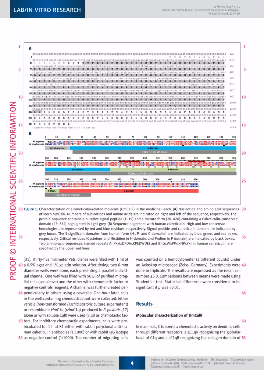
Molecular characterization of HmCalR
In mammals, C1q exerts a chemotactic activity on dendritic cells through different receptors: a gC1qR recognizing the globular head of C1q and a cC1qR recognizing the collagen domain of
A
B
Figure 1. Characterization of a calreticulin-related molecule (HmCalR) in the medicinal leech. (A) Nucleotide and amino acid sequences of leech HmCalR. Numbers of nucleotides and amino acids are indicated on right and left of the sequence, respectively. The protein sequence contains a putative signal peptide (1–19) and a mature form (20–420) containing a Calreticulin-conserved domain (23–334) highlighted in light grey. (B) Sequence alignment with human calreticulin. High and low consensus homologies are represented by red and blue residues, respectively. Signal peptide and calreticulin domain are indicated by grey boxes. The 3 significant domains from human form (N-, P- and C-domains) are indicated by blue, green, and red boxes, respectively. Critical residues (Cysteines and Histidine in N-domain, and Proline in P-domain) are indicated by black boxes. Two amino-acid sequences, named repeats A (PxxIxDPDAxKPEDWDE) and B (GxWxPPxIxNPxYx) in human calreticulin are specified by the upper red lines.
4Indexed in: [Current Contents/Clinical Medicine] [SCI Expanded] [ISI Alerting System] [ISI Journals Master List] [Index Medicus/MEDLINE] [EMBASE/Excerpta Medica] [Chemical Abstracts/CAS] [Index Copernicus]
Le Marrec-Croq F. et al.: Calreticulin contributes to C1q-dependent recruitment of microglia…
© Med Sci Monit, 2014; 20:
This work is licensed under a Creative CommonsAttribution-NonCommercial-NoDerivs 3.0 Unported License
LAB/IN VITRO RESEARCHPR
OO
F ©
INTE
RNAT
ION
AL S
CIEN
TIFI
C IN
FORM
ATIO
N
1
5
10
15
20
25
30
35
40
45
50
53
1
5
10
15
20
25
30
35
40
45
50
53

C1q. The presence of a putative calreticulin (cC1qR) was in-vestigated in the leech H. medicinalis. Based on the H. me-dicinalis genome analysis, a sequence homologous to known calreticulin sequences was detected. The design of specific primers then allowed the amplification of a full-length mRNA sequence for a calreticulin-related molecule, named HmCalR (for Hirudo medicinalis Calreticulin). Hmcalr mRNA (Genbank KF709537) encodes a 420-amino-acid sequence with a theo-retical molecular weight of 48 610 Da. The protein sequence presents a putative signal peptide and a calreticulin domain in Met1-Ser19 and in Val23-Ile334 amino-acid regions, respective-ly (Figure 1A). In addition, the analysis of HmCalR protein us-ing a BLAST-P program shows a high conservation with hu-man calreticulin, particularly in different domains that are already described for mammalian calreticulins [32]. As pre-sented in Figure 1B, HmCalR might possess a single disulfide bridge (Cys107-Cys139) and contains a single histidine residue (His172). These residues were also described in an N-domain of the human mature calreticulin in Cys88-Cys120 and His153 po-sitions, respectively, (numerated from the signal peptide-free sequence) and were demonstrated as essential for the chap-eroning function [33,34]. The HmCalR sequence is also Proline-rich and exhibits 2 repeated amino-acid sequences – repeats A (PxxIxDPDAxKPEDWDE) and B (GxWxPPxIxNPxYx) – as ob-served in the human form’s P-domain (Figure 1B). In leeches, some non-essential amino acids have been substituted with different ones having equivalent physicochemical properties (Glu214 instead of Asp in human repeat A; and Val268 instead of Ile in human repeat B). Finally, the high conservation of nu-merous acidic residues in HmCalR suggests the presence of a C-domain, as in mammals.
Thus HmCalR presents sequence similarities with mamma-lian calreticulins and also with other forms from vertebrate, protostomian, and diploblastic species, as revealed by a
neighbor-joining phylogenetic tree using selected sequences among them (Figure 2). Depending on the 200 highest similar-ities between calreticulin molecules and HmCalR, some were chosen to check the evolutive position of HmCalR according to species. The distribution of sequences in the cladogram respects the species organization between vertebrates and protostomi-ans. The neighbor-joining phylogenetic tree shows that leech calreticulin is positioned in lophotrochozoan organisms shar-ing the same origin with Eisenia andrei, another annelid organ-ism, and also close to mollusks. Arthropods have calreticulins that form a distinct group in protostomians, which is relevant in their distinction from the lophotrochozoans. Otherwise, al-though diploblastic organisms, cnidarians present higher sim-ilarities in calreticulin sequences with lophotrochozoans than with those of arthropods. Finally, vertebrate calreticulins are positioned in a distinct clade.
Localization of hmcalr mRNA and HmCalR protein in leech microglia
Cells expressing hmcalr transcripts in the leech nervous sys-tem were investigated by specific fluorescence in situ hybrid-ization (FISH) on injured nerve cords after different times.
Amblyomma cooperi (Tick)Ixodes scapularis (Tick)Apis mellifera (Honey bee)Nasonia vitripennis (Wasp)
Hyriopsis cumingii (Triangle sail mussel)Crossotrea gigas (Paci�c oyster)Pinctada fucata (Pearl oyster)Salmo solar (Atlantic salmon)Danio rerio (Zebra�sh)Tetraodon nigroviridis (Green spotted pu�er)
Homo sapiens (Human)Mus musculus (House mouse)Rattus norvegicus (Brown rat)
Xenopus leavis (African clawed frog)Gallus gallus (Red jungle�ow)
Hydra magnipapillata (Hydra)Nematostella vectensis (Starlet sea anemona)
Arthropods
Cnidarians
Annelids
Mollusks
AmphibiansBirds
Mammals
Actinopterygians
Eisenia andrei (Tiger worm)Hirudo medicinalis (Medicinal leech)
Figure 2. Neighbor-joining phylogenetic tree relating amino acid sequences of Hirudo medicinalis calreticulin (HmCalR) and calreticulins of selected species. Calreticulin amino acid sequences were chosen in the 200 highest similarities after a BLAST-P program obtained from the NCBI. The calreticulin sequences from the following species (including protein accession numbers) present a respective homology with HmCalR: Amblyomma cooperi (AAR29934) 70.5%, Ixodes scapularis (AAQ18696) 69.5%, Apis mellifera (XP_392689) 67.9%, Nasonia vitripennis (NP_001155151) 63.4%, Hydra magnipapillata (XP_002161300) 66.4%, Nematostella vectensis (XP_001640172) 77.1%, Eisenia andrei (ABI74618) 80%, Hyriopsis cumingii (AFR69202) 75.2%, Crassostrea gigas (BAF63639) 76.5%, Pinctada fucata (ABR68546) 75.7%, Salmo salar (ACI33338) 66.2%, Danio rerio (NP_956007) 68.8%, Tetraodon nigroviridis (CAG07986) 69.7%, Xenopus laevis (NP_001080765) 69.5%, Gallus gallus (AAS49610) 69.0%, Homo sapiens (NP_004334) 71.8%, Mus musculus (NP_031617) 72%, and Rattus norvegicus (NP_071794) 71.2%.
5Indexed in: [Current Contents/Clinical Medicine] [SCI Expanded] [ISI Alerting System] [ISI Journals Master List] [Index Medicus/MEDLINE] [EMBASE/Excerpta Medica] [Chemical Abstracts/CAS] [Index Copernicus]
Le Marrec-Croq F. et al.: Calreticulin contributes to C1q-dependent recruitment of microglia…© Med Sci Monit, 2014; 20:
This work is licensed under a Creative CommonsAttribution-NonCommercial-NoDerivs 3.0 Unported License
LAB/IN VITRO RESEARCH
PRO
OF
© IN
TERN
ATIO
NAL
SCI
ENTI
FIC
INFO
RMAT
ION
1
5
10
15
20
25
30
35
40
45
50
53
1
5
10
15
20
25
30
35
40
45
50
53
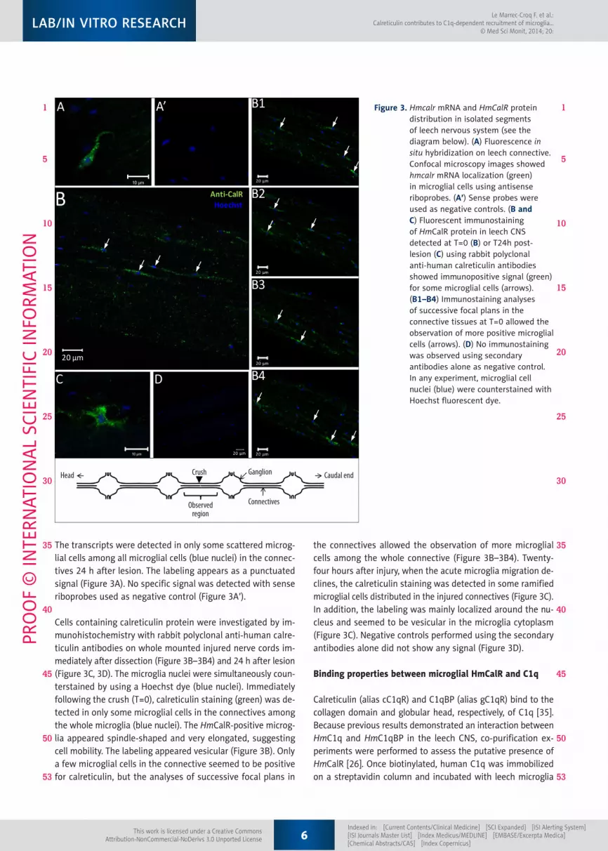
The transcripts were detected in only some scattered microg-lial cells among all microglial cells (blue nuclei) in the connec-tives 24 h after lesion. The labeling appears as a punctuated signal (Figure 3A). No specific signal was detected with sense riboprobes used as negative control (Figure 3A’).
Cells containing calreticulin protein were investigated by im-munohistochemistry with rabbit polyclonal anti-human calre-ticulin antibodies on whole mounted injured nerve cords im-mediately after dissection (Figure 3B–3B4) and 24 h after lesion (Figure 3C, 3D). The microglia nuclei were simultaneously coun-terstained by using a Hoechst dye (blue nuclei). Immediately following the crush (T=0), calreticulin staining (green) was de-tected in only some microglial cells in the connectives among the whole microglia (blue nuclei). The HmCalR-positive microg-lia appeared spindle-shaped and very elongated, suggesting cell mobility. The labeling appeared vesicular (Figure 3B). Only a few microglial cells in the connective seemed to be positive for calreticulin, but the analyses of successive focal plans in
the connectives allowed the observation of more microglial cells among the whole connective (Figure 3B–3B4). Twenty-four hours after injury, when the acute microglia migration de-clines, the calreticulin staining was detected in some ramified microglial cells distributed in the injured connectives (Figure 3C). In addition, the labeling was mainly localized around the nu-cleus and seemed to be vesicular in the microglia cytoplasm (Figure 3C). Negative controls performed using the secondary antibodies alone did not show any signal (Figure 3D).
Binding properties between microglial HmCalR and C1q
Calreticulin (alias cC1qR) and C1qBP (alias gC1qR) bind to the collagen domain and globular head, respectively, of C1q [35]. Because previous results demonstrated an interaction between HmC1q and HmC1qBP in the leech CNS, co-purification ex-periments were performed to assess the putative presence of HmCalR [26]. Once biotinylated, human C1q was immobilized on a streptavidin column and incubated with leech microglia
Figure 3. Hmcalr mRNA and HmCalR protein distribution in isolated segments of leech nervous system (see the diagram below). (A) Fluorescence in situ hybridization on leech connective. Confocal microscopy images showed hmcalr mRNA localization (green) in microglial cells using antisense riboprobes. (A’) Sense probes were used as negative controls. (B and C) Fluorescent immunostaining of HmCalR protein in leech CNS detected at T=0 (B) or T24h post-lesion (C) using rabbit polyclonal anti-human calreticulin antibodies showed immunopositive signal (green) for some microglial cells (arrows). (B1–B4) Immunostaining analyses of successive focal plans in the connective tissues at T=0 allowed the observation of more positive microglial cells (arrows). (D) No immunostaining was observed using secondary antibodies alone as negative control. In any experiment, microglial cell nuclei (blue) were counterstained with Hoechst fluorescent dye.
Head Crush
Observedregion
Ganglion
Connectives
Caudal end
6Indexed in: [Current Contents/Clinical Medicine] [SCI Expanded] [ISI Alerting System] [ISI Journals Master List] [Index Medicus/MEDLINE] [EMBASE/Excerpta Medica] [Chemical Abstracts/CAS] [Index Copernicus]
Le Marrec-Croq F. et al.: Calreticulin contributes to C1q-dependent recruitment of microglia…
© Med Sci Monit, 2014; 20:
This work is licensed under a Creative CommonsAttribution-NonCommercial-NoDerivs 3.0 Unported License
LAB/IN VITRO RESEARCHPR
OO
F ©
INTE
RNAT
ION
AL S
CIEN
TIFI
C IN
FORM
ATIO
N
1
5
10
15
20
25
30
35
40
45
50
53
1
5
10
15
20
25
30
35
40
45
50
53
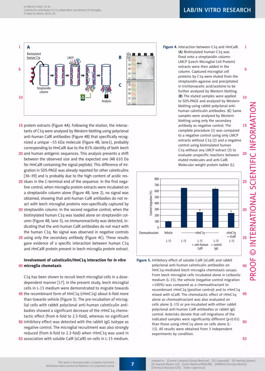
protein extracts (Figure 4A). Following the elution, the interac-tants of C1q were analyzed by Western blotting using polyclonal anti-human CalR antibodies (Figure 4B) that specifically recog-nized a unique ~55 kDa molecule (Figure 4B, lane1), probably corresponding to HmCalR due to the 81% identity of both leech and human antigenic sequences. This analysis presents a shift between the observed size and the expected one (48 610 Da for HmCalR containing the signal peptide). This difference of mi-gration in SDS-PAGE was already reported for other calreticulins [36–39] and is probably due to the high content of acidic res-idues in the C-terminal end of the sequence. In the first nega-tive control, when microglia protein extracts were incubated on a streptavidin column alone (Figure 4B, lane 2), no signal was obtained, showing that anti-human CalR antibodies do not re-act with leech microglial proteins non-specifically captured by streptavidin column. In the second negative control, when the biotinylated human C1q was loaded alone on streptavidin col-umn (Figure 4B, lane 3), no immunoreactivity was detected, in-dicating that the anti-human CalR antibodies do not react with the human C1q. No signal was observed in negative controls using only the secondary antibody (Figure 4C). These results gave evidence of a specific interaction between human C1q and HmCalR protein present in leech microglia protein extract.
Involvement of calreticulin/HmC1q interaction for in vitro microglia chemotaxis
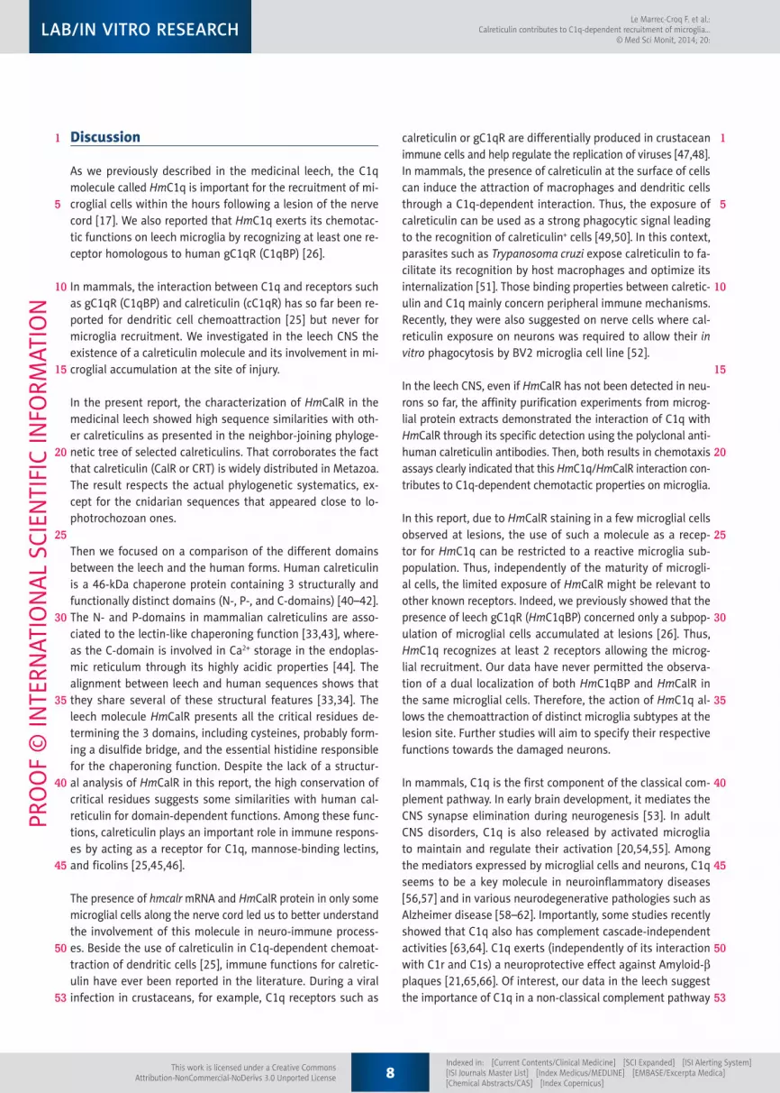
C1q has been shown to recruit leech microglial cells in a dose-dependent manner [17]. In the present study, leech microglial cells in L-15 medium were demonstrated to migrate towards the recombinant form of HmC1q (rHmC1q) about 6-fold more than towards vehicle (Figure 5). The pre-incubation of microg-lial cells with rabbit polyclonal anti-human calreticulin anti-bodies showed a significant decrease of the rHmC1q chemo-tactic effect (from 6-fold to 2.5-fold), whereas no significant inhibitory effect was detected with the rabbit IgG isotype as negative control. The microglial recruitment was also strongly reduced (from 6-fold to 2.3-fold) when rHmC1q was used in association with soluble CalR (sCalR) on cells in L-15 medium.
Figure 4. Interaction between C1q and HmCalR. (A) Biotinylated human C1q was fixed onto a streptavidin column. LMCP (Leech Microglial Cell Protein) extracts were then added in the column. Captured microglial cell proteins by C1q were eluted from the streptavidin-agarose and precipitated in trichloroacetic acid/acetone to be further analyzed by Western blotting. (B) The eluted samples were applied to SDS-PAGE and analyzed by Western blotting using rabbit polyclonal anti-human calreticulin antibodies. (C) Same samples were analyzed by Western blotting using only the secondary antibody as negative control. The complete procedure (1) was compared to a negative control using only LMCP extracts without C1q (2) and a negative control using biotinylated human C1q without any LMCP extract (3) to evaluate unspecific reactions between eluted molecules and anti-CalR. Molecular weight protein ladder (L).
Biotinylatedhuman C1q LMCP
Streptavidincolumn
Analysis1
1 2 3LkDa100
705040
35
25100
705040
35
25
A
B
C
Figure 5. Inhibitory effect of soluble CalR (sCalR) and rabbit polyclonal anti-human calreticulin antibodies on HmC1q-mediated leech microglia chemotaxis assays. From leech microglial cells incubated alone in Leibovitz medium (L-15), the vehicle (negative control migration =100%) was compared as a chemoattractant to recombinant rHmC1q (positive control) and to rHmC1q mixed with sCalR. The chemotactic effect of rHmC1q alone as chemoattractant was also evaluated on cells alone (L-15) or pre-incubated with either rabbit polyclonal anti-human CalR antibodies or rabbit IgG control. Asterisks denote that cell migrations of the indicated samples were significantly different (p<0.01) than those using rHmC1q alone on cells alone (L-15). All results were obtained from 3 independent experiments by condition.
800
700
600
500
400
300
200
100
0Chemoattractant Vehicle
L-15 L-15
* *
L-15+ anti-human
CaIR
L-15+ control
IgG
L-15
rHmC1q+ sCaIR
rHmC1q
Cont
rol m
igrat
ion (%
)
7Indexed in: [Current Contents/Clinical Medicine] [SCI Expanded] [ISI Alerting System] [ISI Journals Master List] [Index Medicus/MEDLINE] [EMBASE/Excerpta Medica] [Chemical Abstracts/CAS] [Index Copernicus]
Le Marrec-Croq F. et al.: Calreticulin contributes to C1q-dependent recruitment of microglia…© Med Sci Monit, 2014; 20:
This work is licensed under a Creative CommonsAttribution-NonCommercial-NoDerivs 3.0 Unported License
LAB/IN VITRO RESEARCH
PRO
OF
© IN
TERN
ATIO
NAL
SCI
ENTI
FIC
INFO
RMAT
ION
1
5
10
15
20
25
30
35
40
45
50
53
1
5
10
15
20
25
30
35
40
45
50
53
Discussion
As we previously described in the medicinal leech, the C1q molecule called HmC1q is important for the recruitment of mi-croglial cells within the hours following a lesion of the nerve cord [17]. We also reported that HmC1q exerts its chemotac-tic functions on leech microglia by recognizing at least one re-ceptor homologous to human gC1qR (C1qBP) [26].
In mammals, the interaction between C1q and receptors such as gC1qR (C1qBP) and calreticulin (cC1qR) has so far been re-ported for dendritic cell chemoattraction [25] but never for microglia recruitment. We investigated in the leech CNS the existence of a calreticulin molecule and its involvement in mi-croglial accumulation at the site of injury.
In the present report, the characterization of HmCalR in the medicinal leech showed high sequence similarities with oth-er calreticulins as presented in the neighbor-joining phyloge-netic tree of selected calreticulins. That corroborates the fact that calreticulin (CalR or CRT) is widely distributed in Metazoa. The result respects the actual phylogenetic systematics, ex-cept for the cnidarian sequences that appeared close to lo-photrochozoan ones.
Then we focused on a comparison of the different domains between the leech and the human forms. Human calreticulin is a 46-kDa chaperone protein containing 3 structurally and functionally distinct domains (N-, P-, and C-domains) [40–42]. The N- and P-domains in mammalian calreticulins are asso-ciated to the lectin-like chaperoning function [33,43], where-as the C-domain is involved in Ca2+ storage in the endoplas-mic reticulum through its highly acidic properties [44]. The alignment between leech and human sequences shows that they share several of these structural features [33,34]. The leech molecule HmCalR presents all the critical residues de-termining the 3 domains, including cysteines, probably form-ing a disulfide bridge, and the essential histidine responsible for the chaperoning function. Despite the lack of a structur-al analysis of HmCalR in this report, the high conservation of critical residues suggests some similarities with human cal-reticulin for domain-dependent functions. Among these func-tions, calreticulin plays an important role in immune respons-es by acting as a receptor for C1q, mannose-binding lectins, and ficolins [25,45,46].
The presence of hmcalr mRNA and HmCalR protein in only some microglial cells along the nerve cord led us to better understand the involvement of this molecule in neuro-immune process-es. Beside the use of calreticulin in C1q-dependent chemoat-traction of dendritic cells [25], immune functions for calretic-ulin have ever been reported in the literature. During a viral infection in crustaceans, for example, C1q receptors such as
calreticulin or gC1qR are differentially produced in crustacean immune cells and help regulate the replication of viruses [47,48]. In mammals, the presence of calreticulin at the surface of cells can induce the attraction of macrophages and dendritic cells through a C1q-dependent interaction. Thus, the exposure of calreticulin can be used as a strong phagocytic signal leading to the recognition of calreticulin+ cells [49,50]. In this context, parasites such as Trypanosoma cruzi expose calreticulin to fa-cilitate its recognition by host macrophages and optimize its internalization [51]. Those binding properties between calretic-ulin and C1q mainly concern peripheral immune mechanisms. Recently, they were also suggested on nerve cells where cal-reticulin exposure on neurons was required to allow their in vitro phagocytosis by BV2 microglia cell line [52].
In the leech CNS, even if HmCalR has not been detected in neu-rons so far, the affinity purification experiments from microg-lial protein extracts demonstrated the interaction of C1q with HmCalR through its specific detection using the polyclonal anti-human calreticulin antibodies. Then, both results in chemotaxis assays clearly indicated that this HmC1q/HmCalR interaction con-tributes to C1q-dependent chemotactic properties on microglia.
In this report, due to HmCalR staining in a few microglial cells observed at lesions, the use of such a molecule as a recep-tor for HmC1q can be restricted to a reactive microglia sub-population. Thus, independently of the maturity of microgli-al cells, the limited exposure of HmCalR might be relevant to other known receptors. Indeed, we previously showed that the presence of leech gC1qR (HmC1qBP) concerned only a subpop-ulation of microglial cells accumulated at lesions [26]. Thus, HmC1q recognizes at least 2 receptors allowing the microg-lial recruitment. Our data have never permitted the observa-tion of a dual localization of both HmC1qBP and HmCalR in the same microglial cells. Therefore, the action of HmC1q al-lows the chemoattraction of distinct microglia subtypes at the lesion site. Further studies will aim to specify their respective functions towards the damaged neurons.
In mammals, C1q is the first component of the classical com-plement pathway. In early brain development, it mediates the CNS synapse elimination during neurogenesis [53]. In adult CNS disorders, C1q is also released by activated microglia to maintain and regulate their activation [20,54,55]. Among the mediators expressed by microglial cells and neurons, C1q seems to be a key molecule in neuroinflammatory diseases [56,57] and in various neurodegenerative pathologies such as Alzheimer disease [58–62]. Importantly, some studies recently showed that C1q also has complement cascade-independent activities [63,64]. C1q exerts (independently of its interaction with C1r and C1s) a neuroprotective effect against Amyloid-b plaques [21,65,66]. Of interest, our data in the leech suggest the importance of C1q in a non-classical complement pathway
8Indexed in: [Current Contents/Clinical Medicine] [SCI Expanded] [ISI Alerting System] [ISI Journals Master List] [Index Medicus/MEDLINE] [EMBASE/Excerpta Medica] [Chemical Abstracts/CAS] [Index Copernicus]
Le Marrec-Croq F. et al.: Calreticulin contributes to C1q-dependent recruitment of microglia…
© Med Sci Monit, 2014; 20:
This work is licensed under a Creative CommonsAttribution-NonCommercial-NoDerivs 3.0 Unported License
LAB/IN VITRO RESEARCHPR
OO
F ©
INTE
RNAT
ION
AL S
CIEN
TIFI
C IN
FORM
ATIO
N
1
5
10
15
20
25
30
35
40
45
50
53
1
5
10
15
20
25
30
35
40
45
50
53

because the analysis of H. medicinalis genome revealed neither C1r- nor C1s-related molecule. By taking into account the im-portance of microglia accumulation at a lesion to engage the axonal sprouting in the leech CNS [12], HmC1q might recruit specific microglial cells that lead to a neuroprotective process.
In addition, other chemotactic factors allow the microglial accu-mulation at a lesion in the leech CNS [13,67] and also use spe-cific receptors. Thus, HmC1q, as well as HmIL-16 and HmEMAPII, allow the accumulation of microglia. So we must now consid-er the leech microglia as a mixture of reactive microglia sub-populations where distinct sets can be recruited through the influence of different chemotactic molecules. In this context, microglia probably contribute to different pro- and/or anti-in-flammatory mechanisms by communicating with other glial cells and neurons. Further studies will investigate the discrim-ination of microglia subpopulations that are recruited to the lesions. They will specify the time course of the accumulation and the specific functions of subpopulations.
Conclusions
The complexity of immune responses following a CNS dam-age is enhanced by multiple cell origin and activation states of microglia [68]. In mammals, the resident microglia that re-sult from the invasion processes during embryonic neurogen-esis are helped by infiltrated bone marrow-derived cells and circulating monocytes during CNS diseases [69]. Nevertheless,
despite in vitro cell analyses, morphological and/or histologi-cal in vivo studies do not permit discrimination of the resident and infiltrated cell types [70]. Besides the cell origin, different functional profiles can simultaneously act and exhibit pro-in-flammatory features (classical activation, M1) or anti-inflam-matory features (alternative activation, M2) depending on the recruitment of specific microglia subtypes at lesions [71]. In this context, comparative immune studies may help to distin-guish specific microglial responses.
The medicinal leech as a model in nerve repair has been used for decades because it represents a new insight into study of the cell processes engaged during the axonal sprouting [72]. In addition, the leech microglia has been demonstrated to be essential for efficient nerve repair [12]. Because they are not supported by a significant infiltration of blood cells, microglial cells in the leech represent an alternate model of interest in comparative microg-lial studies. According to dynamic and functional properties, the microglia subpopulations may be actively involved in the repair capabilities. The impact of microglia recruitment under the in-fluence of HmCalR/HmC1q interaction will be studied to spec-ify the microglial contribution in CNS inflammatory regulation.
Acknowledgements
Authors would like to thank Dr. Elodie Richard of the CCMIC-Université Lille 1 (BICeL) and Pr Natalia Prevarskaya (INSERM U1003, Université Lille 1) for access to confocal microscopy facilities.
References:
1. Nimmerjahn A, Kirchhoff F, Helmchen F: Resting microglial cells are high-ly dynamic surveillants of brain parenchyma in vivo. Science, 2005; 308: 1314–18
2. Olson JK, Miller SD: Microglia initiate central nervous system innate and adaptive immune responses through multiple TLRs. J Immunol, 2004; 173: 3916–24
3. Hanisch UK, Kettenmann H: Microglia: active sensor and versatile effector cells in the normal and pathologic brain. Nat Neurosci, 2007; 10: 1387–94
4. Del Rio-Hortega P: La microglia y su transformacion en células en baston-cito y cuerpos granulo-adiposos. Trab del Lab de Invest Biol, 1920; 18: 37 [in French]
5. Del Rio-Hortega P: Cytology and cellular pathology of the nervous system. Microglia in W Penfield (ed.), P B Hoebaer, New York, NY, 1932; 483–534
6. Sieger D, Peri F: Animal models for studying microglia: The first, the popu-lar, and the new. Glia, 2013; 61: 3–9
7. Baylor DA, Nicholls JG: Patterns of regeneration between individual nerve cells in the central nervous system of the leech. Nature, 1971; 232: 268–70
8. Jansen JK, Nicholls JG: Regeneration and changes in synaptic connections between individual nerve cells in the central nervous system of the leech. Proc Natl Acad Sci USA, 1972; 69: 636–39
9. Coggeshall RE, Fawcett DW: The Fine Structure of the Central Nervous System of the Leech, Hirudo Medicinalis. J Neurophysiol, 1964; 27: 229–89
10. Le Marrec-Croq F, Drago F, Vizioli J et al: The Leech Nervous System: A Valuable Model to Study the Microglia Involvement in Regenerative Processes. Clinical and Developmental Immunology, 2013; 2013: 12
11. Morgese VJ, Elliott EJ, Muller KJ: Microglial movement to sites of nerve le-sion in the leech CNS. Brain Res, 1983; 272: 166–70
12. Ngu EM, Sahley CL, Muller KJ: Reduced axon sprouting after treatment that diminishes microglia accumulation at lesions in the leech CNS. J Comp Neurol, 2007; 503: 101–9
13. Croq F, Vizioli J, Tuzova M et al: A homologous form of human interleukin 16 is implicated in microglia recruitment following nervous system injury in leech Hirudo medicinalis. Glia, 2010; 58: 1649–62
14. Arafah K, Croix D, Vizioli J et al: Involvement of nitric oxide through endo-cannabinoids release in microglia activation during the course of CNS re-generation in the medicinal leech. Glia, 2013; 61: 636–49
15. Duan Y, Sahley CL, Muller KJ: ATP and NO dually control migration of mi-croglia to nerve lesions. Dev Neurobiol, 2009; 69: 60–72
16. Samuels SE, Lipitz JB, Dahl G, Muller KJ: Neuroglial ATP release through in-nexin channels controls microglial cell movement to a nerve injury. J Gen Physiol, 2010; 136: 425–42
17. Tahtouh M, Croq F, Vizioli J et al: Evidence for a novel chemotactic C1q do-main-containing factor in the leech nerve cord. Mol Immunol, 2009; 46: 523–31
18. Nepomuceno RR, Henschen-Edman AH, Burgess WH, Tenner AJ: cDNA clon-ing and primary structure analysis of C1qR(P), the human C1q/MBL/SPA receptor that mediates enhanced phagocytosis in vitro. Immunity, 1997; 6: 119–29
19. Webster SD, Yang AJ, Margol L et al: Complement component C1q modu-lates the phagocytosis of Abeta by microglia. Exp Neurol, 2000; 161: 127–38
20. Farber K, Cheung G, Mitchell D et al: C1q, the recognition subcomponent of the classical pathway of complement, drives microglial activation. J Neurosci Res, 2009; 87: 644–52
9Indexed in: [Current Contents/Clinical Medicine] [SCI Expanded] [ISI Alerting System] [ISI Journals Master List] [Index Medicus/MEDLINE] [EMBASE/Excerpta Medica] [Chemical Abstracts/CAS] [Index Copernicus]
Le Marrec-Croq F. et al.: Calreticulin contributes to C1q-dependent recruitment of microglia…© Med Sci Monit, 2014; 20:
This work is licensed under a Creative CommonsAttribution-NonCommercial-NoDerivs 3.0 Unported License
LAB/IN VITRO RESEARCH
PRO
OF
© IN
TERN
ATIO
NAL
SCI
ENTI
FIC
INFO
RMAT
ION
1
5
10
15
20
25
30
35
40
45
50
53
1
5
10
15
20
25
30
35
40
45
50
53

21. Fraser DA, Pisalyaput K, Tenner AJ: C1q enhances microglial clearance of apoptotic neurons and neuronal blebs, and modulates subsequent inflam-matory cytokine production. J Neurochem, 2010; 112: 733–43
22. Leigh LE, Ghebrehiwet B, Perera TP et al: C1q-mediated chemotaxis by hu-man neutrophils: involvement of gClqR and G-protein signalling mecha-nisms. Biochem J, 1998; 330 (Pt.1): 247–54
23. Kuna P, Iyer M, Peerschke EI et al: Human C1q induces eosinophil migra-tion. Clin Immunol Immunopathol, 1996; 81: 48–54
24. Ghebrehiwet B, Kew RR, Gruber BL et al: Murine mast cells express two types of C1q receptors that are involved in the induction of chemotaxis and chemokinesis. J Immunol, 1995; 155: 2614–19
25. Vegh Z, Kew RR, Gruber BL, Ghebrehiwet B: Chemotaxis of human mono-cyte-derived dendritic cells to complement component C1q is mediated by the receptors gC1qR and cC1qR. Mol Immunol, 2006; 43: 1402–7
26. Tahtouh M, Garcon-Bocquet A, Croq F et al: Interaction of HmC1q with leech microglial cells: involvement of C1qBP-related molecule in the induction of cell chemotaxis. J Neuroinflammation, 2012; 9: 37
27. Altschul SF, Madden TL, Schaffer AA et al: Gapped BLAST and PSI-BLAST: a new generation of protein database search programs. Nucleic Acids Res, 1997; 25: 3389–402
28. Karlin S, Altschul SF: Methods for assessing the statistical significance of molecular sequence features by using general scoring schemes. Proc Natl Acad Sci USA, 1990; 87: 2264–68
29. Kearse M, Moir R, Wilson A et al: Geneious Basic: an integrated and ex-tendable desktop software platform for the organization and analysis of sequence data. Bioinformatics, 2012; 28: 1647–49
30. Nardelli-Haefliger D, Shankland M: Lox2, a putative leech segment identi-ty gene, is expressed in the same segmental domain in different stem cell lineages. Development, 1992; 116: 697–710
31. Kohidai L: Method for determination of chemoattraction in Tetrahymena pyriformis. Curr Microbiol, 1995; 30: 251–53
32. Gelebart P, Opas M, Michalak M: Calreticulin, a Ca2+-binding chaperone of the endoplasmic reticulum. Int J Biochem Cell Biol, 2005; 37: 260–66
33. Guo L, Groenendyk J, Papp S et al: Identification of an N-domain histidine essential for chaperone function in calreticulin. J Biol Chem, 2003; 278: 50645–53
34. Michalak M, Robert Parker JM, Opas M: Ca2+ signaling and calcium binding chaperones of the endoplasmic reticulum. Cell Calcium, 2002; 32: 269–78
35. Kishore U, Reid KB: C1q: structure, function, and receptors. Immunopharmacology, 2000; 49: 159–70
36. Baksh S, Michalak M: Expression of calreticulin in Escherichia coli and iden-tification of its Ca2+ binding domains. J Biol Chem, 1991; 266: 21458–65
37. Rokeach LA, Haselby JA, Hoch SO: High-level bacterial expression, purifica-tion and characterization of human calreticulin. Protein Eng, 1991; 4: 981–87
38. Krause KH: Ca(2+)-storage organelles. FEBS Lett, 1991; 285: 225–29
39. Milner RE, Baksh S, Shemanko C et al: Calreticulin, and not calsequestrin, is the major calcium binding protein of smooth muscle sarcoplasmic retic-ulum and liver endoplasmic reticulum. J Biol Chem, 1991; 266: 7155–65
40. Ellgaard L, Riek R, Braun D et al: Three-dimensional structure topology of the calreticulin P-domain based on NMR assignment. FEBS Lett, 2001; 488: 69–73
41. Ellgaard L, Riek R, Herrmann T et al: NMR structure of the calreticulin P-domain. Proc Natl Acad Sci USA, 2001; 98: 3133–38
42. Schrag JD, Bergeron JJ, Li Y et al: The Structure of calnexin, an ER chaper-one involved in quality control of protein folding. Mol Cell, 2001; 8: 633–44
43. Michalak M, Corbett EF, Mesaeli N et al: Calreticulin: one protein, one gene, many functions. Biochem J, 1999; 344 Pt 2: 281–92
44. Nakamura K, Zuppini A, Arnaudeau S et al: Functional specialization of cal-reticulin domains. J Cell Biol, 2001; 154: 961–72
45. Lacroix M, Dumestre-Perard C, Schoehn G et al: Residue Lys57 in the col-lagen-like region of human L-ficolin and its counterpart Lys47 in H-ficolin play a key role in the interaction with the mannan-binding lectin-associat-ed serine proteases and the collectin receptor calreticulin. J Immunol, 2009; 182: 456–65
46. Pagh R, Duus K, Laursen I, et al: The chaperone and potential mannan-binding lectin (MBL) co-receptor calreticulin interacts with MBL through the binding site for MBL-associated serine proteases. Febs J, 2008; 275: 515–26
47. Wang HC, Leu JH, Kou GH et al: Protein expression profiling of the shrimp cellular response to white spot syndrome virus infection. Dev Comp Immunol, 2007; 31: 672–86
48. Watthanasurorot A, Jiravanichpaisal P, Soderhall I, Soderhall K: A gC1qR prevents white spot syndrome virus replication in the freshwater crayfish Pacifastacus leniusculus. J Virol, 2010; 84: 10844–51
49. Bohlson SS, Fraser DA, Tenner AJ: Complement proteins C1q and MBL are pattern recognition molecules that signal immediate and long-term pro-tective immune functions. Mol Immunol, 2007; 44: 33–43
50. Obeid M, Tesniere A, Ghiringhelli F et al: Calreticulin exposure dictates the immunogenicity of cancer cell death. Nat Med, 2007; 13: 54–61
51. Ramirez G, Valck C, Molina MC et al: Trypanosoma cruzi calreticulin: a nov-el virulence factor that binds complement C1 on the parasite surface and promotes infectivity. Immunobiology, 2011; 216: 265–73
52. Fricker M, Oliva-Martin MJ, Brown GC: Primary phagocytosis of viable neu-rons by microglia activated with LPS or Abeta is dependent on calreticu-lin/LRP phagocytic signalling. J Neuroinflammation, 2012; 9: 196
53. Stevens B, Allen NJ, Vazquez LE et al: The classical complement cascade mediates CNS synapse elimination. Cell, 2007; 131: 1164–78
54. Lynch NJ, Willis CL, Nolan CC et al: Microglial activation and increased syn-thesis of complement component C1q precedes blood-brain barrier dys-function in rats. Mol Immunol, 2004; 40: 709–16
55. Mariani MM, Kielian T: Microglia in Infectious Diseases of the Central Nervous System. J Neuroimmune Pharmacol, 2009; 4(4): 448–61
56. Chen PC, Wang CR, Liu MF et al: Correlation between the renal C1q depo-sition and serum anti-C1q antibody: a potential role of anti-C1q antibody in lupus nephritis. Asian Pac J Allergy Immunol, 2002; 20: 223–27
57. Trendelenburg M: Antibodies against C1q in patients with systemic lupus erythematosus. Springer Semin Immunopathol, 2005; 27: 276–85
58. Bergamaschini L, Donarini C, Gobbo G et al: Activation of complement and contact system in Alzheimer’s disease. Mech Ageing Dev, 2001; 122: 1971–83
59. Tacnet-Delorme P, Chevallier S, Arlaud GJ: Beta-amyloid fibrils activate the C1 complex of complement under physiological conditions: evidence for a binding site for A beta on the C1q globular regions. J Immunol, 2001; 167: 6374–81
60. Fonseca MI, Kawas CH, Troncoso JC, Tenner AJ: Neuronal localization of C1q in preclinical Alzheimer’s disease. Neurobiol Dis, 2004; 15: 40–46
61. Fonseca MI, Zhou J, Botto M, Tenner AJ: Absence of C1q leads to less neu-ropathology in transgenic mouse models of Alzheimer’s disease. J Neurosci, 2004; 24: 6457–65
62. Stephan AH, Madison DV, Mateos JM et al: A dramatic increase of C1q pro-tein in the CNS during normal aging. J Neurosci, 2013; 33: 13460–74
63. Nayak A, Pednekar L, Reid KB, Kishore U: Complement and non-comple-ment activating functions of C1q: a prototypical innate immune molecule. Innate Immun, 2012; 18: 350–63
64. Nayak A, Ferluga J, Tsolaki AG, Kishore U: The non-classical functions of the classical complement pathway recognition subcomponent C1q. Immunol Lett, 2010; 131: 139–50
65. Benoit ME, Hernandez MX, Dinh ML et al: C1q-induced LRP1B and GPR6 proteins expressed early in Alzheimer disease mouse models, are essen-tial for the C1q-mediated protection against amyloid-beta neurotoxicity. J Biol Chem, 2013; 288: 654–65
66. Benoit ME, Tenner AJ: Complement protein C1q-mediated neuroprotection is correlated with regulation of neuronal gene and microRNA expression. J Neurosci, 2011; 31: 3459–69
67. Schikorski D, Cuvillier-Hot V, Boidin-Wichlacz C et al: Deciphering the im-mune function and regulation by a TLR of the cytokine EMAPII in the le-sioned central nervous system using a leech model. J Immunol, 2009; 183: 7119–28
68. Lively S, Schlichter LC: The microglial activation state regulates migration and roles of matrix-dissolving enzymes for invasion. J Neuroinflammation, 2013; 10: 75
69. Mildner A, Schmidt H, Nitsche M et al: Microglia in the adult brain arise from Ly-6ChiCCR2+ monocytes only under defined host conditions. Nat Neurosci, 2007; 10: 1544–53
70. Ransohoff RM: Microgliosis: the questions shape the answers. Nat Neurosci, 2007; 10: 1507–9
71. Prinz M, Mildner A: Microglia in the CNS: Immigrants from another world. Glia, 2011; 59: 177–87
72. von Bernhardi R, Muller KJ: Repair of the central nervous system: lessons from lesions in leeches. J Neurobiol, 1995; 27: 353–66
10Indexed in: [Current Contents/Clinical Medicine] [SCI Expanded] [ISI Alerting System] [ISI Journals Master List] [Index Medicus/MEDLINE] [EMBASE/Excerpta Medica] [Chemical Abstracts/CAS] [Index Copernicus]
Le Marrec-Croq F. et al.: Calreticulin contributes to C1q-dependent recruitment of microglia…
© Med Sci Monit, 2014; 20:
This work is licensed under a Creative CommonsAttribution-NonCommercial-NoDerivs 3.0 Unported License
LAB/IN VITRO RESEARCHPR
OO
F ©
INTE
RNAT
ION
AL S
CIEN
TIFI
C IN
FORM
ATIO
N
1
5
10
15
20
25
30
35
40
45
50
53
1
5
10
15
20
25
30
35
40
45
50
53