bridging traditional knowledge & natural products
TRANSCRIPT

i
0
Editors
Mary Khoo Gaik Hong Chee Beng Jin Getha Krishnasamy
Mazura Md. Pisar Firdaus Kamarulzaman
Bridging Traditional Knowledge &
Natural Products Innovations
Towards Wellness and
Shared Prosperity
Ministry of Energy and Natural Resources


Bridging Traditional Knowledge
& Natural Products Innovations
Towards Wellness and
Shared Prosperity


Bridging Traditional Knowledge
& Natural Products Innovations
Towards Wellness and
Shared Prosperity
Editors
Mary Khoo Gaik Hong Chee Beng Jin Getha Krishnasamy
Mazura Md. Pisar Firdaus Kamarulzaman
https://www.frim.gov.my/publication/maps_tk
2021
Ministry of Energy and Natural Resources

©Forest Research Institute Malaysia 2021 All enquiries should be forwarded to: Director-General Forest Research Institute Malaysia 52109 Kepong Selangor Darul Ehsan Malaysia Tel: +603-62797000 Fax: +603-62731314 http://www.frim.gov.my Perpustakaan Negara Malaysia Cataloguing-in-Publication Data Bridging Traditional Knowledge & Natural Products Innovations Towards Wellness and
Shared Prosperity / Editors: Mary Khoo Gaik Hong, Chee Beng Jin, Getha Krishnasamy, Mazura Md. Pisar & Firdaus Kamarulzaman
Mode of access: Internet eISBN 978-967-2810-05-6
1. Ethnoscience. 2. Traditional medicine. 3. Alternative medicine. 4. Traditional ecological knowledge. 5. Government publication—Malaysia. 6. Electronic books. I. Khoo, Mary Gaik Hong. II. Chee, Beng Jin. III. Getha Krishnasamy. IV. Mazura Md. Pisar. V. Firdaus Kamarulzaman. 306.42
MS ISO 9001:2015


v
Content
Foreword viii
Preface ix
TRADITIONAL KNOWLEDGE, AGRONOMY AND CONSERVATION
Apparatus used in midwifery practice of traditional Malay medicine in Peninsular Malaysia Ida Farah A et al.
2
An overview of herbal medicines as adjunct treatment for cancer in selected hospitals within Ministry of Health Malaysia Teo CS et al.
7
Evaluation of planting media for mass production and yield of tuba roots Angela THG et al.
12
Effects of good agriculture practice on the growth, total phenolic contents and antioxidant status of Labisia pumila var. alata Syafiqah Nabilah SB et al.
16
Observation on affected areas of inoculated Aquilaria malaccensis Abd Majid J et al.
21
Anatomical characterisation of Phyllanthus emblica L.: A plant with therapeutic potential Ummu Hani B et al.
26
Conservation of medicinal and aromatic plants in the Ethnobotanical Garden of FRIM: An effort in tackling biodiversity loss crisis Tan AL et al.
31
Perubatan traditional: Bukan sekadar Jampi Nik Musa’adah M et al.
35
Kajian awal: Kesan penggunaan baja terhadap kadar pertumbuhan dan hasil tongkat ali di Stesen Penyelidikan FRIM, Maran Pahang Syazwan A et al.
40
Kajian perbandingan penanaman ABP016 secara berkelompok di dua lokasi berbeza oleh suku kaum Temiar dan Semelai Siti Salwana H et al.
45
Komposisi dan analisis vegetasi spesies tumbuhan ubatan berpotensi berasaskan pengetahuan tradisi orang asli dalam Hutan Simpan Bera, Tapak Ramsar, Pahang Madihah MN et al.
51
Status kemandirian koleksi tumbuhan ubatan berasaskan pengetahuan tradisi Melayu di Laman Pengetahuan Tradisi, Taman Ethnobotani, FRIM Norbaiah MY et al.
56
NATURAL PRODUCTS DISCOVERY
In vitro antiproliferative activity of dichloromethane (DCM) fraction of Brucea javanica (L.) Merr. (melada pahit) root on selected prostate cancer cell line Murizal Z et al.
62

vi
Cytotoxicity and glutathione level of methanolic leaves extract from Canarium odonthophyllum Miq. (dabai) against UVB induced B164A5 melanoma cell lines Ahmad Rohi G et al.
66
Antioxidant activities of ethanolic extract of Vitex negundo leaves dried at different temperature Ihsan Safwan K et al.
71
TPA-induced mouse ear oedema inhibitory activity of Piper betle leaves extract Ong BK et al.
77
In vitro evaluation of antiinflammatory activity of Melaleuca cajuputi Powell Mazura MP et al.
81
Biological profiling of Swietenia macrophylla supercritical carbon dioxide extract Saidatul Husni S et al.
87
Study of different extracts of kadok (Piper sarmentosum) leaves on the flavonoid profiles and selected bioactivities Zunoliza A et al.
92
Evaluation of a topical formulation containing novel active ingredient from basidiomycetes to treat MRSA skin infections Getha Krishnasamy et al.
99
Comparative study on production kinetics of antibacterial metabolites from batch fermentation of basidiomycete strain FRIM550 in stirred-tank bioreactor Shariffah Nurhidayah SAR et al.
105
Cell based assay for toxicity assessment: A case study of Baeckea frutescens Khoo MGH et al.
111
A preliminary toxicity evaluation of NeoTrai in Sprague Dawley rats Chee BJ et al.
118
Single oral dose 14-day toxicity study of Brucea javanica (melada pahit) fruit on Sprague Dawley rats Teh BP et al.
124
Acute toxicity evaluation of Phaleria macrocarpa (mahkota dewa) leaves and fruits crude extracts in Sprague Dawley rats Norzahirah A et al.
129
Acute oral toxicity study on Lignosus rhinoceros and Curcuma zedoaria Lalitha Suganthi S et al.
134
Preliminary in vivo toxicity assessment of Parkia speciosa (petai) seed, Averrhoa bilimbi (belimbing buluh) fruit and Garcinia mangostana (manggis) rind Nor Azlina Z et al.
139
Acute oral toxicity study of Carica papaya, Syzygium polyanthum, Chromolaena odorata and Annona muricata on Sprague Dawley rats Elda Nurafnie IR et al.
145
Flavonoid-saponin extract from leaves of Mitragyna speciosa as hepatoprotective agent against paracetamol-induced toxicity in rats Hidayatul Khamariah ZA et al.
150
GC-MS analysis of terpenoids from leaves of Canarium odontophyllum Miq. (dabai) Muhammad Wahizul Haswan AA et al.
154

vii
Volatile constituents, antiinflammatory and anticollagenase effect of essential oils from four Cymbopogon species Mailina J et al.
159
Analysis of Cymbopogon winterianus essential oil compounds by means of GC-MS and Z-score technique Khairul Anis Athirah K et al.
165
Chemical composition of Melaleuca cajuputi essential oils from three different locations Mohd Shafik Yuzman T et al.
170
Chemical composition of Meistera ochrea essential oils Nor Azah MA et al.
174
Chemical composition of Hevea brasiliensis seeds (RRIM 2001 clone) Nor Hassifi S
178
Penilaian aktiviti antikanser secara in vitro terhadap sampel tumbuhan UGG004 Nurhanan MY et al.
184
Penilaian aktiviti antiinflamasi bagi spesies ABP 016 dari tiga lokasi terpilih Nurul Haslinda M et al.
188
Pencirian nano perak daun semambu (Azadirachta indica A. Juss) melalui sintesis hijau Noor Rasyila MN et al.
192
Penilaian potensi bioaktiviti SBJ 015 berdasarkan pengetahuan tradisi Orang Asli Bateq di Pahang Shalini M et al.
198
STANDARDISATION, PRODUCT DEVELOPMENT & QUALITY CONTROL
The establishment of three scales of agarwood oils quality index (AOQI) using electronic nose data with Z-score scaling technique Sahrim L et al.
205
Chemical standardisation and quantification of piperine from black peppercorns extract for product development Wong CM & Ling JJ
212
Development of DEET-free mosquito repellent from pepper waste Ling JJ.
217
Real-time stability studies of Baeckea frutescens dried raw materials on quality control element Nurhazwani MH et al.
222
Environment control in natural product quality control laboratory: Air monitoring Amira Rina Nurdiana MS et al.
227
Phenotypic identification of Pseudomonas aeruginosa in herbal based products using biology semi-automated identification system Norulaiman Y et al.
231
Pengoptimuman dan validasi ke atas kaedah ujian bahan campur palsu steroids menggunakan kromatografi cecair bertekanan tinggi Nor Hayati A et al.
235

viii
Foreword Assalamualaikum WBT and greetings! The effort to combine traditional knowledge with modern scientific studies is a good endeavour that will empower and promote the importance of traditional knowledge. This effort will also ensure a fair and equitable sharing of resources with resource owners under the Access to Biological Resources and Benefit Sharing (ABS) Act 2017.
As a country with rich biological diversity, efforts to strengthen natural resource management are critical as a form of mitigation to climate change and natural disasters. Various measures have been taken by the Ministry of Energy and Natural Resources (KeTSA) including working towards realising the establishment of the Malaysian Biodiversity Centre, which serves as a biodiversity research and conservation centre as well as the National Competent Authority to enforce the ABS Act. Through the enactment and enforcement of this act, Malaysia can perform its obligations under the Convention on Biological Diversity (CBD).
The richness in natural resources including medicinal plants is a blessing to Malaysia. Meanwhile, our local wisdom of previous generations using plants in traditional treatment is a treasure warrants to be preserved. In recent times, health and cosmetic products of natural origin are increasingly in demand due to the high awareness of environmental protection as well as the risk of harmful chemicals to consumers. This is a good prospect for researchers to produce high value and safe research output to meet the present trends.
In addition, the recent world situation in facing the unexpected COVID-19 pandemic has also opened the eyes of many that research on infectious diseases and public health should take precedence. In this aspect, the Natural Products Division at FRIM can be a key pillar that plays an imperative role in upholding the knowledge of local traditions through its strengths in scientific research, development and commercialisation.
It is the aspirations and policies of the Malaysian government to increase efforts as well as the use of local natural resources to produce high value products and innovations for the prosperity of the country. I hope that researchers and the policy makers will always work together to shoulder the responsibility of preserving and conserving the country's natural resources as well as biodiversity to ensure the sustainability of our present-day society and the future generations.
This book is a collection of short scientific papers on traditional knowledge and natural product research as well as innovations from researchers in Malaysia. The basis of this publication stemmed from the passion for knowledge-sharing and as a better preservation method of research findings. I hope that this book is able to reach its goal in sharing the work conducted by these scientists to be reviewed as reference by others. Dr. Khali Aziz Hamzah Director General of FRIM

ix
Preface Malaysian culture demonstrates a rich and unique potpourri of knowledge and practices originated from its multiracial and diverse cultural society. The traditional knowledge practiced by the Malay, Chinese, Indian and the orang asli continues to sustain and maintain their community livelihood from the olden days to the present age. Their dependence on the forests is vital which serves as their sustainable green pharmacies. Apparently, the maintenance of good wellbeing and prevention of illness are much accentuated on food and usage of natural remedies from the surroundings, especially from plants and other natural resources. In the current fast-paced modern society, traditional knowledge which encompasses traditional remedies and folk cures gradually faced extinction and very soon to be long forgotten.
The Natural Products Division at the Forest Research Institute Malaysia (FRIM) had come a long way in pioneering research and discoveries of medicinal and aromatic plants along with local traditional knowledge after receiving its mandate from the Government of Malaysia in 1995. The institute had forged collaborations with renowned local and international academics, research institutions and herbal industries in technology transfer, training, sharing of expertise and product development.
This book celebrates Natural Products Division’s involvement for a period of more than 25
years of research in the importance of natural resources related to medicinal plants and microbes in Malaysia. The content of the book acknowledged the tireless labour, years of hard work and patience of scientists and researchers in the field of traditional knowledge, conservation, agronomy, natural products discovery, standardisation and processing technology, product development and commercialisation, quality control as well as issues on regulatory and standards.
The title “Bridging Traditional Knowledge & Natural Product Innovations Towards
Wellness and Shared Prosperity” signifies the effort and aspiration to combine traditional knowledge with new scientific studies to promote measures tending to the betterment of the society in Malaysia. We sincerely hope that the scientific findings contributed by our fellow colleagues and contributors will complement and strengthen each other’s discoveries to produce common solutions to a pressing issue for the benefit of this nation. The Editorial Team Natural Products Division @ FRIM

TRADITIONAL KNOWLEDGE, AGRONOMY AND CONSERVATION

2
APPARATUS USED IN MIDWIFERY PRACTISE OF TRADITIONAL MALAY MEDICINE IN PENINSULAR MALAYSIA Ida Farah A, Nur Salsabeela MR, Siti Hajar MR, Noorashikin AH, Nurmaziah MS, Terence TYC & Ami Fazlin SM Herbal Medicine Research Centre, Institute for Medical Research, National Institutes of Health, Ministry of Health Malaysia, No 1 Persiaran Setia Murni U13/52, Seksyen U13 Setia Alam, 40170 Shah Alam, Selangor, Malaysia. Tel: 03-33627962 Fax: 03-3362 7903 Email: [email protected]
ABSTRACT Apparatus use is vital in traditional Malay medicine (TMM) practices since it is highly acknowledged and widely used. Continuous and comprehensive documentation is needed to ensure the preservation of local ancestral knowledge for future generations. The Malaysian government has taken the initiative to conduct a project titled “Comprehensive Documentation of Malay Traditional Knowledge on Medicinal Plants in Peninsular Malaysia” through the participation of various agencies led by the Forest Research Institute Malaysia (FRIM). The Institute for Medical Research (IMR) is a collaborating agency entrusted to execute documentation of use and collection of apparatus used in TMM, including postnatal care. The objective of this study was to document the apparatus used in postnatal and neonatal care based on TMM knowledge. A preliminary census by FRIM produced a database of TMM practitioners in Peninsular Malaysia. Participants were selected from several states in this database identified by their practice of postnatal and neonatal care which involves the use of traditional apparatus. Data collection involved questionnaire-guided interviews, with prior informed consent acquired. Information on the types, uses and application methods of apparatus regarding midwifery were recorded, transcribed and analysed. A total of 15 out of 65 respondents were identified as practicing postnatal and neonatal care. There were 13 types of apparatus collected, categorised into two treatment regimens: internal recuperation with consumption (23%), and external application for body rejuvenation and recovery (77%). These regimens were applied simultaneously to ensure holistic care and postnatal revitalisation. Documentation of TMM midwifery knowledge can be a firm basis for scientific, evidence-based studies, and for applicability in modern medical practices. However, further research is needed to comprehensively gauge information from more states within Peninsular Malaysia, and from Sabah and Sarawak. Keywords: Traditional knowledge, Malay medicine, midwifery, apparatus, medicinal plants
INTRODUCTION Traditional knowledge (TK) is generally defined by the World Intellectual Property Organisation as “knowledge, know-how, skills and practices that are developed, sustained and passed on from generation to generation within a community, often forming part of its cultural or spiritual identity” (WIPO 2019).
Traditional Malay Medicine (TMM) is one of the practice areas recognised by the Ministry of Health Malaysia, listed in the Traditional and Complementary Medicine Act 2016 (Act 775) (TCM Division 2020a). The Traditional and Complementary Medicine (Recognised Practice Areas) Order 2017 was gazetted and published on 28 July 2017 (TCM Division 2020b). The activities of this practice include treatment of diseases, materials and herbal medicine preparations, acquisition of knowledge from various sources and the cultivation of plants for medical use (Harun 2006). TMM practitioners are distinctly identified according to their expertise or the

3
methods of treatment they offer, such as pawang, dukun, bomoh, bidan and others (Harun 2006; Ida Farah et al. 2016).
In Malaysia, particularly the Malay community, traditional birth attendants or midwives are called bidan. The Malay midwife plays an important role in postpartum, aiming to maintain the health of mothers who have given birth to recover to their normal pre-pregnancy state. This includes maintaining the health of the reproductive system after childbirth (Ali 2007; Yusoff et al. 2018). Midwifery was mainly categorised in manipulative and body-based practices (Siti et al. 2009).
Common postnatal treatments practised within the Malay community were hot compress (bertungku), heat treatment (berdiang or bertangas), herbal bath, body wrap (bengkung/barut), and body massage (Yusoff et al. 2018). These practices are highly influenced by cultural beliefs and knowledge from the local community, with the utilisation of natural plant resources (Barakbah 2007). The types of raw materials used in the medicinal preparations, as well as application of specific tools/apparatus in administering the treatment influenced its effectiveness (Abdul Razak 2006). TMM apparatus are generally classified into three groups, according to their purpose of use:
a. Preparation of medicine — measuring devices, treatment (e.g. herbal material
collection and drying), processing (e.g. grinding) tools, cooking appliances; b. Application (e.g. to apply or to affix onto the body); and c. Storage (e.g. pots, bottles, jars) (Abdul Razak 2006; Ida Farah et al. 2016). To our knowledge, documentation of TMM is scarce. Currently, the only comprehensive
data on TMM practitioners and their practices was established by FRIM through the “Comprehensive Documentation of Malay Traditional Knowledge on Medicinal Plants in Peninsular Malaysia” (2013–2015) project which was funded by the Ministry of Agriculture. This census carried out by FRIM documented a list of TMM practitioners including midwives throughout Peninsular Malaysia. The objective of this project was to identify, document, and establish a collection of artifacts and apparatus used in traditional Malay medicine practice of midwifery.
MATERIALS AND METHODS
Selection of Respondents Survey respondents were selected from the list of TMM practitioners in Peninsular Malaysia from the “Comprehensive Documentation of Malay Traditional Knowledge on Medicinal Plants in Peninsular Malaysia” census. Participants were identified through their practice of postnatal and neonatal care involving the use of traditional apparatus. Selection involved several states to represent the four regions of Peninsular Malaysia (northern, southern, central and eastern) as shown in Table 1. The selection was based on the following criteria:
a. Identified to have used apparatus in their practice, or b. The type of treatment or apparatus is unique or special, or c. Typical treatment and apparatus with varying types, sizes and usage.
Questionnaire Data collection involved questionnaire-guided face to face interviews between the investigator and the respondents. The questionnaire was divided into two sections, firstly covering information on demographics, and then an open-ended section on apparatus used in practice. The questionnaire was drafted, pre-tested, validated and finalised before applying it in the study.

4
The Interview Respondents were given an informed consent form to indicate agreement to participate in the study. Face-to-face interview was carried out in Malay and the questions were read and explained to the respondents. The information on the types, uses and application methods of apparatus were written, transcribed and analysed.
Each respondent was contacted and an appointment was set prior to the interview visit. Before proceeding with the interview, respondents were required to acknowledge agreement to being interviewed by signing the prior informed consent provided. The researchers began the interview and recorded the information on the questionnaire sheet. Notes and photographs were also taken by accompanying researchers to supplement the recording of information. The data collected are transcribed and recorded in a database for further analysis.
RESULTS AND DISCUSSION A total of 65 respondents were interviewed and 15 were identified as practicing midwifery or postnatal care. The age range of the midwives interviewed was 48–75 years old while their practice experience ranged from 5–58 years. Out of the total respondents, 80% (12 of 15) practised midwifery full time and as the only source of income, 13% (2 respondents) mentioned having other jobs which includes goat rearing and working in a factory, while 7% (1 respondent) did not provide any information. Table 1: The categories of apparatus used by the midwives
Categories and Uses Types of Apparatus Form
Internal (for recuperation)
Ubat periuk Decoction as a drink Jamu Herbal supplement Periuk tanah Herbal decoction preparation
External (body rejuvenation and recovery)
Tungku besi Massage
Tungku batu sungai
Tungku peluru besi
Tungku moden
Bengkung Wrap (traditional corset)
Barut
Tangas kering Smoke bath
Tangas basah Steam bath
Minyak urut Massage oil
Param/pilis Herbal paste applied on body and forehead
Out of the 15 midwives, eight of them (53%) prepared and used their own ointments for
massage or herbal remedies (ubat periuk and jamu), while the remaining seven (47%) purchased their preparations from traditional medicine shops or other practitioners. The survey obtained 13 types of apparatus used by the midwives in their practice as shown in Table 1 and Figure 1.
The midwives identified in this study generally practised similar use and application methods for the same apparatus, such as the tungku besi for bertungku or body warming and contouring. Some minor variations were seen within their practices, in terms of medicinal herbs applied and massage techniques. The early phase of this study was met with several challenges including loss of respondent contact details, and unintended cancellations for interviews due to other unexpected commitments. Most of the selected practitioners readily have their apparatus

5
available, and demonstrated its use during the census exercise. Some items were not obtainable or purchased at the time of the visits, of which the practitioners prompted options on obtaining the apparatus from retail outlets or private suppliers. Items that the research team managed to attain throughout the study and beyond the study period are displayed in an exhibit at the Biomedical Museum, Institute for Medical Research. Duplicates that are available for some of the apparatus were sent to be added to FRIM TK Collection.
Figure 1: The percentage of internal and external use categories of apparatus collected.
Although the study investigated the traditional Malay midwives’ practice in detail through
this exploratory study, research gaps still exist on the efficacy and safety of the practice concerned. As this research showed that most of the practice and apparatus used were for postnatal care, further studies should be conducted to evaluate the level of efficacy on parameters relating to postnatal healing such as pain management, wound care, mental health, personal hygiene, and daily activity assessment. Safety assessment of the apparatus used should also be done to ensure proper guidelines can be created as reference for the conduct of traditional midwives.
CONCLUSION In general, the apparatus used in traditional Malay midwifery practices are intended for recuperation, rejuvenation and recovery of mothers in the postpartum period. Given more time and resources, this documentation and collection programme can be extended to more practitioners in other states within Malaysia, ideally to Sabah and Sarawak, where it is also widely practiced. Further, in-depth studies on its efficacy and safety will benefit the Malaysian Traditional and Complementary Medicine industry in the long run.
CONFLICT OF INTEREST The authors would like to declare no conflict of interest regards to publication of this manuscript.
ACKNOWLEDGEMENTS We would like to thank the Director General of Health Malaysia, Deputy Director General of Health (Research & Technical Support), Director of Institute for Medical Research, and Head Centre of Herbal Medicine Research Centre for their support and permission to publish this presentation. We would also like to thank Forest Research Institute Malaysia (FRIM) as the project

6
head and the Ministry of Agriculture under the NKEA EPP1 (Agriculture) managed by the HDO (Herbal Development Office) for funding this project.
REFERENCES Abdul Razak AK. (2006). Analisis Bahasa Dalam Kitab Tib Pontianak. Perpustakaan Negara
Malaysia, Kuala Lumpur. Ali A & Howden-Chapman P. (2007). Maternity services and the role of the traditional birth
attendant, bidan kampung, in rural Malaysia. Journal of Public Health Management Practice 13(3): 278–286.
Barakbah A. (2007). Ensiklopedia Perbidanan Melayu: Sebuah Perbendaharaan Ilmu Perubatan dan Penjagaan Kesihatan. Utusan Publications & Distributors, Cheras, Kuala Lumpur.
Harun MP. (2006). Kitab Tib: Ilmu Perubatan Melayu. Perpustakaan Negara Malaysia. Kementerian Kebudayaan, Kesenian, dan Warisan Malaysia, Kuala Lumpur.
Ida Farah A, Syafinaz Z, Siti Habsah SZ & Ami SM. (2016). Peralatan Perubatan Tradisional Melayu. Institut Penyelidikan Perhutanan Malaysia, Kepong, Selangor.
Siti ZM, Tahir A, Ida Farah A, Ami Fazlin SM, Sondi S, Azman AH, Maimunah AH, Haniza MA, Siti Haslinda MD, Zulkarnain AK, Zakiah I & Wan Zaleha WC. (2009). Use of traditional and complementary medicine in Malaysia: A baseline study. Complementary Therapies in Medicine 17(5–6): 292–299.
Traditional and Complementary (TCM) Division, Ministry of Health Malaysia. (2020). Traditional and Complementary Medicine Act 2016 (Act 775). Retrieved on 19 September 2020 from http://tcm.moh.gov.my/en/index.php/akta-pt-k-2016/akta2016
Traditional and Complementary (TCM) Division, Ministry of Health Malaysia. (2020). The Traditional and Complementary Medicine (Recognised Practice Areas) Order 2017 (Attachment A). Retrieved on 19 September 2020 from http://tcm.moh.gov.my/en/index.php/policy/sct-tcm-2016/recognizedpracticeareas
World Intellectual Property Organization (WIPO). (2019). Traditional knowledge. Retrieved on 19 September 2020 from https://www.wipo.int/tk/en/tk/
Yusoff ZM, Amat A, Naim D & Othman S. (2018). Postnatal care practices among the Malays,
Chinese and Indians: A comparison. Proceedings in SHS Web of Conferences. 45: 05002.
EDP Sciences.

7
AN OVERVIEW OF HERBAL MEDICINES AS ADJUNCT TREATMENT FOR CANCER IN SELECTED HOSPITALS WITHIN MINISTRY OF HEALTH MALAYSIA Teo CS1, Halim D1,2, Lim KY1,2 & Tan PM1 1Traditional and Complementary Medicine Unit, Institut Kanser Negara, Ministry of Health Malaysia, No. 4, Jalan P7, Presint 7, 62250 Putrajaya. 2Pharmacy Department, Institut Kanser Negara, Ministry of Health Malaysia, No. 4, Jalan P7, Presint 7, 62250 Putrajaya. Tel: 03-88925468 Fax: 03-88925588 Email: [email protected]
ABSTRACT This article provides an explicit overview to the integration of herbal medicines as adjunct treatment for comprehensive cancer management in selected public hospitals under the Ministry of Health (MOH) Malaysia. Herbal therapy is one of the Traditional and Complementary Medicine (T&CM) services currently provided in Malaysian national healthcare system. National development of T&CM in Malaysia is supported by the National Policy of T&CM which endorses T&CM to coexist with modern medicine and contribute towards enhancing our nation health. To date, 15 T&CM Units have been established in public hospitals to provide selected T&CM services. Among these are four public hospitals providing herbal medicines services since 2007 as adjunct treatment for cancer. According to the Ministry, the aims of herbal therapy at T&CM Units are to reduce cancer symptoms and complications, minimise side effects resulting from conventional cancer treatment and improve patients’ quality of life. It is currently available at Institut Kanser Negara (Putrajaya), Hospital Kepala Batas (Pulau Pinang), Hospital Sultan Ismail (Johor) and Hospital Wanita dan Kanak-Kanak Sabah (Sabah). Herbal formulas in concentrated granular form are used in T&CM Units. These herbal medicines are registered under National Pharmaceutical Regulatory Agency (NPRA) and governed by legislative acts. As such, herbal medicinal side effects and adverse events are closely monitored by T&CM Units. Research interest in herbal medicines as adjunct treatment for cancer has increased in light of T&CM Blueprint 2018–2027 and National Strategic Plan for Cancer Control Programme 2016–2020. T&CM Units have initiated research on safety and efficacy of herbal medicines. Further active collaboration between T&CM Units, MOH, universities and industry partners are needed to promote herbal medicinal research and development towards healthcare goals. Keywords: Adjunct treatment, cancer, herbal medicines, Ministry of Health, traditional and complementary medicine
INTRODUCTION Traditional and Complementary Medicine (T&CM) is an important component of Malaysia’s healthcare system (Ministry of Health Malaysia 2007). The National Policy of T&CM 2007 stated that T&CM will coexist with modern medicine and contribute towards enhancing our nation health. In 2004, the Ministry of Health (MOH) Malaysia established T&CM Division to regulate and professionalise local T&CM practices. Later in 2007, the Ministry began to form T&CM Units at public hospitals in order to integrate selected T&CM practices into the national healthcare system. As of December 2020, 15 public hospitals (12 in the Peninsular and 3 in East Malaysia) are providing T&CM services, namely the Hospital Sultanah Bahiyah, Hospital Kepala Batas, Hospital Raja Perempuan Zainab II, Hospital Sultanah Nur Zahirah, Hospital Sultanah Hajjah Kalsom, Hospital Sungai Buloh, Hospital Rehabilitasi Cheras, Hospital Putrajaya, Institut Kanser Negara, Hospital Port Dickson, Hospital Jasin, and Hospital Sultan Ismail in the Peninsular; Hospital Umum Sarawak, Hospital Wanita dan Kanak-Kanak Sabah, and Hospital Duchess of Kent in East Malaysia.

8
These facilities are monitored and regulated by the Health Ministry. Available T&CM practices are traditional massage, acupuncture, herbal therapy, shirodhara, external basti therapy and varmam therapy for specific indications only. Herbal therapy is provided at T&CM Units in public hospitals since 2007 as adjunct treatment for cancer. It is a practice of Traditional Chinese Medicine (TCM) which uses herbal medicines based on the fundamental theory of TCM for the prevention and treatment of diseases of the human body and mind (Chen & Zhu 2007). Currently herbal therapy is available at four public hospitals: Institut Kanser Negara (IKN), Hospital Kepala Batas (HKB), Hospital Sultan Ismail (HSI), and Hospital Wanita dan Kanak-Kanak Sabah (Likas).
Figure 1: National development of herbal medicines as adjunct treatment for cancer in Malaysia.
MATERIALS AND METHODS In this study, literature search was carried out using the PubMed and Cochrane Library. Studies with original data related to the use of herbal medicines as adjunct treatment for cancer in Malaysia were identified. This study also focused on available government policies for T&CM regulation and development in Malaysia. Official documents including policy, guideline and blueprint published by the Malaysian government within the field of interest were studied.
RESULTS AND DISCUSSION Practice of Herbal Medicines in MOH The practice of herbal medicines in public hospitals is given as adjunctive treatment for cancer only. According to the National Strategic Plan for Cancer Control Programme 2016–2020, the role of T&CM in cancer care are as follow: i) to allow cancer patients to cope better with cancer treatment; ii) to integrate T&CM practitioner in early detection and prevention of cancer; and iii) to enhance public health education on T&CM role and knowledge in cancer management.
In clinical settings, cancer patients who received conventional cancer treatment may experience side effects, leading them to seek help from complementary and alternative medicines to alleviate the side effects (Carmady & Smith 2011). Previous studies have shown 23.0% of breast cancer patients used TCM in their cancer management (Zulkipli & Islam 2018) while 49.2% of cancer patients in Johor’s public oncology clinics used herbal medicines as adjunct treatment for cancer (Loke & Chong 2017).
In public hospitals, herbal medicines play a supporting role in cancer management. It is used to reduce cancer symptoms and complications, minimise side effects resulting from conventional cancer treatment, improve body immune system, provide a synergistic effect, and

9
improve patients’ quality of life (Ministry of Health Malaysia 2018). Studies have shown that symptoms and complications associated with cancer, such as xerostomia, anorexia, nausea and vomiting, constipation, diarrhea, hot flushes, fatigue, skin rashes, hand-foot syndrome, peripheral neuropathy, pain, insomnia and others, can be alleviated by herbal medicines (Lin 2016). Cancer patients need to be referred by their oncologist or respective specialist to T&CM Unit for herbal medicines service. However, this service is mainly for patients whom have completed conventional cancer treatment or with advanced stage of cancer on palliative treatment. Our previous statistics indicated that breast cancer, nasopharyngeal cancer, lung cancer, and colorectal cancer are the common cancer diagnosis among herbal therapy patients in T&CM Units (Ministry of Health Malaysia 2018).
Proactive measures have been taken to ensure the safe practice of herbal medicines in public hospitals. Studies have addressed the concern of herbal medicines for its possible hepatotoxicity (Lin et al. 2019) and nephrotoxicity (Yang et al. 2018) properties. In accordance with the Malaysian Patient Safety Goal No.7, “To ensure medication safety”, T&CM Units in public hospitals do routine monitoring of patients’ liver function, kidney function and sign of bone marrow suppression throughout the course of herbal therapy. Cancer patients will be assessed by laboratory investigations such as liver function test (LFT), renal profile (RP), and full blood count (FBC) prior to herbal therapy, and on the 1st, 3rd, 6th, and 12th months after the initiation of herbal therapy.
It is also the duty of T&CM practitioners to obtain patient’s consent before initiating herbal therapy. Usually done during consultation, T&CM practitioners must inform the patient on the role of herbal therapy as adjunct treatment in cancer care, as well as its nature and purpose. Patients are free to enquire about the treatment and give consent voluntarily. During the course of treatment, new patients are requested to follow-up weekly or biweekly. After a year, patients will be asked to follow-up biweekly or monthly (Ministry of Health Malaysia 2018).
Herbal Medicinal Products Prescribed in T&CM Units Herbal medicines prescribed in T&CM Units are regulated by legislative acts (Ministry of Health Malaysia 2017) to ensure optimal safety and quality. Aligned with that, herbal formulas have to be registered with NPRA and are subjected to similar criteria for regulation, surveillance, pharmacovigilance, licensing and adverse drug reaction reporting that has been established for pharmaceutical products (Ministry of Health Malaysia 2017). NPRA acts as the local drug control authority which adopts the ASEAN Post-Marketing Alert System (PMAS) to exchange information on product safety among member countries. If a safety concern arises in Malaysia, a NPRA coordinator will notify the agency’s International Affairs Office to ensure that the information is distributed to all other ASEAN countries (www.npra.gov.my). Moreover, plan for a T&CM Formulary was proposed in Malaysian National Medicines Policy (MNMP) 2012. This formulary will serve as a guide for the use of registered T&CM products by healthcare providers.
As mentioned earlier, the safety concerns of herbal medicines are often raised. Despite the active pharmacological components derived from the herbal plants themselves, herbal medicines were reported to contain minerals, heavy metals (Fabricant & Farnsworth 2001) and other contaminants such as pesticides, microbes, chemical toxins and adulterants (Ernst 2002). The MOH has regulations in place to ensure the safety and quality aspects of herbal medicines. For example, the Control of Drugs and Cosmetics Regulations 1984, the Sales of Drugs Act 1952, the Poisons Act 1952, and other relevant T&CM product regulation. Therefore, heavy metals (arsenic, cadmium, lead, and mercury) contamination above permissible limit and adulterations are strictly prohibited in standard practice.
In addition, NPRA stipulated that Certificate of Analysis (COA) is required for every finished herbal medicine (Ministry of Health Malaysia 2019). As one of the quality control measures, T&CM Units have been randomly sending samples of herbal medicines to accredited laboratories for heavy metal testing. T&CM Units also monitor the side effects and adverse events

10
associated with herbal medicines consumption. Any anomaly shall be reported to NPRA via the Adverse Drug Reaction Reporting System.
At T&CM Units, herbal preparations and dispensaries are facilitated and managed by the unit’s pharmacists. When a prescription is ready, a pharmacist will first screen the herbal prescription note to confirm correct dose, frequency, and duration of prescribed herbal medicines. Then, the pharmacist will proceed with herbal preparation, and the end product will be in the form of sachets. Sachets consisting of herbal medicines will be dispensed to the patient together with a counseling session in accordance with the Guide to Good Dispensing Practice published by Pharmaceutical Services Division, MOH.
Research on Herbal Medicines as Adjunct Treatment for Cancer in MOH The call for research and development (R&D) effort in herbal therapy for adjunctive cancer treatment is highlighted by MOH in T&CM Blueprint 2018–2027 and National Strategic Plan for Cancer Control Programme 2016–2020. This is to ensure that the utilisation of herbal therapy is evidence-based (Ministry of Health Malaysia 2017). Thereby, T&CM Units have initiated research on safety and efficacy of herbal medicines as adjunct treatment for cancer. Further collaboration in R&D between T&CM Units, MOH, universities and industry partners are needed to unlock the full potential of T&CM in cancer management. This is in line with the Regional Strategy for Traditional Medicine in the Western Pacific (2011–2020) and the WHO Traditional Medicine Strategy 2014–2023, which emphasises the important role of T&CM in overall healthcare management (World Health Organisation 2019).
CONCLUSION The establishment of herbal medicine services in public hospitals marked an important milestone in the history of T&CM development in Malaysia. This is a subsequent action answering to the National Policy of T&CM which enables T&CM practice to coexist with modern medicine in current national healthcare settings. Herbal medicine services have been provided in public hospitals for 13 years. Serious measures have always been taken to ensure the safety and quality of herbal medicines. Research and development is crucial to support the safe integration of herbal medicines as adjunct treatment for cancer in our nation.
REFERENCES Carmady B & Smith CA. (2011). Use of Chinese medicine by cancer patients: A review of surveys.
Chinese Medicine 6: 22. https://doi.org/10.1186/1749-8546-6-22 Chen D & Zhu ZB. (2007). Formulas of Traditional Chinese Medicine. People’s Medical Publishing
House, Beijing, China. Ernst E. (2002). Toxic heavy metals and undeclared drugs in Asian herbal medicines. Trends in
Pharmacological Sciences 23(3): 136–139. Fabricant DS & Farnsworth NR. (2001). The value of plants used in traditional medicine for drug
discovery. Environmental Health Perspectives 109 Suppl 1(Suppl 1): 69–75. Lin H. (2016). Clinical Practice Guidelines of Chinese Medicine in Oncology. 1st edition. People's
Medical Publishing House, Beijing, China. Lin NH, Yang HW, Su YJ & Chang CW. (2019). Herb induced liver injury after using herbal medicine:
A systemic review and case-control study. Medicine 98(13): e14992. https://doi.org/10.1097/md.0000000000014992
Loke MM, Chong KH, Zakaria NS & Yusof HM. (2017). Use of Chinese herbal medicine and health-related quality of life among cancer patients in Johor, Malaysia. Malaysian Journal of Nutrition 23(2): 227–238.

11
Ministry of Health Malaysia. (2007). National Policy of Traditional and Complementary Medicine. 2nd edition. Traditional and Complementary Medicine Division, Malaysia.
Ministry of Health Malaysia. (2017). National Strategic Plan for Cancer Control Programme 2016–2020. 1st edition. Non-Communicable Disease Section (NCD), Disease Control Division, Malaysia.
Ministry of Health Malaysia. (2017). Traditional and Complementary Medicine Blueprint 2018–2027 Health Care. 1st edition. Traditional and Complementary Medicine Division, Malaysia.
Ministry of Health Malaysia. (2018). Traditional and Complementary Medicine Practice Guideline on Herbal Therapy as Adjunct Treatment for Cancer. 2nd edition. Traditional and Complementary Medicine Division, Malaysia.
Ministry of Health Malaysia. (2019). Guideline on Natural Products with Therapeutic Claim. National Pharmaceutical Regulatory Agency, Malaysia.
Post Market Surveillance Programme. (2018). Retrieved on 21 December 2020 from https://www.npra.gov.my/index.php/my/component/content/article/147english/guidelines-cosmetic/1597-4-post-market-surveillance-programme.html?Itemid=1392
World Health Organisation. (2019). WHO Global Report on Traditional and Complementary Medicine 2019. World Health Organisation, Geneva.
Yang B, Xie Y, Guo M, Rosner MH, Yang H & Ronco C. (2018). Nephrotoxicity and Chinese Herbal Medicine. Clinical Journal of the American Society of Nephrology 13(10): 1605–1611.
Zulkipli AF, Islam T, Mohd Taib NA, Dahlui M, Bhoo-Pathy N, Al-Sadat N, Abdul Majid H, Hussain S & MyBCC Study Group. (2018). Use of complementary and alternative medicine among newly diagnosed breast cancer patients in Malaysia: An early report from the MyBCC study. Integrative Cancer Therapies 17(2): 312–321.

12
EVALUATION OF PLANTING MEDIA FOR MASS PRODUCTION AND YIELD OF TUBA ROOTS Angela THG, Wong CM, Wan A & Genevie SS Research and Development Department, Malaysian Pepper Board, Lot 1115, Jalan Utama, Pending Industrial Area, 93450 Kuching, Sarawak. Email: [email protected]
ABSTRACT Tuba or scientifically known as Derris elliptica had shown promising potential as botanical pesticide against Diconocoris hewetti in our previous study. Its insecticidal activity is attributed to the presence of a compound called rotenone which is found to be most concentrated in the root part of the plant, with a recorded yield of 16.17 ± 0.05% (w/w) using solvent extraction method. The current study was conducted to evaluate media mixtures and the most suitable technique for mass production of tuba root and thereafter, for the extraction of rotenone. The tuba cuttings were planted in different treatment of media ratio, namely soil only (T1), soil and peat moss (T2), soil, peat moss and sand (T3) and lastly, soil and sand (T4). Data was evaluated based on the tuba plant height and the weight of its roots, stem and leaves after harvest. Results indicated that tuba planted in T4 media showed the highest growth with average height of 171.75 cm, whereas T1 media showed the lowest average height of 143.83 cm. Tuba roots, stem and leaves were produced at the highest total weight of 1.76 kg in T3 media compared to the other planting treatments. T1 media produced the lowest total weight of 0.74 kg of roots, stem and leaves. A comparison between potted and modified sand-bin planting method showed that the latter has a higher mean plant height measurement of 166.40 cm in a period of 7 months. From this study, it was concluded that adding sand to the media contributed to higher growth rate in tuba plants. While, a mixture of soil, peat moss and sand contributed to higher root growth and the most suitable media for mass production of tuba roots. Keywords: Tuba, insecticidal activity, rotenone, Derris elliptica, botanical pesticide
INTRODUCTION Derris elliptica is a small shrub originating in the tropical rainforest lowland area (Megir & Paulus, 2011). Locally known as “tuba” in Malaysia, the plant can be propagated vegetatively and harvested by exposing and cutting the shallow roots with diameter between 2–6 mm, since these parts of the plant have the highest rotenone content. Rotenone is a commonly used pesticide and it is neurotoxic, especially for dopaminergic neurons. In research on Parkinson’s disease, rotenone is used to induce Parkinson-like symptoms in laboratory animals (Wiratno et al. 2009).
Tuba root extract is highly toxic to fish but its effectiveness only lasts for 2–3 days as it is rapidly broken down in soil and water. A study of rotenone residues on olives conducted in Italy determined that the half-life of rotenone is 4 days, and at harvest residue levels were above the tolerance limit (Cabras et al. 2002).
Black pepper (Piper nigrum L.), known as the “king” of spices, is economically the most important and the most widely used spice crop in the world (Wiratno et al. 2009). In Malaysia, pepper was originally introduced by the Hakka Chinese in the 1840s. The most widely cultivated pepper varieties in Malaysia are Kuching, Semongok Emas and Semongok Aman. In addition to being a source of national revenue, black pepper is also a source of raw materials for some industrial products, such as food, medicines, and cosmetics (Wiratno et al. 2009).
Like any other commodities and agricultural crops, black pepper is also subjected to pest problems. Various pest control strategies have been studied and developed to manage black pepper pests, but chemical pesticides continue to be the single most widely used strategy due to

13
their ease of application and rapidity of action (Sae-Yun et al. 2006). However, the presence of residues from these chemical pesticides, pest resistance as well as environmental pollution associated with prolonged and excessive use of chemical pesticides has become an increasing concern in Malaysia. Various efforts have been taken in order to overcome these problems by looking at other alternative which is more environmentally-friendly and lower cost compared to chemical pesticides. One of them is by using botanical pesticides such as tuba root extract.
Our previous study indicated that the tuba root extract at a concentration of 40 mg/ml was able to cause 73.33% mortality in adult Diconocorus hewetti or Pepper Tingid Bug after 24 hours of exposure. However, to ensure the success of pest control by using botanical pesticides, resources need to be established to cope with commercial-scale production. Hence, the objective of this study is to evaluate different media mixtures and select the most suitable technique for mass production of tuba root and subsequently for the extraction of rotenone.
MATERIALS AND METHODS
Planting of Tuba Plants The study was conducted in Semenggok Area, near Kuching city. Tuba planting materials were sourced from a local garden by cutting the branches of the plants to a length of 25–30 cm using a pair of secateurs. The cuttings were washed and dipped into a rooting hormone to increase the chances of root development and successful planting. Two planting techniques were observed in this experiment, namely potted method and modified sand bin method.
Potted method was conducted by planting the tuba cuttings in garden pots with diameter of 1 m containing 20 kg planting media comprised of soil, sand and peat moss mixtures (Figure 1a). Each pot was planted with 3 cuttings and any dead cutting were replaced with new ones to ensure uniformity in data collection. The experimental design used was a complete randomised design (CRD) with 4 treatments replicated 4 times each. The planting media were categorised into 4 different treatments, namely, T1: Soil as control, T2: soil and peat moss at ratio of 1:1, T3: Soil, peat moss and sand at ratio of 1:1:1 and T4: soil and sand at ratio of 1:1.
The modified sand bin method was conducted by planting tuba cuttings in sand bed placed on with gunny sacks and guarded with bricks to prevent the sand from running off (Figure 1b). This technique is to explore a low-cost planting method for fast and easy harvesting of tuba roots.
The tuba plants were watered every 2 days and the growth rate data were collected every month prior to harvesting after 7 months of planting.
Figure 1: Cultivation of tuba plants using 2 methods. (a) Potted method with 4 different types of
media. (b) Modified sand-bin method for low-cost planting.
(a) (b)

14
Harvesting of Tuba Plants Plants were harvested after 7 months of planting by uprooting the whole plant before cleaning them thoroughly, particularly the root parts. After cleaning, the tuba plants were cut at the collar (the dividing line between root and stem) to divide them into 2 parts — root part and the upper part comprising of stem and leaves. Subsequently, both the plant parts were weighed to determine the fresh weight before drying under the hot sun until the moisture content was ≤ 4%. Then, the plant parts were weighed again to determine the dry weight.
Parameters Measurement The plant height was measured with a retractable measuring tape from the collar of the plants to the tip of the terminal buds and the average measurements were recorded for each treatment. Mean fresh weight (kg) was determined by weighing the tuba plant parts individually using a digital laboratory scale.
Statistical Analysis The data of all the parameters studied were subjected to two-way analysis of variance (ANOVA) using the XLSTAT 2016.
RESULTS AND DISCUSSION The results for effect of different planting media on growth and root production of tuba plants are shown in Table 1. Table 1: The mean height, growth rate, fresh weight and dry weight of potted tuba plants in 4 different types of media
Treatment Media Height (cm)
Growth Rate (%)
Fresh Weight of Tuba Roots (kg)
Dry Weight of Tuba Roots (kg)
T1 Soil only 143.84 79.50 0.40 0.21
T2 Soil and peat moss
146.84 66.88 0.56 0.27
T3 Soil, peat moss and sand
164.58 38.55 0.72 0.34
T4 Soil and sand 171.75 93.85 0.53 0.28
The findings of the study showed that different media significantly influenced the plant height with T4 media contributing to the highest plant height and growth rate. Between the different potted media, T4 treatment has the highest mean height measurement at 7 months old, followed by T3, T2 and T1. Table 2: The comparison of 2 different planting methods on growth and root production of tuba plants
Planting Method Height (cm) Growth Rate
(%) Fresh Weight of
Root (kg) Dry Weight of
Root (kg)
Potted 156.75 69.69 0.55 0.27
Modified Sand-Bin 166.40 13.56 0.34 0.22

15
Between potted and modified sand-bin method, the latter has a higher mean height measurement, although the results were not significantly different. These results also revealed that the growth of tuba plants in the potted method showed higher percentage of average growth rate (69.69%) compared to modified sand-bin method (13.56%).
Meanwhile, the average final or dry weight of the tuba plant parts, which comprised of roots, stem and leaves are presented in Table 3. Table 3: The comparison of 4 different media and 2 different planting methods on root, stem and leaf production in tuba plants
Media/ Method Average Weight (kg)
Total Weight (kg) Roots Stem Leaf
T1 0.21 0.27 0.27 0.74
T2 0.27 0.36 0.38 1.02
T3 0.34 0.37 0.42 1.76
T4 0.28 0.35 0.33 0.95
Potted method 0.27 0.34 0.35 1.12
Modified Sand-Bin 0.22 1.17 0.69 0.91
Between the 2 planting methods, potted method produced higher total dry weight of roots, stem and leaves compared to modified sand-bin method. Meanwhile, between different potted media, T3 produced the highest total dry weight of roots, stem and leaves of 1.76 kg followed by T2 (1.02 kg), T4 (0.95 kg) and T1 (0.74 kg). This result showed that media containing peat moss contributed to higher production of tuba plant weight and is similar to the findings by Wang and Konow (1999) who observed the highest increase in plant fresh weight in peat comprising medium.
CONCLUSION From this study, it is concluded that the planting media that contained sand contributed to a greater tuba plant height. However, adding peat moss to the media will contribute to higher total plant weight, especially the root part which is important for rotenone production. A mixture of soil, peat moss and sand is the most suitable media for mass production of tuba roots.
REFERENCES Cabras P, Caboni P, Cabras M, Angioni A & Russo M. (2002). Rotenone residues on olives and in
olive oil. Journal of Agriculture and Food Chemistry 50: 2576–2580. Megir G. (2011). Insect pests of pepper and their management. Pp. 113–141 in Lai KF & Sim SL
(eds.). Pepper Production Technology in Malaysia. Malaysian Pepper Board & Department of Agriculture Sarawak.
Sae-Yun A, Ovatlarnporn C, Itharat A & Wiwattanapatapee R. (2006). Extraction of rotenone from Derris Elliptica and Derris Malaccensis by pressurised liquid extraction compared with maceration. Journal of Chromatography A 1125(2): 172–176.
Wang T & Konow EA. (1999). Fertiliser source and medium composition growth of moth orchid, Horticultural Sciences 34: 515–520.
Wiratno D, Taniwiryono E, Van de Berg H, Riksen JAG, Rietjens IMCM, Djiwanti SR, Kammenga JE & Murk AJ. (2009). Nematicidal activity of plant extracts against the root-knot nematodes Meloidogyne incognita. The Open Natural Products Journal 2: 77–85.

16
EFFECTS OF GOOD AGRICULTURE PRACTICE ON THE GROWTH, TOTAL PHENOLIC CONTENTS AND ANTIOXIDANT STATUS OF Labisia pumila var. alata Syafiqah Nabilah SB1, Farah Fazwa MA1, Noorsiha A2, Ahmad Fathul Ilmi AZ2, Norhayati S1, Masitah MT1, Samsuri TH1, Rohaidah N1, Fara Shazwanie OT1 & Nor Izatty Atikah JS1
1Cawangan Membaikbiak Herba dan Pokok, Bahagian Bioteknologi Perhutanan, Institut Penyelidikan Perhutanan Malaysia, 52109 Kepong, Selangor. 2Program FRIM-WHS, Bahagian Perhutanan dan Alam Sekitar, Institut Penyelidikan Perhutanan Malaysia, 52109 Kepong, Selangor. Tel: 03-62797092 Fax: 03-628 4614 Email: [email protected]
ABSTRACT Labisia pumila is an important herbal plant belonging to the Primulaceae family. Water decoction of this herb is traditionally used in pre- and post-partum treatments. The plant has great demand in Malaysian herbal industry and herbal suppliers usually engaged middlemen (orang asli) to source out the raw material from forest. This study was conducted to evaluate the growth, yield and chemical contents of Labisia pumila grown at a natural forest site and greenhouse. In the forest, the plants were grown in humus-rich soil without fertiliser treatment to mimic the natural habitat. Whereas, Good Agriculture Practices were applied on the plants grown at the greenhouse. Findings showed that the plants grown in the natural forest have passive growth and low production of chemical contents compared to greenhouse plants. The act of harvesting raw materials from the forest may lead to extinction of the species in future as it takes longer for the plants to grow and regenerate. Therefore, this study suggests that major players in herbal industry should establish Labisia pumila plantations commercially and systematically to obtain high quality and sustainable supply of raw materials. Keywords: Agronomy, kacip fatimah, medicinal plant, quality raw material, sustainable supply
INTRODUCTION Labisia pumila or locally known as kacip fatimah is listed as one of the high-value herbal products with bright future in the herbal industry. It is well recognised for its phytoestrogen content that is essential for women’s health care. Traditionally, it has been used in the form of water decoction by confinement mothers. The advancement in biotechnology in Malaysia has helped to discover the benefits of L. pumila in various applications, particularly in pharmaceutical and cosmeceutical purposes. Its positive effects include regulating body weight (Fazliana et al. 2009), preventing photoaging (Hyun-kyung et al. 2010) and possessing antibacterial and antifungal activities (Karimi et al. 2011; Ali & Khan 2011).
As the application of L. pumila is continuously being explored, many new products will be invented, consequently increasing the demand of raw material supply. It has been reported that 50% of the raw materials used by the local herbal industry came from forests (Zurinawati 2004). To date, there are only 4 L. pumila cultivators and 26 raw material suppliers recorded in Peninsular Malaysia (Rohana et al. 2015). Whereas, there are 49 product entrepreneurs who are in demand of the raw materials. These data showed that the number of cultivators is lacking and over time our herbal industry will face insufficient supply of raw materials to cater the growing industry.
Good agricultural and collection practices for medicinal plants are the first steps in quality assurance, on which the safety and efficacy of herbal medicinal products directly depended on. These practices also play an important role in protection of medicinal plant resources for sustainable use (World Health Organisation, 2003). Therefore, this study was developed to

17
evaluate the growth performance of L. pumila in 2 different environments and practices, the natural forest and greenhouse. Comparative assessment on the total phenolic content and antioxidant properties of L. pumila were also conducted to evaluate the quality of raw materials produced from different growth environment. The findings of this study are important to estimate growth rate and sustainability of L. pumila especially in the natural forest.
MATERIALS AND METHODS Preparation of Planting Materials and Plantation The plantlets of L. pumila were propagated through tissue culture technique using the temporary immersion system. About 400 plantlets were produced and acclimatised for 4 weeks at the greenhouse in Herb and Tree Improvement Branch, Forest Research Institute Malaysia (FRIM). The plantation of acclimatised plants was conducted at Hutan Lipur Parit Fall, Cameron Highlands (natural forest) and at the greenhouse in FRIM, Kepong.
Good Agriculture Practice This study was conducted on 1 November 2019 until 1 November 2020. No fertiliser treatment was applied on the plants grown in the natural forest site and plants were watered naturally by rain fall. Leaf compost was added into each of the planting holes during the planting process. On the other hand, plants grown at the greenhouse followed the standard operating procedure of growing L. pumila as below:
Plant Requirements Guidelines
Planting media Combination of top soil, leaf compost and sand Shade requirement 70% black netting Watering Twice daily Fertiliser Organic bio fertiliser once a month Pest control Plant-based bio pesticide twice a month
Plant Growth Performance and Biomass The plant growth performance was identified by measuring the stem height, number of leaves, leaf length, leaf width and collar diameter. The data were collected monthly for a period of 9 months. For total biomass (fresh and dry weights), the assessment was conducted at 3 months interval (3, 6 and 9 months). The plants were randomly harvested and cleaned from remaining soils before being weighed. After the fresh weight was recorded, the samples were dried in oven dryer (Memmert) for 48 hours after which the dry weight constant reading was taken.
Secondary Metabolites Preparation of Extract Leaves of L. pumila were harvested randomly at month 3, 6 and 9 after planting. Matured leaves were collected and cut into small parts and weighed at about 5 g. Then, leaf samples were soaked in 50 ml of 95% ethanol in 500 ml conical flasks for 48 hours. The filtered solutions were dried using rotary evaporator for 1 hour at 46°C. The dried extract samples were stored in cold room at 4°C for the analysis of TPC and DPPH.

18
Quantification of Total Phenolic Content (TPC) Determination of TPC was performed at 3 months interval using Folin-Ciocalteu reagent according to the method of Singleton & Rossi (1965), with modifications in high-throughput microplate assay system. A 0.5 mg quantity of extract sample was dissolved in ethanol, distilled water and hydrochloric acid at ratio of 10:1:1. The mixture was centrifuged at 6,000 rpm for 15 min and the supernatant decanted into vials. The supernatant was used for the determination of TPC. A 50.0 µl of supernatant extract was mixed with 100.0 µl of Folin-Ciocalteu reagent (0.1 mL/0.9 mL) in a 96 well microtiter plate, in triplicates. The plate was allowed to stand at room temperature for 5 min. Then, 100.0 µl of sodium bicarbonate (60.0 mg/ml) solution was added and the mixture was allowed to stand at room temperature for 90 min. Absorbance was measured at 725 nm. The TPC content in samples were expressed as gallic acid equivalents GAE-TPC mg/g extract. 1, 1- Diphenyl-2-picrylhydrazyl (DPPH) Assay Antioxidant reducing activity on DPPH radical was estimated according to the method of Blois (1958) with modification in a high-throughput microplate assay system. Extract sample (50 µl of 0.5 mg/ml) was added to 50 µl of DPPH (FG: 394.32) (1 mM in ethanolic solution) and 150 µl of ethanol (absolute, AR Grade) in a 96 well microtiter plate, in triplicates. The plate was shaken (15 s, 500 rpm) and left to stand at room temperature for 30 min. The absorbance of the resulting solution was measured spectrophotometrically at 520 nm.
RESULTS AND DISCUSSION Good agriculture practice significantly influenced (p < 0.05) the growth performance [Figure 1 (a)–(d)] and biomass [Figure 2 (a)–(b)] of L. pumila. Plants grown at the natural forest site showed lower growth performance compared to green house plants. At 9 months observation, the plant height [Figure 1 (a)] of L. pumila at the natural forest site increased only about 0.45 cm with addition of 1.83 leaves number [Figure 1 (b)]. On the other hand, plants in the greenhouse increased about 3.61 cm in height with an increment of 3.4 leaves number. For leaf length [Figure 1 (c)] and leaf width [Figure 1 (d)], greenhouse plants showed sudden increment after 6 months of planting. In contrast, the leaf length and width of natural forest plants slightly increased after 6 months. Plant growth at both environments showed increment of fresh weight [(Figure 2 (a)] and dry weight [Figure 2(b)] at each 3 months interval. However, greenhouse plants grew better than the natural forest-grown plants.
Fertiliser application is vital for plant growth and development. In natural forest, the soil nutrients could be leached and degraded by hydrolysis and decomposition activities (Singh et al. 2015) causing slow growth of the plants. Plants grown under the controlled environment (greenhouse) produced significant value (p < 0.05) of Total Phenolic Contents over the natural forest plants at each 3 months interval [Figure 3 (a)]. Greenhouse plants showed exponential value of TPC from month 3 to 9. Whereas, the natural forest-grown plants produced fluctuating values of TPC which dropped at month 6. At 9 months, greenhouse plants produced 3,924 ± 2.5 mg GAE/100 g while natural forest-grown plants produced 3,600 ± 0.7 mg GAE/100 g. For DPPH, greenhouse plants showed fluctuating values while natural forest-grown plants levelled off at months 6 and 9. The antioxidant status of both plants were considered as low range (< 49%). Based on the data, phenolic compounds were not the predominant antioxidant components in L. pumila as the DPPH percentage does not reflect the TPC value. However, the greenhouse plants produced higher values of TPC and DPPH compared to natural forest-grown plants.

19
Figure 1: Growth performances of L. pumila at natural forest and green house (Good Agriculture
Practices).
Figure 2: Biomass of L. pumila at natural forest and green house (Good Agriculture Practices)
Figure 3: Total phenolic content and DPPH of L. pumila at natural forest and green house (Good Agriculture Practices).
0.00
2.00
4.00
6.00
8.00
10.00
12.00
14.00
0 3 6 9
Pla
nt
hei
ght
(cm
)
Month
Naturalforest
Greenhouse
0.00
2.00
4.00
6.00
8.00
10.00
12.00
14.00
0 3 6 9
No
. of
leav
es
Month
Naturalforest
Greenhouse
0.00
2.00
4.00
6.00
8.00
10.00
12.00
14.00
0 3 6 9
Leaf
len
gth
(cm
)
Month
Naturalforest
Greenhouse
0.001.002.003.004.005.006.007.00
0 3 6 9
Leaf
wid
th (
cm)
Month
Naturalforest
Greenhouse
a b
c d
0.00
2.00
4.00
6.00
8.00
10.00
12.00
14.00
3 6 9
Fres
h w
eigh
t (g
)
Month
Naturalforest
Greenhouse
0.00
0.50
1.00
1.50
2.00
2.50
3.00
3 6 9
Dry
wei
ght
(g)
Month
Naturalforest
Greenhouse
a b
0500
10001500200025003000350040004500
3 6 9
Tota
l Ph
eno
lic C
on
ten
t (m
g G
AE/
10
0g)
Month
NaturalforestGreenhouse
0
5
10
15
20
25
30
35
40
3 6 9
DP
PH
Fre
e R
adic
al
Scav
engi
ng
Act
ivit
y (%
)
Month
NaturalforestGreenhouse
a b

20
CONCLUSION This study concluded that good agriculture practice plays an important role in the sustainability of L. pumila. The plants produced better growth performances, higher yield and good quality under the greenhouse condition. The act of continuous harvesting of this species from natural forest may place the plant in the endangered species list as it has a slow growth rate. Thus, cultivation of L. pumila in appropriate practice is vital to ensure sustainable supply and good quality of raw materials delivered to the industry.
REFERENCES Ali Z & Khan IA. (2011). Alkyl phenols and saponins from the roots of Labisia pumila (kacip
fatimah). Phytochem. 72: 2075–2080. Blois MS. (1958). Antioxidant determinations by the use of a stable free radical. Nature 181(4617): 1199–1200. Fazliana M, Wan Nazaimoon WM, Gu HF & Östenson CG. (2009). Labisia pumila extractregulates
body weight and adipokines in ovariectomised rats. Maturitas 62(1): 91–97. http://doi.org/10.1016/j.maturitas.2008.10.004.
Hyun-kyung C, Dong-hyun K, Jin Wook K, Sulaiman N, Mohamad Roji S & Chang SP. (2010).Labisia pumila extract protects skin cells from photoaging caused by UVB irradiation. Journal of Bioscience and Bioengineering 109(3): 291–296.
Karimi E, Hawa ZEJ & Sahida A. (2011). Phytochemical analysis and antimicrobial activities of methanolic extracts of leaf, stem and root from different varieties of Labisia pumila Benth. Molecules 16: 4438–4450.
Rohana AR, Nur Fazreen Z, Ariff Fahmi AB, Nur Syazni A, Siti Zubaidah S, Lim HF, Mohd Shahidan MA, Rosniza R, Marzalina M & Abd Latif M. (2015). Directory of Herbal Cultivators in Peninsular Malaysia. 1st edition. Forest Research Institute Malaysia. ISBN: 9789670522347.
Singh GD, Ganjoo M, Youssouf MS, Koul A, Sharma R, Singh S, Sangwan PL, Koul S, Ahamad DB & Johri RK. (2009). Sub-acute toxicity evaluation of an aqueous extract of Labisia pumila, a Malaysian herb. Food Chem. Toxicol. 47: 2661–2665.
Singh A, Singh NB, Hussain I, Singh H & Singh SC. (2015). Plant-nanoparticle interaction: an approach to improve agricultural practices and plant productivity. Int. J. Pharm.Sci Invent 4(8): 25–40.
Singleton VL & Rossi JA. (1965). Colorimetry of total phenolics with phosphomolybdic phosphotungstic acid reagents. American Journal of Enology and Viticulture 16(3): 144–158.
World Health Organisation. (2003). WHO Guidelines on Good Agricultural and Collection Practices (GACP) for Medicinal Plants. World Health Organisation, Geneva. Pp. 1–30.
Zurinawati ZA. (2004). Medicinal plants and products Malaysian industry status report. In Proceedings of Medicinal Herbs & Plants: Scope for Diversified and Sustainable Extraction. 22–26 July 2004, Bangalore, India.

21
OBSERVATION ON AFFECTED AREAS OF INOCULATED Aquilaria malaccensis Abd Majid J1,2, Hazandy AH2, Nor Azah MA1, Mohd Kamil I2, Ahmad Azaruddin MN1 & Johar M2
1Forest Research Institute Malaysia, 52109 Kepong, Selangor. 2Institute of Tropical Forestry and Forest Products, Universiti Putra Malaysia, 43400 UPM, Serdang, Selangor. Tel: 03-62797338 Fax: 03-62729805 Email: [email protected]
ABSTRACT Agarwood produced from Aquilaria spp. is a valuable forest product due to its fragrance, aesthetic and medicinal values. The declining resources from natural stands have led to the development of rapid and simple method to stimulate the agarwood formation in forest plantation. This study aimed to determine the affected areas believed to be the agarwood formation from inoculated trees using a non-destructive method (Picus Sonic Tomograph). Data were collected from 10 years Aquilaria malaccensis at FRIM Research Station in Maran, Pahang. A total of 18 planted trees were observed in this study. Height (H) and diameter at breast high (DBH) were measured and grouped into 3 diameter classes, namely below 15 cm, 15–25 cm and above 25 cm. The study was performed using factorial experimental design consisting of 2 inoculant types, 3 DBH classes, 3 inoculation sections and 3 replications. Observations were made every 4 months after inoculation. The results showed that the inoculant types, diameter class and inoculation section had no significant effect on the infected area. However, some decayed were observed within the sapwood. Results indicated that 73% of the areas were not affected, 10% intermediate and 17% damaged. Further study on the quality of affected area will be confirmed by phytochemical analysis. Keywords: Agarwood, Aquilaria malaccensis, inoculation, tomography
INTRODUCTION Malaysia is one of the well-known producers of Agarwood in Asia. Agarwood is an aromatic and valuable natural resin accumulated in the plants especially in Aquilaria species from the family Thymelaeaceae. There are 15 species reported to produce agarwood in Asia (Nor Azah et al. 2008). Among all reported species, Aquilaria malaccensis is the most abundant species found in Peninsular Malaysia (Ding 1960). However, many studies have reported significant reduction in natural populations in Peninsular Malaysia (Mah et al. 1983; Giano 1986; Chua 2008; Lok & Zuhaidi 2016). The high value of agarwood had cause overharvesting, resulting in some genera, notably Aquilaria, being added to the IUCN Red List of Threatened Species (World Conservation Monitoring Centre 1998) and listed in the Convention on International Trade in Endangered Species of Wild Fauna and Flora since 2004 (CITES 2004). Due to the scarcity of natural supplies, some companies and individuals had established plantations with the hope that inducement technology will enable the invaluable heartwood to be formed (Nor Azah et al. 2013). It was estimated at over one million Aquilaria spp. trees were planted throughout Malaysia (Norchahaya et al. 2016). Interestingly, harvesters of wild agarwood have also started to grow Aquilaria trees in home gardens or plantation lands and started experimenting with all kinds of inoculants and wounding techniques.

22
Many scientific experiments were conducted by forest research institutions in several countries including China, India, Thailand, Vietnam, Bhutan and Indonesia but none had successfully identified the best techniques for triggering agarwood production and also to detect the exact formation of agarwood quantitatively in trees after the stimulation techniques (Persoon 2008; Li et al. 2014). The common methods for detecting and harvesting agarwood from infected trees are by felling the infected tree and cutting away the uninfected wood. The bark of tree, trunks and even roots suspected to contain agarwood will be chopped away, leaving only the nearest part of the inner wood core. The main tree trunk will be left to grow back (Mah et al. 1983; Karlinasari et al. 2016).
Other than the vast demand for agarwood in the market, the sustainability of raw material in natural resources and a method to ensure the agarwood quality of this crop are important in guaranteeing customer satisfaction. The use of non-destructive testing (NDT) tools based on sound waves have been developed using imaging technology (tomography). This method is able to reveal the internal condition of materials such as tree trunks (Lin et al. 2011; Indahsuary et al. 2014; Li et al. 2014). This preliminary determination of early agarwood presence in wood is an advantage in harvesting strategy. This study will observe and evaluate the effects of inoculations through different inoculation points together with variant of diameter classes by measuring the infected area using sonic waves instruments.
MATERIALS AND METHODS About 1,000 A. malaccensis trees were planted in 2008 at a FRIM Research Station in Maran, Pahang (3° 38.31′ N and 102° 48.40′ E). Eighteen trees with 3 diameter classes (below 15 cm, 15–25 cm and above 25 cm) were randomly selected and inoculated with Botryotrichum sp. and Penicillium sp., named as inoculant type A and type B, respectively. Inoculated points were made with a 6 mm diameter drill bit at 3 section heights at the bottom, middle and top (30, 130 and 200 cm from the ground). Each inoculated point has a depth of 6 cm into the stem of the tree trunk. Drill bits were slanted at about 30° to ensure no inoculants leakage. Each hole on triplicate trees was filled with 10 ml of type A and type B inoculant using manual injection as shown in Figure 1.
Figure 1: Schematic diagram for inoculation position on standing tree.
Observations and evaluation for the presence of agarwood were made on the inoculated trees every 4 months after inoculation. The sonic measurement system with 6–10 sensors was evenly placed parallel to the trunk in a vertical plane. The sensors were magnetically attached to a nail that was nailed into the bark and sapwood. Sonic wave velocity transmission data were collected by sequentially tapping each nail using electronic hammer as shown in Figure 2. A complete data was obtained through this measurement process and the average values of sonic wave velocity were calculated.

23
Figure 2: Sonic waves measurements to obtain the tomograms image in the stem periphery.
A representative colour tomograms for 3 levels of height (30, 130, 200 cm above ground level) showed that the decay significantly advances from the stem periphery inwards into the core of the inoculated tree (Figure 2). Statistical analysis was performed to determine the affected area influenced by inoculant types, diameter class and inoculation section on the standing trees. Values were significant at the 0.05 confidence level.
RESULTS AND DISCUSSION
Table 1 shows the average percentage effects of solid, intermediate and damage areas at 4 and 20 months after inoculation (MAI). Tomograms reading described the pattern of affected areas in the 18 standing A. malaccensis within 3 inoculation sections, 3 diameter classes and 2 inoculant types. For the first 4 months after inoculation, the coloured tomograms showed 78%, 12% and 11% solid area, intermediate phase and damaged area, respectively. At 20 MAI, the average of solid wood area dramatically decreased to 73% and 10% for intermediate phase, while the damaged area showed 17% increment. The application of both inoculants showed significant effects on the inoculated area at 20 MAI. Table 1: Average percentage areas by Picus Sonic Tomograph at 4 and 20 months after inoculation by 2 inoculant types
Inoculant Types Solid Area Intermediate Area Damage Area
4 MAI 20 MAI 4 MAI 20 MAI 4 MAI 20 MAI
A 79% 72% 10% 12% 10% 16%
B 76% 74% 13% 8% 12% 18%
Average 78% 73% 12% 10% 11% 17% MAI: Month after inoculation.
Table 2 shows the analysis of variance for the affected area on the inoculated A. malaccensis. The analysis showed solid wood area had significant difference (at p < 0.05) between the inoculant and inoculation sections. However, there was no significant difference on DBH alone, while there was significant interaction when combined with inoculant and inoculation section. The intermediate phase showed significant difference only for the inoculants while another treatment showed no effect. The damaged area indicated no significant difference (at p > 0.05) at all. It showed that neither inoculants, diameter class nor inoculation levels of height gave any different to the damaging process. This might be due to the induction treatments, which were applied evenly along the trunk from bottom to top. The consistent damage indicated the invasion in the inoculation sections were successful in stimulating agarwood formation.
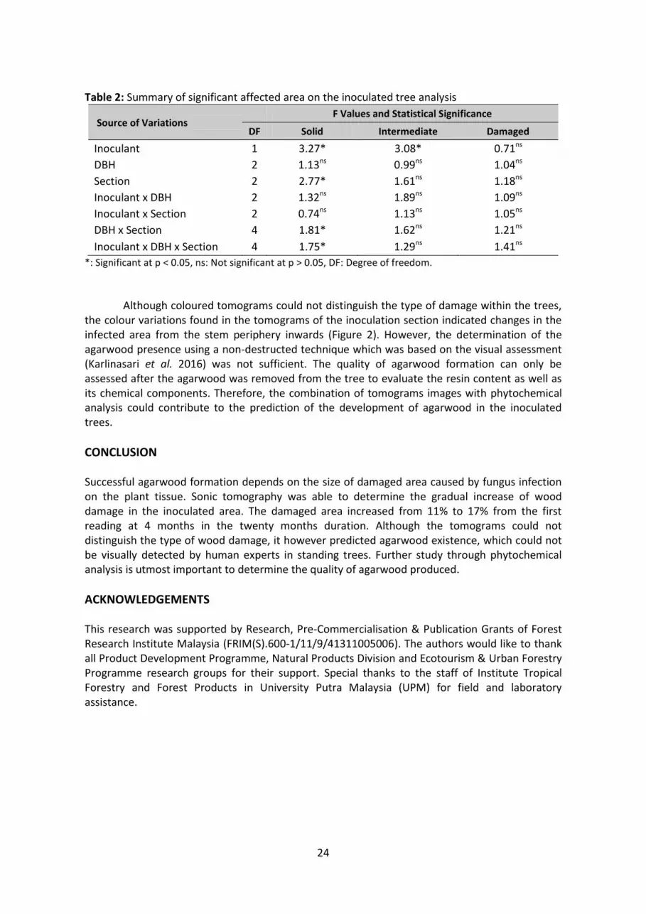
24
Table 2: Summary of significant affected area on the inoculated tree analysis
Source of Variations F Values and Statistical Significance
DF Solid Intermediate Damaged
Inoculant 1 3.27* 3.08* 0.71ns
DBH 2 1.13ns 0.99ns 1.04ns
Section 2 2.77* 1.61ns 1.18ns
Inoculant x DBH 2 1.32ns 1.89ns 1.09ns
Inoculant x Section 2 0.74ns 1.13ns 1.05ns
DBH x Section 4 1.81* 1.62ns 1.21ns
Inoculant x DBH x Section 4 1.75* 1.29ns 1.41ns
*: Significant at p < 0.05, ns: Not significant at p > 0.05, DF: Degree of freedom.
Although coloured tomograms could not distinguish the type of damage within the trees, the colour variations found in the tomograms of the inoculation section indicated changes in the infected area from the stem periphery inwards (Figure 2). However, the determination of the agarwood presence using a non-destructed technique which was based on the visual assessment (Karlinasari et al. 2016) was not sufficient. The quality of agarwood formation can only be assessed after the agarwood was removed from the tree to evaluate the resin content as well as its chemical components. Therefore, the combination of tomograms images with phytochemical analysis could contribute to the prediction of the development of agarwood in the inoculated trees.
CONCLUSION
Successful agarwood formation depends on the size of damaged area caused by fungus infection on the plant tissue. Sonic tomography was able to determine the gradual increase of wood damage in the inoculated area. The damaged area increased from 11% to 17% from the first reading at 4 months in the twenty months duration. Although the tomograms could not distinguish the type of wood damage, it however predicted agarwood existence, which could not be visually detected by human experts in standing trees. Further study through phytochemical analysis is utmost important to determine the quality of agarwood produced.
ACKNOWLEDGEMENTS This research was supported by Research, Pre-Commercialisation & Publication Grants of Forest Research Institute Malaysia (FRIM(S).600-1/11/9/41311005006). The authors would like to thank all Product Development Programme, Natural Products Division and Ecotourism & Urban Forestry Programme research groups for their support. Special thanks to the staff of Institute Tropical Forestry and Forest Products in University Putra Malaysia (UPM) for field and laboratory assistance.

25
REFERENCES Chua LSL. (2008). Agarwood (Aquilaria Malaccensis) in Malaysia. NDF Workshop Case Studies WG
1 – Trees. Case Study 3 Aquilaria malaccensis Country – MALAYSIA. Mexico 2008. Convention on International Trade in Endangered Species of Wild Fauna and Flora (CITES). (2004).
13th Meeting of the Conference of the Parties. 3–14 October 2004, Bangkok. Retrieved from http://www.cites.org/common/cop/13/raw_props/ID-AquilariaGyrinops.pdf
Hou D. (1960). Thymeleaeceae. In van Steenis CGGJ (ed.) Flora Malesiana Series 1, Vol. 6, pp 115. Wolters-Noordhoff Publishing, Groningen, the Netherlands.
Giano R. (1986). The exploitation of resinous products in a lowland Malayan forest. Wallaceana 43: 3–6.
Indahsuary N, Nandika D, Karlinasari L & Santoso E. (2014). Reliability of sonic tomography to detect agarwood in Aquilaria microcarpa Baill. Journal of the Indian Academy Wood Science 11: 65–71.
Karlinasari L, Putri N, Turjaman M, Wahyudi I & Nandika D. (2016). Moisture content effect on sound wave velocity and acoustic tomogram in agarwood trees (Aquilaria malaccensis Lamk.). Turkish Journal of Agriculture & Forestry 40: 696–704.
Li G, Wang X, Wiedenback J & Ross RJ. (2014). Analysis of wave velocity patterns in Black Cherry trees and its effect on internal decay detection. Comput. Electron. Agr. 104: 32–39.
Lin CJ, Chang TT, Juan MY & Lin TT. (2011). Detecting deterioration in royal palm (Roystonea regia) using ultrasonic tomographic and resistance micro drilling techniques. Journal of Tropical Forest Science 23: 260–270.
Lok EH & Zuhaidi AY. (2016). Growth and management of Aquilaria malaccensis for Agarwood: A new domestication perspective. International Journal of Agriculture, Forestry and Plantation 3: 55–60.
Mah YL, Cranbrook E, Wells DR & Furtado JL. (1983). Proposals for a Conservation Strategy for Terengganu. WWF Malaysia. Kuala Lumpur.
Nor Azah MA, Chang YS, Mailina J, Abu Said A, Abd Majid J, Saidatul Husni S, Nor Hasnida H. & Nik Yasmin Y. (2008). Comparison of chemical profiles of selected gaharu oils from Peninsular Malaysia. The Malaysian Journal of Analytical Science 12: 338–340.
Nor Azah MA, Husni SS, Mailina J, Sahrim L, Abdul Majid J & Mohd Faridz Z. (2013). Classification of agarwood (gaharu) by resin content. Journal of Tropical Forest Science 25: 213–219.
Norchahaya H, Mustapa MZ & Kamaruzaman O. (2016). Agarwood. Policy and regulations in Malaysia. Paper presented at the 2nd International Scientific Symposium on Agarwood. Putrajaya.
Persoon GA. (2008). Growing “The Wood of The Gods”: Agarwood Production in Southeast Asia. Institute of Environmental Sciences, Leiden University, P.O. Box 9518, 2300 RA, Leiden, Netherlands. 16 pp.

26
ANATOMICAL CHARACTERISATION OF Phyllanthus pectinatus Hook.f.
(PHYLLANTHACEAE): A PLANT WITH THERAPEUTIC POTENTIAL
Ummu Hani B1, Nur Munirah S1, Hairol Nizam H1, Mohamad Faizal S1 & Hafidz Hadi A2
1Bioresources Programme; 2Phytochemistry Programme, Natural Products Division, Forest Research Institute Malaysia (FRIM), 52109 Kepong, Selangor. Tel: 03-62797602 Fax: 03-62729805 Email: [email protected]
ABSTRACT Phyllanthus pectinatus Hook.f. belongs to the family Euphorbiaceae is a plant native to India but is presently found growing in Malaysia. It is known as Indian gooseberry and Malacca tree in Malaysia. Malacca tree is largely explored for its therapeutic potential against various diseases including cancer. The objective of the study was to characterise the anatomical structure of the leaf and determined the diagnostic characters for identification of this species. Fresh leaf samples were cut and fixed in AA solutions, then sectioned using a sliding microtome. The preparations were then stained in Safranin and Alcian Blue, dehydrated in a series of ethanol and finally mounted using Euparal, before being kept in the oven for 2 weeks at 50°C. Photomicrographs of the leaf transverse sections were captured and processed using TopView standard software. The results showed the petiole outline were slightly circular in shaped, midrib with concave at adaxial side and convex at abaxial side, tip rounded and pointing downwards at margin. Vascular bundles present with close system at petiole and open at midrib; sclerenchyma cells with incompletely ensheathing vascular bundles; presence of simple and multicellular trichomes, calcium oxalate crystals, idioblast tannin and mucilage cells. A combination of these characters were not only valuable in identifying a family, but also able to be used to differentiate between genus and species of the plant. Keywords: Phyllanthus pectinatus, microscopy, anatomy, identification, medicinal plants
INTRODUCTION Phyllanthus pectinatus Hook.f. (syn. Diasperus pectinatus (Hook.f.) Kuntze), commonly known as Indian gooseberry or amla or Malacca tree, family Phyllanthaceae, is widely distributed in subtropical and tropical areas of China, India, Indonesia and Malaysia. Phyllanthus pectinatus is a medium forest tree 36 m tall; leaves crowded on slender, sinuous, small linear, 0.7–2.5 cm long, more or less rusty-scurfy twigs; flower green, 2 mm wide; fruits fleshy and juicy, sour, sway in the wind at the ends of the finely featherly leafy shoots, inside the fruits is a hard stony structure containing the seeds with sharply 3-angled, not splitting and ripening greenish yellow (Whitmore 1972) (Figure 1).
The plant is highly nutritious and an important dietary source of vitamin C, amino acids, minerals and rich in superoxide dismutase (Verma & Gupta 2004). The plant is also used in many traditional medicinal systems, such as the Chinese, Tibetan and Ayurvedic medicine (Zhang et al. 2000). The extract from these fruits are used in traditional medicine to treat symptoms ranging from constipation to the treatment of tumours (Unander et al. 1990). It is also rich in polyphenols and hydrolysable tannin derived compounds which acted as antioxidants (Poltanov et al. 2009; Yang & Liu 2014). Various plant parts have also been reported to show antidiabetic, hypolipidaemic, antibacterial, antioxidant, antiulcerogenic, hepatoprotective, gastroprotective and chemopreventive properties (Krishnaveni & Mirunalini 2010).
According to World Health Organisation (WHO, 1998), more than 80% of the total world population depend on traditional medicines in order to satisfy their primary health care needs.

27
The growing demand for medicinal and aromatic plants makes them remunerative alternative crops for small holder farmers. However, more research is still needed especially in identification of plant species to ensure the raw material used is from the correct and intended species.
Anatomical studies are a systematic line of evidence used in combination with other systematic lines to arrive at a good taxonomic condition (Stace 1980). Anatomical studies apart from special references to systematic position of the taxa can also be used in noting the origin, natural distribution extent of cultivation and cultivars within species of plants (Lawson 1967; Onwueme 1978). From available literatures, series of documented descriptions of the morphological characteristics and ethnobotanical uses of genus Phyllanthus were found to have been reported (Oliver 1959; Hutchinson & Dalziel 1963; Burkill 1994), but there was confusion in recognising individual species of these plants in Malaysia. Furthermore, many of the Phyllanthus sp. had close morphological characteristics, which made them very difficult to be differentiated at species level (Webster 1956; Kandavel et al. 2011). Therefore, the aim of this study was to provide a comprehensive information on the anatomical structures of P. pectinatus leaf and determine the diagnostic characters useful in the identification of the species.
(A) (B) (C) (D)
Figure 1: Phyllanthus pectinatus. (A) Whole plant, (B) leaves, (C) fruits and (D) flowers.
MATERIALS AND METHODS Fresh specimens of P. pectinatus were collected from the Malaysian Agriculture Research and Development Institute (MARDI), Jerangau, Terengganu. Specimens were fixed in a 3:1 AA solution (70% alcohol:30% acetic acid), sectioned using a sliding microtome through the leaves parts and stained in Safranin and Alcian Blue, followed with dehydration in a series of ethanol solutions (50%, 70%, 95% and 100%), and later mounted in Euparal. The fixation and embedding process were based on Johansen (1940) and Sass (1958) with some suitable modifications. Photomicrographs of the leaves sections were captured and processed using TopView standard software.
RESULTS AND DISCUSSION In transverse section, the outline structure of P. pectinatus petiole was slightly circular in shaped (Figure 2A). Whereas, the midrib outline was concave at adaxial side and convex at abaxial (Figure 2B). Parenchyma cortex of the petiole consisted of 8–10 layers of the cells. The vascular tissue arrangements of petiole were observed as close system and circular or O-shaped; the midrib with open system and arc shaped. Clustered sclerenchyma cells were found on the outside of phloem

28
in the vascular bundles with cells incompletely ensheathing the vascular bundles. Only 1 type of trichome was found at the petiole; simple and multicellular type (Figure 2G).
Transverse section of the leaf margin showed that the outline was rounded and slightly pointing downwards at tip (Figure 2I). The lamina consisted of single layer of epidermis adaxial, with height to width ratio 1:1–1:4 and papillae present at the lower epidermis. The chlorenchyma cells were characterised by a dorsiventral organisation (palisade and spongy mesophyll), comprising 1–2 layers of palisade cells, with height of each cells 5–12 times that of the width and 5–7 layers of spongy mesophyll cells with small intercellular spaces (Figure 2D).
The observation on the cells inclusion in studied species showed presence of calcium oxalate crystal, i.e. solitary and druses were few in the phloem cells (Figure 2C). Mucilage cells and idioblast tannin cells were found in parenchyma cells of the petiole and midrib (Figure 2F). On the other hand, thick cuticle waxes were present at the petiole, midrib, lamina and margin.
Figure 2: Anatomical image of P. pectinatus leaves. (A) Transverse section of petiole, (B)
transverse section of midrib, (C) calcium oxalate crystals, (D) transverse section of lamina, (E) papillose epidermis on the lower epidermal, (F) idioblast tannin cell, (G) trichomes, (H)
sclerenchyma cells and (I) transverse section of margin with thick cuticle.
Previous study by Khatijah and Ruzi (2009) reported that the vascular system of the petiolule and midrib were close and open types, respectively. While the midrib outline was convex at abaxial side. The solitary and druses crystal were sparsely present in the phloem but less in the parenchyma cells of the transverse section of petiolule. According to Metclafe & Chalk (1965), multicellular branched hairs and papillae at the lower epidermis cells were generally found in species of Phyllanthus. Tanniniferous cells, filled with brown contents were very widely distributed in the family of Euphorbiaceae. The mesophyll characteristic was used by Awomukwu et al. (2015) to chacterise Phyllanthus sp. in which layer of palisade mesophyll vary between the 5 species studied. One layer thick in P. odontadenius and P. muellerianus while 2 layers thick in P. amarus, P. urinaria and P. niruroides. Large intercellular spaces occurred in the spongy

29
paranchyma of P. urinaria, P. odontadenius and P. muellerianus while P. amarus and P. niruroides had small intercellular spaces.
Therefore, anatomical characteristics such as outline structure, shape and arrangement of vascular bundles in midrib and petiole or petiolule, types of mesophyll and also the presence or absence of selected features could be used to differentiate the Phyllanthus sp. Sajjad & Sharif (2019) also stated that based on morphological and anatomical features, P. urinaria and P. virgatus were easy to differentiate as compared to others. CONCLUSION Results from this study provide important information and data for systematic identification and authentication of P. pectinatus. Transverse section of the fresh plant materials would give the diagnostic characteristic of these species that include the outline structure of petiole, midrib and margin parts, shape and arrangement of vascular bundles, presence and absence of selected features such as solitary and druses crystals, mucilage cells and tannin idioblast cells, all of which were described in this study was of taxonomic value in identification of P. pectinatus from the other species. ACKNOWLEDGEMENTS The author thanked the staff of Malaysian Agriculture Research and Development Institute (MARDI), Jerangau, Terengganu for their technical assistance in the plant specimen collection. Special thanks to Ministry of Agriculture Malaysia and Malaysian Herbal Monograph for funding this research. REFERENCES Awomukwu DA, Nyananyo BL, Joel Uka C & Uwabunkeonye Okeke C. (2015). Identification of the
genus Phyllanthus (Family Phyllanthaceae) in Southern Nigeria using comparative systematic morphological and anatomical studies of the vegetative organs. Journal of Plant Sciences 3(3): 137–149.
Burkill HM. (1994). The Useful Plants of West Tropical Africa. Vol .2. Families E-I. Royal Botanic Garden, Kew. 605 pp.
Hutchinson J & Dalziel JM. (1963). Flora of West Tropical Africa. Vol. 2. Crown Agent, London. 384–388 pp.
Johansen DA. (1940). Plant Microtechnique. McGraw-Hill. New York and London. Kandavel D, Rani SK, Vinithra MG & Sekar S. (2011). Systematic studies in 424 herbaceous
Phyllanthus spp. (region: Tiruchirappalli district in India) and a simple key to 425 authenticate “Bhumyamalaki” complex members. Journal of Phytology 3(2): 37–48.
Khatijah H & Ruzi AR. (2009). Anatomical Atlas of Malaysian Medicinal Plants. Universiti Kebangsaan Malaysia, Bangi, Malaysia. 112–115 pp.
Krishnaveni M & Mirunalini S. (2010). Therapeutic potential of Phyllanthus emblica (amla): the ayurvedic wonder. Journal of Basic Clinical Physiology and Pharmacology 21(1): 93–105.
Lawson JRS. (1967). A key to the Dioscorea species in Nigeria. Journal West African Science Association 12: 1–9.
Metcalfe CR & Chalk L. (1950). Anatomy of the Dicotylendons. Clarendon Press, Oxford, London. 1234–1235 pp.
Oliver B. (1959). Nigeria’s useful plants II (1): Medicinal plants. Nig. Fields 24: 13–34. Onwuememe IC. (1978). The Tropical Tuber Crops: Yam, Cassava, Sweet Potato and Cocoyam.
John Wiley and Sons. Chicelster. 234 pp.

30
Poltanov EA, Shikov AN, Dorman HJD, Pozharitskaya ON, Makarov VG, Tikhonov VP & Hiltunen R. (2009). Chemical and antioxidant evaluation of Indian gooseberry (Emblica officinalis Gaertn., syn. Phyllanthus emblica L.) supplements. Phytotherapy Research. 23(9): 1309–1315.
Sajjad HT & Sharif HL. (2019). Morpho-anatomical observations on Phyllanthus of Southwestern Bangladesh with two new records for Bangladesh. BioRxiv 608711. https://doi.org/10.1101/608711
Sass JE. (1958). Botanical Microtechnique. 3rd edition. Ames, Iowa State University. Stace CA. (1980). Plant Anatomy and Biosystematics. Edward Arnold Publishers, London. 279 pp. Verma RC & Gupta A. (2004). Effect of pre-treatments on quality of solar-dried amla. Journal of
Food Engineering. 65: 397–402. Unander D, Webster GL, & Blumberg BS. (1990). Records of usage or assays in Phyllanthus
(Euphorbiaceae). I. Subgenera Isocladus, Kierganelia, Cicca and Emblica. J. Ethnopharmacol. 30(3): 233–264.
Webster GL. (1956). A monographic study of the West Indian species of Phyllanthus. Journal of the Arnold Arboretum 37(3): 217–268.
Whitmore TC. (1972). Tree Flora of Malaya; A Manual for Foresters. Vol. 2. Longman Malaysia Sdn. Bhd., Malaysia. 123 pp.
World Health Organisation (WHO). (1998). Quality Control Methods for Medicinal Plant Materials. Geneva, Switzerland.
Yang B & Liu P. (2014). Composition and biological activities of hydrolysable tannins of fruits of Phyllanthus emblica. Journal of Agricultural and Food Chemistry 62(3): 529–541.
Zhang YJ, Tanaka T, Iwamoto Y, Yang CR & Kouno I. (2000). Phyllaemblic acid, a novel highly oxygenated norbisabolane from the roots of Phyllanthus emblica. Tetrahedron Letters 41: 1781–1784.

31
CONSERVATION OF MEDICINAL AND AROMATIC PLANTS IN THE ETHNOBOTANICAL GARDEN OF FRIM: AN EFFORT IN TACKLING BIODIVERSITY LOSS CRISIS Tan AL, Madihah MN, Norbaiah MY, Ummu Hani B & Fadzureena J Bioresources Programme, Natural Products Division, Forest Research Institute Malaysia, 52109 Kepong, Selangor. Tel: 03-62797671 Fax: 03-62729806 Email: [email protected]
ABSTRACT The Ethnobotanical Garden in FRIM has been a repository for medicinal and aromatic plants conservation since its establishment in 1995. In the early years, the garden played an important role to promote the interest on medicinal and aromatic plants (MAPS) used by the local communities especially by the orang asli and Malay practitioners to the public. To date, the garden houses more than 250 species of MAPS. Continuous effort has been carried out by the team to collect samples of living medicinal and aromatic plants for conservation not only limited to those in the forest reserves but also those found in the villages and forest fringes to add to the collection in the garden. From the documentation exercises carried out by FRIM Traditional Knowledge Team, a total of about 934 species of medicinal and aromatic plants are being used in Peninsular Malaysia by the local communities. The knowledge seems to be slowly diminishing due to biodiversity loss and also loss of traditional knowledge practitioners. Hence, it is very essential to safeguard the knowledge and biodiversity as new resources to be explored for new potential cures and useful products for the future. Collection of MAPS had been made gradually throughout 2017–2020 with the aim to sustain at least 250–280 MAPS species in the ethnobotanical garden. Collections were designated to cover selected Forest Reserves or National Parks in the 5 regions of Peninsular Malaysia. The task was to document MAPS encountered during collection and through sampling of wildings and cuttings or seeds for conservation purposes in the garden. From the collections made from 2017–2020, the Ethnobotanical Garden successfully conserved a total of 254 species (80 families). A total of 13 MAPS (including 2 MAPS sold in the herbal market) that were frequently encountered in forest reserves, collected from the 5 regions were conserved as germplasm in the ethnobotanical garden. Species with variations in morphology due to different localities and closely related species in the wild were also collected. Among the challenges faced in the conservation of MAPS were the low survivability rate and short life cycle of the plants. Keywords: Ethnobotanical garden, medicinal and aromatic plants, conservation, biodiversity, traditional knowledge
INTRODUCTION Malaysia has been known as one of the 12 mega-biodiversity countries in the world. However, the number of biodiversity is greatly depleting not only due to climate change but also threats from overexploitation and agricultural activities as well as land degradation. Biodiversity plays a major role in the society and people’s well-being as a source of food production, water to medical treatments, especially traditional health care system utilising wild medicinal plants harvested from the forest and home surroundings. Traditional knowledge on medicinal plants documentation carried out in Peninsular Malaysia from the orang asli revealed that there are many more plant species that has great potential to become new biological resources for health product development to drug discovery in long term.
From the documentation exercise carried out by FRIM Traditional Knowledge Team, a total of about 934 species of medicinal and aromatic plants were being used in Peninsular Malaysia by the local communities. Out of 934 species, 488 species had been published in several

32
volumes of books such as Khazanah Perubatan Melayu: Tumbuhan Ubatan (Vol. 1–3), Meneroka Waris Rimba Vol. 1 and Buku Herba Emas Negara (18 NKEA species). The remaining species were currently being added in the write-up for the following volumes of Khazanah Perubatan Melayu Vol. 4 and Meneroka Waris Rimba Vol. 2 and expected to be published in 2021. Recognising the importance and value of medicinal plants to human being and depletion of biodiversity and also traditional knowledge holders, it is high time to conserve our biodiversity be it through ex-situ or in-situ conservation effort.
Ex-situ conservation is often used as the last effort in conserving threatened species especially when the threatened species are located outside protected areas such as forest reserves or national parks. There are several ex-situ conservation methods, such as botanical gardens, herbal gardens, seed bank, gene bank, cryopreservation and tissue culture (Patel 2015). In a broader sense, ex-situ conservation activities not only focus on managing populations conserved, but also activities such as education and raising awareness, supporting research initiatives and collaborating in-situ conservation effort (Mohammed & Mundanthra 2013).
MATERIALS AND METHODS Collection of medicinal and aromatic plants (MAPS) had been constantly and continuously gathered from 2017–2020 with the aim to sustain 250–280 MAPS at the Ethnobotanical Garden in FRIM. Apart from that, the project also aims to increase the number of species in the garden and to establish a germplasm bank for 10 selected MAPS species collected throughout Peninsular Malaysia. Collection of MAPS was based on the 5 regions in Peninsular Malaysia, namely northern, eastern, western, southern and central. Collection trip was carried out in selected forest reserves or national parks. Sampling sites at trekking trails with various habitats such as lowland, hillside, montane forest, riverine, island and water catchment area were chosen for MAPS collection to detect and identify the presence of MAPS species and their variations and performance under different habitats. This is important for sustaining the wild gene of a species and crucial for survivability against climate change and pest control.
RESULTS AND DISCUSSION The flora diversity in Malaysia is estimated to consist of 15,000 species. A checklist for vascular plants for Peninsular Malaysia which was published in 1995 reveals that there are about 8,893 taxa (species, subspecies and varieties) which cover over 8,200 native and 690 naturalised species. It is also known that higher endemism occurred among herbaceous flora, whereby some larger genera consist of 80% endemism of their species (NRE 2014).
From a total of 355 species (99 families) of medicinal and aromatic plants collected together with several dominant species encountered in the forest reserves, to date only about 254 species (80 families) were successfully conserved at the Ethnobotanical Garden, FRIM. Several medicinal plants such as Peliosanthes teta, Mapania cuspidata, Acrotrema costatum, Molineria latifolia, Dianella ensifolia, Stachytarpheta jamaicensis, Thottea spp., Codonoboea spp., Dracaena spp., Pentaphragma spp. and Amischotolype spp. frequently encountered throughout the forest reserves within the 5 regions were collected for conservation. Two famous species popularly sold in the herbal market such as Labisia pumila (including its 3 varieties) and Smilax myosotiflora were also collected and conserved.
Although these species were widespread, they were not very easily cultivated ex-situ. They thrived at certain microclimate on hill side (Thottea spp., Codonoboea spp., Pentaphragma spp.), nearby stream (Acrotrema costatum, Mapania cuspidata), lowland to hill forest (Amischotolype spp., Labisia pumila, Smilax myosotiflora, Dracaena spp.), lowland hill forest to montane forest (Peliosanthes teta, Molineria latifolia, Dianella ensifolia) or open area at forest fringes (Stachytarpheta jamaicensis). Several leaf variations were noted on both Acrotrema costatum (green to green with middle white band), Molineria latifolia (green to glossy dark green)

33
and Amischotolype spp. (green or with purplish abaxial) depending on the light intensity received. Closely related species from similar genera and its variety were also collected whenever encountered.
Other than these species, frequently encountered ginger species in the forest reserves and national parks were also collected for ex-situ conservation in the Ethnobotanical Garden, FRIM. Other than ginger species, several popular medicinal plant species encountered were also collected such as Polyalthia bullata, Prismatomeris tetrandra, Eurycoma longifolia, Smilax calophylla, Goniothalamus spp., Phyllagathis rotundifolia, Piper porphyrophyllum, Cheilocostus speciosus, Leea indica, Alocasia longiloba and Homalomena rostrata. The distribution of these species was not so widespread and was found scattered in certain forest reserves. Some difficulties encountered in conserving some of the tree species were finding the proper saplings size and conserving climber/creeper species when potting in polybags.
Ex-situ conservation was indeed challenging as continuous efforts were needed to retain the collection. Some herbal plants had very short life cycle for less than a year, such as species from the Asteraceae (Acmella uliginosa, Erechthites spp., Blumea balsamifera, Wollastonia biflora), Apiaceae (Centella asiatica, Eryngium foetidum, Hydrocotyle javanicum), Lamiaceae (Ocimum spp., Clerodendrum spp.), Solanaceae (Physalis angulata), Onagraceae (Ludwigia spp.), Polygalaceae (Polygala paniculata) and certain Malvaceae (Hibiscus sabdariffa, Sida spp., Urena spp., Corchorus spp.), Fabaceae (Senna tora, Senna occidentalis, Desmodium heterocarpon) and Euphorbiaceae (Euphorbia hirta, Acalypha indica) species. These species barely outlive the 6-month duration especially when wildings collected already reached maturity (flower or fruit). Hence, younger wildings were collected to prolong species life span.
Furthermore, ex-situ conservation on several plant habit such as fern (Selaginella spp., Helminthostachys zeylanica, Loxogramme sp., Schizaea dichotoma, Actinostachys digitata), slender, climber or creeper (Pellionia repens, Passiflora foetida, Momordica charantia, Cardiospermum halicacabum) and highland (Melastoma sanguineum, Melastoma spp., Arisaema spp., Dissochaeta spp.) or habitat specific (Begonia spp., Persicaria capitata, Tristaniopsis spp., Sonerila spp., Ipomoea pes-caprae) species had proven to be unsuccessful or difficult to maintain for a long period. Several trials and error were made but not very successful. These species were best conserved in-situ. Meanwhile, several species were able to be propagated using seeds or providing similar microclimate for conservation purposes.
Among the most successful propagation methods were through wildings. Conservation through vegetative cuttings was not that successful and very much depending on the species. Several successful propagation using stem cuttings includes Tinospora crispa, Alternanthera sisso, Euphorbia tirucalli, Clinacanthus nutans and Persicaria chinensis. Propagation using seeds collection also tend to have lower success rate and have slow growth rate and might not successfully reach maturity. Moreover, harvesting mature seeds and proper storage before sowing activities could increase the growth chances of the plant.
CONCLUSION Up to now, a total of 254 species belonging to 80 families were successfully conserved and added to the Ethnobotanical Garden collection from the period of 2017–2020. Meanwhile, a germplasm of 13 MAPS species from the 5 regions were conserved for the first time in the garden for future breeding work or genetic study.
ACKNOWLEDGEMENTS The authors thanked the Ethnobotanical Garden staff for their help in maintaining the plants and garden suitable for visitors visit. We are also grateful to all Ethnobotany staff involved in MAPS collection for conservation in the garden. Last but not least, our heartiest gratitude for the

34
projects funding to enable the documentation and conservation of MAPS species to be carried out in Ethnobotanical Garden at FRIM.
REFERENCES Mohammed K & Mundanthra B. (2013). Ex-situ conservation of biodiversity with particular
emphasis to Ethiopia. ISRN Biodiversity. 2013: Article ID 985037. http://dx.doi.org/10.1155/2013/985037
NRE. (2014). Overview of Malaysia’s Biodiversity. Pp. 14–15 in Malaysia’s Fifth National Report to the Convention on Biological Diversity. NRE, Putrajaya.
Patel DK. (2015). Herbal gardens: Role in current scenario for ex-situ conservation of medicinal and aromatic plants. International Journal of Current Research 7(7): 18669–18672

35
PERUBATAN TRADISIONAL: BUKAN SEKADAR JAMPI Nik Musa’adah M, Norbaiah MY, Firdaus K, Siti Nur Aisyah MH, Mazura MP, Fadzureena J, Intan Nurulhani B, Madihah MN, Md Azharulzaman MS, Dionysia, M, Abdul Hayat MS, Tan AL, Noor Aimie MK, Rosniza R, Fatin Nadiah S, Nor Azah MA, Mastura M & Norini H Institut Penyelidikan Perhutanan Malaysia (FRIM), 52109 Kepong, Selangor. Tel: 03-62797345 Faks: 03-62729805 Emel: [email protected]
ABSTRAK Perubatan tradisional sama ada Melayu atau orang asli sering dikaitkan dengan jampi atau mantera. Bagi melihat skop yang lebih luas dalam perubatan tradisional, kertas kerja ini akan cuba melihat hasil-hasil kajian yang dijalankan oleh Institut Penyelidikan Perhutanan Malaysia (FRIM) dan juga merujuk kepada kitab tib atau manuskrip perubatan Melayu. Projek pendokumentasian pengetahuan tradisi berkaitan tumbuhan ubatan yang dijalankan oleh FRIM mendapati perubatan tradisional yang diamalkan oleh masyarakat Melayu dan juga orang asli adalah kaya dengan ilmu-ilmu berkaitan ramuan berasaskan tumbuhan ubatan (materia medika). Malah kaedah perubatan tradisional juga sangat menitik berat kepada kaedah penjagaan kesihatan diri, kaedah mengenali penyakit, dan memahami anatomi badan. Dalam usaha memartabatkan perubatan tradisional, kajian mendalam berkaitan ramuan yang disokong dengan data saintifik perlu dipertingkatkan. Kata kunci: Perubatan tradisional, tumbuhan ubatan, Melayu, orang asli
PENGENALAN Sejak zaman berzaman manusia tidak terkecuali daripada menghidapi pelbagai jenis penyakit. Usaha atau ikhtiar akan diambil untuk menangani atau menyembuhkan penyakit tersebut, antaranya ialah merujuk kepada dukun, bomoh, pawang, tabib, bidan bagi masyarakat Melayu dan poyang atau bomoh bagi orang asli. Pada pandangan umum, kaedah perubatan tradisional ini menjurus kepada jampi serapah atau mantera. Hal ini berkaitan dengan kepercayaan masyarakat dahulu kepada semangat (spirit) atau penunggu yang mengganggu atau menyebabkan penyakit (Abdul Samad 1983; Werner 1986).
Dalam perubatan tradisional orang asli, hubungan baik dengan semangat/penunggu adalah perlu untuk mengelakkan mereka disakiti oleh semangat/penunggu tersebut dengan cara dijangkiti penyakit atau ditimpa bala. Jampi dan mantera digunakan sebagai langkah pencegahan secara spiritual. Terdapat lebih daripada 100 jampi yang diamalkan oleh suku kaum Mah Meri dan Jahut telah disenaraikan oleh Warner (1986) bagi menghindari pelbagai penyakit. Contoh jampi untuk melegakan sakit perut yang diamalkan oleh poyang suku kaum Mah Meri ialah:
Hidung lengat kepala lengat Sangkat batang buluh Hidung jangan lengat Urat jangan lengat Daging jangan luruh
(Werner 1986)
Dalam perubatan tradisional Melayu, doa atau jampi digunakan untuk mengubati penyakit terutama penyakit rohani. Doa atau jampi juga digunakan dalam penyediaan ubat dan air penawar (Harun 2019). Contoh petua dan mantera bagi melegakan sakit perut yang dipetik daripada MSS 1292 PNM:

36
Sebagai lagi ubat sakit perut, dimantera pada lada sulah tujuh biji. Maka dimantera tiga kali senyawa, maka mamah-mamah, tekankan pada pusat. Inilah manteranya: Gendang gendut ijuk talinya Sakit perut kentut jadinya Tajam tumpul bisa tawar Tajam akan penyukur bisa akan penawar Hung, suuh (Harun & Nik Musa’adah 2019)
Tidak dapat dinafikan peranan doa adalah penting kerana Allah adalah penyembuh yang
mutlak. Walau bagaimanapun, berdasarkan manuskrip perubatan Melayu atau kitab tib, doa, jampi atau mantera hanyalah sebahagian daripada usaha penyembuhan tetapi sebahagian besar usaha penyembuhan penyakit adalah dengan menggunakan ramuan ubatan (materia medika) yang lazimnya berasaskan kepada tumbuhan (Harun 2019).
Dalam usaha mengenal pasti perubatan tradisional berasaskan tumbuhan, FRIM telah menjalankan pendokumentasian tumbuhan ubatan berasaskan pengetahuan tradisi orang asli dan juga Melayu di Semenanjung Malaysia.
PERUBATAN TRADISIONAL ORANG ASLI Pendokumentasian tumbuhan ubatan berasaskan pengetahuan tradisi atau kegunaan dalam perubatan tradisional 18 suku kaum orang asli di 22 petempatan orang asli telah menemukan 677 spesies tumbuhan daripada 158 famili. Manakala, sebanyak 3,917 kegunaan telah direkodkan dan telah dikategorikan kepada 10 kategori kegunaan iaitu penyakit tertentu/khusus, kegunaan luaran, kesihatan wanita, kesihatan lelaki, kesihatan kanak-kanak, kecantikan/penjagaan diri, tonik, buang angin, sengatan haiwan/serangga berbisa dan lain-lain (ulaman, masakan, atau spiritual). Sebahagian besar (57%) tumbuhan yang direkodkan diguna untuk mengubati penyakit yang khusus atau tertentu (Tan et al. 2019).
Seterusnya kajian biotinjauan atau bioprospecting telah dilakukan bagi menilai spesies terpilih sebagai agen antioksidan, antiinflamasi, antimikrob, antidiabetes, antiprotozoa dan juga antikanser. Hasil profil bioaktiviti menunjukkan sebanyak 50 spesies daripada 103 spesies yang disaring mempunyai sekurang-kurangnya 2 bioaktiviti. Ini menunjukkan spesies tumbuhan ubatan yang digunakan dalam perubatan tradisional orang asli dapat dibuktikan kesan bioaktivitinya berdasarkan kajian makmal. Kajian lanjutan perlu dilakukan bagi meneroka agen terapeutik berasaskan tumbuhan tersebut.
Kini, sebanyak 4 spesies daripada rekod pendokumentasian dan juga kajian biotinjauan tersebut telah dibuat kajian lanjutan bagi meneroka potensi sebagai agen terapeutik ataupun penjagaan diri. Satu daripada spesies tersebut ialah daripada famili Zingiberaceae dan dikodkan sebagai ABP 016. ABP 016 digunakan oleh 8 suku kaum orang asli (Temiar, Jahai, Semai, Semoq Beri, Mendriq, Semelai, Temuan dan Lanoh) di 8 petempatan (RPS Banun, Gerik; Kg Ulu Gerih, Gopeng; Kg Sg Berua, Kuala Berang; Kg Kuala Lah, Gua Musang; Kg Sg Lui, Jempol; Kg Dusun Kubor, Jelebu; Kg Air Bah, Lawin). Secara tradisionalnya ABP 016 digunakan sebagai mandian untuk meredakan demam atau panas badan. ABP 016 juga diguna untuk melegakan sakit mata. Kajian makmal telah menunjukkan ekstrak rizom, batang, daun dan infloresen mempunyai kesan antiinflamasi berdasarkan aktiviti anti-penyahaslian protein (anti protein denaturation). Hasil daripada penyelidikan ini, ekstrak piawai rizom dan juga 4 prototaip iaitu face and body mist, gel mandian, losen dan sabun mandi telah dibangunkan. Ini menunjukkan perubatan tradisional orang asli berasaskan tumbuhan ubatan boleh ditentusahkan secara saintifik dan berpotensi untuk dibangunkan sebagai produk bernilai tambah.

37
PERUBATAN TRADISIONAL MELAYU Maklumat berkaitan perubatan tradisional boleh dirujuk kepada manuskrip perubatan Melayu atau kitab tib dan juga pengamal perubatan tradisional Melayu. Terdapat lebih daripada 100 naskhah kitab tib yang terdapat di Perpustakaan Negara Malaysia, Dewan Bahasa dan Pustaka, muzium-muzium dan juga beberapa agensi dalam dan luar negara. Merujuk kepada kitab Bustanus Salatin yang ditulis oleh Syeikh Nuruddin Ar-Raniri, ilmu tib atau perubatan ditakrifkan sebagai “pengetahuan akan segala hal badan insan, dan memeliharakan sihat tubuh, lagi menyembuhkan segala penyakit daripada segala wujud” (Naseer 2017). Kaedah penjagaan kesihatan dan pencegahan penyakit juga dibicarakan dalam Kitab al-Rahmah dan juga Kitab Tib MSS 1292 PNM (Mohd Affendi & Hermansyah 2017; Harun & Nik Musa’adah 2019). Sebagai contoh, Kitab Tib MSS 1292 PNM telah membicarakan 16 fasal berkenaan memelihara diri pada waktu sihat antaranya ialah penjagaan makan dan minum, pergerakan tubuh atau riadah, memelihara tidur dan jaga, tatacara wati, memelihara diri daripada kena angin, pengawalan emosi, dan beberapa tatacara penjagaan diri (Harun & Nik Musa’adah 2019). Begitu juga dalam bab ketiga Kitab al-Rahmah juga telah menggariskan 10 perkara yang harus diberi perhatian dalam penjagaan kesihatan antaranya tatacara makan, minum, diam/istirehat, tidur, ketika berjaga, jimak dan keadaan mental atau psikologi dan juga penjagaan diri (Mohd Affendi & Hermansyah 2017).
Perubatan tradisional Melayu juga sangat menitik beratkan kaedah mengenal dan mengesan penyakit disamping memahami anatomi tubuh. Kitab Bustanus Salatin misalnya telah menerangkan berkaitan 40 anggota badan (Naseer 2017). Ilmu perubatan tradisional merangkumi ilmu mengetahui asal penyakit, mengenal keadaan sakit serta meraksi atau pengesanan penyakit (Nornizam 2017; Harun 2019). Ini menunjukkan perubatan tradisional Melayu tidak hanya memfokuskan kepada usaha penyembuhan penyakit tetapi bagaimana mengekalkan kesihatan badan dan juga pemahaman berkaitan anatomi dan fisiologi badan.
Dalam rawatan penyakit, ramuan yang digunakan lazimnya adalah berasaskan tumbuhan. Berdasarkan hasil bancian pengamal perubatan tradisional Melayu di Semenanjung Malaysia yang dijalankan oleh FRIM, 64.1% daripada 2,934 pengamal perubatan tradisional Melayu yang ditemu bual menggunakan kaedah urutan, 7% menggunakan herba, 0.4% bekam dan lain-lain kaedah rawatan iaitu menggunakan kaedah jampi atau doa melibatkan sebanyak 10%, manakala 18.1% menggunakan gabungan rawatan (Statistik Pengamal Perubatan Tradisional Melayu Semenanjung Malaysia (2013–2014) 2015). Bancian ini juga menunjukkan seramai 1,296 (44.2%) pengamal menghasilkan produk sendiri bagi mengubati pesakit mereka sama ada dalam bentuk minyak urut, ramuan herba, majun, ramuan mandian, dan lain-lain. Terdapat juga pengamal yang menanam tumbuhan ubatan di sekitar kediaman mereka. Ini menunjukkan disamping memberi perkhidmatan, mereka juga menyumbang kepada konservasi sumber biologi.
Pendokumentasian penggunaan tumbuhan ubatan dalam kalangan pengamal perubatan tradisional Melayu telah berjaya mengenal pasti sebanyak 586 spesies tumbuhan. Antara spesies yang kerap digunakan ialah serai wangi, kunyit, pandan, lengkuas, inai, mengkudu, sirih, dukung anak, pisang kelat dan kelapa (Dionysia et al. 2018). Selain untuk kesihatan umum, tumbuhan ini juga digunakan untuk penyakit kronik seperti kencing manis, darah tinggi, barah, dan lain-lain. Sebahagian besar ramuan tradisional ini terdiri daripada pelbagai jenis tumbuhan ubatan. Campuran atau kombinasi bahan ini diperlukan untuk mengimbangi kesan komponen utama sama ada mengurangkan kekuatan kesan jika kesannya sangat kuat untuk pesakit, mengurangkan kesan sampingan, memperkuatkan kesan jika kesan komponen utama lemah, atau menjadikan rasa ubat lebih sedap (Abdul Ghani 2015).
Selain daripada ramuan, kaedah penyediaan ubatan merupakan elemen yang penting bermula daripada kaedah pengambilan bahan ubatan, bahagian tumbuhan yang diambil, seterusnya pemprosesan bahan dan disusuli dengan kaedah pengambilan ubatan. Kaedah penggunaan ubatan yang disediakan bergantung kepada jenis penyakit, tahap penyakit dan tujuan penggunaannya. Perubatan tradisional ini juga melibatkan petua dan pantang larang. Ini

38
menunjukkan perubatan tradisional tidak hanya mengubati simptom tetapi merawat punca penyakit dan juga mencegah daripada dijangkiti semula (Nik Musa’adah et al. 2017).
KESIMPULAN Perubatan tradisional sering dikaitkan dengan jampi, mantera dan juga doa yang bertujuan untuk memohon agar dihindari atau disembuhkan penyakit daripada yang lebih berkuasa. Namun, amalan-amalan penjagaan kesihatan diri serta ramuan ubatan (materia medika) adalah komponen yang penting dalam perubatan tradisional. Bidang ini perlu diterokai dan dikaji secara saintifik demi memartabatkan perubatan tradisional. Pendokumentasian secara komprehensif ramuan ubatan serta kajian dari segi kualiti dan efikasinya dapat memastikan perubatan tradisional dapat disokong dengan penemuan santifik.
PENGHARGAAN Perhargaan kepada Kementerian Tenaga dan Sumber Asli, Kementerian Pertanian dan Industri Makanan atas dana penyelidikan dan juga semua agensi kolaborasi, pengamal tradisional dan pegawai pengumpul data yang terlibat dengan projek ini.
RUJUKAN Abdul Ghani H. (2015). MSS 2999 Kitab Tib — Pandangan dan Tafsiran Perubatan
ModenTerhadap Manuskrip Perubatan Melayu. FRIM, Kepong. 396 pp. Abdul Samad A. (1983). Warisan Perubatan Melayu. Dewan Bahasa dan Pustaka, Kuala Lumpur.
165 pp. Dionysia M, Abdul Hayat MS, Nik Musa’adah M, Noor Aimie MK, Md Azharulzaman MS, Fatin
Nadiah MS, Intan Nurulhani B, Madihah MN, Nurul Husna Z, Fadzureena J, Lim HF, Tan AL, Rosniza R, Nor Azah MA, Mastura M & Norini H. (2018). 20 spesies tumbuhan ubatan paling kerap digunakan dalam kalangan pengamal perubatan tradisional Melayu di Semenanjung Malaysia. Pp. 16–22 dalam Mastura et al. (eds.). FRIM Proceedings No. 15: Prosiding Seminar Pemuliharaan & Pemerkasaan Pengetahun Tradisi 2018: Sedekad Melestari Legasi Pengetahuan Tradisi. FRIM, Kepong.
Harun MP. (2019). Kitab Tib Ilmu Perubatan Melayu. Edisi terkini. Perpustakaan Negara Malaysia, Kuala Lumpur. 440 pp.
Harun MP & Nik Musa’adah M. (2019). Kitab Tib MSS 1292 PNM: Edisi dan Suntingan Teks. FRIM, Kepong. 410 pp.
Mohd Affendi MS & Hermansyah MY. (2017). Kitab Perubatan Melayu Al-Rahmah Fi Al-Tibb Wa Al-Hikmah. Abbas Kuta Karang Acheh Dar Al-Salam. Akademi Jawi Malaysia, Kuala Lumpur. 375 pp.
Naseer S. (2018). Bustanus Salatin. Bab Ketujuh Fasal Ketiga: Pada Menyatakan Ilmu Tashrih dan Ilmu Tibb. Nuruddin Ar-Raniri. Baytul Hikma, Pulau Pinang. 193 pp.
Nik Musa’adah M, Abdul Hayat MS & Dionysia M. (2017). Ilmu perubatan Melayu daripada pandangan sains. Pp. 114–124 dalam Harun MP et al. (eds.). Prosiding Persidangan Antarabangsa Manuskrip Melayu 2017. Perpustakaan Negara Malaysia, Kuala Lumpur.
Nornizam N. (2017). Falsafah dan Prinsip Asas Perubatan Melayu. Baytul Hikma, Pulau Pinang. 167 pp.
Statistik Pengamal Perubatan Tradisional Melayu Semenanjung Malaysia (2013–2014). 2015. FRIM, Kepong. 88 pp.

39
Tan AL, Madihah MN, Norbaiah MY, Intan Nurulhani B, Amizan N, Nik Musa’adah M, Fadzureena J, & Norini H. (2019). Status kekayaan penggunaan tumbuhan ubatan oleh masyarakat orang asli di Semenanjung Malaysia. Pp. 7–10 dalam Mazura MP & Firdaus K (eds.). Prosiding Seminar Pemuliharaan & Pemerkasaan Pengetahuan Tradisi 2019. FRIM, Kepong.
Werner R. (1986). Bomoh-Poyang-Traditional Medicine and Ceremonial Art of the Aborigines of Malaysia. University Malaya, Kuala Lumpur. 632 pp.

40
KAJIAN AWAL: KESAN PENGGUNAAN BAJA TERHADAP KADAR PERTUMBUHAN DAN HASIL TONGKAT ALI DI STESEN PENYELIDIKAN FRIM, MARAN PAHANG Syazwan A, Sharmizi I, Hada Masayu I, Mohd Amir Amsyar I, Fadzureena J & Nor Azah MA Program Sumber Biologi, Bahagian Hasilan Semula Jadi, Institut Penyelidikan Perhutanan Malaysia (FRIM), 52109 Kepong, Selangor. Tel: 019-3680019 Emel: [email protected]
ABSTRAK Kajian pertumbuhan dan hasil tongkat ali telah dijalankan di Stesen Penyelidikan FRIM Maran, Pahang. Tujuan kajian ini adalah untuk mengkaji kesan penggunaan baja terhadap kadar pertumbuhan dan hasil tongkat ali. Penanaman dibuat di atas batas menggunakan 2 jenis baja organik, iaitu kompos dan tahi ayam. Kawalan yang digunakan adalah pokok yang ditanam tanpa sebarang rawatan. Rawatan pembajaan tanah diberikan pada peringkat awal sebelum penanaman dibuat dengan kadar sebanyak 40 kg setiap batas. Parameter yang dikaji ialah tinggi, ukur lilit batang, kadar kemandirian, berat basah dan kering akar. Kesan rawatan dilihat melalui bancian tumbesaran pokok yang telah dijalankan setiap bulan selama 6 bulan. Ketinggian dan ukur lilit batang pokok tongkat ali yang dirawat menggunakan baja tahi ayam menunjukkan purata pertumbuhan yang paling tinggi selepas 6 bulan, iaitu masing-masing sebanyak 32.17 cm dan 6.99 mm. Kadar kemandirian pula iaitu 100% adalah pada pokok yang ditanam tanpa sebarang rawatan. Hasil daripada berat basah dan kering akar pokok menunjukkan pokok tongkat ali yang dirawat menggunakan baja kompos mempunyai purata berat paling tinggi, iaitu masing-masing sebanyak 3.38 g dan 2.08 g. Kata kunci: Tongkat ali, penggunaan baja, tumbesaran pokok bancian, kadar kemandirian
PENGENALAN Eurycoma longifolia atau tongkat ali merupakan tumbuhan ubatan yang sering digunakan dalam penyediaan ubatan tradisional masyarakat di Malaysia. Tongkat ali merupakan sejenis tumbuhan yang dikatakan mempunyai kemampuan merawat pelbagai penyakit seperti malaria, demam, ulser, ketoksikan dan lemah tenaga batin (Jagananth & Ng 2000). Kini, pengunaannya lebih meluas, banyak industri dan farmasi mengunakannya untuk penghasilan produk khususnya ubatan. Bhat & Karim (2010) melaporkan bahawa tongkat ali mempunyai pelbagai jenis kegunaan dari bahagian akar hingga ke daun. Kebergantungan terhadap sumber sedia ada dan sumber liar tidak dapat menampung bekalan keperluan. Penanaman secara komersial perlu dipertingkatkan bagi menjamin keperluannya pada masa akan datang. Tongkat ali yang mampu mencecah ketinggian antara 10–15 m boleh dituai seawal 5–7 tahun penanaman, namun kaedah penanaman perlu betul supaya pertumbuhan pokok mencapai kualiti yang dikehendaki. Pelbagai aspek harus dititikberatkan agar tidak membantutkan tumbesarannya. Penggunaan baja organik dapat membantu tumbesaran pokok kerana pembajaan dapat memberi nutrien yang diperlukan oleh pokok. Bahan organik digunakan bertujuan untuk memperbaiki struktur tanah di samping meningkatkan kesuburan tanah (Illani et al. 2019). Kajian ini menggunakan aplikasi baja kompos dan tahi ayam terhadap penanaman tongkat ali bagi melihat kesan baja terhadap pertumbuhan dan hasil tongkat ali.

41
BAHAN DAN KAEDAH
Pemilihan Biji Benih Biji benih atau baka dari variasi yang terbukti keberhasilannya amat penting bagi menjamin hasil yang berkualiti. Biji benih dari baka Stesen Penyelidikan FRIM Maran, Pahang dipilih berdasarkan hasil yang terbukti pengeluarannya sejak beberapa tahun lalu. Pemilihan biji benih dilakukan dengan kutipan daripada pokok ibu yang mempunyai ciri-ciri yang bagus. Biji benih disemai menggunakan medium pasir sungai, dibiarkan selama tempoh 4–8 minggu sehingga anak pokok mengeluarkan 2 pelepah daun untuk dipindahkan ke polibag. Pokok akan mengeluarkan akar serta daun yang banyak dan sedia ditanam di lapangan setelah berumur antara 6–12 bulan. Untuk kajian ini, sebanyak 240 pokok digunakan untuk mengenal pasti kadar pertumbuhan dan hasil daripada penggunaan baja yang digunakan.
Kaedah Penanaman Penanaman di lapangan dibuat di atas batas dengan menggunakan 2 jenis baja organik, iaitu kompos dan tahi ayam. Plot kawalan merupakan pokok tongkat ali yang telah ditanam tanpa sebarang aplikasi baja. Penggemburan tanah dilakukan pada peringkat awal sebelum penanaman menggunakan mesin bajak. Kadar baja yang telah diletakkan bagi setiap batas adalah sebanyak 40 kg. Untuk keseluruhan kajian, sebanyak 12 batas tanaman telah dibangunkan. Data yang diperolehi untuk kajian ini adalah melalui bancian yang telah dilakukan terhadap 3 batas yang pertama, dengan batas 1, 2 dan 3 masing-masing mewakili plot tanaman kawalan, tanaman dengan aplikasi baja kompos dan tanaman dengan aplikasi baja tahi ayam. Rajah 1 menggambarkan kedudukan plot tanaman di lapangan di Stesen Penyelidikan FRIM Maran, Pahang.
Rajah 1: Kaedah penanaman tongkat ali di lapangan.
Parameter Kajian Parameter yang dikaji ialah tinggi pokok, ukur lilit batang, kadar kemandirian, berat basah dan kering akar tongkat ali. Bancian pokok dijalankan setiap bulan selama 6 bulan bagi mengenal pasti parameter yang dikaji. Ketinggian pokok dan ukur lilit batang dibanci setiap 2 bulan manakala kadar kemandirian diambil setiap bulan. Berat basah dan kering akar tongkat ali pula ditentukan melalui penuaian pertama pada 6 bulan penanaman.

42
Kawalan Perosak Serangga perosak utama tanaman yang perlu diberi perhatian ialah ulat harimau (Atteva sciodoxa). Penggunaan racun organik digunakan dengan cara menyembur pada daun dan ulat harimau tersebut. Kawalan dijalankan setiap bulan selama 6 bulan bagi mengawal serangan.
KEPUTUSAN DAN PERBINCANGAN
Ketinggian Pokok Tongkat Ali Berdasarkan kajian yang telah dijalankan, pokok tongkat ali yang ditanam menggunakan baja tahi ayam menunjukkan bacaan tertinggi iaitu sebanyak 32.17 cm selepas 6 bulan penanaman. Manakala penanaman dengan aplikasi kompos mencatatkan bacaan sebanyak 29.74 cm. Tanaman kawalan mencatatkan bacaan sebanyak 26.83 cm. Rajah 2 menunjukkan perbezaan ketinggian pokok di antara penggunaan baja tahi ayam, kompos dan kawalan.
Rajah 2: Ketinggian pokok tongkat ali dengan rawatan yang berbeza.
Ukur Lilit Batang Tongkat Ali Rajah 3 menunjukkan perbezaan ukur lilit pokok tongkat ali yang telah ditanam dengan rawatan yang berbeza. Pokok yang ditanam dengan baja tahi ayam mencatatkan nilai ukur lilit tertinggi iaitu sebanyak 6.98 mm manakala pokok yang ditanam dengan baja kompos mencatatkan ukur lilit sebanyak 6.78 mm. Kedua-dua nilai ukur lilit pokok tongkat ali tersebut adalah lebih tinggi berbanding pokok kawalan.
14.07
25.33 26.91
32.17
14.07
22.16 24.57
29.74
14.07
20.74
24.66 26.83
0
5
10
15
20
25
30
35
0 B U L A N 2 B U L A N 4 B U L A N 6 B U L A N
TIN
GG
I PO
KO
K T
ON
GK
AT
ALI
(C
M)
Tahi Ayam Kompos Kawalan

43
Rajah 3: Ukur lilit pokok tongkat ali dengan rawatan yang berbeza.
Kadar Kemandirian Pokok Tongkat Ali Rajah 4 menunjukkan kadar kemandirian pokok tongkat ali dengan rawatan yang berbeza. Keputusan menunjukkan tiada perbezaan yang ketara terhadap kadar kemandirian bagi plot tanaman yang ditanam dengan baja tahi ayam dan kompos. Kadar kemandirian tertinggi dicatatkan pada kawalan, iaitu dengan tiada kematian pokok tongkat ali manakala plot tanaman pokok dengan aplikasi baja tahi ayam dan baja kompos masing-masing mempunyai 1 dan 2 kematian.
Rajah 4: Kadar kemandirian pokok tongkat ali dengan rawatan yang berbeza.
3.92
4.72
5.55
6.98
3.92 4.52
5.59
6.78
3.92 4.03
5.52 5.85
0
1
2
3
4
5
6
7
8
0 B U L A N 2 B U L A N 4 B U L A N 6 B U L A N
UK
UR
LIL
IT P
OK
OK
TO
NG
KA
T A
LI (
MM
)
Tahi Ayam Kompos Kawalan
100 97 98
0.0
10.0
20.0
30.0
40.0
50.0
60.0
70.0
80.0
90.0
100.0
Kawalan Kompos Tahi Ayam
Kem
and
iria
n P
oko
k To
ngk
at A
li (%
)

44
Berat Basah dan Kering Hasil Tuaian Rajah 5 menunjukkan hasil tuaian yang merupakan akar pokok tongkat ali selepas 6 bulan penanaman. Secara purata, pokok yang ditanam dengan aplikasi kompos mencatatkan nilai tertinggi dengan 3.38 g berat basah dan 2.08 g berat kering. Pokok yang ditanam dengan aplikasi baja tahi ayam pula mencatatkan 2.50 g berat basah dan 1.56 g berat kering. Berat hasil tuaian didapati tinggi pada pokok dengan kedua-dua aplikasi baja, kompos dan tahi ayam, berbanding pokok kawalan. Pokok kawalan hanya mencatatkan berat basah sebanyak 2.34 g dan 1.54 g berat kering.
Rajah 5: Berat basah dan kering akar tongkat ali.
KESIMPULAN Secara keseluruhan, kajian awal ini menunjukkan penggunaan baja tahi ayam mencatatkan pertumbuhan tertinggi berdasarkan parameter yang dikaji, iaitu tinggi pokok dan ukur lilit pokok. Ini menunjukkan penggunaan baja tahi ayam sangat baik untuk pertumbuhan pokok. Untuk pengeluaran hasil, penggunaan baja kompos mencatatkan nilai tertinggi berdasarkan berat basah dan kering akar pokok tongkat ali. Justeru, penggunaan kedua-dua baja mempunyai kelebihan masing-masing bagi pertumbuhan dan hasil pokok tongkat ali.
RUJUKAN Bhat R & Karim AA. (2010) Tongkat Ali (Eurycoma longifolia Jack): A Review on its ethnobotany
and pharmacological importance. Fitoterapia 81: 669679. Illani ZI, Theeba M, Nur Liyana I, Siti Nur Aishikin AH, Farahzety AM, Nur Syafini G, Noorlidawati
AH & Rozalaily Z. (2019). Teknologi penanaman kubis bulat di tanah rendah secara
organik. Buletin Teknologi MARDI 17: 5565. Jaganath IB & Ng LT. (2000). Herbs: The Green Pharmacy of Malaysia. Malaysian Agricultural
Research and Development Institute. 126 pp.
2.34
3.38
2.5
1.54
2.08
1.56
0
0.5
1
1.5
2
2.5
3
3.5
4
Kawalan Kompos Tahi Ayam
Ber
at B
asah
dan
Ker
ing
Aka
r
Ton
gkat
Ali
(g)
Berat Basah Berat Kering

45
KAJIAN PERBANDINGAN PENANAMAN ABP016 SECARA BERKELOMPOK DI DUA LOKASI BERBEZA OLEH SUKU KAUM TEMIAR DAN SEMELAI
Siti Salwana H1, Norini H2, Abdul Rrazak S1, Nik Musa’adah M3, Madihah MN2, Norbaiah MY1, Intan Nurulhani B2 & Amizan N2 1Bahagian Bioteknologi Perhutanan, 2Bahagian Perancangan Penyelidikan, 3Bahagian Hasilan Semula Jadi, Institut Penyelidikan Perhutanan Malaysia (FRIM), 52109, Kepong, Selangor. Tel: 03-62797124 Faks: 03-62804614 Emel: [email protected] Dengan kerjasama Jawatankuasa Teknikal MoU FRIM-JAKOA.
ABSTRAK
Berasaskan penemuan awal analisis makmal, ekstrak piawai berskala pandu serta prototaip daripada spesies ABP 016 telah dan sedang dibangunkan. Penemuan saringan fitokimia dan bioaktiviti adalah selari dengan kegunaan tradisional oleh 8 suku kaum orang asli di Malaysia, seperti batuk serta demam, sakit kepala dan selesema. Bagi menyokong usaha komersialisasi secara lestari, pergantungan sepenuhnya kepada bahan mentah dari hutan asli mungkin tidak terjamin. Oleh itu, penghasilan bahan tanaman dan bahan mentah untuk tujuan penubuhan ladang perlu dikaji. Dipendekkan kata, penyelidikan dan pembangunan yang melibatkan penghasilan bahan mentah ABP016 bagi menyokong aktiviti komersialisasi amat perlu. Kajian perbandingan penanaman ini dijalankan bagi menentukan kaedah tanaman yang terbaik bagi penghasilan bahan mentah dan bagi menentukan sama ada perbezaan lokasi dan suku kaum turut mempengaruhi penghasilan bahan mentah. Dalam kajian ini, 2 suku kaum telah dipilih iaitu suku kaum Temiar di Pos Tuel, Lojing, Kelantan dan suku kaum Semelai di Sg Lui, Jempol, Negeri Sembilan. Pendekatan penanaman secara berkelompok melibatkan 10 orang peserta dan 100 anak pokok bagi setiap kaedah penanaman sama ada secara batas, tanpa batas dan dalam polibeg dikaji. Sumber bahan tanaman yang digunakan diperoleh daripada kawasan penempatan suku kaum terpilih, dan disemai di tapak semaian FRIM selama 3 bulan sebelum ditanam di petak kajian. Secara keseluruhan, sebanyak 600 anak pokok telah digunakan dalam kajian ini dan melibatkan 60 orang peserta. Hasil bahan mentah yang dituai digunakan sebagai salah satu parameter yang dibincangkan dalam kertas kerja ini. Kajian penanaman telah dijalankan selama 15 bulan. Analisis data menunjukkan penanaman menggunakan kaedah batas menghasilkan pulangan lebih lumayan berbanding kaedah lain. Manakala perbezaan lokasi mahupun suku kaum tidak mempengaruhi hasil tanaman. Kata kunci: ABP 016, batas, tanpa batas, polibeg, lokasi
PENGENALAN Spesies ABP 016 telah dikenal pasti berpotensi untuk dimajukan melalui analisis awal makmal. Ekstrak piawai ABP 016 yang berjaya dibangunkan menunjukkan penggunaannya selari dengan kegunaan tradisional untuk meredakan demam dan mengawal kesan kepanasan badan. Prototaip seperti face and body mist, gel mandian dan losen sedang dibangunkan (Nik Musa’adah et al. 2019). Bagi memastikan sumber bahan mentah juga mencukupi pada masa hadapan, penyelidikan awal turut dijalankan bagi menentukan kaedah penanaman dan penghasilan bahan mentah yang terbaik. Penanaman percubaan pada fasa pertama telah dijalankan di 2 lokasi di negeri Perak melibatkan suku kaum Jahai dan Temiar (di RPS Banun, Gerik) serta Semai di Kg. Ulu Geroh. Plot penyelidikan awal dibangunkan mengikut lokasi penempatan peserta secara individu. Hasil kajian dan rumusan dalam fasa pertama ini mendapati beberapa pendekatan yang diperkenalkan perlu ditambah baik (Siti Salwana et al. 2019) untuk meningkatkan hasil tanaman. Kajian penanaman

46
pada fasa kedua telah dijalankan dengan beberapa penambahbaikan melibatkan 2 lokasi suku kaum terpilih. Tiga objektif kajian ini dijalankan ialah untuk menentukan kaedah tanaman terbaik serta penghasilan rizom yang paling tinggi, menentukan kesan perbezaan lokasi terhadap jumlah penghasilan rizom dan menentukan interaksi antara kaedah tanaman dan lokasi.
BAHAN DAN KAEDAH
Lokasi Penanaman Pemilihan 2 lokasi kajian berdasarkan penggunaan spesies oleh suku kaum yang terlibat sebagai bahan ubatan tradisional dalam kehidupan seharian selari dengan kegunaan oleh suku kaum Jahai dan Temiar dari RPS Banun, Gerik, Perak di mana spesies ABP016 pertama kali didokumentasikan. Dua lokasi yang di pilih mewakili 2 suku kaum Orang Asli Temiar di Pos Tuel, Lojing, Kelantan dan Semelai di Sg. Lui, Jempol, Negeri Sembilan. Pemilihan kawasan juga berdasarkan habitat asal yang lembap dan berhampiran sungai kecuali kawasan penanaman batas di Sg. Lui, terletak agak jauh dari sumber air sungai. Jarak kawasan penanaman bagi setiap kaedah adalah antara 2–3 km bagi setiap lokasi.
Reka Bentuk Kajian Tiga kaedah penanaman telah gunakan sebagai perbandingan dalam kajian ini iaitu tanaman menggunakan batas, tanpa batas dan dalam polibeg. Strategi penanaman dijalankan secara berkelompok. Setiap kaedah tanaman melibatkan 100 anak pokok dan 10 orang peserta. Setiap peserta menanam dan menjaga sebanyak 10 pokok. Sebanyak 300 anak pokok dan 30 peserta terlibat bagi setiap lokasi kajian. Keluasan kawasan tanaman bagi setiap kaedah adalah 12 × 12 m2, menggunakan jarak tanaman 1 × 1 m. Susun atur anak pokok adalah 10 baris × 10 jalur (Rajah 1). Kaedah yang sama digunakan di lokasi kajian kedua sebagai perbandingan untuk menentukan sama ada lokasi dan suku kaum yang berbeza mempengaruhi hasil kajian.
Tanaman batas
Tanaman tanpa batas Tanaman dalam polibeg
Rajah 1: Rekabentuk kaedah tanaman yang digunakan dalam kajian ini.
Bahan Tanaman Rizom yang digunakan sebagai sumber bahan tanaman dalam kajian ini diperoleh dari kawasan penempatan lokasi kajian. Rizom yang telah matang dan sihat dipilih dan dibersihkan. Setiap 200 g rizom dibahagikan dan disemai dalam polibeg 9 × 12 inci yang mengandungi media campuran tanah, bahan organik dan pasir dalam nisbah 1:3:1 dan ditempatkan di bawah teduhan cahaya sebanyak 70% di tapak semaian FRIM. Pembajaan menggunakan baja cecair melalui semburan diberi bagi menggalakkan pertumbuhan pucuk setiap bulan. Bahan tanaman dijaga selama 3

47
bulan sebelum dipindahkan ke lokasi kajian. Sepanjang penanaman di lapangan pembanjaan dilakukan setiap 2 bulan dengan kadar 250 g per pokok.
Analisis Data Perisian SAS versi 9.4 digunakan untuk menganalisis data menggunakan prosedur GLM pada akhir kajian. Ujian t (LSD) dan ujian signifikan dilakukan bagi menentukan kumpulan dan perbezaan antara variabel kajian. Akhir sekali ujian pool LSD dijalankan bagi menentukan interaksi antara 2 variabel kaedah tanaman dan lokasi.
PENEMUAN DAN PERBINCANGAN Hasil semaian rizom di tapak semaian selama 3 bulan menunjukkan pertumbuhan yang seragam dengan bilangan pucuk antara 1–3 batang dan mempunyai ketinggian antara 30–50 cm. Walau bagaimanapun, terdapat tanda-tanda serangan perosak (larva) yang mengorek batang menyebabkan pucuk layu dan mati. Serangan perosak hanya pada batang pokok sahaja. Penggunaan racun organik (menggunakan bawang putih, minyak dan sabun yang dilarutkan dalam air) berjaya mengawal serangan perosak tersebut.
Anak-anak pokok ditanam di lapangan oleh peserta terpilih daripada 2 suku kaum. Kaedah penyediaan tapak dan cara penanaman diajar kepada peserta melalui bengkel yang dianjurkan oleh pihak FRIM pada 18 Jun 2019 di Pos Tuel dan 24 Jun 2019 di Sg. Lui. Pemantauan selepas 3 bulan penanaman menunjukkan kadar kemandirian pokok ABP016 yang ditanam ialah 98%. Anak pokok yang mati didapati kesan serangan serangga perosak yang sama semasa di tapak semaian menyebabkan daun kuning dan layu. Hasil pemerhatian juga mendapati pokok-pokok yang tumbuh di kawasan kajian juga mempunyai simtom oleh perosak yang sama. Penuaian hasil telah dibuat selepas pokok berusia 15 bulan bermula daripada tarikh semaian di tapak semaian pada 23 Jun 2020 di Sg. Lui dan 30 Jun 2020 di Pos Tuel.
Jumlah sampel yang digunakan dalam kajian ini adalah sebanyak 600 anak pokok dan melibatkan 60 orang peserta di 2 lokasi kajian. Dua variabel dalam kajian ini melibatkan kaedah penanaman (tanaman batas, tanpa batas dan dalam polibeg) dan lokasi (Pos Tuel dan Sg Lui yang mewakili 2 suku kaum).
Jadual 1 menunjukkan data hasil berat rizom yang dituai. Analisis berat rizom yang diperoleh berbanding kaedah penanaman menunjukkan kaedah batas menghasilkan nilai paling tinggi iaitu 336 kg (43%). Analisis berat rizom berbanding lokasi menunjukkan hasil tuaian di Pos Tuel paling tinggi iaitu 422.04 kg (54%) berbanding Sg. Lui sebanyak 359.47 kg (46%) manakala analisis interaksi antara kaedah dan lokasi menunjukkan penanaman kaedah batas di Pos Tuel menghasilkan rizom paling berat iaitu 187.15 kg (24%).
Hipotesis nul dalam kajian ini adalah tiada perbezaan hasil rizom yang dihasilkan melalui 3 kaedah penanaman dan di 2 lokasi yang berbeza. Analisis data bagi pengukuran data statistik deskriptif ke atas 3 kaedah penanaman dan lokasi diukur berdasarkan data purata berat rizom (10 pokok mewakili setiap orang peserta). Bagi menentukannya, ujian t (LSD) telah digunakan bagi menganalisis data purata rizom (kg).
Jadual 2 menunjukkan analisis hasil rizom daripada 3 kaedah penanaman batas, polibeg dan tanpa batas menunjukkan semua kaedah masing-masing berada dalam kumpulan yang berbeza, iaitu A, B dan C. Manakala analisis berbanding lokasi tidak menunjukkan perbezaan, kedua lokasi berada dalam kumpulan A. Sebaliknya analisis hubungan antara kaedah dan lokasi menunjukkan 4 kumpulan yang berbeza (A, AB, C dan D). Taburan rizom berbanding dengan 2 variabel dan interaksi antara 2 variabel ditunjukkan dalam Rajah 2.

48
Jadual 1: Berat rizom (kg) dan peratus (%) mengikut kaedah tanaman, lokasi dan interaksi antara kaedah tanaman dan lokasi
Kaedah Tanaman N Jumlah Rizom (kg) Jumlah Rizom (%)
Batas 200 335.59 43 Polibeg 200 290.45 37 Tanpa Batas 200 155.47 20
Lokasi
Pos Tuel 300 422.04 54 Sg Lui 300 359.47 46
Kaedah*(Lokasi)
Batas (Pos Tuel) 100 187.15 24 Batas (Sg Lui) 100 148.44 19 Polibeg (PosTuel) 100 139.45 18 Polibeg (Sg. Lui) 100 151.00 19 Tanpa batas (Pos Tuel) 100 95.44 12 Tanpa Batas (Sg Lui) 100 60.03 8
Jadual 2: Analisis ujian t (LSD) kaedah tanaman, lokasi, interaksi kaedah dan lokasi dan berat rizom
Kaedah Tanaman N M *t-grouping
Batas 20 1.788 A
Polibeg 20 1.611 B
Tanpa Batas 20 0.825 C
Lokasi N M
Pos Tuel 30 1.458 A
Sg Lui 30 1.358 A
Kaedah*(Lokasi) N M
Batas (Pos Tuel) 10 1.909 A
Polibeg (Sg Lui) 10 1.713 AB
Batas (Sg Lui) 10 1.667 AB
Polibeg (Pos Tuel) 10 1.509 B
Tanpa batas (Pos Tuel) 10 0.956 C
Tanpa batas (Sg Lui) 10 0.694 D Huruf (*t-grouping) yang sama menunjukkan tiada perbezaan signifikan pada p ≥ 0.01.
Hasil rizom dan kaedah
tanaman Hasil rizom dan lokasi Hasil rizom dan interaksi
antara Kaedah dan lokasi
Rajah 2: Taburan rizom mengikut variabel kajian.

49
Ujian signifikan dijalankan bagi menentukan aras perbezaan signifikan antara kaedah tanaman, lokasi dan interaksi antara kaedah dan lokasi dengan berat rizom yang dihasilkan ditunjukkan dalam Jadual 3. Hasil analisis kaedah tanaman menunjukkan perbezaan signifikan di mana nilai p < 0.0001. Tiada perbezaan signifikan antara lokasi dan interaksi antara kaedah*(lokasi) di mana nilai p > 0.1572 dan 0.0125.
Hasil analisis statistik bagi menentukan aras signifikan menunjukkan hipostesis nul adalah ditolak dan dalam keadaan sebenar terdapat perhubungan atau perbezaan signifikan antara variabel kaedah tanaman yang dikaji. Manakala hipotesis nul diterima dalam kajian antara 2 lokasi kerana tidak menunjukkan perbezaan signifikan dan dalam keadaan sebenar tidak terdapat perbezaan atau perhubungan antara variabel. Jadual 3: Ujian signifikan t-Test (LSD) antara kaedah tanaman dan rizom
Sumber Nilai F Pr > F
Kaedah 72.09 <0.0001
Lokasi 2.06 0.1572
Kaedah*(lokasi) 4.76 0.0125
Berbeza secara statistik pada p ≤ 0.01.
RUMUSAN Hasil dapatan daripada analisis penyelidikan awal menunjukkan berat rizom sangat dipengaruhi oleh perbezaan kaedah tanaman. Jumlah hasil tuaian rizom yang diperoleh juga mencapai sasaran di mana kaedah tanaman batas menghasilkan purata hasil berat rizom paling tinggi bagi setiap pokok iaitu sebanyak 1.7 kg berbanding sasaran 1 kg per pokok. Hasil tuaian daripada kaedah polibeg juga tinggi iaitu 1.6 kg per pokok. Walau bagaimanapun, pemerhatian menunjukkan kandungan akar yang terhasil daripada tanaman dalam polibeg lebih tinggi iaitu dengan nisbah rizom:akar (60:40) berbanding dalam kaedah batas dan tanpa batas 80:20. Faktor ini juga mungkin dipengaruhi oleh cara penyebaran akar yang terhad dalam polibeg berbanding 2 kaedah lagi lebih terselerak.
Pemilihan penanaman secara berkelompok juga merupakan faktor utama dalam menyumbang kepada keseragaman hasil rizom yang diperoleh. Keseragaman tanah, bekalan air, persekitaran juga merupakan faktor hasil rizom yang agak seragam. Selain semangat kerjasama sebagai satu pasukan bagi setiap kumpulan yang dipilih dan diketuai oleh wakil sangat membantu sepanjang tempoh melaksanakan tempoh kajian ini selama 15 bulan.
Hasil kajian juga menunjukkan tiada perbezaan signifikan antara 2 lokasi kajian dan interaksi antara kaedah tanaman dan lokasi menunjukkan hasil tanaman tidak dipengaruhi oleh lokasi. Penemuan yang diperoleh daripada kajian ini memberikan maklumat awal kepada pengusaha ladang yang berminat bagi penghasilan rizom secara skala besar. Bahan mentah yang mencukupi lagi seragam tentunya menyokong pengeluaran produk bermutu untuk dikomersialkan.

50
RUJUKAN Nik Musa’adah M, Mazura MP, Firdaus K, Shalini M, Fadzureena J, Siti Nur Aisyah MH, Adiana MA,
Khoo MGH, Nor Hidayatul Khamariah ZA, Chee BJ, Madihah MN, Tan AL, Norbaiah Y, Intan Nurulhani B & Norini H. (2019). Perkembangan terkini dan hala tuju penyelidikan spesies ABP 016. Pembentangan di Seminar Pemuliharaan & Pemerkasaan Pengetahuan Tradisi pada 6 November 2019, FRIM, Selangor.
Siti Salwana H, Abdul Razak S, Nik Musa’adah M, Norini H, Madihah MN & Intan Nurulhani B. (2019). Kajian perbandingan tiga teknik penanaman ABP 016 oleh peserta persendirian daripada suku kaum orang asli terpilih di perak. Pembentangan di Seminar Pemuliharaan & Pemerkasaan Pengetahuan Tradisi pada 6 November 2019, FRIM, Selangor.

51
KOMPOSISI DAN ANALISIS VEGETASI SPESIES TUMBUHAN UBATAN BERPOTENSI BERASASKAN PENGETAHUAN TRADISI ORANG ASLI DALAM HUTAN SIMPAN BERA, TAPAK RAMSAR, PAHANG Madihah MN1, Intan Nurulhani B2, Tan AL1, Amizan N2, Mohd Rizzal MY2, Amelia BA2, Nurul Haslinda M1, Md Azharulzaman MS1, Nik Musa’adah M1, Fadzureena J1 & Norini H 1Bahagian Hasilan Semula Jadi, 2Bahagian Perancangan Penyelidikan, Institut Penyelidikan Perhutanan Malaysia (FRIM), 52109 Kepong, Selangor. Tel: 03-62797496 Emel: [email protected]
ABSTRAK Pelaksanaan inventori telah dijalankan dalam Hutan Simpan (HS) Bera, Tapak Ramsar, Pahang untuk mengetahui komposisi spesies tumbuhan ubatan berpotensi dan analisis kuantitatif bagi kelas dirian pokok serta kelas tumbuhan bawah. Sebanyak 20 plot kajian bersaiz 20 x 100 m telah didirikan secara rawak pada altitud 38–126 m atas paras laut. Sejumlah 1,588 individu yang mewakili 10 spesies daripada 21 spesies tumbuhan ubatan berpotensi berjaya direkodkan. Daripada jumlah tersebut, sebanyak 170 individu tergolong dalam kelas dirian pokok manakala 1,418 individu tergolong dalam kelas tumbuhan bawah. Spesies C3 daripada famili Connaraceae mencatatkan jumlah tertinggi daripada keseluruhan individu iaitu 1,028 individu manakala spesies C2, juga daripada famili Connaraceae mencatatkan jumlah yang terendah iaitu 3 individu. Analisis kuantitatif bagi kelas dirian pokok mendapati spesies T12 mempunyai kepadatan relatif dan kekerapan relatif tertinggi iaitu 47.06% dan 40%. Manakala spesies T6 mencatatkan kedominanan relatif tertinggi iaitu 88.33%. Spesies T6 dan T12 merupakan spesies yang paling dominan dengan Indeks Nilai Kepentingan (IVI) spesies tersebut ialah 94.45 dan 91.14. Analisis kuantitatif bagi kelas tumbuhan bawah mendapati spesies C3 mencatatkan kepadatan relatif, kekerapan relatif dan IVI tertinggi iaitu 72.5%, 43.77% dan 116.27 masing-masing. Kata kunci: Inventori, analisis kuantitatif, Indeks Nilai Kepentingan, tumbuhan ubatan berpotensi
PENGENALAN
Pembangunan produk berasaskan pengetahuan tradisi (PT) daripada tumbuhan ubatan dilihat mampu menyumbang kepada kemajuan industri herba di Malaysia. Usaha mendokumentasikan PT secara saintifik seterusnya menganalisis kandungan biologi dan kimia tumbuhan ubatan oleh Institut Penyelidikan Perhutanan Malaysia (FRIM) adalah sangat penting bagi penentusahan PT dan penilaian potensi baharu. Sehingga tahun 2020, sebanyak 103 sampel tumbuhan ubatan telah dianalisis dan hasilnya, sebanyak 37 spesies dikenal pasti mempunyai pelbagai potensi. Daripada 37 spesies ini, 3 spesies telah dibangunkan sebagai produk contoh. Usaha yang berterusan perlu dilakukan agar pembangunan produk berasaskan PT ini mampu dikomersialkan. Bagi melonjakkan produk berasaskan PT tumbuhan ubatan, satu penilaian kecukupan sumber bahan mentah perlu dilakukan. Bahan mentah ini boleh diperoleh sama ada dari kawasan hutan atau penanaman secara ladang (Norini et al. 2019). Namun begitu, keberadaan sumber bahan mentah tumbuhan ubatan di lokasi asal (kawasan hutan) perlu ditentukan sebelum usaha penanaman secara ladang dilakukan. Bertitik tolak daripada pembangunan produk contoh pertama iaitu Semelai Secret (spesies kod: T7), FRIM mengambil inisiatif untuk menjalankan inventori spesies ini di lokasi asal (HS Bera) di samping 20 spesies yang lain.

52
BAHAN DAN KAEDAH
Pengumpulan Data Pelaksanaan inventori tumbuhan ubatan berpotensi telah dijalankan pada Ogos–September 2015 di HS Bera, Tapak Ramsar, Pahang. Hutan Simpan Bera ini terletak di daerah Bera, Pahang dan merupakan hutan paya gambut (Jabatan Perhutanan Negeri Pahang 2020). Sejumlah 20 plot bancian bersaiz 20 x 100 m setiap satu telah dibina secara rawak dengan altitud 38–126 m atas paras laut. Setiap plot dibahagikan kepada 10 subplot bersaiz 20 x 10 m menjadikan keseluruhan kawasan kajian bersaiz 4 hektar. Pemilihan kawasan kajian merangkumi kepelbagaian habitat iaitu kawasan berpaya, hutan primer, hutan sekunder, kawasan berbukit dan belukar. Setiap data yang diperoleh daripada kesemua plot bancian dikumpulkan dan dibahagikan kepada 2 kumpulan iaitu kelas dirian pokok dan kelas tumbuhan bawah. Tumbuhan yang mempunyai ketinggian melebihi 1.3 m dikategorikan sebagai kelas dirian pokok, manakala ketinggian kurang daripada 1.3 m dikategorikan sebagai kelas tumbuhan bawah. Parameter yang direkodkan bagi kelas dirian pokok ialah kod spesies, bilangan individu dan diameter (diameter paras dada; 1.3 m) manakala bagi kelas tumbuhan bawah ialah kod spesies, diameter pangkal dan bilangan rumpun atau individu. Spesimen daun, bunga dan buah turut dikutip untuk proses pengeringan dan pengecaman. Bancian telah dijalankan ke atas 21 spesies tumbuhan ubatan berpotensi dengan kod khas berdasarkan habit iaitu:
i. Pokok (T; tree): 11 spesies diwakili oleh T1–T10 dan T12 ii. Renek (S; shrub): 1 spesies diwakili oleh S1
iii. Pepanjat (C; climber): 4 spesies diwakili oleh C1–C3 dan C5 iv. Paku-pakis (F; fern): 1 spesies diwakili oleh F1 v. Tepus (G; ginger): 3 spesies diwakili oleh G1–G3
vi. Herba (H; herbaceous): 1 spesies diwakili oleh H1
Analisis Data Data yang diperoleh di lapangan, dianalisis dengan menggunakan beberapa parameter dan formula untuk menentukan komposisi spesies. Menurut Norhajar et al. (2010), analisis vegetasi adalah cara yang terbaik untuk mengkaji komposisi spesies, struktur vegetasi di dalam ekosistem dan Indeks Nilai Kepentingan (IVI). IVI dikira berdasarkan hasil tambah kepadatan relatif, kedominan relatif dan kekerapan relatif bagi setiap spesies untuk kelas dirian pokok (Ajayi & Obi 2015) dengan formula seperti berikut: Kekerapan (F) : (Bilangan subplot bagi spesies yang ditemukan/Jumlah keseluruhan subplot) x
100 Kekerapan relatif (FR) : (Kekerapan spesies x/Jumlah kekerapan semua spesies) x 100 Kepadatan relatif (KR) : (Bilangan individu spesies/Jumlah individu semua spesies) x 100 Luas pangkal : πD
2/40,000 di mana, D = dbh dalam unit cm
Kedominan relatif (DR) : (Luas pangkal spesies x/Jumlah luas pangkal semua spesies) x 100 IVI spesies x : Kepadatan relatif (KR) + Kedominan relatif (DR) + Kekerapan relatif (FR)
Walau bagaimanapun, menurut Norhajar et al. (2010), pengiraan kedominan relatif
berdasarkan luas pangkal tidak sesuai digunakan untuk tumbuhan bawah (bukan dirian pokok) dan termasuk anak pokok. Oleh itu, pengiraan dan formula IVI telah diubahsuai seperti berikut: IVI spesies x : Kepadatan relatif (KR) + Kekerapan relatif (FR)

53
PENEMUAN DAN PERBINCANGAN
Komposisi Spesies Tumbuhan Ubatan Berpotensi Hasil bancian terhadap 21 spesies tumbuhan ubatan berpotensi di dalam 4 hektar plot kajian ditunjukkan melalui Jadual 1. Sebanyak 10 spesies daripada 21 spesies dijumpai dengan jumlah individu bagi keseluruhan penemuan adalah sebanyak 1,588 individu. Spesies yang mencatatkan peratusan penemuan tertinggi ialah C3 iaitu 64.7% (1,028 individu) manakala peratusan penemuan terendah ialah spesies C2 iaitu 0.2% (3 individu) daripada keseluruhan hasil kajian. Spesies C3 direkodkan tertinggi kerana spesies C3 mempunyai taburan yang meluas iaitu boleh dijumpai di kesemua jenis hutan termasuk hutan primer, hutan sekunder, kawasan berpaya dan kawasan batu kapur dengan altitud di kawasan tanah rendah hingga 800 m atas paras laut serta taburannya yang meluas dan tidak terancam kepupusan (Lemmens & Bunyapraphatsara 2003). Bagi spesies C2 menunjukkan penemuan yang paling rendah di HS Bera, spesies C2 lazimnya ditemukan di kawasan hutan dengan altitud 200 m atas paras laut dan sering kali dijumpai di sepanjang tebing sungai dan hutan belukar (Leenhouts 1958). Jadual 1: Penemuan spesies, bilangan individu dan peratusan
Bilangan Famili Spesies Bilangan Individu Peratus (%)
1 Connaraceae C3 1,028 64.7 2 Celastraceae C1 164 10.3 3 Hypericaceae T12 164 10.3 4 Ebenaceae T9 105 6.6 5 Hypericaceae T4 41 2.6 6 Sapindaceae T5 39 2.5 7 Primulaceae H1 17 1.1 8 Anacardiaceae T6 17 1.1 9 Primulaceae S1 10 0.6
10 Connaraceae C2 3 0.2
Jumlah keseluruhan 1,588 100
Berdasarkan vegetasi yang dibanci, Jadual 2 menunjukkan spesies dominan yang dijumpai di HS Bera mengikut habit adalah jenis pepanjat iaitu 75% (1,195 individu) diikuti pokok 23% (366 individu), herba 1% (17 individu) dan renek 1% (10 individu). Manakala paku-pakis dan tepus tidak ditemukan dalam plot bancian. Jadual 2: Jumlah individu spesies mengikut jenis tumbuhan
Jenis Tumbuhan Bilangan Individu Peratus (%)
Pepanjat 1,195 75 Pokok 366 23 Herba 17 1 Renek 10 1
Paku-pakis 0 0 Tepus 0 0
Jumlah 1,588 100

54
Analisis Kuantitatif Jadual 3 menunjukkan analisis kuantitatif bagi vegetasi spesies tumbuhan ubatan berpotensi bagi kelas dirian pokok di HS Bera. Sebanyak 5 spesies yang berketinggian melebihi 1.3 m (DBH) dengan jumlah 170 individu dijumpai di dalam plot bancian. Spesies T6 daripada famili Anacardiaceae dan spesies T12 daripada famili Hypericaceae merupakan spesies yang tertinggi nilai IVI iaitu 94.45 dan 91.14. Spesies T5 merupakan spesies dengan nilai IVI terendah iaitu sebanyak 16.96. Kedominan relatif tertinggi ialah spesies T6 sebanyak 88.33% berbanding spesies T9 (5.37%), T4 (4.73%), T12 (4.08%) dan T5 (1.49%). Dominasi T6 ini disebabkan oleh saiz pokok yang besar berbanding spesies lain yang dijumpai. Spesies T12 didapati hadir dengan kelimpahan yang paling tinggi iaitu sebanyak 80 individu (47.06%) telah direkodkan. Jadual 3: Analisis kuantitatif bagi vegetasi spesies tumbuhan ubatan berpotensi (kelas dirian pokok) di HS Bera
Bil Kod
Spesies N
Min DBH (cm)
Luas Pangkal
(M2)
K D F KR (%) DR (%) FR (%)
IVI
1 T6 7 14.69 0.01695 0.0412 0.8433 3 4.118 84.328 6 94.446
2 T12 80 3.24 0.00082 0.4706 0.0408 20 47.059 4.080 40 91.138
3 T9 41 3.70 0.00108 0.2412 0.0537 14.5 24.118 5.373 29 58.491
4 T4 31 3.49 0.00095 0.1824 0.0473 8 18.235 4.726 16 38.962
5 T5 11 1.94 0.0003 0.0647 0.0149 4.5 6.471 1.493 9 16.963
170 27.05 0.0201 1 1 50 100 100 100 300 N: Bilangan individu, k: Kepadatan, D: Kedominan, F: Kekerapan, KR: Kepadatan relatif, DR: Dedominan relatif, FR: Kekerapan relatif, IVI: Indeks nilai kepentingan.
Jadual 4 menunjukkan analisis kuantitatif bagi vegetasi spesies tumbuhan ubatan berpotensi bagi kelas tumbuhan bawah (bukan pokok). Sebanyak 10 daripada 21 spesies yang berketinggian kurang daripada 1.3 m (DBH) dengan jumlah 1,418 individu dijumpai. Spesies yang mencatatkan nilai IVI, kepadatan relatif dan kekerapan relatif tertinggi ialah C3 iaitu dengan nilai 116.27, 72.50% dan 43.77%. Ia menunjukkan jurang yang besar antara spesies kedua tertinggi nilai IVI (spesies C2) dengan nilai jurang adalah 78.01. Ini disebabkan oleh jumlah individu C3 yang tinggi dengan kekerapan yang tinggi iaitu 61.5 dijumpai di dalam plot bancian berbanding spesies C2, C1, H1, S1, T12, T4, T5, T6 dan T9.
Daripada 11 spesies yang tidak ditemukan dalam plot semasa bancian dijalankan, 2 spesies (T7 dan T8) adalah digunakan oleh orang asli suku kaum Semelai di RPS Iskandar, Tasek Bera, Pahang. Hasil pemerhatian, spesies T7 dan T8 ini ditemukan di luar plot yang dibina. Spesies tepus (G1, G2 dan G3), pakis (F1) dan pepanjat (C5) tidak dijumpai di kawasan HS Bera kerana spesies-spesies ini kebiasaannya berada di kawasan tepi sungai, tebing sungai atau kawasan berair berbanding HS Bera merupakan jenis paya gambut. Bagi spesies T1, T2, T3 dan T10 pula, ia berada di kawasan habit yang spesifik iaitu kawasan tanah tinggi dengan altitud 200–3,200 m atas paras laut.

55
Jadual 4: Analisis kuantitatif bagi vegetasi spesies tumbuhan ubatan berpotensi (kelas tumbuhan bawah) di HS Bera
Bil Kod
Spesies N K F KR (%) FR (%) IVI
1 C1 164 0.12 37.5 11.57 26.69 38.26 2 C2 3 0.00 1 0.21 0.71 0.92 3 C3 1,028 0.72 61.5 72.50 43.77 116.27 4 H1 17 0.01 2.5 1.20 1.78 2.98 5 S1 10 0.01 3.5 0.71 2.49 3.20 6 T12 84 0.06 11.5 5.92 8.19 14.11
7 T4 10 0.01 2.5 0.71 1.78 2.48 8 T5 28 0.02 6 1.97 4.27 6.25
9 T6 10 0.01 2.5 0.71 1.78 2.48
10 T9 64 0.05 12 4.51 8.54 13.05
1,418 1 141 100 100 200 N: Bilangan individu, k: Kepadatan, F: Kekerapan, KR: Kepadatan relatif, FR: Kekerapan relatif, IVI: Indeks nilai kepentingan.
RUMUSAN Secara kesimpulan, komposisi vegetasi spesies tertinggi serta rekod penemuan individu tertinggi di dalam HS Bera ialah spesies C3 daripada famili Connaraceae yang merupakan sejenis pepanjat dengan bilangan 1,028 individu (64.7%). Nilai IVI tertinggi bagi kelas dirian pokok ialah spesies T6 dengan nilai 94.45 manakala nilai IVI tertinggi bagi kelas tumbuhan bawah ialah spesies C3 dengan nilai 116.27.
RUJUKAN Ajayi S & Obi RL. (2015). Tree species composition, structure and importance value index (IVI) of
Okwangwo Division, Cross River National Park, Nigeria. International Journal of Science and Research 5(12): 85–93.
Jabatan Perhutanan Negeri Pahang. (2020). Retrieved from http://forestry.pahang.gov.my/index.php
Leenhouts PW. (1958). Connaraceae. Flora Malesiana Series 1 5: 495–541. Lemmens RHMJ & Bunyapraphatsara N. (eds.). (2003). Plant Resources of South-East Asia
(PROSEA) No 12(3). Medicinal and Poisonous Plants 3. Backhuys Publishers, Leiden. Norhajar E, Kamziah AK, Nazre M & Awang Noor AG. (2010). Medicinal plant diversity and
vegetation analysis of logged over hill forest of Tekai Tembeling Forest Reserve, Jerantut, Pahang. Journal of Agricultural Science 2(3): 189–210.
Norini H, Nik Zanariah NM & Abd Latiff M. (eds.). (2019). Sedekad Memartabatkan Pengetahuan Tradisi Tumbuhan Ubatan. FRIM, Kepong.

56
STATUS KEMANDIRIAN KOLEKSI TUMBUHAN UBATAN BERASASKAN PENGETAHUAN TRADISI MELAYU DI LAMAN PENGETAHUAN TRADISI, TAMAN ETNOBOTANI, FRIM Norbaiah MY1, Nik Musa’adah M1, Md Azharulzaman MS 1, Dionysia M6, Abdul Hayat, MS2, Noor Aimie MK 1, Fatin Nadiah S3, Intan Nurulhani B4, Madihah MN1, Fadzureena J1, Tan AL1, Rosniza R4, Nor Azah MA1 & Norini H5
1Bahagian Hasilan Semula Jadi, 2Bahagian Perhutanan Dan Alam Sekitar, 3Bahagian Bioteknologi Perhutanan, 4Bahagian Perancangan Penyelidikan, 5Pejabat Timbalan Ketua Pengarah Penyelidikan dan Pembangunan, Institut Penyelidikan Perhutanan Malaysia (FRIM), 52109 Kepong, Selangor. 6Bahagian Pengurusan Hutan, Jabatan Perhutanan Semenanjung Malaysia, Jalan Sultan Salahuddin, 50660 Kuala Lumpur. Tel: 03-62797790 Emel: [email protected]
ABSTRAK Konservasi germplasma tumbuhan ubatan adalah satu daripada aktiviti yang dijalankan dalam “Projek Pendokumentasian Secara Komprehensif Pengetahuan Tradisi Melayu Berkaitan Tumbuhan Ubatan di Semenanjung Malaysia” bermula fasa pertama sehingga perlaksanaan projek fasa ketiga. Pada tahun 2018, Laman Pengetahuan Tradisi (Laman TK) telah dibangunkan bagi memastikan usaha konservasi gemplasma dilaksanakan dengan lebih lestari dan tersusun. Koleksi germplasma telah diperolehi daripada pengamal perubatan tradisional terpilih semasa aktiviti survei penggunaan tumbuhan ubatan dan juga kutipan sampel. Daripada 592 koleksi germplasma yang berjaya dikumpulkan dari seluruh Semenanjung Malaysia semasa fasa pertama dan kedua, 479 individu telah dipindahkan dan ditanam di Laman TK bermula pada Oktober 2018 dan selesai pada Disember 2018. Bagi memantau status kemandirian germplasma di Laman TK, bancian dan penyelenggaraan melibatkan enam plot penanaman mengikut jenis tumbuhan dilakukan setiap bulan. Sehingga Jun 2020 (selepas satu tahun enam bulan penanaman), didapati kadar kemandirian germplasma tumbuhan ubatan menurun kepada 84.6% dengan kadar kehilangan atau kematian sebanyak 74 individu. Berdasarkan pemantauan, didapati faktor gangguan persekitaran telah menyebabkan kehilangan 15.4% germplasma. Beberapa inisiatif perlu diambil untuk mengurangkan risiko gangguan ini untuk mengekalkan kemandirian germplasma tumbuhan ubatan ini. Kata kunci: Tumbuhan ubatan, pengetahuan tradisi, kemandirian
PENGENALAN Inisiatif untuk mendokumentasikan maklumat berkaitan tumbuhan ubatan telah bermula pada tahun 2013 dengan menjalankan projek bertajuk “Pendokumentasian Secara Komprehensif Pengetahuan Tradisi Melayu Berkaitan Tumbuhan Ubatan di Semenanjung Malaysia”. Kini, satu daripada objektif utama projek ini adalah untuk memperkayakan koleksi spesimen dan germplasma berasaskan perubatan tradisional Melayu (PTM). Dalam usaha untuk mencapai objektif ini, satu kawasan konservasi ex-situ tumbuhan ubatan hasil koleksi daripada pengamal perubatan tradisional di Semenanjung Malaysia telah ditubuhkan (Md Azharulzaman et al. 2019), iaitu Laman Pengetahuan Tradisi atau Laman TK. Penubuhan Laman TK ini juga dapat membantu penyelidik dalam pengecaman tumbuhan, terutama bagi spesies yang tidak lengkap dari segi morfologinya seperti buah dan bunga. Seiring dengan penubuhan Laman TK sebagai kawasan konservasi ex-situ tumbuhan ubatan untuk memastikan objektif projek dalam memperkayakan koleksi spesimen dan germplasma berasaskan perubatan tradisional Melayu, status penilaian

57
kemandirian di Laman TK dilakukan secara berkala. Secara amnya, penilaian berkenaan status kemandirian dibuat adalah sebagai satu daripada cara untuk mengenal pasti kejayaan (Paudel & Acharya 2018) penubuhan Laman TK, dan juga untuk menentukan kaedah pengurusannya. Oleh itu keperluan untuk memahami status kemandirian perlu dilakukan dengan membuat inventori yang sistematik terhadap keberadaan (hidup dan mati) tumbuhan yang ditanam di Laman TK ini.
BAHAN DAN KAEDAH Laman TK terletak di dalam kawasan Taman Etnobotani, FRIM dan mempunyai keluasan hampir ± 0.5 ekar. Sebanyak 479 individu tumbuhan ubatan telah ditanam bermula pada Oktober 2018 sehingga Disember 2018. Komposisi tumbuhan ubatan yang ditanam di Laman TK terdiri daripada 63 famili tumbuhan dengan kumpulan famili tertinggi adalah famili Zingiberaceae dengan 118 individu (24.6%), diikuti Acanthaceae 45 individu (9.4%), Rubiaceae 35 individu (7.3%), Marantaceae 28 individu (5.8%) dan famili selebihnya adalah kurang daripada 5%. Jarak penanaman yang digunakan semasa awal penanaman adalah 3 x 4 m dan 3 x 3 m, bergantung kepada jenis tumbuhan ubatan yang ditanam. Kawasan penanaman tumbuhan ubatan berdasarkan pengetahuan tradisi Melayu merangkumi enam zon iaitu Zon A, B, C, D, E dan F (Jadual 1).
Jadual 1: Kawasan penanaman tumbuhan ubatan pengetahuan tradisi Melayu
Kawasan Jenis Tumbuhan
A Tumbuhan jenis herba, renek dan pokok yang mempunyai rintangan tinggi terhadap cahaya matahari
B Tumbuhan jenis tepus C Tumbuhan jenis herba tidak berkayu D Tumbuhan jenis palma dan pakis E Tumbuhan jenis memanjat,menjalar, rumput dan herba F Tumbuhan jenis renek,pokok kecil (batang berkayu) dan herba
Sumber: Md Azharulzaman et al. 2019
Inventori tumbuhan ubatan yang ditanam dijalankan pada setiap bulan sepanjang projek ini berjalan. Data keberadaan setiap individu tumbuhan ubatan yang ditanam serta catatan terhadap pertumbuhan bunga dan buah direkodkan. Inventori dilakukan mengikut zon atau kawasan penanaman (Jadual 1). Tempoh kajian yang diambil untuk analisa kemandirian tumbuhan ubatan di Laman TK adalah selama 18 bulan selepas penanaman.
PENEMUAN DAN PERBINCANGAN Secara keseluruhannya, kemandirian tumbuhan ubatan yang ditanam di Laman TK menurun sedikit dengan pengurangan sebanyak 74 individu pokok selepas 18 bulan penanaman dengan kadar kehilangan sebanyak 15.4% (Jadual 2). Kadar kehilangan pokok tertinggi adalah pada bulan Ogos 2019 iaitu sebanyak 33 individu (6.9%), diikuti bulan Februari 2019 sebanyak 25 individu (5.2%) manakala terdapat pertambahan semula tumbuhan ubatan pada bulan Mac, April, Mei, Julai dan Oktober 2019 (Rajah 1). Secara puratanya, kadar kemandirian berkurang sebanyak 1% atau 5 individu setiap bulan.

58
Jadual 2: Keseluruhan kemandirian tumbuhan ubatan berdasarkan pengetahuan tradisi Melayu yang ditanam di Laman Pengetahuan Tradisi (Laman TK)
Tahun 2018 2019 2020
Bulan Dis Jan Feb Mac Apr Mei Jun Jul Ogs Sep Okt Nov Dis Jan Feb Jun
Zon A 70 70 72 73 73 73 72 73 73 73 73 73 73 72 71 68 Zon B 118 109 89 89 93 95 89 107 105 105 105 105 105 103 103 99 Zon C 93 88 88 88 87 88 89 89 82 82 82 82 82 85 85 85 Zon D 11 11 10 10 10 11 11 11 11 11 11 9 9 9 10 11 Zon E 62 63 63 62 63 63 63 63 63 63 62 62 62 53 51 45 Zon F 125 123 117 122 120 121 118 116 92 92 109 109 109 101 101 97
Jumlah 479 464 439 444 446 451 442 459 426 426 442 440 440 423 421 405
Rajah 1: Peratus kadar kemandirian dan kehilangan tumbuhan ubatan yang ditanam selama lapan
belas bulan.
Mengikut pecahan zon lokasi penanaman, kadar kehilangan tumbuhan ubatan yang ditanam adalah tinggi di kawasan zon B sejak awal penanaman sehingga bulan Julai 2019. Zon F pula mencatatkan kadar kehilangan tertinggi pada bulan Ogos sehingga September dan peningkatan kadar kehilangan tumbuhan semakin bertambah sehingga Jun 2020. Secara keseluruhannya, Zon A menunjukkan kehilangan pokok terendah dengan mencatatkan kehilangan hanya 2 individu(0.4%) tumbuhan ubatan yang ditanam (Rajah 2).
Rajah 2: Peratus kehilangan tumbuhan ubatan yang ditanam selama lapan belas mengikut zon
penanaman.
Berdasarkan Rajah 3, famili Zingiberaceae menunjukkan kadar kehilangan tumbuhan ubatan tertinggi mengikut pengkelasan famili tumbuhan iaitu sebanyak 4.4% (21 individu), diikuti dengan Rubiaceae 1.9% (9 individu), Piperaceae 1.3% (6 individu), Acanthaceae 1% (5 individu) dan famili selebihnya mencatatkan kurang daripada 1% (3 individu). Berdasarkan rekod awal penanaman, famili Zingiberaceae mempunyai variasi dan jumlah individu tertinggi di Laman TK, kadar kehilangan yang tinggi bagi tumbuhan dalam famili Zingiberaceae ini mungkin adalah sejajar dengan bilangan tumbuhan bagi famili ini di awal penanaman. Walau
80
85
90
95
100
DIS
20
18
JAN
FEB
MA
C
AP
R
MA
Y
JUN
JUL
OG
S
SEP
OK
T
NO
V
DIS
20
19
JAN
FEB
JUN
20
20
% k
em
and
iria
n
-5
0
5
10
15
DIS
20
18
JAN
FEB
MA
C
AP
R
MA
Y
JUN
JUL
OG
S
SEP
OK
T
NO
V
DIS
20
19
JAN
FEB
JUN
20
20
% T
idak
dit
em
ui
0
5
10
15
% T
idak
dit
em
ui F
E
D
C
B

59
bagaimanapun kadar kehilangan tumbuhan dalam famili Zingiberaceae ini akan berkurangan dan bertambah dari masa ke semasa, hal ini kerana berdasarkan pemantauan bulanan rizom dari tumbuhan ini akan mengeluarkan pucuk baru. Selain itu, jumlah kehilangan tumbuhan ubatan berdasarkan kumpulan famili lain adalah kurang dari 1%.
Rajah 3: Peratus kehilangan tertinggi enam famili tumbuhan ubatan yang ditanam selama lapan belas bulan.
Penurunan kadar kemandirian tumbuhan yang ditanam di Laman TK adalah disebabkan beberapa faktor, ini termasuklah gangguan persekitaran dan juga faktor individu tumbuhan itu sendiri. Walau bagaimanapun kadar kematian yang tinggi di bulan Ogos 2019 tidak dapat diterangkan kerana tiada sebarang kejadian yang dicatatkan berlaku pada tempoh tersebut. Secara keseluruhannya, kadar kemandirian tumbuhan ubatan yang ditanam di Laman TK ini adalah ditahap yang baik dan dikira berjaya, berdasarkan Nowak et al. (1990), kadar kemandirian di antara 70 hingga 100% dikira sangat berjaya pada awal penanaman tumbuhan.
Kadar penurunan kemandirian tumbuhan di zon B adalah berkait dengan kadar kehilangan yang tinggi pada famili Zingiberaceae. Hal ini adalah kerana keseluruhan tumbuhan yang ditanam di zon B adalah daripada famili Zingiberaceae. Kadar pengurangan kemandirian di kawasan ini adalah berkadar terus dengan faktor kehilangan tumbuhan daripada famili Zingeberaceae ini. Dalam konteks tumbuhan yang ditanam di Laman TK, kadar kehilangan (tidak ditemui) digunakan berbanding kadar kematian tumbuhan kerana terdapat tumbuhan ubatan yang ditanam akan tumbuh semula selepas beberapa ketika terutama bagi kaum tepus atau famili Zingiberaceae. Hal ini disebabkan sifat semula jadi tumbuhan jenis ini yang akan mati atau layu apabila batangnya sudah mencapai tahap kematangan.
Selain itu, kadar pengurangan kemandirian tumbuhan ubatan yang di tanam di Laman TK juga adalah disebabkan gangguan faktor persekitaran termasuklah persaingan dengan rumpai liar, serangan perosak, gangguan haiwan dan impak sekahan dahan pokok. Sifat semula jadi rumpai liar yang tumbuh dengan cepat mengganggu kemandirian tumbuhan ubatan yang ditanam terutamanya di kawasan kanopi yang terbuka. Rumpai yang tumbuh akan bersaing untuk mendapatkan kelembapan, nutrisi dan cahaya (David & Luz 1990). Keadaan ini adalah antara penyebab kadar pengurangan kemandirian tinggi di zon F. Gangguan rumpai ini juga meningkatkan risiko terpotong tumbuhan ubatan yang ditanam sewaktu kerja penyelenggaraan oleh kakitangan di luar bidang projek ini.
Selain daripada persaingan pertumbuhan dengan rumpai yang pantas, tumbuhan ubatan yang ditanam juga diganggu oleh haiwan liar kerana terdapat kesan patah terutamanya pada pelepah tumbuhan jenis tepus (Md Azharulzaman et al. 2019). Sekahan dahan dari pokok sedia ada juga turut menyumbang kepada penurunan kadar kemandirian ini terutamanya tumbuhan di zon B dan F, hal ini adalah kerana kejadian dahan sekah telah berlaku di zon ini. Faktor adaptasi dan daya tahan terhadap persekitaran spesies itu juga memberi impak terhadap kadar kemandirian dan pertumbuhan di Laman TK terutamanya di peringkat awal tahun penanaman (Thomas et al. 2016).
Serangan perosak juga menjadi punca penurunan kadar kemandirian tumbuhan yang ditanam di Laman TK. Berdasarkan laporan terdahulu, terdapat serangan kulat pada akar
0.6
0.6
1.0
1.3
1.9
4.4
0.0 0.5 1.0 1.5 2.0 2.5 3.0 3.5 4.0 4.5 5.0
Acanthaceae
Menispermaceae
Lamiaceae
Piperaceae
Rubiaceae
Zingiberaceae
Fam
ili
(%) Tidak ditemui

60
tumbuhan terutamanya bagi tumbuhan jenis herba yang berada di bawah kanopi pokok sedia ada. Selain itu, serangan ulat pada daun memberi ancaman kepada pertumbuhan tumbuhan (Md Azharulzaman et al. 2019) dan menyebabkan kematian pada tumbuhan itu sendiri (William 2001).
Walau bagaimanapun beberapa penambahbaikan telah dilakukan untuk menstabilkan kadar kemandirian tumbuhan ubatan yang ditanam di Laman TK, antaranya termasuk merumput dan meletakkan fiber kelapa di sekeliling radius setiap individu tumbuhan ubatan yang ditanam terutamanya yang bersaiz kecil supaya persaingan rumpai dapat dikurangkan dan mengurangkan risiko terpotong semasa penyelenggaraan. Selain itu, penukaran label dengan warna yang lebih menyerlah dan penukaran pancang buluh kepada pancang PVC juga dapat mengurangkan risiko tumbuhan tersebut terpotong. Aktiviti sulaman juga telah mula dilakukan dengan menanam tumbuhan mengikut kesesuaian habit dan kawasan tumbuhan itu sendiri. RUMUSAN Secara keseluruhannya, kemandirian tumbuhan ubatan yang ditanam di Laman TK masih lagi ditahap yang baik iaitu pada kadar 84.6% berbanding 15.4% kadar kehilangan. Walau bagaimanapun untuk memastikan kelestarian kemandirian tumbuhan ubatan yang ditanam di Laman TK, beberapa langkah mitigasi dan penambahbaikan telah dilakukan untuk mengurangkan risiko kehilangan ini.
PENGHARGAAN Setinggi penghargaan kepada pengamal PTM dan para penyelidik yang terlibat secara langsung dalam usaha menjalankan kajian projek penyelidikan ini serta kakitangan Cawangan Taman Etnobotani dalam membantu merealisasikan kawasan konservasi dan pemeliharaan tumbuhan ubatan PTM di Laman TK. Perhargaan juga kepada Kementerian Tenaga dan Sumber Asli dan Kementerian Pertanian dan Industri Makanan atas dana penyelidikan.
RUJUKAN David EB & Luz GC (1990). Common Weed of Malaysia and Their Control. 349 pp. Md Azharulzaman MS, Noor Aimie MK, Fatin Nadiah S, Nik Musa’adah M, Norini H, Dionysia M,
Fadzureena J, Intan Nurulhani B, Madihah MN. (2019). Laman pengetahuan tradisi (TK): Konservasi tumbuhan ubatan hasil koleksi daripada pengamal perubatan Melayu di Semenanjung Malaysia. Pp. 28–30 dalam Mazura MP & Firdaus K (eds.). Prosiding Seminar Pemuliharaan & Pemerkasaan Pengetahuan Tradisi 2019. Insitut Penyelidikan Perhutanan Malaysia, Kepong.
Nowak J, Joe RM & Russel AB. (1990). Newly planted street tree growth and mortality. Journal of Arboriculture 16(5): 124–129.
Paudel G & Acharya R. (2018). Survival status of young plantations in Parbat district, Nepal. Banko Janakari 27(3): 21–26.
Thomas VP, Kris V, Lander B, Quentin P, Bart M. (2016). Biodiversity as insurance for sapling survival in experimental tree plantations. Journal Applied Ecology 53: 1777–1786.
William MC. (2001). Protecting Plantations from Pests and Diseases. Working Paper FP/10. Food and Agriculture Organisation of the United Nations.

NATURAL PRODUCTS DISCOVERY

62
IN VITRO ANTIPROLIFERATIVE ACTIVITY OF DICHLOROMETHANE (DCM) FRACTION OF Brucea javanica (L.) Merr. (MELADA PAHIT) ROOT ON SELECTED PROSTATE CANCER CELL LINE Murizal Z1, Afzan A1, Nurhana MJ1, Nor Syaidatul Y1, Akmal Hayat AK1, Lam KW2 & Ridzuan MAR1
1Herbal Medicine Research Centre, Institute for Medical Research, NIH Complex, Setia Alam Selangor. 2Faculty of Pharmacy, Universiti Kebangsaan Malaysia, Jalan Raja Muda Abdul Aziz, 50300 Kuala Lumpur. Tel: 03-33627980 Email: [email protected]
ABSTRACT The number of prostate cancer patients is rising in Malaysia. Currently, prostate cancer is ranked as the fifth common cancer in male (National Cancer Registry 2015). The Malaysian Oncology Society lists prostate cancer incident at 5.7% of cancer cases among men locally. Medicinal plants produced phytochemicals that are important for their development and protection. Some of these compounds have inhibitory effects on the growth of cancer cells. Brucea javanica (L.) Merr. of the family Simaroubaceae, locally known as “melada pahit” or “lada pahit” has been traditionally used to treat malaria, dysentery and skin diseases such as warts and corns. Extract from this plant exhibited promising antiproliferative activity towards several cancer cell lines by inducing cell death via apoptosis. Powdered B. javanica roots were extracted using dichloromethane and the crude extract and 5 different fractions of the extracts were tested for cytotoxicity and apoptosis activity. Dichloromethane fraction of B. javanica roots exhibited antiproliferative activity on selected prostate cancer cell lines, i.e. DU-145, LNCaP and PC3 cells in vitro with significant IC50 value comparable to the positive control drug, Dacotaxel. Keywords: Brucea javanica, prostate cancer, cytotoxicity, apoptosis, in vitro
INTRODUCTION Cancer has become the second leading cause of death in the world. An estimated of 9.6 million deaths, or one in six deaths took placed in 2018. Lung, prostate, colorectal, stomach and liver cancer are the most common types of cancer in men, whilst among women, breast, colorectal, lung, cervical and thyroid cancer are the most common cancer (WHO 2020). In Malaysia, prostate cancer is ranked as the fifth common cancer among men and constitutes about 8.8% among other cancers reported in the country (Bary et al. 2018).
Prostate cancer incidence is influenced by age since the risks of being diagnosed with this type of cancer increases with age (Chan et al. 1998]. Apart from age and race, it was reported (Attard et al. 2016) that family history of a close relative such as father, son or brother with prostate cancer has surfaced as the greatest risk factor. However, there is evident that a process that associates risk factors with cancer is inflammation (Mantovani et al. 2008).
Like the other cancers, prostate cancer is an expensive disease and imposes a great burden on both the health system and patients. Treatment and patient care expenditures are increasing year by year due to over-treatment, over work-up or over-diagnosis and increased survival (Roehrborn & Black 2011). In 2010, the budget expended for prostate cancer care in the United States was $11.8 billion, and in 2013 and 2017, this budget was $13.0 and $14.8 billion, respectively (National Cancer Institute 2018).
There is evidence where the discovery of new modern drugs originated from plant extracts is used to treat diseases in humans. The plants with medicinal properties are potential sources of natural bioactive compounds that are, but not limited to, secondary metabolites

63
(Ghasemzadeh et al. 2011). Plants with medicinal properties represent a rich pool of new and bioactive chemical entities for the development of chemotherapeutic agents with many exhibiting favourable side effects and toxicity profiles compared to conventional chemotherapeutic agents (Novio et al. 2016; Choi et al. 2016).
Brucea javanica (L.) Merr. of the family Simaroubaceae is known as “melada pahit” or “lada pahit” in Malaysia. Besides Malaysia, this plant can also be found in Indonesia, Southern China, Sri Lanka, Taiwan and Australia. Traditionally, this plant has been used to treat malaria, dysentery and skin diseases such as warts and corns. Previous studies have found that B. javanica can induce cytotoxic effects through apoptosis induction (Lau et al. 2008). Brucea javanica can arrest the cell cycle in G0/G1 phase to inhibit cell growth. It has been reported that this plant also has antiamoebic and antimalarial effects. Canthin-6-one and bruceoline are two alkaloids isolated from the roots of B. javanica. Canthine-6-one exhibited antiproliferative activity by inducing G2/M phase in prostate cancer cell line (PC3) and HeLa cell line (Samatet al. 2017). A study conducted on various parts of B. javanica plant showed that dichloromethane roots extract exhibited potent antiproliferative activity in human breast cancer cell line (MDA-MB-231), human liver cancer cell line (HepG2), human prostate cancer cell line (DU-145) and human colon cancer cell line (HT-29) (unpublished data). This study was proposed to further investigate antiproliferative activity of B. javanica roots dichloromethane fractions against selected prostate cancer lines, i.e. DU-145 and PC3.
MATERIALS AND METHODS
Brucea javanica Root Extract Preparation and Fractionation Brucea javanica roots were obtained from 3 different locations, namely Merbuk and Jitra in Kedah and Bandar Enstek in Negeri Sembilan. The extraction of the dichloromethane (DCM) extract from the raw material of B. javanica was conducted according to the standard extraction method optimised in Phytochemistry Laboratory of Herbal Medicine Research Centre. A fractionation procedure by vacuum liquid concentration or liquid-liquid partitioning was carried out to concentrate the targeted components into several portions and to remove interfering compounds, for instance free sugars, fats and lipids from the matrices. In order to obtain consistent fractions and adequate amount for bioactivity evaluation, the fractions were further analysed by thin-layer chromatography. The extract and subsequent fractions were subsequently
tested for in vitro antiproliferative activity on the selected prostate cancer cell lines.
Cell Culture Human prostate cancer cell lines (DU-145 and PC3) were obtained from American Type Culture Collection (ATCC). The cells were maintained in growth medium supplemented with 10% heat-inactivated fetal bovine serum, 100 U/mL penicillin and 100 mg/mL streptomycin in a humidified atmosphere of 5% CO2 in air at 37oC. Cells were kept in logarithmic growth phase by routine passage.
Determination of Cytotoxicity The sulforhodamine B (SRB) assay, which was developed in 1990, remains one of the most widely used methods for in vitro cytotoxicity screening. The assay relies on the ability of SRB to bind to protein components of cells that have been fixed to tissue culture plates by trichloroacetic acid (TCA). SRB is a bright-pink aminoxanthene dye with two sulfonic groups that bind to basic amino-acid residues under mild acidic conditions, and dissociate under basic conditions. As the binding of SRB is stoichiometric, the amount of dye extracted from stained cells is directly proportional to the cell mass. The cells were seeded at 8 × 10³ cells/well in 200 µL of complete culture medium

64
for a 24 hours exposure and 3 × 10³ cells/well in 200 µL of complete culture medium for a 96 hours exposure in a sterile 96-well plate. The cells were exposed to various concentrations of B. Javanica extract for 24 and 96 hours, respectively. After an incubation period, cell monolayers were fixed with 10% (w/v) trichloroacetic acid and stained for 30 min, after which the excess dye was removed by washing repeatedly with 1% (v/v) acetic acid. Subsequently, the optical density (OD) of the samples was measured with microplate reader (FLUOstar Omega Microplate Reader) at 510 nm. The positive control used in this study was the docetaxel, a commercial chemotherapeutic drug used in the treatment of prostate cancer.
RESULTS AND DISCUSSION HPTLC chemical fingerprinting of the extracts from 3 locations were performed and indicated obvious differences between locations, especially between Bandar Enstek and the other 2 locations (Figure 1). The samples collected from Merbuk and Jitra seem to share huge similarity of the chromatogram whilst the samples from Bandar Enstek exhibited marked differences compared to the samples from Merbuk and Jitra.
Figure 1: HPTLC fingerprinting for the 3 extracts prepared from roots collected from different
locations, viewed 4 four detection methods.
The sample from Jitra exhibited high similarity with sample from Merbuk. In 2011, the sample from Merbuk was found to be cytotoxic to several cancer cell lines tested in vitro (unpublished data). Therefore, for the subsequent bioassay-guided fractionation study, extract from Jitra, Kedah was used in the subsequent cytotoxicity investigation. Centrifugal partition chromatography of the Jitra samples yielded 5 main fractions from the crude extract of the B. Javanica roots DCM extracts. These fractions and the crude extracts were subsequently tested for cytotoxicity activity. Crude extract and fraction 1 of the DCM roots extract of B. Javanica exhibited promising cytotoxicity activity in the DU145 and PC3 prostate cancer cell lines in vitro after 72 hours. The yellow tetrazolium MTT (3-(4, 5-dimethylthiazolyl-2)-2, 5-diphenyltetrazolium bromide) assay conducted on canthine -6-one isolated from the bioassay guided fractionation of B. javanica roots methanol extract exhibited ED50 values of 34.7–72.9 µM (Samat et al. 2017). The chloroform-soluble partition of a methanol extract from the mixed B. javanica leaves, twigs and inflorescence collected in Vietnam was studied for its phytochemistry and efficacy of its inhibitory effect on these cancer cell lines (Lu1, LNCaP and MCF-7). Two quassinoids, bruceantin and bruceine A, demonstrated significant inhibitory activities for all the 3 human cancer cell lines. Bruceantin exhibited extremely potent cytotoxicity activity on the selected cancer cell line (Pan et al. 2009). Further analysis will be carried to isolate the compound and to determine the mechanism of action of the compound in inducing cellular toxicity on the selected prostate cancer cell line.

65
ACKNOWLEDGMENTS We would like to thank the Director General of Health Malaysia for his permission to publish this extended abstract. We also like to thank the staff of Bioassay, Phytochemistry and Toxicology and Pharmacology Units, Herbal Medicine Research Centre, Institute for Medical Research, NIH Complex, Setia Alam, Malaysia who has contributed in this study.
REFERENCES Attard G, Parker C, Eeles RA, Schroder F, Tomlins SA, Tannock I, Drake CG & de Bono JS. (2016).
Prostate cancer. Lancet 387: 70–82. Bary F, Ferlay J, Soerjomataram I, Siegel RL, Torre LA & Jemal A. (2018). Global cancer statistics
2018: GLOBOCAN estimates of incidence and mortality worldwide for 36 cancers in 185
countries. CA Cancer J. Clin. 68(6): 394424. Erratum in CA Cancer J. Clin. (2020). 70(4): 313.
Chan JM, Stampfer MJ & Giovannucci EL. (1998). What causes prostate cancer? A brief summary of the epidemiology. Seminar in Cancer Biology 8:263–273.
Choi YJ, Choi YK, Lee KM, Cho SG, Kang SY & Ko SG. (2016). SH003 induces apoptosis of du145 prostate cancer cells by inhibiting ERK-involved pathway. BMC Complementary & Alternative Medicine 16(1): 507. https://doi.org/10.1186/s12906-016-1490-5
Ghasemzadeh A & Ghasemzadeh N. (2011). Flavonoids and phenolic acids: Role and biochemical activity in plants and human. Journal of Medicinal Plants Research 5: 6697–6703.
Lau ST, Lin ZX, Zhao M & Leung PS. (2008). Brucea javanica fruit induces cytotoxicity and
apoptosis in pancreatic adenocarcinoma cell lines. Phytotherapy Research 22(4): 477486. Mantovani A, Allavena P, Sica A & Balkwill F. (2008). Cancer-related inflammation. Nature 454:
436–444. National Cancer Registry. (2015). Malaysian National Cancer Registry Report 2007-2011. National
Cancer Institute, Ministry of Health, Malaysia. Retrieved from http://nci.moh.gov.my National Cancer Institute. (2018). Online Summary of Trends in Us Cancer Control Measures.
Retrieved on 12 September 2018 from https://progressreport.cancer.gov/after/economic_burden.
Novio S, Cartea ME, Soengas P, Freire-Garabal M & Nunez-Iglesias MJ. (2016). Effects of Brassicaceae isothiocyanates on prostate cancer. Molecules 21: 626. https://doi.org/10.3390/molecules21050626
Pan L, Chin YW, Chai HB, Ninh TN, Soejarto DD & Kinghorn AD. (2009). Bioactivity-guided isolation of cytotoxic constituents of Brucea javanica collected in Vietnam. Bioorg. Med. Chem. 17(6): 2219–2224. Erratum in Bioorg. Med. Chem. (2011). 19(4): 1562.
Roehrborn CG & Black LK. (2011). The economic burden of prostate cancer. BJU International 108: 806–813.
Samat N, Ng MF & Lee HM & Ling SK, Tan PJ & Patel V. (2017). Canthin-6-one isolated from Brucea javanica root blocks cancer cells in the G2/M phase and synergises with cisplatin. Natural
Product Communications 12: 771778. WHO. (2020). Retrieved from https://www.who.int/health-topics/cancer#tab=tab_1

66
CYTOTOXICITY AND GLUTATHIONE LEVEL OF METHANOLIC LEAVES EXTRACT FROM Canarium odontophyllum Miq. (DABAI) AGAINST UVB INDUCED B164A5 MELANOMA CELL LINES Ahmad Rohi G, Chan KS, Dayang Fredalina B & Noraisah AA School of Diagnostic & Applied Health Science, Faculty of Health Science, Universiti Kebangsaan Malaysia, Jalan Raja Muda Abdul Aziz, 50300 Kuala Lumpur. Tel: 019-2268976 Email: [email protected]
ABSTRACT Canarium odontophyllum Miq. (dabai) is a local seasonal fruit that is mainly found in Sarawak, Malaysia. The underutilised fruit is rich with many phytochemicals that provide multi-biological activities such as antibacteria, antifungal and antioxidant activities. Previous studies had shown dabai fruit could be developed as a chemoprevention agent. Our present research is to study the chemopreventive activities of methanolic leaves extract from C. odontophyllum Miq. against UVB induced B16 melanoma cell in vitro. MTT assay was carried out to evaluate the cytotoxicity of the methanolic leaves extract against UVB induced B16 melanoma cells and IC50 0.85 ± 0.01 mg/mL was obtained. Following MTT assay, IC10 concentration was selected for further assays. Glutathione (GSH) assays were carried out to evaluate the antioxidant capacity of the leaves extract. Ascorbic acid acted as the positive control. The GSH level was 0.08 ± 0.0006 mM/mg protein and statistically significant (p < 0.05) to negative control. In conclusion, methanolic leaves extract from Codontophyllum Miq. has chemopreventive activity against UVB induced B16 melanoma cell. Our preliminary study can pave way for further research for a natural product as a skin cancer chemoprevention drug. Keywords: Chemoprevention, dabai, antioxidant, UVB induced
INTRODUCTION Ultraviolet (UV) radiation can be divided into UVA, UVB and UVC. UVC is effectively blocked by stratospheric ozone from reaching the earth. UVA and UVB reached the earth and give rise to substantial biological effects to both skin and eyes. Exposure of skin to UVB can cause sunburn, erythema and finally leads to skin cancer as UVB is carcinogenic. UVA is considered as weakly carcinogenic as compared to UVB and it mainly causes aging and wrinkling to the skin (Matsumura & Ananthaswamy 2004).
Skin cancer can be classified into melanoma and non-melanoma which comprises of squamous cell carcinoma and basal cell carcinoma (Penta et al. 2018). Melanoma skin cancer is more aggressive and metastatic as compared to non-melanoma but it has low incidence (Wang et al. 2017). Many studies have shown that factors such as childhood’s sunburn, genetic factors, family history and UV exposure can increase the skin cancer development risk (Gandini et al. 2011). Among these, repeated UV exposure has been confirmed as major skin cancer risk factor (Narayanan et al. 2010).
Repeated UV exposure to the skin can lead to oxidative damage, DNA chain breaks, gene mutation and photoproducts production such as cyclopyrimidine dimers and 6-4 photoproducts (D’orazio 2013). UV exposure can also produce substantial free radicals such as hydrogen peroxides, superoxides and hydroxyl radicals. When the cell system is overwhelmed with numerous free radicals, this will result in oxidative damage to DNA, protein and lipids. DNA mutation may follow and initiate skin carcinogenesis (De Jager et al. 2017).
The toxic effects and chemotherapy resistance are a challenge in skin cancer treatment (Roy et al. 2017). In our study, we had used chemoprevention strategy to approach these

67
challenges. Chemoprevention is a strategy that uses natural or synthetic compounds to prevent, slow or reverse the carcinogenesis (Gupta & Mukhtar 2001) and many fruits and vegetables have been shown to have chemoprevention potential.
Canarium odontophyllum Miq. is a local seasonal fruit that is mainly found in Sarawak, Malaysia. Locally known as “dabai”, the fruit is available during October to December (Shakirin et al. 2010). The fruits were extensively investigated for its phytonutrients and potential biological activities. Previous results had shown the fruits contained many promising phytonutrients to be developed as medicinal drugs in the future (Chew et al. 2011).
However, the medicinal potentials of dabai leaves are rarely studied. In the present study, methanolic leaves extract from C. odontophyllum Miq. (dabai) was used to study its cytotoxicity and antioxidant capacity as a potential chemoprevention agent against UVB induced B164A5 melanoma cell lines.
MATERIALS AND METHOD
Preparation of Extracts Canarium odontophyllum Miq. leaves were authenticated and deposited at Herbarium Universiti Kebangsaan Malaysia with voucher specimen no. UKMB 40052. The succession method was used for the methanolic leaves extract by referring to Basri et al. (2015). Canarium odontophyllum Miq. powder was soaked with methanol in the ratio of 1:5 for 24 hours. The mixture was then filtered and the crude was obtained by drying in fumehood.
Cell Culture The B164A5 murine melanoma cell line was purchased from the European Collection of Authenticated Cell Culture (ECACC). The cells were cultured in Dulbecco’s Modified Eagle Medium (DMEM) that was enriched with 10% fetal bovine serum, 1% penstrep, glucose and L-glutamine. The cells were incubated at 37°C in 5% CO2. Sub-culture was done when the cells had about 80% confluency.
MTT Assay The cytotoxicity of the methanolic leaves extract was evaluated by MTT assay according to Mosmann (1983) method. The cells were subjected to UVB exposure at 30 mj/cm2 for 36.4 seconds. For positive control, the cells were treated with menadione (0.0625, 0.125, 0.25, 0.5 and 1 mM) and for the treatment group, the cells were treated with extracts (0.0625, 0.125, 0.25, 0.5 and 1 mg/mL). Untreated cells were regarded as negative control. The absorbance reading was taken at 570 nm. The percentage of cell viability was calculated as follows:
Cell viability (%) =
Cell Lysate Preparation Cell lysate was prepared using the method by Inayat-Hussain et al. (2010). The cells were collected by trypsinisation. The collected cells then lysed with 100 µL lysis buffer. The lysate was sonicated for 20 seconds for 3 cycles and followed with 10,000 x g centrifugation for 10 min at 4:C. The supernatant was taken for further analysis. The supernatant can be kept at -80:C for 2 days
Mean OD of treated cells
Mean OD of negative control X 100

68
Glutathione (GSH) Assay GSH level was measured according to Ellman (1959) method. Prior to sample analysis, GSH calibration was plotted (0–1.25 mM). 30 µL phosphate buffer pH 8.0 was added into 96 wells plate and followed with 50 µL sample. Then, 10 µL DTNB was added into the mixture and incubated at room temperature for 15 min in the dark. Absorbance reading was taken at 412 nm.
Statistical Analysis The SPSS v25 software was used for statistical analysis. Each data was presented as the mean ± SEM of triplicates from 3 different experiments (n = 3). Independent T-test was used for mean comparisons. p value < 0.05 was considered as statistically significant.
RESULTS AND DISCUSSION According to Figure 1, the menadione as a positive control to MTT assay showed a potent cytotoxicity against the treated cells with IC50 of 0.03 ± 0.002 mM. This finding was in agreement with Osada et al. (2008) where they obtained IC50 of 0.0421 mM on pancreas cancer cells. Each concentrations of menadione (0.0625, 0.125, 0.25, 0.5 and 1 mM) showed statistically significant (p < 0.05) to the negative control.
The methanolic leaves extract exhibited a mild cytotoxicity against the treated cell especially at higher concentrations as in Figure 2. IC50 of 0.85 ± 0.01 mg/mL was obtained with hormesis phenomenon at 0.0625–0.25 mg/mL. Our findings were opposite to the findings by Basri et al. (2015). In their study, the methanolic leaves extract had exhibited an effective cytotoxicity against human colorectal cell HCT 116 with IC50 of 0.10 mg/mL. This difference can be postulated that the extract showed chemoprotective to B164A5 melanoma cell lines but anticancer activity to human colorectal cell HCT 116.
Based on Figure 3, the extract showed a promising high level of GSH level, which was 0.08 ± 0.0006 mM/mg protein and the level was significantly (p < 0.05) increased than the negative control. The GSH level for ascorbic acid (positive control) was 0.07 ± 0.0009 mM/mg protein. It showed that the extract had higher GSH level than the positive control as well. The increased level of GSH can be explained by two possible mechanisms. The first mechanism is the inhibition consumption of GSH by the extract and the second mechanism is the extract stimulates the intracellular GSH synthesis. Our explanation was supported by Masaki et al. (2002).
Figure 1: Cytotoxicity of menadione against UVB induced B164A5 melanoma cell lines. Each point represents the mean ± SEM of triplicates from 3 different experiments (n = 3). *Shows statistically
significant compared to negative control, p < 0.05.
0102030405060708090
100110
0 0.2 0.4 0.6 0.8 1 1.2
Ce
ll V
iab
ility
(%
)
Concentration of menadione (mM)
IC50
* * * * *

69
Figure 2: Cytotoxicity of methanolic leaves extract against UVB induced B164A5 melanoma cell lines. Each point represents the mean ± SEM of triplicates from 3 different experiments (n = 3).
*Shows statistically significant compared to negative control, p <0.05.
Figure 3: GSH level. Each point represents the mean ± SEM of triplicates from 3 different experiments (n = 3). *Shows statistically significant compared to negative control, p < 0.05.
Neg: negative control, A.acid: ascorbic acid.
CONCLUSION In conclusion, methanolic leaves extract from C. odontophyllum Miq. (dabai) had chemopreventive activity against UVB induced B16 melanoma cell through its antioxidant capacity. The extract also exhibits no cytotoxicity at lower range concentrations, which showed potential chemoprotective activity. Our preliminary study could pave way for further research for skin cancer chemoprevention agent development.
ACKNOWLEDGEMENTS This research was funded by DIP-2018-034. The permit for export and the permit for research and development were obtained from Sarawak Biodiversity Centre with permit number SBC-2020-EP-58-MWH and SBC-2019-RDP-20-MWH, respectively.
0102030405060708090
100110120
0 0.1 0.2 0.3 0.4 0.5 0.6 0.7 0.8 0.9 1 1.1
Ce
ll vi
abili
ty (
%)
Concentration (mg/mL)
IC50
*
*
0.00
0.01
0.02
0.03
0.04
0.05
0.06
0.07
0.08
0.09
Neg A.acid Dabai leaves extracts
GSH
leve
l (m
M/m
g p
rote
in)
Groups
* *

70
REFERENCES Basri DF, Alamin ZAZ & Chan KM. (2015). Assessment of cytotoxicity and genotoxicity of stem
bark extracts from Canarium odontophyllum Miq.(dabai) against HCT 116 human colorectal cancer cell line. BMC Complementary and Alternative Medicine 16: 36. https://doi.org/10.1186/s12906-016-1015-2
Chew LY, Khoo HE, Amin I, Azrina A & Lau CY. (2012). Analysis of phenolic compounds of dabai (Canarium odontophyllum Miq.) fruits by high-performance liquid chromatography. Food
Analytical Methods 5: 126137. De Jager TL, Cockrell AE & Du Plessis SS. (2017). Ultraviolet light induced generation of reactive
oxygen species. Pp. 15–23 in Ultraviolet Light in Human Health, Diseases and Environment. Springer, Cham.
D'Orazio J, Jarrett S, Amaro-Ortiz A & Scott T. (2013). UV radiation and the skin. International
Journal of Molecular Sciences 14(6): 1222212248.
Ellman GL. (1959). Tissue sulfhydryl groups. Archives of Biochemistry and Biophysics 82(1): 7077. Gandini S, Autier P & Boniol M. (2011). Reviews on sun exposure and artificial light and
melanoma. Progress in Biophysics and Molecular Biology 107(3): 362366. Gupta S & Mukhtar H. (2001). Chemoprevention of skin cancer through natural agents. Skin
Pharmacology and Physiology 14(6): 373385. Inayat-Hussain SH, Chan KM, Rajab NF, Din LB, Chow SC, Kizilors A, Farzaneh F & Williams GT.
(2010). Goniothalamin-induced oxidative stress, DNA damage and apoptosis via caspase-2 independent and Bcl-2 independent pathways in Jurkat T-cells. Toxicology Letters 193(1):
108114. Masaki H, Okano Y, Ochiai Y, Obayashi K, Akamatsu H & Sakurai H. (2002). α-Tocopherol increases
the intracellular glutathione level in HaCaT keratinocytes. Free Radical Research 36(6):
705709. Matsumura Y & Ananthaswamy HN. (2004). Toxic effects of ultraviolet radiation on the skin.
Toxicology and Applied Pharmacology 195(3): 298–308. Mosmann T. (1983). Rapid colorimetric assay for cellular growth and survival: application to
proliferation and cytotoxicity assays. Journal of Immunological Methods 65(1–2): 5563. Narayanan DL, Saladi RN & Fox JL. (2010). Ultraviolet radiation and skin cancer. International
Journal of Dermatology 49(9): 978986. Osada S, Tomita H, Tanaka Y, Tokuyama Y, Tanaka H, Sakashita F & Takahashi T. (2008). The utility
of vitamin K3 (menadione) against pancreatic cancer. Anticancer Research 28(1A): 4550. Penta D, Somashekar BS & Meeran SM. (2018). Epigenetics of skin cancer: Interventions by
selected bioactive phytochemicals. Photodermatology, Photoimmunology & Photomedicine 34(1): 42–49.
Roy A, Attre T & Bharadvaja N. (2017). Anticancer agent from medicinal plants: A review. Pp. 54– 73 in Tiezzi A & Karpinski TM (eds.). New Apects in Medicinal Plants and Pharmacognosy.
Shakirin FH, Prasad KN, Ismail A, Yuon LC & Azlan A. (2010). Antioxidant capacity of underutilised Malaysian Canarium odontophyllum (dabai) Miq. fruit. Journal of Food Composition and
Analysis 23(8): 777781. Wang S, Shen P, Zhou J & Lu Y. (2017). Diet phytochemicals and cutaneous carcinoma
chemoprevention: A review. Pharmacological Research 119: 327346.

71
ANTIOXIDANT ACTIVITIES OF ETHANOLIC EXTRACT OF Vitex negundo LEAVES DRIED AT DIFFERENT TEMPERATURE Ihsan Safwan K, Hada Masayu I, Juliza M, Syazwan A, Sharmizi I & Mohd Hafizi R Natural Products Division, Forest Research Institute Malaysia (FRIM), 52109 Kepong, Selangor. Tel: 03-62797713 Fax: 03-62729805 Email: [email protected]
ABSTRACT Vitex negundo (Lamiaceae), locally known as legundi in Malay is a large aromatic shrub that can grow up to 5 m in height. This plant was used as an analgesic and reported to treat several ailments such as fever, asthma, headaches, inflammation, ingestion problem and as an antidote for snake-bite. In this study, the effect of post-harvest drying of V. negundo leaves on antioxidant activity and phenolic content were tested. The V. negundo leaves were dried at various temperatures (40, 50, 60 and 70˚C) before subjected to ethanol extraction and the ready extracts were further evaluated for DPPH scavenging activity and total phenolic content (TPC). The results indicated that drying temperature affects the antioxidant activity of V. negundo leaves. DPPH scavenging activity of V. negundo leaves extract (VNLE) dried at 40, 50, 60 and 70˚C were 34.92 ± 0.72, 25.42 ± 2.59, 25.86 ± 0.54 and 34.46 ± 0.31%, respectively. The TPC values of VNLE were 33.9 ± 0.67, 22.4 ± 5.36, 8.6 ± 1.83 and 51.1 ± 6.33 mg GAE/g of extract, respectively. A positive correlation was found between DPPH inhibition and TPC value with R2 value of 0.9852. This study showed that V. negundo leaves dried at 40 and 70˚C exhibited the highest antioxidant property with the ability to scavenging DPPH free radical, while V. negundo leaves dried at 70˚C showed the highest TPC value of the tested extracts. This study provides crucial information to industrial players that are involved in post-harvest of V. negundo, especially in terms of the best quality of V. negundo dried leaves material. Keywords: Vitex negundo, drying temperature, antioxidant, DPPH scavenging, total phenolic content
INTRODUCTION Vitex negundo (Lamiaceae), locally known as legundi in Malay, is a large aromatic shrub which can grow up to 5 m in height (Kirtikar & Basu 1976). Vitex negundo was traditionally used in Ayurveda medicine as antiinflammatory, analgesic and antiitching agents internally and externally (Gunatillake 1994). The plant also has been used to treat several ailments such as fever, asthma, headache, inflammation, ingestion problem and as antidote for snake bite (Roshni et al. 2019). Vitex negundo contains several flavonoids such as casticin, orientin, isoorientin, luteolin, lutecin-7-O-glucoside, corymbosin, gardenins A and B, 3-O-desmethylartemetin, 5- O-desmethylnobiletin and 30,40,5,50,6,7,8-heptamethyoxyflavone and many glycosidic iridoids, alkaloids, and terpenoids (Banerji et al. 1969; Benerji et al. 1988; Dayrit et al. 1987).
Drying is one of the conventional methods used for food preservation due to its simplicity and low cost. It is well known that drying process reduced the risk of food contamination by yeast and bacteria, thus extending their shelf life (Prasad et al. 2006). Various studies have been conducted on the different drying techniques and different drying temperatures of food and plant products in determining the optimum condition to obtain highest quality of processed food and plant material. Due to the growing popularity of V. negundo among herbal industry player and its wide usage as traditional herbal medicine, it is important to study the effect of different temperatures on the antioxidant properties of this plant. In relation to that, this study was conducted to determine the best drying temperature to preserve the quality of V. negundo leaves with regard to its antioxidant properties and total phenolic content. This study could provide

72
crucial information to industrial players who are involved in the post-harvest of V. negundo in order to get the best quality of V. negundo dried leaves material.
MATERIALS AND METHODS
Plant Collection Fresh plant of V. negundo was harvested from Maran Research Station, Forest Research Institute Malaysia (FRIM). The plant was sorted manually for its leaves to be used in the experiments. The leaves were cleaned from dirt using tap water, rinsed and then kept in polystyrene box while being transported to the laboratory.
Drying of Vitex negundo Leaves of V. negundo were dried at either at 40, 50, 60 or 70˚C using convection oven dryer (UFE 500 type, Memmert, Germany). Leaves of V. negundo from an initial moisture content of 69.98% (wet basis) was distributed uniformly on aluminium tray placed in the drying chamber and were left to dry until they reached equilibrium moisture content.
Extraction Ethanolic extracts of V. negundo leaves dried at different temperatures were prepared by immersing 60 g of dried leaves in 100 mL of ethanol (absolute, AR Grade) and shaken at 100 rpm for 72 hours. The extracts were then filtered with Whatman filter paper and the filtrates were concentrated by using rotary evaporator.
DPPH Inhibition DPPH radical inhibition of V. negundo leaves extract (VNLE) was determined according to the method of Blois (1958) with minor modifications. 50 μL of VNLE (1.0 mg/mL) was added to 50 μL of DPPH (200 µM final concentration) and 150 μL of ethanol (absolute, AR Grade) in a 96 well microtiter plate, in triplicates. The plate was shaken for 15 seconds at 500 rpm and left to stand at room temperature in dark for 30 min. The absorbance of the resulting solution was measured spectrophotometrically at 520 nm. Green tea extract was used as positive control for comparison. % of DPPH scavenging effect = 100 – [(Asample-Ablank)/Acontrol x 100].
Total Phenolic Content (TPC) TPC values of VNLE dried at different temperatures were performed using Folin-Ciocalteu reagent according to the method of Singleton & Rossi (1965), with modifications for high-throughput microplate system. Distilled water 0.1 mL and 0.1 mL diluted Folin-Ciocalteu reagents (0.1 mL/0.9 mL) were added to 50 μL of dried VNLE. The samples were set aside for 5 min before 0.1 mL of 7.5% sodium carbonate (w/v) was added. After 2 hours, the absorbance was measured at 765 nm wavelength using a spectrophotometer. The calibration curve of Gallic acid (GA) was used for the estimation of sample activity capacity.
Statistical Analyses All data were presented as mean ± standard deviation (SD). The differences between groups was analysed by one-way analysis of variance with Tukey’s post hoc test using Graphpad Prism 7 software. P value < 0.05 was considered as significantly different.

73
RESULTS AND DISCUSSION Antioxidants are substances that can reduce or eliminate the effect of free radicals. Free radicals are unstable and highly reactive substances or molecules which can either donate or accept an electron from other molecules (Lobo et al. 2010). Free radicals can be formed by normal metabolic processes or from external sources like air pollution, industrial chemicals, exposure to UV radiation or by cigarette smoking. When the free radicals level is higher than available antioxidants in our body, a condition called oxidative stress occurs. Free radicals attack the DNA, protein and lipid and adversely alter them, leading to many human diseases.
DPPH is one of the inexpensive and relatively simple methods to effectively determine antioxidant properties. DPPH, a stable free radical which is able to accept an electron or hydrogen radical has an absorbance around 515–520 nm in its oxidised form (Jadid et al. 2016). Foods or plant extracts which have antioxidant properties are able to donate their electron to neutralise the DPPH radical, thus changing its colour from dark purple to yellow.
Phenolics, group of chemicals that can be ubiquitously found in plant are associated with antioxidant capacity and redox potentials. Phenolics constitute major secondary metabolites in plants and can be commonly found in vegetables, fruits and beverages (Bahorun et al. 2004; Luximon-Ramma et al. 2005). In recent decades, phenolics compound has gained growing interest by consumers and researchers due to its great attribute to antioxidant properties. Foods and natural products with great antioxidant properties are sought after due to the findings in epidemiological studies which linked consumption of diets with high natural oxidants with reduced risks of diseases associated with oxidative stress, such as cancer and cardiovascular disease (Hatamina 2013).
Different drying techniques and temperatures have been proven vital in preservation of chemical compositions and biological properties of some medicinal plants (Gupta et al. 2011; Rababah et al. 2015; Michalska et al. 2017; Cherrat et al. 2019). In the present study, the DPPH inhibition properties and TPC values of VNLE varied greatly between different drying temperatures applied to them (Figure 1 and 2). DPPH inhibition properties of VNLE dried at 40, 50, 60 and 70˚C were 34.92 ± 0.72, 25.42 ± 2.59, 25.86 ± 0.54 and 34.46 ± 0.31% of inhibition, respectively, while TPC values of VNLE dried at 40, 50, 60 and 70˚C were 33.9 ± 0.67, 22.4 ± 5.36, 8.6 ± 1.83 and 51.1 ± 6.33 mg GAE/g of extract, respectively.
VNLE dried at 40 and 70˚C showed significantly higher DPPH inhibition properties compared to those of VNLE dried at 50 and 60˚C while TPC value of VNLE dried at 70˚C exhibited substantially higher value than other drying temperatures. The observed result was in agreement to recently performed studies which showed decreased of antioxidant activity and total phenolic content over increasing temperature up to 60˚C (Michalska et al. 2017). However, VNLE dried at 70˚C showed different pattern as it exhibited higher antioxidant and phenolic content, even higher than the lowest temperature studied (40˚C), which needs further investigation.

74
Gre
en te
a C
40
C
50
C
60
C
70
0
20
40
60
80
100
VNLE (100 g/ml)
a
b bc c
DP
PH
In
hib
itio
n
(%)
Figure 1: DPPH radical inhibition properties of VNLE dried at different temperatures. Data are expressed as mean ± standard deviation. Bars with different letter are significantly different
(p < 0.05).
Gre
en te
a C
40
C
50
C
60
C
70
0
50
100
150
200
250
a
b b,cc
d
VNLE
mg
GA
E/g
of
extr
act
Figure 2: TPC values of VNLE dried at different temperatures. Data are expressed as mean ±
standard deviation. Bars with different letter are significantly different (p < 0.05).
The correlation between DPPH inhibition properties of VNLE and their TPC values was very high with R2 value of 0.9852 (Figure 3). This result was consistent with findings of previous study which demonstrate high correlation between total phenolic content and antioxidant activity of plant extracts and natural products (Fidrianny et al. 2018; Asem et al. 2020). The high correlation signifies the positive effect of phenolic content on antioxidant capacity of VNLE.

75
0 50 100
150
200
0
20
40
60
80
100
R2 = 0.9852
TPC (mg GAE/g of extract)
DP
PH
In
hib
itio
n
(%)
Figure 3: Correlation between DPPH inhibition properties and TPC values of VNLE.
CONCLUSION In conclusion, both drying temperatures of 40°C and 70˚C showed highest DPPH inhibition properties but drying temperature of 70˚C showed higher TPC value compared to all other drying temperatures. Therefore, it is suggested that drying at temperature of 70˚C is the ultimate choice for the post-harvest treatment of V.negundo leaves in order to preserve the antioxidant capacity of this plant.
REFERENCES Asem N, Abdul Gapar NA, Abd Hapit NH & Omar EA. (2020). Correlation between total phenolic
and flavonoid contents with antioxidant activity of Malaysian stingless bee propolis extract.
Journal of Apicultural Research 59(4): 437442. Bahorun T, Luximon-Ramma A, Crozier A & Aruoma OI. (2004). Total phenol, flavonoid,
proanthocyanidin and Vitamin C levels and antioxidant activities of Mauritian vegetables. Journal of the Science of Food and Agriculture 84: 1553–1561.
Banerji A, Chadha MS & Malshet VG. (1969). Isolation of 5-Hydroxy-3,6,7,3’,4’-Pentamethoxyflavone from Vitex negundo. Phytochemistry 8(2): 511–512.
Benerji J, Das B, Chakrabarti R & Jha H. (1988). Isolation of 4,4′-dimethoxy-trans-stilbene & flavonoids from leaves & twigs of Vitex negundo Linn. Indian Journal of Chemistry 27B:
597599. Blois MS. (1958). Antioxidant determinations by the use of a stable free radical. Nature 181:
1199–1200. Dayrit FM, Rassario G, Lapid MC, Janet V & Lagurin LC. (1987). Phytochemical studies on the
leaves of Vitex negundo, L. (“Lagundi”), 1: Investigation of the bronchial relaxing
constituents [Philippines]. Phillippines Journal of Science 116(4): 403410. Fidrianny I, Suhendy H & Insanu M. (2018). Correlation of phytochemical content with antioxidant
potential of various sweet potato (Ipomoea batatas) in West Java, Indonesia. Asian Pacific
Journal of Tropical Biomedicine 8: 2530 Gunatillake S. (1994). Nika in Osuturu Visituru 3. Department of Ayurveda, Sri Lanka, Colombo, Sri
Lanka. Pp. 144–149. Gupta S, Cox S & Abu-Ghannam N. (2011). Effect of different drying temperatures on the
moisture and phytochemical constituents of edible Irish brown seaweed. LWT – Food
Science and Technology 44(5): 12661272.

76
Hataminia AA, Abbaspour N & Darvishzadeh R. (2013). Antioxidant activity and phenolic profile of different parts of Bene (Pistacia atlantica subsp. kurdica) fruits. Food Chemistry 145: 306–311.
Jadid N, Hidayati D, Hartanti SR, Arraniry, BA, Rachman RY & Wikanta W. (2016). Antioxidant activities of different solvent extracts of Piper retrofractum Vahl. using DPPH assay. AIP Conf. Proc. 1854: 020019. https://doi.org/10.1063/1.4985410
Kirtikar KR & Basu BD. (1976). Indian Medicinal Plant. Orient Longman. 5: 387. Lobo V, Patil A, Phatak A & Chandra N. (2010). Free radicals, antioxidants and functional foods:
Impact on human health. Pharmacognosy Review 4(8): 118–126. Luximon-Ramma A, Bahorun T, Crozier A, Zbarsky V, Datla KK Dexter DT & Aruoma OI. (2005).
Characteristion of the antioxidant functions of flavonoids and proanthocyanidins in Mauritian black teas, Food Research International 38:357–367.
Michalska A, Wojdyło A, Lech K, Łysiak GP & Figiel A. (2017). Effect of different drying techniques on physical properties, total polyphenols and antioxidant capacity of blackcurrant pomace powders. LWT – Food Science and Technology 78: 114–121.
Prasad J, Vijay VK, Tiwari GN & Sorayan VPS. (2006). Study on performance evaluation of hybrid drier for turmeric (Curcuma longa L.) drying at village scale. Journal of Food Engineering 75:
497502. Rababah TM, Al-u’datt M, Alhamad M, Al-Mahasneh, M, Ereifej K, Andrade J, Altarifi B, Almajwal
A & Yang W. (2015). Effects of drying process on total phenolics, antioxidant activity and flavonoid contents of common Mediterranean herbs. International Journal of Agricultural
and Biological Engineering 8(2): 145150.

77
TPA-INDUCED MOUSE EAR OEDEMA INHIBITORY ACTIVITY OF Piper betle LEAVES EXTRACT Ong BK, Nor Hayati A, Norulaiman Y, Nurhazwani MH & Amira Rina Nurdiana MS
Natural Products Division, Forest Research Institute Malaysia (FRIM), 52109 Kepong, Selangor. Tel : 03-62797370 Fax: 03-62729805 Email: [email protected]
ABSTRACT Piper betle is part of the larger family of peppers called Piperaceae. Species from Piperaceae has been reported with pharmacology actions such as anthelmintic, antiperiodic, carmination and antipyretic. Piper betle, known locally as sireh, is a popular medicinal plant which has been used either singly or as an essential component of herbal remedies to treat various illnesses and symptoms such as swollen, intermittent fever, bloody nose, constipation, cough, asthma, headache and also as health tonic. In this study, Piper betle leaves methanolic crude extract was obtained via Soxhlet preparation at 40oC. The crude extract was then subjected to liquid-liquid partition to generate petroleum ether, chloroform, butanol and water extracts. The petroleum ether, chloroform, butanol and water extracts were then evaluated for antiinflammatory activity using TPA-induced mouse ear oedema assay. Chloroform extract showed the most prominent ear oedema inhibitory activity. The percentage inhibition for Piper betle chloroform extract was 78% as compared with petroleum ether, butanol and water extracts which were 51%, 12% and 8%, respectively. The percentage inhibition for Indomethacin, a known inflammatory inhibitor was 81%. Keywords: Piper betle, antiinflammatory, TPA-induced ear oedema inhibitory assay
INTRODUCTION Piper betle is an evergreen and perennial creeper which originated from South and South East Asia (Burkill et al. 1966). It is known as "sireh” in Malaysia, “ikmo” in Philippines, “plue” in Thailand, “trau khong” in Vietnam, “paan” in India and “bulath” in Sri Lanka. Piper betle has been popularly used in the South and South East Asia folk medicine. It has been used either singly or as an essential component in herbal remedies to treat various illness and symptomatic diseases or as a health tonic (Latiff 1992 & Lim 1992; Goh et al. 1995). Lee (2005) recorded some traditional uses of Piper betle such as for rheumatism, cold, asthma, cough, eczema, wound, pulmonary disorder, stomach ache, disinfectant and relieve fatigue also as a tonic after childbirth. This plant contains a series of main chemical components such as betal-phenol, chavicol and other phenolic compounds. Various biological activities, such as antioxidant, antifungal, antiulcerogenic, antiplatelet, antidiabetic, immunomodulatory, antileishmanial, antiamoebic, antiinflammatory, antifilarial and antimicrobial activities have been reported in the above isolated Piper betle compounds.
Inflammation is a complex stereotypical reaction of the body expressing the response to damage of its cells and vascularised tissues via a series of chemical mediators. The primary objective of this physiological response is to localise and eradicates the irritant and to repair the surrounding tissues. The symptoms manifested by the above reaction, medically termed oedema, include heat, pain, swelling, reddening and disturbed tissue function. Prolonged manifestation of the above condition will cause detrimental effect against the host, where it can lead to death, as in anaphylactic shock, or debilitating diseases, as in arthritis and gout. In this study, we report the results of the antiinflammatory properties of Piper betle using TPA-induced mouse ear oedema assay.

78
MATERIALS AND METHODS
Collection of Plant Material The Piper betle plant was collected from Teluk Intan, Perak. The authenticity of this plant species was confirmed by a botanist from Forest Research Institute Malaysia (FRIM). Specimen voucher was prepared and deposited at the Natural Products Division, FRIM. Figure 1: Extraction process of Piper betle leaves; a) Petroleum ether extract (A), b) Chloroform extract (B),
c) n-Butanol extract (C) and d) Water extract (D) were obtained through liquid-liquid partition.
Extraction The ground leaves of Piper betle were defatted with petroleum ether to obtain the petroleum ether extract. The retained residue was then extracted with methanol at 40oC via Soxhlet preparation. After filtration, the filtrate was evaporated to a small volume in vacuo and lyophilised. The defatted methanol extract was then partitioned between chloroform and water to yield chloroform extract and aqueous layer. The aqueous layer was repartitioned with water saturated n-butanol. Both the butanol and water phase was evaporated and lyophilised to dryness to give butanol extract and water extract. Figure 1 depicts the extraction process described above
TPA-Induced Mouse Ear Oedema Assay Animals Female Bulb/C mice, 7 weeks of age, were obtained from Animal Center, Institute of Medical Research, Kuala Lumpur. The animals were housed in an air-conditioned room (22-23°C), lit from 08.00 to 20.00 h. Food and water were available ad libitum. Assay of TPA-Induced Inflammation The antiinflammatory activity of Piper betle was evaluated using a modification of methods of Hirota et al. (1980). Tetradecanoylphorbol acetate, TPA (1 µg) dissolved in acetone (20 µl) was applied to the ear of the mice by means of a micropipette. The test samples (petroleum ether, chloroform, butanol and water extracts) dissolved in methanol were applied topically to the inner surface of the right ear
Defatted with Petroleum ether
(40-60°C)
Petroleum ether extract (A)
Extracted with MeOH
Residue
Dried
CHCl3
extract (B)
-
n-Butanol extraction
n-BuOH
extract (C) H2O
extract (D)
H2O layer
Partitioned into CHCl3:H2O (2:1)
MeOH extract
Residue
Dried MeOH extract
Dried Piper betle leaves
(Soxhlet at 40o
C)

79
at 2 mg/ear about 30 min before each TPA treatment. The other ear which acted as a control was applied with methanol, the sample vehicle. The resulting oedema was measured 8 h after TPA treatment. Indomethacin, a known inflammatory inhibitor was used as a positive control for this study. The results were expressed as percentage inhibition, taken to mean the complete suppression of erythema in the test animals. Each value used was the mean of individual determinations from 5 mice. The inhibitory effects (IE %) of each extract was calculated as the ratio of the weight increase of the ear sections, according to the following formula:
Inhibitory effect (IE %) = [(L-R)/ (L-C*)] x 100 = [(L-R)/(L-(L/2.41))]x 100 whereby L = weight of leaf ear treated with TPA only
R = weight of right ear treated with TPA plus tested extract *C = weight of normal ear (untreated ear)
*C is calculated weight. It has been found that treating a normal ear with 0.5 µg TPA resulted in a 2.41 times increase in weight.
RESULTS AND DISCUSSION The occurrence of inflammation involves a series of complex pathophysiological pathways, which is influenced by various chemical mediators such as prostaglandins, leukotrienes and platelet-activating factor (PAF). Assay models which based on inhibiting the above pathophysiological conditions have been used in screening and evaluating phytochemicals for antiinflammatory properties. One of the popular topical methods used to evaluate antiinflammatory compounds is the Tetradecanoylphorbol acetate or TPA-induced mouse ear oedema assay (Bird et al. 1986). Carlson (1985) also reported the usefulness of the above assay method for evaluating the in vivo activities of non-steroidal antiinflammatory drugs (NSAIDs), mixed lypoxygenase inhibitors and cyclooxygenase inhibitors. The Piper betle petroleum ether, chloroform, butanol and water extracts were evaluated for antiinflammatory activity using TPA-induced mouse ear oedema assay. The chloroform extract showed the most prominent ear oedema inhibitory activity. The percentage inhibition for this particular extract was 78% as compared with petroleum ether, butanol and water extracts which were 51%, 12% and 8%, respectively (Table 1). Indomethacin which is a member of the non-steroidal antiinflammatory drug (NSAIDs) family was used as a positive control for the above study. The drug is prescribed for reducing pain and swelling involved in osteoarthritis, rheumatoid arthritis, bursitis, tendonitis, gout, ankylosing spondylitis and headaches. The inhibition percentage for Indomethacin for the above assay was 81%. Table 1: Effect of topically applied Piper betle petroleum ether, chloroform, n-butanol and water extracts on TPA-induced mouse ear oedema (2 mg/ear/20µl)
Sample % Inhibition
Piper betle petroleum ether extract 51 ± 3
Piper betle chloroform extract 78 ± 14
Piper betle n-butanol extract 12 ± 5
Piper betle water extract 8 ± 2
Indomethacin 81 ± 9

80
CONCLUSION The present study has provided some evidence on the antiinflammatory activity of P. betle. TPA- induced mouse ear oedema assay showed that the chloroform extract from the above plant exhibited some prominent ear oedema inhibitory activity. The percentage inhibition for the above fraction (2 mg/ear) is comparable with Indomethacin, a known inflammatory inhibitor. Thus, P. betle could be a potential source of antiinflammatory agents. Further work should be forwarded to isolate the bioactive components as well as to examine the mechanism of action for the above antiinflammatory principles of Piper betle.
REFERENCES Bird J, Kim HP & Lee HJ. (1986). Topical anti-inflammatory activity of esters of steroid 21-oic acid.
Steroid 47(1): 35–40. Burkill IH. (1966). A Dictionary of The Economic of The Malay Peninsula. Ministry of Agriculture and Co-
operative, Kuala Lumpur. Pp. 1000–1002. Carlson RP, O’neill-Davis L, Chang J & Lewis AJ. (1985). Modulation of mouse ear oedema by
cyclooxygenase and lipoxygenase inhibitors and other pharmacologic agents. Agent Actions 17(2): 197–204.
Goh SH, Chuah SH, Mok JSL & Soepadmo E. (1995). Malaysian Medicinal Plants for Treatment of Cardiovascular Disease. Pelanduk Publication. 95 pp.
Hirota M, Mori T, Yoshida M & Iriye R. (1990). Suppression of tumor promoter-induced inflammation of mouse ear by ursolic acid and 4,4-dimethylcholestane derivatives. Agriculture Biological Chemistry 54(4): 1073–1075.
Latiff A. (1992). Plant resources as natural products: An ethnobotanical survey. Pp. 1–12 in Khozirah S, Azizol AK & Abd Razak MA (eds.). Medicinal Products from Tropical Rain Forest: Proceeding of the Conference. 1–12. Forest Research Institute Malaysia, Kepong.
Lee SY, Quek P, Cho GT, Hong LT, Dorothy C, Park YJ, Batugal PA & Rao VR. (2005). Medicinal Plants Research in Asia: Volume II Catalogue for Ex-Situ Collection to Facilitate Conservation and Effective Utilisation of Medicinal Plants in 12 Asian Countries. International Plant Genetic Resources Institute, Serdang.
Lim HF. (1992). Knowledge and use of forest product as traditional medicine: The case of the forest-dwelling communities in medicinal products from tropical rain forest. Pp. 385–400 in Khozirah S, Azizol AK & Abd Razak MA (eds.). Medicinal Products from Tropical Rain Forest: Proceeding of the Conference. Forest Research Institute Malaysia, Kepong.

81
IN VITRO EVALUATION OF ANTIINFLAMMATORY ACTIVITY OF Melaleuca cajuputi Powell 1Mazura MP, 1Nor Azah MA, 1Siti Nur Aisyah MH, 1Mailina J, 1Abdul Majid J, 2Mohd Ghazali H, 2Mohammad Faridz Z & 1Nurul Haslinda M
Natural Products Division, 2Forestry and Environment Division, Forest Research Institute Malaysia, 52109 Kepong, Selangor. Tel: 03-62797359 Fax: 03-62729805 Email: [email protected]
ABSTRACT The search for novel antiinflammatory agents with a higher therapeutic activity and fewer side effects are desirable. Plants have always been among the common sources of medicines, either processed as traditional preparations or used to extract pure active principles. At present, the potential of developing successful natural products for the management of inflammation-based diseases is still largely unexplored. Therefore, this study evaluated the phytochemical constituents of different parts of Melaleuca cajuputi and their antiinflammatory activity. A preliminary phytochemical investigation revealed the presence of flavonoids, tannins, saponins, terpenes and steroid. The extracts of various plant parts showed variable degrees of antiinflammatory activity (i.e. inhibition of lipoxygenase, xanthine oxidase, hyaluronidase and protein denaturation). Ethanol extracts from the leaves, twigs and fruits presented strong inhibition against lipoxygenase with inhibition of more than 70% at 100 µg/mL. While the extracts obtained from aqueous extraction exhibited high capacity for protein stabilisation. Of all the extracts that were tested for their in vitro inhibition of xanthine oxidase activity, only ethanolic extracts from the fruits showed high activity. The results of the present investigation demonstrated the effects of extraction solvent on antiinflammatory activity of different plant parts of M. cajuputi. The biological activities observed could be attributed to phytoconstituents presented in the extracts. The results obtained may be useful in strengthening the selection of botanicals as an excellent source for natural antiinflammatory agents for therapeutic application. Keywords: Antiinflammatory activity, Melaleuca cajuputi
INTRODUCTION Inflammation is a protective signalling response to numerous pathological stimulatory conditions, involving complex biochemical pathways and forms the fundamental root cause of various chronic diseases, among which diabetes, asthma, atherosclerosis and cancer are the most prominent diseases. Hyaluronidases (HYA) are enzymes that degrade hyaluronan, an important constituent of the extracellular matrix and upregulation of this enzyme occurs in many chronic inflammatory conditions such angiogenesis, joint disease and tumourigenesis. Natural hyaluronidase inhibitors have beneficial properties in the food and cosmetics applications and therapeutically, these inhibitors have potential significance in the treatment of diseases such as cancer, arthritis, bacterial infections and inflammation among many other (Taylor & Gallo 2006; Wang et al. 2006). While xanthine oxidase (XO) catalyses the conversion of hypoxanthine to uric acid which leads to a raise serum urate concentration (hyperuricaemia) and deposition of urate crystals around joints that can cause irreversible joint damage. Xanthine oxidase is also an important source of superoxide radicals which contributes to the oxidative stress leading to many pathological processes such as inflammation, atherosclerosis, cardiovascular diseases, diabetes, aging and many more. Besides gout, therapies that directly inhibit the production of uric acid may be effective to prevent and/or treat hyperuricemia-related cardiovascular disease and other diseases (Desco et al. 2002; Kostic et al. 2015).

82
Damaged cells following inflammatory reactions trigger the release of cyclooxygenase and lipoxygenase (LOX), of which the latter has been associated with various diseases including asthma, cancers, atherosclerosis and inflammatory bowel diseases (Samuelsson et al. 1987; Kuhn & O’Donnell 2006). Therefore, LOX inhibitors are considered promising agents for the treatment of inflammatory diseases on the basis of the important roles of LOX pathways. In another aspect, denaturation of proteins is a well-documented cause of inflammatory, hypersensitivity reaction and arthritic diseases. Thus, protection against protein denaturation would be beneficial in the management of disease, in which denaturation may play a major role.
In view of the growing need for effective antiinflammatory agents, the potential for natural products to serve as safe and effective therapeutic agents has gained increasing attention. Extensive scientific research deals with the finding, extracting, pharmacological effects and mechanism by which natural products exert their activity. Melaleuca cajuputi from the Myrtaceae family is a fast growing tree and widely distributed in Northern Australia, Indonesia, Malaysia, Thailand, Vietnam and Cambodia. It is an evergreen tree, easily adapted to both waterlogged and well drained soils. This essential-oil producing species, or locally known as gelam or kayu putih has been used in folk medicine for various applications. The oil from the leaves is internally used for coughs, colds, stomach cramp and asthma. External application of the oil includes pain relief, rheumatism, toothache and earache (Doran et al. 1999, PROSEA 2016). Pharmacological studies of this species revealed broad spectrum of activity including antimicrobial, antifungal and insecticidal (Ko et al. 2009; Sharifi-Rad et al. 2017). Extracts and some naturally phytoconstituents found in M. cajuputi were reported to have anticancer, antibacterial, antioxidant, hepatoprotective and also antiinflammatory properties (Liu 1995; Wolter et al. 2002; Saravanan & Pugalendi 2006; Al-Abd et al. 2015). In the current study, we evaluated the antiinflammatory activity of ethanol and aqueous extracts of various parts of M. cajuputi with in vitro methods based on different inflammation mechanisms.
MATERIALS AND METHODS
Collection and Preparation of Extracts The plant materials were collected from Batu Berendam, Melaka. The plant materials were taxonomically identified by Ms. Tan Ai Lee, a botanist of the Forest Research Institute Malaysia (FRIM). The voucher specimens were deposited in the department herbarium for future references (SBID 056/19). The plant materials were processed, oven dried and ground to mesh size 40–60 using a grinding machine. The dried pulverised materials were soaked in organic solvent at 10:1 solvent-to-sample ratio (v/w) for 72 hours. Then, the mixtures were filtered through Whatman filter paper and fresh solvent was added to the residue at the same ratio. This step was repeated 3 times in order to achieve maximal extraction yield. Finally, the filtered extracts were concentrated under reduced pressure using rotary evaporator to yield the crude extracts. For aqueous extraction, samples were extracted at the same ratio and shaken mechanically (1,100 rpm) for 3 hours at 50oC in an orbital shaker. The mixtures were filtered and concentrated, frozen at -80oC and freeze dried under vacuum. All extracts were kept at -20oC until further analysis.
Phytochemical Screening Analysis A preliminary screening of raw dried materials was performed following the standard phytochemical analysis protocols (Abell et al. 1952, Culveneor & Fitzgerald 1963, Goh et al. 1993 & Simes et al. 1995). Each sample was tested for the presence of alkaloids, saponins, flavonoids, tannins, triterpenoids and steroids.

83
In Vitro Antiinflammatory Analyses The antiinflammatory activity of ethanolic and aqueous extracts of various parts of M. cajuputi was evaluated using 4 in vitro-based assays, namely lipoxygenase inhibition, xanthine oxidase inhibition, hyaluronidase inhibition and protein denaturation inhibition according to previously reported methods of Azhar et al. (2004), Noro et al. (1983), Ling et al. (2003) and Williams et al. (2008), respectively, with minor modifications. The results were expressed as mean of the percentage inhibition ± SEM of at least three separate independent experiments measured in triplicate.
RESULTS AND DISCUSSION Preliminary qualitative phytochemical analysis was carried out to identify the secondary metabolites present in the various parts of M. cajuputi (Table 1.). Qualitative determination of phytochemical showed various plant secondary metabolites in which moderate presence of saponins, flavonoids and condensed tannins in all parts examined. Alkaloids was absent in all parts with mere presence of triterpenes was observed in the stems. This is in contrast to a previous study by Al-Abd et al. (2015) that reported an occurrence of high concentration of alkaloids and terpenoids in this species. The variation in the levels of phytochemicals could be a result of a number of factors, i.e location, plant age, climatic and extraction method used for the analysis. Table 1: Preliminary qualitative phytochemical analysis of various parts of M. cajuputi
Leaves Twigs Stems
Alkaloids - - - Saponins 1+ 2+ 2+ Flavonoids 2+ 1+ 2+ Condensed Tannins 2+ 1+ 1+ Hydrolysable Tannins - - - Triterpenes - - 1+ Steroids 1+ 1+ -
The study of antiinflammatory potential of various parts of M. cajuputi was conducted on
ethanol and aqueous extracts by means of 4 in vitro models. The inhibitory activities of all crude extracts at a concentration of 100 µg/mL are illustrated in Figure 1. Lipoxygenase enzyme has been implicated in many pathological states mainly inflammation-related illnesses. When tested for inhibition in vitro of the enzyme soybean lipoxygenase, which peroxidises polyunsaturated fatty acids such as linoleic acid or arachidonic acid to their respective hydroperoxy derivatives, the inhibitory potential of all the extracts varied across the solvents used for extraction and also plant parts. The ethanolic extracts of leaf, twig and fruit exhibited high potency against the enzyme, among which the fruit extract demonstrated the strongest activity with inhibition percentage of
96.19 3.81 % at the tested concentration of 100 µg/mL. Water extract from the fruit also showed high inhibition than that of other parts.

84
Figure 1: In vitro lipoxygenase (LOX), xanthine oxidase (XO), hyaluronidase(HYA) and protein
denaturation (PD) inhibitory activities of ethanol (ETOH) and aqueous (AQ) extracts from various
parts of M. cajuputi. Values are expressed as mean inhibition (%) SEM of triplicate measurements from 3 independent experiments. High (70–100%), moderate (40–69%) and
low (0–39%).
On another aspect, destruction of protein structure due to external stressor or heat could lead to biological dysfunctional of the protein and this process is recognised as a marker of inflammation. The nonsteroidal antiinflammatory drugs (NSAIDs) were found not only inhibiting the production of prostaglandin but also capable of inhibiting the precipitation of denatured protein aggregates (Saso et al. 2001). In this study, water extracts of all parts of M. cajuputi, except the fruit were more active in inhibiting heat-induced denaturation of serum albumin in vitro (percentage inhibition of more than 80%). However, ethanolic extracts exhibited a low ability to inhibit heat-induced albumin denaturation. Solvent polarity appears to be a determining factor for the extraction of molecules with protein stabilisation activity. It can be deduced that the more polar components reacts with the aqueous protein solution leading to stronger hydrophobic interactions in the protein and reduces the hydrogen rupturing potency of water molecules, thus renders high level of protection. On the other hand, it was observed that the highest xanthine
oxidase inhibition was recorded in the fruit ethanolic extract with 79.95 4.28% while the rest of the parts from both ethanol and aqueous extracts showed less than 40% or no inhibition on the enzyme. In addition, both type of extracts from all parts demonstrated negligible level of hyaluronidase inhibitory activity at the tested concentration.
CONCLUSION Given the intolerable side effects of currently approved agents to combat inflammatory-related disease, there is a need for rational plant-derived antiinflammatory agents. The lack of any previous scientific investigations on these plants in that respect is a strong incentive. Modern in
0
10
20
30
40
50
60
70
80
90
100
ETOH AQ ETOH AQ ETOH AQ ETOH AQ
LOX XO HYA PD
Pe
rcen
tage
Inh
ibit
ion
(%
)
Leaves Twigs Stems Stem Barks Fruits

85
vitro assay technologies would provide higher sensitivity and allow the identification of defined mechanisms of action at a very early stage. It was demonstrated in the present study that ethanol and water were found to be good solvents to extract lipoxygenase and protein denaturation inhibitors from the tested samples, respectively. The choice of solvent is very important as they must be able to dissolve the bioactive components in the extracts in addition to being non-toxic. This study suggests that high levels of phenolic compounds which include tannins and flavonoids can be found in various parts of M. cajuputi and that could be related to the antiinflammatory activity as shown by the inhibition of enzyme lipoxygnease and protein denaturation. The results from this study exemplified that M. cajuputi exhibited an antiinflammatory effect which could be credited to their secondary metabolites. These extracts could be promising sources for the treatment of inflammation-related disorders. Thus, further downstream studies are warranted on the wide range of potential biological activities of the extracts, if available, essential oil and the isolated pure compounds.
REFERENCES Abell LL, Levy BB, Brodie BB & Kendall FE. (1952). A simplified method for the estimation of total
cholesterol in serum and demonstration of its specificity. Journal of. Biological Chemistry
195: 357366. Al-Abd NM, Mohamed Nor Z, Mansor M, Azhar F, Hasan MS & Kassim M. (2015). Antioxidant,
antibacterial activity and phytochemical characterisation of Melaleuca cajuputi extract.
BMC Complement & Alternative Medicine 15: 385397. Azhar-Ul-Haq, Malik A, Anis I, Khan SB, Ahmed E, Ahmed Z, Nawaz SA & Choudhary MI. (2004).
Enzymes inhibiting lignans from Vitex negundo. Chemical & Pharmaceutical Bulletin 52(11):
12691272.
Culvenor CCJ & Fitzgerald JS. (1963). Journal of Pharmaceutical Science 52: 303304. Desco MC, Asensi M, Marquez R, Martinez-Valls J, Vento M, Pallardo FV, Sastre J & Vina J. 2002.
Xanthine oxidase in involved in free radical production in type 1 diabetes. Protection by
Allopurinol. Diabetes 51: 11181124. Doran JC. (1999). Melaleuca cajuputi Powell. In Janson et al. (eds.). Plant Resources of South East
Asia: Essential-oil Plants. PROSEA Foundation, Bogor, Indonesia. Goh SH, Soepadmo E & Chuah CH (1993). Phytochemical Guide for Malaysian Flora. Institute of
Advance Studies, University of Malaya, Kuala Lumpur. Ko K, Juntarajumnong W & Chandrapatya A. (2009). Repellency, fumigant and contact toxicities of
Melalueca cajuputi Powell against Sitophilus zeamais Motschulsky and Tribolium castaneum
Herbst. Thai Journal of Agricultural Science 42(1): 2735. Kostic DA, Dimitrijevic DS, Stojanovic GS, Palic IR, Dirdevic AS & Ickovski JD. (2015). Xanthine
oxidase: Isolation, assays of activity and inhibition. Journal of Chemistry 2015: 294858. https://doi.org/10.1155/2015/294858
Kuhn H & O’Donnell VB. (2006). Inflammation and immune regulation by 12/15-lipoxygenases. Progress in Lipid Research 45: 334–356.
Ling SK, Takashi T & Isao K. (2003). Effects of iridoids on lipoxygenase and hyaluronidase activities and their activation by β-glucosidase in the presence of amino acids. Biological &
Pharmaceutical Bulletin 26(3): 352356. Liu J. (1995). Pharmacology of oleanolic acid and ursolic acid. Journal of Ethnopharmacology 49:
57–68. PROSEA. (2016). Melaleuca cajuputi. Retrieved on 7 August 2020 from https://uses.plantnet-
project.org/en/Melaleuca_cajuputi Samuelsson B, Dahlén S-E, Lindgren J-å, Rouzer CA & Serhan CN. (1987). Leukotrienes and
lipoxins: Structures, biosynthesis, and biological effects. Science 237: 1171–1176. Saravanan R & Pugalendi V. (2006). Impact of ursolic acid on chronic ethanol-induced oxidative
stress in the rat heart. Pharmacological Reports 58: 4146.

86
Saso L, Valentini G, Casini ML, Grippa E, Gatto MT, Leone G & Silvestrini B. (2001). Inhibition of heat-induced denaturation of albumin by nonsetroidal antiinflammatory drugs (NSAIDs):
Pharmacological implications. Archive of Pharmacal Research: 24: 150158. Sharifi-Rad J, Salehi B, Varoni EM, Sharopov F, Yousaf Z, Ayatollahi SA, Kobarfard F, Sharifi-Rad M,
Afdhei MH & Iriti M. (2017). Plants of the Melaleuca genus as antimicrobial agents: From
farm to pharmacy. Phytotherapy Research 31: 14751494. Simes JJH, Tracey JG, Webb IJ & Dunstaon WJ. (1995). Australian Phytochemical Survey III.
Saponins in the Eastern Australia Flowering Plants. Bulletin No. 281, CSIRO, Melbourne, Australia.
Taylor KR & Gallo RL. (2006). Glycosaminoglycans and their proteoglycans: Host-associated molecular patterns for initiation and modulation of inflammation. FASEB Journal 20: 9–22.
Wang CT, Lin YT, Chiang BL, Lin YH & Hou SM. (2006). High molecular weight hyaluronic acid down-regulates the gene expression of osteoarthritis-associated cytokines and enzymes in fibroblast-like synoviocytes from patients with early osteoarthritis. Osteoarthritis Cartilage 14: 1237–1247.
Wolter F, Clausnitxzer A, Akoglu B & Stein J. (2002). Piceatannol, a natural analog resveratrol, inhibits progression through the S-phase of the cell cycle in colorectal cancer cell lines. J.
Nutrition 132: 298302. Noro T, Oda Y, Miyase T, Ueno A & Fukushima S. (1983). Inhibitors of xanthine oxidase from
flowers and buds of Daphne genkwa. Chemical & Pharmaceutical Bulletin 31: 39843987. Williams LAD, Connar AO, Latore L, Dennis O, Ringer S, Whittaker JA, Conrad J, Vogler B, Rosner H
& Kraus W. (2008). The in vitro anti-denaturation effects induced by natural products and non-steroidal compounds in heat treated (immunogenic) bovine serum albumin is proposed as a screening assay for the detection of anti-inflammatory compounds, without the use of animals in the early stages of drug discovery process. West Indian Medical Journal 57(4): 327–331.

87
BIOLOGICAL PROFILING OF Swietenia macrophylla SUPERCRITICAL CARBON DIOXIDE EXTRACT Saidatul Husni S1, Mohd Shafik Yuzman T1, Firdaus K2, Mazura MP2, Shalini M2, Mailina J1, Siti Aida Nazirah H1 & Nor Azah MA1. 1Herbal Product Development Programme, 2Bioactivity Programme, Natural Products Division, Forest Research Institute Malaysia (FRIM), 52109 Kepong, Selangor. Tel: 03-62797349 Fax: 03-62729805 Email: [email protected]
ABSTRACT Swietenia macrophylla also known as tunjuk langit has been traditionally used to treat hypertension. In this study, S. macrophylla seeds were extracted using supercritical carbon dioxide as solvent at 6 different conditions. All extracts produced from this technique were in form of solid yellowish white wax. The yield of the extracts ranged from 3.43–15.07%. The highest yield was produced at 300 bar, 50°C. The extracts were evaluated in vitro for their biological activities including effect on digestive enzymes (α-amylase and α-glucosidase), hyaluronidase inhibition, protein denaturation, free radical scavenging activities and lipophilic-oxygen radical absorbance capacity (L-ORAC). All extracts were found to have high activities (more than 80%) in inhibiting α-amylase, while assessment on inhibition of α-glucosidase, only 3 extracts showed high activities, and the other 3 extracts showed moderate activities (40–79%). All extracts showed low activities for antihyaluronidase and antiprotein denaturation (0–40%). L-ORAC value for the extracts were in the range of 14,000–34,000 μmol Trolox equivalent/mg. Due to insufficient amount for chemical analysis, only the highest yield extract was analysed for its chemical constituents using gas chromatography mass spectrum (GCMS). The major compound identified from the extract were cis-vaccenic acid (29.02%), (Z,Z)-9,12-octadecadienoic acid (28.64%) and n-hexadecanoic acid (14.18%). Keywords: Swietenia macrophylla, supercritical carbon dioxide extract, antidiabetic, antiinflammatory, antioxidant
INTRODUCTION Switenia macrophylla (King) from the Meliaceae family is a valuable timber tree that can be found in Malaysia, India and Mexico. The tree produces excellent wood and is also used for treating various human ailments. For example, the seeds are used traditionally to treat hypertension (Kojima et al. 1997), diabetes and relieve pain (Moghadamtousi et al. 2013). Guevara et al. (2013) reported the seeds have antiinflammatory, antimutagenicity and antitumour activities. From previous study, limonoids and their derivatives are the major constituents identified from S. macrophylla (Moghadamtousi et al. 2013). Supercritical fluids extraction (SFE) technology applies solvents in condition above their critical points, resulting in the solvents having viscosity like gas, density like liquid and better diffusivity. Carbon dioxide (CO2) is commonly used in SFE due to its properties which are non-toxic, inexpensive, has moderate critical points and easily volatile. Thus, there is no thermal or chemical degradations of sensitive bioactives substances during the extraction process.

88
MATERIALS AND METHODS
Raw Material and Sample Preparation In this study, dried seeds of S. macrophylla were obtained from local herbs trader in Sungai Buloh, Selangor. The dried seeds were ground using a commercial blender (Waring). The moisture content was maintained at below 10% and particle size is 1–2 mm.
Supercritical Fluid Extraction (SFE) Six sets of extractions were carried out on the samples with pressure ranging from 200–300 bar and temperature ranging of 40–60°C. For each experiment, static extraction process was carried out at least for 30 min and dynamic extraction process for 60 min. During the dynamic extraction, the flow rate was kept constant at 35 g/min.
Percentage of yield of the extracts, A (% w/w), was calculated based on weight of extract, Z (g), over weight of dried plant material, Y (g), multiplied with 100%. The following formula was used for calculation:
A = Z x 100% Y
Chemical Composition Chemical analyses were carried out on the highest yield extract using Gas Chromatography Mass Spectrometry (GCMS). Approximately 10 uL of the extract was injected to Gas Chromatography/Mass Spectrometry (GC/MS) system for identification of chemical constituents. The system was equipped with a capillary column BP-20 (30 m x 250 µm x 0.25 µm) and using splitless mode injection technique under the following conditions: helium as carrier gas; temperature for injector and detector was set at 250oC; operating conditions were set initially at 50oC for 3 min, then up to 250oC at 6oC/min for 5 min; the total runtime was 41.33 min. The chemical constituents were identified by matching their mass spectra with database library Nist11.L.
Biological Activities Antidiabetic Assays Antidiabetic activity of the extracts was determined by α-amylase and α-glucosidase inhibition assays (Firdaus et al. 2016). For both assays, inhibition values of more than 80% were classified as high antidiabetic activity. Antiinflammatory Assays Two in vitro assays were carried out to determine the antiinflammatory activities of the samples, i.e. hyaluronidase inhibitory activity and antiprotein denaturation. Both assays were described by Nurul Haslinda et al. (2019).
Antioxidant assays Antioxidant activity of the extracts was determined via Lipophilic-Oxygen Radical Absorbance Capacity (L-ORAC) measurement. Generally, ORAC measures the scavenging capacity against peroxyl radicals induced by 2,2’-azobis-(2-amidinopropane) dihydrochloride (AAPH) using fluorescein as the fluorescence probe. In order to measure lipophilic antioxidants, randomly methylated β-cyclodextrin (RMCD) was used as solubility enhancer.

89
Two miligram of all extracts were dissolved in 2 mL of acetone and then diluted with 8 mL of a 1.4% RMCD solution (50% acetone/50% water, v/v). Any further dilution was done using 1.4% RMCD solution. The 7% RMCD solution was used as a blank and to dissolve the Trolox standards for the lipophilic assay. This solution (25 μL) was added to the 96 well solid black microplate. Each wells received 150 μL of fluorescein solution. Samples were incubated for 10 min at 37°C. Reactions were initiated by the addition of 25 μL of AAPH solution (240 mM) using the microplate reader’s injector for a final reaction volume of 200 μL. The fluorescence was measured in BMG Omega Spectrophotometer at every 60 seconds with an excitation filter of 485 nm bandpass and emission filter of 528 nm bandpass. The data were analysed by a MARS Data Analysis Reduction Software (version 2.4).
RESULTS AND DISCUSSION
Supercritical Fluid Extraction (SFE) All extracts produced from this technique were in form of solid yellowish white waxes (Figure 1). The yield of the extracts ranged from 3.43–15.07%. The highest yield extract was produced at 300 bar, 50°C. This extract was coded as SMSFE 5. Table 1 presents the yield of S. macrophylla SFE extracts.
Figure 1: Example of S. macrophylla SFE extract.
Table 1: Yield of S. macrophylla SFE extracts
Sample Name Pressure
(bar) Temperature
(°C) Sample
Weight (g) Extract
Weight (g) Total
Weight (g) Yield (%)
SMSFE1 200b 40C 100.07 4.41 5.85 5.85 SMSFE2 200b 50C 100.16 4.48 6.43 6.43 SMSFE3 200b 60C 98.40 4.36 4.39 4.46 SMSFE4 300b 40C 100.25 0.8 3.44 3.43 SMSFE5 300b 50C 120.44 16.87 18.15 15.07 SMSFE6 300b 60C 100.97 2.11 5.06 5.01
Chemical Analysis Qualitative and quantitative compositions of SMSFE5 are presented in Table 2. Thirteen compounds were identified. The major compounds were cis-vaccenic acid (29.02%), (Z,Z)-9,12-octadecadienoic acid (28.64%), (Z)-6-octadecenoic acid (16.37%) and n-hexadecanoic acid (14.18%). Based on Moghadamtousi et al. (2013), stearic acid methyl ester was identified as one of the chemical constituents from seed.

90
Table 2: Chemical constituents from SMSFE5
Retention time (min.) % Compound
30.21 0.13 Methyl ester hexadecanoic acid 33.74 0.06 Methyl stearate 33.97 0.41 Methyl ester, 15-octadecenoic acid 34.68 0.35 Methyl ester , 9,12- octadecadienoic acid 35.66 0.14 Methyl ester, (Z,Z,Z)-9,12,15-octadecatrienoic acid 38.49 0.07 4-pentadecyl ester, Cyclohexanecarboxylic acid 40.98 14.18 n-hexadecanoic acid 41.18 0.35 Cis-9- hexadecanoic acid 44.27 29.02 Cis-vaccenic acid 44.54 16.37 (Z)-6-octadecenoic acid 45.11 28.64 (Z,Z)-9,12-octadecadienoic acid 45.80 9.70 (Z,Z,Z)-9,12,15-octadecatrienoic acid 46.49 0.58 Eicosanic acid
Biological Activities
All SFE extracts were tested for their biological activities. For antidiabetic activity, SFE extracts were evaluated using α-amylase and α-glucosidase inhibition assays at the concentration of 100 μg/mL. Acarbose and 1-deoxynojirimycin were used as positive controls in α-amylase and α-glucosidase inhibition assays, respectively. All extracts showed high α-amylase inhibition but only SMSFE1, SMSFE2 and SMSFE6 exhibited high activity for α-glucosidase inhibition (Table 3). According to Mohamad Jemain et al. (2011), inhibitors of α-amylase and α-glucosidase help contribute to the management of diabetes by lowering glucose production, regulating proper glucose metabolism and lowering postprandial glycaemic level.
Inflammation can cause production of lysosome enzyme such as hyaluronidase which may lead to osteoarthritis and other effects such as allergy, migration of cancer cells and disruption of skin structure (Mazura et al. 2019). In this study, the antiinflammatory activities of SFE extracts were determined using hyaluronidase inhibitory assay and antiprotein denaturation assay at the concentration of 100 μg/ml. Apigenin and diclofenac were used as the positive controls in hyaluronidase inhibitory assay and antiprotein denaturation assay, respectively. All extracts showed low activity for both assays.
SMSFE4 showed the highest L-ORAC value (34,000 ± 100 µmol TE/100 mg) compared to other extracts, followed by SMSFE2 (26,000 ± 7800 µmol TE/100 mg). L-ORAC is a preferable measuring tool to determine lipophilic antioxidant capacity against peroxyl radicals.
Table 3: Biological activities of S. macrophylla SFE extracts Sample α-Amylase
Inhibition (%) α-Glucosidase Inhibition (%)
Hyaluronidase Inhibition (%)
Antiprotein Denaturation (%)
L-ORAC µmol TE/100 mg
SMSFE1 92.82 ± 8.29 97.37 ± 0.79 35.60 ± 6.73 19.96 ± 1.76 15,000 ± 2100 SMSFE2 100.0 ± 6.02 83.49 ± 2.31 37.50 ± 6.54 20.90 ± 1.07 26,000 ± 7800 SMSFE3 100.0 ± 4.34 60.93 ± 1.88 30.77 ± 1.64 13.96 ± 2.18 24,000 ± 7100 SMSFE4 100.0 ± 2.15 64.54 ± 1.90 39.04 ± 2.07 8.04 ± 1.1 34,000 ± 100 SMSFE5 100.0 ± 1.50 65.78 ± 1.76 37.15 ± 0.14 16.53 ± 2.61 14,000 ± 400 SMSFE6 92.50 ± 4.55 86.46 ± 2.42 34.68 ± 1.83 20.22 ± 3.85 20,000 ± 2800
Acarbose 98.50 ± 1.58 - - - - 1-deoxy-
nojirimycin - 90.84 ± 0.17 - - -
Apigenin - - 98.70 ± 1.83 - - Diclofenac - - - 84.17 ± 3.37 - High: 70–100%, moderate: 40–69%, low: 0–39%.

91
CONCLUSION Although SMSFE5 had the highest yield but SMSFE2 was the promising extract from S. macrophylla. SMSFE2 had the second highest yield (6.43%) compared to other extracts, showed high antidiabetic activity and high L-ORAC value. The correlation between chemical constituents and biological activities has yet to be ascertained in order to understand which chemical compound contributes to the biological activities. Safety test needs to be carried out before developing product using the SFE extracts.
ACKNOWLEDGEMENTS This project was funded by the 11th Malaysia Plan (23410108006). The authors would like to thank FRIM, especially the staff from Natural Products Division for their technical assistance and support.
REFERENCES Firdaus K, Nik Musa’adah M, Fadzureena J, Nurul Husna Z, Intan Nurulhani B, Tan AL, Madihah
MN, Adiana MS, Azman M & Lim HF. (2016). Aktiviti antidiabetik ekstrak metanol tumbuhan ubatan terpilih daripada komuniti orang asli sub-etnik Kintak dan Kensiu. Pp.
4245 dalam Tan AL, Mastura M, Chee BJ, Adiana MA & Shalini M (eds.). Prosiding Seminar Pemuliharaan & Pemerkasaan Pengetahuan Tradisi 2016. FRIM, Kepong.
Guevara A, Apilado A, Sakurai H, Kozuka M & Takuda H. (1996). Antiinflammatory, antimutagenicity and antitumor-promoting activities of mahogany seeds, Swietenia
macrophylla (Meliaceae). Philippines Journal Science 125: 271277. Kojima K, Isaka K & Ogihara Y. (1998). Tetranortriterpenoids from Swietenia macrophylla. Chem.
Pharm. Bull. 46(3): 523525. Mazura MP, Siti Nur Aisyah MH, Nurul Haslinda M, Hani Idayu B, Intan Nurulhani B, Madihah MN,
Tan AL, Norbaiah MY, Fadzureena J, Nik Musa’adah M & Norini H. (2019). Kajian bioprospektif tumbuhan ubatan berasaskan pengetahuan tradisi orang asli di
Semenanjung Malaysia sebagai agen antiinflamasi. Pp. 7984 dalam Mazura MP & Firdaus K (eds.). Prosiding Seminar Pemuliharaan & Pemerkasaan Pengetahuan Tradisi 2019. FRIM, Kepong.
Mohamad Jemain MR, Nik Musa’adah M, Rohaya A, Abdul Rashid L & Nor Hadiani I. (2011). In vitro antihyperglycaemic effects of some Malaysian plants. Journal of Tropical Forest
Science 23(4): 467472. Moghadamtousi SZ, Goh BH, Chan CK, Shabab T & Habsah AK. (2013). Biological activities and
phytochemicals of Swietenia macrophylla King. Molecules 18: 1046510483. Nurul Haslinda M, Mazura MP, Siti Nur Aisyah MH, Madihah MN, Tan AL, Fadzureena J, Nik
Musa’adah M & Norini H. (2019). Penilaian aktiviti antinflamasi ekstrak tumbuhan berasaskan pengetahuan tradisi orang asli suku kaum temuan, bateq dan orang seletar.
Pp. 8588 dalam Mazura MP & Firdaus K (eds.). Prosiding Seminar Pemuliharaan & Pemerkasaan Pengetahuan Tradisi 2019. FRIM, Kepong.

92
STUDY OF DIFFERENT EXTRACTS OF KADOK (Piper sarmentosum) LEAVES ON THE FLAVONOID PROFILES AND SELECTED BIOACTIVITIES Zunoliza A, Ling SK, Salbiah M, Nor Azlianie A, Mohd Hafidz Hadi A, Firdaus K, Nik Musa’adah M, Mazura MP, Shalini M, Mastura M, Fauziah A & Adiana MA. Natural Products Division, Forest Research Institute Malaysia (FRIM), 52109 Kepong, Selangor. Tel: 03-62797339 Fax: 03-62729805 Email: [email protected]
ABSTRACT Standardisation is one of the important elements in determining the consistency and quality of the product produced and it is applicable in all fields and applications. In the context of the production of herbal products, standardisation has an impact on quality improvement based on scientific data and directly increases the chances of penetration into the global market. Optimisation of parameters such as extraction method, solvent type and temperature, as well as determination of bioactivity will help to improve the quality of the resulting extract. In this study, the leaves of Piper sarmentosum, locally known as daun kaduk was selected as the research material on the basis of numerous reports on its biological activities such as antibacterial, antifungal, antioxidant, hepatoprotective effect, antiinflammatory, antipyretic, antiatheroslerosis and hypoglycaemic activities. This paper will report on the different solvent extraction of kadok leaves, together with the evaluation of its potential using selected biological assays. The quality of the resulting extracts were analysed by monitoring the presence of 2 flavonoid compounds, vitexin and vitexin 2"-O-β-D-glucopyranoside using chromatographic analysis. The optimised extraction condition was 70% EtOH at room temperature. Profiling analysis by HPLC showed the presence of vitexin and vitexin 2"-O-β-D-glucopyranoside at 7.203 and 4.984 min, respectively and this extract showed good α-amylase inhibitory activity. Keywords: Piper sarmentosum, extraction, chromatographic analysis, α-amylase inhibition
INTRODUCTION Demand for plant-based products either as medicinal products or nutraceuticals and cosmeceuticals has been increasing year by year. The World Health Organisation (WHO) has stated in the World Health Organisation Strategy 2002–2005 that approximately 70% of the world’s population has been using medicinal herbs as an alternative medicine in both developed and developing countries over the past 20 years. As the chemical composition of medicinal herbs may vary depending on a variety of factors such as geography, climate, plant parts and species, agronomy, primary processing, post-harvest handling, extraction procedure, analytical methods and finally the manufacturing process for obtaining the final product, standardisation is one of the important elements that will guarantee the consistent quality of the plant-based products. Because of the complexity of ingredients and composition, different extracts may be needed to adequately assess the quality and property of each of these plant materials for drug discovery, product development and quality assessment.
Piper sarmentosum was selected for this study based on its application as herbal remedy in traditional medicine. It is locally known as kadok, which has been traditionally used to relieve fever in influenza patients, treat coughs, flu, joint aches, toothache, headaches and asthma (Perry & Metzger 1980; Subramaniam et al. 2003; Rukachaisirikul et al. 2004). The crushed leaves are added to water and used for bathing in treating kidney stones and difficulty in urination (Ong et al. 1999). There were many reports on its pharmacological properties including anticancer, hypoglycaemic, antituberculosis, antioxidant, antimalarial, antiplatelet aggregation, antibacterial, antiplasmodial, antiprotozoal, antiinflammatory, antinociceptive, antipyretic and antimicrobial.

93
Piper sarmentosum has been reported to have various classes of phytochemicals that include amides, alkaloids, flavonoids, pyrones, dihydrochalcones, phenylpropanoids, lignans and neolignans (Parmar et al. 1997). Some of these phytochemicals are bioactive compounds, examples are sarmentosine, sarmentine, pellitorine, guineensine, bracyamide and chaplupyrrolidones B that exhibit antituberculosis, antiplasmodial, antifungal and α-glucosidase inhibition activities.
In this study, different types of solvent and extraction parameters were used in the preparation of the extracts. Selected extract based on its biological activities was further partitioned into fractions using column chromatography. The extracts and resulting fractions were evaluated for their potential biological effects which include antioxidant, antiinflammatory and antidiabetic activities. The quality of the extracts and fractions was analysed by monitoring the presence of 2 flavonoid compounds, namely vitexin and vitexin-2"-O-β-D-glucopyranoside using chromatographic analysis.
MATERIAL AND METHODS
Plant Material and Extract Preparation Leaves of P. sarmentosum were harvested from Stesen Penyelidikan FRIM, Maran, Pahang. The leaves were washed and dried at 50°C for about 72 hours. Then, the dried leaves were ground and extracted with 5 different solvents, namely 100% ethanol, 70% ethanol at room temperature, water at 70°C and 70% ethanol and methanol at 50°C. Each of the extract was filtered and the filtrates were concentrated under reduced pressure followed by freeze drying. All extracts were
kept in tight bottles at -20oC until further analysis.
HPLC Analysis HPLC analysis was conducted on a WATERS HPLC system equipped with a photodiode array (PDA) and quaternary pump. Exactly 20 mg of each extract was reconstituted in 1 mL methanol and placed in an ultrasonic instrument for 15 min. Sample solution was then filtered using 0.45 µm PTFE membrane filter before the HPLC analysis through a Phenomenex Luna C18 (2) column (5 µm, 2 mm x 250 mm). A gradient elution was carried out with a mobile phase consisting of 0.1% formic acid in water (A) and acetonitrile (B). The gradient profile used was 20–30% B in 3 min, 30% B in 5 min, 30–50% B in 7 min, 50–80% B in 3 min and 80% B for 17 min. The flow rate was 1 mL/min and injection volume was 10 µL. Detection was carried out at 280 and 330 nm. Vitexin and vitexin-2’’-O-glucopyranoside were used as reference standards.
Evaluation of Potential Bioactivities In vitro Antioxidant Analyses The antioxidant activity of all extracts and fractions were evaluated using 2 in vitro assays, namely xanthine/xanthine oxidase superoxide scavenging (SOD) activity and DPPH free radical scavenging activity, according to the method previously reported by Blois (1958) and Chang et al. (1996), respectively. The results were expressed as mean of the percentage inhibition ± SEM of at least 3 separate independent experiments measured in triplicate. In vitro Antiinflammatory Analyses The antiinflammatory activity of all extracts and fractions were evaluated using 4 in vitro assays, namely lipoxygenase inhibition, xanthine oxidase inhibition, hyaluronidase inhibition and protein denaturation inhibition, according to previously reported methods of Azhar et al. (2004), Noro et al. (1983), Ling et al. (2003) and Williams et al. (2008), respectively, with minor modifications. The

94
results were expressed as mean of the percentage inhibition ± SEM of at least 3 separate independent experiments measured in triplicate. In vitro antidiabetic analyses The antidiabetic activity of all extracts and fractions were evaluated using 2 in vitro assays, namely α-amylase inhibitory assay and α-glucosidase inhibitory assay based on the method previously reported by Md Norodin et al. (2018). The results were expressed as mean of the percentage inhibition ± SEM of at least 3 separate independent experiments measured in triplicate.
RESULTS AND DISCUSSION Our previous study has successfully identified 2 compounds in the methanol extract of the leaves of P. sarmentosum, namely vitexin 2"-O-β-D-glucopyranoside (Figure 1(a)) and vitexin (Figure 1 (b)) (unpublished data). Both the compounds were used as reference compounds in the HPLC profiling of different extracts and fractions of P. sarmentosum leaves.
Figure 1: Chemical structures of vitexin 2"-O-β-D-glucopyranoside (a) and vitexin (b) identified in
methanol extract of the leaves of P. sarmentosum.
In this study, 5 extracts were prepared from the leaves using 5 different solvents; 100% ethanol and 70% ethanol at room temperature, water at 70°C and 70% ethanol and methanol at 50°C (extracts were labelled as PSE, PS70E1, PSW, PS70E2 and PSM, respectively). Extract PS70E2 and PSM were prepared at a later time. Based on the SOD inhibitory activity for antioxidant effect, the extract PS70E1 was further subjected to open column chromatography employing Diaion HP20SS eluted with water and ethanol which resulted in 4 fractions, i.e. PS-F1, PS-F2, PS-F3 and PS-F4. The chemical profiles of the extracts and fractions were analysed by HPLC with gradient solvent system consisting of 0.1% formic acid in water and acetonitrile. The HPLC chromatograms of extracts and fractions are shown in Figure 1 and 2, extracted at wavelength 280 nm and 330 nm, respectively.
The HPLC chromatograms clearly showed the presence of compound (a) and compound (b) in PS70E1, PSW and PS-F3 labelled as peak 4 and 5 (Figure 2 and 3). There were differences in retention times of both peaks in the chromatograms due to different units of HPLC instruments being used. In addition, there were 2 other chromatographic peaks present with high intensity observed at the retention time of 27.32 and 27.52 min (or 25.44 and 25.60 min), labelled as peak 6 and 7, each with UVmax at 232 nm and 291 nm and 258 nm and 314 nm, respectively. Thorough observation of HPLC chromatogram of the extracts labelled PS70E1 and PS70E2 revealed that the different extraction temperature used contributed to the variation in the overall HPLC profile. At higher temperature, 2 more chromatographic peaks labelled as peak 6 and 7 were present in higher intensity as shown in extract PS70E2 compared to PS70E1 and other extracts. Further, compound (a) and (b) were clearly observed to be present in the extracts obtained using a
(b) (a)
O
O O H
H O
O H
O H
O
H O
O H H O
O
O
H O
O H
O H
O
O
O H H O
O
O H
H O O H
H O
H O

95
mixture of solvent containing alcohol and water, which indicated the preferred solvents for the solubility of the compounds.
(A) (B) Figure 2: HPLC profiles of different extracts (A) and fractions from PS70E1 extract (B) observed at
280 nm. (PSE: 100% ethanol at room temperature, PS70E1: 70% ethanol at room temperature, PSW: water at 70°C, PS70E2: 70% ethanol at 50°C and PSM: methanol at 50°C).
(A) (B) Figure 3: HPLC profiling of different extracts (A) and fractions from 70% EtOH extract (B) observed at 330 nm.(PSE: 100% ethanol at room temperature, PS70E1: 70% ethanol at room temperature,
PSW: water at 70°C, PS70E2: 70% ethanol at 50°C and PSM: methanol at 50°C).
a)
b)
c)
e)
d)
308.5
3
269.4 338.24
269.4 338.2
5
195.3
282.4
1
310.9 2
232.8291.9
6
258.8
314.4
7
238.7301.4
8
5.5
35
5.9
74
9.8
17
27.3
22
27.5
23
28.8
24
AU
0.00
0.05
0.10
0.15
0.20
0.25
0.30
0.35
0.40
Minutes0.00 2.00 4.00 6.00 8.00 10.00 12.00 14.00 16.00 18.00 20.00 22.00 24.00 26.00 28.00 30.00 32.00 34.00
3.7
21
6.1
69
3 4 51
2
86
7 PSE5.4
85
5.9
23 6.1
36
9.7
69 2
7.3
05
27.5
04
AU
0.00
0.05
0.10
0.15
0.20
Minutes0.00 2.00 4.00 6.00 8.00 10.00 12.00 14.00 16.00 18.00 20.00 22.00 24.00 26.00 28.00 30.00 32.00 34.00
3.7
06
34
51
2
8
6
7
PS70E1
5.5
02
5.9
39
6.1
61
9.7
91
27.3
19
AU
0.00
0.05
0.10
0.15
0.20
Minutes0.00 2.00 4.00 6.00 8.00 10.00 12.00 14.00 16.00 18.00 20.00 22.00 24.00 26.00 28.00 30.00 32.00 34.00
3.6
83
3 4 5
12
6
PSW
5.1
19
7.4
14
25.4
42
25.6
46
27.0
27A
U
0.00
0.50
1.00
1.50
Minutes5.00 10.00 15.00 20.00 25.00 30.00 35.00
5
67
83
4
1
2
PS70E2
6 8
18.7
31
22.1
09
25.3
80
25.5
81
26.9
14
AU
0.00
0.20
0.40
0.60
Minutes5.00 10.00 15.00 20.00 25.00 30.00 35.00
7 PSM
231.2290.3
6
267.8 334.44
267.8 335.65
7254.8312.9
8
237.1 299.8
4.8
91
5.2
40
AU
0.00
0.10
0.20
Minutes5.00 10.00 15.00 20.00 25.00 30.00 35.00
4.8
34
5.1
74
AU
0.00
1.00
2.00
Minutes5.00 10.00 15.00 20.00 25.00 30.00 35.00
4.9
84
7.2
03
18.5
90
21.7
57
25.3
66
AU
0.00
0.50
1.00
Minutes5.00 10.00 15.00 20.00 25.00 30.00 35.00
5
4
6
18.8
85
21.8
26
25.4
07
25.6
04
26.9
76A
U
0.00
1.00
2.00
Minutes5.00 10.00 15.00 20.00 25.00 30.00 35.00
6
7
a)
b)
c)
d)
8
PS-F1
PS-F2
PS-F3
PS-F4
214.7
267.8 340.34
213.6
267.8 336.8
5
228.1
310.9 2
214.1
258.8
315.6
7
PSM
5.0
66
7.2
95
25.5
75
AU
0.00
0.20
0.40
0.60
0.80
1.00
1.20
Minutes2.00 4.00 6.00 8.00 10.00 12.00 14.00 16.00 18.00 20.00 22.00 24.00 26.00 28.00 30.00 32.00 34.00
4 5
7
PS70E2
5.1
07
7.3
97
25.6
23
AU
0.00
0.50
1.00
1.50
2.00
Minutes2.00 4.00 6.00 8.00 10.00 12.00 14.00 16.00 18.00 20.00 22.00 24.00 26.00 28.00 30.00 32.00 34.00
4 5
7
PSW
5.5
02
6.1
62
9.7
87
27.5
20
AU
0.00
0.02
0.04
0.06
0.08
0.10
0.12
Minutes0.00 2.00 4.00 6.00 8.00 10.00 12.00 14.00 16.00 18.00 20.00 22.00 24.00 26.00 28.00 30.00 32.00 34.00
2
4
5
7
PS70E1
5.4
85 6
.139
9.7
63
27.5
05
AU
0.00
0.10
0.20
0.30
Minutes0.00 2.00 4.00 6.00 8.00 10.00 12.00 14.00 16.00 18.00 20.00 22.00 24.00 26.00 28.00 30.00 32.00 34.00
4 5
2
7
PSE
5.5
36
6.1
69
9.8
27
27.5
24
AU
0.00
0.10
0.20
0.30
0.40
0.50
Minutes0.00 2.00 4.00 6.00 8.00 10.00 12.00 14.00 16.00 18.00 20.00 22.00 24.00 26.00 28.00 30.00 32.00 34.00
4 5
7
2
a)
b)
c)
e)
d)
305.8 3
267.8 337.94
213.6
267.8 335.65
226.5
310.5 2
212.4257.2
314.1
7
4.8
43
5.1
91
7.2
67
AU
0.00
0.05
0.10
0.15
Minutes2.00 4.00 6.00 8.00 10.00 12.00 14.00 16.00 18.00 20.00 22.00 24.00 26.00 28.00 30.00 32.00 34.00
3
5
2
PS-F1
4.8
33
5.1
73
AU
0.00
0.50
1.00
1.50
2.00
Minutes2.00 4.00 6.00 8.00 10.00 12.00 14.00 16.00 18.00 20.00 22.00 24.00 26.00 28.00 30.00 32.00 34.00
42
PS-F2
4.9
85
7.2
04
25.5
71
AU
0.00
0.50
1.00
1.50
Minutes2.00 4.00 6.00 8.00 10.00 12.00 14.00 16.00 18.00 20.00 22.00 24.00 26.00 28.00 30.00 32.00 34.00
45
7
PS-F3
5.1
42
7.4
16
25.6
43
AU
0.00
0.50
1.00
1.50
2.00
Minutes2.00 4.00 6.00 8.00 10.00 12.00 14.00 16.00 18.00 20.00 22.00 24.00 26.00 28.00 30.00 32.00 34.00
3 5
7
PS-F4
a)
b)
c)
d)

96
The biological properties of the extracts and fractions on antioxidant, antiinflammatory and antidiabetic potentials were evaluated using in vitro assays and the results of the study is summarised in Table 1. Final concentration of all the extracts and fractions was fixed at 100 µg/mL. The antidiabetic assay was excluded for extract PSE, PS70E1 and PSW due to insufficient amount of extract.
As shown in Table 1, during the first batch of bioactivity evaluation, only extracts PS70E1 and PSW showed moderate percentage inhibition on SOD assay with 68.9 ± 0.4% and 64.6 ± 1.7%, respectively. Therefore, the extract PS70E1 was selected to be further fractionated and all fractions together with the 2 extracts prepared during the second stage were evaluated for all the activities. The results showed that both extract PS70E2 and PSM, as well as 2 fractions, i.e. PSF3 and PSF4 exhibited potential α-amylase inhibition activity with more than 96% inhibition comparable to the positive control, acarbose, which exhibited 96.88 ± 0.43% inhibition. These extracts and fractions however did not show any inhibition on β-glucosidase activity. On the other hand, the 2 extracts and 4 fractions only exhibited moderate to low percentage inhibition on the antioxidant and antiinflammatory activities.
α-amylase is an enzyme secreted by the pancreas and salivary glands and this enzyme hydrolyses starches and oligosaccharides into simple sugars that are absorbed into the blood. Inhibition of the enzyme activity delays carbohydrates digestion thus affecting the rate of glucose absorption into the blood (Wang et al. 2010; Yao et al. 2010; Ghosh et al. 2014;). Controlling the absorption of sugar into the blood was reported as one of the most effective methods in preventing hyperglycaemia (Rines et al. 2016). A few synthetic enzyme inhibitors such as acarbose and voligbose have been developed but they have been reported to give some side effects including flatulence and digestive and liver function disorders. As such, more efforts to obtain these enzyme inhibitors from natural sources should be continued in the hope that they do not adversely affect consumers.
Table 1: The bioactivity profiles of the extracts and fractions expressed as percentage inhibition on in vitro assays for antioxidant, antiinflammatory and antidiabetic activities
Sample Name
Antioxidant Assays Antiinflammatory Assays Antidiabetic Assays
SOD Inhibition
(%)
DPPH Inhibition
(%)
LOX Inhibition
(%)
XO Inhibition
(%)
HYA Inhibition
(%)
Protein Denaturation Inhibition (%)
α-Amylase Inhibition
(%)
α -Glucosidase Inhibition (%)
PSE 44.1 ± 2.4 33.6 ± 0.1 5.14 ± 3.2 22.65 ± 3.43 16.74 ± 3.47 10.08 ± 3.47 NT NT
PS70E1 68.9 ± 0.4 32.2 ± 0.4 NA 17.76 ± 3.42 3.41 ± 0.21 NA NT NT
PSW 64.6 ± 1.7 23.7 ± 1.6 NA 6.58 ± 3.55 3.00 ± 0.32 53.70 ± 1.04 NT NT
PS70E2 46.52 ± 3.95 34.42 ± 0.19 26.97 ± 1.32 2.93 ± 0.53 27.76 ± 1.56 22.24 96.76 ± 0.56 NA
PSM 47.60 ± 0.80 11.06 ± 0.75 6.52 ± 4.35 1.54 ± 0.84 48.57 12.57 ± 5.47 96.26 ± 0.58 NA
PSF1 36.47 ± 1.31 2.67 ± 0.12 5.29 ± 2.13 NA 3.16 ± 0.25 NA NA NA
PSF2 42.95 ± 4.51 22.90 ± 0.12 13.33 ± 3.79 NA 10.01 ± 0.10 6.50 ± 2.50 NT NT
PSF3 46.58 ± 1.09 48.75 ± 1.09 9.16 ± 2.25 NA 12.00 ± 1.10 1.71 ± 0.67 99.13 ± 0.95 NA
PSF4 39.74 ± 2.43 9.38 ± 0.65 52.52 ± 5.35 26.61 ± 0.78 26.68 ± 2.09 6.06 ± 1.99 96.78 ± 0.91 NA NT: Not tested, NA: Not active.
Comparison and analysis of the HPLC profiles among the extracts and fractions suggested the presence of both reference compounds vitexin 2"-O-β-D-glucopyranoside (a) and vitexin (b), together with 2 more peaks labelled as peak 6 and 7 may contribute to the inhibition of α-amylase activity. Study by Razia et al. (2020) on a few types of vegetable extracts also suggested that the presence of vitexin and vitexin glycosides may contribute to higher inhibitory activity on α-amylase activity.

97
CONCLUSION The results of the study showed that selected extracts and fractions, PSF3 and PSF4 exhibited their ability as an excellent α-amylase inhibitor, which is comparable to the drug, acarbose. The chemical analysis on these extracts and fractions indicated the presence of vitexin 2"-O-β-D-glucopyranoside and vitexin, together with 2 unidentified compounds. Therefore, their presence either individually or in combination is likely contributing to the ability of the extracts and fractions to inhibit α-amylase enzyme activity.
ACKNOWLEDGEMENTS This work has been funded by development project grant under 11th Malaysian Plan (RMK-11) [Vot number: 23410108006].
REFERENCES Azhar-Ul-Haq, Malik A, Anis I, Khan SB, Ahmed E, Ahmed Z, Nawaz SA & Choudhary M. (2004).
Enzymes inhibiting lignans from Vitex negundo. Chem. Pharm. Bull. 52(11): 12691272. Blois MS. (1958). Antioxidant determinations by the use of a stable free radical. Nature
181(4617):11991200. Chang WS, Lin CC, Chuang SC & Chiang HC. (1996). Superoxide anion scavenging effect of
coumarins. The American Journal of Chinese Medicine 24(01): 117. Ghosh S, More P, Derle A, Patil AB, Markad P, Asok A, Kumbhar N, Shaikh ML, Ramanamurthy B,
Shinde VS, Dhavale DD & Chopade B. (2014). Diosgenin from Dioscorea bulbifera: Novel hit for the treatment of type II diabetes mellitus with inhibitory activity against α-amylase and α-glucosidase. PLoS One 9(9): e106039. https://doi.org/10.1371/journal.pone.0106039
Ling SK, Takashi T & Isao K. (2003). Effects of iridoids on lipoxygenase and hyaluronidase activities and their activation by β-glucosidase in the presence of amino acids. Biol. Pharm. Bull.
26(3): 352356. Md Norodin N, Md Salleh L, Yusof N, Mustapha NM, Kamarulzaman F, Mohamed Zahari M &
Bakeri N. (2018). Inhibitory effects of Swietenia mahagoni seeds extract on α-glucosidase
and α-amylase. International Journal of Engineering 31(8): 13081317. Noro T, Oda Y, Miyase T, Ueno A & Fukushima S. (1983). Inhibitors of xanthine oxidase from
flowers and buds of Daphne genkwa. Chem. Pharm. Bull. 31: 39843987.
Ong H & Norzalina J. (1999). Malay herbal medicine in Gemencheh. Fitoterapia 70(1): 1014. Perry LM & Metzger J. (1980). Medicinal Plants of East and Southeast Asia: Attributed Properties
and Uses. MIT press, USA. Razia S, Adeola MA, Khaleda I, Md Saifullah, Emdad HC & Rotimi E. (2020). Inhibitory activities of
polyphenolic extracts of Bangladeshi vegetables against α-amylase, α-glucosidase, pancreatic lipase, renin, and angiotensin-converting enzyme. Foods 9(7): 844. https://doi.org/10.3390/foods9070844
Rines AK, Sharabi K, Tavares CDJ & Puigserver P. (2016). Targeting hepatic glucose metabolism in
the treatment of type 2 diabetes. Nature Reviews Drug Discovery 15: 786804. Rukachaisirikul T, Siriwattanakit P &S ukcharoenphol K. (2004). Chemical constituents and
bioactivity of Piper sarmentosum. J. Ethnopharmacol. 93(2): 173176. Subramaniam V, Adenan MI, Ahmad AR & Sahdan R. (2003). Natural antioxidants: Piper
sarmentosum (kadok) and Morinda elliptica (mengkudu). Malays. J. Nutr. 9(1): 4151. Wang H, Du YD & Song H. (2010). α-Glucosidase and α-amylase inhibitory activities of guava
leaves. Food Chem. 123: 613.

98
Williams LAD, Connar AO, Latore L, Dennis O, Ringer S, Whittaker JA, Conrad J, Vogler B, Rosner H & Kraus W. (2008). The in vitro antidenaturation effects induced by natural products and non-steroidal compounds in heat treated (immunogenic) bovine serum albumin is proposed as a screening assay for the detection of antiinflammatory compounds, without the use of animals in the early stages of drug discovery process. West Indian Medical
Journal 57(4): 327331. Yao Y, Sang W, Zhou M & Ren G. (2010). Antioxidant and α-glucosidase inhibitory activity of
colored grains in China. J. Agric. Food Chem. 58: 770774.

99
EVALUATION OF A TOPICAL FORMULATION CONTAINING NOVEL ACTIVE INGREDIENT FROM BASIDIOMYCETES TO TREAT MRSA SKIN INFECTIONS Getha K1, Saidatul Husni S2, Muhd Syamil A3, Shariffah Nurhidayah SAT1& Noraziah MZ4 1Bioactivity Programme, 2Herbal Products Development Programme, 3Phytochemistry Programme, Natural Products Division, Forest Research Institute Malaysia, 52109 Kepong, Selangor. 4Faculty of Health Sciences, Universiti Kebangsaan Malaysia, Jalan Raja Muda Abdul Aziz, 50300 Kuala Lumpur. Tel: 03-62797652 Fax: 03-62729805 Email: [email protected]
ABSTRACT Safety and efficacy properties were evaluated for an antibacterial topical gel formulation containing NeoTRAI, a terpene-rich active ingredient derived from fermentation culture extracts of an indigenous strain of basidiomycete fungus. NeoTRAI exhibited strong antibacterial activity against a panel of 12 reference and clinical strains of methicilin-resistant Staphylococcus aureus (MRSA) and fusidic acid-resistant MRSA with minimum inhibitory concentration (MIC) values ranging from 1.95–7.81 µg/ml. Quality control tests indicated that the NeoTRAI samples were free from microbial and heavy metal contamination. The active ingredient was dissolved in suitable cosmetic solvent to obtain the selected test concentration and formulated into a topical gel product. In vitro evaluation for skin irritancy using reconstructed human epidermal model EpiDermTM showed the formulation to be non-irritant. In vivo acute dermal toxicity test showed that the formulation was non-irritant to test animal after administration with a median lethal dermal dose (LD50) of > 2,000 mg/kg body weight. Agar diffusion assay of paper discs coated with the gel sample at a concentration of 0.23 mg/cm2 showed that the formulation demonstrated complete inhibition (> 99%) against MRSA and the effect was rapid with apparent inhibition after 1 hour exposure and long lasting. Our results strongly suggest that NeoTRAI has the potential to be further developed into a new class of topical antibacterial agent for treating multidrug-resistant MRSA skin infections. Keywords: Basidiomycetes secondary metabolites, antibacterial, methicillin-resistant Staphylococcus aureus, fusidic acid-resistant MRSA, skin infection
INTRODUCTION Skin infections caused by multidrug-resistant bacteria often present therapeutic challenges for practitioners, devastating consequences for patients and increased healthcare costs. Staphylococcus aureus is the primary pathogen responsible for majority of bacterial skin and soft tissue infections and of these, almost 60% were caused by methicillin-resistant S. aureus (MRSA) (Moran et al. 2006). Although fusidic acid (Howden & Grayson, 2006) and mupirocin (Stevens et al. 2005) are mainly used as the first-line topical antibacterial agents for treating S. aureus skin infections, monotherapy with both has been strongly associated with the emergence of resistance among MRSA strains worldwide. This necessitates the development of new classes of topical antibacterials to treat serious multidrug-resistant MRSA infections.
Basidiomycetes, from the phylum Basidiomycota, are a group of higher fungi with distinctive fruiting bodies and reproductive structures. Secondary metabolites such as terpenes, anthraquinones, quinolines, cyclic peptides, steroids and polysaccharides are normally extracted from fruiting bodies or mycelial cultures of basidiomycetes (Kaur et al. 2015). The bioactive constituents from this fungal group have been reported to exhibit a wide range of pharmacological activities including antistaphylococcal activity (Vimala et al. 2020).

100
FRIM550 is a wild strain of an indigenous basidiomycete species which produced bioactive secondary metabolites in mycelial cultures. In this study, submerged fermentation of FRIM550 mycelial culture was carried out for 9-days in stirred-tank bioreactor as an alternative to solid-state fermentation method which takes longer period up to several months to produce pharmaceutical products from basidiomycetes (El-Enshasy et al. 2010). FRIM550 fermentation culture extract contains a patented (MY-174409-A) sesquiterpene compound coded as PkC with high activity against MRSA (Getha et al. 2020). Compound PkC was used as a biomarker for quality control and standardisation of the active fraction from FRIM550 crude extract. The active fraction, known as terpene-rich active ingredient (NeoTRAI), was used in product formulation.
Herein, we report our effort to gauge the potential of a product formulated from NeoTRAI for further development as a topical antibacterial to treat MRSA skin infections. Efficacy of the active ingredient was first compared with the standard drug vancomycin based on minimum inhibitory concentrations (MICs) against MRSA and fusidic acid-resistant MRSA. NeoTRAI was then formulated into a gel product and subjected to safety assessments and efficacy evaluation based on in vitro MRSA growth inhibition assay.
MATERIALS AND METHODS
Test Bacteria, Active Ingredient Preparation and Gel Formulation Reference MRSA strains (ATCC 43300 and ATCC 33591) were purchased from ATCC (USA), while the 10 clinical MRSA isolates were obtained from Pusat Perubatan Universiti Kebangsaan Malaysia (PPUKM). Vancomycin was purchased from Sigma-Aldrich (USA).
Strain FRIM550 was grown for 4 days in Y5MG10 seed media at 28oC/200 rpm and inoculated at 5% (v/v) into Y5MG25 media in a 5 L stirred-tank bioreactor (Minifors-Infors HT). Fermentation was performed under optimised parameters for 9 days and culture filtrate was extracted using butyl acetate. The dried crude extract was then fractionated using methanol in LH20 column chromatography to elute out a terpene-rich active ingredient (NeoTRAI).
The gel formulation was made by dissolving NeoTRAI in propylene glycol (PG) at the selected concentration and mixed smoothly with other ingredients such as water, soothing and skin moisturising agents, gelling agent, pH modifier and neutraliser, and non-paraben preservative. The NeoTRAI gel formulation is bright yellow, homogenous, smooth and clear texture, and has a pH value 5.54 which is considered as suitable for skin.
Efficacy and Quality Analyses of Active Ingredient
Minimum inhibitory concentrations (MICs) of NeoTRAI in DMSO and vancomycin against reference and clinical MRSA strains were determined according to broth microdilution method from the Clinical and Laboratory Standards Institute guidelines (CLSI 2018).
Samples of NeoTRAI dissolved in PG were analysed using a Perkin Elmer Model Analyst 600 Atomic Absorption Spectrometry (AAS) to determine the concentration of heavy metals such as mercury, lead, cadmium and arsenic. Total aerobic microbial count (TAMC) and total combined yeasts/moulds count (TYMC) in samples were determined by microbial enumeration test, while presence of specific microorganisms was ascertained by test methods according to the British Pharmacopoeia.
Safety-Biocompatibility Testing of Gel Formulation In vitro skin irritation test consists of topical exposure of the NeoTRAI gel samples to reconstructed three-dimensional human epidermal model, EpiDermTM (MatTek, USA) and followed by a cell viability test. Positive and negative control chemicals were also exposed to the skin model and viability assays done. In vivo acute dermal toxicity: fixed dose procedure test was

101
conducted on 8–9 weeks old female Sprague Dawley rats to determine the toxicity potential of NeoTRAI gel samples from a single topical (dermal) application. Both tests were conducted in accordance with the test guidelines of Organisation for Economic Co-operation and Development (OECD TG439 and TG402) and were performed by an independent testing laboratory (SIRIM QAS International Sdn Bhd).
MRSA Growth Inhibition Assay for Gel Formulation Agar diffusion plate assay modified from Bulman et al. (2017) was used in this study. Colonies of MRSA ATCC 33591 bacteria from overnight cultures on Mueller Hinton (MH) agar were suspended in MH broth and the turbidity adjusted using a spectrophotometer to a 0.5 McFarland standard. A sterile cotton swab was dipped into the suspension and lawned evenly over the surface of MH agar plates. Paper discs were coated with pre-weighed NeoTRAI gel sample equated to the required amount of active ingredient to be tested (0.2 mg/cm2), and allowed to dry at room temperature before placing on bacteria inoculated agar surface. Discs coated with Povidone-iodine (PVP-I, iodine 1% w/v) represented commercial antibacterial dressing solution, while discs coated with a basic gel formulation free from active ingredient were used as control. Triplicate test plates for each sample were incubated at 37°C and examined after 1, 4 and 24 hours for inhibition zones around the discs by measuring the diameter of zones using a ruler. Plates were also examined by removing the test discs from agar surface. Swabs taken directly from area under the discs using sterile cotton buds were inoculated on MH agar and incubated overnight for bacterial growth.
RESULTS AND DISCUSSION Figure 1 shows the lowest concentration of NeoTRAI test sample needed to inhibit growth of 10 MRSA and 2 FRSA strains. Vancomycin, a last-line drug used to treat serious multidrug-resistant staphylococcal infections, served as the positive control to demonstrate level of NeoTRAI efficacy. The active ingredient exhibited potent activity of MIC ≤ 4 µg/ml against both reference MRSA, 1 clinical FRSA and 4 clinical MRSA strains. This is below the drug resistant breakpoint (> 4 µg/ml) for MRSA, indicating clinical importance of NeoTRAI. Interestingly its strong activity against clinical FRSA 1 also indicated the possibility of using the active ingredient to control MRSA bacteria which has acquired resistance to other antibiotics including the first-line topical fusidic acid used to treat topical infections.
Figure 1: Minimum inhibitory concentration (MIC) of NeoTRAI and positive control (vancomycin) against methicilin-resistant S. aureus (MRSA) and fusidic acid-resistant MRSA (FRSA) strains.
1.95
3.91
1.95
7.81
3.91 3.91 3.91 3.91
7.81 7.81 7.81 7.81
0.78 1.17 0.78 0.58 0.58 0.78 0.39 0.39 1.17 1.17 1.56
0.39
0
2
4
6
8
10
Sam
ple
co
nce
ntr
atio
n (
µg/
ml)
NeoTRAI Vancomycin

102
Quality control analyses were carried out on NeoTRAI samples to ensure that current bioprocess and procedures produced contaminant-free active ingredient from the basidiomycete FRIM550. Results showed that the limit for heavy metals (Table 1) and microbial load (Table 2) in test sample complied and did not exceed the maximum limit in Drug Registration Guidance Document (DRGD), 2nd Edition – September 2016, revised January 2019 by National Pharmaceutical Regulatory Division, Ministry of Health, Malaysia. Table 1: The limit for heavy metals based on DRGD
Heavy Metal Elements Limit (mg/kg or mg/L or ppm)
Mercury 0.04 (NMT 0.5) Cadmium 0.01 (NMT 0.3) Arsenic 0.03 (NMT 5.0)
Lead 0.44 (NMT 10.0) NMT: Not more than.
Table 2: The acceptance criteria of microbial load based on DRGD
Microorganisms Microbial Load
TAMC NMT 2 x 10⁴ CFU/g TYMC NMT 2 x 10² CFU/g
Escherichia coli Absence (in 1 g or 1 ml) Salmonella spp. Absence (in 10 g or 10 ml)
NMT: Not more than.
In vitro skin irritation test was conducted to determine whether NeoTRAI gel formulation
caused irritation to the human skin model EpiDerm™ which closely mimics the histological, morphological, biochemical and physiological properties of epidermal layer of human skin. The test sample showed cell viability of more than 50% after 60 min exposure and post-incubation. Therefore, the NeoTRAI gel formulation was classified as non-irritant to in vitro skin model EpiDerm™ under the test condition (Table 3). Table 3: Cell viabilities of 3D human skin model EpiDermTM in skin irritation test
Samples % Viability (Mean ± Standard Deviation) In Vitro results
NeoTRAI gel formulation 110.8 ± 0.80 Non-irritant Negative control 100.0 ± 1.38 Non-irritant Positive control 2.5 ± 0.65 Irritant
In the in vivo acute dermal toxicity test, NeoTRAI gel formulation was administered topically on the test sites of animals and detailed clinical observations were done immediately after patching. After 24-hour exposure, test sample was removed and observation done using the Draize criteria. The in vivo dermal toxicity test confirmed the positive biocompatibility results observed during in vitro skin irritation test. Under the test conditions, NeoTRAI gel formulation did not show mortality in animals and did not demonstrate any abnormal behavior during the observation period. Necropsy observations showed no gross abnormalities in all organs and the animals did not show any skin damage including erythema and eschar or oedema formation. The test sample is non-irritant to animal skin with a median lethal dermal dose (LD50) of > 2,000 mg/kg body weight and was classified as Category 5/Unclassified according to the Globally Harmonised System (GHS).

103
The gel formulation was subjected to in vitro MRSA growth inhibition assay to gauge NeoTRAI’s potential for further development. In this experiment, gel formulation was coated onto discs (0.2 mg NeoTRAI/cm2) and placed on agar plates pre-streaked with MRSA. The NeoTRAI gel formulation has an inhibitory effect on MRSA with inhibition zone exceeding 1 mm and no growth of bacteria in the agar medium under the discs at all test periods (1, 4 and 24 hours). According to the stipulated criteria in Bulman et al. (2017) for defining effect of an antibacterial treatment, the gel formulation showed good antibacterial effect.
Figure 2: Effect of different samples on the growth of MRSA (A: NeoTRAI gel, B: PVP-I and C: basic control gel) after overnight incubation.
The NeoTRAI gel-coated discs inhibited growth of MRSA in the area underlying or in direct immediate contact with the discs, but not in the distal areas (Figure 2A). This demonstrates that the gel formulation is suitable for wound dressings in suppressing bacteria within and on the surface of the dressing, while not releasing the antibacterial active ingredient further into the wound bed that could retard healing. Also, the absence of a leached agent ensures minimum possibility for bacteria to develop resistant strains. The PVP-I-coated discs demonstrated large zone of inhibition without clear margin, due to the leached active ingredient iodine (Figure 2B). On the other hand, discs coated with the basic control gel showed no inhibition zone and heavy MRSA growth under the disc (Figure 2C).
Upon removal of the control gel-coated discs from agar plates at different incubation period (1, 4, 24 hours), heavy growth of MRSA was obtained from the swabs taken at the contact zone under the discs indicating no antibacterial effect. For the commercial antibacterial solution PVP-I coated discs, complete inhibition of MRSA after 1 and 4 hours incubation was observed. However, the effect was not long lasting and heavy growth was observed after 24 hour incubation.
Upon removal of NeoTRAI gel-coated discs from the MRSA test plates, no growth was obtained from the swabs taken from zone under the discs at all incubation periods. This indicates a highly effective antibacterial activity observed in the gel formulation. It is also noted that the gel formulation showed rapid action where complete inhibition was apparent after 1 hour incubation. The antibacterial effect was also long lasting with no growth observed even after 24 hour incubation.
The ability of NeoTRAI gel formulation in reducing the bacteria bioburden in this manner is significant for wound infection control, especially to reduce colonisation at wound sites when MRSA or other multidrug resistant S. aureus strains such as FRSA were present. For these hard-to-heal wounds, the goal is to protect the wound against bacterial colonisation by using a dressing that will provide the highest level of biocompatibility and efficacy to prevent critical colonisation and infection to set in.
A C B

104
CONCLUSION Based on the results reported here, we showed that NeoTRAI can potentially be developed into a topical antibacterial agent for the treatment of MRSA skin infections. Its rapid action and long lasting effect can be advantageous. Future studies will look at its efficacy in animal infection models.
ACKNOWLEDGEMENTS This research was funded by the Ministry of Energy and Natural Resources through the RMK-11 project grant and Ministry of Education Malaysia through the FRGS/1/2018/SKK10/NRE/02/1 grant.
REFERENCES Bulman SEL, Tronci G, Goswami P, Carr C & Russell SJ. (2017). Antibacterial properties of
nonwoven wound dressings coated with manuka honey or methylglyoxal. Materials 10: 954. https://doi.org/10.3390/ma10080954
CLSI. (2018). Methods for dilution antimicrobial susceptibility tests for bacteria that grow aerobically. 11th ed. CLSI standard M07. Wayne, PA: Clinical and Laboratory Standards Institute.
El-Enshasy H, Daba A, El-Demellawy M, Ibrahim A, El-Sayed S & El-Badry I. (2010). Bioprocess development for large scale production of anticancer exo-polysaccharide by Pleurotus ostreatus in submerged culture. J. Appl. Sci. 10(21): 2523–2529.
Getha K, Mohd Farid A, Lee SS, Hatsu M, Wong HJ & Khor SE. (2020). Antimicrobial agent against multidrug resistant bacteria from basidiomycetes strain FRIM550. Malaysian Patent Grant No. MY-174409-A. Forest Research Institute Malaysia, 16 April 2020.
Howden BL & Grayson ML. (2006). Dumb and Dumber — The potential waste of a useful antistaphylococcal agent: Emerging fusidic acid resistance in Staphylococcus aureus. Clin. Infect. Dis. 42: 394–400.
Kaur H, Sharma S, Khanna PK & Kapoor S. (2015). Evaluation of Ganoderma lucidum strains for the production of bioactive components and their potential use as antimicrobial agents. J. Appl. Nat. Sci. 7: 298–303.
Moran GJ, Krishnadasan A, Gorwitz RJ, Fosheim GE, McDougal LK, Carey RB & Talan DA. (2006). Methicillin-resistant Staphylococcus aureus infections among patients in the emergency department. N. Engl. J. Med. 355: 666–674.
Stevens DL, Bisno AL, Chambers HF, Everett ED, Dellinger P, Goldstein EJ, Gorbach SL, Hirschmann JV, Kaplan EL, Montoya JG & Wade JC. (2005). Practice guidelines for the diagnosis and management of skin and soft-tissue infections. Clin. Infect. Dis. 41: 1373–1406.
Vimalah V, Getha K, Noraziah MZ & Mazlyzam AL. (2020). A review on antistaphylococcal secondary metabolites from basidiomycetes. Molecules 25(24): 5848. https://doi.org/10.3390/molecules25245848

105
COMPARATIVE STUDY ON PRODUCTION KINETICS OF ANTIBACTERIAL METABOLITES FROM BATCH FERMENTATION OF BASIDIOMYCETE STRAIN FRIM55O IN STIRRED-TANK BIOREACTOR Shariffah Nurhidayah SAR, Getha K, Muhammad Syamil A, Lili Sahira H, Hema TG & Mohd Faizulzaki MY Natural Products Division, Forest Research Institute Malaysia (FRIM), 52109 Kepong, Selangor. Tel: 03-62797653 Email: [email protected]
ABSTRACT This study addresses the bioprocess kinetics in bench-top 5 L and 6 L stirred-tank bioreactor for production of antibacterial metabolites by a Malaysian basidiomycete fungus FRIM550. The crude extract from submerged mycelial culture of FRIM550 contains a novel anti-MRSA active compound (PkC). Based on time course analysis of the specific cell growth and specific metabolite yield formation rates, a comparative study on kinetics was proposed to achieve an improved production process for the bioactive metabolites. Four runs of batch fermentation (R1, R2, R3 and R4) were successfully performed in a defined medium and parameter settings which included stirrer speed range of 100–250 rpm, gassing rate of 0.5–1.5 working vessel volumes min-1 (vvm) and dissolved oxygen (DO) concentration at 25 ± 2%. Results showed that the average specific growth rate, µ (h-1) for batches R1, R2, R3 and R4 were 7.805 ± 0.997, 3.040 ± 1.570, 3.145 ± 1.492 and 1.710 ± 1.103, respectively, and a pH growth range of 4.62 ± 0.892–4.85 ± 0.775. However, the extract yield productivity (g/L/h) and yield of metabolite on glucose, Yp/s (g/g) were nearly identical for all 4 runs with values of 0.157 (g/L/h) and 0.070 ± 0.010 (g/g); 0.181 (g/L/h) and 0.072 ± 0.021 (g/g); 0.135 (g/L/h) and 0.044 ± 0.004 (g/g); and 0.133 (g/L/h) and 0.041 ± 0.003 (g/g), respectively. Overall, the results showed a high reproducibility in production of antibacterial metabolites from FRIM550 using the current parameters in all 4 runs of batch fermentation. The fermentation process has been successfully transferred from shake-flask level to lab scale bench-top stirred-tank bioreactor, with satisfactory production levels at both the 5 L and 6 L scales.
Keywords: Bioreactor, antibacterial metabolites, basidiomycetes, kinetics, reproducibility
INTRODUCTION In Malaysia, National Surveillance of Antimicrobial Resistance (NSAR) reported that the prevalence rate of MRSA clinical isolates ranged from 17.2–28.1%, whereby 18.0, 19.8 and 19.4% rates were recorded for the years 2016, 2017 and 2018, respectively (Ministry of Health Malaysia 2019). The continuous dissemination of MRSA not only contributes to new resistance mechanism, but also has a detrimental impact whereby the efficacy of current antibiotics was drastically reduced, leading to therapeutic failure (Nik Amirah et al. 2019). This attributed to the high exploration efforts taken by researchers to screen for antimicrobial compounds comprised in crude extracts of higher fungi in the search for better substitutes for conventional antibiotics.
Previous study reported that antimicrobial natural products with a low molecular weight (≤ 500 g/mol) such as terpenes and their derivatives have shown bacteriostatic and bactericidal effects against susceptible and resistant pathogens (Zacchino et al. 2017). Higher fungi (also known as macrofungi), especially luminescent mushrooms, are among the potential producers of natural products such as terpenoids (Xiao & Zhong 2016). These natural compounds have a wide range of bioactivities, significant to pharmaceutical and healthcare industries, particularly potential antimicrobial activity (anti-MRSA). Malaysian bioluminescent basidiomycetes FRIM550 was discovered in previous studies at FRIM as a potential producer of active ingredients which

106
contains a novel anti-MRSA active compound PkC from submerged mycelium fermentation process. The overall objective of this study was to implement a kinetic design for fermentation of FRIM550 using optimised controlled and uncontrolled process parameters to establish suitable bioprocess model for scale-up.
MATERIALS AND METHOD
Microorganism and Culture Condition The strain (FRIM550) used in this study was provided by the FRIM Microbial Culture Collection (FRIM-MCC) laboratory. It was maintained at room temperature on water agar slant. Seed culture were prepared by inoculating 5 mL homogenised mycelia of a 8 days old culture grown on potato dextrose agar (PDA) in 150 mL seeding media (Y5MG10 medium in 1 L Erlenmeyer flask). The seed culture was then incubated for 4 days at 28°C under continuous stirring of 200 rpm agitation (Incubator-Shaker Sartorius CERTOMAT BS-1). A 5% v/v inoculum was inoculated into 5 L and 6 L bench-top bioreactor Minifors-Infors HT containing 3 L (R1 & R3) and 4 L (R2 & R4) production medium (Y5MG25 medium), respectively. The fermentation was performed for 9 days at 26°C, and the dissolved oxygen (DO) concentration was maintained at 25–28% with cascading DO control to agitation speed (100–250 rpm) with aeration controlled at a range of 0.5–1.5 vvm. Table 1 explains the differences of 4 batches bioreactor run in this study. Table 1: Fermentation of four batches bench-top in 5 L and 6 L stirred-tank bioreactor
Bioreactor Scale Seed A Seed B
5 L Batch R1 Batch R3 6 L Batch R2 Batch R4
Determination of Dry Cell Weight Culture broth samples were withdrawn from fermenter for analysis at regular intervals for every 24 hours. The biomass from duplicate samples of 35 mL broth was harvested by filtration using a 2 layered gauze, and the mycelia mass was separated from the filtrate and oven-dried at 85°C to constant weight (24 hours). Dry cell mass was expressed as oven-dried weight in gram per litre of growth medium.
Metabolites Extraction The extraction was performed by adding butyl acetate into the culture filtrate in 1:1 ratio (v/v), the mixture then was shaken in an orbital shaker YIH DER TS-585 for 3 hours at room temperature and 200 rpm agitation speed. Solvent layer and their mixture were separated using separating funnel, then the aqueous layer was re-extract using butyl acetate in 0.5:1 ratio and shake for 30 min. The aqueous layer was discarded and the combined solvent layer was added with sodium sulphate anhydrous and the mixture was shake until the extract become clear. The extract was filtered through a filter paper and dried in a vacuum using rotary evaporator Heidolp (Hei-VAP Value).
Bioprocess Kinetic Analysis For successful describing of fermentation process is necessary to know microbial kinetics that expressed correlations between rates and reactant/product concentration and other physical

107
parameters (pH, temperature, flow rate and aeration) (Qazizada 2016). Growth and metabolites production in basidiomycetes strain FRIM550 can be monitored by a two state variables nonlinear system: cell mass concentration in g/L (x) and extract yield concentration in g/L (p).
RESULTS AND DISCUSSION
Cell Growth Phase The average amount of cell mass harvested after 9 days from 4 batches of bioreactor runs ranged from 0.45–14.33 g/L with maximum accumulation observed in batch R1. For interpretation of the kinetic analysis of biomass production, time profiles of specific growth rate (µ/d) and cell yield on glucose ratio (g/g) from 4 batches of fermentation (R1, R2, R3 & R4) are shown below (Figure 1).
In accordance to the plot characteristics, approximately 87.5% correlation strength between that two points kinetic parameters were observed. As shown in Figure 1, the profile of cell yield on glucose ratio was proportional to the profile of specific growth rate in all batches except for early stage of R4 batch. This may be due to the low relative consumption of glucose in culture broth during early stages of fermentation observed in R4, unlike in the other 3 batches, which could have affected biomass production. After the cell growth reached the maximum specific growth rate (µmax) (3–4 days), the consumption of glucose decreased as the cell yield on glucose ratio decreased. This indicates that the specific growth rate is dependent on the level of glucose consumption by strain FRIM550.
Figure 1: Time profile of specific growth rate (µ) and cell yield on glucose ratio (g/g) during cultivation of basidiomycetes strain FRIM550 for four 5 L/6 L bioreactor batch culture.
0.0
0.5
1.0
1.5
2.0
2.5
3.0
3.5
0.0
0.5
1.0
1.5
2.0
2.5
3.0
1 2 3 4 5 6 7 8 9
Spec
ific
gro
wth
rat
e (µ
)
Cel
l yie
ld o
n g
luco
se r
atio
(g/
g)
Time (d)
Cell yield on glucose ratio (g/g)Specific growth rate, d⁻; (µ)
0.0
0.5
1.0
1.5
2.0
2.5
3.0
3.5
0.0
0.5
1.0
1.5
2.0
2.5
3.0
1 2 3 4 5 6 7 8 9
Spec
ific
gro
wth
rat
e (µ
)
Cel
l yie
ld o
n g
luco
se r
atio
(g/
g)
Time (d)
Cell yield on glucose ratio (g/g)Specific growth rate, dˉˡ (µ)
0.0
0.5
1.0
1.5
2.0
2.5
3.0
3.5
0.0
0.5
1.0
1.5
2.0
2.5
3.0
1 2 3 4 5 6 7 8 9
Spec
ific
gro
wth
rat
e (µ
)
Cel
l yie
ld o
n g
luco
se r
atio
(g/
g)
Time (d)
Cell yield on glucose ratio (g/g)Specific growth rate, d¯¹ (µ)
0.0
0.5
1.0
1.5
2.0
2.5
3.0
3.5
0.0
0.5
1.0
1.5
2.0
2.5
3.0
1 2 3 4 5 6 7 8 9
Spec
ific
gro
wth
rat
e (µ
)
Cel
l yie
ld o
n g
luco
se r
atio
(g/
g)
Time (d)
Cell yield on glucose ratio (g/g)Specific growth rate, d¯¹ (µ)
R1 R2
R3 R4

108
Biomass productivity rate and efficiency of the whole bioprocess can be evaluated by applying Pirt equation (Sikander et al. 2018), productivity index = InXm/X0 + 0.693(tL/td). Therefore, the productivity index were 12.68, 16.03, 6.97 and 7.66 for fermentation batches R1, R2, R3 and R4, respectively. Higher biomass productivity index presented higher average of biomass accumulation (g/L) produced by the strain. The average of biomass (g/L) were 6.34, 7.30, 2.56 and 5.20 g/L, respectively.
Metabolites Production Phase The pH, metabolites (extract) productivity and glucose uptake rate profiles in the bioreactor cultures of strain FRIM550 are shown in Figure 2. In all cases, the pH of the cultivation broth decreased from its initial value of 5.41–3.48 within the first 5 days of cultivation, and then slightly increased afterwards. Two possible reasons could relate to the pH increase towards the end of culturing are glucose limitation at the centre of mycelial pellets and low glucose uptake rate (stationary curve) during low residual glucose levels as visualized in the Figure 2.
Meanwhile, the metabolites productivity for R1, R2, R3 and R4 started to increase after 3–4 days cultivation time which is during the exchange phase of secondary metabolites. The exchange phase described the low glucose uptake rate during the decrease of growth cell and triggered the increase of metabolites production. From the observation, the increase of metabolites productivity occurs at low pH levels ranging from 3.48–3.71. This profile suggests that metabolites production of strain FRIM550 is triggered by pH in that range.
Figure 2: Time profile of metabolite productivity (g/L/d), glucose uptake rate/d and culture pH in four bioreactor batch (R1, R2, R3 and R4) during submerged fermentation of basidiomycetes
strain FRIM550.
0.0
1.0
2.0
3.0
4.0
5.0
6.0
0.00
0.05
0.10
0.15
0.20
1 2 3 4 5 6 7 8 9
Gl.u
pta
ke r
ate
& p
H
Extr
act
Pro
du
ctiv
ity
(g/L
/d)
Time (d)
Extract Productivity (g/L/d)Gl.uptake rate/dpH
0.0
1.0
2.0
3.0
4.0
5.0
6.0
0.00
0.05
0.10
0.15
0.20
1 2 3 4 5 6 7 8 9
Gl.u
pta
ke r
ate
& p
H
Extr
act
Pro
du
ctiv
ity
(g/L
/d)
Time (d) Extract Productivity (g/L/d)G.uptake rate/dpH
0.0
1.0
2.0
3.0
4.0
5.0
6.0
0.00
0.05
0.10
0.15
0.20
1 2 3 4 5 6 7 8 9
Gl.u
pta
ke r
ate
& p
H
Extr
act
Pro
du
ctiv
ity
(g/L
/d)
Time (d)
Extract Productivity (g/L/d)Gl.uptake rate/dpH
0.0
1.0
2.0
3.0
4.0
5.0
6.0
0.00
0.05
0.10
0.15
0.20
1 2 3 4 5 6 7 8 9
Gl.u
pta
ke r
ate
& p
H
Extr
act
Pro
du
ctiv
ity
(g/L
/d)
Time (d) Extract Productivity (g/L/d)Gl.uptake rate/dpH
R1 R2
R3 R4

109
Table 2 summarises the kinetic parameters for biomass and metabolites accumulated in 4 different batches of fermentation (R1, R2, R3 and R4) in 5 L/6 L bench-top bioreactors by strain FRIM550. From this table, it can be deduced that higher average specific growth rate and doubling time do not necessarily promotes metabolites (extract) production in strain FRIM550. Based on Tukey test (one-way ANOVA), the increase in biomass production did not significantly effect (p < 0.05) the production of the fungal metabolites where secondary metabolites production phase usually takes place as the cell growth declines. Table 2: The kinetic parameters of biomass and metabolites extract by the four batches cultivation No. Kinetic Parameters Batch R1 Batch R2 Batch R3 Batch R4
1 Average of specific growth rate (µ)
4.435 ± 0.926 3.040 ± 1.570 3.145 ± 1.492 2.210 ± 0.396
2 Average of doubling time (Td)
0.669 ± 0.843 0.487 ± 0.755 1.452 ± 0.927 1.074 ± 1.369
3 Average Cell yield on glucose (g/g)
1.630 ± 0.057 0.547 ± 0.022 0.340 ± 0.057 0.940 ± 0.014
4 Maximum metabolites extract titre (g/L)
1.413 ± 0.180 1.628 ± 0.456 1.213 ± 0.025 1.196 ± 0.062
5 Average of metabolites productivity (g/L/d)
0.116 ± 0.053 0.114 ± 0.060 0.103 ± 0.038 0.099 ± 0.026
6 Average of metabolites yield on glucose (g/g)
0.070 ± 0.010 0.072 ± 0.021 0.044 ± 0.004 0.041 ± 0.014
CONCLUSION The aim of this paper was to highlight the comparative study of biomass and bioactive compound accumulated in bench-top bioreactor. Interpretation of the results indicates that the production of bioactive metabolite by strain FRIM550 was not significantly affected by the biomass production. In conclusion, the typical profile plot of the 2 phases for growth and metabolites production among 4 batches cultivation shows that the comparative study on different bench-top bioreactor at 5 L and 6 L scale was successfully achieved with high reproducibility level.
ACKNOWLEDGMENTS Authors would like to thank the Ministry of Energy and Natural Resources for the RMK-11 project grant (23410108006) and Ministry of Education for the FRGS grant (FRGS/1/2018/SKK10/NRE/02/1).
REFERENCES Ministry of Health Malaysia. (2019). National Antimicrobial Guideline. 3rd edition. Ministry of
Health Malaysia, Petaling Jaya. Nik Amirah M, Yang S-K, Moo C-L, Song AA-L, Chong C-M, Chong C-W, Abushelaibi A, Lim S-HE, Lai
K-S. (2019). Terpene derivatives as a potential agent against antimicrobial resistance (AMR) pathogens. Molecules 24(14): 2631. https://doi.org/10.3390/molecules24142631 Qazizada ME. (2016). Design of batch stirred fermenter for ethanol production. Procedia Engineering 149: 389–403.
Sikander A, Syeda TA, Nadeem SF & Muneeba S. (2018). Strategies and kinetics of industrial fermentation for the mass production of various primary and secondary metabolites from microbes. European Journal of Pharmaceutical and Medical Research 5(6): 595–606.

110
Xiao H & Zhong JJ. (2016). Production of useful terpenoids by higher-fungus cell factory and synthetic biology approaches. Trends in Biotechnology 34(3): 242–255.
Zacchino SA, Butassi E, Cordisco E & Svetaz LA. (2017). Hybrid combinations containing natural products and antimicrobial drugs that interfere with bacterial and fungal biofilms. Phytomedicine 37: 14–26.

111
CELL-BASED ASSAY FOR TOXICITY ASSESSMENT: A CASE STUDY OF Baeckea frutescens Khoo MGH, Saidatul Husni S, Siti Nur Aisyah MH, Mailina J, Rohana S & Fadzureena J. Natural Products Division, Forest Research Institute Malaysia (FRIM), 52109 Kepong, Selangor. Tel: 03-62797342 Fax: 03-62729805 Email: [email protected]
ABSTRACT Toxicity assessment is an integral part of drug/product development. It provides evidence for determining the acceptable level of risks in human, especially for rare and low frequency events that are associated with potentially irreversible damage. Cell-based assay is a popular choice for toxicity assessment, especially during the early stage of drug/product development. This preliminary study was conducted to evaluate the toxicity of B. frutescens standardised extract, essential oil, active ingredient and formulated product using cell-based assay. The MTT and SRB assays were used to assess cell viability after BALB/3T3 clone A31 cells were exposed to the test samples. The cytotoxicity level of B. frutescens standardised extract appeared to be time-dependent. The cytotoxicity was more pronounced after 48- and 72-hour exposure compared to 24-hour exposure. The essential oil was also cytotoxic to BALB/3T3 clone A31 cells but the toxicity was not affected by exposure time. The active ingredient can be considered non-cytotoxic while the formulated product did not affect viability and for both, the effect was not time-dependent. Keywords: Cell-based assay, toxicity assessment, cytotoxicity, Baeckea frutescens
INTRODUCTION Toxicity assessment of new chemicals or biological entities is an integral part of drug/product development. It generates information about a substance’s toxic profile so that health risks can be adequately evaluated. Despite the huge efforts taken to better understand the mechanism underlying safety and toxicity, attrition rates are still high, mainly due to unacceptable safety issues. Drugs/products already on the market may also be recalled or withdrawn due to adverse effects discovered later after market introduction, such as the classical case of troglitazone and cerivastatin. Cell-based assays are a popular choice for toxicity assessment, especially during the early stage of product development. These methods avoid the ethical issues related to animal studies, require small quantities of test materials, relatively inexpensive, easily controlled, simple, and allow quick repetition. Nevertheless, cell-based assay are complex due to the wide choice of cell lines and measurement methods available, lack of standardisation, and interpretation of the results are not straight forward. Cell cultures also do not completely mimic the conditions of living organisms, requirement of special facilities, dependence on the skill and experience of the experimenters. Although cell-based assays are not perfect, they provide a good starting point for in vitro experimentation.
In this paper, we will discuss the toxicity of Baeckea frutescens evaluated using cell-based cytotoxicity testing. Baeckea frutescens is locally known as cucur atap from the family Myrtaceae. It is a small tree that has been widely studied for its medicinal activities, such as anticancer (Fujimoto et al. 1996; Makino & Fujimoto 1999; Nisa et al. 2016; Shahruzaman et al. 2019 & 2019b), antioxidant (Quang et al. 2008; Fadzureena et al. 2011), anticariogenic (Hwang et al. 2004), antibacterial (Razmavar et al. 2014) and inhibitory activity against Plasmodium falciparum and Babesia gibsoni (GlobinMed 2018). Research conducted at Forest Research Institute Malaysia (FRIM) found that B. frutescens exhibited antiinflammatory effects (Fadzureena et al. 2011) and the use of the plant for the treatment of gout is protected via the lodgement of a Malaysian patent (PI 2014000187)

112
MATERIALS AND METHODS
Test Samples Preparation
The test samples used in this study were B. frutescens standardised extract, essential oil, active ingredient and formulated spray product. Fresh samples of B. frutescens were collected from FRIM Research Station (SPF) in Setiu, Terengganu and voucher specimen (CA(BF)-STU-001) deposited at Natural Products Division, FRIM herbarium.
For the preparation of the standardised extract, the leaves and small branches were cut into small pieces and dried in an oven at 60°C for 2 days. The dried samples were then ground into fine powder. The powdered material (10 g) was soaked in a mixture of ethanol:water, filtered and excess solvent from the resulting extract was removed by evaporation under reduced pressure using a rotary evaporator. The dried crude extract produced was stored at -20°C. The extract was standardised against a specific compound coded as BF6322.
The leaves were processed immediately for essential oil once the plant samples were brought back to the laboratory. The leaves were cut into small pieces and subjected to hydrodistillation for 6 hours using a Clavenger-type apparatus. The oils were collected and isolated from the hydrosol using anhydrous sodium sulphate. The pure oil was kept in the fridge prior further analysis.
Formulation of B. frutescens active ingredient was developed by dissolving the standardised extract in propylene glycol and water at certain ratio. The active ingredient was then used to formulate a spray product for relieving muscle ache. The ingredients of the formulated product include surfactant, co-surfactant, emollient, non-ionic emulsifier, deionised water, skin condition agent, humectants, preservative, natural fragrance, warming and cooling agents. A cold process was used to mix lipophilic ingredients and hydrophilic ingredients separately in the beginning. The hydrophilic phase was then poured into the lipophilic phase while stirring. Once the mixture turned clear solution, active ingredient, preservative, natural fragrance, warming and cooling agent were added.
Cytotoxicity Testing
BALB/3T3 clone A31 cell line (ATCC CCL-163, murine fibroblast) was used in this study. The cells were cultured in Dulbecco’s modified Eagle’s medium (DMEM) supplemented with 10% heat
inactivated newborn calf serum, 1% penicillin/streptomycin, and maintained at 37C and 5% CO2/95% air.
For experimentation, exponentially growing cells were seeded in a 96-well plate at the
density of 1 104 cells/well, 100 µL/well of complete culture medium. After an overnight incubation, the culture medium was removed and the cells were washed with 150 µL/well phosphate buffered saline (PBS). The cells were then treated with the test samples at various concentrations and incubated for 24-, 48- and 72-hour. After the treatment incubation period, cell viability was assessed by the MTT (3-(4, 5-dimethylthiazolyl-2)-2, 5-diphenyltetrazolium bromide) (Mosmann 1983) and sulforhodamine B (SRB) (Skehan et al. 1990) assays with slight modifications. The dose-response curves were plotted and the median inhibitory concentration (IC50) was determined by non-linear regression (GraphPad Prism 6, GraphPad Software Inc.).
RESULTS AND DISCUSSION Figure 1 shows the dose-response curves of the 4 test samples after 24 hours of exposure on BALB/3T3 clone A31 cells. Taxol, a cytotoxic drug, which served as the positive control clearly decreased cell viability in a dose-dependent manner. Baeckea frutescens standardised extract also decreased cell viability in a dose-dependent manner but at higher concentrations (250 and 500 µg/mL), viability seems to increase. The essential oil of B. frutescens also decreased cell

113
viability in a dose-dependent manner. The active ingredient only decreased approximately 20% of cell viability at the highest concentration while the formulated product did not affect cell viability and therefore, both the active ingredient and formulated product can be considered non-cytotoxic
Figure 1: Dose-response curves of standardised B. frutescens extract (SBE), essential oil (EO), active ingredient (AI) and formulated product (FP) on BALB/3T3 clone A31 cells after 24-hour
exposure based on MTT assay. Cells were treated with the test samples in serum-free and phenol
red-free medium (100 uL/well). Data shown are mean ± S.E.M. (n 3 independent experiments).
The increment of viability at higher concentrations for the standardised extract was initially thought to be caused by interference between the extract with MTT assay. MTT assay measures cell viability based on enzymatic reductive conversion of water soluble tetrazolium compound to water insoluble formazan crystals by dehydrogenases occurring in the mitochondria of living cells. Perhaps the extract has strong reducing power and converted the yellow MTT to the purple formazan, giving a false positive reading for viability since B. frutescens extracts have been reported to exhibit strong superoxide and DPPH scavenging activities (Fadzureena et al. 2011). The same experiment was repeated but viability determined by an assay that uses different measuring principle. The SRB assay is based on the principle that sulforhodamine B, a protein dye, binds electrostatically and pH dependent on basic amino acid residues of tricholoracetic acid-fixed cells. The dye binds under mild acidic conditions and under mild basic conditions, it can be extracted from the cells and solubilised for measurement (Voigt 2005).

114
Figure 2 shows the dose-response curves of cell viability determined using SRB assay. All 4 test samples showed similar pattern to those measured by MTT assay. Cell viability decreased with increasing concentrations of the standardised extract but at the highest concentration, cell viability increased again; the essential oil decreased cell viability in a dose-dependent manner; the active ingredient decreased cell viability less than 30% and can still be considered non-cytotoxic; while the formulated product did not affect cell viability at the concentrations tested. It can therefore be deduced that the increment of viability at higher concentrations of the standardised extract was not due to interference with MTT assay.
Figure 2: Dose-response curves of standardised B. frutescens extract (SBE), essential oil (EO), active ingredient (AI) and formulated product (FP) on BALB/3T3 clone A31 cells after 24-hour
exposure based on SRB assay. Cells were treated with the test samples in serum-free and phenol
red-free medium (100 uL/well). Data shown are mean ± S.E.M. (n 3 independent experiments).
When the cells were exposed for longer period (48- and 72-hour), the standardised extract finally exhibited concentration-dependent effect (Figure 3 and 4). Higher concentration of standardised extracts caused more cell death. Perhaps the cells were only compromised but basic functions of the cells were still intact after 24-hour of incubation. Taxol also exhibited greater cytotoxic effect at 48- and 72-hour exposure compared to 24-hour. Taxol is a microtubule-stabilising drug that causes cell death by inducing mitotic arrest and also chromosome missegregation on multipolar spindles (Weaver 2014). Hence the effect is more prominent at longer exposure period. Increasing the exposure time however does not appear to affect cell viability for the essential oil, active ingredient and formulated product compared to 24-hour. The IC50 values are tabulated in Table 1.

115
Figure 3: Dose-response curves of standardised B. frutescens extract (SBE), essential oil (EO), active ingredient (AI) and formulated product (FP) on BALB/3T3 clone A31 cells after 24-, 48- and 72-hour exposure based on MTT assay. Culture medium for 48- and 72-hour incubation contained 2% serum while the culture medium for 24-hour was serum-free. Data shown are mean ± S.E.M.
(n 3 independent experiments).
Table 1: IC50 values after 24-, 48- and 72-hour exposure on BALB/3T3 clone A31 cells Test Samples IC50 (µg/mL)
24-hour Exposure 48-hour Exposure 72-hour Exposure
MTT SRB MTT SRB MTT SRB
Standardised extract
NT NT 18.93 ± 7.23 27.85 ± 4.59 33.81 ± 9.54 47.79 ± 9.12
Essential oil 160.80 ± 63.65
392.03 ± 59.67
183.80 ± 25.15
229.90 ± 16.88
254.40 ± 9.40
326 40 ± 53.52
Active ingredient
> 500 > 500 302.80 ± 87.25
> 500 > 500 > 500
Formulated product
> 500
> 500 > 500 > 500 > 500 > 500
Taxol 0.1649 ± 0.0913
0.1838 ± 0.0997
0.0317 ± 0.0187
0.0317 ± 0.0187
0.0357 ± 0.0100
0.0583 ± 0.0175
NT: Not determined.

116
Figure 4: Dose-response curves of standardised B. frutescens extract (SBE), essential oil (EO),
active ingredient (AI) and formulated product (FP) on BALB/3T3 clone A31 cells after 24-, 48- and 72-hour exposure based on SRB assay. Culture medium for 48- and 72-hour incubation contained 2% serum while the culture medium for 24-hour was serum-free. Data shown are mean ± S.E.M.
(n 3 independent experiments).
CONCLUSION Taxol and B. frutescens standardised extract displayed different level of cytotoxicity on BALB/3T3 clone A31 cells when exposed to different incubation time. But time does not seem to affect the cytotoxicity of essential oil, active ingredient and formulated product. In fact, the active ingredient can be considered non-cytotoxic at the concentration tested while the formulated product did not affect cell viability. Findings from this study also showed the complexity of data interpretation with cell-based assay. If one is not careful, it could lead to misinterpretation of data.
ACKNOWLEDGEMENTS The authors would like to thank all colleagues for the useful discussions and critiques. This work was supported the Ministry of Energy and Natural Resources through the RMK-11 project grant.

117
REFERENCES Fadzureena J, Mazura MP, Vimala S, Hani IB, Juliza M & Nor Farizan MN. (2011). In vitro
antiinflammatory and antioxidant evaluation of leaf extract of Baeckea frutescens L. Pp. 75–81 in Mastura M, Adiana MB, Hada Masayu I@D & Pin KY (eds.). Harnessing the Tropical Herbal Heritage: Recent Advances in R&D and Commercialisation. Proceedings of the Seminar on Medicinal and Aromatic Plants 2010. FRIM, Kepong.
Fujimoto Y, Usui S, Makino M & Sumatra M. (1996). Phloroglucinols from Baeckea frutescens. Phytochemistry 41(3): 923–925.
GlobinMed. (2018). Malaysian Herbal Monograph — Cucur Atap Leaves. Retrieved on 5 January 2021 from https://www.globinmed.com/index.php?option=com_content&view=article&
Hwang J, Shim J & Chung J. (2004). Anticariogenic activity of some tropical medicinal plnats against Streptococcus mutans. Fitoterapia 75(6) 596–598. id=105805:baeckea-frutescens-l-105805&catid=209&Itemid=143
Makino M & Fujimoto Y. (1999). Flavanones from Baeckea frutescens. Phytochemistry 50(2): 273–277.
Mosmann T. (1983). Rapid colourimetric assay for cellular growth and survival: Application to proliferation and cytotoxicity assays. J. Immunol. Methods 65: 55–63.
Nisa K, Ito T, Kodama T, Tanaka M, Okamoto Y, Asakawa Y, Imagawa H & Morita H. (2016). New cytotoxic phloroglucinols, baeckenones D–F, from the leaves of Indonesian Baeckea frutescens. Fitoterapia 109: 236–240.
Quang TH, Cuong NX, Minh CV & Kiem PV. (2008). New flavonoids from Baeckea frutescens and their antioxidant activity. Natural Products Communications 3(5): 755–758.
Razmavar S, Abdulla MA, Ismail SB & Hassandarvish P. (2014). Antibacterial activity of leaf extracts of Baeckea frutescens against methicillin-resistant Staphylococcus aureus. BioMed Research International 2014: 521287. https://doi.org/10.1155/2014/521287
Shahruzaman SH, Mustafa MF, Ramli S, Maniam S, Fakurazi S & Maniam S. (2019b). The cytotoxic effect and glucose uptake modulation of Baeckea frutescens on breast cancer cells. BMC Complementary and Alternative Medicine 19: 220. https://doi.org/10.1186/s12906-019-2628-z
Shahruzaman SH, Mustafa MF, Ramli S, Maniam S, Fakurazi S & Maniam S. (2019). The cytotoxic properties of Baeckea frutescens branches extracts in eliminating breast cancer cells. Evidence-Based Complementary and Alternative Medicine 2019: Article ID 9607690. https://doi.org/10.1155/2019/9607590
Skehan P, Storeng R, Scudiero D, Monks A, McMahon J, Vistica D, Warren JT, Bokesch H, Kenney S & Boyd MR. (1990). New colourimetric cytotoxicity assay for anticancer-drug screening. J. Natl. Cancer Inst. 82(13): 1107–1112.
Voigt. (2005). Sulforhodamine B assay and chemosensitivity. Methods Mol. Med. 110: 39–48. Weaver BA. (2014). How taxol/paclitaxel kills cancer cells. Mol. Biol. Cell 25: 2677–2681.

118
A PRELIMINARY TOXICITY EVALUATION OF NEOTRAI IN SPRAGUE DAWLEY RATS Chee BJ, Getha K, Muhammad Syamil A, Shariffah Nurhidayah SAR & Saidatul Husni S Natural Products Division, Forest Research Institute Malaysia (FRIM), 52109 Kepong, Selangor. Tel: 03-62797352 Fax: 03-62729805 Email: [email protected]
ABSTRACT A standardised novel active ingredient NeoTRAI, which demonstrated strong antibacterial activity against gram positive bacteria, including multidrug-resistant strains of Staphylococcus aureus (MRSA) was derived from secondary metabolites of a Malaysian bioluminescent Basidiomycete fungus strain FRIM550. This preliminary acute toxicity study was carried out to develop a safety data for NeoTRAI, a new natural antibacterial (anti-MRSA) agent as part of the investigation on its potentials for future product development. The toxicity evaluation of NeoTRAI was conducted based on the Organisation of Economic Co-operation and Development guideline 420 (OECD 420) on female Sprague Dawley rats. The administration of NeoTRAI at the dose of 2,000 mg/kg body weight for 14 days in the acute toxicity study did not result in behavioral changes, cause adverse effects or death in any of the experimental animals. There were also no significant differences detected in the relative weight of liver, kidney, spleen, heart, lung, ovary and stomach of the animals and no gross abnormalities found. NeoTRAI also did not cause abnormal changes to the body weight of the treated animals. The water and food intake were also normal and did not show any significant changes between control and treated animals. In conclusion, NeoTRAI treatment did not cause acute toxicity at the dose of 2,000 mg/kg body weight. The estimated LD50 value for the treatment was more than 2 g/kg body weight. Based on the Global Harmonise System (GSH), the sample was classified into Category 5 (2 g/kg ˂ LD50 ˂ 5 g/kg) and was considered relatively low in acute toxicity level. In order to access the long term exposure of the treatment, further study on sub chronic and chronic toxicity effect were recommended. Keywords: Multidrug-resistant Staphylococcus aureus (MRSA), Basidiomycete, OECD 420, acute toxicity study, Sprague Dawley rats
INTRODUCTION High incidences of methicillin-resistant Staphylococcus aureus (MRSA) infections in healthcare facilities occur through transfer of pathogen from healthcare personnel or the environment to patient. Hospital acquired MRSA strains exhibit a high degree of antibiotic resistance, and some clinical isolates are resistant to most of the currently available antimicrobial drugs. Hence, the emergence of multidrug-resistant S. aureus strains created a pressing need for new antimicrobial agents with unique mechanisms of action to manage MRSA infection.
The present study investigates the properties of a standardised novel active ingredient NeoTRAI, which demonstrates strong antibacterial activity against gram positive bacteria, including multidrug-resistant strains of S. aureus. The active ingredient is derived from secondary metabolites of a Malaysian bioluminescent Basidiomycete fungus strain FRIM550 in a series of procedures comprising of fermentation of the fungal culture in bioreactor, extraction of culture filtrate using solvents, and fractionation of terpene-rich active ingredient from the extract (Getha et al. 2009).
The research team has carried out extensive studies on the antibacterial properties of NeoTRAI and its efficacy performance when used as an active ingredient in prototype formulations targeted to control the spread of topical bacterial infection, especially by MRSA. In this paper, we report on the preliminary acute toxicity effects of NeoTRAI. The main objective is to

119
develop safety data for this new natural antibacterial (anti-MRSA) agent in order to investigate its potentials for future product development.
MATERIALS AND METHODS
Acute Toxicity Study Toxicity evaluation of NeoTRAI was conducted according to the Organisation of Economic Co-operation and Development guideline 420 (OECD 420, 2002). Healthy young and nulliparous, non-pregnant Sprague Dawleys female rats were used for the study. The animals were housed under standard environmental conditions under 12 hours dark light cycle and allowed free access to drinking water and standard pellet diet and acclimatised to new environment for seven days. Food except water was withheld four hours prior to the experiments.
The sighting dose of NeoTRAI was started at 300 mg/kg body weight based on the recommendation of the OECD Guideline on one animal. The single dose was orally gavage in the animals. The main dose was selected on the basis of the sighting dose results, which produce some signs of toxicity without causing severe toxic effects of mortality. In the main dose study of dosage 2,000 mg/kg, another 4 animals were added to the first tested animal. The animals were observed for toxic symptoms for 48 hours and for behavioral changes, signs of toxicity and mortality at the first, second, fourth and sixth hour and once daily for 14 days.
Daily observation on the 5 rats was done to check for mortality and abnormal clinical manifestations such as piloerection, salivation and lacrimation. The rats were weighed on day 1, 7 and 14. On the 14th day, all animals were fasted overnight and then sacrificed for necropsy examination on day 15. Organs such as liver, kidney, spleen, heart, lung, ovary and stomach were excised, weighed and undergone gross macroscopic physical examination.
All procedures done in this study were performed based to the Institutional Animal Care and Use Committee, FRIM approval and review (IACUC No: IACUC-FRIM/04/1-2020).
Statistical Analysis All findings such as changes in body weight, relative organ weight, food and water consumption were tabulated and analysed using the Statistical Package for the Social Sciences (SPSS) Software. Data were expressed as mean ± standard deviation. The mean values and standard deviation were calculated for each variable measured and were analysed by analysis of variance (ANOVA) to define the significant differences between groups at p < 0.05.
RESULTS AND DISCUSSION
Physical Observation NeoTRAI at a dose of 2,000 mg/kg had no adverse effect on the behavioural responses of the treated animals up to 14 days of observation. Physical observations indicated no signs of changes in the skin, fur, eyes mucous membranes, behaviour patterns, tremors, salivation and diarrhoea in the animals. There was also no mortality observed at the tested dose of 2,000 mg/kg body weight for 14 days (Figure 1).

120
Figure 1: Animals were observed on daily behavioral responses for 14 days.
Body Weight Body weight change was considered an important index or indicator for the assessment of toxicity. A significant increase or decrease of body weight of animals treated with test samples might indicate signs of toxicity (Vahalia et al. 2011). The body weight of the animals was measured on the first day of the experiment and on weekly basis in the 14 days toxicity test. All animals for the treatment and control groups showed an increase in their body weight (Table 1). There was no significant (p > 0.05) difference in the increment of body weight between the control rats and those treated with NeoTRAI. The results show that NeoTRAI did not affect the normal metabolism in the treated rats. Table 1: Body weight changes of control and NeoTRAI treated rats in the acute toxicity studies
Group
Average Body Weight (g) at Respective Time
Increase in Body Weight (g)
Day 1 Day 7 Day 14 Day 14 – Day 1
NeoTRAI (treated at 2,000 mg/kg b.w.)
173.54 ± 2.97 188.90 ± 5.20 204.26 ± 8.20 30.72 ± 6.61
Control (untreated) 174.02 ± 2.63 187.67 ± 5.61 201.32 ± 9.40 27.30 ± 8.02 Data indicate mean ± SD, n = 5. There is no significant (p > 0.05) difference between the control and the NeoTRAI treated rats in their body weight increment.
Macroscopic Examination of Internal Organs On 15th day, necropsy was carried out on all the laboratory animals. All the animals were sacrificed by carbon dioxide inhalation euthanasia. Gross macroscopic examination of the vital organs of the treated animals revealed no abnormalities in the colour or texture when compared with the organs of the control group (Fig 2, 3, 4, 5, 6, 7 and 8).
Figure 2: Liver of animal treated with NeoTRAI on the left as compared to control on the right.

121
Figure 3: Spleen of animal treated with
NeoTRAI on the left as compared to control on the right.
Figure 5: Stomach of animal treated with
NeoTRAI on the left as compared to control on the right.
Figure 7: Ovary of animal treated with
NeoTRAI on the left as compared to control on the right.
Figure 4: Kidney of animal treated with
NeoTRAI on the left as compared to control on the right.
Figure 6: Heart of animal treated with
NeoTRAI on the left as compared to control on the right.
Figure 8: Lung of animal treated with
NeoTRAI on the left as compared to control on the right.
Relative Organ Weight After all the animals were sacrificed by carbon dioxide inhalation euthanasia on the 15th day, the internal organs such as liver, kidney, spleen, heart, lung, ovary and stomach were removed, cleansed with saline, weighed and preserved in 10 % formalin. Relative organ weight was calculated as (weight of organ / body weight of rat on day of sacrifice) x 100 %.
Table 2 shows the relative organs weight of control and NeoTRAI treated rats from the 14 days toxicity studies. There was no significant weight differences (p > 0.05) observed in the liver, kidney, spleen, heart, lung, ovary and stomach of control and NeoTRAI treated rats. Internal animal organs, primarily liver and kidneys are used in toxicity test to assess the safety or toxicity of plant materials or test substances (Satyapal et al. 2008). An extraordinary change in relative organ weight between the normal and treated animals served as a useful indicator of toxicity as any organ weight changes will be expressed in the suppression or increment of body weight. The results show that NeoTRAI did not affect the relative organ weight in the treated animals, indicating that it did not interfere with the normal development and growth of the internal organs in the treated animals.

122
Table 2: Relative organ weight of control and NeoTRAI treated rats in the acute toxicity studies
Relative Organ Weight (%)
Organ NeoTRAI Treatment (2,000 mg/kg)
Control
Liver 4.631 ± 0.560 4.843 ± 0.495 Kidney (left) 0.348 ± 0.040 0.352 ± 0.029 Kidney (right) 0.341 ± 0.041 0.358 ± 0.043 Spleen 0.267 ± 0.028 0.258 ± 0.035 Heart 0.308 ± 0.022 0.309 ± 0.017 Lung 0.770 ± 0.108 0.756 ± 0.171 Ovary (left) 0.021 ± 0.010 0.019 ± 0.004 Ovary (right) Stomach
0.018 ± 0.004 0.663 ± 0.050
0.023 ± 0.009 0.680 ± 0.058
Data indicate mean ± SD, n = 5. There is no significant (p > 0.05) difference in relative organ weight between the control and the NeoTRAI treated rats in their selected organs.
Food and Water Intake The daily food and water intake of each animal were measured in the 14-days toxicity test. All animals for the treatment group and control group showed consistent and normal intake values (Table 3). There was no significant (p > 0.05) differences between the control and the rats treated with NeoTRAI in their daily food and water intake. The results show that the NeoTRAI did not interfere with the water and food intake of the treated animals. The animals were not affected by the treatment and this was reflected in their normal daily intake of food and water. Table 3: Total food and water intake of rats receiving NeoTRAI treatment and control
Group Food Intake (g) Water Intake (ml)
Control 22.53 ± 1.05 31.92 ± 1.48 NeoTRAI treatment 23.61 ± 0.83 32.74 ± 1.42 Data indicate mean ± SD, n = 5. There is no significant (p > 0.05) difference between the control and the NeoTRAI treated rats in their daily food and water intake.
The acute toxicity study was to access the adverse effects that might occur within a short period after the administration of a single dose of test material. It was mostly performed in rodents and was usually done in the early development or the preliminary stage of a new product or test substance (Chambers 1987). The reason was to provide useful information on their potential toxicity.
CONCLUSION The administration of NeoTRAI at the dose of 2,000 mg/kg for 14 days in the acute toxicity study did not result in behavioral changes, cause adverse effects or death in any of the experimental animals. There were also no significant differences observed in the relative weight of liver, kidney, spleen, heart, lung, ovary and stomach of the animals and no gross abnormalities observed in any of the organs. NeoTRAI also did not cause abnormal changes to the body weight of the treated animals. The water and food intake were also normal and did not show any significant changes between control and treated animals.

123
In conclusion, NeoTRAI treatment did not cause acute toxicity at the dose of 2,000 mg/kg. The estimated LD50 value for the treatment is more than 2 g/kg body weight. Based on the Global Harmonise System (GSH), the sample is classified into Category 5 (2 g/kg ˂ LD50 ˂ 5 g/kg). Category 5 is for chemicals which are relatively low in acute toxicity level but in order to access the long term exposure of the treatment, further study on sub chronic and chronic toxicity effect are recommended.
ACKNOWLEDGEMENTS The research was funded by the RMK11 Project under Component 6 (Vote No. 23-41-01-08-006). The authors are grateful to En Azman Mohamed from the In vivo Study Laboratory in FRIM for the usage of the animal research facilities. They also wished to express their appreciation to the Institutional Animal Care and Use Committee of FRIM for the approval of the animal study project.
REFERENCES Chambers FL. (1987). A Text Book of Modern Toxicology in Trend in Pharmacological Sciences.
Hodgson E & Levi PE (eds). Volume 8. Elsevier. 408 pp. Getha K, Hatsu M, Wong HJ & Lee SS. (2009). Submerged cultivation of basidiomycete fungi
associated with root diseases for production of valuable bioactive metabolites. Journal of Tropical Forest Science 21: 1–7.
OECD 420. (2002). Test No. 420: Acute Oral Toxicity — Fixed Dose Procedure, OECD Guidelines for the Testing of Chemicals, Section 4, OECD Publishing, Paris.
Satyapal US, Kadam VJ & Ghosh R. (2008). Hepatoprotective activity of livobond a polyherbal formulation against CCl4 induced hepatotoxicity in rats. International Journal of Pharmacology 4(6): 472–476.
Vahalia MK, Thakur KS, Nadkarni S & Sangle VD. (2011). Chronic toxicity study for tamra bhasma (a generic ayurvedic mineral formulation) in laboratory animals. Recent Research in Science and Technology 3(11): 76–79.

124
SINGLE ORAL DOSE 14-DAY TOXICITY STUDY OF Brucea javanica (MELADA PAHIT) FRUIT ON SPRAGUE DAWLEY RATS Teh BP1, Noorashikin AH1, Jumriani AA1, Dalnitha LN1 & Nor Syaidatul Akmal MY1,2 1Herbal Medicine Research Centre, Institute for Medical Research, National Institutes of Health, Ministry of Health Malaysia, 40170 Shah Alam, Selangor. 2Atta-ur-Rahman Institute for Natural Products Discovery (AuRIns), UiTM Puncak Alam Campus, 42300 Puncak Alam, Selangor. Tel: 03-33627961 Fax: 03-33627903 Email: [email protected]
ABSTRACT Herbs are widely used as traditional medicine in Malaysia and nowadays more consumers are demanding for high-value herbal products. Brucea javanica (L) Merr. (family of Simaroubaceae) also known as melada pahit is often distinguished by its bitterness due to the presence of quassinoids, which has pharmacological potential as anticancer, antimalarial and antiviral. Therefore, toxicity profile of B. javanica is necessary to warrant its safe use. A study to assess the single dose oral toxicity of aqueous extract of B. javanica fruits on female Sprague Dawley rats was conducted. The 14-day toxicity study was conducted in accordance to Organisation for Economic Co-operation and Development Test Guideline 420 at dose 300 and 2,000 mg/kg body weight. The rats were observed for clinical signs for 14 days after a single dose administration of the extract. On necropsy day, their organs were collected and examined. Neither mortality nor any acute toxicity effect was observed at 300 mg/kg sighting dose. One rat administered 2,000 mg/kg sighting dose was found dead after 8-hour of dosing, which could be due to the high dose of the extract. Therefore, lower dose (300 mg/kg) was administered to another 4 rats (main study). Body weight and food intake of the rats increased weekly. Gross abnormalities were found on some uterine horns and liver. Administration of the extract at 300 mg/kg body weight did not cause mortality to the treated rats but could potentially produce tissue toxicity. Repeated dose and longer study duration are highly recommended to further investigate the toxicity effects of this plant. Keywords: Toxicity, rat, Brucea javanica, melada pahit, herb
INTRODUCTION Herbal medicine has been used as traditional medicine in many countries, including Malaysia and they come in various preparations. Herbal preparation is defined as herbal formulation that is produced by extraction, fractionation, purification, concentration or other processes (such as steeping, steaming, roasting, stir-baking, heating with other materials) (World Health Organisation 2000). The demand for herbal-related market in Malaysia is estimated to reach up to USD 100 billion (average growth rate of 15–20% per year) (Ramlan 2003). In conjunction to this, the herbal industry in Malaysia is expected to expand 15% annually (Mohd Hafizudin 2015).
Brucea javanica (L) Merr. (family of Simaroubaceae; B. javanica commonly known as melada pahit) is a native plant of Malaysia (The Plant List 2012). The melada pahit contains many compounds such as quassinoids (Ye et al. 2015) and triterpenoids (Luyengi et al. 1996). This plant has been traditionally used for scurf, ringworm, boils, centipede bites, colic, dysentery, fever and
body ache (Burkill 1935). In addition, in vitro and in vivo scientific evidence shows some promising biological activities of this plant such as antihypertension (Roswiem et al. 2012), antidiabetes (Ablat et al. 2017) and antiinflammation (Yang et al. 2013). Although this plant is well known for its traditional use, its safety information is limited (Shahida et al. 2011; Angelina et al. 2012). Therefore, it is crucial to identify the toxicity effect associated with the consumption of B.

125
javanica. In this study, the potential toxicity of an aqueous extract of B. javanica fruits was evaluated in female Sprague Dawley rats following single oral administration according to Organisation for Economic Co-operation and Development (OECD) Test Guideline (TG) 420.
MATERIALS AND METHODS
Preparation of Test Item Aqueous extract of B. javanica fruits was obtained from hot water reflux extraction and concentrated using a hot plate. The yield was kept inside -80°C freezer and then dried using a freeze dryer until fine powder was obtained. The fine freeze-dried powder was added to reverse-osmosis water (vehicle) to achieve concentrations of 30.0 and 200.0 mg/mL. These solutions were then administered (1 mL per 100 g body weight) orally once to the rat using a ball-tipped intubation needle fitted on a syringe.
Experimental Animals Ethical approval for the animal use was granted by the Institutional Animal Care and Use Committee of the Ministry of Health Malaysia with approval number ACUC/KKM/02(1/2013). The animal used in this study was rat (Rattus norvegicus) model, i.e. Sprague Dawley stock. Six female rats (8–9 weeks old upon exposure to the test item, weighed 164.91–200.11 g) were obtained from Institut Farmaseutikal dan Nutraseutikal Malaysia (IPharm), 11700 Pulau Pinang, Malaysia. The rats were housed individually in ventilated cages with corn-cob bedding for 14 days to monitor their health condition. Using the same housing system, the rats were then acclimatised to the laboratory condition and human handling for 5 days prior to the start of test item administration. The experimental room was maintained at temperature of 19–26˚C and humidity at 35–65%, with a 12 hours light-dark cycle. The rats were given a standard rodent pellet diet and an unlimited supply of reverse-osmosis water.
In-life Experiment This study was conducted in accordance with TG 420 under OECD principles of good laboratory practice (OECD 2001) at the institutional animal research laboratory. Care and use of study animals were handled in compliance with the test facility’s standard operating procedures. Due to the absence of in vivo and in vitro data from the same mixture of the formulated test item, the 300 mg/kg body weight was fixed as the first sighting dose and second dose was 2,000 mg/kg. The dose selected for the Main Study was the highest dose among the 2 sighting doses that is possible to be toxic but does not cause death.
The rats were randomly divided into respective groups (Table 1) and only those body weights within ± 20% of the mean weight of previously dosed rats were selected. Administration of the test item was performed as stated in section preparation of test item. After administration of test item on the first day, the rats were observed for morbidity and mortality twice daily and general clinical observations once daily for 14 days. The body weight, food and water balance for each rat were measured prior to dosing and weekly thereafter. On day 15, the rats were euthanised using overdose inhalation of carbon dioxide gas. All rats were subjected to necropsy and gross pathology examination of body surface, subcutis and organs by the attending veterinarian. The mean value and standard deviation were calculated using Microsoft Excel for each measured variable.

126
Table 1: Dose levels used in the single oral dose 14-day toxicity study. Total number of rats involved was 6 females
Phase Number of Rats Used Dose Level
(mg/kg body weight)
Sighting 1 300 Sighting 1 2,000 Main 4 300*
*Highest dose among the 2 sighting doses that is possible to be toxic but does not cause death.
RESULTS AND DISCUSSION
Mortality and Clinical Observations There were no clinical signs of toxicity that could be attributed to sighting and main study treatment at dose 300 mg/kg body weight. However, toxic effects and mortality were found at sighting dose 2,000 mg/kg body weight. At 2-hour post dosing, the rat experienced mild diarrhoea. At the 3rd hour, the rat showed signs of lethargy, piloerection, sunken and watery eyes, mild diarrhoea, and hunch back. After almost 8-hour of dosing, the rat was found dead. Similar findings were reported in mice administered with 4,500 mg/kg body weight of B. javanica (Angelina et al. 2012). This suggests the lethal dose for this extract could be starting from 2,000 mg/kg.
Body Weight, Food and Water Consumption The calculated mean and standard deviation are data of 1 rat from the sighting study and 4 rats from the main study at dose 300 mg/kg body weight. The body weight increased weekly (mean value: 200.85–219.74 g) throughout the 14-day period. Overall, the food intake increased weekly (mean value: 105.03–113.67 g) throughout the same study period but not the water intake (mean intake reduced by 16.4 mL) (Figure 1). Nevertheless, the water intake observed was considered normal as the rat grow (Holdstock 1973).
Figure 1: Mean values of body weight, food and water consumption of the rats on week 1 and
week 2 (n = 5; W1: Week 1; W2: Week 2). The error bars represent the calculated standard deviations.

127
Gross Pathology Examination The macroscopic evaluation of all organs revealed no abnormality, except 1 rat administered 300 mg/kg dose had caudate lobe of liver (right side) with the presence of whitish/white discolouration on the edges of liver lobe. Another 3 rats in the main study had dilated uterine horns bilaterally with prominent blood vessels (Table 2). The observed dilated uterine horns bilaterally with prominent blood vessels may be due to the female rats experiencing oestrus cycle (Westwood 2008). These findings, including lesion on liver should be analysed for detailed histopathology examination to investigate the potential toxic effect for dose 300 mg/kg body weight. Table 2: Summary of gross pathology findings on the organs
Dose (mg/kg) (Group)
Organs 300 (Sighting) 2,000 (Sighting) 300 (Main)
Liver 1/1 0/1 0/4 Kidneys 0/1 0/1 0/4 GIT 0/1 0/1 0/4 Uterine horn 0/1 0/1 3/4
Values indicate the number of rat(s) with organ(s) that showed macroscopic lesions during gross examination but not necessarily test item administration-related. GIT: Gastrointestinal tract.
The calculated human equivalent dose (HED) for the 300 mg/kg body weight by dividing with the safety factor of 10 is 4.84 mg/kg body weight (equivalent to 360 mg for a 75 kg human) (Nair & Jacob 2016).
CONCLUSION A single oral administration of the B. javanica fruits extract at 300 mg/kg body weight did not cause mortality to the treated rats, except at dose 2,000 mg/kg. Therefore, it is highly recommended to further investigate the toxicity effects of this plant through a longer administration period.
ACKNOWLEDGEMENTS We would like to thank the Director General of Health Malaysia for his permission to publish this extended abstract. The authors acknowledged the study team for their contribution in this study. This study was funded by the Ministry of Agriculture Malaysia and obtained approval from the Ministry of Health Malaysia Research Grant (NMRR-12-356-11163). The authors would like to declare no conflict of interest with regards to the study, authorship and publication of this manuscript.
REFERENCES
Ablat A, Halabi MF, Mohamad J, Hasnan MHH, Hazni H, Teh S, Shilpi JA, Mohamed Z & Awang K. (2017). Antidiabetic effects of Brucea javanica seeds in type 2 diabetic rats. BMC Complementary and Alternative Medicine 17: 94. https://doi.org/10.1186/s12906-017-1610-x
Angelina M, Dewijanti ID, Banjarnahor SDS, Megawati M & Yuliani T. (2012). Acute toxicity studies of Brucea javanica Merril leaves extract on mice. The Journal of Tropical Life Science 2(2): 29–31.

128
Burkill IH. (1935). A Dictionary of The Economic Products of the Malay Peninsula Volume 1. Published on behalf of the governments of the Straits settlements and Federated Malay states by the Crown agents for the colonies, London. Pp. 370–372.
Holdstock TL. (1973). Body weight and water consumption in rats. Physiological Psychology: 1(1): 21–23.
Luyengi L, Suh N, Fong HH, Pezzuto JM & Kinghorn AD. (1996). A lignan and four terpenoids from Brucea javanica that induce differentiation with cultured HL-60 promyelocytic leukemia cells. Phytochemistry 43(2): 409–412.
Mohd Hafizudin Z. (2015). Review of Policies and Issues in the Malaysian Herbal Industry. Retrieved on 10 September 2020 from https://ap.fftc.org.tw/article/828#:~:text=The%20market%20value%20of%20the,between%208%25%20%2D%2015%25.&text=The%20Malaysian%20government%20has%20acknowledged,promising%20industries%20in%20the%20future
Nair AB & Jacob S. (2016). A simple practice guide for dose conversion between animals and human. Journal of Basic and Clinical Pharmacy 7(2): 27–31.
Organisation for Economic Co-operation and Development, OECD. (2001). OECD Guideline for Testing of Chemicals. Number 420: Acute Oral Toxicity Study — Fixed Dose Procedure. Retrieved on 10 September 2020 from https://ntp.niehs.nih.gov/iccvam/suppdocs/feddocs/oecd/oecd_gl420.pdf
Ramlan AB. (2003). Turning Malaysian into a Global Herbal Producer: A Personal Perspective. Penerbit Universiti Teknologi Malaysia, Kuala Lumpur.
Roswiem AP, Kiranadi B, Bachtiar TSP & Ranasasmita R. (2012). Antihypertensive effect of Brucea javanica (L.) Merr. fruit extract. Makara Journal of Science: 16(2): 71–76.
Shahida AN, Choo CY & Wong TW. (2011). Acute/subchronic oral toxicity of Brucea javanica seeds with hypoglycemic activity. Journal of Natural Remedies 11: 60–68.
The Plant List. (2012). Brucea javanica (L.) Merr. Retreived on 10 September 2020 from http://www.theplantlist.org/tpl1.1/record/kew-2683970
Westwood FR. (2008). The female rat reproductive cycle: a practical histological guide to staging. Toxicologic Pathology 36(3): 375–384.
World Health Organisation. (2000). General guidelines for methodologies on research and evaluation of traditional medicine. Retrieved on 10 September 2020 from http://whqlibdoc.who.int/hq/2000/ WHO_EDM_TRM_ 2000.1.pdf
Yang J, Li S, Xie C, Ye H, Tang H, Chen L & Peng A. (2013). Anti-inflammatory activity of ethyl acetate fraction of the seeds of Brucea javanica. Journal of Ethnopharmacology 147(2): 442–446.
Ye QM, Bai LL, Hu SZ, Tian HY, Ruan LJ, Tan YF, Hua LP, Yea WC, Zhang DM & Jiang RW. (2015). Isolation, chemotaxonomic significance and cytotoxic effects of quassinoids from Brucea javanica. Fitoterapia 105: 66–72.

129
ACUTE TOXICITY EVALUATION OF Phaleria macrocarpa (MAHKOTA DEWA) LEAVES AND FRUITS CRUDE EXTRACTS IN SPRAGUE DAWLEY RATS Norzahirah A, Umi Rubiah S, Nurul Syahira S, Bazilah J & Teh BP Herbal Medicine Research Centre, Institute for Medical Research, National Institutes of Health, Ministry of Health Malaysia, 40170 Shah Alam, Selangor. Tel: 03-26162423 Fax: 03-33627903 Email: [email protected]
ABSTRACT Different plant parts of Phaleria macrocarpa also known as mahkota dewa have been studied for their medicinal values, among which are antibacterial and antiinflammatory properties. As the chemical constituents such as benzophenone derivatives and macromolecules vary in different plant parts, their acute toxicity effect when orally consumed is of interest. For each treatment group (leaves and fruits extracts), sighting study rats were given single dosage of 300 and 2,000 mg/kg body weight. First, one rat was orally administered with the lowest dose and observed for any signs of acute toxicity at 0.5-, 1-, 2-, 4- and 24-hour post-dosing. As the first rat survived, a second rat was administered the highest dose and observed in the same manner and then 4 new rats were given 2,000 mg/kg body weight of the extracts. The in-life phase parameters include clinical examination, body weight changes, food and water consumption. At necropsy on the 15th day post-dosing, gross observations of organs were recorded and abnormal organs were collected. No death was recorded and all rats showed no sign of toxicity. There was a normal increased in body weight in both treatment groups. However, rats given the high dose (leaves) showed a slight declined in food and water intake. There were abnormal dilated uterine horns (n = 5) and abnormal growth in the spleen (n = 1) in both treatment groups. These gross changes should be further confirmed microscopically. The LD50 values for both crude extracts of P. macrocarpa are more than 2,000 mg/kg body weight as the maximum dose administered did not cause any acute death. Keywords: Acute toxicity, Phaleria macrocarpa, mahkota dewa, Sprague Dawley
INTRODUCTION Phaleria macrocarpa or more popularly known as mahkota dewa is native to Southeast Asian countries such as Malaysia and Indonesia. Traditionally, the fruit pulp is consumed for flu-like symptoms and joint pain, whereas the leaves are used to treat intestinal discomfort and allergies (Harmanto 2003). In recent years, the leaves are being evaluated for antioxidative and antihypercholesterolaemic activities. The fruit pulp has also been largely studied for its activity against diabetes, hypertension and bacterial infections. Various chemical constituents such as benzophenone derivatives, sterols, carbohydrates, flavonoids and other polyphenolic compounds such as mangiferin are found in either parts that may be responsible for the pharmacological activities (GlobinMed 2016a; GlobinMed 2016b). As these compounds may also give rise to unwanted effects such as toxicity when consumed, acute toxicity evaluation of the different plant parts is of interest. In this study, the acute toxicity of aqueous extracts of the leaves and fruits of P. macrocarpa were evaluated via a fixed-dose procedure in a rat model.

130
MATERIALS AND METHODS
Test Item Aqueous extract of P. macrocarpa leaves (PML) and P. macrocarpa fruits (PMF) were obtained from hot water reflux extraction and concentrated using a hot plate before being kept in -80°C freezer. The yields were freeze-dried to obtain a fine powder. The freeze-dried powder was added into reverse-osmosis water accordingly to achieve concentrations of 30.0 and 200.0 mg/mL.
Experimental Animal A total of 12 rats aged 8–9 weeks old female (178.65–232.05 g) Sprague Dawley stock (Rattus norvegicus), healthy and displaying normal activity were used. The rats were obtained from Institut Farmaseutikal dan Nutraseutikal Malaysia (IPharm), 11700 Pulau Pinang, Malaysia. The rats were randomly divided into 2 groups (leaves and fruits extract, n = 6 per group) with the starting weight maintained within 20% of previously dosed rat. The rats were placed individually in a ventilated cage with corn-cob bedding for 14 days to monitor their health conditions. All rats were acclimatised to the laboratory conditions and human handling for 5 days before treatment using the same housing system. The experimental room was maintained at temperature of 19–26˚C and humidity at 35–65%, with 12 hours light-dark cycle. The rats were provided with standard rodent pellet diet and reverse-osmosis water ad libitum. This study was approved by the Institutional Animal Care and Use Committee of Ministry of Health Malaysia (approval number ACUC/KKM/02(1/2013). Care and use of study animals were handled in compliance with the test facility’s standard operating procedures.
Acute Toxicity Test This study was conducted in accordance with test guideline 420 under Organisation for Economic Cooperation and Development principles of good laboratory practice (OECD 2001). The dosage 300 mg/kg body weight was fixed as the first sighting dose because there is no reported in vivo and in vitro data of the test item. The rats were weighed and fasted for r hours (water was provided) before receiving the test item. The test item was then administered orally using a ball-tipped intubation needle fitted on a syringe according to the body weight of the rats (10 mL per 1 kg body weight). The oral route was preferred as the recommended route in this study because it is the proposed use of the test item for human consumption.
One rat administered orally with the lowest dose (300 mg/kg body weight) was observed for any signs of acute toxicity at 0.5-, 1-, 2-, 4- and 24-hour post-dosing (sighting study). As the first rat survived, a second rat was administered the highest dose and observed in the same manner. This highest dose did not cause acute death and hence 4 new rats (main study) were given 2,000 mg/kg body weight of the extract. The rats were observed for morbidity and mortality twice daily. General clinical observations were conducted once daily for 14 days. Body weight, food and water consumption were recorded on a weekly basis. The weekly body weight changes, as well as food and water consumption were then calculated for the mean value and standard deviation. On the 15th day post-dosing, the rats were euthanised using an overdose inhalation of carbon dioxide gas. The rats were necropsied and gross observations of body surface, subcutis and organs were recorded and abnormal organs were collected.

131
RESULTS AND DISCUSSION No statistical comparison was made as the method used was fixed dose procedure with emphasis on the acute clinical response and no acute death was observed. The body weight (Figure 1a) showed an increasing trend from the initial weight in both the leaves and fruits extract groups. The percentage of body weight gain (Figure 1b) calculated at the end of the study showed an increment between 14.5–17.2%. The weekly food intake (Figure 2a) and water intake (Figure 2b) of the rats that received the leaves extract showed a slight decrease. The body weight trend and percentage body weight gain showed increment similar to those reported in other studies (Han et al. 2010; Filho et al. 2017; He et al. 2017).
Figure 1: (a) Weekly body weight and (b) the percentage body weight gain recorded between the initial and final body weight, recorded in the 14-day acute study of P. macrocarpa leaves and
fruits extracts. Graphs for the main study is expressed as mean ± SD of 5 rats given the 2,000 mg/kg body weight (n = 5). The sighting study graph is plotted based on only 1 rat in the
sighting (300 mg/kg body weight) study group (n = 1). The error bars represent standard deviation. (S: Sighting study, M: Main study)
Figure 2: (a) Weekly food intake and (b) weekly water intake, recorded in the 14-day acute study of P. macrocarpa leaves and fruits extracts. Graphs for the main study is expressed as mean ± SD of 5 rats given the 2,000 mg/kg body weight (n = 5). The sighting study graph is plotted based on only 1 rat in the sighting (300 mg/kg body weight) study group (n = 1). The error bars represent
standard deviation. (S: Sighting study, M: Main study)
Gross pathology examination found dilated uterine horns (Table 1). The dilated uterine horn may not be caused by the presence of toxicity effect. In the beginning of oestrus phase of the female reproductive cycle, the uterine lumen is dilated (Westwood 2008). An abnormal

132
growth on the spleen was observed in 1 rat from the main study leaves extract group. These gross changes should be further confirmed microscopically. Table 1: Summary of gross pathology findings on the collected organs in the 14-day acute toxicity study of P. macrocarpa leaves and fruits extract groups
Several previously conducted in vivo studies in rats have used the leaves or fruits of P. macrocarpa extracts at concentrations up to 6,000 mg/kg body weight (Estuningtyas et al. 2018; Nasution et al. 2019; Hanif et al. 2020). Some toxicity effects were reported when organic solvent extracts were used (Ahmad 2006; Armenia et al. 2006), while other studies have reported contrarily (Dzulsuhaimi et al. 2016; Hanif et al. 2020). Previous findings suggested that the water extract of the plant is safe (Sundari et al. 2018; Nasution et al. 2019). Subjects in previously reported human studies consumed the fruit without solvent extraction or as a stew (Meiyanti et al. 2006 & 2018; Sudewa et al. 2014). The calculated human equivalent dose (HED) calculated with the safety factor of 10 from the highest dose used in this present study is 32 mg/kg for both leaves and fruits extracts. Although the calculated HED is low, the dose selection for future efficacy studies in rats is recommended to be within the LD50 determined in this study.
CONCLUSION The suggested LD50 values for both crude extracts of P. macrocarpa are more than 2,000 mg/kg body weight as the maximum dose administered did not cause any acute death.
ACKNOWLEDGEMENTS We would like to thank the Director General of Health Malaysia for his permission to publish this manuscript. The authors acknowledged the study team for their contribution in this study. This study was funded by the Ministry of Agriculture Malaysia and obtained approval from the Ministry of Health Malaysia Research Grant (NMRR-12-356-11163). The authors would like to declare no conflict of interest with regards to the study, authorship and publication of this manuscript.
REFERENCES Ahmad S. (2006). Effect of butanol extract of maturated mahkota dewa (Phaleria macrocarpa)
fruit on kidney tissue of mice (Mus musculus). Biodiversitas 7(3): 278–281. Armenia EF, Widya RM, Rusdi DJ & Netty M. (2006). Antiatherosclerotic effect and liver toxicity of
ethanolic extract of Phaleria macrocarpa (Scheff. Boerl) fruit on Japanese Quail. Asian Symposium on Medicinal Plants, Spices and Other Natural Product XII (ASOMP). Padang, Indonesia.
Group GIT, Liver, Kidney Uterine horns Spleen
P. macrocarpa leaves Sighting X X X
Main X B C
P. macrocarpa fruits Sighting X A X
Main X B X
A: Some abnormal findings were found in 1 out of 2 sighting study rats; B: Some abnormal findings were found in 2 out of 4 main study rats; C: Some abnormal findings were found in 1 out of 4 main study rats; X: No abnormality detected; GIT: Gastrointestinal tract.

133
Dzulsuhaimi D, Nur 'Amirah B, Norrizah JS & Alene T. (2016). Phaleria macrocarpa fruits methanolic extract reduces blood pressure and blood glucose in spontaneous hypertensive rats (SHR). Journal of Applied Pharmaceutical Science 6: 158–161.
Estuningtyas A, Widiasari S & Kusmardi K. (2018). Acute toxicity of chitosan nanoparticles containing mahkota dewa (Phaleria macrocarpa) leaf extract and anti-inflammatory effects in a dextran sodium sulfate-induced mouse model of ulcerative colitis. International Journal of Applied Pharmaceutics 10: 6. https://doi.org/10.22159/ijap.2018.v10s1.02
Filho J, Lima CC, Paunksnis MRR, Silva AA, Perilhão MS, Caldeira M, Bocalini D & de Souza RR. (2017). Reference database of haematological parameters for growing and aging rats. The Aging Male 21(2): 145–148.
Global Information Hub on Integrated Medicine. Phaleria macrocarpa (Scheff.) Boerl. (fruit). (2016). Retrieved on 15 September 2020 from https://www.globinmed.com/index.php?option=com_content&view=article&id=105955:phaleria-macrocarpa-scheff-boerl&catid=209&Itemid=143
Global Information Hub on Integrated Medicine. Phaleria macrocarpa (Scheff.) Boerl. (leaves). (2016). Retrieved on 15 September 2020 from https://www.globinmed.com/index.php?option=com_content&view=article&id=105956:phaleria-macrocarpa-scheff-boerl-leaves&catid=209&Itemid=143
Han ZZ, Xu HD, Kim KH & Ahn TH. (2010). Reference data of the main physiological parameters in control Sprague-Dawley rats from pre-clinical toxicity studies. Laboratory Animal Research 26(2): 153–164.
Hanif M, Yuandani Y & Harahap U. (2020). Evaluation of toxic effect of Phaleria macrocarpa (Scheff.) Boerl leaf extract on haematological parameters. Asian Journal of Pharmaceutical Research & Development 8(3): 1–4.
Harmanto N. 2003. Conquering Disease in Unison with Mahkota Dewa Phaleria macrocarpa. 1st ed. PT Mahkotadewa, Jakarta, Indonesia. 14 p.
He Q, Su G, Liu K, Zhang F, Jiang Y, Gao J, Liu L, Jiang Z, Jin M & Xie H. (2017). Sex-specific reference intervals of haematologic and biochemical analytes in Sprague-Dawley rats using the nonparametric rank percentile method. PLoS One 12(12): e0189837. https://doi.org/10.1371/journal.pone.0189837
Meiyanti, Dewoto HR & Suyatna FD. (2006). Efek hipoglikemik daging buah Mahkota dewa (Phaleria macrocarpa (Scheff.) Boerl.) terhadap kadar gula darah pada manusia sehat setelah pembebanan glukosa. Universa Medicina 25(3): 114–120.
Meiyanti, Margo E, Pusparini, Merijanti LT & Lesmana A. (2018). Hypoglycaemic effect of Phaleria macrocarpa (Scheff.) Boerl dry extract in healthy adults. Universa Medicina 37(3): 195–202.
Nasution AN, Aziz H, Tjong DH, Zein R. (2019). Effect of Phaleria macrocarpa flesh fruits extract on MDA level, SGOT and SGPT activity in serum of experimental rats contaminated by Cd (II) ion. Macedonian Journal of Medical Sciences 7(23): 3950–3954.
OECD Guideline for Testing of Chemicals. Number 420: Acute oral toxicity — Fixed Dose Procedure. Retrieved on 15 September 202 from https://ntp.niehs.nih.gov/iccvam/suppdocs/feddocs/oecd/oecd_gl420.pdf
Sudewa IWB, Ismanto AY & Rompas S. (2014). Pengaruh buah mahkota dewa (Phaleria macrocarpa) terhadap penurunan tekanan darah pada penderita hipertensi di desa werdhi agung kecamatan dumoga tengah kabupaten bolaang mongondow. Jurnal Keperawatan 2(2): 1–8.
Sundari N, Soetikno V, Louisa M, Wardhani BW & Tjandrawinata R. (2018). Protective effect of Phaleria macrocarpa water extract (proliverenol) against carbon tetrachloride-induced liver fibrosis in rats: role of TNF-α and TGF-β1. Journal of Toxicology 2018: 2642714. https://doi.org/10.1155/2018/2642714
Westwood FR. (2008). The female rat reproductive cycle: A practical histological guide to staging. Toxicologic Pathology 36(3): 375–384.

134
ACUTE ORAL TOXICITY STUDY ON Lignosus rhinocerus AND Curcuma zedoaria Lalitha Suganthi S, Nor Azlina Z, Umi Rubiah S, Tavinraj R & Teh BP Herbal Medicine Research Centre, Institute for Medical Research, National Institutes of Health, Ministry of Health Malaysia, 40170 Shah Alam, Selangor. Tel: 03-26162633 Fax: 03-33627903 Email: [email protected]
ABSTRACT Lignosus rhinocerus (kulat susu rimau) is an underground fungus of the Polyporaceae family and Curcuma zedoaria (temu kuning) is a perennial rhizomatous herb classified under the Zingiberaceae family. Both have very limited safety information and therefore required further investigation. This study evaluated the potential acute oral toxicity of L. rhinocerus and C. zedoaria in a single dose oral administration in Sprague Dawley rats. Both extracts were prepared into freeze-dried aqueous crude form and orally administered once to the rats and observed for 14 days. Body weight, food and water intake, general behaviour, adverse effects and mortality were recorded. The experiment was conducted according to the Organisation for Economic Co-operation and Development test guideline 420. Body weight of the rats increased weekly for both extracts. Macroscopic evaluation on the organs of rats administered with L. rhinocerus revealed no organ abnormalities besides a single incidence of dilated uterine horns bilaterally with prominent blood vessels. Similarly, no abnormality found on the organs of rats administered with C. zedoaria, except rough surface and bilaterally multifocal pinpoint raised nodules on lung lobes was observed on a rat administered with 300 mg/kg dose. Median lethal dose of both extracts is suggested to be more than 2,000 mg/kg body weight. Repeated dose and longer study duration should be conducted to further evaluate the potential toxicity effect of these extracts. Keywords: Medicinal, herb, toxicity, Lignosus rhinocerus, Curcuma zedoaria
INTRODUCTION Zedoary (Curcuma zedoaria) also known as white turmeric or temu kuning is a herb belonging to the Zingiberaceae family. It is a perennial plant, native to countries of South Asia and South East Asia; but has now localised across several Western nations such as the United States of America (Tariq et al. 2016). The plant has high value across traditional Austronesian cultures for its various healing and medicinal properties, for example as blood purifier, antiseptic for wound healing, as well as digestive aid for relieving colic and flatulence (Lan et al. 2019). Tiger milk mushroom (Lignosus rhinocerus) belongs to the Polyporaceae family. This mushroom is abundant in the tropical rainforest of Thailand, China, Malaysia, Philippines, Indonesia and Papua New Guinea (Yap et al. 2018). In Malaysia, this mushroom is locally revered as a natural healer to traditionally treat various health conditions such as coughing, vomiting, wound healing, indigestion and food poisoning. Usages among the Indigenous Malaysian communities include boiling this mushroom with other local herbs and flowers as a traditional preparation or tonic for physical strength (Nallathamby et al. 2018; Fung & Tan 2019).
Toxicity study is important in determining the safety and tolerance levels in an intake of a substance, which could pave the way for further research on exploration of the nutritional benefits induced by the substance upon dietary consumption (Kong et al. 2016). Up to the present time, reported studies on the toxicity levels of both herbs are inadequate (Lee et al. 2011; Lee et al. 2013; Srividya et al. 2013; Zhou et al. 2013). Therefore, the aim of this study was to examine the potential acute oral toxicity of C. zedoaria rhizome and L. rhinocerus sclerotium respectively on Sprague Dawley rats.

135
MATERIALS AND METHODS
Test Item Aqueous extract of L. rhinocerous sclerotium (LR) and C. zedoaria rhizome (CZ) were obtained from hot water reflux extraction and concentrated using a hot plate. The yield was kept in a -80 °C freezer before freeze dried. The fine freeze-dried powder was reconstituted with reverse-osmosis water to achieve concentrations of 30.0 and 200.0 mg/mL.
Experimental Animals The animal used in this study was the albino rat (Rattus norvegicus); i.e. Sprague Dawley strain. Twelve female rats weighing between 188.10–241.57 g (8–9 weeks old) were obtained from Institut Farmaseutikal dan Nutraseutikal Malaysia in Pulau Pinang, Malaysia. The rats were housed individually in ventilated cages with corn-cob bedding, standard rodent pellet diet and an unlimited supply of reverse-osmosis water throughout the study for a quarantine (14 days) and acclimatisation period (at least 5 days). The experimental room temperature was maintained at 19–26˚C and humidity at 35–65%. Due to the fact that rat is a nocturnal animal, the experimental room maintained a 12 hours of light-dark cycle system. Animal ethical approval for this study was granted by the Institutional Animal Care and Use Committee of the Ministry of Health Malaysia with approval number ACUC/KKM/02(1/2013).
Oral Acute Toxicity Test This study was conducted in accordance to the Organisation for Economic Co-operation and Development test guideline 420 (OECD 2001). The rats were handled in compliance with the test facility’s standard operating procedures. Due to the absence of in vivo and in vitro data from the same preparation of test items, the 300 mg/kg body weight was fixed as the first sighting dose and second dose was 2,000 mg/kg. The dose selected for the Main Study was the highest dose among the 2 sighting doses that was possibly toxic but did not cause death.
The rats were randomly divided into 2 groups (n = 6 per group), i.e. 1 group was administered with LR and another group was administered with CZ. The test items were administered via the oral route by using a ball tipped intubation needle at a quantity of 1 mL per 100 g body weight. The rats were kept under observations for a period of 14 days, which consisted of morbidity and mortality monitoring twice daily and general clinical observations once daily. The body weight, food and water balance for each rat was measured prior to dosing and weekly thereafter. On the 15th day post-dosing, the rats were euthanised using carbon dioxide gas. The rats were subjected to necropsy and the harvested organs were examined. The measured parameters were calculated in the form of mean value and standard deviation using Microsoft Excel.
RESULTS AND DISCUSSION Changes in body weight were used as an indicator of adverse effects of tested drugs and chemicals (El Hilaly et al. 2004; Mukinda & Eagles 2010). The increased body weight patterns found in this study (Figure 1, Figure 2a and Figure 2b) indicated that the oral administration of both test items did not affect the normal growth of the rats (Taconic 2020).

136
Figure 1: Weekly body weight (g) recorded in 14 days acute study of L. rhinocerous sclerotium (LR) and C. zedoaria rhizome (CZ). Graphs for the main study is expressed as mean of 5 rats given the 2,000 mg/kg body weight in the acute study (n = 5). The sighting study is plotted based on only 1
rat in the sighting (300 mg/kg body weight) study group (n = 1). The error bars represent standard deviation values.
(a)
(b)
Figure 2: (a) Weekly food intake (g) and (b) weekly water intake (mL), recorded in 14 days acute study of L. rhinocerous sclerotium (LR) and C. zedoaria rhizome (CZ). Graphs for the main study is
expressed as mean of 5 rats given the 2,000 mg/kg body weight in the acute study (n = 5). The sighting study is plotted based on only 1 rat in the sighting (300 mg/kg body weight) study group
(n = 1). The error bars represent standard deviation values.
Oral administration of the LR and CZ did not caused morbidity nor any mortality in all the treated rats. The gross examination of organs (Table 1) of the rats revealed no signs of abnormalities, except gritty surface and bilaterally multifocal pinpoint raised nodules on lung lobes were observed for 1 rat. The finding of dilated uterine horn implies that the rat was

137
experiencing oestrus cycle (Westwood 2008). However, abnormalities on the organs could be confirmed via detailed histopathology examination if relate to the administration of test items. Table 1: Summary of gross pathology findings on the collected organs in the 14 days acute toxicity study of L. rhinocerous sclerotium and C. zedoaria rhizome extract group
Group GIT, Liver, Kidney, Spleen Lung Uterine Horn
L. rhinocerous sclerotium Sighting 0/2 0/2 0/2
Main 0/4 0/4 1/4
C. zedoaria rhizome Sighting 0/2 1/2 0/2
Main 0/4 0/4 0/4
1/2: Some abnormal findings were found in 1 out of 2 rats in sighting study; 1/4: Some abnormal findings were found in 1 out of 4 rats in main study; 0/2 and 0/4: No abnormality detected. GIT: Gastrointestinal tracts.
The calculated human equivalent dose (HED) for the 2,000 mg/kg body weight by dividing
with the safety factor of 10 is 32.26 mg/kg body weight (equivalent to 1,935.48 mg for a 60 kg human) (Shin et al. 2010).
CONCLUSION There were no abnormalities observed in this study distinctly related to the administered doses. Therefore, the suggested median lethal dose for both L. rhinocerous sclerotium and C. zedoaria rhizome is more than 2,000 mg/kg body weight. Repeated dose and longer study duration should be conducted to further evaluate the potential toxicity effect of these extracts.
ACKNOWLEDGEMENTS We would like to thank the Director General of Health Malaysia, Deputy Director General of Health (Research & Technical Support), the Director of Institute for Medical Research and Head Centre of Herbal Medicine Research Centre for their support and permission to publish this manuscript. The authors acknowledged the study team for their contribution in this study. This study was funded by the Ministry of Agriculture Malaysia and obtained approval from the Ministry of Health Malaysia Research Grant (NMRR-12-356-11163). The authors would like to declare no conflict of interest with regards to the study, authorship and publication of this manuscript.
REFERENCES El Hilaly J, Israili ZH & Lyoussi B. (2004). Acute and chronic toxicological studies of Ajuga iva in
experimental animals. Journal of Ethnopharmacology 91(1): 43–50. Fung SY & Tan CS. (2019). Tiger milk mushroom (the lignosus trinity) in Malaysia: A medicinal
treasure trove. Pp. 349–369 in Agrawal D, Dhanasekaran M (eds.). Medicinal Mushrooms. Springer, Singapore
Kong BH, Tan NH, Fung SY & Pailoor J. (2016). Sub-acute toxicity study of tiger milk mushroom Lignosus tigris Chon S. Tan Cultivar E sclerotium in Sprague Dawley rats. Frontiers in Pharmacology 7: 246. https://doi.org/10.3389/fphar.2016.00246

138
Lan TTP, Huy ND, Luong NN, Quang HT, Tan TH, Huy NX & Loc NH. (2019). Effect of salicylic acid and yeast extract on curcuminoids biosynthesis gene expression and curcumin accumulation in cells of Curcuma zedoaria. Journal of Plant Biotechnology 46(3): 172–179.
Lee SS, Enchang FK, Tan NH, Fung SY, Pailoor J & Sim SM. (2011). Evaluation of the subacute toxicity of the sclerotium of Lignosus rhinocerus (Cooke), the tiger milk mushroom. Journal of Ethnopharmacology 138(1): 192–200.
Lee SS, Enchang FK, Tan NH, Fung SY & Pailoor J. (2013). Preclinical toxicological evaluations of the sclerotium of Lignosus rhinocerus (Cooke), the tiger milk mushroom. Journal of Ethnopharmacology 147(1): 157–163.
Mukinda JT & Eagles FK. (2010). Acute and sub-chronic oral toxicity profiles of the aqueous extract of Polygala fruticosa in female mice and rats. Journal of Ethnopharmacology 128(1): 236–240.
Nallathamby N, Phan, CW, Seow SLS, Baskaran A, Lakshmanan H, Abd Malek SN & Sabaratnam V. (2018). A status review of the bioactive activities of tiger milk mushroom Lignosus rhinocerotis (Cooke) Ryvarden. Frontiers in Pharmacology 8: 998. https://doi.org/10.3389/fphar.2017.00998
OECD guideline for testing of chemicals. (2001). Number 420: Acute Oral Toxicity Study — Fixed Dose Procedure. Retrieved from https://ntp.niehs.nih.gov/iccvam/suppdocs/feddocs/ oecd/oecd_gl420.pdf
Shin JW, Seol IC & Son CG. (2010). Interpretation of animal dose and human equivalent dose for drug development. The Journal of Korean Oriental Medicine 31(3): 1–7.
Srividya AR, Dhanabal SP, Kumar SMN & Vishnuvarthan VJ. (2013). Genotoxic activities of hydro alcoholic extract of Curcuma aromatic Salisb, Curcuma zedoaria and curcumin by chromosomal aberration test. International Journal of Biotechnology 111(111): 145–160.
Taconic Biosciences. (2020). Sprague Dawley® Rats: Preferred for Safety and Efficacy, Surgical Modifications and Reproductive Studies. Retrieved from https://www.taconic.com/pdfs/sprague-dawley-rat.pdf
Tariq S, Imran M, Mushtaq Z & Asghar N. (2016). Phytopreventive antihypercholesterolmic and antilipidemic perspectives of zedoary (Curcuma zedoaria Roscoe.) herbal tea. Lipids in Health and Disease 15: 39. https://doi.org/10.1186/s12944-016-0210-y
Westwood FR. (2008). The female rat reproductive cycle: A practical histological guide to staging. Toxicologic Pathology 36(3): 375–384.
Yap HYY, Tan NH, Ng, ST, Tan CS & Fung SY. (2018). Inhibition of protein glycation by tiger milk mushroom [Lignosus rhinocerus (Cooke) Ryvarden] and search for potential antidiabetic activity-related metabolic pathways by genomic and transcriptomic data mining. Frontiers in Pharmacology 9: 103. https://doi.org/10.3389/fphar.2018.00103
Zhou L, Zhang K, Li J, Cui X, Wang A, Huang S, Zheng S, Lu Y & Chen W. (2013). Inhibition of vascular endothelial growth factor-mediated angiogenesis involved in reproductive toxicity induced by sesquiterpernoids of Curcuma zedoaria in rats. Reproductive Toxicology 37: 62–69.

139
PRELIMINARY IN VIVO TOXICITY ASSESSMENT OF Parkia speciosa (PETAI) SEED, Averrhoa bilimbi (BELIMBING BULUH) FRUIT AND Garcinia mangostana (MANGGIS) RIND Nor Azlina Z, Umi Rubiah S, Lalitha Suganthi S, Tavinraj R & Teh BP Herbal Medicine Research Centre, Institute for Medical Research, National Institutes of Health, Ministry of Health Malaysia, 40170 Shah Alam, Selangor. Tel: 03-26162633 Fax: 03-33627903 Email: [email protected]
ABSTRACT The surging interest towards the potential health benefit of herbs has triggered great concern on their safety profiles especially in the understanding of consumption safety limit. The Parkia speciosa (PS), Averrhoa bilimbi (AB) and Garcinia mangostana (GM) plants are traditionally claimed to be consumed for certain health conditions. However, there is still insufficient data on in vivo safety study of these plants. A preliminary in vivo assessment was conducted to investigate the toxicity level of these plants. A total of 18 female Sprague Dawley rats (n = 6 per plant) were orally administered with single dose of aqueous extract of the plants at 300 or 2,000 mg/kg body weight based on the Organisation for Economic Co-operation and Development Guideline for Testing of Chemicals, No. 420. The animals were monitored daily for 14 days for mortality and toxicity signs. In this study, all animals survived at the end of the study period. Gross pathological observation during necropsy on Day 15 showed bilateral dilation of uterine horns of certain animals (n = 1 for PS, n = 2 for AB, n = 3 for GM). Other parameters showed absence of toxicity effect caused by the extracts. The highest dose used in the study did not cause death to the animals. Thus, the median lethal dose (LD50) of PS, AB and GM is suggested to be > 2,000 mg/kg body weight. Histopathology interpretation of the abnormality finding is required to conclude the toxicity effect of aqueous extracts of PS, AB and GM. Subsequent repeated dose toxicity study for these herbs will be useful to providing an insight to adverse effect resulting from repeated and longer duration herb intake. Keywords: Toxicity, in vivo, Parkia speciosa, Averrhoa bilimbi, Garcinia mangostana
INTRODUCTION Herbs or herbal medicines are generally perceived to be non-poisonous and regarded as safe because of their sources from nature, thus are widely used among the public (Jordan et al. 2010; Ya’akob et al. 2018). It was estimated that 20% of Malaysian population used herbal products in their daily life (Ya’akob et al. 2018). Due to their popularity and ongoing demand, arrays of herbal based products are freely accessible in the local market. In addition, the adverse reaction caused by modern drugs has also contributed to the shift towards the use of herbal medicine as an alternative to conventional modern medicine (Zhang et al. 2015).
The traditional use of herbs in the olden days did not guarantee the herbs’ safety with regards to human consumption. Multiple test guidelines were developed for the purpose of toxicity assessment for herbs or herbal medicines (Aydin et al. 2016). These guidelines, which covered both the in vitro as well as in vivo toxicity studies, may act as guidance and guideline for the establishment of safety profile of herbs.
Parkia speciosa (PS), Averrhoa bilimbi (AB) and Garcinia mangostana (GM) are among the many cultivated medicinal plants in Malaysia. The usage of PS, AB and GM were practised in folk medicine to treat kidney problem (Samuel et al. 2010), cooling agent for fever (Burkill 1935) and relieve chronic intestinal inflammation (Burkill 1966), respectively. The pharmacological activities of PS, AB, GM and their constituents were reported by Zaini and Mustaffa (2017), Alhassan and

140
Ahmed (2017) and Shandiz et al. (2017), respectively. Nevertheless, there is still lack of scientific evidences on in vivo study to endorse the safety aspect of PS, AB and GM aqueous extracts. Therefore, this study was initiated to evaluate the possible acute toxicity of PS seed, AB fruit and GM rind aqueous extracts in animal model.
MATERIALS AND METHODS Plant Extract Preparation Hot water reflux extraction technique was applied to obtain the aqueous extract of PS (seed), AB (fruit) and GM (rind). Each extraction yield was concentrated using hot plate and placed inside -80°C freezer prior to freeze-drying procedure. Each test sample, which was in the form of fine powder obtained through freeze-dry technique, was formulated with reverse osmosis (RO) water to achieve extracts with concentrations of 30.0 and 200.0 mg/mL.
Experimental Animal A total of 18 female Sprague Dawley rats (n = 6 per plant), aged 8–9 weeks old upon exposure to test item were used in the study. The animals were purchased from Institut Farmaseutikal dan Nutraseutikal Malaysia at Pulau Pinang, Malaysia. Prior to the in life experiment phase, the rats were acclimatised in the experiment room for a period of 5 days.
Throughout the study period, the rats were housed in individually ventilated cages filled with corn cob bedding (one rat per cage) and supplied with standard rodent pellet and reverse osmosis drinking water. The experimental room condition was maintained at 19–26°C with humidity between 35–65%. All rats were randomly assigned to their respective dose level. The body weight of rat chosen for either sighting or main study was within the ± 20% range of the weight of previously dosed rats. The rats were fasted for at least 4 hours prior to dosing and necropsy procedures.
In Life Experiment The study design was approved by the Animal Care and Use Committee of Ministry of Health, Malaysia (ACUC/KKM/02(1/2013)) and in accordance with the Organisation for Economic Co-
operation and Development Guideline for Testing of Chemicals, No. 420: Acute Oral Toxicity Fixed Dose Procedure. The sighting study was first carried out to determine the starting dose level for the main study. One rat was first administered with 300 mg/kg body weight of aqueous extracts. After a period of 24-hour, another rat was given the aqueous extracts at 2,000 mg/kg body weight in the sighting study and observed for 24 hours. The highest dose at 2,000 mg/kg body weight that did not cause death in the sighting study was then chosen as the starting dose in the main study, which involved 4 rats. The aqueous extracts were administered in a single dose to the rats and they were observed for a period of 14 days (Day 0–14). Rat feed was placed in the cage 3-hour post dosing procedure.
At the end of the study (Day 15), all rats were euthanised by overdose of carbon dioxide gas and necropsied. The study parameters were mortality and morbidity observation (twice daily), clinical observation (at 0.5, 1, 2, 3, and 4 hours post dosing and once daily thereafter), weekly body weight, weekly food intake, weekly water intake and gross organ examination during necropsy.

141
RESULTS AND DISCUSSION All rats in the sighting study and main study of PS, AB and GM did not exhibit any treatment-related clinical symptom, starting from day of dosing (Day 0) until the last day of observation (Day 14). The survival rate of rats administered with the extracts was 100% as there was no incidence of mortality reported during the study.
On Day 7 and Day 14, increase in body weight was seen in the individual body weight of rats receiving 300 mg/kg body weight of extracts, as well as mean body weight of rats receiving 2,000 mg/kg body weight of the extracts (Figure 1). Overall, the percentage of weekly body weight gain on Day 7 and Day 14 was between 8.00–14.24% and 3.70–7.21%, respectively. The body weight of the rats for Day 7 and Day 14 were presumed as normal based on the similar values for the range of body weight reported for rats of the same age groups (Lillie et al. 1996; Charles River 2020,).
Figure 1: Body weight of rats administered with aqueous extract of PS seed, AB fruit and GM rind.
The error bars represent standard deviation of the mean body weight of ratsadministered with extracts at 2,000 mg/kg body weight. PS(300), AB(300) and GM(300) = mean body weight of rat
treated with extracts at300 mg/kg body weight (n = 1 per plant) while data for PS(2,000), AB(2,000) and GM(2,000) = mean body weight of rats treated with extracts at 2,000 mg/kg body
weight (n = 5 per plant).
Food and water intake data showed that the amount of consumption by the rats given either 300 or 2,000 mg/kg body weight of the extracts varies between week 1 (Day 0–7) and week 2 (Day 7–14). The plotted data did not depict a consistent upward or downward trend in the food and water consumption of week 2 compared to week 1 (Figure 2(a) and 2(b)). However, the growth of rats was at normal rate and no clinical signs showing the mobility of rats were impaired, indicating the ability of rats to reach food and water was unaffected. Therefore, administration of PS, AB and GM extracts was postulated to have no toxicity effect on the food and water intake.
Several rats were observed to have bilateral dilation of uterine horns with prominent blood vessels as diagnosed by the attending veterinarian (Table 1). However, the alteration in the morphology of uterine horns was not necessarily to be triggered by the presence of toxicity effect. Dilation of uterine horns was due to modification of cells lining on the uterus during proestrus and oestrus stages of the female reproductive cycle (Westwood 2008). Hence, confirmation of the gross pathology finding through microscopic examination was essential to deduce whether the structural change of the uterine horns was caused by the presence of toxicity effect or due to normal biological cycle.

142
Figure 2: (a) Weekly food intake and (b) weekly water intake of rats administered with the
aqueous extract of PS seed, AB fruit and GM rind. The error bars represent standard deviation of the mean food and water intake of rats administered with extracts at 2,000 mg/kg. PS(300),
AB(300) and GM(300) represent the food and water intake of rat treated with extracts at 300 mg/kg body weight (n = 1 per plant) while data for PS(2,000), AB(2,000) and GM(2,000) represent
mean food and water intake of rats treated with extracts at 2,000 mg/kg body weight (n = 5 per plant).
Table 1: Summary of gross pathology finding on uterine horns
Animal Study Phase Dosage
(mg/kg body weight) PS AB GM
1 Sighting 300 - X X 2 Sighting 2000 - - X 3 Main 2000 - - X 4 Main 2000 X - - 5 Main 2000 - X - 6 Main 2000 - - -
X: Abnormality was identified by veterinarian.
Although only female rats was utilised in the experiment, this did not hamper the scientific interpretation of the results (OECD 2001). The study design adopted in the experiment is globally recognised by the regulatory authority and it is one of the tests recommended for single dose toxicity assessment (Aydin et al. 2016). It also complied with the application of replacement, refinement and reduction (3Rs) concepts in animal research without jeopardising the scientific value of the research (Robinson 2005). In vivo study was imperative as it provides useful information for estimation of human equivalent dose (HED). From the experiment, the dosage of 2,000 mg/kg body weight for rats was equivalent to human dosage of 32 mg/kg body weight (FDA 2005). The HED value could assist in justifying the starting dose selected for future clinical trial in human.
CONCLUSION In conclusion, the median lethal dose (LD50) for PS seed, AB fruit and GM rind aqueous extract were suggested to be > 2,000 mg/kg body weight, based on the highest dose used in the study, which did not induce any mortality. The no observed adverse effect level (NOAEL) of these extracts will be determined after completion of histopathology analysis. Future repeated dose
PS(300)
PS(2000)
AB(300)
AB(2000)
GM(3
00)
GM(2
000)0
20
40
60
80
100
120
140
Food
inta
ke (g
)
Week 1 Week 2
PS(300)
PS(2000)
AB(300)
AB(2000)
GM(3
00)
GM(2
000)0
50
100
150
200
250
Wat
er in
take
(mL)
Week 1 Week 2(a) (b)

143
study is vital for further understanding on the toxic property of these herbs in the case of prolonged ingestion.
ACKNOWLEDGEMENT We thanked the Director General of Health Malaysia for his permission to publish this manuscript. The authors acknowledged the study team for their contribution in this study. This study was funded by the Ministry of Agriculture Malaysia and obtained approval from the Ministry of Health Malaysia Research Grant (NMRR-12-356-11163). The authors also declared no conflict of interest with regards to the study, authorship and publication of this manuscript.
REFERENCES Alhassan AM & Ahmed QU. (2016). Averrhoa bilimbi Linn.: A review of its ethnomedicinal uses,
phytochemistry, and pharmacology. Journal of Pharmacy & Bioallied Sciences 8(4): 265–271.
Aydin A, Aktay G & Yesilada E. (2016). A Guidance manual for the toxicity assessment of traditional herbal medicines. Natural Product Communications 11(11): 1763–1773.
Burkill IH. (1935). A Dictionary of the Economic Products of the Malay Peninsula, Volume 1. Published on behalf of the governments of the Straits Settlements and Federated Malay states by the Crown agents for the colonies, London. 2402 pp.
Burkill IH. (1966). A Dictionary of the Economic Products of the Malay Peninsula, Volume 2. Published on behalf of the governments of Malaysia and Singapore by the Ministry of Agriculture and Cooperatives, Kuala Lumpur. 2444 pp.
Charles River. (2020). Growth Chart of CD® (Sprague Dawley) IGS Rat. Retrieved from https://www.criver.com/products-services/find-model/cd-sd-igs-rat?region=3616
FDA. (2005). Guidance for Industry: Estimating the Maximum Safe Starting Dose in Initial Clinical Trials for Therapeutics in Adult Healthy Volunteers. Retrieved from https://www.fda.gov/media/72309/download
Jordan SA, Cunningham DG & Marles RJ. (2010). Assessment of herbal medicinal products: challenges, and opportunities to increase the knowledge base for safety assessment. Toxicology and Applied Pharmacology 243(2): 198–216.
Lillie LE, Temple NJ & Florence LZ. (1996). Reference values for young normal Sprague-Dawley rats: weight gain, haematology and clinical chemistry. Human & Experimental Toxicology 15(8): 612–616.
OECD. (2001). The OECD guidelines for the Testing of Chemicals: 420. Acute Oral Toxicity — Fixed Dose Procedure. Retrieved from https://ntp.niehs.nih.gov/iccvam/suppdocs/feddocs/ oecd/oecd_gl420.pdf6
Robinson V. (2005). Finding alternatives: an overview of the 3Rs and the use of animals in research. School Science Review 87(319). Retrieved from http://citeseerx.ist.psu.edu/viewdoc/download?doi=10.1.1.372.3672&rep=rep1&type= pdf
Samuel AJSJ, Kalusalingam A, Chellappan DK, Gopinath R, Radhamani S, Husain HA, Muruganandham V & Promwichit P. (2010). Ethnomedical survey of plants used by the Orang Asli in Kampung Bawong, Perak, West Malaysia. Journal of Ethnobiology and Ethnomedicine 6:5. https://doi.org/10.1186/1746-4269-6-5
Shandiz TH, Razavi BM & Hosseinzadeh H. (2017). Review of Garcinia mangostana and its xanthones in metabolic syndrome and related complications. Phytotherapy Research 31(8): 1173–1182.
Westwood FR. (2008). The female rat reproductive cycle: A practical histological guide to staging.
Toxicologic Pathology 36: 375–384.

144
Ya’akob H, Lim JR & Nur Athirah H. (2018). Current scenario of herbal industry in Malaysia. Journal
of Engineering and Science Research 2(3): 7–13. Zaini NA, & Mustaffa F. (2017). Review: Parkia speciosa as valuable, miracle of nature. Asian
Journal of Medicine and Health 2(3): 1–9. Zhang J, Onakpoya IJ, Posadzki P & Eddouks M. (2015). The safety of herbal medicine: from
prejudice to evidence. Evidence-Based Complementary and Alternative Medicine. 2015: 316706. http://dx.doi.org/10.1155/2015/316706

145
ACUTE ORAL TOXICITY STUDY OF Carica papaya, Syzygium polyanthum, Chromolaena odorata AND Annona muricata ON SPRAGUE DAWLEY RATS Elda Nurafnie IR, Hemahwathy CK, Farah Huda M, Izwah H , & Teh BP Herbal Medicine Research Centre, Institute for Medical Research, National Institutes of Health, Ministry of Health Malaysia, 40170 Shah Alam, Selangor. Tel: 03-33627967 Fax: 03-33627903 Email: [email protected]
ABSTRACT Carica papaya (CP), Syzygium polyanthum (SP), Chromolaena odorata (CO) and Annona muricata (AM) are Malaysian native plants, commonly known for their therapeutic benefits. In recent years, traditional and complementary medicine in developed and developing countries including Malaysia has gained popularity but data on safety assessment are insufficient. In order to provide safety assurance to the public, acute toxicity of CP, SP, CO and AM leaves were determined. The study was conducted in accordance to OECD Test Guideline 420. Sighting (300 mg/kg and 2,000 mg/kg body weight, n = 1/dose level) and main studies (2,000 mg/kg body weight; n = 4) were performed to determine the median lethal dose (LD50). Body weight, food and water intake were measured for 14 days after dosing. On day 15, all rats were euthanised and the following organs were harvested; gastrointestinal tract, kidney, liver and any other organs that showed abnormalities. Body weight, food and water intake of all rats were normal. No mortality and clinical signs of toxicity were observed at all dose levels for all treatments. Macroscopic evaluation of all organs revealed no abnormality except some of the rats had dilated uterine horns bilaterally with prominent blood vessels in CP-2,000 mg/kg, SP-300 mg/kg, CO-2,000 mg/kg group in the sighting study and AM-2,000 mg/kg group in the main study. Based on these findings, LD50 is estimated to be greater than 2,000 mg/kg body weight for these plants. However, further investigation should be conducted to identify toxicity effects of these plants from repeated dose study.
Keywords: Carica papaya, Syzygium polyanthum, Chromolaena odorata, Annona muricata, toxicity, rat, herbal plant
INTRODUCTION Carica papaya (CP), Syzygium polyanthum (SP), Chromolaena odorata (CO) and Annona muricata (AM) are Malaysian native plants, commonly known for their therapeutic effects. Various parts of these plants have different therapeutic values due to their different active ingredients and medicinal properties. Leaves are commonly used in traditional medicine as it contains high concentrations of secondary metabolites and signifies the majority of photosynthetic activity in a plant (Augustine & Alex 2017). Traditionally, CP leaves were used for irregular menstruation while infusion of young leaves was used for fever (Ong et al. 2011). In an efficacy study, extract of CP leaves was proven to significantly increase platelets level in dengue patients with thrombocytopenia (Ajeet et al. 2016). Leaves from SP or “Indonesian bay leaf” (Tri et al. 2015) is broadly used in different types of culinary due to its distinctive flavour and fragrance (Azlini & Wan 2019). In several in vivo and in vitro studies, SP leaves were reported to exhibit antihyperglycaemic effect (Tri et al. 2015) and effective in the treatment of diabetes and hypertension (Azlini et al. 2013). CO is known in many countries as wound and burn healing agent due to its antiinflammatory and antimicrobial activities (Sirinthipaporn & Jiraungkoorskul 2017). Findings from in vivo study on AM leaves exhibited gastroprotective and antiulcerogenic activities (Soheil et al. 2015) which was also consumed by tradition for stomach ailments, dyspepsia and gastrointestinal disorder (Siti et al. 2018). These 4 plants have shown high potential in traditional

146
and complementary medicine supported by numerous scientific evidences. However, their safety data is still incomplete. Therefore, the present study was conducted to determine acute toxicity of leaves extracts from these plants.
MATERIALS AND METHODS
Test Item Aqueous extract of each plant (CP, SP, CO and AM) was obtained from hot water reflux extraction and concentrated using a hot plate. The yield was kept inside a -80 °C freezer before drying using a freeze dryer. The freeze-dried powders were stored at 4°C prior to reconstitution with deionised water at concentration of 30 mg/mL and 200 mg/mL. The doses were then calculated according to the body weight of the rats (1 mL per 100 g body weight).
Experimental Animals Female Sprague Dawley rats aged 8–12 weeks were used in the study. The animals were obtained from Institut Farmaseutikal dan Nutraseutikal Malaysia in Pulau Pinang, Malaysia and quarantined for 14 days and acclimatised for at least 5 days in their respective individual ventilated cages. The study was carried out in compliance with the year 2000 Guidelines of Handling of Laboratory Animals by the Ministry of Health Malaysia. The use of the laboratory animals and the study design were approved by the Institutional Animal Care and Use Committee of Ministry of Health (ACUC number: ACUC/KKM/02(1/2013)).
Study Design and Selection of Doses The Organisation for Economic Co-operation and Development Guideline for the Testing of Chemicals (2001), Number 420 was adopted to perform the acute oral toxicity test. The selections of doses for the study were 300 mg/kg and 2,000 mg/kg body weight; as recommended by the guideline.
Experimental Procedures The test items were dosed according to the pre-determined dosing volume of 10 mL/kg body weight. All the rats were fasted overnight prior to administration of the test items. The administration of the test items was by gavage using a ball-tipped intubation needle fitted on a syringe. The test items were administered in a single dose and the rats were observed for 14 days for signs of toxicity.
The rats were randomly divided into sighting study and main study, in which 2 rats were used in sighting study and 4 rats were used in the main study. Sighting study was performed with initial dose of 300 mg/kg body weight on 1 rat. If there was no sign of toxicity or mortality observed after 24 hours post dosing, another sighting study was performed with the dose of 2,000 mg/kg body weight and was similarly monitored. The dose selected for the main study was the highest dose used in the sighting study that was possible to be toxic but does not cause death to the test system.

147
General Clinical Observation All the rats were monitored for clinical signs after dosing for the first 30 min followed by hourly for the first 4 hours, and at least once a day then after. Morbidity and motility assessment were performed twice daily. The clinical observation was conducted for a period of 14 days until the day of necropsy.
Body Weight, Food and Water Consumption The body weight, food and water consumption of the rats were measured weekly throughout 14 days. The differences in body weight calculated were considered as the weight gained for the rat. Approximately 200 g of feed and 300 mL of water was initially placed in the cage. After a week, the remaining feed and water were measured and replaced with a new batch of feed and water. The differences in feed weight and amount of water were considered as the food and water consumption of the rat.
Gross Pathology Examination At the end of study, all the rats were euthanised using overdose inhalation of carbon dioxide gas and were subjected to necropsy. Gross examination was performed by attending veterinarian and any abnormalities on organs observed were recorded.
Statistical Analysis Data collected during the study were tabulated and analysed. Mean value and standard deviation were calculated for each measured parameter.
RESULTS AND DISCUSSION All rats receiving 2,000 mg/kg body weight of each test item showed an average of 7.93% in body weight increment from week 1 to week 2 during the 14 days period (Figure 1). Food and water consumption increased weekly, except for AM (Table 1) but this range of intake is still within the normal range (Taconic 2020). No clinical signs of toxicity and behavioral changes (aggression, tremors and unusual locomotion) that could be attributed to the administration of the test items (Table 2).
0
100
200
300
CP SP CO AM
Bo
dy
We
igh
t (g
) Week 1 Week 2
Figure 1: Body weight of rats dosed with CP, SP, CO and AM. The value of mean ± standard deviation (n = 5) was calculated using values from the rats administrated 2,000 mg/kg body
weight. The error bars represent standard deviation values.

148
Table 1: Feed and water intake of rats dosed with CP, SP, CO and AM
Test Item Feed Intake (g)
Week 1 Week 2
CP 116.66 ± 9.18 118.69 ± 10.86 SP 100.34 ± 5.01 107.24 ± 7.24 CO 110.29 ± 2.97 113.65 ± 2.72 AM 105.72 ± 10.05 103.6 ± 5.72
Test Item Water intake (mL)
Week 1 Week 2
CP 190 ± 26.7 190 ± 21.2 SP 159 ± 17.5 180.4 ± 35.9 CO 210 ± 48.3 180 ± 30.6 AM 153.6 ± 25.3 144 ± 17.8
The value of mean ± standard deviation (n = 5) was calculated using values from the rats administrated 2,000 mg/kg body weight.
Table 2: Mortality and clinical signs of toxicity of rats dosed with CP, SP, CO and AM
Mortality
(Died/Dosed) Clinical Signs of Toxicity
Incidence of Clinical Signs of Toxicity
Test Item CP SP CO AM CP SP CO AM CP SP CO AM
300 mg/kg (Sighting)
0/1 0/1 0/1 0/1 NAD NAD NAD NAD 0/1 0/1 0/1 0/1
2,000 mg/kg (Sighting)
0/1 0/1 0/1 0/1 NAD NAD NAD NAD 0/1 0/1 0/1 0/1
2,000 mg/kg (Main)
0/4 0/4 0/4 0/4 NAD NAD NAD NAD 0/4 0/4 0/4 0/4
NAD: No abnormality detected.
Gross examinations on most organs did not reveal any changes. Some rats (administered with CP-2,000 mg/kg body weight, SP-300 mg/kg body weight, CO-2,000 mg/kg body weight and AM-2,000 mg/kg body weight) had dilated uterine horns bilaterally with prominent blood vessels. During the oestrus phase, it was reported that uterus lumen dilated in the beginning of ovulation and returned to its normal shape and volume in the late stage (Darlene et al. 2014). Therefore, the observed dilated uterine horns were random and may not be due to the administration of these extracts. The calculated human equivalent dose (HED) for the 2,000 mg/kg body weight by diving with the safety factor of 10 is 32.26 mg/kg body weight (equivalent to 2,096.77 mg for a 65 kg human) (Shin et al. 2010).
CONCLUSION Based on the findings from this study, the LD50 is estimated to be greater than 2,000 mg/kg body weight for CP, SP, CO and AM dosed rats because no mortality and acute toxicity effect were observed. However, further investigation should be conducted to identify toxicity effects of these plants from repeated dose study.

149
ACKNOWLEDGEMENT We would like to the Director General of Health Malaysia for his permission to present these findings. We acknowledged the study team for their contribution and also to FRIM and USM for the test items. This study was funded by the Ministry of Agriculture and Food Industries Malaysia and approved by the Ministry of Health Malaysia Research Grant (NMRR-12-356-11163). We declared that there was no conflict of interest with regards to the study, authorship and publication.
REFERENCES Ajeet KG, Ankit BS, Chitresh C, Pankaj T, Sirohi P & Agrawal RP. (2016). Effect of Carica papaya leaf
extract capsule on platelet count in patients of dengue fever with thrombocytopenia. Journal of the Association of Physicians of India 64(6): 22–25.
Azlini I, Mahaneem M, Siti AS & Wan ANWA (2013). Autonomic nervous system mediates the hypotensive effects of aqueous and residual methanolic extracts of Syzygium polyanthum (Wight) Walp. var. polyanthum leaves in anaesthetised rats. Evidence-Based Complementary and Alternative Medicine 2013: 716532. https://doi.org/10.1155/2013/716532
Azlini I & Wan ANWA. (2019). Syzygium polyanthum (Wight) Walp: A potential phytomedicine. Pharmacognosy Journal 11(2): 429–438.
Augustine AB & Alex A. (2017). Documentation of herbal medicines used for the treatment and management of human diseases by some communities in Southern Ghana. Evidence- Based Complementary and Alternative Medicine 2017: 3043061. https://doi.org/10.1155/2017/3043061
Darlene D, Roger A, Ute B, Karyn C , George LF , Johannes HH , Richard H, Ronald H , Anke H , Gerald L, Michael M, Karen R, Eric VE, Westwood FR, Justin V & Midori Y. (2014). Nonproliferative and proliferative lesions of the rat and mouse female reproductive system. Journal of Toxicologic Pathology 27(3–4 Suppl.): 1S–107S.
OECD guideline for testing of chemicals. (2001).Number 420: Acute Oral Toxicity Study — Fixed Dose Procedure. Retrieved from https://ntp.niehs.nih.gov/iccvam/suppdocs/feddocs/oecd /oecd_gl420.pdf
Ong HC, Rosnaini MZ & Milow P. (2011). Traditional knowledge of medicinal plants among the Malay villagers in Kampung Mak Kemas, Terengganu, Malaysia. Journal of Ethnobiology and Ethnomedicine 5(3): 175–185.
Shin JW, Seol IC & Son CG. (2010). Interpretation of animal dose and human equivalent dose for drug development. The Journal of Korean Oriental Medicine 31(3): 1–7.
Siti MAW, Ibrahim J, Md AH & Laiba A. (2018). Exploring the leaves of Annona muricata L. as a source of potential antiinflammatory and anticancer agents. Frontiers in Pharmacology 9: 661. https://doi.org/10.3389/fphar.2018.00661
Sirinthipaporn A & Jiraungkoorskul W. (2017). Wound healing property review of Siam Weed, Chromolaena odorata. Pharmacognosy Review 11(21): 35–38.
Soheil ZM, Elham R, Hamed K, Mehran F, Mahmood AA & Habsah AK. (2014). Gastroprotective activity of Annona muricata leaves against ethanol-induced gastric injury in rats via Hsp70/Bax involvement. Drug Design, Development and Therapy 8: 2099–2111.
Taconic Biosciences. (2020). Sprague Dawley® Rats: Preferred for Safety and Efficacy, Surgical Modifications and Reproductive Studies. Retrieved from https://www.taconic.com/pdfs/sprague-dawley-rat.pdf
Tri W, Nor AY, Mohd ZA & Mariam A. (2015). Antihyperglycaemic effect of methanol extract of Syzygium polyanthum (Wight.) leaf in streptozotocin-induced diabetic rats. Nutrients 7(9): 7764–7780.

150
FLAVONOID-SAPONIN EXTRACT FROM LEAVES OF Mitragyna speciosa AS HEPATOPROTECTIVE AGENT AGAINST PARACETAMOL-INDUCED TOXICITY IN RATS Hidayatul Khamariah ZA, Abd. Rashid L, Mohd Kamal NH, Muhammad Khair MA, Mohd Radzi A & Azman M
Natural Products Division, Forest Research Institute Malaysia (FRIM), 52109 Kepong, Selangor. Tel: 03-62797337 Fax: 03-62729805 Email: [email protected]
ABSTRACT Mitragyna speciosa or ketum is known for its various medicinal properties. A flavonoid-saponin rich extract was produced by reflux from the leaves. The extract was used as treatment on paracetamol-induced toxicity in rats and liver enzyme levels from all experimental animal groups were measured. Based on the results, the readings for alkaline phosphatase (ALP), aspartate aminotransferase (AST) and alanine aminotransferase (ALT) enzymes from the flavonoid-saponin extract treated group did not show any significant difference compared to the normal and positive treatment animal groups. The observations showed that the extract may have protected the liver against paracetamol toxicity, thus making it a good candidate for a hepatoprotective agent. Keywords: Mitragyna speciosa, hepatoprotective agent, paracetamol-induced toxicity, flavonoid-saponin rich extract, liver enzyme
INTRODUCTION Mitragyna speciosa, commonly known as kratom, is a tropical evergreen tree in the coffee family, native to Southeast Asia and has recently attracted significant attention due to increased use in Western cultures as an alternative medicine. Two compounds in kratom leaves, mitragynine and 7-α-hydroxymitragynine, interact with opioid receptors in the brain, producing sedation, pleasure and decreased pain. They act in plants as antioxidants, antimicrobials, photoreceptors, visual attractors and feeding repellants. According to León et al. (2009), the flavonoid epicatechin, the saponin daucosterol, the triterpenoid saponins quinovic acid 3-O-β-D-quinovopyranoside, quinovic acid 3-O-β-D-glucopyranoside, as well as several glycoside derivatives including 1-O-feruloyl-β-D-glucopyranoside, benzyl-β-D-glucopyranoside, 3-oxo-α-ionyl-O-β-D-glucopyranoside, roseoside, vogeloside and epivogeloside were isolated from the leaves of kratom. In another study by Shaik Mossadeq et al. (2009), the methanol extract of the kratom leaves indicated the presence of alkaloids and flavonoids in high concentration, saponins in a moderate concentration, while tannins and sterols were detected in low concentration. Saponins from various sources have been reported to have a range of biological activities such as haemolysis, pesticidal, molluscidal, antimicrobial, insecticidal, anthelmintic, analgesic, antiinflammatory, sedative and antitumour activities (Lacaille-Dubois & Wagner 1996; Rao & Gurfinkel 2000). Evidence for antiinflammatory properties of saponins has been provided by several studies using different models of inflammation (Capra 1972; Chandel & Rastogi 1980; Singh et al. 1992; Gepdiremen et al. 2005; Cheeke et al. 2006). Oxidative stress (OS) is one of the responsible factors for causing liver diseases. Oxidative stress occurs when there is an imbalance between the production of free radicals and the body's ability to counteract their damaging effects through neutralisation with antioxidants. Oxidative damage is harmful to cells and tissues that are unable to keep up with free radical production. Flavonoid-saponin fraction will reduce paracetamol-induced hepatic damage by inhibiting lipid peroxidation and restoring the levels of antioxidant enzymes and the results can be seen from the levels of alkaline phosphatase (ALP), aspartate aminotransferase (AST) and alanine aminotransferase (ALT).

151
MATERIALS AND METHODS
Sample Preparation and Chemical Analysis Fresh M. speciosa leaves samples were dried in an oven for 72 hours at 45°C, followed by sample grinding. The aqueous extract of kratom leaves was obtained by suspending the dried powder of kratom leaves in reversed osmosis (RO) water and allowed to reflux for 2 hours. The aqueous extract was filtered and concentrated to about one-third of the original volume. Subsequently, 3 times volume of 95% ethanol was added to the concentrated aqueous extract until jelly-like precipitate was formed in the solution. The supernatant (flavonoid-saponin extract) was collected by centrifugation, followed by freeze-drying. The flavonoid-saponin extract was tested using UV-Vis spectrophotometer and in in vivo study. Total saponin and flavonoid content were analysed at 540 nm and 420 nm, respectively. Diosgenin was used as the standard to produce a linear equation.
In Vivo Hepatoprotective Study In the hepatoprotective study, the animals were divided into 6 groups containing 6 rats in each group. On the day of experiment, the dosing was scheduled as follows: Group I served as the normal control and was given normal saline at the dose of 2 mL/kg/day; group II as negative control received 50 mg in 2 mL/kg body weight/day of paracetamol; group III as positive control received standard drug of 50 mg silymarin in 2 mL/kg body weight/day; group IV, V and VI received 400 mg/kg, 200 mg/kg and 100 mg/kg of flavonoid-saponin extract, respectively, in 2 mL/kg body weight/day each. The drug suspensions and normal saline were administered orally by an intragastric feeding tube. A single dose of paracetamol 2 g/kg body weight/day was given to groups III, IV, V and VI on the 8th day of the experiment, and administered after overnight fasting of the animals, i.e. the diet was restricted 12 hour prior to the administration of paracetamol. However, free access to water was permitted. On the 10th day, blood was collected from the hearts of the animals under light ether anesthesia. The blood was kept undisturbed for 30 min and the clot was dispersed with a glass rod. The samples were centrifuged for 15–20 min at 2,000 rpm to separate the serum and then submitted for liver function tests, namely total serum protein, albumin globulin ratio, ALP, AST and ALT.
RESULT AND DISCUSSION Based on UV-Vis spectrophotometer, the total amount of flavonoid glycoside content was 60–70%, the total content for saponin was 14–16%. In the blood analysis, the ALP test was used to detect liver disease or bone disorders. In conditions affecting the liver, damaged liver cells release higher amount of ALP into the blood. Higher than normal level of ALP in the blood may indicate a problem with the liver or gallbladder. This could include hepatitis, cirrhosis, liver cancer, gallstones or a blockage in the bile ducts. A deficiency in zinc may cause decreased level of this enzyme. Moreover, a low level of ALP also may indicate that the test subject did not consume food for a long period or malnourished. The normal level is in the range of 20–120 U/L. In this experiment, the result showed that only the negative group had significantly higher reading compared to normal group. The rats that were treated with M. speciosa (400 mg/kg, 200 mg/kg and 100 mg/kg) were not significantly different (p < 0.05) compared to the normal group.

152
Figure 1: Effect of M. speciosa treatment on plasma ALP level of paracetamol-induced rat.
ALT is an enzyme found inside liver cells. Elevated ALT level can be a sign of liver damage. The normal level is in the range of 5–50 U/L. As shown in Figure 3, the normal group, positive group, negative group and M. speciosa (400 mg/kg, 200 mg/kg and 100 mg/kg) groups were not significantly different (p < 0.05) when compared to each other.
Figure 2: Effect of M. speciosa treatment on plasma ALT level of paracetamol-induced rat.
AST is an enzyme found in cells throughout the body but mostly in the heart and liver, and to a lesser extent, in the kidneys and muscles. When liver or muscle cells are injured, they release AST into the blood. Normal range of AST concentration is between 60–139 U/L. Based on Figure 3, the negative group showed significantly higher level compared to normal group. The rats treated with M. speciosa (400 mg/kg, 200 mg/kg and 100 mg/kg) showed no significant difference (p < 0.05) compared to normal group and negative group.
Figure 3: Effect of M. speciosa treatment on plasma AST level of paracetamol-induced rat.

153
CONCLUSION
In conclusion, M. speciosa aqueous extract is rich in flavonoid glycoside and saponin contents. Findings from in vivo study suggest that oral administration of this extract exerts possible hepatoprotective activity against paracetamol-induced liver toxicity in rats.
REFERENCES Capra C. (1972). Antiinflammatory activity of the saponins from Ruscus aculeatus. Fitoterapia
43(4): 99–113. Chandel RS & Rastogi RP. (1980). Triterpenoid saponins and sapogenins: 1973–1978.
Phytochemistry 19(9): 1889–1908. Cheeke PR, Piacente S & Oleszek W. (2006). Antiinflammatory and antiarthritic effects of Yucca
schidigera: A review. Journal of Inflammation 3: 6. https://doi.org/10.1186/1476-9255-3-6
Gepdiremen A, Mshvildadze V, Suleyman H & Elias R. (2005). Acute antiinflammatory activity of four saponins isolated from ivy: Alpha-hederin, hederasaponin-C, hederacolchiside-E and hederacolchiside-F in carrageenan-induced rat paw oedema. Phytomedicine 12(6): 440–444.
Lacaille-Dubois MA & Wagner H. (1996). A review of the biological and pharmacological activities of saponins. Phytomedicines 2(4): 363–386.
León F, Habib E, Adkins JE, Furr EB, McCurdy CR & Cutler SJ. (2009). Phytochemical characterisation of the leaves of Mitragyna speciosa grown in USA. Natural Product Communications 4(7): 907–910.
Rao AV & Gurfinkel DM. (2000). The bioactivity of saponins: triterpenoid and steroidal glycosides. Drug Metabolism and Drug Interaction 17(1–4): 211– 236.
Shaik Mossadeq WM, Sulaiman MR, Tengku Mohamad TA, Chiong HS, Zakaria ZA, Jabit ML, Baharuldin MTH & Israf D. (2009). Antiinflammatory and antinociceptive effects of Mitragyna speciosa Korth methanolic extract. Medical Principles and Practice 18(5): 378–384.
Singh GB, Singh S, Bani S, Gupta BD & Banerjee SK. (1992). Antiinflammatory activities of oleanolic acid in rats and mice. Journal of Pharmacy and Pharmacology 44(5): 456–458.

154
GC-MS ANALYSIS OF TERPENOIDS FROM LEAVES OF Canarium odontophyllum Miq. (DABAI) Muhammad Wahizul Haswan AA1,2, Siti Fathiah M1, Dayang Fredalina B3 & Ahmad Rohi G1
1Centre for Toxicology and Health Risk Studies (CORE), Faculty of Health Sciences, Universiti Kebangsaan Malaysia, Jalan Raja Muda Abdul Aziz, 50300 Kuala Lumpur, Malaysia. 2Department of Para-clinical Sciences, Faculty of Medicine and Health Sciences, Universiti Malaysia Sarawak, Jalan Datuk Mohammad Musa, 94300 Kota Samarahan, Sarawak, Malaysia. 3Centre for Diagnostic, Therapeutic & Investigative Studies (CODTIS), Faculty of Health Sciences, Universiti Kebangsaan Malaysia, Jalan Raja Muda Abdul Aziz, 50300 Kuala Lumpur, Malaysia. Tel: 019-2268976 Fax: 03-26914304 Email: [email protected]
ABSTRACT Terpenoids are defined as secondary metabolites with carbon backbone molecular structures consisting of isoprene (2-methylbuta-1, 3-diene) units. They demonstrate important biological activities, such as antibacterial, antiviral, antimalarial, antiinflammatory, anticancer and cholesterol synthesis inhibition activities. Canarium odontophyllum Miq. or locally known as “dabai” is an endemic plant in Sarawak, Malaysia. Its leaf compositions were examined by using the GC-MS analysis in order to compare and contrast their volatile terpenoids constituents. The terpenoids content were 36.67% and 14% for hexane and ethanol extracts, respectively. n-Hexadecanoic acid, phytol and octadecanoic acid were the major terpenoids constituents from the leaves of C. odontophyllum Miq. n-Hexadecanoic acid (20.22%), phytol (8.74%) and octadecanoic acid (7.54%) were found to be predominant in the hexane extract, while phytol (21.02%) and n-hexadecanoic acid (14.52%) were major constituents in the ethanol extract. The C. odontophyllum Miq. leaf constituents are also related to their biological activities and would offer promising therapeutic effects. Further investigation should be conducted to develop it as a potential therapeutic drug. Keywords: Canarium odontophyllum, dabai, GC-MS, biological activities, terpenoids
INTRODUCTION Terpenoids are classified as secondary metabolites with carbon backbone-containing molecular structures made up of isoprene (2-methylbuta-1,3-diene) units. In growth and development, thousands of terpenoids produced by plants have no discernible role and thus are classified as “secondary” metabolites. Important medicinal activities are shown by the terpenoids group such as antiviral, antibacterial, antimalarial, antiinflammatory, cholesterol synthesis inhibition and anticancer (Mahato & Sen 1997).
It has been shown that plants of the genus Canarium contain different biological activities, such as antioxidant, antibacterial, antifungal, antitumour, antiinflammatory, hepatoprotective, analgesic and antidiabetic (Mogana & Wiart 2011; Basri & Nor 2014). To date, only several biological studies had been conducted to investigate the properties of C. odontophyllum Miq. Canarium odontophyllum Miq. or locally known as “dabai” is an indigenous fruit to Sarawak, Malaysia and devoured as snack food by the natives (Latiff et al. 2000). Dabai fruit comprises of edible skin (5–6%) and flesh (54–60 %), and kernel (35–40 %). However, C. odontophyllum Miq. is classified as an underutilised fruit and has not been fully explored due to lack of promotion. Our study investigated and determined the terpenoids from C. odontophyllum leaf hexane and ethanol crude extracts.

155
MATERIALS AND METHODS
Plant Sample
Figure 1: Leaves of C. odontophyllum Miq. (dabai).
Fresh leaves of C. odontophyllum Miq. (Figure 1) were collected from Kuching, Sarawak, Malaysia in December 2019. The permit for export, and the permit for research and development were obtained from Sarawak Biodiversity Centre with permit number SBC-2020-EP-58-MWH and SBC-2019-RDP-20-MWH, respectively. The leaf was deposited in Universiti Kebangsaan Malaysia (UKM) Herbarium with voucher number ID028/2020.
Preparation of C. odontophyllum Miq. Leaf Extracts The plant leaves were air dried for about 3 days at room temperature. A commercial grinder was used to grind the dried leaves. In order to obtain different extracts, extraction was done using solvents of different polarities, specifically n-hexane and ethanol. Then, the extracts were evaporated using Rotavapor (Buchi) to dryness (Basri et al. 2014).
GC-MS Determination of Terpenoids The Marina et al. (2013) method was used. Analyses were performed on an Agilent 7890 series Gas Chromatograph coupled to an Agilent 5975 N MSD quadrupole mass spectrometer (Agilent Technologies). Compounds extracted from various extracts were identified based on the GC retention time on the HP-5MS column and the matching of the spectra with standard computer software data (Replib and Mainlab GC-MS systems data) and cross-matched with the massfinder terpenoids library (Dr Hochmuch scientific consulting).
RESULTS AND DISCUSSION Various crude extracts from C. odontophyllum Miq. leaves have unique physical features. The ethanol extract was green to dark green with a sweetened, strong scent. Extraction using n-hexane yielded 0.624% while ethanol yielded 9.9%.
Figure 2 shows the chromatograms for both C. odontophyllum Miq. leaf hexane and ethanol extracts after GC-MS analysis.

156
(a)
(b)
Figure 2: GC-MS chromatogram of (a) hexane extract and (b) ethanol extract of C. odontophyllum
Miq. leaf.
Based on the GS-MS analysis, the terpenoids content were 36.67% and 14% for hexane and ethanol extracts, respectively. The terpenoids detected are listed in Table 1. It was shown that hexane extract contains richer amount and number of terpenoids compared to the ethanol extract. Hence, for any study regarding plant terpenoids, hexane extract will be the best choice to study properties of terpenoids as major constituent.

157
Table 1: Terpenoids content in C. odontophyllum Miq. leaf (a) hexane and (b) ethanol extracts
C. odontophyllum Hexane Extract C. odontophyllum Ethanol Extract
Terpenoids
alpha-cadinol, alpha-pinene, beta-bisabolene, beta-humulene, gamma-himachalene, gamme-muurolene, 1-nonadecene, 2-methyltetracosane, 6,10,14-trimethyl 2-pentadecanone, alloaromadendrene, dehydro-aromadendrene, caryophyllene, copaene, D-limonene, decane, dodecane, humulene, methyl stearate, n-hexadecanoic acid, nonadecane, nonanal, nonanoic acid, 3-ethyl-5-(2-ethylbutyl)-octadecane & octadecanoic acid.
beta-bisabolene, aromandendrene, camphene, caryophyllene, n-hexadecanoic acid, octadecanoic acid, pentadecanoic acid, phytol & tetradecanoic acid.
n-Hexadecanoic acid, phytol and octadecanoic acid were the major terpenoids constituents of C. odontophyllum Miq. leaf (Table 2). n-Hexadecanoic acid (20.22%), phytol (8.74%) and octadecanoic acid (7.54%) were predominant in hexane extract, while phytol (21.02%) and n-hexadecanoic acid (14.52%) were major in ethanol extract. Table 2: Major terpenoids in hexane and ethanol extracts of Canarium odontophyllum Miq. leaf with claimed biological activities
Retention Time (RT)
Library/ID Terpenoids
Qual Biological Activities Claimed
30.527 (Hex) 26.344 (EtOH)
n-Hexadecanoic acid (C16H32O2)
99 99
Antioxidant, antitumor (nasopharynx), tumor necrosis production inhibitor factor. (U.S. Department of Agriculture, Agricultural Research Service 2019)
26.792 (Hex) 28.066 (EtOH)
Phytol (C20H40O) 91 96
Antimicrobial, antiinflammatory, diuretic, anticancer. (U.S. Department of Agriculture, Agricultural Research Service 2019)
29.132 (Hex) Octadecanoic acid (C18H36O2)
91 Antifungal, antitumor activity, antibacterial. (Hsouna et al. 2011; Geha et al. 2009)
The C. odontophyllum Miq. leaf would offer promising therapeutic effects based on the content of terpenoids found in it. Our study could also help to predict the terpenoids structure and formula of biomolecules. In addition, further research may lead to the isolation and purification of terpenoids from bioactive compounds and their structural elucidation by screening for their biological activities will be beneficial for further drug development.
CONCLUSION Canarium odontophyllum Miq. leaf hexane extract contains higher amount of terpenoids than the ethanol extract and will be beneficial for further research and drug development. Nevertheless, the isolation and biological activity of individual terpenoids will certainly yield rewarding findings, thus will open up new areas of research into particular compounds and their pharmacological potential.

158
ACKNOWLEDGEMENTS We would like to acknowledge the financial support from Universiti Kebangsaan Malaysia via Dana Impak Perdana (DIP-2018-034).
REFERENCES Basri DF & Nor NHM. (2014). Phytoconstituent screening and antibacterial activity of the leaf
extracts from Canarium odontophyllum Miq. American Journal of Plant Sciences 2014(5): 2878–2888.
Basri DF, Heng KY, Meng CK & Ghazali AR. (2014). Screening of antioxidant phytoextracts of Canarium odontophyllum Miq. leaves in vitro. IOSR Journal of Pharmacy 4(12): 1–6.
Gehan MA, Hanan AE, Hassan AHI, & Okbah MA. (2009). Marine natural products and their potential applications as antiinfective agents. World Sciences Journal 7(7): 872–880.
Hsouna AB, Trigie M, Mansour RB, Jarraya RM, Damak M & Jaoua S. (2011). Chemical composition, cytotoxicity effect and antimicrobial activity of Ceratonia silisqua essential oil with preservative effects against listeria inoculated in minced beef meat. International Journal of Food Microbiology 148(1): 66–72.
Latiff A, Faridah HI & Zakri AH. (2000). The importance of biodiversity conservation research and collaboration in the ASEAN: A view from ASEAN scientists. Pp. 24 in Proceedings of the First Scientific Planning Conference & Report of the Second Scientific Experts Meeting (SEC) Meeting of ARCBC, Kuala Lumpur.
Mahato SB & Sen S. (1997). Advances in triterpenoid research, 1990–1994. Phytochemistry 44: 1185–236.
Marina DC, Carmela I & Emmanuel M. (2013). Determination of terpenoids in plant leaves by GC-MS: Development of the method and application to Ocimum basilicum and Nicotiana langsdorffii. Analytical Letters 46(4): 630–639.
Mogana R. & Wiart C. (2011). Canarium L.: A phytochemical and pharmacological review. Journal of Pharmaceutical Research 4(8): 2482–2489.
U.S. Department of Agriculture, Agricultural Research Service. (2019). Dr. Duke's Phytochemical and Ethnobotanical Databases. Retrieved on 12 September 2018 from https://phytochem.nal.usda.gov/phytochem/search/list

159
VOLATILE CONSTITUENTS, ANTIINFLAMMATORY AND ANTICOLLAGENASE EFFECT OF ESSENTIAL OILS FROM FOUR CYMBOPOGON SPECIES Mailina J1, Mazura MP1, Shalini M1, Mohd Shafik Yuzman T1, Abd Majid J1, Saidatul Husni S1, Mohd Faridz ZP1, Khairul Anis Athirah K2, Husna Sabrina M3 & Nor Azah MA1 1Natural Products Division, Forest Research Institute Malaysia (FRIM), 52109 Kepong, Selangor. 2Faculty of Electrical Engineering, Universiti Teknologi MARA (UiTM), 40450 Shah Alam, Selangor. 3Department of Plant Science, Kulliyyah of Science, International Islamic University Malaysia (IIUM), Jalan Sultan Ahmad Shah, Bandar Indera Mahkota, 25200 Kuantan, Pahang. Tel: 03-62797349 Fax: 03-62729805 Email: [email protected]
ABSTRACT The genus of Cymbopogon is a member of the Poaceae (Gaminae) family, popularly known for their high content of essential oil. In this study, leaves of C. winterianus, C. martini, C. nardus and C. citratus were extracted to produce the essential oil through lab-scale hydrodistillation technique. GC and GC/MS analyses revealed the presence of monoterpene and sesquiterpene compounds in each of the essential oils. Geranyl acetate (57.6%), linalool (3.6%), citronellal (3.2%) and citronellol (0.3%) were identified as major compounds in C. winterianus oil. The main compounds for C. martini oil were geraniol (15.6%), methyl eugenol (10.2%), (E)-methyl isoeugenol (10.1%), borneol (4.4%), citronellal (4.2%) and citronellol (3.4%). Cymbopogon nardus oil contained geraniol (40.8%), citronellal (15.7%), (E)-citral (9.4%), citronellol (8.8%) and (Z)-citral (5.7%) as major compounds. Meanwhile, C. citratus oil was rich in (Z)-citral (30.7%) and (E)-citral (37.9%), followed by geranial (5.1%) and myrcene (3.0%). These oils were screened for antiinflammatory activity by in vitro assays, namely antihyaluronidase, antixanthine oxidase, antilipoxygenase and antiprotein denaturation assays, as well as anticollagenase assay for antiaging effect. Cymbopogon citratus oil showed the highest percentage of inhibition against lipoxygenase activity (84.45 ± 2.85%), followed by C. martini which exhibited moderate inhibition effect (41.08 ± 2.70%). All essential oils showed low activity for inhibition of xanthine oxidase and hyaluronidase. No inhibition of protein denaturation was observed for C. winterianus and C. citratus, while C. martini and C. nardus demonstrated negligible activity. All essential oils exhibited profound anticollagenase activity (57–74%) except C. nardus oil (21%). These findings provided evidence that the Cymbopogon oils could be used as an active ingredient in product formulation for antiinflammatory and antiaging effects. Keywords: Cymbopogon spp., essential oil, volatile compounds, antiinflammatory, antiaging
INTRODUCTION Cymbopogon spp. belong to the family Poaceae (Graminae). They comprised of nearly 140 species reported to be found in Africa, India, Australia, America, Europe and South Asia (Nakahara et al. 2013; Wany et al. 2013). They are known worldwide for their high content of essential oil. In different countries, traditional use of Cymbopogon spp. showed a range of applications including as insect repellent, insecticide, common tea, flu control and medicinal supplement (Shah et al. 2011; Wany et al. 2013; Avoseh et al. 2015). Pharmacological activities such as antiamoebic, antibacterial, antidiarrhoeal, antiinflammatory, antiobesity, antinociceptive, antimalarial, antifungal, antianxiety and antioxidant have been reported in Cymbopogon spp. (Wannissorn et al. 2005; Shah et al. 2012; Nishijima et al. 2014; Kusmardiyani et al. 2016; Miral et al. 2016). Essential oils from Cymbopogon spp. are commonly used in the formulation of skincare products. The discovery of new natural inhibitors of pro-inflammatory and pro-aging enzymes could be interesting for the formulation of active and safe cosmetic ingredients for skin protection. In this

160
study, the essential oils from selected Cymbopogon species, namely C. winterianus, C. nardus, C. martinii and C. citratus were screened for volatile compounds and in vitro antiinflammatory as well as antiaging activities. The results obtained will be useful to justify the use of these plants as antiinflammatory and antiaging agents for further studies in product development.
MATERIALS AND METHODS
Collection and Preparation of Sample The fresh leaf samples of C. winterianus, C. nardus and C. martinii were collected from MARDI Linggi, Melaka. While C. citratus leaf samples were collected from Branang, Negeri Sembilan. The samples were air-dried for 2 days until the moisture content of samples reached 30–40%. Then, they were cut into small pieces before being weighed and subjected to water distillation technique for 6 hours using Clavenger-type apparatus. The oils were collected and isolated from their hydrosol using anhydrous sodium sulphate. The pure oils were kept in a fridge prior to further analysis.
GC and GC/MS Analysis Analysis of the oils was conducted by Gas Chromatography (GC) using Shimadzu GC-2010 Plus capillary chromatograph which was equipped with a flame ionisation detector (FID) and the split/splitless mode injection technique was used under the following conditions: carrier gas helium; similar temperature for injector and detector at 250oC. A non-polar capillary column BP-5
(30 m by 0.25 mm, film thickness 0.25 m) was used and the operating conditions were as follows: initial oven temperature, 60oC for 10 min, up to 230oC at 3oC/min and then 230oC for 10 min. Gas Chromatography/Mass Spectrometry (GC/MS) analysis was conducted on Agilent Technologies GCMS 7890A/5975C Series MSD under similar conditions as described in GC
programs using HP-5MS column (30 m by 0.25 mm, film thickness 0.25 m). The chemical constituents were identified by comparison of retention times and calculated Kovats indices with reference and matching their mass spectra with database library (HPCH2205.L; Wiley7Nist05.L; NIST05a.L).
Bioactivity Tests In vitro Antiinflammatory Activity The antiinflammatory activity of essential oils of Cymbopogon spp. were evaluated using 4 in vitro assays, namely lipoxygenase inhibition, xanthine oxidase inhibition, hyaluronidase inhibition and protein denaturation inhibition assays according to methods of Azhar et al. (2004), Noro et al. (1983), Ling et al. (2003) and Williams et al. (2008), respectively, with minor modifications. The results were expressed as mean of the percentage inhibition ± standard error of mean (SEM) of at least 3 separate independent experiments measured in triplicate. Collagenase Inhibitory Assay (Antiaging Activity) The anticollagenase assay was adopted from Thring et al. (2009) and was slightly modified. This assay was performed in tricine buffer pH 7.5 (50 mM tricine with 10 mM CaCl2 and 400 mM sodium chloride, pH 7.5 at 25°C). The reaction mixture (150 µL total volume) contained 37 µL tricine buffer and 20 µL of collagenase enzyme (0.1 U/Ml) in a 96 well plate, in triplicates. A 50 µL of essential oil (10 mg/mL) was added into 96 well microtiter plate and pre-incubated for 10 min at 25°C. Then, 60 uL of FALGPA (1 mM) substrate was added into all the wells except blank and the absorbance was measured at 340 nm using a spectrophotometer.

161
RESULTS AND DISCUSSION The essential oil from the leaves of C. winterianus, C. martini, C. nardus and C. citratus were extracted by hydrodistillation and yielded 3.36, 1.31, 3.27 and 0.87% v/w, respectively (on dry weight basis). The essential oils were subjected to GC and GC/MS analysis. Table 1 shows the major compounds of each essential oil. Geranyl acetate (57.6%), linalool (3.6%), citronellal (3.2%) and citronellol (0.3%) were identified as major compounds in C. winterianus oil. The main compounds for C. martini oil were geraniol (15.6%), methyl eugenol (10.2%), (E)-methyl isoeugenol (10.1%), borneol (4.4%), citronellal (4.2%) and citronellol (3.4%). Cymbopogon nardus oil contained geraniol (40.8%), citronellal (15.7%), (E)-citral (9.4%), citronellol (8.8%) and (Z)-citral (5.7%) as major compounds. Citronellal is responsible for their distinctive lemony scent (Wany et al. 2013). Meanwhile, C. citratus oil was rich in (Z)-citral (30.7%) and (E)-citral (37.9%). Citral is one of the widely used raw material in perfumery, confectionery and vitamin A production industries (Khanuja et al. 2005). Table 1: Major chemical constituents of Cymbopogon spp. essential oils
No. Chemical Name RT Percentage (%)
C. winterianus C. martini C. nardus C. citratus
1 Camphene 946 - 3.28 - -
2 6-Methyl-5-hepten-2-one 981 0.06 0.04 0.17 1.72
3 Myrcene 988 - 0.31 - 2.97
4 Limonene 1024 1.74 4.21 - -
5 Linalool 1095 3.61 0.78 1.09 1.48
6 Citronellal 1148 3.29 4.24 15.73 -
7 Borneol 1165 - 4.36 - -
8 -Terpineol 1186 0.16 1.06 - -
9 Citronellol 1223 0.27 3.43 8.81 0.44
10 Neral 1235 1.87 0.76 5.65 30.73
11 Geraniol 1249 2.79 15.6 40.75 5.04
12 Geranial 1264 3.16 1.18 9.93 38.31
13 Citronellyl acetate 1350 3.66 0.88 0.83 -
14 Eugenol 1356 1.20 0.03 1.94 -
15 Geranyl acetate 1379 57.62 2.98 2.39 2.02
16 -Elemene 1389 - 1.12 0.04 -
17 Methyl eugenol 1403 - 10.18 - -
18 -Caryophyllene 1417 4.45 0.21 1.15 0.11
19 -cis-Bergamotene 1432 - 6.43 - 0.73
20 (E)-Methyl isoeugenol 1491 - 10.08 0.09 -
21 -Cadinene 1513 4.04 - 2.93 -
22 Elemol 1548 7.00 4.19 0.84 -
23 -Eudesmol 1630 0.43 1.54 0.09 -
24 -Eudesmol 1652 0.59 1.41 0.15 -

162
Table 2 shows the findings for antiinflammatory and antiaging effect of all essential oils. They were screened for antiinflammatory activity by means of in vitro assay of antihyaluronidase, antixanthine oxidase, antilipoxygenase and antiprotein denaturation as well as anticollagenase assay for antiaging effect. At the final concentration of 100 µg/mL, C. citratus oil elicited the highest inhibition of the enzyme lipoxygenase (84.45 ± 2.85%) while C. martini demonstrated moderate inhibition effect (41.08 ± 2.70%). The possible association between the observed inhibition of enzyme lipoxygenase and essential oil composition (Table 1) could be due to 2 main components identified as geranial and neral, representing more than 30% of the total content in C. citratus. Liao et al. (2015) showed that both neral and geranial demonstrated better efficacy in inhibiting the expression of the proinflammatory mediators. These compounds also elicited significant in vivo antiallergic and antiinflammatory effects by supressing an immunoglobulin E (IgE)-induced passive cutaneous anaphylactic reaction in mice and a 12-O-tetradecanoylphorbol-13-acetate (TPA)-induced inflammatory mouse ear oedema, respectively (Mitoshi et al. 2014). All essential oils showed low activity for inhibition of xanthine oxidase and hyaluronidase enzymes. There was no inhibition of protein denaturation of C. winterianus and C. citratus. While, C. martini and C. nardus only inhibited at low percentage. For anticollagenase assay, all essential oils at the final concentration of 2.5 mg/mL had high collagenase inhibiting activity (57–74%) except for C. nardus oil (21%). Thus, the essential oils of Cymbopogon spp. have potential capabilities to protect the degradation of collagen from collagenase, and slows down aging symptoms.
Table 2: In vitro antiinflammatory and antiaging activities
Notes: Values are expressed as mean inhibition (%) ± Standard Error Mean (SEM) of triplicate measurements from 3 independent experiments.
aFinal concentration of samples/positive controls in reaction mixture was fixed at
100 µg/mL. bFinal concentration of samples/positive controls in reaction mixture was fixed at 2.5 mg/mL.
CONCLUSION The monoterpene composition of essential oils of Cymbopogon species markedly varied among the species. Monoterpenes are responsible for the characteristic odours of essential oils and scents of Cymbopogon species. The fact that antiinflammatory and antiaging assays used in this study involved different inflammatory mechanisms, led us to suggest a possible mechanism of action for these essential oils. This finding suggests that the mechanism involved in the antiinflammatory effect of essential oil from C. citratus may be related to inhibition of enzymes involved in the production of proinflammatory leukotrienes. Neral and geranial which represented major constituents in the plant, are the bioactive components conferring the biological activity.
Species
Antiinflammatory Antiaging
Lipoxygenase (% ± SEM)
a Xanthine Oxidase
(% ± SEM)a
Hyaluronidase
(% ± SEM)a
Protein Denaturation
(% ± SEM)a
Anticollagenase (% ± SEM)
b
C. winterianus 5.97 ± 3.45 5.04 ± 3.59 2.38 ± 0.23 NA 71.99 ± 6.53 C. nardus 22.51 ± 4.93 8.49 ± 1.76 2.33 ± 0.90 3.64 ± 1.47 21.78 ± 0.20 C. martinii 41.08 ± 2.70 3.33 ± 1.95 1.63 ± 0.33 1.20 ± 0.75 57.14 ± 3.43 C. citratus 84.45 ± 2.85 3.99 ± 2.00 1.80 ± 0.19 NA 74.40 ± 5.23
Positive controls
NDGA 97.62 ± 1.19 - - - - Allopurinol - 99.77 ± 0.22 - - - Apigenin - - 82.58 ± 6.04 - - Diclofenac sodium
- - - 93.18 ± 0.93 -
EGCG - - - - > 100.00

163
ACKNOWLEDGEMENT We thankfully acknowledge the financial support of FRGS grant (FRGS/1/2018/WAB01/NRE/1). We would like to express heartfelt thanks to all staff from MARDI Linggi, Melaka involved in collecting the plant materials and to all staff from Natural Product Division FRIM for their technical assistance and encouragements during the work.
REFERENCES Avoseh O, Oyedeji O, Rungqu P, Nkeh-Chungag B & Oyedeji A. (2015). Cymbopogon Species;
ethnopharmacology, Phytochemistry and the pharmacological importance. Molecules 20:
74387453. Azhar-Ul-Haq, Malik A, Anis I, Khan SB, Ahmed E, Ahmed Z, Nawaz SA & Choudhary MI. (2004).
Enzymes inhibiting lignans from Vitex negundo. Chem. Pharm. Bull. 52(11): 12691272. Khanuja SPS, Shasany AK, Pawar A, Darokar RKLMP, Naqvi AA, Rajkumar S, Sundaresan V, Lal N &
Kumar S. (2005). Essential oil constituents and RAPD markers to establish species relationship in Cymbopogon spreng. (Poaceae). Biochemical Systematics and Ecology 33: 171–186.
Kusmardiyani S, Fitria A & Irda F. (2016). Antioxidant profile and phytochemical content of three kinds of lemon grass grown in West Java-Indonesia. Asian Journal of Pharmaceutical and
Clinical Research 9(4): 381385. Liao PC, Yang TS, Chou JC, Chen J, Lee SC, Kuo YCH, Ho CL & Chao LKP. (2015). Antiinflammatory
activity of neral and geranial isolated from fruits of Litsea cubeba Lour. J. Functional Food
19: 248258. Ling SK, Takashi T & Isao K. (2003). Effects of iridoids on lipoxygenase and hyaluronidase activities
and their activation by β-Glucosidase in the presence of amino acids. Biol. Pharm. Bull.
26(3): 352356. Miral RT, Jigisha KP & Meghal AD. (2016). Isolation of essential oil from the leaves of Cymbopogon
martinii using hydrodistillation: Effect on yield of essential oil, yield of geraniol and
antimicrobial activity. Journal of Essential Oil Bearing Plants 19(8): 19431956. Mitoshi M, Kuriyama I, Nakayam H, Miyazato H, Kobayashi Y, Jippo T, Yoshida H & Mizushima Y.
(2014). Supression of allergic and inflammatory responses by essential oils derived from
herbal plants and citrus fruits. Int. J. Molec. Med. 33: 16431651. Mukherjee PK, Maity N, Nema NK & Sarkarm BK. (2011). Bioactive compounds from natural
resources against skin ageing. Phytomedicine 19: 6473. Nakahara K., Alzoreky NS, Yoshihashi T, Nguyen HTT & Trakoontivakorn G. (2013). Chemical
composition and antifungal aactivity of essential oil from Cymbopogon nardus (citronella grass). Japan Agricultural Research Quarterly 37 (4): 249–252.
Nishijima CM, Ganev EG, Mazzardo-Martins L, Martins DF, Rocha LRM, Santos ARS & Hiruma-Lima CA. (2014). Citral: A monoterpene with prophylactic and therapeutic anti-nociceptive effects in experimental models of acute and chronic pain. European Journal of
Pharmacology 736: 1625. Noro T, Oda Y, Miyase T, Ueno A & Fukushima S. (1983). Inhibitors of xanthine oxidase from
flowers and buds of Daphne genkwa. Chem. Pharm. Bull. 31: 39843987. Shah G, Kaur M, Dhabiliya F ,& Shri R. (2012). Pharmacognostic Standardisation of Cymbopogon
citratus (dc.) stapf leaves. Pharmacognosy Journal 2 (29): 1925. Shah G, Shri R, Panchal V, Sharma N, Singh B ,& Mann AS. (2011). Scientific basis for the
therapeutic use of Cymbopogon citratus, stapf (lemon grass). Journal of Advanced
Pharmaceutical Technology & Research 2(1): 38. Wannissorn B, Jarikasem S & Siriwangchai T. (2005). Antifungal activity of lemon grass and lemon
grass oil cream. Phytotherapy Research 10: 551554.

164
Wany A, Jha S, Vinod KN & Dev MP. (2013). Chemical analysis and therapeutic uses of citronella oil from Cymbopogon winterianus: A short review. International Journal of Advanced
Research 1(6): 504521. Williams LAD, Connar AO, Latore L, Dennis O, Ringer S, Whittaker JA, Conrad J, Vogler B, Rosner H
& Kraus W. (2008). The in vitro anti-denaturation effects induced by natural products and non-steroidal compounds in heat treated (immunogenic) bovine serum albumin is proposed as a screening assay for the detection of anti-inflammatory compounds, without the use of animals in the early stages of drug discovery process. West Indian Medical
Journal 57(4): 327331.

165
ANALYSIS OF Cymbopogon winterianus ESSENTIAL OIL COMPOUNDS BY MEANS OF GC-MS AND Z-SCORE TECHNIQUE Khairul Anis Athirah K1, Mailina J2, Sahrim L2, Abd Majid J2, Khoo MGH1,Nor Azah MA2, Nurlaila I1 & Mohd Nasir T1 1Faculty of Electrical Engineering, Universiti Teknologi MARA (UiTM), Shah Alam. 2Natural Products Division, Forest Research Institute Malaysia (FRIM), 52109 Kepong, Selangor. Email: [email protected]
ABSTRACT Cymbopogon winterianus is famously known for its production of citronella oils and is grown commercially for the perfumery and health care industries. This research aimed to determine the quality of essential oil compounds of C. winterianus from various locations (Kelantan, Melaka and Pahang) using GC-MS and Z-score technique. The input was the abundances (%) of C. winterianus oil compounds and the output was the analysis of chemical compounds for C. winterianus oils. The input and output data were pre-processed by data transformation using Z-score technique. The result shows that the significant chemical compound found in C. winterianus include limonene, citronellal, citronellol, geraniol, geranyl acetate and germacrene D. Keywords: C. winterianus, citronella, Z-score technique, essential oils
INTRODUCTION Cymbopogon winterianus is an aromatic grass belonging to the Poaceae family (Ganjewala & Luthra 2010; Wany et al. 2013). The essential oils are natural products and these oils are commonly used in therapeutic studies, e.g. as an antifungal agent, antiparasitic agent, a potent mosquito repellent and antibacterial agent (Fradin 1998; Shasany et al. 2000; Trongtokit et al. 2005; Sakulku et al. 2009; Nakahara et al. 2013), for their biological activities such as analgesic, anticonvulsant and anxiolytic (Almeida et al. 2001; de Almeida et al. 2004), in commercial industries such as soap, perfumery, cosmetic and flavouring industries throughout the world (Katz et al. 2008; Simic et al. 2008; Silva et al. 2011) and in traditional medicine such as aromatic tea, vermifuge, diuretic and antispasmodic (Wany et al. 2013). Cymbopogon winterianus belongs to Java type citronella oil (Wany et al. 2013) and these plants are very stress-tolerant plants which fit easily with edaphoclimatic environment (Singh-Sangwan et al. 1994). This plant is mostly cultivated in parts of tropical and subtropical areas of Asia, Africa and America (Shasany et al. 2000). In Malaysia, it can be easily found in Kelantan, Pahang, Selangor and Melaka. All the plant parts of C. winterianus contain oil especially in the leaves. Studies have reported that oil extraction was conducted by the process of steam and hydrodistillation (Cassel & Vargas 2006; Ismail et al. 2013). Cymbopogon winterianus essential oil is rich with citronellal, geraniol, citronellol (Katiyar 2011), limonene, linalool, geranyl acetate and germacrene D (Wany et al. 2013). In this study, the chemical compounds of C. winterianus oils were extracted by hydrodistillation and gas chromatography-mass spectrometry (GC-MS) together with Z-score technique was used to analyse the significant chemical components from the extraction.

166
MATERIAL AND METHOD
Plant Material Twenty five fresh samples of cultivated C. winterianus plants were collected from Peninsular Malaysia such as Pahang, Melaka and Selangor. All the specimens were identified by researcher from Forest Research Institute Malaysia (FRIM), Kepong, Selangor. Firstly, the samples were cut into small pieces before hydrodistilled for 6–8 hours to extract the oil. After that, the essential oils were stored in amber vial for analysis purposes.
GC-MS Analysis GC-MS analysis was carried out using Agilent Technologies 7890A/5975C Series MSD with HP-5MS column (30 m x 0.25 mm ID x 0.25 μm film thickness). The carrier gas was helium. The oven temperature was programmed from an initial temperature of 60–to 230°C at 3°C/min and finally held at 230°C for 1 min. The chemical compounds were identified by matching them to the mass spectral library (HPCH2205.L; Wiley7Nist05a.L; NIST05a.L).
Z-score Technique Z-score is a technique which is used to study the mean and standard deviations of data (Matoušek & Petersén 1973; Thatcher et al. 2004). This is the preferred technique to standardise, arrange, classify and categorise the data into specific group (Brown 1990; DiStefano et al. 2009).
The following formula was used: Z =
Whereby x is an individual value, μ is mean of population and σ is the standard deviation of the population.
RESULTS AND DISCUSSION Figure 1 shows a part of the GC-MS raw data for C. winterianus. The total number of compounds detected was 68. All the compounds were analysed using Z-score technique. The data were standardised based on mean and standard deviation. Figure 2 shows a part of the data transformation. Only compounds with positive value were selected. The results obtained was used as a guide within the selection for significant compounds. Table 1 shows the most significant compounds that have positive number compared with the previous literature review (Katiyar 2011; Wany et al. 2013). From this table, it can be seen that the selected significant compounds are limonene, citronellal, citronellol, geraniol, geranyl acetate and germacrene D. Linalool compound was excluded because all the samples showed negative value.

167
Figure 1: GCMS raw data for C. winterianus.
Figure 2: Data transformation using Z-score technique.
Table 1: The selected significant compounds
Based on literature review Based on Z-score technique
Limonene Citronellol Citronellol Germacrene D
Citronellal Citronellal Geraniol Limonene
Geranyl acetate Geranyl acetate Germacrene D Geraniol
Linalool

168
Figure 3: The abundance of C. winterianus compounds.
Figure 3 shows the pattern of compound abundance of C. winterianus after applying Z-score technique. From this figure, the pattern of the abundance can be seen clearly. The highest peak belongs to citronellal, followed by geraniol. The highest peak of limonene with 7.623% belongs to sample 6, and this is followed by sample 5 with 5.623%. For citronellal, the highest peak belongs to sample 10 and 9 with the values of 48.647% and 48.424%, respectively. For citronellol dataset, the maximum value of abundance is 19.172%, which is peak number 11. For geraniol, it can be seen that the highest peak is 37.514%, belongs to samples 7, followed by sample 24, which is 34.798%. The highest peak for geranyl acetate is 11.473% from the sample 24, followed by sample 16 with the value of 8.706%. For germacrene D, the highest peak is 4.721%, which belongs to sample number 5. Most of the values of abundance for germacrene D compound are in the range of 0.000–4.721%.
CONCLUSION The study showed that Z-score technique can identify significant compounds from a set of data obtained from GC-MS analysis. The significant chemical compounds detected include limonene, geraniol, citronellal, citronellol, geranyl acetate and germacrene D. Therefore, this study showed that Z-score technique is able to produce fast and accurate results.
ACKNOWLEDGEMENTS
This research was supported by FRGS fund (FRGS/1/2018/WAB01/NRE/1) and staff from Natural Products Division, Forest Research Institute Malaysia (FRIM), Kepong and Faculty of Electrical Engineering, Universiti Teknologi MARA (UiTM), Shah Alam, Selangor.
REFERENCES Almeida R, Navarro D & Barbosa-Filho J. (2001). Plants with central analgesic activity.
Phytomedicine 8: 310–322. Brown T C. (1990). Scaling of Ratings: Concepts and Methods. USDA Forest Service, Rocky
Mountain Forest and Range Experiment Station.
0.000
10.000
20.000
30.000
40.000
50.000
LIMONENE CITRONELLAL CITRONELLOL GERANIOL GERANYL
ACETATE
GERMACRENE
D
Ab
un
dan
ce (
%)
Compounds
C. winterianus species 12345678910111213141516171819202122232425

169
Cassel E & Vargas RM. (2006). Experiments and modeling of the Cymbopogon winterianus essential oil extraction by steam distillation. Journal of the Mexican Chemical Society 50: 126–129.
de Almeida RN, Motta SC, de Brito Faturi C, Catallani B & Leite JR. (2004). Anxiolytic-like effects of rose oil inhalation on the elevated plus-maze test in rats. Pharmacology Biochemistry and Behavior 77: 361–364.
DiStefano C, Zhu M & Mindrila D. (2009). Understanding and using factor scores: Considerations for the applied researcher. Practical Assessment, Research & Evaluation 14: 1 –11.
Fradin MS. (1998). Mosquitoes and mosquito repellents: A clinician's guide. Annals of Internal Medicine 128: 931–940.
Ganjewala D & Luthra R. (2010). Essential oil biosynthesis and regulation in the genus Cymbopogon. Natural Product Communications 5(1): 163–172.
Ismail N, Rahiman MHF, Taib MN, Ali NAM, Jamil M & Tajuddin SN. (2013). Classification of the quality of agarwood oils from Malaysia using Z-score technique. Pp. 78–82 in 2013 IEEE 3rd International Conference on System Engineering and Technology. Shah Alam, Malaysia.
Katiyar C. (2011). Ayurpathy: A modern perspective of Ayurveda. Ayu 32(3): 304–305. Katz TM, Miller JH & Hebert AA. (2008). Insect repellents: Historical perspectives and new
developments. Journal of the American Academy of Dermatology 58: 865–871. Matoušek M & Petersén I. (1973). Automatic evaluation of EEG background activity by means of
age-dependent EEG quotients. Electroencephalography and Clinical Neurophysiology 35, 603–612.
Nakahara K, Alzoreky NS, Yoshihashi T, Nguyen HT & Trakoontivakorn G. (2013). Chemical composition and antifungal activity of essential oil from Cymbopogon nardus (citronella grass). Japan Agricultural Research Quarterly 37: 249–252.
Sakulku U, Nuchuchua O, Uawongyart N, Puttipipatkhachorn S, Soottitantawat A & Ruktanonchai U. (2009). Characterisation and mosquito repellent activity of citronella oil nanoemulsion. International Journal of Pharmaceutics 372: 105–111.
Shasany A, Lal R, Patra N, Darokar M, Garg A, Kumar S & Khanuja S. (2000). Phenotypic and RAPD diversity among Cymbopogon winterianus Jowitt accessions in relation to Cymbopogon nardus Rendle. Genetic Resources and Crop Evolution 47: 553–559.
Silva C, Moura F, Mendes M & Pessoa F. (2011). Extraction of citronella (Cymbopogon nardus) essential oil using supercritical CO2: Experimental data and mathematical modeling. Brazilian Journal of Chemical Engineering 28: 343–350.
Simic A, Rančic A, Sokovic M, Ristic M, Grujic-Jovanovic S, Vukojevic J & Marin PD. (2008). Essential oil composition of Cymbopogon winterianus and Carum carvi and their antimicrobial activities. Pharmaceutical Biology 46: 437–441.
Singh-Sangwan N, Abad Farooqi A & Singh Sangwan R. (1994). Effect of drought stress on growth and essential oil metabolism in lemongrasses. New Phytologist 128: 173–179.
Thatcher RW, Biver CJ & North DM. (2004). Z-Score EEG Biofeedback: Technical foundations. Applied Neurosciences Inc.
Trongtokit Y, Rongsriyam Y, Komalamisra N & Apiwathnasorn C. (2005). Comparative repellency of 38 essential oils against mosquito bites. Phytotherapy Research: An International Journal Devoted to Pharmacological and Toxicological Evaluation of Natural Product Derivatives 19: 303–309.
Wany A, Jha S, Nigam VK & Pandey DM. (2013). Chemical analysis and therapeutic uses of citronella oil from Cymbopogon winterianus: A short review. International Journal of Advanced Research 1: 504–521.

170
CHEMICAL COMPOSITION OF Melaleuca cajuputi ESSENTIAL OILS FROM THREE DIFFERENT LOCATIONS Mohd Shafik Yuzman T1, Mailina J1, Mazura MP2, Saidatul Husni S1, Abd Majid J1, Siti Nur Aisyah MH2, Mohd Ghazali H3, Muhammad Faridz ZP1, Nurul Haslinda M2, Nor Azah MA & Tariq Mubarak H3 1Herbal Product Development Programme, 2Bioactivity Programme, Natural Products Division, 3Forestry and Environmental Division, Forest Research Institute Malaysia (FRIM), 52109 Kepong, Selangor. Tel: 03-62797364 Fax: 03-62729805 Email: [email protected]
ABSTRACT Melaleuca cajuputi is from the Myrtaceae family and commonly found in open forest and swamp edges in South East Asia countries. Locally known as gelam in Malaysia, M. cajuputi is used as native medicine to ease headache, rheumatism and convulsion. In this study, M. cajuputi samples were collected from 3 different locations in Malaysia, namely Batu Berendam, Kuala Linggi and Setiu. Leaf and twig samples were hydrodistilled to obtain their essential oils. The essential oils were analysed using gas chromatography (GC) and gas chromatography mass spectrum (GCMS) as well as to record their retention index. The major compounds in the leaves essential oil from Batu Berendam samples was mainly made of melaleucol while terpinolene was found in both Kuala Linggi and Setiu samples. The major compounds in the twigs essential oil from Batu Berendam sample was also melaleucol, while Kuala Linggi sample contained terpinolene and Setiu sample comprised of platyphyllol. Keywords: Melaleuca cajuputi, essential oils, chemical composition, gas chromatography, gas chromatography mass spectrum
INTRODUCTION Melaleuca cajuputi (Myrtaceae) is commonly found in open forest and swamp edges in South East Asia countries (Padalia et al. 2015). Locally known as gelam in Malaysia, M. cajuputi is used as native medicine to ease headache, rheumatism and convulsions (Barbosa et al. 2013). Melaleuca cajuputi can grow up to 33 m in height with slender crown and usually only single stem although some may develop multiple stems. The leaves of M. cajuputi are greyish green in colour with length between 4–10 cm and width of about 2 cm. At full bloom, the flower is whitish pink or purple colour with ivory white stamen around 16 cm in length (Zainon et al. 2019).
Caryophyllene is a compound typically present in Melaleuca leaves. This compound was proven in previous studies to have antiinflammatory, insecticidal and fungicidal activities (Arrhenius et al. 1983). Melaleuca cajuputi solid residue was reported to contain high amount of volatile fatty acid with digestible organic and dry matter, proven as potential livestock feed (Widiana et al. 2014). The objective of this study was to differentiate and identify the chemical composition of M. cajuputi essential oils collected from 3 different locations.

171
MATERIALS AND METHODS
Collection of Sample The samples were collected from 3 different locations, namely Batu Berendam, Kuala Linggi and Setiu. The leaves and twigs were collected and cut into small pieces to increase total sample surface area over volume. This will greatly increase the yield of oils from the samples.
Hydrodistillation The prepared samples were distilled using hydrodistillation method. The samples were immersed under water in a round bottom flask attached with a top condenser. Distillation process was executed for 4 hours. The extracted oils were collected and measured. Some of the raw samples were taken for moisture content analysis. The results for moisture content analysis were used to calculate the percentage yield for each sample.
Gas Chromatography Analysis The essential oils were then analysed using gas chromatography with mass spectrometer (GCMS) and gas chromatography with flame ionisation detector (GCFID). The GCMS instrument (Agilent
7890A) was equipped with HP-5MS column (30 m × 0.25 mm × 0.25 m film thickness). The GCFID
instrument (Shimadzu GC 2010 Plus) was installed with DB-5 column (30 m × 0.25 mm × 0.25 m film thickness). The chromatograms produced were integrated by the GCMS software and compared with preinstalled mass spectra library (WileyNIST and Robert P. Adams libraries) to identify compounds within the oils.
RESULTS AND DISCUSSION All essential oils from the 3 locations had similar colour and smell. The oils were light yellow in colour and had fresh herbal smell. The various chemical compounds detected in the leaves and twigs of M. cajuputi oil from all locations are shown in Table 1.
Based on Table 1, Batu Berendam leaf samples had the highest number of detected chemical compounds compare to Kuala Linggi and Setiu. Among the 35 compounds detected,
melaleucol had the highest value of 17.64%, followed by -caryophyllene at 13.53% and terpinolene at 10.19%. A total of 29 compounds were identified from Kuala Linggi leaves sample.
Terpinolene was the highest with value of 24.52%, followed by -terpinene at 20.37% and o-cymene at 8.34%. As for Setiu leaves sample, 28 compounds were confirmed with terpinolene
as the highest at 14.84%, followed by o-cymene at 13.85% and -Terpinene at 11.42%. In the twig oils, the highest number of compounds identified was from the Batu
Berendam samples with 31 compounds, followed by Kuala Linggi with 30 compounds and Setiu with 29 compounds. The highest value detected in Batu Berendam twigs essential oil was
melaleucol at 19.10%, followed by -caryophyllene at 12.61% and terpinolene at 5.87%. For the Kuala Linggi twigs oil, the highest recorded value was terpinolene at 18.38%, o-cymene at 15.10% and terpinene at 13.97%. Twigs samples from Setiu on the other hand, managed to identify
platyphyllol as the highest detected compound with 14.44%, followed by -eudesmol at 11.94%
and -eudesmol at 6.85%. One previous study showed that extracted leaves oil using steam distillation technique
yielded 3 major compounds, i.e. caryophyllene (20.16%), -terpinolene (17%) and -humulene
(11.91%) (Zainon et al. 2019). Another study conducted by KoKo et al. (2009) found -pinene
(4.26%), limonene (2.91%), -terpinene (4.44%) and -terpineol (1.09%). According to records from Silva et al. (2007), the major compound for M. cajuputi leaves oil was 1,8-cineole but this

172
was not the detected in our finding because the major compounds in our extracted oil was
melaleucol, followed by -caryophyllene and terpinolene. Studies by Caboi et al. (2002) and Barbosa et al. (2013) reported that 1,8-cineole caused skin irritation even in a small amount. Therefore, oil with none or low amount of 1,8-cineole is considered safe for skin.
Zainon et al. (2019) stated that change in chemical composition in M. cajuputi oil could be due to climatic factors and different soil types. Furthermore, geographic variations could also affect the composition of chemical compounds in leaves (Kim et al. 2005). Tawatsin et al. (2006) also claimed that there were a few more factors influencing the essential oil quality in a plant such as the plant species variety, plant maturity, plant cultivation process, storage procedures and extraction method. Table 1: Compounds detected in the leaves and twigs of M. cajeputi
No. Compound Kovats Index
Percent Area (%) Leaves Twigs
Batu Berendam
Kuala Linggi Setiu Batu
Berendam Kuala Linggi Setiu
1 -Thujene 924 0.57 3.71 1.54 0.18 1.22 Trace
2 -Pinene 932 0.77 3.31 3.76 0.52 1.80 0.41
3 Sabinene 969 - 0.16 - - Trace -
4 -Pinene 974 0.10 0.32 0.61 Trace 0.20 Trace
5 Myrcene 988 0.29 0.78 0.52 0.16 0.56 Trace
6 -Phellandrene 1002 1.09 3.04 1.46 0.34 1.49 0.37
7 -3-Carene 1008 0.10 - - Trace - -
8 -Terpinene 1014 1.44 3.69 2.26 0.43 2.03 0.56
9 ortho-Cymene 1022 2.77 8.24 13.85 4.57 15.10 5.17
10 Limonene 1024 0.98 2.40 2.61 0.74 2.31 1.67
11 1,8-Cineole 1026 0.99 - - 0.44 - -
12 -Terpinene 1054 8.64 20.37 11.42 3.99 13.97 4.36
13 Terpinolene 1086 10.19 24.52 14.84 5.87 18.38 6.35
14 Linalool 1095 0.35 0.49 0.71 0.59 1.05 1.75
15 Terpinen-4-ol 1174 2.13 3.90 4.59 1.39 3.69 2.20
16 para-Cymen-8-ol 1179 0.08 0.43 0.89 0.15 0.69 0.46
17 -Terpineol 1186 0.90 1.22 1.57 0.61 1.32 1.06
18 -Elemene 1335 - 0.12 Trace - Trace -
19 Eugenol 1356 0.14 0.19 Trace - 0.37 -
20 -Copaene 1374 0.28 - - 0.55 - 0.32
21 -Elemene 1389 3.27 1.19 0.66 3.20 1.27 1.23
22 Methyl eugenol 1403 0.16 - - 12.61 3.26 3.20
23 -Caryophyllene 1417 13.53 3.60 3.34 6.24 1.60 1.98
24 -Humulene 1452 6.39 1.55 1.97 - - -
25 Germacrene D 1484 1.59 0.87 - 1.21 0.49 1.44
26 -Selinene 1489 0.61 0.21 0.58 1.56 0.60 0.78
27 -Selinene 1498 - - 0.43 - - 0.47
28 Bicyclogermacrene 1500 1.66 0.58 - 0.94 0.64 -
29 -Cadinene 1522 0.58 0.31 - - - 1.41
30 Spathulenol 1577 - 0.79 - - 2.75 -
31 Caryophyllene oxide 1582 1.71 - 0.41 5.84 - 2.52
32 Globulol 1590 1.15 0.42 - 1.59 0.89 -
33 Viridiflorol 1592 0.92 0.34 - 1.74 0.82 -
34 Guaiol 1600 - - 1.55 - - 6.76
35 Platyphyllol 1607 7.80 5.76 18.41 2.97 6.64 14.44
36 1-epi-Cubenol 1627 0.40 - - - - 6.85
37 -Eudesmol 1630 - - 2.43 - 1.04 -
38 epi--Muurolol 1640 1.35 - - - - 11.94
39 -Eudesmol 1649 - - 4.88 - - 2.76
40 -Eudesmol 1652 - - 0.88 5.82 - -
41 -Cadinol 1652 3.40 - - - 2.80 -
42 Melaleucol 1706 17.64 3.49 Trace 19.10 5.36 0.57
43 Bancroftinone 1768 0.29 - - 0.31 - -

173
CONCLUSION As a conclusion, the essential oils from the 3 different location have different number of identified compounds with Batu Berendam having the highest number of detected compounds for leaves and twigs samples compare to samples from Kuala Linggi and Setiu.
ACKNOLEDGEMENTS We thanked FRIM and Forestry Department of Peninsular Malaysia for the project financial assistance through Project Vote 23-41-02-07-001.
REFERENCES Arrhenius SP, Foster CE, Edmonds CG & Langenheim JH. (1983). Sesquiterpenes in leaf pocket
resins of Copaifera species. Phytochemistry 22: 471–472. Barbosa LCA, Silva CJ, Teixeira RR, Meira RMSA & Pinheiro AL (2013). Chemistry and biological
activities of essential oils from Melaleuca L. species. Agriculturae Conspectus Scientificus 78: 11–23.
Caboi F, Murgia S, Monduzzi M & Lazzari P. (2002). NMR investigation on Melaleuca alternifolia essential oil dispersed in the monoolein aqueous system: Phase behaviour and dynamics. Langmuir 18: 7916–7922.
Kim JH, Liu KH, Yoon Y, Sornnuwat Y, Kitirattrakarn T & Anantachoke C. (2005). Essential leaf oils from Melaleuca cajuputi. Acta Hort. 680: 65–72.
Ko Ko, Juntarajumnong W, & Chandrapatya A. (2009). Repellency, fumigant and contact toxicities of Melalueca cajuputi Powell against Sitophilus zeamais Motschulsky and Tribolium castaneum Herbs. Thai Journal of Agricultural Science 42(1): 27–33.
Padalia R, Verma R, Chauhan A, Goswami P, Verma S & Darokar M (2015). Chemical composition of Melaleuca linarrifolia Sm. from India: A potential source of 1,8- cineole. Ind. Crops Prod. 63: 264–268.
Silva CJ, Barbosa LCA, Maltha CRA, Pinheiro AL & Fyaz MDI. (2007). Comparative study of the essential oils of seven Melaleuca (Myrtaceae) species grown in Brazil. Flavour and Fragrance Journal Volume 22(6): 474–478.
Tawatsin A, Asavadachanukorn P, Thavara U, Wongsinkongman P, Bansidhi J, Boonruad T, Chavalittumrong P, Soonthornchareonnon N, Komalamisra N & Mulla MS. (2006). Repellent of essential oils extracted from plants in Thailand against four mosquito vectors (Diptera: Culicidae) and oviposition deterrent effects against Aedes aegypti (Diptera: Culicidae). Southeast Asian J. Trop. Med. Public Health 37(5): 915–931.
Widiana A, Taufikurahman, Limin SH, Hernaman I & Manurung R. (2014). Utilisation of solid residue Melaleuca cajuputi Powell leaves as cattle feed. Pakistan Journal of Nutrition 13(10): 554–556.
Zainon MS, Alifah FK & Nurul Jannah J. (2019). Chemical composition of Melaleuca cajuputi Powell. International Journal of Engineering and Advanced Technology (IJEAT) 9: 3479–3483.

174
CHEMICAL COMPOSITION OF Meistera ochrea ESSENTIAL OILS Nor Azah MA1, Sam YY2, Mailina J1, Nadhirah R3 & Mohd Shafik Yuzman T1 1 Natural Products Division, Forest Research Institute Malaysia (FRIM), 52109 Kepong, Selangor. 2 Forest Biodiversity Division, FRIM, 52109 Kepong, Selangor. 3Kulliyyah of Science, Department of Plant Science, International Islamic University of Malaysia, 25200 Kuantan, Pahang. Tel: 03-62797330 Fax: 03-62729805 Email: [email protected]
ABSTRACT This work is aimed to examine the essential oil composition of an unexplored species, Meistera ochrea or formerly known as Amomum ochreum. The plant parts (leaves, stems, rhizomes) were extracted via hydrodistillation method and analysed by Gas Chromatography System, GC-FID and GC-MS. In this study, the leafy stem and rhizome oils were mainly dominated by α-pinene representing 53.0% and 28.3%, respectively while viridiflorol at 51.3% was the major oxygenated sesquiterpene alcohol in the leafy part. Chemical compounds present in appreciable amounts in the leafy stem oils were β-phellandrene (10.9%), myrcene (3.3%), α-humulene (9.2%) and viridiflorol (6.0%). Whereas the rhizome oils contained similar chemical compounds but at
different proportions; β-phellandrene (12.7%), myrcene (2.3%), -humulene (14.7%) and viridiflorol (14.2%). The leaf oils are predominantly made up of sesquiterpenoids and devoid of monoterpenoids. However, presence of camphor and terpinen-4-ol should be noticeable as this may reflect the medicinal or spicy value of this species. Keywords: Miestera ochrea, essential oils, chemical profiling
INTRODUCTION The Zingiberaceae family is a family of aromatic plants comprising of 52 genera and with more than 1,300 species throughout the world (Asia, Africa and America). The Zingiberaceae family is divided into 4 subfamilies and has been revised from time to time. Zingiberaceae family is well known for its culinary, condiments and medicinal uses. Amongst popular genera is the Amomum, generally known as the black cardamom. Cardamom is widely used as condiments due to its spicy and pungency aroma. Amomum species have been reported having many flavonoids and diterpenes apart from its essential oils. Many common Amomum species were well studied including Amomum uliginosum (Mailina et al. 2007), A. longiligulare (Chau et al. 2015), A. agastyyamalayanum and A. newmanii (Kurup et al. 2018). Classification of the genus Amomum has recently been investigated using a multi-marker phylogenetic framework which led to the resurrection of 3 genera; Conamomum, Meistera and Wurfbaina (de Boer et al. 2018). As a result of this, Amomum ochreum has been revised as Miestera ochrea.
Miestera ochrea is an evergreen ginger native to Peninsular Malaysia and Sumatra. The species has tall leafy stems growing from stout underground rhizome. In some instances, the plants can attain 5 m tall with large leaves arranged on 2 opposite rows on the upper half stems. The oblong shaped leaves measuring at 40–50 cm long have distinctly long tip up to 5 cm or more, in a twisted and drooping form. Another obvious feature is the wavy appearance of the dark green leaves that can be sighted afar. Unlike the conspicuous leafy stems, the flowers of M. ochrea are usually unnoticeable, being partially hidden by leaf litter at the ground. The broadly ovoid inflorescences consist of more than 50 flowers; each flower is subtended by a narrowly ovate, thin and brownish to pale floral bracts. The flowers are attractively red and several bloom simultaneously. Fruits of M. ochrea are considered one of the largest among the native gingers in

175
Peninsular Malaysia with diameter up to 6 cm. The near spherical fruits have many short spines on the surface and ripening brown (Holttum 1950).
Locally known as tepus batu or tepus minyak by the aborigine Temuan Tribe in Selangor, batu meaning stone or rock in Malay language which probably refers to its big hard fruits (Burkill 1966). There is very little ethnobotanical documentation of this species even though the rhizomes are strongly aromatised when crushed. To the best of our knowledge there is no report of the chemical constituents from M. ochrea. Thus, the chemical composition of essential oils of this unexplored species will be reported in this paper.
MATERIALS AND METHODS
Plant Material The leaves, leafy stems and rhizome parts of M. ochrea were collected from Hulu Langat Selangor in 2019. A voucher specimen of this plant (FRI 69262) has been deposited in FRIM Herbarium for authentication and reference.
Distillation of Essential Oils The fresh leaves, stems and rhizomes of M. ochrea were cut into small pieces and immediately extracted by water-distillation for 6 hours. The oily layers obtained were separated and dried over anhydrous sodium sulphate. The yields were calculated on a dry weight basis of the plant material (%, v/w).
Chemical Analysis Analyses of the essential oils were carried out using Shimadzu GC2010Plus and Agilent Technologies GCMS 7890A/5975C Series MSD apparatus. Both systems were equipped with fused silica capillary columns HP-5MS (30 m x 0.25 mm, 0.25 mm film thickness). The gas chromatograph (GC) was equipped with FID using split mode injection technique and the operating parameters were helium gas as carrier gas at a flow rate of 1 mL/min, injector temperature 250°C and detector temperature 250°C. The gas chromatography was programmed initially at 60°C for 10 min, then to 230°C for 1 min at 3°C/min. The temperature programme for gas chromatography/mass spectrometry (GCMS) analysis was set similar to GC programme. The chemical constituents were confirmed by comparison of the samples spectra to mass spectral library (HPCH2205.L; Wiley7Nist05.L). The results of the peak areas were expressed as peak area counts.
RESULTS AND DISCUSSION The yields of essential oils obtained for leaf, leafy stem and rhizome were 0.04%, 0.17% and 0.14% (v/w), respectively. Table 1 represents the chemical composition of all plant parts, their percentages and retention indices (RI) of each compounds using HP-5MS capillary column. Overall, a total of 30 compounds were identified. Table 1 shows that the leafy stem and rhizome oils were found mainly to be dominated by α-pinene representing 53.0% and 28.3%, respectively while viridiflorol at 51.3% was the major oxygenated sesquiterpene alcohol in the leafy parts. α-Pinene is a well-known representative of the monoterpenes group and is found in several essential oils. α-Pinene (17.1 %) was found as the major constituent in the rhizome oil of Amomum newmanii (Kurup et al. 2018) and also in the oils of A. uliginosum contributing to 10.4% of the total oils (Mailina et al 2007). The compound has been reported as having a wide range of pharmacological activities including antibiotic resistance modulation, anticoagulant, antitumour, antimicrobial, antimalarial, antioxidant, antiinflammatory, anti-Leishmania and analgesic effects

176
(Salehi et al. 2019). On the other hand, viridiflorol has been reported by Trevian et al. (2016) to have moderate antioxidant activity.
The other appreciable amounts of chemical compounds present in the leafy stem oils were β-phellandrene (10.9%), α-humulene (9.2%), viridiflorol (6.0%), myrcene (3.3%), bicyclogermacrene (2.4%) and β-caryophyllene (2.0%). On the other hand, the rhizome oil contained similar chemical compounds but with different proportions; β-phellandrene (12.7%), myrcene (2.9%), α-humulene (9.2%), viridiflorol (14.2%), bicyclogermacrene (1.8%) and β-caryophyllene (3.6%). Though, the leafy stem and rhizome oils contained monoterpenoids as their major constituents, the leaf oils were predominantly made up of sesquiterpenoids and devoid of monoterpenoids. Other chemical constituents found in the leaf oils were β-caryophyllene (0.7 %), α-humulene (1.9 %), globulol (3.5 %), epi-α-cadinol (2.9 %), α-cadinol (0.6 %) and phytol (5.3 %). The presence of camphor and terpinen-4-ol should be noticeable as this may reflect the medicinal or spicy value of this species. However, camphor was only found in the rhizome oils (0.3 %), while terpinen-4-ol was found in leafy stem and rhizome oils at 1.0 % and 1.1 %, respectively. Table 1: Chemical composition of essential oils from M. ochreum
No. RI Compound Name Relative Peak Area (%) Identification
Leaf Leafy Stem Rhizome
1 932 α-Pinene 53.0 28.3 RI, GCMS
2 946 Camphene 0.2 0.6 RI, GCMS
3 969 Sabinene Trace RI, GCMS
4 974 -Pinene 0.9 0.9 RI, GCMS
5 988 Myrcene 3.3 2.3 RI, GCMS
6 1002 α-Phellandrene 1.0 0.5 RI, GCMS
7 1022 o-Cymene Trace RI, GCMS
8 1025 -Phellandrene 10.9 12.7 RI, GCMS
9 1026 1,8- Cineole Trace RI, GCMS
10 1044 (E)- -Ocimene 0.5 RI, GCMS
11 1054 -Terpinene 0.3 0.6 RI, GCMS
12 1086 Terpinolene 0.2 0.2 RI, GCMS
13 1095 Linalool 0.7 RI, GCMS
14 1118 cis-p-Menth-2-en-1-ol 0.1 RI, GCMS
15 1141 Camphor 0.3 RI, GCMS
16 1174 Terpinen-4-ol 1.0 1.1 RI, GCMS
17 1186 α-Terpineol 2.0 0.2 RI, GCMS
14 1409 α-Gurjunene Trace 0.2 RI, GCMS
15 1417 -Caryophyllene 0.7 2.5 3.6 RI, GCMS
16 1439 Aromadendrene Trace 0.4 0.8 RI, GCMS
17 1452 α-Humulene 1.9 9.2 14.7 RI, GCMS
18 1458 Allo-aromadendrene Trace RI, GCMS
19 1464 9-epi-(E)-Caryophyllene Trace Trace RI, GCMS
20 1492 -Selinene Trace RI, GCMS
21 1500 Bicyclogermacrene Trace 2.4 1.8 RI, GCMS
22 1505 (E,E)- α-Farnesene Trace RI, GCMS
23 1513 -Cadinene Trace RI, GCMS
24 1522 -Cadinene Trace 0.3 0.4 RI, GCMS
25 1590 Globulol 3.5 Trace 1.2 RI, GCMS
26 1592 Viridiflorol 51.3 6.0 14.2 RI, GCMS
27 1638 epi-α-Cadinol 2.9 RI, GCMS

177
28 1652 α-Cadinol 0.6 0.5 RI, GCMS
29 1700 n-Heptadecane Trace RI, GCMS
30 1942 Phytol 5.3 RI, GCMS
CONCLUSION The distribution and accumulation profiles of chemical compounds in the volatile oils of M. ochrea may contribute towards species identification as well as chemotaxonomy characterisation of the species.
ACKNOWLEDGEMENTS The authors are grateful to the Government of Malaysia for supporting this work under the RMK11 grants. The technical assistance of Mohd Faridz Zoll Patah and Biodiversity research team are acknowledged.
REFERENCES Burkill IH. (1966). A Dictionary of the Economic Products of the Malay Peninsula. 4th ed. Ministry of
Agriculture, Kuala Lumpur, Malaysia. Pp. 136. Chau LT, Thang TD, Huong LT & Ogunwande IA. (2015). Constituents of essential oils from
Amomum longiligulare from Vietnam. Chemistry of Natural Compounds 51(6): 1181–1183.
de Boer H, Newman M, Poulsen AD, Droop AJ, Tomas F, Hien LTT, Hlavata K, Lamxay V, Richardson JE, Steffen K. & Skornickova JL. (2018). Convergent morphology in Alpinieae (Zingiberaceae): Recircumscribing Amomum as a monophyletic genus. Taxon. 67(1): 6–36.
Holttum RE. (1950). The Zingiberaceae of the Malay Peninsula. Gardens’ Bulletin Singapore 13: 208.
Kurup R, Thomas VP, Jose J, Dan M, Sabu M & Baby S. (2018). Chemical composition of rhizome essential oils of Amomum agastyamalayanum and Amomum newmanii from South India. Journal of Essential Oil Bearing Plants 21(3): 803–810.
Mailina J, Nor Azah MA, Sam YY, Chua LL & Ibrahim J. (2007). Chemical composition of the essential oil of Amomum uliginosum. Journal of Tropical Forest Science 19(4): 240–242.
Salehi B, Upadhyay S, Orhan IE, Jugran AK, Jayaweera SL, Dias DA, Sharopov F, Taheri Y, Martins N, Baghalpour N, Cho William C. & Sharifi-Rad J. (2019). Therapeutic potential of α- and β-pinene: A miracle gift of nature. Biomolecules 9(11): 738. https://doi.org/10.3390/biom9110738

178
CHEMICAL COMPOSITION OF Hevea brasiliensis SEEDS (RRIM 2001 CLONE) Nor Hassifi S Crop Management Unit, Production Development Division, Malaysian Rubber Board, 47000 Sungai Buloh, Selangor. Tel: 03-61459561 Email: [email protected]
ABSTRACT The present study was carried out to determine the chemical and mineral composition, phytochemical and oil characteristic of the Hevea rubber seeds. The results showed that the seeds of the RRIM 2001 clone contained 22% moisture, 36.45% oil, 12.90% protein, 3.15% ash and 25.50% carbohydrates. Nitrogen was the predominant mineral in the rubber seeds, followed by potassium, phosphorus, magnesium and calcium. Manganese, zinc, iron and copper were also present in the rubber seeds. Preliminary phytochemical screening of rubber seeds showed that it consists of tannin, terpenoid and flavonoids. Meanwhile, the physicochemical properties of rubber seed oil were acid at 2.00 mgKOH/g and free fatty acids at 1.01%. Rubber seed oil contained high levels of arachidic and oleic acids, in addition to heneicosanoic, stearic and pentadecenoic acids as minor components. Seeds of Hevea brasiliensis are consider as a good source for nutritional, medicinal and industrial applications. Keywords: Hevea brasiliensis, RRIM 2001, proximate analysis, phytochemicals, rubber seed oil
INTRODUCTION The world population will reach 9.6 billion in 2050 from the current 7.4 billion (United Nations 2016). The world population growth increases the food, feed and fuel demand. It is estimated that oil crops must increase by 133 million tonnes to reach 282 million tonnes in order to fulfil the demand. Major oil crops, i.e. oil palm, soybean, rape, and sunflower account for about 83% of the world edible oil production (FAO 2012), in which 60% of the seed oil is produced by both America and Europe whereas a smaller production of the seed oil is from tropical countries such as Malaysia, Indonesia and Africa (Sharma 2012). The most important tropical oil crops include oil palm and coconut. However, there are other oil seeds in tropical areas that are yet to be exploited. One is forced to look beyond the conventional into alternative sources in meeting the world’s growing need for food, feed and fuel from sustainable and renewable sources. One potential source would be the rubber seeds. Several studies on the chemical composition of rubber seed have been conducted (Achinewhu 1998; Oyekunle & Omode 2008). To the best of our knowledge, chemical composition of this seed obtained from Malaysia has not been fully studied. Indeed, the information on mineral contents and phytochemical analysis of rubber seed is still limited. Therefore, the objective of this study was to determine the chemical compositions, mineral contents, phytochemical and oil characteristic of Hevea rubber seeds from one of the elite clones, i.e. RRIM 2001.
MATERIALS AND METHODS
Sampling Bulk of rubber seeds (RRIM 2001) was de-hulled manually to free kernels from its shell. Then, the kernels were ground into powder using a household dry blender. The seed powder was packaged in a sealed plastic bags and stored in a freezer at −20oC until further analyses.

179
Chemical Analysis of the Seeds Moisture Content The fresh weights of the seed were recorded. The samples were dried in a ventilated oven at 103°C for 17 hours (ISTA 1985). The dry weight was recorded and the percentage of seed moisture (wet basis) was determined. Determination of Total Protein Total protein was analysed using Kjeldhal method based on AOAC 988.05 (17th edition). Determination of Total Oil Content Total lipid was carried out using the method of Folch et al. (1957). One gram of seed powder was added to 40 mL of chloroform:methanol (2:1 v/v). The mixture was allowed to stand for one hour and was filtered through a filter paper Whatman no. 1 into a separate flask. 12 mL of 0.9% NaCl solution was added to the supernatant, shaken moderately and allowed to separate into 2 layers. Then, the lower phase, which contained lipid was collected and dried under room temperature before weighing. Determination of Ash Content The test was carried out according to the procedure of Nielson (2010). The sample was weighed into silica crucible and placed in muffle furnace at 550°C for 4 hours. It was cooled in desiccators and weighed. The percentage of ash content was calculated on the basis of initial sample. Determination of Carbohydrate Carbohydrate content was estimated by the difference of mean values, i.e. 100% – [sum of percentage of moisture, protein, lipid, and ash] (Barminas et al. 1999). Mineral Contents of Seed Seed powder was used for nutrient analysis of nitrogen (N), phosphorus (P), potassium (K), magnesium (Mg), calcium (Ca), manganese (Mn), zinc (Zn), iron (Fe), and copper (Cu). N was determined by Kjeldhal digestion method while P was determined by Bray’s method and both were quantified by a calorimetric autoanalyser. K was determined by using a flame photometer while Mg, Ca, Mn, Cu, Zn, and Fe were determined by using an absorption spectrometer in specific wavelengths. The absorption wavelengths for Mg, Ca, Mn, Cu, Fe, and Zn are 2025 Ao, 4226 Ao, 2794 Ao, 3247 Ao, 2483 Ao, and 2138 Ao, respectively.
Phytochemical Analysis of Rubber Seeds Twenty grams of rubber seed powder was extracted with 200 mL ethanol. Then the extraction was used for phytochemical screening and was analysed for the presence of flavonoids, terpenoid and tannins.
Physicochemical Analysis of Seed Oil Determination of Acid Value The free fatty acid (FFA) determination was performed according to the method of Soetjipto et al. (2018). Two grams of oil was added to 50 mL of 95% ethanol (v/v) and 3 drops phenolphthalein indicator, then titrated using potassium hydroxide 0.1 N until pale pink colour occurred.

180
Fatty Acid Composition of Seed Oil Oil content was converted into fatty acid methyl esters (FAMEs) by transesterification using the sodium methoxide method. Then, the FAMEs were analysed on a gas chromatograph equipped with a flame ionization detector (FID). The test was carried out according to the procedure of Nielson (2010).
Statistical Analyses Analysis was arranged in completely randomised design with 3 replications. Each replicate consists of 5 seeds. Data obtained were subjected to the analysis of variance (ANOVA) using statistics software Prism 5.
RESULTS AND DISCUSSION
Chemical Analysis of the Seeds The results of the proximate analysis of rubber seeds are shown in Table 1. The moisture content of seeds was 22%. The seeds contained high amounts of oil (33.50%) and protein (12.90%), but low ash (3.15%). The study showed that the rubber seeds contain a high amount of moisture, oil and protein, besides some amount of ash. The results of moisture and oil contents are in line with the findings by Shuib et al. (2018) at 24.20% and 32.05%, respectively. Meanwhile, the finding on the protein content was similar to Eka et al. (2010). Based on the relatively high oil yield, i.e. up to 33.5%, rubber seeds appear as a viable feedstock for biodiesel production. Besides that, rubber seeds can be regarded as a good source of protein for human and livestock (Eka et al. 2010). Ash content is an indication of the level of inorganic in the sample (Oyekunle & Omode 2008). Table 1: Proximate analysis of rubber seeds
Components Means
Moisture (%) 22.00 ± 1.75 Oil (%) 33.50 ± 0.29 Protein (%) 12.90 ± 0.59 Ash (%) 3.15 ± 0.73 Carbohydrates (%) 25.50 ± 0.84
Data are expressed as means ± S.E.
Mineral Contents of Rubber Seeds There were 5 macrominerals (i.e. nitrogen, phosphorus, potassium, magnesium and calcium) and 4 microminerals (i.e. manganese, zinc, iron and copper) detected in rubber seeds (Table 2). The most abundant mineral was nitrogen followed by potassium, phosphorus, magnesium and calcium. Meanwhile, microminerals were detected in small amount in the rubber seeds. The present study showed that the rubber seeds contain significant amount of minerals. Minerals are essential nutrients for a human body, which it needs in small amounts to work properly. Calcium is needed for muscle contraction, blood vessel contraction and expansion, the secretion of hormones and enzymes, and sending messages through the nervous system. Iron is a key component of red blood cells and many enzymes, while magnesium is for heart rhythm, muscles and nerve function.

181
Table 2: Mineral compositions of rubber seeds
Nutrients Means
Nitrogen (%) Phosphorus (%) Potassium (%) Magnesium (%) Calcium (%) Manganese (mg kg-1) Zinc (mg kg-1) Iron (mg kg-1) Copper (mg kg-1)
3.08 0.49 0.83 0.27 0.08 12 48 49 13
Phytochemical Screening Test Table 3 shows the presence of plant chemicals in the rubber seeds. The phytochemical screening of the extract of rubber seeds revealed the presence of flavonoid, tannins and terpenoid. The presence of several secondary metabolites may contribute to a pharmacological activities. Active compounds produced during secondary vegetal metabolism are usually responsible for the biological properties of some plant species for various purposes, including treatment of infectious diseases (Singh & Kumar 2015). The flavonoid responds to antioxidant property, terpenoid relates to anticancer and antiinflammatory, while tannin can be associated to the antioxidant and antidiarrheal properties. Table 3: Phytochemical screening of rubber seeds
Test Observation Result
Tannin Color change from reddish yellow to blue-black Positive Flavonoid Color change from reddish yellow to yellowish Positive Terpenoid Color changed from reddish yellow to reddish brown Positive
Physicochemical Analysis of Seed Oil The selected physicochemical properties of rubber seed oil are listed in Table 4. The concentration of acid and free fatty acids were 2.00 mgKOH/g and 1.01%, respectively. Acid and free fatty acids (FFA) are low in fresh rubber seeds. The data are supported by Oyekunle & Omode (2008), which reported on the low acid value and FFA in Nigeria rubber seeds. A low acid produces oil desirable for nutritional applications. According to Ouilly et al. (2017), the low values of acid and FFA are a result of lower hydrolysis of triglycerides and thus signified that the oil could have a long shelf life, which allows it to be consumed as virgin edible oil.
Table 4: Physicochemical characteristics of rubber seed oil
Analysis Values
Acid value (mgKOH/g) 2.00 ± 0.44 Free fatty acid (%) 1.01 ± 0.44
Data are expressed as means ± S.E.

182
Fatty Acid Composition of Seed Oil Pentadecenoic (C15:1), stearic (C18:0), arachidic (C20:0), heneicosanoic (C21:0) and oleic (C18:1) acids were detected (Table 5). The results showed that rubber seed oil contains high levels of arachidic (40.81 g/100 g) and oleic (30.15 g/100 g) acids, besides lower levels of heneicosanoic, stearic and pentadecenoic acids. As oleic acid (C18:1) is a predominant constituent, the use of rubber seed oil could be expanded to cosmetic industry, where oleic acid has been shown to be beneficial for skin (Kittigowittana et al. 2008). Besides that, Ghaskadvi & Dennin (2000) have reported on the benefits of saturated fatty acid (i.e. heneicosanoic acid) in the production of paints, foam and viscous materials.
Table 5: Fatty acid composition in rubber seed oil
Fatty Acids Content (g/100 g)
C15:1 8.52 ± 0.04 C18:0 10.00 ± 0.14 C18:1 30.14 ± 0.22 C20:0 40.81 ± 0.08 C21:0 10.54 ± 0.12
Data are expressed as means ± S.E.
CONCLUSION The present study of the chemical and nutrient composition of rubber seeds suggests that the seed could be considered as an alternative source of oil, protein and minerals. The seed is also able to be used in medical and pharmaceutical industries due to the positive results in phytochemical screening. Rubber seed oil contains high level of oleic acid, which is a popular compound in pharmaceutical and cosmetic industries, in addition to application in chemical intermediates, surfactants, detergents and protective coatings. Despite the promising acid and FFA contents in rubber seed in which it could be used as a good source for many applications, the supply chain of rubber seed should be taken into account. In addition to intensive use of rubber seeds as a source of planting material, there are only 2 rubber seedfall seasons every year. Therefore, several attempt should be made such as establishment of seed production area, improving agronomic practices to increase flower/fruit as well as finding a suitable seed storage method to maintain the quality of rubber seed in order to ensure sufficient rubber seed production.
REFERENCES Achinewhu SC. (1998). Nuts and seeds. Pp. 134–159 in Osaige AU & Eka OU (eds.). Nutritional
Quality of Plant Foods. University of Benin, Nigeria. Barminas JT, James MK & Abubakar UM. (1999). Chemical composition of seeds and oil of Xylopia
aethiopica grown in Nigeria. Plant Foods for Human Nutrition 53(3): 193–198. Eka HD, Tajul Aris Y & Wan Nadiah WA. (2010). Potential use of Malaysian rubber (Hevea
brasiliensis) seed as food, feed and biofuel. International Food Research Journal 17: 527–534.
Folch J, Lees M & Sloanestanley H. (1957). A simple method for the isolation and purification of total lipids from animal tissues. J. Biol. Chem. 226: 497–509.
FAO. (2012). World Agriculture towards 2030/2050: the 2012 Revision. FAO, Rome. Ghaskadvi RS & Dennin M. (2000). Alternate measurement of the viscosity peak in heneicosanoic
acid monolayers. Langmuir 16(26): 10553–10555.

183
International Seed Testing Association. (1985). International rules for seed testing. Seed Science and Technology 13: 299–355.
Kittigowittana K, Wongsakul S, Krisdaphong P, Jimtaisong A & Saewan N. (2008). Fatty acid composition and biological activities of seed oil from rubber (Hevea brasiliensis) cultivar RRIM 600. Int. J. Appl. Res. Nat. Prod. 6(2): 1–7.
Nielsen SS. (2010). Food Analysis Laboratory Manual. Springer, Boston. 249 pp. Ouilly JT, Bazongo P, Bougma A, Kaboré N, Lykke AM, Ouédraogo A & Bassolé IHN. (2017).
Chemical composition, physicochemical characteristics, and nutritional value of Lannea kerstingii seeds and seed oil. Journal of Analytical Methods in Chemistry 2017: 2840718. https://doi.org/10.1155/2017/2840718
Shuib NH, Ismail AI, Adinan A & Shaikh Abd Hadi SMH. (2018). Study on biochemical properties of Hevea brasiliensis seeds stored at three different temperatures. Research Journal of Seed Science 11: 1–11.
Singh SK & Kumar SS. (2015). Phytochemical and antibacterial efficacy of Hevea brasiliensis. Journal of Chemical and Pharmaceutical Research 7(12): 777–783.
Soetjipto H, Riyanto CA & Victoria T. (2018). Chemical characteristics and fatty acid profile of butterfly tree seed oil (Bauhinia purpurea L). IOP Conf. Series: Materials Science and Engineering 349: 1–5.

184
PENILAIAN AKTIVITI ANTIKANSER SECARA IN VITRO TERHADAP SAMPEL TUMBUHAN UGG004 Nurhanan MY, Fadzureena J, Nik Musa’adah M, Nor Jannah S, Madihah MN, Siti Nur Aisyah MH, Firdaus K, Ariff Fahmi AB & Norini H Bahagian Hasilan Semula Jadi, Institut Penyelidikan Perhutanan Malaysia (FRIM), 52109 Kepong, Selangor. Tel: 03-62797659 Emel: [email protected]
ABSTRAK Kanser boleh terjadi kepada semua sel-sel dan organ di dalam tubuh manusia dan kadangkala memerlukan pelbagai jenis ubatan untuk merawat pelbagai jenis kanser ini. Pencarian calon ubatan kanser daripada tumbuh-tumbuhan perlu dipergiatkan kerana masih banyak lagi tumbuh-tumbuhan yang belum disaring. Di Malaysia, terdapat 15,000 tumbuh-tumbuhan dan kebanyakannya mempunyai fungsinya mengikut amalan tradisional dan juga pengetahuan tradisi orang asli. Sehingga kini, kajian penyaringan aktiviti antikanser secara in vitro menggunakan kaedah Sulphorhodamine B telah dijalankan ke atas lebih daripada 82 spesies dan 273 ekstrak sejak tahun 2012 yang diperoleh daripada kumpulan penyelidikan pengetahuan tradisi. Antara sampel yang aktif ialah sampel berkod UGG004. Terdapat 4 sampel UGG004 yang diuji daripada bahagian kulit batang dan buah, dan dari 2 kawasan iaitu Stesen Penyelidikan FRIM Selandar, Melaka dan kampung orang asli di Pos Tuel, Lojing, Kelantan. Sampel-sampel tersebut diuji ke atas 5 sel-sel kanser iaitu HeLa (servikal), HT29 (kolorektal), A2780 (ovari), A375 (kulit) dan MCF-7 (payudara). Analisis antikanser secara in vitro telah menunjukkan keputusan IC50 adalah antara julat 2.52 ± 0.03 µg/mL hingga 17.99 ± 1.36 µg/mL (julat yang aktif) ke atas semua sel-sel kanser yang diuji kecuali sel kanser A375 (kulit). Kata kunci: Sulphorhodamine B, 5 jenis sel-sel kanser, UGG004, kulit batang, buah
PENGENALAN Ubatan tradisional yang digunakan dalam pengetahuan tradisi etnik orang asli dan lain-lain etnik di Malaysia adalah berasaskan ramuan yang dirumus daripada pelbagai tumbuh-tumbuhan kini telah mula didokumentasikan sebagai rujukan yang berharga. Institut Penyelidikan Perhutanan Malaysia (FRIM) terlibat dalam mendokumentasi pengetahuan tradisi mengenai penggunaan tumbuhan ubatan oleh orang Melayu dan orang asli. Antara buku yang telah diterbitkan termasuklah Khazanah Perubatan Melayu: Tumbuhan Ubatan Jilid 1–3, dan Meneroka Rahsia Rimba Warisan Orang Asli Semenanjung Malaysia Jilid 1. Melalui penerbitan 3 jilid buku Khazanah Perubatan Melayu: Tumbuhan Ubatan, penggunaan sejumlah 465 spesies tumbuhan ubatan dan beraroma dalam perubatan Melayu telah direkodkan (Nik Musa’adah et al. 2017a, 2017b, 2019). Buku Meneroka Rahsia Rimba Warisan Orang Asli Semenanjung Malaysia Jilid 1 pula mendokumentasikan penggunaan sejumlah 100 spesies tumbuhan ubatan dan beraroma berasaskan pengetahuan tradisi orang asli di Semenanjung Malaysia (Madihah et al. 2019). Buku-buku yang diterbitkan ini boleh menjadi rujukan di dalam menjalankan kajian saintifik ke arah penemuan dan pengenalpastian sumber baharu bagi penghasilan calon ubatan untuk merawat penyakit kronik seperti penyakit kanser. Dijangkakan terdapat peningkatan kes kanser sebanyak 18.1 juta pada 2018 kepada 29.4 juta pada 2 dekad akan datang (WHO 2020). WHO turut melaporkan jumlah kematian disebabkan kanser dianggarkan sebanyak 9.6 juta pada tahun 2018 dan 70% kes adalah dari negara-negara membangun (WHO 2020). Antara sebab terdapat kes kematian yang banyak di negara-negara membangun adalah kerana kekurangan akses bagi melakukan pengesanan (diagnosis) kanser dan juga rawatan yang canggih. Kebergantungan

185
rawatan seperti ubatan kanser dari luar negara melibatkan kos yang tinggi. Sudah tiba masanya negara-negara membangun seperti Malaysia turut terlibat dalam pembangunan ubatan dengan menggunakan sumber semula jadi yang terdapat di dalam hutan-hutannya.
Kajian bioprospek penyaringan aktiviti antikanser secara in vitro telah dimulakan sejak tahun 2012. Sebanyak 273 ekstrak daripada 82 spesies tumbuhan terpilih yang dikutip daripada 18 suku kaum orang asli di Semenanjung Malaysia telah dikaji kesan antiproliferasi ke atas sel-sel kanser dan dilaporkan oleh Nurhanan et al. (2019). Antara sampel spesies tumbuhan yang paling aktif ialah sampel UGG004 (bahagian kulit batang, batang dan akar) ke atas sel-sel kanser ovari (SKOV-3). Namun kesan antiproliferasi ke atas sel-sel kanser yang lain belum dilakukan. Oleh itu, objektif utama kajian kali ini adalah untuk melakukan evaluasi kesan antikanser secara in vitro ke atas sel-sel kanser servikal (HeLa), kolorektal (HT29), ovari (A2780), kulit (A375) dan payudara (MCF-7).
BAHAN DAN KAEDAH
Sampel Tumbuhan dan Proses Pengekstrakan Sebanyak 4 ekstrak daripada spesies tumbuhan berkod UGG004 telah dikutip dari 2 lokasi di Semenanjung Malaysia (Jadual 1). Setiap bahagian tumbuhan dikeringkan secara berasingan sebelum dikisar. Sampel yang telah dikisar kemudian direndam dalam pelarut organik metanol (100%) selama 3 hari sebelum disingkir keluar menggunakan penyejat berputar di bawah tekanan. Ekstrak yang terhasil ditimbang dan disimpan pada suhu -20°C sehingga penilaian antikanser in vitro dijalankan.
Kajian Antikanser In Vitro Ujian antikanser in vitro terhadap sampel UGG004 dijalankan ke atas koleksi sel-sel kanser yang disimpan di Makmal Kultur Sel Haiwan, FRIM. Sel kanser servikal (HeLa), kolorektal (HT29), kulit (A375) dan payudara (MCF-7) diperoleh daripada American Type Culture Collections (ATCC), USA. Manakala, sel kanser ovari (A2780) diperoleh daripada European Collection of Authenticated Cell Cultures (ECACC), UK. Sel dikultur di dalam kelalang kultur tisu T25 cm2 sehingga konfluen dan
disubkulturkan pada suhu 37C dan 5% karbon dioksida sehingga jumlah sel-sel mencukupi untuk kajian dijalankan. Sel-sel kanser tersebut dipindahkan ke dalam plat bertelaga 96 dengan amaun 4,000–6,000 sel-sel/telaga dan dibiarkan selama 24 jam untuk melekat ke dasar telaga. Seterusnya, sel-sel kanser tersebut dirawat dengan 4 ekstrak pada 5 kepekatan berlainan seperti
1, 5, 25, 125 dan 625 µg/mL dan diinkubasi pada suhu 37C dan 5% karbon dioksida selama 72 jam. Pengasaian viabiliti sel dilakukan dengan kaedah Sulphorhodamine B (SRB) (Skehan et al. 1990). Kematian sel dianalisis menggunakan nilai IC50 (Nurhanan et al. 2017). Penilaian aktiviti antikanser secara in vitro dibuat berdasarkan 3 kategori yang ditetapkan iaitu aktif (nilai IC50 ≤ 20 μg/mL), sederhana aktif (20 μg/mL < nilai IC50 ≤ 50 μg/mL) dan tidak aktif (nilai IC50 > 50 μg/mL).
PENEMUAN DAN PERBINCANGAN
Analisis Antikanser In Vitro Sejumlah 4 ekstrak yang diperoleh diuji ke atas 5 jenis sel-sel kanser seperti yang dinyatakan di dalam Jadual 1.

186
Jadual 1: Ekstrak UGG004 yang diuji ke atas pelbagai jenis sel-sel kanser
Kod Ekstrak
Lokasi Bahagian
Pokok
Sel-sel Kanser (Nilai IC50 dalam µg/mL)
HeLa (Servikal)
HT29 (Kolorektal)
A2780 (Ovari)
A375 (Kulit)
MCF-7 (Payudara)
UGG004(a)
SPF Selandar
Kulit batang
28.55 ± 1.41
46.73 ±
1.07 17.99 ± 1.36
51.56 ± 1.37
46.32 ±
0.69 UGG004(b) SPF
Selandar Kulit
batang 7.96 ±
0.26 18.92 ±
1.00 10.36 ±
0.61 40.09 ± 0.61
33.53 ± 0.56
UGG004(c) Lojing, Kelantan
Kulit batang
7.51 ±
0.13 10.08 ±
0.35 13.16 ±
0.07 32.22 ± 1.11
19.61 ± 1.03
UGG004(d) Lojing, Kelantan
Biji benih/ buah
8.64 ±
0.36 17.93 ±
0.61 2.52 ± 0.03
37.86 ± 0.75
26.61 ± 1.13
Kesan antiproliferasi ke atas sel-sel kanser ovari (SKOV-3) telah dilaporkan oleh Nurhanan et al. (2019) mendapati ekstrak UGG004 daripada kulit batang, akar dan batang adalah aktif dengan nilai IC50 5.00, 6.32 dan 6.36 µg/mL. Oleh kerana sampel kulit batang UGG004 paling aktif, kumpulan penyelidik telah memilih untuk melakukan kajian lanjutan untuk melihat jika terdapat perbezaan di antara sampel kulit batang UGG004 yang diperoleh daripada penanaman (UGG004(a) dan (b)) dan yang tumbuh liar di dalam hutan (UGG004(c)). Didapati sampel UGG004(c) dari hutan di Lojing, Kelantan menunjukkan potensi antikanser yang paling meluas ke atas 4 jenis sel-sel kanser iaitu sel-sel kanser servikal, kolorektal, ovari dan payudara berbanding dengan sampel yang ditanam iaitu UGG004(a) dan (b). Terdapat perbezaan aktiviti antikanser in vitro di antara ekstrak UGG004(a) dan (b) walaupun kedua-duanya dikutip dari kawasan penanaman yang sama. Sampel UGG004(b) mempunyai kesan antikanser in vitro yang lebih meluas berbanding UGG004(a). UGG004(a) berusia dua setengah tahun, manakala UGG004(b) berusia satu setengah tahun ketika dikutip. Perbezaan usia tumbuhan mungkin memainkan peranan dalam penghasilan entiti bioaktif dalam kedua-dua ekstrak tersebut. Bukti saintifik perlu dihasilkan bagi menyokong penemuan ini dengan melakukan pemprofailan kimia dan pemencilan entiti bioaktif di masa akan datang.
Ekstrak daripada biji benih/buah UGG004(d) adalah baru pertama kali diuji dan turut memberi kesan antikanser in vitro yang sangat aktif ke atas sel-sel kanser servikal, kolorektal dan ovari. Kesemua ekstrak yang diuji memberikan kesan antikanser in vitro yang sederhana aktif ke atas sel-sel kanser kulit.
RUMUSAN Hasil daripada kajian ini, penemuan baru adalah ekstrak metanol daripada kulit batang UGG004 mempunyai kesan antikanser in vitro bukan sahaja ke atas sel-sel kanser ovari, namun turut aktif ke atas sel-sel kanser servikal, kolorektal dan payudara. Namun, kajian lanjutan perlu dijalankan ke atas sampel UGG004 yang ditanam untuk menghasilkan ekstrak yang mempunyai kesan aktif optimum pada tahap usia tertentu dan kesan yang meluas ke atas lebih banyak jenis sel-sel kanser. Kajian bioasai berpandukan fraksi dan isolasi sebatian kimia akan dilakukan di masa akan datang bagi mendapatkan entiti aktif ke arah kajian penemuan dan pembangunan ubatan antikanser.

187
PENGHARGAAN Penulis merakamkan setinggi-tinggi penghargaan kepada semua ahli kumpulan projek TKOA FRIM dalam aktiviti pengutipan sampel dan penyediaan ekstrak tumbuhan. Penghargaan juga kepada Pn. Ruzana Rabuzin dan semua staf Makmal Penyelidikan Biomolekul serta Bahagian Hasilan Semula Jadi yang membantu dalam kajian ini.
RUJUKAN Madihah MN, Tan AL, Intan Nurulhani B, Amizan N, Dionysia M, Fadzureena J, Abdul Hayat MS,
Amelia BA & Nurul Husna Z. (2019). Meneroka Rahsia Rimba Warisan Orang Asli Semenanjung Malaysia Jilid 1. FRIM, Kepong. 362 pp.
Nik Musa'adah M, Nik Zanariah NM, Nor Azah MA & Norini H (eds.). (2017a). Khazanah Perubatan Melayu: Tumbuhan Ubatan Jilid 1. FRIM, Kepong. 519 pp.
Nik Musa'adah M, Nik Zanariah NM, Nor Azah MA & Norini H (eds.). (2017b). Khazanah Perubatan Melayu: Tumbuhan Ubatan Jilid 2. FRIM, Kepong. 594 pp.
Nik Musa'adah M, Nik Zanariah NM, Abd. Latif M, Nor Azah MA & Norini H (eds.). (2019). Khazanah Perubatan Melayu: Tumbuhan Ubatan Jilid 3. FRIM, Kepong. 503 pp.
Nurhanan MY, Nor Azah MA, Zunoliza A, Siti Humeriah AG, Siti Syarifah MM & Nor Hayati A (2017). In vitro anticancer activity and high-performance liquid chromatography profiles of Aquilaria subintegra fruit and seed extracts. Journal of Tropical Forest Science 29(2): 37–43.
Nurhanan MY, Nor Jannah S, Adiana MA, Mohd Hafidz Hadi A, Nik Musa’adah M, Fadzureena J, Tan AL, Madihah MN & Norini H. (2019). Penilaian aktiviti antikanser secara in vitro terhadap tumbuhan ubatan berasaskan pengetahuan tradisi 18 suku kaum orang asli di Semenanjung Malaysia. Pp. 100–104 dalam Mazura MP & Firdaus K (eds.). Prosiding Seminar Pemuliharaan & Pemerkasaan Pengetahuan Tradisi 2019. FRIM, Kepong.
Skehan P, Storeng R, Scudiero D, Monks A, McMahon J, Vistica D, Warren JT, Bokesch H, Kenney S, Boyd MR. (1990). New colourimetric assay for anticancer-drug screening. Journal of National Cancer Institute 82: 1107–1112.
WHO. (2020). WHO Report on Cancer: Setting Priorities, Investing Wisely and Providing Care for All. Geneva: World Health Organisation. 160 pp.

188
PENILAIAN AKTIVITI ANTIINFLAMASI BAGI SPESIES ABP 016 DARI TIGA LOKASI TERPILIH Nurul Haslinda M, Mazura MP, Nik Musa’adah M, Siti Nur Aisyah MH, Firdaus K, Siti Salwana H, Madihah MN, Norbaiah MY, Fadzureena J, Intan Nurulhani B & Norini H Institut Penyelidikan Perhutanan Malaysia (FRIM), 52109 Kepong, Selangor. Tel: 03-62797612 Faks: 03-62729805 Emel: [email protected]
ABSTRAK Penilaian potensi bioaktiviti merupakan salah satu aktiviti pendokumentasian pengetahuan tradisi (PT) yang telah dijalankan sejak 2011 lagi. Spesies ABP 016 telah dikenal pasti sebagai tumbuhan yang memiliki nilai dan kegunaan dalam bidang perubatan tradisi masyarakat orang asli di Malaysia, serta berpotensi untuk dibangunkan sebagai produk perubatan alternatif. Spesies daripadai famili Zingiberaceae ini digunakan oleh 8 suku kaum orang asli iaitu Temiar, Jahai, Semai, Semoq Beri, Mendriq, Semelai, Temuan dan Lanoh secara tradisional untuk meredakan demam dan mengawal kepanasan badan. Jesteru ekstrak piawai ABP 016 telah dibangunkan pada skala pandu untuk kajian lanjut sasaran untuk pembangunan prototaip. Pembangunan ekstrak piawai ABP 016 adalah berdasarkan aktiviti antipenyahaslian protein untuk menilai aktiviti antiinflamasi pada usia pokok 9, 12 dan 15 bulan. Ekstrak piawai spesies ini telah dibangunkan dari 3 lokasi penanaman, iaitu dari RPS Air Banun, Kg. Ulu Geroh dan Nurseri FRIM. Secara keseluruhannya, 25 sampel ekstrak piawai ABP 016 telah berjaya dihasilkan, merangkumi semua bahagian pokok termasuk daun, batang, rizom dan air tadahan infloresen. Kesemua sampel diuji dengan bioasai antipenyahaslian protein. Hasil penilaian yang dijalankan, peratusan perencatan penyahaslian protein berada dalam julat 30–50% pada usia tanaman 9 bulan, berjulat 50–75% pada usia 12 bulan dan 55–90% pada usia 15 bulan. Ujian penilaian aktiviti antiinflamasi ekstrak piawai ABP 016 yang dijalankan ini merupakan kajian saintifik awal untuk menyokong dan menentusahkan kegunaan secara tradisi spesies ini di kalangan komuniti orang asli tersebut, dan memerlukan kajian yang lebih mendalam pada masa akan datang.
PENGENALAN Pendokumentasian pengetahuan tradisi (PT) telah dijalankan oleh pasukan penyelidik FRIM sejak tahun 2011 telah melalui fasa-fasa penyelidikan yang merangkumi peringkat pendokumentasian di kalangan komuniti orang asli di Semenanjung Malaysia. Antara aktiviti penyelidikan yang dijalankan adalah dengan mengadakan bengkel pendokumentasian pengetahuan tradisi orang asli di beberapa komuniti yang terpilih. Berdasarkan data-data tumbuhan ubatan yang telah direkodkan, beberapa spesies yang mempunyai nilai perubatan tradisi dan berpotensi untuk dibangunkan telah dipilih untuk kajian lanjut. Mengikut data yang telah direkod, spesies ABP 016 merupakan salah satu spesies yang dikenal pasti mempunyai nilai perubatan tinggi daripada segi kegunaannya secara tradisi. Spesies daripada famili Zingiberaceae ini dipercayai dapat membantu mengawal panas badan disebabkan oleh demam (Nik Musa’adah et al. 2019). Penyelidikan diteruskan dengan pembangunan ekstrak piawai bagi spesies ini berdasarkan aktiviti antipenyahaslian protein untuk menilai aktiviti antiinflamasi pada usia pokok 9, 12 dan 15 bulan. Kajian antiinflamasi dijalankan untuk mengenal pasti potensi perubatan pokok ini secara saintifik.
Antiinflamasi merupakan kebolehan sesuatu bahan untuk mengurangkan faktor inflamasi yang berlaku hasil tindak balas komplek dalam badan. Inflamasi atau keradangan ialah tindak balas tubuh terhadap sebarang kecederaan seperti luka, bengkak, iritasi, interaksi kimia, jangkitan bakteria, kulat, virus dan sebagainya (National Library of Medicine 2015). Antara bioasai antiinflamasi yang terlibat secara langsung dalam proses inflamasi ialah asai antipenyahaslian protein. Sampel ekstrak telah diuji untuk mengenalpasti peratusan perencatan penyahaslian menggunakan spektrofotometer. Asai ini berdasarkan protokol William et al. (2008) dengan

189
sedikit pengubahsuaian. Pembangunan ekstrak berskala pandu spesies ABP 016 bagi fasa berikutnya sedang dijalankan.
BAHAN DAN KAEDAH
Penanaman dan Penuaian Pokok ABP 016 telah melalui fasa penanaman pertama di 3 lokasi terpilih yang dijadikan petak percubaan penanaman. Lokasi tersebut melibatkan Kg. Ulu Geroh Gopeng, RPS Air Banun Gerik dan Nurseri FRIM. Penuaian sampel dilakukan pada setiap 3 peringkat umur iaitu pada usia pokok 9, 12 dan 15 bulan. Setiap tuaian dilakukan untuk tujuan penghasilan ekstrak pada skala makmal. Pokok yang ditanam dituai sepenuhnya pada usia pokok 15 bulan untuk dijadikan ekstrak piawai berskala pandu.
Pengekstrakan Sampel pokok yang telah dituai dikumpulkan dan dibawa ke makmal untuk diproses. Proses pengekstrakan ini melibatkan aktiviti membersih, meracik, pengeringan, pengisaran, penimbangan, inkubasi, penyejatan dan freeze dry. Selepas sampel dibersihkan dan diracik, setiap bahagian pokok dikeringkan secara berasingan di dalam ketuhar selama 3 hari pada suhu 40–
50°C. Sampel yang telah kering dikisar halus. Kemudian, sampel ditimbang sebelum dilarutkan dengan air suling pada nisbah 1:10 dan diinkubasi pada suhu 50°C selama 3 jam menggunakan incubator shaker pada kelajuan putaran 110 rpm. Sampel kemudian akan melalui proses penyejatan dan dipekatkan menggunakan rotary evaporator sebelum dikeringkan menggunakan
freeze dryer.
Ujian Antiinflamasi: Asai Antipenyahaslian Protein Ujian antiinflamasi melibatkan bioasai antipenyahaslian protein. Asai ini berdasarkan protokol Williams et al. (2008) dengan sedikit pengubahsuaian. Triz buffer saline (TBS) disediakan dan diselaraskan pada pH 6.74 menggunakan asid asetik glasier. Larutan stok bovine serum albumin (BSA) 0.1% (w/v) disediakan menggunakan TBS. Sebanyak 50 µL metanol (kawalan produk) dan 50 µL larutan sampel dimasukkan ke dalam tube appendorf 1.5 mL dengan rekaan telaga seperti plat mikrotiter 24-telaga. Kemudian, 500 µL BSA ditambah ke dalam setiap sampel sebelum dipanaskan pada suhu 72°C selama 10 min. Selepas disejukkan, semua larutan dipindahkan ke dalam plat 24-telaga secara berasingan. Peratus perencatan antipenyahaslian protein diukur dengan spektrofotometer pada jarak gelombang 416 nm. Diclofenac sodium digunakan sebagai kawalan positif.
KEPUTUSAN DAN PERBINCANGAN Sebanyak 25 ekstrak piawai berjaya dihasilkan untuk tujuan penilaian bioaktiviti antiinflamasi. Hasil penilaian yang dijalankan menunjukkan peratusan perencatan penyahaslian protein meningkat dari julat 30–50% pada usia tanaman 9 bulan, berjulat 50–75% pada usia 12 bulan dan berjulat 55–90% pada usia 15 bulan (Rajah 1). Secara keseluruhan, sampel ekstrak yang dituai dan dibangunkan pada usia 12 dan 15 bulan menunjukkan peratusan aktiviti antiinflamasi yang tinggi. Untuk menyokong lagi kajian ini, kajian yang lebih lanjut dan mendalam perlu dilaksanakan.
Kajian analisa bioprospek ini dijalankan bertujuan mencari alternatif dalam penggunaan ubat dalam kehidupan seharian. Dalam perubatan moden, Diclofenac Sodium tergolong dalam kelas ubat yang dikenali sebagai ubat antiradang bukan steroid (NSAID) yang digunakan untuk rawatan kesakitan, demam, dan keradangan ringan hingga sederhana. Namun secara umumnya,

190
penahan sakit ini sering dikaitkan dengan kesan sampingan seperti alahan, ruam, mengantuk, loya dan lain-lain.
Mengikut analisa yang dijalankan, ekstrak yang diuji menunjukkan peratusan perencatan antipenyahaslian protein yang tinggi iaitu sebanyak 73.47% pada usia pokok 12 bulan dan 85.76% pada usia 15 bulan. Ini dapat dirumuskan bahawa spesies ini dapat memberi kesan yang baik untuk mengurangkan penyahaslian protein yang optimum pada usia pokok 12 dan 15 bulan.
Jadual 1: Profil antiinflamasi ekstrak piawai spesies ABP 016 mengikut lokasi dan usia
Lokasi Penanaman
Usia Pokok Bahagian
Tumbuhan Kod Ekstrak
Anti-penyahaslian
Protein
Nurseri, FRIM
9 bulan
Daun ABP 016/F/9/D 40.91 ± 0.66
Batang ABP 016/F/9/B 33.81 ± 3.82 Rizom ABP 016/F/9/R 38.27 ± 1.91
Ulu Geroh, Gopeng
Daun ABP 016/U/9/D 43.94 ± 0.91
Batang ABP 016/U/9/B 44.16 ± 1.75
Rizom ABP 016/U/9/R 43.38 ± 3.86
Air Banun, Gerik
Daun ABP 016/U/9/D 43.94 ± 0.91
Batang ABP 016/U/9/B 44.16 ± 1.75
Rizom ABP 016/A/9/R 49.38 ± 4.64
Ulu Geroh, Gopeng
12 bulan
Daun ABP 016/U/12/D 58.46 ± 3.64
Batang ABP 016/U/12/B 64.85 ± 3.42
Rizom ABP 016/U/12/R 60.92 ± 2.87
Air Banun, Gerik
Daun ABP 016/A/12/D 73.47 ± 4.53
Batang ABP 016/A/12/B 59.92 ± 2.28
Rizom ABP 016/A/12/R 69.88 ± 7.08
Air Banun, Gerik
15 bulan
Daun ABP 016/A/15/D 82.42 ± 5.21
Batang ABP 016/A/15/B 74.56 ± 7.40
Rizom ABP 016/A/15/R 56.05 ± 4.51
Rizom ABP 016/A/15/R 75.08 ± 6.39
Rizom ABP 016/A/15/R 85.76 ± 1.21
Infloresen ABP 016/A/15/I 79.50 ± 4.39 Air Banun + Ulu
Geroh Air tadahan infloresen
ABP 016/A+U/15/AB 63.70 ± 5.86
Ulu Geroh, Gopeng
Daun ABP 016/U/15/D 76.85 ± 6.79
Batang ABP 016/A/15/B 74.56 ± 7.40
Rizom ABP 016/A/15/R 56.05 ± 4.51
Nurseri, FRIM
Daun ABP 016/F/15/D 70.29 ± 2.79 Batang ABP 016/F/15/B 64.13 ± 3.63 Rizom ABP 016/F/15/R 62.39 ± 4.75
Air tadahan infloresen
ABP 016/F/15/AB 64.60 ± 5.88
Kawalan positif (Diclofenac sodium) 95.94 ± 0.39
% Perencatan adalah dalam min ± S.E.M. Kepekatan akhir sampel/kawalan positif ialah 100 µg/mL. Tinggi: 71–100%, sederhana: 41–70%, rendah: 0–40% dan NA: Tidak aktif.

191
Rajah 1: Peratusan antipenyahaslian protein berdasarkan lokasi dan usia pokok.
RUMUSAN Analisa saringan yang dijalankan bagi kesemua sampel yang dibangunkan bagi fasa 1 projek penyelidikan ini merupakan kajian awal untuk menyokong data kajian lepas. Kajian lanjut yang lebih mendalam dan teliti perlu dilaksanakan. Bagi projek penyelidikan ini, fasa 2 telah dijalankan dan analisa saringan bioprospek akan dilaksanakan untuk menyokong lagi data-data kajian yang telah dijalankan sebelum ini.
RUJUKAN National Library of Medicine. (2015). What is inflammation? National Centre for Biotechnology
Information, US National Library of Medicine, USA. Retrieved on 20 March 2018 from https://www.ncbi.nlm.nih.gov
Nik Musa’adah M, Mazura MP, Firdaus K, Shalini M, Fadzureena J, Siti Nur Aisyah MH, Adiana MA, Khoo MGH, Nor Hidayatul Khamariah ZA, Chee BJ, Madihah MN, Tan AL, Norbaiah Y, Intan Nurulhani B & Norini H. (2019). Perkembangan terkini dan hala tuju penyelidikan spesies ABP 016. Pp. 68 dalam Mazura MP & Firdaus K. (eds.). Prosiding Seminar Pemuliharan dan Pemerkasaan Pengetahuan Tradisi 2019. FRIM, Kepong.
Williams LAD, O’Connar A, Latore L, Dennis O, Ringer S, Whittaker JA, Conrad J, Vogler B, Rosner H & Kraus W. (2008). The in vitro anti-denaturation effects induced by natural products and non-steroidal compounds in heat treated (immunogenic) bovine serum albumin is proposed as a screening assay for the defection of anti-inflammatory compounds, without the use of animals, in the early stages of the drug discovery process. West Indian Medical Journal 57 (4): 327–331.

192
PENCIRIAN NANO PERAK DAUN SEMAMBU (Azadirachta indica A. Juss) MELALUI SINTESIS HIJAU Noor Rasyila MN1, Siti Zulaiha G3, Khairul Mohd Fadzli M3, Saidatul Husni S1, Hada Masayu I2 & Nor Azah MA1 1Program Pembangunan Produk Herba, 2Program Sumber Biologi, Bahagian Hasilan Semula Jadi, Institut Penyelidikan Perhutanan Malaysia (FRIM), 52109 Kepong, Selangor.
3Institut Penyelidikan Perubatan Molekul (INFORMM), Kampus Kesihatan Universiti Sains Malaysia (USM), 16150 Kubang Kerian, Kelantan. Tel: 03-62797782 Faks: 03-62729805 Emel: [email protected]
ABSTRAK Azadirachta indica A. Juss atau lebih dikenali pohon semambu telah berkurun lama digunakan sebagai sumber ubatan Ayurvedik untuk mengubati demam campak, meningkatkan sistem keimunan badan, mengurangkan demam malaria, merawat kulat pada kaki, merawat sendi otot dan ancaman anai-anai. Sintesis hijau nanopartikel logam seperti nano perak telah menjadi salah satu cabang terpenting dalam bidang nanoteknologi. Permintaan komersil terhadap nanopartikel turut meningkat disebabkan aplikasinya yang meluas. Kajian ini menumpu kepada pengurangan saiz nitrat perak (AgNO3) ke nanopartikel perak (AgNP) menggunakan ekstrak air tumbuhan ubatan iaitu daun A. indica A. Juss dari SPF Maran, Pahang yang telah menjalani proses autentikasi. Ekstrak air ini bertindak sebagai agen pengurangan saiz partikel larutan 1 mM AgNO3 melalui proses sintesis. Ciri-ciri nano perak yang diperoleh akan dikenalpasti menggunakan UV-Vis, DLS dan SEM. Nano perak yang terhasil menunjukkan puncak gelombang penyerapan maksimum UV-Vis di 419 nm dengan taburan saiz nano perak yang diperoleh melalui teknik DLS di
antara 3143 nm. Kajian saiz dan morfologi melalui gabungan analisa SEM dan UV-Vis
menunjukkan nano perak mempunyai bentuk sfera dengan diameter 3545 nm. Kajian pencirian nano perak menggunakan daun semambu ini dapat membuktikan bahawa sintesis nitrat perak merupakan satu kaedah kimia hijau berskala makmal yang sangat mudah dan efisien. Ini kerana ekstrak daun mambu dapat menggalakkan pembentukkan partikel nano dengan cepat melalui penggunaan bahan kimia tidak berbahaya pada persekitaran ambien. Selain itu, kajian ini juga dijadikan sebagai rujukan perbandingan dari aspek kualiti nanopartikel untuk daun semambu. Kata Kunci: Mambu, agen pengurangan, nano perak, UV-Vis, SEM, kimia hijau
PENGENALAN Semambu (Azadirachta indica A. Juss) merupakan sebahagian daripada ramuan tradisional yang terkenal di kalangan pengamal perubatan Ayurveda sejak 5,000 tahun dahulu. Daun semambu mempunyai lebih daripada 130 sebatian aktif biologi yang mampu memberi kesan sebagai antiviral dan antibakteria serta perangsang keimunan yang kuat. Berpandukan ilmu perubatab Ayurveda, ia boleh digunakan sebagai pengubat luka; sakit mata seperti kegatalan, merah dan keletihan; sakit kepala dan penyakit kulit. Ia juga digunakan dalam aromaterapi kerana aroma yang menyenangkan (Malik 2018).
Logam perak digunakan dalam pelbagai bidang, di antaranya dalam bidang farmaseutikal sebagai antimikrob. Berasaskan nanoteknologi, saiz partikel perak dapat dikurangkan kepada skala nano dengan kadar luas permukaan per isipadu yang lebih besar agar dapat membantu meningkatkan keberkesanan dan memudahkan penetrasi partikel perak melalui membran mikrob. Formulasi nanopartikel menggunakan minyak pati dan ekstrak air merupakan teknologi hijau yang menggantikan polimer sebagai agen perlekatan. Sintesis nanopartikel menjadi bidang penyelidikan yang semakin meluas kerana potensi aplikasinya. Sintesis hijau terhadap

193
nanopartikel menggunakan ekstrak tumbuhan sejak kebelakangan ini telah menarik minat para saintis kerana kos sintesis yang rendah dan tidak toksik.
Ekstrak tumbuhan yang mengandungi flavonoids dan terpenoids mampu bertindak sebagai agen pengurangan saiz partikel dan juga perlekatan secara semula jadi (Girish 2017; Pragyan et al. 2017). Objektif utama kajian ini adalah untuk mencirikan nano perak melalui pengurangan saiz nitrat perak (AgNO3) ke nanopartikel perak (AgNP) menggunakan kaedah sintesis ekstrak daripada daun A. indica A. Juss. Dalam kajian ini, AgNO3 yang digunakan bertindak sebagai logam pelopor untuk sintesis nanopartikel menggunakan sampel tumbuhan.
BAHAN DAN KAEDAH
Penyediaan Sampel Daun semambu segar yang dikutip dari Stesen Penyelidikan FRIM di Maran, Pahang akan melalui proses pengecaman oleh ahli botani FRIM dan dikenali sebagai Azadirachta indica A. Juss, daripada keluarga Meliaceae (nombor authetikasi A1001/15). Daun yang dikutip, dicuci dan dikeringkan selama 2 hari pada suhu 60°C. Setelah kering, sampel dikisar halus dan dibungkus sebelum disimpan.
Penyediaan Ekstrak Air Daun Semambu Kaedah pengekstrakan air daripada hasil kajian Veerasamy et al. (2011) telah digunakan dengan sedikit pengolahan terhadap berat sampel dan teknik refluks yang digunakan. Daun semambu kering ditimbang sebanyak 10 g dan 100 ml air suling ditambah ke dalam kelalang bulat. Sampel dipanaskan selama 60 minit melalui kaedah refluks. Kemudian, sampel ditapis dengan kertas turas Whatsman No. 1 setelah sejuk. Hasil turasan sedia digunakan untuk proses sintesis AgNP seterusnya atau boleh dibekukan untuk simpanan.
Sintesis AgNP Menggunakan Ekstrak Air Daun Semambu Sintesis AgNP disediakan menggunakan 1 mM nitrat perak (AgNO3) akueus di dalam kelalang Erlenmeyer. Seterusnya 5 mL ekstrak air daun semambu ditambahkan dengan 95 mL 1 mM AgNO3 ke dalam kelalang bulat 250 mL untuk aktiviti pengurangan saiz ion Ag+. Campuran kedua-dua bahan akan disintesis selama 60 minit pada suhu 90°C bersama 2 lagi kelalang yang berisi larutan AgNO3 dan ekstrak daun semambu sahaja yang akan bertindak sebagai kawalan positif dan negatif. Pengurangan secara biologi ion Ag+ dalam campuran ekstrak daun semambu dan AgNO3 diperhatikan dan ditentusahkan berlaku melalui perubahan warna larutan tersebut daripada kekuningan ke perang gelap (Ahmed et al. 2016). Larutan yang telah mengalami pengurangan saiz sepenuhnya diempar pada kelajuan 5,000 rpm selama 30 minit. Cecair supernatan dibuang dan palet yang tertinggal dicampurkan sekali lagi dengan air ternyahion. Proses pengemparan diulang 2–3 kali supaya sebarang bahan terlarut lain pada permukaan AgNP dapat dibersihkan (Veerasamy et al. 2011).
Pencirian Nanopartikel Perak (AgNP) Kehadiran AgNP di dalam larutan akueus diteliti menggunakan spektrofotometer UV-Vis. Penyerapan spektrofotometer UV-Vis yang digunakan adalah beresolusi 1 nm antara 200–800 nm dengan kelajuan pengimbasan 240 nm/min. Larutan AgNP dimasukkan ke dalam kuvet kuartza selepas disonikasi dan divortek supaya nanopartikel tersebar dengan sekata sebelum AgNP dianalisa. Penganalisa Zetasizer digunakan untuk mengukur saiz nanopartikel dengan memberikan bacaan purata diameter (d.nm) nanopartikel. Manakala, mikroskop elektron pengimbas (SEM) digunakan untuk mengesan saiz dan bentuk AgNP daun semambu.

194
AgNP semambu yang telah dibeku-kering, dilekatkan pada cakera spesimen dengan pita 2 sisi, dilapisi dengan emas di pelapis sputter dan diperiksa pada 20 kV dengan sudut kecondongan 45°. Saiz, struktur dan komposisi diukur dan dianalisa.
PENEMUAN DAN PERBINCANGAN
Sintesis AgNP Menggunakan Ekstrak Air Daun Semambu AgNO3 disintesis kepada AgNP menggunakan agen pengurangan saiz iaitu ekstrak air daun semambu dengan suhu 90°C dan pembentukan AgNP diperhatikan melalui perubahan warna serta ditentusahkan menggunakan spektroskopi UV-Vis. Hasil daripada tindak balas sintesis, warna larutan berubah daripada kuning ke perang gelap (Rajah 1). Warna perang gelap yang terhasil adalah disebabkan oleh fenomena resonan plasmon permukaan (Sasikala & Savithramma 2012). Perubahan warna yang sama turut diperhatikan iaitu daripada tiada warna ke kuning kepada perang dan perang gelap (Khalil et al. 2013; Basavegowda et al. 2014). Ini menunjukkan biosintesis telah berlaku membentuk AgNP dengan cepat melalui proses pengurangan saiz ion perak (ion Ag kepada Ag+). Kajian menunjukkan sebatian fitokimia yang bertindak sebagai agen pengurangan saiz partikel dalam daun semambu adalah terpenoids. Komponen pengurangan saiz partikel ini juga bertindak sebagai agen pelekat dan penstabil seperti yang dibuktikan melalui kajian FTIR (Sajesh Kumar et al. 2015). a) AgNO3 + ekstrak b) Ekstrak sahaja c) AgNO3
i)
ii)
Rajah 1: Biosintesis sebelum (i) dan selepas (ii) pemanasan selama 60 minit pada suhu 90°C.
Tiada perubahan warna berlaku pada 2 sampel kawalan iaitu ekstrak sahaja (Rajah 1b)
dan kawalan AgNO3 sahaja (Rajah 1c). Sampel kawalan disediakan bagi memastikan pembentukan nanopartikel tulen dibentuk melalui biosintesis ekstrak bersama AgNO3 akueus.
Pencirian Nano Perak Pencirian seterusnya adalah menggunakan spektrofotometer UV-Visible, analisa saiz partikel menggunakan penyerakkan cahaya dinamik (DLS) dan mikroskop elektron pengimbas (SEM). Spektroskopi UV-Vis merupakan teknik penting dan paling mudah untuk pengesahan pembentukan nanopartikel yang secara amnya dapat mengenalpasti saiz dan bentuk nanopartikel terampai di dalam larutan. Selain itu, ia juga bertindak sebagai salah satu kaedah untuk menyelidik pembentukkan dan pengumpalan nanopartikel (Foko et al. 2019).

195
Rajah 2: Spektra UV-Vis nano perak daun semambu pada panjang gelombang 200–800 nm.
Puncak gelombang penyerapan maksimum UV-VIS pada 419 nm dapat dilihat dalam lingkungan 200–800 nm (Rajah 2 dan Jadual 1). Ini menunjukkan kebarangkalian tinggi larutan ekstrak dan AgNO3 mengandungi AgNP berbanding AgNO3 dan ekstrak daun semambu akueus sahaja yang masing-masing tidak menunjukkan sebarang puncak gelombang. Ciri-ciri akueus perak boleh dikesan melalui pembentukkan getaran plasmon yang menyebabkan ayunan bebas elektron pada permukaan logam yang terletak pada 400–450 nm untuk logam perak, 540 nm dan 560 nm untuk logam emas dan 360 nm untuk logam TiO2 (Anandalakshmi et al. 2016). Jadual 1: Panjang gelombang maksimum (nm) dan penyerapan (A) nano perak, AgNO3 (kawalan positif) dan ekstrak daun semambu sahaja (kawalan negatif)
Sampel Puncak Gelombang Maksimum (nm) Penyerapan (A)
AgNO3 TK TK
Ekstrak sahaja TK TK
AgNO3 + Ekstrak 419 3.412
TK: Tidak dikesan.
Corak penyerakkan cahaya dinamik (DLS) bagi ampaian nano perak semambu ditunjukkan pada Rajah 3. Taburan saiz yang diperoleh melalui DLS adalah di antara julat 31–43 nm dan ini seiring dengan kajian yang dilaporkan oleh Ahmed S et al. (2016).
Rajah 3: Taburan saiz biosintesis menggunakan ekstrak daun semambu.
Menurut Foko et al. (2019), penyerakkan cahaya dinamik (DLS) merupakan teknik analitikal yang biasa digunakan untuk pencirian taburan saiz nanopartikel berdasarkan jejari hidrodinamik. Zarah-zarah yang diperlukan untuk DLS adalah yang berukuran kurang daripada 1 µm supaya ia dapat terampai secara homogen dalam cecair, sama ada akueus atau organik. Purata Z-average (d.nm) nano perak (d.nm) adalah 40.91 manakala indeks penyebaran (PDI) ialah

196
0.359. Hasil kajian Priyadarshini et al. (2019) pula, purata Z-average (d.nm) nano perak semambu yang diperolah adalah 155.4. Namun begitu, menurut Mankad et al. (2020), perbezaan nilai purata Z-average bagi ekstrak tumbuhan mungkin disebabkan beberapa faktor termasuk kepekatan dan masa intubasi ekstrak tumbuhan yang mempengaruhi pembentukkan saiz nanopartikel. Semakin tinggi suhu tindak balas, semakin homogen Ag+ dan partikel yang dihasilkan semakin kecil (Khalil et al. 2013).
(i) (ii) Rajah 4: (i) Imej SEM pembesaran 100,000 × bagi ekstrak akueus daun semambu dan (ii) imej
SEM pembesaran 100,000 × bagi nano perak semambu.
Pada pembesaran 100,000 ×, Rajah 4 menunjukkan imej analisis SEM ekstrak akueus daun semambu lebih besar berbanding nano perak. Saiz diameter ekstrak yang disintesis adalah 100 nm manakala size nano perak semambu lebih kecil iaitu 35–45 nm. Ini juga membuktikan ekstrak daun semambu telah mengalami pengurangan saiz AgNO3 ke AgNP semasa proses sintesis. Kebanyakkan ekstrak dan nano perak semambu berbentuk sfera. Selain itu, saiz nanopartikel yang diperoleh melalui SEM seiring dengan saiz yang diramalkan dari UV-Vis pada 419 nm iaitu purata diameter saiz 40 nm. Oleh yang demikian, boleh disimpulkan bahawa sebahagian besar Nano perak yang disintesiskan menggunakan ekstrak daun semambu adalah berbentuk sfera dan di antara saiz 35–45 nm. Kajian terdahulu oleh Asimuddin et al. (2020) turut menyatakan pemerhatian nanopartikel menggunakan SEM adalah dalam julat 20–50 nm dan kebanyakannya berbentuk sfera.
RUMUSAN Pencirian nano perak telah dibuktikan melalui sintesis ekstrak daun semambu bersama AgNO3. Kemudian diteruskan dengan mengesan pembentukan AgNP, mengesahkan morfologi serta saiz
partikel di antara julat 3545 nm yang berbentuk sfera berpandukan DLS dan SEM. Kaedah sintesis bagi penghasilan Nano perak semambu ini cepat, ringkas tanpa bahan kimia berbahaya sebagai agen pengurangan atau perlekatan dan ekonomik. Selain itu, kajian ini juga dapat dijadikan sebagai rujukan perbandingan daripada aspek kualiti nanopartikel untuk daun semambu (Azadirachta indica A. Juss) yang telah diauthentikasikan.

197
RUJUKAN
Ahmed S, Ahmad M, Saifullah, Swami, BL & Ikram S. (2016). Green synthesis of silver nanoparticles using Azadirachta indica aqueous leaf extract. Journal of Radiation Research and Applied Sciences 9(1): 1–7.
Anandalakshmi K, Venugobal J & Ramasamy V. (2016). Characterisation of silver nanoparticles by green synthesis method using Pedalium murex leaf extract and their antibacterial activity. Applied Nanoscience 6(3): 399–408.
Asimuddin M, Rafi M, Farooq S, Rafiq M, Siddiqui H, Alwarthan A & Khan M. (2020). Azadirachta indica based biosynthesis of silver nanoparticles and evaluation of their antibacterial and cytotoxic effects. Journal of King Saud University-Science 32(1): 648–656.
Basavegowda N, Idhayadhulla A & Lee YR. (2014). Preparation of Au and Ag nanoparticles using Artemisia annua and their in vitro antibacterial and tyrosinase inhibitory activities. Journal of Materials Science and Engineering C 43: 58–64.
Foko LPK, Francois Eya’ane Meva, CEEM, Ntoumba A, Njila MIN, Kedi PBELA & Lehman LG. (2019). A systematic review on antimalarial drug discovery and antiplasmodial potential of green synthesis mediated metal nanoparticles: overview, challenges, and future perspectives. Malaria Journal 18(337): 1–14.
Girish K. (2018). Neem (Azadirachta indica A. Juss) as a source for green synthesis of nanoparticles. Asian Journal of Pharmaceutical and Clinical Research 11(3): 15–18.
Khalil MMH, Ismail EH, El-Baghdady KZ & Mohamed D. (2013). Green synthesis of silver nanoparticles using olive leaf extract and its antibacterial activity. Arabian Journal of Chemistry 7: 1131–1139.
Malik K. (2018). 10 Wonderful Benefits and Uses of Neem: A Herb That Heals. Retrieved from https://food.ndtv.com/health/benefits-and-uses-of-neem-a-herb-that-heals-1231051
Mankad M, Patil G, Patel D, Patel P & Patel A. (2020). Comparative studies of sunlight mediated green synthesis of silver nanoparticles from Azadirachta indica leaf extract and its antibacterial effect on Xanthomonas oryzae pv. oryzae. Arabian Journal of Chemistry 13(1): 2865–2872.
Pragyan R, Bhagyalaxmi D, Abhipsa M & Sujata M. (2017). Green synthesis of silver nanoparticles using Azadirachta indica leaf extract and its antimicrobial study. Appl. Nanosci. 7: 843–850.
Priyadarshini S, Sulava S, Bhol R & Jena S. (2019). Green synthesis of silver nanoparticles using Azadirachta indica and Ocimum sanctum leaf extract. Current Science 117: 1300–1307.
Sajesh Kumar NK, Vazhacharickal PJ, Mathew JJ & Joy J. (2015). Synthesis of silver nano particles from neem leaf (Azadirachta indica) extract and its antibacterial activity. CIBTech J. Biotechnol. 4: 20–31.
Sasikala A & Savithramma N. (2012). Biological synthesis of silver nanoparticles from Cochlospremum religiosum and their antibacterial efficacy. J. Pharm. Sci. & Res. 4(6): 1836– 1839.
Veerasamy R, Xin TZ, Gunasagaran S, Wei Xiang TF, Chou Yang EF, Jeyakumar N & Dhanaraj SA. (2011). Biosynthesis of silver nanoparticles using mangosteen leaf extract and evaluation of their antimicrobial activities. Journal of Saudi Chemical Society 15: 113–120

198
PENILAIAN POTENSI BIOAKTIVITI SBJ 015 BERASASKAN PENGETAHUAN TRADISI ORANG ASLI BATEQ DI PAHANG Shalini M, Mazura MP, Firdaus K, Nik Musa’adah M, Siti Nur Aisyah MH, Fadzureena J, Adiana MA, Nurul Haslinda M, Juliza M, Nor Hidayatul Khamariah ZA, Madihah MN, Norbaiah MY, Tan AL, Intan Nurulhani B & Norini H Institut Penyelidikan Perhutanan Malaysia, 52109 Kepong, Selangor. Tel: 03-62797355 Faks: 03-62729805 Emel: [email protected] Dengan kerjasama Jawatankuasa Teknikal MoU FRIM-JAKOA.
ABSTRAK Masyarakat orang asli di Semenanjung Malaysia kaya dengan ilmu pengetahuan tradisi dan berkemahiran dalam pengurusan alam semula jadi sewaktu menjalani kehidupan seharian. Setiap komuniti mempunyai ilmu perubatan tradisional yang unik dan tersendiri, begitu juga suku kaum Bateq di Pahang. Hasil pendokumentasian maklumat pengetahuan tradisi orang asli suku kaum Bateq di Kg. Sg. Berjuang di Jerantut, Pahang, mendapati daun dan lateks daripada batang SBJ 015 digunakan dalam rawatan dan penyembuhan luka. SBJ 015 adalah pokok renek memanjat yang tergolong dalam famili Apocynaceae. Ia menghasilkan sejenis lateks yang dilaporkan mempunyai nilai ubatan. Kajian ini bertujuan untuk mengenal pasti potensi bioaktiviti (antioksidan, antiinflamasi dan antidiabetes) serta kandungan polifenolik pada tumbuhan SBJ 015. Sebanyak 3 ekstrak SBJ 015 telah dinilai potensi bioaktiviti yang merangkumi pelbagai bahagian tumbuhan seperti daun, akar dan batang. Keputusan kajian menunjukkan SBJ 015 berpotensi sebagai agen antioksidan dan antiinflamasi serta kehadiran sebatian polifenolik yang merupakan antara faktor utama yang berperanan penting bagi proses penyembuhan luka. Selain itu, penemuaan potensi sebagai agen antidiabetes merupakan penemuaan baharu yang tidak digunakan dalam aplikasi perubatan tradisi suku kaum Bateq di Pahang. Kata Kunci: Apocynaceae, bioprospek, luka, Bateq
PENGENALAN Setiap masyarakat mempunyai ciri-ciri budaya tersendiri yang menjadi lambang kepada identiti mereka (Yusof 2010). Orang asli wujud sebagai sebuah masyarakat minoriti peribumi Malaysia yang kaya dengan budaya, adat dan ilmu pengetahuaan yang diwarisi dalam salasilah keturunannya. Mereka mempunyai pendedahan dan ilmu pengetahuan yang luas tentang alam sekitar berasaskan pengetahuan tradisi yang bersifat sains dan gunaan (Hood 2004). Kemahiran ini membantu mereka dalam memenuhi segala keperluan asas kehidupan seharian khasnya rawatan dan amalan perubatan herba tradisional (Abu Samah 1997). Menurut kajian lepas, sebanyak 315 spesies tumbuhan telah digunakan untuk pelbagai tujuan seperti makanan, perubatan dan upacara adat resam oleh orang asli (Abu Samah 1997).
Projek Pendokumentasian Pengetahuan Tradisi Orang Asli yang dijalankan oleh Institut Penyelidikan Perhutanan Malaysia (FRIM) adalah pemudah cara kepada komuniti orang asli di Semenanjung Malaysia dalam memulihara pengetahuan tradisi mereka melalui rekod dan pendokumentasian yang betul dan bersistematik sebagai medium kesinambungan warisan untuk pengetahuan, rujukan dan tatapan generasi akan datang. Pendokumentasian pengetahuan tradisi juga menyumbang kepada pengenalpastian bio-prospek baharu sesuatu spesies tumbuhan.
SBJ 015 adalah pokok renek dan memanjat, tergolong dalam famili Apocynaceae yang menghasilkan sejenis lateks yang mempunyai nilai perubatan. Menurut (Wiart 2006), lateks ini digunakan sebagai plaster untuk luka. Hasil daripada pendokumentasianan pengetahuan tradisi

199
suku kaum Bateq di Kg. Sg. Berjuang, Jerantut, Pahang mendapati daun dan batang SBJ 015 digunakan dalam rawatan dan penyembuhan luka. Justeru, penyelidikan yang dijalankan ini bertujuan untuk mengkaji potensi baharu bioaktiviti (antipengoksidaan, antiinflamasi dan antidiabetik) serta kandungan polifenolik tumbuhan SBJ 015. Sebanyak 3 ekstrak telah dinilai daripada SBJ 015 merangkumi bahagian daun, akar dan batang.
METODOLOGI
Sampel Tumbuhan dan Proses Pengekstrakan
Spesies tumbuhan terpilih ini dikutip dari Kg. Sg. Berjuang di Jerantut, Pahang berdasarkan pengetahuan tradisi Orang Asli Bateq dan dibawa pulang ke makmal di FRIM untuk dikeringkan pada suhu 45°C selama 3 hari dan dikisar. Sampel yang telah siap dihantar ke makmal fitokimia untuk proses pengekstrakan. Ekstrak metanol tersedia disimpan pada suhu -20°C sehingga uji kaji dijalankan.
Penilaian Aktiviti Antioksidan dan Kandungan Fenolik Penilaian penindasan radikal bebas DPPH Ujian ini berdasarkan kaedah Blois (1958) yang diubah suai untuk sistem plat mikrotiter 96-telaga. Larutan 50 μL ekstrak SBJ 015 (1.0 mg/mL) ditambah kepada 50 μL DPPH (1 mM) dan 150 μL etanol (grad analisis) dalam plat mikrotiter 96-telaga. Seterusnya, untuk pencampuran yang berkesan, plat mikrotiter dipasang pada penggoncang digital (30 min, 100 rpm) pada suhu bilik. Kadar penyerapan gelombang tindak balas terhasil diukur menggunakan spektrofotometer pada 520 nm. Aktiviti penindasan radikal bebas DPPH dinyatakan dalam bentuk peratusan. Penentuan kandungan fenolik total (TPC) Penentuan kandungan fenolik total dilakukan menggunakan reagen Folin-Ciocalteu mengikut kaedah Singleton dan Rossi (1965) dengan pengubahsuaian untuk sistem plat mikrotiter 96-telaga. Sebanyak 1 mg sampel SBJ 015 diekstrak dengan 1 mL pelarut yang mengandungi 80.0% metanol, 1.0% asid hidroklorik dan 1.0% air suling. Supernatan yang terhasil daripada proses pengemparan (6,000 rpm, 15 min) digunakan untuk penentuan kandungan fenolik total. Ekstrak supernatant SBJ 015 (50 µL) dipindahkan bersama 100 µL reagen Folin-Ciocalteu (0.1 mL/0.9 mL) ke dalam plat mikrotiter, diinkubasi pada suhu bilik selama 5 min. Kemudian, 100 µL natrium bikarbonat (60 mg/mL) ditambah dan campuran diinkubasi semula pada suhu bilik selama 90 min. Penyerapan gelombang diukur pada 725 nm menggunakan spektrofotometer. Kuantifikasi kandungan fenolik total ekstrak SBJ 015 dinyatakan setara piawai asid gallik mg GAE/100 g.
Penilaian Aktiviti Antiinflammatori Asai perencatan enzim lipoksigenase Protokol ujian dilaksanakan mengikut kaedah Azhar et al. (2004) dengan sedikit pengubahsuaian. Asid demetildihidrositrulik (NDGA) digunakan sebagai kawalan positif. DMSO 100% digunakan untuk melarutkan kawalan positif dan ekstrak SBJ 015 pada kepekatan 20 mg/mL. Sebanyak 130 µL larutan 100 nM disodium hidrogen fosfat monosodium fosfat buffer (Na2H2PO4) pada kepekatan pH 8.0, 10 µL enzim soya lipoksigenase type1B dipipet ke dalam plat mikrotiter 96-telaga sebelum dieram selama 15 min pada suhu 25°C. Kemudian, tindak balas enzim diaruh dengan penambahan 100 µL asid linoleik sodium sebelum dieram selama 10 min dan diukur dengan spektrofotometer pada jarak gelombang 234 nm.

200
Asai perencatan enzim xanthin Asai dijalankan berdasarkan protokol Noro et al. (1983) dengan sedikit pengubahsuaian. Alupurinol digunakan sebagai kawalan positif. Kawalan positif dan ekstrak SBJ 015 dilarutkan dalam DMSO 100% pada kepekatan 20 mg/mL. Sebanyak 130 µL larutan penimbal kalium fosfat 0.05M (pH 70.5), 10 µL larutan sampel ujian dan 10 µL enzim XO dipipet masuk ke dalam plat miktotiter 96-telaga dan dieram selama 15 min pada suhu 25°C. Selepas itu, tindak balas enzim diaruh dengan penambahan 100 µL substrat sebelum dieram selama 10 min pada suhu sama. Penghasilan asid urik dan hidrogen peroksida diukur menggunakan spektrofotometer pada jarak gelombang 295 nm. Asai perencatan enzim hyaluronidase Asai dijalankan berdasarkan protocol SIGMA dengan sedikit pengubahsuaian (Ling et al. 2003). Larutan enzim hyaluronidase (300–500 U) disediakan menggunakan penimbal fosfat. Sebanyak 100 µL enzim dipipet ke dalam plat mikrotiter 48-telaga, diikuti dengan 25 µL ekstrak SBJ 015. Plat dieram pada suhu 37°C selama 45 min pada suhu sama. Kemudian, 1 mL asid albumin ditambah pada semua larutan dan dieram selama 10 min pada suhu bilik. Serapan optik diukur menggunakan spektrofotometer pada jarak gelombang 600 nm. Apigenin (50 µM) digunakan sebagai kawalan positif.
Penilaian Aktiviti Antidiabetik Perencatan enzim α-glucosidase Asai ini menilai aktiviti perencatan enzim α-glukosidase dalam pengurusan diabetes melitus. Kaedah yang dijalankan telah diadaptasi daripada Lee et al. (2008) dengan sedikit pengubahsuaian. Ekstrak SBJ 015, larutan enzim amilase pankrease porsin (EC 3.2.1.1, jenis VI, Sigma) dan air ternyahion dicampurkan di dalam plat mikrotiter dan dieram selama 5 min pada suhu bilik. Kemudian, larutan kanji 0.5% (w/v) dalam penimbal fosfat pH 6.9 ditambah dan plat dieram selama 7 min pada suhu bilik. Selepas itu, larutan reagen pewarna DNS (96 mM 3,5-asid dinitrosalisilik dan 5.31 M natrium kalium tartrat dalam 2 M NaOH) ditambah dan plat dieram pada suhu 85°C selama 30 min. Serapan optik diukur pada 540 nm menggunakan spektrofotometer. Acarbose digunakan sebagai kawalan positif. Perencatan enzim α-amylase Asai perencatan α-amilase yang dijalankan diadaptasi daripada kaedah Ali et al. (2006) dengan sedikit pengubahsuaian. Ekstrak SBJ 015 dicampurkan bersama enzim amilase pankreas porsin (EC 3.2.1.1, jenis VI) dan dieram selama 5 min pada suhu bilik sebelum larutan kanji ditambah dan dieram lagi selama 7 min. Campuran tindak balas akhir mengandungi 10 μg/mL sampel, 1 U/mL enzim dan 0.25 % (w/v) kanji. Reagen pewarna DNS (96 mM 3,5-asid dinitrosalisilik dan 5.31 M natrium kalium tartrat dalam 2 M NaOH) ditambah dan plat dieram pada suhu 85°C selama 30 min. Serapan optik diukur pada 540 nm menggunakan spektrofotometer. Peratus perencatan enzim dikira daripada nisbah serapan sampel berbanding kawalan negatif.
Analisa Statistik Analisis varians sehala (ANOVA) menggunakan perisian Minitab 17 dilakukan untuk menilai perbezaan statistik antara bahagian tumbuhan SBJ 015 dan ujian bioterapeutik. Ujian perbandingan Tukey digunakan sebagai ujian post hoc. Nilai p < 0.05 dianggap signifikan secara statistik.

201
KEPUTUSAN DAN PERBINCANGAN
Penilaian Aktiviti Antioksidan (DPPH) and Kandungan Fenolik Total (TPC) Hasil ujian di Jadual 1 mendapati ekstrak daun SBJ 015 (95.02 ± 0.19%) dan akar (94.21 ± 0.52%) mencatatkan nilai peratusan yang tinggi bagi ujian penindasan radikal bebas DPPH dalam penentuan aktiviti antioksidan tanpa perbezaan signifikan (p < 0.05) secara statistik di antara 2 ekstrak tersebut. Ekstrak batang SBJ 015 merekodkan nilai peratusan penindasan radikal bebas DPPH rendah iaitu 36.71 ± 0.11% dengan perbezaan signifikan (p < 0.05). Ujian TPC menunjukkan ekstrak batang dan akar SBJ 015 mengandungi kandungan fenolik yang tinggi dengan masing-masing 11,069 ± 249 mg GAE/100 g dan 11,886 ± 603 mg GAE/100 g tanpa perbezaan signifikasi (p < 0.05) berbanding ekstrak daun 6,845 ± 22 mg GAE/100 g dengan perbezaan yang signifikasi secara ketara (p < 0.05). Antioksidan dan fenolik mempunyai peranan penting dalam proses penyembuhan luka melalui mekanisme pembaikan secara semula jadi (Ktari et al. 2017). Antioksidan seperti fenolik bertindak sebagai penderma hidrogen, mengurangkan kesan radikal bebas pada sel dan mempercepatkan penyembuhan luka. Ekstrak akar SBJ 015 berpotensi tinggi sebagai agen antioksidan dan sebatian fenolik dalam menstabilkan tekanan oksidatif, menyediakan perlindungan dan mencegahan kecederaan sel dan disfungsi secara langsung membantu proses penyembuhan luka. Jadual 1: Penilaian aktiviti antioksidan dan kandungan fenolik bagi SBJ 015 mengikut bahagian tumbuhan
Bahagian Tumbuhan DPPH (%) TPC mg GAE/100g
Daun 95.02 ± 0.19a 6,845 ± 22b Batang 36.71 ± 0.11b 11,069 ± 249a
Akar 94.21 ± 0.52a 11,886 ± 603a Nilai-nilai dalam min ± SD triplikat; kepekatan akhir sampel ialah 100 µg/mL; nilai min tidak berkongsi abjad pada lajur sama, berbeza secara ketara. Tinggi (70–100%), sederhana (40–69%), rendah (0–39%), NA: Tidak aktif.
Penilaian Aktiviti Antiinflamatori Berdasarkan analisa di Jadual 2, aktiviti perencatan enzim lipoksigenase didapati tinggi (> 80%) bagi ekstrak batang dan akar tanpa perbezaan signifikan (p < 0.05), manakala esktrak daun menunjukkan perencatan sederhana (62.67 ± 3.61%) dengan perbezaan signifikan (p < 0.05). Aktiviti perencatan xanthin oksida didapati sederhana pada batang dan akar SBJ 015 tanpa perbezaan signifikan (p < 0.05), manakala perbezaan signifikan (p < 0.05) pada daun dengan peratusan perencatan (14.29 ± 3.18%). Ketiga-tiga bahagian ekstrak SBJ 015 (daun, batang dan akar) menunjukkan tiada perbezaan signifikan dalam perencatan enzim hyaluronidase. Hasil kajian ini mendapati ekstrak batang dan akar SBJ 015 berpotensi sebagai agent antiinflamasi untuk merencat enzim pro-inflamasi (lipoksigenase, hyaluronidase dan xanthin oksida) untuk mengurangkan kesan inflamasi dan mempercepatkan proses penyembuhan luka. Jadual 2: Penilaian aktiviti antiinflammatori bagi SBJ 015 mengikut bahagian tumbuhan
Bahagian Tumbuhan Lipoksigenase (%) Xanthin oksida (%) Hyaluronidase (%)
Daun 62.67 ±3.61 b 14.29 ± 3.18b 38.98 ± 5.46a Batang 80.31 ± 2.52a 54.34 ± 9.48a 40.97 ± 1.16a
Akar 80.79 ± 4.21a 53.74 ± 4.25a 28.43 ± 9.00a Nilai-nilai dalam min ± SD triplikat; kepekatan akhir sampel ialah 100 µg/mL; nilai min tidak berkongsi abjad pada lajur
sama, berbeza secara signifikan. Tinggi (70–100%), sederhana (40–69%), rendah (0–39%), NA: Tidak aktif.

202
Penilaian Aktiviti Antidiabetik Hasil kajian antidiabetes seperti di Jadual 3 mendapati daun, batang dan akar SBJ 015 menunjukkan aktiviti perencatan yang tinggi (> 90%) terhadap kedua-dua enzim α-amilase dan α-glukosidase. Ekstrak daun dan akar SBJ 015 didapati tidak mempunyai perbezaan hasil perencatan enzim α-amilase signifikan, manakala ekstrak batang menunjukkan perbezaan perencatan enzim α-amilase yang signifikan (p < 0.05). Keputusan analisis juga mendapati tiada perbezaaan yang signifikan diantara daun, batang dan akar ekstrak SBJ 015 bagi perencatan enzim α-glukosidase. Penemuan SBJ 015 yang berpotensi agen antidiabetik adalah baharu kerana tumbuhan ini digunakan oleh suku kaum Bateq untuk tujuan aplikasi perubatan tradisional selain diabetes. Jadual 3: Penilaian aktiviti antidiabetik bagi SBJ 015 mengikut bahagian tumbuhan
Bahagian Tumbuhan α-Amilase (%) α-Glukosidase (%)
Daun 98.60 ± 1.06a 97.47 ± 2.79a Batang 92.00 ± 4.84b 95.56 ± 3.62a
Akar 100.00 ± 1.22a 98.73 ± 1.29a Nilai-nilai dalam min ± SD triplikasi; kepekatan akhir sampel ialah 100 µg/mL; nilai min tidak berkongsi abjad pada lajur sama, berbeza secara ketara. Tinggi (70–100%), sederhana (40–69%), rendah (0–39%), NA: Tidak aktif.
KESIMPULAN Penemuan ini menunjukkan SBJ 015 berpotensi sebagai agen antioksidan dan antiinflamasi yang dapat menyokong aplikasi tradisional sebagai penyembuh luka oleh suku kaum Bateq. Kajian ini juga membawa kepada pengenalpastian bio-prospek baharu iaitu sebagai agen antidiabetik yang berpotensi sebagai terapi tambahan dalam pengurusan penyembuhan luka pesakit diabetes di samping menurunkan aras gula dalam darah. Walau bagaimanapun, kajian lanjut penentuan penanda biokimia dan kajian in vitro dan in vivo diperlukan untuk menjelaskan biopotensi SBJ 015 sebagai penyembuh luka.
PENGHARGAAN Pengarang berterima kasih kepada komuniti orang asli serta ahli penyelidik projek TKOA FRIM yang terlibat.
RUJUKAN Abu Samah Z. (1997). Kajian Etnobotani ke atas Enam Suku Kaum Orang Asli di Semenanjung
Malaysia. Master Thesis, University of Malaya. Ali H, Houghton PJ & Soumyanath A. (2006). α-Amylase inhibitory activity of some Malaysian
plants used to treat diabetes; with particular reference to P. amarus. J. Ethnopharmacol. 107(3): 449–455.
Azhar UH, Abdul M, Itrat A, Sher BahadarK, Ejaz A, Zaheer A, Sarfraz AN & Muhammad Iqbal C. (2004). Enzymes inhibition lignin from V. negundo. Chem. Pharm. Bull. 52(11): 1269–1272.
Blois MS. (1958). Antioxidant determinations by the use of a stable free radical. Nature 181(4617): 1199–1200.
Hood S. (2004). Dunia Peribumi dan Alam Sekitar: Langkah ke Hadapan. Penerbit UKM, Bangi. Ktari N, Trabelsi I, Bardaa S, Triki M, Bkhairia I, Salem RB, Nasri M & Salah RB. (2017). Antioxidant
and haemolytic activities, and effects in rat cutaneous wound healing of a novel polysaccharide from fenugreek seeds. Int. J. Biol. Macromol. 95: 625–634.

203
Lee SS, Lin HC & Chen CK. (2008). Acylated flavonol monorhamnosides, α-glucosidase inhibitors, from Machilus philippinensis. Phytochemistry 69(12): 2347–2353.
Ling SK, Tanaka T & Kouno I. (2003). Effects of iridoids on lipoxygenase and hyaluronidase activities and their activation by β-glucosidase in the presence of amino acids. Biol. Pharm. Bull. 26(3): 352–356.
Noro T, Oda Y, Miyase T, Ueno A & Fukushima S. (1983). Inhibitors of xanthine oxidase (EC 1.2.3.2) from the flowers and buds of Daphne genkwa. Chem. Pharm. Bull. 31: 3984–3987.
Singleton VL & Rossi JA. (1965) Colourimetry of total phenolics with phosphomolybdic-phosphotungstic acid reagents. Am. J. Enol. Viticulti. 16(3): 144–158.
Wiart C. (2006). Medicinal Plants of the Asia-Pacific: Drugs for the Future? World Scientific. CRC Press, Taylor & Francis Group, Florida.
Yusof R. (2010). Asas Sains Sosial: Dari Perspektif Sosiologi. Dewan Bahasa dan Pustaka, Kuala Lumpur.

STANDARDISATION, PRODUCT DEVELOPMENT & QUALITY CONTROL

205
THE ESTABLISHMENT OF THREE SCALES OF AGARWOOD OILS QUALITY INDEX (AOQI) USING ELECTRONIC NOSE DATA WITH Z-SCORE SCALING TECHNIQUE Sahrim L1, Nor Azah MA1, Mohd Shafik Yuzman T1, Mailina J1, Nurlaila I2 & Mohd Nasir T2
1Herbal Product Development Programme, Natural Products Division, Forest Research Institute Malaysia (FRIM), 52109 Kepong, Selangor. 2Faculty of Electrical Engineering, Universiti Teknologi MARA, 40450 Shah Alam, Selangor.
Tel: 03-62797364 Fax: 03-62729805 Email: [email protected]
ABSTRACT Agarwood is known to be costly yet offering many uses. However, there is less specific standard of grading system or mechanism from scent properties used worldwide or even in Malaysia. For traditional grading technique, the grading is based on the physical descriptions such as colour and scent with some addition of subjective experiences from the evaluator. These assessment process produces inconsistency results, is costly, time consuming and not very accurate. Moreover, human nose gets fatigued easily when classifying many agarwood oils. Recently, a proposal related to agarwood oil grading was presented in international industry during the 16th meeting of Conference of the Parties (CoP16). The proposal showed that there is critical need for establishing a standard quality mechanism to grade agarwood oil based on their mixture properties. This paper proposes an indexing technique for grading agarwood oils quality based on EN scent data. Z-score technique was used to cluster and establish an agarwood oil index according to the agarwood oil volume mixture percentage in all samples. Subsequently, 2 z-score threshold values selected were -0.5649 and 0.7113, which were 15% and 50% reflected to the original data. As a result, 3 Agarwood Oils Quality Index (AOQI) values were established and classified into INDEX 1 (low) ≤ 15%, 15% < INDEX 2 (middle) < 50% and INDEX 3 (high) ≥ 50%. Finally, k-NN 80:20 classifier was successfully used to validate the AOQI with test accuracy of 84.2%. Keywords: Agarwood oils, electronic nose (EN), z-score, Agarwood Oils Quality Index (AOQI) INTRODUCTION Agarwood woodchips and oils are known to have high price internationally because they require several meticulous processes such as wood cleaning, wood carving and oil extraction which consume a lot of time to reach the end products stage. Thus, the price can reach up to USD 90/g or USD 90,000/kg for grade AAAAA wood and USD 6,000–14,000 per kg or USD72–68 per tola (12 g) (Ahmad Zuhaidi et al. 2018). Current grading methods are based on its usage, colour (shinny and resin content estimation), size, shape, odour, thickness and density (Muhammad et al. 2018). The grading methods are very subjective and the opinion of the harvesters is often downgraded. As a solution, there are many techniques used to grade and classify the agarwood woodchips and essential oils. The agarwood woodchips grading technique currently used includes colour assessments scale template for grade A, grade B and grade C based on wood colour (FDPM, 2015). MTIB grading is based on different agarwood usage (aroma, block, classic, dust, extractable wood and fragrance) (MTIB, 2014).
For agarwood oils, there is limited scientific grading or quality assessment techniques published and offered to the agarwood industry as technical service. The techniques used include gas chromatography (GC), gas chromatography–mass spectrometry (GCMS), solid phase micro extraction (SPME) and EN. Unfortunately, these techniques are only used to identify the selected predefined quality of agarwood oils and blended mixture oils for pure, mixtures, low grade and high grade classification (Ismail et al. 2013a, 2013b, 2014a, 2014b, 2017; Lias et al. 2016, 2018; Zubir et al. 2017; Haslina et al. 2018). To date, no research has been conducted to investigate the

206
percentage of varying agarwood oil volume in the mixture oils using electronic nose (EN) inputs for export regulation (tax or tax exemption) implementation and agarwood traders’ usage as standard evaluation.
This paper proposed a technique to cluster the EN data generated from blending 3 essential oils including agarwood, gurjum balsam and sandalwood into 3 indexes agarwood oil groups (INDEX 1: low, INDEX 2: middle and INDEX 3: high) known as Agarwood Oils Quality Index (AOQI). The AOQI was established based from the Agarwood oil intensity and volume in percentage values in the mixture oils. The 3 indexes were then validated by dividing into training and testing portion using k-nearest neighbor (k-NN) 80:20 with classification accuracy, classification error rate, sensitivity and specificity.
MATERIALS AND METHODS Sample Selection and Preparation
All 10 agarwood, sandalwood and gurjum balsam essential oils samples were obtained from the Natural Products Division, FRIM essential oil collection whereby they were initially bought from trusted agarwood traders. Three essential oils, namely REFF (pure reference agarwood high grade oil), base B1 (pure gurjum balsam) and base B2 (pure sandalwood) were selected as reference essential oils. Johor Baharu Deluxe (JBD) agarwood sample was selected as high grade oil benchmark for the agarwood oil sample reference (REFF) as it was reported to contain all significant chemical compounds related to high grade agarwood as current literature. The selection of new high grade reference agarwood oil (NREFF) based on its EN finger print aroma by comparing all 7 new samples include EXQ-OUDO30, HER-OUDO35, MAL1-OUDOI9, MAL2-OUDOI10, MAL3-OUDOI11, MAJ-OUDOI12 and REG-OUDOI13 from FRIM’s latest collection using statistical correlation of EN data with JBD EN data.
All 3 selected agarwood (NREFF), sandalwood (B2) and gurjum balsam (B3) pure oil samples were blended according to the specific blending volume ratio as in Table 1. All samples were then divided into 2 groups including 3 pure and 19 blended mixture samples for EN experiments covering classifier training, testing and validation stages. The blending formula for blended ratio in Table 1 was based on fix volume of B2 and B3 while the volume of B1 varied from 0.00–90%. Sample M12 comprised of an equal volume ratio of NREFF, B2 and B3 with ratio of NREFF: B2: B3 was equal to 33.33%:33.33%:33.33%. The percentage value of agarwood oil represented the percentage value of its volume in the blended oil samples. Nineteen mixture oils were produced M1–M19 as in Table 2.
Table 1: Essential oil blending mixture ratio and agarwood quality index

207
Table 2: EN Sample preparation parameters
Samples Preparation Setup
Vial size and type 10 mL glass vial with magnetic clamp cap
Vial per sample 5 Sample volume ±10 µL per vial (no dilution)
Sample code Sample blending ratio
REFF, NREFF, EXQ-OUDO30, HER-OUDO35, MAL1-OUDOI9, MAL2-OUDOI10, MAL3-OUDOI11, MAJ-OUDOI12 and REG-OUDOI13, B2, B3, M1–M19 19 samples: NREFF varies 1–19: B2 and B3 (fixed)
Electronic Nose (EN) Experiment
A commercial FOX4000 EN from Alpha-MOS (Toulouse, France) bundled with AlphaSOFT version 12 data analysis software comprising of 18 metal oxide semiconductors (MOSs) chemical sensors was used. The EN sensor chambers were divided into 3 types of high performance (HiP) chambers; Chamber A: doped metal oxide sensors (T30/1, P10/1, P10/2, P40/1, T70/2 and PA2), chamber B: doped metal oxide sensors (P30/1, P30/2, P40/2, T40/2, T40/1 and TA2) and chamber CL2: undoped metal oxide sensors (LY2/AA, LY2/G, LY2/gCT, LY2/gCTl, LY2/Gh and LY2/LG). The carrier gas used was purified air grade (P = 5 psi). Each of EN experiment started with samples preparation processes, sample heating, sample injection into EN and recording the sensors response data into the main database. EN was used to capture the uniqueness of agarwood essential oils samples scent and transform the information into sensor responses data.
A specific EN experiment protocol was optimised prior to the EN experiment. The internal EN hardware operations flow was illustrated in Figure 1 (a) and all EN experiments followed the steps in Figure 1 (b). The injection port was the main sample inlet to the EN hardware. Each headspace generated from heated sample was injected into the injection port with special gas tide syringe manually. The system allowed the purified air as the headspace carrier gas to carry the sample headspace through all 3 chambers in the system and exit at the exhaust port. Simultaneously, the AlphaSOFT software recorded all EN raw data for database establishment. A total of 135 sample vials were used in the EN experiment for the database development process.
Exhaust
S4 S5 S6
s1 S2 S3
S10 S11 S12
S7 S8 S9
S16 S17 S18
S13 S14 S15
Chamber CL2
LY2 sensors
Chamber A
P and T sensors
Chamber B
P and T sensors
Injection port
Mass Flow
Meter
Baseline
(Purified air)
Sample volatile
head space
Computer with AlphaSoft
V12.0 Software
Raw data recording
Heater
Heater
Heater
Headspace travel
Data transfer
50°C
Heating block
(sample is heated)
Manual transfer
(a) (b)
Figure 1: (a) Alpha-MOS FOX4000 EN internal structures with sample injection flow and (b) Alpha-MOS FOX4000 EN experiments flow.
Vial (Vn) < 5?
Manual injection to E-Nose
Vial = Vn+1
E-Nose responses = OK?YES
YES
Sample heating
Sample preparation
RepeatVial = Vn
Acquisition duration (120 sec)
E-Nose sensors response display
Chambers purging(480 sec)
E-NOSE EXPERIMENT AND DATA RECORDING
E-NOSE EXPERIMENT AND DATA RECORDING
END
NO
NO

208
Each vial produced 120 EN continuous data corresponding to 120 seconds of acquisition
duration with 1 second sampling rate. The “Delta R/R0” option displayed the sensor values as relative resistance variation whereby R was sensor response correspond to the sample injected and RO was the initial resistance value as a baseline (no sample injected). For the main EN database, the “Maximum (automatic selection of signal extrema)” option criteria were selected. Therefore, only the maximum peak data (+ve peak and -ve peak) as in Figure 2 from each sensor with a total of 18 data per vial were extracted.
Figure 2: EN responses and data extraction using maximum (automatic selection of signal extrema) method selection.
Data Analysis (Z-score Data Scaling and k-NN 80:20 Classification)
All raw EN data in the main database were collected and analysed using AlphaSOFT version 12.0 and MATLAB version R2012a software. The z-score was a standard score method which normalised each score to its number of standard deviations that it is distant from the mean score. For z-score data scaling process, the main EN database data were converted into x-scale values using equation 1 (Pandey & Jain, 2017).
Prior to classification stage, the raw data undergo the normalisation process. There are
many techniques available to suit the main data type and structure including 2 techniques named as min-max and z-score used by (Pandey & Jain 2017) and from the results, min-max technique produced better results in term of k-NN accuracy. Numerous classification techniques are available to classify different data grouping and one of the famous and easy to apply is k-NN. The classifier performance parameters were then calculated from the confusion matrix parameters including accuracy, sensitivity, specificity and precision using equation 2, 3, 4 and 5 (Tharwat 2018), where P and N indicate the number of positive and negative samples, respectively.
Maximum -ve peak responses
(Chamber CL2)
Maximum +ve peak responses
(Chamber A and B)

209
RESULTS AND DISCUSSION Z-score produced 2 main threshold values, i.e. -0.5649 (closest to -0.5) and 0.7113 (closest to 0.5) reflected to original data 15% and 50%, respectively. Three indexes were selected for AOQI with z-score and actual values were illustrated in Table 3. As a result, 3 values for AOQI were established, i.e. INDEX 1 (low) ≤ 15%, 15% < INDEX 2 (middle) < 50% and INDEX 3 (high) ≥ 50% and represented in Equation 6. Table 3: Agarwood oil blending and volume (%) indexing using Z-score
For the k-NN 80:20 EN data classification, 2 main classification procedures training and testing stages results are illustrated in Figure 3 (a) and Figure 3 (b). The best performance of the kNN 80:20 classifier including test accuracy was 84.2%, test sensitivity was 73.7%, test specificity was 100% and test precision was 100%, respectively generated from k=1 and k=2 as in Table 4.
[
]
(a) (b)
Figure 3: Confusion-Matrix Based Performance Measure for k-NN 80:20 (a) training; (b) testing.
1 2 3 4 5 6 7 8 9 100.75
0.8
0.85
0.9
0.95
1
Number of neighbours (k value)
Score
Agarwood Index k-NN 80:20 Classifier Training Performances
TrainPRE TrainACC TrainSEN TrainSPE
1 2 3 4 5 6 7 8 9 100.65
0.7
0.75
0.8
0.85
0.9
0.95
1
Number of neighbours (k value)
Score
Agarwood Index k-NN 80:20 Classifier Testing Performances
TestPRE TestACC TestSEN TestSPE

210
Table 4: The summary of training and testing k-NN 80:20 Confusion-Matrix Based Performance Measure on AOQI for kth from 1 to 10
k-NN 80:20 Training (kth 1-5)
Parameters/kth values
1 2 3 4 5
Accuracy (ACC) 1.000* 1.000* 0.829 0.882 0.855
Sensitivity (SEN) 1.000* 1.000* 0.875 0.938 0.938
Specificity (SPE) 1.000* 1.000* 0.909 0.909 0.886
Precision (PRE) 1.000* 1.000* 0.875 0.882 0.857
k-NN 80:20 Training (kth 6-10)
Parameters/kth values
6 7 8 9 10
Accuracy (ACC) 0.855 0.816 0.803 0.750 0.750
Sensitivity (SEN) 0.938 0.906 0.938 0.844 0.875
Specificity (SPE) 0.864 0.818 0.864 0.841 0.864
Precision (PRE) 0.833 0.784 0.833 0.794 0.824
k-NN 80:20 Testing (kth 1-5)
Parameters/kth values
1 2 3 4 5
Accuracy (ACC) 0.842* 0.842* 0.737 0.684 0.711
Sensitivity (SEN) 0.737* 0.737* 0.737 0.684 0.737
Specificity (SPE) 1.000* 1.000* 0.947 0.895 0.789
Precision (PRE) 1.000* 1.000* 0.933 0.867 0.778
k-NN 80:20 Testing (kth 6-10)
Parameters/kth values
6 7 8 9 10
Accuracy (ACC) 0.684 0.711 0.684 0.763 0.711
Sensitivity (SEN) 0.737 0.789 0.789 0.842 0.842
Specificity (SPE) 0.789 0.789 0.737 0.789 0.737
Precision (PRE) 0.778 0.789 0.750 0.800 0.762
*The best/highest selected values.
CONCLUSION
As a conclusion, REG-OUDOI13 sample was selected among 7 new samples collection as NREFF because it exhibited the closest aroma to t REFF (JBD). The NREFF was then mixed with 2 other essential oils including GB and SW to produced 19 blended samples prior to the EN experiments for database establishment. From z-score scaling transformation process, the EN data was successfully indexed into to 3 Agarwood Oils Quality Index (AOQI): INDEX 1 (low) ≤ 15%, 15% < INDEX 2 (middle) < 50% and INDEX 3 (high) ≥ 50%. The AOQI index was then successfully validated using k-NN 80:20 classification technique with test accuracy was 84.2%, test sensitivity was 73.7%, test specificity was 100% and test precision was 100%.
ACKNOWLEDGEMENTS All support from Forest Research Institute Malaysia (FRIM) and Natural Products Division staff are very much appreciated.

211
REFERENCES Ahmad Zuhaidi Y, Ho WM & Nor Hasnida H (eds.). (2018). Karas (Aquilaria malaccensis): A
Plantation Species in Peninsular Malaysia. FRIM, Kepong. 107 pp. Forestry Department Peninsular Malaysia (FDPM). (2015). Manual in Grading of Agarwood.
Forestry Department Peninsular Malaysia, Selangor. Haslina E, Latib A, Najib MS, Mohd C, Che A & Tajuddin SN. (2018). Analysis of different quality
agarwood oil (Aquilaria malaccensis) and sensory study. Journal of Telecommunication, Electronic and Computer Engineering 10(1): 57–61.
Ismail N, Rahiman MHF, Taib MN, Ali NAM, Jamil M & Tajuddin SN. (2013a). Analysis of chemical compounds of agarwood oil based on headspace-solid phase microextraction combined with gas chromatography mass-spectrometry. 2013 IEEE 9th International Colloquium on Signal Processing and Its Applications pp. 215–218.
Ismail N, Rahiman MHF, Taib MN, Ali NAM, Jamil M & Tajuddin SN. (2013b). Classification of the quality of agarwood oils from Malaysia using Z-score technique. 2013 IEEE 3rd International Conference on System Engineering and Technology pp. 78–82.
Ismail N, Rahiman MHF, Taib MN, Ali NAM, Jamil M & Tajuddin SN. (2014a). Application of ANN in agarwood oil grade classification. 2014 IEEE 10th International Colloquium on Signal Processing and Its Applications pp. 216–220.
Ismail N, Rahiman MHF, Taib MN, Ali NAM, Jamil M & Tajuddin SN. (2014b). A review study of agarwood oil and its quality analysis. Jurnal Teknologi 68(1): 37–42.
Ismail SN, Maulidiani M, Akhtar MT, Abas F, Ismail IS, Khatib A, Ali NAM & Shaari K. (2017). Discriminative analysis of different grades of gaharu (Aquilaria malaccensis Lamk.) via1H-NMR-based metabolomics using PLS-DA and random forests classification models. Molecules 22(10): 1612. https://doi.org/10.3390/molecules22101612
Lias S, Mohamad Ali NA, Jamil M, Tolmanan MSY & Misman MA. (2018). A study on the application of electronic nose coupled with DFA and statistical analysis for evaluating the relationship between sample volumes versus sensor intensity of agarwood essential oils blending ratio. MATEC Web of Conferences 201(2): 02008. https://doi.org/10.1051/matecconf/201820102008
Lias S, Ali NAM, Jamil M, Jalil AM & Othman MF. (2016). Discrimination of pure and mixture agarwood oils via electronic nose coupled with k-NN kfold classifier. Procedia Chemistry 20: 63–68.
Malaysian Timber Industry Board (MTIB). (2014). Panduan Pengkelasan Produk Gaharu Malaysia. Malaysian Timber Industry Board, Kuala Lumpur.
Muhammad I, Jusoh MZ & Sajap AS. (2018). Gaharu Komoditi Ketiga Negara. University Putra Malaysia (UPM), Serdang.
Pandey A & Jain A. (2017). Comparative analysis of KNN algorithm using various normalization techniques. International Journal of Computer Network and Information Security 9(11): 36–42.
Tharwat A. (2018). Classification assessment methods. Applied Computing and Informatics. https://doi.org/10.1016/j.aci.2018.08.003
Zubir NSA, Abas MA, Ismail N, Ali NAM, Rahiman MHF, Mun NK, Saiful NT & Taib MN. (2017). Analysis of algorithms variation in Multilayer Perceptron Neural Network for agarwood oil qualities classification. 2017 IEEE 8th Control and System Graduate Research Colloquium Proceedings pp. 122–126.

212
CHEMICAL STANDARDISATION AND QUANTIFICATION OF PIPERINE FROM BLACK PEPPERCORNS EXTRACT FOR PRODUCT DEVELOPMENT Wong CM & Ling JJ Research and Development Division, Malaysian Pepper Board, Lot 1115, Jalan Utama, Pending Industrial Area, 93450 Kuching, Sarawak. Email: [email protected]
ABSTRACT Piper nigrum L. is well-known for its vast medicinal properties besides its renowned use for culinary purposes. These valuable properties are due to the rich phytochemical in the plant such as alkaloids, terpenoids, flavonoids and phenols. The plant parts of interest are normally extracted with solvent to release the plant secondary metabolites effectively. Plant extract comprised of many constituents, therefore they are capable of variation from batch to batch due to several factors. Meanwhile, the increasing interest in the use of natural products in medicine and cosmetic nowadays give rise to the need to ensure that the natural ingredients content in the consumer products are of consistent quality, safety, potency and efficacy. Thus, the aim of this study was to standardise different batches of peppercorns extract using piperine as a marker for reproducibility. Efficacy and quality control are two important aspects of the yield of extraction as the extract will be further evaluated for their biological activities. Hence, there is a need for extract standardisation whereby specific standards were determined by experimentation such as HPLC profiling, antioxidant bioassay and skin irritation properties evaluation for pepper extract quality assurance. Black peppercorns were sourced from our reputable supplier — Saraspice. As a result, HPLC profiling showed consistent amount of piperine in all 3 batches of extracts. The DPPH antioxidant activity for every batch of pepper extract was consistent at the concentration of 100 µg/mL supported with FRAP assay and TPC determination. All 3 batches were classified as non-irritant to in vitro skin model EpiDerm™. From the recent study, it can be concluded that black peppercorns sourced from Saraspice for product development was standardised, controlled and reproducible with the established protocols. Further bioassays need to be carried out specifically to suit the application of each desired product as stated in the Guidelines for Registration of Natural Products. Keywords: Natural product, chemical standardisation, quality control, piperine, Piper nigrum L.
INTRODUCTION Plant product has been searched widely for therapeutic use because of their natural resources and wide availability (Sasidharan et al. 2010; Thakur et al. 2011). Besides its renowned use for culinary purposes, Piper nigrum L. is known to folk for its vast medicinal properties. These valuable properties are due to the rich phytochemicals in the plant such as alkaloids, terpenoids, flavonoids and phenols. The plant parts are extracted with solvent to release the plant secondary products which is the yield of the plant extraction process.
Efficacy and quality control are 2 important aspects of the yield of extraction as the extract will be further evaluated for their biological activities. Plant extract comprised of many constituents, therefore they are capable of variation. The increasing interest in the use of natural products in medicine and cosmetic nowadays give rise to the need to ensure that the natural ingredients used in the consumer products are of consistent quality, safety, potency and efficacy. Hence, there is need for extract standardisation whereby specific standards are determined by experimentation such as HPLC, antioxidant bioassay and skin irritation properties evaluation. Thus, the aim of this study was to standardise the batches of plant extract using piperine as a

213
marker compound to ensure that it is reproducible with the same chemical quality and purity for every batch of black pepper extract produced.
MATERIALS AND METHODS
Sample Preparation and Extraction Black peppercorns were procured quarterly from reputable local retailer — Saraspice for 3 different batches of sample in 1 year. The peppercorns were coarsely ground and extracted with analytical grade ethanol at room temperature using maceration method. The sample to solvent ratio used was 1:10. The solvent was then evaporated to dryness in a rotary evaporator. The extraction of each batch of sample was repeated 3 times. The ethanolic crude extract was stored at 4°C until used.
Analytical Method The piperine standard used was from Sigma Aldrich. The determination of piperine in the 3 batches of extract was done by means of a HPLC system (Waters 2535 quarternary gradient module, Waters 2707 Autosampler and Waters 2998 photodiode array detector). The chromatograph analysis used the 4.6 mm i.d x 250 mm Phenomenex Luna C18 column. For elution of the constituents, 2 solvents, 0.1% aqueous formic acid and acetonitrile were employed and was delivered at a flow rate of 1.0 mL/min. Samples (10 µL) were injected into the column at ambient temperature. Chromatogram of piperine standard and the 3 batches of crude extracts were extracted at 345 nm. The data collected was analysed statistically.
DPPH Free Radical Scavenging Activity According to Mensor et al. (2001), 1 mL from 0.3 mM methanol solution of 2,2-diphenyl-1-pycrylhydrazyl (DPPH) was added into 2.5 mL sample or standards. The solution was mixed vigorously and left to stand at room temperature for 30 min in the dark. The mixture was then measured spectrophotometrically at 518 nm. The antioxidant activity (AA) was calculated as below:
AA%= 100 - [ ] × 100, where Abs is absorbance.
IC50, the amount of sample extracted into 1 mL solution necessary to decrease by 50% the
initial DPPH concentration was derived from the percentage disappearance versus concentration plot. The results were also expressed as ascorbic acid equivalent antioxidant capacity (AEAC) (Leong & Shui 2002) using the following equation:
AEAC =
x 105, whereby AA is ascorbic acid.
The FRAP (Ferric Reducing/Antioxidant Power) Assay This procedure was done according to Benzie & Strain (1996) with slight modification. The working FRAP reagent was produced by mixing 300 mM acetate buffer (pH 3.6), 10 mM 2,4,6-tripyridyl-s-triazine (TPTZ) solution and 20 mM FeCl3.6H2O in a 10:1:1 ratio prior to use and was heated to 37°C in water bath. A total of 3.0 mL FRAP reagent was added to a cuvette and blank reading was taken at 593 nm using a spectrophotometer. A total of 100 μL selected sample extract and 300 μL distilled water was added to the cuvette. After addition of the sample to the
(Abs sample – Abs empty sample) (Abs control)
IC50 (AA) IC50 (sample)

214
FRAP reagent, a second reading was performed at 593 nm after 4 min. The change in absorbance after 4 min from initial blank reading was compared with a standard curve. A standard of known Fe (II) concentration was conducted using several concentrations from 100–1,000 μM. A standard curve was prepared by plotting the FRAP value of each standard versus its concentration. The FRAP values for the samples were determined using this standard curve. The final result was expressed as the concentration of antioxidant having a ferric reducing ability.
Total Phenolic Content The total phenolic was determined using Folin-Ciocalteu’s reagent as adapted from Velioglu et al. (1998). Two hundred milligrams of sample were extracted for 2 hours with 2 mL of 80% ethanol containing 1% hydrochloric acid at room temperature on an orbital shaker set at 200 rpm. The mixture was centrifuged at 1,000 g for 15 min and the supernatant decanted into 4 mL vials. The pellets were extracted under identical conditions. Supernatants were combined and used for total phenolic assay. One hundred microliters of extract were mixed with 0.75 mL of Folin-Ciocalteu’s reagent (previously diluted 10-fold with distilled water) and allowed to stand at 22°C for 5 min; 0.75 mL of sodium bicarbonate (60 g/L) solution was added to the mixture. After 90 min at 22°C, absorbance was measured at 725 nm. Results were expressed as ferulic acid equivalents.
In Vitro Skin Irritation (SIT) Evaluation This test involved topical exposure of the crude extract samples to reconstructed human epidermal model EpiDerm™ tissues, followed by a cell viability test. This test was done using kits and according to the manufacturer directions. After 60 min of exposure, tissues were thoroughly rinsed, blotted to remove the test extract, and transferred to fresh medium. After 24 hours incubation period, the medium was changed and tissues were incubated for another 18 hours. MTT assay was then performed by transferring the tissues to 6-well plates containing MTT medium of 1 mg/mL. After 3 hours of incubation, the blue formazan salt formed by cellular mitochondria was extracted with 2.0 mL isopropanol/tissue. The optical density of the extracted formazan was determined using a spectrophotometer at 570 nm. Relative cell viability was calculated for each tissue as percentage (%) of the mean of the negative control tissues. The skin irritation potential was classified according to the remaining cell viability obtained after treatment. Irritant chemicals were identified by their ability to decrease cell viability below defined threshold levels (i.e. ≤ 50%, for UN GHS Category 2). Depending on the regulatory framework and applicability of the test guideline, chemicals that produce cell viability above the defined threshold level may be considered non-irritants (i.e. > 50%, no category).
RESULTS AND DISCUSSION
HPLC Analysis of Piperine The HPLC chromatogram of piperine standard and 3 crude extracts are shown in Figure 1. The retention times and UV spectra of the major peaks analysed are as shown in the chromatogram. The total run time of piperine was 33 min. The piperine appeared on chromatogram at 8.835, 8.807, 8.817 and 8.824 min for reference standard (piperine), CPN1, CPN2 and CPN3, respectively. As for quantitative estimation of the marker piperine in the crude extracts, the average concentration found in CPN1, CPN2 and CPN3 were 41.36%, 32.18% and 33.28%, respectively.

215
Figure 1: HPLC chromatogram of (a) piperine standard; (b) CPN1; (c) CPN2 and (d) CPN3.
Antioxidant Properties Peppercorns crude extracts (CPN1, CPN2 and CPN3) showed comparable DPPH antioxidant activity with BHT at the concentration ranging from 3.13–12.50 µg/mL (Table 1). These indicate that the pepper crude extracts were as good as DPPH scavenger (BHT) at the concentration below 12.50 µg/mL. Moreover, the result revealed that peppercorns crude extracts exhibited higher antioxidant activity than piperine in the DPPH assay at all of 6 concentrations tested. Table 1: DPPH radical scavenging activity of the black pepper
Sample % Scavenging Activity Against DPPH Free Radical (µg/mL)
3.13 6.25 12.50 25.00 50.00 100.00
CPN1 51.49±0.66 53.29±0.56 52.63±0.35 52.91±0.58 52.11±0.73 56.17±3.74
CPN2 56.79±1.88 57.89±0.38 58.26±0.53 57.49±0.41 57.54±0.77 56.77±1.13
CPN3 56.45±5.80 53.13±1.12 51.50±0.87 52.91±4.15 54.53±0.57 56.86±0.59
Piperine 36.72±3.45 33.96±2.90 36.38±0.50 36.95±0.60 34.30±0.37 36.18±0.66
BHT 42.71±1.13 48.18±1.35 53.87±0.53 62.96±0.87 77.96±1.09 86.11±2.54
Quercetin 89.49±1.04 91.03±1.06 92.83±1.28 93.09±0.08 94.65±1.01 95.28±1.03
Values represent mean ± S.E (n = 3).
For another antioxidant assay, namely Ferric Reducing Antioxidant Power (FRAP), the trend of the result was in the agreement with DPPH assay. Same applied to the total phenolic content (TPC) result. The piperine content was revealed with the lowest activity for both antioxidant assays and TPC determination (Table 2). Strong correlation was found in the DPPH-FRAP and DPPH-TPC for pepper crude extracts with 92.3%. This showed that the antioxidant properties were contributed by phenolic content in black pepper. Table 2: FRAP assay and total phenolic content analysis of black pepper extracts
Sample (1 mg/mL)
FRAP Assay Total Phenolic Content
(mM Fe(II) per litre) (mg GAE/g dw)
CPN1 1.15±0.25 3.76±0.62
CPN2 1.36±0.33 3.41±0.22
CPN3 1.57±0.58 3.38±0.05
Piperine 0.13±0.01 0.35±0.02
(a)
(b)
(c)
(d)

216
In this project, piperine was compared with the pepper crude extracts as it is one of the major alkaloid found in black pepper. The consistent trend of higher scavenging activities of peppercorns crude extracts was detected as compared to piperine. This indicates that the crude extracts have synergistic effect with combination of piperine and other compounds.
SIT Test The in vitro skin irritation test (SIT) was conducted to determine whether the crude extracts cause irritation to the in vitro skin model EpiDerm™. All 3 samples, i.e. CPN1, CPN2 and CPN3 did not reduce viability of the EpiDerm™ tissue to below 50% of the negative control. Therefore, under the condition of this test, all 3 samples were considered as non-irritant to in vitro skin model EpiDerm™ as depicted in Table 3. Table 3: Prediction model of the 3 batches of crude extracts
Sample Mean of Viability
SD of Viability In Vitro Result
(Mean Tissue Viability) In Vivo
Prediction
CPN 1 86.60% 4.18% > 50% Non-Irritant CPN 2 94.10% 10.17% > 50% Non-Irritant CPN 3 95.40% 8.05% > 50% Non-Irritant
Negative ctrl 100.00% 0.32% > 50% Non-Irritant Positive ctrl 5.20% 0.29% < 50% Irritant
CONCLUSION The current research focused on standardising pepper extracts. The extracts were extracted from various batches of dried black peppercorns. 3 different batches of peppercorns were selected. The different batches of pepper extracts were then submitted to few tests such as HPLC profiling, antioxidant assay and skin irritation test. HPLC analysis showed consistent amount of the marker piperine in all 3 batches of extracts. The antioxidant activities of all 3 batches of extract were also consistent, and all 3 extracts were classified as non-irritant to in vitro skin model EpiDerm™. From the recent study, it can be concluded that pepper extract can be standardised for further studies of its bioactivities.
REFERENCES Benzie IFF & Strain JJ. (1996). The ferric reducing ability of plasma (FRAP) as a measure of
“antioxidant power”: The FRAP assay. Analytical Biochemistry 239: 70–76. Leong LP & Shui G. (2002). An investigation of antioxidant capacity of fruits in Singapore markets.
Food Chemistry 76: 69–75. Mensor LI, Menezes FS, Leitao GG, Reis AS, dos Santos T, Coube CS & Leitao SG. (2001). Screening
of Brazillian plant extracts for antioxidant activity by the use of DPPH free radical method. Phytotherapy Research 15: 127–130.
Sasidharan S, Nilawatyi R, Xavier R, Latha LY & Amala R. (2010). Wound healing potential of Elaeis guineensis Jacq leaves in an infected albino rat model. Molecules 15(5): 3186–3199.
Thakur RP, Sharma R & Rao VP. (2011). Screening techniques for pearl millet diseases. Information Bulletin No. 89. International Crops Research Institute for the Semi-Arid Tropics, Andhra Pradesh, India. 56 pp.
Velioglu YS, Mazza G, Gao L & Oomah BD. (1998). Antioxidant activity and total phenolics in selected fruits, vegetables, and grain products. Journal of Agricultural and Food Chemistry 46(10): 4113–4117.

217
DEVELOPMENT OF DEET-FREE MOSQUITO REPELLENT FROM PEPPER WASTE Ling JJ Research and Development Division, Malaysian Pepper Board, Kuching, Sarawak. Tel: 082-331811 Fax: 082-336877 Email: [email protected]
ABSTRACT Green-technology has gained market visibility for personal health care products in recent trend as consumers are more health-conscious nowadays. Since there is high demand in the market for natural products, an intensive study on Piper nigrum L. berries and waste (leaves, stem and stalk) have been fully utilised to identify their potential as mosquito repellent. With this, phase titration technique was adopted for the development of micro-emulsion for the first step of product development. The pepper-based DEET-free mosquito repellent was basically made up from oil, water, surfactant and co-surfactant to create a clear or translucent emulsion. Other essential oils were paired up to boost the synergistic effect of the protection time. Furthermore, laboratory testing of mosquito repellent efficiency was conducted in accordance to Malaysian Standard (MS 1497:2007). As a result, no skin irritation such as allergy, eczema, hives or redness was observed between 4 hours post-application of the formulations. These formulations were proven safe, non-irritant and non-toxic to adults and kids and also effective with re-application within 2 hours post-application for maximum result. The result was comparable to one of the commercially available mosquito repellent, in which it claimed to give protection up to 8 hours. With the success of this project, more demand on pepper cultivation for pepper is foreseen to cater the needs of downstream industry. Keywords: Natural product, DEET-free, product formulation, downstream industry
INTRODUCTION Dengue fever, malaria, chikungunya and other mosquito transmitted vector diseases are alarming. According to the Ministry of Health’s (MOH) Crisis Preparedness Response Centre (CPRC), there were 80,615 cases in 2018, and as of December 2019, an increment of 61.4% from the same period in 2018. Thus, there is a need to develop new plant-based mosquito repellent. DEET-free mosquito repellent is preferred by the consumers as there is awareness of the safety issue of DEET that can cause skin irritation and nerve damage.
Thus far, mosquito repellence studies were mostly conducted on seed of P. nigrum L. by past researchers (Kamaraj et al. 2011) and also targeted on unripe P. nigrum fruits (Simas et al. 2007). In this study, the major focus of mosquito repellence study was on pepper farm processing wastes such as stalks, leaves and stems of P. nigrum L.
Since Malaysia is rich in biodiversity and current market trend tends to draw natural products as the main ingredient in nutraceutical and cosmeceutical industries, it is a great opportunity to exploit these resources to improve the quality of life. Piper nigrum L. extracts and essential oil will be of great interest to be investigated for its appropriateness and effectiveness as a blend with other essential oil for skin care and therapeutic use. The objectives of this research were to analyse the chemical composition of the samples with anti-mosquito properties, to formulate and to study the efficacy of the formulated product in repelling mosquito.

218
MATERIAL AND METHODS
Sampling and Sample Processing Pepper shoots, leaves, stem, stalks and peppercorns were collected from Bau, Kuching. The samples were cleaned and left to dry at 40°C in the laboratory oven. The dried samples were proceed to homogenisation and stored at 4°C until further usage.
Extraction of Piper nigrum Extracts Successive extraction of samples involved sequential extraction employing maceration by using solvents with increasing polarity. Following each extraction, the marc was dried before being extracted using solvent with higher polarity; hexane-dichloromethane-ethyl acetate-acetone. The filtrate of each solvent undergone rotary evaporator at its specific boiling point. The crude extracts were stored in the refrigerator (4°C) until further use (Simas et al. 2007).
HS-SPME-GCMS Analysis
The samples were analysed by HS-SPME-GCMS technique, Agilent Technologies model using HP-5MS column and DVB/CAR/PDMS fibre. The initial temperature was set at 60˚C (10 min). Programme time was set 3˚C/min and final temperature was 180˚C (1 min).
Mosquito Repellent Test The test was conducted essentially following Malaysian Standards MS 1497:2007. Experiments were conducted in screened cages measuring 60 x 60 x 60 cm. The cage was divided into 2 compartments with a 15 cm diameter circular openings fitted with cloth sleeves on each compartments. A fresh batch of 25 female sucrose-fed Aedes aegypti mosquitoes (aged 5–7 days old) was introduced into each compartment through the circular opening. The right arms of human volunteers were left untreated whilst the left arms were treated with the test sample. A square area of 3 x 8 cm (approximately 24 cm2) corresponding to the cut out area in the rubber sleeve was drawn on forehand of the human volunteer. An amount of 0.5 g of test sample was applied evenly on one designated area. Application was left to dry before covering it with a rubber sleeve within 15 min after treatment. For each arm, a surface area of 24 cm2 (3 cm x 8 cm) was exposed to mosquito landing/biting activities through an opening on the rubber sleeves. Both hands (up to the wrists) were covered with thick rubber gloves to prevent unwanted bites. The arms were exposed simultaneously for 5 min every 30 min and the number of mosquitoes landing/biting was recorded. The assessment period was continued up to 4 hours post-application of the test sample. A fresh batch of mosquitoes was introduced to replace the exposed mosquitoes at each assessment period.
The experiment was done in 3 replicates. All tests were conducted at temperature of 26 ±
2C and relative humidity of 65–85%. The effectiveness of a repellent product was assessed by determining the percentage reduction of mosquito biting/landing on treated arm when compared with the untreated arm.

219
Formulation Formulation of pepper-based mosquito repellent was prepared in micro-emulsion form using phase titration method which was more stable and for long term use/storage. Basically, the formulation was made up of oil, surfactant, co-surfactant and water. Blending of essential oil from other natural products was conducted based on trial and error method. Each formulation was tested for repellent activity. The formulations with the best efficiency of bioactivity were taken for further study (Flick 1992).
RESULTS AND DISCUSSION
HS-SPME-GCMS Results Monoterpenes such as α-pinene, cineole, eugenol, limonene, terpinolene, citronellol, citronellal, camphor and thymol are the common constituents of a number of essential oil described in the literature to have mosquito repellent activity (Ibrahim & Zaki 1998; Yang et al. 2004; Park et al. 2005; Jaenson et al. 2006). Among sesquiterpenes, β-caryophyllene was most cited as a strong repellent against Aedes aegypti (Gillij et al. 2008). Although repellent properties of several essential oil regularly appear to be associated with the presence of monoterpenoids and sesquiterpenes (Sukumar et al. 1991; Jaenson et al. 2006; Kiran & Devi 2007), other authors (Odalo et al. 2005) have found that phytol, a linear diterpene alcohol, has high repellent activity against Anopheles gambiae.
From the results, β-caryophyllene compounds were found as the major sesquiterpenes in all the samples (Figure 1). β-Caryophyllene compound was found with anti-mosquito property especially for Aedes aegypti mosquito. Thus, all of the fractions tested were taken into consideration for mosquito repellent test.
Figure 1: TIC chromatogram of volatile compounds extracted by SPME from fraction (a) stalk, (b) stem, (c) black pepper, and (d) black pepper powder using DVB/CAR/PDMS fibre.
(a) (b)
(c) (d)

220
Mosquito Repellent Efficacy Formulation of pepper-based mosquito repellent was prepared in the form of micro-emulsion using phase titration method which was more stable. Basically, the formulation was made up of oil, surfactant, co-surfactant and water which are safe for adult and kids. The formulation 1 (F1) and formulation 2 (F2) were investigated in laboratory for its anti-mosquito properties according to MS 1497:2007 guideline. Results showed that the strength of the repellence based on total percentage of biting reduction were as follows: Malaysian Standard (MS) > F1 > Brand X > F2 (Figure 2). It was recommended that the re-application interval of 1–2 hours for maximum protection against mosquitoes for F1, F2 and Brand X (commercialised product as control). Brand X claimed to have up to 8 hours protection against mosquitoes. However, according to the actual laboratory test conducted in accordance to MS 1497:2007, Brand X only showed the maximum protection for one and a half hour.
Figure 2: Reduction percentage of Aedes aegypti mosquito against pepper-based formulations
with comparison to Malaysian Standard (10% DEET).
The pepper-based DEET-free mosquito repellent will have a high prospect in the pepper industry, with its new application in mosquito repellent. With the success of this project, there will be more demand on pepper cultivation for pepper to cater the needs of downstream industry. Pepper grower and entrepreneurs might not only target the production and processing of peppercorns, they might also diversify to pepper by-products such as pepper stalk as the price for black pepper and white pepper are still low at this moment and for the few years to come.
CONCLUSION This study was focused on the development of P. nigrum-based DEET-free mosquito repellent products using pepper waste. Caryophyllene rich active compound in the pepper stalk was the major component and contributes to this anti-mosquito property. Both the formulations were tested efficient in laboratory. Laboratory testing of the mosquito repellent efficiency was conducted in accordance to Malaysian Standard (MS1497:2007). No skin irritation such as allergy, eczema, hives or redness was observed 4 hours post-application of the formulation. The findings were filed for patent at MyIPO with application number PI2009000289.
ACKNOWLEDGEMENT Special thanks to Malaysian Pepper Board for funding this project.

221
REFERENCES Ibrahim J & Zaki ZM. (1998). Development of environment-friendly insect repellents from the leaf
oils of selected Malaysian plants. ASEAN Rev. Biodiv. Environ. Conserv. 6: 1–7. Flick EW. (1992). Cosmetic and Toiletry Formulations. 2nd ed. Vol. 2. Noyes Publications, New
Jersey. Gillij YG, Gleiser RM & Zygadlo JA. (2008). Mosquito repellent activity of essential oils of aromatic
plants growing in Argentina. Bioresour. Technol. 99(7): 2507–2515. Jaenson TG, Palsson K & Borg-Karlson AK. (2006). Evaluation of extracts and oils of mosquito
(Diptera: Culicidae) repellent plants from Sweden and Guinea-Bissau J. Med. Entomol. 43: 113–119.
Kamaraj C, Abdul Rahuman A, Bagavan A, Elango G, Zahir AA & Santhoshkumar T. (2011). Larvicidal and repellent activity of medicinal plant extracts from Eastern Ghats of South India against malaria and filariasis vectors. Asia Pasific Journal of Tropical Medicine 4(9): 698–705.
Kiran S & Devi P. (2007). Evaluation of mosquitocidal activity of essential oil and sesquiterpenes from leaves of Chloroxylon swietenia. DC. Parasitol. Res. 101(2): 413–418.
Odalo JO, Omolo MO, Malebo H, Angira J, Njeru PM, Ndiege IO & Hassanali A. (2005). Repellency of essential oils of some plants from the Kenyan coast against Anopheles gambiae. Acta Trop. 95(3): 210–218.
Park BS, Choi WS, Kim JH & Lee SE. (2005). Monoterpenes from thyme (Thymus vulgaris) as potential mosquito repellents. J. Am. Mosq. Control Assoc. 21: 80–83.
Simas NK, Costa Lima ED, Kuster RM, Lage CLS & Oliveira Filho AMD. (2007). Potential use of P. nigrum ethanol extracts against pyrethroid-resistant Aedes aegypti larvae. Revista da Sociedade Brasileira de Medicina Tropical 40(4): 405–407.
Sukumar K, Perich MJ & Boobar LR. (1991). Botanical derivatives in mosquito control: a review. J. Am. Mosq. Control Assoc. 7(2): 210–237.
Yang YC, Lee EH, Lee HS, Lee DK & Ahn YJ. (2004). Repellency of aromatic medicinal plant extracts and a steam distillate to Aedes aegypti. J. Am. Mosq. Control Assoc. 20(2): 146–149.

222
REAL-TIME STABILITY STUDIES OF Baeckea frutescens DRIED RAW MATERIAL ON QUALITY CONTROL ELEMENTS Nurhazwani MH1, Hada Masayu I2, Norulaiman Y1, Amira Rina Nurdiana MS1, Fadzureena J2, Syazwan A2, Sharmizi I2, Mohd Hafizi R2, Nor Hayati A1 & Ong BK1
1Herbal Product Development Programme, 2Bioresources Programme, Natural Products Division, Forest Research Institute Malaysia (FRIM), 52109 Kepong, Selangor. Tel: 03-62797785 Fax: 03-62729805 Email: [email protected]
ABSTRACT The current study involved real-time stability assessment of up to 8 months storage of Baeckea frutescens dried raw material based on organoleptic, moisture content, microbial load and heavy metal testing. This work aimed to imitate long storage conditions of dried raw material since real-time studies are modelled as a function of time. The moisture content range of fresh leaves was 43.49–56.25%. It was then dried in a solar dryer and a convention oven at 60°C for 24 hours for both techniques. The samples were then stored in a constant climate chamber with constant temperature and relative humidity (RH) at 30°C and 75%, respectively. It was discovered that the samples maintained its stability over an 8-month time period with respect to the gradual increase in moisture content and microbial limit without exceeding the regulated limits set by the National Pharmaceutical Regulatory Agency (NPRA), Ministry of Health of Malaysia (MOH). Keywords: Stability study, real time, quality control, dried raw material
INTRODUCTION Baeckea frutescens L. is a coastal shrub known by its local name as cucur atap. Traditionally, the leaves of B. frutescens are used as medicinal tea for the treatment of fever in China (Satake et al. 1998). Previous studies reported that B. frutescens leaf extract can be beneficial in the management of different inflammatory and oxidative stress induced diseases (Fadzureena et al.
2010), antibacterial (Somayeh et al. 2014) and cytotoxicity against leukimia cells (Fujimoto et al. 1999). Due to the reported bioactivities in this plant, B. frutescens has the potential to be developed as herbal products with medicinal values.
Stability of raw material is a key aspect that needs to be emphasised in the development of products from herbal resources. Stability (i.e. shelf-life) is the period of time during which sample characteristics remain within the desired specification (Magari 2002). Stability studies can scientifically evaluate the changes in quality of dried herbal raw materials over time under the influence of environmental and human factors such as temperature, light, oxygen, humidity, processing, microbial load, heavy metals and other quality control elements. Medicinal plants are often dried and stored for a long time before use in manufacturing of various types of products and this can affect its stability (Lin et al. 2011). Thus, the aim of this study was to carry out real-time stability assessment up to 8 months storage of B. frutescens dried raw material on the organoleptic properties, moisture content, microbial load and heavy metal concentration.

223
MATERIALS AND METHODS
Raw Material Baeckea frutescens leaves were harvested from Maran Research Station, Forest Research Institute Malaysia (FRIM). The leaves were cleaned from dirt using reverse osmosis water. The initial moisture content for the fresh leaves was in the range of 43.49–56.25 % (wet basis) measured using calibrated Halogen Moisture Analyser (Model AND MS-70, Japan) before drying the samples. Drying was done in a custom-made solar dryer and a conventional oven (UF750 Type, Brand Memmert) at 60°C for 24 hours for both techniques.
Real-time Shelf-life Storage The samples of 0, 3 and 8 months were packed in two layers of plastics with double sealed by heat sealer. It was then stored in a constant climate chamber (Model HPP260, Brand Memmert) with a constant temperature and relative humidity (RH) of 30°C and 75%, respectively, for a continuous real time shelf-life of 8 months. This is used as a prediction of stability at normal storage conditions.
Organoleptic Test Organoleptic testing for the dried raw material involves the assessment of appearance and colour. This optical assessment was performed to monitor changes in the organoleptic properties throughout the shelf-life of dried raw material. It was carried out during the storage of the samples as an on-going physically quality assurance.
Determination of Moisture Content The percentage of moisture content for each sample interval (0, 3 & 8 months) was determined using a calibrated Moisture Analszer (Model MB45, Brand OHAUS). It operates on thermogravimetric principle which consists of a precision balance and an integral halogen dryer, used to determine moisture from the weight loss of a sample dried by heating.
Limit Test for Heavy Metals The instrument used in determining the concentrations of heavy metals in dried raw material was a Perkin Elmer Model Analyst 600 Atomic Absorption Spectrometry (AAS) equipped with Zeeman background correction. Flow Injection for Atomic Spectroscopy System (FIAS 100) was used to determine mercury metal while Graphite Furnace Atomic Absorption Spectrometry (GFAAS) was used to determine lead, cadmium and arsenic metals. Heavy metals testing was carried out only once at 0 month in this real-time shelf-life study.
As regulated in Drug Registration Guidance Document (DRGD), Second Edition–September 2016, July 2020 Revision by National Pharmaceutical Regulatory Division, Ministry of Health, Malaysia, herbal raw materials for medicinal or natural products purposes must comply and should not exceed the maximum limit as in Table 1.

224
Table 1: The limit for heavy metals in Drug Registration Guidance Document (DRGD)
Heavy metal elements Limit (mg/kg or mg/L or ppm)
Lead NMT 10.0 Cadmium NMT 0.3 Mercury NMT 0.5 Arsenic NMT 5.0 NMT: Not more than.
Tests for Microbial Load The microbial load test consists of two tests, namely microbial enumeration test and test for specified microorganisms, determined according to British Pharmacopoeia. For total aerobic microbial count (TAMC) and total combined yeasts/moulds count (TYMC), the level of contamination was determined by the total count of microorganism colonies growing on the media plate. Meanwhile the presence of specific microorganisms (Escherichia coli and Salmonella spp) was ascertained by tests for specified microorganisms. It was tested at 3 intervals of shelf-life (0, 3 and 8 months). The acceptance criteria of microbial quality are shown in Table 2. Table 2: The acceptance criteria of microbial quality in Drug Registration Guidance Document (DRGD)
Microbial Quality Acceptance Criteria
TAMC NMT 5 x 10⁷ CFU/g TYMC NMT 5 x 10⁷ CFU/g Escherichia coli NMT 1 x 10³ CFU/g Salmonella spp Absence (25g) NMT: Not more than.
RESULTS AND DISCUSSION Table 3 shows the results of 8 months real-time stability of B. frutescens dried raw material. There was no significant change on the organoleptic properties of the samples over 8 months of study. With respect to the heavy metals analysis, results revealed that the amount of cadmium, mercury and arsenic present in the samples at 0 month were below the regulated limit. Meanwhile, the amount of lead was showed to be less than 0.01 mg/kg. Moisture content increased gradually at each interval throughout 8 months of the storage time. For microbial load analysis, the total aerobic microbial count (TAMC) and total combined yeasts/moulds count (TYMC) were between 104 and 103 colony forming units (CFU)/mL without significant growth over storage period. Escherichia coli maintained fewer than 10 probable numbers (PN) per gram and Salmonella spp. was absent at all times. Overall, the results appeared to be within the limits of permissible levels stipulated by National Pharmaceutical Regulatory Agency (NPRA), Ministry of Health of Malaysia (MOH).

225
Table 3: The results of 8 months real-time stability of B. frutescens dried raw material
Test Frequency of Test (Month)
0 3 8
Sample description (organoleptic) powder, brownish green in colour
powder, brownish green in colour
powder, brownish green in colour
Heavy metal; Lead (Pb) ND (< 0.01) mg/kg Not applicable
Heavy metal; Cadmium (Cd) 0.01 mg/kg Not applicable
Heavy metal; Mercury (Hg) 0.11 mg/kg Not applicable
Heavy metal; Arsenic (As) 0.10 mg/kg Not applicable
Moisture content 7.73% 8.70% 9.74%
Total Aerobic Microbial Count (TAMC)
3.7 x 10⁴ CFU/g 3.1 x 10³
CFU/g 1.1 x 10⁴ CFU/g
Total Combined Yeasts/Moulds Count (TYMC)
4.9 x 10⁴ CFU/g 1.1 x 10³
CFU/g 3.7 x 10³ CFU/g
Escherichia coli < 10 PN/g < 10 PN/g < 10 PN/g
Salmonella spp. Absent Absent Absent
ND: Not detected.
CONCLUSION Based on the results obtained during the 8 months of stability test, B. frutescens dried raw materials were stable with insignificant changes which could affect its quality. Therefore, B. frutescens dried raw material stored for 8 months (at relative humidity of 75% and temperature of 30°C) can be used as herbal raw material in the manufacture of various types of medicinal or food products.
ACKNOWLEDGEMENTS The authors wish to acknowledge the support and help given by the staff of Natural Products Division, Forest Research Institute Malaysia (FRIM). This project was supported by the 11th Malaysian Plan funds.
REFERENCES Drug Registration Guidance Document (DRGD), 2nd Edition — September 2016, July 2020 Revision.
Pp. 334–335. Fadzureena J, Mazura MP, Vimala S, Hani IB, Juliza M & Nor Farizan MN. (2010). In vitro
antiInflammatory and antioxidant evaluation of leaf extract of Baeckea frutescencs. Pp. 75–81 in Mastura M, Adiana MA, Hada Masayu I@D, Pin KY (eds.). Proceedings of the Seminar on Medicinal and Aromatic Plant: Recent Advances in R&D and Commercialisation. Forest Research Institute Malaysia (FRIM), Kepong, Selangor.
Fujimoto Y, Usui S, Makino M & Sumatra M. (1995). Phloroglucinols from Baeckea frutescens. Phytochemistry 41(3): 923–925.
Lin SD, Sung JM & Chen CL. (2011). Effect of drying and storage conditions on caffeic acid derivatives and total phenolics of Echinacea purpurea grown in Taiwan. Food Chemistry 125: 226–231.

226
Magari RT. (2002). Estimating degradation in real-time and accelerated stability tests with random lot-to-lot variation: A simulation study. Journal of Pharmaceutical Sciences 91(3): 893–899.
Satake T, Kamiya K, Saiki Y, Hama T, Fujimoto Y, Endang H & Umar M. (1999). Chromone C-glycosides from Baeckea frutescencs. Phytochemistry 50: 303–306.
Somayeh R, Mahmood AA, Salmah I & Pouya H. (2014). Antibacterial activity of leaf extracts of Baeckea frutescens against methicillin-resistant Staphylococcus aureus. BioMed Research International 2014: 521287. https://doi.org/10.1155/2014/521287

227
ENVIRONMENT CONTROL IN NATURAL PRODUCT QUALITY CONTROL LABORATORY: AIR MONITORING Amira Rina Nurdiana MS, Norulaiman Y, Nurhazwani MH, Nor Hayati A & Ong BK
Herbal Products Development Programme, Natural Products Division, Forest Research Institute Malaysia (FRIM), 52109 Kepong, Selangor. Tel: 03-62797785 Fax: 03-6272980 Email: [email protected]
ABSTRACT Herbal industries need to implement quality control to ensure the consistency, safety and efficacy of their products. Quality control testing is encouraged to be performed at every stage of the production line such as raw materials, extractions, end product and storage. As an accredited laboratory, Natural Products Quality Control Laboratory (NPQC) needs to implement laboratory sanitation process and perform hygiene level testing in laboratory environment especially on air monitoring system. Air monitoring was conducted at 5 sampling points. Air sampler was used together with tryptic soy agar (TSA) plate. Then, the surrounding air was aspirated over the media agar surface of the plate and airborne particles were captured on the media agar by impaction. Sampling duration was set for 60 s with flow rate of 100 m3 of air per minute. After sampling, the media agar plate was removed and incubated for 24 hrs at 35°C. The frequency of performing air monitoring in NPQC laboratory is once a month, if the result exceeds the recommended limit, corrective and preventive actions such as laboratory cleaning and any other actions should be executed immediately. Regular air monitoring is important to lower the risk of cross-contamination during handling and testing of natural product samples. Keywords: Natural product, quality control, air monitoring
INTRODUCTION Nowadays, Malaysia’s market is flooded with various types of natural products. Entrepreneurs of herbal industry need to implement quality control in herbal production to ensure the consistency, safety and efficacy of their products. Every stage of the production line such as selection of raw materials, extractions, end product and storage are crucial and needs to conduct quality control testing for their final product quality assurance.
Microorganisms in the air are one of the potential contaminants of natural products, other than soil from raw materials, water sources, or people handling those materials. Thus, it is important to monitor air contamination specifically in microbiology laboratories where samples of natural product are tested.
Natural Products Quality Control Laboratory (NPQC), FRIM has been accredited by Department of Standards Malaysia and recognised as a panel laboratory for traditional product analyses services by National Pharmaceutical Regulatory Agency (NPRA). NPQC is competent to provide quality control technical services including evaluation of microbial contamination. Therefore, as an accredited laboratory, NPQC needs to implement laboratory sanitation process and perform hygiene level testing in laboratory environment including air monitoring system.

228
MATERIALS AND METHODS
Sampling Site Air monitoring was conducted at NPQC involving 5 sampling points as shown in Figure 1. The details of the selected sampling points are listed in Table 1.
Figure 1: Layout of Natural Products Quality Control Laboratory (NPQC).
Table 1: List of sampling point and activities
Sampling Point Main Activities
A1 Laminar flow Culturing and testing A2 Biohazard safety cabinet Culturing and testing B Work bench 1 Sample Preparation C Work bench 2 Media Preparation DL Floor in front of the incubator Incubation and Microbial Identification area
Instrumentation and Air Sampling MAS-100 NT Microbial Air Sampler by Merck was used to evaluate the microbial air contamination at 5 sampling points in the laboratory. Air sampler was used together with tryptic soy agar (TSA) plate. Then, the surrounding air was aspirated over the media agar surface of the plate and airborne particles were captured on the media agar by impaction. Sampling duration was set for 60 s with flow rate of 100 m3 of air per minute. After sampling, the media agar plate was removed and incubated for 24 hrs at 35°C. After the plates were incubated, colonies of the microbial organisms were counted and recorded in colony-forming unit (CFU).

229
RESULTS AND DISCUSSION The results are shown in Table 2, the number of detected colony-forming unit (CFU) meets the specification limit for all 5 sampling points recommended by US Pharmacopoeia (2008), with the highest colonies count detected at DL point. This result is consistent with the data that higher CFU counts are normally detected at spaces with high occupancy and it might be contributed by the occupant through their movements and activities. Therefore, only authorise personel are allowed inside NPQC laboratory. In addition, cleanliness monitoring programme in NPQC are also carried out on personnel and laboratory work surface. The frequency of performing air monitoring in NPQC laboratory is once a month. If the result exceeds the recommended limit, corrective and preventive actions such as laboratory cleaning, sanitation and fumigation or any other actions should be executed immediately. Table 2: Result of colonies of the microbial organisms on agar plate at laboratory sampling point
Air Monitoring Sampling Point Result (CFU) Criteria (CFU)
A1 Laminar flow 0 <1 (grade A) A2 Biohazard safety cabinet 0 <1 (grade A) B Work bench 1 1 ≤100 (grade C) C Work bench 2 0 ≤100 (grade C) DL Floor in front of the incubator 2 ≤100 (grade C)
According to Mui et al. (2007), most enclosed buildings including laboratories are often designed and supplied with a mechanical ventilation and air conditioning (MVAC) system for the purpose of removing airborne contaminant and to regulate the temperature, relative humidity (RH), and ventilation rate. Nonetheless, a concern has been raised regarding the issue of poor maintenance and services of air ventilation system which could be a major factor that contribute to the increased level of air pollutants through various kinds of mechanisms (Yau et al. 2012).
Other than MVAC system, many studies have proved that the heating, ventilation and air-conditioning (HVAC) systems can be contaminated with organic pollutants, bacteria and fungi. Fungi have been proven to be a source of airborne contaminant in air-conditioning systems (Ljaljevid M et al. 2008), the species isolated were Aspergillus flavus, Aspergillus niger, Aspergillus fumigatus and Cladosporium herbarum (Amira et al. 2019). Meanwhile, common bacteria species found were Staphylococcus aureus, Staphylococcus epidermis and Bacillus subtilis (Jayashree & Sanjeev 2013). Therefore, NPQC laboratory uses split unit air conditioning system to prevent the possible contamination from centralised air conditioning which can be a major cross-contamination from other areas. Other than that, NPQC laboratoy is assembled with double sliding door to control the outdoor and indoor air movement.
CONCLUSION Regular air monitoring is important to lower the risk of cross-contamination during handling and testing of natural product samples. A proper managed laboratory will provide a good testing environment that reduces the chance of error and lead to more consistent and reliable testing performance as well as a safe laboratory environment.
ACKNOWLEDGEMENTS The author wish to acknowlegde the support and help given by the staff of Natural Product Quality Control Laboratory.

230
REFERENCES
Amira HA, Sarah AA & Hmidah AA. (2019). Impact of air-conditioning filters on microbial growth and indoor air pollution. In Morosuk T & Sultan M (eds.). Low-temperature Technologies. IntechOpen. https://doi.org/10.5772/intechopen.88548
Jayashree K & Sanjeev D. (2013). Common contaminants of bacteriology laboratory: microbiological paramores. International Journal of Pharmaceutical Science Invention. 2(11): 36–37.
Ljaljevid M, Vukojevid J & Stupar M. (2008). Fungal colonisation of air-conditioning systems. Archives of Biological Science 60(2): 201–206.
Mui KW, Chan WY, Wong LT & Hui PS. (2007). Fungi — An indoor air quality assessment parameter for air-conditioned offices. Building Services Engineering Research and Technology 28(3): 265–274.
Yau YH, Chew BT & Saifullah AZA. (2012). Studies on the indoor air quality of pharmaceutical laboratories in Malaysia. International Journal of Sustainable Built Environment 1: 110– 124.
US Pharmacopoeia. (2008). <1116> Microbiological Evaluation of Clean Rooms. Pp. 582–589.

231
PHENOTYPIC IDENTIFICATION OF Pseudomonas aeruginosa IN HERBAL BASED PRODUCTS USING BIOLOG SEMI-AUTOMATED IDENTIFICATION SYSTEM Norulaiman Y, Amira Rina Nurdiana MS, Nurhazwani MH, Nor Hayati A & Ong BK
Herbal Product Development Programme, Natural Products Division, Forest Research Institute Malaysia, 52109 Kepong, Selangor. Tel: 03-62797783 Fax: 03-62729805 Email: [email protected]
ABSTRACT Current legislation in herbal products requires the product to be free from Pseudomonas aeruginosa. Pseudomonas aeruginosa is an opportunistic pathogen affecting primarily immunocompromised people. Detection of P. aeruginosa in herbal products was performed by inoculating the herbal sample in enrichment broth and subculture on selective isolation agar medium. Growth of suspected colonies were then identified for the confirmation of P. aeruginosa species. In this study, confirmation test of P. aeruginosa species were carried out on 23 isolates using Biolog ID system. Based on the phenotypic fingerprint results, 7 isolates were confirmed and identified as P. aeruginosa species. Seven other isolates identified with 2 isolates each as P. fluorescens, P. putida, P. fulva and 1 isolate as P. viridilivida. The remaining 9 isolates were identified up until Pseudomonas genus. Our results suggest the importance of confirmation test in identifying the correct species. Thus, false positive identification could cause herbal product failure in meeting the required specification. Keywords: Pseudomonas aeruginosa, confirmation test, phenotypic identification, Biolog system, herbal product
INTRODUCTION Legislation in herbal product has listed P. aeruginosa as one of the objectionable species in herbal based product for topical use and cosmetic. Most Pseudomonas spp. are not considered as human pathogen. However, several species are associated with human infection. Pseudomonas aeruginosa is recognised as an infectious agent transmitted by food and water and widely distributed in nature. It needs very minimal nutritional requirement as it could also be found in tap water. Contamination with P. aeruginosa in cosmetics could become an outbreak and it is one of the most common identified species of contaminant (Neza & Centini 2016). Pseudomonas aeruginosa is an opportunistic pathogen affecting immunocompromised people. The bacterium is difficult to eradicate from contaminated area and colonisation in human due to its protective mechanism such as formation of mucoid biofilm and multidrug resistance efflux pumps.
In microbial contamination test carried out on herbal product, detection of P. aeruginosa using standard method is based on cell morphology on selective media. Even though bacterial colony morphology plays a key role in the preliminary identification of the bacteria, the reliant solely on the morphological characteristic on selective agar is not sufficient to identify P. aeruginosa from other Pseudomonas spp. The lack of identification step can lead to the misidentification of non-pathogenic Pseudomonas spp. as P. aeruginosa, the pathogenic species. This will cause unnecessary product failure in meeting the required specifications or product recalls from the market. This study was undertaken to confirm and identify the contaminated species in herbal-based product using phenotypic characteristic based on carbon source utilisation.

232
MATERIALS AND METHODS
Bacterial Isolates Twenty three isolates suspected with positive colonies from herbal product were investigated for the presence of P. aeruginosa using standard method from the British Pharmacopoeia. One gram of herbal product was inoculated into 9 ml of Tryptic soy broth (TSB) and incubated at 32.5 ± 2.5°C for 24 hours, followed by subculture on the Cetrimide agar. After incubation, the isolates were revived from the selective media, i.e. Cetrimide agar (BP2019).
Phenotypic Assay Bacteria isolates were grown on Tryptic soy agar (TSA) for 24 hours at 32.5 ± 2.5°C. Pure colony from overnight culture were inoculated into inoculation fluid. A homogenous suspension of inoculum was made and diluted to a transmittance of 90–98%. The cell suspension densities were standardised using a turbidimeter. Bacterial suspension were aseptically transferred to Biolog GENIII microplate at 100 µl per well. The microplate was incubated at 35°C for 18–24hours to allow phenotypic fingerprint to form. After incubation, microplates with purple colour pattern with intensity were read using a microplate reader at 590 nm. The phenotypic fingerprints of the purple wells were automatically compared with Microlog database for species identification.
RESULTS AND DISCUSSION
Isolates on Cetrimide Agar Among the 23 isolates, 11 isolates showed coloured colonies and the rest were small white colonies without any pigmentation. As stated in the standard method growth of colonies, the Cetrimide agar could indicate possible present of P. aeruginosa. Thus the revived colonies on the agar were further identified by confirmation test. The growth of P. aeruginosa colonies on Cetrimide agar were easily recognised with distinctive morphology characteristics such as bright green colour colony, well growth, and large opaque with rise as compare with other species colony morphology (Laine et al. 2009). Pyocynin is the blue green pigment produced only by P. aeruginosa and it is one of the major characteristic to distinguish P. aeruginosa colonies from other Pseudomonas spp. on media agar (Brown & Lowbury 1965)
Result of Biolog Phenotypic Profile Fourteen out of the 23 isolates were identified until species level. Seven isolates were confirmed and identified as P. aeruginosa species. Seven other isolates identified with 2 isolates each as P. fluorescens, P. putida, P. fulva and 1 isolate as P. viridilivida. The remaining 9 isolates were identified up until the Pseudomonas genus. Previous study and detection of bacterial contaminant in strawberry plants using Biolog identification have identified majority of Pseudomonas species isolates. The species identified were P. flourescens, P. corrugate, P. tolaasii and P. paucimobilis (Piyarak & Barbara 1997).
Phenotypic are observable characteristics in a cell. The phenotypic assays were performed in Biolog GEN III plate; analysed microorganism in 94 phenotypic tests with 71 carbon source utilisation assays and 23 chemical sensitivity assays. The test panel provided phenotypic fingerprint of the microorganism used to identify microbes at the species level. The microplate wells contained dried chemical in the well bottom which create unique culture conditions after rehydration. After incubation, some of the wells formed various shades of purple colour due to reduction of tetrazolium dye due to the respiration of microorganism cells. The variable level of

233
purple colours indicated that the cells were metabolically active and respiring in some wells but not all wells.
Similarities and Distance Result Among the 7 P. aeruginosa identified, 6 isolates gave probability of 99% and 1 isolate gave 96% with similarity index more than 0.700 and 0.598, respectively (Table 1). Similarity index of ≥ 0.500 showed significant match to identified species on the Microlog database. From the distance result, all P. aeruginosa species showed the closest related species with P. fluorescens. And none of the other identified Pseudomonas species, namely P. fulva, P. fluorescens, P. putida and P. viridilivida showed distance result closely related to P. aeruginosa. Table 1: Biolog identification result of isolates from herbal product
Carbon Utilisation of Bacterial Isolate Biolog microplate showed the ability of microorganism to utilise different carbon sources. The test yielded a pattern of coloured wells that constituted a metabolic fingerprint of the inoculated microorganism. The strongly metabolised carbon source rapidly form dark purple colour whereas carbon source that were weakly metabolised slowly form a light purple colour. Biolog microplate assay measured respiration instead of growth because it was more sensitive, allowing the measurement of more cellular pathways and could be used to measure phenotypes of cells that could not be cultured (Barry 2009).
Comparative study by Tang et al. (1998) concluded the phenotypic identification relied on biochemical pathways and carbon source utilisation and were able to identify 55 of 65 (84.6%) isolates in clinical specimen to the species level when compared to genotypic identification with 58 of 65 (89.2%) isolates identified. In another study, Klinger (1992) showed that 76% of microorganism species were correctly identified from the American Type Culture Collection (ATTC) reference. Evaluation of ATCC culture identification showed the Biolog identification system was capable to identify up to the species level. In addition, in the economic comparison of phenotypic fingerprint method, Biolog identification was cost effective compared to molecular genotypic fingerprint method (Wiedmann et al. 2000).
Identified Species
Probabilities (%)
Similarity Index
Distance Distance From Close Related
Species
P. aeruginosa 100 0.802 3.611 6.622 (P. fluorescens) P. aeruginosa 99.8 0.801 3.591 5.950 (P. fluorescens) P. aeruginosa 99.7 0.800 3.228 5.412 (P. fluorescens) P. aeruginosa 99.7 0.800 3.534 5.654 (P. fluorescens) P. aeruginosa 99.7 0.724 4.891 7.053 (P. fluorescens) P. aeruginosa 99.6 0.799 3.309 5.302 (P. fluorescens)
P. aeruginosa 96.9 0.598 6.703 7.968 (P. fluorescens) P. fulva 99.5 0.760 4.010 6.014 (P. putida) P. fulva 99.4 0.760 4.030 6.012 (P. putida) P. fluorescens 92.8 0.640 5.790 6.883 (P. fragi)
P. fluorescens 84.1 0.580 5.894 6.638 (P. viridilivida)
P. putida 90.8 0.694 3.981 5.045 (P. viridilivida)
P. putida 84.1 0.643 3.866 4.746 (P. maculicola)
P. viridilivida 91.8 0.633 5.859 6.976 (P. putida)

234
CONCLUSION In conclusion, identification process using bacteria colony morphology on selective media was insufficient to identified P. aeruginosa from the many other Pseudomonas species. Further identification was essential to confirm the species in order to avoid misidentification of objectionable species in herbal products. Biolog identification using phenotypic microarray was easy to use, reliable and with rapid short time identification. Carbon substrate utilisation in the Biolog identification system was a dependable method for identifying most of the Pseudomonas spp. that were included in the database from the phenotypic fingerprint results.
ACKNOWLEDGEMENTS This work was jointly supported by grants from the 10th and 11th Malaysian Plan. We gratefully acknowledged and thanked all our colleagues in Natural Products Division for their support.
REFERENCES Barry RB. (2009). Global phenotypic characterisation of bacteria. FEMS Microbiol. Rev. 33: 191–
205. Brown VI & Lowbury EJL. (1965). Use of an improved cetrimide agar medium and other culture
methods for Pseudomonas aeruginosa. Journal of Clinical Pathology 18: 752–756. Klinger JM, Stowe RP, Obenhuber DC, Groves TO, Mishra SK & Pierson DL. (1992). Evaluation of
the biology automated microbial identification system. Applied and Environmental Microbiology 58(6): 2089–2092.
Laine L, Perry JD, Lee J, Olive M, James Al, De La Foate C, Halimi D, Orenga S, Galloway A & Gould FK. (2009). A novel chromogenic medium for isolation of Pseudomonas aeruginosa from sputa of cystic fibrosis patients. Journal of Cystic Fibrosis 8: 143–149.
Neza E & Centini M. (2016). Microbiologically contaminated and over-preserved cosmetic products according Rapex 2008–2014. Cosmetics 3(3): 1–11.
Piyarak T & Barbara MR. (1997). Detection and identification of bacterial contaminants from strawberry runner explants. In Vitro Cellular & Developmental Biology - Plant 33: 221–226.
Tang YW, Ellis NM, Hopkins MK, Smith DH, Dodge D.E & Persing DH. (1998). Comparison of phenotypic and genotypic techniques for identification of unusual aerobic pathogenic gram-negative bacilli. Journal of Clinical Microbiology 36(12): 3674–3679.
Wiedmann M, Weilmeier D, Dineen SS, Ralyea R & Boor KJ. (2000). Molecular and phenotypic characterization of Pseudomonas spp. Isolated from milk. Applied and Environmental Microbiology 66(5): 2085–2095.

235
PENGOPTIMUMAN DAN VALIDASI KE ATAS KAEDAH UJIAN BAHAN CAMPUR PALSU STEROID MENGGUNAKAN KROMATOGRAFI CECAIR BERTEKANAN TINGGI Nor Hayati A, Ong BK, Norulaiman Y, Nurhazwani MH & Amira Rina Nurdiana MS Program Pembangunan Produk Herba, Bahagian Hasilan Semula Jadi, Institut Penyelidikan Perhutanan Malaysia, 52109 Kepong, Selangor. Tel: 03-62797357 Faks: 03-62729805 Emel: [email protected]
ABSTRAK Kaedah ujian penyaringan dan penentuan bahan campur palsu steroid menggunakan alat HPLC dibangunkan untuk 2 jenis produk herba yang popular iaitu tablet dan kapsul. Kromatografi mengunakan kolum YMC-Triart; C18, 5 µm (saiz partikel), 25 cm (panjang) x 4.6 mm (diameter dalam) dan menggunakan pengesan photodiode array atau PDA pada 254 nm. Fasa penggerak menggunakan acetonitrile, air dan metanol pada kadar aliran 1.0 mL/min. Kaedah HPLC yang dibangunkan ini dioptimakan dahulu sebelum proses validasi. Parameter validasi seperti linearity, repeatability, dan recovery telah digunakan untuk memastikan kaedah yang dibangunkan mampu memberikan keputusan yang tepat. Kaedah penyediaan sampel juga dioptimakan supaya memberikan peratusan hasil steroid yang tinggi. Kata kunci: Bahan campur palsu, steroid, HPLC, validasi, produk herba
PENGENALAN Bahan campur palsu atau adulterant adalah bahan terlarang yang terdapat di dalam produk seperti makanan atau alat kosmetik. Tujuan penambahan adalah untuk meningkatkan keberkesanan produk dengan menggunakan kos yang rendah. Kebanyakan bahan campur palsu hanya memberikan kesan sementara namun mendatangkan kemudaratan kepada pengguna. Sebahagian bahan campur palsu tidak mendatangkan kesan buruk tapi merendahkan kualiti produk.
Peningkatan permintaan terhadap produk tambahan berasaskan herba dewasa ini meningkatkan kebimbangan yang serius terhadap pencemaran produk herba dengan bahan campur palsu yang boleh memberi kesan buruk kepada pengguna. Pihak berkuasa yang mengawal kandungan dan keselamatan produk farmasi dan herba, iaitu NPRA (National Phamaceutical and regulatory Agency) telah menyenaraikan bahan campur palsu yang popular mengikut kategori iaitu dexamethasone (steroid), sildenafil, tadalafil dan analog (agen nyahfungsi erektil), chlorpheniramine, dextromethorphan, promethazine (antihistamine), phenylbutazone (non-steroidal antiinflammatory drugs atau NSAIDs), paracetamol (analgesic), sibutramine, N-desmethylsibutramine (analog sibutramine; ejen menguruskan badan), metformin, glibenclamide, ripaglinide (antidiabetic) dan lovastatin (statin). Sildenafil, tadalafil dan analog-analog sering digunakan di dalam produk kesihatan lelaki. Sibutramine dan fenfluramine merupakan campuran dalam produk melangsingkan badan, manakala NSAIDs dan steroid sering ditemui dalam produk melegakan kesakitan otot dan sendi.
Antara kesan buruk bahan campur palsu steroid adalah ia boleh mengakibatkan kehilangan jisim otot, patah tulang, peningkatan paras gula dalam darah sehingga mendapat penyakit kencing manis, tekanan darah tinggi, dan Sindrom Cushing yang disifatkan boleh menyebabkan muka membulat serta peningkatan berat badan terutamanya pada bahagian abdomen (Lonse et al. 2008). Corticosteroids adalah salah satu kelas steroid yang terdiri daripada dexamethasone, prednisolone, prednisone, triamcinolone, betamethasone, prednisolone dan cortisone (Rajah 1).

236
Rajah 1: Struktur kimia corticosteroids.
Dengan peningkatan penggunaan produk herba, maka tahap kesedaran untuk melakukan penyaringan terhadap kehadiran bahan campur palsu yang mungkin terdapat di dalam produk juga meningkat. Keperluan melaksanakan ujian bahan campur palsu adalah berdasarkan Akta Jualan Dadah 1952 yang menyatakan offences dan penalty bagi bahan campur palsu pada Seksyen 10 dan Seksyen 15 yang menjelaskan maksud bahan campur palsu.
Kromatografi cecair bertekanan tinggi (HPLC) adalah satu alat yang dapat menganalisa sampel kompleks seperti dalam ekstrak tumbuhan, tisu haiwan, matriks tablet, dan banyak lagi kerana ia boleh memisahkan komponen dalam campuran serta mengurangkan kesan gangguan dari matriks sampel (Quan et al. 2008). Beberapa kaedah analisa menggunakan HPLC telah di laporkan sebelum ini berkenaan beberapa corticosteroids dan sebatian berkaitan (Pirkko 2000; Quan et al. 2008; Vita 2017).
Kajian ini dijalankan untuk menyediakan satu kaedah analisa kandungan corticostesroids menggunakan HPLC berdasarkan seperti yang disenaraikan oleh pihak NPRA iaitu dexamethasone, prednisolone, prednisone, triamcinolone, betamethasone, prednisolone dan cortisone. Kaedah penyediaan untuk sampel jenis kapsul dan tablet telah dioptimumkan bagi memperolehi hasil recovery corticosteroids yang maksimum. Rajah 1 menunjukkan struktur kimia corticosteroids yang dikaji adalah hampir menyerupai antara satu sama lain yang mana ini memberikan satu cabaran untuk mendapatkan satu kaedah HPLC yang dapat memisahkan mereka supaya memberikan satu profil kromatogram yang beresolusi tinggi.
BAHAN DAN KAEDAH
Bahan Kimia, Pelarut dan Sampel Semua sampel kapsul, tablet dan standard steroid disediakan menggunakan pelarut metanol gred kromatografi cecair (Merck) dan air ultrapure 18ΩM. Dexamethasone, prednisolone, prednisone, triamcinolone, betamethasone dan cortisone (DPPTBC) diperolehi dari Sigma Aldrich.
Penyediaan Sampel Sampel kapsul 1 mg atau tablet 10 mg ditimbang ke dalam vial dan dicampurkan dengan 50% metanol-air dan kemudian disonikasi selama 15 minit. Sebelum sonikasi, sejumlah larutan standard mengandungi amaun steroid DPPTBC yang diketahui dimasukkan ke dalam sampel.
prednisone
prednisolone
hydrocortisone
cortisone
triamcinolone
Betamethasone

237
Sampel kemudian dipindahkan ke dalam vial plastik untuk centrifuge selama 5 min pada 10,000 rpm. Sampel kemudian ditapis menggunakan syringe filter 0.5 um dan dipindahkan ke dalam vial sampel HPLC sebelum analisis.
Kaedah HPLC HPLC model E2605 Waters digunakan untuk analisa yang dijalankan menggunakan photodiode array detector (PDA) berbilang panjang gelombang, pam untuk fasa bergerak atau pelarut, autosampler dan oven untuk kolum. Data kromatogram diperolehi dan diproses menggunakan program Empower. Komposisi pelarut secara gradient yang digunakan adalah antara metanol, air dan acetonitrile pada kelajuan 1 mL/min dan suhu kolum dikekalkan pada 40oC.
PENEMUAN DAN PERBINCANGAN Graf linear untuk setiap corticosteroids yang dikaji disediakan terlebih dahulu untuk membolehkan kuantifikasi kandungan setiap steroid di dalam sampel dapat dilakukan. Menggunakan persamaan garis lurus berikut, nilai kepekatan setiap steroid dalam graf dapat dikira iaitu
di mana adalah nilai keluasan puncak pada kromatogram, adalah nilai kecerunan graf, adalah nilai kepekatan dan adalah nilai pintasan garisan lurus pada graf. Persamaan garis lurus untuk setiap steroid adalah seperti berikut:
TRM PDN PDL CRT BET DEX
Kromatogram HPLC untuk DPPTBC menggunakan kaedah HPLC yang dioptimakan adalah
seperti Rajah 2.
Rajah 2: Chromatogram HPLC campuran standard steroids DPPTBC di mana 1-Triamcinolone; 2-
Prednisone; 3-Prednisolone; 4-Cortisone; 5-Betamethasone; 6-Dexamethasone.
2 3
4
5
1
6

238
Parameter validasi yang dikaji adalah linearity, recovery dan repeatability. Sekurang-kurangnya 6 replikat digunakan untuk setiap nilai bacaan.
Untuk mendapatkan nilai linearity, sampel sama ada kapsul atau tablet akan dikenakan spike dengan amaun steroid yang diketahui supaya memberi kepekatan akhir sebagai 5, 10, 15, 20 dan 25 ppm. Daripada data analisa menggunakan HPLC, nilai keluasan puncak setiap steroid kemudian diplotkan melawan nilai kepekatan supaya memberikan graf garis lurus. Linearity dinyatakan dalam bentuk pemalar korelasi R2 seperti di dalam Jadual 1. Rajah 3 menunjukkan graf nilai kepekatan (ppm) setiap steroid melawan nilai keluasan puncaknya dalam kromatogram HPLC.
Rajah 3: Graf linearity bagi setiap steroid DPPTBC dimana TRM-Triamcinolone; PDL-Prednisolone;
BET-Bethamethasone; PDN-Prednisone; CRT-Cortisone dan DEX- Dexamethasone. Jadual 1: Nilai R2 (pemalar kalibrasi), % RSD dan % Recovery setiap steroid DPPTBC di dalam tablet dan kapsul (TRM-Triamcinolone; PDL-Prednisolone; BET-Bethamethasone; PDN-Prednisone; CRT-Cortisone dan DEX- Dexamethasone)
Steroid % Recovery % RSD R2
Kapsul Tablet Kapsul Tablet Kapsul Tablet
TRM 87.15 124.26 0.21 0.21 0.9991 0.9722
PDN 90.95 97.61 0.49 0.22 0.9992 0.9927
PDL 95.08 94.96 0.75 0.96 0.9991 0.9857
CRT 101.45 101.43 0.29 0.52 0.9997 0.9929
BET 95.89 99.58 0.10 0.45 0.9993 0.9916
DEX 98.74 112.82 0.17 0.52 0.9994 0.9810
Nilai parameter recovery dinyatakan di dalam nilai peratusan (%). Formula mengira recovery adalah seperti berikut:
di mana CF adalah nilai kepekatan steroid dalam sampel yang dikenakan spike, CU adalah nilai kepekatan steroid dalam sampel asal dan CA nilai steroid dalam sampel secara pengiraan.
Daripada eksperimen yang dilakukan, nilai recovery bagi setiap steroid di dalam kedua-dua kapsul dan tablet adalah di antara 80–120% yang mana adalah merupakan keperluan di bawah ICH Harmonised Tripartite Guideline (Jadual 1).

239
Nilai repeatability pula menunjukkan perbezaan antara replikat yang berbeza. Nilainya dinyatakan dalam bentuk RSD (%). Repeatability untuk setiap steroid DPPTBC adalah didapati di bawah 7.3% kecuali beberapa bacaan yang mungkin disebabkan oleh kesilapan teknikal daripada faktor kemanusiaan. Menurut AOAC Peer-Verified Methods Programme, Manual on Policies and Procedures, Arlington, Va., USA (1998), nilai RSD (%) untuk di bawah 100 ppm (mg/kg) yang diterima adalah di bawah 7.3%. Oleh itu, nilai bacaan RSD antara setiap replikat individu adalah dianggap menunjukkan perbezaan yang kecil dan tidak ketara antara setiap replikat bagi setiap nilai kepekatan (Jadual 1).
RUMUSAN Satu kaedah HPLC berjaya dibangun dan divalidasikan bagi mengesan dan mengukur kuantiti corticosteroids terpilih di dalam produk tradisional berbentuk kapsul dan tablet. Kaedah ujian yang dibangunkan adalah accurate, precise dan linear sepanjang julat analitikal. Sistem HPLC didapati sesuai untuk sampel yang dikaji iaitu kapsul dan tablet.
RUJUKAN AOAC Peer-Verified Methods Programme. (1998). Manual on Policies and Procedures. Arlington,
VA, USA. ICH Harmonised Tripartite Guideline. (2005). Validation of Analytical Procedures: Text and
Methodology Q2 (R1) in International Conference on Harmonisation of Technical Requirements for Registration of Pharmaceuticals for Human Use.
Lonser RR, Nieman L & Oldfield EH. (2017). Cushing’s disease: Pathobiology, diagnosis, and management. Journal of Neurosurgery 126(2): 404–417.
Pirko V. (2000). Analysis of steroidal lipids by gas and liquid chromatography. Journal of Chromatography A 935(1–2): 125–140.
Quan C, Dorota Z, Jenny C, Andrew K, David W & Steve N. (2008). A validated, stability-indicating HPLC method for the determination of dexamethasone related substances on dexamethasone-coated drug-eluting stents. Journal of Pharmaceutical and Biomedical Analysis 48: 732–738.
Vita G, Giuseppe P, Andrea M, Gaetano C & Vincenzo F. (2017). Determination of ten corticosteroids in illegal cosmetic products by a simple, rapid, and high-performance LC-MS/MS method. International Journal of Analytical Chemistry 2017: 3531649. https://doi.org/10.1155/2017/3531649


This book is a collection of short scientific papers on traditional knowledge and natural product research and innovations from researchers in Malaysia. The basis of this
publication stemmed from the passion for knowledge-sharing. It is our humble desire to share the work conducted by these scientists to be reviewed as reference by others.