bone substitute - growth factors 2008.pdf - unisi
TRANSCRIPT
Bone substitutes and growthfactors as an alternative ⁄complement to autogenous bonefor grafting in implant dentistry
MA T S HA L L M A N & AN D R E A S TH O R
Autogenous bone, with its osteogenic, osteoinductive
and osteoconductive properties, has long been
considered the ideal grafting material in bone
reconstructive surgery (26, 85). However, drawbacks
with autogenous bone include morbidity, availability
and unpredictable graft resorption (85, 93, 94, 128,
167, 174).
Recent advances in biotechnology have provided
the implant surgeon with access to a great variety of
bone grafting materials and the possibility of easier
implant treatment for the patient as well as for the
surgeon. However, the perfect grafting material has
yet to be identified. Current research focuses on
proteins and carriers for delivering growth factors to
the surgical site; however, drawbacks of high pro-
duction costs and unpredictable results exist. The
clinical usefulness of a great variety of materials for
bone augmentation in implant dentistry has been
seriously questioned (56). The use of osteconductive
osteobiologics in implant dentistry remains an
experimental procedure until more knowledge
becomes available regarding the clinical and biologic
aspects of these materials.
Osteoinduction denotes a process of accelerated
bone formation that provides an abbreviated healing
period. Using solely an osteoconductive grafting
material may prolong the healing period with
2–6 months, which may be of clinical significance.
Uncontrolled case reports, which suggest a graft
healing period of 3–4 months for osteoconductive
deproteinized bovine bone or biphasic materials,
may mislead the inexperienced dentist. Furthermore,
clinical recommendations seem premature when
based upon a few animal studies rather than
upon comprehensive long-term investigations in
humans.
This review discusses clinical studies of bone sub-
stitutes, growth factors and bone graft procedures
employed with the purpose of augmenting peri-
implant sites.
Experimental and clinical studiesof autogenous bone grafts andgrowth factors
The graft
Autogenous grafting may include cortical, cancellous
or cortico-cancellous bone, which can appear in one
piece, en bloc, or in a particulated form. The grafted
bone can, on the one hand, be regarded as mainly a
partially necrotic tissue that in an unknown time-
frame goes through stages of resorption, later to act
as a scaffold for new bone formation. On the other
hand, swift and gentle handling of the bone graft may
permit cell survival and revitalization of the graft
in situ. Because the survival of osteocytes depends on
the presence of a vascular supply within a distance of
0.1 mm (43), cortical bone grafts lacking vascular and
cellular pools on endosteal and periosteal surfaces
may not be able to sustain cellular viability. Cancel-
lous bone grafts may have a greater likelihood of
supporting cell survival because of the possibility of
diffusion of nutrients and revascularization from the
recipient bed.
172
Periodontology 2000, Vol. 47, 2008, 172–192
Printed in Singapore. All rights reserved
� 2008 The Authors.
Journal compilation � 2008 Blackwell Munksgaard
PERIODONTOLOGY 2000
Healing of autogenous bone grafts entails both
osteoconduction, where new bone is gradually
formed around the resorbing graft, and osteoinduc-
tion, where released proteins are capable of stimu-
lating osteoblasts or pre-osteoblasts to form new
bone. In many aspects, the healing of bone grafts is
similar to the healing of fractures. Gordh & Alberius
(70) discussed factors of importance for the suc-
cessful incorporation of autogenous bone grafts,
including the embryonic origin of the graft, the rate
and extent of revascularization, structural and bio-
mechanical features, rigid fixation of the graft to the
recipient site, graft orientation and the availability of
local growth factors.
The survivability in vivo of cancellous and cortical
bone grafts was studied by Albrektsson et al. in a
rabbit tibia model with an implanted titanium
chamber (3, 4). In this model, surgical trauma to a
graft compromised cell viability and caused a delay in
the revascularization and remodelling of the trau-
matized graft (15 days) compared with a carefully
handled graft (7 days). Cancellous bone grafts also
exhibited a faster rate of revascularization (maximum
0.2–0.4 mm ⁄ day) than cortical bone grafts (0.15–
0.30 mm ⁄ day). The faster revascularization of can-
cellous bone grafts than of cortical bone grafts has
been confirmed in several studies (34, 110, 144).
Perhaps variation in the micro-architecture of grafted
bone (relative cortical and cancellous composition)
can explain study differences in graft volumetric
stability and revascularization during healing (25,
133, 134). When a bone block in the rabbit chamber
model was cut out, rotated and replaced, the devel-
opment of new vessels was evident after 5–8 days and
remodelling was apparent after 3 weeks of grafting.
However, when the rabbit model revealed the pres-
ence of smaller vessels (>30 lm in diameter) and
functional end-to-end anastomoses, remodelling of a
graft was observed as early as 1 week after grafting
(2).
The importance of the embryologic origin of bone
is another important consideration in bone grafting.
Experimental observations in different animal mod-
els and in human clinical studies have made it clear
that membranous bone grafts (i.e. cranial bone), as a
result of less resorption over time, are preferable to
endochondral bone grafts (i.e. iliac crest bone) (50,
81, 204, 214, 215).
In the rabbit model, pure cortical membranous and
cortical endochondral bone grafts and pure cancel-
lous endochondral bone grafts, were placed as onlays
onto the outside of the rabbit cranium (133). At the
end of the 16-week study period, the cancellous bone
grafts were almost totally resorbed, whereas the
cortical bone grafts had lost only 50% of their original
volume. Cortical bone of either membranous or
endochondral origin showed a similar rate of
resorption. The study also suggested that resorption
of a graft placed under the periosteum takes place
mostly in the height (and less along the perimeter) of
the graft (133). The study also revealed a slow change
in character of a dense cortical bone graft into a more
cortico-cancellous type of bone when placed on a
surface of the cranio-facial skeleton. Microcomputed
analysis of the grafts during healing confirmed a de-
crease in mineralized bone content and an increase
in internal surface area as a result of the appearance
of more trabeculated bone, progressively resembling
the structure of the recipient bone (134).
Soft tissue pressure from the periosteum and from
the flap covering the graft can increase osteoclastic
activity, as suggested by experiments that varied the
pressure from the periosteum by means of pre-oper-
ative tissue expansion around the recipient bed (68,
69). Rigid fixation of a block bone graft is also impor-
tant for healing, because there is probably a limit for
the motion that is accepted by invading progenitor
cells differentiating into soft-tissue-forming cells
(fibroblasts) or bone-producing cells (osteoblasts).
Studies have shown that a bone graft survives better
when rigidly fixated to the recipient site (138, 139).
Gordh & Alberius (70) concluded that a unicortical
cortico-cancellous bone graft is best placed with the
cancellous part against the recipient site and the
cortical part acting as a barrier and �space-keeper�against the pressure from the flap. A dual-sided cor-
tical bone graft may increase resorption of the re-
cipient site. Also, exposure of the bone marrow of the
recipient site by cortical perforation can facilitate
revascularization. However, as a block bone graft may
be difficult to adapt to the recipient site (i.e. in the
maxilla), block bone is sometimes particulated in a
mill to facilitate placement into bony grooves and
pits (117, 182, 185, 186). Grafting with particulate
bone can also reduce the risk of soft tissue ingrowth
between the recipient bed and the graft.
Particulated bone grafts are used in sinus-inlay
situations (21, 23) and in mandibular (31, 114, 119,
180) and maxillary (22, 185, 187) reconstructions.
Bone harvested for particulation for use in maxillo-
facial reconstruction is obtained from intra-oral sites,
such as the posterior lateral part of the mandible, or
from extra-oral sites, such as the iliac crest. The bone
chips created may vary in density depending upon
the ratio of available cortical or cancellous bone, but
are, after milling, transformed into a homogenous
173
Bone substitutes and growth factors for grafting in implant dentistry
paste-like mixture, which will be embedded in blood
at the recipient site.
Other arguments for particulating bone include
the possibility of more rapid vascular ingrowth and
of obtaining a more homogenous and dense graft
compared with a cortico-cancellous bone onlay
graft from the iliac crest. However, the volumetric
stability of the particulated graft has been ques-
tioned (42), especially if placed outside the skeletal
envelope (22). How particle size, quality and via-
bility, and how various methods of collecting the
graft, may affect osteoconductive capability, have
been addressed, although not to the same extent as
the mode of periodontal surgery and the usefulness
of xenografts (15, 162, 212). An orthopedic study
found that bone particles collected by reamers
retained vital osteoblasts (86), and more vital cells
were found in unmilled and cancellous bone than in
milled or cortical bone (85). A primate study found
that the bone particles should exceed 125 lm in
size in order to prevent removal by macrophages
(162). Also, the use of sharp instruments for
harvesting bone chips seems to benefit graft vitality
(35, 54, 55).
Growth factors in bone and in tissuehealing
Growth factors are present at low concentrations in
bone matrix and plasma, but execute important
biologic functions. Growth factors bind to trans-
membrane receptor molecules on mammalian cells
and induce cytoplasmic cascade reactions, which
give rise to transcription of mRNA and intracellular
and extracellular protein release (192).
Levander (108) observed, in 1938, ectopic bone
formation around periosteal-placed and surface-
placed free bone grafts in nonskeletal sites. Urist
(195) later showed that protein extract from demin-
eralized bone matrix was able to induce bone for-
mation and named, in 1971, the responsible factors
bone morphogenetic proteins (197). The tissue-
forming potential of bone morphogenetic proteins is
closely related to the delivery matrix of the agent and
is not species specific (157, 158). Bone morphoge-
netic proteins are not only capable of inducing bone
and cartilage but are also important regulators of
morphogenesis during development (11, 207). Bone
morphogenetic proteins form a subgroup of the
transforming growth factor-b superfamily, which is a
large group of proteins that affect cell growth,
migration and differentiation, and play a regulatory
role in tissue homeostasis and repair in adult
organisms (96, 101). The transforming growth factor-
b superfamily includes bone morphogenetic proteins,
osteogenic proteins, cartilage-derived morphogenetic
proteins and growth differentiation factors and bone
morphogenetic protein-like molecules (53). At least
30 bone morphogenetic proteins have been identi-
fied. Bone morphogenetic protein-2 to bone mor-
phogenetic protein-8 show high osteogenic potential
(170).
Bone morphogenetic proteins can induce a local
immediate action, bind to extracellular antagonists at
the site of secretion, or interact with extracellular
matrix proteins and subsequently target cells. In vitro,
mesenchymal stem cells, from which osteoblasts
differentiate, exhibit a great number of bone mor-
phogenetic protein receptors (154). Mesenchymal
stem cells also synthesize the bone morphogenetic
protein antagonists noggin, gremlin, follistatin and
sclerostin, which are capable of blocking osteogenesis
as mesenchymal stem cells differentiate into osteo-
blasts. Bone morphogenetic protein-blocking factors
are important also in normal bone turnover and reg-
ulation. Bone morphogenetic protein-9 may be highly
osteogenic because it is unable to bind to these
regulatory molecules (i.e. noggin). Osteoblasts secrete
bone morphogenetic proteins as well as their antag-
onists by a delicate regulatory mechanism during
bone formation and remodeling (1).
Transforming growth factor-b has five isoforms,
which have various biologic effects (101). Trans-
forming growth factor-b is found at the highest
concentration in platelets (9) but is quantitatively
most abundant in bone, being present at a con-
centration of approximately 200 lg ⁄ kg of tissue
(161). Transforming growth factor-b is produced
by osteoblasts, stimulates the expression of bone
matrix proteins (208) and suppresses the degrading
activity of matrix metalloproteinases and other
enzymes (131). Transforming growth factor-b also
induces the differentiation or proliferation of
osteoblastic cells while inhibiting the formation of
osteoclast precursors and, in greater concentrations,
may exert an inhibitory effect on mature osteoclasts
(20). Smads are the signalling pathways from the
membrane of the effector cell to the nucleus for the
transforming growth factor-b superfamily (45).
Smad-proteins are found in several animal species,
which has enabled scientists to use more simple
models to understand the transcriptional events of
cells affected after cytokine stimulation (171). In
contrast to bone morphogenetic proteins, trans-
forming growth factor-b does not induce ectopic
bone formation (109).
174
Hallman & Thor
Transforming growth factor-b release (transform-
ing growth factor-b1, -b2 and -b3), as well as the
release of bone morphogenetic proteins 1–8 and
growth differentiation factors 1, 5, 8 and 10, are
abundant during the healing of fractures (36). Sig-
nalling molecules of importance during fracture
healing can be categorized into three groups: (i) the
pro-inflammatory cytokines (interleukin-1, inter-
leukin-6 and tumor necrosis factor-a), (ii) the
transforming growth factor-b superfamily (bone
morphogenetic proteins and transforming growth
factor-b) and other growth factors (platelet-derived
growth factor, fibroblast growth factor and insulin-
like growth factors I and II) and (iii) the angiogenic
factors [vascular endothelial growth factor, angio-
poietins 1 and 2 and matrix metalloproteinases
(that degrade bone and cartilage and enable vessel
invasion)] (46).
The cytokines interleukin-1, interleukin-6 and
tumor necrosis factor-a occur early in the repair cas-
cade. These cytokines are secreted by macrophages
and mesenchymal cells present in the periosteum and
respond to injury with a peak in expression during the
first 24 hours, but are also active in the cartilaginous
and remodelling phase of a fracture. These cytokines
exert chemotactic activity on inflammatory cells,
enhance cellular matrix synthesis and stimulate
angiogenesis (46, 104).
Platelet-derived growth factor is a potent mitogen
for mesenchymal cells from, for example, the
periosteal layer. Platelet-derived growth factor is
synthesized by platelets, monocytes, macrophages,
endothelial cells and osteoblasts (5). The platelet-
derived growth factor is composed of two poly-
peptide chains (A and B) and these chains form
either a heterodimer or a homodimer. Of the three
platelet-derived growth factors (platelet-derived
growth factor AB, AA or BB), platelet-derived growth
factor BB is biologically most potent. In the early
stages of fracture healing, platelet-derived growth
factor is a powerful chemotactic agent for inflam-
matory cells and a stimulus for osteoblasts and
macrophages (109).
Fibroblast growth factor is produced by mono-
cytes, macrophages, mesenchymal cells, chondro-
cytes and osteoblasts. Fibroblast growth factor is
important in chondrogenesis and bone resorption.
The target cells are mesenchymal and epithelial cells
as well as chondrocytes and osteoblasts. Two iso-
forms exist: a-fibroblast growth factor and b-fibro-
blast growth factor. The a-fibroblast growth factor
plays a role in chondrocyte proliferation, and the b-
fibroblast growth factor (the more potent isoform) is
important for the maturation of chondrocytes and
bone resorption during fracture healing (46).
The role of insulin-like growth factors in bone
formation has been disputed (12, 61). Sources of
insulin-like growth factor are bone matrix, endothe-
lial cells, osteoblasts and chondrocytes (109, 173). Of
the two isoforms of insulin-like growth factor, insu-
lin-like growth factor-I is the more potent and in-
volved in bone matrix formation. Insulin-like growth
factor-II acts in the later stages of endochondral bone
formation. The insulin-like growth factor-binding
proteins modulate the action of insulin-like growth
factor in a cell-specific manner (163).
In the late phases of fracture healing (i.e. endo-
chondral ossification), and in bone remodeling,
cartilage and bone are degraded by matrix metallo-
proteinases. This allows angiogenic factors to
regulate vessel ingrowth by either the vascular-
endothelial growth factor-dependent pathway or the
angiopoietin-dependent pathway (67). Vascular
endothelial growth factor is found in four isoforms (A,
B, C and D) and the protein is produced by several
cells, including macrophages, smooth muscle cells
and osteoblasts. Hypoxia has been found in vitro to
stimulate vascular endothelial growth factor pro-
duction by smooth muscle cells and osteoblasts (24,
176). Vascular endothelial growth factor induces the
migration and proliferation of endothelial cells by the
use of transmembrane adhesion proteins (�inter-
grins�), which transmit signals from the extracellular
surroundings to the cellular genes (106). Vascular
endothelial growth factor also induces relaxation in
the cell-to-cell contact of endothelial cells, resulting
in hyperpermeability of blood vessels. In addition,
the stimulated endothelial cells produce matrix-
degrading enzymes, which facilitate cell migration
(106). Vascular endothelial growth factor was recently
shown to be an important factor for enhancing and
directing stem cell motility (189).
Platelet-rich plasma in humanreconstructive surgery
Platelets
Platelets are un-nucleated fragments of bone marrow
megakaryocytes, have a diameter of 1.5–3.0 lm and
are the second most abundant particulate body in
blood (43, 213). In a resting state, platelets circulate
in blood for 9–10 days. Their role is central to hemo-
stasis, wound healing and inflammation. Several
activators of platelets are known and some are
175
Bone substitutes and growth factors for grafting in implant dentistry
produced by platelets themselves. Platelet activators
include collagen, thrombin, thromboxane A2, adeno-
sin phosphate, P-selectin and molecules that ligand
to protease-activated receptors, of which three of four
identified are expressed on platelets (40, 213).
Inhibitors of platelet activation work through the
blockade of different receptors on the platelet sur-
face. Inhibitors include several of the coagulation
factors, adenosine diphosphate-receptor inhibitors
and aspirin (88).
The granules of platelets, released upon activation,
are of three major types: dense core granules, a-
granules and lysosomes. The dense core granules
contain nucleotides, cations and amines, such as
serotonin and histamine. The a-granules contain: (i)
adhesion molecules (P-selectin, platelet endothelial
cell adhesion molecule-1, glycoprotein IIb ⁄ IIIa, von
Willebrand factor, thrombospondin-1, vitronectin
and fibronectin), (ii) mitogenic factors (platelet-
derived growth factor, vascular endothelial growth
factor and transforming growth factor-b), (iii) coag-
ulation factors (fibrinogen, plasminogen, protein S,
kininogens and factors V, VII, XI, and XIII) and (iv)
protease inhibitors (C1 inhibitor, plasminogen acti-
vator inhibitor-1 and tissue factor pathway inhibitor).
Lysosomes contain glycosidases, proteases and
cationic proteins (213).
Literature reviews of platelet-rich plasma
The rationale for adding �extra� platelets in tissue
wound healing has been reviewed by Anitua et al. (7)
and Soffer et al. (172). Further reviews on the subject
are available; some enthusiastic as to the clinical
outcome (7, 8, 115, 155, 190), and others more critical
(21, 62, 71, 143, 172, 201). The concept of adding
platelet-rich plasma to bone grafts was introduced to
the maxillofacial and dental community primarily by
Tayapongsak et al. (180), Whitman et al. (205) and
Marx et al. (116).
The usefulness of platelet-rich plasma has also
been reviewed in foot and ankle surgery (66, 72), knee
surgery (60), spine fusion surgery (111), thoracic
surgery (97) and general surgery (58). Other medical
fields that have used autologous platelets are plastic
surgery (17, 113), healing of skin and diabetic ulcers
(10, 52, 142), eye surgery (135) and sports medicine
with tendon and ligament repair (124, 125).
The literature uses different terms for platelet-rich
plasma, such as platelet-rich plasma-gel, platelet gel,
platelet-rich plasma-clot, plasma-rich in growth
factors and modified concept of platelet-rich fibrin
(18, 37, 38, 47–49).
From fibrin glue to platelet-rich plasma
Hematologic and immunologic research have exam-
ined platelet-rich plasma and platelet-poor plasma
for about 50 years (100). The clinical use of platelet-
rich plasma evolved from the idea of a fibrin glue
(149, 164), which is used during surgical intervention
to control bleeding and adhere and seal tissues to-
gether. Recently, fibrin was suggested to act as a
protector of the blood clot by preventing a leukocyte-
mediated premature degradation. Fibrin glue can be
prepared from platelet-rich plasma or by mixing
concentrated fibrinogen solutions with thrombin, by
fibrinogen precipitation (separation by centrifugation
of blood elements and concentration of fibrinogen),
by cryoprecipitation (fibrinogen is concentrated from
plasma by freezing and thaw cycles), or by chemically
induced precipitation (ammonium sulphate, ethanol
or polyethylene glycol). A wide range of applications
have been described for fibrin glue, similar to areas
where platelet-rich plasma has been used, including
fracture repair, bone grafting, tendon repair, nerve
sealing and spine surgery (164).
Preparation of platelet-rich plasma
By using the common centrifugation technique for
separating whole blood in hematology, platelet-rich
plasma can be prepared in the operating room during
surgery. Depending on the amount of platelet-rich
plasma needed, blood is drawn from a large or a
small peripheral vein of the patient. The whole blood
is treated with citrate–phosphate–dextrose to prevent
coagulation. Differential centrifugation is achieved
using a first �hard spin� that separates the platelet-
poor plasma from the red blood cells and platelet-
rich plasma. The second �soft spin� then separates
red blood cells from the platelet-rich plasma. The
blood components with the highest specific gravity
are located on the bottom of the plasma-filled
tube. The platelets are found in a small �pellet� that,
before use, must be dispersed evenly in the plasma.
Calcium chloride and thrombin are subsequently
added for recalcification and initiation of clot
formation.
Human studies on platelet-rich plasma
Platelet-rich plasma has been studied extensively in
human and animal models. However, as many clin-
ical reports on platelet-rich plasma are merely case
studies, which lack controls, they will not be reviewed
here. Published data on platelet-rich plasma-assisted
176
Hallman & Thor
grafting show a great variation in clinical outcome,
regardless of whether or not the study authors are in
support of the platelet-rich plasma concept. The
placement of a combined graft of platelet-rich plas-
ma and particulated bone for alveolar ridge aug-
mentation before implant installation is illustrated in
Figs 1–5 for the maxilla and in Fig. 6a–d for the
mandible.
The clinical use of platelet-rich plasma (sinus
grafting as a model) was recently reviewed by Boya-
pati et al. (21). Platelet-rich plasma was proposed to
improve the handling of particulate grafts, to facili-
tate graft placement and stability, to improve the rate
and quality of vascular ingrowth, to increase bone
regeneration, to enhance soft tissue healing, and to
exert mitogenic effects on critical cells (21). Platelet-
rich plasma may also constitute an inexpensive and
readily available source of growth factors and a nat-
ural biologic sealant that is free of disease-transmis-
sion risks.
Grageda (71) proposed a research protocol, based
upon histomorphometric specimen analysis, to
quantify platelet yield in whole blood, platelet-rich
plasma, and growth factors. The need for studies to
correlate histomorphometric results with growth
factor levels has been emphasized (21).
Periodontal defects and platelet-richplasma
Periodontal defects have been resolved using mem-
brane-mediated guided tissue regeneration. The
usefulness of bovine porous bone mineral and
platelet-rich plasma, in conjunction with guided tis-
sue regeneration, has also been evaluated (29, 30, 80,
107). However, the effect of platelet-rich plasma is
difficult to determine, as all studies show a good
healing response of periodontal defects in both test
and control groups. Ouyang et al. (130) found an
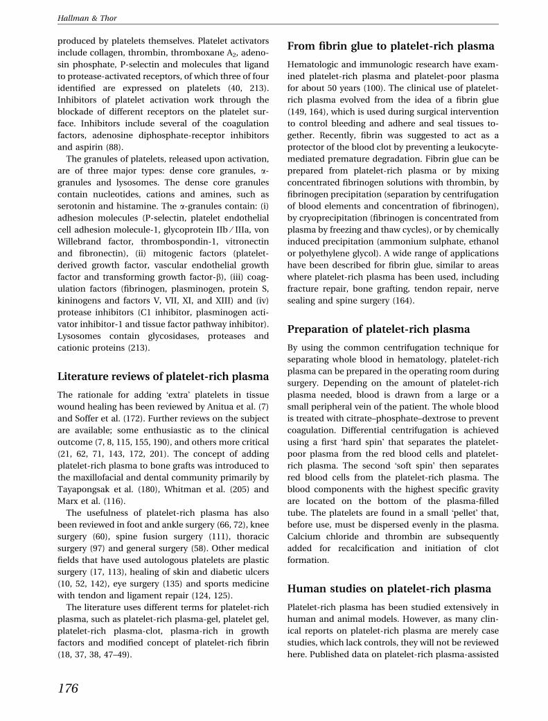
Fig. 1. Extensive resorption of a maxillary alveolar pro-
cess, requiring bone augmentation prior to implant
placement.
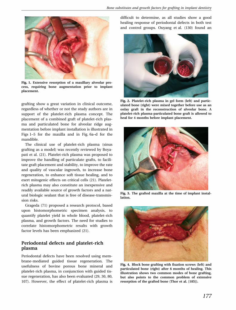
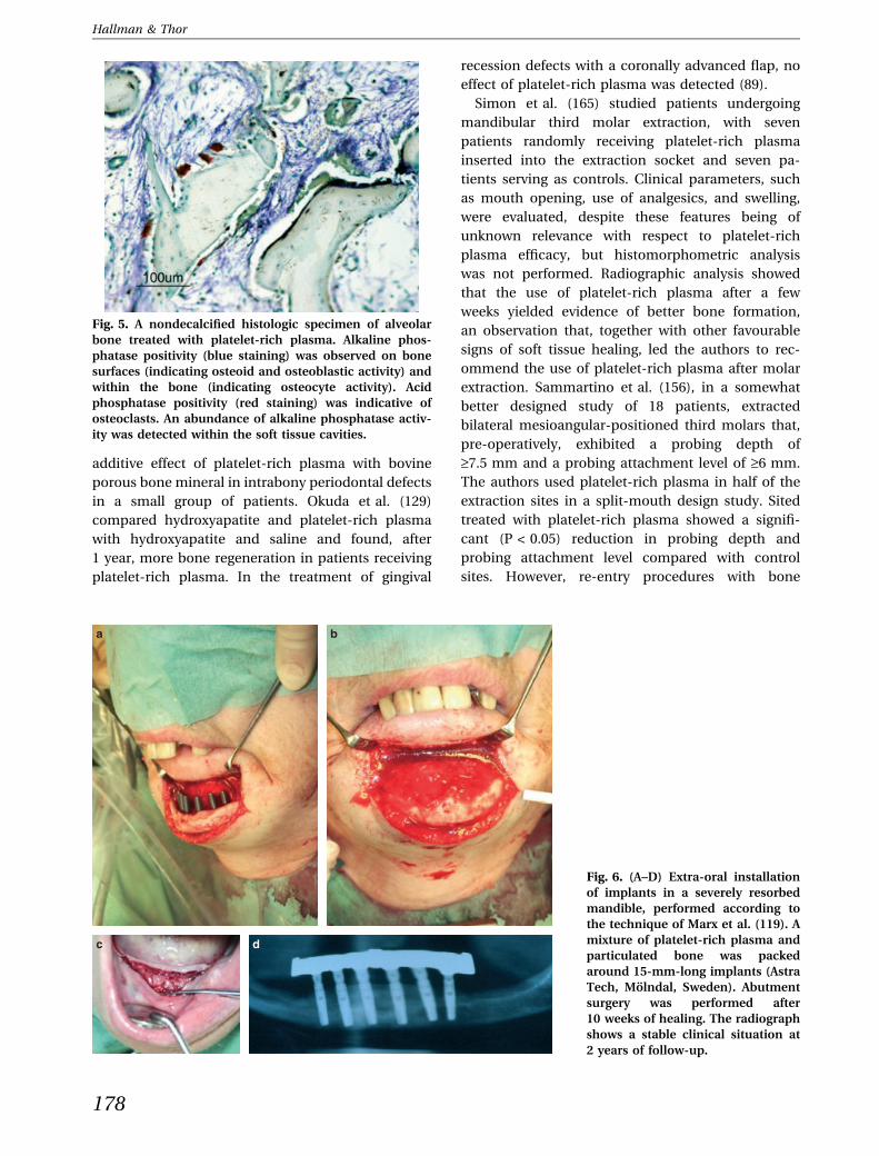
Fig. 3. The grafted maxilla at the time of implant instal-
lation.
Fig. 2. Platelet-rich plasma in gel form (left) and partic-
ulated bone (right) were mixed together before use as an
onlay graft in the reconstruction of alveolar bone. A
platelet-rich plasma-particulated bone graft is allowed to
heal for 4 months before implant placement.
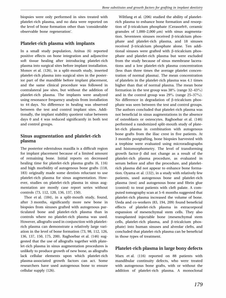
Fig. 4. Block bone grafting with fixation screws (left) and
particulated bone (right) after 6 months of healing. This
illustration shows two common modes of bone grafting,
but also points to the common problem of extensive
resorption of the grafted bone (Thor et al. [185]).
177
Bone substitutes and growth factors for grafting in implant dentistry
additive effect of platelet-rich plasma with bovine
porous bone mineral in intrabony periodontal defects
in a small group of patients. Okuda et al. (129)
compared hydroxyapatite and platelet-rich plasma
with hydroxyapatite and saline and found, after
1 year, more bone regeneration in patients receiving
platelet-rich plasma. In the treatment of gingival
recession defects with a coronally advanced flap, no
effect of platelet-rich plasma was detected (89).
Simon et al. (165) studied patients undergoing
mandibular third molar extraction, with seven
patients randomly receiving platelet-rich plasma
inserted into the extraction socket and seven pa-
tients serving as controls. Clinical parameters, such
as mouth opening, use of analgesics, and swelling,
were evaluated, despite these features being of
unknown relevance with respect to platelet-rich
plasma efficacy, but histomorphometric analysis
was not performed. Radiographic analysis showed
that the use of platelet-rich plasma after a few
weeks yielded evidence of better bone formation,
an observation that, together with other favourable
signs of soft tissue healing, led the authors to rec-
ommend the use of platelet-rich plasma after molar
extraction. Sammartino et al. (156), in a somewhat
better designed study of 18 patients, extracted
bilateral mesioangular-positioned third molars that,
pre-operatively, exhibited a probing depth of
‡7.5 mm and a probing attachment level of ‡6 mm.
The authors used platelet-rich plasma in half of the
extraction sites in a split-mouth design study. Sited
treated with platelet-rich plasma showed a signifi-
cant (P < 0.05) reduction in probing depth and
probing attachment level compared with control
sites. However, re-entry procedures with bone
a
c d
b
Fig. 6. (A–D) Extra-oral installation
of implants in a severely resorbed
mandible, performed according to
the technique of Marx et al. (119). A
mixture of platelet-rich plasma and
particulated bone was packed
around 15-mm-long implants (Astra
Tech, Molndal, Sweden). Abutment
surgery was performed after
10 weeks of healing. The radiograph
shows a stable clinical situation at
2 years of follow-up.
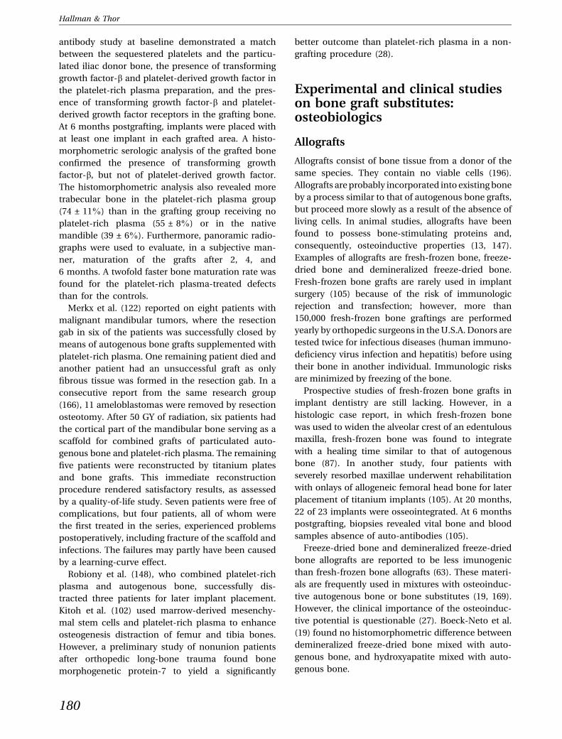
Fig. 5. A nondecalcified histologic specimen of alveolar
bone treated with platelet-rich plasma. Alkaline phos-
phatase positivity (blue staining) was observed on bone
surfaces (indicating osteoid and osteoblastic activity) and
within the bone (indicating osteocyte activity). Acid
phosphatase positivity (red staining) was indicative of
osteoclasts. An abundance of alkaline phosphatase activ-
ity was detected within the soft tissue cavities.
178
Hallman & Thor
biopsies were only performed in sites treated with
platelet-rich plasma, and no data were reported on
the level of bone formation other than �considerable
observable bone regeneration�.
Platelet-rich plasma with implants
In a small study population, Anitua (6) reported
positive effects on bone integration and adjunctive
soft tissue healing after introducing platelet-rich
plasma into surgical sites before implant installation.
Monov et al. (126), in a split-mouth study, inserted
platelet-rich plasma into surgical sites in the poster-
ior part of the mandible before implant placement,
and the same clinical procedure was followed in
contralateral jaw sites, but without the addition of
platelet-rich plasma. The implants were analyzed
using resonance frequency analysis from installation
to 44 days. No difference in healing was observed
between the test and control implant sites. Addi-
tionally, the implant stability quotient value between
days 0 and 4 was reduced significantly in both test
and control groups.
Sinus augmentation and platelet-richplasma
The posterior edentulous maxilla is a difficult region
for implant placement because of a limited amount
of remaining bone. Initial reports on decreased
healing time for platelet-rich plasma grafts (6, 116)
and high morbidity of autogenous bone grafts (118,
183) originally made some dentists reluctant to use
platelet-rich plasma for sinus augmentation. How-
ever, studies on platelet-rich plasma in sinus aug-
mentation are mostly case report series without
controls (73, 112, 120, 136, 137, 150).
Thor et al. (184), in a split-mouth study, found,
after 3 months, significantly more new bone in
biopsies from sinuses grafted with autogenous par-
ticulated bone and platelet-rich plasma than in
controls where no platelet-rich plasma was used.
However, allografts used in conjunction with platelet-
rich plasma can demonstrate a relatively large vari-
ation in the level of bone formation (73, 98, 112, 126,
136, 137, 150, 175, 200). Raghoebar et al. (146) sug-
gested that the use of allografts together with plate-
let-rich plasma in sinus augmentation procedures is
unlikely to produce growth of new bone, as allografts
lack cellular elements upon which platelet-rich
plasma-associated growth factors can act. Some
researchers have used autogenous bone to ensure
cellular supply (126).
Wiltfang et al. (206) studied the ability of platelet-
rich plasma to enhance bone formation and resorp-
tion of b-tricalcium phosphate (Cerasorb�, ceramic
granules of 1,000–2,000 lm) with sinus augmenta-
tion. Seventeen sinuses received b-tricalcium phos-
phate and platelet-rich plasma, and 18 sinuses
received b-tricalcium phosphate alone. Ten addi-
tional sinuses were grafted with b-tricalcium phos-
phate and platelet-rich plasma but were excluded
from the study because of sinus membrane lacera-
tions and a low platelet-rich plasma concentration
(less than three times the average platelet concen-
tration of normal plasma). The mean concentration
of platelets in the platelet-rich plasma was 4.1 times
higher than that of normal plasma. The mean bone
formation in the test group was 38% (range 32–43%)
and in the control group was 29% (range 25–37%).
No difference in degradation of b-tricalcium phos-
phate was seen between the test and control groups.
The authors concluded that platelet-rich plasma was
not beneficial in sinus augmentations in the absence
of osteoblasts or osteocytes. Raghoebar et al. (146)
performed a randomized split-mouth study of plate-
let-rich plasma in combination with autogenous
bone grafts from the iliac crest in five patients. At
3 months postgrafting, bone biopsies harvested with
a trephine were evaluated using microradiographs
and histomorphometry. The level of transforming
growth factor-b did not change as a result of the
platelet-rich plasma procedure, as evaluated in
serum before and after the procedure, and platelet-
rich plasma did not appear to enhance bone forma-
tion. Oyama et al. (132), in a study with relatively few
patients, used autogenous bone and platelet-rich
plasma (test) and autogenous bone and fibrin glue
(control) to treat patients with cleft palate. A com-
puted tomography scan at 5–6 months suggested that
platelet-rich plasma increased the volume of bone.
Ueda and co-workers (83, 194, 209) found beneficial
effects of platelet-rich plasma in extracorporal
expansion of mesenchymal stem cells. They also
transplanted injectable bone (mesenchymal stem
cells, platelet-rich plasma, and b-tricalcium phos-
phate) into human sinuses and alveolar clefts, and
concluded that platelet-rich plasma can be beneficial
in those types of treatment.
Platelet-rich plasma in large bony defects
Marx et al. (116) reported on 88 patients with
mandibular continuity defects, who were treated
with autogenous bone grafts, with or without the
addition of platelet-rich plasma. A monoclonal
179
Bone substitutes and growth factors for grafting in implant dentistry
antibody study at baseline demonstrated a match
between the sequestered platelets and the particu-
lated iliac donor bone, the presence of transforming
growth factor-b and platelet-derived growth factor in
the platelet-rich plasma preparation, and the pres-
ence of transforming growth factor-b and platelet-
derived growth factor receptors in the grafting bone.
At 6 months postgrafting, implants were placed with
at least one implant in each grafted area. A histo-
morphometric serologic analysis of the grafted bone
confirmed the presence of transforming growth
factor-b, but not of platelet-derived growth factor.
The histomorphometric analysis also revealed more
trabecular bone in the platelet-rich plasma group
(74 ± 11%) than in the grafting group receiving no
platelet-rich plasma (55 ± 8%) or in the native
mandible (39 ± 6%). Furthermore, panoramic radio-
graphs were used to evaluate, in a subjective man-
ner, maturation of the grafts after 2, 4, and
6 months. A twofold faster bone maturation rate was
found for the platelet-rich plasma-treated defects
than for the controls.
Merkx et al. (122) reported on eight patients with
malignant mandibular tumors, where the resection
gab in six of the patients was successfully closed by
means of autogenous bone grafts supplemented with
platelet-rich plasma. One remaining patient died and
another patient had an unsuccessful graft as only
fibrous tissue was formed in the resection gab. In a
consecutive report from the same research group
(166), 11 ameloblastomas were removed by resection
osteotomy. After 50 GY of radiation, six patients had
the cortical part of the mandibular bone serving as a
scaffold for combined grafts of particulated auto-
genous bone and platelet-rich plasma. The remaining
five patients were reconstructed by titanium plates
and bone grafts. This immediate reconstruction
procedure rendered satisfactory results, as assessed
by a quality-of-life study. Seven patients were free of
complications, but four patients, all of whom were
the first treated in the series, experienced problems
postoperatively, including fracture of the scaffold and
infections. The failures may partly have been caused
by a learning-curve effect.
Robiony et al. (148), who combined platelet-rich
plasma and autogenous bone, successfully dis-
tracted three patients for later implant placement.
Kitoh et al. (102) used marrow-derived mesenchy-
mal stem cells and platelet-rich plasma to enhance
osteogenesis distraction of femur and tibia bones.
However, a preliminary study of nonunion patients
after orthopedic long-bone trauma found bone
morphogenetic protein-7 to yield a significantly
better outcome than platelet-rich plasma in a non-
grafting procedure (28).
Experimental and clinical studieson bone graft substitutes:osteobiologics
Allografts
Allografts consist of bone tissue from a donor of the
same species. They contain no viable cells (196).
Allografts are probably incorporated into existing bone
by a process similar to that of autogenous bone grafts,
but proceed more slowly as a result of the absence of
living cells. In animal studies, allografts have been
found to possess bone-stimulating proteins and,
consequently, osteoinductive properties (13, 147).
Examples of allografts are fresh-frozen bone, freeze-
dried bone and demineralized freeze-dried bone.
Fresh-frozen bone grafts are rarely used in implant
surgery (105) because of the risk of immunologic
rejection and transfection; however, more than
150,000 fresh-frozen bone graftings are performed
yearly by orthopedic surgeons in the U.S.A. Donors are
tested twice for infectious diseases (human immuno-
deficiency virus infection and hepatitis) before using
their bone in another individual. Immunologic risks
are minimized by freezing of the bone.
Prospective studies of fresh-frozen bone grafts in
implant dentistry are still lacking. However, in a
histologic case report, in which fresh-frozen bone
was used to widen the alveolar crest of an edentulous
maxilla, fresh-frozen bone was found to integrate
with a healing time similar to that of autogenous
bone (87). In another study, four patients with
severely resorbed maxillae underwent rehabilitation
with onlays of allogeneic femoral head bone for later
placement of titanium implants (105). At 20 months,
22 of 23 implants were osseointegrated. At 6 months
postgrafting, biopsies revealed vital bone and blood
samples absence of auto-antibodies (105).
Freeze-dried bone and demineralized freeze-dried
bone allografts are reported to be less imunogenic
than fresh-frozen bone allografts (63). These materi-
als are frequently used in mixtures with osteoinduc-
tive autogenous bone or bone substitutes (19, 169).
However, the clinical importance of the osteoinduc-
tive potential is questionable (27). Boeck-Neto et al.
(19) found no histomorphometric difference between
demineralized freeze-dried bone mixed with auto-
genous bone, and hydroxyapatite mixed with auto-
genous bone.
180
Hallman & Thor
Xenografts
Xenografts consist of bone mineral from animals or
bone-like minerals (calcium carbonate) derived from
corals or algae (59, 92).
Deproteinized bovine bone is the most researched
grafting material and is widely used in dentistry be-
cause of its similarity to human bone. Proteins in
deproteinized bovine bone have been extracted to
avoid immunologic rejection after implantation;
however, as the deproteinizing procedure eliminates
the osteoinductive capacity, deproteinized bovine
bones act solely as an osteoconductive scaffold.
In a rat calvarial model, three grafting materials
were compared: plaster of Paris, particulated dentine
and deproteinized bovine bone (99). All three grafting
materials generated new bone and all were found to
be suitable for use as bone substitutes. However,
deproteinized bovine bone showed the largest and
most rapid bone formation. In a histomorphometric
study in dogs, Berglundh et al. (16) found a reduction
from 17 to 11% in the amount of deproteinized
bovine bone particles in biopsies obtained during a
3–7-month time period. In a chimpanzee study,
McAllister et al. (121) detected a decrease in the
percentage of anorganic area, from 19 ± 14% to
6 ± 3% over a time period of 7.5–18 months. Other
animal studies also found promising bone formation
using deproteinized bovine bone (65, 74, 91, 103).
However, whether deproteinized bovine bone is
resorbable or nonresorbable is a topic of discussion.
A study on defects in rabbit skulls found that
deproteinized bovine bone particles had almost
completely disappeared after 14 days of healing, as a
result of the action of multinucleated cells (103). In
a chimpanzee study, Hurzeler et al. (91) identified
osteoclastic resorption of deproteinized bovine bone.
In a dog study where a biologic glue (Tisseel�, Duo
Quick; Immuno, Vienna, Austria) was added to
the graft, no bone formation was found around
the deproteinized bovine bone particles but a large
number of the particles had disappeared as a result of
giant cell resorption (32).
In a minipig model, augmentation of the maxillary
sinus was performed using deproteinized bovine
bone, with or without recombinant human osteo-
genic protein (¼ bone morphogenetic protein-7)
(181). A significant increase in bone formation was
found (38.6–80.0%) when recombinant human
osteogenic protein was added to the graft. Other
studies, which compared recombinant human osteo-
genic protein and platelet-rich plasma in bilateral
sinus grafts using anorganic bone as a carrier, found
platelet-rich plasma to be ineffective but re-
combinant human osteogenic protein to stimulate
bone formation (151, 152).
In human studies of maxillary sinus augmentation,
deproteinized bovine bone with or without a mixture
with autogenous bone has histologically been asso-
ciated with active bone formation (64, 75, 77, 159,
168, 179, 198, 199, 210). Froum et al. (64) used
platelet-rich plasma together with anorganic bovine
bone in the maxillary sinus, but found no statistical
increase in the amount of vital bone produced as a
result of adding platelet-rich plasma, confirming the
findings in the experimental animal studies. Piattelli
et al. (140) performed maxillary sinus augmentation
using deproteinized bovine bone and autogenous
bone in 20 patients and found, in biopsies harvested
after 6–9 months of healing, an average of 30% newly
formed bone, 30% deproteinized bovine bone, and
40% bone marrow. Increased mineralization was
detected after 14 months, and osteoclastic resorption
of the deproteinized bovine bone particles was found
after 4 years. Valentini et al. (199) used deproteinized
bovine bone in the floor of the maxillary sinus in 15
patients. Biopsies harvested after 6 months revealed
21% new bone, 39% deproteinized bovine bone, and
40% marrow, and after 12 months 28% new bone,
27% deproteinized bovine bone, and 45% marrow
was found. The reason for the 12% decrease in the
amount of deproteinized bovine bone particles dur-
ing the second 6-month study interval is not known.
Yildirim et al. (210) studied maxillary sinus augmen-
tation using deproteinized bovine bone mixed with
blood in 15 sinuses of 11 patients. After 4–9.5 months
of healing, biopsies revealed 14.7% new bone, 55.6%
soft tissue, and 29.7% deproteinized bovine bone.
Twenty-nine per cent of the deproteinized bovine
bone particles were in contact with newly formed
bone (210). In another study, by Yildirim et al. (211),
13 maxillary sinuses in 12 patients were treated with a
mixture of deproteinized bovine bone and auto-
genous bone along with a resorbable membrane on
the lateral side of the graft (BioGide�; Geistleich,
Pharma AG, Wolhusen, Switzerland). Biopsies
harvested after 6–9 months revealed 19% new bone,
51% connective tissue and 30% deproteinized bovine
bone. Thirty-three per cent of the deproteinized bo-
vine bone particles were in contact with newly
formed bone (211). Hallman et al. (75) used an
80:20% mixture of deproteinized bovine bone and
autogenous bone in the floor of the sinus. Biopsies
harvested after 6 months of healing showed 54%
fibrous connective tissue, 21% lamellar bone,
14% deproteinized bovine bone particles and 10%
181
Bone substitutes and growth factors for grafting in implant dentistry
immature bone; 28% of the particles were in contact
with newly formed bone. The corresponding figures
from 3-year biopsies were 36% fibrous connective
tissue, 51% lamellar bone, 12% deproteinized bovine
bone particles and 1% immature bone; 54% of the
particles were in contact with newly formed bone.
The area of deproteinized bovine bone particles did
not decrease during the 3-year follow-up period (75).
In another study carried out by Hallman et al. (77),
an 80:20% mixture of deproteinized bovine bone and
autogenous bone was compared with deproteinized
bovine bone only or with autogenous bone only.
Micro-implants harvested with a surrounding bone
core at 6–9 months after placement showed no
statistical difference in implant osseointegration and
bone formation for the three grafting materials used
(77). This finding, and those of other studies (56, 76,
210), indicate that autogenous bone may not be
needed as a grafting material in the floor of the
maxillary sinus.
Several long-term follow-up studies have con-
cluded that osteoconduction proceeds for several
years, but no data are available to determine whether
a mixture of autogenous bone and deproteinized
bovine bone may shorten the healing time of the graft
(75–77, 140, 191). However, a healing period of
8 months is recommended for deproteinized bovine
bone when used as the only grafting material, com-
pared with a healing period of 6 months for
autogenous bone grafts (76, 77). Excellent results of
implant survival for implants placed after sinus floor
augmentation with DBB have also been presented
and it is concluded that today this material is con-
sidered the standard of care for sinus augmentation
procedures (56).
The resorption of deproteinized bovine bone by
means of osteoclastic activity is a topic of contro-
versy, especially when comparing results from
experimental animal studies and clinical human
studies. Some human studies have detected resorp-
tion of deproteinized bovine bone particles (140,
202). Wallace et al. (202) found no signs of deprote-
inized bovine bone particles after 20 months of
healing; whether this finding reflects the biopsy
technique used or was the result of true resorption is
unclear. Piatelli et al. (140) observed osteoclastic
activity around deproteinized bovine bone particles
in 4-year specimens, although the deproteinized
bovine bone particles seemed to remain intact.
Schlegel & Donath (159) found no signs of resorption
of deproteinized bovine bone particles after 6 years.
Skovlund et al. (168) suggested that deproteinized
bovine bone particles were slowly degraded (168).
However, most studies failed to detect evidence of
deproteinized bovine bone resorption (75, 77, 159,
179, 198, 199, 210, 211). The reported variation in
resorption of deproteinized bovine bone may stem
from differences in response from animal studies,
surgical technique, biopsy technique, and histologic
preparation technique (decalcified preparation or
standard sectioning). The histologic decalcification
process of the deproteinized bovine bone particles
always causes shrinkage, which might be misinter-
preted as resorption.
From a biologic point of view, resorption requires
adhesion molecules (arginine–glycine–asparagine
sequences) for the attachment of osteoclastic cells to
plasma and extracellular matrix proteins, including
fibronectin, fibrinogen, vitronectin, type I collagen,
osteopontin and bone sialoprotein (141). As depro-
teinized bovine bone is free of proteins (14, 203),
osteoclastic resorption can probably not occur.
However, Schwartz et al. (160) found transforming
growth factor-ß and bone morphogenetic protein-2
in deproteinized bovine bone particles, which might
explain the lacunae found on some deproteinized
bovine bone particles. Some degree of degradation by
macrophagial phagocytosis may occur, but is prob-
ably too limited to explain the reported degradation
of deproteinized bovine bone in some animal (16, 33,
78, 79, 91, 103, 121, 123) and human (140, 202)
studies.
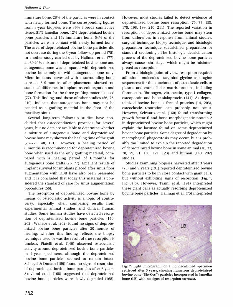
Studies examining biopsies harvested after 3 years
(75) and 9 years (191) reported deproteinized bovine
bone particles to be in close contact with giant cells,
but without exhibiting signs of resorption (Fig. 7,
Fig. 8a,b). However, Traini et al. (191) interpreted
these giant cells as actually resorbing deproteinized
bovine bone particles. Hallman et al. (75) interpreted
LBB
Fig. 7. Light micrograph of a nondecalcified specimen
retrieved after 3 years, showing numerous deproteinized
bovine bone (Bio-Oss�) particles incorporated in lamellar
bone (LB) with no signs of resorption (arrows).
182
Hallman & Thor
possible signs of resorption, such as lacunae on the
surfaces of deproteinized bovine bone particles, as
being lacunae present in the original donor material,
or as evidence of osteoclastic resorption mediated by
later surface adsorption of proteins containing argi-
nine–glycine–asparagine sequences, such as fibro-
nectin or fibrinogen (153). Adhesion proteins may
originate either from the fibrin glue or from the pa-
tient�s own blood. Nonetheless, the great reduction in
deproteinized bovine bone particles seen in many
animal studies is difficult to reconcile with available
data from human studies. Even though the type of
giant cells in the vicinity of deproteinized bovine
bone particles may vary among species or individu-
als, osteoclastic cells are unlikely to resorb or degrade
bovine bone that has been deproteinized. It seems
safe to suggest that deproteinized bovine bone is
basically a nonresorbable grafting material in hu-
mans.
In sinus augmentation, it may be of importance
that deproteinized bovine bone is nonresorbable,
whereas autogenous bone can experience more than
50% resorption (94). Cobb et al. (39) discussed the
advantage of using a nonresorbable or a low-grade
resorption bone substitute and concluded that a
mixture of equal volumes of a nonresorbable and of
autogenous bone was optimal for grafting. An
increased amount of bone substitute enhances the
risk of fibrous encapsulation. However, in some
studies that used 100% deproteinized bovine bone
as a grafting material, no encapsulation appeared to
take place, with the results observed being similar to
those obtained with a mixture of deproteinized
bovine bone and autogenous bone (77, 84, 90, 91,
210, 211).
Lateral augmentation of edentulous alveolar ridge
defects can also benefit from deproteinized bovine
bone grafting (82, 84, 216). Hellem et al.(82), using a
50:50 (wt ⁄ wt) mixture of deproteinized bovine bone
and autogenous bone, reported a 3-year implant
survival rate of 96%. Zitzman & Scharer (216), har-
vested biopsies after 6–7 months, and found that
deproteinized bovine bone particles (Bio-Oss�;
Geistlich, Pharma AG, Wolhusen, Switzerland) had
37% contact with newly formed bone and that 31%
of the biopsy area was occupied by the Bio-Oss
particles.
Xenografts derived from marine carbonated algae,
chemically converted into hydroxyapatite, have been
used in implant dentistry for several years (59, 188).
This material is probably also nonresorbable because
it does not contain any proteins and is not dissolv-
able. Ewers (59) found a loss of 27 of 614 loaded
implants in sinus sites grafted with the marine algae-
based material, yielding an implant survival rate of
95.6%. Also, the volume of the xenograft material
decreased by 14% after 6 months compared with
49.5% for autogenous bone (59).
Alloplastic bone substitutes
Alloplastic bone substitutes represent a large group
of chemically diverse synthetic calcium-based
biomaterials, including calcium phosphate, calcium
sulphate, bioactive glasses, and polymers. These
osteobiologics vary in structure, chemical composi-
tion, and mechanical and biological properties. Also,
some osteobiologics are nonresorbable, whereas
BioOss
B
a
b
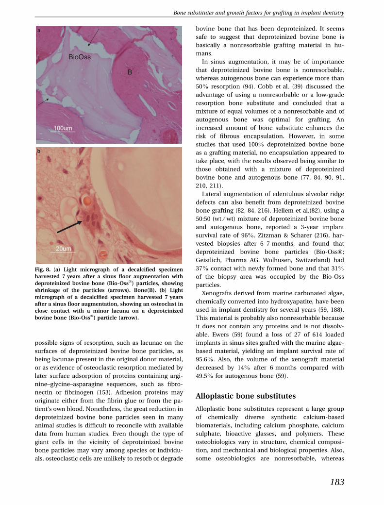
Fig. 8. (a) Light micrograph of a decalcified specimen
harvested 7 years after a sinus floor augmentation with
deproteinized bovine bone (Bio-Oss�) particles, showing
shrinkage of the particles (arrows). Bone(B). (b) Light
micrograph of a decalcified specimen harvested 7 years
after a sinus floor augmentation, showing an osteoclast in
close contact with a minor lacuna on a deproteinized
bovine bone (Bio-Oss�) particle (arrow).
183
Bone substitutes and growth factors for grafting in implant dentistry
others are chemically resorbable with a concomitant
release of bioactive ions.
The pore size of osteobiologics seems to be a sig-
nificant determinant of the ability to form bone. Pore
sizes of >300 lm show enhanced formation of new
capillaries and bone (95). Alloplastic material with
pore sizes of <100 lm may not permit cell and cap-
illary invasion and therefore may not induce bone
formation. Most of the current commercial osteobi-
ologics do not exhibit any pores. The manipulation of
pore size is critical for creating an osteobiologic
material that resembles natural bone.
Most alloplastic materials consist of hydroxy-
apatite, b-tricalcium phosphate, biphasic calcium
phosphate, or some type of nonsintered cal-
cium phosphate (reduced calcium content). Calcium
phosphates are manufactured from tricalcium phos-
phate powder. Pure calcium phosphate is less stable
than hydroxyapatite and thus can be dissociated
more easily into potentially bone-stimulating ions
(41). Tricalcium phosphate can be sintered into a
uniform material, b-tricalcium phosphate (Cera-
sorb�; Curasan AG, Kleinostheim, Germany), which
is one of the most frequently used alloplasts in im-
plant dentistry (177). A drawback of b-tricalcium
phosphate is a rapid resorption rate, which limits its
use in bone augmentation procedures performed for
esthetic purposes.
Biphasic alloplastic material is produced by sin-
tering hydroxyapatite and tricalcium phosphate to a
chemically united material. These grafting materials
have pore sizes of >100 lm and have been shown to
be effective in repairing skeletal defects (127). Syn-
thetically produced alloplasts used in implant den-
tistry include Calcitec� Inc. (Austin, TX), Osteogen�(Impladent Ltd, Holliswood, NY), Tricos� (Baxter,
Bern, Switzerland) and Bone Ceramic� (Straumann,
Basel, Switzerland). However, sufficient documenta-
tion of the clinical utility of several of these alloplasts
is still lacking.
Calcium phosphates can be bound to collagen
carriers or mixed with fibrin. The concept is that
collagen and fibrin form a network on which
minerals can crystallize. Collagen can also bind to
extracellular matrix proteins of importance in the
mineralization process. Healos� (Orquest, Mountain
View, CA) is a mixture of hydroxyapatite and bovine
collagen, and Collagraft� (Zimmer Corp, Warsaw, IN)
is composed of 65% hydroxyapatite and 35% trical-
cium phosphate combined with bovine collagen.
Tricos�, mentioned in the previous paragraph, is a
mixture of hydroxyapatite, tricalcium phosphate, and
fibrin.
Calcium sulphate has been used in craniofacial
surgery for more than 100 years. Dreesman (51) was
the first to use surgical plaster to fill skeletal defects,
and De Leonardis & Pecora (44) have used the
material for sinus floor augmentation in implant
dentistry (44). Calcium sulphate resorbs quickly and
is substituted by new bone. The rapid resorption rate
can pose a potential problem because the volume of
the graft may not be maintained for a sufficiently
long period of time to yield reliable grafting results in
the esthetic zone.
Bioglass consists of silica and has an optimal
particle size of 300 lm for bone formation (178).
Bioglass corrodes when placed in fluid followed by a
migration of hydrogen ions to the surface of the
material. Sodium and silica then precipitates and
calcium ions are released, which can stimulate stem
cells to produce bone-building cells (41). As Bioglass
is slowly resorbed, it probably takes 12–16 months
before the graft is replaced by newly formed bone, a
factor that has to be considered when calculating the
graft healing time (178, 193). Tadjoedin et al. (178)
compared a mixture of autogenous bone and bioac-
tive glass particles (300–355 lm in size) with autog-
enous bone alone. The biopsies showed more new
bone in the autogenous bone group after 4–6 months
and similar values for both groups after 16 months.
Turunen et al. (193) compared a mixture of auto-
genous bone and bioactive glass granules (800–
1,000 lm in size) with autogenous bone alone as a
grafting material in the floor of the maxillary sinus.
Biopsies harvested after 21–34 weeks and after
49–62 weeks revealed a similar bone-forming out-
come in both study groups (193). The studies by
Tadjoedin et al. (178) and Turunen et al. (193) sug-
gest that bioglass can be used in a mixture with
autogenous bone at the floor of the maxillary sinus,
thus decreasing the amount of autogenous bone
required.
Conclusion
Dentists have access to an increasing number of
different biomaterials for use in bone augmentation
procedures prior to implant placement. However,
most of them are not well clinically documented
(57).
It appears that a variety of biomaterials can
provide excellent bone formation in the floor of
the maxillary sinus. On the other hand, in some
patients it is only necessary to tent the sinus
membrane to achieve new bone for stability of
184
Hallman & Thor
implants. Lateral bone augmentation of the alveo-
lar ridge constitutes another important indication
for biomaterial grafting. In general, prospective
clinical studies are lacking for many of the bone
augmentation materials and techniques currently
available.
Resorption and complete remodelling into new
bone is the ideal outcome of a grafting material.
However, the resorption rate of a graft material can
vary greatly. Some materials resorb quickly, which
can severely compromise their usefulness, especially
in the esthetic zone. Relatively rapid resorption of
autogenous bone grafts has led to a search for more
stable bone substitutes.
Currently, there is no clinical evidence for
superiority of autogenous bone grafts in sinus aug-
mentation procedures. The complex procedure of
harvesting autogenous bone from the iliac crest is
still necessary when a large amount is needed.
However, studies during the past decade have sug-
gested that nonresorbable grafting materials may be
capable of predictably maintaining the graft volume.
For minor grafting procedures, such as bone aug-
mentation of the sinus floor or of alveolar defects, it
seems feasible to use bone substitutes with or with-
out adding autogenous bone harvested locally or
from the mandibular ramus.
The platelet-rich plasma concept in reconstructive
surgery has been evaluated in various experimental
and clinical models, with conflicting results obtained.
Some studies point to the need for the presence of
appropriate target cells to interact with the growth
factors in platelet-rich plasma. Moreover, in cell
culture models, platelet-rich plasma seems to exert
most activity in the early stages of cell proliferation.
The effect of platelet-rich plasma in humans may be
beneficial only in the early phases of the formation of
bone and possibly soft tissue. Experimental and
clinical investigations are needed to delineate the
interaction among the various growth factors in
platelet-rich plasma during tissue healing. Also,
controversy still exists as to the clinical benefit of
combining platelet-rich plasma with bone grafts, and
to the utility of platelet-rich plasma in implant den-
tistry. Whether the extra cost and time spent on the
platelet-rich plasma procedure is justified remains a
topic for further study.
References
1. Abe E. Function of BMPs and BMP antagonists in
adult bone. Ann N Y Acad Sci 2006: 1068: 41–53.
2. Albrektsson T. In vivo studies of bone grafts. The possi-
bility of vascular anastomoses in healing bone. Acta
Orthop Scand 1980: 51: 9–17.
3. Albrektsson T. Repair of bone grafts. A vital microscopic
and histological investigation in the rabbit. Scand J Plast
Reconstr Surg 1980: 14: 1–12.
4. Albrektsson T, Albrektsson B. Microcirculation in grafted
bone. A chamber technique for vital microscopy of
rabbit bone transplants. Acta Orthop Scand 1978: 49:
1–7.
5. Andrew JG, Hoyland JA, Freemont AJ, Marsh DR. Platelet-
derived growth factor expression in normally healing
human fractures. Bone 1995: 16: 455–460.
6. Anitua E. Plasma rich in growth factors: preliminary re-
sults of use in the preparation of future sites for implants.
Int J Oral Maxillofac Implants 1999: 14: 529–535.
7. Anitua E, Andia I, Ardanza B, Nurden P, Nurden AT.
Autologous platelets as a source of proteins for
healing and tissue regeneration. Thromb Haemost 2004:
91: 4–15.
8. Anitua E, Sanchez M, Nurden AT, Nurden P, Orive G,
Andia I. New insights into and novel applications for
platelet-rich fibrin therapies. Trends Biotechnol 2006: 24:
227–234.
9. Assoian RK, Komoriya A, Meyers CA, Miller DM, Sporn
MB. Transforming growth factor-beta in human platelets.
Identification of a major storage site, purification, and
characterization. J Biol Chem 1983: 258: 7155–7160.
10. Atri SC, Misra J, Bisht D, Misra K. Use of homologous
platelet factors in achieving total healing of recalcitrant
skin ulcers. Surgery 1990: 108: 508–512.
11. Balemans W, Van Hul W. Extracellular regulation of BMP
signaling in vertebrates: a cocktail of modulators. Dev Biol
2002: 250: 231–250.
12. Barnes GL, Kostenuik PJ, Gerstenfeld LC, Einhorn TA.
Growth factor regulation of fracture repair. J Bone Miner
Res 1999: 14: 1805–1815.
13. Becker W, Urist MR, Tucker LM, Becker BE, Ochsenbein C.
Human demineralized freeze-dried bone: inadequate in-
duced bone formation in athymic mice. A preliminary
report. J Periodontol 1995: 66: 822–828.
14. Benke D, Olah A, Mohler H. Protein-chemical analysis of
Bio-Oss bone substitute and evidence on its carbonate
content. Biomaterials 2001: 22: 1005–1012.
15. Berengo M, Bacci C, Sartori M, Perini A, Della Barbera M,
Valente M. Histomorphometric evaluation of bone grafts
harvested by different methods. Minerva Stomatol 2006:
55: 189–198.
16. Berglundh T, Lindhe J. Healing around implants placed in
bone defects treated with Bio-Oss. An experimental study
in the dog. Clin Oral Implants Res 1997: 8: 117–124.
17. Bhanot S, Alex JC. Current applications of platelet gels in
facial plastic surgery. Facial Plast Surg 2002: 18: 27–33.
18. Bielecki T, Gazdzik TS, Szczepanski T. What do we use:
platelet-rich plasma or platelet-rich gel? Bone 2006: 39:
1388.
19. Boeck-Neto RJ, Gabrielli M, Lia R, Marcantonio E, Shibli
JA, Marcantonio E Jr. Histomorphometrical analysis of
bone formed after maxillary sinus floor augmentation by
grafting with a combination of autogenous bone and
demineralized freeze-dried bone allograft or hydroxy-
apatite. J Periodontol 2002: 73: 266–270.
185
Bone substitutes and growth factors for grafting in implant dentistry
20. Bonewald LF, Mundy GR. Role of transforming growth
factor-beta in bone remodeling. Clin Orthop Relat Res
1990: 60: 261–276.
21. Boyapati L, Wang HL. The role of platelet-rich plasma in
sinus augmentation: a critical review. Implant Dent 2006:
15: 160–170.
22. Boyne PJ, Cole MD, Stringer D, Shafqat JP. A technique for
osseous restoration of deficient edentulous maxillary rid-
ges. J Oral Maxillofac Surg 1985: 43: 87–91.
23. Boyne PJ, James RA. Grafting of the maxillary sinus floor
with autogenous marrow and bone. J Oral Surg 1980: 38:
613–616.
24. Brogi E, Wu T, Namiki A, Isner JM. Indirect angiogenic
cytokines upregulate VEGF and bFGF gene expression in
vascular smooth muscle cells, whereas hypoxia upregu-
lates VEGF expression only. Circulation 1994: 90: 649–652.
25. Buchman SR, Ozaki W. The ultrastructure and resorptive
pattern of cancellous onlay bone grafts in the craniofacial
skeleton. Ann Plast Surg 1999: 43: 49–56.
26. Burchardt H. The biology of bone graft repair. Clin Orthop
Relat Res 1983: 174: 28–42.
27. Buser D, Hoffmann B, Bernard JP, Lussi A, Mettler D,
Schenk RK. Evaluation of filling materials in membrane–
protected bone defects. A comparative histomorphometric
study in the mandible of miniature pigs. Clin Oral
Implants Res 1998: 9: 137–150.
28. Calori GM, D�Avino M, Tagliabue L, Albisetti W, d�Im-
porzano M, Peretti G. An ongoing research for evaluation
of treatment with BMPs or AGFs in long bone non-union:
protocol description and preliminary results. Injury 2006:
37: S43–S50.
29. Camargo PM, Lekovic V, Weinlaender M, Vasilic N,
Madzarevic M, Kenney EB. Platelet-rich plasma and
bovine porous bone mineral combined with guided tissue
regeneration in the treatment of intrabony defects in hu-
mans. J Periodontal Res 2002: 37: 300–306.
30. Camargo PM, Lekovic V, Weinlaender M, Vasilic N,
Madzarevic M, Kenney EB. A reentry study on the use of
bovine porous bone mineral, GTR, and platelet-rich
plasma in the regenerative treatment of intrabony defects
in humans. Int J Periodontics Restorative Dent 2005: 25:
49–59.
31. Carlson ER, Marx RE. Part II. Mandibular reconstruction
using cancellous cellular bone grafts. J Oral Maxillofac
Surg 1996: 54: 889–897.
32. Carmagnola D, Berglundh T, Araujo M, Albrektsson T,
Lindhe J. Bone healing around implants placed in a jaw
defect augmented with Bio-Oss. An experimental study in
dogs. J Clin Periodontol 2000: 27: 799–805.
33. Carmagnola D, Berglundh T, Lindhe J. The effect of a fi-
brin glue on the integration of Bio-Oss with bone tissue. A
experimental study in labrador dogs. J Clin Periodontol
2002: 29: 377–383.
34. Chen NT, Glowacki J, Bucky LP, Hong HZ, Kim WK,
Yaremchuk MJ. The roles of revascularization and
resorption on endurance of craniofacial onlay bone grafts
in the rabbit. Plast Reconstr Surg 1994: 93: 714–722;
discussion 723–724.
35. Chiriac G, Herten M, Schwarz F, Rothamel D, Becker J.
Autogenous bone chips: influence of a new piezoelectric
device (Piezosurgery) on chip morphology, cell viability
and differentiation. J Clin Periodontol 2005: 32: 994–999.
36. Cho TJ, Gerstenfeld LC, Einhorn TA. Differential temporal
expression of members of the transforming growth factor
beta superfamily during murine fracture healing. J Bone
Miner Res 2002: 17: 513–520.
37. Choukroun J, Diss A, Simonpieri A, Girard MO, Schoeffler
C, Dohan SL, Dohan AJ, Mouhyi J, Dohan DM. Platelet-
rich fibrin (PRF): a second-generation platelet concen-
trate. Part V: histologic evaluations of PRF effects on bone
allograft maturation in sinus lift. Oral Surg Oral Med Oral
Pathol Oral Radiol Endod 2006: 101: 299–303.
38. Choukroun J, Diss A, Simonpieri A, Girard MO, Schoeffler
C, Dohan SL, Dohan AJ, Mouhyi J, Dohan DM. Platelet-rich
fibrin (PRF): a second-generation platelet concentrate. Part
IV: clinical effects on tissue healing. Oral Surg Oral Med
Oral Pathol Oral Radiol Endod 2006: 101: e56–e60.
39. Cobb CM, Eick JD, Barker BF, Mosby EL, Hiatt WR.
Restoration of mandibular continuity defects using com-
binations of hydroxylapatite and autogenous bone:
microscopic observations. J Oral Maxillofac Surg 1990: 48:
268–275.
40. Coughlin SR. Protease-activated receptors and platelet
function. Thromb Haemost 1999: 82: 353–356.
41. Daculsi G. Biphasic calcium phosphate concept applied to
artificial bone, implant coating and injectable bone sub-
stitute. Biomaterials 1998: 19: 1473–1478.
42. Dado DV, Izquierdo R. Absorption of onlay bone grafts in
immature rabbits: membranous versus enchondral bone
and bone struts versus paste. Ann Plast Surg 1989: 23: 39–
48.
43. Davies JE, Hosseini MM. Histodynamics of endosseus
wound healing. In: Davies JE, editor. Bone Engineering.
Toronto, Canada: em squared incorporated, 2000: 1–14.
44. De Leonardis D, Pecora GE. Prospective study on the
augmentation of the maxillary sinus with calcium sulfate:
histological results. J Periodontol 2000: 71: 940–947.
45. Derynck R, Gelbart WM, Harland RM, Heldin CH, Kern SE,
Massague J, Melton DA, Mlodzik M, Padgett RW, Roberts
AB, Smith J, Thomsen GH, Vogelstein B, Wang XF.
Nomenclature: vertebrate mediators of TGFbeta family
signals. Cell 1996: 87: 173.
46. Dimitriou R, Tsiridis E, Giannoudis PV. Current concepts
of molecular aspects of bone healing. Injury 2005: 36:
1392–1404.
47. Dohan DM, Choukroun J, Diss A, Dohan SL, Dohan AJ,
Mouhyi J, Gogly B. Platelet-rich fibrin (PRF): a second-
generation platelet concentrate. Part II: platelet-related
biologic features. Oral Surg Oral Med Oral Pathol Oral
Radiol Endod 2006: 101: e45–e50.
48. Dohan DM, Choukroun J, Diss A, Dohan SL, Dohan AJ,
Mouhyi J, Gogly B. Platelet-rich fibrin (PRF): a second-
generation platelet concentrate. Part I: technological
concepts and evolution. Oral Surg Oral Med Oral Pathol
Oral Radiol Endod 2006: 101: e37–e44.
49. Dohan DM, Choukroun J, Diss A, Dohan SL, Dohan AJ,
Mouhyi J, Gogly B. Platelet-rich fibrin (PRF): a second-
generation platelet concentrate. Part III: leucocyte acti-
vation: a new feature for platelet concentrates? Oral Surg
Oral Med Oral Pathol Oral Radiol Endod 2006: 101:
e51–e55.
50. Donovan MG, Dickerson NC, Hellstein JW, Hanson LJ.
Autologous calvarial and iliac onlay bone grafts in mini-
ature swine. J Oral Maxillofac Surg 1993: 51: 898–903.
186
Hallman & Thor
51. Dreesman H. Uber knochenplombierung. Bietr Klin Chir
1892: 9: 804–810.
52. Driver VR, Hanft J, Fylling CP, Beriou JM, Autologel Dia-
betic Foot Ulcer Study G. A prospective, randomized,
controlled trial of autologous platelet-rich plasma gel for
the treatment of diabetic foot ulcers. Ostomy Wound
Manage 2006: 52: 68–70, 72, 74 passim.
53. Ducy P, Karsenty G. The family of bone morphogenetic
proteins. Kidney Int 2000: 57: 2207–2214.
54. Eriksson AR, Albrektsson T. Temperature threshold levels
for heat-induced bone tissue injury: a vital-microscopic
study in the rabbit. J Prosthet Dent 1983: 50: 101–107.
55. Eriksson A, Albrektsson T, Grane B, McQueen D. Thermal
injury to bone. A vital-microscopic description of heat
effects. Int J Oral Surg 1982: 11: 115–121.
56. Esposito M, Grusovin MG, Coulthard P, Worthington HV.
The efficacy of various bone augmentation procedures for
dental implants: a Cochrane systematic review of ran-
domized controlled clinical trials. Int J Oral Maxillofac
Implants 2006: 21: 696–710.
57. Esposito M, Grusovin MG, Worthington HV, Coulthard P.
Interventions for replacing missing teeth: bone augmen-
tation techniques for dental implant treatment. Cochrane
Database Syst Rev 2006: 00: CD003607.
58. Everts PA, Knape JT, Weibrich G, Schonberger JP, Hoff-
mann J, Overdevest EP, Box HA, van Zundert A. Platelet-
rich plasma and platelet gel: a review. J Extra Corpor
Technol 2006: 38: 174–187.
59. Ewers R. Maxilla sinus grafting with marine algae derived
bone forming material: a clinical report of long-term
results. J Oral Maxillofac Surg 2005: 63: 1712–1723.
60. Floryan KM, Berghoff WJ. Intraoperative use of auto-
logous platelet-rich and platelet-poor plasma for ortho-
pedic surgery patients. AORN J 2004: 80: 668–674, quiz
675–678.
61. Fowlkes JL, Thrailkill KM, Liu L, Wahl EC, Bunn RC,
Cockrell GE, Perrien DS, Aronson J, Lumpkin CK Jr.
Effects of systemic and local administration of
recombinant human IGF-I (rhIGF-I) on de novo bone
formation in an aged mouse model. J Bone Miner Res 2006:
21: 1359–1366.
62. Freymiller EG, Aghaloo TL. Platelet-rich plasma: ready or
not? J Oral Maxillofac Surg 2004: 62: 484–488.
63. Friedlaender GE, Horowitz MC. Immune responses to
osteochondral allografts: nature and significance. Ortho-
pedics 1992: 15: 1171–1175.
64. Froum SJ, Wallace SS, Tarnow DP, Cho SC. Effect of
platelet-rich plasma on bone growth and osseointegration
in human maxillary sinus grafts: three bilateral case re-
ports. Int J Periodontics Restorative Dent 2002: 22: 45–53.
65. Fukota K, Hr-Shai Y, Collares M, Lichten J, Jackson I.
Comparison of inorganic bovine bone mineral particles
with porous hydroxyapatite granules and cranial bone
dust in reconstruction of full-thickness skull defect.
J Craniofac Surg 1992: 3: 25–29.
66. Gandhi A, Bibbo C, Pinzur M, Lin SS. The role of platelet-
rich plasma in foot and ankle surgery. Foot Ankle Clin
2005: 10: 621–637, viii.
67. Gerstenfeld LC, Cullinane DM, Barnes GL, Graves DT,
Einhorn TA. Fracture healing as a post-natal develop-
mental process: molecular, spatial, and temporal aspects
of its regulation. J Cell Biochem 2003: 88: 873–884.
68. Goldstein J, Mase C, Newman MH. Fixed membranous
bone graft survival after recipient bed alteration. Plast
Reconstr Surg 1993: 91: 589–596.
69. Goldstein JA, Mase CA, Newman MH. The influence of
bony architecture on fixed membranous bone graft sur-
vival. Ann Plast Surg 1995: 34: 162–167.
70. Gordh M, Alberius P. Some basic factors essential to
autogeneic nonvascularized onlay bone grafting to the
craniofacial skeleton. Scand J Plast Reconstr Surg Hand
Surg 1999: 33: 129–146.
71. Grageda E. Platelet-rich plasma and bone graft materials:
a review and a standardized research protocol. Implant
Dent 2004: 13: 301–309.
72. Grant WP, Jerlin EA, Pietrzak WS, Tam HS. The utilization
of autologous growth factors for the facilitation of fusion
in complex neuropathic fractures in the diabetic popula-
tion. Clin Podiatr Med Surg 2005: 22: 561–584, vi.
73. Graziani F, Ducci F, Tonelli M, El Askary AS, Monier M,
Gabriele M. Maxillary sinus augmentation with platelet-
rich plasma and fibrinogen cryoprecipitate: a tomographic
pilot study. Implant Dent 2005: 14: 63–69.
74. Haas R, Donath K, Fodinger M, Watzek G. Bovine
hydroxyapatite for maxillary sinus grafting: comparative
histomorphometric findings in sheep. Clin Oral Implants
Res 1998: 9: 107–116.
75. Hallman M, Lundgren S, Sennerby L. Histologic analysis of
clinical biopsies taken 6 months and 3 years after maxil-
lary sinus floor augmentation with 80% bovine hydroxy-
apatite and 20% autogenous bone mixed with fibrin glue.
Clin Implant Dent Relat Res 2001: 3: 87–96.
76. Hallman M, Nordin T. Sinus floor augmentation with
bovine hydroxyapatite mixed with fibrin glue and later
placement of nonsubmerged implants: a retrospective
study in 50 patients. Int J Oral Maxillofac Implants 2004:
19: 222–227.
77. Hallman M, Sennerby L, Lundgren S. A clinical and his-
tologic evaluation of implant integration in the posterior
maxilla after sinus floor augmentation with autogenous
bone, bovine hydroxyapatite, or a 20:80 mixture. Int J Oral
Maxillofac Implants 2002: 17: 635–643.
78. Hammerle CH, Chiantella GC, Karring T, Lang NP. The
effect of a deproteinized bovine bone mineral on bone
regeneration around titanium dental implants. Clin Oral
Implants Res 1998: 9: 151–162.
79. Hammerle CH, Karring T. Guided bone regeneration at
oral implant sites. Periodontol 2000 1998: 17: 151–175.
80. Hanna R, Trejo PM, Weltman RL. Treatment of intrabony
defects with bovine-derived xenograft alone and in com-
bination with platelet-rich plasma: a randomized clinical
trial. J Periodontol 2004: 75: 1668–1677.
81. Hardesty RA, Marsh JL. Craniofacial onlay bone grafting:
a prospective evaluation of graft morphology, orienta-
tion, and embryonic origin. Plast Reconstr Surg 1990: 85:
5–14.
82. Hellem S, Astrand P, Stenstrom B, Engquist B, Bengtsson
M, Dahlgren S. Implant treatment in combination with
lateral augmentation of the alveolar process: a 3-year
prospective study. Clin Implant Dent Relat Res 2003: 5:
233–240.
83. Hibi H, Yamada Y, Ueda M, Endo Y. Alveolar cleft osteo-
plasty using tissue-engineered osteogenic material. Int J
Oral Maxillofac Surg 2006: 35: 551–555.
187
Bone substitutes and growth factors for grafting in implant dentistry
84. Hising P, Bolin A, Branting C. Reconstruction of severely
resorbed alveolar ridge crests with dental implants using a
bovine bone mineral for augmentation. Int J Oral Max-
illofac Implants 2001: 16: 90–97.
85. Hjorting-Hansen E. Bone grafting to the jaws with special
reference to reconstructive preprosthetic surgery. A his-
torical review. Mund Kiefer Gesichtschir 2002: 6: 6–14.
86. Hoegel F, Mueller CA, Peter R, Pfister U, Suedkamp NP.
Bone debris: dead matter or vital osteoblasts. J Trauma
2004: 56: 363–367.
87. Holmquist P, Sennerby L, Hallman M. Reconstruction of
the atrophied narrow alveolar crest in the maxilla using
morcelised impacted bone allograft and later placement of
dental implants. Clin Impl Dent Rel Res 2007: ???: ??? Ac-
cepted.
88. Hong J. Investigation of incompatibility reactions caused
by biomaterials in contact with whole blood using a new
in vitro model. Thesis 2001. Department of Oncology,
Radiology and Clinical Immunology. Uppsala: Uppsala
University.
89. Huang LH, Neiva RE, Soehren SE, Giannobile WV, Wang
HL. The effect of platelet-rich plasma on the coronally
advanced flap root coverage procedure: a pilot human
trial. J Periodontol 2005: 76: 1768–1777.
90. Hurzeler MB, Kirsch A, Ackermann KL, Quinones CR.
Reconstruction of the severely resorbed maxilla with
dental implants in the augmented maxillary sinus: a 5-
year clinical investigation. Int J Oral Maxillofac Implants
1996: 11: 466–475.
91. Hurzeler MB, Quinones CR, Schupback P, Morrison EC,
Caffesse RG. Treatment of peri-implantitis using guided
bone regeneration and bone grafts, alone or in combina-
tion, in beagle dogs. Part 2: histologic findings. Int J Oral
Maxillofac Implants 1997: 12: 168–175.
92. Jensen SS, Aaboe M, Pinholt EM, Hjorting-Hansen E,
Melsen F, Ruyter IE. Tissue reaction and material char-
acteristics of four bone substitutes. Int J Oral Maxillofac
Implants 1996: 11: 55–66.
93. Johansson B, Grepe A, Wannfors K, Aberg P, Hirsch JM.
Volumetry of simulated bone grafts in the edentulous
maxilla by computed tomography: an experimental study.
Dentomaxillofac Radiol 2001: 30: 153–156.
94. Johansson B, Grepe A, Wannfors K, Hirsch JM. A clinical
study of changes in the volume of bone grafts in the atrophic
maxilla. Dentomaxillofac Radiol 2001: 30: 157–161.
95. Karageorgiou V, Kaplan D. Porosity of 3D biomaterial
scaffolds and osteogenesis. Biomaterials 2005: 26: 5474–
5491.
96. Karsenty G. Genetics of skeletogenesis. Dev Genet 1998: 22:
301–313.
97. Kashima I, Ueda T, Shimizu H, Mitsumaru A, Tsutsumi
K, Iino Y, Enoki C, Koizumi K, Kawada S. Efficacy of
autologous platelet-rich plasma in thoracic aortic aneu-
rysm surgery. Jpn J Thorac Cardiovasc Surg 2000: 48:
708–712.
98. Kassolis JD, Reynolds MA. Evaluation of the adjunctive
benefits of platelet-rich plasma in subantral sinus aug-
mentation. J Craniofac Surg 2005: 16: 280–287.
99. Kim S, Kim H, Lim S. Combined implantation of particu-
late dentine, plaster of Paris, and a bone xenograft (Bio-
Oss�) for bone regeneration in rats. J Cranio-Maxillofac
Surg 2001: 29: 282–288.
100. Kingsley CS. Blood coagulation; evidence of an antagonist
to factor VI in platelet-rich human plasma. Nature 1954:
173: 723–724.
101. Kingsley DM. The TGF-beta superfamily: new members,
new receptors, and new genetic tests of function in dif-
ferent organisms. Genes Dev 1994: 8: 133–146.
102. Kitoh H, Kitakoji T, Tsuchiya H, Mitsuyama H, Nakamura
H, Katoh M, Ishiguro N. Transplantation of marrow-
derived mesenchymal stem cells and platelet-rich plasma
during distraction osteogenesis–a preliminary result of
three cases. Bone 2004: 35: 892–898.
103. Klinge B, Alberius P, Isaksson S, Jonsson J. Osseous re-
sponse to implanted natural bone mineral and synthetic
hydroxylapatite ceramic in the repair of experimental
skull bone defects. J Oral Maxillofac Surg 1992: 50: 241–
249.
104. Kon T, Cho TJ, Aizawa T, Yamazaki M, Nooh N, Graves D,
Gerstenfeld LC, Einhorn TA. Expression of osteoprote-
gerin, receptor activator of NF-kappaB ligand (osteo-
protegerin ligand) and related proinflammatory cytokines
during fracture healing. J Bone Miner Res 2001: 16:
1004–1014.
105. Kondell PA, Mattsson T, Astrand P. Immunological re-
sponses to maxillary on-lay allogeneic bone grafts. Clin
Oral Implants Res 1996: 7: 373–377.
106. Lakey L, Akella R, Ranieri J. Angiogenesis: implications
for tissue repair. In: Davies JE, editor. Bone Engineering.
Toronto, Canada: em squared incorporated, 2000: 137–
141.
107. Lekovic V, Camargo PM, Weinlaender M, Vasilic N,
Kenney EB. Comparison of platelet-rich plasma, bovine
porous bone mineral, and guided tissue regeneration
versus platelet-rich plasma and bovine porous bone
mineral in the treatment of intrabony defects: a reentry
study. J Periodontol 2002: 73: 198–205.
108. Levander G. A study of bone regeneration. Surg Gynecol
Obstet 1938: 67: 705–714.
109. Lieberman JR, Daluiski A, Einhorn TA. The role of
growth factors in the repair of bone. Biology and clinical
applications. J Bone Joint Surg Am 2002: 84-A: 1032–
1044.
110. Lin KY, Bartlett SP, Yaremchuk MJ, Fallon M, Grossman
RF, Whitaker LA. The effect of rigid fixation on the survival
of onlay bone grafts: an experimental study. Plast Reconstr
Surg 1990: 86: 449–456.
111. Lowery GL, Kulkarni S, Pennisi AE. Use of autologous
growth factors in lumbar spinal fusion. Bone 1999: 25:
47S–50S.
112. Maiorana C, Sommariva L, Brivio P, Sigurta D, Santoro F.
Maxillary sinus augmentation with anorganic bovine bone
(Bio-Oss) and autologous platelet-rich plasma: pre-
liminary clinical and histologic evaluations. Int J Perio-
dontics Restorative Dent 2003: 23: 227–235.
113. Man D, Plosker H, Winland-Brown JE. The use of autolo-
gous platelet-rich plasma (platelet gel) and autologous
platelet-poor plasma (fibrin glue) in cosmetic surgery.
Plast Reconstr Surg 2001: 107: 229–237; discussion 238–
239.
114. Marx RE. Mandibular reconstruction. J Oral Maxillofac
Surg 1993: 51: 466–479.
115. Marx RE. Platelet-rich plasma: evidence to support its use.
J Oral Maxillofac Surg 2004: 62: 489–496.
188
Hallman & Thor
116. Marx RE, Carlson ER, Eichstaedt RM, Schimmele SR,
Strauss JE, Georgeff KR. Platelet-rich plasma: growth fac-
tor enhancement for bone grafts. Oral Surg Oral Med Oral
Pathol Oral Radiol Endod 1998: 85: 638–646.
117. Marx RE, Ehler WJ, Peleg M. ‘‘Mandibular and facial
reconstruction’’ rehabilitation of the head and neck can-
cer patient. Bone 1996: 19: 59S–82S.
118. Marx RE, Morales MJ. Morbidity from bone harvest in
major jaw reconstruction: a randomized trial comparing
the lateral anterior and posterior approaches to the ilium.
J Oral Maxillofac Surg 1988: 46: 196–203.
119. Marx RE, Shellenberger T, Wimsatt J, Correa P. Severely
resorbed mandible: predictable reconstruction with soft
tissue matrix expansion (tent pole) grafts. J Oral Maxillo-
fac Surg 2002: 60: 878–888 discussion 888–889.
120. Mazor Z, Peleg M, Garg AK, Luboshitz J. Platelet-rich
plasma for bone graft enhancement in sinus floor aug-
mentation with simultaneous implant placement: patient
series study. Implant Dent 2004: 13: 65–72.
121. McAllister BS, Margolin MD, Cogan AG, Buck D, Hollinger
JO, Lynch SE. Eighteen-month radiographic and histologic
evaluation of sinus grafting with anorganic bovine bone in
the chimpanzee. Int J Oral Maxillofac Implants 1999: 14:
361–368.
122. Merkx MA, Fennis JP, Verhagen CM, Stoelinga PJ.
Reconstruction of the mandible using preshaped 2.3 mm
titanium plates, autogenous particulate cortico-cancellous
bone grafts and platelet rich plasma: a report on eight
patients. Int J Oral Maxillofac Surg 2004: 33: 733–739.
123. Merkx MA, Maltha JC, Freihofer HP, Kuijpers-Jagtman
AM. Incorporation of particulated bone implants in the
facial skeleton. Biomaterials 1999: 20: 2029–2035.
124. Mishra A, Pavelko T. Treatment of chronic elbow tendi-
nosis with buffered platelet-rich plasma. Am J Sports Med
2006: 34: 1774–1778.
125. Molloy T, Wang Y, Murrell G. The roles of growth factors in
tendon and ligament healing. Sports Med 2003: 33: 381–
394.
126. Monov G, Fuerst G, Tepper G, Watzak G, Zechner W,
Watzek G. The effect of platelet-rich plasma upon implant
stability measured by resonance frequency analysis in the
lower anterior mandibles. Clin Oral Implants Res 2005: 16:
461–465.
127. Moore DC, Chapman MW, Manske D. The evaluation of a
biphasic calcium phosphate ceramic for use in grafting
long-bone diaphyseal defects. J Orthop Res 1987: 5: 356–
365.
128. Nkenke E, Schultze-Mosgau S, Radespiel-Troger M, Kloss
F, Neukam FW. Morbidity of harvesting of chin grafts: a
prospective study. Clin Oral Implants Res 2001: 12: 495–
502.
129. Okuda K, Tai H, Tanabe K, Suzuki H, Sato T, Kawase T,
Saito Y, Wolff LF, Yoshiex H. Platelet-rich plasma com-
bined with a porous hydroxyapatite graft for the treatment
of intrabony periodontal defects in humans: a compara-
tive controlled clinical study. J Periodontol 2005: 76: 890–
898.
130. Ouyang XY, Qiao J. Effect of platelet-rich plasma in the
treatment of periodontal intrabony defects in humans.
Chin Med J (Engl) 2006: 119: 1511–1521.
131. Overall CM, Wrana JL, Sodek J. Independent regulation of
collagenase, 72-kDa progelatinase, and metalloendopro-
teinase inhibitor expression in human fibroblasts by
transforming growth factor-beta. J Biol Chem 1989: 264:
1860–1869.
132. Oyama T, Nishimoto S, Tsugawa T, Shimizu F. Efficacy of
platelet-rich plasma in alveolar bone grafting. J Oral
Maxillofac Surg 2004: 62: 555–558.
133. Ozaki W, Buchman SR. Volume maintenance of onlay
bone grafts in the craniofacial skeleton: micro-architec-
ture versus embryologic origin. Plast Reconstr Surg 1998:
102: 291–299.
134. Ozaki W, Buchman SR, Goldstein SA, Fyhrie DP. A com-
parative analysis of the microarchitecture of cortical
membranous and cortical endochondral onlay bone grafts
in the craniofacial skeleton. Plast Reconstr Surg 1999: 104:
139–147.
135. Paques M, Chastang C, Mathis A, Sahel J, Massin P,
Dosquet C, Korobelnik JF, Le Gargasson JF, Gaudric A.
Effect of autologous platelet concentrate in surgery for
idiopathic macular hole: results of a multicenter, double-
masked, randomized trial. Platelets in Macular Hole
Surgery Group. Ophthalmology 1999: 106: 932–938.
136. Philippart P, Brasseur M, Hoyaux D, Pochet R. Human
recombinant tissue factor, platelet-rich plasma, and
tetracycilne induce a high-quality human bone graft: a
5-year survey. Int J Oral Maxillofac Implants 2003: 18:
411–416.
137. Philippart P, Daubie V, Pochet R. Sinus grafting using re-
combinant human tissue factor, platelet-rich plasma gel,
autologous bone, and anorganic bovine bone mineral
xenograft: histologic analysis and case reports. Int J Oral
Maxillofac Implants 2005: 20: 274–281.
138. Phillips JH, Rahn BA. Fixation effects on membranous and
endochondral onlay bone-graft resorption. Plast Reconstr
Surg 1988: 82: 872–877.
139. Phillips JH, Rahn BA. Fixation effects on membranous and
endochondral onlay bone graft revascularization and bone
deposition. Plast Reconstr Surg 1990: 85: 891–897.
140. Piatelli M, Favero G, Scarano A, Orsini G, Piatelli A. Bone
reactions to anorganic bovine bone (Bio-Oss) used in
sinus augmentation procedures: a histologic long-term
report of 20 cases in humans. Int J Oral Maxillofac
Implants 1999: 14: 835–840.
141. Pierschbacher MD, Ruoslahti E. Variants of the cell
recognition site of fibronectin that retain attachment
promoting activity. Proc Natl Acad Sci USA 1984: 81: 5985–
5988.
142. Pietramaggiori G, Kaipainen A, Czeczuga JM, Wagner CT,
Orgill DP. Freeze-dried platelet-rich plasma shows bene-
ficial healing properties in chronic wounds. Wound Repair
Regen 2006: 14: 573–580.
143. Pietrzak WS, Eppley BL. Platelet rich plasma: biology and
new technology. J Craniofac Surg 2005: 16: 1043–1054.
144. Pinholt EM, Solheim E, Talsnes O, Larsen TB, Bang G,
Kirkeby OJ. Revascularization of calvarial, mandibular,
tibial, and iliac bone grafts in rats. Ann Plast Surg 1994: 33:
193–197.
145. Puleo DA, Nanci A. Understanding and controlling
the bone-implant interface. Biomaterials 1999: 20: 2311–
2321.
146. Raghoebar GM, Schortinghuis J, Liem RS, Ruben JL,
van der Wal JE, Vissink A. Does platelet-rich plasma
promote remodeling of autologous bone grafts used for
189
Bone substitutes and growth factors for grafting in implant dentistry
augmentation of the maxillary sinus floor? Clin Oral Im-
plants Res 2005: 16: 349–356.
147. Reddi AH, Wientroub S, Muthukumaran N. Biologic
principles of bone induction. Orthop Clin North Am 1987:
18: 207–212.
148. Robiony M, Polini F, Costa F, Politi M. Osteogenesis dis-
traction and platelet-rich plasma for bone restoration of
the severely atrophic mandible: preliminary results. J Oral
Maxillofac Surg 2002: 60: 630–635.
149. Rock G, Neurath D, Lu M, Alharbi A, Freedman M. The
contribution of platelets in the production of cryo-
precipitates for use in a fibrin glue. Vox Sang 2006: 91:
252–255.
150. Rodriguez A, Anastassov GE, Lee H, Buchbinder D, Wettan
H. Maxillary sinus augmentation with deproteinated
bovine bone and platelet rich plasma with simultaneous
insertion of endosseous implants. J Oral Maxillofac Surg
2003: 61: 157–163.
151. Roldan JC, Jepsen S, Miller J, Freitag S, Rueger DC, Acil Y,
Terheyden H. Bone formation in the presence of platelet-
rich plasma vs. bone morphogenetic protein-7. Bone 2004:
34: 80–90.
152. Roldan JC, Jepsen S, Schmidt C, Knuppel H, Rueger DC,
Acil Y, Terheyden H. Sinus floor augmentation with
simultaneous placement of dental implants in the pres-
ence of platelet-rich plasma or recombinant human bone
morphogenetic protein-7. Clin Oral Implants Res 2004: 15:
716–723.
153. Rooney MM, Farrell DH, van Hemel BM, de Groot PG,
Lord ST. The contribution of the three hypothesized in-
tegrin-binding sites in fibrinogen to platelet-mediated clot
retraction. Blood 1998: 92: 2374–2381.
154. Rosen V. BMP and BMP inhibitors in bone. Ann N Y Acad
Sci 2006: 1068: 19–25.
155. Roukis TS, Zgonis T, Tiernan B. Autologous platelet-rich
plasma for wound and osseous healing: a review of the
literature and commercially available products. Adv Ther
2006: 23: 218–237.
156. Sammartino G, Tia M, Marenzi G, di Lauro AE, D�Agostino
E, Claudio PP. Use of autologous platelet-rich plasma
(PRP) in periodontal defect treatment after extraction of
impacted mandibular third molars. J Oral Maxillofac Surg
2005: 63: 766–770.
157. Sampath TK, Reddi AH. Homology of bone-inductive
proteins from human, monkey, bovine, and rat extra-
cellular matrix. Proc Natl Acad Sci U S A 1983: 80: 6591–
6595.
158. Sampath TK, Reddi AH. Importance of geometry of the
extracellular matrix in endochondral bone differentiation.
J Cell Biol 1984: 98: 2192–2197.
159. Schlegel AK, Donath K. BIO-OSS–a resorbable bone
substitute? J Long Term Eff Med Implants 1998: 8: 201–
209.
160. Schwartz Z, Weesner T, van Dijk S, Cochran DL, Mellonig
JT, Lohmann CH, Carnes DL, Goldstein M, Dean DD,
Boyan BD. Ability of deproteinized cancellous bovine
bone to induce new bone formation. J Periodontol 2000:
71: 1258–1269.
161. Seyedin SM, Thomas TC, Thompson AY, Rosen DM, Piez
KA. Purification and characterization of two cartilage-
inducing factors from bovine demineralized bone. Proc
Natl Acad Sci U S A 1985: 82: 2267–2271.
162. Shapoff CA, Bowers GM, Levy B, Mellonig JT, Yukna RA.
The effect of particle size on the osteogenic activity of
composite grafts of allogeneic freeze-dried bone and
autogenous marrow. J Periodontol 1980: 51: 625–630.
163. Shimasaki S, Ling N. Identification and molecular char-
acterization of insulin-like growth factor binding proteins
(IGFBP-1, -2, -3, -4, -5 and -6). Prog Growth Factor Res
1991: 3: 243–266.
164. Silver FH, Wang MC, Pins GD. Preparation and use of
fibrin glue in surgery. Biomaterials 1995: 16: 891–903.
165. Simon D, Manuel S, Geetha V, Naik BR. Potential for
osseous regeneration of platelet-rich plasma–a compara-
tive study in mandibular third molar sockets. Indian J
Dent Res 2004: 15: 133–136.
166. Simon EN, Merkx MA, Shubi FM, Kalyanyama BM, Stoel-
inga PJ. Reconstruction of the mandible after ablative
surgery for the treatment of aggressive, benign odonto-
genic tumours in Tanzania: a preliminary study. Int J Oral
Maxillofac Surg 2006: 35: 421–426.
167. Sjostrom M. On healing of titanium implants in iliac crest
bone grafts. Odontological dissertations, No. 94, 2006.
Umea University, Umea.
168. Skoglund A, Hising P, Young C. A clinical and histologic
examination in humans of the osseous response to
implanted natural bone mineral. Int J Oral Maxillofac
Implants 1997: 12: 194–199.
169. Smiler DG, Johnson PW, Lozada JL, Misch C, Rosenlicht
JL, Tatum OH Jr, Wagner JR. Sinus lift grafts and endos-
seous implants. Treatment of the atrophic posterior
maxilla. Dent Clin North Am 1992: 36: 151–186 discussion
187–188.
170. Sodek J. Molecular Regulation of Osteogenesis. In: Davies
JE, editor. Bone Engineering. Toronto, Canada: em squared
incorporated, 2000: 31–43.
171. Sodek J, Cheifetz S. Molecular regulation of osteogenesis.
Toronto: em squared incorporated, 2000: 31–43.
172. Soffer E, Ouhayoun JP, Meunier A, Anagnostou F. Effects
of autologous platelet lysates on ceramic particle resorp-
tion and new bone formation in critical size defects: the
role of anatomical sites. J Biomed Mater Res B Appl Bio-
mater 2006: 79: 86–94.
173. Solheim E. Growth factors in bone. Int Orthop 1998: 22:
410–416.
174. Springer IN, Terheyden H, Geiss S, Harle F, Hedderich J,
Acil Y. Particulated bone grafts–effectiveness of bone
cell supply. Clin Oral Implants Res 2004: 15: 205–
212.
175. Steigmann M, Garg AK. A comparative study of bilateral
sinus lifts performed with platelet-rich plasma alone ver-
sus alloplastic graft material reconstituted with blood.
Implant Dent 2005: 14: 261–266.
176. Steinbrech DS, Mehrara BJ, Saadeh PB, Chin G, Dudziak
ME, Gerrets RP, Gittes GK, Longaker MT. Hypoxia
regulates VEGF expression and cellular proliferation by
osteoblasts in vitro. Plast Reconstr Surg 1999: 104: 738–
747.
177. Szabo G, Suba Z, Hrabak K, Barabas J, Nemeth Z. Autog-
enous bone versus beta-tricalcium phosphate graft alone
for bilateral sinus elevations (2- and 3-dimensional com-
puted tomographic, histologic, and histomorphometric
evaluations): preliminary results. Int J Oral Maxillofac
Implants 2001: 16: 681–692.
190
Hallman & Thor
178. Tadjoedin ES, de Lange GL, Holzmann PJ, Kulper L, Burger
EH. Histological observations on biopsies harvested fol-
lowing sinus floor elevation using a bioactive glass mate-
rial of narrow size range. Clin Oral Implants Res 2000: 11:
334–344.
179. Tawil G, Mawla M. Sinus floor elevation using a bovine
bone mineral (Bio-Oss) with or without the concomitant
use of a bilayered collagen barrier (Bio-Gide): a clinical
report of immediate and delayed implant placement. Int J
Oral Maxillofac Implants 2001: 16: 713–721.
180. Tayapongsak P, O�Brien DA, Monteiro CB, Arceo-Diaz LY.
Autologous fibrin adhesive in mandibular reconstruction
with particulate cancellous bone and marrow. J Oral
Maxillofac Surg 1994: 52: 161–165, discussion 166.
181. Terheyden H, Jepsen S, Moller B, Tucker MM, Kueger DC.
Sinus floor augmentation with simultaneous placement of
dental implants using a combination of deproteinized
bone xenografts and recombinant human osteogenic
protein-1. A histometric study in miniature pigs. Clin Oral
Implants Res 1999: 10: 510–521.
182. Thor A. Reconstruction of the anterior maxilla with
platelet gel, autogenous bone, and titanium mesh: a case
report. Clin Implant Dent Relat Res 2002: 4: 150–155.
183. Thor A, Farzad P, Larsson S. Fracture of the tibia: com-
plication of bone grafting to the anterior maxilla. Br J Oral
Maxillofac Surg 2006: 44: 46–48.
184. Thor A, Franke-Stenport V, Johansson CB, Rasmusson L.
Early bone formation in human bone grafts treated with
platelet-rich plasma: preliminary histomorphometric
results. Int J Oral Maxillofac Surg 2007: 36: 1164–
1171.
185. Thor A, Wannfors K, Sennerby L, Rasmusson L. Recon-
struction of the severely resorbed maxilla with autogenous
bone, platelet-rich plasma, and implants: 1-year results of
a controlled prospective 5-year study. Clin Implant Dent
Relat Res 2005: 7: 209–220.
186. Thor A, Warfvinge G, Fernandes R. The course of a long-
standing glandular odontogenic cyst: marginal resection
and reconstruction with particulated bone graft, platelet-
rich plasma, and additional vertical alveolar distraction.
J Oral Maxillofac Surg 2006: 64: 1121–1128.
187. Tideman H, Samman N, Cheung LK. Immediate recon-
struction following maxillectomy: a new method. Int J
Oral Maxillofac Surg 1993: 22: 221–225.
188. Tidwell JK, Blijdorp PA, Stoelinga PJ, Brouns JB, Hinderks
F. Composite grafting of the maxillary sinus for placement
of endosteal implants. A preliminary report of 48 patients.
Int J Oral Maxillofac Surg 1992: 21: 204–209.
189. Tischer E, Gospodarowicz D, Mitchell R, Silva M, Schilling
J, Lau K, Crisp T, Fiddes JC, Abraham JA. Vascular endo-
thelial growth factor: a new member of the platelet-
derived growth factor gene family. Biochem Biophys Res
Commun 1989: 165: 1198–1206.
190. Tozum TF, Demiralp B. Platelet-rich plasma: a promising
innovation in dentistry. J Can Dent Assoc 2003: 69:
664.
191. Traini T, Valentini P, Iezzi G, Piattelli A. A histologic and
histomorphometric evaluation of anorganic bovine bone
retrieved 9 years after a sinus augmentation procedure.
J Periodontol 2007: 78: 955–961.
192. Trippel SB. Growth factors as therapeutic agents. Instr
Course Lect 1997: 46: 473–476.
193. Turunen T, Peltola J, Yli-Urpo A, Happonen RP. Bioactive
glass granules as a bone adjunctive material in maxillary
sinus floor augmentation. Clin Oral Implants Res 2004: 15:
135–141.
194. Ueda M, Yamada Y, Ozawa R, Okazaki Y. Clinical case
reports of injectable tissue-engineered bone for alveolar
augmentation with simultaneous implant placement. Int J
Periodontics Restorative Dent 2005: 25: 129–137.
195. Urist MR. Bone: formation by autoinduction. Science 1965:
150: 893–899.
196. Urist MR. Bone Transplants and Implants. Philadelphia: JB
Lipicott, 1980: 331–368.
197. Urist MR, Strates BS. Bone morphogenetic protein. J Dent
Res 1971: 50: 1392–1406.
198. Valentini P, Abensur D, Densari D, Graziani JN,
Hammerle C. Histological evaluation of Bio-Oss in a
2-stage sinus floor elevation and implantation proce-
dure. A human case report. Clin Oral Implants Res 1998:
9: 59–64.
199. Valentini P, Abensur D, Wenz B, Peetz M, Schenk R. Sinus
grafting with porous bone mineral (Bio-Oss) for implant
placement: a 5-year study on 15 patients. Int J Periodontics
Restorative Dent 2000: 20: 245–253.
200. Velich N, Nemeth Z, Toth C, Szabo G. Long-term results
with different bone substitutes used for sinus floor ele-
vation. J Craniofac Surg 2004: 15: 38–41.
201. Wallace SS, Froum SJ. Effect of maxillary sinus
augmentation on the survival of endosseous dental im-
plants. A systematic review. Ann Periodontol 2003: 8:
328–343.
202. Wallace SS, Froum SJ, Tarnow DP. Histologic evaluation of
a sinus elevation procedure: a clinical report. Int J Perio-
dontics Restorative Dent 1996: 16: 46–51.
203. Wenz B, Oesch B, Horst M. Analysis of the risk of trans-
mitting bovine spongiform encephalopathy through bone
grafts derived from bovine bone. Biomaterials 2001: 22:
1599–1606.
204. Whitaker LA. Biological boundaries: a concept in facial
skeletal restructuring. Clin Plast Surg 1989: 16: 1–10.
205. Whitman DH, Berry RL, Green DM. Platelet gel: an
autologous alternative to fibrin glue with applications in
oral and maxillofacial surgery. J Oral Maxillofac Surg 1997:
55: 1294–1299.
206. Wiltfang J, Schlegel KA, Schultze-Mosgau S, Nkenke E,
Zimmermann R, Kessler P. Sinus floor augmentation with
beta-tricalciumphosphate (beta-TCP): does platelet-rich
plasma promote its osseous integration and degradation?
Clin Oral Implants Res 2003: 14: 213–218.
207. Wozney JM, Rosen V, Celeste AJ, Mitsock LM, Whitters MJ,
Kriz RW, Hewick RM, Wang EA. Novel regulators of bone
formation: molecular clones and activities. Science 1988:
242: 1528–1534.
208. Wrana JL, Maeno M, Hawrylyshyn B, Yao KL, Domen-
icucci C, Sodek J. Differential effects of transforming
growth factor-beta on the synthesis of extracellular matrix
proteins by normal fetal rat calvarial bone cell popula-
tions. J Cell Biol 1988: 106: 915–924.
209. Yamada Y, Ueda M, Hibi H, Nagasaka T. Translational
research for injectable tissue-engineered bone regenera-
tion using mesenchymal stem cells and platelet-rich
plasma: from basic research to clinical case study. Cell
Transplant 2004: 13: 343–355.
191
Bone substitutes and growth factors for grafting in implant dentistry
210. Yildirim M, Spiekermann H, Biesterfeld S, Edelhoff D.
Maxillary sinus augmentation using xenogenic bone
substitute material Bio-Oss in combination with venous
blood. A histologic and histomorphometric study in
humans. Clin Oral Implants Res 2000: 11: 217–229.
211. Yildirim M, Spiekermann H, Handt S, Edelhoff D. Maxil-
lary sinus augmentation with the xenograft Bio-Oss and
autogenous intraoral bone for qualitative improvement of
the implant site: a histologic and histomorphometric
clinical study in humans. Int J Oral Maxillofac Implants
2001: 16: 23–33.
212. Zaner DJ, Yukna RA. Particle size of periodontal bone
grafting materials. J Periodontol 1984: 55: 406–409.
213. Zarbock A, Polanowska-Grabowska RK, Ley K. Platelet-
neutrophil-interactions: linking hemostasis and inflam-
mation. Blood Rev 2006: 21: 99–111.
214. Zins JE, Whitaker LA. Membranous vs endochondral bone
autografts: implications for craniofacial reconstruction.
Surg Forum 1979: 30: 521–523.
215. Zins JE, Whitaker LA. Membranous versus endochondral
bone: implications for craniofacial reconstruction. Plast
Reconstr Surg 1983: 72: 778–785.
216. Zitzmann NU, Scharer P, Marinello CP, Schupbach P,
Berglundh T. Alveolar ridge augmentation with Bio-Oss: a
histologic study in humans. Int J Periodontics Restorative
Dent 2001: 21: 288–295.
192
Hallman & Thor