bone morphogenetic protein-6 production in human osteoblastic cell lines. selective regulation by...
TRANSCRIPT

Estrogen Regulates Osteoblast BMP-6 Synthesis
413
The Journal of Clinical InvestigationVolume 101, Number 2, January 1998, 413–422http://www.jci.org
Bone Morphogenetic Protein-6 Production in Human Osteoblastic Cell Lines
Selective Regulation by Estrogen
David J. Rickard,* Lorenz C. Hofbauer,* Susan K. Bonde,* Francesca Gori,* Thomas C. Spelsberg,
‡
and B. Lawrence Riggs*
*
Endocrine Research Unit and
‡
Department of Biochemistry and Molecular Biology, Mayo Clinic, Rochester, Minnesota 55905
Abstract
Bone morphogenetic proteins (BMPs) induce differentiationof osteoblast and chondroblast lineage cells from uncommit-ted mesenchymal precursors. Because estrogen has potentosteochondrogenic actions, we investigated its effect on BMPproduction in two estrogen-responsive, human immortal-ized cell lines (hFOB/ER3 and hFOB/ER9) that display themature osteoblast phenotype. These cell lines were pro-duced by stable transfection of the estrogen receptor (ER)
gene into immortalized fetal osteoblasts at low (
z
800 ER/nucleus) and at high (
z
3,900 ER/nucleus) levels, respec-tively. As assessed by reverse transcriptase PCR, treatmentwith 17
b
-estradiol (10
2
10
2
10
2
7
M) increased steady-statelevels of BMP-6 mRNA dose dependently by twofold in thehFOB/ER3 cells and by over threefold in the hFOB/ER9cells. Messenger RNA levels for transforming growth fac-tors-
b
1
and -
b
2
and BMPs-1 through
2
5 and
2
7 levels wereunchanged. The results were confirmed by sequence deter-mination of the PCR product and by Northern blot analysisfor total RNA. 17
b
-estradiol increased BMP-6 protein pro-duction sixfold by Western analysis. Cotreatment with anties-trogens (ICI 182,780 or 4-hydroxytamoxifen) antagonizedthe effects of 17
b
-estradiol. These data suggest that some ofthe skeletal effects of estrogen on bone and cartilage may bemediated by increased production of BMP-6 by osteoblasts. (
J.Clin. Invest.
1998. 101:413–422.) Key
words: osteoblasts
•
differentiation
•
antiestrogens
•
estrogen
•
BMP
Introduction
Estrogen has major effects on skeletal homeostasis (1). It playsa role in initiating and sustaining the adolescent growth spurt,and in the closure of the epiphyseal growth plates. In the adult,it decreases bone turnover and maintains existing skeletalmass. Estrogen has direct effects on osteoblasts (2, 3) and os-teoclasts (4, 5) by acting through high affinity receptors. How-ever, a large amount of data suggest that many of the effects ofestrogen on decreasing osteoclastic activity may be mediatedby paracrine effects of bone-active cytokines secreted by os-teoblasts, including IL-1 and -6 (6, 7), tumor necrosis factor(6), and TGF-
b
(8). Estrogen may also have autocrine effectson osteoblasts through increased secretion of IGF-I (9) and byincreasing the level of inhibitory IGF-binding protein-4 (10).
The bone morphogenetic proteins (BMP-2 to BMP-13)
1
are members of the TGF
b
superfamily (BMP-1 is an unrelatedbut copurifying protease [11]) and were originally identified asthe active factors in demineralized bone matrix that inducedthe ectopic formation of new cartilage and bone when im-planted in adult animals (12, 13). The BMPs act as morpho-gens during embryogenesis (14). BMP-2, -4, and -6 are pro-duced in a spatially and temporally distinct manner duringlimb bud development, which suggests that sequential expres-sion of different TGF
b
/BMP genes may coordinate the forma-tion of bone and cartilage (15, 16). Because BMP-2 was theearliest BMP detected in condensing prechondrocytic mesen-chyme of developing limb buds (15) and because in vitro it in-duces first a chondrogenic and then an osteogenic phenotypein undifferentiated limb bud cells (17), it may be the factor thatinitiates the endochondral bone formation cascade. In vitro,the BMPs have been shown to induce differentiation of multi-potential mesenchymal cells to the osteoblastic (18, 19) andchondroblastic lineages (20) and thus may play a role in boneremodeling and fracture healing. However, although many ofthe actions of the BMPs on osteoblast and chondroblast devel-opment are well documented, little is known about the regula-tion of BMP synthesis.
Because estrogen affects key aspects of bone growth andremodeling and because the BMPs represent a novel class ofcytokines that modulate the growth and differentiation of os-teoblastic and chondroblastic lineage cells, we tested the hy-pothesis that estrogen regulates the production of one or moreof the BMPs by osteoblasts. We report here that estrogen se-lectively stimulates the production of BMP-6 in two estrogen-responsive human osteoblastic cell lines.
Methods
All materials were purchased from Sigma Chemical Co. (St. Louis,MO) unless otherwise stated. Hygromycin B was obtained from ICNBiochemicals Inc. (Aurora, OH) and geneticin disulfate, G418, wasfrom Gibco BRL (Gaithersburg, MD). Radioisotopes were obtainedfrom New England Nuclear (Boston, MA). Tissue culture plasticwarewas purchased from Corning (Corning, NY). The pure antiestrogenICI 182,780 was generously provided by Zeneca Pharmaceuticals(Macclesfield, United Kingdom).
Cell culture.
The hFOB/ER3 and -9 cell lines were produced inour laboratories by stable transfection of the hFOB1.19 human fetalosteoblast cell line (21) with an expression vector containing the wild-type human ER gene (ER) (22). Both cell lines closely resemble thephenotype of the mature osteoblast except that they have a bluntedincrease in cAMP after PTH treatment and do not increase TGF
b
production after estrogen treatment (unpublished data). The FOB/ER3 and -9 cell lines contain
z
800 and 3,900 functional ER/nucleus,respectively, which are within the lower part of the physiologic range
Address correspondence to B. Lawrence Riggs, Mayo Clinic, 200First Street SW, North 6 Plummer, Rochester, MN 55905. Phone: 507-284-3961; FAX: 507-284-8271; E-mail: [email protected]
Received for publication 18 February 1997 and accepted in revisedform 11 November 1997.
1.
Abbreviations used in this paper:
BMP, bone morphogenetic pro-tein; ER, estrogen receptor; GAPDH, glyceraldehyde 3-phosphatedehydrogenase; RT-PCR, reverse transcriptase PCR.

414
Rickard et al.
of ER concentrations for both bone and reproductive tissue. Humanosteoblastic cells from orthopedic surgical waste bone contained amean 1,615 functional ER/nucleus (3). Human reproductive tissue(breast and endometrium) contain between 3,000 to 9,000 functionalER/nucleus (23, 24). Treatment of the FOB/ER cells with 17
b
-estra-diol increased osteoblast differentiation (25) and induced expressionof progesterone receptor, a characteristic feature of estrogen respon-sive tissue (22). Both cell lines were cultured in a 1:1 (vol/vol) mixtureof phenol red-free DME and Ham’s F-12 medium supplemented with10% (vol/vol) FBS that had been depleted of steroids by dextran/charcoal treatment. Medium containing either hygromycin B (100
m
g/ml) or geneticin sulfate (300
m
g/ml) was used alternately every 2 dfor the continuous selection of cells that express both the tempera-ture-sensitive (tsA58) T antigen gene and the ER gene, as described(22). Cells were incubated in a humidified atmosphere of 5% CO
2
/95% air at 34
8
C, the permissive temperature for the mutant T antigenprotein. For each analysis, the cells were passaged into 10-cm tissueculture dishes, grown in selective medium until near confluence, andthen pretreated for 48 h with the “pure” antiestrogen ICI 182,780 at10
2
8
M in medium containing 10%(vol/vol) steroid-depleted FBS toabolish the actions of residual estrogen (26). After this pretreatment,the cell layers were washed twice with PBS before addition of 17
b
-estradiol (17
b
-E
2
), ICI 182,780, or vehicle in DME/Ham’s F-12 me-dium containing 0.1% (wt/vol) BSA. The cell-conditioned mediumwas collected and RNA was extracted from cell layers at the times in-dicated in the text and figure legends. All experiments were per-formed at 34
8
C except when stated otherwise.Because many of the TGF
b
/BMP superfamily genes were origi-nally isolated and cloned from the U2-OS human osteosarcoma cellline (12, 27), this cell line was used as a positive control for the ex-pression of each gene analyzed. U2-OS cells were cultured in phenolred-free DME/Ham’s F-12 medium supplemented with 10% (vol/vol)
heat-inactivated FBS, penicillin (100 U/ml) and streptomycin (100
m
g/ml) and were grown in a humidified atmosphere of 95% air/5%CO
2
at 37
8
C.
RNA extraction, cDNA synthesis, and reverse transcriptase PCR(RT-PCR).
RNA was extracted from hFOB/ER3, hFOB/ER9, andU2-OS cells cultured in 10-cm dishes by the method of Chomczynskiand Sacchi (28). cDNA was synthesized from 4
m
g of total RNA in a20-
m
l reaction mixture containing 1
3
reverse transcriptase buffer(10 mM MgCl
2
, 50 mM KCl, 50 mM Tris-HCl at pH 8.3, 10 mM DTT,0.5 mM spermidine [Promega Corp., Madison, WI]), dCTP, dGTP,dATP, and dTTP each at 2 mM (Boehringer Mannheim, Indianapo-lis, IN), 20 U of RNase inhibitor (Promega Corp.), 8-10 U of AMVreverse transcriptase (Promega Corp.), 200 pmol of random hexamerprimer, and 50 pmol of poly-dT15 primer (Boehringer Mannheim).Reaction time was at least 3 h at 42
8
C.Aliquots (4%) of the total cDNA were amplified in each PCR in
a 20-
m
l reaction mixture that contained 5 pmol of 5
9
and 3
9
primers,1
3
PCR buffer (50 mM KCl, 10 mM Tris-HCl at pH 9.0, 0.1% (vol/vol) Triton X-100 [Promega Corp.]), dCTP, dGTP, dATP and dTTPeach at 0.2 mM, 1.5 mM MgCl
2
, 0.25
m
l of ([
a
32
P]dCTP [10
m
Ci/
m
l]),and 0.5 U of Taq polymerase (Promega Corp. or Perkin-Elmer, Nor-walk, CT). To improve sensitivity in PCR for TGF/BMP genes, TaqStart antibody was included in the reaction mixture according to themanufacturer’s (Clontech Laboratories Inc., Palo Alto, CA) instruc-tions. Each cDNA sample was run in duplicate for every PCR. Thesame reaction profile was used for all primer sets: an initial denatur-ation at 94
8
C for 2 min and then cycles of 94
8
C for 30 s, 55
8
C for 2 min,and 72
8
C for 2 min. Amplification reactions specific for the followingcDNAs were performed: TGF
b
1
and -
b
2
, BMPs 1–7, and the house-keeping gene glyceraldehyde 3-phosphate dehydrogenase (GAPDH).The PCR primer sequences are given in Table I. The reactions wereperformed in a GeneAmp 9600 thermal cycler (Perkin-Elmer), typi-
Table I. Oligodeoxynucleotide Primers Used in the PCR
Target cDNA Primer sequence (5
9
-3
9
) Location within gene sequence Product size
bp
TGF-
b
1
5
9
: GCCCTGGACACCAACTATTGC Mature 3383
9
: TCAGCTGCACTTGCAGGAGC MatureTGF-
b
2
5
9
: CAGCTTGTGCTCCAGACAGT Propeptide 4753
9
: ATATGTGGAGGTGCCATCAAT PropeptideBMP-1 5
9
: AGGTACAGCAGGCTGTGGAT Domain A 5623
9
: AGGCTCCATCTTCAGGAAGTT Domain ABMP-2 5
9
: TTGCGGCTGCTCAGCATGTT Propeptide 3153
9
: CATCTTGCATCTGTTCTCGGAA PropeptideBMP-3 5
9
: AGGTCTCTGAACACATGCTG Propeptide 6233
9
: GGTGTCCCTGTAAGCTTGAT PropeptideBMP-4 5
9
: GACCTATGGAGCCATTCCGTA 5
9
untranslated 3973
9
: TCAGGGATGCTGCTGAGGTT PropeptideBMP-5 5
9
: AGCCGTCTTCTGCTACATCA 5
9
untranslated 3973
9
: CTGGAGTGAACATGATTGTCT PropeptideBMP-6 5
9
: TGGTCTGTAGCAAGCTGAGTT 3
9
untranslated 458(noncoding) 3
9
: GAGTACAGCAAATGGAGGATT 3
9
untranslatedBMP-6 5
9
: ACATGGTCATGAGCTTTGTGA Propeptide 528(coding) 3
9
: GTAGAGCGATTACGACTCTGT MatureBMP-7 5
9
: CAGCCTGCAAGATAGCCATT Propeptide 2763
9
: GAGCAGGAAGAGATCCGATT PropeptideGAPDH 5
9
: ACCACAGTCCATGCCATCAC — 4513
9
: TCCACCACCCTGTTGCTGTA —
For the majority of TGF/BMP genes, the primers were designed to the propeptide or noncoding regions where sequence homology with the other su-perfamily members is less than within the mature domain.
Estrogen Regulates Osteoblast BMP-6 Synthesis
415
cally for 30–35 cycles depending on the product intensity. To ensuregreater accuracy in quantitation of cDNAs for BMP-6 and GAPDH,amplification reaction profiles for these were initially determined sothat subsequent reactions could be terminated during the linearphase of amplification: after 34 and 24 cycles for BMP-6 andGAPDH, respectively.
Reaction products were analyzed by electrophoresis in 1.5% (wt/vol) agarose gels. The amplified DNA fragments were visualized byethidium bromide staining and quantified by measuring the radioac-tivity in gel slices. Quantitative differences between cDNA sampleswere calculated after normalizing for the radioactivity present in thecorresponding GAPDH PCR product. The coefficients of variationfor duplicate measurements of the more readily detectable TGF
b
/BMP mRNAs by RT-PCR in the presence of the TaqStart antibody,and of GAPDH, were calculated to be between 2 and 10%.
Two sets of PCR primers were used in RT-PCR for BMP-6: prim-ers 1899/2357 amplified part of the 3
9
untranslated region, and prim-ers 797/1325 amplified a fragment from both propeptide and matureregion coding sequences (Fig. 1).
Synthesis and sequence determination of BMP-6 cDNA probes.
BMP-6 primer sets 1899/2357 and 797/1325 were used in two-stepPCRs to generate cDNA probes comprising sequences from the 3
9
untranslated region and part of the coding region, respectively (seeFig. 1). These cDNA fragments were subsequently used as probes inNorthern blot analysis (see below). In the first round PCR, eachprimer set was used to amplify sequences from U2-OS cDNA in thepresence of the TaqStart antibody, and aliquots of the first roundPCR were then diluted 1:10 and amplified again in a second round ofPCR using the same primer set. Products from 20-s round reactionswere pooled and electrophoresed on 1% (wt/vol) low-melting pointagarose gels, the band of the correct size was excised, and the DNAwas purified using the Wizard PCR DNA purification kit (PromegaCorp.).
For sequence analysis,
z
60 ng of each purified cDNA fragmentwas added to 3.2 pmol of either 5
9
or 3
9
primer and analyzed in bothdirections by using Ampli-Taq FS DNA polymerase (Applied Biosys-tems Inc., Foster City, CA) in an automated DNA sequence analyzer.
Northern blot hybridization.
Steady state mRNA levels werequantified by Northern blot hybridization analysis. Poly (A)
1
RNAwas isolated from hFOB/ER3, hFOB/ER9, and U2-OS cells using theMicro Fast-Track mRNA isolation kit according to the manufac-turer’s instructions (Invitrogen Corp., San Diego, CA). Poly (A)
1
RNA (1.5–7
m
g) was denatured in glyoxal buffer at 50
8
C for 1 h andelectrophoresed on 1% (wt/vol) agarose gels with 0.01M Na
2
HPO
4
,pH 6.5 as circulating buffer. The RNA was transferred to nylon mem-
branes (HyBond-N; Amersham Corp., Arlington Heights, IL) bydownward alkaline blotting with 3 M NaCl, 8 mM NaOH for 3 h.Membranes were hybridized overnight at 42
8
C to
32
P-labeled DNAprobes in Hybridsol (Oncor, Gaithersburg, MD) hybridization buffer(6
3
SSC, 50% [vol/vol] formamide, 10% [wt/vol] dextran sulfate, and1% [wt/vol] SDS).
Double-labeled BMP-6 probes were synthesized from 60 ng ofpurified BMP-6 DNA fragment (generated by PCR, see above) with100 pmol of the appropriate 3
9
PCR primer (primer 2357 or 1325), 50
m
Ci each of [
a
-
32
P]dCTP and [
a
-
32
P]dATP (10
m
Ci/
m
l), and 2 U ofKlenow fragment (Ambion Inc., Austin, TX) in reaction buffer (5 mMMgCl
2
, 10 mM Tris-HCl, 7.5 mM DTT, 20
m
M dGTP, and 20
m
MdTTP). Labeling reactions were performed for 2 h at 37
8
C. The DNAprobes were separated from unincorporated nucleotides on SephadexG-50 spin columns (Pharmacia Biotechnology Inc., Piscataway, NJ)and had a specific activity of
.
10
9
cpm/
m
g DNA. The
b
-actin DNAprobes were synthesized from 50 ng of a 27-nucleotide fragment of
b-actin cDNA (Clontech Laboratories Inc.) using a Decaprime IIrandom priming labeling kit (Ambion Inc.) and 50 mCi [a-32P]dCTPto a specific activity of . 108 cpm/mg DNA.
After hybridization, the membranes were washed in 23 SSC/0.1% (wt/vol) SDS for 20 min at 378C and then in 0.13 SSC/0.1% (wt/vol) SDS for 30 min at 378C (for BMP-6 probes) or 558C (for b-actinprobes). The extent of hybridization to membranes was determinedby autoradiography with x-ray film (XOMAT; Eastman Kodak Co.,Rochester, NY) on intensifying screens at 2808C.
Western blot analysis. To estimate the amount of secreted BMP-6,Western blot analysis was performed using an anti–human BMP-6mAb. After 24-h treatment with ICI 182,780 (1026 M) followed by 48-htreatment in serum-free medium with 17b-E2 (1028 M), ICI 182,780(1027 M), 4-hydroxytamoxifen (1028 M), and 17a-E2 (1028 M), hFOB/ER9 cell-conditioned media were concentrated by z 10-fold usingcentriprep-3 concentrators (Amicon Inc.). Aliquots of cell-conditionedmedia corresponding to 50 mg protein (determined by the Bradfordmethod) were suspended in electrophoresis buffer (29) and electro-phoresed in SDS-PAGE (12.5% [vol/vol]) under reducing conditions,using protein mixture (Amersham Corp.) as standards. The blotswere then electrotransferred for 30 min onto a nitrocellulose mem-brane (Schleicher & Schuell, Keene, NH). All subsequent steps wereperformed at room temperature. The blots were blocked for 2 h inPBS (pH 7.4) containing 0.3% (wt/vol) BSA (blocking buffer),washed with PBS containing 0.1% (vol/vol) Tween 20 and probedwith an anti–human BMP-6 mAb (1 mg/ml in blocking solution). Theanti–BMP-6 mAb, Ab h1b5/1.8.5, was raised against the mature re-gion of rhBMP-5 but reacts equally well in Western analysis to ma-ture BMP-6 but not to BMP-2, -4, or -7. Recombinant human BMP-6served as a control. The blots were then washed twice with PBS/Tween 20 and incubated in blocking buffer for 30 min with a peroxi-dase-conjugated rat anti–mouse IGg1 (Biosource International, Cam-arillo, CA). After two washes in PBS/Tween 20 and one in PBS,immunoreactive proteins were visualized by using the ECL chemilu-minescence detection kit (Amersham Corp.) according to the manufac-turer’s instructions. The anti–rhBMP-6 mAb and rh-BMP-6 were gen-erous gifts from Dr. G.E. Riedel (Genetics Institute, Cambridge, MA).
Statistical analyses. The results are presented as mean6SE. Sta-tistical significance between each treatment group and its controlswas calculated by the two-tailed, nonpaired Student’s t test, and P ,0.05 was considered to show statistical significance. The significanceof dose-dependent changes in variables was assessed by multiplemeasures ANOVA.
Results
RT-PCR analysis
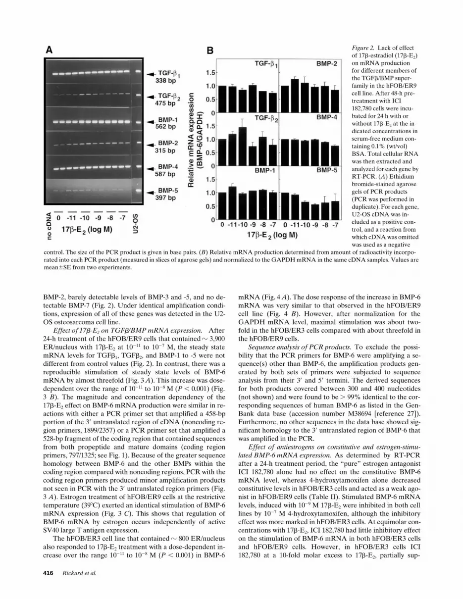
Constitutive expression of TGFb/BMP mRNAs. In the hFOB/ER9 cells, there were detectable steady state levels of TGFb1
and BMP-1, -4, and -6, somewhat lower levels of TGFb2 and
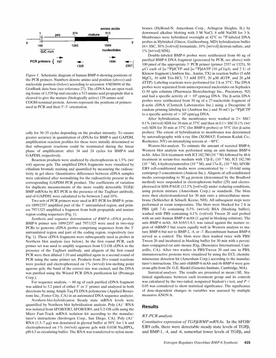
Figure 1. Schematic diagram of human BMP-6 showing positions of the PCR primers. Numbers denote amino acid position (above) and nucleotide position (below) according to accession 'M38694 of the GenBank data base (see reference 27). The cDNA has an open read-ing frame of 1,539 bp and encodes a 513-amino acid propeptide that is cleaved to give the mature (biologically active) 139-amino acid COOH-terminal protein. Arrows represent the positions of primers used in PCR and their 59-39 orientation.
416 Rickard et al.
BMP-2, barely detectable levels of BMP-3 and -5, and no de-tectable BMP-7 (Fig. 2). Under identical amplification condi-tions, expression of all of these genes was detected in the U2-OS osteosarcoma cell line.
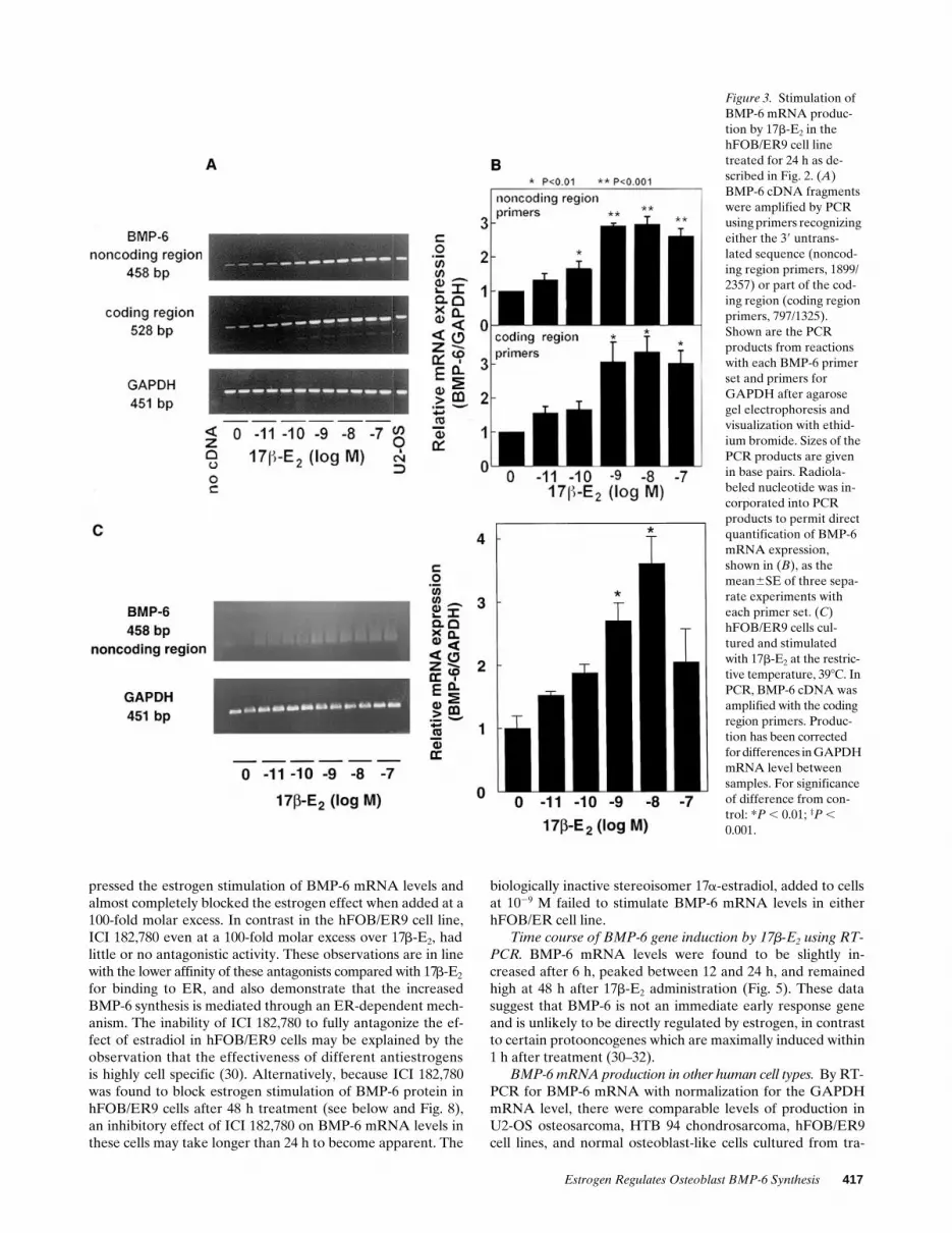
Effect of 17b-E2 on TGFb/BMP mRNA expression. After24-h treatment of the hFOB/ER9 cells that contained z 3,900ER/nucleus with 17b-E2 at 10211 to 1027 M, the steady statemRNA levels for TGFb1, TGFb2, and BMP-1 to -5 were notdifferent from control values (Fig. 2). In contrast, there was areproducible stimulation of steady state levels of BMP-6mRNA by almost threefold (Fig. 3 A). This increase was dose-dependent over the range of 10211 to 1028 M (P , 0.001) (Fig.3 B). The magnitude and concentration dependency of the17b-E2 effect on BMP-6 mRNA production were similar in re-actions with either a PCR primer set that amplified a 458-bpportion of the 39 untranslated region of cDNA (noncoding re-gion primers, 1899/2357) or a PCR primer set that amplified a528-bp fragment of the coding region that contained sequencesfrom both propeptide and mature domains (coding regionprimers, 797/1325; see Fig. 1). Because of the greater sequencehomology between BMP-6 and the other BMPs within thecoding region compared with noncoding regions, PCR with thecoding region primers produced minor amplification productsnot seen in PCR with the 39 untranslated region primers (Fig.3 A). Estrogen treatment of hFOB/ER9 cells at the restrictivetemperature (398C) exerted an identical stimulation of BMP-6mRNA expression (Fig. 3 C). This shows that regulation ofBMP-6 mRNA by estrogen occurs independently of activeSV40 large T antigen expression.
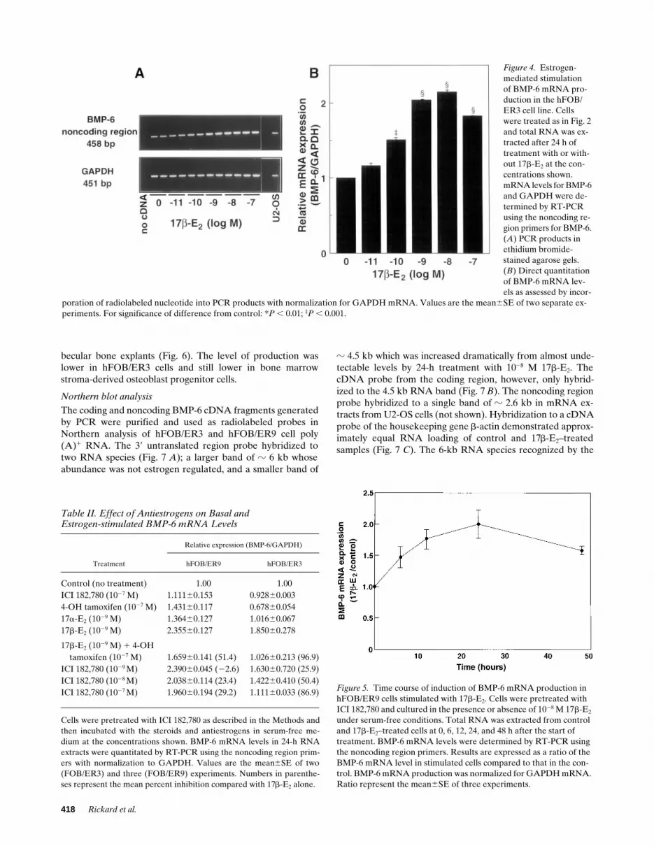
The hFOB/ER3 cell line that contained z 800 ER/nucleusalso responded to 17b-E2 treatment with a dose-dependent in-crease over the range 10211 to 1028 M (P , 0.001) in BMP-6
mRNA (Fig. 4 A). The dose response of the increase in BMP-6mRNA was very similar to that observed in the hFOB/ER9cell line (Fig. 4 B). However, after normalization for theGAPDH mRNA level, maximal stimulation was about two-fold in the hFOB/ER3 cells compared with about threefold inthe hFOB/ER9 cells.
Sequence analysis of PCR products. To exclude the possi-bility that the PCR primers for BMP-6 were amplifying a se-quence(s) other than BMP-6, the amplification products gen-erated by both sets of primers were subjected to sequenceanalysis from their 39 and 59 termini. The derived sequencesfor both products covered between 300 and 400 nucleotides(not shown) and were found to be . 99% identical to the cor-responding sequences of human BMP-6 as listed in the Gen-Bank data base (accession number M38694 [reference 27]).Furthermore, no other sequences in the data base showed sig-nificant homology to the 39 untranslated region of BMP-6 thatwas amplified in the PCR.
Effect of antiestrogens on constitutive and estrogen-stimu-lated BMP-6 mRNA expression. As determined by RT-PCRafter a 24-h treatment period, the “pure” estrogen antagonistICI 182,780 alone had no effect on the constitutive BMP-6mRNA level, whereas 4-hydroxytamoxifen alone decreasedconstitutive levels in hFOB/ER3 cells and acted as a weak ago-nist in hFOB/ER9 cells (Table II). Stimulated BMP-6 mRNAlevels, induced with 1029 M 17b-E2 were inhibited in both celllines by 1027 M 4-hydroxytamoxifen, although the inhibitoryeffect was more marked in hFOB/ER3 cells. At equimolar con-centrations with 17b-E2, ICI 182,780 had little inhibitory effecton the stimulation of BMP-6 mRNA in both hFOB/ER3 cellsand hFOB/ER9 cells. However, in hFOB/ER3 cells ICI182,780 at a 10-fold molar excess to 17b-E2, partially sup-
Figure 2. Lack of effect of 17b-estradiol (17b-E2) on mRNA production for different members of the TGFb/BMP super-family in the hFOB/ER9 cell line. After 48-h pre-treatment with ICI 182,780 cells were incu-bated for 24 h with or without 17b-E2 at the in-dicated concentrations in serum-free medium con-taining 0.1% (wt/vol) BSA. Total cellular RNA was then extracted and analyzed for each gene by RT-PCR. (A) Ethidium bromide-stained agarose gels of PCR products (PCR was performed in duplicate). For each gene, U2-OS cDNA was in-cluded as a positive con-trol, and a reaction from which cDNA was omitted was used as a negative
control. The size of the PCR product is given in base pairs. (B) Relative mRNA production determined from amount of radioactivity incorpo-rated into each PCR product (measured in slices of agarose gels) and normalized to the GAPDH mRNA in the same cDNA samples. Values are mean6SE from two experiments.
Estrogen Regulates Osteoblast BMP-6 Synthesis 417
pressed the estrogen stimulation of BMP-6 mRNA levels andalmost completely blocked the estrogen effect when added at a100-fold molar excess. In contrast in the hFOB/ER9 cell line,ICI 182,780 even at a 100-fold molar excess over 17b-E2, hadlittle or no antagonistic activity. These observations are in linewith the lower affinity of these antagonists compared with 17b-E2
for binding to ER, and also demonstrate that the increasedBMP-6 synthesis is mediated through an ER-dependent mech-anism. The inability of ICI 182,780 to fully antagonize the ef-fect of estradiol in hFOB/ER9 cells may be explained by theobservation that the effectiveness of different antiestrogensis highly cell specific (30). Alternatively, because ICI 182,780was found to block estrogen stimulation of BMP-6 protein inhFOB/ER9 cells after 48 h treatment (see below and Fig. 8),an inhibitory effect of ICI 182,780 on BMP-6 mRNA levels inthese cells may take longer than 24 h to become apparent. The
biologically inactive stereoisomer 17a-estradiol, added to cellsat 1029 M failed to stimulate BMP-6 mRNA levels in eitherhFOB/ER cell line.
Time course of BMP-6 gene induction by 17b-E2 using RT-PCR. BMP-6 mRNA levels were found to be slightly in-creased after 6 h, peaked between 12 and 24 h, and remainedhigh at 48 h after 17b-E2 administration (Fig. 5). These datasuggest that BMP-6 is not an immediate early response geneand is unlikely to be directly regulated by estrogen, in contrastto certain protooncogenes which are maximally induced within1 h after treatment (30–32).
BMP-6 mRNA production in other human cell types. By RT-PCR for BMP-6 mRNA with normalization for the GAPDHmRNA level, there were comparable levels of production inU2-OS osteosarcoma, HTB 94 chondrosarcoma, hFOB/ER9cell lines, and normal osteoblast-like cells cultured from tra-
Figure 3. Stimulation of BMP-6 mRNA produc-tion by 17b-E2 in the hFOB/ER9 cell line treated for 24 h as de-scribed in Fig. 2. (A) BMP-6 cDNA fragments were amplified by PCR using primers recognizing either the 39 untrans-lated sequence (noncod-ing region primers, 1899/2357) or part of the cod-ing region (coding region primers, 797/1325). Shown are the PCR products from reactions with each BMP-6 primer set and primers for GAPDH after agarose gel electrophoresis and visualization with ethid-ium bromide. Sizes of the PCR products are given in base pairs. Radiola-beled nucleotide was in-corporated into PCR products to permit direct quantification of BMP-6 mRNA expression, shown in (B), as the mean6SE of three sepa-rate experiments with each primer set. (C) hFOB/ER9 cells cul-tured and stimulated with 17b-E2 at the restric-tive temperature, 398C. In PCR, BMP-6 cDNA was amplified with the coding region primers. Produc-tion has been corrected for differences in GAPDH mRNA level between samples. For significance of difference from con-trol: *P , 0.01; ‡P , 0.001.
418 Rickard et al.
becular bone explants (Fig. 6). The level of production waslower in hFOB/ER3 cells and still lower in bone marrowstroma-derived osteoblast progenitor cells.
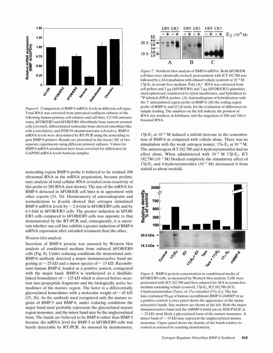
Northern blot analysis
The coding and noncoding BMP-6 cDNA fragments generatedby PCR were purified and used as radiolabeled probes inNorthern analysis of hFOB/ER3 and hFOB/ER9 cell poly(A)1 RNA. The 39 untranslated region probe hybridized totwo RNA species (Fig. 7 A); a larger band of z 6 kb whoseabundance was not estrogen regulated, and a smaller band of
z 4.5 kb which was increased dramatically from almost unde-tectable levels by 24-h treatment with 1028 M 17b-E2. ThecDNA probe from the coding region, however, only hybrid-ized to the 4.5 kb RNA band (Fig. 7 B). The noncoding regionprobe hybridized to a single band of z 2.6 kb in mRNA ex-tracts from U2-OS cells (not shown). Hybridization to a cDNAprobe of the housekeeping gene b-actin demonstrated approx-imately equal RNA loading of control and 17b-E2–treatedsamples (Fig. 7 C). The 6-kb RNA species recognized by the
Figure 4. Estrogen-mediated stimulation of BMP-6 mRNA pro-duction in the hFOB/ER3 cell line. Cells were treated as in Fig. 2 and total RNA was ex-tracted after 24 h of treatment with or with-out 17b-E2 at the con-centrations shown. mRNA levels for BMP-6 and GAPDH were de-termined by RT-PCR using the noncoding re-gion primers for BMP-6. (A) PCR products in ethidium bromide-stained agarose gels. (B) Direct quantitation of BMP-6 mRNA lev-els as assessed by incor-
poration of radiolabeled nucleotide into PCR products with normalization for GAPDH mRNA. Values are the mean6SE of two separate ex-periments. For significance of difference from control: *P , 0.01; ‡P , 0.001.
Table II. Effect of Antiestrogens on Basal andEstrogen-stimulated BMP-6 mRNA Levels
Treatment
Relative expression (BMP-6/GAPDH)
hFOB/ER9 hFOB/ER3
Control (no treatment) 1.00 1.00ICI 182,780 (1027 M) 1.11160.153 0.92860.0034-OH tamoxifen (1027 M) 1.43160.117 0.67860.05417a-E2 (1029 M) 1.36460.127 1.01660.06717b-E2 (1029 M) 2.35560.127 1.85060.278
17b-E2 (1029 M) 1 4-OH tamoxifen (1027 M) 1.65960.141 (51.4) 1.02660.213 (96.9)
ICI 182,780 (1029 M) 2.39060.045 (22.6) 1.63060.720 (25.9)ICI 182,780 (1028 M) 2.03860.114 (23.4) 1.42260.410 (50.4)ICI 182,780 (1027 M) 1.96060.194 (29.2) 1.11160.033 (86.9)
Cells were pretreated with ICI 182,780 as described in the Methods andthen incubated with the steroids and antiestrogens in serum-free me-dium at the concentrations shown. BMP-6 mRNA levels in 24-h RNAextracts were quantitated by RT-PCR using the noncoding region prim-ers with normalization to GAPDH. Values are the mean6SE of two(FOB/ER3) and three (FOB/ER9) experiments. Numbers in parenthe-ses represent the mean percent inhibition compared with 17b-E2 alone.
Figure 5. Time course of induction of BMP-6 mRNA production in hFOB/ER9 cells stimulated with 17b-E2. Cells were pretreated with ICI 182,780 and cultured in the presence or absence of 1028 M 17b-E2 under serum-free conditions. Total RNA was extracted from control and 17b-E2–treated cells at 0, 6, 12, 24, and 48 h after the start of treatment. BMP-6 mRNA levels were determined by RT-PCR using the noncoding region primers. Results are expressed as a ratio of the BMP-6 mRNA level in stimulated cells compared to that in the con-trol. BMP-6 mRNA production was normalized for GAPDH mRNA. Ratio represent the mean6SE of three experiments.
Estrogen Regulates Osteoblast BMP-6 Synthesis 419
noncoding region BMP-6 probe is believed to be residual 28Sribosomal RNA in the mRNA preparation, because prelimi-nary analysis of total cellular RNA revealed cross-reactivity ofthis probe to 28S RNA (not shown). The size of the mRNA forBMP-6 detected in hFOB/ER cell lines is in agreement withother reports (33, 34). Densitometry of autoradiograms andnormalization to b-actin showed that estrogen stimulatedBMP-6 mRNA levels by z 2.4-fold in hFOB/ER9 cells and by6.3-fold in hFOB/ER3 cells. The greater induction in hFOB/ER3 cells compared to hFOB/ER9 cells was opposite to thatdemonstrated by the RT-PCR and, consequently, it is uncer-tain whether one cell line exhibits a greater induction of BMP-6mRNA expression after estradiol treatment than the other.
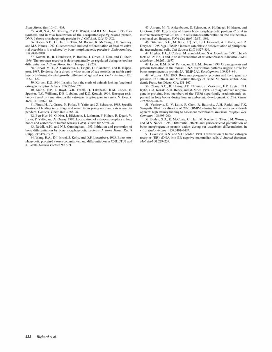
Western blot analysis
Secretion of BMP-6 protein was assessed by Western blotanalysis of conditioned medium from cultured hFOB/ER9cells (Fig. 8). Under reducing conditions the monoclonal anti–BMP-6 antibody detected a major immunoreactive band mi-grating at z 25 kD and a minor species of z 15 kD. Recombi-nant human BMP-6, loaded as a positive control, comigratedwith the major band. BMP-6 is synthesized as a disulfide-linked homodimer of z 125 kD which is cleaved before secre-tion into propeptide fragments and the biologically active ho-modimer of the mature region. The latter is a differentiallyglycosylated homodimer with a molecular weight of z 45 kD(35, 36). As the antibody used recognized only the mature re-gions of BMP-5 and BMP-6, under reducing conditions themajor band most probably represents the glycosylated matureregion monomer, and the minor band may be the unglycosylatedform. The bands are believed to be BMP-6 rather than BMP-5because the mRNA level for BMP-5 in hFOB/ER9 cells wasbarely detectable by RT-PCR. As assessed by densitometry,
17b-E2 at 1028 M induced a sixfold increase in the concentra-tion of BMP-6 as compared with vehicle alone. There was nostimulation with the weak estrogen isomer, 17a-E2 at 1028 M.The antiestrogens ICI 182,780 and 4-hydroxytamoxifen had noeffect alone. When administered with 1028 M 17b-E2, ICI182,780 (1027 M) blocked completely the stimulatory effect of17b-E2 and 4-hydroxytamoxifen (1028 M) attenuated it fromsixfold to about twofold.
Figure 6. Comparison of BMP-6 mRNA levels in different cell types. Total RNA was extracted from untreated confluent cultures of the following human primary cell cultures and cell lines: U2-OS osteosar-coma, hFOB/ER3 and hFOB/ER9, fibroblastic bone marrow stromal cells (stromal), differentiated trabecular bone-derived osteoblast-like cells (osteoblasts), and HTB 94 chondrosarcoma (chondro). BMP-6 mRNA levels were determined by RT-PCR using the noncoding re-gion BMP-6 primers. Results are presented as the mean6SE of two separate experiments using different primary cultures. Values for BMP-6 mRNA production have been corrected for differences in GAPDH mRNA levels between samples.
Figure 7. Northern blot analysis of BMP-6 mRNA. Both hFOB/ER cell lines were identically treated; pretreatment with ICI 182,780 was followed by a 24-h incubation with ethanol vehicle (control) or 1028 M 17b-E2 in serum-free medium. Poly (A)1 RNA was extracted from cell pellets and 3 mg (hFOB/ER9) and 7 mg (hFOB/ER3) quantities electrophoresed, transferred to nylon membranes, and hybridized to 32P-labeled cDNA probes. (A) Autoradiogram of hybridization with the 39 untranslated region probe of BMP-6; (B) the coding region probe of BMP-6; and (C) b-actin, for the evaluation of differences in sample loading. The numbers on the left indicate the position of RNA size markers, in kilobases, and the migration of 28S and 18S ri-bosomal RNA.
Figure 8. BMP-6 protein concentration in conditioned media of hFOB/ER9 cells, as measured by Western blot analysis. Cells were pretreated with ICI 182,780 and then cultured for 48 h in serum-free medium containing vehicle (control), 17b-E2, ICI 182,780 (ICI),4-hydroxytamoxifen (Tam), or 17a-estradiol (17a-E2). The lastlane contained 50 mg of human recombinant BMP-6 (rhBMP-6) asa positive control. Lower panel shows the appearance of the immu-noreactive bands. Size markers are shown at the left. Both the major immunoreactive band and the rhBMP-6 band ran on SDS-PAGE at z 25 kD, most likely a glycosylated form of the mature monomer. A minor band of z 15 kD may represent the unglycosylated mature monomer. Upper panel shows the density of the bands relative to control as assessed by scanning densitometry.

420 Rickard et al.
Discussion
Although the BMPs play important roles in skeletal develop-ment and in the differentiation of osteoblasts and chondro-blasts, little is known about the regulation of their synthesis,and there have been no studies assessing the interaction of sexsteroids with the BMPs. Using fetal osteoblastic cell lines thatstably express the estrogen receptor gene, we found that estro-gen stimulated the production of BMP-6 mRNA as assessedby RT-PCR and by Northern blot analysis and that this effectwas specific because it was blocked by coadministration ofantiestrogens. Sequence analysis of the PCR product con-firmed that the estrogen-regulated gene was BMP-6. We alsoconfirmed, by Western blot analysis of conditioned media, thatestrogen treatment in vitro stimulated the secretion of BMP-6protein. Interestingly, although expression of BMP-6 was foundto be strongly regulated by estrogen, the related genes forTGFb1, TGFb2, and BMPs-1 to -5 and -7 were not. The findingof selective stimulation of BMP-6 by estrogen suggests thatthis process may be physiologically important. In view of theclose structural and functional relationships between TGFband BMP, it may have been predicted that their synthesiswould be similarly regulated. Indeed, the failure to detect in-duction of TGFb1 mRNA by estrogen in hFOB/ER cells con-flicts with previous findings in normal adult human osteoblastcells (8). The reason for the lack of TGFb induction by estro-gen in our cells is unclear but may relate to differences be-tween fetal and adult osteoblasts. Alternatively, whereas bothtranscriptional and posttranscriptional regulation of TGFbsynthesis by estrogen has been observed in osteoblasts fromadult bone, regulation in hFOB/ER cells may be predomi-nantly posttranscriptional.
In principle, the stimulation of BMP-6 by estrogen couldcontribute to any or all of the actions of estrogen on bone andcartilage cells. In the adult, estrogen acts to maintain bonemass by regulating bone remodeling and turnover (1). How-ever, in the fetus and in the growing skeleton of the child andadolescent, it acts on both bone and cartilage cells to regulateendochondral bone formation. Fetal osteoblasts contain ER(37) and thus presumably respond to estrogen. Also, estradiolacts directly on chondrocytes in vitro to stimulate matrix syn-thesis (38). Both ER gene deletion (knockout) mice (39) and ahuman with a null mutation of the ER gene (40) were found tohave open epiphyseal growth plates. ER are present within theproliferating and hypertrophic zones of the epiphyseal carti-lage of young rodents (41) and human fetuses (42).
Thus far, it has not been possible to distinguish clearly be-tween the action of BMP-6 and that of other members of theBMP family. BMP-2, -3, -4, -6, and -7 all can induce the localformation of ectopic bone and cartilage when injected intosubcutaneous or muscle tissue of rats (33, 43). BMP-2, -4, and -6promote the differentiation of multipotential mesenchymalcells along the osteoblast lineage (44–46) as well as stimulate theproduction of bone nodules in fetal rat calvarial cell cultures(47). Despite these general similarities, the existence of thelarge number of BMPs in itself implies that each one may per-form related but distinct function(s), perhaps acting predomi-nantly on different mesenchymal lineages (48), on different stagesof development or cell differentiation (49), or even on differ-ent embryonic segments (14). However, the often overlappingpattern of production of certain BMPs during embryonic de-velopment also implies a degree of functional redundancy.
Although BMP-6 mRNA and protein have been detectedin skeletal tissues as well as in nonskeletal tissues such as brain,skin, and fracture callus, in both embryos and adults, BMP-6differs from other members of the BMP family by its concen-tration in cartilage of the fetus. In situ hybridization studieshave localized mRNAs for various BMPs to different stages offetal skeletal development. Thus, BMP-2 and -4 localize to theectodermal ridge and, later, to the underlying mesenchyme(15, 16), and BMP-2 and the growth/differentiation factor(GDF)-5 localize to the differentiating precartilaginous mes-enchyme (15, 50). In contrast, production of BMP-6 mRNA inthe skeleton is localized to hypertrophic cartilage during skele-tal development but not to perichondrium, periosteum, orbone (15). By immunocytochemical studies, BMP-6 proteinwas found to localize to matrix and cells of hypertrophic carti-lage (33). Although the genes for BMP-7 and GDF-6 are ex-pressed in hypertrophic cartilage, these are also expressed inperichondrium, periosteum, and centers of ossification (50,51). Thus, although other BMPs besides BMP-6 are producedin hypertrophic cartilage during skeletal development, thehighly restricted localization of BMP-6 to this tissue is consis-tent with a role in the modulation of endochondral bone for-mation. Because ER are also found in hypertrophic cartilageand because estrogen is required for growth plate closure inthe immature skeleton, it is possible that at least some of theparacrine/autocrine actions of estrogen at this site are medi-ated by its effect on stimulating BMP-6. Thus far, however, wehave demonstrated this regulation only in osteoblastic cells,and further studies will be required to test this hypothesis ex-plicitly.
Because the hFOB/ER cell lines that were used in thisstudy have a phenotype that closely resembles that of the ma-ture osteoblast (21, 22), the demonstration that estrogen in-creases the secretion of BMP-6 suggests a possible role forBMP-6 in modulating bone remodeling in the adult skeleton, aprocess that requires recruitment, proliferation, and subse-quent differentiation of osteoprogenitor cells. However, thefounder cells of the hFOB/ER cell lines were fetal osteoblasts.Therefore, we cannot exclude the possibility that some of thestimulatory effect of estrogen on BMP-6 production is limitedto fetal bone cells. The differentiation of osteoblasts in cul-tures of fetal rat calvaria-derived cells, measured by the bonenodule formation assay, is stimulated by several BMPs includ-ing BMP-6 (47, 52). Interestingly, in these cultures glucocorti-coids selectively increase production of BMP-6 (36), an effectwe have also observed in primary cultures of human bone mar-row stromal cells (unpublished observations). Moreover, usingantisense oligonucleotides to BMP-6, the glucocorticoid-stim-ulated development of mineralized bone nodules was found tobe mediated by BMP-6 (36). Therefore, the selective regula-tion of BMP-6 by two osteotropic hormones (estrogen and glu-cocorticoid) strongly suggests that BMP-6 may play a key rolein early stages of osteoblast differentiation and bone develop-ment. The effect of BMP-6 on proliferation and differentiationof the hFOB/ER cell lines has so far not been tested, but estro-gen treatment, in addition to stimulating production of BMP-6,inhibits the proliferation of hFOB/ER9 cells and enhances cer-tain differentiated osteoblast phenotypic markers (10, 25). Itwould therefore be of interest to determine whether BMP-6can reproduce some or all of these estrogenic actions.
Various cell types overexpressing exogenous ER have fre-quently shown paradoxical responses to estrogen and atypical

Estrogen Regulates Osteoblast BMP-6 Synthesis 421
gene activation (53). However, our findings that BMP-6 wassimilarly regulated by estrogen in both hFOB/ER3 cells (pos-sessing only z 800 ER per nucleus) and hFOB/ER9 cells(which possess z 3,900 ER per nucleus, comparable to recep-tor levels in reproductive tissue) is strong evidence to supportour belief that the regulation of BMP-6 by estrogen representsa novel and potentially important effect. Indeed, the ER levelin hFOB/ER3 cells lies within the range for endogenous ERexpression reported in normal human trabecular osteoblast-like cells (z 1,600 binding sites per cell) and ROS 17/2.8 (200–500 sites per cell) in which estrogen responses have both beenobserved (2, 3, 8). Furthermore, estrogen induction of theprogesterone receptor gene has been observed in both hFOB/ER cell lines; a classical estrogenic response common to allER-positive tissues. These and other estrogen responses elic-ited by the hFOB/ER cells, taken together, lead us to believethat the regulation of BMP-6 by estrogen may be physiologi-cally relevant.
In conclusion, we have demonstrated that estrogen selec-tively increases the production of BMP-6, but not other BMPs,in estrogen-responsive, highly differentiated osteoblastic celllines of fetal origin. Thus, we speculate that some of the recog-nized effects of estrogen on the skeleton could be mediated byincreased production of BMP-6, which may then act in a para-crine/autocrine fashion to modulate the differentiation of un-committed progenitors to osteoblasts and, in the immatureskeleton, to chondrocytes.
Acknowledgments
The authors are indebted to Drs. S.A. Harris, M. Subramaniam, andS. Khosla, and K.C. Hicok for their valuable suggestions and adviceduring the course of these studies. We also thank Nurit Geller for as-sistance in analysis of the data.
This work was supported by National Institutes of Health grantAG-04875. Dr. Hofbauer is a recipient of a postgraduate fellowshipfrom the Deutsche Forschungsgemeinschaft (Ho 1875/1-1).
References
1. Turner, R.T., G.L. Evans, and G.K. Wakley. 1993. Mechanism of actionof estrogen on cancellous bone balance in tibiae of ovariectomized growingrats: inhibition of indices of formation and resorption. J. Bone Miner. Res. 8:359–366.
2. Komm, B.S., C.M. Terpening, D.J. Benz, K.A. Graeme, B.W. O’Malley,and M.R. Haussler. 1988. Estrogen binding sites, receptor mRNA, and biologicresponse in osteoblast-like osteosarcoma cells. Science. 241:81–84.
3. Eriksen, E.F., D.S. Colvard, N.J. Berg, M.L. Graham, K.G. Mann, T.C.Spelsberg, and B.L. Riggs. 1988. Evidence of estrogen receptors in normal hu-man osteoblast-like cells. Science. 241:84–86.
4. Oursler, M.J., P. Osdoby, J. Pyfferoen, B.L. Riggs, and T. Spelsberg.1991. Avian osteoclasts as estrogen target cells. Proc. Natl. Acad. Sci. USA. 88:6613–6617.
5. Oursler, M.J., L. Pederson, L. Fitzpatrick, and B.L. Riggs. 1994. Humangiant cell tumors of the bone (osteoclastomas) are estrogen target cells. Proc.Natl. Acad. Sci. USA. 91:5227–5231.
6. Kimble, R.B., A.B. Matayoshi, J.L. Vannice, V.T. Kung, C. Williams, andR. Pacifici. 1995. Simultaneous block of interleukin-1 and tumor necrosis factoris required to completely prevent bone loss in the early postovariectomy pe-riod. Endocrinology. 136:3054–3061.
7. Manolagas, S.C., and R.L. Jilka. 1995. Bone marrow, cytokines, and boneremodeling: emerging insights into the pathophysiology of osteoporosis. N.Engl. J. Med. 332:305–311.
8. Oursler, M.J., C. Cortese, P.E. Keeting, M.A. Anderson, S.K. Bonde,B.L. Riggs, and T.C. Spelsberg. 1991. Modulation of transforming growth fac-tor-b production in normal human osteoblast-like cells by 17b-estradiol andparathyroid hormone. Endocrinology. 129:3313–3320.
9. Ernst, M., J.K. Heath, and G.A. Rodan. 1989. Estradiol effects on prolif-eration, messenger ribonucleic acid for collagen and insulin-like growth factor-
I, and parathyroid hormone-stimulated adenylate cyclase activity in osteoblas-tic cells from calvariae and long bones. Endocrinology. 125:825–833.
10. Kassem, M., R. Okazaki, D. DeLeon, S.A. Harris, J.A. Robinson, T.C.Spelsberg, C.A. Conover, and B.L. Riggs. 1996. Potential mechanism of estro-gen-mediated decrease in bone formation: estrogen increases production of in-hibitory insulin-like growth factor binding protein-4. Proc. Assoc. Am. Physi-cians. 108:155–164.
11. Kessler, E., K. Takahara, L. Biniaminov, M. Brusel, and D.S.Greenspan. 1996. Bone morphogenetic protein-1: the type I procollagen C-pro-teinase. Science. 271:360–362.
12. Wozney, J.M., V. Rosen, A.J. Celeste, L.M. Mitsock, M.J. Whitters,R.W. Kriz, R.M. Hewick, and E.A. Wang. 1988. Novel regulators of bone for-mation: molecular clones and activities. Science. 242:1528–1534.
13. Wang, E.A., V. Rosen, J.S. D’Alessandro, M. Bauduy, P. Cordes, T.Harada, D.I. Israel, R.M. Hewick, K.M. Kerns, P. LaPan, et al. 1990. Recombi-nant human bone morphogenetic protein induces bone formation. Proc. Natl.Acad. Sci. USA. 87:2220–2224.
14. Kingsley, D.M. 1994. What do BMPs do in mammals? Clues from themouse short-ear mutation. Trends Genet. 10:16–21.
15. Lyons, K.M., R.W. Pelton, and B.L. Hogan. 1989. Patterns of expressionof murine Vgr-1 and BMP-2a RNA suggest that TGFb-like genes co-ordinatelyregulate aspects of embryonic development. Genes Dev. 3:1657–1668.
16. Jones, C.M., K.M. Lyons, and B.L.M. Hogan. 1991. Involvement ofbone morphogenetic protein-4 (BMP-4) and Vgr-1 in morphogenesis and neu-rogenesis in the mouse. Development (Camb.) 111:531–542.
17. Rosen, V., J. Nove, J.J. Song, R.S. Thies, K. Cox, and J.M. Wozney.1994. Responsiveness of clonal limb bud cell lines to BMP-2 reveals a sequen-tial relationship between cartilage and bone phenotypes. J. Bone Miner. Res. 9:1759–1768.
18. Thies, R.S., M. Bauduy, B.A. Ashton, L. Kurtzberg, J.M. Wozney, andV. Rosen. 1992. Recombinant human bone morphogenetic protein-2 inducesdifferentiation in W-20-17 stromal cells. Endocrinology. 130:1318–1324.
19. Amedee, J., R. Bareille, F. Rouais, N. Cunningham, H. Reddi, and M.F.Harmand. 1994. Osteogenin (BMP-3) inhibits proliferation and stimulates dif-ferentiation of osteoprogenitors in human bone marrow. Differentiation. 58:157–164.
20. Vukicevic, S., F.P. Luyten, and A.H. Reddi. 1989. Stimulation of expres-sion of osteogenic and chondrogenic phenotypes in vitro by osteogenin. Proc.Natl. Acad. Sci. USA. 86:8793–8797.
21. Harris, S.A., R.J. Enger, B.L. Riggs, and T.C. Spelsberg. 1995. Develop-ment and characterization of a conditionally immortalized human fetal osteo-blastic cell line. J. Bone Miner. Res. 10:178–186.
22. Harris, S., K.R. Tau, R.J. Enger, D.O. Toft, B.L. Riggs, and T.C. Spels-berg. 1995. Estrogen response in the hFOB1.19 human fetal osteoblastic cellline stably transfected with the human estrogen receptor gene. J. Cell. Biochem.59:193–201.
23. Spelsberg, T.C., M.L. Graham, N.J. Berg, T. Umehara, E. Riehl, C.B.Coulam, J.N. Ingle. 1987. A nuclear binding assay to assess the biological activ-ity of steroid receptors in isolated animal and human tissues. Endocrinology.121:631–644.
24. Colvard, D.S., W.R. Jankus, N.J. Berg, M.L. Graham, N.-S. Jiang, J.N.Ingle, and T.C. Spelsberg. 1988. Microassay for nuclear binding of steroid re-ceptors with use of intact cells from small samples of avian and human tissues.Clin. Chem. 34:363–369.
25. Robinson, J.A., S.A. Harris, B.L. Riggs, and T.C. Spelsberg. 1997. Es-trogen regulation of human osteoblast cell proliferation and differentiation.Endocrinology. 138:2919–2927.
26. Parker, M.G. 1993. Action of ‘pure’ antiestrogens in inhibiting estrogenreceptor action. Breast Cancer Res. Treat. 26:131–137.
27. Celeste, A.J., J.A. Iannazzi, R.C. Taylor, R.M. Hewick, V. Rosen, E.A.Wang, and J.M. Wozney. 1990. Identification of transforming growth factor-bfamily members present in bone-inductive protein purified from bovine bone.Proc. Natl. Acad. Sci. USA. 87:9843–9847.
28. Chomczynski, P., and N. Sacchi. 1987. Single-step method of RNA isola-tion by acid guanidinium thiocyanate-phenol-chloroform extraction. Anal. Bio-chem. 162:156–159.
29. Laemmli, U.K. 1970. Cleavage of structural proteins during the assem-bly of the head of bacteriophage T4. Nature. 227:680–685.
30. Katzenellenbogen, J.A., B.W. O’Malley, and B.S. Katzenellenbogen.1996. Tripartite steroid hormone receptor pharmacology: Interaction with mul-tiple effector sites as a basis for the cell- and promoter-specific action of thesehormones. Mol. Endocrinol. 10:119–131.
31. Schuchard, M., J.P. Landers, N. Punkay-Sandhu, and T.C. Spelsberg.1993. Steroid hormone regulation of nuclear proto-oncogenes. Endocr. Rev. 14:659–669.
32. Weisz, A., and F. Bresciani. 1988. Estrogen induces expression of c-fosand c-myc protooncogenes in rat uterus. Mol. Endocrinol. 2:816–824.
33. Gitelman, S.E., M.S. Kobrin, J.-Q. Ye, A.R. Lopez, A. Lee, and R.Derynck. 1994. Recombinant Vgr-1/BMP 6-expressing tumors induce fibrosisand endochondral bone formation in vivo. J. Cell Biol. 126:1595–1609.
34. Carey, D.E., and X. Liu. 1995. Expression of bone morphogenetic pro-tein-6 messenger RNA in bovine growth plate chondrocytes of different size. J.
422 Rickard et al.
Bone Miner. Res. 10:401–405.35. Wall, N.A., M. Blessing, C.V.E. Wright, and B.L.M. Hogan. 1993. Bio-
synthesis and in vivo localization of the decapentaplegic-Vg-related protein,DVR-6 (bone morphogenetic protein-6). J. Cell Biol. 120:493–502.
36. Boden, S.D., G. Hair, L. Titus, M. Racine, K. McCuaig, J.M. Wozney,and M.S. Nanes. 1997. Glucocorticoid-induced differentiation of fetal rat calva-rial osteoblasts is mediated by bone morphogenetic protein-6. Endocrinology.138:2820–2828.
37. Komm, B., R. Henderson, P. Bodine, J. Green, J. Lian, and G. Stein.1996. The estrogen receptor is developmentally up-regulated during osteoblastdifferentiation. J. Bone Miner. Res. 11(Suppl.1):S258.
38. Corvol, M.-T., A. Carrascosa, L. Tsagris, O. Blanchard, and R. Rappa-port. 1987. Evidence for a direct in vitro action of sex steroids on rabbit carti-lage cells during skeletal growth: influence of age and sex. Endocrinology. 120:1422–1429.
39. Korach, K.S. 1994. Insights from the study of animals lacking functionalestrogen receptor. Science. 266:1524–1527.
40. Smith, E.P., J. Boyd, G.R. Frank, H. Takahashi, R.M. Cohen, B.Specker, T.C. Williams, D.B. Lubahn, and K.S. Korach. 1994. Estrogen resis-tance caused by a mutation in the estrogen receptor gene in a man. N. Engl. J.Med. 331:1056–1061.
41. Pinus, H., A. Ornoy, N. Patlas, P. Yaffe, and Z. Schwartz. 1993. Specificb-estradiol binding in cartilage and serum from young mice and rats is age de-pendent. Connect. Tissue Res. 30:85–98.
42. Ben-Hur, H., G. Mor, I. Blickstein, I. Likhman, F. Kohen, R. Dgani, V.Insler, P. Yaffe, and A. Ornoy. 1993. Localization of estrogen receptors in longbones and vertebrae of human fetuses. Calcif. Tissue Int. 53:91–96.
43. Reddi, A.H., and N.S. Cunningham. 1993. Initiation and promotion ofbone differentiation by bone morphogenetic proteins. J. Bone Miner. Res. 8(Suppl.2):S499–S502.
44. Wang, E.A., D.I. Israel, S. Kelly, and D.P. Luxenberg. 1993. Bone mor-phogenetic protein-2 causes commitment and differentiation in C3H10T1/2 and3T3 cells. Growth Factors. 9:57–71.
45. Ahrens, M., T. Ankenbauer, D. Schroder, A. Hollnagel, H. Mayer, andG. Gross. 1993. Expression of human bone morphogenetic proteins -2 or -4 inmurine mesenchymal C3H10T1/2 cells induces differentiation into distinct mes-enchymal cell lineages. DNA Cell Biol. 12:871–880.
46. Gitelman, S.E., M. Kirk, J.Q. Ye, E.H. Filvaroff, A.J. Kahn, and R.Deryack. 1995. Vgr-1/BMP-6 induces osteoblastic differentiation of pluripoten-tial mesenchymal cells. Cell Growth Diff. 6:827–836.
47. Hughes, F.J., J. Collyer, M. Stanfield, and S.A. Goodman. 1995. The ef-fects of BMP-2, -4 and -6 on differentiation of rat osteoblast cells in vitro. Endo-crinology. 136:2671–2677.
48. Lyons, K.M., R.W. Pelton, and B.L.M. Hogan. 1990. Organogenesis andpattern formation in the mouse: RNA distribution patterns suggest a role forbone morphogenetic protein-2A (BMP-2A). Development. 109:833–844.
49. Wozney, J.M. 1993. Bone morphogenetic proteins and their gene ex-pression. In Cellular and Molecular Biology of Bone. M. Noda, editor. Aca-demic Press, San Diego, CA. 131–167.
50. Chang, S.C., B. Hoang, J.T. Thomas, S. Vukicevic, F.P. Luyten, N.J.Ryba, C.A. Kozak, A.H. Reddi, and M. Moos. 1994. Cartilage-derived morpho-genetic proteins. New members of the TGFb superfamily predominantly ex-pressed in long bones during human embryonic development. J. Biol. Chem.269:28227–28234.
51. Vukicevic, S., V. Latin, P. Chen, R. Batorsky, A.H. Reddi, and T.K.Sampath. 1994. Localization of OP-1 (BMP-7) during human embryonic devel-opment: high affinity binding to basement membranes. Biochem. Biophys. Res.Commun. 198:693–700.
52. Boden, S.D., K. McCuaig, G. Hair, M. Racine, L. Titus, J.M. Wozney,and M.S. Nanes. 1996. Differential effects and glucocorticoid potentiation ofbone morphogenetic protein action during rat osteoblast differentiation invitro. Endocrinology. 137:3401–3407.
53. Levenson, A.S., and V.C. Jordan. 1994. Transfection of human estrogenreceptor (ER) cDNA into ER-negative mammalian cells. J. Steroid Biochem.Mol. Biol. 51:229–239.