bmrat cover - biomedical research and therapy
TRANSCRIPT
BIOMEDICAL RESEARCH AND THERAPY
ISSN: 2198-4093 www.bmrat.org
Volume 4 Issue 6
June 2017
Biomed. Res. Ther. 4(6), 2017
Editorial Team
Editor-in-Chief Phuc Van Pham University of Science, Vietnam National University, HCMC
Managing editor Lili Hami University of Science, Vietnam National University, HCMC
Associate Editors (Alphabetical order) Alexander E. Berezin, Cardiology Unit of Internal Medicine Department, State Medical University, Zaporozhye, Ukraine Amit Parashar, Department of Engineering Chemistry, GL Bajaj Group of Institutions, India Arya Sobhakumari, California Animal Health and Food Safety Laboratory, University of California Davis, United States Debmalya Barh, Institute of Integrative Omics and Applied Biotechnology (IIOAB), India Dong Kee Jeong, College of Applied Life Sciences, Jeju National University, Jeju, Korea Francesca Paino, Second University of Naples, Italy Fuyu Tamanoi, Jonsson Comprehensive Cancer Center, University of California, Los Angeles, United States Goothy Sai Sailesh Kumar, Little Flower Medical Research Centre, Angamaly – 683 572, Kerala, India Jae-Bong Park, Department of Biochemistry, College of Medicine, Hallym University, Korea Kalyani Raju, Sri Devaraj Urs Medical College, Sri Devaraj Urs Academy of Higher Education and Research, Kolar, Karnataka, India Kevin Dzobo, Faculty of Health Sciences, University of Cape Town, South Africa Kiyoshi Fukui, The Institute for Enzyme Research, Division of Enzyme Pathophysiology, The University of Tokushima, Japan Lam Hoang Dang, Memorial Sloan Kettering Cancer Center (MSKCC) , New York, United States Li Suan Mai, Institute of Physics, Polish Acad Sci, Warsaw, Poland Liem Minh Phan, MD Anderson Cancer Center, The University of Texas, Houston, United States Meng Yang, AntiCancer Biotech Co., Ltd, China Mohammed RafiqKhan, Department of Biotechnology, Sree Narayana Guru College, K G Chavadi, Coimbatore-105, Tamilnadu, India Nedime Serakinci, Genetics and Cancer Diagnosis-Research Centre & Faculty of Medicine, Near East University, Turkey Paolo Carloni, German Research School for Simulation Sciences GmbH, Jülich, Germany

Ravirajsinh N. Jadeja, Department of Biochemistry and Molecular Biology, Augusta University, Augusta, United State Redhwan A. Al-Naggar, Faculty of Medicine, Universiti Teknologi MARA, Malaysia Shikha Saini, Department of Microbiology and Immunology, University of Illinois at Chicago, United States Somi Kim Cho, College of Applied Life Sciences, Jeju National University, Jeju, Korea Suaib Luqman, Central Institute of Medical and Aromatic Plants, India Tauseef Ahmad, Hazara University Mansehra, Pakistan Thach Nguyen, University of Arizona Medical Center, Tucson, AZ-USA Vy Phan Lai, Center for Global Mentoring, UCLA-DOE Institute, UCLA, United States Yasuhiko Nishioka, Institute of Health Biosciences, University of Tokushima Graduate School, Japan Zhenghong Lee, School of Medicine, Case Western Reserve University, United States
Advisory Board (Alphabetical order) Dong Van Le, Vietnam Military Medical University, Hanoi, Vietnam Kiet Dinh Truong, University of Medicine & Pharmacy, Ho Chi Minh City, Vietnam Michael Robert Doran, Translational Research Institute, Queensland University of Technology, Australia Ngoc Kim Phan, University of Science, Vietnam National University, Ho Chi Minh city, Vietnam Son Nghia Hoang, Institute of Tropical Biology, Vietnam Academy of Science and Technology, Vietnam Thai Duc Nguyen, University of Science, Vietnam National University, Ho Chi Minh city, Vietnam Thuoc Linh Tran, University of Science, Vietnam National University, Ho Chi Minh city, Vietnam Toan Linh Nguyen, Vietnam Military Medical University, Hanoi, Vietnam
Language Editor Vy Phan Lai, Center for Global Mentoring, UCLA-DOE Institute, UCLA, United States
Editorial Secretary Hoa Trong Nguyen, University of Science, Vietnam National University, HCMC Ngoc Bich Vu, University of Science, Vietnam National University, HCMC

Contact us
Journal Contact
BIOMEDPRESS (BMP) Laboratory of Stem Cell Research and Application University of Science, Vietnam National University, Ho Chi Minh city 227 Nguyen Van Cu District 5, Ho Chi Minh city Vietnam Email: [email protected]
PRINCIPAL CONTACT Lili Hami BIOMEDPRESS (BMP) Laboratory of Stem Cell Research and Application University of Science, Vietnam National University, Ho Chi Minh city 227 Nguyen Van Cu District 5, Ho Chi Minh city Vietnam Email: [email protected]
SUPPORT CONTACT Support Team Email: [email protected]
EDITOR-IN-CHIEF Phuc Van Pham Email: [email protected]
Table of Contents
Vol 4 No 6 (2017): 1341 - 1431
Letter to Editor
Intensification of Metformin treatment in diabetic patients with Insulin versus Sulfonylureas Mahdi Mohammadian, Hamid Salehiniya, Salman Khazaei, Abdollah Mohammadian-Hafshejani 1341-1343 DOI: https://doi.org/10.15419/bmrat.v4i06.177
Reviews
Up-to-date clinical approaches of biomarkers’ use in heart failure Alexander E. Berezin 1344-1373 DOI: https://doi.org/10.15419/bmrat.v4i06.178
Research articles
Comparative treatment efficiency of adipose and bone marrow derived allogenic mesenchymal stem cell transplantation in mouse models of liver fibrosis Nam Hai Nguyen, Trinh Van Le, Huy Quang Do, Dat Quoc Ngo, Huy Minh Le, Nhung Hai Truong 1374-1387 DOI: https://doi.org/10.15419/bmrat.v4i06.179
Comparative study of sperm motility in Metformin-using and Insulin-dependent diabetics Awais Ali Zaidi, Mahtab Ahmed Khan, Ali Sharif, Lubna Shakir, Atif Irshad, Arsalan Ali, Zaib Ali Shaheryar 1388-1399 DOI:https://doi.org/10.15419/bmrat.v4i06.180

Estimates of global HIV/AIDS mortality, prevalence and incidence rates, and their association with the Human Development Index Kamyar Mansori, Erfan Ayubi, Fatemeh Khosravi Shadmani, Shiva Mansouri Hanis, Somayeh Khazaei, Mohadeseh Sani, Yousef Moradi, Salman Khazaei, Abolfazl Mohammadbeigi 1400-1410 DOI: https://doi.org/10.15419/bmrat.v4i06.181
Determinants of placenta previa: a case-control study Fatemeh Shobeiri, Ensiyeh Jenabi, Manoochehr Karami, Simin Karimi 1411-1419 DOI: https://doi.org/10.15419/bmrat.v4i06.182
The impact of 8 weeks selected corrective exercises on neck pain, range of motion in the shoulder and neck of lifesaver women who suffering from forward head posture and myofascial pain syndrome Melody Tabatabaei, Behrouz Barjasteh Mohebbi, Alireza Rahimi 1420-1431 DOI: https://doi.org/10.15419/bmrat.v4i06.183
Biomed Res Ther 2017, 4(6): 1341-1343 1341
*For correspondence:
Competing interests: The authorsdeclare that no competing interests exist.
Received: 06 March 2017Accepted: 04 June 2017Published: 25 June 2017
Copyright The Author(s) 2017. This article is published with open access by BioMedPress (BMP).
This article is distributed under the terms of the Creative Commons Attribution License (CC-BY 4.0) which permits any use, distribution, and reproduction in any medium, provided the original author(s) and the source are credited.
Letter to Editor
Intensification of Metformin treatment in diabetic patients with Insulin versus Sulfonylureas
Mahdi Mohammadian1, Hamid Salehiniya2, Salman Khazaei3, Abdollah Mohammadian-Hafshejani4,5,*
1Department of Social Medicine, School of Medicine, Dezful University of Medical Sciences, Dezful, Iran 2Zabol University of Medical Sciences, Zabol, Iran 3Department of Epidemiology, School of Public Health, Hamedan University of Medical Sciences, Hamedan, Iran 4Department of Epidemiology and Biostatistics, School of Public Health, Shahrekord University of Medical Sciences, Shahrekord, Iran 5Department of Epidemiology and Biostatistics, School of Public Health, Tehran University of Medical Sciences, Tehran, Iran
Keywords Diabetic Patients, Insulin, Metformin, Sulfonylureas, Treatment
Diabetes is one of the most common chronic diseases, so that the number of diabetics was 221 million worldwide in 2010. Diabetes has no decisive cure and can lead to fatal complications. This disease is the most common cause of amputation, blindness and chronic renal failure and one of the most important risk factors for coronary heart diseases (Masoudi et al., 2004). The global incidence of diabetes is increasing due to the increased obesity and decreased physical activity (Bidarpour et al., 2003). Non-insulin dependent or type 2 diabetes is now an epidemic in the United States; and it had the prevalence of 7 percent in adults over 30 years in 2000 (Hillier and Pedula, 2001).
DOI: 10.15419/bmrat.v4i06.177
Biomed Res Ther 2017, 4(6): 1341-1343 1342
Despite the fact that the complete prevention of diabetes complications is not possible, the evidence suggests that the Glycemic control in diabetic patients can delay or reduce the incidence of debilitating and even fatal diabetes complications (Pringle et al., 1993). Based on the recommendations of the American Diabetes Association, the treatment should be started by changes in lifestyle and taking Metformin in diabetic patients with proper renal function with the aim at achieving the level of glycosylated hemoglobin (HbA1c) equal to or less than 7% (Fenton et al., 2006). Typically, the patients should take another supplemental drug in addition to Metformin in order to achieve this goal. However, there is not any scientific evidence or consensus on the type of received medication by patient as the adjunct therapy (Inzucchi et al., 2012). Clinicians utilize insulin for fast and flexible control of blood glucose levels. Furthermore, several studies of clinical trial have found that the early start of insulin is effective in preserving the beta cell function (Harrison et al., 2012). Accordingly, the insulin therapy is increasingly associated with monotherapy of Metformin as the adjunctive therapy (Holden et al., 2011). However, most of the patients tend to postpone the use of insulin because of concern about difficult insulin injection, hypoglycemia and overweight. According to study by Roumie et al, the risk of diabetes complications and mortality is higher in group of patients, who receive insulin plus Metformin, compared to a group of patients who receive Metformin and Sulfonylureas, so that the ratio of adjusted risk is equal to 1.21 (1.07-1.58, 95% CI) for cardiovascular disease and overall mortality, and 1.44 (1.15-1.79, 95% CI) for all-cause mortality, 1.21 (0.74-2, 95% CI) for death from cardiovascular disease, and 1.85 (1.21-2.84, 95% CI) for death from cancer. Therefore, due to the insufficient scientific evidence, it is difficult to make decisions about the use of insulin or Sulfonylureas as the adjunct therapy to Metformin (Roumie et al., 2014). Therefore, it is suggested conducting more clinical and epidemiological trials for determining the best drug as the intensified therapy in diabetic patients, so that the clinicians can more effectively select and prescribe the best drug combination for treatment of diabetics based on the obtained results.
Abbreviations CI: Confidence Interval
HbA1c: glycated haemoglobin (A1c)

Biomed Res Ther 2017, 4(6): 1341-1343 1343
References
Bidarpour, F., HOLAKOUEI, N.K., Rahimi, A., and ESMAEILNASAB, N. (2003). A survey of risk factors for type 2 diabetes in patients of Kurdistan Diabetic Center in 2001. Scientific Journal of Kurdistan university of medical sciences, 20-35. Fenton, J.J., Von Korff, M., Lin, E.H., Ciechanowski, P., and Young, B.A. (2006). Quality of preventive care for diabetes: effects of visit frequency and competing demands. The Annals of Family Medicine 4, 32-39. Harrison, L.B., Adams-Huet, B., Raskin, P., and Lingvay, I. (2012). beta-cell function preservation after 3.5 years of intensive diabetes therapy. Diabetes Care 35, 1406-1412. Hillier, T.A., and Pedula, K.L. (2001). Characteristics of an adult population with newly diagnosed type 2 diabetes. Diabetes care 24, 1522-1527. Holden, S.E., Poole, C.D., Morgan, C.L., and Currie, C.J. (2011). Evaluation of the incremental cost to the National Health Service of prescribing analogue insulin. BMJ open 1, e000258. Inzucchi, S.E., Bergenstal, R.M., Buse, J.B., Diamant, M., Ferrannini, E., Nauck, M., Peters, A.L., Tsapas, A., Wender, R., and Matthews, D.R. (2012). Management of hyperglycemia in type 2 diabetes: a patient-centered approach: position statement of the American Diabetes Association (ADA) and the European Association for the Study of Diabetes (EASD). Diabetes Care 35, 1364-1379. Masoudi, A.N., Ghofranipour, F., Ahmadi, F., Babaei, G.H., and Rajab, A. (2004). Evaluation of effectiveness of community based interventions on controlling diabetes mellitus in Tehran, 1382. Iranian Journal of Diabetes And Lipid Disorders, 85-93. Pringle, M., Stewart-Evans, C., Coupland, C., Williams, I., Allison, S., and Sterland, J. (1993). Influences on control in diabetes mellitus: patient, doctor, practice, or delivery of care? Bmj 306, 630-634. Roumie, C.L., Greevy, R.A., Grijalva, C.G., Hung, A.M., Liu, X., Murff, H.J., Elasy, T.A., and Griffin, M.R. (2014). Association between intensification of metformin treatment with insulin vs sulfonylureas and cardiovascular events and all-cause mortality among patients with diabetes. Jama 311, 2288-2296.
ISSN: 2198-4093 www.bmrat.org
*For correspondence:
[email protected] [email protected]
Competing interests: The authorsdeclare that no competing interests exist.
Received: 02 June 2017Accepted: 19 June 2017Published: 25 June 2017
Copyright The Author(s) 2017. This article is published with open access by BioMedPress (BMP).
This article is distributed under the terms of the Creative Commons Attribution License (CC-BY 4.0) which permits any use, distribution, and reproduction in any medium, provided the original author(s) and the source are credited.
Review
Up-to-date clinical approaches of biomarkers’ use in heart failure
Alexander E. Berezin1,2
1Private Clinic “Vita-Center”, 3 Sedova Str., Zaporozhye, Ukraine 2Senior Consultant of Therapeutic Unit, Internal Medicine Department, State Medical University of Zaporozhye, 26 Mayakovsky Av., Zaporozhye, Ukraine
Abstract
Heart failure (HF) is considered a leading cause of death in patients with established cardiovascular (CV) and metabolic diseases. Although current treatment strategy has improved survival rate and clinical outcomes of HF, the HF prevalence exhibits growth especially in older patients’ population and survivors after coronary atherothrombotic events. Current clinical guidelines regarding treatment and prevention of HF claim the role of biological markers as pretty easy and powerful tool for diagnosis, risk stratification, and prognostication of HF. However, there is not clear whether all these biological markers are able to equally predict CV death and HF-related outcomes in patients with acute and chronic HF as well as in various phenotypes of HF. The aim of the review is to discuss a role of in risk stratification and individual treatment in patients with different phenotypes of HF.
Keywords
Biomarker guided-therapy, Biomarkers, Heart failure, Prediction, Stratification
Introduction
Heart failure (HF) is considered a leading cause of premature cardiovascular (CV) death in patients with established CV disease (Ponikowski et al., 2016). Prevalence of HF has been exhibiting a strong tendency to growth worldwide, despite the scientific progress in the field of the two past decades. HF is also characterized by an elevated rate of primary and secondary hospitalization and
!Biomed Res Ther 2017, 4(6): 1344-1373 !1344DOI: 10.15419/bmrat.v4i06.178
�ISSN: 2198-4093
www.bmrat.org increased economic burden for patients and their families. Although there are pretty numbers of clinical guidelines, which clearly indicated diagnosis, prevention and evidence-based treatment of HF, a strategy regarding exclusion of HF diagnosis, as well as risk stratification of HF, nature evolution of disease is not well established and requires more development (Wettersten and Maisel, 2016). In this context, biological markers reflected several pathophysiological stages of HF have become a powerful and convenient noninvasive tool for diagnosis of HF, a stratification of HF patients at risk of progression, HF severity, and biomarker-guided therapy (Ledwidge et al., 2013). The aim of the review is to discuss a role of biomarker-based approaches for much more pretty accurate diagnosis, in-depth risk stratification and individual targeting in treatment amongst patients with HF.
Conventionally used biomarkers of heart failure
Currently updated clinical recommendations have been reported that the natriuretic peptides (NPs), including brain NP (BNP), mid-regional pro-atrial NP (MR-proANP), NT-pro-brain NP (NT-proBNP), mid-regional pro-brain NP (MR-proBNP), galectin-3, high-sensitivity cardiac troponins and soluble suppressor of tumorigenicity-2 (sST2) receptor are the most frequently used biomarkers in routine clinical practice to stratify patients at risk of HF development, a risk of admission / re-admission to the hospital due to HF-related reasons, and a risk of death (Table 1). Most data on cardiac biomarkers have been derived from chronic HF individuals. In contrast, risk prediction in patients admitted with ADHF remains a challenge.
Natriuretic peptides
First NPs were recommended by the European Society of Cardiology and American Heart Association for exclusion HF, and then they were discussed as a tool for risk stratification, and NPs-guided therapy (Wettersten and Maisel, 2016). The majority of NPs’ family members (Atrial NP [ANP] and brain [BNP] apart from C-type of NP (CNP) are mechanical stress-related markers. They are actively released by cardiomyocytes as a result in fluid overload, cardiac stretching, as well as due to exposure other causes, i.e. ischemia/necrosis, metabolic and toxic damage, membrane stability loss, and inflammation. In contrast, CNP is secreted from activated endothelial cells and renal cells in response to cytokines’ activation and through endothelium-dependent agonists, i.e. acetylcholine. The biological effects of ANP and BNP are ensured by binding with appropriate NP receptor type A (NPRA). NPRA are expressed at the surfaces of the target cells and are cooperated with cGMP mediating water/electrolytes’ homeostatic effects, i.e. wateruresis/natriuresis, increasing glomerular filtration rate, volume of circulating plasma, as well as suppressing systemic sympathetic activities, maintenance of cardiac output and regulation of blood pressure. Therefore, NPs may ensure anti-proliferative activity and anti-
!Biomed Res Ther 2017, 4(6): 1344-1373 !1345

�ISSN: 2198-4093
www.bmrat.org mutagenic effect, mediate vascular dilatation and prevent vascular wall hypertrophy. Additionally, NPs affect modest anti-aldosterone and endothelin-1 effects.
Table 1. Utility of biomarkers in HF management
Suggestions for use Patients COR LOE
NPs (BNP, NT-proBNP or MR-proANP)
Rule-in or support of initial working diagnosis
Patients with suspected HF in non-acute setting condition with dyspnea I A
Patients with suspected acute and chronic HF, when the etiology of dyspnea is unclear I A
Patients with suspected HF in acute setting condition Iib C
Exclusion of important cardiac dysfunction
Outpatients with uncertain signs and symptoms of HF I A
Prognosis of HF
Outpatients / inpatients with established HF I A
Patients who admitted to the hospital with acutely decompensated HF I A
Postdischarged patients Iia B
Prevent development of LV dysfunction or new-onset HF Patients at risk of HF development Iia B
Target therapy Outpatients with established HF in euvolemic condition Iia B
Biomarkers of myocardial injury (cardiac troponins)
Risk stratification
Patients with established HF I A
Patients who admitted to the hospital with acutely decompensated HF I A
Biomarkers of myocardial fibrosis (galectin-3)
Risk stratification
Outpatients with established chronic HF Iib B
Inpatients with established acute and chronic HF Iib A
Postdischarged patients Iia B
sST2
Prognosis of HF
Outpatients / inpatients with established HF I A
Patients who admitted to the hospital with acutely decompensated HF I A
Postdischarged patients Iia B
Abbreviations: HF, heart failure; NPs, natriuretic peptides; BNP, brain NP; NT-proBNP, N-terminal fragment of brain NP; sST2, soluble suppressor of tumorigenicity-2; MR-proANP, mid-regional pro-atrial NP; COR, classes of recommendations; LOE, level of evidence
!Biomed Res Ther 2017, 4(6): 1344-1373 !1346
�ISSN: 2198-4093
www.bmrat.org In patients with HF the plasma levels of BNP and NT-proBNP are typically >100pg/ml and >250pg/mL, respectively, while there is high individual biological variability of both biomarkers irrespective presentation of HFrEF or HFpEF. Elevated levels of NPs are well correlated with clinical status and severity of HFrEF/HFpEF patients, a risk of acute HF/acutely decompensated HF (ADHF) regardless etiology of disease, risk of hospital admission / re-admission, as well as all-cause mortality, CV and HF death in individuals with established HF including at discharge period from the hospital after solving HF decompensation. Recently ended STOP-HF (The St Vincent’s Screening to Prevent Heart Failure) trial (Ledwidge et al., 2013) was reported that BNP-based screening was able to reduce the composite endpoint of asymptomatic cardiac dysfunction (regardless to systolic or diastolic) with or without newly diagnosed chronic HFrEF/HFpEF that confirms the immense role of screening NPs and early intervention may prevent HF. More recent evidence suggests that NPs along with the next generation of CV biomarkers could provide added predictive value to drug therapy of HF, which could potentially lower HF-related risk of outcomes (Chow et al., 2017; Yancy et al., 2017).
Biomarkers of myocardial fibrosis
Galectin-3
Galectin-3 is a soluble β-galactoside-binding protein, which is actively secreted by activated mononuclears and macrophages due to inflammatory stimulation. The main biological function of galectin-3 is to activate the fibroblasts for further collagen synthesis (Boulogne et al., 2017). Recent pre-clinical and clinical studies revealed the pivotal role of galectin-3 in progressive accumulation of extracellular matrix leading to cardiac fibrosis, cardiac remodeling, and worsening cardiac performances associated with impaired systolic and diastolic function, dilation of cardiac cavities, and induce of cardiac arrhythmias (de Freitas Souza et al., 2017). Increased expression of galectin-3 was found in acute HF, ADHF, and chronic HFrEF/HFpEF regardless etiology of disease [8]. Moreover, galectin-3 in exaggerated concentrations was measured in a serum of the patients at risk of HF and CV disease (Imran et al., 2017). In patients with acute HF and acutely decompensated HF galectin-3 was associated with NT-proBNP levels and the estimated glomerular filtration rate (GFR) but not with age and serum cardiac troponins (Besler et al., 2017). Nowadays galectin-3 is concerned the predictive biomarker for all-cause mortality, CV mortality and HF-related clinical outcomes in patients with established HF. Recent clinical trials have shown that galectin-3 was not superior to NT-proBNP, sST2 receptor, Growth Differentiation Factor (GDF)-15 or high-sensitive C-reactive protein (hsCRP) in prediction of CV mortality and HF death. However the combination of NPs and galectin-3 was much more pretty accurate in predicting HF death compared to either of other biomarkers alone (Srivatsan et al., 2015).
!Biomed Res Ther 2017, 4(6): 1344-1373 !1347
�ISSN: 2198-4093
www.bmrat.org Soluble suppressor of tumorigenicity-2 receptor
Soluble suppressor of tumorigenicity-2 receptor (sST2) belongs to the interleukin (IL)-1 receptor family members, which was found in two comprising isoforms, i.e. membrane-bound (ST2L) and soluble (sST2) isoforms. ST2 interplays with its ligand IL-33 and through myocardial mRNA expressions of Th1-related cytokines (tumor necrosis factor-alpha) may directly enhance in cardiac hypertrophy, aggravating fibrosis, cavities dilation with impaired cardiac function. However, the IL-33/ST2 pathway is involved in the pathogenesis of HF across all pathophysiological stages of the disease and regardless its etiology.
sST2 is posed as a cardiac mechanical strain biomarker having useful ability in independent prognostic stratification of HF patients. It has been found that serum levels of sST2 in acute HF / acutely decompensated HF were dramatically increased on admission and appeared to be decreased rapidly depending on clinical improvement. Therefore, sST2 in HF has well correlated with BNP and GDF15 levels (Boulogne et al., 2017). Prognostic importance of sST2 was served for prediction of all-cause mortality, CV death and HF admission in HFrEF / HFpEF. However, sST2 levels at discharge were better predictor of HF re-admission than ones at admission. Although both biomarkers of myocardial fibrosis (sST2 receptor and galectin-3) are predictive of HF-related admission to the hospital and CV death (Billebeau et al., 2017), direct head-to-head comparison of sST2 and galectin-3 revealed superiority of sST2 over galectin-3 in HF risk stratification (Bayes-Genis et al., 2014). However, both biomarkers of fibrosis may provide better incremental prognostic value either NPs’ levels in patients with HF.
Biomarkers of myocardial injury
There are some biomarkers of myocardial injury and necrosis (troponins T and I, myoglobin, heart type of fatty acid binding protein, glutathione transferase P1), which are investigated in details as potential predictors of HF onset and HF-related outcomes (Anguita, 2017).
Cardiac troponins
Since last two decades high-sensitivity cardiac troponins (troponins T and I) had been suggested to be prognosticators of higher risk of CV mortality and combined adverse CV outcomes in acutely decompensated or chronic HFrEF/HFpEF (Nagarajan et al., 2012). However, cardiac troponins releases from reversibly/irreversibly injured cardiomyocytes and have frequently found in elevated concentrations in patients with acute and chronic HF, there are speculative opinions and controversial evidence regarding their independent relation to acute HF outcomes (Masson et al., 2010). Thus, more clinical investigations are required to clear the predictive role of these biomarkers in HF prediction and nature evolution.
!Biomed Res Ther 2017, 4(6): 1344-1373 !1348
�ISSN: 2198-4093
www.bmrat.org Heart type of fatty acid binding protein
The main biological role of heart type of fatty acid binding protein (hFABP) is to facilitate the long-chain fatty acids re-uptake, attenuate calcium transport in cardiomyocytes and regulate inflammatory response in reply to some lipid signals (Chmurzyńska, 2006). hFABP is predominantly expressed in cardiomyocytes and is powerful biomarker of myocardial injury. Recent studies have shown that the hFABP has better predicted CV outcomes to other biomarkers of cardiac damage, i.e. myoglobin and high-sensitive cardiac troponins (Qian et al., 2016), whereas elevated intestinal FABP would identify patients with advanced HF who had severe fluid retention and intestinal congestion (Kitai et al., 2017). Overall, the hFABP may better provide prognostic information on survival and more precise reflect a risk of major CV events during hospitalization period and short-time after discharge than natriuretic peptides, cardiac troponins and galectin-3. However, the role of several types of FABP in HF is not fully clear. Large clinical studies are required to more accurately explain the predictive value of these biomarkers especially in head-to-head comparison with other molecules, i.e. myoglobin, and glutathione transferase P1.
Biomarker(s)-based strategies of pharmacological and non-pharmacological therapies
Biomarker(s)-guided therapy with serial biomarker values is considered a pretty reliable and as it is suggesting effective method for timely therapeutic adjustment in HF management. Although there are some speculations regarding strong evidence of biomarker-guided HF therapy, the proof-of-concept appears to be promising for individualizing medical care including rehabilitation methods in HF.
Biomarker-based HF-guided therapy
As it had been suggested the biomarker(s)-guided HF therapy could improve a routine clinical management through adjusted doses / routes of drug(s) and increase a competence regarding decision-making for an admission to the hospital before urgent state onset. Indeed, NP guided HF therapy improves titration of medications. However, taking into consideration the results of recently completed multi center randomized clinical trials there has not obviously become whether biomarker(s)-guided therapy would associate with better HF clinical outcomes during average 6-12 month follow-up (Wettersten and Maisel, 2016). Meanwhile, serial measurements biomarkers could be useful for determining severity of HF for interference of ambulatory and in-hospital medical care. Additionally, NT-proBNP, but not BNP, is better suited during HF therapy based on the new angiotensin-receptor-neprilysin-inhibitor (ARNI). Indeed, new era in use of NPs in monitoring of HF evolution has been opened after implementation in the routine clinical practice (Malek and Gaikwad, 2017).
!Biomed Res Ther 2017, 4(6): 1344-1373 !1349
�ISSN: 2198-4093
www.bmrat.org Recent clinical trials have been shown that nephrilisin inhibition auxiliary to chronic renin-angiotensin system blockage with LCZ696 (Sacubitril/Valsartan) may increase the bioavailability of NPs and promotes additional benefits the cardio-renal system and hence protected against all-cause mortality, CV mortality and HF death (Wong et al., 2017). Because of biologically active BNP is degraded by neprilysin, in HF patients treated with ARNI circulating level of BNP sufficiently increases, whereas NT-proBNP concentration declines dramatically. On this occasion, the principles of NPs-based HF guided therapy are challenging. Apparently, monitoring of BNP levels is not suitable for risk stratification and HF adjusted medical care, when ARNIs are used, however, NT-proBNP remains to be a main key for initiated risk assessment and appraised HF stratification regardless drugs’ prescription (Luchner et al., 2017).
There are expectations regarding that the galectin-3- and pro-calcitonin-based HF therapies would be better than NP-guided treatment strategy in HFrEF / HFpEF. However, there is no strong evidence for clinically-proven data about this conception because there are findings for suboptimal sensitivity and/or specificity of HF management (Aspromonte et al., 2016).
Biomarker-based cardiac rehabilitation programs
There is a large body of evidence regarding that NT-proBNP, galectin-3, sST2, MR-proADM, and mid-regional pro-adrenomedullin (MR-proANP) could have much more prognostic importance for cardiac rehabilitation programs in HF individuals (Billebeau et al., 2017; Nakanishi et al., 2017). It has suggested that an overall improvement in the neuro-hormonal profile due to cardiac rehabilitation may correspond to increase of survival probability (Nymo et al., 2017), rather in patients with HF with reduced ejection fraction (HFrEF) than in individuals with HF with preserved ejection fraction (HFpEF). Finally, majority of experts believe that a combination of biomarkers may ultimately prove to be more informative in their predictive ability than single biomarker, while this issue is pretty discussable (Nymo et al., 2017).
Limitations in use of conventional biomarkers in HF
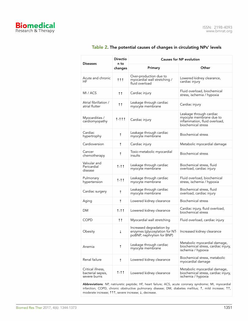
Confusingly, the role of NPs in modification of treatment care considerably relates to aging, CV disease and metabolic co-morbidities, kidney clearance, metabolism (neprilysin for BNP, glycosylation, methylation, oxidation for other NPs), toxic effect (cardiotoxicity) (Berezin, 2016f). Therefore, higher individual biological variability of these biomarkers, which negatively effects on interpretation of measure results (Favresse and Gruson, 2017). Additionally, there is a huge list of the diseases associated with increased level of NPs beyond HF development (Table 2).
!Biomed Res Ther 2017, 4(6): 1344-1373 !1350
�ISSN: 2198-4093
www.bmrat.org
Table 2. The potential causes of changes in circulating NPs’ levels
DiseasesDirectio
n to changes
Causes for NP evolution
Primary Other
Acute and chronic HF ↑↑↑
Over-production due to myocardial wall stretching / fluid overload
Lowered kidney clearance, cardiac injury
MI / ACS ↑↑ Cardiac injury Fluid overload, biochemical stress, ischemia / hypoxia
Atrial fibrillation / atrial flutter ↑↑ Leakage through cardiac
myocyte membrane Cardiac injury
Myocardities / cardiomyopathy ↑-↑↑↑ Cardiac injury
Leakage through cardiac myocyte membrane due to inflammation, fluid overload, biochemical stress
Cardiac hypertrophy ↑ Leakage through cardiac
myocyte membrane Biochemical stress
Cardioversion ↑ Cardiac injury Metabolic myocardial damage
Cancer chemotherapy ↑ Toxic-metabolic myocardial
insults Biochemical stress
Valvular and Pericardial disease
↑-↑↑ Leakage through cardiac myocyte membrane
Biochemical stress, fluid overload, cardiac injury
Pulmonary hypertension ↑-↑↑ Leakage through cardiac
myocyte membraneFluid overload, biochemical stress, ischemia / hypoxia
Cardiac surgery ↑ Leakage through cardiac myocyte membrane
Biochemical stress, fluid overload, cardiac injury
Aging ↑ Lowered kidney clearance Biochemical stress
DM ↑-↑↑ Lowered kidney clearance Cardiac injury, fluid overload, biochemical stress
COPD ↑↑ Myocardial wall stretching Fluid overload, cardiac injury
Obesity ↓Increased degradation by enzymes (glycosylation for NT-poBNP, nephrylisin for BNP)
Increased kidney clearance
Anemia ↑ Leakage through cardiac myocyte membrane
Metabolic myocardial damage, biochemical stress, cardiac injury, ischemia / hypoxia
Renal failure ↑ Lowered kidney clearance Biochemical stress, metabolic myocardial damage
Critical illness, bacterial sepsis, severe burns
↑-↑↑ Lowered kidney clearanceMetabolic myocardial damage, biochemical stress, cardiac injury, ischemia / hypoxia
Abbreviations: NP, natriuretic peptide; HF, heart failure; ACS, acute coronary syndrome; MI, myocardial infarction; COPD, chronic obstructive pulmonary disease; DM, diabetes mellitus; ↑, mild increase; ↑↑, moderate increase; ↑↑↑, severe increase; ↓, decrease.
!Biomed Res Ther 2017, 4(6): 1344-1373 !1351
�ISSN: 2198-4093
www.bmrat.org Although galectin-3 is an independent predictor of all-cause mortality, CV death and occurrence of HF, there is an inverse relationship between serum galectin-3 and estimated glomerular filtration rate (Besler et al., 2017). Accordingly, lowered kidney clearance should be taken into consideration, when data of galectin-3 measurement are interpreted. Therefore, older patients contributed to higher galectin-3 concentrations than younger individuals (Krintus et al., 2017). Amongst other biomarkers (NPs, GDF-15, high-sensitivity troponin T, sST2, aldosterone, phosphate, parathyroid hormone, plasma renin concentration, and creatinine) galectin-3 had the lowest indices of individual biological variability, whereas NPs and GDF-15 has the highest ones (Meijers et al., 2016). Additionally, in contrast to NPs serum galectin-3 levels did not appear to be significantly related to circulating level of cardiac troponins, left ventricular (LV) ejection fraction and LV mass index (Agnello et al., 2017). Therefore, there was a positive correlation galectin-3 levels with NT-proBNP in HF individuals. Thus, galectin3 and NPs might be allocated as the best tool for both short- and long term death prediction in HF regardless kidney function and age. Unfortunately, no one biomarker predicted the short-term composite HF endpoints in acute HF and actually decompensated chronic HF (Miró et al., 2017). Additionally, there are controversial findings regarding that there was no association of galectin-3 concentration with adverse outcomes in chronic HF (Wojciechowska et al., 2017).
Optimistically results of recent clinical trial about higher predictive value of sST2 receptor in HF (Maisel and Di Somma, 2016) have associated with some evidence regarding that sST2 was related to increased age, female sex, and some comorbidities including diabetes, atrial fibrillation, inflammatory diseases, kidney insufficiency and myocardial infarction (Berezin, 2016b) . Additionally, sST2 was not associated with LV structure or LV systolic or diastolic function (AbouEzzeddine et al., 2017). Thus, these findings confirmed that the sST2 is rather a systemic inflammatory marker of extra cardiac origin of HF deteriorations than a single prognosticator of HF evolution, while meta-analysis provided by Aimo et al (2017) (Aimo et al., 2017) pointed that sST2 level may predict all-cause and CV death in HF patients at admission and at discharge from the hospital. Overall, there is a large body of evidence regarding that improved discriminative value of multiple biomarkers in HF patients requires much more accurate confirmation (Berezin et al., 2016b; Pouleur, 2015). In this context, novel biomarkers are needed to improve in prediction models and assist in the titration of medical therapy.
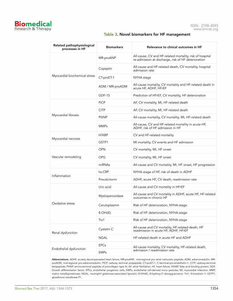
Novel biomarkers for HF management
The discovery of new biomarkers remains to be promised, but rarely novel molecules prove to be significantly better in diagnostic and predictive manner than established biomarkers. However, additionally to various types of NPs, galectin-3, sST2, and highly sensitive cardiac troponins, multiple other biomarkers, including those of inflammation, oxidative stress, vascular
!Biomed Res Ther 2017, 4(6): 1344-1373 !1352
�ISSN: 2198-4093
www.bmrat.org dysfunction and reparation state, biochemical myocardial stress and matrix remodeling, have been implicated in HF management (Table 3). Additionally, there is no strong and clear evidence regarding that the new biomarkers are able to predict clinically significant end-points (i.e., all-cause and CV mortality, HF admission/re-admission, and HF death) in both HF phenotypes - HFpEF and HFrEF. Recent clinical trials have been revealed that majority of new biomarkers indicated rather HF phenotype-related clinical outcomes than independently predicted any end points regardless presentation of HFpEF / HFrEF. Probably, biomarker-based approach could be useful to characterize pathophysiological differences between HFrEF and HFpEF patients.
Inflammatory biomarkers
C-reactive protein
The high-sensitive C-reactive protein (hsCRP) is well-established independent predictor for adverse CV outcome including CV death, all-cause mortality, sudden cardiac death, and HF-related death in general population, patients at higher CV risk and amongst individuals with known CV disease (Zhu et al., 2017). Recent clinical studies have shown that the levels of hs-CRP were considerably higher in HFpEF than in HFrEF and independently associated with HFrEF development (Tromp et al., 2017). Moreover, in HFrEF patients serum hs-CRP levels have positively correlated with circulating NT-proBNP and inversely with left ventricular ejection fraction (LVEF) (Kang et al., 2017). In contrast, low to moderate hs-CRP levels did not exhibit an association with HFpFF, while they had to be going to support CV risk.
Calprotectin
Calprotectin (myeloid-related protein 8/14) is an inflammatory marker, which has been found elevated in patients suffering from cardiac conditions, e.g. myocardial infarction, unstable angina and HF. Calprotectin is predominantly expressed in activated human neutrophils, monocytes, adipocytes, and innate immunity cells including macrophages, but not in normal tissue macrophages. Calprotectin binds with Toll-like receptor 4 and acts as innate amplifier of infection, autoimmunity, and cancer. Although calprotectin was found as a nonspecific marker for atherosclerosis, kidney damage, vascular complications in metabolic disease including vascular calcification and endothelial dysfunction, it role in HF is not understood (Berezin, 2015a). It was established that patients with chronic HF regardless of LVEF had significantly higher levels of calprotectin than patients without HF (Bruhn et al., 2017). However, predictive value of calprotectin requires to be conformed in future.
!Biomed Res Ther 2017, 4(6): 1344-1373 !1353
�ISSN: 2198-4093
www.bmrat.org Table 3. Novel biomarkers for HF management
Related pathophysiological processes in HF Biomarkers Relevance to clinical outcomes in HF
Myocardial biochemical stress
MR-proANP All-cause, CV and HF-related mortality, risk of hospital re-admission at discharge, risk of HF deterioration
Copeptin All-cause and HF-related death, CV mortality, hospital admission rate
CT-proET-1 NYHA stage
ADM / MR-proADM All-cause mortality, CV mortality and HF-related death in acute HF, ADHF, HFrEF
GDF-15 Prediction of HFrEF, CV mortality, HF deterioration
Myocardial fibrosis
PICP AF, CV mortality, MI, HF-related death
CITP AF, CV mortality, MI, HF-related death
PIIINP All-cause mortality, CV mortality, MI, HF-related death
MMPs All-cause, CV and HF-related mortality in acute HF, ADHF, risk of HF admission in HF
Myocardial necrosishFABP CV and HF-related mortality
GSTP1 MI mortality, CV events and HF admission
Vascular remodeling
OPN CV mortality, MI, HF onset
OPG CV mortality, MI, HF onset
miRNAs All-cause and CV mortality, MI, HF onset, HF progression
Inflammationhs-CRP NYHA stage of HF, risk of death in ADHF
Procalcitonin ADHF, acute HF, CV death, readmission rate
Oxidative stress
Uric acid All-cause and CV mortality in HFrEF
Myeloperoxidase All-cause and CV mortality in ADHF, acute HF, HF-related outcomes in chronic HF
Ceruloplasmin Risk of HF deterioration, NYHA-stage
8-OHdG Risk of HF deterioration, NYHA-stage
Trx1 Risk of HF deterioration, NYHA-stage
Renal dysfunctionCystatin C All-cause and CV mortality, HF-related death, HF
readmission in acute HF, ADHF, HFrEF
NGAL HF-related death in acute HF and ADHF
Endothelial dysfunctionEPCs All-cause mortality, CV mortality, HF-related death,
admission / readmission rateEMPs
Abbreviations: ADHF, acutely decompensated heart failure; MR-proANP, mid-regional pro atrial natriuretic peptide; ADM, adrenomedullin; MR-proADM, mid-regional pro-adrenomedullin; PICP, carboxy terminal propeptide; CT-proET-1, C-terminal-pro-endothelin-1; CITP, carboxy-terminal telopeptide; PIIINP, amino-terminal peptide of procollagen type III; AF, atrial fibrillation; HF, heart failure; hFABP, fatty acid binding protein; GDF, Growth differentiation factor; EPCs, endothelial progenitor cells; EMPs, endothelial cell-derived micro particles; MI, myocardial infarction; MMP, matrix metalloproteinase; NGAL, neutrophil gelatinase-associated lipocalin; 8-OHdG, 8-hydroxy-2'-deoxyguanosine; Trx1, thioredoxin 1; GSTP1; glutathione transferase P1.
!Biomed Res Ther 2017, 4(6): 1344-1373 !1354
�ISSN: 2198-4093
www.bmrat.org Procalcitonin
Procalcitonin is known a precursor of the calcitonin, which is produced and actively secreted by the parafollicular C cells of the thyroid gland and involved in regulation of calcium homeostasis (Ryu et al., 2015). Recent clinical studies have shown that procalcitonin as an inflammatory biomarker had a pretty accurate diagnostic ability to sepsis, shock, bacterial complications of some diseases (Hayashida et al., 2017; Morgenthaler et al., 2006; Reiner et al., 2017; Remde et al., 2016; Simon et al., 2004). Additionally, this biomarker may help to manage the patients with HF when antibiotic use is needed or the critical state has been verified (Ryu et al., 2015). However, there is not strong evidence regarding procalcitotin use in biomarker-guided therapy to adjust dosage of drugs for HF individuals.
Biomarkers of biochemical myocardial stress
Copeptin
Copeptin is C-terminal peptide derived from the precursor molecule of arginine vasopressin, which plays a pivotal role in fluid retention and electrolyte homeostasis (Morgenthaler et al., 2006). In the general population elevated level of copeptin strongly associated with increased CV mortality (Remde et al., 2016). Additionally, based on results of serial measurements of copeptin level it has been suggested that the increased copeptin concentration or trend to elevation of one are an independent risk factor for long-term HF-related clinical outcomes and sudden death in patients with established CV disease (Krane et al., 2017; Moayedi and Ross, 2017; Yan et al., 2017). Being able to better predict all-cause mortality rate and HF-related risks including death and admission to the hospital copeptin might be considered as much more accurate biomarker than NPs for optimize medical care in HF patients (Berezin, 2015b). Unfortunately, there are large body of evidence regarding that the level of copeptin might relate closely to some metabolic abnormalities including hyperglycemia that sufficiently limits the predictive power of the biomarker in serial measurements especially in patients with diabetes mellitus and abdominal obesity (Savic-Radojevic et al., 2017). However, the improvement of diagnostic reliability of copeptin may achieve by means use of combined biomarker strategy, in particular it might be based on copeptin and NPs (Puente et al., 2017; Sahin et al., 2016; Smaradottir et al., 2017). Finally, circulating level of copeptin has now recognized a promising biomarker with better discriminative value for both all-cause mortality and HF-related outcomes general population and individuals with established CV disease.
Growth differentiation factor-15
Growth differentiation factor (GDF)-15 belongs to the superfamily of transforming growth factor-β (Kempf and Wollert, 2009). GDF-15 is widely expressed on the surfaces of various cells. In HF GDF-15 is secreted by injured
!Biomed Res Ther 2017, 4(6): 1344-1373 !1355
�ISSN: 2198-4093
www.bmrat.org cardiomyocytes in response to ischemia, reperfusion, inflammatory cytokine stimulation and exposure to biomechanical stress (Berezin, 2016d). Elevated level of circulating GDF-15 was found in HF individuals irrespectively etiology of cardiac dysfunction (Chan et al., 2016). There is strong evidence regarding being tight interrelationship between circulating level of GDF-15 and HF signs and symptoms, reduced LVEF (Hage et al., 2017). Although serial biomarker evaluation has not showed superiority of incremental predictive ability in GDF-15 versus NPs in acute HF (Demissei et al., 2017), in chronic HFrHF / HFpEF biomarker strategy based on GDF-15, galectin-3 and NPs might exhibit several advantages before conventional approach in ability to predict all-cause mortality, CV mortality and HF-related outcomes in outpatients with HF (Berezin, 2015f; Berezin et al., 2015c).
Endothelial cell-derived micro particles and endothelial progenitor cells
Impaired endothelial function plays a pivotal role in the HF development and HF-related complications and also associates with appearance in peripheral blood specific circulating biomarkers, i.e. endothelial micro-particles (EMPs) and endothelial progenitor cells (EPCs) (Berezin, 2015f; Berezin and Kremzer, 2013c; Berezin et al., 2015c). EPCs are involved in the repair of vascular wall and myocardium and, therefore, their ability to restore integrity and functionality of vasculature strongly relates to EMPs. EMPs are not only cargo for cell-to-cell transfer of variety molecules (i.e. peptides, DNAs, RNAs, active molecules, growth factors and hormones), but they are independent regulators of immunity, inflammation, reparation, proliferative response and malignancy (Jansen et al., 2017). Number, origin (received from activated cells or apoptotic cells) and immune phenotypes of EMPs can be key factors in ensuring function of endogenous repair system (Berezin and Kremzer, 2014b). Thus, EPCs and EMPs are epigenetic co-regulators of vascular function playing a pivotal role in maintenance of endothelium integrity across all stages of HF (Berezin, 2016c).
Recent clinical studies have shown that an ability of mature endothelial cells and their precursors to release of secretome progressively worse depended on HF stage and severity (Berezin, 2007). Moreover, increased number of apoptotic EMPs and decreased number of EPCs in circulation has found as powerful predictor of CV death, HF-related admission to the hospital and prognosticator of positive reply to medical therapy in short-term period (Berezin, 2015c; Berezin et al., 2015a; Berezin et al., 2014b). There is novel HF risk prediction score created by means of biomarkers including NPs, galectin-3, high sensitive CRP and estimated ratio between both numbers of apoptotic EMPs and EPCs (Berezin, 2015c). However, there is not clear whether new predictive model would be effective in discriminative concern of HF treatment (Berezin et al., 2015b). More clinical trials are required to improve our understanding in the field of individualized therapy of HF under biomarker control.
!Biomed Res Ther 2017, 4(6): 1344-1373 !1356

�ISSN: 2198-4093
www.bmrat.org Biomarkers of collagen metabolism
Recent studies have shown that impaired collagen metabolism may alter the myocardial collagen network and thereby it exerts cardiovascular remodeling, promotes the fibrotic substrate and mediates HF complications, i.e. atrial fibrillation / flatter, sudden death, and declining LV pump function (Löfsjögård et al., 2014). Altered collagen type I synthesis and advance in degradation of one associated with appearance of circulating biomarkers, i.e. carboxy-terminal propeptide (PICP), amino-terminal peptide of procollagen type III (PIIINP) and carboxy-terminal telopeptide (CITP). Interestingly, there is evidence regarding direct causative role of BNP in alterations in collagen type I metabolism in HFrEF. In the OPTIMAL (The Optimizing Congestive Heart Failure Outpatient Clinic trial) was found that disturbances of collagen type I metabolism have exhibited an independent prognostication for long-term all-cause mortality and CV mortality in HFrEF individuals (Löfsjögård et al., 2017). In the Cardiovascular Health Study (n=880) in which was included 146 patients with HFrEF (LVEF <55%) 175 patients with HFpEF (LVEF ≥55%), 280 individuals with traditional CV risk factors without chronic HF, and 279 healthy and elderly volunteers with CV disease at risk, biomarkers of collagen turn-over (PIIINP and CITP) were significantly associated with CV outcomes, i.e. death, myocardial infarction, and advanced HF. Therefore, circulating CITP is probably independent predictor of survival in patients with HFrEF. Moreover, CITP level added to NT-proBNP level exhibited an additive predictive value compared with NT-proBNP level alone (Tziakas et al., 2012). The estimated negative predictive value for both biomarkers for long-term CV outcomes was 94%. Thus, biomarkers of collagen turn-over might be a powerful component for novel multi-marker strategy for risk stratification of HF.
Biomarkers of cardiovascular remodeling
Matrix metalloproteinase
Adverse cardiac remodeling strong relates to an accumulation of non-fibrillar extracellular matrix (ECM) and matricellular proteins, which contribute to disease progression (Berezin and Samura, 2013). In fact, matrix metalloproteinases (MMPs), which are modulated by bio-mechanical and oxidative myocardial stress, neurohormonal and inflammation, essentially determine extracellular reposition of collagen and mediates pro-fibrotic process (Sakamuri et al., 2016). According to current understanding in HF the MMPs correspond to an immune activation, inflammation, cardiac injury/vascular dysfunction to maintain tissue structure and metabolism (Rienks and Papageorgiou, 2016). MMPs play a pivotal role in cardiac and vascular remodeling through enhancing cell-to-cell interactions acting as regulator of cell growth, proliferation, differentiation, survival, and migration (Valiente-Alandi et al., 2016). However, the pathogenetic role of MMPs in HF appears to be uncertain and could relate to etiology of cardiac dysfunction (Berezin, 2015b). Resent pre-clinical and clinical studies have shown that impairment of cardiac function may relate to collagen accumulation due to imbalance between expression of MMPs, predominantly MMP-1, MMP-3,
!Biomed Res Ther 2017, 4(6): 1344-1373 !1357
�ISSN: 2198-4093
www.bmrat.org MMP-6, MMP-9, and suppression of their tissue inhibitors (Berezin et al., 2016c; Collier et al., 2011; Hutchinson et al., 2010). However, the predictive potency of these biomarkers did not confirm and requires more investigations in future.
Bone-related proteins
Bone-related proteins (BRPs) belong to the family of matricellular proteins, which incorporate into extracellular matrix and play in the bone developing, vascular remodeling, and tissue regeneration (Alford and Hankenson, 2006). Amongst family of BRPs osteopontin (OPN), osteoprotegerin (OPG), osteonectin (OSN), osteocalcin (OCN), sclerostin, and some components of RANKL/RANK system are the most known. Overall, all these molecules are mediators for paracrine signaling in cell metabolism and extracellular matrix regulation that relate inflammation with epigenetic regulation of cell function (Berezin, 2015e). The role of BRPs in CV disease including HF is controversial. As multiple functional growth factors some members of the BRPs’ family are able to cause cell differentiation and growth, bone and ectopic calcification, vascular remodeling, atherosclerotic plaque shaping, angiogenesis and neovascularisation acting predominantly in result in a hypoxic/ischemia, metabolic, oxidative and inflammatory stimuli (Berezin et al., 2016a). On the other hand, BRPs may prevent cardiac dysfunction, hypertrophy and fibrosis through blocking cellular signaling systems (i.e., PI3K and Akt phosphorylation), reduced expression of extracellular matrix and hypertrophy genes (Li et al., 2017).
Although OPN, OPG, and RANKL/RANK components are well-known biomarkers of vascular calcification, systemic inflammation, atherosclerosis, kidney dysfunction, and cardiac remodeling, their predictive role in HF is still uncertain (Berezin and Kremzer, 2013a; Berezin and Kremzer, 2014a). There is evidence regarding that the sRANKL/OPG complex may relate to HFrEF development, whereas circulating levels of OPN and OPG corresponded to HFpEF (Berezin and Kremzer, 2014a, c). Moreover, an expression of OPN, OPG and OSN genes in myocardium or vasculature sufficiently distinguished in HFrEF and HFpEF and thereby it has been suggested that BRPs could be markers to suggest the development of different HF phenotypes (Cabiati et al., 2016). Indeed, in the PEACE trial levels of OPN were independently associated with the composite CV outcomes and incident HFpEF hospitalization (Abdalrhim et al., 2016). Finally, OSN, OPN and OPG have exhibited the predictive value to mortality rate in HFpEF regardless of its etiology, while there was no axillary discriminative effect in entire predictive score when these biomarkers were added to the NPs, hs-CRP, galectin-3 and sST2 (Berezin and Kremzer, 2015).
Other biomarkers of vasculogenesis
Although vascular endothelial growth factors (VEGF) act through appropriate receptors A and B, neurophilin may bind some VEGF molecules and contributes in vascular reparation. Thus, both factors are important components of endogenous repair system. Noted, that linear regression followed by network
!Biomed Res Ther 2017, 4(6): 1344-1373 !1358
�ISSN: 2198-4093
www.bmrat.org analyses revealed prominent inflammation and angiogenesis-associated interactions through VEGF-related mechanisms in HFpEF and mainly myocardial stretch-associated interactions in HFrEF (Berezin, 2015b). The neuropilin has demonstrated a predictive value for all-cause mortality and HF-related re-admission at 18 months in HFpEF, but not in HFrEF. Overall, the role of VEGF-related biomarkers in prediction of HF is not clear and needs to be explained in details in future.
Biomarkers of oxidative stress
Serum uric acid
Observational and clinical studies have shown that the elevated serum uric acid (SUA) is common feature for patients with CV disease including HF, hypertension, atherosclerosis, obesity, diabetes mellitus and chronic renal disease (Borghi et al., 2015; Grassi et al., 2013). Evidence regarding the role of SUA in pathogenesis of CV disease is controversial. On the one hand, SUA attenuates oxidative stress through overproduction of reactive oxygen species and consequently it often worse vascular / endothelial function indirectly via inflammatory damage, inducing vascular calcification and directly via cell membranes deterioration effect (Berezin and Kremzer, 2013b). On the other hand, low-grading inflammation that is frequently found in HF may cause xanthine oxidase over-activity and leads to increased tissue SUA accumulation, which acts as scavenger of free radicals and protects against an damage effect of oxidative stress (Berezin, 2014). Additionally, an increase of SUA may be an attribute of lowered kidney clearance as a result in a progress of HF. Therefore, there is evidence regarding the regulatory role of SUA in EPC mobbing and differentiation that allow discussing SUA as mediator of reparation of tissue in HF (Berezin et al., 2014a).
Numerous clinical studies have emphasized the predictive role of baseline SUA for early post-discharge HF outcomes (Amin et al., 2017; Okazaki et al., 2016). Although levels of SUA did not significantly changed for admission period in HF patients, SUA at admission could be considered as powerful prognosticator of ADHF (Okazaki et al., 2017). On the other hand, an elevated SUA level on admission in patients with acute HF or ADHF associated not only with HF severity, but with the presence of chronic renal disease and the use of loop diuretics, which are able to cause negative clinical outcomes and independent predictor of 1-year mortality through elevation of SUA (Okazaki et al., 2017; Okazaki et al., 2016). Interestingly, the activity of xanthine oxidoreductase that is a key rate-limiting enzyme of purine degradation may be more accurate predictor of HFrEF severity and HF clinical outcomes than SUA (Otaki et al., 2017). Consequently, SUA remains a well-known risk factor of HF related clinical outcomes in acute HF and ADHF, while poor prognosis in patients with both phenotypes of chronic HF (HFrEF and HFpEF) is not elucidated.
!Biomed Res Ther 2017, 4(6): 1344-1373 !1359
�ISSN: 2198-4093
www.bmrat.org Other biomarkers
Serum levels of myeloperoxidase, ceruloplasmin and 8-hydroxy-2'-deoxyguanosine closely correlated with stage of chronic HF regardless LVEF and predict a development of HFrEF, while the role of these biomarkers of oxidative stress remains under scientific discussion and requires more investigations (Berezin, 2015b).
Biomarkers of renal dysfunction
Cystatin C
Cystatin C is an endogenous inhibitor of cysteine proteases and this biomarker is widely discussed an alternative predictor of CV events in acute and chronic HF patients with any types of cardiorenal syndrome. The patients with HFrEF demonstrated elevated serum cystatin C, especially in cases with serious risk of CV complications. Additionally, in hypertensive patients with HFpEF increased cystatin C level was found (Huerta et al., 2016). Therefore, it associated with LV diastolic dysfunction and alterations in collagen metabolism regardless of estimated GFR [106]. Although cystatin C has now validated a powerful predictor of CV outcomes and kidney injury, its sensitivity in patients with chronic HF is sufficiently inferior to that of hs-CRP and NPs (Berezin, 2015b). In contrast, in acute HF and ADHF Cystatin C provided an incremental value for prognosis more than NT-proBNP and SUA (Kim et al., 2013; Kim et al., 2015).
Other biomarkers of kidney injury in HF
There is a large body of perspective biomarkers of kidney injury that could be useful for stratification of HF at risk, i.e. stromal cell-derived factor-1, exosomes, MPs, neutrophil gelatinase-associated lipocalin, kidney injury molecule-1, interleukin-18 and miRNAs (Taub et al., 2012). Although they are able to emerge at the early stages of renal dysfunction prior to any elevations in serum creatinine, the prognostication of clinical outcomes due to acute HF, ADHF and chronic HF require more investigation.
Genetic biomarkers
By now, genetic testing has incorporated as a part of patient evaluation for suspected inherited cardiomyopathies (Teekakirikul et al., 2013; Teo et al., 2015). It turns out the epigenetic modifications through DNA methylation, ATP-dependent chromatin remodeling, histone modifications with an involvement of microRNA-related mechanisms might be sufficient pathophysiological factors contributing to adverse cardiac remodeling and altered cardiac function (Hershberger and Siegfried, 2011). In this context, the novel risk scores reflecting variabilities in genetic and epigenetic features in HF development appear to be promised (Berezin, 2016c; Lopes and Elliott, 2013; Yang et al., 2015). Indeed,
!Biomed Res Ther 2017, 4(6): 1344-1373 !1360
�ISSN: 2198-4093
www.bmrat.org some early studies have reported interested results with respect to genetic precursors of HFpEF and HFrEF (Berezin, 2016d; Fazakas et al., 2016; Friedrich et al., 2013; Hofman et al., 2010; Kolder et al., 2012; McNamara et al., 2014; Sutter et al., 2013). As biomarkers particularly used to scrutiny single nucleotide polymorphisms (SNPs) of genes encoding enzymes related to oxidative stress (Berezin, 2016e), genotype of guanine nucleotide-binding proteins (G-proteins) beta-3 subunit (GNB3) (Fazakas et al., 2016), transcription factor Islet-1 gene (McNamara et al., 2014), troponin T (Friedrich et al., 2013), CYP2D6 polymorphism (Hofman et al., 2010), cardiac myosin binding protein-C mutations (Friedrich et al., 2013), renin-angiotensin-aldosterone system polymorphism (Sutter et al., 2013) etc. Indeed, it is well known that angiotensin-converting enzyme (ACE) I/D gene D allele was associated with higher overall mortality as compared with the I allele in HF patients and that the effect could be modified by ACE inhibitors’ given (Kolder et al., 2012). Additionally, ACE DD and angiotensin-1-receptor 1166 CC genotypes may synergistically increase the predisposition to HFpEF (Wu et al., 2010).
Unfortunately, in ARIC (Atherosclerosis Risk in Communities) study was reported that none of the metabolite SNPs including pyroglutamine, dihydroxy docosatrienoic acid were individually associated with incident HF, whereas a genetic risk score created by summing the most sufficient risk alleles from each metabolite determined 11% greater risk of HF per allele (Wu et al., 2009). Ganna et al (2013) (Yu et al., 2013) have reported that amongst 707 common SNPs associated with 125 diseases including HF it would not be easily obtained explainable results by common genetic variants related to HF development. Consequently, a close gene-gene interaction may determine an individual risk to development of HF through different pathways including epigenetic modifications. All these findings lead to assume that genes score might be a powerful tool for prediction of HF development.
More successful genome-wide linkage studies toward genes-related contribution in HF have been devoted incorporating SNPs of several genes (i.e. the bradykinin type 1 receptor gene, angiotensin-II type I receptor gene, the β1-adrenoceptor gene and CYP2D6 polymorphysm) in predictive score to benefit and suffer harm from HF therapy. Although these parmacogenetic studies have focused on promised topics, the obtained results have not been absolutely consistent (Bondar et al., 2014; Ganna et al., 2013; Nelveg-Kristensen et al., 2015; Yip and Pirmohamed, 2013). Nelveg-Kristensen et al (2015) (Yip and Pirmohamed, 2013) have found no sufficient association between pharmacogenetic scores and fatal outcomes in HF patients. In contrast, Bondar et al (2014) (Nelveg-Kristensen et al., 2015) have guessed that the gene expression profiling might be useful rather for risk prediction in HF than for choosing HF treatment regime. Thus, the clinical implementation of the HF therapy based on genes scoring remains uncertain and requires more evaluation in the future (Berezin, 2016a).
!Biomed Res Ther 2017, 4(6): 1344-1373 !1361
�ISSN: 2198-4093
www.bmrat.org Multiple biomarker predictive scores
Multiple biomarkers’ use strategies based on the combination of NPs with other biomarkers have been discussed as priority in creating of much more accurate predictive scores in HF (Bayes-Genis and Ordonez-Llanos, 2015). Although there are several predictive scores based on biomarker measurement and approved for chronic HF, predictive scores for and acutely decompensated HF have not been validated (Cohen-Solal et al., 2015). Current multiple biomarker score toward prognostication, risk stratification and diagnosis of HF is based on NPs in combination with biomarkers of myocardial injury and fibrosis (galectin-3 and sST2 receptor). It is validated by American Heart Association / American College of Cardiology at 2017 and the score is suitable for patients at risk of HF, individuals with established chronic HF (for both HFrEF and HFpEF), patients with suspected acute HF and documented acute / acutely decompensated HF, as well as patients with HF at discharge from the hospital. However, there is need to compare novel scores with recently created and the scores used in HFrEF and HFpEF to optimize the treatment approach in HF management (Berezin, 2015d).
Recently developed biomarkers, i.e. mid-regional pro-A-type natriuretic peptide (Mid Pro-ANP), mid-regional-proadrenomedullin (MR-proADM), pro-endothelin, and copeptin, when were added to the predictive model based on well-known prognostic biomarkers (NPs, troponin, hs-CRP, pro-calcitonin), have been investigated in 28-days predictive value of entire score in patients with severe acute dyspnea and suspecting to acute HF or ADHF. Although three biomarkers - Mid Pro-ANP, MR-proADM and pro-endothelin - have been independently associated with prognosis of acute and chronic HF regardless LFEF, MR-proADM had improved discriminative value of NPs in combination with copeptin and troponin T (Ara-Somohano et al., 2017). Overall, there is no clarity and consistent evidence for multiple biomarker strategy in improvement in CV mortality and CV outcomes. It has been suggesting that sST2, MR-proADM and galectin-3 could improve prognostication of HF-related hospitalization and death, when they are added to NPs.
Conclusions
There are several controversies regarding importance of predictive value for survival and incremental prognostication in diagnosis of HFrEF and HFpEF. Probably, biomarkers of inflammation and vascular remodeling are predominantly observed in HFpEF, while biomarkers of biomechanical stress and collagen metabolism much more accurately predicted clinical outcome in HFrEF. All these require improving clinical guideline recommendations for optimizing HF therapy in routine clinical practice under biomarkers’ control. There is need in larger clinical trials to head-to-head compare different biomarkers and clear their role in diagnosis and guided therapy of HF.
!Biomed Res Ther 2017, 4(6): 1344-1373 !1362

�ISSN: 2198-4093
www.bmrat.org Abbreviations
ADM: adrenomedullin ANP: atrial natriuretic peptide ARNI: angiotensin receptor neprilysin inhibitors BNP: brain natriuretic peptide BRPs: Bone-related proteins cGMP: cyclic guanylyl monophosphate CITP: carboxy-terminal telopeptide CNP: C-type natriuretic peptide CRP: C-reactive protein CT-proET-1: C-terminal-pro-endothelin-1 CV: cardiovascular EMPs: endothelial microparticles EPCs: endothelial progenitor cells Gal-3: galectin-3 GDF-15: Growth differentiation factor-15 GFR: glomerular filtration rate HF: heart failure hFABP: heart type of fatty acid binding protein HFpEF: heart failure with preserved ejection fraction HFrEF: heart failure with reduced ejection fraction LV: left ventricular MMP: matrix metalloproteinase MPs: micro particles MR: proANP – mid-regional pro-atrial natriuretic peptide MR: proADM - mid-regional pro-adrenomedullin NPs: natriuretic peptides OCN: osteocalcin OPG: osteoprotegerin OPN: osteopontin OSN: osteonectin PICP: carboxy-terminal propeptide RANKL: receptor activator of nuclear factor-κB ligand sST2: soluble suppressor of tumorigenicity-2 receptor SUA: serum uric acid
!Biomed Res Ther 2017, 4(6): 1344-1373 !1363
�ISSN: 2198-4093
www.bmrat.org References Abdalrhim, A.D., Marroush, T.S., Austin, E.E., Gersh, B.J., Solak, N., Rizvi, S.A., Bailey, K.R., and Kullo, I.J. (2016). Plasma Osteopontin Levels and Adverse Cardiovascular Outcomes in the PEACE Trial. PloS one 11, e0156965.
AbouEzzeddine, O.F., McKie, P.M., Dunlay, S.M., Stevens, S.R., Felker, G.M., Borlaug, B.A., Chen, H.H., Tracy, R.P., Braunwald, E., and Redfield, M.M. (2017). Suppression of Tumorigenicity 2 in Heart Failure With Preserved Ejection Fraction. Journal of the American Heart Association 6, e004382.
Agnello, L., Bivona, G., Sasso, B.L., Scazzone, C., Bazan, V., Bellia, C., and Ciaccio, M. (2017). Galectin-3 in acute coronary syndrome. Clinical Biochemistry S0009-9120, 30245-X.
Aimo, A., Vergaro, G., Ripoli, A., Bayes-Genis, A., Figal, D.A.P., de Boer, R.A., Lassus, J., Mebazaa, A., Gayat, E., and Breidthardt, T. (2017). Meta-Analysis of Soluble Suppression of Tumorigenicity-2 and Prognosis in Acute Heart Failure. JACC: Heart Failure 5, 287-296.
Alford, A.I., and Hankenson, K.D. (2006). Matricellular proteins: extracellular modulators of bone development, remodeling, and regeneration. Bone 38, 749-757.
Amin, A., Chitsazan, M., Shiukhi Ahmad Abad, F., Taghavi, S., and Naderi, N. (2017). On admission serum sodium and uric acid levels predict 30 day rehospitalization or death in patients with acute decompensated heart failure. ESC Heart Failure 4, 162-168.
Anguita, M. (2017). High-sensitivity troponins and prognosis of heart failure. Revista clinica espanola 217, 95-96.
Ara-Somohano, C., Bonadona, A., Carpentier, F., Pavese, P., Vesin, A., Hamidfar-Roy, R., Minet, C., Vanzetto, G., Schwebel, C., and Timsit, J. (2017). Evaluation of eight biomarkers to predict short term mortality in patients with acute severe dyspnea. Minerva anestesiologica, 10.23736/S20375-29393.23717.10882-23735.
Aspromonte, N., Gulizia, M., Clerico, A., Di Tano, G., Emdin, M., Feola, M., Iacoviello, M., Latini, R., Mortara, A., and Valle, R. (2016). ANMCO/ELAS/SIBioC Consensus document: Recommendations for the use of cardiac biomarkers in heart failure patients. Giornale italiano di cardiologia (2006) 17, 615.
Bayes-Genis, A., de Antonio, M., Vila, J., Peñafiel, J., Galán, A., Barallat, J., Zamora, E., Urrutia, A., and Lupón, J. (2014). Head-to-head comparison of 2 myocardial fibrosis biomarkers for long-term heart failure risk stratification: ST2 versus galectin-3. Journal of the American College of Cardiology 63, 158-166.
Bayes-Genis, A., and Ordonez-Llanos, J. (2015). Multiple biomarker strategies for risk stratification in heart failure. Clinica Chimica Acta 443, 120-125.
Berezin (2015a). The Myeloid-Related Protein Complex Calprotectin as Biomarker of Cardiovascular Risk in Diabetes Mellitus Patients. Diabetes Res Treat, 129-136.
Berezin (2015b). Part 4. Diagnostic and prognostic value of biological markers at risk stratification among patients with heart failure. (Moskow: LAMBERT Academic Publishing GmbH).
Berezin (2016a). Genetic Predictive Scores in Heart Failure: Possibilities and Expectations. J Data Mining Genomics Proteomics 7, e127-e128.
Berezin, A. (2014). Serum uric acid as a metabolic regulator of endothelial reparative processes in heart failure patients. Stem Cell and Translational Investigation 1, e432.
!Biomed Res Ther 2017, 4(6): 1344-1373 !1364
�ISSN: 2198-4093
www.bmrat.org Berezin, A. (2015c). Impaired pattern of endothelial derived microparticles in heart failure patients. J Mol Genet Med 9, 2380-0682.
Berezin, A. (2016b). Biomarkers for cardiovascular risk in diabetic patients. Heart 102, 1939-1941.
Berezin, A. (2016c). Epigenetics in heart failure phenotypes. BBA Clin 6, 31-37.
Berezin, A., and Kremzer, A. (2013a). Circulating osteopontin as a marker of early coronary vascular calcification in type two diabetes mellitus patients with known asymptomatic coronary artery disease. Atherosclerosis 229, 475-481.
Berezin, A., and Kremzer, A. (2013b). Serum uric Acid as a marker of coronary calcification in patients with asymptomatic coronary artery disease with preserved left ventricular pump function. Cardiology research and practice 2013, 129369.
Berezin, A.E. (2007). Endothelial derived micro particles: biomarkers for heart failure diagnosis and management. Br J Haematol 137, 36-48.
Berezin, A.E. (2015d). Biomarker-Guided Therapy for Chronic Heart Failure. In Biomarkers in Cardiovascular Disease, V.B. Patel, and V.R. Preedy, eds. (Dordrecht: Springer Netherlands), pp. 1-21.
Berezin, A.E. (2015e). Bone-Related Proteins as Markers in Vascular Remodeling. In Biomarkers in Bone Disease, V.R. Preedy, ed. (Springer), pp. 1-22.
Berezin, A.E. (2015f). The risk stratification in heart failure patients: The controversial role of high-sensitive ST2. inflammation 411, 20-27.
Berezin, A.E. (2016d). Diabetes mellitus related biomarker: The predictive role of growth-differentiation factor-15. Diabetes & Metabolic Syndrome: Clinical Research & Reviews 10, S154-S157.
Berezin, A.E. (2016e). Epigenetically Modified Endothelial Progenitor Cells in Heart Failure. Journal of Clinical Epigenetics 2, 13.
Berezin, A.E. (2016f). Prognostication in different heart failure phenotypes: the role of circulating biomarkers. Journal of Circulating Biomarkers 5, 6.
Berezin, A.E., and Kremzer, A.A. (2013c). Analysis of various subsets of circulating mononuclear cells in asymptomatic coronary artery disease. Journal of clinical medicine 2, 32-44.
Berezin, A.E., and Kremzer, A.A. (2014a). Predictive Value of Circulating SPARC-Related protein Osteonectin in Patients with Symptomatic Moderate-to-Severe Ischemic-Induced Chronic Heart Failure. International Journal of Cardiology and Lipidology Research 1, 43-51.
Berezin, A.E., and Kremzer, A.A. (2014b). Relationship between circulating endothelial progenitor cells and insulin resistance in non-diabetic patients with ischemic chronic heart failure. Diabetes & Metabolic Syndrome: Clinical Research & Reviews 8, 138-144.
Berezin, A.E., and Kremzer, A.A. (2014c). Relationship between serum RANKL/osteoprotegerin complex and endothelial progenitor cells in ischemic chronic heart failure. Journal of Cardiology and Therapy 1, 189-195.
Berezin, A.E., and Kremzer, A.A. (2015). Predictive value of circulating osteonectin in patients with ischemic symptomatic chronic heart failure. Biomedical Journal 38, 523-530.
Berezin, A.E., Kremzer, A.A., Berezina, T.A., and Martovitskaya, Y.V. (2015a). Pattern of circulating microparticles in chronic heart failure patients with metabolic syndrome: Relevance to neurohumoral and inflammatory activation. BBA clinical 4, 69-75.
!Biomed Res Ther 2017, 4(6): 1344-1373 !1365
�ISSN: 2198-4093
www.bmrat.org Berezin, A.E., Kremzer, A.A., Berezina, T.A., Martovitskaya, Y.V., and Gromenko, E.A. (2016a). Relation of osteoprotegerin level and numerous of circulating progenitor mononuclears in patients with metabolic syndrome. Biomedical Research and Therapy 3, 501-513.
Berezin, A.E., Kremzer, A.A., Martovitskaya, Y.V., Berezina, T.A., and Samura, T.A. (2016b). The utility of biomarker risk prediction score in patients with chronic heart failure. Clinical hypertension 22, 3.
Berezin, A.E., Kremzer, A.A., Martovitskaya, Y.V., Samura, T.A., and Berezina, T.A. (2015b). The predictive role of circulating microparticles in patients with chronic heart failure. BBA clinical 3, 18-24.
Berezin, A.E., Kremzer, A.A., Martovitskaya, Y.V., Samura, T.A., Berezina, T.A., Zulli, A., Klimas, J., and Kruzliak, P. (2015c). The utility of biomarker risk prediction score in patients with chronic heart failure. International journal of clinical and experimental medicine 8, 18255-18264.
Berezin, A.E., Kremzer, A.A., and Samura, T.A. (2016c). Circulating thrombospondin-2 in patients with moderate-to-severe chronic heart failure due to coronary artery disease. Journal of biomedical research 30, 32.
Berezin, A.E., Kremzer, A.A., Samura, T.A., Berezina, T.A., and Martovitskaya, Y.V. (2014a). Serum uric Acid predicts declining of circulating proangiogenic mononuclear progenitor cells in chronic heart failure patients. Journal of cardiovascular and thoracic research 6, 153.
Berezin, A.E., Kremzer, A.A., Samura, T.A., and Martovitskaya, Y.V. (2014b). Apoptotic microparticles to progenitor mononuclear cells ratio in heart failure: relevance of clinical status and outcomes. JCvD 2, 50-57.
Berezin, A.E., and Samura, T.A. (2013). Prognostic value of biological markers in myocardial infarction patients. Asian Cardiovascular and Thoracic Annals 21, 142-150.
Besler, C., Lang, D., Urban, D., Rommel, K.-P., von Roeder, M., Fengler, K., Blazek, S., Kandolf, R., Klingel, K., and Thiele, H. (2017). Plasma and Cardiac Galectin-3 in Patients With Heart Failure Reflects Both Inflammation and Fibrosis. Circulation: Heart Failure 10, e003804.
Billebeau, G., Vodovar, N., Sadoune, M., Launay, J.M., Beauvais, F., and Cohen-Solal, A. (2017). Effects of a cardiac rehabilitation programme on plasma cardiac biomarkers in patients with chronic heart failure. Eur J Prev Cardiol, 2047487317705488.
Bondar, G., Cadeiras, M., Wisniewski, N., Maque, J., Chittoor, J., Chang, E., Bakir, M., Starling, C., Shahzad, K., Ping, P., et al. (2014). Comparison of whole blood and peripheral blood mononuclear cell gene expression for evaluation of the perioperative inflammatory response in patients with advanced heart failure. PloS one 9, e115097.
Borghi, C., Rosei, E.A., Bardin, T., Dawson, J., Dominiczak, A., Kielstein, J.T., Manolis, A.J., Perez-Ruiz, F., and Mancia, G. (2015). Serum uric acid and the risk of cardiovascular and renal disease. Journal of hypertension 33, 1729-1741.
Boulogne, M., Sadoune, M., Launay, J., Baudet, M., Cohen-Solal, A., and Logeart, D. (2017). Inflammation versus mechanical stretch biomarkers over time in acutely decompensated heart failure with reduced ejection fraction. International Journal of Cardiology 226, 53-59.
!Biomed Res Ther 2017, 4(6): 1344-1373 !1366

�ISSN: 2198-4093
www.bmrat.org Bruhn, L.V., Lauridsen, K.G., Schmidt, A.S., Rickers, H., Bach, L.F., Løfgren, B., and Hornung, N. (2017). Elevated calprotectin in patients with atrial fibrillation with and without heart failure. Scandinavian Journal of Clinical and Laboratory Investigation 77, 210-215.
Cabiati, M., Svezia, B., Matteucci, M., Botta, L., Pucci, A., Rinaldi, M., Caselli, C., Lionetti, V., and Del Ry, S. (2016). Myocardial Expression Analysis of Osteopontin and Its Splice Variants in Patients Affected by End-Stage Idiopathic or Ischemic Dilated Cardiomyopathy. PloS one 11, e0160110.
Chan, M.M., Santhanakrishnan, R., Chong, J.P., Chen, Z., Tai, B.C., Liew, O.W., Ng, T.P., Ling, L.H., Sim, D., and Leong, K.T.G. (2016). Growth differentiation factor 15 in heart failure with preserved vs. reduced ejection fraction. European journal of heart failure 18, 81-88.
Chmurzyńska, A. (2006). The multigene family of fatty acid-binding proteins (FABPs): function, structure and polymorphism. Journal of applied genetics 47, 39-48.
Chow, S.L., Maisel, A.S., Anand, I., Bozkurt, B., de Boer, R.A., Felker, G.M., Fonarow, G.C., Greenberg, B., Januzzi, J.L., Jr., Kiernan, M.S., et al. (2017). Role of Biomarkers for the Prevention, Assessment, and Management of Heart Failure: A Scientific Statement From the American Heart Association. Circulation 135, e1054-e1091.
Cohen-Solal, A., Laribi, S., Ishihara, S., Vergaro, G., Baudet, M., Logeart, D., Mebazaa, A., Gayat, E., Vodovar, N., Pascual-Figal, D.A., et al. (2015). Prognostic markers of acute decompensated heart failure: the emerging roles of cardiac biomarkers and prognostic scores. Archives of cardiovascular diseases 108, 64-74.
Collier, P., Watson, C.J., Voon, V., Phelan, D., Jan, A., Mak, G., Martos, R., Baugh, J.A., Ledwidge, M.T., and McDonald, K.M. (2011). Can emerging biomarkers of myocardial remodelling identify asymptomatic hypertensive patients at risk for diastolic dysfunction and diastolic heart failure? European journal of heart failure 13, 1087-1095.
de Freitas Souza, B.S., Silva, D.N., Carvalho, R.H., de Almeida Sampaio, G.L., Paredes, B.D., França, L.A., Azevedo, C.M., Vasconcelos, J.F., Meira, C.S., and Neto, P.C. (2017). Association of Cardiac Galectin-3 Expression, Myocarditis, and Fibrosis in Chronic Chagas Disease Cardiomyopathy. The American Journal of Pathology 187, 1134-1146.
Demissei, B.G., Cotter, G., Prescott, M.F., Felker, G.M., Filippatos, G., Greenberg, B.H., Pang, P.S., Ponikowski, P., Severin, T.M., and Wang, Y. (2017). A multimarker multi-time point-based risk stratification strategy in acute heart failure: results from the RELAX-AHF trial. European Journal of Heart Failure, 10.1002/ejhf.1749.
Favresse, J., and Gruson, D. (2017). Natriuretic peptides: degradation, circulating forms, dosages and new therapeutic approaches. Paper presented at: Annales de Biologie Clinique.
Fazakas, Á., Szelényi, Z., Szénási, G., Nyírő, G., Szabó, P.M., Patócs, A., Tegze, N., Fekete, B.C., Molvarec, A., and Nagy, B. (2016). Genetic predisposition in patients with hypertension and normal ejection fraction to oxidative stress. Journal of the American Society of Hypertension 10, 124-132.
Friedrich, F.W., Dilanian, G., Khattar, P., Juhr, D., Gueneau, L., Charron, P., Fressart, V., Vilquin, J.T., Isnard, R., and Gouya, L. (2013). A novel genetic variant in the transcription factor Islet-1 exerts gain of function on myocyte enhancer factor 2C promoter activity. European journal of heart failure 15, 267-276.
!Biomed Res Ther 2017, 4(6): 1344-1373 !1367
�ISSN: 2198-4093
www.bmrat.org Ganna, A., Rivadeneira, F., Hofman, A., Uitterlinden, A.G., Magnusson, P.K., Pedersen, N.L., Ingelsson, E., and Tiemeier, H. (2013). Genetic determinants of mortality. Can findings from genome-wide association studies explain variation in human mortality? Human genetics 132, 553-561.
Grassi, D., Ferri, L., Desideri, G., Di Giosia, P., Cheli, P., Del Pinto, R., Properzi, G., and Ferri, C. (2013). Chronic hyperuricemia, uric acid deposit and cardiovascular risk. Current pharmaceutical design 19, 2432-2438.
Hage, C., Michaëlsson, E., Linde, C., Donal, E., Daubert, J.-C., Gan, L.-M., and Lund, L.H. (2017). Inflammatory Biomarkers Predict Heart Failure Severity and Prognosis in Patients With Heart Failure With Preserved Ejection Fraction. Circulation: Cardiovascular Genetics 10, e001633.
Hayashida, K., Kondo, Y., Hara, Y., Aihara, M., and Yamakawa, K. (2017). Head-to-head comparison of procalcitonin and presepsin for the diagnosis of sepsis in critically ill adult patients: a protocol for a systematic review and meta-analysis. BMJ open 7, e014305.
Hershberger, R.E., and Siegfried, J.D. (2011). Update 2011: clinical and genetic issues in familial dilated cardiomyopathy. J Am Coll Cardiol 57, 1641-1649.
Hofman, N., van Langen, I., and Wilde, A.A. (2010). Genetic testing in cardiovascular diseases. Current opinion in cardiology 25, 243-248.
Huerta, A., López, B., Ravassa, S., San José, G., Querejeta, R., Beloqui, Ó., Zubillaga, E., Rábago, G., Brugnolaro, C., and Díez, J. (2016). Association of cystatin C with heart failure with preserved ejection fraction in elderly hypertensive patients: potential role of altered collagen metabolism. Journal of hypertension 34, 130-138.
Hutchinson, K.R., Stewart, J.A., and Lucchesi, P.A. (2010). Extracellular matrix remodeling during the progression of volume overload-induced heart failure. Journal of molecular and cellular cardiology 48, 564-569.
Imran, T.F., Shin, H.J., Mathenge, N., Wang, F., Kim, B., Joseph, J., Gaziano, J.M., and Djoussé, L. (2017). Meta-Analysis of the Usefulness of Plasma Galectin-3 to Predict the Risk of Mortality in Patients With Heart Failure and in the General Population. The American Journal of Cardiology 119, 57-64.
Jansen, F., Nickenig, G., and Werner, N. (2017). Extracellular Vesicles in Cardiovascular Disease. Circulation research 120, 1649-1657.
Kang, S., Fan, L., Chen, M., Li, J., and Liu, Z. (2017). Relationship of high-sensitivity C-reactive protein concentrations and systolic heart failure. Current vascular pharmacology, 10.2174/1570161115666170404121619.
Kempf, T., and Wollert, K.C. (2009). Growth differentiation factor-15: a new biomarker in cardiovascular disease. Herz 34, 594-599.
Kim, H., Yoon, H.J., Park, H.S., Cho, Y.K., Nam, C.W., Hur, S.H., Kim, Y.N., and Kim, K.B. (2013). Potentials of cystatin C and uric acid for predicting prognosis of heart failure. Congestive Heart Failure 19, 123-129.
Kim, T.-H., Kim, H., and Kim, I.-C. (2015). The potential of cystatin-C to evaluate the prognosis of acute heart failure: A comparative study. Acute Cardiac Care 17, 72-76.
Kitai, T., Kim, Y.-H., Kiefer, K., Morales, R., Borowski, A.G., Grodin, J.L., and Tang, W.W. (2017). Circulating intestinal fatty acid-binding protein (I-FABP) levels in acute decompensated heart failure. Clinical Biochemistry 50, 491-495.
!Biomed Res Ther 2017, 4(6): 1344-1373 !1368

�ISSN: 2198-4093
www.bmrat.org Kolder, I.C., Michels, M., Christiaans, I., Ten Cate, F.J., Majoor-Krakauer, D., Danser, A.H., Lekanne Deprez, R.H., Tanck, M., Wilde, A.A., Bezzina, C.R., et al. (2012). The role of renin-angiotensin-aldosterone system polymorphisms in phenotypic expression of MYBPC3-related hypertrophic cardiomyopathy. European journal of human genetics : EJHG 20, 1071-1077.
Krane, V., Genser, B., Kleber, M.E., Drechsler, C., März, W., Delgado, G., Allolio, B., Wanner, C., and Fenske, W. (2017). Copeptin Associates with Cause-Specific Mortality in Patients with Impaired Renal Function: Results from the LURIC and the 4D Study. Clinical Chemistry 63, 997-1007.
Krintus, M., Kozinski, M., Fabiszak, T., Kubica, J., Panteghini, M., and Sypniewska, G. (2017). Establishing reference intervals for galectin-3 concentrations in serum requires careful consideration of its biological determinants. Clinical Biochemistry 50, 599-604.
Ledwidge, M., Gallagher, J., Conlon, C., Tallon, E., O’Connell, E., Dawkins, I., Watson, C., O’Hanlon, R., Bermingham, M., and Patle, A. (2013). Natriuretic peptide–based screening and collaborative care for heart failure: the STOP-HF randomized trial. Jama 310, 66-74.
Li, J., Yousefi, K., Ding, W., Singh, J., and Shehadeh, L.A. (2017). Osteopontin RNA aptamer can prevent and reverse pressure overload-induced heart failure. Cardiovascular research 113, 633-643.
Löfsjögård, J., Kahan, T., Díez, J., López, B., González, A., Ravassa, S., Mejhert, M., Edner, M., and Persson, H. (2017). Usefulness of Collagen Carboxy-Terminal Propeptide and Telopeptide to Predict Disturbances of Long-Term Mortality in Patients≥ 60 Years With Heart Failure and Reduced Ejection Fraction. The American Journal of Cardiology 119, 2042-2048.
Löfsjögård, J., Persson, H., Díez, J., López, B., González, A., Edner, M., Mejhert, M., and Kahan, T. (2014). Atrial fibrillation and biomarkers of myocardial fibrosis in heart failure. Scandinavian Cardiovascular Journal 48, 299-303.
Lopes, L.R., and Elliott, P.M. (2013). Genetics of heart failure. Biochimica et biophysica acta 1832, 2451-2461.
Luchner, A., Von Haehling, S., Holubarsch, C., Keller, T., Knebel, F., Zugck, C., and Laufs, U. (2017). Indications and Clinical Implications of the Use of the Cardiac Markers BNP and NT-proBNP. Deutsche medizinische Wochenschrift (1946) 142, 346.
Maisel, A.S., and Di Somma, S. (2016). Do we need another heart failure biomarker: focus on soluble suppression of tumorigenicity 2 (sST2). European Heart Journal, ehw462.
Malek, V., and Gaikwad, A.B. (2017). Neprilysin inhibitors: A new hope to halt the diabetic cardiovascular and renal complications? Biomedicine & pharmacotherapy = Biomedecine & pharmacotherapie 90, 752-759.
Masson, S., Latini, R., and Anand, I.S. (2010). An update on cardiac troponins as circulating biomarkers in heart failure. Current heart failure reports 7, 15-21.
McNamara, D.M., Taylor, A.L., Tam, S.W., Worcel, M., Yancy, C.W., Hanley-Yanez, K., Cohn, J.N., and Feldman, A.M. (2014). G-protein beta-3 subunit genotype predicts enhanced benefit of fixed-dose isosorbide dinitrate and hydralazine: results of A-HeFT. JACC: Heart Failure 2, 551-557.
Meijers, W.C., van der Velde, A.R., Muller Kobold, A.C., Dijck-Brouwer, J., Wu, A.H., Jaffe, A., and de Boer, R.A. (2016). Variability of biomarkers in patients with chronic heart failure and healthy controls. European Journal of Heart Failure 19, 357-365.
!Biomed Res Ther 2017, 4(6): 1344-1373 !1369
�ISSN: 2198-4093
www.bmrat.org Miró, Ò., González de la Presa, B., Herrero-Puente, P., Fernández Bonifacio, R., Möckel, M., Mueller, C., Casals, G., Sandalinas, S., Llorens, P., and Martín-Sánchez, F.J. (2017). The GALA study: relationship between galectin-3 serum levels and short-and long-term outcomes of patients with acute heart failure. Biomarkers, 10.1080/1354750X.1352017.1319421.
Moayedi, Y., and Ross, H.J. (2017). Advances in heart failure: a review of biomarkers, emerging pharmacological therapies, durable mechanical support and telemonitoring. Clinical science (London, England : 1979) 131, 553-566.
Morgenthaler, N.G., Struck, J., Alonso, C., and Bergmann, A. (2006). Assay for the measurement of copeptin, a stable peptide derived from the precursor of vasopressin. Clinical chemistry 52, 112-119.
Nagarajan, V., Hernandez, A.V., and Tang, W.W. (2012). Prognostic value of cardiac troponin in chronic stable heart failure: a systematic review. Heart, heartjnl-2012-301779.
Nakanishi, M., Nakao, K., Kumasaka, L., Arakawa, T., Fukui, S., Ohara, T., Yanase, M., Noguchi, T., Yasuda, S., and Goto, Y. (2017). Improvement in Exercise Capacity by Exercise Training Associated With Favorable Clinical Outcomes in Advanced Heart Failure With High B-Type Natriuretic Peptide Level. Circulation Journal, CJ-16-1268.
Nelveg-Kristensen, K.E., Busk Madsen, M., Torp-Pedersen, C., Kober, L., Egfjord, M., Berg Rasmussen, H., and Riis Hansen, P. (2015). Pharmacogenetic Risk Stratification in Angiotensin-Converting Enzyme Inhibitor-Treated Patients with Congestive Heart Failure: A Retrospective Cohort Study. PloS one 10, e0144195.
Nymo, S.H., Aukrust, P., Kjekshus, J., McMurray, J.J., Cleland, J.G., Wikstrand, J., Muntendam, P., Wienhues-Thelen, U., Latini, R., Askevold, E.T., et al. (2017). Limited Added Value of Circulating Inflammatory Biomarkers in Chronic Heart Failure. JACC Heart failure 5, 256-264.
Okazaki, H., Shirakabe, A., Kobayashi, N., Hata, N., Shinada, T., Matsushita, M., Yamamoto, Y., Shibata, Y., Shibuya, J., Shiomura, R., et al. (2017). Are atherosclerotic risk factors associated with a poor prognosis in patients with hyperuricemic acute heart failure? The evaluation of the causal dependence of acute heart failure and hyperuricemia. Heart and Vessels 32, 436-445.
Okazaki, H., Shirakabe, A., Kobayashi, N., Hata, N., Shinada, T., Matsushita, M., Yamamoto, Y., Shibuya, J., Shiomura, R., and Nishigoori, S. (2016). The prognostic impact of uric acid in patients with severely decompensated acute heart failure. Journal of cardiology 68, 384-391.
Otaki, Y., Watanabe, T., Kinoshita, D., Yokoyama, M., Takahashi, T., Toshima, T., Sugai, T., Murase, T., Nakamura, T., and Nishiyama, S. (2017). Association of plasma xanthine oxidoreductase activity with severity and clinical outcome in patients with chronic heart failure. International Journal of Cardiology 228, 151-157.
Ponikowski, P., Voors, A.A., Anker, S.D., Bueno, H., Cleland, J.G., Coats, A.J., Falk, V., González-Juanatey, J.R., Harjola, V.-P., and Jankowska, E.A. (2016). 2016 ESC Guidelines for the diagnosis and treatment of acute and chronic heart failure: The Task Force for the diagnosis and treatment of acute and chronic heart failure of the European Society of Cardiology (ESC) Developed with the special contribution of the Heart Failure Association (HFA) of the ESC. European heart journal 37, 2129-2200.
Pouleur, A.-C. (2015). Which biomarkers do clinicians need for diagnosis and management of heart failure with reduced ejection fraction? Clinica Chimica Acta 443, 9-16.
!Biomed Res Ther 2017, 4(6): 1344-1373 !1370
�ISSN: 2198-4093
www.bmrat.org Puente, P.H., García, B.P., García, M.G., Jacob, J., Martín-Sánchez, F.J., Pascual-Figal, D., Bueno, H., Gil, V., Llorens, P., and Vázquez-Alvarez, J. (2017). Predictive capacity of a multimarker strategy to determine short-term mortality in patients attending a hospital emergency department for acute heart failure. BIO-EAHFE study. Clinica Chimica Acta 466, 22-30.
Qian, H.-Y., Huang, J., Yang, Y.-J., Yang, Y.-M., Li, Z.-Z., and Zhang, J.-M. (2016). Heart-type Fatty Acid Binding Protein in the Assessment of Acute Pulmonary Embolism. The American Journal of the Medical Sciences 352, 557-562.
Reiner, M.M., Khoury, W.E., Canales, M.B., Chmielewski, R.A., Patel, K., Razzante, M.C., Cloughtery, C.O., and Rowland, D.Y. (2017). Procalcitonin as a Biomarker for Predicting Amputation Level in Lower Extremity Infections. The Journal of Foot and Ankle Surgery 56, 484-491.
Remde, H., Dietz, A., Emeny, R., Riester, A., Peters, A., de Las Heras Gala, T., Then, C., Seissler, J., Beuschlein, F., Reincke, M., et al. (2016). The cardiovascular markers copeptin and high-sensitive C-reactive protein decrease following specific therapy for primary aldosteronism. Journal of hypertension 34, 2066-2073.
Rienks, M., and Papageorgiou, A.-P. (2016). Novel regulators of cardiac inflammation: matricellular proteins expand their repertoire. Journal of molecular and cellular cardiology 91, 172-178.
Ryu, J.A., Yang, J.H., Lee, D., Park, C.M., Suh, G.Y., Jeon, K., Cho, J., Baek, S.Y., Carriere, K.C., and Chung, C.R. (2015). Clinical Usefulness of Procalcitonin and C-Reactive Protein as Outcome Predictors in Critically Ill Patients with Severe Sepsis and Septic Shock. PloS one 10, e0138150.
Sahin, I., Gungor, B., Ozkaynak, B., Uzun, F., Küçük, S.H., Avci, I.I., Ozal, E., Ayça, B., Cetın, S., and Okuyan, E. (2016). Higher copeptin levels are associated with worse outcome in patients with hypertrophic cardiomyopathy. Clinical Cardiology 40, 32-37.
Sakamuri, S.S., Takawale, A., Basu, R., Fedak, P.W., Freed, D., Sergi, C., Oudit, G.Y., and Kassiri, Z. (2016). Differential impact of mechanical unloading on structural and nonstructural components of the extracellular matrix in advanced human heart failure. Translational Research 172, 30-44.
Savic-Radojevic, A., Pljesa-Ercegovac, M., Matic, M., Simic, D., Radovanovic, S., and Simic, T. (2017). Chapter Four-Novel Biomarkers of Heart Failure. Advances in Clinical Chemistry 79, 93-152.
Simon, L., Gauvin, F., Amre, D.K., Saint-Louis, P., and Lacroix, J. (2004). Serum procalcitonin and C-reactive protein levels as markers of bacterial infection: a systematic review and meta-analysis. Clinical infectious diseases 39, 206-217.
Smaradottir, M.I., Ritsinger, V., Gyberg, V., Norhammar, A., Näsman, P., and Mellbin, L.G. (2017). Copeptin in patients with acute myocardial infarction and newly detected glucose abnormalities–A marker of increased stress susceptibility? A report from the Glucose in Acute Myocardial Infarction cohort. Diabetes and Vascular Disease Research 14, 69-76.
Srivatsan, V., George, M., and Shanmugam, E. (2015). Utility of galectin-3 as a prognostic biomarker in heart failure: where do we stand? European journal of preventive cardiology 22, 1096-1110.
Sutter, M.E., Gaedigk, A., Albertson, T.E., Southard, J., Owen, K.P., Mills, L.D., and Diercks, D.B. (2013). Polymorphisms in CYP2D6 may predict methamphetamine related heart failure. Clinical toxicology (Philadelphia, Pa) 51, 540-544.
!Biomed Res Ther 2017, 4(6): 1344-1373 !1371
�ISSN: 2198-4093
www.bmrat.org Taub, P.R., Borden, K.C., Fard, A., and Maisel, A. (2012). Role of biomarkers in the diagnosis and prognosis of acute kidney injury in patients with cardiorenal syndrome. Expert review of cardiovascular therapy 10, 657-667.
Teekakirikul, P., Kelly, M.A., Rehm, H.L., Lakdawala, N.K., and Funke, B.H. (2013). Inherited cardiomyopathies: molecular genetics and clinical genetic testing in the postgenomic era. The Journal of Molecular Diagnostics 15, 158-170.
Teo, L.Y.L., Moran, R.T., and Tang, W.W. (2015). Evolving approaches to genetic evaluation of specific cardiomyopathies. Current heart failure reports 12, 339-349.
Tromp, J., Khan, M.A., Klip, I.T., Meyer, S., de Boer, R.A., Jaarsma, T., Hillege, H., van Veldhuisen, D.J., van der Meer, P., and Voors, A.A. (2017). Biomarker Profiles in Heart Failure Patients With Preserved and Reduced Ejection Fraction. Journal of the American Heart Association 6, e003989.
Tziakas, D.N., Chalikias, G.K., Stakos, D., Chatzikyriakou, S.V., Papazoglou, D., Mitrousi, K., Lantzouraki, A., Thomaidi, A., Boudoulas, H., and Konstantinides, S. (2012). Independent and additive prognostic ability of serum carboxy-terminal telopeptide of collagen type-I in heart failure patients: a multi-marker approach with high-negative predictive value to rule out long-term adverse events. European journal of preventive cardiology 19, 62-71.
Valiente-Alandi, I., Schafer, A.E., and Blaxall, B.C. (2016). Extracellular matrix-mediated cellular communication in the heart. Journal of molecular and cellular cardiology 91, 228-237.
Wettersten, N., and Maisel, A.S. (2016). Biomarkers for heart failure: An update for practitioners of internal medicine. The American journal of medicine 129, 560-567.
Wojciechowska, C., Romuk, E., Nowalany-Kozielska, E., and Jacheć, W. (2017). Serum Galectin-3 and ST2 as predictors of unfavorable outcome in stable dilated cardiomyopathy patients. Hellenic Journal of Cardiology S1109-9666, 30249-30244.
Wong, P.C., Guo, J., and Zhang, A. (2017). The renal and cardiovascular effects of natriuretic peptides. Advances in physiology education 41, 179-185.
Wu, C.K., Luo, J.L., Tsai, C.T., Huang, Y.T., Cheng, C.L., Lee, J.K., Lin, L.Y., Lin, J.W., Hwang, J.J., and Chiang, F.T. (2010). Demonstrating the pharmacogenetic effects of angiotensin-converting enzyme inhibitors on long-term prognosis of diastolic heart failure. The pharmacogenomics journal 10, 46-53.
Wu, C.K., Luo, J.L., Wu, X.M., Tsai, C.T., Lin, J.W., Hwang, J.J., Lin, J.L., Tseng, C.D., and Chiang, F.T. (2009). A propensity score-based case-control study of renin-angiotensin system gene polymorphisms and diastolic heart failure. Atherosclerosis 205, 497-502.
Yan, J.J., Lu, Y., Kuai, Z.P., and Yong, Y.H. (2017). Predictive value of plasma copeptin level for the risk and mortality of heart failure: a meta-analysis. Journal of Cellular and Molecular Medicine, 10.1111/jcmm.13102.
Yancy, C.W., Jessup, M., Bozkurt, B., Butler, J., Casey, D.E., Jr., Colvin, M.M., Drazner, M.H., Filippatos, G.S., Fonarow, G.C., Givertz, M.M., et al. (2017). 2017 ACC/AHA/HFSA Focused Update of the 2013 ACCF/AHA Guideline for the Management of Heart Failure: A Report of the American College of Cardiology/American Heart Association Task Force on Clinical Practice Guidelines and the Heart Failure Society of America. Journal of cardiac failure, 10.1016/j.cardfail.2017.1004.1014.
Yang, J., Xu, W.W., and Hu, S.J. (2015). Heart failure: advanced development in genetics and epigenetics. BioMed research international 2015, 352734.
!Biomed Res Ther 2017, 4(6): 1344-1373 !1372

�ISSN: 2198-4093
www.bmrat.org Yip, V.L., and Pirmohamed, M. (2013). Expanding role of pharmacogenomics in the management of cardiovascular disorders. American journal of cardiovascular drugs : drugs, devices, and other interventions 13, 151-162.
Yu, B., Zheng, Y., Alexander, D., Manolio, T.A., Alonso, A., Nettleton, J.A., and Boerwinkle, E. (2013). Genome-wide association study of a heart failure related metabolomic profile among African Americans in the Atherosclerosis Risk in Communities (ARIC) study. Genetic epidemiology 37, 840-845.
Zhu, L., Zou, Y., Wang, Y., Luo, X., Sun, K., Wang, H., Jia, L., Liu, Y., Zou, J., and Yuan, Z. (2017). Prognostic Significance of Plasma High-Sensitivity C-Reactive Protein in Patients With Hypertrophic Cardiomyopathy. Journal of the American Heart Association 6, e004529.
!Biomed Res Ther 2017, 4(6): 1344-1373 !1373
ISSN: 2198-4093 www.bmrat.org
*For correspondence:
Competing interests: The authorsdeclare that no competing interests exist.
Received: 03 May 2017 Accepted: 19 June 2017 Published: 25 June 2017
Copyright The Author(s) 2017. This article is published with open access by BioMedPress (BMP).
This article is distributed under the terms of the Creative Commons Attribution License (CC-BY 4.0) which permits any use, distribution, and reproduction in any medium, provided the original author(s) and the source are credited.
Original Research
Comparative treatment efficiency of adipose and bone marrow derived allogenic mesenchymal stem cell transplantation in mouse models of liver fibrosis
Nam Hai Nguyen1, Trinh Van Le1, Huy Quang Do1, Dat Quoc Ngo3, Huy Minh Le3, Nhung Hai Truong1,2 *
1Laboratory of Stem cell Research and Application, University of Science, VNU-HCM, Ho Chi Minh City, Vietnam2Biology Faculty, University of Science, VNU-HCM, Ho Chi Minh City, Vietnam3University of Medicine and Pharmacy Ho Chi Minh City, Ho Chi Minh City, Vietnam
Abstract
Background: The application of mesenchymal stem cell (MSC) therapy in liver fibrosistreatment has been increasingly investigated in recent years. MSCs obtained from a variety of sources (e.g. bone marrow, umbilical cord blood and adipose tissue) have been studied and have achieved remarkable results. In this study, we compared the effects of adipose-derived mesenchymal stem cells (AD-MSC) transplantation with bone marrow-derived mesenchymal stem cell (BM-MSC) transplantation in a mouse model of liver fibrosis, induced by carbon tetrachloride (CCl4). Methods: Eight-week old micewere treated with CCl4 for 11 weeks to induce liver fibrosis then 5x10⁵ cells were transplanted into mice via the tail vein. Results: After 21 days of transplantation, theresults showed that the stem cell treated groups ameliorated better than the placebo group. MSC treated groups showed reduced AST and ALT levels, down-regulated expression of extracellular matrix (ECM) genes, and improved liver histopathology. Both sources of MSCs (bone marrow and adipose tissue) were effective in the mouse model of liver fibrosis. Conclusion: Our results also indicated that AD-MSC transplantation in miceaccelerated liver regeneration better than BM-MSC transplantation.
Keywords AD-MSC, BM-MSC, CCl4, liver cirrhosis, Mesenchymal stem cell, MSC, Stem cell therapy
Biomed Res Ther 2017, 4(6): 1374-1387 !1374DOI: 10.15419/bmrat.v4i06.179
ISSN: 2198-4093 www.bmrat.org
Introduction
Liver cirrhosis is a serious disease with a high mortality risk, ranking among the top ten causes of death in Eastern Europe, Central Asia and high-income countries (Mortality and Causes of Death, 2015). The number of patients with liver cirrhosis worldwide in 2015 was estimated at more than 39 million; this number represents an increase of approximately 2 million, compared to that of 2005 (Kassebaum et al., 2016). Common causes of liver cirrhosis include hepatitis B virus (HBV), hepatitis C virus (HCV), and alcohol abuse. By histology, the hallmark features of cirrhosis are diffuse nodules in the liver. The fibrotic tissues are formed by excessive accumulation of the extracellular matrix. Liver cirrhosis leads to severe consequences such as portal hypertension, ascites, liver failure and death. To date, liver transplantation has been the gold standard choice for patients with advanced cirrhosis. However, this therapy is high-risk due to graft rejection, viral re-infection, and complications of long-term immunosuppressive treatment (Schuppan and Afdhal, 2008).
MSCs have many prominent properties which are related to their capability of liver tissue regeneration; these include differentiation into hepatocyte-like cell in vitro (Harn et al., 2012), immune modulation (Bifari et al., 2008), reduction of activation of hepatic stellate cells (Baligar et al., 2016), and amelioration of hepatitis (Seki et al., 2013). In recent years, many studies have been conducted using MSCs for the treatment of cirrhosis. The sources of MSCs are diverse, such as cord blood (Abdel Aziz et al., 2010; Choudhery et al., 2013), bone marrow (Abdel Aziz et al., 2007; Yuan et al., 2014; Zhao et al., 2005), and adipose tissue (Choudhery et al., 2013; Seki et al., 2013; Zhang et al., 2014). Although these studies have shown that MSCs are a promising therapeutic choice for liver fibrosis (Berardis et al., 2015), the origin of MSCs from different sources may be problematic, with the varying safety and effectiveness of the cells (Reinisch et al., 2015). Therefore, more robust investigations on the varied sources of MSCs are necessary for approving MSC application for therapy. In this study, we evaluated the effectiveness of MSCs obtained from two sources (bone marrow and adipose tissue) to study how MSCs from different sources influence the therapeutic efficacy.
Materials - MethodsCirrhosis model mouse
Eight-week-old male Swiss mice were used in this study. The mice were treated with 1.0 mL/kg (99.5% purity, UNI-CHEM Chemical Reagent, China) 3 times per week for 11 weeks. Prior to CCl4 administration, mice were fasted for 4 h. The animal study was accepted by the Institutional Ethics Committee of SCL Laboratories (Laboratory of Stem Cell Research and Application, University of Science, VNU-HCM, Vietnam).
Biomed Res Ther 2017, 4(6): 1374-1387 !1375
ISSN: 2198-4093 www.bmrat.org
AD-MSC isolation
Adipose tissue was isolated from mouse testicles and stored in PBS containing 10% antibiotic- antimycotic, 100 X (Gibco/Thermal Fisher Scientific, Waltham, MA). Tissues were washed 2 times with PBS containing 10% antibiotic-antimycotic. The ADSC extraction kit (Geneworld Ltd. Co, Saigon High-tech Park, Ho Chi Minh, Vietnam) was used according to the manufacturer’s instructions to separate out stromal vascular fractions (SVFs). The SVF component (containing cells) was then cultured in a 25 cm2 Roux flask containing complete DMEM/F-12 medium (i.e. DMEM/F-12 medium (Sigma-Aldrich, St. Louis, MO), 15% fetal bovine serum (FBS) (Gibco, Waltham, MA), and 1% of 100X antibiotic-antimycotic (Gibco). The cells were cultured at 37oC, 5% CO2. The medium was changed every 3 days.
BM-MSC isolation
The femurs of 4-week old male mice were collected and stored in PBS containing 10% of 100X antibiotic-antimycotic (Gibco) for bone marrow extraction. Muscle, tendon and cartilage were removed. Mononuclear cells (BM-MNCs) were isolated from bone marrow by flushing with medium through a 22-G needle to obtain single cells. BM-MNCs were cultured in a 25 cm2 flask in complete DMEM/F-12 medium at 37oC, 5% CO2. The medium was changed every 3 days.
Characterization of AD-MSCs and BM-MSCs
After the 3rd passage, candidate cells were evaluated for MSCs markers and differentiation capacity into adipogenic cells. In brief, MSCs markers were assessed by flow cytometry on a FACSCalibur system (BD Biosciences, Franklin Lakes, NJ) using FITC-, PerCP-, PE- or APC-conjugated anti-CD34, anti-CD45, anti-CD14, anti-CD44, anti-CD90, and anti-CD106 antibodies.
In this study, we tested the adipogenic potential of MSCs using adipogenic induction medium (i.e. DMEM/F-12 supplemented with 10% FBS, 1% of 100X antibiotic-antimycotic solution, 10mM dexamethasone (Sigma-Aldrich), 2.79 mM indomethacin (Sigma-Aldrich), 5 mg/mL insulin (Sigma-Aldrich), and 0.5 M 1-Methyl-3-isobutylxanthine (IBMX) (Sigma-Aldrich). After 21 days of differentiation, the differentiated cells were assessed by oil red staining.
AD-MSC and BM-MSC transplantation
Mice with liver fibrosis were divided into 3 groups (n=5 per group). The groups were as follows: Group 1 (Placebo; PBS injection), Group 2 (AD-MSC treatment), and Group 3 (BM-MSC treatment). Passage 3 (P3) MSCs were trypsinized with 0.25% trypsin and washed with PBS. MSCs were filtered through a 70 μm filter to obtain single cells then 5x10⁵ MSCs were resuspended in 0.15 ml PBS and injected into the tail vein.
Biomed Res Ther 2017, 4(6): 1374-1387 !1376
ISSN: 2198-4093 www.bmrat.org
Evaluations after AD-MSC and BM-MSC transplantation
After 21 days of transplantation, mice were evaluated for:
Level of AST, ALT and ALB in serum
200 μL of plasma from each mouse was collected to assess the level of AST (IFCC Mod.LiquiUV test, Germany), ALT (IFCC Mod. LiquiUV test, Germany) and ALB (QuantiChrom Bilirubin Assay Kit, Bioassay Systems, CA, USA), according to the manufacturer’s instructions.
Expression of fibrosis related genes
Liver tissues in the left liver lobe were obtained for RNA extraction using the Easy_BLUE Total RNA Extraction Kit (INTRON Biotechnology, Korea) according to the manufacturer’s instructions. Gene expression was determined by quantitative RT-PCR method (Brilliant II QRT-PCR Master Mix Kit 1-Step, Agilent, CA) using the specific primers listed in Table 1.
Table 1. List of RT-PCR Primers used in this study
Histopathology
Liver tissues (approximately 1 cm2) were obtained from left liver lobes. The samples were washed with PBS two times and fixed in paraformaldehyde for 24 h before sectioning in paraffin. For hematoxylin & eosin (H&E) staining, liver sections were deparaffinized in xylene, dehydrated using alcohol, and washed with PBS. Liver sections was then stained in hematoxylin (Merck Millipore, Germany) for 5 min, washed quickly, and then differentiated by 1% acid alcohol for 30 seconds then washed for 10 min. Slides were stained with eosin solution
Genes Primer sequences (3’-5’)
GAPDH F: AAGTTGTCATGGATGACCR: TCACCATCTTCCAGGAGC
Fibronectin F: ATGTGGACCCCTCCTGATAGTR: GCCCAGTGATTTCAGCAAAGG
Integrin F: GCCAGGGCTGGTTATACAGAR: TCACAATGGCACACAGGTTT
TGF-beta F: CTTCAGCTCCACAGAGAAGAACTGCR:CACAATCATGTTGGACAACTGCTCC
Procollagen F:CCTGGACGCCATCAAGGTCTACR: CCAAGTTCCGGTGTGACTCG
Nt5e (CD73) F: TTTGGAAGGTGGATTTCCTG R: CCTCTCAAATCCAGGGACAA
Biomed Res Ther 2017, 4(6): 1374-1387 !1377

ISSN: 2198-4093 www.bmrat.org
(Merck Millipore, Germany) for 2-3 min, washed and then mounted. For Masson staining, liver sections were deparaffinized, dehydrated and washed. Slides were stained with Weigert’s iron hematoxylin for 5 min, washed, then stained with Biebrich scarlet acid fuchsin solution for 5 min and washed again. Slides were differentiated in 1% phosphomolybdic-phosphotungstic acid solution for 5 min, then transferred to aniline blue solution, and stained for 5 min. Finally, sections were differentiated in 1% acetic acid solution for 1 min, washed, dehydrated, and mounted with mounting medium.
Statistical Analysis
Data analysis was conducted using GraphPad Prism 6 software (La Jolla, CA). Statistical significance was set at p<0.05.
Results
Isolating and characterizing MSCs
After culturing primary cells for 48 h, a number of cells adhered and spread onto the surface; these cells had a spindle shape. At the third passage, candidate cells showed the distinct fibroblast-like morphology (Fig. 1A, B).
Figure 1. Morphology of MSC cells. A. BM-MSCs at passage 3. B. AD-MSCs at passage 3. C. Differentiation of BM-MSCs into adipogenic cells at day 21 of induction (positive oil red staining). D. Differentiation of AD-MSCs into adipogenic cells at day 21 of induction (positive oil red staining).
Biomed Res Ther 2017, 4(6): 1374-1387 !1378
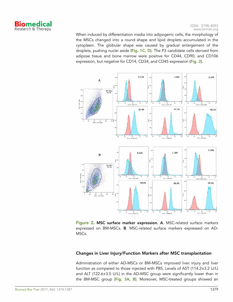
ISSN: 2198-4093 www.bmrat.org
When induced by differentiation media into adipogenic cells, the morphology of the MSCs changed into a round shape and lipid droplets accumulated in the cytoplasm. The globular shape was caused by gradual enlargement of the droplets, pushing nuclei aside (Fig. 1C, D). The P3 candidate cells derived from adipose tissue and bone marrow were positive for CD44, CD90, and CD106 expression, but negative for CD14, CD34, and CD45 expression (Fig. 2).
Figure 2. MSC surface marker expression. A. MSC-related surface markers expressed on BM-MSCs. B. MSC-related surface markers expressed on AD-MSCs.
Changes in Liver Injury/Function Markers after MSC transplantation
Administration of either AD-MSCs or BM-MSCs improved liver injury and liver function as compared to those injected with PBS. Levels of AST (114.2±3.2 U/L) and ALT (122.6±3.5 U/L) in the AD-MSC group were significantly lower than in the BM-MSC group (Fig. 3A, B). Moreover, MSC-treated groups showed an
Biomed Res Ther 2017, 4(6): 1374-1387 !1379
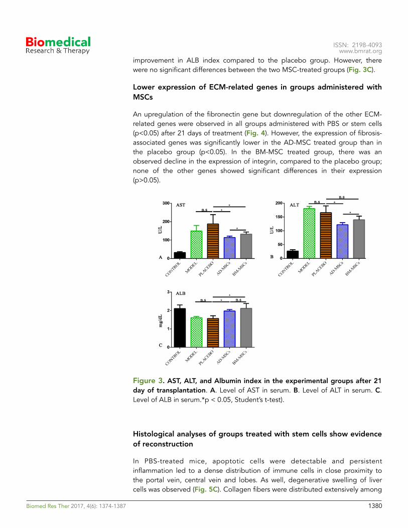
ISSN: 2198-4093 www.bmrat.org
improvement in ALB index compared to the placebo group. However, there were no significant differences between the two MSC-treated groups (Fig. 3C).
Lower expression of ECM-related genes in groups administered with MSCs
An upregulation of the fibronectin gene but downregulation of the other ECM-related genes were observed in all groups administered with PBS or stem cells (p<0.05) after 21 days of treatment (Fig. 4). However, the expression of fibrosis-associated genes was significantly lower in the AD-MSC treated group than in the placebo group (p<0.05). In the BM-MSC treated group, there was an observed decline in the expression of integrin, compared to the placebo group; none of the other genes showed significant differences in their expression (p>0.05).
Figure 3. AST, ALT, and Albumin index in the experimental groups after 21 day of transplantation. A. Level of AST in serum. B. Level of ALT in serum. C. Level of ALB in serum.*p < 0.05, Student’s t-test).
Histological analyses of groups treated with stem cells show evidence of reconstruction
In PBS-treated mice, apoptotic cells were detectable and persistent inflammation led to a dense distribution of immune cells in close proximity to the portal vein, central vein and lobes. As well, degenerative swelling of liver cells was observed (Fig. 5C). Collagen fibers were distributed extensively among
Biomed Res Ther 2017, 4(6): 1374-1387 !1380
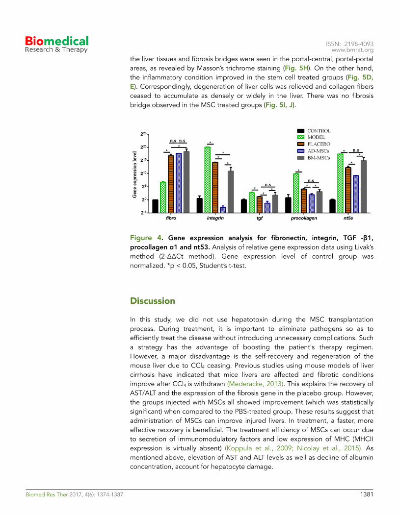
ISSN: 2198-4093 www.bmrat.org
the liver tissues and fibrosis bridges were seen in the portal-central, portal-portal areas, as revealed by Masson’s trichrome staining (Fig. 5H). On the other hand, the inflammatory condition improved in the stem cell treated groups (Fig. 5D, E). Correspondingly, degeneration of liver cells was relieved and collagen fibers ceased to accumulate as densely or widely in the liver. There was no fibrosis bridge observed in the MSC treated groups (Fig. 5I, J).
Figure 4. Gene expression analysis for fibronectin, integrin, TGF -β1, procollagen α1 and nt53. Analysis of relative gene expression data using Livak’s method (2-ΔΔCt method). Gene expression level of control group was normalized. *p < 0.05, Student’s t-test.
DiscussionIn this study, we did not use hepatotoxin during the MSC transplantation process. During treatment, it is important to eliminate pathogens so as to efficiently treat the disease without introducing unnecessary complications. Such a strategy has the advantage of boosting the patient's therapy regimen. However, a major disadvantage is the self-recovery and regeneration of the mouse liver due to CCl4 ceasing. Previous studies using mouse models of liver cirrhosis have indicated that mice livers are affected and fibrotic conditions improve after CCl4 is withdrawn (Mederacke, 2013). This explains the recovery of AST/ALT and the expression of the fibrosis gene in the placebo group. However, the groups injected with MSCs all showed improvement (which was statistically significant) when compared to the PBS-treated group. These results suggest that administration of MSCs can improve injured livers. In treatment, a faster, more effective recovery is beneficial. The treatment efficiency of MSCs can occur due to secretion of immunomodulatory factors and low expression of MHC (MHCII expression is virtually absent) (Koppula et al., 2009; Nicolay et al., 2015). As mentioned above, elevation of AST and ALT levels as well as decline of albumin concentration, account for hepatocyte damage.
Biomed Res Ther 2017, 4(6): 1374-1387 !1381
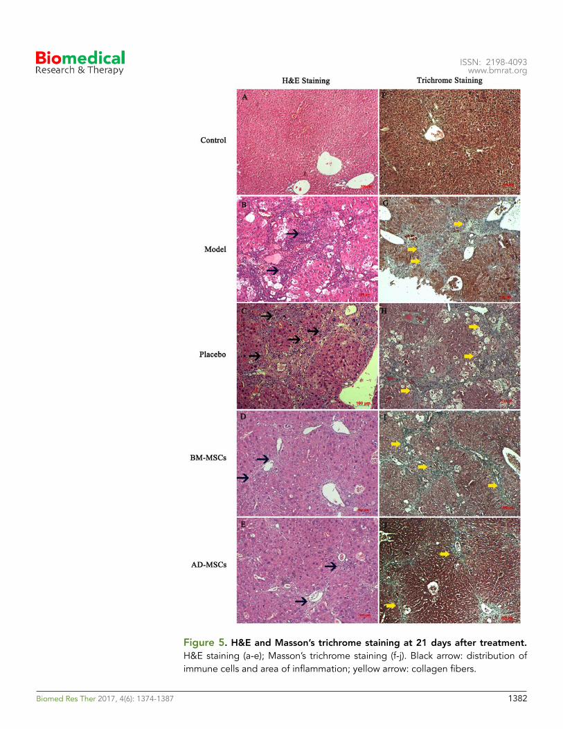
ISSN: 2198-4093 www.bmrat.org
Figure 5. H&E and Masson’s trichrome staining at 21 days after treatment.H&E staining (a-e); Masson’s trichrome staining (f-j). Black arrow: distribution of immune cells and area of inflammation; yellow arrow: collagen fibers.
Biomed Res Ther 2017, 4(6): 1374-1387 !1382
ISSN: 2198-4093 www.bmrat.org
In this study, the improvement of the biochemical indexes observed in both groups transplanted with MSCs suggests a protective effect of MSCs on hepatocytes. The protective effect might be explained by the ability of MSCs to secrete cytokines such as hepatocyte growth factor (HGF) (Matsuda-Hashii et al., 2004) and nerve growth factor (NGF). As well, MSCs can secrete anti-inflammatory molecules, such as interleukin (IL)-10 and interleukin 1 receptor antagonist (IL1-RA), which have anti-apoptotic and proliferative effects on hepatocytes and which help improve liver functions (Francois et al., 2013). Other studies have revealed the role of MSC transplantation in improving oxidative stress in cells within damaged areas (Eirin et al., 2015). The alleviation of liver fibrosis in MSC-treated groups could be related to the paracrine mechanism of MSCs and their differentiation into functional cells; paracrine mechanisms are the predominant means by which MSCs exert their action (Berardis et al., 2015; Parekkadan et al., 2007).
Co-culturing of MSCs and hepatic stellate cells (HSCs) can lead to an increase in the number of G0-stage HSCs, which is consistent with the decrease in number of S-stage HSCs. Such observations suggest a role for MSCs in inhibiting HSCs (Zhao et al., 2005). Moreover, MSCs play a role in inhibiting the activation and proliferation of HSCs, thereby enhancing the clearance of abnormal ECM in fibrotic structures (Parekkadan et al., 2007), and in the regulation of fibrosis-related genes (Ali and Masoud, 2012). The modulatory effects of MSCs, as mentioned above, can be used to explain the results of the decline of expression of fibrosis-related genes in the treated groups, compared to placebo, in our study. However, one limitation in our study is that the mechanism of action of MSCs (e.g. paracrine secretion, differentiation into hepatocyte in vivo, and anti-fibrosis effects) has not been determined in our model. Further investigations into the mechanism of action of MSCs in CCl4- induced mouse model of liver fibrosis will aid in a greater understanding of MSCs in therapy.
Histological studies were consistent with gene expression results, suggesting an improvement in all of the groups when CCl4 administration was ceased. However, the alleviation in fibrosis occurred significantly faster in mice treated with MSCs than in untreated mice, reflecting the important role of MSCs. There are two mechanisms that could be associated with the clearance of ECM in fibrotic liver tissues: decrease of structural ECM proteins (mainly collagen) and elevation of enzymes (which dissolve the ECM). Research by Abdel et al. (2007) have indicated that MSC transplantation reduces collagen expression (Abdel Aziz et al., 2007), that MSCs have an ability to inhibit activities of HSCs, and that MMPs secretion can result in decomposing the abnormal ECM in fibrotic livers.
In our study, there were various questions concerning AD-MSC and BM-MSC therapies, mainly whether or not the different types of stem cells from various sources would result in differences in treatment efficiency. Previous in vitro studies have proven that MSCs from various sources share a similarity in the differentiation potential or possibilities for therapeutic applications (Choudhery et al., 2013). However, recent results from the study by Reinisch et al. (2015)
Biomed Res Ther 2017, 4(6): 1374-1387 !1383
ISSN: 2198-4093 www.bmrat.org
have revealed that epigenetic or microenvironment factors in origin of stem cells significantly affect the potential of MSCs. Both in vitro and in vivo studies have suggested differences in differentiation capacity and genetic profile amongst populations of cells, regardless of similarities in cell morphology, surface markers or differentiation potential (Reinisch et al., 2015). Studies by Bigot et al. (2015) have also indicated that the oxygen levels can affect the stability of the genetic profiles of AD-MSCs and BM-MSCs in different ways (Bigot et al., 2015). These phenomena might account for variations in the efficiency of MSCs acquired from various sources for therapeutic applications.
Conclusion
All mice in the experiment groups showed improvement of their liver fibrosis; even mice in the placebo group showed improvement when CCl4 administration was ceased. Importantly, the MSC-treated group showed a great improvement in their AST/ALT ratios. Even histological structures in the treated mice looked significantly better than that of placebo. Overall, comparison of the efficiencies between the two sources of MSCs (AD-MSCs versus BM-MSCs), the AST/ALT ratios, and the gene expression and histology indicated that AD-MSCs were better as a therapeutic platform than BM-MSCs.
Abbreviations
AD-MSCs: Adipose tissue-derived mesenchymal stem cells
ALB: Albumin
ALT: Alanine transaminase
AST: Aspartate aminotransferase
BM-MNCs: Bone marrow-derived mononuclear cells
CCl4: Carbon tetrachloride
ECM: Extracellular matrix
HBV: Hepatitis B virus
HCV: Hepatitis C virus
HGF: Hepatocyte growth factor
HSC: Hepatic stellate cells
IL: Interleukin
SVFs: Stromal vascular fractions
VNU-HCM: Vietnam National University, Ho Chi Minh City
Biomed Res Ther 2017, 4(6): 1374-1387 !1384

ISSN: 2198-4093 www.bmrat.org
AcknowledgementsThis study was funded by Vietnam National University, Ho Chi Minh City (grant number B2012-18-07TD).
Author contribution
Nam Hai Nguyen carried out experiments, acquisition of data and compose the first manuscript. Trinh Van Le and Huy Quang Do conducted the mouse model, acquisition data of liver function, gene-expression. Huy Minh Le and Dat Quoc Ngo made substantial contributions to analyze the histology change. Nhung Hai Truong made substantial contributions to conception and design, data analysis and interpretation of data. Being corresponding author, Truong Hai Nhung give final approval of the manuscript to be submitted and any revised version.
Biomed Res Ther 2017, 4(6): 1374-1387 !1385
ISSN: 2198-4093 www.bmrat.org
ReferencesAbdel Aziz, M.T., Atta, H.M., Mahfouz, S., Fouad, H.H., Roshdy, N.K., Ahmed, H.H., Rashed, L.A., Sabry, D., Hassouna, A.A., and Hasan, N.M. (2007). Therapeutic potential of bone marrow-derived mesenchymal stem cells on experimental liver fibrosis. Clinical biochemistry 40, 893-899. Abdel Aziz, M.T., El Asmar, M.F., Mostafa, S., Salama, H., Atta, H.M., Mahfouz, S., Roshdy, N.K., Rashed, L.A., Sabry, D., Hasan, N., et al. (2010). Reversal of Hepatic Fibrosis by Human CD34(+) Stem/Progenitor Cell Transplantation in Rats. International Journal of Stem Cells 3, 161-174. Ali, G., and Masoud, M.S. (2012). Bone marrow cells ameliorate liver fibrosis and express albumin after transplantation in CCl(4)-induced fibrotic liver. Saudi journal of gastroenterology : official journal of the Saudi Gastroenterology Association 18, 263-267. Baligar, P., Mukherjee, S., Kochat, V., Rastogi, A., and Mukhopadhyay, A. (2016). Molecular and Cellular Functions Distinguish Superior Therapeutic Efficiency of Bone Marrow CD45 Cells Over Mesenchymal Stem Cells in Liver Cirrhosis. Stem cells 34, 135-147. Berardis, S., Dwisthi Sattwika, P., Najimi, M., and Sokal, E.M. (2015). Use of mesenchymal stem cells to treat liver fibrosis: current situation and future prospects. World journal of gastroenterology 21, 742-758. Bifari, F., Lisi, V., Mimiola, E., Pasini, A., and Krampera, M. (2008). Immune Modulation by Mesenchymal Stem Cells. Transfusion medicine and hemotherapy : offizielles Organ der Deutschen Gesellschaft fur Transfusionsmedizin und Immunhamatologie 35, 194-204. Bigot, N., Mouche, A., Preti, M., Loisel, S., Renoud, M.L., Le Guevel, R., Sensebe, L., Tarte, K., and Pedeux, R. (2015). Hypoxia Differentially Modulates the Genomic Stability of Clinical-Grade ADSCs and BM-MSCs in Long-Term Culture. Stem cells 33, 3608-3620. Choudhery, M.S., Badowski, M., Muise, A., and Harris, D.T. (2013). Comparison of human mesenchymal stem cells derived from adipose and cord tissue. Cytotherapy 15, 330-343. Eirin, A., Zhu, X.Y., Ferguson, C.M., Riester, S.M., van Wijnen, A.J., Lerman, A., and Lerman, L.O. (2015). Intra-renal delivery of mesenchymal stem cells attenuates myocardial injury after reversal of hypertension in porcine renovascular disease. Stem cell research & therapy 6, 7. Francois, S., Mouiseddine, M., Allenet-Lepage, B., Voswinkel, J., Douay, L., Benderitter, M., and Chapel, A. (2013). Human mesenchymal stem cells provide protection against radiation-induced liver injury by antioxidative process, vasculature protection, hepatocyte differentiation, and trophic effects. BioMed research international 2013, 151679. Harn, H.J., Lin, S.Z., Hung, S.H., Subeq, Y.M., Li, Y.S., Syu, W.S., Ding, D.C., Lee, R.P., Hsieh, D.K., Lin, P.C., et al. (2012). Adipose-derived stem cells can abrogate chemical-induced liver fibrosis and facilitate recovery of liver function. Cell transplantation 21, 2753-2764.
Biomed Res Ther 2017, 4(6): 1374-1387 !1386
ISSN: 2198-4093 www.bmrat.org
Kassebaum, N.J., Arora, M., Barber, R.M., Bhutta, Z.A., Brown, J., Carter, A., Casey, D.C., Charlson, F.J., Coates, M.M., Coggeshall, M., et al. (2016). Global, regional, and national disability-adjusted life-years (DALYs) for 315 diseases and injuries and healthy life expectancy (HALE), 1990–2015: a systematic analysis for the Global Burden of Disease Study 2015. The Lancet 388, 1603-1658. Koppula, P.R., Chelluri, L.K., Polisetti, N., and Vemuganti, G.K. (2009). Histocompatibility testing of cultivated human bone marrow stromal cells - a promising step towards pre-clinical screening for allogeneic stem cell therapy. Cellular immunology 259, 61-65. Matsuda-Hashii, Y., Takai, K., Ohta, H., Fujisaki, H., Tokimasa, S., Osugi, Y., Ozono, K., Matsumoto, K., Nakamura, T., and Hara, J. (2004). Hepatocyte growth factor plays roles in the induction and autocrine maintenance of bone marrow stromal cell IL-11, SDF-1 alpha, and stem cell factor. Experimental hematology 32, 955-961. Mederacke, I. (2013). Liver fibrosis - mouse models and relevance in human liver diseases. Zeitschrift fur Gastroenterologie 51, 55-62. Mortality, G.B.D., and Causes of Death, C. (2015). Global, regional, and national age-sex specific all-cause and cause-specific mortality for 240 causes of death, 1990-2013: a systematic analysis for the Global Burden of Disease Study 2013. Lancet 385, 117-171. Nicolay, N.H., Lopez Perez, R., Debus, J., and Huber, P.E. (2015). Mesenchymal stem cells - A new hope for radiotherapy-induced tissue damage? Cancer letters 366, 133-140. Parekkadan, B., van Poll, D., Megeed, Z., Kobayashi, N., Tilles, A.W., Berthiaume, F., and Yarmush, M.L. (2007). Immunomodulation of activated hepatic stellate cells by mesenchymal stem cells. Biochemical and biophysical research communications 363, 247-252. Reinisch, A., Etchart, N., Thomas, D., Hofmann, N.A., Fruehwirth, M., Sinha, S., Chan, C.K., Senarath-Yapa, K., Seo, E.Y., Wearda, T., et al. (2015). Epigenetic and in vivo comparison of diverse MSC sources reveals an endochondral signature for human hematopoietic niche formation. Blood 125, 249-260. Schuppan, D., and Afdhal, N.H. (2008). Liver cirrhosis. Lancet 371, 838-851. Seki, A., Sakai, Y., Komura, T., Nasti, A., Yoshida, K., Higashimoto, M., Honda, M., Usui, S., Takamura, M., Takamura, T., et al. (2013). Adipose tissue-derived stem cells as a regenerative therapy for a mouse steatohepatitis-induced cirrhosis model. Hepatology 58, 1133-1142. Yuan, S., Jiang, T., Zheng, R., Sun, L., Cao, G., and Zhang, Y. (2014). Effect of bone marrow mesenchymal stem cell transplantation on acute hepatic failure in rats. Experimental and therapeutic medicine 8, 1150-1158. Zhang, Y., Chen, X.M., and Sun, D.L. (2014). Effects of coencapsulation of hepatocytes with adipose-derived stem cells in the treatment of rats with acute-on-chronic liver failure. The International journal of artificial organs 37, 133-141. Zhao, D.C., Lei, J.X., Chen, R., Yu, W.H., Zhang, X.M., Li, S.N., and Xiang, P. (2005). Bone marrow-derived mesenchymal stem cells protect against experimental liver fibrosis in rats. World journal of gastroenterology 11, 3431-3440.
Biomed Res Ther 2017, 4(6): 1374-1387 !1387
Biomed Res Ther 2017, 4(6): 1388-1399 1388
*For correspondence:
Competing interests: The authorsdeclare that no competing interests exist.
Received: 06 March 2017Accepted: 04 June 2017Published: 25 June 2017
Copyright The Author(s) 2017. This article is published with open access by BioMedPress (BMP).
This article is distributed under the terms of the Creative Commons Attribution License (CC-BY 4.0) which permits any use, distribution, and reproduction in any medium, provided the original author(s) and the source are credited.
Original Research
Comparative study of sperm motility in Metformin-using and Insulin-dependent diabetics
Awais Ali Zaidi1, Mahtab Ahmed Khan1, Ali Sharif1, Lubna Shakir2, Atif Irshad3, Arsalan Ali2, Zaib Ali Shaheryar1,*
1Faculty of Pharmacy, University of Lahore, Lahore-Pakistan 2Faculty of Pharmacy, Hajvery University, Lahore-Pakistan 3Good Hope Hospital, Rectory Road Sutton Coldfield, B75 7RR
AbstractBackground: Diabetes mellitus (DM) represents one of the greatest threats to modernglobal health. DM may affect male reproductive function at multiple levels as a result of its effects on spermatogenesis, sperm motility, sperm morphology, and change in sperm structure. Method: The present study deals with sperm motility and spermmorphological changes associated with diabetes in the male population. In this study, 50 insulin-dependent and 50 metformin users were selected, with ages of males ranging from 26-54 years and duration of diabetes distributed over 3-15 years. Both insulin-dependent and metformin-using diabetic subjects were evaluated for sperm analysis. Results: Sperm analysis data showed a significant increase (p ±0.0005) in total spermcount in insulin-dependent diabetic men. However, sperm motility was found to be about 10-15% less in insulin-dependent patients compared to metformin users. Moreover, sperm morphology was improved in 6% of metformin users compared to insulin-dependent diabetics. Conclusion: Our study concludes that metformin does notsignificantly affect sperm count. However, it does significantly affect sperm motility, when compared to insulin-dependent diabetic men. This study established an important relationship between diabetes and sperm motility, which reflects the reproductive capabilities of men.
Keywords Insulin-dependent, Metformin, Morphological changes, Sperm motility
DOI: 10.15419/bmrat.v4i06.180
Biomed Res Ther 2017, 4(6): 1388-1399 1388
Introduction
Diabetes mellitus (DM) is a metabolic disorder that is generally characterized as Type 1 diabetes (T1D) or Type 2 diabetes (T2D). T1D arises from a complete lack of insulin due to defective insulin secretion. T2D arises from a partial lack of insulin and is associated with alterations in carbohydrate, protein and lipid metabolism (Association, 2014). In fact, T2D can result from a combination of inadequate insulin secretion and insulin resistance (Association, 2010). In T2D, the symptoms can be detected only when the disease is in its advanced phase. Indeed, the changes produced in tissues and at the cellular level may not be reversed even when treated with the appropriate drug therapy (Donner and Munoz, 2012).
DM is one of the most prominent population health threats in developed societies, with incidence on the rise each year. In 2002, the World Health Organization (WHO) reported that there were about 171 million DM patients worldwide; that number represented a 60% higher incidence compared to data from 1995 (Organization, 2002). DM has been closely linked to long-term complications related to mortality and morbidity, such as hypertension and kidney failure (McBrien et al., 2012; Torgerson et al., 2004).
Insulin stimulates glucose uptake in muscle and fat cells. In the muscles, glucose is taken up and stored as glycogen; in fat cells glucose is absorbed and converted mostly to glycerin for long-term storage. The primary role of insulin is glucose transport into the plasma membrane of muscle and fat cells. The change in glucose transport activity due to the exposure to insulin brought changes and therefore different enzymes may act on different type of carbohydrates and fat metabolism takes place (Denton et al., 1981).
Metformin is an oral diabetes drug which helps control blood sugar levels, particularly in those with type 2 diabetes. Metformin primarily acts on hepatic glucose ejection by inhibiting gluconeogenesis. However, it can cause weight loss and affect adipose tissues (Stumvoll et al., 1995). The metabolism and mechanism of action of metformin are still poorly understood, despite the fact that it has been a form of medicine (for non-insulin-dependent diabetes mellitus (NIDDM)) for the past for 30 years.
DM is responsible for various homeostatic and biochemical alterations resulting in male infertility of sub-infertility. Individuals with diabetes have described sexual problems such as weakened sexual desire, likely related to a hyperglycemic state (Ewing, 1985). Another sexual disorder linked to diabetes is erectile dysfunction (Sexton and Jarow, 1997) and a decline in ejaculation (Fedele, 2005). There appears to be a direct relationship between the incidence
Biomed Res Ther 2017, 4(6): 1388-1399 1389
of DM and fertility, and incidence of DM and decrease in birth rates (Lutz, 2006). The prevalence of DM in patients of reproductive age is a purported cause. The majority of patients are diagnosed with T1D before the age of 30 years and the rest are diagnosed with T2D (Agbaje et al., 2007; Silink, 2002).
In DM the molecular mechanisms and specific pathways, which induce defects in the male reproductive system and its functions, are both impacted. Spermatogenesis is altered in DM patients (Daubresse et al., 1978). Several research studies comparing young or adult diabetics (to control individuals) have shown that diabetic males have lower sperm counts and significant changes in sperm morphology and motility (Baccetti et al., 2002). Indeed, DM induces change in sperm count as well as sperm volume (Bartak et al., 1974). Some other studies have shown that the concentration and sperm count are increased in diabetics but that sperm motility and semen volume are decreased (Ali et al., 1993).
Blood glucose concentrations depend upon and adjust the function of different tissues and organs. Liver and fat are normally known to have strict control on glucose variations and are particularly known to play key roles in the use and storage of nutrients by hormonally regulated mechanisms. In testes, glucose metabolism is a vital event. Spermatogenesis preservation in vivo relies on glucose metabolism despite the fact that there are low levels of this sugar in tubular liquid (Robinson and Fritz, 1981; Zysk et al., 1975). Metformin helps to restore the body's response to insulin, making it possible to produce the amount of glucose and gastrointestinal absorption needed from the liver. Metformin (a biguanide derivative) reduces complications associated with blood glucose control.
Some studies have reported that there is no affiliation between age, sperm motility, T1D diagnosis and span of illness. In the study herein, we demonstrate that the parameters used to assess sperm motility (e.g. progressive velocity, path velocity, way speed and lateral head displacement) remained unchanged. For example, the direct linear index (which unveils the straightness of sperm swimming) was expanded in diabetic patients and normal individuals (Niven et al., 1995). Our study shows that the effect of T1D on male fertility may be associated to the complications of the disease and not to the disease itself. The sperm cells of diabetic males are reported to have high fructose and glucose content. However, a relationship could not be established between an ineffective metabolic control and the observed alterations in the semen. A broad study of spermatozoa cryopreservation from patients with diverse pathologies showed that only sperm from diabetic men presented considerable differences in the parameters used to assess sperm (Ranganathan et al., 2002). Despite the fact that there is some ambiguity with regards to sperm parameters, there appears to
Biomed Res Ther 2017, 4(6): 1388-1399 1390
be a genuine effect on the regenerative arrangement of males with DM. Furthermore, aside from the immediate studies on sperm, some new and critical results have been confirmed by in vitro hormonal tests. For example, diabetic patients do not show changes in sex hormones such as testosterone (Ding et al., 2006).
Thus, hormonal control of sperm cell metabolism has a direct effect on spermatogenesis (O'Donnell et al., 2017) and should deserve special attention when studying metabolic diseases that are also related to hormonal regulation or lack of. Moreover, individuals with diabetes have severe insulin deregulation, which continues to be an important consideration factor when studying DM. Euglycemia is very difficult to maintain in diabetic patients. Hyperinsulinemia / hypoglycemia and hypoglycemia / hyperinsulinemia are common events faced by diabetic patients daily. Thus, insulin can play a highly important role in male sexual dysfunction in those afflicted with DM. In fact, recent research reports show that only a few hours of insulin deficiency can alter sperm cell glucose metabolism (Alves et al., 2013) and completely suppress in vitro acetate production (Alves et al., 2012). The role of insulin in processes vital for normal spermatogenesis clearly indicates that the molecular mechanisms by which DM impacts male reproductive function may also be associated with insulin fluctuations and glucose concentrations.
Methods In our comparative study, 100 subjects were evaluated. There were 50 participants with insulin-dependent diabetes mellitus (IDDM), with ages ranging from 26-54 years. Likewise, there were 50 participants with NIDDM, with ages ranging from 35-54 years. All diabetic subjects involved in the study were males.
Inclusion criteria included
- Male patients with T1D receiving insulin only
- Male patients with T2D receiving metformin only
Exclusion criteria included
- Patients with pelvic surgery
- Patients with hernia repair
- Patients with thyroid disease
- Patients with hypertension
Biomed Res Ther 2017, 4(6): 1388-1399 1391
- Patients with testicular cancer
- Patients on tamoxifen
- Patients with prostate cancer
- Patients with nephropathy
- Patients with neuropathy
- Patients with testicular varicocele
- Patients with genital infections
- Patients with leukocytospermia
- Patients with chronic illnesses
- Patients with serious systemic diseases
Methods
The collection and examination of semen were done by properly standardized procedures according to WHO guidelines. Semen samples were collected by masturbation after a recommended sexual abstinence period of 2–5 days. Spermatozoa for ICSI were prepared using density gradient method. During the research study, tests were conducted on semen volume, semen pH, semen count, semen morphology, sperm motility, and serum. Testosterone levels were conducted using computer-assisted semen analysis (CASA) according to WHO recommendation (WHO, 1999) and DNA fragmentation analysis by SCD (define abbreviation). All data were analyzed using computer software. Analysis of variance (ANOVA) test was applied to compare the efficacy of groups. P-value < 0.05 was considered statistically significant.
Ethical Approval
The study was approved by the Research and Ethics Committee (REC) of Faculty of Pharmacy, University of Lahore, Lahore-Pakistan (Ethical-No (EN-3121-2016) dated 12-09-2016).
Results Only 100 male patients met the inclusion criteria. Each took the required tests which were used to assess sperm motility, sperm morphology, semen profile, and semen pH. Semen volume was also evaluated, and the effect of insulin and metformin on diabetic patients were also compared. Figure 1 describes themean difference between semen volume of insulin and metformin users.
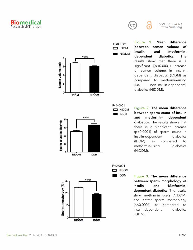
Biomed Res Ther 2017, 4(6): 1388-1399 1392
Figure 1. Mean difference between semen volume of insulin- and metformin- dependent diabetics. Theresults show that there is a significant ((p<0.0001) increase of semen volume in insulin-dependent diabetics (IDDM) as compared to metformin-using (i.e. non-insulin-dependent) diabetics (NIDDM).
Figure 2. The mean difference between sperm count of insulin and metformin- dependent diabetics. The results shows thatthere is a significant increase (p<0.0001) of sperm count in insulin-dependent diabetics (IDDM) as compared to metformin-using diabetics (NIDDM).
Figure 3. The mean difference between sperm morphology of insulin- and Metformin- dependent diabetics. The resultsshow metformin users (NIDDM) had better sperm morphology (p<0.0001) as compared to insulin-dependent diabetics (IDDM).
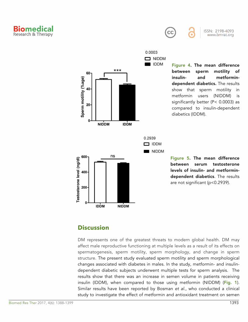
Biomed Res Ther 2017, 4(6): 1388-1399 1393
Figure 4. The mean difference between sperm motility of insulin- and metformin- dependent diabetics. The resultsshow that sperm motility in metformin users (NIDDM) is significantly better (P< 0.0003) as compared to insulin-dependent diabetics (IDDM).
Figure 5. The mean difference between serum testosterone levels of insulin- and metformin- dependent diabetics. The resultsare not significant (p<0.2939).
Discussion
DM represents one of the greatest threats to modern global health. DM may affect male reproductive functioning at multiple levels as a result of its effects on spermatogenesis, sperm motility, sperm morphology, and change in sperm structure. The present study evaluated sperm motility and sperm morphological changes associated with diabetes in males. In the study, metformin- and insulin-dependent diabetic subjects underwent multiple tests for sperm analysis. The results show that there was an increase in semen volume in patients receiving insulin (IDDM), when compared to those using metformin (NIDDM) (Fig. 1).Similar results have been reported by Bosman et al., who conducted a clinical study to investigate the effect of metformin and antioxidant treatment on semen
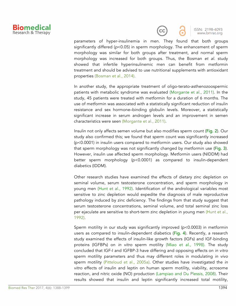
Biomed Res Ther 2017, 4(6): 1388-1399 1394
parameters of hyper-insulinemia in men. They found that both groups significantly differed (p<0.05) in sperm morphology. The enhancement of sperm morphology was similar for both groups after treatment, and normal sperm morphology was increased for both groups. Thus, the Bosman et al. study showed that infertile hyperinsulinemic men can benefit from metformin treatment and should be advised to use nutritional supplements with antioxidant properties (Bosman et al., 2014).
In another study, the appropriate treatment of oligo-terato-asthenozoospermic patients with metabolic syndrome was evaluated (Morgante et al., 2011). In the study, 45 patients were treated with metformin for a duration of 6 months. The use of metformin was associated with a statistically significant reduction of insulin resistance and sex hormone–binding globulin levels. Moreover, a statistically significant increase in serum androgen levels and an improvement in semen characteristics were seen (Morgante et al., 2011).
Insulin not only affects semen volume but also modifies sperm count (Fig. 2). Our study also confirmed this; we found that sperm count was significantly increased (p<0.0001) in insulin users compared to metformin users. Our study also showed that sperm morphology was not significantly changed by metformin use (Fig. 3). However, insulin use affected sperm morphology. Metformin users (NIDDM) had better sperm morphology (p<0.0001) as compared to insulin-dependent diabetics (IDDM).
Other research studies have examined the effects of dietary zinc depletion on seminal volume, serum testosterone concentration, and sperm morphology in young men (Hunt et al., 1992). Identification of the andrological variables most sensitive to zinc depletion would expedite the diagnosis of male reproductive pathology induced by zinc deficiency. The findings from that study suggest that serum testosterone concentrations, seminal volume, and total seminal zinc loss per ejaculate are sensitive to short-term zinc depletion in young men (Hunt et al., 1992).
Sperm motility in our study was significantly improved (p<0.0003) in metformin users as compared to insulin-dependent diabetics (Fig. 4). Recently, a research study examined the effects of insulin-like growth factors (IGFs) and IGF-binding proteins (IGFBPs) on in vitro sperm motility (Miao et al., 1998). The study concluded that IGF-I and IGFBP-3 have differing and opposing effects on in vitro sperm motility parameters and thus may different roles in modulating in vivo sperm motility (Pitteloud et al., 2005a). Other studies have investigated the in vitro effects of insulin and leptin on human sperm motility, viability, acrosome reaction, and nitric oxide (NO) production (Lampiao and Du Plessis, 2008). Their results showed that insulin and leptin significantly increased total motility,
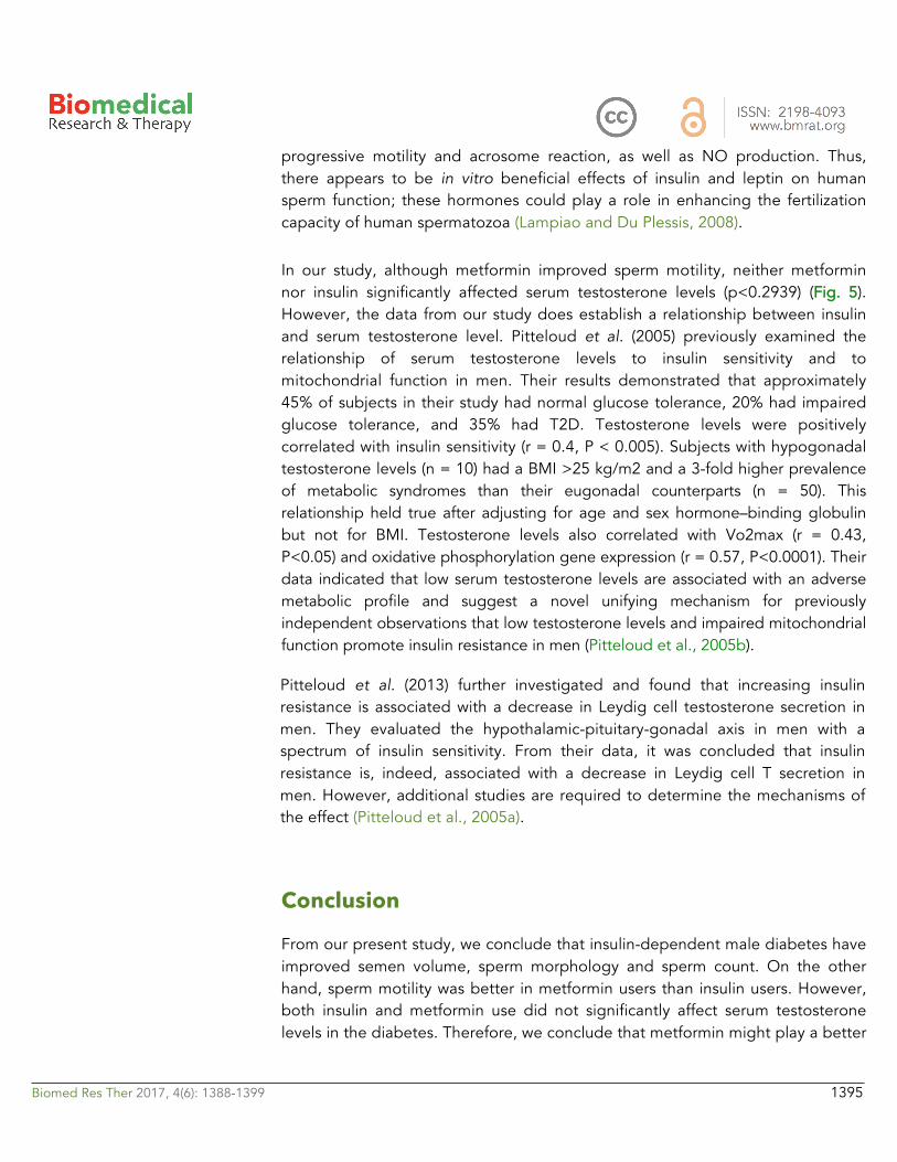
Biomed Res Ther 2017, 4(6): 1388-1399 1395
progressive motility and acrosome reaction, as well as NO production. Thus, there appears to be in vitro beneficial effects of insulin and leptin on human sperm function; these hormones could play a role in enhancing the fertilization capacity of human spermatozoa (Lampiao and Du Plessis, 2008).
In our study, although metformin improved sperm motility, neither metformin nor insulin significantly affected serum testosterone levels (p<0.2939) (Fig. 5). However, the data from our study does establish a relationship between insulin and serum testosterone level. Pitteloud et al. (2005) previously examined the relationship of serum testosterone levels to insulin sensitivity and to mitochondrial function in men. Their results demonstrated that approximately 45% of subjects in their study had normal glucose tolerance, 20% had impaired glucose tolerance, and 35% had T2D. Testosterone levels were positively correlated with insulin sensitivity (r = 0.4, P < 0.005). Subjects with hypogonadal testosterone levels (n = 10) had a BMI >25 kg/m2 and a 3-fold higher prevalence of metabolic syndromes than their eugonadal counterparts (n = 50). This relationship held true after adjusting for age and sex hormone–binding globulin but not for BMI. Testosterone levels also correlated with Vo2max (r = 0.43, P<0.05) and oxidative phosphorylation gene expression (r = 0.57, P<0.0001). Their data indicated that low serum testosterone levels are associated with an adverse metabolic profile and suggest a novel unifying mechanism for previously independent observations that low testosterone levels and impaired mitochondrial function promote insulin resistance in men (Pitteloud et al., 2005b).
Pitteloud et al. (2013) further investigated and found that increasing insulin resistance is associated with a decrease in Leydig cell testosterone secretion in men. They evaluated the hypothalamic-pituitary-gonadal axis in men with a spectrum of insulin sensitivity. From their data, it was concluded that insulin resistance is, indeed, associated with a decrease in Leydig cell T secretion in men. However, additional studies are required to determine the mechanisms of the effect (Pitteloud et al., 2005a).
Conclusion
From our present study, we conclude that insulin-dependent male diabetes have improved semen volume, sperm morphology and sperm count. On the other hand, sperm motility was better in metformin users than insulin users. However, both insulin and metformin use did not significantly affect serum testosterone levels in the diabetes. Therefore, we conclude that metformin might play a better
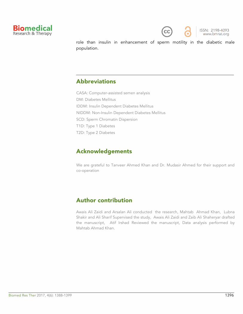
Biomed Res Ther 2017, 4(6): 1388-1399 1396
role than insulin in enhancement of sperm motility in the diabetic male population.
Abbreviations
CASA: Computer-assisted semen analysis
DM: Diabetes Mellitus
IDDM: Insulin Dependent Diabetes Mellitus
NIDDM: Non-Insulin Dependent Diabetes Mellitus
SCD: Sperm Chromatin Dispersion
T1D: Type 1 Diabetes
T2D: Type 2 Diabetes
Acknowledgements
We are grateful to Tanveer Ahmed Khan and Dr. Mudasir Ahmed for their support and co-operation
Author contribution
Awais Ali Zaidi and Arsalan Ali conducted the research, Mahtab Ahmad Khan, Lubna Shakir and Ali Sharif Supervised the study, Awais Ali Zaidi and Zaib Ali Shaheryar drafted the manuscript, Atif Irshad Reviewed the manuscript, Data analysis performed by Mahtab Ahmad Khan.
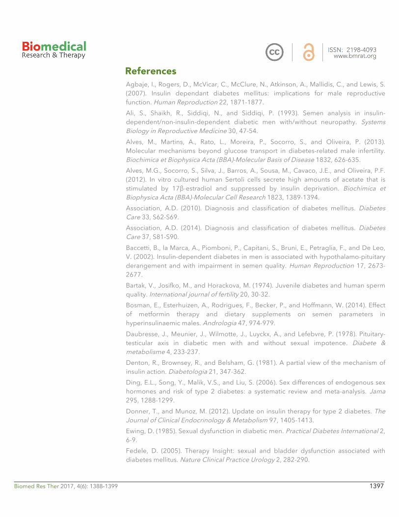
Biomed Res Ther 2017, 4(6): 1388-1399 1397
References Agbaje, I., Rogers, D., McVicar, C., McClure, N., Atkinson, A., Mallidis, C., and Lewis, S. (2007). Insulin dependant diabetes mellitus: implications for male reproductive function. Human Reproduction 22, 1871-1877. Ali, S., Shaikh, R., Siddiqi, N., and Siddiqi, P. (1993). Semen analysis in insulin-dependent/non-insulin-dependent diabetic men with/without neuropathy. Systems Biology in Reproductive Medicine 30, 47-54. Alves, M., Martins, A., Rato, L., Moreira, P., Socorro, S., and Oliveira, P. (2013). Molecular mechanisms beyond glucose transport in diabetes-related male infertility. Biochimica et Biophysica Acta (BBA)-Molecular Basis of Disease 1832, 626-635. Alves, M.G., Socorro, S., Silva, J., Barros, A., Sousa, M., Cavaco, J.E., and Oliveira, P.F. (2012). In vitro cultured human Sertoli cells secrete high amounts of acetate that is stimulated by 17β-estradiol and suppressed by insulin deprivation. Biochimica et Biophysica Acta (BBA)-Molecular Cell Research 1823, 1389-1394. Association, A.D. (2010). Diagnosis and classification of diabetes mellitus. Diabetes Care 33, S62-S69. Association, A.D. (2014). Diagnosis and classification of diabetes mellitus. Diabetes Care 37, S81-S90. Baccetti, B., la Marca, A., Piomboni, P., Capitani, S., Bruni, E., Petraglia, F., and De Leo, V. (2002). Insulin-dependent diabetes in men is associated with hypothalamo-pituitary derangement and with impairment in semen quality. Human Reproduction 17, 2673-2677. Bartak, V., Josifko, M., and Horackova, M. (1974). Juvenile diabetes and human sperm quality. International journal of fertility 20, 30-32. Bosman, E., Esterhuizen, A., Rodrigues, F., Becker, P., and Hoffmann, W. (2014). Effect of metformin therapy and dietary supplements on semen parameters in hyperinsulinaemic males. Andrologia 47, 974-979. Daubresse, J., Meunier, J., Wilmotte, J., Luyckx, A., and Lefebvre, P. (1978). Pituitary-testicular axis in diabetic men with and without sexual impotence. Diabete & metabolisme 4, 233-237. Denton, R., Brownsey, R., and Belsham, G. (1981). A partial view of the mechanism of insulin action. Diabetologia 21, 347-362. Ding, E.L., Song, Y., Malik, V.S., and Liu, S. (2006). Sex differences of endogenous sex hormones and risk of type 2 diabetes: a systematic review and meta-analysis. Jama 295, 1288-1299. Donner, T., and Munoz, M. (2012). Update on insulin therapy for type 2 diabetes. The Journal of Clinical Endocrinology & Metabolism 97, 1405-1413. Ewing, D. (1985). Sexual dysfunction in diabetic men. Practical Diabetes International 2, 6-9. Fedele, D. (2005). Therapy Insight: sexual and bladder dysfunction associated with diabetes mellitus. Nature Clinical Practice Urology 2, 282-290.
Biomed Res Ther 2017, 4(6): 1388-1399 1398
Hunt, C.D., Johnson, P.E., Herbel, J., and Mullen, L.K. (1992). Effects of dietary zinc depletion on seminal volume and zinc loss, serum testosterone concentrations, and sperm morphology in young men. The American journal of clinical nutrition 56, 148-157. Lampiao, F., and Du Plessis, S.S. (2008). Insulin and leptin enhance human sperm motility, acrosome reaction and nitric oxide production. Asian journal of andrology 10, 799-807. Lutz, W. (2006). Fertility rates and future population trends: will Europe's birth rate recover or continue to decline? International Journal of Andrology 29, 25-33. McBrien, K., Rabi, D.M., Campbell, N., Barnieh, L., Clement, F., Hemmelgarn, B.R., Tonelli, M., Leiter, L.A., Klarenbach, S.W., and Manns, B.J. (2012). Intensive and standard blood pressure targets in patients with type 2 diabetes mellitus: systematic review and meta-analysis. Archives of internal medicine 172, 1296-1303. Miao, Z.R., Lin, T.K., Bongso, T., Zhou, X., Cohen, P., and Lee, K.O. (1998). Effect of insulin‐like growth factors (IGFs) and IGF‐binding proteins on in vitro sperm motility. Clinical endocrinology 49, 235-239. Morgante, G., Tosti, C., Orvieto, R., Musacchio, M.C., Piomboni, P., and De Leo, V. (2011). Metformin improves semen characteristics of oligo-terato-asthenozoospermic men with metabolic syndrome. Fertility and sterility 95, 2150-2152. Niven, M., Hitman, G., and Badenoch, D. (1995). A study of spermatozoal motility in type 1 diabetes mellitus. Diabetic medicine 12, 921-924. O'Donnell, L., Stanton, P., and de Kretser, D.M. (2017). Endocrinology of the Male Reproductive System and Spermatogenesis. In Endotext, C.G. De Groot LJ, Dungan K, et al., ed. (South Dartmouth (MA)). Organization, W.H. (2002). Diabetes: the cost of diabetes (Fact sheet No. 236). World health organization (WHO) www who intlmediacentre/factsheets/fs236/en. Pitteloud, N., Hardin, M., Dwyer, A.A., Valassi, E., Yialamas, M., Elahi, D., and Hayes, F.J. (2005a). Increasing insulin resistance is associated with a decrease in Leydig cell testosterone secretion in men. The Journal of Clinical Endocrinology & Metabolism 90, 2636-2641. Pitteloud, N., Mootha, V.K., Dwyer, A.A., Hardin, M., Lee, H., Eriksson, K.-F., Tripathy, D., Yialamas, M., Groop, L., and Elahi, D. (2005b). Relationship between testosterone levels, insulin sensitivity, and mitochondrial function in men. Diabetes Care 28, 1636-1642. Ranganathan, P., Mahran, A.M., Hallak, J., and Agarwal, A. (2002). Sperm cryopreservation for men with nonmalignant, systemic diseases: a descriptive study. Journal of andrology 23, 71-75. Robinson, R., and Fritz, I.B. (1981). Metabolism of glucose by Sertoli cells in culture. Biology of reproduction 24, 1032-1041. Sexton, W.J., and Jarow, J.P. (1997). Effect of diabetes mellitus upon male reproductive function. Urology 49, 508-513. Silink, M. (2002). Childhood diabetes: a global perspective. Hormone Research in Paediatrics 57, 1-5.

Biomed Res Ther 2017, 4(6): 1388-1399 1399
Stumvoll, M., Nurjhan, N., Perriello, G., Dailey, G., and Gerich, J.E. (1995). Metabolic effects of metformin in non-insulin-dependent diabetes mellitus. New England Journal of Medicine 333, 550-554. Torgerson, J.S., Hauptman, J., Boldrin, M.N., and Sjöström, L. (2004). XENical in the Prevention of Diabetes in Obese Subjects (XENDOS) Study A randomized study of orlistat as an adjunct to lifestyle changes for the prevention of type 2 diabetes in obese patients. Diabetes Care 27, 155-161. WHO (1999). WHO laboratory manual for the examination of human semen and sperm-cervical mucus interaction (Cambridge university press). Zysk, J., Bushway, A., Whistler, R., and Carlton, W. (1975). Temporary sterility produced in male mice by 5-thio-D-glucose. Journal of reproduction and fertility 45, 69-72.
ISSN: 2198-4093 www.bmrat.org
*For correspondence:
Competing interests: The authorsdeclare that no competing interests exist.
Received: 06 March 2017Accepted: 04 June 2017Published: 27 June 2017
Copyright The Author(s) 2017. This article is published with open access by BioMedPress (BMP).
This article is distributed under the terms of the Creative Commons Attribution License (CC-BY 4.0) which permits any use, distribution, and reproduction in any medium, provided the original author(s) and the source are credited.
Original Research
Estimates of global HIV/AIDS mortality, prevalence and incidence rates, and their association with the Human Development Index
Kamyar Mansori1,2, Erfan Ayubi3, Fatemeh Khosravi Shadmani4, Shiva Mansouri Hanis5, Somayeh Khazaei6, Mohadeseh Sani7, Yousef Moradi8, Salman Khazaei9,10,*, Abolfazl Mohammadbeigi11
1Social Development & Health Promotion Research Center, Gonabad University of Medical Sciences, Gonabad, Iran 2Department of Epidemiology, School of Public Health, Iran University of Medical Sciences, Tehran, Iran 3Department of Epidemiology, School of Public Health, Shahid Beheshti University of Medical Sciences, Tehran, Iran 4Modeling in Health Research Center , Institute for Futures Studies in Health, Kerman University of Medical Sciences, Kerman, Iran 5School of Public Health, Dezful University of Medical Sciences, Dezful, Iran 6Rafsanjan University of Medical Sciences, Rafsanjan, Iran 7School of Medicine, Zabol University of Medical Sciences, Zabol, Iran 8Pars Advanced and Minimally Invasive Manners Research Center, Pars Hospital, Iran University of Medical Science, Tehran, Iran 9Department of Epidemiology, School of Public Health, Hamadan University of Medical Sciences, Hamadan, Iran 10Department of Epidemiology and Biostatistics, School of Public Health, Tehran University of Medical Sciences, Tehran, Iran 11Research Center of Gastroenterology and Hepatology, Qom University of Medical Sciences, Qom, Iran
Abstract
Background: HIV/AIDS is one of greatest global public health concerns today due to thehigh incidence, prevalence and mortality rates. The aim of this research was investigate and estimate the global HIV/AIDS mortality, prevalence and incidence rates, and explore their associations with the Human Development Index.
!Biomed Res Ther 2017, 4(6): 1400-1410 !1400DOI: 10.15419/bmrat.v4i06.181
�ISSN: 2198-4093
www.bmrat.org Methods: The global age-standardized rates of mortality, prevalence and incidence of HIV/AIDS were obtained from the UNAIDS for different countries in 2015. The human development indexes (HDIs) were obtained from the World Bank database. The surveyed countries were divided into four groups according to the HDI distribution. The Spearman correlation coefficient and one-way ANOVA test were used for assessing the association of HIV/ AIDS indicators and HDI. Results: The highest rates of HIV/AIDS prevalence and incidence, and associated mortality in East and Southern Africa countries were 51.73%, 46.33% and 42.3%, respectively. Moreover, the highest and lowest global age-standardized rates of incidence and prevalence of HIV/AIDS was seen in adults ranging from 15-49 years of age for both low and high HDI countries. The prevalence and incidence rates of HIV/AIDS each had an inverse correlation with HDI and its four indicators (life expectancy at birth, mean years of schooling, expected years of schooling, and GNI per capita). Conclusion: Less developed countries with lower HDI show greater severity of the AIDS epidemic. Thus, it is essential to pay more attention to HIV/AIDS control and prevention programs in these countries.
Keywords HDI, HIV/AIDS, Human development index, Incidence, Mortality, Prevalence
Introduction Sustainable Development Goals (SDGs) is an UN plan for achieving a better future for all people over the next 15 years (2015-2030). It consists of 17 goals and 169 targets. Among the goals, the third one to to ensure healthy lives and promote well-being for all ages. With regard to this, one of the critical targets is to end the epidemics of AIDS by the year 2030.
UNAIDS has estimated that 36.7 million people globally are living with HIV. In 2015, approximately 2.1 million people became newly infected with HIV and 1.1 million people died from AIDS-related illnesses (UNADIS, 2016). HIV/AIDS and the related illnesses represented 2.44 % of global deaths (Institute for Health Metrics and Evaluation (IHME), 2015). HIV/AIDS is the 5th leading cause of DALY in recent years (Murray et al., 2013). However, the burden of epidemic HIV/AIDS varies considerably in different regions and geographical divisions (WHO, 2016).
The difference among countries depends on various factors. The most notable factors or influences are wealth, education, poverty, immigration, addiction, and sexually transmitting diseases (Piot et al., 2007; Yancy et al., 2017). The Human Development Index (HDI) is associated with the development of societies; it is composed of the mean scores of the three indexes of life expectancy at the time of birth, gross national product (GNP) per capita, and education. Some of the
!Biomed Res Ther 2017, 4(6): 1400-1410 !1401
�ISSN: 2198-4093
www.bmrat.org studies have reported that HDI affects HIV (Boulogne et al., 2017; Boutayeb, 2009; Colecraft, 2008; de Freitas Souza et al., 2017).
This present study aims to investigate the correlation of HIV related death incidence and HDI to understand how HIV/AIDS affect global development. Understanding this helps provide a basis for more purposeful medical and preventive measures which may be implemented.
Materials-Methods
The present study is an epidemiological study which utilized the global dataset of HIV/AIDS (this global dataset includes incidence, prevalence and mortality rates of HIV/AIDS worldwide).
Global HIV/AIDS data
The global age-standardized rates of mortality, prevalence and incidence of HIV/AIDS were obtained from the UNAIDS for different countries in 2015 (UNAIDS, 2016a). In fact, UNAIDS produces a biannual estimation of these indicators for HIV/AIDS for all countries (UNAIDS, 2016b).
HDI
The HDI data and its components, including life expectancy at birth, mean years of schooling, expected years of schooling and gross national income (GNI) per capita, were taken from the World Bank database from 2015 (Ledwidge et al., 2013).The HDI index has a range between 0 to 1; values closer to 1 reflect a higher HDI. In general, countries in the database are divided into four categories according to the value of HDI. The categories are as follows: very high (HDI≥0.8), high (0.7≤HDI≤0.799), medium (0.55≤HDI≤0.699), and low (HDI<0.55) (Ledwidge et al., 2013).
Statistical analysis
Descriptive statistics such as charts and tables were used to present data. Continual assessment of changes in trend of global incidence, prevalence and mortality of HIV/HIDS occurred during the time period of 2000-2015. The Cochran–Armitage test was used. Comparisons among HDI- categorized countries (very high, high, medium, and low) for HIV/AIDS incidence and prevalence was done with one-way ANOVA. Spearman correlation coefficient was used to evaluate the relationship between the prevalence of HIV/AIDS and incidence to the national HDI.
The significant level was set as p<0.05. Data were analyzed using Stata computer software version 12 (StataCorp, College Station, TX, USA).
!Biomed Res Ther 2017, 4(6): 1400-1410 !1402
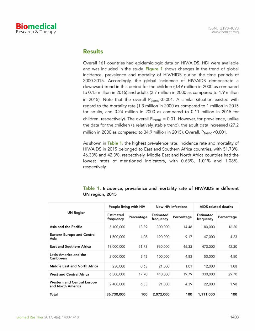
�ISSN: 2198-4093
www.bmrat.org
Results
Overall 161 countries had epidemiologic data on HIV/AIDS. HDI were available and was included in the study. Figure 1 shows changes in the trend of global incidence, prevalence and mortality of HIV/HIDS during the time periods of 2000-2015. Accordingly, the global incidence of HIV/AIDS demonstrate a downward trend in this period for the children (0.49 million in 2000 as compared to 0.15 million in 2015) and adults (2.7 million in 2000 as compared to 1.9 million
in 2015). Note that the overall Ptrend<0.001. A similar situation existed with regard to the mortality rate (1.3 million in 2000 as compared to 1 million in 2015 for adults, and 0.24 million in 2000 as compared to 0.11 million in 2015 for
children, respectively). The overall Ptrend = 0.01. However, for prevalence, unlike the data for the children (a relatively stable trend), the adult data increased (27.2
million in 2000 as compared to 34.9 million in 2015). Overall. Ptrend<0.001.
As shown in Table 1, the highest prevalence rate, incidence rate and mortality of HIV/AIDS in 2015 belonged to East and Southern Africa countries, with 51.73%, 46.33% and 42.3%, respectively. Middle East and North Africa countries had the lowest rates of mentioned indicators, with 0.63%, 1.01% and 1.08%, respectively.
Table 1. Incidence, prevalence and mortality rate of HIV/AIDS in different UN region, 2015
UN RegionPeople living with HIV New HIV infections AIDS-related deaths
Estimated frequency Percentage Estimated
frequency Percentage Estimated frequency Percentage
Asia and the Pacific 5,100,000 13.89 300,000 14.48 180,000 16.20
Eastern Europe and Central Asia 1,500,000 4.08 190,000 9.17 47,000 4.23
East and Southern Africa 19,000,000 51.73 960,000 46.33 470,000 42.30
Latin America and the Caribbean 2,000,000 5.45 100,000 4.83 50,000 4.50
Middle East and North Africa 230,000 0.63 21,000 1.01 12,000 1.08
West and Central Africa 6,500,000 17.70 410,000 19.79 330,000 29.70
Western and Central Europe and North America 2,400,000 6.53 91,000 4.39 22,000 1.98
Total 36,730,000 100 2,072,000 100 1,111,000 100
!Biomed Res Ther 2017, 4(6): 1400-1410 !1403
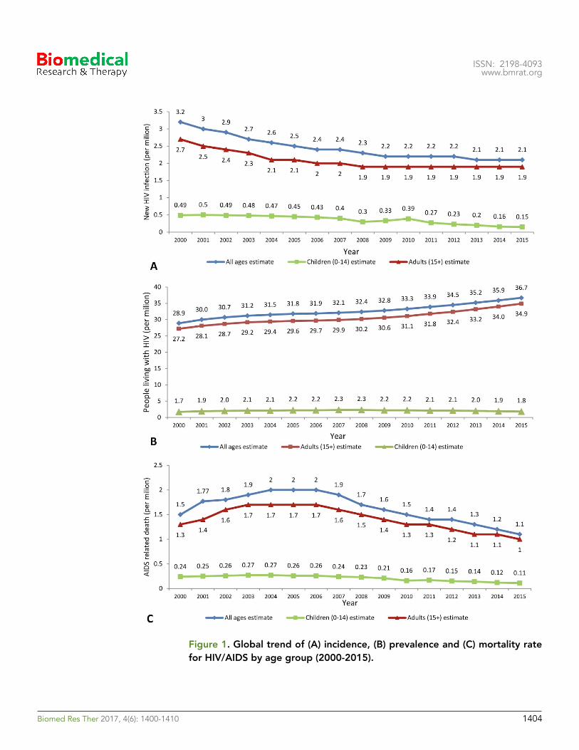
�ISSN: 2198-4093
www.bmrat.org
Figure 1. Global trend of (A) incidence, (B) prevalence and (C) mortality rate for HIV/AIDS by age group (2000-2015).
!Biomed Res Ther 2017, 4(6): 1400-1410 !1404
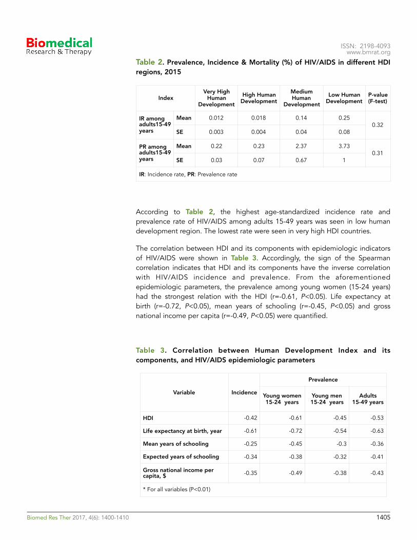
�ISSN: 2198-4093
www.bmrat.org Table 2. Prevalence, Incidence & Mortality (%) of HIV/AIDS in different HDI regions, 2015
According to Table 2, the highest age-standardized incidence rate and prevalence rate of HIV/AIDS among adults 15-49 years was seen in low human development region. The lowest rate were seen in very high HDI countries.
The correlation between HDI and its components with epidemiologic indicators of HIV/AIDS were shown in Table 3. Accordingly, the sign of the Spearman correlation indicates that HDI and its components have the inverse correlation with HIV/AIDS incidence and prevalence. From the aforementioned epidemiologic parameters, the prevalence among young women (15-24 years) had the strongest relation with the HDI (r=-0.61, P<0.05). Life expectancy at birth (r=-0.72, P<0.05), mean years of schooling (r=-0.45, P<0.05) and gross national income per capita (r=-0.49, P<0.05) were quantified.
Table 3. Correlation between Human Development Index and its components, and HIV/AIDS epidemiologic parameters
IndexVery High
Human Development
High Human Development
Medium Human
DevelopmentLow Human
DevelopmentP-value (F-test)
IR among adults15-49 years
Mean 0.012 0.018 0.14 0.250.32
SE 0.003 0.004 0.04 0.08
PR among adults15-49 years
Mean 0.22 0.23 2.37 3.730.31
SE 0.03 0.07 0.67 1
IR: Incidence rate, PR: Prevalence rate
Variable Incidence
Prevalence
Young women 15-24 years
Young men 15-24 years
Adults 15-49 years
HDI -0.42 -0.61 -0.45 -0.53
Life expectancy at birth, year -0.61 -0.72 -0.54 -0.63
Mean years of schooling -0.25 -0.45 -0.3 -0.36
Expected years of schooling -0.34 -0.38 -0.32 -0.41
Gross national income per capita, $ -0.35 -0.49 -0.38 -0.43
* For all variables (P<0.01)
!Biomed Res Ther 2017, 4(6): 1400-1410 !1405
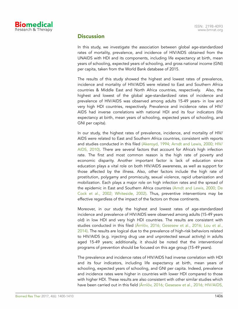
�ISSN: 2198-4093
www.bmrat.org Discussion
In this study, we investigate the association between global age-standardized rates of mortality, prevalence, and incidence of HIV/AIDS obtained from the UNAIDS with HDI and its components, including life expectancy at birth, mean years of schooling, expected years of schooling, and gross national income (GNI) per capita, taken from the World Bank database of 2015.
The results of this study showed the highest and lowest rates of prevalence, incidence and mortality of HIV/AIDS were related to East and Southern Africa countries & Middle East and North Africa countries, respectively. Also, the highest and lowest of the global age-standardized rates of incidence and prevalence of HIV/AIDS was observed among adults 15-49 years- in low and very high HDI countries, respectively. Prevalence and incidence rates of HIV/AIDS had inverse correlations with national HDI and its four indicators (life expectancy at birth, mean years of schooling, expected years of schooling, and GNI per capita).
In our study, the highest rates of prevalence, incidence, and mortality of HIV/AIDS were related to East and Southern Africa countries, consistent with reports and studies conducted in this filed (Akeroyd, 1994; Arndt and Lewis, 2000; HIV/AIDS, 2010). There are several factors that account for Africa’s high infection rate. The first and most common reason is the high rate of poverty and economic disparity. Another important factor is lack of education since education plays a vital role on both HIV/AIDS awareness, as well as support for those affected by the illness. Also, other factors include the high rate of prostitution, polygamy and promiscuity, sexual violence, rapid urbanization and mobilization. Each plays a major role on high infection rates and the spread of the epidemic in East and Southern Africa countries (Arndt and Lewis, 2000; De Cock et al., 2002; Whiteside, 2002). Thus, preventive interventions may be effective regardless of the impact of the factors on those continents.
Moreover, in our study the highest and lowest rates of age-standardized incidence and prevalence of HIV/AIDS were observed among adults (15-49 years old) in low HDI and very high HDI countries. The results are consistent with studies conducted in this filed (Ärnlöv, 2016; Gesesew et al., 2016; Lou et al., 2014). The results are logical due to the prevalence of high-risk behaviors related to HIV/AIDS (e.g. injecting drug use and unprotected sexual activity) in adults aged 15-49 years; additionally, it should be noted that the interventional programs of prevention should be focused on this age group (15-49 years).
The prevalence and incidence rates of HIV/AIDS had inverse correlation with HDI and its four indicators, including life expectancy at birth, mean years of schooling, expected years of schooling, and GNI per capita. Indeed, prevalence and incidence rates were higher in countries with lower HDI compared to those with higher HDI. These results are also consistent with other similar studies which have been carried out in this field (Ärnlöv, 2016; Gesesew et al., 2016; HIV/AIDS,
!Biomed Res Ther 2017, 4(6): 1400-1410 !1406
�ISSN: 2198-4093
www.bmrat.org 2010; Lou et al., 2014). Many studies have shown that HIV/AIDS has a significant effect on health and that good indicators include mortality rates and life expectancy. For example, two studies in the field of global inequality of life expectancy have shown that the 6 years of the difference in life expectancy between Africa and North America is related to AIDS (Dorling D; Wong et al., 2017).
Moreover, in this study the mean years of schooling had an inverse correlation with prevalence and incidence rates of HIV/AIDS. Education is an important component of HDI and it is necessary to include that parameter, especially in countries with low- and medium-income. In most African countries, HIV/AIDS has shown an inverse association with primary education, especially in girls (Wong et al., 2017). In these countries the death of parents due to AIDS is frequent; the issue of family economic situations will aggravate and also will increase the number of children dismissed from school (Boulogne et al., 2017; Lou et al., 2014). On the other hand, an important factor that has a major role in prevention of HIV transmission is mean years of schooling. This factor is essential in interventional programs of prevention.
Various studies have shown that HIV/AIDS is a disease of poverty. More than 60% of people living with HIV/AIDS in the world have been deployed in sub-Saharan Africa. Indeed, the countries of this region have low economic growth and cannot pay the cost of high antiretroviral therapy nor prevention programs (Billebeau et al., 2017). Hence, poverty makes people susceptible to HIV/AIDS. Also, in this study the negative correlation between GNI per capita and HIV/AIDS prevalence, and incidences rates were verified.
Conclusion
The results of our study revealed an inverse relationship between the global age-standardized rates of mortality, prevalence, and incidence of HIV/AIDS with its components including life expectancy, years of schooling, and GNI per capita. Less developed countries, as measured by HDI. In other words, the less developed countries with lower HDI have greater severity of the AIDS epidemic. Therefore, it is essential to pay greater attention to control and prevention programs for HIV/AIDS in these countries.
Abbreviations
GNI: Gross National Income GNP: Gross National Product HDI: Human Development Index
!Biomed Res Ther 2017, 4(6): 1400-1410 !1407

�ISSN: 2198-4093
www.bmrat.org SDGs: Sustainable Development Goals UN: United Nation UNAIDS: United Nations Program on HIV/AIDS
Acknowledgements
We would like to thank UNAIDS and World Bank database for provide data.
Author contribution
FKS, EA and SK designed the study. KM, SMH and SK processed the data. EA, MS and KM performed the statistical analysis. FKS, SK and KM interpreted the results. FKS, KM, SK and EA wrote the first draft. FKS, SK and EA revised the final draft. All authors read and approved the final manuscript.
!Biomed Res Ther 2017, 4(6): 1400-1410 !1408
�ISSN: 2198-4093
www.bmrat.org
References Akeroyd, A.V. (1994). HIV/AIDS in eastern and southern Africa. Review of African Political Economy 21, 173-184. Arndt, C., and Lewis, J.D. (2000). The macro implications of HIV/AIDS in South Africa: a preliminary assessment. South African Journal of Economics 68, 380-392. Ärnlöv, J. (2016). Estimates of global, regional, and national incidence, prevalence, and mortality of HIV, 1980-2015: the Global Burden of Disease Study 2015. The Lancet HIV 3, E361-E387. Billebeau, G., Vodovar, N., Sadoune, M., Launay, J.M., Beauvais, F., and Cohen-Solal, A. (2017). Effects of a cardiac rehabilitation programme on plasma cardiac biomarkers in patients with chronic heart failure. European journal of preventive cardiology, 2047487317705488. Boulogne, M., Sadoune, M., Launay, J., Baudet, M., Cohen-Solal, A., and Logeart, D. (2017). Inflammation versus mechanical stretch biomarkers over time in acutely decompensated heart failure with reduced ejection fraction. International Journal of Cardiology 226, 53-59. Boutayeb, A. (2009). The impact of HIV/AIDS on human development in African countries. BMC Public Health 9, S3. Colecraft, E. (2008). HIV/AIDS: nutritional implications and impact on human development. Proceedings of the Nutrition Society 67, 109-113. De Cock, K.M., Mbori-Ngacha, D., and Marum, E. (2002). Shadow on the continent: public health and HIV/AIDS in Africa in the 21st century. The Lancet 360, 67-72. de Freitas Souza, B.S., Silva, D.N., Carvalho, R.H., de Almeida Sampaio, G.L., Paredes, B.D., França, L.A., Azevedo, C.M., Vasconcelos, J.F., Meira, C.S., and Neto, P.C. (2017). Association of Cardiac Galectin-3 Expression, Myocarditis, and Fibrosis in Chronic Chagas Disease Cardiomyopathy. The American Journal of Pathology 187, 1134-1146. Dorling D, S.M., Smith GD HIV and global health: global inequality of life expectancy due to AIDS. BMJ: British Medical Journal 2006;332:662-4 . Gesesew, H.A., Mwanri, L., Ward, P., Woldemicahel, K., and Feyissa, G.T. (2016). Factors associated with discontinuation of anti-retroviral therapy among adults living with HIV/AIDS in Ethiopia: a systematic review protocol. JBI Database of Systematic Reviews and Implementation Reports 14, 26-37. HIV/AIDS, J.U.N.P.o. (2010). Global report: UNAIDS report on the global AIDS epidemic 2010 (UNAIDS). Institute for Health Metrics and Evaluation (IHME) (2015). GBD Compare. Seattle, WA: IHME, University of Washington, 2015. Ledwidge, M., Gallagher, J., Conlon, C., Tallon, E., O’Connell, E., Dawkins, I., Watson, C., O’Hanlon, R., Bermingham, M., and Patle, A. (2013). Natriuretic peptide–based screening and collaborative care for heart failure: the STOP-HF randomized trial. Jama 310, 66-74. Lou, L.-X., Chen, Y., Yu, C.-H., Li, Y.-M., and Ye, J. (2014). National HIV/AIDS mortality, prevalence, and incidence rates are associated with the Human Development Index. American journal of infection control 42, 1044-1048.
!Biomed Res Ther 2017, 4(6): 1400-1410 !1409
�ISSN: 2198-4093
www.bmrat.org Murray, C.J., Vos, T., Lozano, R., Naghavi, M., Flaxman, A.D., Michaud, C., Ezzati, M., Shibuya, K., Salomon, J.A., and Abdalla, S. (2013). Disability-adjusted life years (DALYs) for 291 diseases and injuries in 21 regions, 1990–2010: a systematic analysis for the Global Burden of Disease Study 2010. The lancet 380, 2197-2223. Piot, P., Greener, R., and Russell, S. (2007). Squaring the circle: AIDS, poverty, and human development. PLoS Med 4, e314. UNADIS (2016). Fact sheet 2016 (http://www.unaids.org/en/resources/fact-sheet). UNAIDS (2016a). Aids information: Data (http://aidsinfo.unaids.org/). UNAIDS (2016b). How AIDS changed everything — MDG6: 15 years, 15 lessons of hope from the AIDS response (http://www.unaids.org/en/resources/documents/2015/MDG6_15years-15lessonsfromtheAIDSresponse). Whiteside, A. (2002). Poverty and HIV/AIDS in Africa. Third world quarterly 23, 313-332. WHO (2016). Global Health Observatory (GHO) data(HIV/AIDS) (http://dx.doi.org/10.1080/01436590220126667). Wong, P.C., Guo, J., and Zhang, A. (2017). The renal and cardiovascular effects of natriuretic peptides. Advances in physiology education 41, 179-185. Yancy, C.W., Jessup, M., Bozkurt, B., Butler, J., Casey, D.E., Jr., Colvin, M.M., Drazner, M.H., Filippatos, G.S., Fonarow, G.C., Givertz, M.M., et al. (2017). 2017 ACC/AHA/HFSA Focused Update of the 2013 ACCF/AHA Guideline for the Management of Heart Failure: A Report of the American College of Cardiology/American Heart Association Task Force on Clinical Practice Guidelines and the Heart Failure Society of America. Journal of cardiac failure, 10.1016/j.cardfail.2017.1004.1014.
!Biomed Res Ther 2017, 4(6): 1400-1410 !1410
ISSN: 2198-4093 www.bmrat.org
*For correspondence:
Competing interests: The authorsdeclare that no competing interests exist.
Received: 23 May 2017Accepted: 21 June 2017Published: 28 June 2017
Copyright The Author(s) 2017. This article is published with open access by BioMedPress (BMP).
This article is distributed under the terms of the Creative Commons Attribution License (CC-BY 4.0) which permits any use, distribution, and reproduction in any medium, provided the original author(s) and the source are credited.
Original Research
Determinants of placenta previa: a case-control study
Fatemeh Shobeiri1, Ensiyeh Jenabi2,*, Manoochehr Karami3, Simin Karimi4
1Maternal and Child Care Research Center, Hamadan University of Medical Sciences, Hamadan, Iran 2Student Research Committee, Hamadan University of Medical Sciences, Hamadan, Iran 3Social Determinates of Health Research Center, Hamadan University of Medical Sciences, Hamadan, Iran 4Department of Midwifery, Medical Faculty, Arak Branch, Islamic Azad University, Arak, Iran
Abstract
Background: The risk factors of placenta previa differ around the world. This studyevaluated risk factors of pregnancies complicated with placenta previa during a 5-year period in a referral center in Hamadan, Iran. Methods: This case control study wasconducted in Hamadan city (Hamadan Province of Iran) from April 2013 to March 2017. The cases were women whose deliveries were complicated by placenta previa and the controls were those who delivered without placenta previa. We recruited 130 cases and 130 controls. Multivariate unconditional logistic regression analysis was conducted, and odds ratios (ORs) and 95% confidence intervals (CIs) were calculated. Results: The OR ofplacenta previa was 4.08 (95% CI= 1.44, 11.58) by maternal age, 4.08 (95% CI =1.44, 11.58) by preterm labor, and 6.64 (95% CI =1.09, 40.45) by prior operations of the uterine cavity, compared to normal deliveries and after adjusting for other variables. Multiparity, prior spontaneous abortions, and prior cesarean sections were not statistically significant risk factors for placenta previa, when adjusted for other variables. Conclusion: Our study suggests that high maternal age and prior operations of theuterine cavity are risk factors for placenta previa.
Keywords Age, Iran, Placenta previa, Prior cesarean section, Prior operations on uterine, Prior spontaneous abortion
!Biomed Res Ther 2017, 4(6): 1411-1419 1411DOI: 10.15419/bmrat.v4i06.182
ISSN: 2198-4093 www.bmrat.org
Introduction
Placenta previa occurs when the placenta becomes implanted in the lower uterine segment or near the internal cervical os (opening into the uterus from the cervix). It takes place in less than 0.5% of all pregnancies. Placenta previa is correlated with morbidity and mortality of both mother and neonate (Saleh Gargari et al., 2016). Placenta previa is created by invasion of placental villi beyond the decidua basalis which causes the placenta accreta or increta to form (Miller et al., 1997).
Based on recent studies, several factors have been reported to contribute to placenta previa, including cesarean sections, smoking, abortions, assisted reproductive techniques (ART), and high-aged pregnancies (i.e. women of ages 35-45) (Gibbins et al., 2017; Karami et al., 2017; Rasmussen et al., 2000; Shobeiri and Jenabi, 2017; Usta et al., 2005).
In 2003 a study in Croatia found that the risk factors for placenta previa to be: advanced age of mother (over 34 years of age), three or more previous pregnancies, parity of 2 and higher, high number of previous abortions, and history of previous cesarean sections (Tuzovic et al., 2003). Additionally, a study in Austria in 2016 showed that risk factors and maternal outcomes were not related to the classification of placenta previa (major and minor placenta previa) (Kollmann et al., 2016). Moreover, Kashani et al. in the north of Iran showed that placenta previa is clearly associated with prior cesarean sections (Kashani et al., 2011).
Overall, the risk factor factors of placenta previa differ around the world (Tuzovic et al., 2003) and studies of this topic have been limited in general. To our knowledge, no reports in this field have been published from the west side of Iran. In this study, we aimed to evaluate determinates of pregnancies complicated with placenta previa in Iran.
Materials-MethodsThis case-control study was conducted in women who experienced childbirth complicated with placenta previa. Controls included women who had childbirth without complication at the Women's Hospital (Fatemieh, Hamadan City), Hamadan University of Medical Sciences, located in Western Iran from April 7 2013 to March 2017. This study was approved by the Student Research Committee of Hamadan University of Medical Sciences.
The inclusion criteria for the cases were:
(1) single pregnancy,
!Biomed Res Ther 2017, 4(6): 1411-1419 !1412
ISSN: 2198-4093 www.bmrat.org
(2) no physical or mental diseases, and
(3) confirmation of placenta previa by sonographic imaging.
The exclusion criteria was women that had pregnancy failures or complications, according to hospital data record. Women in this study only had single pregnancies since twin pregnancies may intensify the effect of the risk factors (Ananth et al., 2003).
Diagnosis of placenta previa was identified by transabdominal ultra sonographic imaging as conducted by the physician. The control subjects were matched to the cases by childbirth delivery method and by area of residence (rural or urban) since other variables could be considered as risk factors.
The sample size was based on results of a study conducted by Sohrabi et al. (Sohrabi et al.). In the study, the proportion of control exposure was 49%, the proportion of cases with exposure was 78.5%; a two-sided type I error of 5 percent and 80 percent statistical power estimated a minimum of 41 for each group of case and control. For our study, during the 5-year period, 130 cases and 130 controls were selected. We used data records from the Fatemieh Hospital. Data were collected by a checklist, which included data on maternal age, parity, the area of residence, preterm labor (defined as a gestational age of less than 37 completed weeks), prior operations on the uterine cavity, prior spontaneous abortion, prior cesarean section, and prior ART. The validity and reliability of the checklist were assessed.
The logistic regression analysis was conducted to control the effect of various risk factors on placenta previa. Crude and adjusted odd ratios (ORS) were calculated to determine the association between placenta previa and risk factors by applying a significance level of 0.05 using the SPSS Statistics Software (V16.0, IBM Analytics Software, Chicago, IL).
Results
During the study period, 130 cases of placenta previa were confirmed. The mean (and standard deviation) of the maternal age of case and control groups was 28.90(±6.24) and 25.03(±5.97), respectively (p <0.1).
The characteristics of cases and controls are shown in Table 1. Additionally,Table 2 shows the results of simple and multiple logistic regression analyses of the predictors of placenta previa. Adjustments (in multiple logistic regression analysis) were made for the following variables: age, parity, prior spontaneous abortions, prior ART, prior cesarean sections, gestational age, and prior operations on the uterine cavity.
!Biomed Res Ther 2017, 4(6): 1411-1419 1413
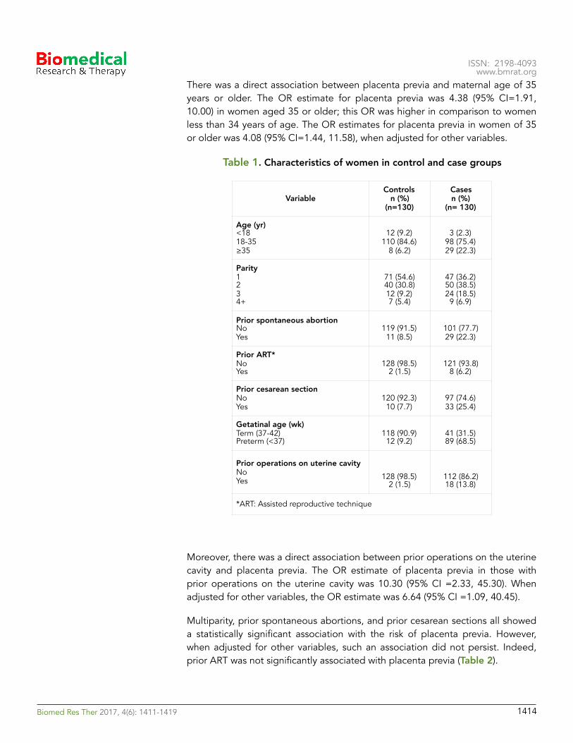
ISSN: 2198-4093 www.bmrat.org
There was a direct association between placenta previa and maternal age of 35 years or older. The OR estimate for placenta previa was 4.38 (95% CI=1.91, 10.00) in women aged 35 or older; this OR was higher in comparison to women less than 34 years of age. The OR estimates for placenta previa in women of 35 or older was 4.08 (95% CI=1.44, 11.58), when adjusted for other variables.
Table 1. Characteristics of women in control and case groups
Moreover, there was a direct association between prior operations on the uterine cavity and placenta previa. The OR estimate of placenta previa in those with prior operations on the uterine cavity was 10.30 (95% CI =2.33, 45.30). When adjusted for other variables, the OR estimate was 6.64 (95% CI =1.09, 40.45).
Multiparity, prior spontaneous abortions, and prior cesarean sections all showed a statistically significant association with the risk of placenta previa. However, when adjusted for other variables, such an association did not persist. Indeed, prior ART was not significantly associated with placenta previa (Table 2).
VariableControls
n (%) (n=130)
Cases n (%)
(n= 130)
Age (yr) <18 18-35 ≥35
12 (9.2) 110 (84.6)
8 (6.2)
3 (2.3) 98 (75.4) 29 (22.3)
Parity 1 2 3 4+
71 (54.6) 40 (30.8) 12 (9.2) 7 (5.4)
47 (36.2) 50 (38.5) 24 (18.5) 9 (6.9)
Prior spontaneous abortion No Yes
119 (91.5) 11 (8.5)
101 (77.7) 29 (22.3)
Prior ART* No Yes
128 (98.5) 2 (1.5)
121 (93.8) 8 (6.2)
Prior cesarean section No Yes
120 (92.3) 10 (7.7)
97 (74.6) 33 (25.4)
Getatinal age (wk) Term (37-42) Preterm (<37)
118 (90.9) 12 (9.2)
41 (31.5) 89 (68.5)
Prior operations on uterine cavity No Yes 128 (98.5)
2 (1.5)112 (86.2) 18 (13.8)
*ART: Assisted reproductive technique
!Biomed Res Ther 2017, 4(6): 1411-1419 1414
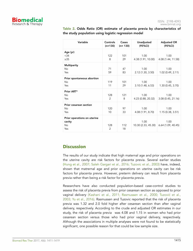
ISSN: 2198-4093 www.bmrat.org
Table 2. Odds Ratio (OR) estimate of placenta previa by characteristics of the study population using logistic regression model
Discussion
The results of our study indicate that high maternal age and prior operations on the uterine cavity are risk factors for placenta previa. Several earlier studies (Hung et al., 2007; Saleh Gargari et al., 2016; Tuzovic et al., 2003) have, indeed, shown that matrernal age and prior operations on uterine cavity can be risk factors for placenta previa. However, preterm delivery can result from placenta previa rather than being a risk factor for placenta previa.
Researchers have also conducted population-based case-control studies to assess the risk of placenta previa from prior cesarean section as opposed to prior vaginal delivery (Kashani et al., 2011; Rasmussen et al., 2000; Tuzović et al., 2003; Yu et al., 2016). Rasmussen and Tuzovic reported that the risk of placenta previa was 1.32 and 2.0 fold higher after cesarean section than after vaginal delivery, respectively. According to the crude and adjusted OR estimates in our study, the risk of placenta previa was 4.08 and 1.15 in women who had prior cesarean section versus those who had prior vaginal delivery, respectively. Although the associations in multiple analyses were not found to be statistically significant, one possible reason for that could be low sample size.
Variable Controls (n=130)
Cases (n= 130)
Unadjusted (95%CI)
Adjusted OR (95%CI)
Age (yr) <34 ≥35
122 8
101 29
1.00 4.38 (1.91, 10.00)
1.00 4.08 (1.44, 11.58)
Multiparity No Yes
71 59
47 83
1.00 2.12 (1.30, 3.50)
1.00 1.02 (0.49, 2.11)
Prior spontaneous abortion No Yes
119 11
101 29
1.00 3.10 (1.48, 6.53)
1.00 1.30 (0.45, 3.70)
Prior ART* No Yes
128 2
121 8
1.00 4.23 (0.88, 20.32)
1.00 3.08 (0.45, 21.16)
Prior cesarean section No Yes
120 10
97 33
1.00 4.08 (1.91, 8.70)
1.00 1.15 (0.38, 3.51)
Prior operations on uterine cavity No Yes
128 2
112 18
1.00 10.30 (2.33, 45.30)
1.00 6.64 (1.09, 40.45)
!Biomed Res Ther 2017, 4(6): 1411-1419 1415
ISSN: 2198-4093 www.bmrat.org
Studies from Kollmann et al. (Kollmann et al., 2016) and Tuzovic et al. (Tuzovic et al., 2003) reported that women aged 35 or older, and with pariety of 2 or more, showed an increased the risk of placenta previa. In our study, the crude OR estimates were in line with previous studies which found advanced maternal age and high parity to be associated with an increased rate of placenta previa (Hung et al., 2007; Saleh Gargari et al., 2016; Tuzovic et al., 2003; Usta et al., 2005). Although these associations were not statistically significant, if the number of these events had been greater, the associations might be statistically significant. Another study conducted in Iran by Sohrabi et. al reported that prior cesarean, parity, age of the mother, prior abortions and prior placenta previa significantly increased the risk of placenta previa (Sohrabi et al.).
Based on a meta-analysis in 2016 (Shobeiri and Jenabi, 2017), smoking is a key risk factor for placenta previa. In fact, smoking increased the risk of placenta previa by more than 1.2 fold. However, since women in case and control groups in our study did not smoke cigarrete, we could not estimate the OR associated with smoking.
The main limitation of our retrospective study is how to accurately assess the effect of certain risk factors on the outcome. To do so would require reliable sources of data, which is limited in our study. The quality and accuracy of the results depend primary on the quality of the recorded data; however, we were unable to verify the accuracy of the data which might result in data bias.
Conclusion
Our study reports that maternal age and prior operations on the uterine cavity are associeted with a risk of placenta previa. However, studies based on larger cohort and under different conditions are needed to fully validate our results. Awareness and education of determinates of placenta previa in pregnant women by midwives and obstetricians in health centers have the potential to reduce the risk of placentia previa during pregnancy.
Abbreviations
!Biomed Res Ther 2017, 4(6): 1411-1419 1416
ART: Assisted Reproductive TechniquesCI: Confidence IntervalOR: Odds Ratio

ISSN: 2198-4093 www.bmrat.org
Author contribution
EJ, Fs and MK designed the study. SK and EJ processed the data. MK and EJ performed the statistical analysis. EJ, Fs and MK interpreted the results. EJ, Fs, MK and SK wrote the first draft. EJ, FS and MK revised the final draft. All authors read and approved the final manuscript
!Biomed Res Ther 2017, 4(6): 1411-1419 1417
ISSN: 2198-4093 www.bmrat.org
References Saleh Gargari S, Seify Z, Haghighi L, Khoshnood Shariati M, Mirzamoradi M. Risk Factors and Consequent Outcomes of Placenta Previa: Report From a Referral Center. Acta medica Iranica. 2016;54(11):713-7. Miller DA, Chollet JA, Goodwin TM. Clinical risk factors for placenta previa–placenta accreta. American journal of obstetrics and gynecology. 1997;177(1):210-4. Shobeiri F, Jenabi E. Smoking and placenta previa: a meta-analysis. The journal of maternal-fetal & neonatal medicine : the official journal of the European Association of Perinatal Medicine, the Federation of Asia and Oceania Perinatal Societies, the International Society of Perinatal Obstet. 2017:1-6. Rasmussen S, Albrechtsen S, Dalaker K. Obstetric history and the risk of placenta previa. Acta obstetricia et gynecologica Scandinavica. 2000;79(6):502-7. Usta IM, Hobeika EM, Musa AA, Gabriel GE, Nassar AH. Placenta previa-accreta: risk factors and complications. American journal of obstetrics and gynecology. 2005;193(3 Pt 2):1045-9. Gibbins KJ, Einerson BD, Varner MW, Silver RM. Placenta Previa and Maternal Hemorrhagic Morbidity. The journal of maternal-fetal & neonatal medicine : the official journal of the European Association of Perinatal Medicine, the Federation of Asia and Oceania Perinatal Societies, the International Society of Perinatal Obstet. 2017:1-17. Karami M, Jenabi E, Fereidooni B. The association of placenta previa and assisted reproductive techniques: a meta-analysis. The journal of maternal-fetal & neonatal medicine : the official journal of the European Association of Perinatal Medicine, the Federation of Asia and Oceania Perinatal Societies, the International Society of Perinatal Obstet. 2017:1-8. Tuzovic L, Djelmis J, Ilijic M. Obstetric risk factors associated with placenta previa development: case-control study. Croatian medical journal. 2003;44(6):728-33. Kollmann M, Gaulhofer J, Lang U, Klaritsch P. Placenta praevia: incidence, risk factors and outcome. The journal of maternal-fetal & neonatal medicine : the official journal of the European Association of Perinatal Medicine, the Federation of Asia and Oceania Perinatal Societies, the International Society of Perinatal Obstet. 2016;29(9):1395-8. Kashani E, Tabandeh A, Zare E, Roshandel G. Risk factors and outcomes of placenta previa in pregnant women. Journal of Gorgan University of Medical Sciences. 2011;12(4):Pe46-Pe50. Ananth CV, Demissie K, Smulian JC, Vintzileos AM. Placenta previa in singleton and twin births in the United States, 1989 through 1998: a comparison of risk factor profiles and associated conditions. American journal of obstetrics and gynecology. 2003;188(1):275-81. Sohrabi D, Assadi F, Shamseddin M. Preavalance and Rrisk Factors of Placenta Previa in Valie Asr Hospital of Zanjan. Scientific Journal of Hamadan Nursing & Midwifery Faculty.15(1):11-21. Hung TH, Hsieh CC, Hsu JJ, Chiu TH, Lo LM, Hsieh TT. Risk factors for placenta previa in an Asian population. International journal of gynaecology and obstetrics: the official organ of the International Federation of Gynaecology and Obstetrics. 2007;97(1): 26-30.
Tuzović L, Djelmiš J, Ilijić M. Obstetric Risk Factors Associated with Placenta Previa Development: Case-Control Study. Croatian medical journal. 2003;44(6):728-33.
!Biomed Res Ther 2017, 4(6): 1411-1419 !1418

ISSN: 2198-4093 www.bmrat.org
Yu L, Hu KJ, Yang HX. [A retrospective analysis on the pernicious placenta previa from 2008 to 2014]. Zhonghua fu chan ke za zhi. 2016;51(3):169-73.
!Biomed Res Ther 2017, 4(6): 1411-1419 !1419
ISSN: 2198-4093 www.bmrat.org
*For correspondence:
Competing interests: The authorsdeclare that no competing interests exist.
Received: 28 January 2017Accepted: 27 June 2017Published: 30 June 2017
Copyright The Author(s) 2017. This article is published with open access by BioMedPress (BMP).
This article is distributed under the terms of the Creative Commons Attribution License (CC-BY 4.0) which permits any use, distribution, and reproduction in any medium, provided the original author(s) and the source are credited.
Original Research
The impact of 8 weeks selected corrective exercises on neck pain, range of motion in the shoulder and neck of lifesaver women who suffering from forward head posture and myofascial pain syndrome
Melody Tabatabaei1, Behrouz Barjasteh Mohebbi2,*, Alireza Rahimi1
1Department of Physical Education and Sport Sciences, Karaj Branch, Islamic Azad University, Karaj, Iran 2Department of Physical Education, Iran University of Science and Technology (IUST), Narmak, Tehran
Abstract
Background: The purpose of this research was studying the impact of 8 weeks selectedcorrective exercises on neck pain, range of motion in the shoulder and neck of lifesaver women who suffering from forward head posture and myofascial pain syndrome. Methods: The method of research is semi-experimental. The population consists of 30lifesaver women who suffering from forward head posture and myofascial pain syndrome who they placed randomly in two groups of experimental (33±2.2) and control (33±2.5). Research plan was as the pre-test and post-test with control group. The exercise protocol was carried out by experimental groups for 8 weeks, 3 sessions per week, each session lasting 45 minutes. Studied variables include myofascial pain in the neck, shoulder and neck range of motion. The mean and standard deviation were used as descriptive statistics and in the section of inferential statistics analysis of covariance was used. Results: Results of research indicated that, the protocol on the reform exercise ofneck pain (P=0.001), range of motion of shoulder joint (P=0.001) and neck range of motion (P=0.001) has significant difference. Conclusion: Therefore, lifesavers women canbenefit from it as a training program to improve and prevent damage caused by head forward and myofascial pain syndrome.
Keywords Myofascial Neck Pain, Range of Motion, Trapezius Muscle
!Biomed Res Ther 2017, 4(6): 1420-1431 !1420DOI: 10.15419/bmrat.v4i06.183

�ISSN: 2198-4093
www.bmrat.org Introduction
In many parts of the body, there are receptors that stimulate them, transferring unpleasant emotions as pain from the related organ to the brain. The human brain interprets pain and attempts to get rid of the unpleasant feeling of pain (Alter, 1996). To create pain it is necessary to stimulate receptors that are present in many parts of the body. The stimulators of pain are divided in two chemical and physical groups. For example, when our hands are burning, chemical stimuli are active and when the pain signal arrives at the hand, physical receivers are responsible for that (Luo et al., 2004). Chronic pain is and continues to be one of the major medical problems throughout the world. Annually millions of human beings suffer from chronic pain but, unfortunately, many patients do not receive proper treatment.
Chronic pain is the most common cause of human suffering and disability across the world, and seriously affects quality of human life (Shahi moridi, 2009). Research studies have shown that today musculoskeletal pain has become more common than 40 years ago. Muscles as dynamic factors have a leading role in the movement and activities of daily living. Muscle tissues are more exposed to small ruptures than the body during daily activities and experience pain. Therapists have been focusing more attention during patient examinations on interpretation of pain in the bones, joints, and peripheral and central nervous systems.
Muscle trigger points are commonly seen in humans. These points appear as a tightened band in skeletal muscle fibers and manifest as pain and hypersensitivity. Pain in these areas can be released to the outer regions and lead to local extension responses. Complex theories have been suggested about bad performance and muscle pain. One of the key components of these theories is energy crisis, considered to be the pathophysiology of the muscle trigger points. These theories argue that factors such as increased muscle and prolonged release of calcium, increased metabolic activity, local ischemia and vascular release of active substances can all lead to the creation of a defect cycle (Hanten et al., 2000).
All people in life have experienced some sort of pain. Muscle tissue consists of 40% to 50% of body weight and almost 85% of people in their lifetime will complain of muscle aches (Amanolahi Asadollah, 2009). Myofascial pain syndrome is the most common cause of skeletal muscle pain, with trigger points originating from one or more connective tissues about 30-70% of the time, as reported in various studies (Schleip, 2003). Trigger points can be referred to as pain in a special point of skeletal muscle or fascia muscle which is sensitive to touch such that exertion of pressure on that point can lead to sensitivity or reflective pain (Hou et al., 2002).
The advent of pain with origin of trigger points in most patients can be associated with spasms, stiffness, sensitivity to pressure, muscle weakness,
!Biomed Res Ther 2017, 4(6): 1420-1431 !1421

�ISSN: 2198-4093
www.bmrat.org limitation of movement, and limited range of motion in the neck and upper back (trapezius muscle). The cause of this pain is still not fully understood (Timmons and Ley, 1994). However, some general reasons for pain are pressure caused by frequent contractions, small repetitive blows, stagnant and limited body movement in administrative jobs, poor body posture, lack of exercise, and stress. The above factors can all cause pain, regardless of the originating muscle pain. Generally, pain is observed more in women than men because of muscle weakness (Salari, 2009).
Pain associated with limitations in performance and decreased range of motion can occur in almost all muscles of the body (Ghiasi et al., 2008). Trigger points are present in all parts of the body but particularly for the trapezius muscles, given their role in stabilization and support and their frequent use (Naroii et al., 2010). Often, according to life circumstances, people are put in situations where the muscles are shortened or stretched. Immobilization affects the structure and function of the muscles. The repetitive micro-trauma leads to an increase of the pressure on the muscle fibers and trigger points. The area of chronic pain will eventually show a decreased range of motion of the related muscles (Shahi moridi, 2009).
Any type of damage to the body tissue will cause inflammation which will, in turn, activate pain receptors in the body and begin a protective mechanism to increase muscle tension, leading to muscle spasms. Such muscle spasms are not like leg cramps. As a result of spasm, adhesions begin to take shape in the connective tissue. The adhesion leads to weakness and inelastic matrix which will result in reduced elasticity of soft tissue. The final result is change of tension length (which leads to reciprocal inhibition change), change of connections of pair power (which impacts excellence performance of partner muscles), and malfunction in Artrokintic performance (which causes change in motion of joints). If does not pay attention to these bonds, they can lead to the formation of permanent structural changes in soft tissues (Hodi et al., 2010). Consequences of incorrect posture can be so extensive, resulting in a negative impact on physical, psychological, economic and social aspects.
So far, many methods have been used to treat trigger points; these include ice massage, retching to make muscles active, and acupuncture or laser therapy (more or less for pain relief) (Roach et al., 2013). Today, corrective movements are considered as a branch of Physical Education Science and part of a non-invasive, low-risk, low-cost and yet highly enjoyable and refreshing solution to improve pain caused by trigger points (Kumar et al., 2015). Maintaining physical function is necessary to perform self-care activities in individuals with chronic conditions such as pain with origin of trigger points. In this regard, doing exercises regularly and improving physical strength can reduce the severity of pain and fatigue, while leading to an increased sense of confidence (Lundeberg et al., 1984).
!Biomed Res Ther 2017, 4(6): 1420-1431 !1422
�ISSN: 2198-4093
www.bmrat.org The recommended protocol includes treatment movements, stretching and resistance training. On one hand, these exercises help by removing waste materials in the tissues, leading to reduced pain and discomfort (the effect of increasing blood circulation will reduce complications such as muscle soreness). On the other hand, these exercises raise the pain threshold for nerve receptors since the treatment movements gradually add to the intensity of activity (in essence, the sensitivity of pain receptors decreases and the pain threshold increases) (Timmons and Ley, 1994). Pain and discomfort during the stretch run will lessen, while pain tolerance from strength exercises will increase and elongation tissues may possibly increase in length. Muscle stretching techniques also cause an increase in muscle flexibility and range of motion (Hanten et al., 2000). Doing some stretching movements may reduce treatment costs and complications derived from shortness of muscle tissues and prevent workforce disruptions and absences caused by muscle pain (Kumar et al., 2015).
Pharmacological treatment of trigger points only has a soothing role and cannot eliminate the underlying cause of the disorder (Lundeberg et al., 1984). On the contrary, physical therapy and massage therapy reduce pain and can restore normal muscle function as well (Hanten et al., 2000). The other treatment advantages are that they are less risky and have higher security and also their costs are less. Thus it seems that using such regular programs can have a major share in reducing effects of pain caused by trigger points, reduce the amount of drug use, mental and spiritual health of the patients. according to high prevalence of trigger pain among musculoskeletal pain and side effects such as headache, dizziness and neck pain, muscle stiffness, decreased range of motion and insomnia that followed the pain of trigger points and given that the impact of therapy on pain of trigger movements in a form of movement therapy has not been specifically investigated. Therefore, doing a study with the purpose of study the impact of selected corrective exercises protocol on the shoulder and neck pain, neck range of motion in the upper trapezius muscle lifesavers women with the forward head posture syndrome is necessary. Lifesavers because of the stress caused by stress and repetitive movements of the muscles involved in this job are one of the potential options to cause pain with trigger points. Understanding this issue that incorrect postures and repetitive movements can cause malfunction of the connective tissue system of human movement, physical fitness is essential for health professionals. So in this research, researchers are trying to answer the question that Is the protocol selected corrective exercises leads to decrease pain and increase range of motion in the shoulder and neck lifesavers forward head and myofascial pain syndrome? If yes to these questions, the reform as a non-invasive method used to reduce pain and correct deformities.
!Biomed Res Ther 2017, 4(6): 1420-1431 !1423
�ISSN: 2198-4093
www.bmrat.org Materials-Methods
The purpose of this research was studying the impact of 8 weeks selected corrective exercises on neck pain, range of motion in the shoulder and neck of lifesaver women suffering from forward head posture and myofascial pain syndrome. The method of research is semi-experimental. The population consisted of 30 people of lifesaver women suffering from forward head posture and myofascial pain syndrome which they place randomly in two groups of experimental age (33±2.2) and control (33±2.5). Research plan was as the pre-test and post-test with control group. The exercise protocol was carried out by experimental groups for 8 weeks, 3 sessions per week, each session lasting 45 minutes. Studied variables include myofascial pain in the neck, shoulder and neck range of motion. The mean and standard deviation were used as descriptive statistics and in the section of inferential statistics analysis of covariance was used.
The protocol of selected corrective exercises
In this research according to previous studies the protocol of corrective exercises of forward head posture (NASM) was used as the protocol of selected corrective exercises. This protocol consists of four parts of the inhibitory techniques; stretching techniques, techniques activation and integration techniques that was conducted duration of 45 minutes in each stage of the exercise (10 minutes with exercise movements dedicated to warm up was 30 minutes of exercise was based on a research protocol and the 5-minute was done for recovery).the protocol of corrective exercises designed by the researcher according to the chain basics principle of national Academy of Sports Medicine training of America and after consultation with the consultants and supervisors was applied on participants. Selected protocol was applied on the experimental group and the control group did not conduct any activity related to the corrective exercises when the other group did activities and the implementation of corrective exercises.
Results
The mean, standard deviation, minimum and maximum gathering demographic characteristics including age, height, weight and experience are presented in Table 1. To test this hypothesis, analysis of covariance was used to effect corrective exercise on women with neck pain control and experimental groups specified. The results of this test are given in Table 2.
As can be seen in Table 2, the results of analysis of covariance showed that there are statistically significant differences between the two groups (p=0.001) therefore the hypothesis of research is confirmed as it can be seen partial eta
!Biomed Res Ther 2017, 4(6): 1420-1431 !1424

�ISSN: 2198-4093
www.bmrat.org squared also shows that more than 0.32 variance is explained by the difference between the two groups. The test statistic also shows that the pretest scores had no significant effect on the post-test scores (p=0.852). To test this hypothesis, analysis of covariance was used to determine the effect of corrective exercise on the extent of the neck range of women in both experimental and control groups. The results of this test are given in Table 3.
Table 1. Descriptive information of participants in the experimental and control group
Table 2. The results of analysis of covariance to compare the two groups in variable of pain level
As can be seen in Table 3, the results of analysis of covariance showed that there are statistically significant differences between the two groups (p=0.001). So the hypothesis is confirmed. As can be seen, partial eta squared also shows that more than 0.49 variance is explained by the difference between the two groups. The test statistic also shows that the pretest scores had no significant effect on the post-test scores (p=0.315). To test this hypothesis, analysis of covariance was used to test to be determined the effect of corrective exercises on the shoulder range of motion of women in both experimental and control groups. The results of this test are given in Table 4.
Group name Variable n mean SD min max
Experimental group
age 15 33.93 2.2 32 40
length 15 165.6 2.9 157 170
weight 15 67.2 3.7 60 74
Experience 15 11 2.7 6 15
Control group
age 15 33.73 2.5 30 38
length 15 166.4 1.8 164 171
weight 15 67.07 5.3 54 80
Experience 15 11.9 3.8 6 20
Source SS Df MS F Sig. Partial Eta squared
Pretest 0.058 1 0.058 0.035 0.852 0.001
Experimental and Control Group
20.88 1 20.88 12.79 0.001 0.321
Error 44.07 27 1.63
*significance level a> 0.05
!Biomed Res Ther 2017, 4(6): 1420-1431 !1425

�ISSN: 2198-4093
www.bmrat.org Table 3. The results of analysis of covariance to compare the two groups in the variable of range of neck movement
As can be seen in Table 4, the results of analysis of covariance showed that there are statistically significant differences between the two groups (p=0.001). So the hypothesis is confirmed. As can be seen, partial eta squared also shows that more than 0.61 variance is explained by the difference between the two groups. The test statistic also shows that the pretest scores had no significant effect on the post-test scores (p=0.765). To test this hypothesis, analysis of covariance was used to test to be determined the effect of corrective exercises on the shoulder range of motion of women in both experimental and control groups.
Table 4. The results of analysis of covariance to compare the two groups in the shoulder flexion
Discussion
Results showed that selected corrective exercises have impact on neck pain intensity of lifesavers women who suffering from forward head posture and pain myofascial syndrome that probably its reason was reduction in trigger points in some neck muscles including sternocleidomastoid, corner tissues and upper trapezius muscles. Results of this study are consistent with results of Kumar et al (2015), Hanten et al (2000) and inconsistent with findings of Lundeberg et al
Source SS Df MS F Sig. Partial eta squared
Pretest 61.82 1 64.52 1.12 0.315 0.041
Experimental and Control Group
1813.8 1 1713.5 28.67 0.001 0.492
Error 1541.5 27 59.54
Source SS Df MS F Sig. Partial eta squared
Pretest 5.15 1 4.05 0.089 0.765 0.003
Experimental and Control Group
1894.4 1 2054.8 41.75 0.001 0.619
Error 1247.01 27 41.6
!Biomed Res Ther 2017, 4(6): 1420-1431 !1426
�ISSN: 2198-4093
www.bmrat.org (1984), Cagnie et al (2013), Salari et al (2009), Ghiasi et al (2008) (Cagnie et al., 2013; Ghiasi et al., 2008; Hanten et al., 2000; Kumar et al., 2015; Lundeberg et al., 1984; Salari, 2009). Self-myofascial tissue release as one of the corrective exercises by increasing endorphins and serotonin hormone secretion may be effective in reducing muscle pain. Another reason of effectiveness of corrective exercises can be noted the quickly selected higher than the pain sensory. Messages transmitted through corrective exercises faster transmitted to the brain and causes the release of serotonin and blocking the pain signals to the spinal cord at the beginning point of arrival. Generally, two main ways of treating trigger points, are increase local blood flow and increase the length of the sarcomere. Self-myofascial release tissue, destroys muscle tonus caused by nerve impulses coming from the spinal cord to the muscles and leads to increase blood circulation in the muscles and capillaries and ultimately reduces pain. As well as selected corrective exercises has impact on the range of motion of the neck in women lifesavers suffering from myofascial pain syndrome and forward head. As has been noted in recent researches, the effects of corrective exercises the muscles of the body can mention be more active motor units, increase blood circulation, increase the size, strength, endurance and muscle endurance in the face of external pressure and improve muscle elasticity9.results of present study are consistent with findings of Morimoto et al (2000), Ylinen et al (2007), Salari et al (2009) and Ghiasi et al (2008) (Ghiasi et al., 2008; Morimoto et al., 2000; Salari, 2009; Ylinen et al., 2007). Various studies have shown that the range of motion in people with neck trigger points are lower than healthy people and the pain leads to restrict movement. Required to achieve the desired maximum range of motion is that Antagonist muscles would be relaxed to the joint allowed to move. If there is pain, muscle tension around the joint and is needed more than the optimum stress. So the muscles become sensitive to stretch and do not allow reaching a maximum range of motion. According to the corrective exercises cause to reduce pain, this causes of decrease pain the muscles to release tension and allow more movement in the joint. On the other hand, given that the most important benefit of stretching exercises is improving joint range of motion of joint and increasing elasticity, it seems that the reason for increasing neck motion range in various direction after involving people in combined training, Considering proper stretching exercises as part of a training program in this study. In addition, selected corrective exercises can cause negative effects on the dynamics of motion neck. Because in addition to relieving pain, improve range of motion in the neck. Another reason is that the selected corrective exercises could be looking at the physiological effects of massage can offer relief from stiffness and muscle spasms in the neck muscle trigger points. As a result of these changes increases range of motion.
Results indicated that 8 weeks selected corrective training has impact on motor range of shoulder joint of women suffering from forward head posture and myofascial pain. These findings are consistent with results of Lucas (2004), Ashraf et al (2008), Kamali et al (2012), Cagnie et al (2013), Ylinen et al (2007), Salari et
!Biomed Res Ther 2017, 4(6): 1420-1431 !1427

�ISSN: 2198-4093
www.bmrat.org al (2012) and Ghiasi et al (2008) (Ashraf et al., 2008; Cagnie et al., 2013; Ghiasi et al., 2008; Kamali, 2010; Lucas et al., 2004; Salari, 2009; Ylinen et al., 2007).
It was seen in a research that conducted by Mohammad et al (2013) that existence an active trigger points in the muscle, causing the muscle to enter with dely. In other words, in the muscle with active trigger points, caused inhibition hence the upper trapezius muscle in healthy individuals for the movement comes into play. This action may be to increase the plate area to rotate the arm. Because according to the perspective of Moore (1992), the upward scapular rotation muscles, includes all parts of the trapezius muscle and the lower part of the anterior muscles. When the upper trapezius muscle is active trigger points, almost comes into play when the arm began to move from the side of the body but when the muscle is no active trigger point, in particular, to move the arm from the body is activated.
One of the goals of earlier activity of the upper trapezius at the initial stage of getting up arm is starting get up Akhromi joint through Acromioclavicular joint to increase space under Akhromi. The presence of trigger points in the muscle is associated with delay. That is sort of inhibition process occurs, resulting in inefficient and later the muscle activity during movement, leads to an increase in the potential for entrapping the structures under Akhromi. The results showed that self-myofascial tissue release and pull the trigger points has a positive effect such a change process and its negative effects on the movements of the shoulder joint and neck. Corrective exercises can improve muscle elasticity, which in turn can better strength and endurance due to the processes involved in. It can be stated that self-myofascial release technique is non-invasive and without side effects induced relaxation in the affected person and if it should be taught to patients and other individuals, certainly a lot of their problems and the need to reduce consumption of painkillers and sedatives.
Conclusion
Generally, it can be concluded from this study that while there are the different ways to improve the conditions of patients with trigger points but the process of corrective exercises can cause negative effects on the movements of the shoulder joint and neck because in addition to relieving pain, improve joint range of motion in the neck and shoulder patients. Therefore, it is recommended that corrective training methods be used rather than those invasive procedures that have used to treatment these people.
!Biomed Res Ther 2017, 4(6): 1420-1431 !1428

�ISSN: 2198-4093
www.bmrat.org Author contribution
MT performed data acquisition, data analysis; BBM performed designed the study, data analysis and manuscript preparation; AR performed data acquisition, and manuscript preparation. All authors approved the manuscript.
!Biomed Res Ther 2017, 4(6): 1420-1431 !1429
�ISSN: 2198-4093
www.bmrat.org References
Alter, M. (1996). Science of flexibility . Champaign, IL: Human Kinetics (google books). Amanolahi Asadollah, H.M.T., Shamsedini Ali Reza (2009). The effect of acupuncture on shoulder trigger points in patients with muscle pain syndrome. Journal of Veterinary Medicine 2, 22-34. Ashraf, A., Mirshams, S., Salavati, A., and Yazdani, A. (2008). A comparison of the stretching with ethyl chloride spray and stretching after injection of the lidocaine in the treatment of the trigger points: single blind randomized clinical trial (RCT). Cagnie, B., Dewitte, V., Coppieters, I., Van Oosterwijck, J., Cools, A., and Danneels, L. (2013). Effect of ischemic compression on trigger points in the neck and shoulder muscles in office workers: a cohort study. Journal of manipulative and physiological therapeutics 36, 482-489. Ghiasi, F., Akbari, A., and Abed, M. (2008). Comparison of muscle energy techniques with ultrasound therapy in myofascial trigger point treatment in upper trapezius. Journal of Babol University of Medical Sciences 10, 7-14. Hanten, W.P., Olson, S.L., Butts, N.L., and Nowicki, A.L. (2000). Effectiveness of a home program of ischemic pressure followed by sustained stretch for treatment of myofascial trigger points. Physical therapy 80, 997-1003. Hodi, F.S., O'Day, S.J., McDermott, D.F., Weber, R.W., Sosman, J.A., Haanen, J.B., Gonzalez, R., Robert, C., Schadendorf, D., Hassel, J.C., et al. (2010). Improved survival with ipilimumab in patients with metastatic melanoma. The New England journal of medicine 363, 711-723. Hou, C.-R., Tsai, L.-C., Cheng, K.-F., Chung, K.-C., and Hong, C.-Z. (2002). Immediate effects of various physical therapeutic modalities on cervical myofascial pain and trigger-point sensitivity. Archives of physical medicine and rehabilitation 83, 1406-1414. Kamali, F.A.a.-A.S., Sara; Evangelism; Maryam; Shams Salehi, Somayeh (2010). Comparison of therapeutic effect of ischemic pressure technique on tracheal ulcer muscle in the normal position and posture muscle elasticities in computer users. Research in rehabilitation sciences 10, 56-69. Kumar, G.Y., Sneha, P., and Sivajyothi, N. (2015). Effectiveness of Muscle energy technique, Ischaemic compression and Strain counterstrain on Upper Trapezius Trigger Points: A comparative study. International Journal of Physical Education, Sports and Health 1, 22-26. Lucas, K.R., Polus, B.I., and Rich, P.A. (2004). Latent myofascial trigger points: their effects on muscle activation and movement efficiency. Journal of Bodywork and Movement Therapies 8, 160-166. Lundeberg, T., Nordemar, R., and Ottoson, D. (1984). Pain alleviation by vibratory stimulation. Pain 20, 25-44. Luo, X., Pietrobon, R., Sun, S.X., Liu, G.G., and Hey, L. (2004). Estimates and patterns of direct health care expenditures among individuals with back pain in the United States. Spine 29, 79-86. Morimoto, M., Kawata, K., Tsuchiya, N., Murakami, H., Kura, M., and Koga, Y. (2000). [A case of acupuncture needle dermatitis]. Masui The Japanese journal of anesthesiology 49, 887-889.
!Biomed Res Ther 2017, 4(6): 1420-1431 !1430

�ISSN: 2198-4093
www.bmrat.org Naroii, S., Akbari, A., Asad, M., and Farahani, A. (2010). Comparing the effects of vibration and ultrasound waves accompanied with stretching exercises on myofascial trigger points of posterior neck muscles in athletes. Journal of Shahrekord University of Medical Sciences 12, 43-52. Roach, S., Sorenson, E., Headley, B., and San Juan, J.G. (2013). Prevalence of myofascial trigger points in the hip in patellofemoral pain. Archives of physical medicine and rehabilitation 94, 522-526. Salari, s., pilevarzadeh motahhare, M, Shafi'i, Nematollahzadeh (2009). Myofascial trigger point massage effect of short-term indices of the relaxation response. Sabzevar Faculty of Medical Sciences 11, 45-60. Schleip, R. (2003). Fascial plasticity–a new neurobiological explanation Part 2. Journal of Bodywork and movement therapies 7, 104-116. Shahi moridi, D., eghbali, Mansouri vaziri nejad, Reza; Najafzadeh, Neda (2009). The effect of low level laser therapy in the treatment of trapezius muscle trigger points. Mayvfasyal Journal of Medical Sciences 8, 99-108. Timmons, B.H., and Ley, R. (1994). Behavioral and psychological approaches to breathing disorders (Springer Science & Business Media). Ylinen, J., Kautiainen, H., Wirén, K., and Häkkinen, A. (2007). Stretching exercises vs manual therapy in treatment of chronic neck pain: a randomized, controlled cross-over trial. Journal of rehabilitation medicine 39, 126-132.
!Biomed Res Ther 2017, 4(6): 1420-1431 !1431

ISSN: 2198-4093 www.bmrat.org
*For correspondence:
Competing interests: The authorsdeclare that no competing interests exist.
Received: 28 January 2017Accepted: 27 June 2017Published: 30 June 2017
Copyright The Author(s) 2017. This article is published with open access by BioMedPress (BMP).
This article is distributed under the terms of the Creative Commons Attribution License (CC-BY 4.0) which permits any use, distribution, and reproduction in any medium, provided the original author(s) and the source are credited.
Original Research
The impact of 8 weeks selected corrective exercises on neck pain, range of motion in the shoulder and neck of lifesaver women who suffering from forward head posture and myofascial pain syndrome
Melody Tabatabaei1, Behrouz Barjasteh Mohebbi2,*, Alireza Rahimi1
1Department of Physical Education and Sport Sciences, Karaj Branch, Islamic Azad University, Karaj, Iran 2Department of Physical Education, Iran University of Science and Technology (IUST), Narmak, Tehran
Abstract
Background: The purpose of this research was studying the impact of 8 weeks selectedcorrective exercises on neck pain, range of motion in the shoulder and neck of lifesaver women who suffering from forward head posture and myofascial pain syndrome. Methods: The method of research is semi-experimental. The population consists of 30lifesaver women who suffering from forward head posture and myofascial pain syndrome who they placed randomly in two groups of experimental (33±2.2) and control (33±2.5). Research plan was as the pre-test and post-test with control group. The exercise protocol was carried out by experimental groups for 8 weeks, 3 sessions per week, each session lasting 45 minutes. Studied variables include myofascial pain in the neck, shoulder and neck range of motion. The mean and standard deviation were used as descriptive statistics and in the section of inferential statistics analysis of covariance was used. Results: Results of research indicated that, the protocol on the reform exercise ofneck pain (P=0.001), range of motion of shoulder joint (P=0.001) and neck range of motion (P=0.001) has significant difference. Conclusion: Therefore, lifesavers women canbenefit from it as a training program to improve and prevent damage caused by head forward and myofascial pain syndrome.
Keywords Myofascial Neck Pain, Range of Motion, Trapezius Muscle
!Biomed Res Ther 2017, 4(6): 1420-1431 !1420DOI: 10.15419/bmrat.v4i06.183
�ISSN: 2198-4093
www.bmrat.org Introduction
In many parts of the body, there are receptors that stimulate them, transferring unpleasant emotions as pain from the related organ to the brain. The human brain interprets pain and attempts to get rid of the unpleasant feeling of pain (Alter, 1996). To create pain it is necessary to stimulate receptors that are present in many parts of the body. The stimulators of pain are divided in two chemical and physical groups. For example, when our hands are burning, chemical stimuli are active and when the pain signal arrives at the hand, physical receivers are responsible for that (Luo et al., 2004). Chronic pain is and continues to be one of the major medical problems throughout the world. Annually millions of human beings suffer from chronic pain but, unfortunately, many patients do not receive proper treatment.
Chronic pain is the most common cause of human suffering and disability across the world, and seriously affects quality of human life (Shahi moridi, 2009). Research studies have shown that today musculoskeletal pain has become more common than 40 years ago. Muscles as dynamic factors have a leading role in the movement and activities of daily living. Muscle tissues are more exposed to small ruptures than the body during daily activities and experience pain. Therapists have been focusing more attention during patient examinations on interpretation of pain in the bones, joints, and peripheral and central nervous systems.
Muscle trigger points are commonly seen in humans. These points appear as a tightened band in skeletal muscle fibers and manifest as pain and hypersensitivity. Pain in these areas can be released to the outer regions and lead to local extension responses. Complex theories have been suggested about bad performance and muscle pain. One of the key components of these theories is energy crisis, considered to be the pathophysiology of the muscle trigger points. These theories argue that factors such as increased muscle and prolonged release of calcium, increased metabolic activity, local ischemia and vascular release of active substances can all lead to the creation of a defect cycle (Hanten et al., 2000).
All people in life have experienced some sort of pain. Muscle tissue consists of 40% to 50% of body weight and almost 85% of people in their lifetime will complain of muscle aches (Amanolahi Asadollah, 2009). Myofascial pain syndrome is the most common cause of skeletal muscle pain, with trigger points originating from one or more connective tissues about 30-70% of the time, as reported in various studies (Schleip, 2003). Trigger points can be referred to as pain in a special point of skeletal muscle or fascia muscle which is sensitive to touch such that exertion of pressure on that point can lead to sensitivity or reflective pain (Hou et al., 2002).
The advent of pain with origin of trigger points in most patients can be associated with spasms, stiffness, sensitivity to pressure, muscle weakness,
!Biomed Res Ther 2017, 4(6): 1420-1431 !1421
�ISSN: 2198-4093
www.bmrat.org limitation of movement, and limited range of motion in the neck and upper back (trapezius muscle). The cause of this pain is still not fully understood (Timmons and Ley, 1994). However, some general reasons for pain are pressure caused by frequent contractions, small repetitive blows, stagnant and limited body movement in administrative jobs, poor body posture, lack of exercise, and stress. The above factors can all cause pain, regardless of the originating muscle pain. Generally, pain is observed more in women than men because of muscle weakness (Salari, 2009).
Pain associated with limitations in performance and decreased range of motion can occur in almost all muscles of the body (Ghiasi et al., 2008). Trigger points are present in all parts of the body but particularly for the trapezius muscles, given their role in stabilization and support and their frequent use (Naroii et al., 2010). Often, according to life circumstances, people are put in situations where the muscles are shortened or stretched. Immobilization affects the structure and function of the muscles. The repetitive micro-trauma leads to an increase of the pressure on the muscle fibers and trigger points. The area of chronic pain will eventually show a decreased range of motion of the related muscles (Shahi moridi, 2009).
Any type of damage to the body tissue will cause inflammation which will, in turn, activate pain receptors in the body and begin a protective mechanism to increase muscle tension, leading to muscle spasms. Such muscle spasms are not like leg cramps. As a result of spasm, adhesions begin to take shape in the connective tissue. The adhesion leads to weakness and inelastic matrix which will result in reduced elasticity of soft tissue. The final result is change of tension length (which leads to reciprocal inhibition change), change of connections of pair power (which impacts excellence performance of partner muscles), and malfunction in Artrokintic performance (which causes change in motion of joints). If does not pay attention to these bonds, they can lead to the formation of permanent structural changes in soft tissues (Hodi et al., 2010). Consequences of incorrect posture can be so extensive, resulting in a negative impact on physical, psychological, economic and social aspects.
So far, many methods have been used to treat trigger points; these include ice massage, retching to make muscles active, and acupuncture or laser therapy (more or less for pain relief) (Roach et al., 2013). Today, corrective movements are considered as a branch of Physical Education Science and part of a non-invasive, low-risk, low-cost and yet highly enjoyable and refreshing solution to improve pain caused by trigger points (Kumar et al., 2015). Maintaining physical function is necessary to perform self-care activities in individuals with chronic conditions such as pain with origin of trigger points. In this regard, doing exercises regularly and improving physical strength can reduce the severity of pain and fatigue, while leading to an increased sense of confidence (Lundeberg et al., 1984).
!Biomed Res Ther 2017, 4(6): 1420-1431 !1422

�ISSN: 2198-4093
www.bmrat.org The recommended protocol includes treatment movements, stretching and resistance training. On one hand, these exercises help by removing waste materials in the tissues, leading to reduced pain and discomfort (the effect of increasing blood circulation will reduce complications such as muscle soreness). On the other hand, these exercises raise the pain threshold for nerve receptors since the treatment movements gradually add to the intensity of activity (in essence, the sensitivity of pain receptors decreases and the pain threshold increases) (Timmons and Ley, 1994). Pain and discomfort during the stretch run will lessen, while pain tolerance from strength exercises will increase and elongation tissues may possibly increase in length. Muscle stretching techniques also cause an increase in muscle flexibility and range of motion (Hanten et al., 2000). Doing some stretching movements may reduce treatment costs and complications derived from shortness of muscle tissues and prevent workforce disruptions and absences caused by muscle pain (Kumar et al., 2015).
Pharmacological treatment of trigger points only has a soothing role and cannot eliminate the underlying cause of the disorder (Lundeberg et al., 1984). On the contrary, physical therapy and massage therapy reduce pain and can restore normal muscle function as well (Hanten et al., 2000). The other treatment advantages are that they are less risky and have higher security and also their costs are less. Thus it seems that using such regular programs can have a major share in reducing effects of pain caused by trigger points, reduce the amount of drug use, mental and spiritual health of the patients. according to high prevalence of trigger pain among musculoskeletal pain and side effects such as headache, dizziness and neck pain, muscle stiffness, decreased range of motion and insomnia that followed the pain of trigger points and given that the impact of therapy on pain of trigger movements in a form of movement therapy has not been specifically investigated. Therefore, doing a study with the purpose of study the impact of selected corrective exercises protocol on the shoulder and neck pain, neck range of motion in the upper trapezius muscle lifesavers women with the forward head posture syndrome is necessary. Lifesavers because of the stress caused by stress and repetitive movements of the muscles involved in this job are one of the potential options to cause pain with trigger points. Understanding this issue that incorrect postures and repetitive movements can cause malfunction of the connective tissue system of human movement, physical fitness is essential for health professionals. So in this research, researchers are trying to answer the question that Is the protocol selected corrective exercises leads to decrease pain and increase range of motion in the shoulder and neck lifesavers forward head and myofascial pain syndrome? If yes to these questions, the reform as a non-invasive method used to reduce pain and correct deformities.
!Biomed Res Ther 2017, 4(6): 1420-1431 !1423

�ISSN: 2198-4093
www.bmrat.org Materials-Methods
The purpose of this research was studying the impact of 8 weeks selected corrective exercises on neck pain, range of motion in the shoulder and neck of lifesaver women suffering from forward head posture and myofascial pain syndrome. The method of research is semi-experimental. The population consisted of 30 people of lifesaver women suffering from forward head posture and myofascial pain syndrome which they place randomly in two groups of experimental age (33±2.2) and control (33±2.5). Research plan was as the pre-test and post-test with control group. The exercise protocol was carried out by experimental groups for 8 weeks, 3 sessions per week, each session lasting 45 minutes. Studied variables include myofascial pain in the neck, shoulder and neck range of motion. The mean and standard deviation were used as descriptive statistics and in the section of inferential statistics analysis of covariance was used.
The protocol of selected corrective exercises
In this research according to previous studies the protocol of corrective exercises of forward head posture (NASM) was used as the protocol of selected corrective exercises. This protocol consists of four parts of the inhibitory techniques; stretching techniques, techniques activation and integration techniques that was conducted duration of 45 minutes in each stage of the exercise (10 minutes with exercise movements dedicated to warm up was 30 minutes of exercise was based on a research protocol and the 5-minute was done for recovery).the protocol of corrective exercises designed by the researcher according to the chain basics principle of national Academy of Sports Medicine training of America and after consultation with the consultants and supervisors was applied on participants. Selected protocol was applied on the experimental group and the control group did not conduct any activity related to the corrective exercises when the other group did activities and the implementation of corrective exercises.
Results
The mean, standard deviation, minimum and maximum gathering demographic characteristics including age, height, weight and experience are presented in Table 1. To test this hypothesis, analysis of covariance was used to effect corrective exercise on women with neck pain control and experimental groups specified. The results of this test are given in Table 2.
As can be seen in Table 2, the results of analysis of covariance showed that there are statistically significant differences between the two groups (p=0.001) therefore the hypothesis of research is confirmed as it can be seen partial eta
!Biomed Res Ther 2017, 4(6): 1420-1431 !1424
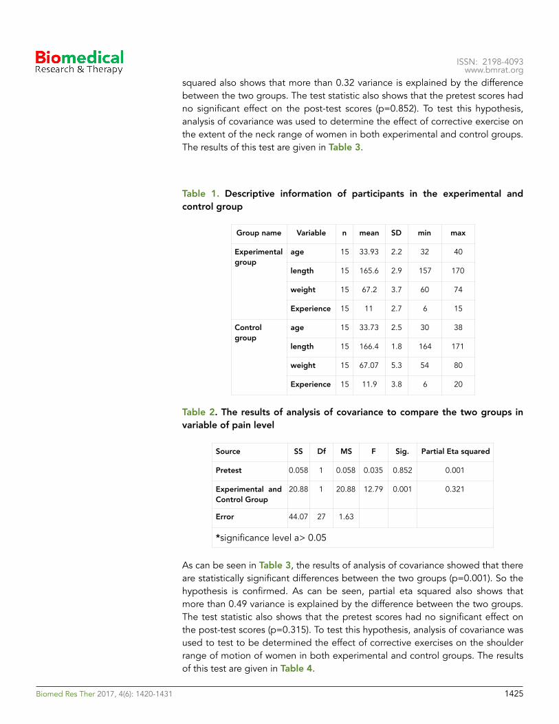
�ISSN: 2198-4093
www.bmrat.org squared also shows that more than 0.32 variance is explained by the difference between the two groups. The test statistic also shows that the pretest scores had no significant effect on the post-test scores (p=0.852). To test this hypothesis, analysis of covariance was used to determine the effect of corrective exercise on the extent of the neck range of women in both experimental and control groups. The results of this test are given in Table 3.
Table 1. Descriptive information of participants in the experimental and control group
Table 2. The results of analysis of covariance to compare the two groups in variable of pain level
As can be seen in Table 3, the results of analysis of covariance showed that there are statistically significant differences between the two groups (p=0.001). So the hypothesis is confirmed. As can be seen, partial eta squared also shows that more than 0.49 variance is explained by the difference between the two groups. The test statistic also shows that the pretest scores had no significant effect on the post-test scores (p=0.315). To test this hypothesis, analysis of covariance was used to test to be determined the effect of corrective exercises on the shoulder range of motion of women in both experimental and control groups. The results of this test are given in Table 4.
Group name Variable n mean SD min max
Experimental group
age 15 33.93 2.2 32 40
length 15 165.6 2.9 157 170
weight 15 67.2 3.7 60 74
Experience 15 11 2.7 6 15
Control group
age 15 33.73 2.5 30 38
length 15 166.4 1.8 164 171
weight 15 67.07 5.3 54 80
Experience 15 11.9 3.8 6 20
Source SS Df MS F Sig. Partial Eta squared
Pretest 0.058 1 0.058 0.035 0.852 0.001
Experimental and Control Group
20.88 1 20.88 12.79 0.001 0.321
Error 44.07 27 1.63
*significance level a> 0.05
!Biomed Res Ther 2017, 4(6): 1420-1431 !1425
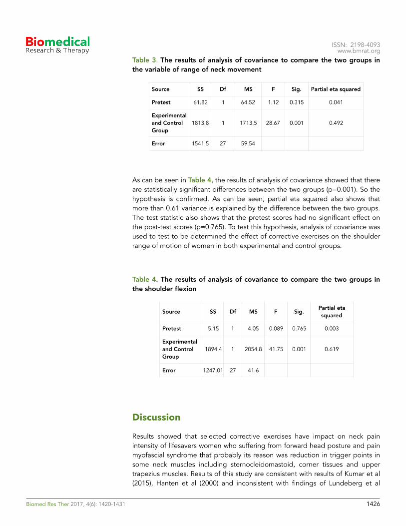
�ISSN: 2198-4093
www.bmrat.org Table 3. The results of analysis of covariance to compare the two groups in the variable of range of neck movement
As can be seen in Table 4, the results of analysis of covariance showed that there are statistically significant differences between the two groups (p=0.001). So the hypothesis is confirmed. As can be seen, partial eta squared also shows that more than 0.61 variance is explained by the difference between the two groups. The test statistic also shows that the pretest scores had no significant effect on the post-test scores (p=0.765). To test this hypothesis, analysis of covariance was used to test to be determined the effect of corrective exercises on the shoulder range of motion of women in both experimental and control groups.
Table 4. The results of analysis of covariance to compare the two groups in the shoulder flexion
Discussion
Results showed that selected corrective exercises have impact on neck pain intensity of lifesavers women who suffering from forward head posture and pain myofascial syndrome that probably its reason was reduction in trigger points in some neck muscles including sternocleidomastoid, corner tissues and upper trapezius muscles. Results of this study are consistent with results of Kumar et al (2015), Hanten et al (2000) and inconsistent with findings of Lundeberg et al
Source SS Df MS F Sig. Partial eta squared
Pretest 61.82 1 64.52 1.12 0.315 0.041
Experimental and Control Group
1813.8 1 1713.5 28.67 0.001 0.492
Error 1541.5 27 59.54
Source SS Df MS F Sig. Partial eta squared
Pretest 5.15 1 4.05 0.089 0.765 0.003
Experimental and Control Group
1894.4 1 2054.8 41.75 0.001 0.619
Error 1247.01 27 41.6
!Biomed Res Ther 2017, 4(6): 1420-1431 !1426

�ISSN: 2198-4093
www.bmrat.org (1984), Cagnie et al (2013), Salari et al (2009), Ghiasi et al (2008) (Cagnie et al., 2013; Ghiasi et al., 2008; Hanten et al., 2000; Kumar et al., 2015; Lundeberg et al., 1984; Salari, 2009). Self-myofascial tissue release as one of the corrective exercises by increasing endorphins and serotonin hormone secretion may be effective in reducing muscle pain. Another reason of effectiveness of corrective exercises can be noted the quickly selected higher than the pain sensory. Messages transmitted through corrective exercises faster transmitted to the brain and causes the release of serotonin and blocking the pain signals to the spinal cord at the beginning point of arrival. Generally, two main ways of treating trigger points, are increase local blood flow and increase the length of the sarcomere. Self-myofascial release tissue, destroys muscle tonus caused by nerve impulses coming from the spinal cord to the muscles and leads to increase blood circulation in the muscles and capillaries and ultimately reduces pain. As well as selected corrective exercises has impact on the range of motion of the neck in women lifesavers suffering from myofascial pain syndrome and forward head. As has been noted in recent researches, the effects of corrective exercises the muscles of the body can mention be more active motor units, increase blood circulation, increase the size, strength, endurance and muscle endurance in the face of external pressure and improve muscle elasticity9.results of present study are consistent with findings of Morimoto et al (2000), Ylinen et al (2007), Salari et al (2009) and Ghiasi et al (2008) (Ghiasi et al., 2008; Morimoto et al., 2000; Salari, 2009; Ylinen et al., 2007). Various studies have shown that the range of motion in people with neck trigger points are lower than healthy people and the pain leads to restrict movement. Required to achieve the desired maximum range of motion is that Antagonist muscles would be relaxed to the joint allowed to move. If there is pain, muscle tension around the joint and is needed more than the optimum stress. So the muscles become sensitive to stretch and do not allow reaching a maximum range of motion. According to the corrective exercises cause to reduce pain, this causes of decrease pain the muscles to release tension and allow more movement in the joint. On the other hand, given that the most important benefit of stretching exercises is improving joint range of motion of joint and increasing elasticity, it seems that the reason for increasing neck motion range in various direction after involving people in combined training, Considering proper stretching exercises as part of a training program in this study. In addition, selected corrective exercises can cause negative effects on the dynamics of motion neck. Because in addition to relieving pain, improve range of motion in the neck. Another reason is that the selected corrective exercises could be looking at the physiological effects of massage can offer relief from stiffness and muscle spasms in the neck muscle trigger points. As a result of these changes increases range of motion.
Results indicated that 8 weeks selected corrective training has impact on motor range of shoulder joint of women suffering from forward head posture and myofascial pain. These findings are consistent with results of Lucas (2004), Ashraf et al (2008), Kamali et al (2012), Cagnie et al (2013), Ylinen et al (2007), Salari et
!Biomed Res Ther 2017, 4(6): 1420-1431 !1427
�ISSN: 2198-4093
www.bmrat.org al (2012) and Ghiasi et al (2008) (Ashraf et al., 2008; Cagnie et al., 2013; Ghiasi et al., 2008; Kamali, 2010; Lucas et al., 2004; Salari, 2009; Ylinen et al., 2007).
It was seen in a research that conducted by Mohammad et al (2013) that existence an active trigger points in the muscle, causing the muscle to enter with dely. In other words, in the muscle with active trigger points, caused inhibition hence the upper trapezius muscle in healthy individuals for the movement comes into play. This action may be to increase the plate area to rotate the arm. Because according to the perspective of Moore (1992), the upward scapular rotation muscles, includes all parts of the trapezius muscle and the lower part of the anterior muscles. When the upper trapezius muscle is active trigger points, almost comes into play when the arm began to move from the side of the body but when the muscle is no active trigger point, in particular, to move the arm from the body is activated.
One of the goals of earlier activity of the upper trapezius at the initial stage of getting up arm is starting get up Akhromi joint through Acromioclavicular joint to increase space under Akhromi. The presence of trigger points in the muscle is associated with delay. That is sort of inhibition process occurs, resulting in inefficient and later the muscle activity during movement, leads to an increase in the potential for entrapping the structures under Akhromi. The results showed that self-myofascial tissue release and pull the trigger points has a positive effect such a change process and its negative effects on the movements of the shoulder joint and neck. Corrective exercises can improve muscle elasticity, which in turn can better strength and endurance due to the processes involved in. It can be stated that self-myofascial release technique is non-invasive and without side effects induced relaxation in the affected person and if it should be taught to patients and other individuals, certainly a lot of their problems and the need to reduce consumption of painkillers and sedatives.
Conclusion
Generally, it can be concluded from this study that while there are the different ways to improve the conditions of patients with trigger points but the process of corrective exercises can cause negative effects on the movements of the shoulder joint and neck because in addition to relieving pain, improve joint range of motion in the neck and shoulder patients. Therefore, it is recommended that corrective training methods be used rather than those invasive procedures that have used to treatment these people.
!Biomed Res Ther 2017, 4(6): 1420-1431 !1428

�ISSN: 2198-4093
www.bmrat.org Author contribution
MT performed data acquisition, data analysis; BBM performed designed the study, data analysis and manuscript preparation; AR performed data acquisition, and manuscript preparation. All authors approved the manuscript.
!Biomed Res Ther 2017, 4(6): 1420-1431 !1429

�ISSN: 2198-4093
www.bmrat.org References
Alter, M. (1996). Science of flexibility . Champaign, IL: Human Kinetics (google books). Amanolahi Asadollah, H.M.T., Shamsedini Ali Reza (2009). The effect of acupuncture on shoulder trigger points in patients with muscle pain syndrome. Journal of Veterinary Medicine 2, 22-34. Ashraf, A., Mirshams, S., Salavati, A., and Yazdani, A. (2008). A comparison of the stretching with ethyl chloride spray and stretching after injection of the lidocaine in the treatment of the trigger points: single blind randomized clinical trial (RCT). Cagnie, B., Dewitte, V., Coppieters, I., Van Oosterwijck, J., Cools, A., and Danneels, L. (2013). Effect of ischemic compression on trigger points in the neck and shoulder muscles in office workers: a cohort study. Journal of manipulative and physiological therapeutics 36, 482-489. Ghiasi, F., Akbari, A., and Abed, M. (2008). Comparison of muscle energy techniques with ultrasound therapy in myofascial trigger point treatment in upper trapezius. Journal of Babol University of Medical Sciences 10, 7-14. Hanten, W.P., Olson, S.L., Butts, N.L., and Nowicki, A.L. (2000). Effectiveness of a home program of ischemic pressure followed by sustained stretch for treatment of myofascial trigger points. Physical therapy 80, 997-1003. Hodi, F.S., O'Day, S.J., McDermott, D.F., Weber, R.W., Sosman, J.A., Haanen, J.B., Gonzalez, R., Robert, C., Schadendorf, D., Hassel, J.C., et al. (2010). Improved survival with ipilimumab in patients with metastatic melanoma. The New England journal of medicine 363, 711-723. Hou, C.-R., Tsai, L.-C., Cheng, K.-F., Chung, K.-C., and Hong, C.-Z. (2002). Immediate effects of various physical therapeutic modalities on cervical myofascial pain and trigger-point sensitivity. Archives of physical medicine and rehabilitation 83, 1406-1414. Kamali, F.A.a.-A.S., Sara; Evangelism; Maryam; Shams Salehi, Somayeh (2010). Comparison of therapeutic effect of ischemic pressure technique on tracheal ulcer muscle in the normal position and posture muscle elasticities in computer users. Research in rehabilitation sciences 10, 56-69. Kumar, G.Y., Sneha, P., and Sivajyothi, N. (2015). Effectiveness of Muscle energy technique, Ischaemic compression and Strain counterstrain on Upper Trapezius Trigger Points: A comparative study. International Journal of Physical Education, Sports and Health 1, 22-26. Lucas, K.R., Polus, B.I., and Rich, P.A. (2004). Latent myofascial trigger points: their effects on muscle activation and movement efficiency. Journal of Bodywork and Movement Therapies 8, 160-166. Lundeberg, T., Nordemar, R., and Ottoson, D. (1984). Pain alleviation by vibratory stimulation. Pain 20, 25-44. Luo, X., Pietrobon, R., Sun, S.X., Liu, G.G., and Hey, L. (2004). Estimates and patterns of direct health care expenditures among individuals with back pain in the United States. Spine 29, 79-86. Morimoto, M., Kawata, K., Tsuchiya, N., Murakami, H., Kura, M., and Koga, Y. (2000). [A case of acupuncture needle dermatitis]. Masui The Japanese journal of anesthesiology 49, 887-889.
!Biomed Res Ther 2017, 4(6): 1420-1431 !1430

�ISSN: 2198-4093
www.bmrat.org Naroii, S., Akbari, A., Asad, M., and Farahani, A. (2010). Comparing the effects of vibration and ultrasound waves accompanied with stretching exercises on myofascial trigger points of posterior neck muscles in athletes. Journal of Shahrekord University of Medical Sciences 12, 43-52. Roach, S., Sorenson, E., Headley, B., and San Juan, J.G. (2013). Prevalence of myofascial trigger points in the hip in patellofemoral pain. Archives of physical medicine and rehabilitation 94, 522-526. Salari, s., pilevarzadeh motahhare, M, Shafi'i, Nematollahzadeh (2009). Myofascial trigger point massage effect of short-term indices of the relaxation response. Sabzevar Faculty of Medical Sciences 11, 45-60. Schleip, R. (2003). Fascial plasticity–a new neurobiological explanation Part 2. Journal of Bodywork and movement therapies 7, 104-116. Shahi moridi, D., eghbali, Mansouri vaziri nejad, Reza; Najafzadeh, Neda (2009). The effect of low level laser therapy in the treatment of trapezius muscle trigger points. Mayvfasyal Journal of Medical Sciences 8, 99-108. Timmons, B.H., and Ley, R. (1994). Behavioral and psychological approaches to breathing disorders (Springer Science & Business Media). Ylinen, J., Kautiainen, H., Wirén, K., and Häkkinen, A. (2007). Stretching exercises vs manual therapy in treatment of chronic neck pain: a randomized, controlled cross-over trial. Journal of rehabilitation medicine 39, 126-132.
!Biomed Res Ther 2017, 4(6): 1420-1431 !1431
www.bmrat.org
Scope
Biomedical Research and Therapy (ISSN 2198-4093) is the major forum for basic and translational research into therapies. An international peer-reviewed journal, it publishes high quality open access research articles with a special emphasis on basic, translational and clinical research into molecular therapeutics and cellular therapies, including animal models and clinical trials. The journal also provides reviews, viewpoints, commentaries and reports. Biomedical Research and Therapy's Editorial Policies follow the recommendations of the International Committee of Medical Journal Editors (ICMJE), the World Association of Medical Editors (WAME), and the Committee on Publication Ethics (COPE) for guidance on policies and procedures related to publication ethics.
The journal is published monthly, 12 issues per year.
Peer review policy The decision to publish a manuscript is based on the opinion of the editor and at least two other reviewers. Articles containing statistical analysis will also receive a statistical review. Reviewers’ names will not be revealed to the author, nor will authors’ names be revealed to editors. Manuscripts are accepted for publication on the understanding that they have not been submitted simultaneously to another journal and that the work was not previously published. Prior publication of abstracts will not prejudice publishing of the complete study. The editors reserve the right to make editorial and grammatical corrections. The editors cannot be considered responsible for damage or loss of
typescripts, illustrations or photographs. Statements and opinions expressed in the articles are those of the authors and the editors disclaim any responsibility or liability for this material.
Please read details at here: http://www.bmrat.org/index.php/BMRAT/peerreviewprocess
Manuscript preparation Please read details at here: http://www.bmrat.org/index.php/BMRAT/guidelines