blockade of nogo-66, myelin-associated glycoprotein, and oligodendrocyte myelin glycoprotein by...
TRANSCRIPT
Development/Plasticity/Repair
Blockade of Nogo-66, Myelin-Associated Glycoprotein, andOligodendrocyte Myelin Glycoprotein by Soluble Nogo-66Receptor Promotes Axonal Sprouting and Recovery afterSpinal Injury
Shuxin Li,1 Betty P. Liu,1 Stephane Budel,1 Mingwei Li,2 Benxiu Ji,2 Lee Walus,2 Weiwei Li,2 Adrienna Jirik,2
Sylvia Rabacchi,2 Eugene Choi,2 Dane Worley,2 Dinah W. Y. Sah,2 Blake Pepinsky,2 Daniel Lee,2 Jane Relton,2 andStephen M. Strittmatter1
1Departments of Neurology and Neurobiology, Yale University School of Medicine, New Haven, Connecticut 06510, and 2BiogenIdec, Inc., Cambridge,Massachusetts 02140
The growth of injured axons in the adult mammalian CNS is limited after injury. Three myelin proteins, Nogo, MAG (myelin-associatedglycoprotein), and OMgp (oligodendrocyte myelin glycoprotein), bind to the Nogo-66 receptor (NgR) and inhibit axonal growth in vitro.Transgenic or viral blockade of NgR function allows axonal sprouting in vivo. Here, we administered the soluble function-blocking NgRectodomain [aa 27–310; NgR(310)ecto] to spinal-injured rats. Purified NgR(310)ecto-Fc protein was delivered intrathecally after mid-thoracic dorsal over-hemisection. Axonal sprouting of corticospinal and raphespinal fibers in NgR(310)ecto-Fc-treated animals corre-lates with improved spinal cord electrical conduction and improved locomotion. The ability of soluble NgR(310)ecto to promote axongrowth and locomotor recovery demonstrates a therapeutic potential for NgR antagonism in traumatic spinal cord injury.
Key words: spinal cord; axon; Nogo; Nogo-66 receptor; serotonin; magnetic stimulation
IntroductionAfter CNS injury, the growth of injured and spared axons has thepotential to restore function. The restorative potential of axongrowth is most obvious in spinal cord injury (SCI). Axon growthin the adult mammalian CNS is inhibited in part by myelin-derived inhibitors (Schwab and Caroni, 1988; Savio and Schwab,1989; McGee and Strittmatter, 2003) and in part by glial scar-derived chondroitin sulfate proteoglycans (Snow et al., 1990; Da-vies et al., 1999; Bradbury et al., 2002). Several of the myelininhibitory proteins, including Nogo-A (Fournier et al., 2001),myelin-associated glycoprotein (MAG) (Domeniconi et al., 2002;Liu et al., 2002), and oligodendrocyte myelin glycoprotein(OMgp) (Curristin et al., 2002; K. C. Wang et al., 2002a), cansignal to the Nogo-66 receptor (NgR). The structure of theligand-binding domain of the NgR has been determined (Bartonet al., 2003; He et al., 2003) but requires a second domain foreffective signaling of axon growth inhibition (Fournier et al.,
2002). Additional components of the NgR signaling complex caninclude p75-NTR and Lingo-1 (K. C. Wang et al., 2002b; Wong etal., 2002; Mi et al., 2004). In the accompanying work, we demon-strate that transgenic expression of the NgR ligand-binding do-main allows axonal sprouting and improved recovery from SCI inmice (S. Li, S. Budel, J. E. Kim, T. G. Hampton, and S. M. Stritt-matter, unpublished observations). Viral expression of adominant-negative version of the NgR lacking the signaling do-main promotes axonal regeneration from retinal ganglion cells(Fischer et al., 2004). Thus, NgR-mediated control of axonalgrowth plays a role in determining the degree of axonal growthafter adult CNS injury in mammals.
Pharmacological methods to perturb the CNS myelin inhibi-tor system have included anti-Nogo antibodies (Schnell andSchwab, 1990; Bregman et al., 1995), a NgR antagonist peptide(GrandPre et al., 2002; Li and Strittmatter, 2003), Rho pathwayinhibitors (Dergham et al., 2002; Fournier et al., 2003), andcAMP elevation (Neumann et al., 2002; Qiu et al., 2002; Nikulinaet al., 2004; Pearse et al., 2004). All four methods have someefficacy in vivo. The first two approaches are specific for Nogoand do not disrupt MAG or OMgp action. Blockade of Rhosignaling blocks myelin and glial scar inhibition but may mod-ify many other forms of cellular motility. Alterations of cAMPsignaling may also have pleiotropic effects on numerous sig-naling pathways.
Here, we have sought to administer the function-blockingsoluble NgR protein fragment intrathecally in rat SCI. We find
Received July 14, 2004; revised Sept. 2, 2004; accepted Oct. 13, 2004.This work was supported by grants from the Christopher Reeve Paralysis Foundation and the Institut Interna-
tional de Recherche en Paraplegie, Geneva, to S.L. and from the National Institutes of Health to S.M.S. We thankYiguang Fu and Ji Liao for expert technical assistance with in vivo studies. We also thank Raymond Boynton, AzitaKaffashan, Konrad Miatkowski, Maria Slight, and Rich Tizard for technical assistance.
Correspondence should be addressed to Dr. Stephen M. Strittmatter, Department of Neurology, Yale UniversitySchool of Medicine, P.O. Box 208018, New Haven, CT 06510. E-mail: [email protected].
DOI:10.1523/JNEUROSCI.2828-04.2004Copyright © 2004 Society for Neuroscience 0270-6474/04/2410511-10$15.00/0
The Journal of Neuroscience, November 17, 2004 • 24(46):10511–10520 • 10511
that the degree of axonal sprouting is more robust than withNogo-66-specific antagonist peptide treatment (GrandPre et al.,2002; Li and Strittmatter, 2003). Furthermore, we show that im-proved recovery of electrophysiological function parallels axonalsprouting and behavioral improvement after SCI.
Materials and MethodsNgR binding studies. Microtiter well binding assays were performed asdescribed previously (Barton et al., 2003). Each well was coated with 0.5�g of purified NgR protein. Binding of 100 nM AP-Nogo-66, AP-MAG,or AP-OMgp was assessed in the presence of 25 �g of NgR(310)ecto-Fcor 100 �g of NEP1– 40 peptide after 4 hr at 23°C (Barton et al., 2003).
Preparation of NgR(310)ecto-Fc protein. The cDNA encoding the first310 amino acid residues of rat NgR1 was cloned by PCR from the adultrat brain Marathon-Ready cDNA (Clontech, Cambridge, UK) with oli-gomers 5�-GAATAGCGGCCGCGCCGCCACCATGAAGAGGGCG-TCCTCCGGAGG-3� (including nucleotides 1–23 of GenBank accessionnumber AF462390) and 5�-ATAATGCGGCCGCTCAAGCACAACCC-TGTAAGTCACTGGC-3� (including the complement of nucleotides907–930 of GenBank accession number AF462390). This cDNA frag-ment was fused to the cDNA encoding the hinge and Fc region of rat IgG1[NgR(310)ecto-Fc] in the mammalian stable expression vector PV90.The nucleotide sequence was confirmed by DNA sequencing, andNgR(310)ecto-Fc was expressed in Chinese hamster ovary (CHO)-DG44cells. A high expressor clone (�10 mg/l, as determined using an anti-ratFc ELISA) was chosen for large-scale cell culture in BCM16 medium (80l). The culture supernatant obtained was concentrated �10-fold using aspiral cartridge concentrator (30 kDa cutoff; Millipore, Bedford, MA)and filtered. Tris-HCl (1.0 M, pH 8.9), NaCl, and glycine were added tothe filtered supernatant to a final concentration of 100 mM Tris-HCl, pH8.9, 3 M NaCl, and 1.5 M glycine. This was loaded to a 300 ml proteinA-Sepharose column (Pharmacia Biosciences, Piscataway, NJ), and thecolumn was washed with two column volumes of binding buffer (100 mM
Tris-HCl, pH 8.9, 3 M NaCl, and 1.5 M glycine), followed by one columnvolume of 5 mM Tris-HCl and 3 M sodium chloride, pH 8.9.NgR(310)ecto-Fc was eluted with 25 mM phosphate, pH 2.8, 100 mM
NaCl, and neutralized with one-tenth volume of 0.5 M sodium phos-phate, pH 8.6. The eluted protein was dialyzed against PBS, filtered,aliquoted, and stored at �70°C. Purity was �95% as assessed by SDS-PAGE, and, as expected, the N-terminal sequence began with cysteineresidue 27. Under reducing and nonreducing SDS-PAGE, NgR(310)ecto-Fc had apparent masses of 60 and 120 kDa, respectively. The endo-toxin level in the product was �4 endotoxin units/mg. NgR(310)ecto-Fcbinds directly to 125I-Nogo66, AP-Nogo66, glutathione S-transferase–Nogo66, and MAG-Fc; blocks the binding of the individual myelin pro-teins (Nogo, MAG, and OMgp) to NgR1 expressed on cells; and enhancesneurite outgrowth in rat dorsal root ganglia [postnatal day 4 (P4) to P7]and cerebellar granule neurons (P10 –P12) cultured on a bovine CNSmyelin substrate.
Rat SCI and NgR(310)-Fc administration. Female Sprague Dawley rats(190 –250 gm) were deeply anesthetized with ketamine (60 mg/kg) andxylazine (10 mg/kg). Laminectomy was conducted at spinal levels ofT6 –7, and the spinal cord was exposed. The dorsal half of the spinal cordwas cut with a 30 gauge needle and a pair of microscissors to sever thedorsal parts of corticospinal tracts (CSTs), and the depth of the lesion(1.8 mm) was assured by passing the sharp part of a number 11 bladeacross the dorsal half of the cord (GrandPre et al., 2002). An osmoticminipump (Alzet 2ML4; 2 ml volume, 2.5 �l/hr, 28 d delivery), whichwas filled with 1.2 mg of rat IgG in PBS or 1.2 mg of NgR(310)ecto-Fcfusion protein in PBS, was sutured to muscles under the skin on the backof the animals. A catheter connected to the outlet of the minipump wasinserted into the intrathecal space of the spinal cord at the T7– 8 levelthrough a small hole in the dura.
Rat CST tracing and 5,7-dihydrotryptamine injection. For the rats re-ceiving CST tracing, a burr hole was made on each side of the skulloverlying the sensorimotor cortex of the lower limbs 14 d after over-hemisection. The anterograde neuronal tracer biotin dextran amine(BDA) (10% in PBS; 3.5 �l per cortex) was applied at seven injection sites
at a depth of 1.5 mm from the dura on each side (coordinates: 1.0 –3.0mm posterior to bregma; 1.8 –3.8 mm lateral) (GrandPre et al., 2002).Two weeks after BDA injection, these animals were perfused with PBS,followed by 4% paraformaldehyde, and tissue was collected for histology.
For the rats receiving intracerebroventricular serotonin neurotoxin,5,7-dihydroxytryptamine (105 �g dissolved in 7 �l of 0.2% ascorbic acidin normal saline), the tip of the glass micropipette was positioned into theright lateral cerebral ventricle (coordinates: 0.8 mm posterior to bregma;1.5 mm lateral from the sagittal suture; 3.5 mm deep from the corticalsurface) 3 weeks after SCI. The duration of injection lasted over 5 min,and the micropipette was kept in position for an additional 5 min beforewithdrawal. Thirty minutes before the 5,7-dihydrotryptamine (DHT)injection, the monoamine uptake inhibitor desipramine (25 mg/kg, i.p.),was administrated. Two weeks after DHT injections, these rats were per-fused for histological examination.
Histology for BDA tracing, lesion depth, and serotonin fiber staining. ForNgR(310)ecto-Fc injection experiments in rats, the spinal cord extendingfrom 10 mm rostral to 10 mm caudal from the lesion site was cut parasag-ittally (50 �m) on a vibrating microtome. Transverse sections were col-lected from the spinal cord 11–16 mm rostral to and 11–16 mm caudal tothe injury site. The sections were incubated with avidin– biotin–peroxi-dase complex, and the BDA tracer was visualized by nickel-enhanced
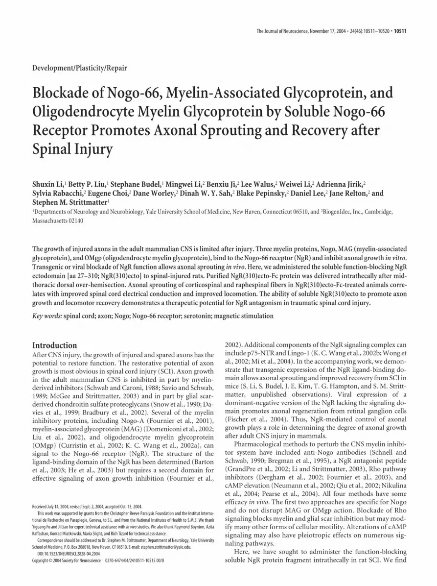
Figure 1. NgR(310)Ecto-Fc antagonism of myelin inhibitors. A, Schematic of wild-typeNgR and the NgR(310)ecto-Fc protein for intrathecal infusion. SS, Signal sequence; LRR,leucine-rich repeat; LRRNT, LRR N-terminal cysteine-rich region; LRRCT, LRR C-terminalcysteine-rich region; TM/GPI, transmembrane/GPI anchorage site; Fc, constant region ofrat IgG. B, Soluble NgR antagonist NgR(310)ecto protein significantly blocks immobilizedNgR binding by all the three myelin inhibitors: Nogo-66, MAG, and OMgp. In contrast, NgRantagonist NEP1– 40 peptide only blocks the binding of Nogo-66 to NgR but not that ofMAG and OMgp. Means � SEM are reported from 6 –12 determinations. The indicatedvalues in the presence of the inhibitor were statistically different from control bindingwithout inhibitor (*p � 0.01; Student’s t test).
10512 • J. Neurosci., November 17, 2004 • 24(46):10511–10520 Li et al. • Soluble NgR Promotes Recovery from SCI

diaminobenzidine HRP reaction or with avidin-Alexa594 (GrandPre etal., 2002). To verify the origin of caudal CST fibers in a subset of animals,the path of individual axons near the transection area was traced from acomplete set of consecutive parasagittal sections. Some sections wereprocessed for serotonin immunohistochemistry (anti-5-HT antibody;Immunostar) by indirect immunofluorescence. To examine the reinner-vation of regenerating axons in the distal spinal cord, transverse sections17–20 mm caudal to the lesion were doubly stained with BDA–synapto-physin (Sigma, St. Louis, MO), or BDA–SMI-32 (motor neuron marker;Sternberger Monoclonals, Lutherville, MD), or 5-HT–synaptophysin.To visualize the lesion area, some sections were double stained withantibody directed against GFAP (Sigma). The sections were mounted,dehydrated, and covered with mounting medium. The transection depthwas measured from GFAP staining and differential interference contrastphotographs from all consecutive parasagittal sections for each animal. Thedorsoventral linear depth of spared and transected tissue was measured.
Serotonin fiber length in the ventral horn of trans-verse sections rostral or caudal to the lesion sitewere measured with NIH Image software. Threesections were analyzed for each animal, and thevalues from these sections are averaged.
Electrophysiological recordings. To measuresignal conduction in motor pathways after SCI,transcranial magnetic motor-evoked potentials(tcmMEP) were measured from rats 3 and 5weeks after injury. To record the tcmMEP, ratswere anesthetized with avertin (250 mg/kg bodyweight). Magnetic stimulation was delivered bypositioning the center of a stimulator coil (5 cmin diameter; Cadwell Laboratories, Kennewick,WA) over the cranium, 0.5 cm lateral tobregma. The evoked responses were recordedfrom the contralateral gastrocnemius muscle.Potentials evoked by electrical stimulation ofspinal cord (SCEP) were also obtained fromthese rats immediately before they were killed.For SCEP recording, the animals were anesthe-tized with avertin (250 mg/kg) supplementedwith ketamine (30 mg/kg) and xylazine (5 mg/kg). Laminectomy was performed at the T1 spi-nal level. The spinal cord was exposed, and asmall hole in the dura was made. To directlystimulate the dorsal CST (dCST) in the spinalcord at the T1 level (rostral to the SCI site), anelectrical pulse (100 �sec, 7 mA) was deliveredto the dCST from a glass micropipette filledwith saline and placed stereotaxically at a depth1.1 mm from the dorsal surface of the midlineof the spinal cord. The evoked signals were re-corded from the exposed right sciatic nervewith hooked needle electrodes. Onset latencyand amplitude for both tcmMEP and SCEPwere analyzed. The stimulation threshold fortcmMEP was measured as a percentage of max-imal power applied to the magnetic coil. At leastfive responses were recorded and averaged fromeach side (right and left) of each animal.
Behavioral analysis. For behavioral testing,the Basso, Beattie, and Bresnahan (BBB) loco-motor scale, grid walking, and footprint analy-sis were performed. For grid walking, the ratswere trained to walk on a wire grid (70 cm longwith 2.54 cm squares), and the number of in-stances in which the hindpaw dropped belowthe grid plane was counted. For footprint anal-ysis, the walking patterns of hindpaws were re-corded with ink during a continuous locomo-tion across a 90 cm runway, and stride length oneach side and stride width were calculated(Metz et al., 2000).
ResultsNgR(310)ecto as a blocker of three myelin inhibitorsPrevious work had demonstrated that the NEP1– 40 peptide sig-nificantly, but partially, blocks myelin inhibition of axon out-growth (GrandPre et al., 2002). A soluble fragment of the NgRcontaining the ligand-binding domain is more effective in block-ing myelin-dependent inhibition (Fournier et al., 2002; Liu et al.,2002). To promote stability and purification, the ligand-bindingdomain of rat NgR was fused to the rat IgG1 Fc domain (Fig. 1A).Protein was purified from stably transfected CHO cells. To ex-plore the molecular basis of this differential effectiveness, theability of these two NgR antagonists to block the binding of theAP-tagged version of Nogo-66, MAG, or OMgp to purified NgR-
Figure 2. Intrathecal NgR(310)ecto-Fc ecto stimulates dCST sprouting in rats rostral to an over-hemisection. A, Transversesections rostral to the lesion display a similar degree of dCST labeling in both control and NgR(310)ecto-Fc-treated rats. Thesetransverse sections were obtained 15 mm rostral to a dorsal over-hemisection site from vehicle- or NgR(310)ecto-Fc-treated(NgR-ecto) rats, as indicated. Both CSTs are labeled, and the midline is at the center of each panel; dorsal is up in all sections. Alsonote the increased density of ectopic sprouts lateral to the dCST in the NgR(310)ecto-Fc-treated animals. B, Schematic of trans-verse spinal cord section illustrating the dCST and the location of the high-magnification images in C and D. C, D, BDA-labeled CSTfiber in gray matter adjacent to the dCST from an IgG-treated rat ( C) and a NgR(310)ecto-Fc-treated rat ( D). E, Ectopic CST fibersoutside of the dCST and dlCST and �100 �m in length are counted from transverse sections 11–15 mm rostral to the over-hemisection. Ctrl, Vehicle. Means � SEM are reported from seven to nine determinations. The indicated values in the presence ofNgR(310)ecto-Fc were statistically different from control (**p � 0.01; Student’s t test).
Li et al. • Soluble NgR Promotes Recovery from SCI J. Neurosci., November 17, 2004 • 24(46):10511–10520 • 10513

coated wells was assessed. The NEP1– 40peptide selectively blocks Nogo-66 bind-ing to purified NgR without displacingMAG or OMgp (Fig. 1B). In contrast, ex-cess soluble ectodomain of the NgR effec-tively prevents the binding of all three li-gands to immobilized NgR protein. Thus,the soluble NgR(310)ecto-Fc protein pro-vides a means to perturb simultaneouslyall three NgR ligands.
Intrathecal NgR(310)ecto-Fc proteininduces CST sprouting in the injuredspinal cordAs a pharmacological test of the growth-promoting benefit of NgR(310)ecto-Fc afterspinal trauma, we administered the purifiedprotein intrathecally. NgR(310)ecto-Fc pro-tein was delivered intrathecally to rats with amid-thoracic dorsal over-hemisection in-jury through an osmotic minipump. Be-cause we used rat NgR fused to rat IgG-Fc,there is no definitive method to distinguishinfused from endogenous protein immuore-activity in tissue sections. Thus, it was notpossible to demonstrate the distribution ofinfused NgR(310)ecto-Fc in the tissue fromSCI animals. During a 4 week survival periodafter SCI, 1.2 mg of NgR(310)ecto-Fc pro-tein was administered locally in each rat.At the end of this period, the CST wastraced by BDA injection into the cerebralcortex. The overall intensity of BDA label-ing in the CST rostral to the injury is indis-tinguishable between the groups (Fig. 2A).Compared with the tightly bundled dCSTfibers from vehicle-treated rats, sectionsrostral to the lesion from injured rats re-ceiving NgR(310)ecto-Fc protein exhibitectopic fibers sprouting from the dCST(Fig. 2A–D). The density of such fibers isincreased fivefold by NgR(310)ecto-Fctreatment.
Longitudinal sections spanning thelesion site from vehicle-treated rats dis-play no detectable CST axons or a verysmall number of BDA-labeled ventralCST (vCST) fibers below the lesion level(Fig. 3) (Weidner et al., 2001; GrandPreet al., 2002). Similar sections fromNgR(310)ecto-Fc-treated rats demon-strate many BDA-labeled fibers in thecaudal spinal cord (Fig. 3). This differ-ence cannot be attributed to altered le-sion depth because immunostaining for the astrocytic markerGFAP reveals that the lesion extends deeper than the centralcanal area (Fig. 3A). The lesion depth (70%) and the amountof spared tissue (30%) in control and treated groups are iden-tical (Fig. 3B). Photomicrographs of CST fibers caudal to thetransection reveal a tortuous branching course consistent withNgR(310)ecto-Fc-induced axon growth rather than CST spar-ing (Fig. 3C–I ). Counts of CST fibers from sagittal sectionsdocument �20 BDA-labeled axons at 1–2 mm caudal to the
lesion and 15 traced axons at 7– 8 mm distal to the lesion fromNgR(310)ecto-Fc-treated rats (Fig. 4 D). In one example ofdCST regeneration, a fiber is seen to bypass the lesion siteventrally and extend into the caudal gray matter (Fig. 3C–F ).However, in the vast majority of cases, the origin of caudalfibers in treated animals could not be reconstructed. It is plau-sible that a subset of caudal sprouting fibers in NgR(310)ecto-Fc-treated rats are derived from the vCST. The documentedincrease of caudal CST fiber counts in the treated rats may
Figure 3. Local administration of soluble NgR(310)ecto-Fc stimulates the growth of CST fibers into the caudal spinal cord. A,Camera lucida drawings of CST fibers from all sagittal sections from each rat. A typical control (Ctrl) animal shows a few vCST fibersin the distal spinal cord. Three separate NgR(310ecto)-Fc-treated rats (Ecto) exhibit a high density of sprouting from the lesioneddCST. Increased numbers of CST fibers are observed in the caudal spinal cord, particularly in gray matter. Parasagittal sectionsimmunostaining for astrocytic marker GFAP from the same animals demonstrate that the transection lesion extends beyond thecentral canal area in these examples. The injury site is indicated with an arrow. B, The dorsoventral linear extent of transected andspared spinal cord measured from consecutive parasagittal sections of each animal (n � 6 in each group). C, D, Schematic drawingof a transverse and sagittal spinal cord displays the location of sections shown in E. E, F, A composite sagittal section containing the lesionarea (arrow) from a NgR(310)ecto-Fc-treated rat illustrates a BDA–HRP-labeled dCST regenerating axon (dark) projects into distal spinalcord through the spared ventral spinal cord bridging tissue. A higher-magnification image is shown in F. G–I, Parasagittal sectionscontaining the transection site (arrows) from a NgR(310)ecto-Fc-treated rat illustrates the transection of BDA-labeled dCST fibers andsome branched, sprouting fibers caudal to the over-hemisection site. Higher-magnification images of these areas in H and I demonstratethe meandering course of the regenerating CST fibers. Scale bars: A, 1 mm; E, G, 250 �m; F, H, I, 25 �m.
10514 • J. Neurosci., November 17, 2004 • 24(46):10511–10520 Li et al. • Soluble NgR Promotes Recovery from SCI

reflect a combination of both axotomized CST fibers that growinto the distal cord plus new sprouts from the vCST that areproduced in the presence of NgR(310)ecto-Fc.
Generally, the number of sprouting CST fibers is similar tothat observed from local NEP1– 40 peptide-treated animals (Fig.4C), but a greater collateral branching from each sprout is seen inthe sections from animals treated with NgR(310)ecto-Fc protein(Fig. 4A,B). An assessment of sprout complexity demonstratesthat the total collateral length of each sprout in NgR(310)ecto-Fc-treated rats is twice as great as that from NEP1– 40-treatedanimals (Fig. 4F). The number of sprouts (�200 �m in length) at1–10 mm caudal to the spinal cord in both NgR antagonist-treated groups is �20 – 40 times greater than control groups (Fig.4E). More sprouts are seen from NgR(310)ecto-Fc-treated ratsthan with local NEP1– 40 treatment (�50 vs 25 sprouts/rat), butthis difference does not reach statistical significance.
Sprouting CST axons are observed in transverse sections ofthe spinal cord 11–15 mm caudal to over-hemisection in ratsreceiving NgR(310)ecto-Fc treatment (Fig. 4G–I). These fibersare detected in both the gray matter (Fig. 4G) and the whitematter (Fig. 4 H) of the spinal cord. The fibers detected in gray
matter often exhibit more collateralbranching than in white matter area. Incontrast, in transverse sections from thevehicle-treated group, only very rareBDA-labeled fibers are seen in the ven-tral white matter area, consistent withthe uninjured vCST axons. At this levelof the distal spinal cord, the averagenumber of BDA-labeled CST fibers fromboth NgR antagonist-treated groups[NgR(310)ecto-Fc and NEP1– 40] are�20-fold greater than vehicle-treated rats(Fig. 4I). Both Nogo receptor antagonists,NgR(310)ecto-Fc protein and NEP1–40peptide, result in dramatic CST axon growthin the caudal spinal cord, but the sproutinginduced by the former exhibits a more highlybranched pattern.
Do sprouting CST fibers contribute tosynaptic connections with motor neuronsin the caudal spinal cord? BDA-positivevaricosities in the caudal spinal cord wereexamined for colocalization of the synap-tic marker synaptophysin (Fig. 5A). Intransverse sections from control animals,BDA-traced CST fibers are extremely rarecaudal to the injury, and few BDA–synap-tophysin double-positive puncta are ob-served (Fig. 5B). In contrast, BDA–synap-tophysin double-labeled varicosities arecommonly seen in the NgR(310)ecto-Fc-treated samples with 16 –58 examplesdetected in each section (Fig. 5A,B).Moreover, BDA-traced CST fibers are fre-quently in close apposition to ventral mo-tor neurons, as detected by staining withSMI-32 anti-neurofilament antibody (Fig.5C,D). Thus, sprouting CST axons in-duced by NgR antagonist protein treat-ment appear to contribute to synaptic in-nervation in the lumbar spinal cord.
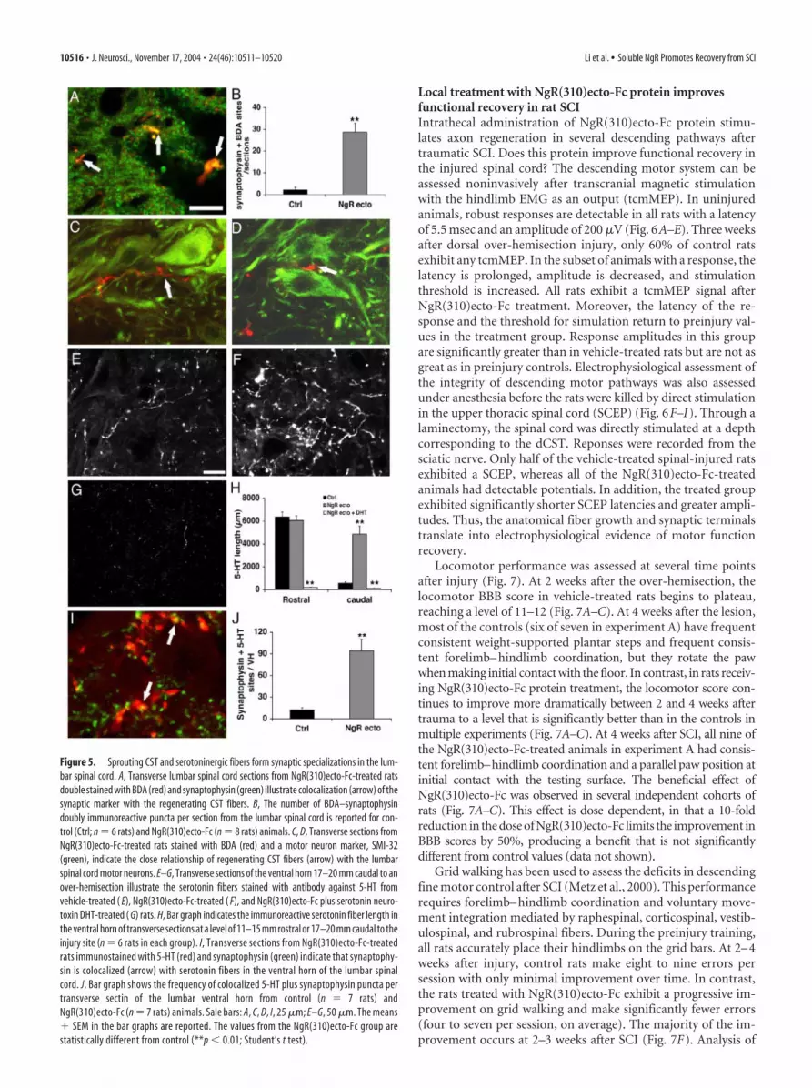
NgR(310)ecto-Fc induces sprouting and synaptic connectionof serotonergic axonsSeveral descending pathways contribute to the degree of locomo-tor function after SCI. The raphespinal system was also examinedin NgR(310)ecto-Fc-treated spinal injured rats. Immunostainingdemonstrates a density of serotonergic fibers 11–16 mm rostral tothe lesion that is similar in the vehicle- and NgR(310)ecto-Fc-treated groups. In the sections 17–20 mm below the lesion, sero-tonin fibers in NgR(310)ecto-Fc-treated rats are four times asnumerous as those in the control group (Fig. 5E,F,H). Moreimportantly, the formation of serotonergic synaptic contactsdefined by 5-HT–synpatophysin-positive varicosities in theventral horn is increased fivefold by NgR(310)ecto-Fc treat-ment (Fig. 5 I, J ). Because the restoration of raphespinal fiberis essentially complete with either NgR(310)ecto-Fc orNEP1– 40 treatment (GrandPre et al., 2002; Li and Strittmat-ter, 2003), a comparison of the two reagents by this parameteris not informative. Overall, it is clear that responsiveness toNgR(310)ecto-Fc treatment is not limited to CST fibers, withat least one other descending tract, the raphespinal, beingstimulated to grow by NgR antagonism.
Figure 4. CST fibers in the caudal spinal cord of NgR(310)ecto-Fc-treated rats. A, B, Parasagittal sections of the distal spinal cordfrom different NgR(310)-Fc-treated rats show CST fibers with a highly branched pattern. C, Parasagittal section of the distal spinalcord from an NEP1– 40-treated rat illustrates regenerating fibers with a lesser degree of branch complexity. D, CST fiber numberat various distances caudal to the injury site from NgR(310)ecto-Fc (n � 6 rats) and control (Ctrl; n � 6 rats) animals is reported.E, Counts of fibers (number of fibers �200 �m in length per rat) outside of the vCST from sagittal sections 1–10 mm caudal to SCIshow a greater number in the NgR(310)ecto-Fc-treated (n � 6 rats) and NEP1– 40-treated groups (n � 7 rats) than in the controlgroup (n � 6 rats). F, CST sprout complexity (total length of fiber arbor per sprout as visualized in a single section) isquantitated for vehicle-treated (n � 7 rats), NgR(310)ecto-Fc-treated (n � 7 rats), and NEP1– 40-treated (n � 7 rats)rats. The arbor length per sprout in both sets of NgR antagonist-treated rats is much greater than control rats. Thedifference between NgR(310)-Fc and NEP1– 40 is significant ( p � 0.01; Student’s t test). G, H, Transverse section at alevel 11–15 mm caudal to the lesion from an NgR(310)ecto-Fc-treated rat illustrates a number of CST fibers with branchingpatterns in caudal spinal cord gray matter ( G) and white matter ( H ). I, Quantification of CST fibers from transverse sections11–15 mm distal to the lesion indicates a greater number BDA-labeled fibers in NgR(310)ecto-Fc-treated (n � 6 rats) andNEP1– 40-treated (n � 7 rats) animals than the in control group (n � 6). Scale bar: A–C, G, H, 25 �m. Means � SEM inbar graphs are reported. The values from the NgR(310)ecto-Fc- or NEP1– 40-treated group are statistically different fromthe control group (D, E, I ) (*p � 0.05; **p � 0.01; Student’s t test).
Li et al. • Soluble NgR Promotes Recovery from SCI J. Neurosci., November 17, 2004 • 24(46):10511–10520 • 10515
Local treatment with NgR(310)ecto-Fc protein improvesfunctional recovery in rat SCIIntrathecal administration of NgR(310)ecto-Fc protein stimu-lates axon regeneration in several descending pathways aftertraumatic SCI. Does this protein improve functional recovery inthe injured spinal cord? The descending motor system can beassessed noninvasively after transcranial magnetic stimulationwith the hindlimb EMG as an output (tcmMEP). In uninjuredanimals, robust responses are detectable in all rats with a latencyof 5.5 msec and an amplitude of 200 �V (Fig. 6A–E). Three weeksafter dorsal over-hemisection injury, only 60% of control ratsexhibit any tcmMEP. In the subset of animals with a response, thelatency is prolonged, amplitude is decreased, and stimulationthreshold is increased. All rats exhibit a tcmMEP signal afterNgR(310)ecto-Fc treatment. Moreover, the latency of the re-sponse and the threshold for simulation return to preinjury val-ues in the treatment group. Response amplitudes in this groupare significantly greater than in vehicle-treated rats but are not asgreat as in preinjury controls. Electrophysiological assessment ofthe integrity of descending motor pathways was also assessedunder anesthesia before the rats were killed by direct stimulationin the upper thoracic spinal cord (SCEP) (Fig. 6F–I). Through alaminectomy, the spinal cord was directly stimulated at a depthcorresponding to the dCST. Reponses were recorded from thesciatic nerve. Only half of the vehicle-treated spinal-injured ratsexhibited a SCEP, whereas all of the NgR(310)ecto-Fc-treatedanimals had detectable potentials. In addition, the treated groupexhibited significantly shorter SCEP latencies and greater ampli-tudes. Thus, the anatomical fiber growth and synaptic terminalstranslate into electrophysiological evidence of motor functionrecovery.
Locomotor performance was assessed at several time pointsafter injury (Fig. 7). At 2 weeks after the over-hemisection, thelocomotor BBB score in vehicle-treated rats begins to plateau,reaching a level of 11–12 (Fig. 7A–C). At 4 weeks after the lesion,most of the controls (six of seven in experiment A) have frequentconsistent weight-supported plantar steps and frequent consis-tent forelimb– hindlimb coordination, but they rotate the pawwhen making initial contact with the floor. In contrast, in rats receiv-ing NgR(310)ecto-Fc protein treatment, the locomotor score con-tinues to improve more dramatically between 2 and 4 weeks aftertrauma to a level that is significantly better than in the controls inmultiple experiments (Fig. 7A–C). At 4 weeks after SCI, all nine ofthe NgR(310)ecto-Fc-treated animals in experiment A had consis-tent forelimb–hindlimb coordination and a parallel paw position atinitial contact with the testing surface. The beneficial effect ofNgR(310)ecto-Fc was observed in several independent cohorts ofrats (Fig. 7A–C). This effect is dose dependent, in that a 10-foldreduction in the dose of NgR(310)ecto-Fc limits the improvement inBBB scores by 50%, producing a benefit that is not significantlydifferent from control values (data not shown).
Grid walking has been used to assess the deficits in descendingfine motor control after SCI (Metz et al., 2000). This performancerequires forelimb– hindlimb coordination and voluntary move-ment integration mediated by raphespinal, corticospinal, vestib-ulospinal, and rubrospinal fibers. During the preinjury training,all rats accurately place their hindlimbs on the grid bars. At 2– 4weeks after injury, control rats make eight to nine errors persession with only minimal improvement over time. In contrast,the rats treated with NgR(310)ecto-Fc exhibit a progressive im-provement on grid walking and make significantly fewer errors(four to seven per session, on average). The majority of the im-provement occurs at 2–3 weeks after SCI (Fig. 7F). Analysis of
Figure 5. Sprouting CST and serotoninergic fibers form synaptic specializations in the lum-bar spinal cord. A, Transverse lumbar spinal cord sections from NgR(310)ecto-Fc-treated ratsdouble stained with BDA (red) and synaptophysin (green) illustrate colocalization (arrow) of thesynaptic marker with the regenerating CST fibers. B, The number of BDA–synaptophysindoubly immunoreactive puncta per section from the lumbar spinal cord is reported for con-trol (Ctrl; n � 6 rats) and NgR(310)ecto-Fc (n � 8 rats) animals. C, D, Transverse sections fromNgR(310)ecto-Fc-treated rats stained with BDA (red) and a motor neuron marker, SMI-32(green), indicate the close relationship of regenerating CST fibers (arrow) with the lumbarspinal cord motor neurons. E–G, Transverse sections of the ventral horn 17–20 mm caudal to anover-hemisection illustrate the serotonin fibers stained with antibody against 5-HT fromvehicle-treated ( E), NgR(310)ecto-Fc-treated ( F), and NgR(310)ecto-Fc plus serotonin neuro-toxin DHT-treated ( G) rats. H, Bar graph indicates the immunoreactive serotonin fiber length inthe ventral horn of transverse sections at a level of 11–15 mm rostral or 17–20 mm caudal to theinjury site (n � 6 rats in each group). I, Transverse sections from NgR(310)ecto-Fc-treatedrats immunostained with 5-HT (red) and synaptophysin (green) indicate that synaptophy-sin is colocalized (arrow) with serotonin fibers in the ventral horn of the lumbar spinalcord. J, Bar graph shows the frequency of colocalized 5-HT plus synaptophysin puncta pertransverse sectin of the lumbar ventral horn from control (n � 7 rats) andNgR(310)ecto-Fc (n � 7 rats) animals. Sale bars: A, C, D, I, 25 �m; E–G, 50 �m. The means� SEM in the bar graphs are reported. The values from the NgR(310)ecto-Fc group arestatistically different from control (**p � 0.01; Student’s t test).
10516 • J. Neurosci., November 17, 2004 • 24(46):10511–10520 Li et al. • Soluble NgR Promotes Recovery from SCI

hindpaw footprints in the control group demonstrates decreasedstride length and increased stance width at 4 weeks after over-hemisection, compared with uninjured rats or SCI animals re-ceiving NgR(310)ecto-Fc treatment (Fig. 7D,E). Therefore, mul-tiple behavioral tests demonstrate that blockade of NgR functionwith local infusion of antagonist protein improves locomotorrecovery after SCI.
Because anatomical studies demonstrate growth of both CSTand serotonergic fibers after NgR(310)ecto-Fc treatment, the lo-comotor improvement might be attributable to one or both fibersystems. To assess whether the presence of 5-HT axons is neces-sary for the improved function, neurotoxin studies were per-formed in treated and control spinal-injured rats. DHT was ad-ministered intracerebroventricularly to selectively lesionserotonergic neurons. Animals were pretreated with desipramineto protect catecholaminergic neurons. The loss of 5-HT fibersfrom treated rats was verified histologically (Fig. 5G,H). Seroto-nergic lesioning at 3 weeks after SCI did not alter the performanceof control spinal-injured rats as measured by the BBB score or bytheir ability to ascend a smooth inclined plane (Fig. 7G,H). Agroup of NgR(310)ecto-Fc-treated rats performed significantlybetter than control animals on these tests before DHT treatment,and the DHT lesion reduced their superiority over control rats.This demonstrates that the presence of a raphespinal system,which is induced to grow by NgR(310)ecto-Fc treatment (Fig.5E,F,H), is necessary for some, but not all, of the locomotorimprovement observed in NgR(310)ecto-Fc animals.
DiscussionFrom in vitro studies, the NgR has beenshown to constitute a site of signal conver-gence for three axon inhibitory proteinsfound in CNS myelin (McGee and Stritt-matter, 2003). We blocked NgR-depen-dent signaling in adult rodents by mechan-ical delivery of a soluble version of theNgR. This allows an assessment of the sig-nificance of summated inhibition fromNogo-66, MAG, and OMgp. The solublefragment of the NgR almost certainly actsas a decoy receptor, creating a fruitlesscomplex that cannot signal into the axonalcytoplasm to limit axonal extension(Fournier et al., 2002; Liu et al., 2002; Bar-ton et al., 2003). The blockade of signalingby soluble NgR(310)ecto is most likely toinvolve sequestration of ligand from en-dogenous active glycophosphatidylinosi-tol (GPI)-linked NgR. Because solubleNgR(310)ecto can bind to membrane-associated NgR, this may provide an addi-tional mechanism for the soluble receptorfragment to disrupt signaling function(Fournier et al., 2002; Liu et al., 2002).However, the much lower affinity of NgR–NgR interactions compared with NgR–li-gand renders this second mechanismunlikely.
NgR(310)ecto-stimulated growth inmultiple pathwaysThe NgR protein is widely expressed in theadult brain with most, but not all, neurons
expressing the protein (Hunt et al., 2002; Josephson et al., 2002,2003; X. Wang et al., 2002c). Likewise, multiple fiber tracts thatnormally contribute to locomotor function are susceptible tospinal cord trauma (Raineteau and Schwab, 2001; Bareyre et al.,2004). The descending CST has the longest trajectory and hasbeen the focus of study in numerous previous SCI studies. Inuntreated wild-type animals, mid-thoracic spinal trauma is notassociated with significant sprouting or long-distance growth(GrandPre et al., 2002; Kim et al., 2003), although a modest de-gree of local sprouting from uninjured fibers has been observed(Weidner et al., 2001). Here, significant CST sprouting rostral tothe injury is observed in the presence of dominant-negativeNgR(310)ecto. Some CST fiber branches meander around thelesion site and follow a tortuous course in the caudal spinal cord.Although the number of growing fibers is far greater than incontrol cases, it is also far less than the number observed in un-injured animals. Increased fiber density extends into the caudalspinal cord for at least 4 – 8 mm. Because the efficiency of BDAlabeling in the CST is �1–2% (Brosamle and Schwab, 1997), theobservation of 30 BDA-labeled CST fibers is likely to represent atotal of 2000 CSTs innervating the caudal spinal cord under theinfluence of NgR(310)ecto-Fc. From ultrastructural studies, avalue of 2000 lumbar CST fibers would comprise �5% of thenormal innervation density (Leenen et al., 1985). Consistent withthis estimate, our BDA-labeling method detects �300 BDA-labeled CST fibers in the upper thoracic cord of rats rostral to aSCI site (GrandPre et al., 2002).
Figure 6. Intrathecal delivery of NgR(310)ecto-Fc promotes signal conduction in injured spinal cord. A, Representative tcm-MEP responses recorded from one pre-SCI rat, two control rats, and two NgR(310)ecto-Fc rats 3 weeks after SCI by transcranialmagnetic stimulation. Calibration: 5 msec, 50 �V. B, A tcmMEP response was detected from all the rats treated withNgR(310)ecto-Fc protein (n � 18 legs from 9 rats) but were recorded from only 64% of rats in the control group (n � 18 legs from9 rats) 5 weeks after injury. C–E, Bar graphs indicate the stimulation threshold ( C), onset latency ( D), and amplitude ( E) fortcmMEP recorded from the preinjury rats (n � 12 legs from 6 rats), control SCI rats (n � 12 legs from 6 rats), and NgR(310)ecto-Fc-treated rats (n �18 legs from 9 rats) 5 weeks after SCI. The stimulation threshold is reported as a percentage of maximal powerdelivered to the magnetic coil. F, Representative SCEP responses recorded from two control and two NgR(310)ecto-Fc rats 5 weeksafter SCI with electrical stimulation of dCST area at T1 spinal level. Calibration: 5 msec, 50 �V. G, The SCEP responses were recordedfrom all the rats receiving NgR(310)ecto-Fc treatment (n �9 rats) but were only recorded in 55% of rats treated with vehicle (n �9 rats) 5 weeks after SCI. H, I, Bar graphs indicate the onset latency ( H ) and amplitude ( I ) of SCEP from control rats (n � 5 rats)and NgR(310)ecto-Fc-treated rats (n � 9 rats) 5 weeks after the lesion. Means � SEM are reported in all the graphs. The valuesof the NgR(310)ecto-Fc group are statistically different from control (*p � 0.05; **p � 0.01; Student’s t test).
Li et al. • Soluble NgR Promotes Recovery from SCI J. Neurosci., November 17, 2004 • 24(46):10511–10520 • 10517

Another descending fiber system stud-ied here, the serotonergic raphespinal sys-tem, is partially damaged in this surgery.In the presence of the soluble NgR(310)ecto protein, the 5-HT fiber density in thelumbar ventral horn of the caudal spinalcord increases greatly, returning to levelsindistinguishable from that in uninjuredanimals. Thus, at least two descending fi-ber systems damaged in this dorsal over-hemisection model exhibit regenerativegrowth in the spinal cord in the presence ofthe function-blocking NgR(310)ecto solu-ble protein. The extent of growth is muchdifferent from control animals and rangesfrom complete restitution of fiber densityto only partial restoration of prelesionaxon density.
Specificity of axon growth andsynaptic connectionsWhen certain degrees of sprouting and fi-ber growth are observed after injury, it iscritical to consider whether these fiberstarget to appropriate sites. For the 5-HTsystem, there is a low density of fibersremaining after injury that is situated inthe correct target area. Therefore, localsprouting from remaining axons requireslittle guidance to generate appropriate re-gional localization of innervation, as wasobserved here. CST fibers are faced withmore challenging guidance issues. It isclear that the course of those fibers thatsprout rostral to the injury create novelpaths not present in the uninjured ani-mals. Distal to the injury, those CST fibersobserved in the soluble NgR(310)ecto-Fc-treated rats and NgR(310)ecto-expressingmice follow trajectories very differentfrom the CST of uninjured animals. Theircourse is not linear and not confined to thetypical white matter location. Thus, theguidance of such fibers does not recapitu-late the course that these fibers follow dur-ing development. Still, a number of fibersreach the distal spinal cord. Both thesprouting serotonergic and CST fibers ex-hibit anatomical specializations consistent with synaptic forma-tion. It is conceivable that suprasegmental input, even withouttopographic precision, may contribute to neuronal function. Adelineation of the precision of synaptic connectivity will requireelectrophysiological mapping from discrete supraspinal territo-ries and has not yet been determined for these fibers growing inthe presence of soluble NgR(310)ecto protein. However, fromthe tcmMEP and SCEP measurements, it is clear thatNgR(310)ecto-Fc treatment promotes a degree of electrophysio-logical connectivity in descending motor pathways.
Locomotor recoveryThe dorsal over-hemisection destroys a proportion of supraspi-nal input to the lumbar spinal cord and produces significant, butincomplete, loss of hindlimb function during locomotion. The
partial recovery of function in control animals may be attributedto distal rearrangements of those descending fibers spared at thetime of the initial lesion and to the function of distal circuits in theabsence of suprasegmental input. Recently, it has been demon-strated that local plasticity after SCI creates new circuits betweendescending CST fibers and cervical propriospinal neurons(Bareyre et al., 2004).
In animals receiving the function-blocking solubleNgR(310)ecto, the recovery of locomotor function is improvedby several measures, including the BBB open-field score, a gridwalking task, and by stride length analysis. There are several po-tential mechanisms for this improvement. The raphespinal sys-tem is necessary for a proportion of the improvement, as docu-mented by selective neurotoxin lesioning. However, theraphespinal fiber response to NgR(310)ecto-Fc does not appear
Figure 7. Intrathecal NgR(310)ecto-Fc improves behavioral recovery after rat dorsal over-hemisection. A–C, The locomotorBBB score is reported as a function of time after dorsal over-hemisection in the vehicle- or NgR(310)ecto-Fc-treated rats from threeseparate experiments. For the specific rats in which tissue sparing was measured in Figure 3B, the results were indistinguishablein A. For example, the 21 d BBB control score was 12.5 � 0.9, and the NgR(310)ecto-Fc score was 15.5 � 0.2 ( p � 0.01).Experiment A (n � 7–9 rats per group) was performed at Yale University, experiment B was performed at BiogenIdec(n � 25–26 rats per group), and experiment C was performed at Yale University (n � 8 –9 rats per group). D, Examples of therepresentative footprints from one control (top) or two NgR(310)ecto-Fc-treated (bottom) rats 4 weeks after the dorsal over-hemisection. E, Footprint analysis reveals a shorter stride length and a greater stride width in control rats (n � 7 rats) than inuninjured (n � 6 rats) or injured/NgR(310)ecto-Fc (n � 9 rats) rats. F, Hindlimb errors during grid walking are reported as afunction of post-SCI time in the control (n � 7 rats) or NgR(310)ecto-Fc-treated (n � 9 rats) groups. G, BBB score is plotted as afunction of time after the dorsal over-hemisection in the animals receiving intracerebroventricular serotonin neurotoxin DHTinjection 3 weeks after SCI (n � 9 rats in each group). Notably, the locomotor BBB score in NgR(310)ecto-Fc-treated rats 3 weeksafter SCI was significantly decreased after DHT administration. H, The maximal tolerated inclined plane angle is reported as afunction of time after SCI from these rats receiving intracerebroventricular DHT injections (n � 9 rats in each group). The inclinedplane angle in NgR(310)ecto-Fc-treated rats was significantly decreased at 4 weeks after injury because of DHT application. Means� SEM are reported in all the graphs. The values of NgR(310)ecto-Fc group are statistically different from control as indicated inA and F–H. The control IgG treatment values are statistically different from no-SCI or SCI plus NgR(310)ecto-Fc rats in E. In G andH, the 28 and 35 d values for the NgR(310)ecto-Fc-treated rats were significantly different from the same animals at day 21 beforeDHT. (*p � 0.05; **p � 0.01; Student’s t test).
10518 • J. Neurosci., November 17, 2004 • 24(46):10511–10520 Li et al. • Soluble NgR Promotes Recovery from SCI

to be sufficient for the improved function. The role of spinalserotonin levels in facilitating spinal locomotor function in bothrodents and humans is well documented (Saruhashi et al., 1996;Schmidt and Jordan, 2000). The recovery of the tcmMEP and theSCEP in the NgR(310)ecto-Fc-treated animals suggests that CSTfiber growth into the lumbar cord also plays an important role inthe recovery. Although the plasticity of connections among inter-neurons localized in the distal spinal cord has not been charac-terized here, this remains an additional site where increased fibergrowth might contribute to functional recovery. The inherentplasticity of the injured spinal cord in forming new circuitry(Bareyre et al., 2004) may be enhanced by NgR(310)ecto-Fc.
Comparison with other NgR antagonistsIn previous studies, a peptide antagonist of Nogo-66 was identi-fied. This subfragment of Nogo-66 binds to NgR and blocksNogo action, but not MAG or OMgp action. When infused intothe injured rat spinal cord, the NEP1– 40 antagonist peptide pro-motes CST and raphespinal axon sprouting and fiber growth aswell as locomotor recovery (GrandPre et al., 2002). The NgRprotein reagent in this study blocks Nogo-66, MAG, and OMgpfunction. Therefore, a comparison of the two studies can be con-sidered as a means to assess the relative roles of Nogo-66 versusMAG and OMgp. Thus, the modest increase in sprouting com-plexity and in fiber number seen for CST fibers in the currentstudy may reflect the contribution of MAG and OMgp. Despitegreater CST sprouting, NgR(310)ecto-Fc does not produce a de-gree of locomotor recovery that is signficantly different from thatcaused by NEP1– 40 treatment (GrandPre et al., 2002).
Viral-mediated expression of the dominant-negativemembrane-bound NgR fragment has been shown to enhanceoptic nerve regeneration (Fischer et al., 2004). In the case ofretinal ganglion cells, axonal growth with NgR antagonism alonewas minor, unless an enhanced growth state was enhanced by lensinjury. In such a state, the effect of dominant-negative NgR waspronounced. In the current study, no adjunctive treatment wasadministered to enhance a moderate degree of axonal growth. Itmay be that axonal growth after SCI would be more complete ifNgR antagonism were combined with agents mimicking the ef-fects of lens injury on retinal ganglion cells.
Conclusion and perspectivesThe NgR(310)ecto-Fc protein provides blockade of several CNSmyelin inhibitors in vitro. When delivered by pharmacologicalmeans, the protein supports CST and raphespinal axon growth invivo with improved functional recovery from SCI. These datasuggest that function-blocking NgR protein preparations havepotential clinical use. Alternative mechanisms of achieving thesimilar beneficial effects after CNS injury might include anti-NgRantibody reagents. The recent determination of the structure ofthe NgR(310)ecto ligand-binding domain (Barton et al., 2003;He et al., 2003) could potentially facilitate the rational design ofsmall molecule NgR antagonists.
SCI is perhaps the clearest example of a clinical condition inwhich few neurons are lost but axonal discontinuity is the basis offunctional deficits. However, many other neurological condi-tions result, at least in part, from axonal disconnection. Suchconditions include head trauma, stroke, and chronic progressivemultiple sclerosis. The promotion of adult CNS axon growth mayalleviate deficits in any or all of these conditions.
ReferencesBareyre FM, Kerschensteiner M, Raineteau O, Mettenleiter TC, Weinmann
O, Schwab ME (2004) The injured spinal cord spontaneously forms anew intraspinal circuit in adult rats. Nat Neurosci 7:269 –277.
Barton WA, Liu BP, Tzvetkova D, Jeffrey PD, Fournier AE, Sah D, Cate R,Strittmatter SM, Nikolov DB (2003) Structure and axon outgrowth in-hibitor binding of the Nogo-66 receptor and related proteins. EMBO J22:3291–3302.
Bradbury EJ, Moon LD, Popat RJ, King VR, Bennett GS, Patel PN, FawcettJW, McMahon SB (2002) Chondroitinase ABC promotes functional re-covery after spinal cord injury. Nature 416:636 – 640.
Bregman BS, Kunkel-Bagden E, Schnell L, Dai HN, Gao D, Schwab ME(1995) Recovery from spinal cord injury mediated by antibodies to neu-rite growth inhibitors. Nature 378:498 –501.
Brosamle C, Schwab ME (1997) Cells of origin, course, and terminationpatterns of the ventral, uncrossed component of the mature rat cortico-spinal tract. J Comp Neurol 386:293–303.
Curristin SM, Cao A, Stewart WB, Zhang H, Madri JA, Morrow JS, Ment LR(2002) Disrupted synaptic development in the hypoxic newborn brain.Proc Natl Acad Sci USA 99:15729 –15734.
Davies SJ, Goucher DR, Doller C, Silver J (1999) Robust regeneration ofadult sensory axons in degenerating white matter of the adult rat spinalcord. J Neurosci 19:5810 –5822.
Dergham P, Ellezam B, Essagian C, Avedissian H, Lubell WD, McKerracher L(2002) Rho signaling pathway targeted to promote spinal cord repair.J Neurosci 22:6570 – 6577.
Domeniconi M, Cao Z, Spencer T, Sivasankaran R, Wang K, Nikulina E,Kimura N, Cai H, Deng K, Gao Y, He Z, Filbin M (2002) Myelin-associated glycoprotein interacts with the Nogo66 receptor to inhibit neu-rite outgrowth. Neuron 35:283–290.
Fischer D, He Z, Benowitz LI (2004) Counteracting the Nogo receptor en-hances optic nerve regeneration if retinal ganglion cells are in an activegrowth state. J Neurosci 24:1646 –1651.
Fournier AE, GrandPre T, Strittmatter SM (2001) Identification of a recep-tor mediating Nogo-66 inhibition of axonal regeneration. Nature409:341–346.
Fournier AE, Gould GC, Liu BP, Strittmatter SM (2002) Truncated solubleNogo receptor binds Nogo-66 and blocks inhibition of axon growth bymyelin. J Neurosci 22:8876 – 8883.
Fournier AE, Takizawa BT, Strittmatter SM (2003) Rho kinase inhibitionenhances axonal regeneration in the injured CNS. J Neurosci23:1416 –1423.
GrandPre T, Li S, Strittmatter SM (2002) Nogo-66 receptor antagonist pep-tide promotes axonal regeneration. Nature 417:547–551.
He XL, Bazan JF, McDermott G, Park JB, Wang K, Tessier-Lavigne M, He Z,Garcia KC (2003) Structure of the Nogo receptor ectodomain: a recog-nition module implicated in myelin inhibition. Neuron 38:177–185.
Hunt D, Mason MR, Campbell G, Coffin R, Anderson PN (2002) Nogoreceptor mRNA expression in intact and regenerating CNS neurons. MolCell Neurosci 20:537–552.
Josephson A, Trifunovski A, Widmer HR, Widenfalk J, Olson L, Spenger C(2002) Nogo-receptor gene activity: cellular localization and develop-mental regulation of mRNA in mice and humans. J Comp Neurol453:292–304.
Josephson A, Trifunovski A, Scheele C, Widenfalk J, Wahlestedt C, Brene S,Olson L, Spenger C (2003) Activity-induced and developmental down-regulation of the Nogo receptor. Cell Tissue Res 311:333–342.
Kim JE, Li S, GrandPre T, Qiu D, Strittmatter SM (2003) Axon regenerationin young adult mice lacking Nogo-A/B. Neuron 38:187–199.
Leenen LP, Meek J, Posthuma PR, Nieuwenhuys R (1985) A detailed mor-phometrical analysis of the pyramidal tract of the rat. Brain Res359:65– 80.
Li S, Strittmatter SM (2003) Delayed systemic Nogo-66 receptor antagonistpromotes recovery from spinal cord injury. J Neurosci 23:4219 – 4227.
Liu BP, Fournier A, GrandPre T, Strittmatter SM (2002) Myelin-associatedglycoprotein as a functional ligand for the Nogo-66 receptor. Science297:1190 –1193.
McGee AW, Strittmatter SM (2003) The Nogo-66 receptor: focusing myelininhibition of axon regeneration. Trends Neurosci 26:193–198.
Metz GA, Merkler D, Dietz V, Schwab ME, Fouad K (2000) Efficient testingof motor function in spinal cord injured rats. Brain Res 883:165–177.
Mi S, Lee X, Shao Z, Thill G, Ji B, Relton J, Levesque M, Allaire N, Perrin S,
Li et al. • Soluble NgR Promotes Recovery from SCI J. Neurosci., November 17, 2004 • 24(46):10511–10520 • 10519

Sands B, Crowell T, Cate RL, McCoy JM, Pepinsky RB (2004) LINGO-1is a component of the Nogo-66 receptor/p75 signaling complex. Nat Neu-rosci 7:221–228.
Neumann S, Bradke F, Tessier-Lavigne M, Basbaum AI (2002) Regenera-tion of sensory axons within the injured spinal cord induced by intragan-glionic cAMP elevation. Neuron 34:885– 893.
Nikulina E, Tidwell JL, Dai HN, Bregman BS, Filbin MT (2004) The phos-phodiesterase inhibitor rolipram delivered after a spinal cord lesion pro-motes axonal regeneration and functional recovery. Proc Natl Acad SciUSA 101:8786 – 8790.
Pearse DD, Pereira FC, Marcillo AE, Bates ML, Berrocal YA, Filbin MT,Bunge MB (2004) cAMP and Schwann cells promote axonal growth andfunctional recovery after spinal cord injury. Nat Med 10:610 – 616.
Qiu J, Cai D, Dai H, McAtee M, Hoffman PN, Bregman BS, Filbin MT (2002)Spinal axon regeneration induced by elevation of cyclic AMP. Neuron34:895–903.
Raineteau O, Schwab ME (2001) Plasticity of motor systems after incom-plete spinal cord injury. Nat Rev Neurosci 2:263–273.
Saruhashi Y, Young W, Perkins R (1996) The recovery of 5-HT immunore-activity in lumbosacral spinal cord and locomotor function after thoracichemisection. Exp Neurol 139:203–213.
Savio T, Schwab ME (1989) Rat CNS white matter, but not gray matter, isnonpermissive for neuronal cell adhesion and fiber outgrowth. J Neurosci9:1126 –1133.
Schmidt BJ, Jordan LM (2000) The role of serotonin in reflex modulation
and locomotor rhythm production in the mammalian spinal cord. BrainRes Bull 53:689 –710.
Schnell L, Schwab ME (1990) Axonal regeneration in the rat spinal cordproduced by an antibody against myelin-associated neurite growth inhib-itors. Nature 343:269 –272.
Schwab ME, Caroni P (1988) Oligodendrocytes and CNS myelin are non-permissive substrates for neurite growth and fibroblast spreading in vitro.J Neurosci 8:2381–2393.
Snow DM, Lemmon V, Carrino DA, Caplan AI, Silver J (1990) Sulfatedproteoglycans in astroglial barriers inhibit neurite outgrowth in vitro. ExpNeurol 109:111–130.
Wang KC, Koprivica V, Kim JA, Sivasankaran R, Guo Y, Neve RL, He Z(2002a) Oligodendrocyte-myelin glycoprotein is a Nogo receptor ligandthat inhibits neurite outgrowth. Nature 417:941–944.
Wang KC, Kim JA, Sivasankaran R, Segal R, He Z (2002b) p75 interacts withthe Nogo receptor as a co-receptor for Nogo, MAG and OMgp. Nature420:74 –78.
Wang X, Chun SJ, Treloar H, Vartanian T, Greer CA, Strittmatter SM(2002c) Localization of Nogo-A and Nogo-66 receptor proteins at sites ofaxon-myelin and synaptic contact. J Neurosci 22:5505–5515.
Weidner N, Ner A, Salimi N, Tuszynski MH (2001) Spontaneous cortico-spinal axonal plasticity and functional recovery after adult central nervoussystem injury. Proc Natl Acad Sci USA 98:3513–3518.
Wong ST, Henley JR, Kanning KC, Huang KH, Bothwell M, Poo MM (2002)A p75(NTR) and Nogo receptor complex mediates repulsive signaling bymyelin-associated glycoprotein. Nat Neurosci 5:1302–1308.
10520 • J. Neurosci., November 17, 2004 • 24(46):10511–10520 Li et al. • Soluble NgR Promotes Recovery from SCI