bite force as a parameter for comparision between
TRANSCRIPT
i
BITE FORCE AS A PARAMETER FOR COMPARISION
BETWEEN 2.0 mm TITANIUM LOCKING MINIPLATES AND
2.0 mm TITANIUM 3-D MINIPLATES FOR MANDIBULAR
FRACTURES: A RANDOMISED CLINICAL TRIAL
By
Dr. RAHUL KUMAR SANKLECHA
Dissertation Submitted to the
Rajiv Gandhi University of Health Sciences, Bengaluru, Karnataka.
In partial fulfillment of the requirements for the degree of
MASTER OF DENTAL SURGERY
In
ORAL AND MAXILLOFACIAL SURGERY
Under the guidance of
Dr. K. S. MANJUNATHAM.D.S.
PROFESSOR and H.O.D.
DEPARTMENT OF ORAL AND MAXILLOFACIAL SURGERY
SRI HASANAMBA DENTAL COLLEGE AND HOSPITAL
HASSAN.
2016-2019

viii
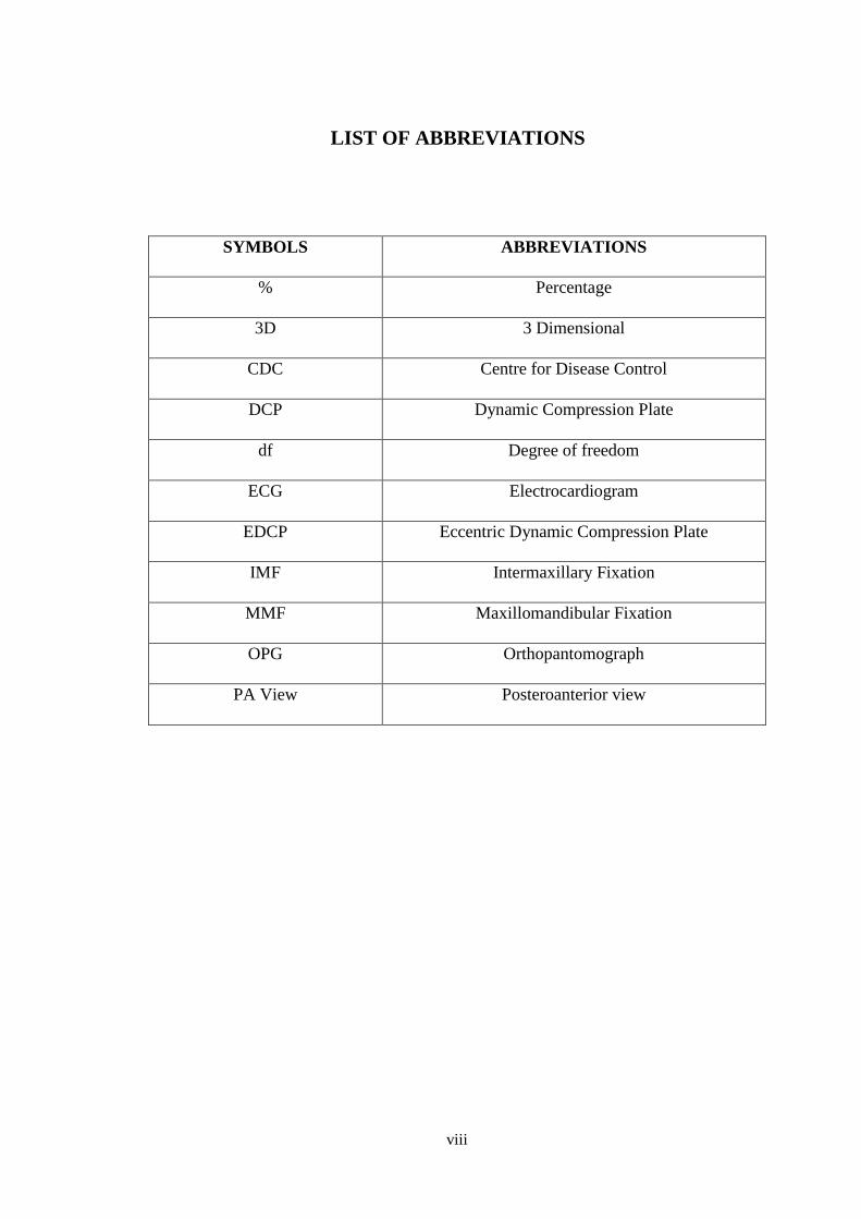
LIST OF ABBREVIATIONS
SYMBOLS
ABBREVIATIONS
%
Percentage
3D
3 Dimensional
CDC
Centre for Disease Control
DCP
Dynamic Compression Plate
df
Degree of freedom
ECG
Electrocardiogram
EDCP
Eccentric Dynamic Compression Plate
IMF
Intermaxillary Fixation
MMF
Maxillomandibular Fixation
OPG
Orthopantomograph
PA View
Posteroanterior view
ix
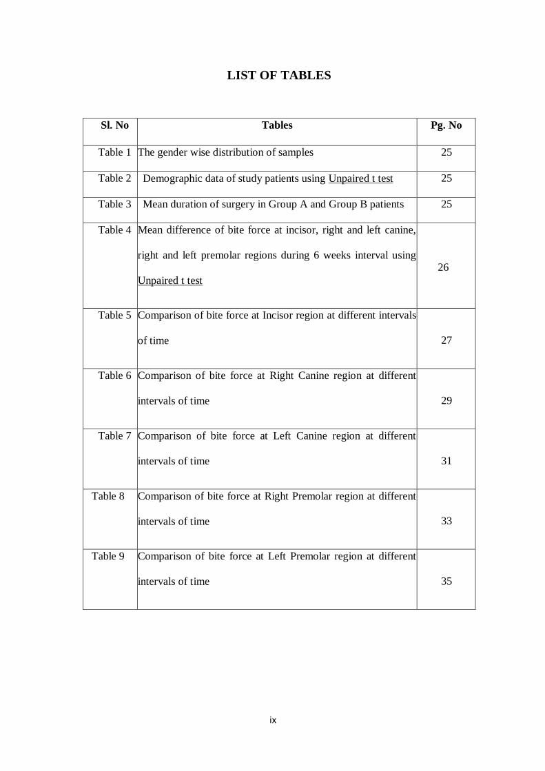
LIST OF TABLES
Sl. No Tables Pg. No
Table 1 The gender wise distribution of samples 25
Table 2 Demographic data of study patients using Unpaired t test 25
Table 3 Mean duration of surgery in Group A and Group B patients 25
Table 4 Mean difference of bite force at incisor, right and left canine,
right and left premolar regions during 6 weeks interval using
Unpaired t test
26
Table 5 Comparison of bite force at Incisor region at different intervals
of time
27
Table 6 Comparison of bite force at Right Canine region at different
intervals of time
29
Table 7 Comparison of bite force at Left Canine region at different
intervals of time
31
Table 8 Comparison of bite force at Right Premolar region at different
intervals of time
33
Table 9 Comparison of bite force at Left Premolar region at different
intervals of time
35
x
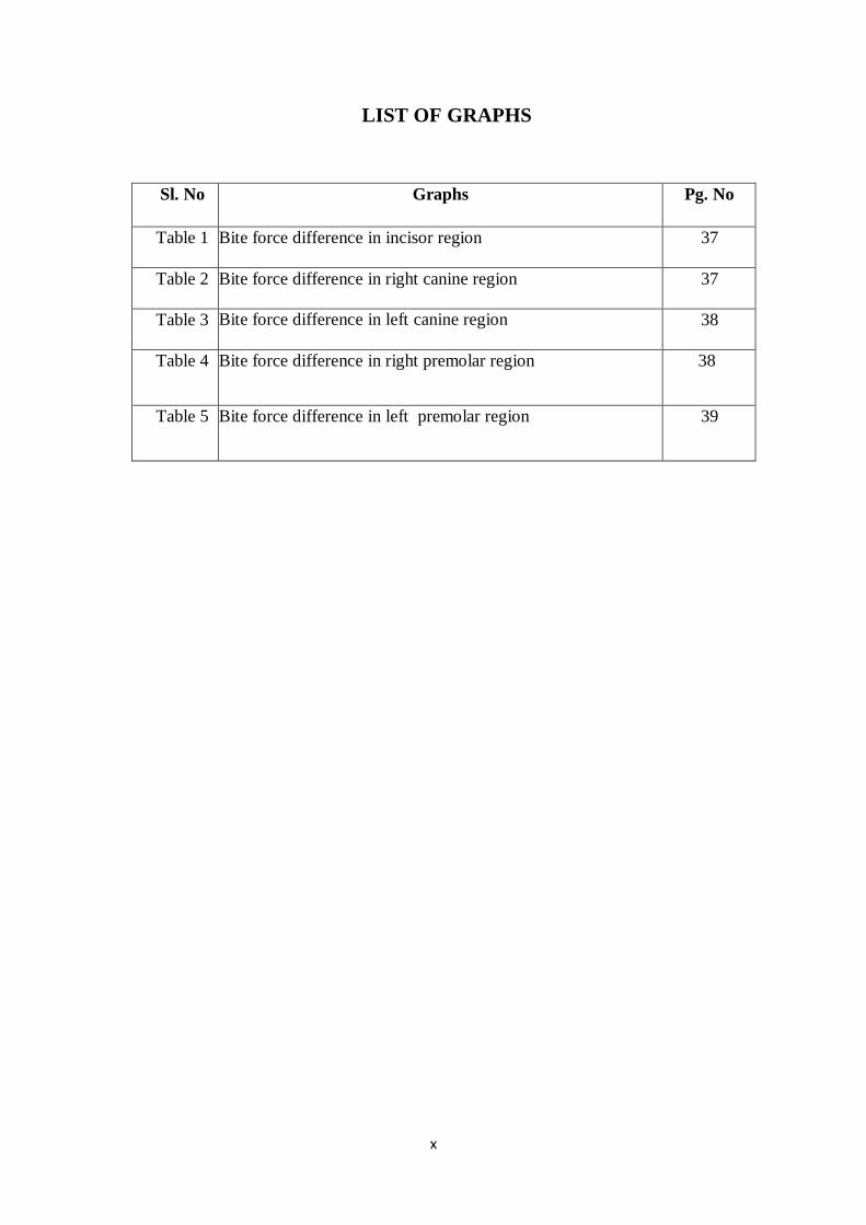
LIST OF GRAPHS
Sl. No Graphs Pg. No
Table 1 Bite force difference in incisor region 37
Table 2 Bite force difference in right canine region 37
Table 3 Bite force difference in left canine region 38
Table 4 Bite force difference in right premolar region 38
Table 5 Bite force difference in left premolar region 39
xi
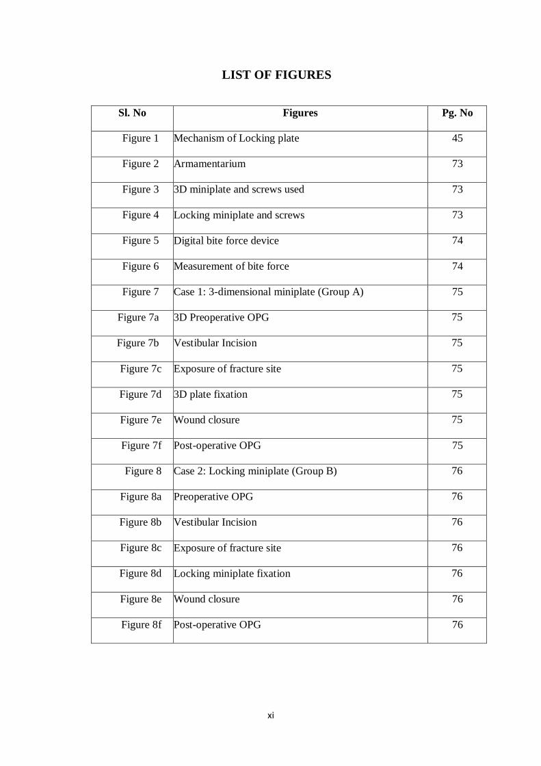
LIST OF FIGURES
Sl. No Figures Pg. No
Figure 1 Mechanism of Locking plate 45
Figure 2 Armamentarium 73
Figure 3 3D miniplate and screws used 73
Figure 4 Locking miniplate and screws 73
Figure 5 Digital bite force device 74
Figure 6 Measurement of bite force 74
Figure 7 Case 1: 3-dimensional miniplate (Group A) 75
Figure 7a 3D Preoperative OPG 75
Figure 7b Vestibular Incision 75
Figure 7c Exposure of fracture site 75
Figure 7d 3D plate fixation 75
Figure 7e Wound closure 75
Figure 7f Post-operative OPG 75
Figure 8 Case 2: Locking miniplate (Group B) 76
Figure 8a Preoperative OPG 76
Figure 8b Vestibular Incision 76
Figure 8c Exposure of fracture site 76
Figure 8d Locking miniplate fixation 76
Figure 8e Wound closure 76
Figure 8f Post-operative OPG 76
Abstract
1
ABSTRACT
“BITE FORCE AS A PARAMETER FOR COMPARISION
BETWEEN 2.0 mm TITANIUM LOCKING MINIPLATES AND
2.0 mm TITANIUM 3-D MINIPLATES FOR MANDIBULAR
FRACTURES: A RANDOMISED CLINICAL TRIAL”
Background & Objectives: To compare the efficacy of titanium 2-mm 3
dimensional miniplates with 2-mm titanium locking miniplates in the osteosynthesis
of mandibular fractures on the basis of bite force recordings.
Methods: A prospective randomized clinical trial was carried out for the treatment of
mandibular fractures using open reduction and internal fixation. The patients were
randomly divided into 2 groups, group A with 2-mm 3 dimensional titanium
miniplates and group B with 2-mm titanium locking miniplates. The assessment of
patients was done at weekly intervals for 6 weeks using bite force recordings.
Results: A total of 20 patients met the inclusion criteria, with 10 patients in each
group. In both the groups incisor, canine and premolar bite forces at the right and left
side increased at progressive follow up visits during 6 weeks. Statistically significant
difference was found only at the incisor region (p<0.001) whereas no statistically
significant differences were found at the other sites.
Interpretation & Conclusion: The clinical outcome of both the 3 dimensional and
locking miniplate systems in the present study were similar, however, following
Abstract
2
advantages with the use of 3 dimensional miniplates and locking miniplates can be
highlighted:
3 dimensional stability of the fracture site and simultaneous stabilization at
superior and inferior borders in fixation of mandibular fractures with 3 D
miniplates.
Locking miniplate can be used as a “one-plate-for-all” system minimizing the
number of fixation systems necessary to be stocked in the operating room.
Keywords: 3D plate system, Locking plate system, Mandibular fracture, Internal
fixation.
Introduction
3
INTRODUCTION
The individuality of a man is represented by his face, his identity is the key to
his personality and interaction. Any disfigurement of the face due to trauma or
otherwise would immensely affect an individual both physically and psychologically.
One of the most rewarding and demanding aspects of dental and surgical practice is
the management of the patient who has suffered facial trauma.
Despite the fact that the mandible is the largest and strongest facial bone, by
virtue of its prominence and its position on the face, is commonly fractured when
maxillofacial trauma is sustained. There are various anatomical and biomechanical
reasons for the increased vulnerability of the mandible to get fractured. The osteology
of the mandible, various muscle attachments and their influence, and the presence or
absence of dentition plays a vital role in producing the inherent weaknesses.
Therefore, fractures are seen more frequently in certain isolated areas such as the
angle, subcondylar region, region lateral to mental protuberance and mental foramen
region.1
Etiology of mandibular fractures varies, depending upon the population
studied. In the urban populations, road traffic accidents are more common while the
rural populations are often subjected to interpersonal violence. Other possible causes
of fracture of the lower jaw are due to:1,2
a) Assault injuries
b) Industrial injuries
c) Fall injuries
d) Sports related injuries
e) Interpersonal violence/assault
f) Pathologic fractures,
Introduction
4
g) Agricultural related injuries among the rural populations such as handling
submersible pumps without proper protective devices.
It was after World War II that the treatment modality has changed from closed
reduction to open reduction and direct fixation using transosseous wiring, bone plates
and screws. Modern traumatology started with the development of osteosynthesis,
which was a major innovation in craniomaxillofacial surgery3.
Open reduction with internal fixation [ORIF] which are in use today include
many techniques starting from wire osteosynthesis, Luhr’s Vitallium compression
plates, Schmoker and Spiessl’s dynamic compression plate [DCP], eccentric dynamic
compression plates [EDCP], reconstruction plate, monocortical noncompression
miniplates, lag screws, and 3- dimensional [3D] plates4 .
All of these methods are aimed to make patient return to function as soon as
possible and to avoid the need of long term IMF and to attain anatomic reduction,
achieve good occlusion with excellent aesthetic results. Dynamic compression plate,
eccentric dynamic compression plates were designed in such a way that when the
screws were tightened the two fractured fragments came into approximation as close
as possible. As the surgical technique was technique sensitive, many cases ended up
with step formation, infection, last but not the least cost factor also added to this5,6
.
But Champy’s miniplates and lag screws are less technique sensitive and produced
consistently good results with regard to occlusion and are economical and also helped
to avoid the need for long term IMF7.
The 3 dimensional titanium miniplate system is one of the newest internal semi rigid
fixations for maxillo-mandibular surgery in recent years. The shortcomings of rigid
and semi rigid fixation led to the development of 3-dimensional (3D) miniplates8
with
interconnecting cross struts. Unlike compression and reconstruction plates, their
Introduction
5
stability is not derived from the thickness of the plate. In combination with the
monocortical screws fixed to the outer cortex, the rectangular plate forms cuboids,
which possesses 3D stability. Although experimental studies on biomechanics have
confirmed sufficient stability of the 3D plating system, only a few studies have
previously reported clinical experiences with these plates in the treatment of
mandibular angle fractures. The 3 dimensional plating systems are based on the
principle of obtaining support through geometrically stable configuration. The
quadrangle geometry of plate assures a good stability in three dimensions of fractured
mandibular sites, since it offers good resistance against torque forces.
The management of trauma thus evolved greatly over the past many years
from supportive bandages, splints, circum-mandibular wiring, extra oral pins and
semirigid fixation with transosseous wiring to rigid fixation with compression plates
and more lately back to semirigid fixation with miniplates. Currently modifications in
miniplates like locking plates/screw system have been developed to overcome a few
disadvantages of conventional bone plate.9,10
There are a large number of experimental studies and a few clinical studies in
the literature which have proposed various biomechanical and technical advantages of
the 3-D miniplate system and the locking miniplate system and no study has
compared the bite force in patients treated with 3-D and locking miniplates and 2 mm
titanium locking miniplates till the commencement of this study. This prompted us to
perform the present study, to compare between these two systems of osteosynthesis in
the treatment of mandibular fractures based on the bite force recordings.
Aims & Objectives
6
AIMS AND OBJECTIVES
The aim of the study is to compare the efficacy of 3-dimensional
titanium miniplates with that of the titanium locking miniplates in the treatment of
mandibular fractures on the basis of bite force measurements to know the stability of
the miniplates.
The objective is to evaluate and compare the biomechanical function
between the two systems by measuring the maximal bite force preoperatively and at
1st to 6
th week postoperatively at weekly intervals.
Review of Literature
7
REVIEW OF LITERATURE
Historical insight improves understanding of current techniques and provides
the basis for the development of new methods. Even though the principles of the
treatment of mandibular fractures have changed drastically, the objective of re-
establishing the occlusion and masticatory function remains the same.
Brons and Boering (1970) introduced the lag-screw technique of
osteosynthesis. These screws had threads on the distal end and a smooth shank at the
proximal end which allowed compression of the segments between the outer and the
inner components.11
Schmoker and Niederdellmann(1973) developed the eccentric dynamic
compression plate (EDCP) which provided compression at the tension zones of
mandible. When the screws closest to the fracture were tightened, the fracture line
could be placed under compression, when eccentric terminals were tightened, the
alveolar segment would be reduced.12
Kahnberg K.E., A. Ridell (1980) conducted a study of bone plate fixation
with the fractures of mandible treated between 1970 to 1978, 1400 patients with
different jaw fractures were treated. They concluded that the plate fixation provides a
rigid stabilization of the fracture both in horizontal and vertical directions while
conventional wiring serves merely as a complement to the intermaxillary fixation. The
possibility to avoid intermaxillary fixation with bone plate fixation is a great
advantage especially in older edentulous patients with a single dislocated fracture. In
partially edentulous patient where remaining teeth don't occlude the bone plate
fixation serves as an alternative to IMF.13
Review of Literature
8
William C. Ardary, (1989) Conducted a prospective evaluation which was
made of 71 patients with 102 mandible fractures treated with rigid internal
compression plate and screw osteosynthesis and allowed to function immediately
following surgery revealed that the method of internal fixation was an effective and
predictable alternative for the treatment of fractures of the mandible.14
Dodson TB, Perrott DH, Kaban LB and Gordon NC (1990) in their
prospective study of 92 patients with 143 uninfected, isolated mandibular fractures
compared standard therapy (closed or open reduction with wire osteosynthesis and 4
weeks of MMF) with rigid internal fixation (compression plates or screws with 2 days
of MMF) and found no statistically significant difference in the treatment results
between the two groups.15
Hayter J. P, Cawood J. I. (1993) Presented a review of the application of
mini plates in maxillofacial surgery, with an emphasis on maxillofacial trauma. The
functional advantages of mini plates were improved jaw function, in terms of mouth
opening and bite force; decreased weight loss and improved pulmonary function.
Other advantages were improved speech and oral hygiene, leading to enhanced social
interaction and ability to return to work earlier, particularly in jobs where
communication is important.16
Farmand M (1993) treated patients with three-dimensional mini plates
system. He used the plates in different age groups, in craniofacial orthognathic,
reconstructive surgeries and in trauma cases. He stated that the complication rate was
low and that the plates offered good stability against traction and torsional forces.17
Shetty V, McBrearty D, Fourney M and Caputo AA (1995) did a study on
6 sets of mandible analogue, each set consisting of 3 mandibles. Fracture was
Review of Literature
9
simulated in the study models and EDCP, Wurzburg plate, Luhr plate, Lag screw,
Champy miniplate, Mennen clamp plate were used for fixation and a load of 22 DaN
was applied on all mandibles and the displacement along the fracture line to occlusal
forces, was measured and it was concluded that compression fixation system (EDCP,
Wurzburg, Luhrplate, Lag screw) are biomechanically superior to adaptive system
(Champy miniplate, Mennen clamp plate).18
Renton TF and Wiesenfeld D (1996) in their retrospective study on 205
cases of mandible fractures of which 83 were treated with mini plates following
Champy’s principles, 40 cases ignoring Champy’s principles and 82 cases by
transosseous wiring with a minimum follow up of 6 weeks and found that the patients
treated with miniplate osteosynthesis adhering to Champy’s principles were most
successful with minimal complication rates.19
Sikes JW, Smith BR, Mukherjee DP and Coward KA (1998) in an in vitro
study compared the fixation strengths of locking head and conventional screws in
fracture and reconstruction model and concluded that locking head screws provided
significantly increased resistance to displacement when only two screws per segment
were used in the reconstruction model. When four screws were used there was no
significant difference between locking head and conventional screw types. They also
postulated that the effect of bony buttressing is significant and may explain why
miniplate often fail in the atrophic mandible but are successful in fully dentate
patients.20
Herford AS and Ellis E III (1998) conducted a prospective study in 84
patients with mandibular fractures and continuity defects to examine the use of a
locking reconstruction bone plate/ screw system up to an average follow up of 16
Review of Literature
10
weeks and found them to be simple and advantageous over conventional bone plates
by not requiring the plate to be compressed to bone in providing the stability.9
Ellis E III and Graham J (2002) in their prospective study of 80 fractures in
59 patients evaluated the use of a 2 mm locking plate/ screw system for mandibular
fracture treatment over a period of 6 weeks postoperatively. 102 locking plates were
applied to 80 fractures; 59 fractures received 1 plate and 21 received 2 plates without
postoperative maxillomandibular fixation. They concluded that the use of 2 mm
locking plate/ screw system was found to be simple and to provide sound fixation in
all cases.10
Bolourian R, Lazow S and Berger J (2002) in their prospective study on 44
mandibular fractures in 31 patients assessed the efficacy of intraoral treatment of
mandibular fractures using a 2.0 mm miniplate of titanium alloy and 2 weeks of MMF
followed up for a period of 8 weeks. A single 2.0 mm miniplate was adapted along
Champy’s lines of osteosynthesis and secured with four 8 mm, monocortical screws
and it was concluded that this is a viable treatment modality.21
Gerlach KL and Schwarz A (2002) in their prospective study examined bite
forces in 22 patients after treatment of mandibular angle fractures with miniplate
osteosynthesis according to Champy. Bite forces were recorded from 1st to 6th week
postoperatively, at weekly intervals using transducers at the incisor, right and left
canine; right and left molar regions with the subject seated with head upright, looking
forward, and in an unsupported head position and were compared with a control group
and concluded that after 1 week postoperatively only 31% of the maximal vertical
loading found in controls was registered which increased to 58% at the 6th week
Review of Literature
11
postoperatively. In the present study, the above-mentioned methodology for bite force
measurement is used.22
Gabrielli MAC, Gabrielli MFR, Marcantonio E, and Hochuli-Vieira E
(2003) in their retrospective study of 191 patients with 280 mandibular fractures that
were treated with 2.0-mm mini plates without postoperative MMF found that the
overall incidence of complications, including infections, were similar to those
described for rigid fixation.23
Alpert B, Gutwald R and Schmelzeisen R (2003) in an article titled “New
innovations in craniomaxillofacial fixation- 2.0mm lock system” described in detail
the 2.0mm lock system and highlighted its following advantages:
• Locking 2.0 mini plates utilize double threaded screws which both lock to the
bone and the plate creating a mini- internal fixator.
• They form a more rigid construct with less distortion of the fracture or
osteotomy, and the screws do not loosen.
• They offer the prospect of less instrumentation and faster application.
• They are more retentive in cancellous bone, a significant advance in
cancellous block bone grafting.
• Interference with the vascularity of bone is minimal since the plate is not
pressed tightly against the bone.
They also described the 3 configurations of the lock system which are available
namely the thinner and medium varieties that are useful in transoral plating of
fractures utilizing the Champy technique and the heavier, longer variety used in
unilateral edentulous fractures in the symphysis and parasymphysis as well as an aid
to tumor resection and reconstruction with both free and vascularized grafts.24
Review of Literature
12
Kirkpatrick D, Gandhi R and Sickels JEV (2003) in their retrospective
review of 56 locking reconstruction plates placed in 42 mandibular fracture patients of
which eight(19%) patients had preoperative infection which persisted in 3 (37.5%) of
them, 2 patients (5.8%) with 3 fracture sites (6.4%) developed postoperative infection
that required further intervention and all of them were heavy smokers. Thus the
postoperative infection was attributed to the history of preoperative infection and
smoking and concluded that the use of locking reconstruction plates can facilitate the
management of complicated fractures; however, it did not eliminate complications.25
Collins CP, Leonard GP, Tolas A and Alcalde R (2004) in their prospective
randomised clinical trial in 90 patients with 122 fractures compared standard 2.0-mm
monocortical plates (in 53 fracture sites) to 2.0-mm locking plates (in 64 fracture
sites) in the treatment of mandible fractures with a follow up of 6 weeks and found
that mandible fractures treated with 2.0-mm locking plates and 2.0-mm standard
plates present with similar short-term complication rates.26
Egol KA, Kubiak EN, Fulkerson E, Kummer FJ and Koval KJ (2004) in
a systemic review of complete orthopaedic literature to compare and contrast the
function and roles of conventional unlocked plates to locked plates in fracture fixation
concluded that the locking and conventional fixation techniques rely on completely
different mechanical principles and thus provides different environment for fracture
reduction and healing.27
Militsakh O, Wallace DI, Kriet D, Girod DA, Olvera MS and Tsuue TT
(2004) in their retrospective study on 43 patients with composite resection of the
mandible and reconstruction with osteocutaneous flap, which was fixed using 2.0 mm
locking reconstruction plates over an average period of 11 months, concluded that the
Review of Literature
13
use of locking reconstruction plate is a reliable in settings of previously irradiated
mandible or in mandible where postoperative radiotherapy has been planned. Its
technical ease of application, contouring malleability and very low profile has proven
to be advantageous in oromandibular reconstruction.28
Gear AJL, Apasova E, Schmitz JP and Schubert W (2005) in their survey
on current trends in the management of simple, non-comminuted mandibular angle
fractures found that the preferred techniques were single miniplate on the superior
border (Champy technique); dual mini plates; a locking screw plate in the inferior
border only and 3-dimensional plates. A single miniplate on the superior border of the
mandible has become a preferred method of treatment among the AO faculty.29
Chritah A, Lazow SK, and Berger JR (2005) in their prospective study on
34 patients with 50 mandibular fractures treated with 2.0 mm locking miniplate/
screw system with a 1 week of MMF and a follow up of 6 weeks, found that primary
bone healing was achieved in 98% of cases, 3 complications (6%) were observed and
it was concluded that a single 2.0-mm locking miniplate placed along Champy’s ideal
line of osteosynthesis with four 8-mm monocortical locking screws plus 1 week of
MMF fixation is a reliable and effective treatment modality for mandibular
fractures.30
R.Mukerji, G.Mukerji, M.McGurk(2006) In this study they concluded that
the principles of treatment of mandibular fractures have changed recently, although
the objective of reestablishing the occlusion at masticatory function remains the same.
Splinting of teeth is an old way of immobilizing fractures but the advent of modern
biomaterials changed clinical practice towards plating the bone and early restoration
Review of Literature
14
of function. They presented a brief historical overview of techniques and systems that
have been used for stabilization of mandibular fractures.3
Juergen Zix, Olivier Lieger, Tateyuki Lizuka (2007) The aim of this
follow-up study was to evaluate the clinical usefulness of a new type of 3 dimensional
(3D) mini plates for open reduction and monocortical fixation of mandibular angle
fractures. In 20 consecutive patients, noncomminuted mandibular angle fractures were
treated with open reduction and fixation using a 2 mm 3D miniplate by the transoral
approach. All patients were systematically monitored until six months
postoperatively. Among the outcome parameters recorded were an infection,
hardware failure, wound dehiscence, and sensory disturbance of the inferior alveolar
nerve. The 3D plating system described here is suitable for fixation of simple
mandibular angle fractures and is an easy to use alternative to conventional mini
plates. The system may be contraindicated in patients in whom insufficient
interfragmentary bone contact causes minor stability of the fracture.6
Sauerbier S, Schon R, Otten JE, Schmelzeisen R and Gutwald R (2008) in
their review have described the evolution of miniplate osteosynthesis and highlighted
its advantages like the intra-oral approach and the easy adaptability, and minimal
bone exposure required for its fixation.31
Saikrishna D, Shetty SK and Marimallappa TR (2009) in their prospective
randomised clinical study on 40 patients with mandibular fractures (20 in each group)
compared effectiveness of 2.0-mm locking mini-plates and screws over 2.0-mm
standard mini-plates and screws for the treatment over 6 weeks and concluded that
locking plate/screw system proved to be more rigid than conventional plate/screw
Review of Literature
15
system, thereby reducing the need and duration of intermaxillary fixation. However,
there was no difference in complication rates.32
Jimson S, Sankar. A, Prasad. R (2009) gave the concept of three-
dimensional (3D) plates as “a geometrically closed quadrangular plate secured with
bone screws that creates stability in three dimensions”.42 patients with mandibular
fractures were treated with open reduction and fixation using SS mini plates,3D
plates, and titanium 3D plates through transoral/extraoral approaches under general
anesthesia with 14 patients in each group. Treatment outcomes were assessed for 6
months, which included the following criteria: occlusal stability, lingual splaying,
neurological deficit, infection, surgeon preference, and patient function. Titanium 3D
plate fixation proved to be advantageous over conventional miniplate and stainless
steel 3D plate fixation.33
Oguz Y, Uckan S, Ozden AU, Uckan E and Eser A (2009) did a finite
element analysis to evaluate the mechanical stresses over the bone and hardware after
sagittal split ramus osteotomy fixed with standard titanium or locking plates/ screws
and concluded that the looking miniplate/ screw system spreads the load over the
plate and screws and diminishes the amount of force transferred to each unit.34
Scolozzi P, Martinez A, Jaques B (2009) in their prospective study evaluated
45 patients with 74 linear in comminuted mandibular fractures treated with a single
2.0 mm A0 locking reconstruction plate without the use of the second plate over an
average follow up period of 10 months and found that the patients had satisfactory
fracture reduction and successful treatment outcome without major complications.35
Manoj Kumar Jain, K.S.Manjunath, B.K.Bhagawan, Dipit Shah(2010) in
their prospective study compared 3-D, 2mm SS plates with that of 2mm standard mini
Review of Literature
16
plates using Champy’s principles of osteosynthesis and concluded that Champy’s
miniplate system is better and easier method than the 3D miniplate system for fixation
of mandibular fractures. The 3D miniplate system is unfavorable to use in cases of
oblique fractures and is also difficult to adapt in the mental foramen region and also
has excessive implant material. However, the operative time is shorter because of
simultaneous stabilization at both superior and inferior borders.36
Sauerbier S, Kuenz J, Hauptmann S, Hoogedijk CF, Liebehenschel N, Schon R,
et al. (2010) in their retrospective study on 53 patients with 56 mandibular fracture
evaluated the use of a 2.0-mm locking plate system and concluded that the use of a
2.0-mm locking plate system with its advantages of improved handling
characteristics, increased stability, shorter surgical time and preservation of bony
perfusion is a viable alternative to conventional manipulates in the management of
mandibular fractures.37
Ribeiro-Junior PD, Magro- Filho 0, Shastri KA, Papageorge MB (2010),
in an in vitro study on polyurethane hemimandibles with bone-like consistency
evaluated conventional and locking miniplate/screw systems for the treatment of
mandibular angle fractures by loading compressive stress until a 4 mm displacement
occurred between the fractured segments and found that looking plate/screw systems
provided significantly greater resistance to displacement than conventional ones (p <
.01) and thus, they concluded that the locking mini plates offered more resistance than
conventional mini plates.38
Manoj Goyal. Karan Marya.Sonia Chawla. Richa, Pandey (2011) In this
study they compared and evaluated postoperative complications and treatment
outcome in mandibular fracture fixation using 2.0mm titanium miniplates and 3-D
Review of Literature
17
locking plates. Thirty patients were divided randomly into two groups of 15 each
(including comminuted and malunited fractures) Group A was treated with open
reduction internal fixation using 2.0mm mini plates and group B with 3-D locking
plates. A total of 5 complications were observed in four patients: implant exposure
and infection in the miniplate group and postoperative neurosensory deficit, implant
failure and implant exposure in a 3-D group. For fractures in symphysis and the
parasymphysis region, severely displaced angle fractures and for communicated
fractures, 3-D locking plate could be a better option.39
Singh V, Kumar I and Bhagol A (2011) in a prospective randomized study
on 50 patients with 76 fractures compared 2.0 mm locking plate system (36 fractures)
and nonlocking plate system (40 fractures) and concluded that both the systems
presented with similar short-term complication rates.40
Aggarwal M, Mohammad S, Singh RK, Singh V. (2011), in their
prospective randomised study on 20 patients with 32 fractures assessed bite force in
2-mm locking plates versus 2-mm standard plates at l, 3, and 6 weeks and 3 months
interval along with other clinical parameters and found that the use of locking mini
plates in mandibular fracture is efficacious enough to bear the masticatory loads
during osteosynthesis of the fracture. The locking mini plates provided the advantage
of a greater bite force, with clinical results almost similar to those seen with
nonlocking miniplate osteosynthesis.41
Gutwald Schon R, Metzger M, Kreutzer K, Rahn B, Sauerbier S (2011)
conducted a study in sheeps with an aim of comparing a combination of a locking
system with self-tapping (ST-L) or self- drilling- tapping (SF) screws and concluded
that the improved stability of the osteosynthesis with the ST-L system resulted in
Review of Literature
18
early ossification of the osteotomy gap and with minimal amount of callus
formation.42
Mahmoud E.K, Hesham E, Mohamed M, H (2012) in their prospective
study, compared 2mm three-dimensional titanium mini plates with that of 2mm
conventional titanium mini-plates in symphyseal and parasymphyseal fractures of the
mandible. Total of 20 patients with 10 in each group was treated by ORIF and
followed up postoperatively for malocclusion, neurosensory deficit, wound
breakdown, infection, and presence of non-union/malunion. They concluded that 3D
miniplate system is a better and easier method comparatively but difficult to use in
cases of oblique fractures and those involving the mental nerve and there is excessive
implant material because of extra vertical bars.43
Laxmi Gandhi, Vivekananda SK (2012) evaluated the efficacy of 3D
titanium mini-plates in mandibular fractures in 20 patients over a period of 2 years
prospectively. Their aim was to analyse the structural stability of fractured fragments
after fixation, to evaluate biocompatibility and morbidity of 3D plating system. They
concluded that 3D titanium mini-plates provided good stabilization of fractured
fragments in three dimensions and ease of contouring and adapting. They are
biocompatible and no morbidity was seen in this study.44
Methodology
19
METHODOLOGY
SOURCE OF DATA:
This is a prospective randomized clinical trial done on 20 patients with mandibular
fractures who presented to the department of Oral and Maxillofacial Surgery, Sri
Hasanamba Dental College and Hospital and to the Hassan Institute of Medical
Sciences from November 2016 to September 2018 after obtaining the ethical
clearance.
METHOD OF COLLECTION OF DATA:
A randomized clinical trial was conducted in which 20 consecutive patients satisfying
the inclusion criterias as listed below, were randomly assigned to receive 2.0 mm
titanium 3-D miniplates (group A) and 2.0 mm titanium locking miniplates (group B)
with each group containing 10 patients.
Written consent was taken from the patients/guardians before undertaking the study
after explaining the nature of the procedure and possible discomforts and risks.
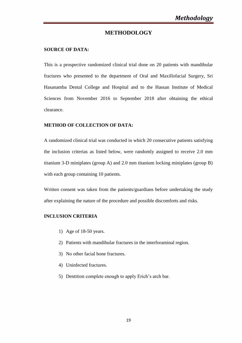
INCLUSION CRITERIA
1) Age of 18-50 years.
2) Patients with mandibular fractures in the interforaminal region.
3) No other facial bone fractures.
4) Uninfected fractures.
5) Dentition complete enough to apply Erich’s arch bar.
Methodology
20
The study design included a thorough case history taking in a case sheet which was
custom made for the study. Demographic data collected consisted of gender, age,
cause of fracture, duration between injury and surgery and the site of fracture.
Preoperative investigations that included complete hemogram, OPG/PA
skull/mandibular occlusal view. ECG and chest X ray were advised for patients
treated under general anaesthesia.
2.0 mm TITANIUM AND 3D MINIPLATE HARDWARE DETAILS:
(FIGURE- 3)
A 4 holed (2x2) titanium 3dimensional miniplate with interconnecting vertical bars
were used. The thickness of plate was 1mm.
Screws: The length was 8mm, width 2mm, diameter at its head was 3mm, and height
of the head was 1mm.
2.0 mm TITANIUM LOCKING MINIPLATE SYSTEM HARDWARE
DETAILS: (Figure-4)
EXCLUSION CRITERIA
Patients in whom intermaxillary fixation was medically contraindicated (epilepsy,
severe asthma, psychiatric condition)
1) Patients with systemic diseases (patients on chemotherapy and/or on
radiotherapy, osteopetrosis, osteoporosis).
2) Mandibular fracture associated with other maxillofacial fractures.
3) Fractures other than interforaminal fractures.
4) Multiple fractures.
5) Grossly infected fractures.
Methodology
21
A 4 holed titanium locking miniplate with bar was used. The thickness of plate was 1
mm.
Locking screws:
The length was 8mm, width 2mm, diameter at its head was 3mm, and height of the
head was 1mm.
Surgical technique:
All patients were given prophylactic antibiotic intravenously 30 minutes prior to the
procedure followed by two times daily for 3 days. Surgical procedures were carried
out under local anaesthesia/ general anaesthesia via nasotracheal intubation.
Armamentarium used for the study is shown in Figure 2. Reduction of the fracture
preoperatively was achieved using maxillary and mandibular arch bars. Following the
strict aseptic precautions, an appropriate intra/extraoral approach (translabial,
vestibular,) was selected based on the site. Fracture site was exposed after the
subperiosteal dissection, fractured segments were reduced and after achieving
adequate occlusion, MMF was done. Fixation was done using either a single 2.0 mm
titanium 3D plate/screw system (Group A) (Figure 7d) or 2.0 mm locking
miniplates/screw system applied as per Champy’s ‘ideal lines of osteosynthesis’
(Group B) (Figure 8d) and screws with dimensions of 2 mm x 8mm were used in
both the groups. Care was taken so as to place the screws lateral to the roots. MMF
was released intraoperatively and the passive occlusion was checked. A watertight
wound closure was achieved. Duration of procedure from incision to wound closure
was noted. Elastoplast pressure dressing was applied extraorally for 24 hrs
postoperatively. Patients were prescribed a 3 day course of oral antibiotics and 5-day
Methodology
22
course of analgesics and anti-inflammatory drugs and were advised to remain on soft
diet for 2 weeks.
Follow up:
Patients were followed up for a period of 6 weeks at weekly intervals. During each
follow up maximum bite forces at the incisor, canine and premolar region were
recorded by the same examiner with the help of a bite force device consisting of a
force transducer designed to measure the maximum force (in Kgs) exerted provided
with a digital indicator (Figure 5). All measurements were made with the patient
seated with the head upright, looking forward and in an unsupported natural head
position. The instrument could be conveniently positioned between a single pair of
antagonising cusps in the region of incisors, right and left canines and 1st
premolars.
The patients were advised to bite as forcefully as possible five times (Figure 6). The
highest value was recorded and entered in the data sheet.
Results were evaluated using paired t test for the intra group measurements and
unpaired t test for the inter group measurements for the bite force recordings and
Mann Whitney U test for other clinical parameters.
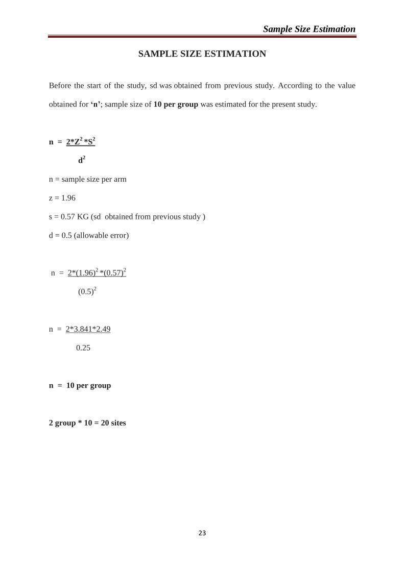
Sample Size Estimation
23
SAMPLE SIZE ESTIMATION
Before the start of the study, sd was obtained from previous study. According to the value
obtained for ‘n’; sample size of 10 per group was estimated for the present study.
n = 2*Z2
*S2
d
2
n = sample size per arm
z = 1.96
s = 0.57 KG (sd obtained from previous study )
d = 0.5 (allowable error)
n = 2*(1.96)2
*(0.57)2
(0.5)
2
n = 2*3.841*2.49
0.25
n = 10 per group
2 group * 10 = 20 sites
Result
24
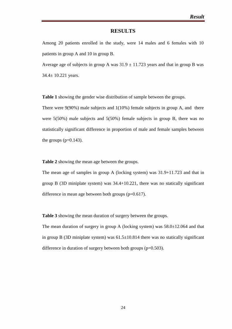
RESULTS
Among 20 patients enrolled in the study, were 14 males and 6 females with 10
patients in group A and 10 in group B.
Average age of subjects in group A was 31.9 ± 11.723 years and that in group B was
34.4± 10.221 years.
Table 1 showing the gender wise distribution of sample between the groups.
There were 9(90%) male subjects and 1(10%) female subjects in group A, and there
were 5(50%) male subjects and 5(50%) female subjects in group B, there was no
statistically significant difference in proportion of male and female samples between
the groups (p=0.143).
Table 2 showing the mean age between the groups.
The mean age of samples in group A (locking system) was 31.9+11.723 and that in
group B (3D miniplate system) was 34.4+10.221, there was no statically significant
difference in mean age between both groups (p=0.617).
Table 3 showing the mean duration of surgery between the groups.
The mean duration of surgery in group A (locking system) was 58.0±12.064 and that
in group B (3D miniplate system) was 61.5±10.814 there was no statically significant
difference in duration of surgery between both groups (p=0.503).
Result
25
Table 1: showing the gender wise distribution of samples
CHI SQUARE TEST
GROUP
X2 df p
LOCKING 3D
SEX
FEMALE
Count 1 5
2.143 1 0.143
% 10.0% 50.0%
MALE
Count 9 5
% 90.0% 50.0%
Table 2: Demographic data of study patients using Unpaired t test
Unpaired t test
GROUP N Mean SD t df p
AGE
LOCKING 10 31.900 11.723
-.508 18 0.617
3D 10 34.400 10.221
Table 3: Mean duration of surgery in Group A and Group B patients
Unpaired t test
GROUP N Mean SD t df p
DURATION
OF SURGERY
LOCKING 10 58.0000 12.06464
-0.683 18 0.503
3D 10 61.5000 10.81409
Result
26
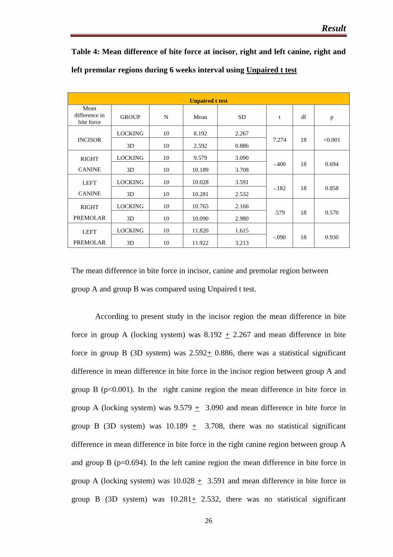
Table 4: Mean difference of bite force at incisor, right and left canine, right and
left premolar regions during 6 weeks interval using Unpaired t test
Unpaired t test
Mean
difference in
bite force GROUP N Mean SD t df p
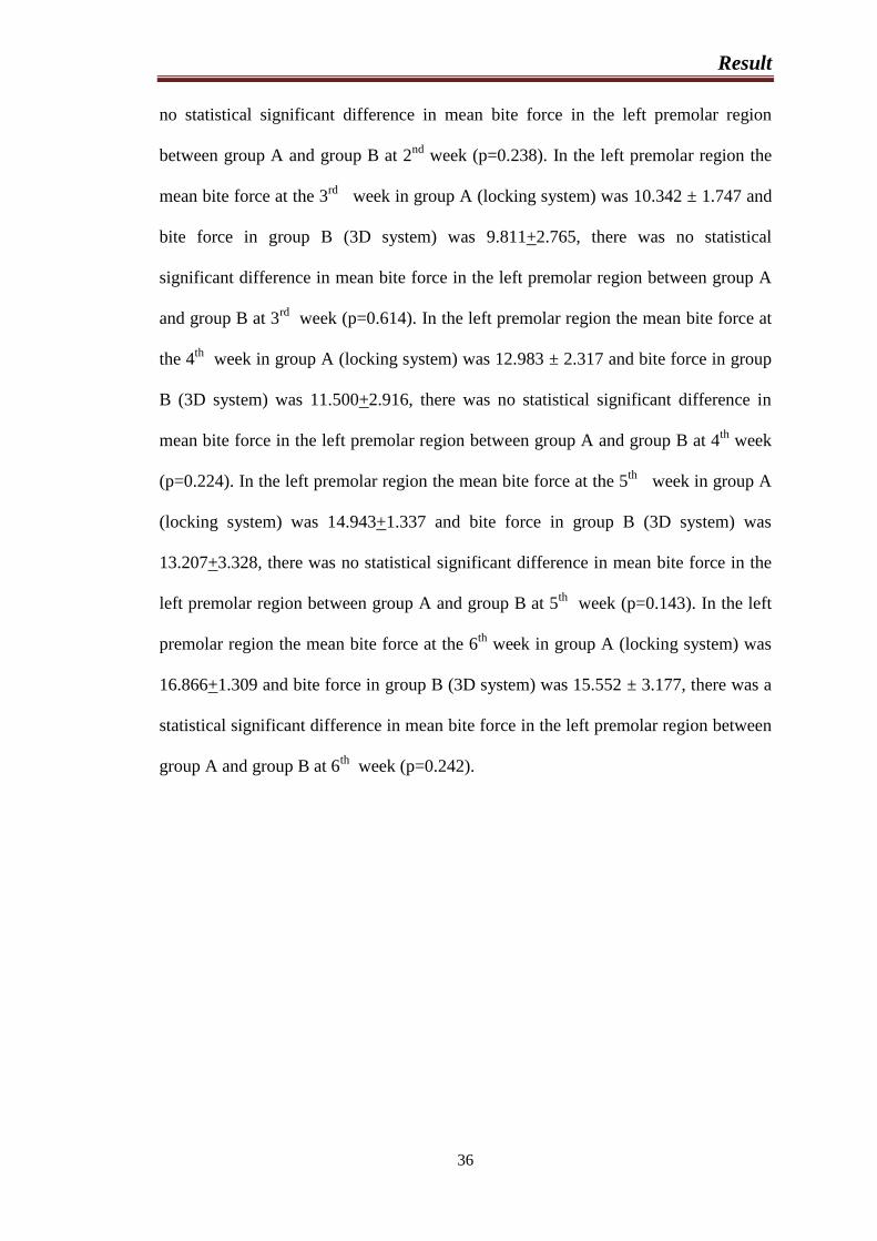
INCISOR LOCKING 10 8.192 2.267
7.274 18 <0.001 3D 10 2.592 0.886
RIGHT
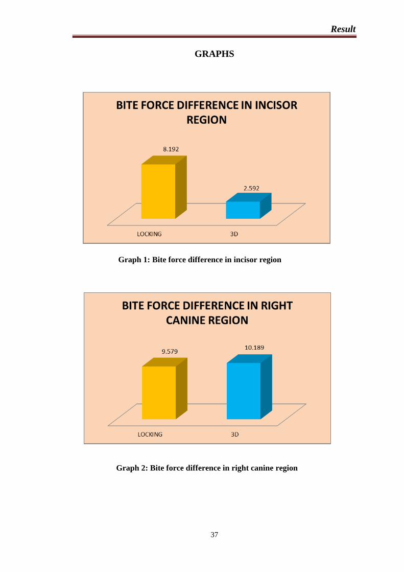
CANINE
LOCKING 10 9.579 3.090 -.400 18 0.694
3D 10 10.189 3.708
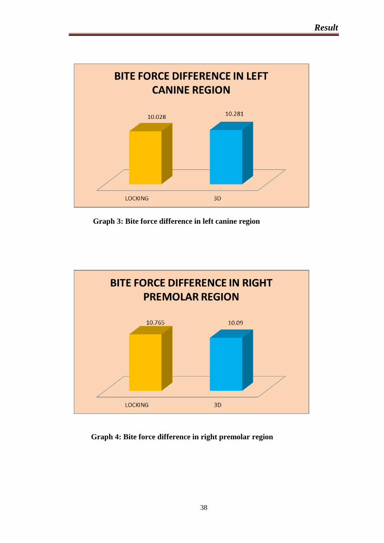
LEFT
CANINE
LOCKING 10 10.028 3.591 -.182 18 0.858
3D 10 10.281 2.532
RIGHT
PREMOLAR
LOCKING 10 10.765 2.166 .579 18 0.570
3D 10 10.090 2.980
LEFT
PREMOLAR
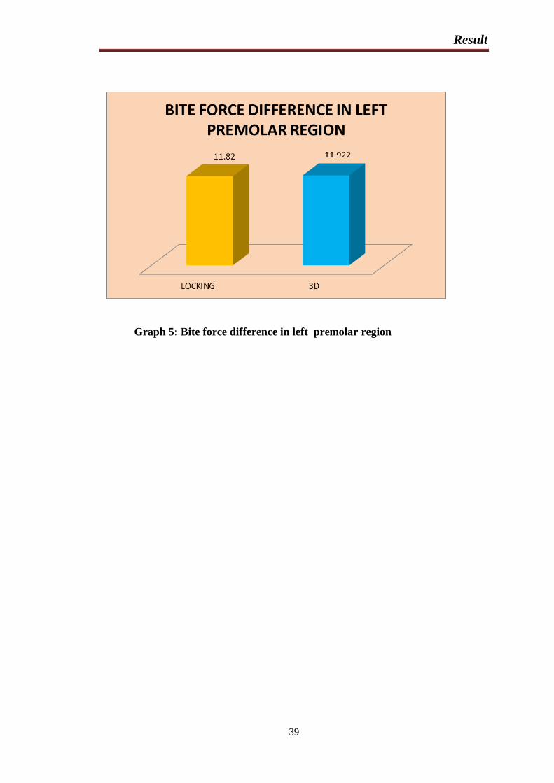
LOCKING 10 11.820 1.615 -.090 18 0.930
3D 10 11.922 3.213
The mean difference in bite force in incisor, canine and premolar region between
group A and group B was compared using Unpaired t test.
According to present study in the incisor region the mean difference in bite
force in group A (locking system) was 8.192 + 2.267 and mean difference in bite
force in group B (3D system) was 2.592+ 0.886, there was a statistical significant
difference in mean difference in bite force in the incisor region between group A and
group B (p<0.001). In the right canine region the mean difference in bite force in
group A (locking system) was 9.579 + 3.090 and mean difference in bite force in
group B (3D system) was 10.189 + 3.708, there was no statistical significant
difference in mean difference in bite force in the right canine region between group A
and group B (p=0.694). In the left canine region the mean difference in bite force in
group A (locking system) was 10.028 + 3.591 and mean difference in bite force in
group B (3D system) was 10.281+ 2.532, there was no statistical significant
Result
27
difference in mean difference in bite force in the left canine region between group A
and group B (p=0.858). In the right premolar region the mean difference in bite force
in group A (locking system) was 10.765+ 2.166 and mean difference in bite force in
group B (3D system) was 10.090+ 2.980, there was no statistical significant
difference in mean difference in bite force in the right premolar region between group
A and group B (p=0.570). In the left premolar region the mean difference in bite
force in group A (locking system) was 11.820+ 1.615 and mean difference in bite
force in group B (3D system) was 11.922+ 3.123, there was a statistical significant
difference in mean difference in bite force in the left premolar region between group
A and group B (p=0.930).
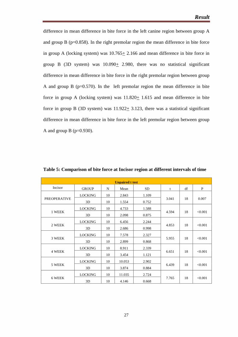
Table 5: Comparison of bite force at Incisor region at different intervals of time
Unpaired t test
Incisor GROUP N Mean SD t df P
PREOPERATIVE LOCKING 10 2.843 1.109
3.041 18 0.007 3D 10 1.554 0.752
1 WEEK LOCKING 10 4.733 1.588
4.594 18 <0.001 3D 10 2.098 0.875
2 WEEK LOCKING 10 6.456 2.244
4.853 18 <0.001 3D 10 2.686 0.998
3 WEEK LOCKING 10 7.578 2.327
5.955 18 <0.001 3D 10 2.899 0.868
4 WEEK LOCKING 10 8.911 2.339
6.651 18 <0.001 3D 10 3.454 1.121
5 WEEK LOCKING 10 10.053 2.902
6.439 18 <0.001 3D 10 3.874 0.884
6 WEEK LOCKING 10 11.035 2.724
7.765 18 <0.001 3D 10 4.146 0.668
Result
28
The mean bite force in the incisor region between group A and group B at different
time interval was compared using Unpaired t test.
According to present study in the incisor region the mean bite force at the
baseline (Preoperative period) in group A (locking system) was 2.843+1.109 and bite
force in group B (3D system) was 1.554+ 0.752, there was a statistical significant
difference in mean bite force in the incisor region between group A and group B at
baseline (p=0.007). In the incisor region the mean bite force at the 1st week in group
A (locking system) was 4.733+1.588 and bite force in group B (3D system) was
2.098+ 0.875, there was a statistical significant difference in mean bite force in the
incisor region between group A and group B at 1st week (p<0.001). In the incisor
region the mean bite force at the 2nd
week in group A (locking system) was
6.456+2.244 and bite force in group B (3D system) was 2.686+ 0.998, there was a
statistical significant difference in mean bite force in the incisor region between group
A and group B at 2nd
week (p<0.001). In the incisor region the mean bite force at the
3rd
week in group A (locking system) was 7.578+2.327 and bite force in group B
(3D system) was 2.899+ 0.868, there was a statistical significant difference in mean
bite force in the incisor region between group A and group B at 3rd
week (p<0.001).
In the incisor region the mean bite force at the 4th
week in group A (locking system)
was 8.911+2.339 and bite force in group B (3D system) was 3.454+ 1.121, there was
a statistical significant difference in mean bite force in the incisor region between
group A and group B at 4th
week (p<0.001). In the incisor region the mean bite force
at the 5th
week in group A (locking system) was 10.053+2.902 and bite force in
group B (3D system) was 3.874+ 0.884, there was a statistical significant difference
in mean bite force in the incisor region between group A and group B at 5th
week
(p<0.001). In the incisor region the mean bite force at the 6th
week in group A
Result
29
(locking system) was 11.035+2.724 and bite force in group B (3D system) was
4.146+ 0.668, there was a statistical significant difference in mean bite force in the
incisor region between group A and group B at 6th
week (p<0.001).
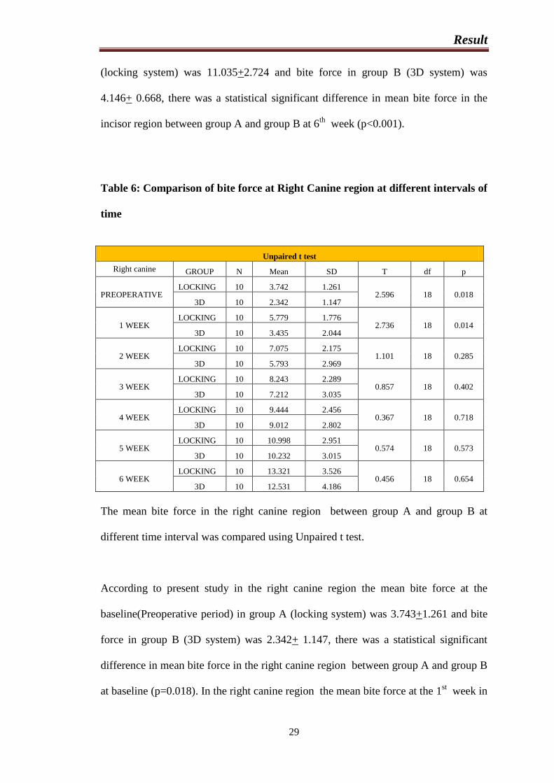
Table 6: Comparison of bite force at Right Canine region at different intervals of
time
Unpaired t test
Right canine GROUP N Mean SD T df p
PREOPERATIVE LOCKING 10 3.742 1.261
2.596 18 0.018 3D 10 2.342 1.147
1 WEEK LOCKING 10 5.779 1.776
2.736 18 0.014 3D 10 3.435 2.044
2 WEEK LOCKING 10 7.075 2.175
1.101 18 0.285 3D 10 5.793 2.969
3 WEEK LOCKING 10 8.243 2.289
0.857 18 0.402 3D 10 7.212 3.035
4 WEEK LOCKING 10 9.444 2.456
0.367 18 0.718 3D 10 9.012 2.802
5 WEEK LOCKING 10 10.998 2.951
0.574 18 0.573 3D 10 10.232 3.015
6 WEEK LOCKING 10 13.321 3.526
0.456 18 0.654 3D 10 12.531 4.186
The mean bite force in the right canine region between group A and group B at
different time interval was compared using Unpaired t test.
According to present study in the right canine region the mean bite force at the
baseline(Preoperative period) in group A (locking system) was 3.743+1.261 and bite
force in group B (3D system) was 2.342+ 1.147, there was a statistical significant
difference in mean bite force in the right canine region between group A and group B
at baseline (p=0.018). In the right canine region the mean bite force at the 1st week in
Result
30
group A (locking system) was 5.779+1.776 and bite force in group B (3D system)
was 3.435+ 2.044, there was a statistical significant difference in mean bite force in
the right canine region between group A and group B at 1st week (p=0.014). In the
right canine region the mean bite force at the 2nd
week in group A (locking system)
was 7.075+2.175 and bite force in group B (3D system) was 5.793+ 2.969, there was
no statistical significant difference in mean bite force in the right canine region
between group A and group B at 2nd
week (p=0.285). In the right canine region the
mean bite force at the 3rd
week in group A (locking system) was 8.243+ 2.289and
bite force in group B (3D system) was 7.212+3.035, there was no statistical
significant difference in mean bite force in the right canine region between group A
and group B at 3rd
week (p=0.402). In the right canine region the mean bite force at
the 4th
week in group A (locking system) was 9.444+2.456 and bite force in group B
(3D system) was 9.012+2.802, there was no statistical significant difference in mean
bite force in the right canine region between group A and group B at 4th
week
(p=0.718). In the right canine region the mean bite force at the 5th
week in group A
(locking system) was 10.998+2.951 and bite force in group B (3D system) was
10.232+ 3.015, there was no statistical significant difference in mean bite force in the
right canine region between group A and group B at 5th
week (p=0.573). In the right
canine region the mean bite force at the 6th
week in group A (locking system) was
13.321+3.526 and bite force in group B (3D system) was 12.531+ 4.186, there was a
statistical significant difference in mean bite force in the right canine region between
group A and group B at 6th
week (p=0.654).
Result
31
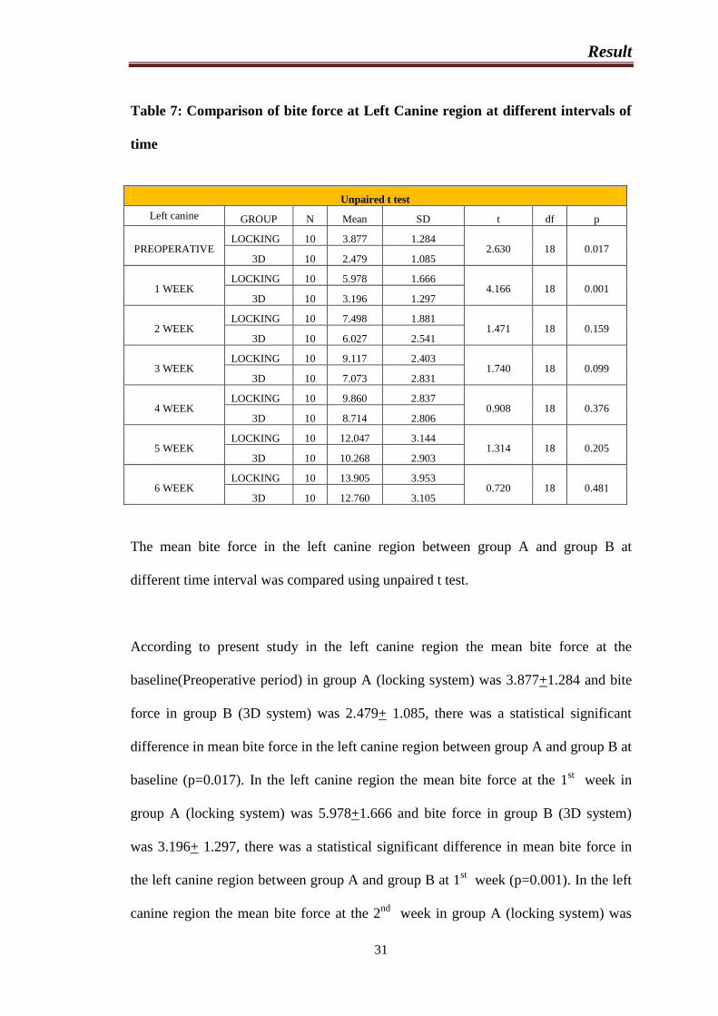
Table 7: Comparison of bite force at Left Canine region at different intervals of
time
Unpaired t test
Left canine GROUP N Mean SD t df p
PREOPERATIVE LOCKING 10 3.877 1.284
2.630 18 0.017 3D 10 2.479 1.085
1 WEEK LOCKING 10 5.978 1.666
4.166 18 0.001 3D 10 3.196 1.297
2 WEEK LOCKING 10 7.498 1.881
1.471 18 0.159 3D 10 6.027 2.541
3 WEEK LOCKING 10 9.117 2.403
1.740 18 0.099 3D 10 7.073 2.831
4 WEEK LOCKING 10 9.860 2.837
0.908 18 0.376 3D 10 8.714 2.806
5 WEEK LOCKING 10 12.047 3.144
1.314 18 0.205 3D 10 10.268 2.903
6 WEEK LOCKING 10 13.905 3.953
0.720 18 0.481 3D 10 12.760 3.105
The mean bite force in the left canine region between group A and group B at
different time interval was compared using unpaired t test.
According to present study in the left canine region the mean bite force at the
baseline(Preoperative period) in group A (locking system) was 3.877+1.284 and bite
force in group B (3D system) was 2.479+ 1.085, there was a statistical significant
difference in mean bite force in the left canine region between group A and group B at
baseline (p=0.017). In the left canine region the mean bite force at the 1st week in
group A (locking system) was 5.978+1.666 and bite force in group B (3D system)
was 3.196+ 1.297, there was a statistical significant difference in mean bite force in
the left canine region between group A and group B at 1st week (p=0.001). In the left
canine region the mean bite force at the 2nd
week in group A (locking system) was
Result
32
7.498+1.881 and bite force in group B (3D system) was 6.027+ 2.541, there was no
statistical significant difference in mean bite force in the left canine region between
group A and group B at 2nd
week (p=0.159). In the left canine region the mean bite
force at the 3rd
week in group A (locking system) was 9.117 + 2.403 and bite force in
group B (3D system) was 7.073+2.831, there was no statistical significant difference
in mean bite force in the left canine region between group A and group B at 3rd
week
(p=0.099). In the left canine region the mean bite force at the 4th
week in group A
(locking system) was 9.860+2.837 and bite force in group B (3D system) was
8.714+2.806, there was no statistical significant difference in mean bite force in the
left canine region between group A and group B at 4th
week (p=0.376). In the left
canine region the mean bite force at the 5th
week in group A (locking system) was
12.047+3.144 and bite force in group B (3D system) was 10.268+ 2.903, there was no
statistical significant difference in mean bite force in the left canine region between
group A and group B at 5th
week (p=0.205). In the left canine region the mean bite
force at the 6th
week in group A (locking system) was 13.905+3.953 and bite force in
group B (3D system) was 12.760+3.105, there was a statistical significant difference
in mean bite force in the left canine region between group A and group B at 6th
week
(p=0.481).
Result
33
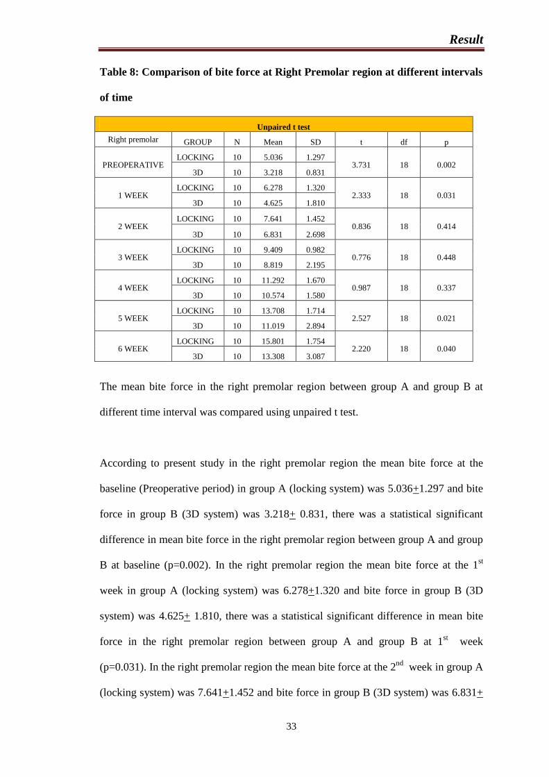
Table 8: Comparison of bite force at Right Premolar region at different intervals
of time
Unpaired t test
Right premolar GROUP N Mean SD t df p
PREOPERATIVE LOCKING 10 5.036 1.297
3.731 18 0.002 3D 10 3.218 0.831
1 WEEK LOCKING 10 6.278 1.320
2.333 18 0.031 3D 10 4.625 1.810
2 WEEK LOCKING 10 7.641 1.452
0.836 18 0.414 3D 10 6.831 2.698
3 WEEK LOCKING 10 9.409 0.982
0.776 18 0.448 3D 10 8.819 2.195
4 WEEK LOCKING 10 11.292 1.670
0.987 18 0.337 3D 10 10.574 1.580
5 WEEK LOCKING 10 13.708 1.714
2.527 18 0.021 3D 10 11.019 2.894
6 WEEK LOCKING 10 15.801 1.754
2.220 18 0.040 3D 10 13.308 3.087
The mean bite force in the right premolar region between group A and group B at
different time interval was compared using unpaired t test.
According to present study in the right premolar region the mean bite force at the
baseline (Preoperative period) in group A (locking system) was 5.036+1.297 and bite
force in group B (3D system) was 3.218+ 0.831, there was a statistical significant
difference in mean bite force in the right premolar region between group A and group
B at baseline (p=0.002). In the right premolar region the mean bite force at the 1st
week in group A (locking system) was 6.278+1.320 and bite force in group B (3D
system) was 4.625+ 1.810, there was a statistical significant difference in mean bite
force in the right premolar region between group A and group B at 1st week
(p=0.031). In the right premolar region the mean bite force at the 2nd
week in group A
(locking system) was 7.641+1.452 and bite force in group B (3D system) was 6.831+
Result
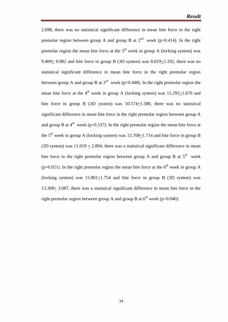
34
2.698, there was no statistical significant difference in mean bite force in the right
premolar region between group A and group B at 2nd
week (p=0.414). In the right
premolar region the mean bite force at the 3rd
week in group A (locking system) was
9.409+ 0.982 and bite force in group B (3D system) was 8.819+2.192, there was no
statistical significant difference in mean bite force in the right premolar region
between group A and group B at 3rd
week (p=0.448). In the right premolar region the
mean bite force at the 4th
week in group A (locking system) was 11.292+1.670 and
bite force in group B (3D system) was 10.574+1.580, there was no statistical
significant difference in mean bite force in the right premolar region between group A
and group B at 4th
week (p=0.337). In the right premolar region the mean bite force at
the 5th
week in group A (locking system) was 13.708+1.714 and bite force in group B
(3D system) was 11.019 + 2.894, there was a statistical significant difference in mean
bite force in the right premolar region between group A and group B at 5th
week
(p=0.021). In the right premolar region the mean bite force at the 6th
week in group A
(locking system) was 15.801+1.754 and bite force in group B (3D system) was
13.308+ 3.087, there was a statistical significant difference in mean bite force in the
right premolar region between group A and group B at 6th
week (p=0.040).
Result
35
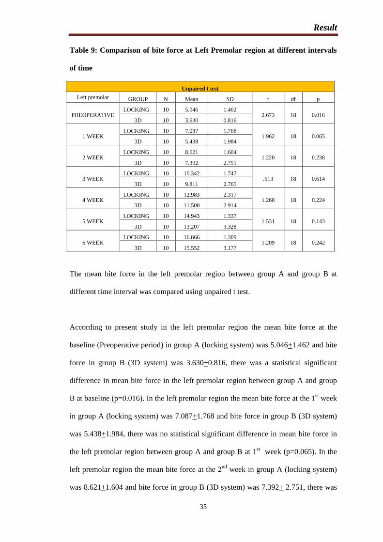
Table 9: Comparison of bite force at Left Premolar region at different intervals
of time
Unpaired t test
Left premolar GROUP N Mean SD t df p
PREOPERATIVE LOCKING 10 5.046 1.462
2.673 18 0.016 3D 10 3.630 0.816
1 WEEK LOCKING 10 7.087 1.768
1.962 18 0.065 3D 10 5.438 1.984
2 WEEK LOCKING 10 8.621 1.604
1.220 18 0.238 3D 10 7.392 2.751
3 WEEK LOCKING 10 10.342 1.747
.513 18 0.614 3D 10 9.811 2.765
4 WEEK LOCKING 10 12.983 2.317
1.260 18 0.224 3D 10 11.500 2.914
5 WEEK LOCKING 10 14.943 1.337
1.531 18 0.143 3D 10 13.207 3.328
6 WEEK LOCKING 10 16.866 1.309
1.209 18 0.242 3D 10 15.552 3.177
The mean bite force in the left premolar region between group A and group B at
different time interval was compared using unpaired t test.
According to present study in the left premolar region the mean bite force at the
baseline (Preoperative period) in group A (locking system) was 5.046+1.462 and bite
force in group B (3D system) was 3.630+0.816, there was a statistical significant
difference in mean bite force in the left premolar region between group A and group
B at baseline (p=0.016). In the left premolar region the mean bite force at the 1st week
in group A (locking system) was 7.087+1.768 and bite force in group B (3D system)
was 5.438+1.984, there was no statistical significant difference in mean bite force in
the left premolar region between group A and group B at 1st week (p=0.065). In the
left premolar region the mean bite force at the 2nd
week in group A (locking system)
was 8.621+1.604 and bite force in group B (3D system) was 7.392+ 2.751, there was
Result
36
no statistical significant difference in mean bite force in the left premolar region
between group A and group B at 2nd
week (p=0.238). In the left premolar region the
mean bite force at the 3rd
week in group A (locking system) was 10.342 ± 1.747 and
bite force in group B (3D system) was 9.811+2.765, there was no statistical
significant difference in mean bite force in the left premolar region between group A
and group B at 3rd
week (p=0.614). In the left premolar region the mean bite force at
the 4th
week in group A (locking system) was 12.983 ± 2.317 and bite force in group
B (3D system) was 11.500+2.916, there was no statistical significant difference in
mean bite force in the left premolar region between group A and group B at 4th
week
(p=0.224). In the left premolar region the mean bite force at the 5th
week in group A
(locking system) was 14.943+1.337 and bite force in group B (3D system) was
13.207+3.328, there was no statistical significant difference in mean bite force in the
left premolar region between group A and group B at 5th
week (p=0.143). In the left
premolar region the mean bite force at the 6th
week in group A (locking system) was
16.866+1.309 and bite force in group B (3D system) was 15.552 ± 3.177, there was a
statistical significant difference in mean bite force in the left premolar region between
group A and group B at 6th
week (p=0.242).
Result
37
GRAPHS
Graph 1: Bite force difference in incisor region
Graph 2: Bite force difference in right canine region
Result
38
Graph 3: Bite force difference in left canine region
Graph 4: Bite force difference in right premolar region
Result
39
Graph 5: Bite force difference in left premolar region
Discussion
40
DISCUSSION
As the saying goes, the only permanent thing in this world is change, for the treatment
of mandibular fractures the newer methods have been tried and older ones have had
improvements. The strategic position of the mandible on the facial skeleton and its
unique role in mastication, deglutition, phonation and aesthetics compels the clinician
to give immediate attention when the lower jaw is injured.
It is well established that the bone healing is optimized by precise anatomic
reduction and rigid immobilization. Once fractures are reduced and immobilized
optimal bone repair is dependent on preservation and maintenance of intact blood
supply. Movement of fractures causes disruption of the osteogenic elements and
capillaries. This results in formation of poorly vascularized fibrous tissue which gives
rise to complications in fracture healing, like fibrous union or sometimes even non-
union.
With advances in our understanding of bone healing after the fracture, there has
been a transformation from anatomic fixation principle to a more biomechanical
fixation principle, in the management of fractures. Various designs of internal fixation
devices are constantly being modified and its biomechanical potentials explored to a
maximum extent.
Today, internal fixation devices such as miniplates, are widely used in traumatology
of maxillofacial area and they have replaced conservative treatment with MMF. But
MMF remains an important and effective tool especially in cases where plates cannot
be used due to economical, personal and logistic reasons. Research continues to focus
on the size, shape, number and biomechanics of plate/screws systems to improve
surgical outcomes.
Discussion
41
Open reduction and internal fixation of mandibular fractures with bone plates was
first described by Schede in 18884.General acceptance of open osteosynthesis did not
appear in maxillofacial literature until an organised research by AO in 1950.Luhr,
Spissel and others derived inspiration from orthopaedic biomechanical studies
performed by Schenk.Champy‟s experiment with miniplates further delineated the
“Ideal lines of Osteosynthesis” with the mandible. Wire osteosynthesis was followed
many years before the plates invention which did not provide stability in 3
dimensions.
In 1913, Lambotte recommended an aluminium geometrically closed quadrangular
plate secured with bone screws for the treatment of fractures of mandibular body via
an extraoral approach. He found that, when the fragments were repositioned properly,
this specially designed plate osteosynthesis offered sufficient stability without further
immobilization45
. Furthermore this system was superior to that of wire osteosynthesis.
Complications of miniplates especially in the angle region have been described in
30% of cases, like movement and loosening of screws leading to failure of fracture
treatment with other limitations of standard miniplates such as need for precise
adaptations and less stability across fracture line46
.
Mostafa Farmand introduced new 3D plating system. The shape of 3D plate is
basically quadrangular geometrically consisting of two horizontal plates
interconnected by vertical struts where they provide stability in three dimensions.
These unique plate consists of square or rectangular units 2x2,3x2,4x2 holes and
provides increased torsional stability which are in consideration with studies shown
by Jimson et al in 2009. The 3D miniplate itself is a misnomer as the plates
Discussion
42
themselves are not 3 dimensional but hold the fracture segments rigidly by resisting
the 3 dimensional forces namely shearing, bending and torsional forces acting on the
fracture site in function.47
Similarly, in the present study, a positive correlation was found between the
preoperative and postoperative weeks and the bite force for the anterior and posterior
regions of the fracture sites in both groups. There was a progressive increase in the
bite force readings from the preoperative period to 6 weeks post operatively in both
the groups. At other regions the bite force in both the groups increased at the same
rate with no statistically significant difference.
In conclusion the results from this study suggests that fixation of non communited
mandibular symphysis and parasymphysis fractures with a 2.0mm 3D miniplates
provides three dimensional stability and carries low morbidity and infection rates,
supporting the studies carried out by Jimson S et al (2009)33
and Goyal. M et al
(2011).39
The 3D plating system have provided the following features:
1. The quadrangle geometry of plate assures a three dimensional stability of
fracture site as it offers good resistance against torque forces.
2. Early restoration of mandibular functions postoperatively.
3. Simplicity, malleability, low profile and ease of application.
4. Less postoperative surgical morbidity.
5. Less operating time.
6. Easy adaptability of the plate.
Discussion
43
The possible limitations of these plates are:
1. Excessive implant material due to the extra vertical bars incorporated for
countering the torque forces limits its use.
2. Difficult to use in communited fractures.
3. Cost of these plates which is slightly more than the conventional plates.
4. Difficult to adapt at the mental foramen region.
The ideal method of treating mandibular fractures is the one that establishes a
functional therapy by movement. Therefore, it should aim for the following48
1. Reestablishment of the lost occlusion
2. Perfect anatomic reduction.
Complete and stable fixation, allowing painless mobilisation of the injured
region.
3. Maintenance of blood supply of the fractured fragments and of the
surrounding tissues.
The biomechanical requirements for ideal osteosynthesis are that48
The plates and screws should withstand the various stresses due to the tensile
and tortional forces to which the mandibular bone is typically subjected to.
The plates have to be malleable for easy adaptation to the bone surface,
especially in the curved symphysis and molar region to secure anatomical
reduction and to restore perfect occlusion.
The dimensions of the plate should ensure minimal periosteal elevation and
fracture site exposure; furthermore, the Oral mucosa must be able to cover the
plate without any difficulty, without any dead space around the plate and head
of the screws
The size of the screws has to be appropriate for the thickness of the cortex.
Discussion
44
This study define ideal line of osteosynthesis in the mandible which corresponds to
the course of a tension line at the base of the alveolar process inferior to the root
apices. In that region a plate can be fixed with monocortical screws as follows-48
Behind the mental foramina the plate is applied immediately below the dental
roots and above the inferior alveolar nerve.
At the angle of the jaw the plate is placed ideally on the inner broad surface of
the external oblique line; if this has been destroyed, the plate is fixed on the
external cortex as high as possible.
In the anterior region, between the mental foramina, in addition to the sub
apical plate, another plate near the lower border of the mandible is necessary
to neutralize the torsion forces.
The result of such a monocortical, stable, dynamic osteosynthesis is the neutralization
of the distraction and torsion forces exerted on the fracture site, while physiological
self compression strains are restored. These developments helped in early functional
recovery of the patients thereby improving the quality of life. Several biomechanical
studies have been conducted to evaluate the post operative functional aspects of the
jaws.48
Over the last two decades miniplate osteosynthesis has induced a revolution in
mandibular fracture treatment. This modern system provides better handling, higher
stability and less pressure on the bone and they have great advantages, like the intra-
oral approach and the easy adaptability. In addition, it is no longer necessary to
expose bone extensively. But with the standard miniplates, loosening of the screws
due to transmission of pressure to the underlying bone led to loss of fracture stability
and then fixation failure. Nevertheless complications in miniplate osteosynthesis of
Discussion
45
the mandible especially in the angle region have been described in upto 30% of cases,
like movement and loosening of screws leading to failure of fracture treatment.46
Fixation failure results in fracture mobility that can subsequently lead to infections,
non union and/or malunion. Fixation fails by a number of mechanisms which include:
improper fixation, fracture of the plate, loosening of the screws, devitalization of bone
around screws. Other limitations of standard miniplates include need for precise
adaptations and less stability across the fracture line.
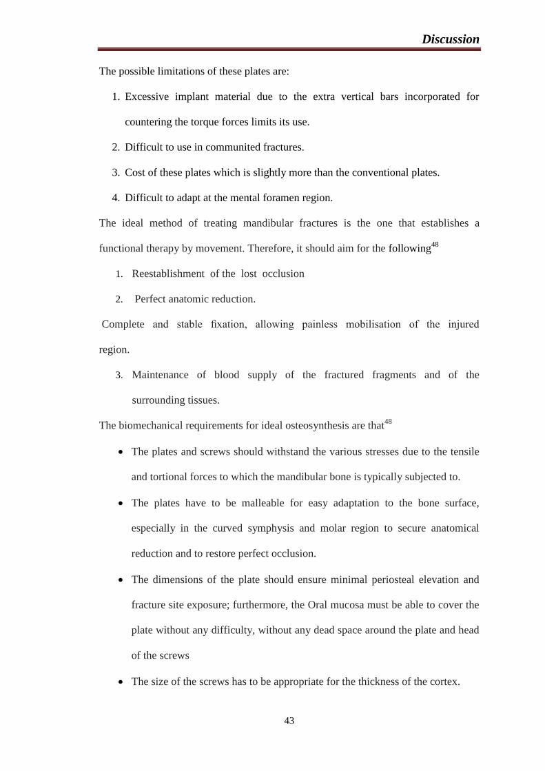
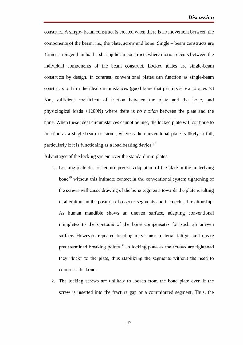
Figure 1: (A) Left : primary stability of the conventional miniplate system depends
on friction (blue arrows) between screw, plate and bone. Right: distribution of load in
a conventional miniplate screw (green arrow), counterforce of the bone for
compensating axial forces (red arrow). (B) Left: the 3- dimensional frame (white box)
is the major principle of stability of the mini – locking system. Right: distribution of
load in a mini – locking screw. The screw‟s geometry distributes load (green arrow)
Discussion
46
to several spots (red arrows): the bone between the thread ridges above the pivot (blue
dot) is under pressure load (yellow arrows), while the opposite happens to the bone on
the other side below the assumed pivot.
These led to the introduction of locking plate system by Ralf Gutwald. He did the first
in vitro biomechanical comparison of the standard and locking miniplate system in
1999 and found that locking plates were more stable at the angle region.49
The first
clinical study comparing the two systems was published by Ellis et al in 2002.10
This
new design of Mini-Locking plate provided locking of the screws on both the plate
and the bone interface, either side of the fracture. Thus, a frame construct was
achieved on either side of the fracture fragments. (Figure: 1). This provided better
stability of the fracure fragments, and thus better healing environment, when
compared to the conventional plates, while retaining the same miniature dimensions.27
It was also proved that the fracture gap and torsion forces at the fracture site were
significantly low in locking plates when compared to that of conventional plates.
They proposed that the frame construct model prevents any pressure from
accumulating beneath the plates and hence prevents any interference of vascular
supply of the bone. Thus, the periosteum grows beneath the plates and over the
fracture site without any interference. The improved immobilization of the fracture
segments, along with the abundant and interruped blood supply, gives a stable
environment for callus formation; the healing enters the phase of remodelling without
delay. Thus, the callus is remodeled into new bone that is capable of taking up the
biomechanical forces, at a faster rate. Hence it is postulated that function is restored
earlier with locking plates.
The newer locked plates control the axial orientation of the screw to the plate, thereby
enhancing screw – beam – bone construct stability by creating a single beam
Discussion
47
construct. A single- beam construct is created when there is no movement between the
components of the beam, i.e., the plate, screw and bone. Single – beam constructs are
4times stronger than load – sharing beam constructs where motion occurs between the
individual components of the beam construct. Locked plates are single-beam
constructs by design. In contrast, conventional plates can function as single-beam
constructs only in the ideal circumstances (good bone that permits screw torques >3
Nm, sufficient coefficient of friction between the plate and the bone, and
physiological loads <1200N) where there is no motion between the plate and the
bone. When these ideal circumstances cannot be met, the locked plate will continue to
function as a single-beam construct, whereas the conventional plate is likely to fail,
particularly if it is functioning as a load bearing device.27
Advantages of the locking system over the standard miniplates:
1. Locking plate do not require precise adaptation of the plate to the underlying
bone50
without this intimate contact in the conventional system tightening of
the screws will cause drawing of the bone segments towards the plate resulting
in alterations in the position of osseous segments and the occlusal relationship.
As human mandible shows an uneven surface, adapting conventional
miniplates to the contours of the bone compensates for such an uneven
surface. However, repeated bending may cause material fatigue and create
predetermined breaking points.37
In locking plate as the screws are tightened
they “lock” to the plate, thus stabilizing the segments without the need to
compress the bone.
2. The locking screws are unlikely to loosen from the bone plate even if the
screw is inserted into the fracture gap or a comminuted segment. Thus, the
Discussion
48
incidence of inflammatory complications from loosening of the hard ware is
decreased.9
3. The amount of stability provided across the fracture segments is greater.27
4. They do not disrupt the underlying cortical bone perfusion as much as the
conventional plates which compress the undersurface of the bone plate to the
cortical bone.27
5. The locking screw plate system also reduces compressive forces between the
undersurface of the plate and lateral bony cortex compared with a
conventional mandibular plate. In a locking screw plate system, forces are
generated between the threaded portion of the plate and the screw. This limits
stress shielding effect and creates a more stable fixation over a period of
time.51
6. Locking plates can be used in all type of mandibular fracture including
comminuted and infected fractures of ramus, angle, body, parasymphysis and
symphysis.
Disadvantages of locking plates:
1. The disadvantages of locking plates are that these plates require to „center‟ the
drill hole with the plate hole to ensure perpendicular placement of the screw. If
screws are not placed perpendicular to plate the screw will not engage the
threaded plate hole precisely and therefore will not lock.
2. Another disadvantage of locking plates, it is little expensive when compared to
the conventional plates.52
Inspite of all the advantages that, these locking plates and screw offer to the surgeon,
they are still far from being the perfect treatment modality. Though clinically more
Discussion
49
effcient than a conventional plate, the locking plate still has the same complication
rates.26,52
Herzberg noticed a higher rate of complications when the interval between trauma and
surgical treatment is delayed more than 6 days.37
However, according to Ellis et al,
there is no convincing evidence in the literature correlating the delay in treatment of
mandibular fractures with the increase in the postoperative complications, such as
infection.53
Even in the present study, no such correlation was found.
Scolozzi et al used single AO 2mm locking reconstruction plate in linear non
comminuted mandibular fractures and found sound bone healing with no major
complications.35
In our study we used single 2mm locking miniplate in 10 fractures
(symphysis and parasymphysis region) and achieved similar results like above
mentioned study without any major complications.
Coredy and co – workers state that the friction between the screw and plate is the
main weak point of the entire fixation.54
In the locking plate system the thread on the
screw locks into the congruent thread of the plate, transforming the screws and plate
into a unit, creating a rigid splint with higher mechanical stability which corresponds
to the principle of an external fixator, where the friction between the plate and the
bone is totally avoided.55
This was easily experienced in the present study by us.
In the prospective study by Cawood, 5.7 % of mandibular fractures, excluding
additional condylar neck fractures, treated by miniplates osteosynthesis had
malocclusion detected on review.56
Champy et al. reported a 4.8% malocclusion rate
in their group treated by miniplate osteosynthesis.57
The first biomechanical
comparison of locking plates found in the maxillofacial surgical literature was made
by Gutwald in 1999 and they concluded that a higher stability was achieved with the
locking plates.49
So, favoring the other similar studies, in the present study inspite of
Discussion
50
using a single locking miniplate at the symphysis and parasymphysis region (n=10) in
comparison of two standard miniplates as per Champy‟s principles no instances of
segmental mobility were noted.
Cawood showed mental parasthesia in 8% of cases of mandibular fractures treated by
miniplate osteosynthesis.56
In another study postoperative parasthesia was found to be
31.52%.23
in a recent study, higher incidence of temporary parasthesia in patients
treated with locking miniplates as compared to the standard miniplates was observed
and was attributed to the necessity for more tissue retraction to accommodate the drill
guide and subsequent placement of perpendicular screws.32
However, in the present
study no instances of mental nerve parasthesia were noted. This difference may be
due to differences in the proximity of the fracture line to the mental foramen, to the
differences in technical expertise of the operator and lastly to the smaller sample size
of the present study.
The bite force is related to a number of factors, including tactile impulses, pain and
pressure reception in the periodontal ligament, the number of residual teeth and
patient age, because a reduction in bite force can occur with age owing to the age-
dependent deterioration of the dentition. It has long been known that a neuromuscular
protective mechanism occurs throughout the body. For instance, one of the first
protective mechanisms called into play when a fracture occurs is “muscle splinting” in
which selective components of the neuromuscular system are activated or deactivated
to remove the force from the damaged bone. In 1994, Tate et al stated that sufficient
internal fixation hardware should be applied to resist the maximal force of
mastication. Thus, they hypothesized that the stability of fracture segment would be
ensured even under the full function of the masticatory system.
Discussion
51
The amount of force generated by the patients with untreated as well as treated
fractures is much less. Furthermore, one must remember that the data reported
concerned the maximal voluntary bite force (i.e., the maximum bite force an
individual can voluntarily generate) the amount of force used during functional
activities would probably be much less. Hence, the fixation requirements, determined
from the maximal voluntarily bite force of noninjured subjects, might be inflated, and
this fixation requirement is perhaps a semirigid form of fixation such as monocortical
fixation.41
In 2002, Gerlach and Schwarz in their study bite force of treated
mandibular angle fractures concluded that the maximal bite force in patients with
mandibular fractures treated with miniplate osteosynthesis had reached only 31% at 1
week postoperatively compared with a healthy control group. These values had
increased to 58% at 6 weeks postoperatively.22
Similarly in the present study, a positive co relation was found between post -
operative anterior and posterior bite force in both the groups. There was a progressive
increase in the bite force readings from the pre – operative to 6 weeks post operatively
in both the groups. The mean difference between the two groups is statistically not
significant at right canine region (p= 0.694), left canine (p= 0.858), right premolar (p=
0.570), left premolar (p= 0.930) regions. But mean difference between the 2 groups is
statistically significant at incisor region with p<0.001. Hence there is no statistically
significant difference in the stability of 3-D miniplates and locking miniplate systems.
Limitations of the present study include the shorter follow up period and a smaller
sample size. Better comparison could have been carried out if the study was done on
isolated mandibular fractures at a particular fractured site of mandible. To show
significant improvements of locking plate / screw system or 3D plates/ screws, further
studies with a larger sample size with longer follow up is required.
Conclusion
52
CONCLUSION
The clinical outcome of both 3D and locking miniplate systems in the present study
were similar, however the following advantages of 3D and locking miniplates can be
highlighted:
Advantages of locking miniplate system-
Can be used as a “one-plate-for-all” system minimizing the number of fixation
systems necessary to be stocked in the operating room.
Lesser trauma to the periosteum and soft tissues with lesser amount of
hardware.
Advantages of 3D miniplate system-
Fixation of mandibular fractures with 3 Dimensional plates facilitates
simultaneous reduction and stabilization at both the superior and inferior
borders.
Post operative long term intermaxillary fixation can be avoided as good
stability was achieved with these plates.
The 3D plates were found to be of high standard in profile, strong yet
malleable, facilitating reduction and stabilization at both the superior and
inferior borders giving three dimensional stability at the fracture site.
Though we cannot comment on the rigidity and malleability of the 3 dimensional
plates and locking plates compared to other systems without a control group or
comparative study with other plating systems our statement is thoroughly based only
on our clinical experience. Further studies are necessary to set gold standards for

Conclusion
53
these plating systems with a control group and comparative studies with the above
mentioned plating systems.
Summary
54
SUMMARY
The study was designed with the aim to compare the efficacy of 3-dimensional
titanium miniplates with that of the titanium locking miniplates in the treatment of
mandibular fractures. Internal fixation using Champy’s miniplates is the preferred
mode of managing mandibular fractures in the interforaminal region. The patients
who required open reduction and internal fixation were selected for the study. As the
case required, the procedures were done under general anaesthesia or local
anaesthesia. The approaches used were both intraoral and extra oral as the case
required. . Patients were evaluated at 1st week to 6
th week for the treatment results and
the complications that might develop. None of the cases reported any immediate or
delayed complications. Malocclusion and hardware failure were not noticed in any of
the cases.
In our study A 4 holed (2x2) titanium 3-dimensional miniplate with interconnecting
vertical bars and a 4 holed titanium locking miniplate with bar was used and there is
no statistically significant difference in the efficacy of 3-D miniplate and Locking
miniplate systems but further prospective randomized comparative studies with larger
sample size and longer follow up are needed to ascertain these findings
authoritatively.
References
55
REFERENCES
1) Gassner R, Tuli T, Hachl O, Rudisch A, Ulmer H. Craniomaxillofacial trauma a 10 year
review of 9543 cases with 21067 injuries. J CraniomaxillofacSurg 2003;31:51-61.
2) Brasileiro BF, PasseriLA : Epidemiological analysis of maxillofacial fractures in Brazil :
a 5-year prospective study. Oral Surg Oral Med Oral Pathol Oral RadiolEndod
2006;102:28-34.
3) Mukerji, M. McGurk. Mandibular fractures: Historical Perspective. British J Oral
MaxillofacSurg 2006; 44: 222-228.
4) Andrew J. L. Gear, Apasova E, Schmitz JP and Schubert W. Treatment modalities for
mandibular angle fractures. J Oral MaxillofacSurg 2005; 63: 655-663.
5) Alkan.A, N. Celebi, B Ozden, B Bas and S. Inal. Biomechanical comparison of different
plating techniques in repair of mandibular angle fractures. Oral Surg Oral Med Oral Path
Oral RadiolEndod 2007; 104:752-756.
6) Zix J, Lieger O and Iizuka T. Use of Straight and Curved 3-Dimensional Titanium
Miniplates for Fracture Fixation at the Mandibular Angle. J Oral Maxillofac Surgery
2007; 65:1758-1763.
7) Peter Bui, NagiDemian, and Patrick Beetar. Infection Rate in Mandibular Angle
Fractures Treated With a 2.0-mm 8-Hole Curved Strut Plate. J Oral MaxillofacSurg
2009; 67: 804-808.
8) Tams J, Loon JP, Otten E, Rozema FR and Bos RRM. A three-dimensional study of
bending and torsion moments for different fracture sites in the mandible: an in vitro
study. Int. J Oral MaxillofacSurg 1997; 26: 383-388.
References
56
9) Herford AS, Ellis E III. Use of locking reconstruction bone plate/screw system for
mandibular surgery. J Oral MaxillofacSurg 1998;56:1261-1265.
10) Ellis E III, Graham J. use of a 2.0 mm locking plate/screw system for mandibular
surgery. J Oral MaxillofacSurg 2002;60:642-645.
11) Brons R, Boering G. Fracture of mandibular body treated by stable internal fixation: A
preliminary report. J Oral Surg 1970;28:407.
12) Ellis E III. Rigid skeletal fixation of fractures. J Oral Maxillofac Surg 1993;51:163-173.
13) Kahn Berg K.E. & A .Ridell: Bone Plate Fixation of Mandibular Fractures:Int. J Oral
MaxillofacSurg: 1980: 9: 267 – 273.
14) William C. Ardary. Prospective Clinical Evaluation of the use of Compression Plates and
Screws in the Management of Mandibular Fractures. Journal of Oral Maxillofacial
Surgery1989;47: 1150-1153.
15) Dodson TB, Perrott DH and Kaban LB, Gordon NC. Fixation of Mandibular Fractures: A
Comparative Analysis of Rigid Internal Fixation and Standard Fixation Techniques. J.
Oral Max FacSurg 1990; 48: 362 - 366.
16) J.P.Hayter,J.I.Cawood. The functional case for miniplates in maxillofacial surgery.
Int.J.Oral Maxillofacial Surgery1993;22:91-96.
17) Farmand M. : The 3d Plating System In Maxillofacial Surgery : J
OralMaxillofacSurg : 1993 : 51(3) : 166 – 167.
18) Shetty V, McBrearty D, Fourney M, Caputo AA. Fracture line stability as a function of
the internal fixation system: An invitro comparison using a mandibular angle fracture
model. J Oral MaxillofacSurg 1995;53:791-801.
References
57
19) Renton TF and Wiesenfeld D: Mandibular fracture osteosynthesis: a comparison of three
techniques. Br J Oral MaxillofacSurg 1996;34:166-73.
20) Sikes JW, Smith BR, Mukherjee DB, Coward KA. Comparison of fixation strengths of
locking head and conventional screws, in fracture and reconstruction models. J Oral
MaxillofacSurg 1998;56:468-473.
21) Bolourian R, Lazow S, Berger J. Transoral 2.0 mm Miniplate fixation of mandibular
fractures plus 2 weeks maxillomandibular fixation: A prospective study. J Oral
MaxillofacSurg 2002; 60:167–170.
22) Gerlach KL, Schwarz A. Bite forces in patients after treatment of Mandibular angle
fractures with miniplateosteosynthesis according to Champy. Int J Oral MaxillofacSurg
2002;31:345–348.
23) Gabrielli MAC, Gabrielli MFR, Marcantonio E, Hochuli-Vieira E. Fixation of
mandibular fractures with 2.0-mm miniplates: review of 191 Cases. J Oral
MaxillofacSurg 2003;61:430-436.
24) Alpert B, Gutwald R, Schmelzeisen R. New innovations in craniomaxillofacial fixation;
the 2.0 lock system. Keio J Med 2003;52:120-127.
25) Kirkpatrik D, Gandhi R, Sickels JEV. Infections associated with locking reconstruction
plates: A retrospective review. J Oral MaxillofacSurg 2003;61:462-466.
26) Collins CP, Leonard GP, Tolas A, and Alcalde R. A prospective randomized clinical trial
comparing 2.0-mm locking plates to 2.0-mm standard plates in treatment of mandible
fractures. J Oral MaxillofacSurg 2004;62:1392-1395.
27) EgolKa, Kubiac EN, Fulkerson E, Kummer FJ, Koval KJ. Biomechanics of locked plates
and screws. J Orthop Trauma 2004;18:488-493.
References
58
28) Militsakh ON, Wallace DI, Kriet D, Girod DA, Olvera MS, Tsue TT. Use of the 2.0mm
locking reconstruction plate in primary oromandibular reconstruction after composite
resection. Otolaryngol Head Neck Surg 2004;131:660-5.
29) Gear AJL, Apasova E, Schmitz JP, Schubert W. Treatment modalities for mandibular
angle fractures. J Oral MaxillofacSurg 2005;63:655-663.
30) Chritah A, Lazow SK, Julius R, Berger JR. Transoral 2.0mm locking miniplate fixation
of mandibular fractures plus 1 week of maxillomandibular fixation: A prospective study.
J Oral MaxillofacSurg 2005;63:1737-1741.
31) Sauerbier S, Schon R, Otten JE, Schmelzeisen R, Gutwald R. The development of plate
osteosynthesis for the treatment of fractures of the mandibular body - A literature review.
J CraniomaxillofacSurg 2008; 36,251-259.
32) Saikrishna D, Shetty SK, Marimallappa TR. A comparison between 2.0mm standard and
2.0mm locking miniplates in the management of mandibular fractures. J Maxillofac Oral
Surg 2009;8:145-149
33) Jimson S, Sankar A, Prasad R. Comparative study of stainless steel miniplates, three
dimensional plate and titanium three dimensional plate for fixation of mandibular
fractures. Int J of maxillofac Surg.2009;03:380.
34) Oguz Y, Uckan S, Ozden AU, Uckan E, Eser A. Stability of locking and conventional 2.0
mm miniplate/ screw systems after sagittal split ramus osteotomy : finite element
analysis. Oral Surg Oral Med Oral Pathol Oral RadiolEndod 2009; 108: 174-177.
35) Scolozzi P, Martinez A, Jaques B. treatment of linear mandibular fractures using a single
2.0mm AO locking reconstruction plate: Is a second plate necessary ?. J Oral
MaxillofacSurg 2009;67:2636-8.
References
59
36) Manoj Kumar Jain, K.S.Manjunath,B.K.Bhagwan, Dipit.K.Shah. Compaison of 3 -
dimensional and standard miniplate fixation in the management of mandibular fractures.
J. Oral Maxillofacial Surg 2010;68:1568-1572.
37) Sauerbier S, Kuenz J, Hauptmann S, Hoogendijk CF, Liebehenschel N, Schon R et al.
Clinical aspects of a 2.0mm locking plate system for mandibular fracture surgery. J
CranioMaxillofacSurg 2010;38:501-504.
38) Riberio-Junior PD, Magro- Filho O, Shastri KA, Papageorge MB. In vitro evaluation of
conventional and locking miniplate/screw systems for the treatment of mandibular angle
fractures. Int J Oral MaxillofacSurg 2010;39:1109-1114.
39) Goyal M, Marya K, Chawla S, Pandey R. Mandibular Osteosynthesis: A Comparative
Evaluation of Two Different Fixation Using 2.0mm Titanium Miniplates and 3-D
Locking Plates. J. Maxillofac. Oral Surg.2011; 10(1):32-37.
40) Singh V, Kumar I, Bhagol A. Comparative evaluation of 2.0mm locking plate system
versus 2.0mm non locking plate system for mandibular fracture; A prospective
randomized study. Int J Oral MaxillofacSurg 2011;40:372-377.
41) Aggarwal M, Mohammed S, Singh RK, Singh V. Prospective randomized clinical trial
comparing bite force in 2.0mm locking plate versus 2.0mm standard plates in treatment
of mandibular fractures. J Oral MaxillofacSurg 2011;69:1995-2000.
42) Gutwald Schon R, Metzger M, Kreutzer K, Rahn B, Sauerbier S. Miniplateosteosynthesis
with four different systems in sheep. Int J Oral MaxillofacSurg 2011;40:94-102.
43) Mahmoud E.K, Hesham E, Mohamed M.H. Titanium Three Dimensional Miniplate
versus Conventional Titanium Miniplate in Fixation of Anterior Mandibular
Fractures.Life Science Journal 2012;9(2).
References
60
44) LaxmiGandi,Vivekanand S.K. Three Dimensional Bone Plating System in the
Management of Mandibular Fractures-A Clinical Study.Annals and Essence of Dentistry
2012;vol IV(2):104-110.
45) Peter Ward Booth, Stephan A.S, Jarg-Erich H. Maxillofacial surgery Vol 1, 2nd
editon,
74-76.
46) Ellis E 3rd
, Walker L. Treatment of mandibular angle fractures using two non
compression miniplates. J Oral Maxillofac Surg. 1994;52:1032-6.
47) Vijay E, Balakrishnan R. Three dimensional miniplate fixation in mandibular fractures.
Indian J Multidisciplinary Dent. 2011;1(2).
48) Haerle F, Champy M, Terry B. Atlas of craniomaxillofacial osteosynthesis: microplates,
miniplates and screws 2nd
edition. Thieme; 2009.p.3-5.
49) Gutwald R, Buscher P, Sehramm A, Rahn B, Gellrich N-C, Schmelzeisen R.
Biomechanical stability og internal mini-fixation-system in maxillofacial osteosynthesis.
Med Biol Eng Compot 1999;37:280-1.
50) Haug RH, Street CC, Goltz M. Does plate adaptation affects stability ? A biomechanical
comparison of locking and non locking plates. J Oral Maxillofac Surg 2002;60:1319-
1326.
51) Chiodo TA, Ziccardi VB, Janal M, Sabitini C. Failure strength of 2.0 locking vs 2.0
conventional synthes mandibular plates: A laboratory model. J Oral Maxillofac Surg
2006;64:1475-1479.
52) Gellrich NC, Suarez-Cunqueiro MM, Otero- Cepeda XL, Schon R, Schmelzeiscn R,
Gutwald R. Comparative study of locking plates in mandibular reconstruction after
References
61
ablative tumor surgery: THORP Versus UniLOCk system. J Oral Maxillofac Surg.
2004;62:186-193.
53) Ellis E III, Miles BA. Fractures of mandible: A technical prospective. Plast Reconstr
Surg 2007;120:76-89.
54) Coredy J, Borgeaud M, Perren SM. Force transfer between the plate and the bone;
Relative importance of the bending stiffness of the screws fiction between plate and
bone. Int J Surg 2000;31:21-8.
55) Gutwald R, Alpert B, Schmelzeisen R. Principle and stability of locking plates. Keio J
Med 2003;52:21-24.
56) Cawood JI. Small plate osteosynthesis of mandibular fractures. Br J Oral Maxillofac Surg
1985;23:77-91.
57) Champy M, Lodde JP, Schmitt R, Jaeger JH, Muster D. Mandibular osteosynthesis by
miniature screwed plates via buccal approach. J Oral Maxillofac Surg 1978;6:14-21.
Consent Form
62
CONSENT FORM
Sri Hasanamba Dental College and Hospital, Hassan
Department Of Oral and Maxillofacial Surgery
Investigators:
Rahul Kumar Sanklecha Dr.K.S.Manjunatha
(P G student) (Guide)
WRITTEN CONSENT
Name of the participant: Hospital number
Address Age/Sex
Phone no :
I have been told and have gone through the information provided about the study
“BITE FORCE AS A PARAMETER FOR COMPARISION BETWEEN 2.0 mm TITANIUM LOCKING MINIPLATES
AND 2.0 mm TITANIUM 3-D MINIPLATES FOR MANDIBULAR FRACTURES: A RANDOMISED CLINICAL
TRIAL.” and I have understood it. I was given the opportunity to seek clarifications for my doubts
and I am satisfied with the answers provided to my questions.
I hereby give my consent to participate/ do not want to participate in the study*
Date signature/ thumb impression
*strike out the inapplicable
Statement of witness
I the undersigned was a witness to the briefing and administration of consent. The
information about the study and the procedure was explained to me in an
understandable language.
Name of witness Signature
63
Proforma Prototype
64
PROFORMA PROTOTYPE
Sri Hasanamba Dental College And Hospital, Hassan
Department Of Oral And Maxillofacial Surgery
Proforma for clinical dissertation
BITE FORCE AS A PARAMETER FOR COMPARISION BETWEEN 2.0 mm
TITANIUM LOCKING MINIPLATES AND 2.0 mm TITANIUM 3-D MINIPLATES
FOR MANDIBULAR FRACTURES: A RANDOMISED CLINICAL TRIAL
Name of the post-graduate trainee: DR. RAHUL KUMAR SANKLECHA
Academic session: 2016-2019
Case no.: Date:
1. NAME OF THE PATIENT:
2. AGE/SEX:
3. O.P. NO.:
4. ADDRESS:
5. PHONE NO.:
6. OCCUPATION:
7. CHIEF COMPLAINT:
8. HISTORY OF PRESENT ILLNESS:
Proforma Prototype
65
9. ON EXAMINATION
EXTRAORAL EXAMINATION
INTRAORAL EXAMINATION
10. PROVISIONAL DIAGNOSIS:
11. INVESTIGATIONS: BLOOD- CT- HB%-
BT- HIV-
HbsAg-
URINE-
12. LOCAL INVESTIGATIONS:
CONVENTIONAL-
SPECIAL (C.T./M.R.I.)-
13. RADIOGRAPHIC INTERPRETATION:
Proforma Prototype
66
14. FINAL DIAGNOSIS:
15. TREATMENT PLAN:
16. TREATMENT DONE:
i. Antibiotics and analgesics:
ii. Procedure:
iii. Type of plate and screws used:
TITANIUM LOCKING MINIPLATE (GROUP A):
TITANIUM 3-D MINIPLATE (GROUP B):
iv. No. of plates used:
v. Duration of surgery:

Proforma Prototype
67
vi. EVALUATION OF BITEFORCE FOR COMPARISON:
1. BITE FORCE MEASUREMENTS:
PRE-
OPERATIVE
1ST
WEEK
2ND
WEEK
3RD
WEEK
4TH
WEEK
5TH
WEEK
6TH
WEEK
INCISORS
CANINE
(RIGHT)
CANINE
(LEFT)
PREMOLAR
(RIGHT)
PREMOLAR
(LEFT)
SIGNATURE OF THE POSTGRADUATE SIGNATURE OF THE GUIDE
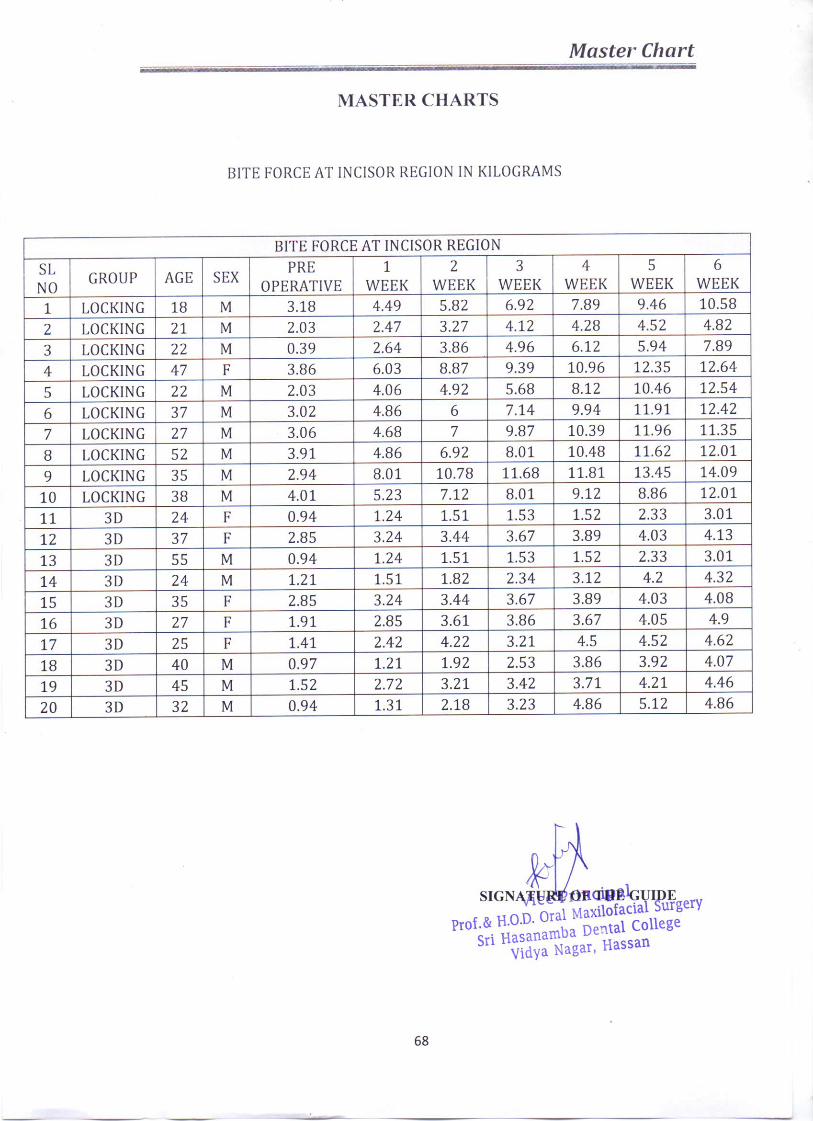

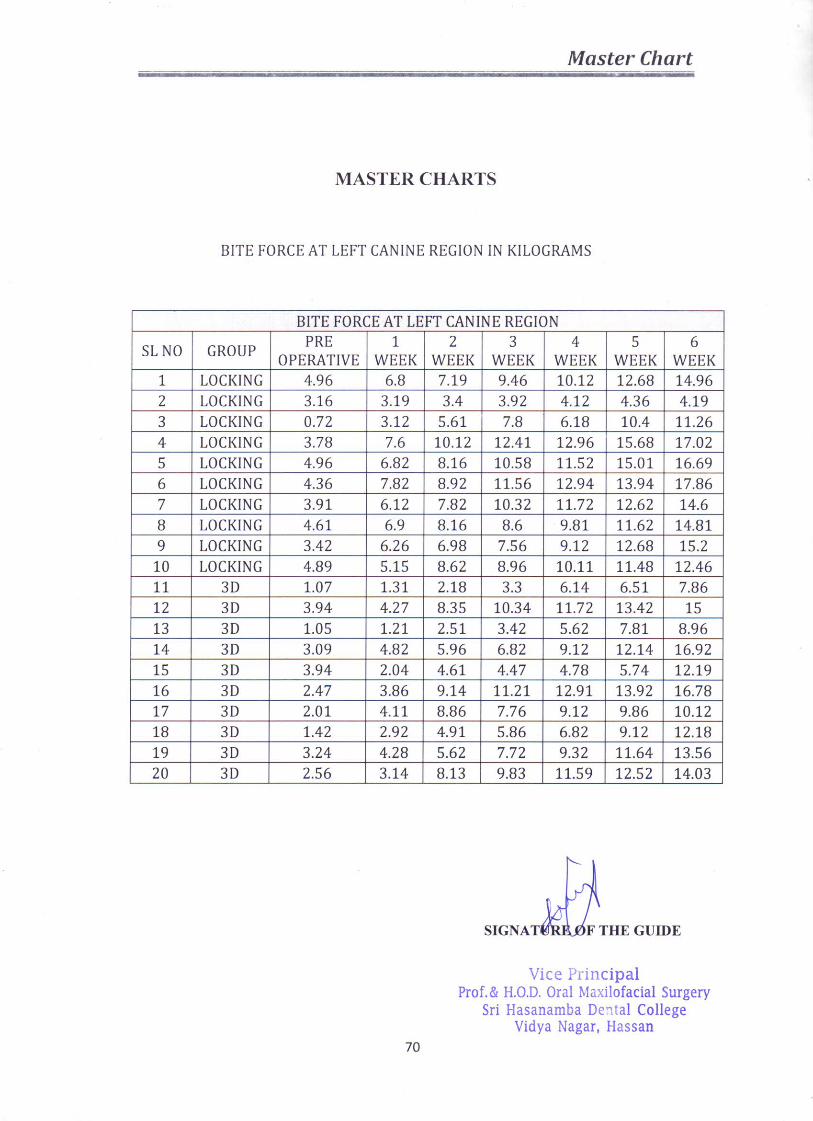
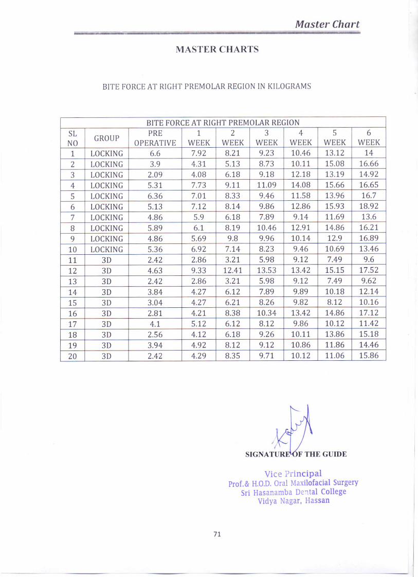
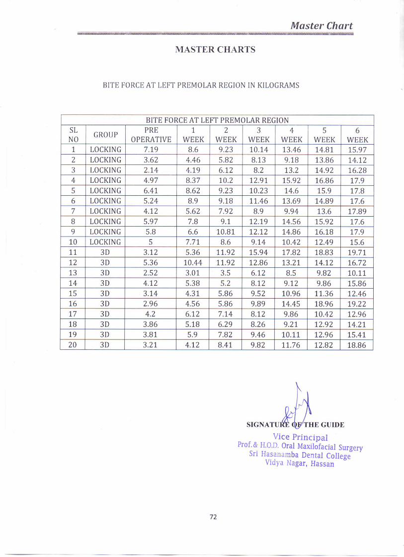
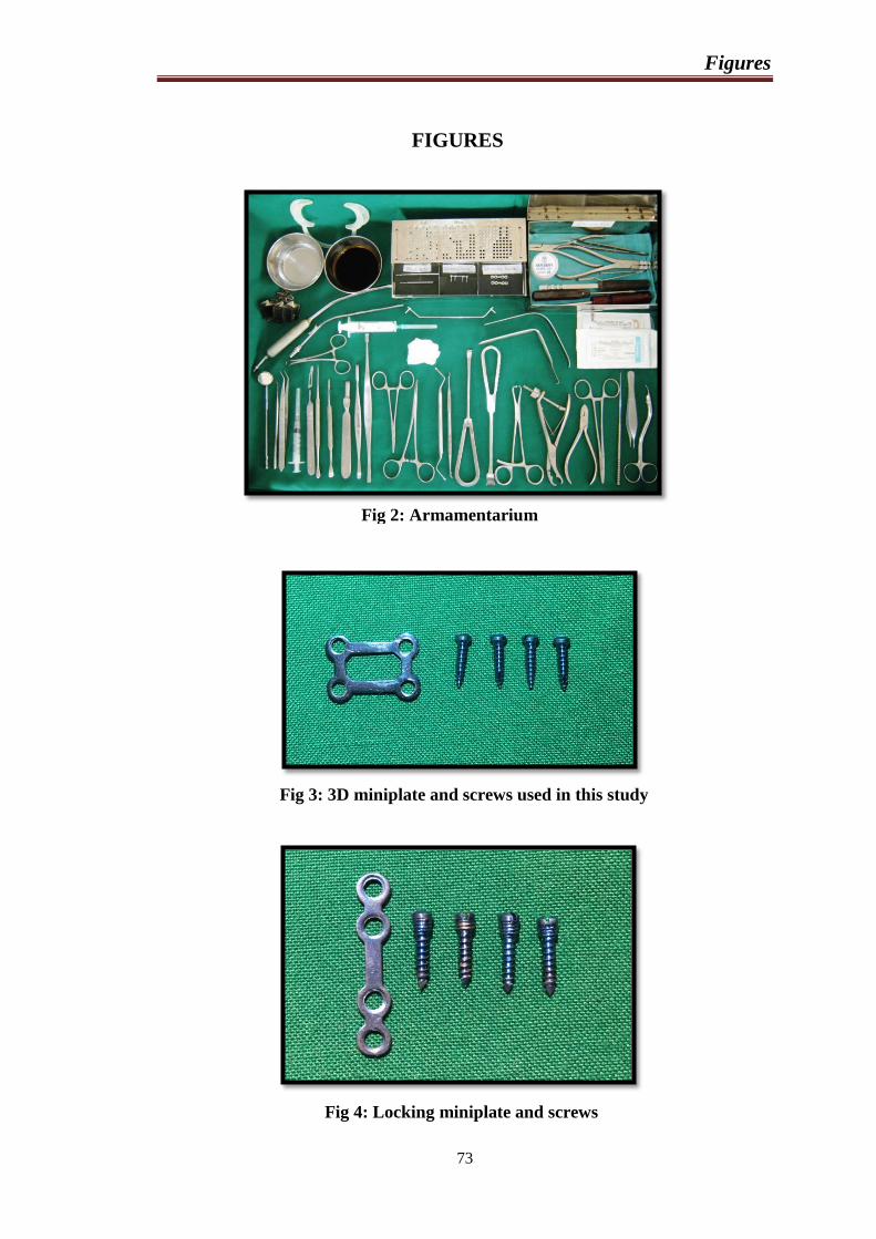
Figures
73
FIGURES
Fig 2: Armamentarium
Fig 3: 3D miniplate and screws used in this study
Fig 4: Locking miniplate and screws
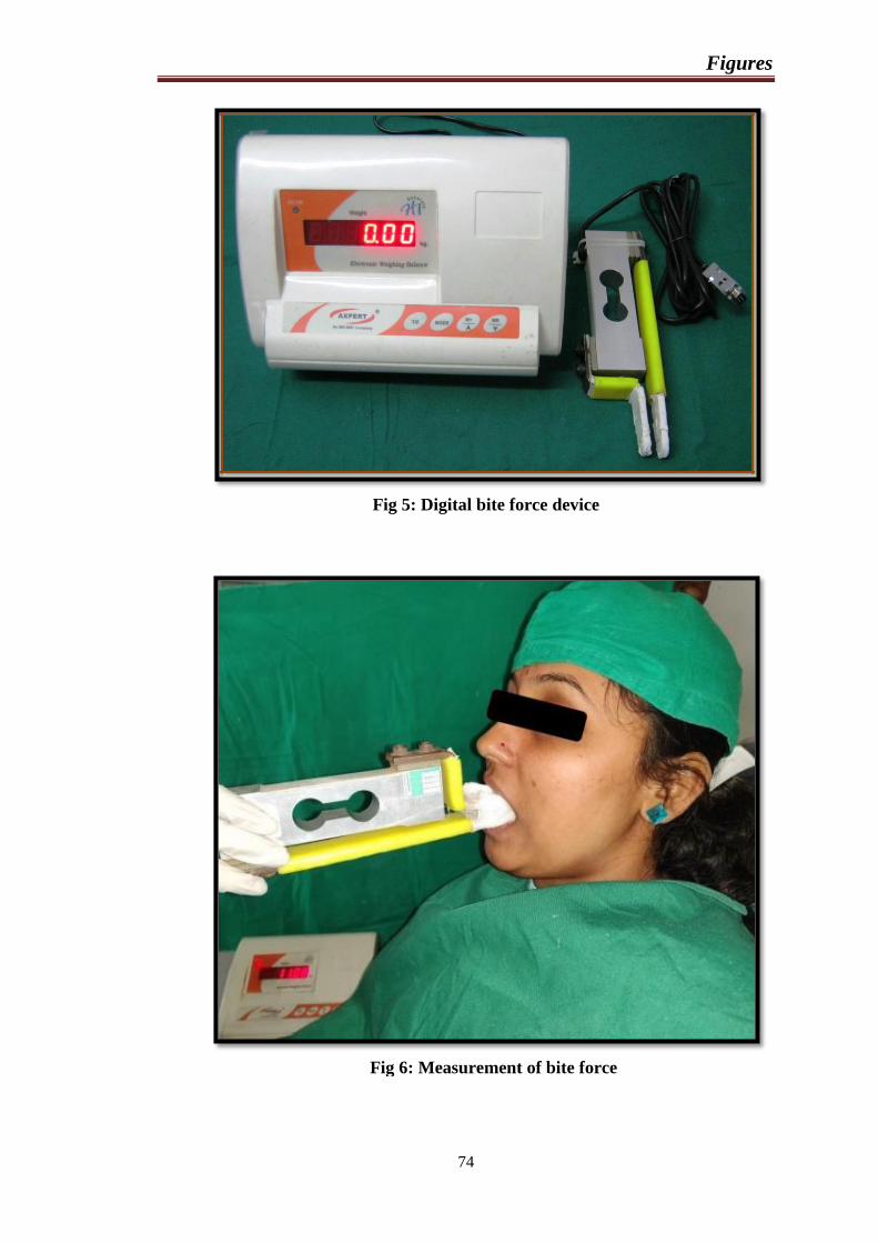
Figures
74
Fig 5: Digital bite force device
Fig 6: Measurement of bite force
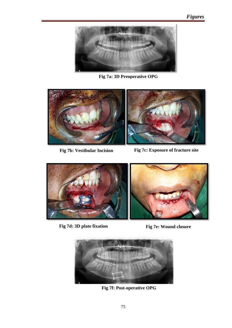
Figures
75
Fig 7a: 3D Preoperative OPG
Fig 7b: Vestibular Incision Fig 7c: Exposure of fracture site
Fig 7d: 3D plate fixation Fig 7e: Wound closure
Fig 7f: Post-operative OPG
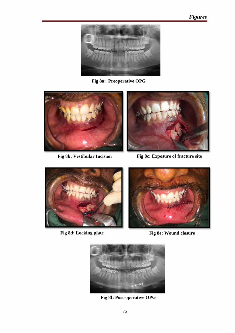
Figures
76
Fig 8a: Preoperative OPG
Fig 8b: Vestibular Incision Fig 8c: Exposure of fracture site
Fig 8d: Locking plate
fixation Fig 8e: Wound closure
Fig 8f: Post-operative OPG
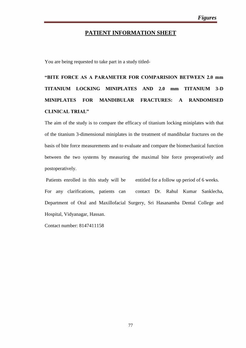
Figures
77
PATIENT INFORMATION SHEET
You are being requested to take part in a study titled-
“BITE FORCE AS A PARAMETER FOR COMPARISION BETWEEN 2.0 mm
TITANIUM LOCKING MINIPLATES AND 2.0 mm TITANIUM 3-D
MINIPLATES FOR MANDIBULAR FRACTURES: A RANDOMISED
CLINICAL TRIAL”
The aim of the study is to compare the efficacy of titanium locking miniplates with that
of the titanium 3-dimensional miniplates in the treatment of mandibular fractures on the
basis of bite force measurements and to evaluate and compare the biomechanical function
between the two systems by measuring the maximal bite force preoperatively and
postoperatively.
Patients enrolled in this study will be entitled for a follow up period of 6 weeks.
For any clarifications, patients can contact Dr. Rahul Kumar Sanklecha,
Department of Oral and Maxillofacial Surgery, Sri Hasanamba Dental College and
Hospital, Vidyanagar, Hassan.
Contact number: 8147411158