bioinformatics and biosynthesis analysis of cellulose synthase operon in zymomonas mobilis zm4
TRANSCRIPT

The IIOAB Journal SPECIAL ISSUE ON FRONTIERS IN INDUSTRIAL MICROBIOLOGY AND ENVIRONMENTAL BIOTECHNOLOGY ISSN: 0976-3104
©IIOAB-India OPEN ACCESS Vol. 2; Issue 3; 2011: 1-7 1
RESEARCH ARTICLE
BIOINFORMATICS AND BIOSYNTHESIS ANALYSIS OF CELLULOSE SYNTHASE OPERON IN ZYMOMONAS MOBILIS ZM4
Sheik Abdul Kader Sheik Asraf, K. Narayanan Rajnish, and Paramasamy Gunasekaran*
Department of Genetics, Centre for Excellence in Genomic Sciences, School of Biological Sciences, Madurai Kamaraj
University, Madurai -625021, INDIA
Received on: 27th
-Sept-2010; Revised on: 11th
-Dec-2010; Accepted on: 12th
-Dec-2010; Published on: 02nd
-Mar-2011 *Corresponding author: Email: [email protected] Tel: +91-452-2458478; Fax: +91-452-2459873
_____________________________________________________
ABSTRACT
Biosynthesis of cellulose has been reported in many species of bacteria. The genes encoding
cellulose biosynthetic enzymes of Z. mobilis have not been studied so far. Preliminary sequence
analysis of the Z. mobilis ZM4 genome revealed the presence of a cellulose synthase operon
comprised of Open Reading Frames (ORFs) ZMO01083 (bcsA), ZMO1084 (bcsB) and ZMO1085 (bcsC).
The first gene of the operon bcsA encodes the cellulose synthase catalytic subunit BcsA. The second
gene of the operon bcsB encodes the cellulose synthase subunit B (BcsB), which shows the presence
of BcsB multi-domain and is inferred to bind c-di-GMP, the regulator of cellulose biosynthesis. The
third gene of the operon bcsC encodes the cellulose synthase operon C domain protein (BcsC), which
belongs to super family of teratrico peptide repeat (TPR) that are believed to mediate protein – protein
interactions for the formation of cellulose. Multiple sequence alignment of the deduced amino acid
sequences of BcsA and BcsC with other closely related homologs showed the presence of PVDPYE,
HAKAGNLN, DCD motif and TPR motif, the characteristic motifs of bacterial cellulose synthases.
Analysis of the nucleotide sequence of the ORF ZMO1085 and neighboring ORFs namely ZMO1083 and
ZMO1084 indicated that all the ORFs are translationally linked and form an operon. Transcript analysis
using Real-time PCR indicated the expression of the genes involved in cellulose synthase operon in
Zymomonas mobilis ZM4. Z. mobilis colonies grown on RM-glucose containing Congo red displayed a
characteristic bright red-brown colour. Z. mobilis colonies grown on RM-glucose medium
supplemented with Calcoflour exhibited fluorescence. The arrangement of Calcofluor stained
microfibrils can be seen in fluorescence microscopy which is an indicative for cellulose biosynthesis.
AFM micrograph of the extracellular matrix of Z. mobilis shows a relatively dense matrix with
bacterial cell residues. The presence of cellulose was confirmed by the Acetic-Nitric (Updegraff)
Cellulose assay. The Bioinformatics and biosynthetic analysis confirm the biosynthesis of cellulose
in Z. mobilis. _____________________________________________________
Key words: biosynthesis, cellulose; open reading frame; operon; transcript; Zymomonas mobilis
[I] INTRODUCTION Cellulose biosynthesis is widespread in plants and microorganism, and cellulose production by bacteria is of special interest. Among the bacterial species, cellulose biosynthesis has been established in Gluconobacter xylinus, Rhizobium leguminosarum, Sarcina ventriculi, Escherichia coli, Klebsiella pneumonia and several species of cyanobacteria [1]. Cellulose production in bacteria is attributed to several reasons. In the case of Rhizobium, cellulose biosynthesis is required for adhesion and aggregation of bacteria at the root hair tip.
Similarly, cellulose is involved in sequential attachment of A. tumefaciens to carrot tissue culture cells. In the case of pathogens such as E. coli and Salmonella, cellulose biosynthesis occurs concomitantly with the production of thin aggregative fimbriae (AGF), the second component of the extracellular matrix of a multicellular morphotype. While thin aggregate of fimbriae form rigid, but fragile interconnections between cells, cellulose connects the cells through elastic, but stable bonds. One of the multicellular morphotype is biofilm formation on abiotic surfaces where cells producing cellulose and thin aggregative fimbriae form distinct adherence patterns [2]. In Zymomonas mobilis, cellulose biosynthesis has been implicated in the formation of cellular aggregates or flocs that can be separated using centrifugation. But, the genes

The IIOAB Journal SPECIAL ISSUE ON FRONTIERS IN INDUSTRIAL MICROBIOLOGY AND ENVIRONMENTAL BIOTECHNOLOGY ISSN: 0976-3104
©IIOAB-India OPEN ACCESS Vol. 2; Issue 3; 2011: 1-7
2
responsible for cellulose synthesis have not been studied. Similarly, biochemical estimation of cellulose has not been carried out so far. Zymomonas mobilis, a gram negative, anaerobic, micro aerotolerant, ethanologenic bacterium uses Enter-Dourdoff (ED) pathway to metabolize glucose [3]. The complete genome of Z. mobilis ZM4 consists of a singular circular chromosome of 2,056,416 bp with an average G+C content of 46.33 % [4]. The predicted ORFs (open reading frames, 1,998) cover 87 % of the genome. Among them, 1,346 ORFs (67.4 %) could be assigned with putative functions, 258 ORFs (12.9 %) were putative coding sequences of unknown functions and the remaining 394 ORFs (19.7 %) showed no similarities to known genes. The preliminary sequence analysis of the Z. mobilis ZM4 genome shows the presence of a cellulose synthase operon comprised of Open Reading Frames (ORFs) ZMO01083, ZMO1084 and ZMO1085. However, there are no reports on the detailed analysis of this operon. In the present study, several bioinformatics tools were used in functional analysis of the genes of the cellulose synthase operon. The precision of these bioinformatics tools has enhanced over a period of time and has its own advantages. In order to make the most precise predictions, numerous methods were used to build up the functional properties of the cellulose synthase operon of Z. mobilis to the highest possible accuracy. We have also provided the experimental evidence for the cellulose production by Z. mobilis.
[II] MATERIALS AND METHODS
2.1. Bioinformatics analysis
The protein sequences of the cellulose synthase catalytic subunit
(BcsA), cellulose synthase subunit B (BcsB) and cellulose synthase
operon C domain protein (BcsC) was obtained from the Z. mobilis ZM4
genome [NC_006526]. The primary sequence was analysed using
Protparam [5]. ProtParam was used to calculate biochemical,
biophysical and physicochemical properties like molecular weight,
theoretical isoelectric point, instability index, extinction coefficient,
aliphatic index, grand average of hydropathicity (GRAVY), estimated
half-life (Escherichia coli, in vivo, in hours), and total number of
negatively and positively charged amino-acid residues. Homology and
similarity searching of the protein sequences against several sequences
was performed using BLASTP [6] against NR and PDB databases.
Multiple sequence alignment and analysis were performed using
ClustalW2. Signal peptide and cleavage site was predicted using
iPSORT [7], PrediSi [8], PSORT [9], SignaIP [10], and SOSUI signal
[11]. iPSORT is a subcellular localization site predictor for N-terminal
sorting signals. Predisi is a software tool for predicting signal peptide
sequences in real time with a high accuracy and is based on a position
weight matrix approach by a frequency correction that takes the amino
acid bias present in proteins in consideration. PSORT analyzes the input
sequence by applying the stored rules for various sequence features of
known protein sorting signals and reports the possibility for the input
protein to be localized at each candidate site with additional information.
SignalP 3.0. incorporates a prediction of cleavage sites and a signal
peptide/non-signal peptide prediction based on a combination of several
artificial neural networks and hidden Markov models. SOSUIsignal
predicts signal peptide of which three-domain (tripartite) structure is
recognized by three modules of the software system.
2.2. Transcript analysis
The mid-growth phase cultures of Z. mobilis grown in RMG were
withdrawn and the total RNA was isolated as described previously [12].
All the RNA samples were treated with DNase I (MBI Fermentas,
Opelstrasse, Germany) to eliminate the genomic DNA contamination
and purified before the PCR was performed. The RNA was quantified
using Nanodrop ND-1000 spectrophotometer (Wilmington, DE, USA),
and the integrity of RNA was analyzed on a formaldehyde agarose gel
[13]. Later, RevertAid First Strand cDNA Synthesis kit (MBI Fermentas,
Germany) was used for the synthesis of first strand cDNA from total
RNA template using gene-specific primers [Table-1].
Table: 1. List of primer pairs used for real time PCR
A negative control reaction was performed using Taq DNA polymerase with RNA as template. qPCR (quantitative PCR) primers were designed using PrimerExpress 3.0 software (Applied Biosystems, USA) and primers were ordered from Sigma Genosys, Bangalore. The levels of expression of ZMO01083, ZMO1084 and ZMO1085 transcripts in Z. mobilis ZM4 was determined by real-time PCR. adhB (alcohol dehydrogenase) gene was used as endogenous control using 100 ng of cDNA as template. Real-time PCR was performed using Power SYBR Green PCR Master Mix (Applied Biosystems, USA) kit in an Applied Biosystems 7500 Real-time PCR system (Applied Biosystems, USA) according to manufacture’s instruction. Power SYBR Green PCR Master Mix (25 µ l) consisted of 2x SYBR Green (12.5 µ l), 900 nM of each of forward and reverse gene-specific primers for the respective genes, 100 ng cDNA template. qPCR cycling conditions were as follows: 50 °C for 2 min, 95 °C for 10 min, 40 cycles each of 95 °C for 15 sec and 60 °C for 1 min. The resulting PCR products were examined to dissociation-curve analysis to confirm the presence of single amplicon obtained from the cDNA template. The qPCR was performed in duplicates. For each gene analyzed, Power SYBR Green PCR Master Mix without template controls were performed. After, qPCR was completed, the threshold cycle (Ct) was calculated using ABI 7500 SDS software version 1.3
(Applied Biosystems, USA). The transcript levels of target ZMO01083, ZMO1084 and ZMO1085 were normalized with respect to endogenous control, adhB. The resultant aliquots (25 µ l) of each qPCR product were electrophoresed on 1.2 % agarose gel in 1 x TAE (40 mM Tris acetate, 1 mM EDTA, pH 8) buffer at constant voltage of 80 V/cm and stained
with ethidium bromide (0.5 µ g ml-1
). The size of each amplified qPCR product was double-checked by Bio-Rad Quantity One software to confirm the single amplicon.
2.3 Reagents, microorganisms, and culture conditions The RNeasy mini kit (Qiagen, Hilden, Germany) was used for the

The IIOAB Journal SPECIAL ISSUE ON FRONTIERS IN INDUSTRIAL MICROBIOLOGY AND ENVIRONMENTAL BIOTECHNOLOGY ISSN: 0976-3104
©IIOAB-India OPEN ACCESS Vol. 2; Issue 3; 2011: 1-7
3
isolation of total RNA. Zymomonas mobilis ZM4 (ATCC31821) was obtained from NRRL, Peoria, IL, USA. Power SYBR Green PCR Master Mix (Applied Biosystems, USA) was used for real-time PCR experiment. Z. mobilis ZM4 was grown in Rich Medium (glucose, 20 g l
-1; KH2PO4,
20 g l-1
and yeast extract 10 g l-1) under static condition at 30 ºC for RNA
isolation.
2.4. Cellulose assay and characterization of cellulose-related phenotype
Cellulose was estimated by Acetic-nitric (Updegraff) cellulose assay. To study the secretion of cellulose, Z. mobilis was grown for 72 h at 30 ºC on RMG agar plates (glucose, 20 g l
-1; KH2PO4, 20 g l
-1 ; yeast extract
10 g l-1
and 1.6 % agar ) supplemented with Congo red 40 µg.ml-1.
Calcofluor binding by Z. mobilis was observed on RMG agar plates (glucose, 20 g l
-1; KH
2PO
4, 20 g l
-1; yeast extract 10 g l
-1 and 1.6 % agar)
supplemented with Calcofluor 200 µg.ml-1. The colonies fluorescence
was observed under fluorescence microscope (Nikon eclipse Ti). The morphology and microstructure of the extracellular cellulosic material was evaluated by atomic force microscope (APE Research A 100).
[III] RESULTS AND DISCUSSION 3.1. Bioinformatics analysis of cellulose synthase operon
Analysis of Z. mobilis genome identified a putative operon
comprising of the cellulose synthase catalytic subunit (BcsA),
cellulose synthase subunit B (BcsB) and cellulose synthase
operon C domain protein (BcsC). This arrangement is similar
to the cellulose biosynthase operon in Acetobacter xylinum
[14]. The domain analysis at the Conserved Domain Database
[Figure−1] provided the following results. The bcsA encodes
the cellulose synthase catalytic subunit BcsA, which belongs to
super family of Glycosyl transferase A and PilZ. The PilZ
domain is the binding protein for cyclic diguanylic acid (c-di-
GMP), an allosteric activator of the cellulose synthase. The
presence of this domain perhaps indicates that the BcsA protein
could be regulated by cyclic-di- GMP [15]. BcsA shows the
presence of PVDPYE, HAKAGNLN, DCD motifs, the
characteristic motifs of bacterial cellulose synthase [2] and a
presence of CelA multi-domain. The specific hit of BcsA is
CESA_CelA_like family proteins. The BcsA protein is
transmembrane protein and belongs to a family of progressive
β-glycosyltransferases. The second gene of the operon bcsB
encodes the cellulose synthase subunit B (BcsB), which belongs
to super family of PRK11114 and shows the presence of BcsB
multi-domain.
Like the BcsA protein, the BcsB protein also poses
transmembrane domain at the C-terminus, and is inferred to
bind c-di-GMP, the regulator of cellulose biosynthesis. The
third gene of the operon bcsC encodes the cellulose synthase
operon C domain protein (BcsC), which belongs to super family
of teratrico peptide repeat (TPR) and Cellulose synthase operon
protein C. BcsC shows the presence of TPR motif. TPR is a 34
amino acid repeated motif that is widespread among
prokaryotes and eukaryotes [16]. In the case of cellulose
biosynthesis, TPR repeat domains are believed to mediate
protein – protein interactions for the formation of cellulose. The
BcsC has transmembrane domains and the TPR repeat domain
at the N-terminus.
The biochemical, biophysical and physicochemical properties
of BcsA, BcsB and BcsC are listed in the [Table-2].BLASTP
analysis of the cellulose synthase catalytic subunit (BcsA),
cellulose synthase subunit B (BcsB) and cellulose synthase
operon C domain protein (BcsC) showed maximum identity to
putative cellulose synthase of Sphingobium japonicum UT26S
and cellulose synthase protein C precursor of Sphingobium
japonicum UT26S respectively.
Fig: 1. Conserved Domain analysis of cellulose synthase operon. a) The first gene of the operon bcsA encodes the
cellulose synthase catalytic subunit BcsA, which belongs to super family of Glycosyl transferase A and PilZ. BcsA shows the presence of DXD motif. The specific hit of BcsA is CESA_CelA_like family proteins. b) The second gene of the operon bcsB encodes the cellulose synthase subunit B (BcsB),
which belongs to super family of PRK11114. There is a presence of BcsB multi-domain. c) The third gene of the operon

The IIOAB Journal SPECIAL ISSUE ON FRONTIERS IN INDUSTRIAL MICROBIOLOGY AND ENVIRONMENTAL BIOTECHNOLOGY ISSN: 0976-3104
©IIOAB-India OPEN ACCESS Vol. 2; Issue 3; 2011: 1-7
4
bcsC encodes the cellulose synthase operon C domain protein (BcsC), which belongs to super family of TPR and Cellulose synthase operon protein C. BcsC shows the presence of TPR motif.
Table 2: Predicted properties of BcsA, BcsB and BcsC
Biochemical / biophysical / physicochemical properties
BcsA BcsB BcsC
Number of amino acids 665 771 1336
Molecular weight (in KDa) 75.38 84.62 147.07
Theoretical isoelectric point (pI)
8.49 6.54 6.83
Aliphatic index 101.08 95.5 73.17
Half-life (E. coli, in vivo, in hr) >10 >10 >10
Gravy index 0.06 -0.101 -0.589
Extinction coefficient 94685 92945 142910
Total number of negatively charged residues (Asp + Glu)
65 79 146
Total number of positively charged residues (Arg + Lys)
69 75 142
Fig: 2. Multiple sequence alignment of the deduced amino acid sequence of ZMO1083 with other bacterial cellulose synthases: The multiple sequence alignment was computed
using the CLUSTAL W2 program. The amino acids that form the conserved motifs of bacterial cellulose synthases are PVDPYE (135-147), HAKAGNLN (200-208) and DCD (222-224) are underlined. The sequences compared with BcsA (ZMO1083, YP_162818.1), include the following: YP_003559640.1 (putative cellulose synthase, Sphingobium japonicum UT26S), YP_001178646.1 (cellulose synthase, Enterobacter sp. 638), and YP_002921737.1 (putative cellulose synthase, Klebsiella pneumoniae NTUH- K0244). Asterisks and dots indicate identical and similar amino acids respectively.
Multiple sequence alignment of the deduced amino acid
sequences of BcsA and BcsC with other closely related
homologs showed the presence PVDPYE, HAKAGNLN and
DCD motifs [Figure−2] and TPR motifs [Figure−3]
respectively. Predisi, SignalP 3.0., iPSORT, SOSUIsignal,
PSORT predicted that BcsA has no signal peptide and it is an
intracellular protein. BcsB and BcsC have been predicted to
have signal peptide and are extracellular proteins.
Fig: 3. Multiple sequence alignment of deduced amino acid sequence of Z. mobilis ZMO1085 with other bacterial cellulose synthase subunit C amino acid sequences: The
multiple sequence alignment was computed using the ClustalW2 program. The amino acids from 305 -395 and 768-900 represent the TPR 1 and 2 repeats of BcsC. Sequence alignment revealed a putative consensus characteristic of the TPR family of proteins. The sequences compared with BcsC (YP_162820.1, Z. mobilis) include YP_003559641.1 (cellulose synthase protein precursor, Sphingobium japonicum UT26S), YP_00178644.1 (cellulose synthase domain containing protein, Enterobacter sp), NP_643822.1 (cellulose synthase subunit C, Xanthomonas
The IIOAB Journal SPECIAL ISSUE ON FRONTIERS IN INDUSTRIAL MICROBIOLOGY AND ENVIRONMENTAL BIOTECHNOLOGY ISSN: 0976-3104
©IIOAB-India OPEN ACCESS Vol. 2; Issue 3; 2011: 1-7
5
axonopodis pv. Citri. Str.306) and YP_0021518201 (cellulose synthase protein, Proteus mirabilis H14320) respectively. Asterisks and dots indicate identical and similar amino acids respectively.
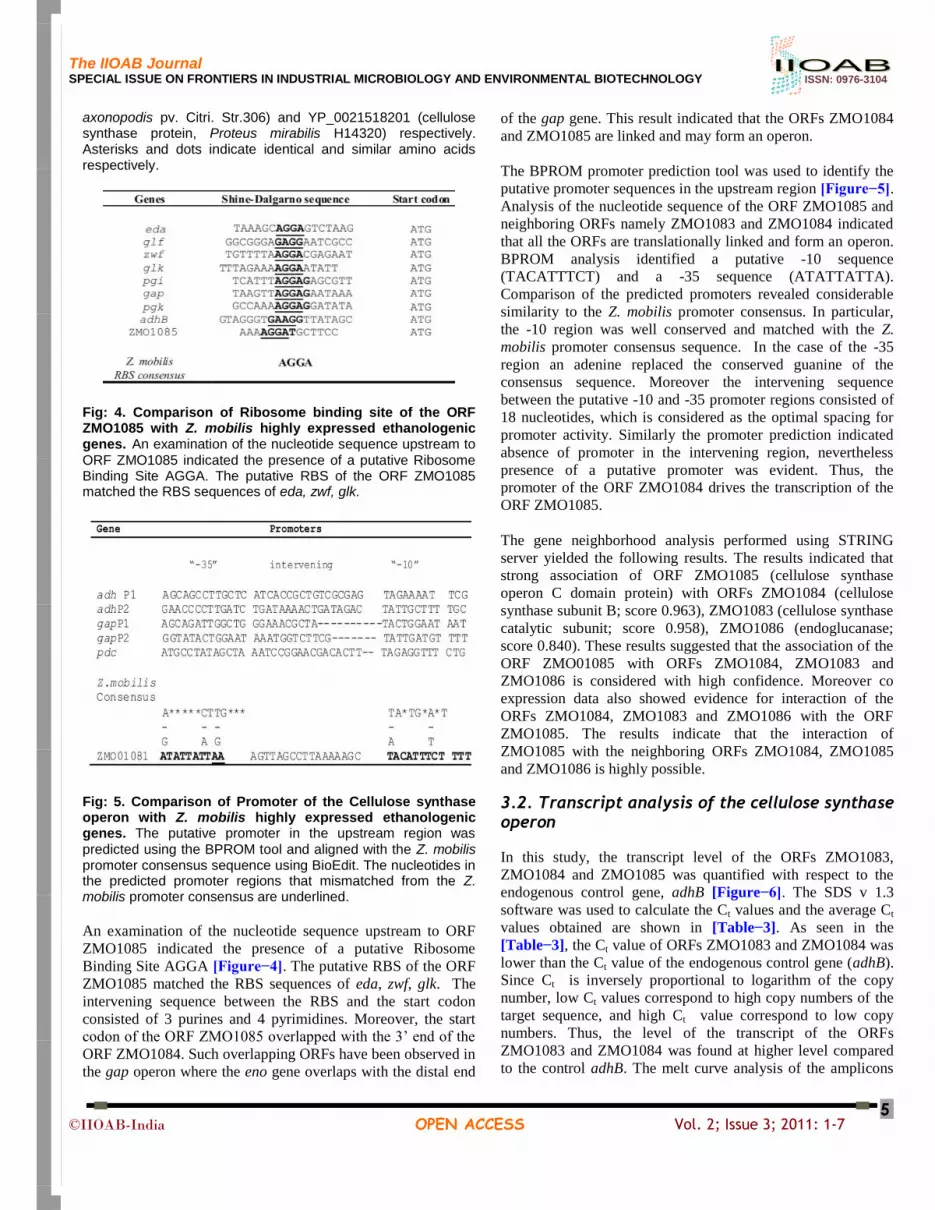
Fig: 4. Comparison of Ribosome binding site of the ORF ZMO1085 with Z. mobilis highly expressed ethanologenic genes. An examination of the nucleotide sequence upstream to
ORF ZMO1085 indicated the presence of a putative Ribosome Binding Site AGGA. The putative RBS of the ORF ZMO1085 matched the RBS sequences of eda, zwf, glk.
Fig: 5. Comparison of Promoter of the Cellulose synthase operon with Z. mobilis highly expressed ethanologenic genes. The putative promoter in the upstream region was
predicted using the BPROM tool and aligned with the Z. mobilis promoter consensus sequence using BioEdit. The nucleotides in the predicted promoter regions that mismatched from the Z. mobilis promoter consensus are underlined.
An examination of the nucleotide sequence upstream to ORF
ZMO1085 indicated the presence of a putative Ribosome
Binding Site AGGA [Figure−4]. The putative RBS of the ORF
ZMO1085 matched the RBS sequences of eda, zwf, glk. The
intervening sequence between the RBS and the start codon
consisted of 3 purines and 4 pyrimidines. Moreover, the start
codon of the ORF ZMO1085 overlapped with the 3’ end of the
ORF ZMO1084. Such overlapping ORFs have been observed in
the gap operon where the eno gene overlaps with the distal end
of the gap gene. This result indicated that the ORFs ZMO1084
and ZMO1085 are linked and may form an operon.
The BPROM promoter prediction tool was used to identify the
putative promoter sequences in the upstream region [Figure−5].
Analysis of the nucleotide sequence of the ORF ZMO1085 and
neighboring ORFs namely ZMO1083 and ZMO1084 indicated
that all the ORFs are translationally linked and form an operon.
BPROM analysis identified a putative -10 sequence
(TACATTTCT) and a -35 sequence (ATATTATTA).
Comparison of the predicted promoters revealed considerable
similarity to the Z. mobilis promoter consensus. In particular,
the -10 region was well conserved and matched with the Z.
mobilis promoter consensus sequence. In the case of the -35
region an adenine replaced the conserved guanine of the
consensus sequence. Moreover the intervening sequence
between the putative -10 and -35 promoter regions consisted of
18 nucleotides, which is considered as the optimal spacing for
promoter activity. Similarly the promoter prediction indicated
absence of promoter in the intervening region, nevertheless
presence of a putative promoter was evident. Thus, the
promoter of the ORF ZMO1084 drives the transcription of the
ORF ZMO1085.
The gene neighborhood analysis performed using STRING
server yielded the following results. The results indicated that
strong association of ORF ZMO1085 (cellulose synthase
operon C domain protein) with ORFs ZMO1084 (cellulose
synthase subunit B; score 0.963), ZMO1083 (cellulose synthase
catalytic subunit; score 0.958), ZMO1086 (endoglucanase;
score 0.840). These results suggested that the association of the
ORF ZMO01085 with ORFs ZMO1084, ZMO1083 and
ZMO1086 is considered with high confidence. Moreover co
expression data also showed evidence for interaction of the
ORFs ZMO1084, ZMO1083 and ZMO1086 with the ORF
ZMO1085. The results indicate that the interaction of
ZMO1085 with the neighboring ORFs ZMO1084, ZMO1085
and ZMO1086 is highly possible.
3.2. Transcript analysis of the cellulose synthase operon
In this study, the transcript level of the ORFs ZMO1083,
ZMO1084 and ZMO1085 was quantified with respect to the
endogenous control gene, adhB [Figure−6]. The SDS v 1.3
software was used to calculate the Ct values and the average Ct
values obtained are shown in [Table−3]. As seen in the
[Table−3], the Ct value of ORFs ZMO1083 and ZMO1084 was
lower than the Ct value of the endogenous control gene (adhB).
Since Ct is inversely proportional to logarithm of the copy
number, low Ct values correspond to high copy numbers of the
target sequence, and high Ct value correspond to low copy
numbers. Thus, the level of the transcript of the ORFs
ZMO1083 and ZMO1084 was found at higher level compared
to the control adhB. The melt curve analysis of the amplicons
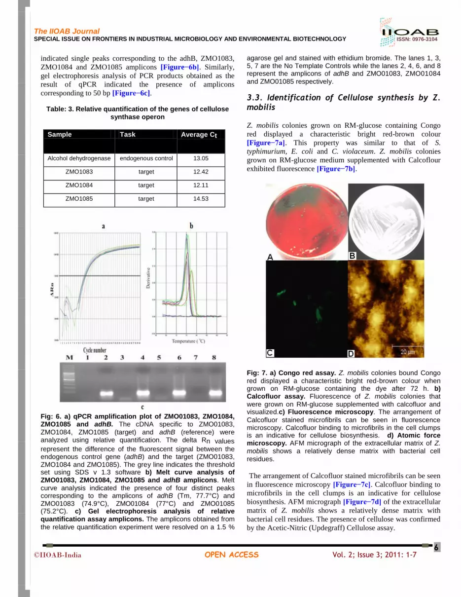
The IIOAB Journal SPECIAL ISSUE ON FRONTIERS IN INDUSTRIAL MICROBIOLOGY AND ENVIRONMENTAL BIOTECHNOLOGY ISSN: 0976-3104
©IIOAB-India OPEN ACCESS Vol. 2; Issue 3; 2011: 1-7
6
indicated single peaks corresponding to the adhB, ZMO1083,
ZMO1084 and ZMO1085 amplicons [Figure−6b]. Similarly,
gel electrophoresis analysis of PCR products obtained as the
result of qPCR indicated the presence of amplicons
corresponding to 50 bp [Figure−6c].
Table: 3. Relative quantification of the genes of cellulose synthase operon
Sample Task Average Ct
Alcohol dehydrogenase endogenous control 13.05
ZMO1083 target 12.42
ZMO1084 target 12.11
ZMO1085 target 14.53
Fig: 6. a) qPCR amplification plot of ZMO01083, ZMO1084, ZMO1085 and adhB. The cDNA specific to ZMO01083,
ZMO1084, ZMO1085 (target) and adhB (reference) were analyzed using relative quantification. The delta Rn values
represent the difference of the fluorescent signal between the endogenous control gene (adhB) and the target (ZMO01083, ZMO1084 and ZMO1085). The grey line indicates the threshold set using SDS v 1.3 software b) Melt curve analysis of ZMO01083, ZMO1084, ZMO1085 and adhB amplicons. Melt
curve analysis indicated the presence of four distinct peaks corresponding to the amplicons of adhB (Tm, 77.7°C) and ZMO01083 (74.9°C), ZMO01084 (77°C) and ZMO01085 (75.2°C). c) Gel electrophoresis analysis of relative quantification assay amplicons. The amplicons obtained from
the relative quantification experiment were resolved on a 1.5 %
agarose gel and stained with ethidium bromide. The lanes 1, 3, 5, 7 are the No Template Controls while the lanes 2, 4, 6, and 8 represent the amplicons of adhB and ZMO01083, ZMO01084
and ZMO01085 respectively.
3.3. Identification of Cellulose synthesis by Z. mobilis
Z. mobilis colonies grown on RM-glucose containing Congo
red displayed a characteristic bright red-brown colour
[Figure−7a]. This property was similar to that of S.
typhimurium, E. coli and C. violaceum. Z. mobilis colonies
grown on RM-glucose medium supplemented with Calcoflour
exhibited fluorescence [Figure−7b].
Fig: 7. a) Congo red assay. Z. mobilis colonies bound Congo
red displayed a characteristic bright red-brown colour when grown on RM-glucose containing the dye after 72 h. b) Calcofluor assay. Fluorescence of Z. mobilis colonies that
were grown on RM-glucose supplemented with calcofluor and visualized.c) Fluorescence microscopy. The arrangement of
Calcofluor stained microfibrils can be seen in fluorescence microscopy. Calcofluor binding to microfibrils in the cell clumps is an indicative for cellulose biosynthesis. d) Atomic force microscopy. AFM micrograph of the extracellular matrix of Z. mobilis shows a relatively dense matrix with bacterial cell
residues.
The arrangement of Calcofluor stained microfibrils can be seen
in fluorescence microscopy [Figure−7c]. Calcofluor binding to
microfibrils in the cell clumps is an indicative for cellulose
biosynthesis. AFM micrograph [Figure−7d] of the extracellular
matrix of Z. mobilis shows a relatively dense matrix with
bacterial cell residues. The presence of cellulose was confirmed
by the Acetic-Nitric (Updegraff) Cellulose assay.

The IIOAB Journal SPECIAL ISSUE ON FRONTIERS IN INDUSTRIAL MICROBIOLOGY AND ENVIRONMENTAL BIOTECHNOLOGY ISSN: 0976-3104
©IIOAB-India OPEN ACCESS Vol. 2; Issue 3; 2011: 1-7
7
[IV] CONCLUSION
The Bioinformatics and biosynthetic analysis confirm the
biosynthesis of cellulose in Z. mobilis.
FINANCIAL DISCLOSURE AND ACKNOWLEDGEMENT Authors thank University Grants Commission, New Delhi (F.4-5/2006 XI Plan) and Department of Science and Technology, New Delhi (SR/SO/BB-50/2007) for the financial support and Junior Research Fellowships to KNR and SSA through the Centre for Excellence in Genomic Sciences. The support received from Centre for Advanced Studies in Functional Genomics and Networking Resource Centre in Biological Sciences, School of Biological Sciences, Madurai Kamaraj University are gratefully acknowledged.
REFERENCES
[1] Romling U. [2002] Molecular biology of cellulose production
in bacteria. Res Microbiol 153: 205–212.
[2] Romling U, Rohde D, Olsen A, Normark S, Reinkoster J.
[2000] AgfD, the checkpoint of multicellular and aggregative
behavior in Salmonella typhimurium regulates atleast two
independent pathways. Mol Microbiol 36: 10–23.
[3] Swings J, De Ley J. [1997] The biology of Zymomonas.
Bacteriol Rev 41:1–46.
[4] Seo JS, et al. [2005] The genome sequence of the ethanologenic
bacterium Zymomonas mobilis ZM4. Nat Biotechnol 23:63–68.
[5] Gasteiger E, Hoogland C, Gattiker A, Duvaud S, Wilkins MR,
Appel RD, Bairoch A. [2005] Protein Identification and
Analysis Tools on the ExPASy Server. In The Proteomics
Protocols Handbook. Edited by Walker JM. Humana Press
571–607.
[6] Altschul SF, Madden TL, Schaffer AA, Zhang J, Zhang Z,
Miller W, Lipman DJ. [1997] Gapped BLAST and PSI-BLAST:
a new generation of protein database search programs. Nucl
Acids Res 25:3389–3402.
[7] Bannai H, Tamada Y, Maruyama O, Nakai K, Miyano S. [2002]
Extensive feature detection of N-terminal protein sorting
signals. Bioinformatics 18:298–305.
[8] Hiller K, Grote A, Scheer M, Munch R, Jahn D. [2004] PrediSi:
prediction of signal peptides and their cleavage positions.
Nucleic Acids Res W375–379.
[9] Nakai K, Horton P. [1999] PSORT: a program for detecting
sorting signals in proteins and predicting their subcellular
localization. Trends Biochem Sci 24:34-36.
[10] Bendtsen JD, Nielsen H, von Heijne G, Brunak S. [2004]
Improved prediction of signal peptides: SignalP 3.0. J Mol Biol
340: 783–795.
[11] Gomi M, Sonoyama M, Mitaku S. [2004] High performance
system for signal peptide prediction: SOSUIsignal. Chem-Bio
Informatics Journal 4:142–147.
[12] Conway T, Fliege R, Jones Kilpatrick D, Liu J, Barnell WO and
Egan SE. [1991] Cloning, characterization and expression of the
Zymomonas mobilis eda gene that encodes 2-keto-3-deoxy-6-
phosphogluconate aldolase of the Entner-Doudoroff pathway.
Mol Microbiol 5: 2901–2911.
[13] Sambrook J, Russel D. [2001] Molecular Cloning-A laboratory
manual. New York: Cold Spring Harbor Press.
[14] Wong HC, Fear AL, Calhoon RD, Eichinger GH, Mayer R,
Amikam D, Benziman M, Gelfand DH, Meade JH, Emerick
AW, Bruner R, Bassat AB, Tal R. [1990] Genetic organization
of the cellulose synthase operon in Acetobacter xylinum. Proc.
Natl. Acad. Sci 87: 8130–8134.
[15] Amikam D, Galperin MY. [2006] PilZ domain is part of the
bacterial c-di- GMP binding protein. Bioinformatics 22(1):3–6
[16] Andrea LD and Regan L. [2003] TPR Proteins: the versatile
helix. Trends Biochem Sci 12: 655–62.
ABOUT AUTHORS
Mr. Sheik Abdul Kader Sheik Asraf is a Junior Research Fellow in Department of Genetics, Center for Excellence in Genomic Sciences, School of Biological Sciences, Madurai Kamaraj University. He is presently working upon the Ph.D. thesis entitled “Functional genomics of selected carbohydrate hydrolases in Zymomonas mobilis”. His research interests include Molecular biology and Genomics of Zymomonas mobilis, Enzyme engineering and enzyme technology and Bioprocess technology for enzymes and biofuel production: Recombinant strains. He has communicated 2 Book chapters (In Press) and 1 Research article (In Review).
Mr. K. Narayanan Rajnish is a Senior Research Fellow in Department of Genetics, Center for Excellence in Genomic Sciences, School of Biological Sciences, Madurai Kamaraj University. He has completed his Ph.D. thesis entitled “Studies on functional genomics of Zymomonas mobilis” His research interests include Molecular biology and Genomics of Zymomonas mobilis, Enzyme engineering and enzyme technology and Bioprocess technology for enzymes and biofuel production: Recombinant strains. He has published 1 Research article, 1 Book-chapter and has communicated 1 Research article (In Review) and 1 Book chapter (In Press).
Prof. Paramasamy Gunasekaran is Senior Professor & Head, Department of Genetics and Coordinator, Center for Excellence in Genomic Sciences, School of Biological Sciences, Madurai Kamaraj University. His research interests include: Metagenomics and bioprospecting, Molecular biology and Genomics of Zymomonas mobilis and filamentous fungi, Bioprocess technology for enzymes and biofuel production: Recombinant strains, Enzyme engineering and enzyme technology, Bioremediation of industrial effluents, and Genetic diversity of Plant growth promoting rhizobacteria. He has more than 130 publications in the peer-reviewed national and international journals. He has written 8 books/manuals in the field of microbial genomics.