biocompatibility study of two diblock copolymeric nanoparticles for biomedical applications by in...
TRANSCRIPT
1 23
��������� ������������������������ �������������������� ���������������������������������� !"#$������%���&����'���������( �)* ��+��%,���!-.��� /� !0 �� %�� ���* �"�
�������� ��������������������� ������������������������������� ������������������� ���������������������������
���������� ��������������������������������������������� ���������������������������������������� !�� ���� ���� �����������"
1 23
Your article is protected by copyright and allrights are held exclusively by Springer Science+Business Media Dordrecht. This e-offprintis for personal use only and shall not be self-archived in electronic repositories. If you wishto self-archive your article, please use theaccepted manuscript version for posting onyour own website. You may further depositthe accepted manuscript version in anyrepository, provided it is only made publiclyavailable 12 months after official publicationor later and provided acknowledgement isgiven to the original source of publicationand a link is inserted to the published articleon Springer's website. The link must beaccompanied by the following text: "The finalpublication is available at link.springer.com”.
RESEARCH PAPER
Biocompatibility study of two diblock copolymericnanoparticles for biomedical applications by in vitro toxicitytesting
Felipe Goni-de-Cerio • Valentina Mariani • Dror Cohen • Lea Madi •
Julie Thevenot • Hugo Oliveira • Chiara Uboldi • Guido Giudetti •
Rosella Coradeghini • Elisabeth Garanger • Francois Rossi • Meital Portugal-Cohen •
Miriam Oron • Rafi Korenstein • Sebastien Lecommandoux • Jessica Ponti •
Blanca Suarez-Merino • Pedro Heredia
Received: 11 June 2013 / Accepted: 3 October 2013! Springer Science+Business Media Dordrecht 2013
Abstract Drugs used for chemotherapy normallycarry out adverse, undesired effects. Nanotechnology
brings about new horizons to tackle cancer disease
with a different strategy. One of the most promisingapproaches is the use of nanocarriers to transport
active drugs. These nanocarriers need to have special
properties to avoid immune responses and toxicity,and it is critical to study these effects. Nanocarriers
may have different nature, but polypeptide-basedcopolymers have attracted considerable attention for
their biocompatibility, controlled and slow biodegrad-
ability as well as low toxicity. Little has been doneregarding specific nanocarriers toxicity. In this study,
we performed a thorough toxicological study of two
different block copolymer nanoparticles (NPs);poly(trimethylene carbonate)-block–poly(L-glutamic
acid) (PTMC-b–PGA) and poly(ethylene glycol)-
block–poly(c-benzyl-L-glutamate) (PEG-b–PBLG)with sizes between 113 and 131 nm. Low blood–
serum–protein interaction was observed. Moreover,
general toxicity assays and other endpoints (apoptosisor necrosis) showed good biocompatibility for both
NPs. Reactive oxygen species increased in only two
Felipe Goni-de-Cerio and Valentina Mariani have contributedequally.
Electronic supplementary material The online version ofthis article (doi:10.1007/s11051-013-2036-0) contains supple-mentary material, which is available to authorized users.
F. Goni-de-Cerio ! B. Suarez-Merino ! P. Heredia (&)GAIKER Technology Centre, Parque Tecnologico deZamudio Ed 202, 48170 Vizcaya, Spaine-mail: [email protected]
V. Mariani ! C. Uboldi ! G. Giudetti ! R. Coradeghini !F. Rossi ! J. PontiNanobiosciences Unit, Institute for Health and ConsumerProtection, Joint Research Centre, European Commission,Via E. Fermi 2749, 21027 Ispra, VA, Italy
D. Cohen ! M. Portugal-Cohen ! M. OronAHAVA, Dead Sea Laboratories, Lod, Israel
D. CohenThe Dead Sea Laboratory for Biochemistry andBiotechnology of the Skin, Dead Sea and Arava ScienceCenter, Ein-Gedi, Israel
L. Madi ! R. KorensteinDepartment of Physiology and Pharmacology, SacklerSchool of Medicine, Tel-Aviv University,69978 Tel Aviv, Israel
J. Thevenot ! H. Oliveira ! E. Garanger !S. LecommandouxUniversite de Bordeaux, ENSCPB, 16 Avenue PeyBerland, 33607 Pessac Cedex, France
J. Thevenot ! H. Oliveira ! E. Garanger !S. LecommandouxCNRS, LCPO, UMR 5629, 33600 Pessac Cedex, France
123
J Nanopart Res (2013) 15:2036
DOI 10.1007/s11051-013-2036-0
Author's personal copy
cell lines (HepG2 and TK6) in the presence of PTMC-
b–PGA. Cytokine production study showed cytokine
induction only in one cell line (A549). We alsoperformed the same assays on human skin organ
culture before and after UVB light treatment, with a
moderate toxicity after treatment independent of NPspresence or absence. Interleukin 1 induction was also
observed due to the combined effect of PEG-b–PBLG
and UVB light irradiation. Future in vivo studies forbiocompatibility and toxicity will provide more valu-
able information, but, so far, the findings presented
here suggest the possibility of using these two NPs asnanocarriers for nanomedical applications, always
taking into account the application procedure and the
way in which they are implemented.
Keywords Amphiphilic copolymers !Nanoparticles ! In vitro toxicity ! Cancertherapy ! Nanoparticles toxicity ! Biomedicine
Introduction
Solid tumours account for more than 85 % of cancer
mortality. Chemotherapeutic drugs are often the
selected treatment for cancer, but tumour structureand blood flow could determine poor penetration and
limited distribution of these drugs, like it was demon-
strated for doxorubicin (Lankelma et al. 1999).Nevertheless, the use of this therapeutic approach
reckons with drug resistance and severe side effects
due to non-selective cytotoxicity. One of the mostpromising approaches used to dodge these problems is
to use nanosized carriers to deliver a chemotherapeuticdrug deeply into the tumour (Upadhyay et al. 2009).
These include natural vectors (protein carriers, viral
vectors for gene therapy), pseudo-synthetic vectors(polymer–antibody hybrids, polymer–protein conju-
gates and antibody-targeted liposomes) and synthetic
vectors (polymer–drug conjugates, polymer micelles,polymer-based nanoparticles, NPs; Chiellini et al.
2006, 2007). Within the synthetic vectors, polypep-
tide-based block copolymers have attracted consider-able interest for their high biocompatibility, low and
controlled degradability (Kricheldorf 2006) and com-
plex and precise secondary conformations (Carlsenand Lecommandoux 2009) making them highly
promising in biomedical applications (Arimura et al.
2005; Duncan et al. 2006; Oh et al. 1999; Mandal and
Chatterjee 2007). Amphiphilic diblock copolymersare made of a hydrophobic block that will constitute
the inner core of the nano-assemblies, acting as a drug
reservoir, and a hydrophilic block that forms thehydrated outer shell, which impedes uptake by the
reticuloendothelial system (Jeong et al. 2004; Dong
et al. 2004).Lecommandoux’s group recently demonstrated
that a block copolymer poly(trimethylene carbonate)-
block–poly(L-glutamic acid) (PTMC-b–PGA) is able toform stable NPs in water (Sanson et al. 2010a, b).
Moreover, they presented a successful method to load
high doxorubicin doses in these NPs, strongly sup-porting the interest of developing PTMC-b–PGA NPs
as carrier for the controlled delivery of chemothera-
peutic drugs (Li 2002; Li et al. 2008). Anotherpromising polypeptide-based block copolymer for
drug encapsulation is poly(ethylene glycol)-block–
poly(c-benzyl-L-glutamate), PEG-b–PBLG (Jeonget al. 1999). The hydrophilic blocks of the two
aforementioned block copolymers, PGA and PEG,
are considered to be non-toxic at the dose required fordrug carriers and prevents interactions with cells and
proteins (Jeong et al. 2005; Vega-Villa et al. 2008).
Nevertheless, studies on the potential toxicity of thetwo complete block copolymers are missing.
In the present study, morphology and physico-
chemical properties of both PTMC-b–PGA and PEG-b–PBLG have been investigated to characterize the
prepared NPs. Likewise, the NPs were carefully
characterized for their biocompatibility using selectedin vitro toxicity tests, specific for the evaluation of the
cytotoxicity, apoptosis–necrosis induction, reactive
oxygen species (ROS) detection and cytokine produc-tion. These assays were used on cell lines, representing
potential target organs such as lung, intestine, skin,
kidney and liver, in which it is described that NPscould accumulate and interact (Curtis et al. 2006;
Lanone and Boczkowski 2006). In the literature there
are not many examples of studies conducted to analysethe toxicity of nanocarriers per se (Li et al. 2012).
Furthermore, a toxic reaction against nanocarriers canbe built on the knowledge of adverse reactions of cells
model against NPs (Nishiyama and Kataoka 2006;
Satchi-Fainaro et al. 2006).To determine the in vitro toxicological profile
induced by PTMC-b–PGA and PEG-b–PBLG, in this
study we have investigated their potential cytotoxicityon at least seven cell lines from several organs and on
Page 2 of 17 J Nanopart Res (2013) 15:2036
123
Author's personal copy

human skin organ culture (HSOC) by Alamar Blue(AB) and colony forming efficiency (CFE) assays as
well as the induction of apoptosis and oxidative stress
by flow cytometry analysis. Moreover, cytokineproduction was also investigated by sandwich
enzyme-linked immunoassays test, in order to mea-
sure cytokine release.
Methods
NPs synthesis and characterisation
PTMC25-b–PGA12 (2,550-b–1,540 g/mol) and
PEG45-b–PBLG13 (2,000-b–2,850 g/mol) were syn-
thesized as previously described and with minormodifications (Sanson et al. 2010a, b; Martinez
Barbosa et al. 2007).
NPs were obtained from amphiphilic block copoly-mers by solvent assisted self-assembly using the
nanoprecipitation method (Sanson et al. 2011).
Z-average hydrodynamic diameter (DH), polydis-persity index (PDI) and size distribution of NPs in
water were evaluated via dynamic light scattering
(DLS) using Milli-Q water. The DLS and surfacecharge (obtained by capillary electrophoresis) mea-
surements were performed on 1 ml sample volume on
a Zetasizer Nano Zs (Malvern Instruments) at 25 "C,following the protocols indicated by ISO 13321:1996.
The data acquisition was carried out using the
automatic adjustment of position, attenuation andmeasurement time. Mean values of DH and PDI
(average on three measures) were obtained from the
ratio of the second order cumulate divided by thesquare of the first order one. Only intensity distribution
data were considered for the analysis. Graphs were
obtained by averaging three measurements per batchusing Malvern’s proprietary software.
Morphologies of stock suspensions were obtained
by transmission electron microscopy analysis (TEM;Hitachi H7650 microscope working at 80 kV
equipped with a GATAN ORIUS SC1100 11 Mega-pixel camera, imaging facility provided by Bordeaux
Imaging Center, Bordeaux University, France). Sam-
ples were coated on copper grids (200 mesh coatedwith carbon or carbon/FormvarTM) by spraying 1 g/l
suspensions of particles using a homemade spraying
tool. After complete drying, samples were stained with
a 1 % w/v uranyl acetate (Agar Scientific) solution inwater.
Interaction of nanocarriers with cell culturemedium and blood serum
Interaction of nanocarriers with cell culture mediumwas carried out using MEM cell culture medium
dispersions prepared at 0.5 mg/ml NP final concen-
tration and incubated 1 h at 37 "C. Measurementswere done at increasing concentrations (0, 0.1, 1 and
10 % v/v) of added foetal bovine serum (FBS, Gibco)
to assess eventual serum protein concentration effectson polydispersity and measured size. DLS measure-
ments were performed in the same conditions
described previously for three independent experi-ments and three replicates for each experimental point.
The interaction of NPs with human plasma serum
assay was performed according to the protocolsestablished at Nanotechnology Characterization Lab-
oratory. Samples were obtained by Dr. Miguel Angel
Vesga from Centro Vasco de Transfusiones, GaldakaoHospital. The Basque Biobank for Research-OEHUN
(www.biobancovasco.org) processed and released the
samples following standard operation procedures withappropriate ethical approval. Plasma was incubated
with 1 mg/ml of PTMC-b–PGA or PEG-b–PBLG for
30 min at 37 "C. Then, plasma–NP suspension wasspun twice to remove any trace of unbound proteins
and to obtain the NPs attached to proteins. Pellet was
then resuspended in 2D-PAGE loading buffer (Biorad)to detach NPs from proteins and spun again. Super-
natant (protein fraction) was recovered and the protein
amount was quantified using EZQ Protein Quantita-tion kit (Invitrogen). Protein sample was later elec-
trofocused (first dimension) using IPG strips pH 3–10
(Biorad). IPG strips were later equilibrated and elec-trophoresed in a 17 cm long, 1.5 mm thick and 12 %
polyacrylamide gel (second dimension). Proteins were
stained with SYPRO Ruby (Invitrogen) and imagedunder VersaDoc image analyser (Biorad). Protein
identification was performed using MALDI-TOF.
Cell and skin model culture conditions
Human keratinocyte cells (HaCaT) were originally
supplied by the German Cancer Research Center
(Germany), human alveolar cells (A549), human
J Nanopart Res (2013) 15:2036 Page 3 of 17
123
Author's personal copy
intestinal cells (Caco-2), dog kidney cells (MDCK),hepatocellular carcinoma cells (HepG2) and human
lymphoblast cells (TK6) were originally purchased
from the American Type Culture Collection. BALB/3T3 mouse fibroblasts were purchased from Hatano
Research Institute (Japan). The cell lines were main-
tained in complete culture mediums according to thesuppliers’ guidelines and were routinely grown as a
monolayer in tissue culture grade flasks. When cells
exceeded 70 % confluence (but less than 90 %), theywere sub-cultured on new culture flasks. All the cell
cultures were maintained in standard cell culture
conditions (37 "C, 5 % CO2 and 95 % humidity). ForHSOC model, skin pieces of breast or abdomen were
obtained with informed consent from healthy,
35–50 year-old women undergoing plastic surgery.Up to 6 h from the surgery the skin was cut to a
0.16 cm2 pieces, and samples were placed with dermis
facing down and epidermis facing up in culture platescontaining DMEM medium with antibiotics (1 %
penicillin–streptomycin). Cultures were incubated for
24 h, at 37 "C under 5 % CO2 for recovery.
Cytotoxicity assays
Cytotoxicity was assessed by AB and CFE on HaCaT,
A549, Caco-2, MDCK, HepG2 and BALB/3T3 cell
lines. AB was also used for testing of TK6 cell line andHSOC model. Necrosis/apoptosis induction, cytokine
production and oxidative stress determination studies
were carried out on all the six–seven cell lines and onHSOC model.
The selected exposure times were 24/72 h and the
concentrations tested were 0.1 and/or 0.5 mg/ml ofPTMC-b–PGA and PEG-b–PBLG suspensions for
cytotoxicity assessment.
AB assay
AB assay was used to assess cytotoxicity induced byPTMC-b–PGA and PEG-b–PBLG dispersions on
seven cell lines and HSOC model after 24 h ofexposure to 0.1 and 0.5 mg/ml. Cells were seeded in
96 well plates (at a density of 5,000–10,000 depending
on the cell line) to reach 70–80 % confluence at the endof the assay, at least six replicates for each treatment
were tested. The day after seeding, cells were exposed
to 0.1 and 0.5 mg/ml of PTMC-b–PGA and PEG-b–PBLG suspensions for 24 h. Each experiment included
a negative control (untreated cells in culture medium)and a positive control (treated with sodium dodecyl
sulphate). At the end of the exposure period, each well
was supplemented with AB solution (Invitrogen).After 4 h incubation under standard cell culture
conditions, data were acquired by means of a spectro-
photometer measuring the absorbance at 570 nm.Afterwards, all experiments were carried out in
quadruplicate and expressed as the percentage of AB
reduction in the test wells compared to the untreatedcontrols.
For the HSOC model, after 24 and 72 h of
incubation with the NPs, the whole human skin culturewas placed with 300 ll of DMEM and 0.01 mg/ml of
Resazurin. The tissue was incubated for 1 h. Data
were acquired by a spectrophotometer measuring theabsorbance at 570 nm.
Colony forming efficiency
Assays were performed according to Ponti et al.
(2010). Cells were seeded in 60 9 15 mm petridishes at a density of 200–300 cells/dish in 3 ml
complete culture medium at least in three replicates
for each treatment. After 24 h, PTMC-b–PGA andPEG-b–PBLG suspensions were directly added to
the cell culture to obtain the appropriate final
concentration of 0.1 mg/ml. After 24 and 72 h ofexposure, the medium was changed with complete
fresh culture medium that was renewed twice
weekly. After 7 days, cells were fixed for 20 minwith 3.7 % formaldehyde solution in PBS. Dishes
were stained for 30 min with 4 % v/v Giemsa
solution (Sigma) in ultrapure water. Colonies weremanually scored under a stereomicroscope. Each
experiment included a negative control and a
positive control (cells exposed to sodium chromate1,000 lM). The corresponding standard error mean
was calculated for three independent experiments
and three replicates for each experimental point.
Apoptosis/necrosis induction
Potential apoptosis and necrosis induction were stud-
ied in the cell lines using Annexin V conjugated withAlexa Fluor 488 (Molecular Probes) and propidium
iodide (PI; Sigma-Aldrich) fluorochromes by flow
cytometry, to measure early apoptotic event andnecrosis, respectively, after exposure to 0.1 and
Page 4 of 17 J Nanopart Res (2013) 15:2036
123
Author's personal copy
0.5 mg/ml of PTMC-b–PGA and PEG-b–PBLG sus-pensions for 24 and 72 h. Apoptosis studies were also
carried out in HSOC models by Ac-DEVD-AMC, a
fluorogenic substrate specific of caspase 3 (Calbio-chem) after exposure to 0.1 and 0.5 mg/ml of PTMC-
b–PGA and PEG-b–PBLG suspensions for 24 h.
HaCaT, Caco-2, A549, MDCK, HepG2 and BALB/3T3 cells were seeded in 24 well plates at a density of
100,000 cells/well for 24 h and 50,000 cells/well for
72 h in all cell lines in complete culture medium andexposed to NPs. 24 and 72 h after the exposure of cells
to NPs (0.1 and 0.5 mg/ml of PTMC-b–PGA and
PEG-b–PBLG), 5 ll of the Annexin V conjugateswere added to each 100 ll of cell suspension (mini-
mum of 10,000 cell/100 ll) and incubated at room
temperature for 15 min. After incubation, cells werewashed and submitted to flow cytometry acquisition.
Without delay, the stained cells were contrasted
immediately with PI (5 lg/ml) to detect necrotic cellsand analyzed by Beckman Coulter FC500 MPL flow
cytometer. As positive control, cells were incubated
with 1 lM of doxorubicin hydrochloride (DiscoveryFine Chemicals, Ltd.) for 24 h. Three independent
experiments and at least three replicates for each
experimental point were carried out. Each experimentincluded a negative control (untreated cells) and a
positive control. Results were expressed as the
percentage of Annexin V and PI positive cells.For the HSOC model, after 24 h of incubation with
the NPs, the skin samples were incubated for 1 min in
PBS at 56 "C, and the epidermis was separated from thedermis with a scalpel. Epidermal sheets were incubated
in PBS containing 2.5 lM Ac-DEVD-AMC with
0.02 % Triton X-100, 10 mM dithiothreitol and20 mM Tris pH 7.4, at 37 "C. Fluorescence was
measured at 355/460 nm on a fluorometer (Thermo
Scientific), and the activity was calculated in the linearrange. Three independent experiments and at least three
replicates for each experimental point were carried out.
Each experiment included a negative control.
Oxidative stress determination
Potential oxidative stress induced by PTMC-b–PGA
and PEG-b–PBLG suspensions was studied on all thecell lines after exposure to 0.5 mg/ml of NPs by
dichlorodihydrofluorescein diacetate (H2DCF-DA,
Invitrogen), used to determine changes in ROS activ-ity. It permeates the cell membrane and accumulates
mostly in the cytosol following deacetylation byesterases to dichlorodihydrofluorescein. This non-
fluorescent product is converted by ROS into dichlo-
rofluorescein (DCF; Ex = 488 nm; Em = 525 nm).Cells were seeded in six well plates at a non-confluent
cell density (105 cell/ml), after 24 h the medium
was removed and the treatment medium containing0.5 mg/ml PTMC-b–PGA or PEG-b–PBLG suspen-
sions was added for an exposure time of 1.5 h. Then
treatment suspensions were replaced with carboxy-H2DCF-DA (10 lM) by adding it to the free-serum
medium. After incubation of 0.5 h, cells were washed
and the intracellular fluorescence of DCF was mea-sured by flow cytometry (FACSCalibur). Three inde-
pendent experiments and at least three replicates for
each experimental point were carried out. Each exper-iment included a negative control (untreated cells) and
a positive control (tert-butyl hydroperoxide 0.5 mM).
Results were expressed as a geometrical mean relativeto control.
Cytokine production determination
Cytokine production induced by PTMC-b–PGA and
PEG-b–PBLG suspensions was studied on six celllines and HSOC model cultures.
Cells and HSOC model were exposed to 0.5 mg/ml
of NPs for 24 h. In addition, controlled cytokineproduction was induced on HSOC cultures irradiating
them at a dose of 500 mJ/cm2 (312 nm). Immediately
after irradiation PTMC-b–PGA and PEG-b–PBLGwere added to the medium to reach 0.5 mg/ml
concentration for 24 h of incubation. Each experiment
included a negative control. After incubation, cytokineconcentrations (TNFa, interleukin [IL] 1a, IL-6, IL-8)
were determined in culture media by sandwich
enzyme-linked immunoassays, using ELISA highsensitivity kits (Biolegend, Inc.). In brief, a polystyrene
microtiter plate was coated with a cytokine-specific,
monoclonal antibody and blocked with 1 % bovineserum albumin. Samples were incubated with the
immobilized antibody, unbound materials werewashed off and a second antibody, directed at a
different epitope of the same antigen, was added and
incubated. After washing away unbound materials, ahorseradish peroxidase-linked antibody was used to
reveal the immobilized complexes. Recombinant
cytokines were used as internal standards. The corre-sponding standard error mean was calculated for three
J Nanopart Res (2013) 15:2036 Page 5 of 17
123
Author's personal copy

independent experiments and three replicates for eachexperimental point compared to the untreated controls.
Statistical analysis
Results were contrasted with a Levene test to confirm
the homogeneity of variance between the differentgroups and Kolmogorov–Smirnoff test for normality.
Unpaired t test and one factor analysis of variance
(ANOVA) with Bonferroni–Dunn’s correction wereperformed to assess differences in all studied param-
eters: p \ 0.005 has been considered as statistically
significant (*).
Results
NP synthesis and characterisation
PTMC-b–PGA and PEG-b–PBLG were analysed by
TEM, and the images obtained showed a vesicular
morphology for PTMC-b–PGA and a particulatemorphology for PEG-b–PBLG (Fig. 1). The mean
DH of the particles’ stock solutions measured by
DLS were 113 ± 44 nm for PEG-b–PBLG and131 ± 19 nm for PTMC-b–PGA (Fig. 1) of 10 dif-
ferent batches prepared on this study. Zeta potential
(f) was markedly higher in absolute values for PTMC-b–PGA (-42 mV) than for PEG-b–PBLG (-9 mV),
indicating a potential higher colloidal stability in water
of the former NP.
Interaction of nanocarriers with cell culture
medium and blood serum
The values for PDI and DH are shown for a single NP
batch in Table 1. There was a very small tendency toPDI increase when increasing serum concentration in
the conditions of this study. This effect was evident
when 10 % serum was used. Both the PTMC-b–PGAand PEG-b–PBLG PDI increased notably, from
0.15 ± 0.01 to 0.3 ± 0.03 and from 0.07 ± 0.01 to
Fig. 1 Chemical structure of the two amphiphilic blockcopolymers, PEG-b–PBLG and PTMC-b–PGA and characteris-tics of the particles prepared for this study. Left column chemicalstructure and average hydrodynamic diameter (DH), polydisper-sity index (PDI) and f for n = 10 (average on different particle
batches) in water. Middle column typical relaxation curve andsize distribution obtained by DLS analysis. Right column TEMpicture showing the morphology of PEG-b–PBLG (top) andPTMC-b–PGA (bottom) particles
Page 6 of 17 J Nanopart Res (2013) 15:2036
123
Author's personal copy
0.17 ± 0.01, respectively, when 10 % serum contain-ing media was used. The overall fold change for both
NPs was 2.00 for PTMC-b–PGA and 2.43 for PEG-b–
PBLG.The quantification analyses of the assays of NPs
interaction with blood serum components are
described in Table 2. The concentration of proteinsin blood serum for different batches, measured in the
conditions of this assay, was between 80 and 100 mg/ml.
The values for the control of precipitation carried outadding PBS to the blood serum instead of NPs were
5.52 ± 0.87 and 9.93 ± 1.95 lg/ml, representing less
that 0.01 % of the total protein value in each sample.The concentration values for precipitated proteins
after treatment with both PTMC-b–PGA andPEG-b–PBLG were 83.73 ± 2.53 and 90.51 ±
10.59 lg/ml, respectively. These values represent
0.107 and 0.075 % of the total protein in plasma andwere around ten times higher than the precipitated
control (PBS), but significantly low compared to blood
serum total protein concentration. After protein quan-tification, the pellet obtained was cleaned to remove
NPs and subjected to 2D-PAGE analysis, according to
the procedure already described. The most represen-tative and abundant proteins identified by MALDI-
TOF are shown in Table 3. Proteins corresponded to
different families and routes like protein transport(apolipoproteins, mitoferrin-2) or immune response
proteins (immunoglobulins, ficolin, etc.).
Cytotoxicity assays in cell lines and HSOC model
To detect the cytotoxicity of PTMC-b–PGA and PEG-b–PBLG, AB and CFE assays were performed. The
AB assays results are shown in Fig. 2a: neither
PTMC-b–PGA nor PEG-b–PBLG (named A and Brespectively in all figures hereafter) showed any
cytotoxicity at tested concentrations for each of the
cell lines considered. Only a slight decrease in TK6viability was shown at 0.5 mg/ml when cells were
treated with PEG-b–PBLG, but it was not statistically
significant.Epidermal viability of HSOC models after treat-
ment with either PTMC-b–PGA or PEG-b–PBLG was
detected using AB assays at 24 h and 72 h followingUVB irradiation (Fig. 2b). The results after 24 h of NP
incubation showed that UVB irradiation induced a
slight decrease in cell viability in all experimentalgroups, although this decrease was not statistically
significant. UVB irradiation effect was significant at
Table 1 Example of DLS study of one batch of PEG-b–PBLGand PTMC-b–PGA NPs in cell culture medium (MEM)untreated and with increasing concentrations of serum (0.1, 1and 10 % [v/v])
FBS (%) PDI ± SD DH (nm) ± SD
PEG-b–PBLG
0 0.07 ± 0.01 94.88 ± 1.13
0.1 0.08 ± 0.02 93.99 ± 1.61
1 0.09 ± 0.02 95.39 ± 0.09
10 0.17 ± 0.01 96.92 ± 2.43
PTMC-b–PGA
0 0.15 ± 0.01 98.44 ± 3.73
0.1 0.16 ± 0.01 97.78 ± 1.12
1 0.18 ± 0.04 102.30 ± 3.33
10 0.30 ± 0.03 101.40 ± 1.31
FBS foetal bovine serum, PDI polydispersity index, DH
hydrodynamic diameter
Table 2 Quantitation of blood proteins associated to PEG-b–PBLG and PTMC-b–PGA after treatment of blood serum with1 mg/ml of each NP for 30 min at 37 "C
Protein (lg/ml) ± SD Protein (% ofcontrol)
PBS (solution) 8.47 9 104 ± 1.11 9 104 100
PBS(precipitate)
5.52 ± 0.87 0.007
PTMC-b–PGA(1 mg/ml)
90.51 ± 10.59 0.107
PBS (solution) 1.11!105 ± 1.27!104 100
PBS(precipitate)
9.93 ± 1.95 0.009
PEG-b–PBLG(1 mg/ml)
83.73 ± 2.53 0.075
Table 3 Proteins identified by MALDI-TOF after 2D-PAGEfor precipitated blood serum fraction after treatment with eitherPTMC-b–PGA or PEG-b–PBLG at 1 mg/ml
PTMC-b–PGA PEG-b–PBLG
Ig c-2, c-3, j, l Keratin
Apolipoprotein A-1, Apolipoprotein A-1,
A-4, E A-4, E
Ficolin-3 Ficolin-3
Mitoferrin-2 Fibrinogen b chain
Fibrinogen C
J Nanopart Res (2013) 15:2036 Page 7 of 17
123
Author's personal copy
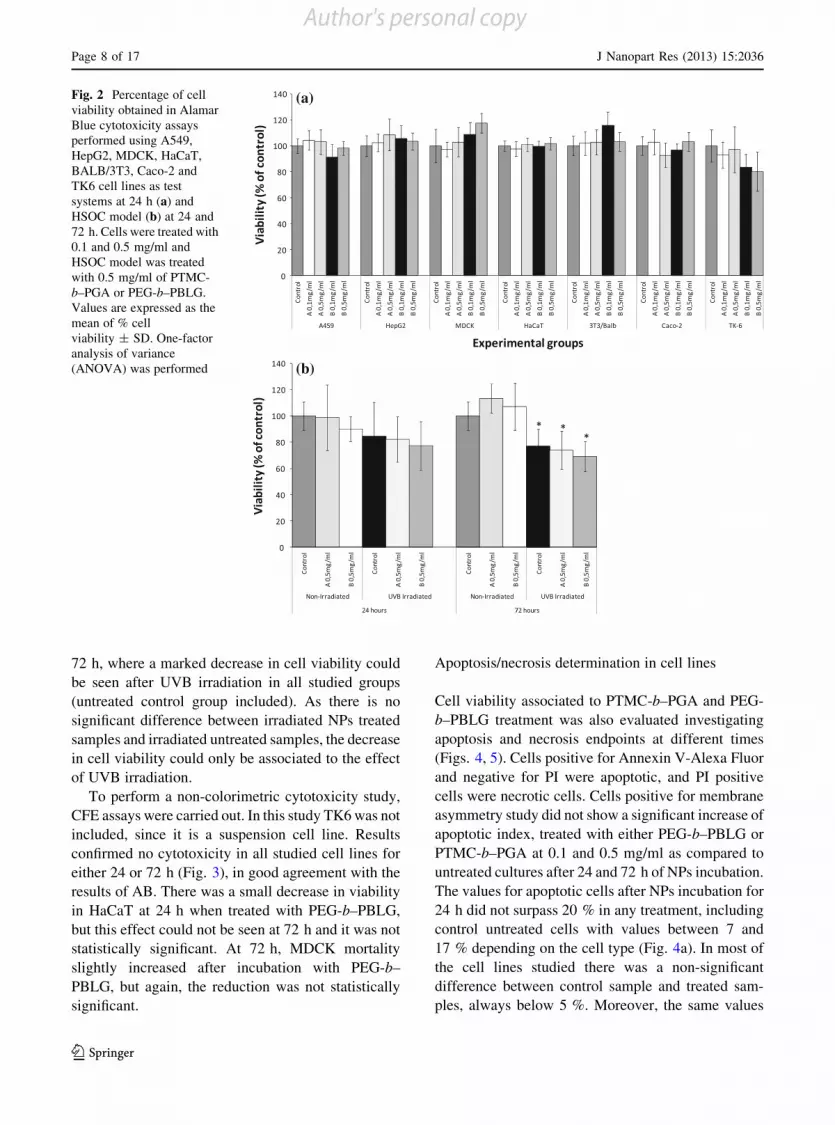
72 h, where a marked decrease in cell viability could
be seen after UVB irradiation in all studied groups
(untreated control group included). As there is nosignificant difference between irradiated NPs treated
samples and irradiated untreated samples, the decrease
in cell viability could only be associated to the effectof UVB irradiation.
To perform a non-colorimetric cytotoxicity study,
CFE assays were carried out. In this study TK6 was notincluded, since it is a suspension cell line. Results
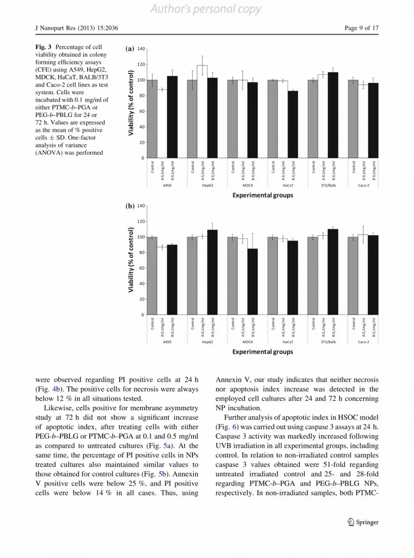
confirmed no cytotoxicity in all studied cell lines for
either 24 or 72 h (Fig. 3), in good agreement with theresults of AB. There was a small decrease in viability
in HaCaT at 24 h when treated with PEG-b–PBLG,
but this effect could not be seen at 72 h and it was notstatistically significant. At 72 h, MDCK mortality
slightly increased after incubation with PEG-b–
PBLG, but again, the reduction was not statisticallysignificant.
Apoptosis/necrosis determination in cell lines
Cell viability associated to PTMC-b–PGA and PEG-
b–PBLG treatment was also evaluated investigating
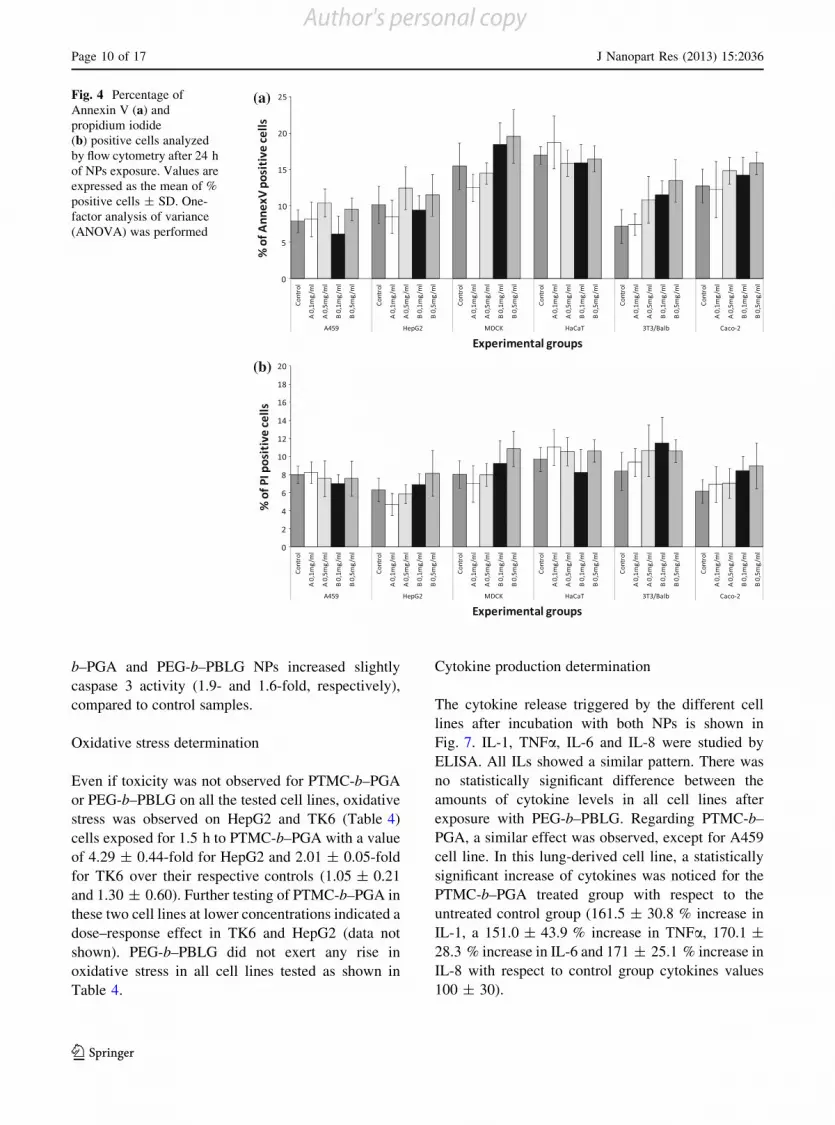
apoptosis and necrosis endpoints at different times(Figs. 4, 5). Cells positive for Annexin V-Alexa Fluor
and negative for PI were apoptotic, and PI positive
cells were necrotic cells. Cells positive for membraneasymmetry study did not show a significant increase of
apoptotic index, treated with either PEG-b–PBLG or
PTMC-b–PGA at 0.1 and 0.5 mg/ml as compared tountreated cultures after 24 and 72 h of NPs incubation.
The values for apoptotic cells after NPs incubation for
24 h did not surpass 20 % in any treatment, includingcontrol untreated cells with values between 7 and
17 % depending on the cell type (Fig. 4a). In most of
the cell lines studied there was a non-significantdifference between control sample and treated sam-
ples, always below 5 %. Moreover, the same values
(a)
(b)
Fig. 2 Percentage of cellviability obtained in AlamarBlue cytotoxicity assaysperformed using A549,HepG2, MDCK, HaCaT,BALB/3T3, Caco-2 andTK6 cell lines as testsystems at 24 h (a) andHSOC model (b) at 24 and72 h. Cells were treated with0.1 and 0.5 mg/ml andHSOC model was treatedwith 0.5 mg/ml of PTMC-b–PGA or PEG-b–PBLG.Values are expressed as themean of % cellviability ± SD. One-factoranalysis of variance(ANOVA) was performed
Page 8 of 17 J Nanopart Res (2013) 15:2036
123
Author's personal copy
were observed regarding PI positive cells at 24 h
(Fig. 4b). The positive cells for necrosis were always
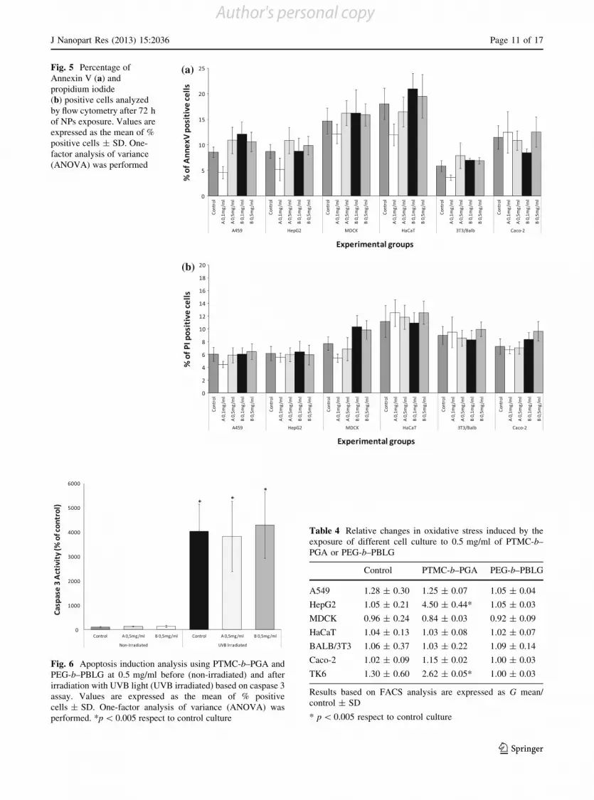
below 12 % in all situations tested.Likewise, cells positive for membrane asymmetry
study at 72 h did not show a significant increase
of apoptotic index, after treating cells with eitherPEG-b–PBLG or PTMC-b–PGA at 0.1 and 0.5 mg/ml
as compared to untreated cultures (Fig. 5a). At the
same time, the percentage of PI positive cells in NPstreated cultures also maintained similar values to
those obtained for control cultures (Fig. 5b). Annexin
V positive cells were below 25 %, and PI positivecells were below 14 % in all cases. Thus, using
Annexin V, our study indicates that neither necrosis
nor apoptosis index increase was detected in the
employed cell cultures after 24 and 72 h concerningNP incubation.
Further analysis of apoptotic index in HSOC model
(Fig. 6) was carried out using caspase 3 assays at 24 h.Caspase 3 activity was markedly increased following
UVB irradiation in all experimental groups, including
control. In relation to non-irradiated control samplescaspase 3 values obtained were 51-fold regarding
untreated irradiated control and 25- and 28-fold
regarding PTMC-b–PGA and PEG-b–PBLG NPs,respectively. In non-irradiated samples, both PTMC-
(a)
(b)
Fig. 3 Percentage of cellviability obtained in colonyforming efficiency assays(CFE) using A549, HepG2,MDCK, HaCaT, BALB/3T3and Caco-2 cell lines as testsystem. Cells wereincubated with 0.1 mg/ml ofeither PTMC-b–PGA orPEG-b–PBLG for 24 or72 h. Values are expressedas the mean of % positivecells ± SD. One-factoranalysis of variance(ANOVA) was performed
J Nanopart Res (2013) 15:2036 Page 9 of 17
123
Author's personal copy
b–PGA and PEG-b–PBLG NPs increased slightlycaspase 3 activity (1.9- and 1.6-fold, respectively),
compared to control samples.
Oxidative stress determination
Even if toxicity was not observed for PTMC-b–PGAor PEG-b–PBLG on all the tested cell lines, oxidative
stress was observed on HepG2 and TK6 (Table 4)
cells exposed for 1.5 h to PTMC-b–PGA with a valueof 4.29 ± 0.44-fold for HepG2 and 2.01 ± 0.05-fold
for TK6 over their respective controls (1.05 ± 0.21
and 1.30 ± 0.60). Further testing of PTMC-b–PGA inthese two cell lines at lower concentrations indicated a
dose–response effect in TK6 and HepG2 (data not
shown). PEG-b–PBLG did not exert any rise inoxidative stress in all cell lines tested as shown in
Table 4.
Cytokine production determination
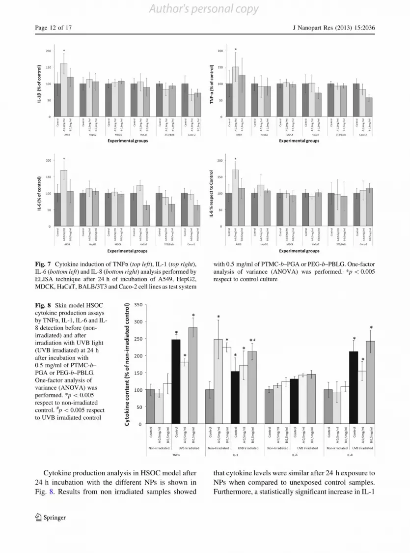
The cytokine release triggered by the different cell
lines after incubation with both NPs is shown inFig. 7. IL-1, TNFa, IL-6 and IL-8 were studied by
ELISA. All ILs showed a similar pattern. There was
no statistically significant difference between theamounts of cytokine levels in all cell lines after
exposure with PEG-b–PBLG. Regarding PTMC-b–PGA, a similar effect was observed, except for A459
cell line. In this lung-derived cell line, a statistically
significant increase of cytokines was noticed for thePTMC-b–PGA treated group with respect to the
untreated control group (161.5 ± 30.8 % increase in
IL-1, a 151.0 ± 43.9 % increase in TNFa, 170.1 ±28.3 % increase in IL-6 and 171 ± 25.1 % increase in
IL-8 with respect to control group cytokines values
100 ± 30).
(a)
(b)
Fig. 4 Percentage ofAnnexin V (a) andpropidium iodide(b) positive cells analyzedby flow cytometry after 24 hof NPs exposure. Values areexpressed as the mean of %positive cells ± SD. One-factor analysis of variance(ANOVA) was performed
Page 10 of 17 J Nanopart Res (2013) 15:2036
123
Author's personal copy
(a)
(b)
Fig. 5 Percentage ofAnnexin V (a) andpropidium iodide(b) positive cells analyzedby flow cytometry after 72 hof NPs exposure. Values areexpressed as the mean of %positive cells ± SD. One-factor analysis of variance(ANOVA) was performed
Fig. 6 Apoptosis induction analysis using PTMC-b–PGA andPEG-b–PBLG at 0.5 mg/ml before (non-irradiated) and afterirradiation with UVB light (UVB irradiated) based on caspase 3assay. Values are expressed as the mean of % positivecells ± SD. One-factor analysis of variance (ANOVA) wasperformed. *p \ 0.005 respect to control culture
Table 4 Relative changes in oxidative stress induced by theexposure of different cell culture to 0.5 mg/ml of PTMC-b–PGA or PEG-b–PBLG
Control PTMC-b–PGA PEG-b–PBLG
A549 1.28 ± 0.30 1.25 ± 0.07 1.05 ± 0.04
HepG2 1.05 ± 0.21 4.50 ± 0.44* 1.05 ± 0.03
MDCK 0.96 ± 0.24 0.84 ± 0.03 0.92 ± 0.09
HaCaT 1.04 ± 0.13 1.03 ± 0.08 1.02 ± 0.07
BALB/3T3 1.06 ± 0.37 1.03 ± 0.22 1.09 ± 0.14
Caco-2 1.02 ± 0.09 1.15 ± 0.02 1.00 ± 0.03
TK6 1.30 ± 0.60 2.62 ± 0.05* 1.00 ± 0.03
Results based on FACS analysis are expressed as G mean/control ± SD
* p \ 0.005 respect to control culture
J Nanopart Res (2013) 15:2036 Page 11 of 17
123
Author's personal copy
Cytokine production analysis in HSOC model after
24 h incubation with the different NPs is shown inFig. 8. Results from non irradiated samples showed
that cytokine levels were similar after 24 h exposure to
NPs when compared to unexposed control samples.Furthermore, a statistically significant increase in IL-1
Fig. 7 Cytokine induction of TNFa (top left), IL-1 (top right),IL-6 (bottom left) and IL-8 (bottom right) analysis performed byELISA technique after 24 h of incubation of A549, HepG2,MDCK, HaCaT, BALB/3T3 and Caco-2 cell lines as test system
with 0.5 mg/ml of PTMC-b–PGA or PEG-b–PBLG. One-factoranalysis of variance (ANOVA) was performed. *p \ 0.005respect to control culture
Fig. 8 Skin model HSOCcytokine production assaysby TNFa, IL-1, IL-6 and IL-8 detection before (non-irradiated) and afterirradiation with UVB light(UVB irradiated) at 24 hafter incubation with0.5 mg/ml of PTMC-b–PGA or PEG-b–PBLG.One-factor analysis ofvariance (ANOVA) wasperformed. *p \ 0.005respect to non-irradiatedcontrol. #p \ 0.005 respectto UVB irradiated control
Page 12 of 17 J Nanopart Res (2013) 15:2036
123
Author's personal copy
values for both PTMC-b–PGA and PEG-b–PBLG NPstreatments in relation to unexposed control (248.01 ±
56.9, 224.73 ± 33.6 and 100 ± 24.2 %, respectively)
was also observed.Regarding UVB irradiation, every studied cytokine
level showed an increase in all UVB irradiated groups
with respect to non-irradiated control. TNFa and IL-8levels showed a statistically significant increase in all
the irradiated untreated and PTMC-b–PGA and PEG-
b–PBLG treated groups compared to non-irradiatedcontrol group. Likewise, all irradiated groups pre-
sented an increase in IL-6 levels in comparison to non-
irradiated samples, but this increment was not statis-tically significant. Anyway, for TNFa, IL-6 and IL-8,
there were no statistically significant differences in the
irradiated NPs treated samples according to UVBirradiated control samples. However, comparing the
IL-1 values of PEG-b–PBLG treated UVB irradiated
samples (212.5 ± 40.1 %) and UVB irradiated con-trol (153.7 ± 30 %), a significant increase was
observed. This effect could not be seen for the
PTMC-b–PGA treated and UVB irradiated samples.
Discussion
PTMC-b–PGA and PEG-b–PBLG NPs have been
described as promising nanocarriers for therapyand/or diagnosis (Sanson et al. 2010a; Li 2002;
Jeong et al. 2005). Previous studies demonstrated the
dispersion stability of these carriers in water. How-ever, in order to allow the in vitro toxicity testing and
to assess their suitability as nanocarriers, it is crucial
to determine their behaviour in physiological condi-tions (i.e. cell culture media or blood serum).
According to the results obtained, NPs synthesis
and characterisation rendered two different NPs withdifferent nature and behaviour. PTMC-b–PGA NPs
show a vesicular morphology with DLS parameters
similar to those already described in Sanson et al.(2010a). A particulate morphology was obtained with
PEG-b–PBLG, with hydrodynamic sizes similar tothose indicated in Martinez Barbosa et al. (2007).
This different nature may actually generate different
toxicological behaviour.According to Fig. 1, it is important to note that
these two NPs have a rather homogeneous size
distribution in water, with a PDI below 0.2 for bothPEG-b–PBLG and PTMC-b–PGA. The DLS analyses
in MEM supplemented with 10 % serum showed anincrease of the PDI value for both NPs. This higher
PDI (as compared to the measure in water or MEM
with 1 % or less serum) could be explained by thepresence of a higher concentration of proteins
which—although they are less diffusive than the
particles—interfere with the measurement, renderingthe dispersion less monodisperse (two populations
with very different characteristic sizes: the particles
around 100 nm and the serum proteins, less than10 nm). This hypothesis is supported by the fact that
the mean hydrodynamic diameters did not signifi-
cantly increase with the serum content (which wouldhave been a sign of particles aggregation). As a result
of the measurements and keeping into account the
considerations for both PTMC-b–PGA and PEG-b–PBLG, they display substantial stability in MEM cell
culture medium.
Furthermore, the precipitated protein amount asso-ciate to both NPs was very low compared to the total
amount of protein present in blood serum, indicating
low or no interaction between NPs and proteins. BothNPs have a polymeric nature, with an amphiphilic
character of the copolymers they are composed of.
They are small with less than 150 nm in diameter andare likely to be transported through the bloodstream by
transporter proteins like apolipoproteins or albumins.
These NPs may be ‘‘coated’’ by proteins to form aprotein–NP complex to be easily transported through
the bloodstream. This needs further research in terms
of pharmacokinetics and distribution which needs tobe done in vivo. In the case of PTMC-b–PGA, the
proteins identified belong in metabolic routes like lipid
transport, antigen presentation and inflammatoryresponse. These last two metabolic routes could be
related to the toxicological effect of this NP and may
indicate a slight immunogenic character of PTMC-b–PGA.
To date, there is a lack of consensus in the published
literature on NP in vitro toxicity mainly due tovariability of methodology and cell lines. Tradition-
ally, in vitro toxicity testing focuses on whether theexposure or not to a potentially toxic agent results in
cell death. The majority of NP in vitro cytotoxicity
assays published measure cell death via colorimetricmethods. The most widely used test is the MTT
viability assay (Chen et al. 2011; Tang et al. 2011;
Yang et al. 2010; Tian et al. 2006). In the present studywe have taken into account that NPs could interfere
J Nanopart Res (2013) 15:2036 Page 13 of 17
123
Author's personal copy
with dyes and dye products in viability assays throughthe adsorption of cell medium constituents (Zhang
et al. 2007; Monteiro-Riviere et al. 2009). We tried to
overcome this by carrying out two different tests; ABassays that determine the activity of a mitochondrial
enzyme and are commonly used to determine the
proliferative activity of cells by colorimetry or fluo-rescence, and CFE assay that indicates cell prolifer-
ation without the use of any cellular dye (Ponti et al.
2010) to discover possible false positives observed byAB assay. Both AB and CFE assays showed a non-
toxic pattern (at cellular level) of PEG-b–PBLG and
PTMC-b–PGA at 24 and 72 h. Although no cell deathmay be apparent after NP exposure, changes in
cellular function may result. So, sub-lethal cellular
changes have also been assessed after NPs exposure onapoptosis induction like ROS production and cyto-
kines release to ensure that valid conclusions were
drawn.Likewise, the systemic effects of some nanocom-
pounds have been shown in different tissues and
organs such as lung, intestine, skin, kidney and liver(Buzea et al. 2007). NPs properties such as size,
surface chemistry, chemical composition or dosage
may pose a threat to human health (Ai et al. 2011). So,cell lines representing these potential target organs
were used to observe cytotoxicity, ROS production or
pro-inflammatory cytotoxic activity that may occur asa result of the effects of PEG-b–PBLG and PTMC-b–
PGA NPs (Geiser and Kreyling 2010; Medina et al.
2007; Revell 2006). In order to increase the complex-ity of the experimental system to get closer to an
in vivo situation, HSOC model was used to further test
the toxicity of these NPs.As described above, some researchers expressed
limited confidence in colorimetric cytotoxicity meth-
ods due to shortcomings in sensitivity and specific-ity (Monteiro-Riviere et al. 2009). This is the reason
why other methods, like cell membrane asymmetry
studies, have been employed. Thus, using AnnexinV, our study indicates that no increase in necrosis
and apoptosis index is detected in the employed cellcultures incubated with both PEG-b–PBLG and
PTMC-b–PGA NPs. HSOC model showed an
apoptosis induction due to UVB irradiation, but itseems to be independent of the NPs treatments
performed.
ROS is a natural byproduct of the normal metab-olism of oxygen and has important roles in cell
signalling and homeostasis (Matsuzawa et al. 2005).These ROS at high concentrations have damaging
effects upon cell viability and function, and in turn,
stimulate innate immune responses (Tomoda et al.2012). According to our flow cytometry analysis,
oxidative stress assays showed no effect in most of the
cell lines tested. Only HepG2 and TK6 showedstatistical significant increase in ROS formation after
treatment with PTMC-b–PGA NP. Moreover, this
effect was dose-dependent for these cell lines (data notshown).
Oxygen species formation is the most common
effect generated by NPs like fullerenes, carbon nano-tubes, quantum dots, etc. (Raab et al. 2008; Oberdor-
ster et al. 2005). Due to the larger surface area to mass
ratio of smaller particles, more ROS could be formedthan in treatments with larger particles (Sioutas et al.
2005; Stone et al. 2000). This is not the case as both
NPs have a similar size and shape, so this effect isprobably due to the different nature of the NPs. The
biggest difference between both NP is their f, which
was -9 and -42 mV for PEG-b–PBLG and PTMC-b–PGA, respectively in water. Moreover, when compar-
ing the effect of PTMC-b–PGA over the whole set of
cell lines used in this study, it seems evident that allcells types did not have the same sensitivity to ROS
formation. The most sensitive cell line seems to be the
hepatocarcinoma derived HepG2, followed by TK6cell line, a human lymphoblast derived cell line. Liver
cells interact with xenobiotics and foreign bodies to
eliminate them and avoid the possible toxic effects theymay cause. Furthermore, hepatocytes take up large
amounts of different molecules from plasma for
oxidation and synthesis (Guo et al. 2006). It might bepossible that HepG2 cells uptake this NP with high
affinity and thus, generate higher levels of ROS.
Lymphocytes, as part of immune system, also interactwith foreign substances, to trigger immune response,
so this might also be a reason why these cells seem to be
more sensitive to ROS formation. It is also interestingto note that according to the results obtained in the
blood–serum interaction assays, antigen presentationis one of the cellular routes associated to this NP,
supporting the idea of a rather immunogenic nature of
PTMC-b–PGA. Anyway, this hypothesis need furtherchecking and more assays need to be done regarding
this issue.
Cytokine production is also a possible adverseeffect upon NP exposure to cells. In this study,
Page 14 of 17 J Nanopart Res (2013) 15:2036
123
Author's personal copy

commonly tested pro-inflammatory cytokines (IL-1b,IL-6, IL-8 and TNFa) were analyzed (Dinarello
2000; Schanen et al. 2009). Results from cytokine
production experiments showed that cytokine levelswere similar after 24 h in cells exposed to PEG-b–
PBLG NPs. However, the cytokine level values in
A549 cell line exposed to PTMC-b–PGA NPs after24 h suffered a statistically significant increase. The
A549 cell line is derived from lung epithelium. Air
pollutants and other substances, like sulphur dioxide,are known to trigger inflammatory response in this
cell line (Backand et al. 2011). In vitro and in vivo
studies support the idea that NPs in general aresignificant contributors to pulmonary fibrosis (Obe-
rdorster 2000). The differential behaviour in cytokine
production of these two NPs might be attributedagain to differences in f. A marked negative f has a
direct relationship in cytokine production compared
the low effect of a neutral or positive charged NP(Yost et al. 2006).
Regarding to HSOC model, it has been published as
a recognized model to study ROS, cytokine productionand structural alteration associated to UVB generated
damage (Portugal-Cohen et al. 2011). In Portugal-
Cohen’s study, inflammatory cytokines were drasti-cally increased after UVB irradiation, a similar effect
observed in this study. NPs addition and further UVB
irradiation did not change that inflammatory profile,indicating a low inflammatory effect of PEG-b–PBLG
and PTMC-b–PGA NPs both in non-irradiated and
UVB irradiated human skin explants. However, IL-1values showed a significant increase after PEG-b–
PBLG NPs incubation. IL-1 is produced by multiple
types of cells, including immune system cells andepithelial cells and, in particular, this IL is related to
caspase protein family, involved in apoptosis (Svobo-
dova et al. 2006).Our results suggest that the main damage occurred
to HSOC model should be allocated to UVB light, but
the use of PEG-b–PBLG enhances that damage,indicating that this NP might not be a good choice as
a vehicle for dermatological applications.
Conclusions
The aim of this thorough study was to ascertain
whether PTMC-b–PGA and PEG-b–PBLG couldpotentially be good candidates for drug delivery based
on a low toxicity profile. Our results indicate thatthese two NPs hardly interact with human blood
serum suggesting that both nanosystems could be
potential candidates for systemic drug delivery.In vitro cytotoxicity is very low and only ROS
formation can be considered as a toxicity mechanism
of action for one of the NPs in two out of the sevencell lines tested. The values for cytokine production
in cell lines are only slightly high in the lung
epithelial cell line, indicating that inhalation routemay represent a problem to overcome if using
PTMC-b–PGA as nanocarrier. Likewise, according
to human skin explants results for PEG-b–PBLG, thismight not be the best system to be used for
dermatological applications.
In general, these two NPs could be good candidatesfor drug delivery systems, but further in vitro and
in vivo studies must be performed in order to have a
clearer and more accurate view of the potential use ofthese polymeric nanosystems.
Acknowledgments This study was supported by the EuropeanCommission FP7 Programme 2007–2013 under NANOTHERproject (www.nanother.eu), Grant Agreement Number CP-IP213631-2 NANOTHER. We want to particularly acknowledgethe patients enrolled in the blood serum study for their partici-pation and the Basque Biobank for Research-OEHUN for itscollaboration.
References
Ai J, Biazar E, Jafarpour M, Montazeri M, Majdi A, AminifardS, Zafari M, Akbari HR, Rad HG (2011) Nanotoxicologyand nanoparticle safety in biomedical designs. Int J Nan-omed 6:1117–1127
Arimura H, Ohya Y, Ouchi T (2005) Formation of core–shelltype biodegradable polymeric micelles from amphiphilicpoly(aspartic acid)-block–polylactide diblock copolymer.Biomacromolecules 6(2):720–725
Backand S, Winder C, Hayes A (2011) Cell viability andcytokine production of human alveolar epithelial cellsfollowing exposure to sulphur dioxide. Int J Occup Hyg3:63–69
Buzea C, Pacheco-Blandino II, Robbie K (2007) Nanomaterialsand nanoparticles: sources and toxicity. Biointerphases2:MR17–MR172
Carlsen A, Lecommandoux S (2009) Self-assembly of poly-peptide-based block copolymer amphiphiles. Curr OpinColloid Interface Sci 14(5):329–339
Chen Y, Wan Y, Wang Y, Zhang H, Jiao Z (2011) Anticancerefficacy enhancement and attenuation of side effects ofdoxorubicin with titanium dioxide NPs. Int J Nanomed6:2321–2326
J Nanopart Res (2013) 15:2036 Page 15 of 17
123
Author's personal copy

Chiellini EE, Chiellini F, Solaro R (2006) Bioerodible poly-meric NPs for targeted delivery of proteic drugs. J NanosciNanotechnol 6(9–10):3040–3047
Chiellini F, Bartoli C, Dinucci D, Piras AM, Anderson R,Croucher T (2007) Bioeliminable polymeric NPs for pro-teic drug delivery. Int J Pharm 343(1–2):90–97
Curtis J, Greenberg M, Kester J, Phillips S, Krieger G (2006)Nanotechnology and nanotoxicology: a primer for clini-cians. Toxicol Sci 25(4):245–260
Dinarello CA (2000) Proinflammatory cytokines. Chest118:503–508
Dong AJ, Deng LD, Sun DX, Zhang YT, Jin JZ, Yuan YJ (2004)Studies on paclitaxel-loaded NPs of amphiphilic blockcopolymer. Yao Xue Xue Bao 39(2):149–152
Duncan R, Ringsdorf H, Satchi-Fainaro R (2006) Polymertherapeutics I. In: Advances in polymer science. Springer,Berlin, pp 1–8
Geiser M, Kreyling WG (2010) Deposition and biokinetics ofinhaled nanoparticles. Part Fibre Toxicol 7(2). doi:10.1186/1743-8977-7-2
Guo W, Huang N, Cai J, Xie W, Hamilton JA (2006) Fatty acidtransport and metabolism in HepG2 cells. Am J PhysiolGastrointest Liver Physiol 290:528–534
Jeong YI, Nah JW, Lee HC, Kim SH, Cho CS (1999) Adria-mycin release from flower-type polymeric micelle basedon star-block copolymer composed of poly(gamma-benzylL-glutamate) as the hydrophobic part and poly(ethyleneoxide) as the hydrophilic part. Int J Pharm 188(1):49–58
Jeong YI, Kang MK, Sun HS, Kang SS, Kim HW, Moon KS,Lee KJ, Kim SH, Jung S (2004) All-trans-retinoic acidrelease from core–shell type NPs of poly(epsilon-capro-lactone)/poly(ethylene glycol) diblock copolymer. Int JPharm 273(1–2):95–107
Jeong YI, Seo SJ, Park IK, Lee HC, Kang IC, Akaike T, Cho CS(2005) Cellular recognition of paclitaxel-loaded polymericNPs composed of poly(gamma-benzyl L-glutamate) andpoly(ethylene glycol) diblock copolymer endcapped withgalactose moiety. Int J Pharm 296(1–2):151–161
Kricheldorf H (2006) Polypeptides and 100 years of chemistryof alpha-amino acid N-carboxyanhydrides. Angew ChemInt Ed Engl 45(35):5752–5784
Lankelma J, Dekker H, Luque FR, Luykx S, Hoekman K, Vander Valk P (1999) Doxorubicin gradients in human breastcancer. Clin Cancer Res 5:1703–1707
Lanone S, Boczkowski J (2006) Biomedical applications andpotential health risks of nanomaterials: molecular mecha-nisms. Curr Mol Med 6(6):651–663
Li C (2002) Poly(L-glutamic acid)–anticancer drug conjugates.Adv Drug Deliv Rev 54(5):695–713
Li S, Wang A, Jiang W, Guan Z (2008) Pharmacokinetic char-acteristics and anticancer effects of 5-fluorouracil loadedNPs. BMC Cancer 8:103–111
Li X, Wang L, Fan Y, Feng Q, Cui F (2012) Biocompatibilityand toxicity of NPs and nanotubes. J Nanomater 2012,Article ID 548389. doi:10.1155/2012/548389
Mandal D, Chatterjee U (2007) Synthesis and spectroscopy ofCdS NPs in amphiphilic diblock copolymer micelles.J Chem Phys 126(13):13450–13457
Martinez Barbosa ME, Montembault V, Cammas-Marion S,Ponchel G, Fontaine L (2007) Synthesis and character-ization of novel poly(c-benzyl-L-glutamate) derivatives
tailored for the preparation of NPs of pharmaceuticalinterest. Polym Int 56:317–324
Matsuzawa A, Saegusa K, Noguchi T, Sadamitsu C, Nishitoh H,Nagai S, Koyasu S, Matsumoto K, Takeda K, Ichijo H(2005) ROS dependent activation of the TRAF6-ASK1-p38 pathway is selectively required for TLR4-mediatedinnate immunity. Nat Immunol 6:587–592
Medina C, Santos-Martinez MJ, Radomski A (2007) Nanopar-ticles: pharmacological and toxicological significance. Br JPharmacol 150:552–558
Monteiro-Riviere NA, Inman AO, Zhang LW (2009) Limita-tions and relative utility of screening assays to assessengineered NP toxicity in a human cell line. Toxicol ApplPharmacol 234:222–235
Nishiyama N, Kataoka K (2006) Polymer therapeutics II. In:Advances in polymer science. Springer, Berlin, pp 67–101
Oberdorster G (2000) Toxicology of ultrafine particles: in vivostudies. Philos Trans R Soc Lond A 358:2719–2740
Oberdorster G, Oberdorster E, Oberdorster J (2005) Nanotoxi-cology: an emerging discipline evolving from studies ofultrafine particles. Environ Health Perspect 113(7):823–839
Oh I, Lee K, Kwon HY, Lee YB, Shin SC, Cho CS, Kim CK(1999) Release of adriamycin from poly(gamma-benzyl-L-glutamate)/poly(ethylene oxide) NPs. Int J Pharm 181(1):107–115
Ponti J, Colognato R, Rauscher H, Gioria S, Broggi F, FranchinF, Pascual C, Giudetti G, Rossi F (2010) Colony formingefficiency and microscopy analysis of multi-wall carbonnanotubes cell interaction. Toxicol Lett 197:29–37
Portugal-Cohen M, Soroka Y, Frusic-Zlotkin M, VerkhovskyL, Bregegere FM, Neuman R, Kohen R, Milner Y (2011)Skin organ culture as a model to study oxidative stress,inflammation and structural alterations associated withUVB-induced photodamage. Exp Dermatol 20(9):749–755
Raab C, Simko M, Gazso A, Fiedeler U, Nentwich M (2008) Wassind synthetische Nanopartikel?. NanoTrust-Dossiers Nr.002, hg. v. Institut fur Technikfolgen-Abschatzung, Wien
Revell PA (2006) The biological effects of nanoparticles.Nanotechnol Percept 2:283–298
Sanson C, Schatz C, Le Meins JF, Brulet A, Soum A, Lecom-mandoux S (2010a) Biocompatible and biodegradablepoly(trimethylene carbonate)-b–poly(L-glutamic acid)polymersomes: size control and stability. Langmuir 26(4):2751–2760
Sanson C, Schatz C, Le Meins JF, Soum A, Thevenot J, Ga-ranger E, Lecommandoux S (2010b) A simple method toachieve high doxorubicin loading in biodegradable poly-mersomes. J Control Release 147:428–435
Sanson C, Diou O, Thevenot J, Ibarboure E, Soum A, Brulet A,Miraux S, Thiaudiere E, Tan S, Brisson A, Dupuis V,Sandre O, Lecommandoux S (2011) Doxorubicin loadedmagnetic polymersomes: theranostic nanocarriers for MRimaging and magneto-chemotherapy. ACS Nano 5:1122–1140
Satchi-Fainaro R, Duncan R, Barnes C (2006) Polymer thera-peutics II. In: Advances in polymer science. Springer,Berlin, pp 1–65
Schanen BC, Karakoti AS, Seal S, Drake DR, Warren WL, SelfWT (2009) Exposure to titanium dioxide nanomaterialsprovokes inflammation of an in vitro human immuneconstruct. ACS Nano 3:2523–2532
Page 16 of 17 J Nanopart Res (2013) 15:2036
123
Author's personal copy
Sioutas C, Delfino RJ, Singh M (2005) Exposure assessment foratmospheric ultrafine particles (UFPs) and implications inepidemiologic research. Environ Health Perspect 113(8):947–955
Stone V, Tuinman M, Vamvakopoulos JE, Shaw J, Brown D,Petterson S, Faux SP, Borm P, MacNee W, MichaelangeliF, Donaldson K (2000) Increased calcium influx in amonocytic cell line on exposure to ultrafine carbon black.Eur Respir J 15(2):297–303
Svobodova A, Walterova D, Vostalova J (2006) Ultraviolet lightinduced alteration to the skin. Biomed Pap Med Fac UnivPalacky Olomouc Czech Repub 150(1):25–38
Tang H, Guo J, Sun Y, Chang B, Ren Q, Yang W (2011) Facilesynthesis of pH sensitive polymer-coated mesoporous sil-ica NPs and their application in drug delivery. Int J Pharm421:388–396
Tian F, Cui D, Schwarz H, Estrada GG, Kobayashi H (2006)Cytotoxicity of single-wall carbon nanotubes on humanfibroblasts. Toxicol In Vitro 20:1202–1212
Tomoda K, Watanabe A, Suzuki K, Inagi T, Terada H, MakinoK (2012) Enhanced transdermal permeability of estradiol
using combination of PLGA nanoparticles system andiontophoresis. Colloids Surf B 97:84–89
Upadhyay KK, Agrawal HG, Upadhyay C, Schatz C, Le MeinsJF, Misra A, Lecommandoux S (2009) Role of blockcopolymer nanoconstructs in cancer therapy. Crit Rev TherDrug Carr Syst 26(2):157–205
Vega-Villa KR, Takemoto JK, Yanez JA, Remsberg CM, For-rest ML, Davies NM (2008) Clinical toxicities of nano-carrier systems. Adv Drug Deliv Rev 60:929–938
Yang Y, Qu Y, Lu X (2010) Global gene expression analysis ofthe effects of gold NPs on human dermal fibroblasts.J Biomed Nanotechnol 6:234–246
Yost GS, Veranth JM, Reilly CA, Veronesi B (2006) Chapter 8:vanilloid receptors in the respiratory tract. In: Gardner DE(ed) Toxicology of the lung, 4th edn. Taylor and FrancisGroup, Boca Raton, pp 297–350
Zhang LW, Zeng L, Barron AR, Monteiro-Riviere NA (2007)Biological interactions of functionalized single-wall car-bon nanotubes in human epidermal keratinocytes. Int JToxicol 26:103–113
J Nanopart Res (2013) 15:2036 Page 17 of 17
123
Author's personal copy