biodistribution, clearance, and biocompatibility of iron oxide magnetic nanoparticles in rats
TRANSCRIPT
Biodistribution, Clearance, and Biocompatibility of IronOxide Magnetic Nanoparticles in Rats
Tapan K. Jain,† Maram K. Reddy,† Marco A. Morales,‡
Diandra L. Leslie-Pelecky,‡ and Vinod Labhasetwar*,†,§
Department of Biomedical Engineering, Lerner Research Institute, CleVeland Clinic,CleVeland, Ohio 44195, Department of Physics & Astronomy and Nebraska Center for
Materials and Nanoscience, UniVersity of Nebraska-Lincoln,Lincoln, Nebraska 68588-0111, and Department of Pharmaceutical Sciences, College of
Pharmacy, Nebraska Medical Center, Omaha, Nebraska 68198-6025
Received September 21, 2007; Revised Manuscript Received November 16, 2007; AcceptedNovember 25, 2007
Abstract: It is essential to determine the biodistribution, clearance, and biocompatibility ofmagnetic nanoparticles (MNPs) for in vivo biomedical applications to ensure their safe clinicaluse. We have studied these aspects with our novel iron oxide MNP formulation, which can beused as a magnetic resonance imaging (MRI) agent and a drug carrier system. Changes inserum and tissue iron levels were analyzed over 3 weeks after intravenous administration ofMNPs to rats. Serum alanine aminotransferase (ALT), aspartate aminotransferase (AST), alkalinephosphatase (AKP) levels, and total iron-binding capacity (TIBC) were also measured with timeto assess the effect of MNPs on liver function. Selected tissues were also analyzed for oxidativestress and studied histologically to determine biocompatibility of MNPs. Serum iron levelsgradually increased for up to 1 week but levels slowly declined thereafter. Biodistribution of ironin various body tissues changed with time but greater fraction of the injected iron localized inthe liver and spleen than in the brain, heart, kidney, and lung. Magnetization measurements ofthe liver and spleen samples showed a steady decrease over 3 weeks, suggesting particledegradation. Serum showed a transient increase in ALT, AST, AKP levels, and TIBC over aperiod of 6-24 h following MNP injection. The increase in oxidative stress was tissue dependent,reaching a peak at ∼3 days and then slowly declining thereafter. Histological analyses of liver,spleen, and kidney samples collected at 1 and 7 days showed no apparent abnormal changes.In conclusion, our MNPs did not cause long-term changes in the liver enzyme levels or induceoxidative stress and thus can be safely used for drug delivery and imaging applications.
Keywords: Imaging; MRI; drug delivery; oxidative stress; nanotoxicity
IntroductionMagnetic nanoparticles (MNPs) are explored for various
biomedical applications such as targeted drug delivery,1,2
cell sorting,3 contrast agents for magnetic resonance
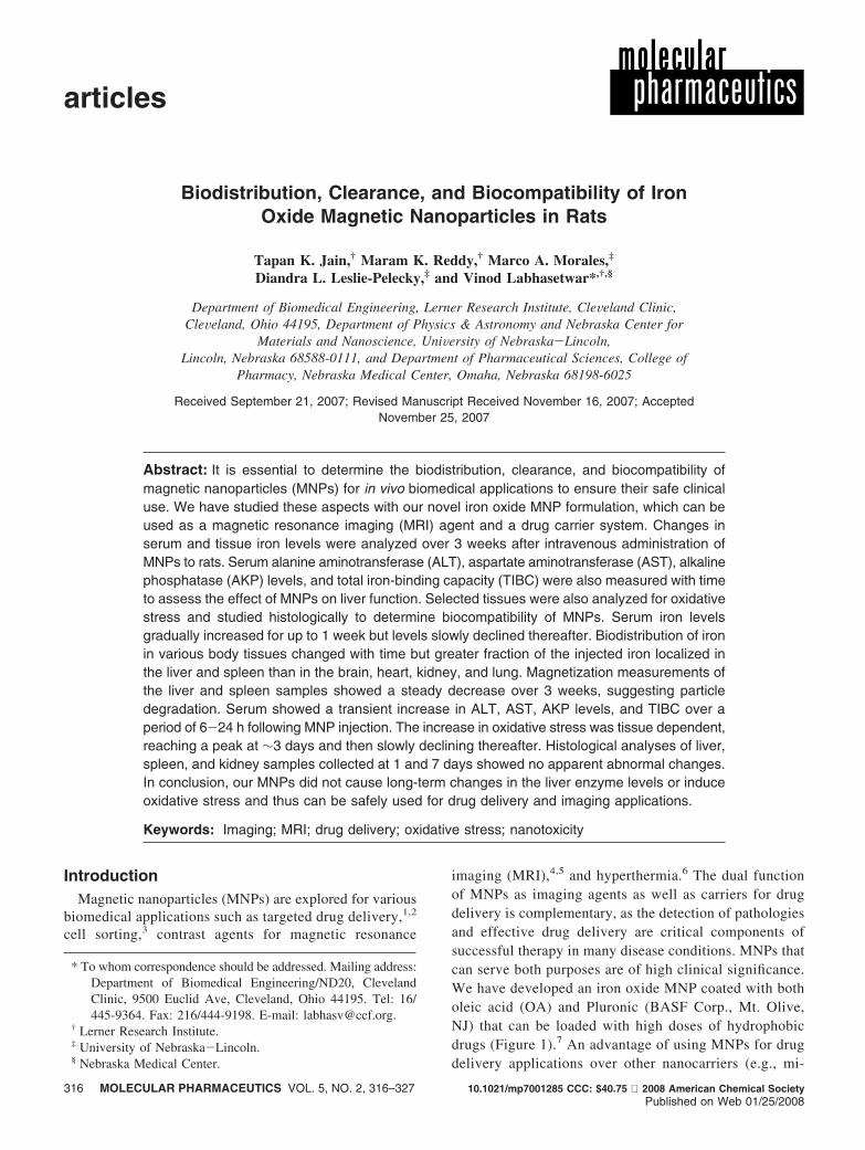
imaging (MRI),4,5 and hyperthermia.6 The dual functionof MNPs as imaging agents as well as carriers for drugdelivery is complementary, as the detection of pathologiesand effective drug delivery are critical components ofsuccessful therapy in many disease conditions. MNPs thatcan serve both purposes are of high clinical significance.We have developed an iron oxide MNP coated with botholeic acid (OA) and Pluronic (BASF Corp., Mt. Olive,NJ) that can be loaded with high doses of hydrophobicdrugs (Figure 1).7 An advantage of using MNPs for drugdelivery applications over other nanocarriers (e.g., mi-
* To whom correspondence should be addressed. Mailing address:Department of Biomedical Engineering/ND20, ClevelandClinic, 9500 Euclid Ave, Cleveland, Ohio 44195. Tel: 16/445-9364. Fax: 216/444-9198. E-mail: [email protected].
† Lerner Research Institute.‡ University of Nebraska-Lincoln.§ Nebraska Medical Center.
articles
316 MOLECULAR PHARMACEUTICS VOL. 5, NO. 2, 316–327 10.1021/mp7001285 CCC: $40.75 2008 American Chemical SocietyPublished on Web 01/25/2008
celles, polymeric nanoparticles, liposomes, etc.) is thattheir magnetic properties allow monitoring and quantita-tive determination of their biodistribution and thus,indirectly, that of the drug associated with MNPs (includ-ing their localization in the target tissue) using noninvasiveMRI and magnetometry.8 This is a crucial issue in drugtherapy because failure to achieve the expected therapeutic
outcome could be due to suboptimal dosing of the drugin the target tissue.9 For example, in cancer chemotherapy,optimized dosing is critical not only for achieving tumorregression, but also to prevent the tumor from developingdrug resistance and contributing to relapse.10
In addition to imaging and drug delivery, MNPs are beinginvestigated using MRI to monitor the migration of particularcell populations in the body (e.g., progenitor cells ormacrophages) that have been tagged with MNPs prior toinjection in disease conditions.11 MRI data can be a usefulresearch tool to determine the targeting efficacy of ligandssuch as peptides or antibodies.12 The suitability of MNPshas also been tested for other intracellular applications, suchas for visualization of gene expression at the molecularlevel.13
It is essential to determine the biodistribution, clearance,and biocompatibility of MNPs that may be used for drugdelivery and imaging applications in ViVo. Biodistributionof MNPs depends on their properties such as surfacecharacteristics, size, and shape,14 which can affect particle-cell interactions and interactions with serum proteins (op-sonization).15 Other factors such as magnetic materials andtheir biodegradation products are important from the pointof view of biocompatibility of MNPs.16 Equally importantis determining iron clearance as MNPs degrade sinceexcessive tissue accumulation of free iron is known to causetoxicity.17 In this paper, we have determined the biodistri-bution,clearance,andbiocompatibilityofouroleicacid-Pluronic-coated iron oxide MNP formulation.7
(1) Alexiou, C.; Arnold, W.; Klein, R. J.; Parak, F. G.; Hulin, P.;Bergemann, C.; Erhardt, W.; Wagenpfeil, S.; Lubbe, A. S.Locoregional cancer treatment with magnetic drug targeting.Cancer Res. 2000, 60, 6641–6648.
(2) Alexiou, C.; Jurgons, R.; Schmid, R. J.; Bergemann, C.; Henke,J.; Erhardt, W.; Huenges, E.; Parak, F. Magnetic drug targeting-biodistribution of the magnetic carrier and the chemotherapeuticagent mitoxantrone after locoregional cancer treatment. J. DrugTarget. 2003, 11, 139–149.
(3) Clement, J. H.; Schwalbe, M.; Buske, N.; Wagner, K.; Schna-belrauch, M.; Gornert, P.; Kliche, K. O.; Pachmann, K.; Weitschies,W.; Hoffken, K. Differential interaction of magnetic nanoparticleswith tumor cells and peripheral blood cells. J. Cancer Res. Clin.Oncol. 2006, 132, 287–292.
(4) Artemov, D. Molecular magnetic resonance imaging with targetedcontrast agents. J. Cell. Biochem. 2003, 90, 518–524.
(5) Jung, C. W.; Jacobs, P. Physical and chemical properties ofsuperparamagnetic iron oxide MR contrast agents: ferumoxides,ferumoxtran, ferumoxsil. Magn. Reson. Imaging 1995, 13, 661–674.
(6) Ito, A.; Tanaka, K.; Kondo, K.; Shinkai, M.; Honda, H.;Matsumoto, K.; Saida, T.; Kobayashi, T. Tumor regression bycombined immunotherapy and hyperthermia using magneticnanoparticles in an experimental subcutaneous murine melanoma.Cancer Sci. 2003, 94, 308–313.
(7) Jain, T. K.; Morales, M. A.; Sahoo, S. K.; Leslie-Pelecky, D. L.;Labhasetwar, V. Iron oxide nanoparticles for sustained deliveryof anticancer agents. Mol. Pharm. 2005, 2, 194–205.
(8) Koning, G. A.; Krijger, G. C. Targeted multifunctional lipid-basednanocarriers for image-guided drug delivery. Anticancer AgentsMed. Chem. 2007, 7, 425–440.
(9) Broxterman, H. J.; Lankelma, J.; Hoekman, K. Resistance tocytotoxic and anti-angiogenic anticancer agents: similarities anddifferences. Drug Resist. Updat. 2003, 6, 111–127.
(10) Bezwoda, W. R. High-dose chemotherapy with hematopoieticrescue in breast cancer: from theory to practice. Cancer Chemoth-er. Pharmacol. 1997, 40, S79–S87.
(11) Frank, J. A.; Miller, B. R.; Arbab, A. S.; Zywicke, H. A.; Jordan,E. K.; Lewis, B. K.; Bryant, L. H. Jr.; Bulte, J. W. M. ClinicallyApplicable Labeling of Mammalian and Stem Cells by CombiningSuperparamagnetic Iron Oxides and Transfection Agents. Radiol-ogy 2003, 228, 480–487.
(12) Gupta, A. K.; Naregalkar, R. R.; Vaidya, V. D.; Gupta, M. Recentadvances on surface engineering of magnetic iron oxide nano-particles and their biomedical applications. Nanomed. 2007, 2,23–39.
(13) Weissleder, R.; Moore, A.; Mahmood, U.; Bhorade, R.; Ben-veniste, H.; Chiocca, E. A.; Basilion, J. P. In vivo magneticresonance imaging of transgene expression. Nat. Med. 2000, 6,351–355.
(14) Chouly, C.; Pouliquen, D.; Lucet, I.; Jeune, J. J.; Jallet, P.Development of superparamagnetic nanoparticles for MRI: effectof particle size, charge and surface nature on biodistribution. J.Microencapsul. 1996, 13, 245–255.
(15) Owens, D. E.; Peppas, N. A. Opsonization, biodistribution, andpharmacokinetics of polymeric nanoparticles. Int. J. Pharm. 2006,307, 93–102.
(16) Gupta, A. K.; Gupta, M. Synthesis and surface engineering ofiron oxide nanoparticles for biomedical applications. Biomaterials2005, 26, 3995–4021.
(17) Weir, M. P.; Gibson, J. F.; Peters, T. J. Haemosiderin and tissuedamage. Cell Biochem. Funct. 1984, 2, 186–194.
Figure 1. Schematic of oleic acid (OA)-Pluronic-coatediron oxide magnetic nanoparticles (MNPs). Hydrophobicdrugs can be partitioned in the OA layer around the ironoxide core. Pluronic anchored onto OA provides aqueousdispersity. Figure modified from ref 7.
Biocompatibility of Magnetic Nanoparticles articles
VOL. 5, NO. 2 MOLECULAR PHARMACEUTICS 317
Experimental SectionIron(III) chloride hexahydrate (FeCl3 ·6H2O) (Fe(III)) pure
granulated 99%, iron(II) chloride tetrahydrate (FeCl2 ·4H2O)(Fe(II)) 99+%, ammonium hydroxide (5 M), and oleic acid(OA) were purchased from Fisher Scientific (Pittsburgh, PA).Pluronic F-127 was received as a gift from BASF Corp. (Mt.Olive, NJ). Polyvinyl alcohol (PVA, average MW 30000–70000), paraformaldehyde, and Mohr’s salt were obtainedfrom Sigma-Aldrich (St. Louis, MO). Poly (D,L-lactide co-glycolide) (PLGA) 50:50 (inherent viscosity 1.32 dL/g) waspurchased from Birmingham Polymers, Inc. (Birmingham,AL). Deionized water freshly purged with nitrogen gas wasused in all the steps involved in the synthesis and formulationof MNPs.
Formulation of MNPs. Formulation of OA-Pluronic-coated iron oxide MNPs was prepared according to ourpreviously described procedure.7 In brief, aqueous solutionsof 0.1 M Fe(III) (30 mL) and 0.1 M Fe(II) (15 mL) weremixed to which 3 mL of 5 M ammonium hydroxide solutionwas added to form iron oxide nanoparticles. To the abovepreparation, 100 mg of OA was added and heated for 30min to 80 °C with stirring to evaporate ammonia. The contentwas cooled to room temperature, and the black precipitatethus obtained was washed twice with 15 mL of deionizedwater to remove excess OA. To the precipitate was added45 mL of deionized water, followed by 100 mg of Pluronic.The suspension was stirred overnight in a closed containerto minimize exposure to atmospheric oxygen to preventoxidation of iron oxide nanoparticles. The particles wereseparated using neodymium iron boron magnets (12200 G;Edmund Scientific, Tonawanda, NY), redispersed, andwashed three times with water to remove soluble salts andexcess Pluronic; particles were separated during each wash-ing step using magnets as described above. The particles wereredispersed in 15 mL of water by sonication in a water-bathsonicator (FS-30, Fisher Scientific) for 10 min and centri-fuged at 1000 rpm for 20 min at 7–11 °C to remove anylarge aggregates. The supernatant containing OA-Pluronic-stabilized MNPs was collected and further concentrated usingmagnetic separation as described above. The mean hydro-dynamic diameter of MNPs was 193 nm (polydispersityindex ) 0.262) and �-potential of -0.22 mV. The corediameter of iron oxide particles as measured by transmissionelectron microscopy was 11 ( 2 nm. As discussed in ourprevious study, OA and Pluronic coatings around the ironoxide core contribute toward the hydrodynamic diameter ofMNPs.7
Determination of Iron Oxide and Iron Content inNanoparticle Formulation. The concentration of iron oxidein the preparation was determined by lyophilizing a portionof the sample in a preweighed vial. The amount of OA andPluronic associated with particles was determined by ther-mogravimetric analysis. This value was then subtracted fromthe total weight of the formulation to determine the iron oxidecontent. The iron levels in the formulation were determined
using the 1,10-phenanthroline colorimetric method.18 To 0.5mg of lyophilized samples, 200 µL of concentrated hydro-chloric acid (HCl) was added and incubated for 1 h at 25°C. The sample was then diluted to 1 mL with water. To 50µL of aliquot was added 2 mL of 0.5 M ammoniumacetate-acetic acid buffer, pH 3.0, followed by 100 µL of10% aqueous solution of hydroxylamine hydrochloride toreduce Fe(III) to Fe(II). To this solution was added 500 µLof 0.3% aqueous solution of 1,10 phenanthroline (Sigma-Aldrich), and the solution was incubated for 1 h at roomtemperature. The absorbance was measured at 511 nm usinga spectrophotometer (UV-1601PC, UV–vis spectrophoto-meter, Shimadzu Scientific Instruments, Inc., Columbia,MD). The concentration of iron in the samples was calculatedfrom a standard plot, which was prepared using Mohr’s saltsolution in 0.01 N HCl in the concentration range of 0.5–4.0µg of Fe/mL. In addition to the iron content, each batch ofMNP formulation was characterized for particle size usingtransmission electron microscopy and dynamic light scat-tering to ensure their dispersion in aqueous solution.
Formulation of PLGA Nanoparticles and OA-Plu-ronic Emulsion. PLGA nanoparticles were used as a modelbiocompatible formulation at the same dose (w/w) as MNPsto determine whether the changes seen in the liver enzymeswere specific to MNPs or a general response to the particulateinjection. PLGA nanoparticles were prepared by a solventevaporation method as previously described.19 In a typicalprocedure, 90 mg of PLGA was dissolved in 3 mL ofchloroform, which was then emulsified in 12 mL of (2%w/v) PVA solution using sonication. This emulsion wasstirred overnight to evaporate chloroform, thereby formingnanoparticles that were recovered by ultracentrifugation,washed, and lyophilized. Similarly, OA-Pluronic emulsion(without iron oxide particles) was used as a control todetermine whether the effect on liver enzymes was due tonanoparticles or the formulation components (i.e., OA orPluronic). The amount of OA and Pluronic associated withnanoparticles was calculated as per our previously describedprotocol.7 Based on that, 6 mg of OA was emulsified in anaqueous solution of Pluronic (5.7 mg/2.0 mL of saline) bysonication in a water bath sonicator for 20 min at roomtemperature. The mean diameter of droplet size measuredusing dynamic light scattering (NICOMP 380 ZLS, ZetaPotential/ Particle Sizer, Santa Barbara, CA) was 86.2 nm(polydispersity index ) 0.18). A 1 mL/kg aliquot of theemulsion was injected intravenously to rats which containedthe same dose of OA and Pluronic that is associated withthe dose of MNPs injected.
In ViWo Studies. Male Sprague–Dawley rats (240–260 g,Charles River Laboratories, Wilmington, MA) were used inthis experiment. All of the procedures complied with thestandards for humane care and use of animal subjects as
(18) Jeffery, G. H.; Bassett, J.; Mendham, J. Denny, R. C. Vogel’sText Book of QuantitatiVe Chemical Analysis, 5th ed.; John Wiley& Sons, Inc.: New York, 1989; pp 690-692.
(19) Davda, J.; Labhasetwar, V. Characterization of nanoparticle uptakeby endothelial cells. Int. J. Pharm. 2002, 233, 51–59.
articles Jain et al.
318 MOLECULAR PHARMACEUTICS VOL. 5, NO. 2
stated in the Guide for the Care and Use of LaboratoryAnimals (Institute of Laboratory Resources, National Acad-emy of Sciences, Bethesda, MD) and animal welfare policyof the University of Nebraska Medical Center. Rats wereanesthetized with an intraperitoneal injection of ketamine (80mg/kg) and xylazine (10 mg/kg) mixture. A suspension ofMNPs (10 mg Fe/mL) was prepared in saline and injectedslowly through the tail vein at a dose of 10 mg Fe/kg. Controlexperiments were carried out with and without injectingsaline to determine if the injection itself caused any changesin liver enzyme levels. Animals were allowed to recoverfollowing nanoparticle injection. At different time points,animals were euthanized with an intraperitoneal injection ofpentobarbital sodium (120 mg/kg), and ∼4 mL of blood wasimmediately collected through a cardiac puncture. Animalswere then perfused transcardially with heparinized saline for20 min, and various tissues (liver, spleen, kidney, heart, lung,and brain) were collected, washed thoroughly with saline,and stored at -80 °C until taken for analysis. These tissueswere used for determination of iron content and for lipidperoxidation using a lipid hydroperoxide (LHPO) assay. Inanimals from which tissues were collected for histologicalexamination, a 4% paraformaldehyde solution which wasprepared by dissolving paraformaldehyde (Sigma-Aldrich)in near-boiling phosphate-buffered saline (PBS, 154 mM,pH ) 7.4) was perfused for 2 min following the salineperfusion as described above.
Analysis of Serum. Blood samples were allowed to clotat room temperature and centrifuged at 1500g for 10 min toseparate the serum for collection. The serum samples wereimmediately sent to the Clinical Core Laboratory for analysisof ALT, AST, and AKP levels. In addition, serum sampleswere analyzed at the Core for iron levels and total ironbinding capacity (TIBC). TIBC represents the amount of ironin the blood that would be present if the total transferrin inplasma were saturated. Since transferrin is produced in theliver, it was used to indirectly indicate liver function.
Analysis of Tissue. Upon procurement of tissue, the wetweight of each sample was recorded. Tissue samples werethen homogenized with a Tissue Tearor homogenizer (ModelNo. 985370, Biospec Products Inc., Bartlesville, OK) innitrogen-purged water (∼20% tissue wet weight) in an icebath. The total volume of each homogenate was measured.Tissue homogenates were used to determine iron content andfor the LHPO assay. To determine iron levels in tissues, 1mL of each tissue homogenate was lyophilized in a test tubefor 2 days at -60 °C and 7 µmHg vacuum (Lyph-Lock 12Labconco, Kansas City, MO); weight of the tissue wasdetermined by comparing the difference in weight of theempty tubes and that after lyophilization of the tissue. Toeach dry tissue sample was added 2 mL of 6 N HCl, thetubes were placed in a closed glass container, and the sampleswere heated overnight in an incubator set at 55 °C. Eachsample was vortexed and centrifuged at 1000 rpm for 15min (RT 7 Centrifuge, Sorvall, DuPont, CT), and 1 mL ofthe supernatant from each sample was collected in a separatetest tube. Samples were dried under a stream of nitrogen
gas. To each residue was added 1 mL of 0.01 N HCl, andthe mixture was vortexed and centrifuged at 1000 rpm for15 min. The resulting supernatant solution was diluted 50times and analyzed using inductively coupled plasma-massspectrometry (ICP-MS; Varian 800-MS, Palo Alto, CA). Astock solution of Fe was prepared using Mohr’s salt in 0.01N HCl. Suitable dilutions of the stock solution were preparedin 0.01 N HCl to obtain a standard plot in the range of50–1000 parts per billion with respect to Fe. Samples havinghigher concentration outside the calibration curves wereappropriately diluted. 57Fe isotope counts were used toanalyze the Fe content. From the weight of the tissue andvolume of homogenate, iron levels were normalized toamount per gram wet weight of tissue. The fraction of theinjected dose in each tissue at different time points wascalculated from the total iron content in each tissue, fromwhich the endogenous iron content for the respective tissuefrom control animals (saline injected) was subtracted.
Magnetic Susceptibility. Tissue samples from liver,spleen, and kidney were used for magnetic susceptibilitymeasurements as these tissues showed higher iron levels thanother tissues analyzed. Each powdered tissue sample (3–6mg) prepared following homogenization and lyophilizationwas sealed along with 2–4 mg of paraffin wax (Sigma-Aldrich) in polyethylene bags. The bags were heated in awater bath at 70 °C to melt the paraffin, which producedhomogeneously dispersed tissue powder in solid paraffin afterthe samples cooled to room temperature. Magnetizationversus field measurements, M(H), were performed using analternating gradient force magnetometer (Model Micromag2900, Princeton Measurements Corporation, Princeton, NJ).Measurements were carried out at room temperature with amaximum applied magnetic field of 1.2 T. The magnetizationdata were normalized to the iron content as determined byICP-MS. Analysis of the normalized data was performed byfitting the curves to a combination of a superparamagnetic(Langevin function) plus a diamagnetic (linear) function.20,21
Oxidative Stress Analysis. The change in the lipidhydroperoxide (LHPO) levels in different tissues with timewas used as a biochemical marker to assess the effect ofinjected MNPs on oxidative stress using a lipid peroxidationassay kit (Cayman Chemical Co., Ann Arbor, MI).22 Lipidhydroperoxides were extracted from each tissue homogenate(prepared as above) into chloroform as per the instructionsprovided with the assay kit. Typically, 0.5 mL of tissuehomogenate was mixed with 0.5 mL of saturated methanolicsolution of Extract R (a component of the kit) by vortexing,
(20) Stearns, M. B.; Cheng, Y. Determination of para- and ferromag-netic components of magnetization and magnetoresistance ofgranular Co/Ag films. J. Appl. Phys. 1994, 75, 6894–6899.
(21) Noyau, R. H.; Middleton, B. K.; Miles, J. J.; Mackintosh, N. D.Modelling digital recording in thin film media. IEEE Trans. Magn.1988, 24, 2494–2496.
(22) Mihaljevic, B.; Katusin-Razem, B.; Razem, D. The reevaluationof the ferric thiocyanate assay for lipid hydroperoxides with specialconsiderations of the mechanistic aspects of the response. FreeRadic. Biol. Med. 1996, 21, 53–63.
Biocompatibility of Magnetic Nanoparticles articles
VOL. 5, NO. 2 MOLECULAR PHARMACEUTICS 319
to which 1 mL of cold chloroform was added and mixedthoroughly again by vortexing. This mixture was centrifugedat 1500g for 5 min at 0 °C to separate it into two layers; thechloroform layer (∼700 µL) from the bottom of the tubewas collected carefully and stored in an ice bath until takenfor analysis. To 200 µL of the chloroform layer collected asabove, 950 µL of chloroform-methanol (2:1 v:v) mixture wasadded, followed by 50 µL of chromogen (provided in thekit). Each sample was incubated at room temperature for 5min, and the absorbance was measured at 500 nm using aUV/vis spectrophotometer. A standard plot of LHPO pro-vided with the assay kit was prepared similarly, in theconcentration range of 0.5-5.0 nM.
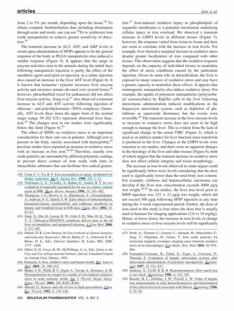
Histology. Portions of kidney, liver, and spleen were fixedin 10% buffered formalin-saline (Sigma-Aldrich) at 4 °Covernight and then embedded in paraffin blocks. Tissuesections of 5 µm thickness were stained with hematoxylinand eosin (H&E). The morphology of the tissue was observedunder a microscope (Nikon Eclipse E600, Nikon Inc.,Melville, NY) at 10× and 40× magnification.
Statistical Analysis. Student’s t-test (two tailed, unpaired)was performed between nanoparticle groups vs salinecontrols, and p values less than 0.05 were consideredstatistically significant.
ResultsSerum Iron Levels and Biodistribution of Iron. The
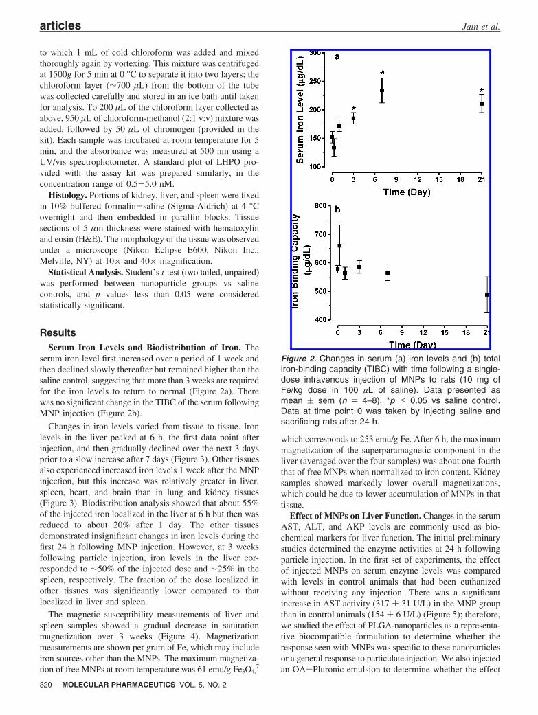
serum iron level first increased over a period of 1 week andthen declined slowly thereafter but remained higher than thesaline control, suggesting that more than 3 weeks are requiredfor the iron levels to return to normal (Figure 2a). Therewas no significant change in the TIBC of the serum followingMNP injection (Figure 2b).
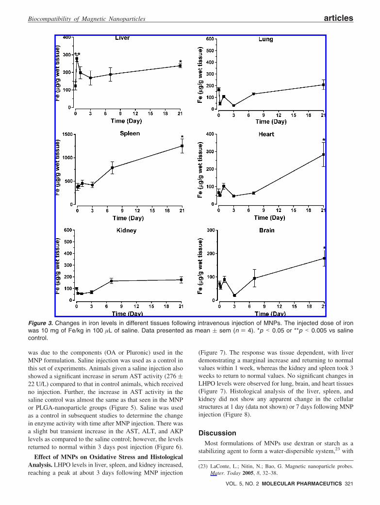
Changes in iron levels varied from tissue to tissue. Ironlevels in the liver peaked at 6 h, the first data point afterinjection, and then gradually declined over the next 3 daysprior to a slow increase after 7 days (Figure 3). Other tissuesalso experienced increased iron levels 1 week after the MNPinjection, but this increase was relatively greater in liver,spleen, heart, and brain than in lung and kidney tissues(Figure 3). Biodistribution analysis showed that about 55%of the injected iron localized in the liver at 6 h but then wasreduced to about 20% after 1 day. The other tissuesdemonstrated insignificant changes in iron levels during thefirst 24 h following MNP injection. However, at 3 weeksfollowing particle injection, iron levels in the liver cor-responded to ∼50% of the injected dose and ∼25% in thespleen, respectively. The fraction of the dose localized inother tissues was significantly lower compared to thatlocalized in liver and spleen.
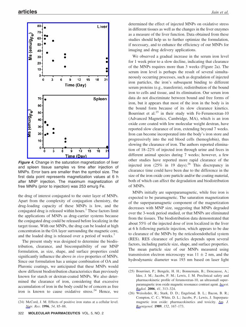
The magnetic susceptibility measurements of liver andspleen samples showed a gradual decrease in saturationmagnetization over 3 weeks (Figure 4). Magnetizationmeasurements are shown per gram of Fe, which may includeiron sources other than the MNPs. The maximum magnetiza-tion of free MNPs at room temperature was 61 emu/g Fe3O4,
7
which corresponds to 253 emu/g Fe. After 6 h, the maximummagnetization of the superparamagnetic component in theliver (averaged over the four samples) was about one-fourththat of free MNPs when normalized to iron content. Kidneysamples showed markedly lower overall magnetizations,which could be due to lower accumulation of MNPs in thattissue.
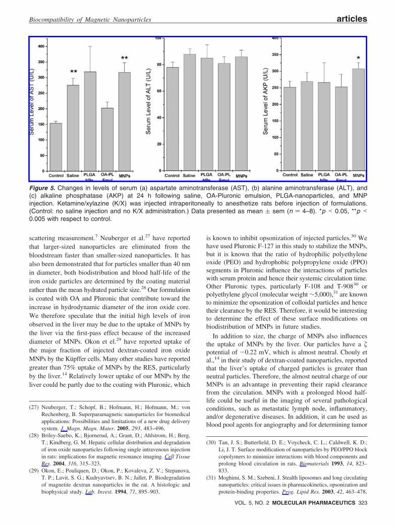
Effect of MNPs on Liver Function. Changes in the serumAST, ALT, and AKP levels are commonly used as bio-chemical markers for liver function. The initial preliminarystudies determined the enzyme activities at 24 h followingparticle injection. In the first set of experiments, the effectof injected MNPs on serum enzyme levels was comparedwith levels in control animals that had been euthanizedwithout receiving any injection. There was a significantincrease in AST activity (317 ( 31 U/L) in the MNP groupthan in control animals (154 ( 6 U/L) (Figure 5); therefore,we studied the effect of PLGA-nanoparticles as a representa-tive biocompatible formulation to determine whether theresponse seen with MNPs was specific to these nanoparticlesor a general response to particulate injection. We also injectedan OA-Pluronic emulsion to determine whether the effect
Figure 2. Changes in serum (a) iron levels and (b) totaliron-binding capacity (TIBC) with time following a single-dose intravenous injection of MNPs to rats (10 mg ofFe/kg dose in 100 µL of saline). Data presented asmean ( sem (n ) 4–8). *p < 0.05 vs saline control.Data at time point 0 was taken by injecting saline andsacrificing rats after 24 h.
articles Jain et al.
320 MOLECULAR PHARMACEUTICS VOL. 5, NO. 2
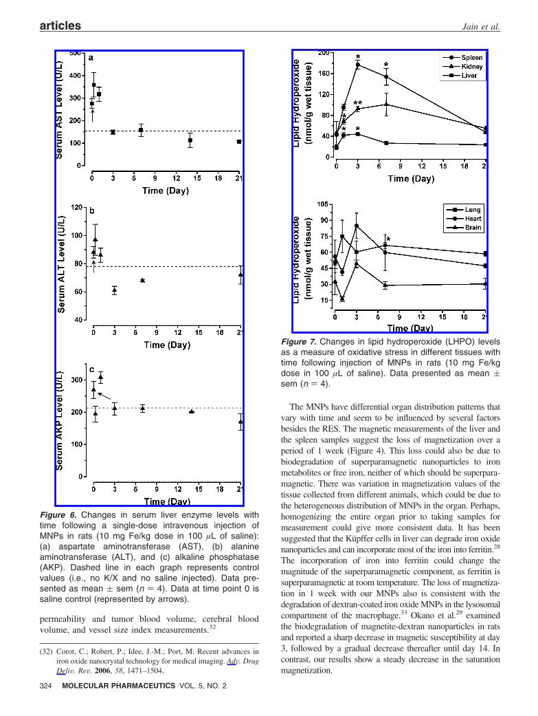
was due to the components (OA or Pluronic) used in theMNP formulation. Saline injection was used as a control inthis set of experiments. Animals given a saline injection alsoshowed a significant increase in serum AST activity (276 (22 U/L) compared to that in control animals, which receivedno injection. Further, the increase in AST activity in thesaline control was almost the same as that seen in the MNPor PLGA-nanoparticle groups (Figure 5). Saline was usedas a control in subsequent studies to determine the changein enzyme activity with time after MNP injection. There wasa slight but transient increase in the AST, ALT, and AKPlevels as compared to the saline control; however, the levelsreturned to normal within 3 days post injection (Figure 6).
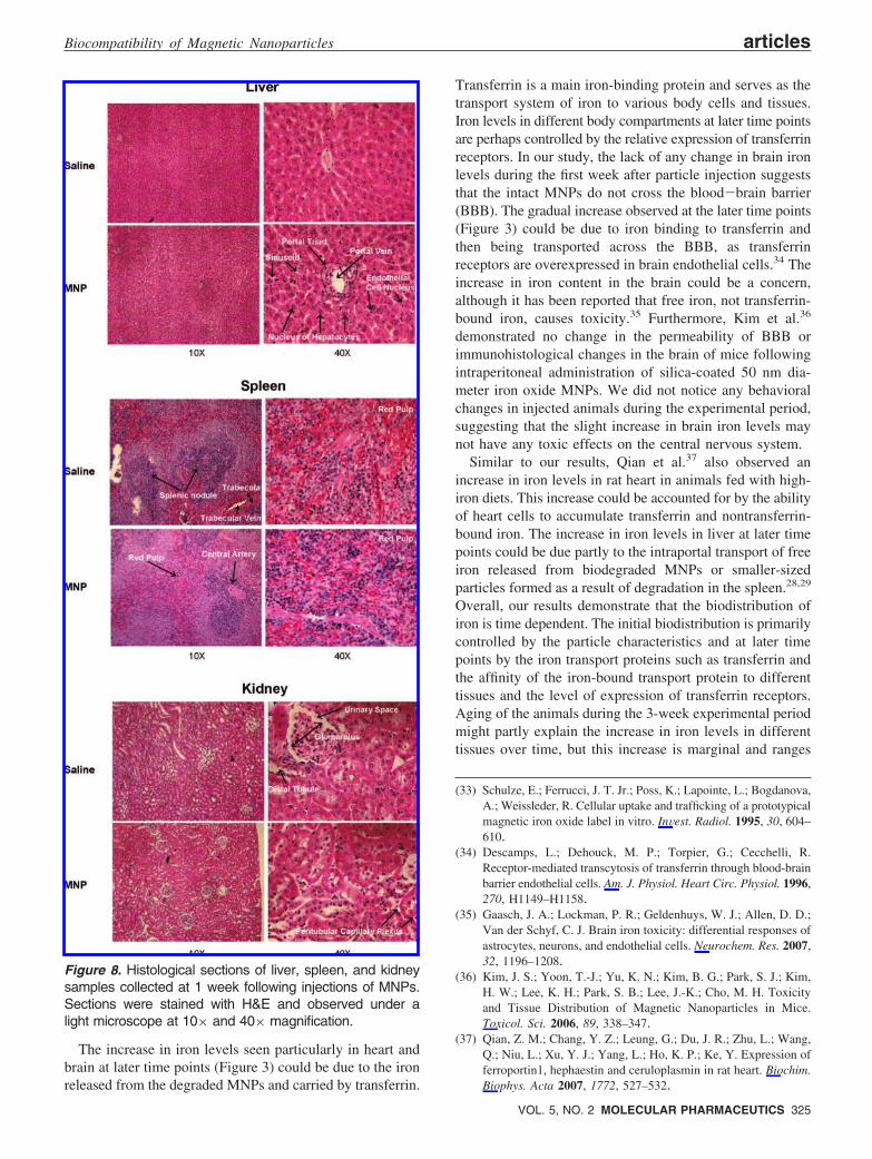
Effect of MNPs on Oxidative Stress and HistologicalAnalysis. LHPO levels in liver, spleen, and kidney increased,reaching a peak at about 3 days following MNP injection
(Figure 7). The response was tissue dependent, with liverdemonstrating a marginal increase and returning to normalvalues within 1 week, whereas the kidney and spleen took 3weeks to return to normal values. No significant changes inLHPO levels were observed for lung, brain, and heart tissues(Figure 7). Histological analysis of the liver, spleen, andkidney did not show any apparent change in the cellularstructures at 1 day (data not shown) or 7 days following MNPinjection (Figure 8).
DiscussionMost formulations of MNPs use dextran or starch as a
stabilizing agent to form a water-dispersible system,23 with
(23) LaConte, L.; Nitin, N.; Bao, G. Magnetic nanoparticle probes.Mater. Today 2005, 8, 32–38.
Figure 3. Changes in iron levels in different tissues following intravenous injection of MNPs. The injected dose of ironwas 10 mg of Fe/kg in 100 µL of saline. Data presented as mean ( sem (n ) 4). *p < 0.05 or **p < 0.005 vs salinecontrol.
Biocompatibility of Magnetic Nanoparticles articles
VOL. 5, NO. 2 MOLECULAR PHARMACEUTICS 321
the drug of interest conjugated to the outer layer of MNPs.Apart from the complexity of conjugation chemistry, thedrug-loading capacity of these MNPs is low, and theconjugated drug is released within hours.1 These factors limitthe applications of MNPs as drug-carrier systems becausethe conjugated drug could be released before localizing in thetarget tissue. With our MNPs, the drug can be loaded at highconcentration in the OA layer surrounding the magnetic core,and the loaded drug is released over a period of weeks.7
The present study was designed to determine the biodis-tribution, clearance, and biocompatibility of our MNPformulation, as size, shape, and surface properties couldsignificantly influence the above in ViVo properties of MNPs.Since our formulation has a unique combination of OA andPluronic coatings, we anticipated that these MNPs wouldshow different biodistribution characteristics than previouslyknown for starch or dextran-coated MNPs. We also deter-mined the clearance of iron, considering that excessiveaccumulation of iron in the body could be of concern as freeiron is known to cause oxidative stress.24 Hence, we
determined the effect of injected MNPs on oxidative stressin different tissues as well as the changes in the liver enzymesas a measure of the liver function. Data obtained from thesestudies should help us to further optimize the formulation,if necessary, and to enhance the efficiency of our MNPs forimaging and drug delivery applications.
We observed a gradual increase in the serum iron levelfor 1 week prior to a slow decline, indicating that clearanceof the MNPs requires more than 3 weeks (Figure 2a). Theserum iron level is perhaps the result of several simulta-neously occurring processes, such as degradation of injectediron particles, the iron’s subsequent binding to differentserum proteins (e.g., transferrin), redistribution of the boundiron to cells and tissue, and its elimination. Our serum irondata do not discriminate between bound and free forms ofiron, but it appears that most of the iron in the body is inthe bound form because of its slow clearance kinetics.Bourrinet et al.25 in their study with Fe-Ferumoxtran-10(Advanced Magnetics, Cambridge, MA), which is an ironoxide core coated with low molecular weight dextran, havereported slow clearance of iron, extending beyond 7 weeks.Iron can become incorporated into the body’s iron store andprogressively into the red blood cells (hemoglobin), thusslowing the clearance of iron. The authors reported elimina-tion of 18–22% of injected iron through urine and feces indifferent animal species during 7 weeks; however, a fewother studies have reported more rapid clearance of theinjected iron (25% in 19 days).26 This discrepancy inclearance time could have been due to the difference in thesize of the iron oxide core particle and/or the coating material,both of which can affect the degradation and biodistributionof MNPs.
MNPs initially are superparamagnetic, while free iron isexpected to be paramagnetic. The saturation magnetizationof the superparamagnetic component of the magnetizationdecreases with MNP size, suggesting that MNPs degradedover the 3-week period studied, or that MNPs are eliminatedfrom the tissues. The biodistribution data demonstrated thatabout 55% of the injected dose of iron localized in the liverat 6 h following particle injection, which appears to be dueto clearance of the MNPs by the reticuloendothelial system(RES). RES clearance of particles depends upon severalfactors, including particle size, shape, and surface properties.The mean particle size of our MNPs measured usingtransmission electron microscopy was 11 ( 2 nm, and thehydrodynamic diameter was 193 nm based on laser light
(24) McCord, J. M. Effects of positive iron status at a cellular level.Nutr. ReV. 1996, 54, 85–88.
(25) Bourrinet, P.; Bengele, H. H.; Bonnemain, B.; Dencausse, A.;Idee, J. M.; Jacobs, P. M.; Lewis, J. M. Preclinical safety andpharmacokinetic profile of ferumoxtran-10, an ultrasmall super-paramagnetic iron oxide magnetic resonance contrast agent. InVest.Radiol. 2006, 41, 313–324.
(26) Weissleder, R.; Stark, D. D.; Engelstad, B. L.; Bacon, B. R.;Compton, C. C.; White, D. L.; Jacobs, P.; Lewis, J. Superpara-magnetic iron oxide: pharmacokinetics and toxicity. Am. J.Roentgenol. 1989, 152, 167–173.
Figure 4. Change in the saturation magnetization of liverand spleen tissue samples vs time after injection ofMNPs. Error bars are smaller than the symbol size. Thefirst data point represents magnetization values at 6 hafter MNP injection. The maximum magnetization offree MNPs (prior to injection) was 253 emu/g Fe.
articles Jain et al.
322 MOLECULAR PHARMACEUTICS VOL. 5, NO. 2
scattering measurement.7 Neuberger et al.27 have reportedthat larger-sized nanoparticles are eliminated from thebloodstream faster than smaller-sized nanoparticles. It hasalso been demonstrated that for particles smaller than 40 nmin diameter, both biodistribution and blood half-life of theiron oxide particles are determined by the coating materialrather than the mean hydrated particle size.28 Our formulationis coated with OA and Pluronic that contribute toward theincrease in hydrodynamic diameter of the iron oxide core.We therefore speculate that the initial high levels of ironobserved in the liver may be due to the uptake of MNPs bythe liver via the first-pass effect because of the increaseddiameter of MNPs. Okon et el.29 have reported uptake ofthe major fraction of injected dextran-coated iron oxideMNPs by the Küpffer cells. Many other studies have reportedgreater than 75% uptake of MNPs by the RES, particularlyby the liver.14 Relatively lower uptake of our MNPs by theliver could be partly due to the coating with Pluronic, which
is known to inhibit opsonization of injected particles.30 Wehave used Pluronic F-127 in this study to stabilize the MNPs,but it is known that the ratio of hydrophilic polyethyleneoxide (PEO) and hydrophobic polypropylene oxide (PPO)segments in Pluronic influence the interactions of particleswith serum protein and hence their systemic circulation time.Other Pluronic types, particularly F-108 and T-90830 orpolyethylene glycol (molecular weight ∼5,000),31 are knownto minimize the opsonization of colloidal particles and hencetheir clearance by the RES. Therefore, it would be interestingto determine the effect of these surface modifications onbiodistribution of MNPs in future studies.
In addition to size, the charge of MNPs also influencesthe uptake of MNPs by the liver. Our particles have a �potential of -0.22 mV, which is almost neutral. Chouly etal.,14 in their study of dextran-coated nanoparticles, reportedthat the liver’s uptake of charged particles is greater thanneutral particles. Therefore, the almost neutral charge of ourMNPs is an advantage in preventing their rapid clearancefrom the circulation. MNPs with a prolonged blood half-life could be useful in the imaging of several pathologicalconditions, such as metastatic lymph node, inflammatory,and/or degenerative diseases. In addition, it can be used asblood pool agents for angiography and for determining tumor
(27) Neuberger, T.; Schopf, B.; Hofmann, H.; Hofmann, M.; vonRechenberg, B. Superparamagnetic nanoparticles for biomedicalapplications: Possibilities and limitations of a new drug deliverysystem. J. Magn. Magn. Mater. 2005, 293, 483–496.
(28) Briley-Saebo, K.; Bjornerud, A.; Grant, D.; Ahlstrom, H.; Berg,T.; Kindberg, G. M. Hepatic cellular distribution and degradationof iron oxide nanoparticles following single intravenous injectionin rats: implications for magnetic resonance imaging. Cell TissueRes. 2004, 316, 315–323.
(29) Okon, E.; Pouliquen, D.; Okon, P.; Kovaleva, Z. V.; Stepanova,T. P.; Lavit, S. G.; Kudryavtsev, B. N.; Jallet, P. Biodegradationof magnetite dextran nanoparticles in the rat. A histologic andbiophysical study. Lab. InVest. 1994, 71, 895–903.
(30) Tan, J. S.; Butterfield, D. E.; Voycheck, C. L.; Caldwell, K. D.;Li, J. T. Surface modification of nanoparticles by PEO/PPO blockcopolymers to minimize interactions with blood components andprolong blood circulation in rats. Biomaterials 1993, 14, 823–833.
(31) Moghimi, S. M.; Szebeni, J. Stealth liposomes and long circulatingnanoparticles: critical issues in pharmacokinetics, opsonization andprotein-binding properties. Prog. Lipid Res. 2003, 42, 463–478.
Figure 5. Changes in levels of serum (a) aspartate aminotransferase (AST), (b) alanine aminotransferase (ALT), and(c) alkaline phosphatase (AKP) at 24 h following saline, OA-Pluronic emulsion, PLGA-nanoparticles, and MNPinjection. Ketamine/xylazine (K/X) was injected intraperitoneally to anesthetize rats before injection of formulations.(Control: no saline injection and no K/X administration.) Data presented as mean ( sem (n ) 4–8). *p < 0.05, **p <0.005 with respect to control.
Biocompatibility of Magnetic Nanoparticles articles
VOL. 5, NO. 2 MOLECULAR PHARMACEUTICS 323
permeability and tumor blood volume, cerebral bloodvolume, and vessel size index measurements.32
The MNPs have differential organ distribution patterns thatvary with time and seem to be influenced by several factorsbesides the RES. The magnetic measurements of the liver andthe spleen samples suggest the loss of magnetization over aperiod of 1 week (Figure 4). This loss could also be due tobiodegradation of superparamagnetic nanoparticles to ironmetabolites or free iron, neither of which should be superpara-magnetic. There was variation in magnetization values of thetissue collected from different animals, which could be due tothe heterogeneous distribution of MNPs in the organ. Perhaps,homogenizing the entire organ prior to taking samples formeasurement could give more consistent data. It has beensuggested that the Küpffer cells in liver can degrade iron oxidenanoparticles and can incorporate most of the iron into ferritin.28
The incorporation of iron into ferritin could change themagnitude of the superparamagnetic component, as ferritin issuperparamagnetic at room temperature. The loss of magnetiza-tion in 1 week with our MNPs also is consistent with thedegradation of dextran-coated iron oxide MNPs in the lysosomalcompartment of the macrophage.33 Okano et al.29 examinedthe biodegradation of magnetite-dextran nanoparticles in ratsand reported a sharp decrease in magnetic susceptibility at day3, followed by a gradual decrease thereafter until day 14. Incontrast, our results show a steady decrease in the saturationmagnetization.
(32) Corot, C.; Robert, P.; Idee, J.-M.; Port, M. Recent advances iniron oxide nanocrystal technology for medical imaging. AdV. DrugDeliV. ReV. 2006, 58, 1471–1504.
Figure 6. Changes in serum liver enzyme levels withtime following a single-dose intravenous injection ofMNPs in rats (10 mg Fe/kg dose in 100 µL of saline):(a) aspartate aminotransferase (AST), (b) alanineaminotransferase (ALT), and (c) alkaline phosphatase(AKP). Dashed line in each graph represents controlvalues (i.e., no K/X and no saline injected). Data pre-sented as mean ( sem (n ) 4). Data at time point 0 issaline control (represented by arrows).
Figure 7. Changes in lipid hydroperoxide (LHPO) levelsas a measure of oxidative stress in different tissues withtime following injection of MNPs in rats (10 mg Fe/kgdose in 100 µL of saline). Data presented as mean (sem (n ) 4).
articles Jain et al.
324 MOLECULAR PHARMACEUTICS VOL. 5, NO. 2
The increase in iron levels seen particularly in heart andbrain at later time points (Figure 3) could be due to the ironreleased from the degraded MNPs and carried by transferrin.
Transferrin is a main iron-binding protein and serves as thetransport system of iron to various body cells and tissues.Iron levels in different body compartments at later time pointsare perhaps controlled by the relative expression of transferrinreceptors. In our study, the lack of any change in brain ironlevels during the first week after particle injection suggeststhat the intact MNPs do not cross the blood-brain barrier(BBB). The gradual increase observed at the later time points(Figure 3) could be due to iron binding to transferrin andthen being transported across the BBB, as transferrinreceptors are overexpressed in brain endothelial cells.34 Theincrease in iron content in the brain could be a concern,although it has been reported that free iron, not transferrin-bound iron, causes toxicity.35 Furthermore, Kim et al.36
demonstrated no change in the permeability of BBB orimmunohistological changes in the brain of mice followingintraperitoneal administration of silica-coated 50 nm dia-meter iron oxide MNPs. We did not notice any behavioralchanges in injected animals during the experimental period,suggesting that the slight increase in brain iron levels maynot have any toxic effects on the central nervous system.
Similar to our results, Qian et al.37 also observed anincrease in iron levels in rat heart in animals fed with high-iron diets. This increase could be accounted for by the abilityof heart cells to accumulate transferrin and nontransferrin-bound iron. The increase in iron levels in liver at later timepoints could be due partly to the intraportal transport of freeiron released from biodegraded MNPs or smaller-sizedparticles formed as a result of degradation in the spleen.28,29
Overall, our results demonstrate that the biodistribution ofiron is time dependent. The initial biodistribution is primarilycontrolled by the particle characteristics and at later timepoints by the iron transport proteins such as transferrin andthe affinity of the iron-bound transport protein to differenttissues and the level of expression of transferrin receptors.Aging of the animals during the 3-week experimental periodmight partly explain the increase in iron levels in differenttissues over time, but this increase is marginal and ranges
(33) Schulze, E.; Ferrucci, J. T. Jr.; Poss, K.; Lapointe, L.; Bogdanova,A.; Weissleder, R. Cellular uptake and trafficking of a prototypicalmagnetic iron oxide label in vitro. InVest. Radiol. 1995, 30, 604–610.
(34) Descamps, L.; Dehouck, M. P.; Torpier, G.; Cecchelli, R.Receptor-mediated transcytosis of transferrin through blood-brainbarrier endothelial cells. Am. J. Physiol. Heart Circ. Physiol. 1996,270, H1149–H1158.
(35) Gaasch, J. A.; Lockman, P. R.; Geldenhuys, W. J.; Allen, D. D.;Van der Schyf, C. J. Brain iron toxicity: differential responses ofastrocytes, neurons, and endothelial cells. Neurochem. Res. 2007,32, 1196–1208.
(36) Kim, J. S.; Yoon, T.-J.; Yu, K. N.; Kim, B. G.; Park, S. J.; Kim,H. W.; Lee, K. H.; Park, S. B.; Lee, J.-K.; Cho, M. H. Toxicityand Tissue Distribution of Magnetic Nanoparticles in Mice.Toxicol. Sci. 2006, 89, 338–347.
(37) Qian, Z. M.; Chang, Y. Z.; Leung, G.; Du, J. R.; Zhu, L.; Wang,Q.; Niu, L.; Xu, Y. J.; Yang, L.; Ho, K. P.; Ke, Y. Expression offerroportin1, hephaestin and ceruloplasmin in rat heart. Biochim.Biophys. Acta 2007, 1772, 527–532.
Figure 8. Histological sections of liver, spleen, and kidneysamples collected at 1 week following injections of MNPs.Sections were stained with H&E and observed under alight microscope at 10× and 40× magnification.
Biocompatibility of Magnetic Nanoparticles articles
VOL. 5, NO. 2 MOLECULAR PHARMACEUTICS 325
from 2 to 5% per month, depending upon the tissue.38 Toobtain complete biodistribution data including eliminationthrough urine and stools, one can use 59Fe to synthesize ironoxide nanoparticles to achieve greater sensitivity of detec-tion.39
The transient increase in ALT, AST, and AKP levels inserum upon administration of MNPs appears to be the generalresponse of the body as PLGA nanoparticles also induced asimilar response (Figure 5). It appears that the surge inenzyme activities seen in the animals during the initial daysfollowing nanoparticle injection is partly the effect of theanesthetic agent used prior to injection, as a saline injectionalso caused an increase in the liver AST level (Figure 6). Itis known that ketamine-xylazine increases liver enzymeactivity and enzymes remain elevated over several hours;40
however, phenobarbital (used for euthanasia) did not affectliver enzyme activity. Jiang et al.41 also observed a transientincrease in ALT and AST activity following injection ofchitosan- and polyethylenimine-DNA complexes. Gener-ally, AST levels three times the upper limit of the normalrange (range 39–262 U/L) represent abnormal liver func-tion.42 The changes seen in our studies were significantlybelow this limit (Figure 6).43
The effect of MNPs on oxidative stress is an importantconsideration for their safe use in patients. Although iron ispresent in the body, mostly associated with hemoglobin,44
previous studies have reported an increase in oxidative stresswith an increase in free iron.45,46 Therefore, usually ironoxide particles are surrounded by different polymeric coatingsto prevent direct contact of iron oxide with inter orintracellular substrates that can facilitate free-radical genera-
tion.47 Iron-induced oxidative injury to phospholipids oforganelle membranes is a potential mechanism underlyingcellular injury in iron overload. We observed a transientincrease in LHPO levels in different tissues (Figure 7);however, the response varied from tissue to tissue and doesnot seem to correlate with the increase in iron levels. Forexample, liver showed a marginal increase in oxidative stressdespite greater localization of iron compared with othertissues. This observation suggests that the oxidative responsedepends on the capacity of individual tissues to neutralizethe effect of stress conditions caused by the particulateinjection. Given its main role in detoxification, the liver isexposed to many sources of oxidative stress and may havea greater capacity to neutralize these effects. It appears thatnonmagnetic nanoparticles also induce oxidative stress. Forexample, the uptake of polymeric nanoparticles (poly(isobu-tyl cynoacrylate)) by Küpffer cells in the liver followingintravenous administration induced modifications in thehepatocyte antioxidant system, such as depletion of glu-tathione or superoxide dismutase, but the events werereversible.48 The transient increase in the liver enzyme levelsor increase in oxidative stress does not seem to be highenough to damage the liver. This is evident from the lack ofsignificant change in the serum TIBC (Figure 2), which isused as an indirect marker for liver function since transferrinis produced in the liver. Changes in the LHPO levels weretransient in our studies, and there were no apparent changesin the histology of the liver and other tissues (Figure 8), bothof which suggest that the transient increase in oxidative stressdoes not affect cellular integrity and tissue morphology.
The increase in iron levels observed in some organs couldbe significantly below toxic levels considering that the doseused is significantly lower than the total body iron content.For example, cirrhosis and hepatocellular carcinoma candevelop if the liver iron concentration exceeds 4000 µg/gwet weight.49,50 In our studies, the liver iron level prior toMNP injection was 123 ( 23 µg/g wet weight, which didnot exceed 300 µg/g following MNP injection at any timeduring the 3-week experimental period. Further, the dose ofiron used in this study is four times the dose that is usuallyused in humans for imaging applications (2.6 vs 10 mg/kg).Hence, at lower doses, the increase in iron levels or changein oxidative stress or liver enzyme levels will be significantly
(38) Cook, C. I.; Yu, B. P. Iron accumulation in aging: modulation bydietary restriction. Mech. Ageing DeV. 1998, 102, 1–13.
(39) Tiefenauer, L. X.; Tschirky, A.; Kuhne, G.; Andres, R. Y. In vivoevaluation of magnetite nanoparticles for use as a tumor contrastagent in MRI. Magn. Reson. Imaging 1996, 14, 391–402.
(40) Thompson, J. S.; Brown, S. A.; Khurdayan, V.; Zeynalzadedan,A.; Sullivan, P. G.; Scheff, S. W. Early effects of tribromoethanol,ketamine/xylazine, pentobarbitol, and isoflurane anesthesia onhepatic and lymphoid tissue in ICR mice. Comp. Med. 2002, 52,63–67.
(41) Jiang, X.; Dai, H.; Leong, K. W.; Goh, S. H.; Mao, H. Q.; Yang,Y. Y. Chitosan-g-PEG/DNA complexes deliver gene to the ratliver via intrabiliary and intraportal infusions. J. Gene Med. 2006,8, 477–487.
(42) Dufour, D. R. Liver Disease. In Tietz textbook of clinical chemistryand molecular diagnostics, 4th ed.; Burtis, C. A., Ashwood, E. R.,Bruns, D. E., Eds.; Elsevier Saunders: St. Louis, MO, 2006;1777-1848.
(43) Olfert, E. D., Cross, B. M., McWilliam, A. A., Eds. Guide to theCare and Use of Experimental Animals, 2nd ed.; Canadian Councilon Animal Care: Ottawa, 1993.
(44) Puntarulo, S. Iron, oxidative stress and human health. Mol. AspectsMed. 2005, 26, 299–312.
(45) Mehta, S. H.; Webb, R. C.; Ergul, A.; Tawak, A.; Dorrance, A. M.Neuroprotection by tempol in a model of iron-induced oxidativestress in acute ischemic stroke. Am. J. Physiol. Regul. Integr.Comp. Physiol. 2004, 286, R283–R288.
(46) Minotti, G. Sources and role of iron in lipid peroxidation. Chem.Res. Toxicol. 1993, 6, 134–146.
(47) Stroh, A.; Zimmer, C.; Gutzeit, C.; Jakstadt, M.; Marschinke, F.;Jung, T.; Pilgrimm, H.; Grune, T. Iron oxide particles formolecular magnetic resonance imaging cause transient oxidativestress in rat macrophages. Free Radic. Biol. Med. 2004, 36, 976–984.
(48) Fernandez-Urrusuno, R.; Fattal, E.; Feger, J.; Couvreur, P.;Therond, P. Evaluation of hepatic antioxidant systems afterintravenous administration of polymeric nanoparticles. Biomate-rials 1997, 18, 511–517.
(49) Anthony, S.; Tavill, B. R. B. Hemochromatosis: How much ironis too much. Hepatology 1986, 6, 142–145.
(50) Bassett, M. L.; Halliday, J. W.; Powell, L. W. Value of hepaticiron measurements in early hemochromatosis and determinationof the critical iron level associated with fibrosis. Hepatology 1986,6, 24–29.
articles Jain et al.
326 MOLECULAR PHARMACEUTICS VOL. 5, NO. 2
lower than those observed in our study. Bourrinet et al.,25
in their study with ferumoxtran-10, reported no treatment-associated changes in cardiovascular and renal functionsfollowing intravenous injections of 2–20 mg Fe/kg in dogs.Only the high dose (200 mg of Fe/kg), which is 75 timesthe intended human dose, caused slight and transient changesin hematological parameters and cardiac functions. Hence,the dose and the dosing interval could be critical inpreventing MNP-associated damage to tissues.
ConclusionsIt appears that the biodistribution of iron and its clearance
depends upon simultaneous complex events, and hence thedynamics of iron concentration in different tissues changeswith time. Since the iron dose of the MNPs was many timeslower than body iron levels, we speculate that the injectediron would be easily metabolized and regulated by the body’snormal physiological iron homeostatic mechanisms. The mostimportant result is that the injected iron did not cause long-term changes in liver enzyme levels or oxidative stress, withmost of the changes being transient. The anticipated use ofMNPs for imaging or drug delivery would not involve
repeated dosing over a short time interval, which would allowthe body to return to its normal state after the initial transientchanges. Future studies, aimed at minimizing the initial RESuptake of MNPs by selecting appropriate Pluronic or PEGfor surface modification, could increase the MNP half-lifein the blood, which in turn could improve drug targetingand imaging applications of the MNP formulation for targetsother than the liver. Given the many studies of MNPs foruse in biomedical applications, understanding the biodistri-bution, elimination, and oxidative stress caused by the MNPsis critical to the successful development of MNPs in theclinical setting.
Acknowledgment. The study reported here is fundedby Grant No. R01 EB005822 from the National Institute ofBiomedical Imaging and Bioengineering of the NationalInstitutes of Health (to V.L.). We also gratefully acknowledgethe gift of Pluronic F-127 from BASF Corp. (Mt. Olive, NJ).We thank Ms. Melissa Jedlicka for proofreading themanuscript.
MP7001285
Biocompatibility of Magnetic Nanoparticles articles
VOL. 5, NO. 2 MOLECULAR PHARMACEUTICS 327