bimodal effects of the k v 7 channel activator retigabine on vascular k + currents
TRANSCRIPT

RESEARCH PAPER
Bimodal effects of the Kv7 channel activatorretigabine on vascular Kþ currents
SYM Yeung1, M Schwake2, V Pucovsky3 and IA Greenwood1
1Division of Basic Medical Sciences, Ion Channels and Cell Signalling Research Centre, St George’s University of London, London, UK;2Institute of Biochemistry, Christian-Albrechts University, Kiel, Germany and 3Cardiovascular Biomedical Research Centre, Schoolof Medicine and Dentistry, Queen’s University Belfast, Belfast, UK
Background and purpose: This study investigated the functional and electrophysiological effects of the Kv7 channel activator,retigabine, on murine portal vein smooth muscle.Experimental approach: KCNQ gene expression was determined by reverse transcriptase polymerase chain reaction (RT-PCR)and immunocytochemical experiments. Whole cell voltage clamp and current clamp were performed on isolated myocytesfrom murine portal vein. Isometric tension recordings were performed on whole portal veins. Kþ currents generated byKCNQ4 and KCNQ5 expression were recorded by two-electrode voltage clamp in Xenopus oocytes.Key results: KCNQ1, 4 and 5 were expressed in mRNA derived from murine portal vein, either as whole tissue or isolatedmyocytes. Kv7.1 and Kv7.4 proteins were identified in the cell membranes of myocytes by immunocytochemistry. Retigabine(2–20 mM) suppressed spontaneous contractions in whole portal veins, hyperpolarized the membrane potential andaugmented potassium currents at �20 mV. At more depolarized potentials, retigabine and flupirtine, decreased potassiumcurrents. Both effects of retigabine were prevented by prior application of the Kv7 blocker XE991 (10 mM). Recombinant KCNQ4 or 5 channels were only activated by retigabine or flupirtine.Conclusions and implications: The Kv7 channel activators retigabine and flupirtine have bimodal effects on vascularpotassium currents, which are not seen with recombinant KCNQ channels. These results provide support for KCNQ4- orKCNQ5-encoded channels having an important functional impact in the vasculature.
British Journal of Pharmacology (2008) 155, 62–72; doi:10.1038/bjp.2008.231; published online 9 June 2008
Keywords: KCNQ; Kv7; retigabine; XE991; vascular smooth muscle; RT-PCR; immunocytochemistry; isometric tension
Abbreviations: mPV, murine hepatic portal vein; PSS, physiological saline solution; RMP, resting membrane potential
Introduction
Kv7 (Kv7.1–Kv7.5) are a subfamily of voltage-gated Kþ
channels encoded by the KCNQ genes. Each Kv7 protein
has a particular distribution with Kv7.1 expressed
predominantly by cardiac myocytes, whereas Kv7.2–7.5 are
considered ‘neuronal’ (Jentsch, 2000; Schroeder et al., 2000)
and Kv7.4 having a specialized cellular location in auditory
neurons (Kharkovets et al., 2000). These Kþ channels are
essential regulators of cardiac and neuronal action potentials
as emphasized by the fact that mutations in their genes
underlie a number of clinical symptoms. Most notably
mutations in Kv7.1 result in cardiac arrhythmias; mutations
in Kv7.2 and Kv7.3 result in neuronal excitability and
underlie benign neonatal familial epilepsy (Jentsch, 2000);
whereas a mutation in Kv7.4 results in hereditary deafness
(Kubisch et al., 1999).
The study of Kv7 channels has largely relied on the use of
the blockers such as linopirdine and XE991, but these
compounds do not discriminate between the five Kv7
isoforms. Recently the anti-convulsant retigabine (D23129,
N-(2-amino-4-(4-flurobenzylamino)-phenyl)carbamic acid
ethyl ester)) has been shown to augment heteromers of
Kv7.2 and Kv7.3 (Main et al., 2000; Wickenden et al., 2000),
which underlie the neuronal M-current (Wang et al., 1998).
Retigabine has now been shown to activate all neuronal Kv7
channels (that is Kv7.2–7.5) but does not affect Kv7.1 in
concentrations up to 100 mM (Tatulian et al., 2001; Schenzer
et al., 2005; Wuttke et al., 2005). There are numerous studies
on the pharmacology of Kv7 channels in expression systems
but fewer using native preparations (Tatulian et al., 2001).
Even less data are available on Kv7 channels in smooth
muscle cells where these genes have recently been identified
(Ohya et al., 2003; Yeung and Greenwood, 2005;
Brueggemann et al., 2006; Yeung et al., 2007). The present
investigation analyses the effects of retigabine on the native
Kþ currents in murine hepatic portal vein (mPV) myocytes,Received 6 March 2008; revised 14 April 2008; accepted 24 April 2008;
published online 9 June 2008
Correspondence: Dr IA Greenwood, Division of Basic Medical Sciences,
St George’s University of London, Cranmer Terrace, London SW17 0RE, UK.
E-mail: [email protected]
British Journal of Pharmacology (2008) 155, 62–72& 2008 Macmillan Publishers Limited All rights reserved 0007–1188/08 $30.00
www.brjpharmacol.org

where the effects of the Kv7 channel inhibitor XE991 have
been characterized extensively (Yeung and Greenwood,
2005). This study provides the first description of retigabine
effects on endogenous Kþ currents in a non-neuronal cell
type. Furthermore, these data give some insight into the
molecular identity of the native Kv7 channel in these
vascular smooth muscle cells.
Materials and methods
Conventional isometric tension recordings
All experiments were performed on portal veins from BALB/c
mice (6–8 weeks) killed by schedule 1 methods, in accordance
with the UK Animals Act. Isometric tension recordings on
whole mPV were performed as described in Yeung and
Greenwood (2005). Briefly, the portal vein was removed from
the animal and all connective tissue and fat were removed.
The mPV was placed into a 10 mL organ bath containing
Krebs solution aerated with 95% O2/5% O2 and maintained at
37 1C then set to a resting tension of 0.1 g. Changes in tension
were recorded using BIOPAC Systems Inc. force transducer
and AcqKnowledge software (version 3.7) sampling at
2.5 kHz. All veins exhibited spontaneous activity within
minutes of being set up and this activity was maintained for
the duration of the experiment (Yeung and Greenwood,
2005). To quantify the data, a single contraction was defined
as an increase in tension from the baseline that was separated
from another contraction by a period at the basal tension of
at least 1 s. Intercontraction interval was taken as the period
between two peaks of individual contractions. Contraction
duration was taken as the total time that the tension
remained above 10% of the maximal contraction.
Cell dissociation
Single smooth muscle cells were isolated enzymatically from
mPV before each experiment using the protocol described
by Yeung and Greenwood (2005). This procedure involved
treating tissues with 100 mM Ca2þ physiological saline
solution (PSS) containing 0.3 mg mL�1 protease (type XIV,
Sigma, UK); then with 0.6 mg mL�1 collagenase (type I,
Calbiochem, UK) for 6 min each. Dispersed cells were kept
on ice until required.
RNA extraction and reverse transcription-PCR
The mPV was excised from the animal and placed immedia-
tely into RNA later (Ambion, UK). All extraneous tissue was
removed and the mPV was cut into smaller pieces and placed
into lysis buffer containing b-mercaptoethanol. Total RNA
was extracted from the tissues using spin column technology
(RNeasy kits, Qiagen, UK) according to the manufacturer’s
protocol and incorporating an initial 10 min incubation
with proteinase K. RNA was finally eluted with water and
quantified using the NanoDrop spectrophotomer (ND-1000,
NanoDrop Technologies, UK). Total RNA from murine heart
and brain were extracted in the same manner and were
subsequently used as positive controls. 100 ng total RNA was
reverse transcribed using the reverse transcriptase enzyme
MMLV (Invitrogen, UK). All samples had a respective RT-
control, that is, no MMLV was put into the sample. PCRs
used outer primers designed specifically to detect KCNQ1–5
(Yeung et al., 2007). All reactions were performed with an
initial denaturation step at 94 1C for 2 min, followed by 35
cycles at 94 1C for 30 s, annealing temperature for 30 s and
72 1C for 1 min. The reaction was completed with a final
extension step at 72 1C for 10 min. For nested PCRs a primary
PCR using outer primers and 25 cycles was employed. This
product was further amplified by a secondary 35 cycle PCR
using inner primers (mKCNQ1, forward: 50-CCATCATTGA
CCTCATCGTG-30, reverse: 50-GGCGAAGACAGAGAAACA
GG-30; mKCNQ2, forward: 50-AGGAAGCCGTTCTGTGTGA
T-30, reverse: 50-GCAGAGGAAGCCAATGTACC-30; mKCNQ3,
forward: 50-AAGACAGGGGCTATGGGAAT-30, reverse: 50-TTT
TGGAGTGGATGGAGGTC-30; mKCNQ4, forward: 50-GAGCA
GTATTCAGCAGGACA-30, reverse: 50-AGTAGAAGCCCAGCA
GCAGA-30; mKCNQ5, forward: 50-CGCCAGAAGCATTTTGA
GA-30, reverse: 50-TGGGAACTCTTGAGCCGTAG-30). All
reactions were carried out using a Touchgene thermal cycler
(Techne, UK). All PCR products were sequenced using the
ABC Sequencing Service at Imperial College London.
Immunocytochemistry
Single cells were fixed and stained for confocal microscopy as
described previously (Saleh et al., 2005; Yeung et al., 2007).
Protein expression was identified by immunofluorescence
using antibody against Kv7.1 (Alomone Laboratories, Israel,
1:600 dilution), two against Kv7.4 (Kv7.4SC from Santa Cruz
Biotechnology, USA, 1:200 dilution) and Kv7.4J (raised
against a different epitope and kindly provided by T Jentsch,
1:50 dilution, Kharkovets et al., 2000). The labelling was
visualized with Alexa Fluor 488-conjugated chicken anti-
rabbit antibodies (1:500), Alexa Fluor 633-conjugated
donkey anti-goat antibodies (1:500) or Alexa Fluor
488-conjugated donkey anti-goat antibodies (1:500).
Confocal microscopy
The cells were imaged using a Zeiss LSM 510 laser scanning
confocal microscope (Carl Zeiss, Germany). The excitation
beam was produced by either argon (488 nm) or helium-
neon (633 nm) laser and delivered to the specimen through a
Zeiss Apochromat 63� oil immersion objective (numerical
aperture 1.4). Emitted fluorescence was captured using LSM
510 software (release 3.2, Carl Zeiss, Germany).
Analysis of images
An image cutting horizontally through approximately the
middle of the cell was selected out of a z-stack of images. To
assess the cellular distribution of Kv7 channels a circular area
of 0.78 mm2 (diameter approx. 1.0 mm; referred to as Region
1) was randomly selected in the subplasmalemmal area of
the cell (Figure 7bi). Another circular area of 0.78 mm2
(Region 2) was selected so that the perimeter of such circle
touched the perimeter of Region 1 and the line going
through the centre of these circles was perpendicular to the
edge of the cell, thought to be the plasma membrane.
Bimodal effects of the Kv7 channel activator retigabineSYM Yeung et al 63
British Journal of Pharmacology (2008) 155 62–72

A percentage of fluorescing pixels (%FP) was calculated in
both Regions using the formula:
%FP ¼ 100�nðp4thresholdÞnðpÞ ;
where n(p4threshold) is the number of pixels within the
Region whose intensity equalled or exceeded the threshold
value (one s.d. of the pixel intensity) and n(p) the total
number of pixels in the Region. The % FP values were then
compared with each other and with the % FP in the whole
confocal plane of the cell.
The average pixel fluorescence (APF) value was used to
compare the overall fluorescence signal between the staining
and its controls and was calculated using the formula:
APF ¼P
iðpÞnðpÞ ðI:U:=pixelÞ;
where i(p) is the intensity of a pixel within the confocal
plane of the cell and n(p) is the total number of pixels of the
plane. Statistical evaluation and graphs were done using
MicroCal Origin software (MicroCal Software Inc., USA) and
final images were produced using CorelDraw 10 software
(Corel Corporation, Canada).
Current and voltage recordings
Whole cell currents were recorded at room temperature
(20–22 1C) using the conventional, ruptured-patch configura-
tion of the whole cell technique with internal and external
solutions as described in Yeung and Greenwood (2005).
Cells were allowed to equilibrate for 5 min following
establishment of the recording, then a current voltage
protocol from �100 to þ60 mV was run (500 ms per step,
20 mV increments) from a holding potential of �60 mV.
After this procedure, the cell was stepped to �20 mV for
500 ms every 20 s whereupon different agents were applied.
Current voltage protocols were run once the effect of a
certain drug had stabilized. Membrane potential recordings
were made using the current clamp setting on the amplifier
with the perforated patch variant of the whole cell recording
configuration. Amphotericin (300 mg mL�1) was included in
the pipette solution in such cases and recordings
commenced when the series resistance was o40 MO. All
recordings were sampled at 5 kHz and low-pass filtered at
2 kHz. Results are expressed as mean±s.e.mean and n as the
number of cells. Statistical analyses were performed using
Student’s t-test and were considered significant at the
Po0.05 level.
Expression in Xenopus laevis oocytes
Starting from KCNQ1, KCNQ4 and KCNQ5 cDNAs, which
were subcloned into expression vector pTLN, capped RNA
was transcribed using SP6 RNA polymerase in mMessage
mMachine kit (Ambion, Austin) after linearization of the
cDNA with HpaI. Individual stage V to VI oocytes were
obtained from anaesthetized frogs and isolated by collage-
nase treatment. 10 ng of total KCNQ cRNA were injected into
oocytes. Following injection, oocytes were kept at 17 1C in
ND96 solution (96 mM NaCl, 2 mM KCl, 1.8 mM CaCl2, 1 mM
MgCl2, 5 mM HEPES, pH 7.4).
Two-electrode voltage clamp electrophysiology
Two to three days after injection two-electrode voltage clamp
measurements were performed at room temperature in ND96
using an npi Turbotec amplifier (npi electronics, Tamm,
Germany) and pClamp9 software (Axon Instruments, Union
City, CA, USA). For experiments examining the effect of
retigabine or flupirtine, 1, 10, or 100 mM of retigabine or
flupirtine was added to ND96 from a 100 mM stock solution,
which was prepared in DMSO and kept at 4 1C in the dark.
Drugs and solutions
PSS contained (in mM): NaCl (125), KCl (5.4), NaHCO3
(15.4), Na2PO4 (0.33), KH2PO4 (0.34), glucose (10) and
HEPES (11), adjusted to pH 7.4 with NaOH. The external
solution contained (in mM) NaCl (126), KCl (5), MgCl2 (1),
CaCl2 (0.1), glucose (11) and HEPES (10), adjusted to pH 7.2
with NaOH. For current clamp experiments, CaCl2 was raised
to 2.5 mM. The internal solution contained (in mM): KCl
(130), MgCl2 (1), ATP (Naþ salt, 3), GTP (0.1), HEPES (10),
EGTA (5), adjusted to pH 7.2 with KOH. The Krebs solution
for the functional experiments contained (in mM): NaCl
(125), KCl (4.6), CaCl2 (2.5), NaHCO3 (15.4), Na2PO4 (1),
MgSO4 (0.6) and glucose (10). XE991 and flupirtine were
purchased from Ascent Scientific (Weston-super-Mare, UK)
and Sigma Chemical Company (Poole, Dorset, UK), respec-
tively. All drugs were made up as a 100 mM stock with
dimethyl sulphoxide (DMSO) and frozen as small aliquots.
Channel and gene terminology conforms to the BJP Guide to
Receptors and Channels (Alexander et al., 2008).
Results
Retigabine decreases mPV contractility
Previous isometric tension experiments showed that retiga-
bine, and a structurally similar compound flupirtine, relaxed
pre-contracted segments of murine aorta and conduit artery
(Yeung et al., 2007). In this study, isometric tension
recordings were undertaken to see whether retigabine had
a similar functional effect in whole mPV. Under control
conditions, all samples exhibited spontaneous rhythmic
contractile activity with various patterns (Figure 1; also
Yeung and Greenwood, 2005). Application of 20 mM
retigabine rapidly affected contractile activity (Figure 1),
manifested as a marked decrease in the amplitude of
individual contractions (74.2±3.7%, Po0.01, n¼4). A
similar inhibitory effect was observed with 3mM retigabine,
which decreased the amplitude of individual contractions
from 0.0075±0.0003 g to 0.0035±0.00015 g (Po0.01, n¼3).
The inhibitory effect of retigabine was readily reversed upon
washout (Figure 1). In contrast to its effects on spontaneous
activity, 20 mM retigabine had no effect on tonic contractions
produced by 60 mM KCl, which were 0.21±0.02 g and
0.19±0.02 g (n¼4) in the absence and presence of retiga-
bine. These data showed that retigabine had a spasmolytic
Bimodal effects of the Kv7 channel activator retigabineSYM Yeung et al64
British Journal of Pharmacology (2008) 155 62–72

effect in whole mPV that was not due to the direct blockade
of voltage-dependent calcium channels.
Retigabine hyperpolarizes the resting membrane potential
Current clamp recordings were made using the amphotericin
-perforated patch configuration to see the effect of retigabine
on membrane potential. In these cells the mean resting
membrane potential (RMP) was �44.2±2 mV (n¼7).
In each cell spontaneous changes in membrane potential
were apparent (Figure 2). Spontaneous depolarizations
ranged in amplitude between 3 and 17 mV (mean¼12±1,
n¼7 cells) and occasionally these depolarizations generated
an overshooting action potential. Spontaneous hyperpolar-
izations were also apparent in all cells (Figure 2). Application
of 3 and 10 mM retigabine hyperpolarized the RMP by
5.5±0.9 and 8.9±1.3 mV, respectively (Figure 2, n¼5 & 6).
Retigabine also prevented the generation of spontaneous
depolarizations. Interestingly, in one cell where no change in
membrane hyperpolarization was apparent after application
of 3mM retigabine, spontaneous depolarizations were still
abolished. These data show that retigabine effectively
suppressed membrane excitability in mPV myocytes.
Retigabine activates Kþ current in mPV
Experiments were undertaken to assess whether the func-
tional effect of retigabine were mediated by an increase in
Kþ current, as observed in neurones. Application of 1 mM
retigabine, produced an increase in current at �20 mV in two
of five cells tested (mean increase was 39 pA) whereas 3 mM
retigabine markedly enhanced currents recorded at �20 mV
in all cells tested (see Figure 3ai for example). The mean
retigabine-sensitive current (exemplified by Figure 3aii) was
77±16 pA (n¼6). Application of 10 mM retigabine produced
an augmentation at �20 mV in all cells tested (mean-
48±12 pA, n¼ 9) whereas 20 mM retigabine produced an
enhancement in 14 out of 17 cells tested (mean increase was
59±12 pA). These effects of retigabine were not voltage-
dependent in the physiological voltage range (from �60 to
�20 mV, Figure 3b) and was associated with a small, leftward
shift of the voltage-dependence for activation (data not
shown, DV12: 3 mM¼13±3 mV (n¼5), 10 mM¼11±2 mV
(n¼5), 20 mM¼11±2 mV (n¼12)). Flupirtine, a structural
analogue of retigabine also augmented currents with a mean
increase at �20 mV of 41±11 pA (n¼4) produced by 30 mM
flupirtine. These data represent the first recording of
retigabine and flupirtine effects on endogenous Kþ currents
from any type of smooth muscle cell.
When the voltage range was extended to more positive
potentials, a marked inhibition of currents by retigabine was
apparent. The peak and steady state currents at 0 mV,
þ20 mV and þ40 mV (Figure 4ai) were clearly diminished
in the presence of 20 mM retigabine (Figure 4aii). This
inhibition was reversed upon washout and therefore does
not represent current rundown. The inhibitory effect of
retigabine is highlighted when the retigabine-sensitive
currents at these test potentials are calculated, as shown in
Figure 4b. The simultaneous stimulation and inhibition of
Kþ currents by retigabine resulted in the appearance of a
‘bell’ shaped current–voltage relationship for the retigabine-
sensitive current (Figure 4c). Moreover, the potential at
which inhibition was recorded was dependent on the
concentration of retigabine. Thus, at þ20 mV the current
Figure 1 Retigabine inhibits excitability of whole mPV tissue. (a) Example of isometric tension recording in the absence and presence of 20 mM
retigabine. Lower traces show magnifications of the sections highlighted. (b) Bar chart shows mean tension in the absence (solid bars) andpresence of retigabine (hatched bars) and upon washout (open bars). ** denotes statistical significance retigabine versus control at Po0.01.
Bimodal effects of the Kv7 channel activator retigabineSYM Yeung et al 65
British Journal of Pharmacology (2008) 155 62–72

was decreased by 78±33 pA (n¼13) and 68±24 pA (n¼4) by
20 and 10 mM retigabine, respectively but was increased by
26±42 pA (n¼5) by 3 mM retigabine. This led to a rightward
shift in the threshold potential for current inhibition from
B�5 mV for 20 mM retigabine to Bþ25 mV for 3 mM
retigabine (see Figure 4c). Furthermore, there was no
Figure 2 Effect of retigabine on membrane potential recordings in current clamp mode. (a) Example of membrane potential in the absenceand presence of retigabine. Each panel shows a 20 s sample of membrane potential recording either in the absence of retigabine or after 5 minapplication of 3 mM and then 10mM retigabine. Right panel shows the membrane potential 7 min after washout of retigabine. Upward anddownward deflections denote spontaneous depolarizations and hyperpolarizations. (b) shows an amplified view of membrane potentialrecordings in the absence and presence of 10mM retigabine showing the lack of spontaneous membrane depolarizations in the presence of thisagent. Panel (c) shows the mean membrane hyperpolarization produced by 3 and 10 mM retigabine (n¼5 and 6, respectively).
Figure 3 Activation of Kþ currents by different concentrations of retigabine. Panel (ai) shows currents evoked by stepping from �60 to�20 mV in the absence and presence of 3 mM retigabine. (aii) shows the retigabine-sensitive current from (ai). (b) shows the effect of a range ofretigabine concentrations on Kþ conductance (I/V-Vr) at �60, �40 and �20 mV.
Bimodal effects of the Kv7 channel activator retigabineSYM Yeung et al66
British Journal of Pharmacology (2008) 155 62–72
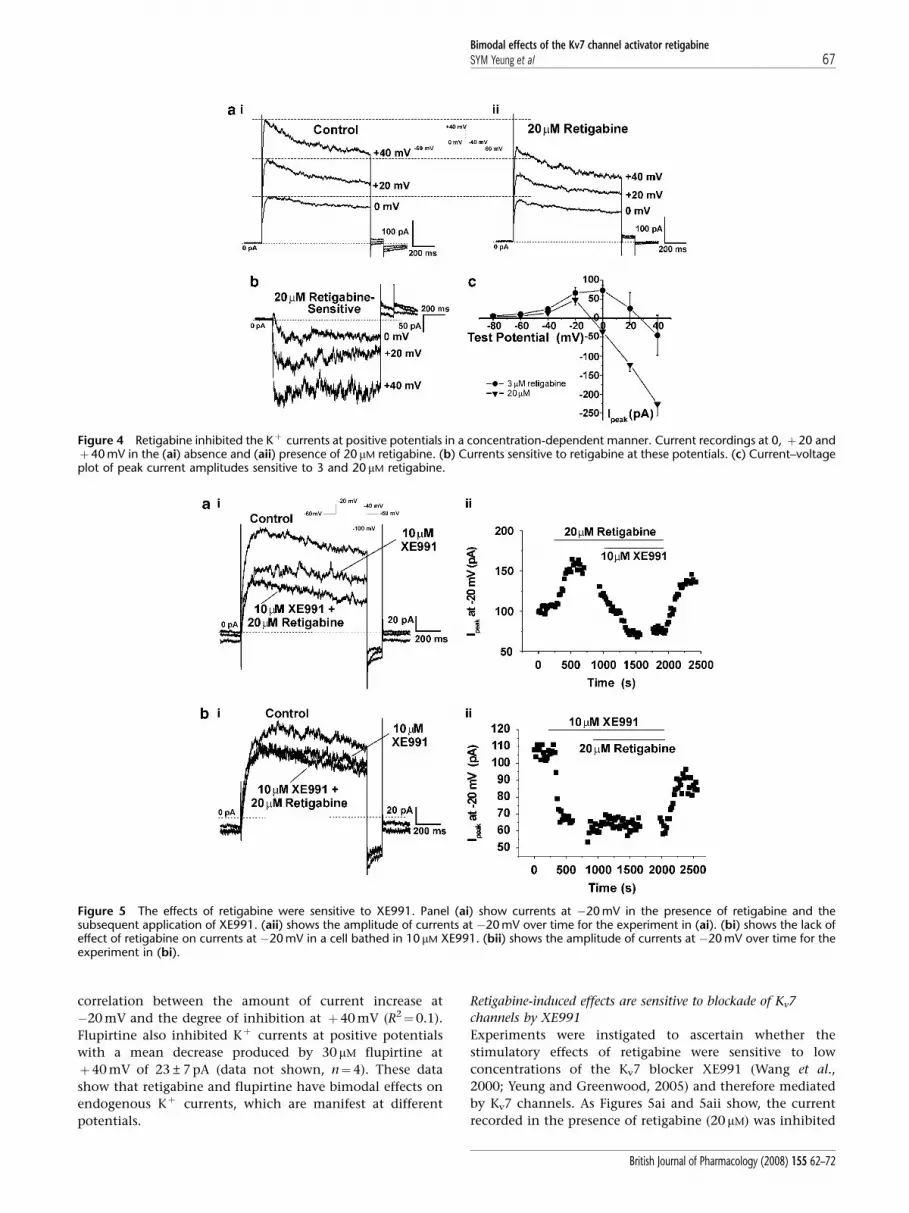
correlation between the amount of current increase at
�20 mV and the degree of inhibition at þ40 mV (R2¼0.1).
Flupirtine also inhibited Kþ currents at positive potentials
with a mean decrease produced by 30 mM flupirtine at
þ40 mV of 23±7 pA (data not shown, n¼4). These data
show that retigabine and flupirtine have bimodal effects on
endogenous Kþ currents, which are manifest at different
potentials.
Retigabine-induced effects are sensitive to blockade of Kv7
channels by XE991
Experiments were instigated to ascertain whether the
stimulatory effects of retigabine were sensitive to low
concentrations of the Kv7 blocker XE991 (Wang et al.,
2000; Yeung and Greenwood, 2005) and therefore mediated
by Kv7 channels. As Figures 5ai and 5aii show, the current
recorded in the presence of retigabine (20 mM) was inhibited
Figure 4 Retigabine inhibited the Kþ currents at positive potentials in a concentration-dependent manner. Current recordings at 0, þ20 andþ40 mV in the (ai) absence and (aii) presence of 20mM retigabine. (b) Currents sensitive to retigabine at these potentials. (c) Current–voltageplot of peak current amplitudes sensitive to 3 and 20 mM retigabine.
Figure 5 The effects of retigabine were sensitive to XE991. Panel (ai) show currents at �20 mV in the presence of retigabine and thesubsequent application of XE991. (aii) shows the amplitude of currents at �20 mV over time for the experiment in (ai). (bi) shows the lack ofeffect of retigabine on currents at �20 mV in a cell bathed in 10mM XE991. (bii) shows the amplitude of currents at �20 mV over time for theexperiment in (bi).
Bimodal effects of the Kv7 channel activator retigabineSYM Yeung et al 67
British Journal of Pharmacology (2008) 155 62–72

by 10 mM XE991. The mean inhibition produced by 10 mM
XE991 was 78.4±5.1% (n¼3) and 64.5±11.0% (n¼9) for
cells bathed in 3 and 20 mM retigabine respectively. As Figure
5bi and the time course (Figure 5bii) show pre-treatment
with 10 mM XE991 prevented the stimulatory effect of
retigabine at negative potentials (representative of five such
experiments). As the outward currents produced by mem-
brane depolarization of PV myocytes represent the net
product of many different types of channels, the inhibition
produced by retigabine at positive potentials might reflect a
bimodal effect on Kv7 channels or an additional effect on a
different Kþ channel. Consequently, studies were under-
taken to determine if the inhibitory effect of retigabine was
additive to that of XE991. Figure 6 shows current recordings
from two different cells isolated from the same animal (a, b).
At VT þ40 mV 20 mM retigabine alone (panel a) inhibited the
peak outward current by 29%. Further block was observed
with the addition of 10 mM XE991 with peak outward current
being reduced in total by 48% in this cell. In the other cell
(panel b), 10 mM XE991 alone (panel b) blocked peak outward
current amplitude by 44% and no further inhibition was
observed when retigabine was added. Mean data for nine
such experiments are presented in the histogram (panel c)
which shows that the inhibition produced by retigabine
alone was significantly less (Po0.001) than XE991 but the
cumulative effect of XE991 and retigabine was not signifi-
cantly different from the effect of XE991 alone. These data
suggest both the activator retigabine and blocker XE991
exert their effects on the same channel protein. In contrast,
pre-application of the selective Kv7.1 channel blocker
chromanol 293B (30 mM, Lerche et al., 2007), which reduced
currents at þ40 mV by 350±54 pA (n¼5), did not prevent
the retigabine-induced inhibition at this potential. Thus, the
mean decrease in current at þ40 mV produced by 10 mM
retigabine in the presence of chromanol 293B was
120±36 pA (n¼5), which was similar to the inhibition
produced by 10 mM retigabine in the absence of chromanol
(152±13 pA, n¼6).
mPV myocytes express KCNQ4 mRNA and Kv7.4 protein
Figure 7ai shows that mRNA for KCNQ1 and KCNQ4 was
detected by RT-PCR in mPV tissue. In three of the four tissues
tested, a very weak signal for KCNQ5 was also observed. This
profile was similar to that described by Ohya et al. (2003) by
quantitative PCR, that is, KCNQ1cKCNQ44KCNQ54KCNQ3. Amplicons for KCNQ1, KCNQ4 and KCNQ5 were
also detected in RNA samples isolated from a pool of 20
single myocytes using a nested RT-PCR approach (Figure
7aii). To consolidate the PCR results, immunocytochemical
imaging using antibodies against different KCNQ expression
products (Kv7.x) was undertaken. Figure 7bi shows positive
staining for Kv7.1 (n¼6 cells), Kv7.4 (Kv7.4J, n¼6 cells,
panel 7ci; Kv7.4SC, n¼6 cells, panel 7di). Figure 7bi shows
positive staining for Kv7.1 (n¼6 cells), panel 7ci the staining
for Kv7.4 (Kv7.4J, n¼6 cells) and in panel 7di, staining for
Kv7.4SC (n¼6 cells). The fluorescent signal for both Kv7.1
and Kv7.4 subunits were located predominantly within 1mm
of the plasma membrane, with significantly less signal
originating in the cytoplasm (Figure 7e). Pre-incubation of
the Kv7.1 antibody with its antigenic peptide (1:2 ratio of
antibody to antigenic peptide) suppressed the fluorescence
strongly (compare panel 7bi to panel 7bii; summary in
Figure 7f, n¼4–6, Po0.001; unpaired Student’s t-test).
Incubation of the Kv7.4SC antibody with its antigenic
peptide also produced a significant reduction in the
Figure 6 The inhibitory effects of retigabine and XE991 were not additive. Current recordings at VT þ40 mV with (a) retigabine alonefollowed by retigabine with added XE991. The inhibition by 10 mM XE991 alone (b) was not altered when retigabine was added. (c) Histogramshows mean±s.e.m. (all n¼7) block of mPV current (***Po0.001).
Bimodal effects of the Kv7 channel activator retigabineSYM Yeung et al68
British Journal of Pharmacology (2008) 155 62–72
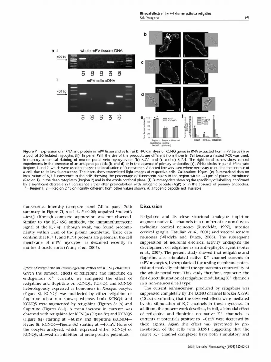
fluorescence intensity (compare panel 7di to panel 7dii;
summary in Figure 7f, n¼4–6, Po0.05; unpaired Student’s
t-test,) although complete suppression was not observed.
Similar to the Kv7.4SC antibody, the immunofluorescent
signal of the Kv7.4J, although weak, was found predomi-
nantly within 1 mm of the plasma membrane. These data
confirm that Kv7.1, and Kv7.4 proteins are present in the cell
membrane of mPV myocytes, as described recently in
murine thoracic aorta (Yeung et al., 2007).
Effect of retigabine on heterologously expressed KCNQ channels
Given the bimodal effects of retigabine and flupirtine on
endogenous Kþ currents, we compared the effect of
retigabine and flupirtine on KCNQ1, KCNQ4 and KCNQ5
heterologously expressed as homomers in Xenopus oocytes
(Figure 8). KCNQ1 was unaffected by either retigabine or
flupirtine (data not shown) whereas both KCNQ4 and
KCNQ5 were augmented by retigabine (Figures 8a–h) and
flupirtine (Figures 8i–l). A mean increase in currents was
observed with retigabine for KCNQ4 (Figure 8c) and KCNQ5
(Figure 8g) starting at �60 mV and flupirtine (KCNQ4—
Figure 8i; KCNQ5—Figure 8k) starting at �40 mV. None of
the oocytes analysed, which expressed either KCNQ4 or
KCNQ5, showed an inhibition at more positive potentials.
Discussion
Retigabine and its close structural analogue flupirtine
augment native Kþ channels in a number of neuronal types
including cortical neurones (Rundfeldt, 1997), superior
cervical ganglia (Tatulian et al., 2001) and visceral sensory
neurones (Wladyka and Kunze, 2006). The subsequent
suppression of neuronal electrical activity underpins the
development of retigabine as an anti-epileptic agent (Porter
et al., 2007). The present study showed that retigabine and
flupirtine also stimulated native Kþ channel currents in
mPV myocytes, hyperpolarized the resting membrane poten-
tial and markedly inhibited the spontaneous contractility of
the whole portal vein. This study therefore, represents the
first direct illustration of retigabine modulating Kþ channels
in a non-neuronal cell type.
The current enhancement produced by retigabine was
suppressed completely by the KCNQ channel blocker XE991
(10 mM) confirming that the observed effects were mediated
by the stimulation of Kv7 channels in these myocytes. In
addition, the present work describes, in full, a bimodal effect
of retigabine and flupirtine on native Kþ channels, as
currents at potentials positive to B0 mV were decreased by
these agents. Again this effect was prevented by pre-
incubation of the cells with XE991 suggesting that the
native Kv7 channel complexes have both stimulatory and
Figure 7 Expression of mRNA and protein in mPV tissue and cells. (a) RT-PCR analysis of KCNQ genes in RNA extracted from mPV tissue (i) ora pool of 20 isolated myocytes (ii). In panel 7aii, the size of the products are different from those in 7ai because a nested PCR was used.Immunocytochemical staining of murine portal vein myocytes for (b) Kv7.1 and (c and d) Kv7.4. The right-hand panels show controlexperiments in the presence of an antigenic peptide (b and d) or in the absence of primary antibodies (c). White circles in panel bi indicateRegions 1 and 2, which were used to analyse the localization of fluorescence. A dotted line was used where necessary to outline the contour ofa cell, due to its low fluorescence. The insets show transmitted light images of respective cells. Calibration: 10 mm. (e) Summarized data onlocalization of Kv7 fluorescence in the cells showing the percentage of fluorescent pixels in the region within B1mm of plasma membrane(Region 1), in the deep cytoplasm (Region 2) and in the whole confocal plane. (f) Summary data showing the specificity of labelling, confirmedby a significant decrease in fluorescence either after preincubation with antigenic peptide (AgP) or in the absence of primary antibodies.10 ¼ Region1; 20 ¼Region 2 *Significantly different from other values shown. #: antigenic peptide not available.
Bimodal effects of the Kv7 channel activator retigabineSYM Yeung et al 69
British Journal of Pharmacology (2008) 155 62–72

inhibitory sites. This bimodal effect has been mentioned
briefly in previous papers using studies on neuronal cells.
Both Rundfeldt (1997) and Tatulian et al. (2001) reported
that the retigabine-sensitive current plotted against test
potential had a distinctive ‘bell’ shape but did not describe
a net inhibition. This is probably because the voltage range
in these studies was truncated at 0 mV, whereas, in the
present study, a net inhibition was only manifest with
the highest concentration used (20 mM). In contrast to the
bimodal effect on native Kþ currents in mPV myocytes,
retigabine or flupirtine simply augmented Kþ currents
produced by the overexpression of KCNQ 4 or 5 in Xenopus
oocytes. Neither compound affected currents produced by
the overexpression of KCNQ1. This is a highly pertinent
observation because it suggests that simple Kv7 homomers
do not constitute the native channel in mPV myocytes.
It is possible that the native channel is formed from
heteromers of Kv7.1, 7.4 or 7.5, as the mPV myocytes
express KCNQ1, 4 and 5. However, all past reports that
analyzed Xenopus oocytes coexpressing these Kv7 subunits,
have so far failed to show an interaction of these
channels (Kubisch et al., 1999; Jentsch, 2000; Schroeder
et al., 2000). Consequently, the bimodal phenomenon
represents an interesting finger-print of the native vascular
Kv7 channels.
The molecular targets of retigabine are the so-called
‘neuronal’ Kv7 channels, that is, those encoded by the genes
KCNQ2–5 (Main et al., 2000; Wickenden et al., 2000; Tatulian
et al., 2001). Retigabine augments currents generated by
KCNQ2/3 hetero-multimers and KCNQ5/3 hetero-multimers
with an EC50 of 0.3–2 mM (Wickenden et al., 2000, 2001;
Tatulian et al., 2001, present study). Retigabine has a similar
concentration dependence for currents generated by the
overexpression of KCNQ3 (1 mM, Tatulian et al., 2001), and
KCNQ4 (5mM, Tatulian et al., 2001, present study). The
stimulatory effect is due mainly to a 20–40 mV leftward shift
in the voltage-dependence of activation, dependent on the
KCNQ isoform (Main et al., 2000; Wickenden et al., 2000;
Tatulian et al., 2001) although KCNQ5-encoded channels are
augmented without an effect on the half-activation voltage
(Dupuis et al., 2002). Interestingly, retigabine produced a
modest shift (B10 mV) of current activation in mPV
myocytes. Retigabine also speeds the rate of activation and
slows deactivation (Main et al., 2000). No inhibition of
channels encoded by KCNQ2–5 has ever been reported and
was not observed in the present study. In stark contrast to
the stimulatory effect on channels encoded by KCNQ2–5,
retigabine in concentrations up to 100 mM does not increase
KCNQ1-generated currents (Tatulian et al., 2001; Schenzer
et al., 2005; Wuttke et al., 2005). In fact, currents generated
Figure 8 Activation of heterologously expressed KCNQ4 and KCNQ5 by retigabine and flupirtine. Typical current traces of oocytes expressingKCNQ4 (a) and KCNQ5 (e) before and after (KCNQ4 (b); KCNQ5 (f)) the application of 10mM retigabine (R). The voltage protocol used forthese experiments is shown as inset in panel a. I/V curves of KCNQ4 (c and i) and KCNQ5 (g and k) as a summary of the current recordings,which were obtained from oocytes measured with different retigabine (KCNQ4 (c) and KCNQ5 (g)) and different flupirtine (F) concentrations(KCNQ4 (i) and KCNQ5 (k)). I/Imax curves of KCNQ4 and KCNQ5 as a function of voltage obtained from tail current analysis and differentretigabine (KCNQ4 (d) and KCNQ5 (h)) or flupirtine (KCNQ4 (j) and KCNQ5 (l)) concentrations. For all experiments n=8.
Bimodal effects of the Kv7 channel activator retigabineSYM Yeung et al70
British Journal of Pharmacology (2008) 155 62–72

by the heterologous expression of KCNQ1 are inhibited in a
voltage-dependent manner by retigabine with an IC50 of
B100mM (Tatulian et al., 2001). This led to the identification
of tryptophan 236 in the S5 domain and glycine301 in the S6
domain of KCNQ2–5 as crucial amino acids for the
stimulatory effect of retigabine (Schenzer et al., 2005; Wuttke
et al., 2005). With respect to the opposing effect of retigabine
on channels encoded by KCNQ1 versus other KCNQ genes,
the bimodal effect observed in mPV myocytes may reflect
differential effects on Kv7 channels. Thus, at negative
potentials, activation of Kv7.4/Kv7.5 by retigabine dominates
leading to current augmentation but at positive potentials,
block of Kv7.1 dominates resulting in the observed current
inhibition. However, the inability of chromanol 293B to
prevent the retigabine-induced current inhibition at
þ40 mV suggests that this model needs to be investigated
further.
KCNQ expression in smooth muscle cells
KCNQ gene expression in smooth muscle cells was first
identified in rat stomach by Ohya et al. (2002a). This
observation was followed by an in-depth study in mPV
where both KCNQ and KCNE gene expression was quantified
(Ohya et al., 2002b, 2003). We have now extended these
observations to include the murine aorta and a number of
different conduit arteries (carotid, femoral and mesenteric,
Yeung et al., 2007). In every case the most abundantly
expressed genes were KCNQ1 and KCNQ4, the latter hitherto
considered to be only expressed in auditory nerves (Kharkovets
et al., 2000; Kubisch et al., 1999). Some KCNQ5 expression
was also detected whereas KCNQ2 and 3 were conspicuous
by their absence (see Ohya et al., 2003; Brueggemann et al.,
2006; Yeung et al., 2007). The previous study on mPV
showed that KCNQ1 was the most abundantly expressed
with KCNQ4 having significant abundance (Ohya et al.,
2003). We have now consolidated these findings and shown
that Kv7.4 protein is present in the plasmalemmal membrane
of portal vein myocytes as well as Kv7.1, similar to the
situation in murine aortic myocytes (Yeung et al., 2007).
Previous studies on mPV showed that the selective KCNQ
channel blocker XE991, which does not discriminate
between different Kv7 isoforms, inhibited Kþ currents,
depolarized the membrane potential and increased sponta-
neous contractions consistent with Kv7 channels regulating
cellular excitability (Yeung and Greenwood, 2005). XE991
also contracts segments of aorta (Yeung et al., 2007), as well
as conduit arteries such as the pulmonary (Joshi et al., 2006),
carotid and femoral (Yeung et al., 2007). These observations
were consistent with Kv7 channels regulating the membrane
potential and suppressing voltage-dependent Ca2þ influx in
smooth muscle cells throughout the vasculature. Moreover,
it is probably not Kv7.1 subunits that are important for this
effect but Kv7.4 or Kv7.5 as Kv7.1-selective blockers (chro-
manol 293B or L-768, 673) failed to mirror the effects of
XE991 (Yeung et al., 2007). This postulate was supported by
the ability of retigabine (2–20 mM) to relax segments of aorta,
as well as carotid, femoral and mesenteric arteries pre-
contracted with phenylephrine, but not with raised external
Kþ (Yeung et al., 2007). The electrophysiological and
mechanical effects obtained in the present study agree with
the hypothesis that Kv7.4 or Kv7.5 channels are key
regulators of vascular excitability. However, as mentioned
above, the bimodal effects of retigabine and flupirtine argue
against simple Kv7 homomers existing in vascular myocytes.
Vascular myocytes do express various members of the KCNE
gene family, (Ohya et al., 2002a; Yeung et al., 2007), which
are known to influence the biophysical and pharmacological
attributes of Kv7 channels (for example, Grunnet et al., 2002;
Strutz-Seebohm et al., 2006). Consequently, the native
‘KCNQ channel’ in vascular smooth muscle cells may result
from a hitherto undefined combination of KCNQ and KCNE
gene products. Future studies will investigate this aspect
further.
To conclude, we have shown that retigabine and flupirtine
activate Kþ channels and this mechanism underlies the
vasorelaxant effect of these agents reported previously
(Yeung et al., 2007). In addition, both agents inhibit
XE991-sensitive currents at positive potentials and this
phenomenon provides an interesting signature in future
studies aimed at deciphering the molecular identity of the
native Kv7 channel in smooth muscle cells.
Acknowledgements
Research in Dr Greenwood’s laboratory was funded by the
British Heart Foundation (PG/O3/085/15747). This research
was also supported by Deutsche Forschungsgemeinschaft
(MS) and a British Heart Foundation Intermediate Research
Fellowship FS/04/052 (VP). We are grateful to Professor
Thomas Bolton for the access to the confocal microscope.
Conflict of interest
The authors state no conflict of interest.
References
Alexander SPH, Mathie A, Peters JA (2008). Guide to Receptors andChannels (GRAC), 3rd edn. Br J Pharmacol 153 (Suppl. 2): S1–S209.
Brueggemann LI, Moran CJ, Barakat JA, Yehy JZ, Cribbs LL, Byron KL(2006). Vasopressin stimulates action potential firing by proteinkinase C dependent inhibition of KCNQ5 in A7r5 rat aorticsmooth muscle cells. Am J Physiol-Heart 292: H1352–H1363.
Dupuis DS, Olesen S-P, Jespersen T, Christensen JK, Christophersen P,Jensen BS (2002). Activation of KCNQ5 Channels stably expressedin HEK293 cells by BMS-204352. Eur J Pharmacol 437: 129–137.
Grunnet M, Jespersen T, Rasmussen HB, Ljungstrom T, Jorgensen NK,Olesen SP et al. (2002). KCNE4 is an inhibitory subunit to theKCNQ1 channel. J Physiol 542: 119–130.
Jentsch TJ (2000). Neuronal KCNQ potassium channels: physiologyand role in disease. Nat Rev Neurosci 1: 21–30.
Joshi S, Balan P, Gurney AM (2006). Pulmonary vasoconstrictoraction of KCNQ potassium channel blockers. Respir Res 7: 31.
Kharkovets T, Hardelin JP, Safieddine S, Schweizer M, El-Amraoui A,Petit C et al. (2000). KCNQ4, a Kþ channel mutated in a form ofdominant deafness, is expressed in the inner ear and the centralauditory pathway. Proc Natl Acad Sci USA 97: 4333–4338.
Kubisch C, Schroeder BC, Friedrich T, Luetjohann B, El-Amraoui A,Marlin S et al. (1999). KCNQ4, a novel potassium channel
Bimodal effects of the Kv7 channel activator retigabineSYM Yeung et al 71
British Journal of Pharmacology (2008) 155 62–72
expressed in sensory outer hair cells, is mutated in dominantdeafness. Cell 96: 437–446.
Lerche C, Bruhova I, Lerche H, Steinmeyer K, Wei AD,Strutz-Seebohm N et al. (2007). Chromanol 293B binding inKCNQ1 (Kv7.1) channels involves electrostatic interactions with apotassium ion in the selectivity filter. Mol Pharmacol 71: 1503–1511.
Main MJ, Cryan JE, Dupere JR, Cox B, Clare JJ, Burbidge SA (2000).Modulation of KCNQ2/3 potassium channels by the novelanticonvulsant retigabine. Mol Pharmacol 58: 253–262.
Ohya S, Asakura K, Muraki K, Watanabe M, Imaizumi Y (2002b).Molecular and functional expression of ERG, KCNQ, and KCNEsubtypes in rat stomach smooth muscle. Am J Physiol (Gastro LivPhysiol) 282: 277–287.
Ohya S, Horowitz B, Greenwood IA (2002a). Functional andmolecular identification of ERG channels in murine portal veinmyocytes. Am J Physiol Cell Physiol 283: C866–C877.
Ohya S, Sergeant G, Greenwood IA, Horowitz B (2003). Molecularvariants of KCNQ channels expressed in murine portal vein myo-cytes: a role in delayed rectifier current. Circ Res 92: 1016–1023.
Porter RJ, Nohria V, Rundfeldt C (2007). Retigabine. Neurotherapeutics4: 149–154.
Rundfeldt C (1997). The new anticonvulsant retigabine (D-23129)acts as an opener of Kþ channels in neuronal cells. Eur J Pharmacol336: 243–249.
Saleh S, Yeung SY, Prestwich S, Pucovsky V, Greenwood IA (2005).Electrophysiological and molecular identification of voltage-gatedsodium channels in murine vascular myocytes. J Physiol 568: 155–169.
Schenzer A, Friedrich T, Pusch M, Saftig P, Jentsch TJ, Grotzinger Jet al. (2005). Molecular determinants of KCNQ (Kv7) Kþ channelsensitivity to the anticonvulsant retigabine. J Neurosci 25:5051–5060.
Schroeder BC, Hechenberger M, Weinrich F, Kubisch C, Jentsch TJ(2000). KCNQ5, a novel channel broadly expressed in brain,mediates M-current. J Biol Chem 275: 24089–24095.
Strutz-Seebohm N, Seebohm G, Fedorenko O, Baltaev R, Engel J,Knirsch M et al. (2006). Functional coassembly of KCNQ4 withKCNE-beta-subunits in Xenopus oocytes. Cell Physiol Biochem 18:57–66.
Tatulian L, Delmas P, Abogadie FC, Brown DA (2001). Activation ofexpressed KCNQ potassium currents and native neuronal M-typepotassium currents by the anti-convulsant drug retigabine.J Neurosci 21: 5535–5545.
Wang H-S, Brown BS, McKinnon D, Cohen IR (2000). Molecular basisfor differential sensitivity of KCNQ and IKs channels to thecognitive enhancer XE991. Mol Pharmacol 57: 1218–1223.
Wang H-S, Pan Z, Shi W, Barry BS, Wymore RS, Cohen IR et al. (1998).KCNQ2 and KCNQ3 potassium channel subunits: molecularcorrelates of the M-channel. Science 282: 1890–1893.
Wickenden AD, Yu W, Zou A, Jegla T, Wagoner PK (2000). Retigabine,a novel anti-convulsant, enhances activation of KCNQ2/Q3potassium channels. Mol Pharmacol 58: 591–600.
Wickenden AD, Zou A, Wagoner PK, Jegla T (2001). Characterizationof KCNQ5/Q3 potassium channels expressed in mammalian cells.Br J Pharmacol 132: 381–384.
Wladyka CL, Kunze DL (2006). KCNQ/M-currents contribute to theresting membrane potential in rat visceral sensory neurones.J Physiol 575: 175–189.
Wuttke TV, Seebohm G, Bail S, Maljevic S, Lerche H (2005). The newanticonvulsant retigabine favors voltage-dependent opening ofthe Kv7.2 (KCNQ2) channel by binding to its activation gate.Mol Pharmacol 67: 1009–1017.
Yeung SYM, Greenwood IA (2005). Electrophysiological and func-tional effects of the KCNQ channel blocker XE991 on murineportal vein smooth muscle cells. Br J Pharmacol 146: 585–595.
Yeung SYM, Pucovsky V, Moffatt JD, Saldanha L, Schwake M, Ohya Set al. (2007). Molecular expression and pharmacological identifi-cation of a role for Kv7 channels in murine vascular reactivity.Br J Pharmacol 151: 758–770.
Bimodal effects of the Kv7 channel activator retigabineSYM Yeung et al72
British Journal of Pharmacology (2008) 155 62–72