best practice on cetacean post mortem
TRANSCRIPT
Delegates are kindly invited to bring their own documents to the Meeting. This document will be available only in electronic format during the Meeting.
26/09/2019 English
Original: English ACCOBAMS-MOP7/2019/Doc 33
BEST PRACTICE ON CETACEAN POST MORTEM INVESTIGATION
AND TISSUE SAMPLING
Agreement on the Conservation of Cetaceans of the Black Sea, Mediterranean Sea and contiguous Atlantic area, concluded under the auspices of the Convention on the Conservation of Migratory Species of Wild Animals (CMS)
Accord sur la Conservation des Cétacés de la Mer Noire, de la Méditerranée et de la zone Atlantique adjacente, conclu
sous l’égide de la Convention sur la Conservation des Espèces Migratrices appartenant à la Faune Sauvage (CMS)
Seventh Meeting of the Parties to ACCOBAMS Istanbul, Republic of Turkey, 5 - 8 November 2019
ACCOBAMS-MOP7/2019/Doc 33
2
Best practice on cetacean post mortem
investigation and tissue sampling
Joint ACCOBAMS and ASCOBANS document
Editors:
Lonneke L. IJsseldijk •Andrew C. Brownlow •Sandro Mazzariol
September 2019
ACCOBAMS-MOP7/2019/Doc 33
3
Editors: Lonneke L. IJsseldijka, Andrew C. Brownlowb & Sandro Mazzariolc
aFaculty of Veterinary Medicine, Utrecht University, Pathology Division, Utrecht, The
Netherlands
b Scottish Marine Animal Stranding Scheme, SRUC Northern Faculty, Inverness Campus,
Inverness Scotland
cDepartment of Comparative Biomedicine and Food Science, Università degli Studi di
Padova, Legnaro, Padova, Italy
With contributions from: Agreement for the Conservation of Cetaceans of the Baltic Sea, Mediterranean Sea and Contiguous Atlantic Area (ACCOBAMS) - Aviad Scheinin Agreement on the Conservation of Small Cetaceans of the Baltic, North East Atlantic, Irish and North Seas (ASCOBANS) Cetacean Strandings Investigation Program, Zoological Society of London, London, United Kingdom - Rob Deaville, Paul D. Jepson, Matthew W. Perkins Cornwall Wildlife Trust Marine Strandings Network, Cornwall, United Kingdom - James Barnett C.Re.Di.Ma. , Istituto Zooprofilattico Sperimentale del Piemonte, Liguria and Valle d’Aosta, Italy - Carla Grattarola, Cristina Caselone Dipartimento di Scienze Fisiche, della Terra e dell’Ambiente, Università degli Studi di Siena, Siena, Italy - Maria Cristina Fossi, Letizia Marsili Facoltà di Medicina Veterinaria, Università degli Studi di Teramo, Teramo, Italy - Giovanni Di Guardo Pathology Division, Faculty of Veterinary Medicine, Utrecht University - Andrea Gröne International Whaling Commission (IWC) - Karen Stockin Institute for Neurosciences of Montpellier (INSERM U1051), France - Maria Morell
ACCOBAMS-MOP7/2019/Doc 33
4
Institute for Terrestrial and Aquatic Wildlife Research, University of Veterinary Medicine Hannover, Büsum, Germany - Miguel Grilo, Kristina Lehnert, Abbo van Neer, Anja Reckendorf, Ursula Siebert Moredun Research Institute, Edinburgh, Scotland, United Kingdom - Mark P. Dagleish Observatoire Pelagis, La Rochelle, France - Willy Dabin, Paula Mendes Fernandez Scottish Marine Animal Stranding Scheme, SRUC Northern Faculty, An Lòchran, Inverness Campus, Inverness Scotland - Nicholas J. Davison, Mariel T.I. ten Doeschate University of Las Palmas de Gran Canaria, Spain - Manolo Arbello, Yara Bernaldo de Quirós, Antonio Jesús Fernández Rodríguez, Jesus de la Fuenta, Eva Sierra Veterinary Faculty, University of Liège, Liège, Belgium - Thierry Jauniaux
ACCOBAMS-MOP7/2019/Doc 33
5
PREFACE
Human society is changing rapidly. Our global gross domestic product has tripled in the last 25
years, with a concomitant increase in human impact on the marine environment. To measure the
effects of this impact on cetacean health, it is crucial to perform long-term monitoring of dead
cetaceans. Such monitoring includes the examination of their organs for pathological changes,
collection of tissue samples for toxicology, and taking measurements for life history, according to a
standardised and internationally harmonised protocol.
I missed such a protocol in the early 1990s, when, as a recently graduated veterinarian, I started
studying the pathology of stranded cetaceans in England and Wales. At meetings of the European
Cetacean Society (ECS), I met biologists who were highly experienced in examining and sampling
stranded cetaceans to learn more about their life history, diet, parasites, and pollutant burdens.
However, there were very few people studying their pathology.
Among the ECS members was another veterinarian, Manuel Garcia Hartmann, who, like me, was
interested in cetacean pathology. In 1991, together with Marjan Addink, we organised a workshop
at the National Natural History Museum in Leiden and brought together biologists and veterinarians
who examined stranded cetaceans, as well as specialists in a variety of relevant laboratory
analyses. During the day, post mortem examinations on harbour porpoises were demonstrated,
and sampling procedures were discussed. At night, assisted by Johnny Walker, Manuel and I
worked up our notes. The end result was a standard post mortem protocol for small cetaceans that
integrated pathology, life history, and toxicology. Appearing at a time when ASCOBANS-based
monitoring programs were being developed in several European Countries, this protocol was
adopted and further developed widely and has been cited over 100 times in scientific publications.
The publication before you now is a more organised and better illustrated edition of that old
protocol, and incorporates advances in laboratory techniques and new knowledge of cetacean
diseases. Its essence has remained the same, namely to integrate the pathological examination of
a dead cetacean with the measurements and sampling for life history and toxicology. This is
illustrated well by the background training of the Editors: biology (Lonneke IJsseldijk) and
veterinary medicine (Andrew Brownlow and Sandro Mazzariol). My wish is similar to the one
Manuel and I expressed 25 years ago: that this protocol will prove useful and be widely adopted,
and so help to provide a view of the biology and pathology of cetaceans that is not limited by the
borders of Countries.
Thijs Kuiken, Rotterdam, August 2019.
ACCOBAMS-MOP7/2019/Doc 33
6
Table of Contents
PREFACE ............................................................................................................................................... 5
Introduction and background .............................................................................................................. 8
Monitoring of stranded or bycaught animals .................................................................................... 10
Glossary .............................................................................................................................................. 11
Multi-Tier Triage Approach ................................................................................................................ 13
Tier One – External examination and stranding data collection ................................................... 14
Tier Two – Post mortem investigations and tissue sampling ........................................................ 14
Tier Three – Post mortem examination with diagnostic aims ....................................................... 14
Legislation and permits ...................................................................................................................... 15
Health and Safety ............................................................................................................................... 17
Biological risks ................................................................................................................................ 17
Environmental risks ........................................................................................................................ 18
Chemical and residue risks ............................................................................................................. 18
Post mortem risks .......................................................................................................................... 18
Carcass disposal ............................................................................................................................. 19
Evaluation of the carcass ................................................................................................................... 20
External Features ........................................................................................................................... 20
Internal Features ............................................................................................................................ 21
Carcass Decomposition Classification ............................................................................................ 21
Description and photographs ........................................................................................................ 23
Effects of decomposition code on investigative tests ................................................................... 24
Tier One: Basic morphometrics, external examination and basic tissue sampling ........................... 27
Data collection and photographs ................................................................................................... 27
Body measurements ...................................................................................................................... 28
Freezing carcases prior to post mortem examination ................................................................... 28
Tier Two: Post mortem investigations and sampling ........................................................................ 30
External examination ..................................................................................................................... 31
Nutritional condition state ............................................................................................................. 32
Subcutaneous examination ............................................................................................................ 33
Visualisation of internal organs ..................................................................................................... 34
Examination of abdominal organs (except GIT) ............................................................................. 34
Examination of GIT ......................................................................................................................... 36
Examination of head and neck region, and thoracic organs ......................................................... 37
Examination of the head ................................................................................................................ 40
Examination of the skeletal system and rete mirabilis .................................................................. 42
ACCOBAMS-MOP7/2019/Doc 33
7
Tissue sampling procedures, storage and analysis ............................................................................ 43
Morphometric studies and imaging ............................................................................................... 43
Tissue sampling and storage .......................................................................................................... 43
Labelling of samples ....................................................................................................................... 44
Tissue and biological specimens’ archive ...................................................................................... 45
Age determination ......................................................................................................................... 45
Diet analysis, marine litter and micro- /nano-plastics ................................................................... 46
Genomic (DNA) studies and RT-PCR ecotoxicological investigations ............................................ 47
Reproduction studies ..................................................................................................................... 47
Histopathology and immunohistochemistry samples ................................................................... 47
Microbiology .................................................................................................................................. 49
Virology .......................................................................................................................................... 50
Parasitology .................................................................................................................................... 50
Toxicology ...................................................................................................................................... 51
Biotoxins ......................................................................................................................................... 52
Suggested sampling tables ................................................................................................................. 52
Acknowledgements ............................................................................................................................ 63
Literature .................................................................................................................................... 64
Annex 1. Decomposition condition coding example pictures ................................................... 68
Annex 2. Additional body measurements to consider .............................................................. 71
Annex 3. Brain sampling image .................................................................................................. 72
ACCOBAMS-MOP7/2019/Doc 33
8
Introduction and background
Monitoring dead stranded cetaceans offers an often unique opportunity to gain insights
into the health of, and threats and stressors affecting, marine ecosystems (e.g. Dierauf &
Gulland 2001; Gulland & Hall 2009; Van Bressem et al. 2009; Peltier et al. 2012, 2013;
Plön et al. 2015). Information derived from the systematic examination of stranded
carcases can provide insights into the at-sea population not easily acquired through other
means, indeed strandings data is the major source of information available for some
species (Reyes et al. 1991; Pyenson 2011). Detailed investigation of carcases can assist
in the determination of causes of death and provides general surveillance on the incidence
of trauma and disease (Siebert et al. 2001, 2006; Jauniaux et al. 2002; Arbelo et al. 2013;
Di Guardo et al. 2013; Lane et al. 2014; Díaz-Delgado et al. 2018). Investigations can
identify existing and emerging threats due to human impact, such as bycatch (Leeney et
al. 2008, Peltier et al. 2016) and marine pollution (Siebert et al. 1999; Jepson et al. 2016),
as well as provide tissues and data for subsequent analysis into a range of biological and
ecological parameters. The collection of data and samples for ancillary investigations over
a range of disciplines can provide information on the general population ecology, helping
countries evaluate and mitigate possible threats affecting species conservation and the
marine ecosystem.
Many EU countries operate cetacean strandings investigation networks as part of their
obligations to international agreements. These include the “Agreement on the
Conservation of Small Cetaceans of the Baltic, North East Atlantic, Irish and North Seas”
(ASCOBANS), the “Agreement for the Conservation of Cetaceans in the Black Sea,
Mediterranean Sea and contiguous Atlantic Area” (ACCOBAMS), the EU Habitats
Directive (NATURA2000), the OSPAR Commission and the “Baltic Marine Environment
Protection Commission” (HELCOM). The Marine Strategy Framework Directive (MSFD)
obliges the member states to develop indicators and descriptors for the surveillance of
“Good Environmental Status” of cetaceans.
Many of these strandings networks are well established and have long-term datasets
comprising extensive biological and pathological information from the systematic collation
and investigation of mortalities. In addition, many European strandings networks curate
uniquely important tissue and pathogen archives. Most networks follow the original
protocol by Kuiken and Hartmann, published in 1993 as a special issue of the European
Cetacean Society. In the subsequent decades, stranding networks throughout Europe
have developed, and new analytical techniques (e.g. assessment of the effects of
ACCOBAMS-MOP7/2019/Doc 33
9
barotrauma) or health impacts e.g. grey seal predation, have been incorporated into
operational methods.
During the VIII ASCOBANS Meeting of the Party (MoP) in 2016, the Advisory Committee
(AC) and Secretariat were requested to engage actively in the work on best practice
guidelines for response to stranding events and in the establishment of an updated post-
mortem protocol within the frameworks of the International Whaling Commission (IWC),
ACCOBAMS and the European Cetacean Society (ECS) under Resolution 8.10. In the
same year, ACCOBAMS endorsed the document on common best practices for a basic
post mortem examination of stranded cetaceans under the Resolution no. 6.22 during the
VI MoP. In the same Recommendation, an approach to ASCOBANS, ECS and IWC was
requested to the Scientific Committee (SC) to review the common definitions, common
data collections and common post-mortem protocols during the triennium. In 2018, during
the 24th ASCOBANS AC and 12th ACCOBAMS SC a joint workshop was proposed to
harmonize the existing initiatives. This meeting was organised in Padua (Italy) in June
2019 involving 24 experts from different countries of the two regional Agreements and
from Macaronesia area representing the MARCET project.
The aim of this document is to update the protocol with the currently available techniques
and knowledge agreed between all member countries of ACCOBAMS and ASCOBANS. It
is hoped that this updated protocol can serve three overall aims:
To provide a reference document for veterinarians and biologists currently engaged in
cetacean post-mortem investigation, summarising a recognised approach to stranding
investigation across European networks;
To highlight areas where harmonisation of data from existing networks could allow for
analysis and inference to be made between networks, of particular relevance for the
transboundary, mobile species;
Provide a start-up guide for researchers attempting to instigate new stranding
monitoring programmes, particularly in regions of the world with limited resources
for extensive, top-down surveillance programmes.
It should be emphasised that this document is not designed to replace existing protocols,
particularly those of longstanding and well established laboratories and stranding
networks, but offers a post mortem framework aiming for consistency across Europe when
conducting examinations on dead cetaceans. By outlining current European best
ACCOBAMS-MOP7/2019/Doc 33
10
practices, it has been assumed that there is sufficient time and resources to carry out a full
post mortem examination, although it is recognised this may not always be the case.
The quality of the information gathered is influenced by logistical capacity, e.g. carcass
accessibility, available equipment/supplies and finances; and the skills, experience and
capacity of the human resources. Nonetheless, it should be emphasized that following a
precise and well defined data collection procedure ensures the information collected
during post-mortem investigations is of high quality. To be able to assess cause of death
and health status, a full post mortem investigation with additional examinations as
proposed below is deemed necessary and therefore highly recommended. If a full
investigation cannot be carried out for any reason, one should always attempt to collect
the following data: species, sex, stranding location, stranding date and (approximate) body
length to assess age class. Additionally, teeth (for ageing), skin (for genetic analysis),
blubber and muscle (for toxicological screening) and swabs of genital slit and blowhole (for
e.g. virological and microbiological analysis) can be relatively easily collected.
Monitoring of stranded or bycaught animals
Data from dead cetaceans, including stranded and bycaught individuals, offer a means to
sample the at-sea population, making it possible to obtain, in some cases, data not
accessible through other means of surveillance. There are discussions around the extent
to which investigations on dead animals can inform on the wider population due to several
biases intrinsic to the stranding process. Strandings are a complex result of biological,
physical and social (effort) processes that influence observed mortality (ten Doeschate et
al. 2018). These should be taken into account when making population level inferences
based on the examination of stranded or bycaught individuals. Where an increase in
strandings could indicate increased mortality or increased abundance (biological variation),
it may well be a consequence of unusual variation in environmental conditions such as
wind or tide or observer efforts. It is therefore encouraged to acknowledge all three
components of the stranding process during mortality investigations.
It needs to be highlighted that the quality and output of post mortem investigations is
strongly dependent on the quality of the stranding network in terms of spatiotemporal
coverage, collecting environmental information in relation with the stranding event and the
time between the collection of the carcass and start of post mortem investigation.
ACCOBAMS-MOP7/2019/Doc 33
11
Glossary
Here is a collection of common terms and definitions frequently used throughout the
document, and general terminology used in stranding events and forensic human and
veterinary medicine.
DEAD CETACEAN: Comprises cetaceans found dead on the shore or floating at sea and
cases which live strand and subsequently die or are euthanized. Includes animals found
dead entangled in fishing gear. Signs of death are absence of breathing, cardiac arrest
and absence of neuronal activity.
STRANDED CETACEAN: A stranded cetacean is one whose body lies entirely on land,
and includes both dead and live animals found in a helpless state after faltering ashore ill,
wounded, weak, or simply lost. In this document it is expanded to include animals either
dead or alive but showing clear signs of physiological dysfunction in shallow waters. On
the basis of the number of animals involved, it is possible to distinguish between single
and mass strandings.
ENTANGLED CETACEAN: Animals found completely or partially entangled in either
marine debris or active or discarded fishing gear, for example ropes, nets or straps.
UNUSUAL MORTALITY EVENT (UME): A UME is an unexpected mortality of cetaceans
at an abnormally large scale compared to average stranding reports for the species or
involves a significant die-off of any marine mammal population, and demands immediate
response. There are seven criteria that make a mortality event "unusual."
1. A marked increase in the magnitude or a marked change in the nature of morbidity, mortality, or strandings when compared with prior records.
2. A temporal change in morbidity, mortality, or strandings is occurring.
3. A spatial change in morbidity, mortality, or strandings is occurring.
4. The species, age, or sex composition of the affected animals is different than that of animals that are normally affected.
5. Affected animals exhibit similar or unusual pathologic findings, behavior patterns, clinical signs, or general physical condition (e.g., blubber thickness).
6. Potentially significant morbidity, mortality, or stranding is observed in species, stocks, or populations that are particularly vulnerable (e.g., listed as depleted, threatened, or endangered or declining). For example, stranding of three or four right whales may be cause for great concern whereas stranding of a similar number of fin whales may not.
7. Morbidity is observed concurrent with or as part of an unexplained continual decline of a marine mammal population, stock, or species.
ACCOBAMS-MOP7/2019/Doc 33
12
Main recognised causes are sudden emergence of an infectious disease or a disease
outbreak, biotoxins, or human interactions (including environmental accidents). Features of
these mass mortalities (i.e. temporal and spatial distribution) do not correspond to mass
strandings, as defined below.
MASS STRANDING: These events involve two or more cetaceans (excluding cow/calf
pairs) stranded at the same time and place. Several causes may be responsible for this
event, including, but not limited to, extreme weather conditions, tidal changes, disease of
one or several group members, or human-related actions. It is noteworthy that some
individuals involved in a mass stranding may be completely healthy.
DISEASE OUTBREAK: An UME specifically involves infectious agents. This can represent
the emergence of a novel pathogen or disease, be caused by a known pathogen not
previously recognized in that species or geographic area, or manifest as an abnormal
increase in the incidence of stranded individuals in a region, season or population. An
outbreak may occur in a restricted geographical area, or may extend over an entire basin
and can range in duration from a few days to several years.
DISSECTION/PROSECTION: Medical and/or biological procedure to dismember the body
of a deceased animal according to specific protocols in order to study its anatomical
structure and/or to evaluate and sample specific organs and tissues.
NECROPSY/AUTOPSY/POST-MORTEM/POST MORTEM EXAMINATION Synonyms for
a specialised medical procedure comprising of a thorough examination of a carcass by
dissection to determine the cause, the mechanism and manner of death through the
collection of evidence. In the case of wild animals this requires the involvement of a
veterinary pathologist or a veterinarian with specific training in animal pathology, diseases
and assessment of health.
POST MORTEM INVESTIGATIONS: All studies and investigations carried out on an
animal’s carcass and/or samples taken after death, including those aimed to determine the
cause of death.
HEALTH STATUS: Subjective assessment of diseases, conditions, or injuries that not only
contributed to the proximal cause of death but which characterize the ante-mortem health
status of the individual and the possible health status of cohort animals.
CAUSE OF DEATH/STRANDING: The disease, injury or abnormality that alone or in
combination with other factors (environmental, other concurrent diseases, age, etc.) is
responsible for initiating the sequence of functional disturbances that resulted in live
ACCOBAMS-MOP7/2019/Doc 33
13
stranding and death. In the case of an aquatic animal stranded on shore, the post mortem
investigation is aimed to determine the cause of stranding. During this procedure the
following may be further defined:
Immediate cause of death: final disease or condition resulting in death;
Underlying cause of death: the disease or injury that initiated the chain of morbid events
that led directly and inevitably to death;
Contributing factors: other significant diseases, conditions, or injuries/impacts/influences
that may have contributed to death but which did not constitute an underlying cause of
death.
MECHANISM OF DEATH: The immediate physiologic derangement resulting in death. A
particular mechanism of death can be produced by a variety of different causes of death.
MANNER OF DEATH: How death came about; in the case of wildlife and, specifically, in
cetaceans, we can distinguish:
Natural, due mainly to natural disease or toxic processes;
Anthropic/anthropogenic, accidental like ship strikes, bycatch, or non-accidental due to a
volitional act or direct killing;
Undetermined, inadequate information regarding the circumstances of death in order to
determine the manner.
Multi-Tier Triage Approach
No two strandings networks are identical; the scientific requirements, political drivers,
resources, infrastructure and experience vary both within and between stranding networks.
Nonetheless, it is possible to maximizes the capacity to compare or combine data
collected under different operational models, whilst minimising inaccuracies and biases.
Here we outline how a tiered approach to carcass triage allows investigations to be
conducted at a number of levels, depending on the resources, facilities or experience of
the stranding network. Whilst the ‘gold standard’ centres around a thorough and detailed
post-mortem investigation conducted by well-resourced and experienced veterinary
pathologists, it is recognised that this capacity is often the exception rather than the rule.
The tiered approach, outlined below, offers a framework for data collection and
interpretation appropriate to the resources available.
Additionally, given the specialised nature of this work, this recognizes that not all analyses
and ancillary tests are available in all countries. An additional section annexed to this
ACCOBAMS-MOP7/2019/Doc 33
14
document will list the detailed literature for specific issues, techniques and investigations
aiming to help establish a mechanism for identifying specialised laboratories to share
expertise and analyses at an international level. Finally, this approach enables information
recovered from individual cases to be optimized depending on the resources available.
Tier One – External examination and stranding data collection
Who can do this?:
Wide range of personnel who have basic training.
What is assessed?:
External examination only, aiming to collect basic morphometric data, assessment of
decomposition condition, sex and age class determination, and photographs of external
features. Based on information collected in this tier, a decision should be made by an
experienced person whether an animal is suitable for further post mortem investigation
(also depending upon logistics etc.). On its own, a tier one examination will not permit any
reliable assessment of health status nor allow conclusions to be drawn as to the cause of
death but can provide vital basic data and identify cases for more detailed investigation
should this be required.
Tier Two – Post mortem investigations and tissue sampling
Who can do this?:
Trained responders with expertise in animal dissections and awareness of potential
hazards e.g. zoonotic infections.
What is assessed?:
In addition to tier one data: thorough post mortem investigation, involving the visualization
and gross inspection of all organ systems and a detailed description of findings. Samples
can be collected to allow subsequent assessment of life history, diet, contaminant or
disease status, identify indicators of trauma and assess both body and carcase condition
scores. In the absence of professional experience (see tier three), findings should however
be considered informative, but not conclusive.
Tier Three – Post mortem examination with diagnostic aims
Who can do this?
Experienced professionals, for example veterinary pathologists and/or biologists
(depending on the country’s legal framework) able synergize diagnostic results from
ACCOBAMS-MOP7/2019/Doc 33
15
multiple sources to provide an overall assessment of health and a cause, mechanism and
manner of death.
What is assessed?:
The aim of a post mortem examination at this level is to establish the cause(s) of death
and to assess the health status of the individual(s) investigated. This process is as much
about excluding potential aetiologies as identifying them.
Whilst a basic set of morphometrics can be collected by any suitably trained personnel
(tiers one and two), a complete necropsy is a specialist undertaking requiring qualified
personnel, a systematic approach and adherence to relevant safety protocols (tier three).
This involves additional or detailed analysis of the data and samples collected during post
mortem investigation (tier two), aiming to understand wider parameters of ecological
health. Analyses can incorporate life history and diet analysis, age determination,
assessment of contaminant burdens and identification of infectious agents. The latter can
be the result of both as “indirect” (sero-epidemiological investigations) and “direct”
(microbiological, virological, parasitological, bimolecular and immunohistochemical,
ecotoxicological) investigations, providing evidence of both active infection and recent and
past pathogen exposure. This tier of investigation usually requires use of specialised
laboratories or collaboration with other stranding investigation groups.
This document describes the best practices for cetacean post mortem
investigations, and outlines basic best practice up to and including tier two.
Guidance in cetacean post mortem examinations or causes of death at tier three is
outwith the scope of the basic protocol outlined below. For this level, it is
recommended that a veterinarian with specific training in pathology is involved in the
examination, and principles and protocols according to professional bodies such as the
European College of Veterinary Pathology (ECVP) are followed.
Legislation and permits
The international community has recognised the necessity to ensure conservation of
cetaceans through the protection of species and their habitats. In this respect, several
international conventions and agreements, including associated protocols, are currently
ratified. Among these are the International Convention for the Regulation of Whaling
(ICRW; Washington, 1946; http://iwc.int, currently: International Whaling Commission
(IWC)).The Convention on International Trade in Endangered Species of Fauna and Flora
ACCOBAMS-MOP7/2019/Doc 33
16
(CITES; Washington, 1973; www.cites.org). The Convention for the Protection of the
Marine Environment and the Coastal Region of the Mediterranean (Barcelona 1976;
www.unepmap.org). The Convention on the Conservation of European Wildlife and
Natural Habitats (Bern, 1979; http://www.coe.int). The Convention on the Conservation of
Migratory Species of Wild Animals (CMS; Bonn, 1979; www.cms.int). The Oslo-Paris
(OSPAR 1992; https://www.ospar.org/) convention. The Helsinki Convention (HELCOM
1992; http://www.helcom.fi/), and many other Agreements that regulate cetaceans’
conservation and protection. ACCOBAMS and ASCOBANS are embodied within CMS.
The following frameworks for the protection of cetaceans in Europe are currently in place:
the Council Directive 92/43/EEC of 21 May 1992 on the conservation of natural habitats
and of wild fauna and flora (Habitat Directive). Directive 2008/56/EC of the European
Parliament and of the Council establishing a framework for community action in the field of
marine environmental policy (Marine Strategy Framework Directive). The Regulation (EC)
1332/2005 on the protection of species of wild fauna and flora by regulating trade therein
(CITES) which substitutes and completes Council Regulation (EC) No 338/97 of 9
December 1996 on the protection of species of wild fauna and flora by regulating trade
therein.
The scope of these legal frameworks range from wider biodiversity conservation to
mechanisms for conservation of specific species and habitats (strict protection of species,
establishment of protected areas, etc.) and supporting environmental protection
mechanisms, such as environmental impact assessments. Many of these conventions,
directives or statutory instruments demand some level of monitoring of the conservation
status of relevant species, and this often provides the policy drivers to support stranding
investigations. Whilst none of the legislation explicitly state how these populations should
be monitored, a thorough investigation of stranded carcases via post mortem examinations
can offer an effective and relatively cost efficient way to meet the stated requirements.
National legal frameworks also play an important role: national legislation will determine
the minimum level of training required to undertake examination of stranded animals.
According to EU regulations for animal carcass disposal, a veterinary assessment of the
zoonotic hazard should be undertaken prior to investigation or disposal. It is also worth
highlighting that, due to their elevated protected status, the national laws of many EU
member countries only permit nominated and trained people to handle, transport or
possess material from cetaceans. Any post mortem investigation should also consider that
each country will have implemented a national approach to animal carcass disposal, in
ACCOBAMS-MOP7/2019/Doc 33
17
order to adhere to biosanitary regulations and protect public health. For this reason, any
manipulation of stranded cetaceans needs to adhere to the relevant legal frameworks and
with agreement from the local authorities. In order to carry out post mortem investigations,
undertake diagnostic analyses or collect, store or transport samples, specific permits and
derogations may be required according to the individual countries’ legal framework and
CITES regulations.
Health and Safety
Live, dead or decaying marine mammal tissues may harbour a variety of potentially
harmful zoonotic pathogens and the collection, transportation and post mortem
examination of dead marine mammals potentially carries several hazards. It is strongly
advised that each scheme conducts a review of their health and safety protocols according
to their local or national operating procedures and legislations.
Whilst it is outside the scope of this protocol to provide comprehensive assessments for all
possible hazards, a few key principles and factors are highlighted below:
Biological risks
The zoonotic risk from marine mammals is reported to be low. There is an infection hazard
from exposure of abraded or broken skin or mucous membranes (including the conjunctiva
and respiratory tract) to fluid from a marine animal. Any such exposure should be reported
to a medical professional. Immediate disinfection, or in case of direct eye contact,
thorough rinsing is necessary and possibly justify appropriate antibiotic prophylaxis. It is
advisable to highlight the potential risks from Brucella- and Mycoplasma exposure (‘seal
finger’, where initial symptoms may include redness and swelling of the hand), along with
that to Neisseria sp., Erysipelothrix rhusiopathiae and to hitherto unknown pathogens.
It is strongly advised that a hygiene protocol be implemented for all individuals that are
involved with the retrieval, transportation or handling of tissue derived from marine
mammal carcasses. All sampling should be done wearing gloves and appropriate personal
protective equipment and, in the case of live animals, with suitable respiratory protection to
guard against aerosol inhalation. Immunocompromised individuals e.g. due to cortisol
treatment, pregnancy, age, certain viral infections etc. have a higher susceptibility to
ACCOBAMS-MOP7/2019/Doc 33
18
zoonotic infections and should avoid contact with carcases or samples derived from
stranded marine mammals.
Environmental risks
Hazards of working on remote locations, possibly with poor cell phone reception, hazards
of the coastal environment such as slips and trips, tide, weather, low visibility and risk of
hypo- and hyperthermia should be taken into account. The physical and machinery
hazards associated with moving a large marine animal should also be considered.
Chemical and residue risks
Drugs administered to the animal ante-mortem or for the purposes of euthanasia,
comprise a notable health and environmental hazard. Most chemical euthanasia
compounds are highly potent and persist in tissues after death, making them a potential
hazard to those conducting post mortem investigations or disposal operations as well as to
the environment (e.g. secondary poisoning of scavengers). It is imperative that the name,
volume and injection site is established for any drugs administered to the animal, with all
the necessary mitigation being also put in place to ensure human, wildlife and
environmental safety. Other hazards may originate from animals with a high pollutant
burden in tissues, e.g. PCB, or in cases of environmental contamination, e.g. oil spill,
which require specific handling and carcass disposal procedures are followed. Chemicals
used during the post mortem examination, for example 10% neutral buffered formalin and
ethanol, are also hazardous and the appropriate health and safety procedures should be
put in place to safeguard operators from exposure.
Post mortem risks
Prior to any examination involving the moving or opening of a carcass, it is essential to
assess and mitigate any potential environmental or public health hazards. Biosecurity
factors to consider includes risks from the leakage of fluids, noxious odours or aerosols
from the carcass, along with the aesthetic and social impacts on civil society and individual
members of the public. It is advised, whenever possible, post mortem examinations are
conducted in a secure area with adequate access to light, clean water and effective
containment of effluent.
ACCOBAMS-MOP7/2019/Doc 33
19
Carcass disposal
Once the post mortem investigation has been completed, the carcass must be disposed in
accordance with existing national legal framework regulations, in order to ensure human
safety and prevent disease transmission. In many countries, responsibility for disposal lies
with the local authorities, however advice may be sought from stranding investigation
teams. In the EU, wild species, including cetaceans are excluded from the EU Regulations
no. 1069/2009 and 142/2011 if not suspected of being infected or affected with a disease
communicable to humans or animals and each member state has developed national
strategies including options for carcass disposal as listed below. Local (veterinary)
authorities and qualified technicians should be contacted before moving the carcass to
start the post mortem procedures.
NATURAL DECOMPOSITION: if the post mortem is carried out on a remote, difficult to
reach, isolated shore, it may be possible to gain permission to leave the carcass open for
natural scavengers. This is not recommended following euthanasia by chemicals. State of
decomposition will vary depending on environmental factors and species, and it is
important to have good images of the state of the carcass when it was left on site so it can
be identified if it re-strands (elsewhere).
BURIAL ON SITE: If the ecology and local regulations allow, beach burial is often the
easiest, most affordable option for larger cetaceans. Local permissions should be sought
to avoid contamination or disruption of water supplies and other environmentally sensitive
areas. Suitable excavators and heavy equipment are however required to ensure the
carcass is buried deep enough to avoid re-exposure during winter storms. Ideally, a hole
should be dug in close proximity to the carcass prior to the start of the post mortem
examination to immediately discard soft tissues during the dissection. If the carcass may
be subsequently exhumed for accession of skeletal remains to a museum collection, burial
sites should be marked precisely with GPS or with ferrous material to assist locating it by
metal detector.
AT-SEA DISPOSAL: Towing carcass remains offshore is an option for larger specimen,
but care needs to be taken to ensure the carcass sinks to prevent it from being a hazard to
shipping, or material washing back ashore.
TRANSPORT TO LANDFILL: Transport for post mortem examination and disposal at a
landfill site is commonly used in more populated regions; however, this often requires the
landfill site obtains extra permissions for disposal. The transport of large specimen through
ACCOBAMS-MOP7/2019/Doc 33
20
cities or populated areas can pose additional challenges and is recommended to be done
in a closed container.
INCINERATION: Transport of remains after post mortem examination could also be
disposed at a incineration company, which could be considered an environmental-
conscious waste disposal solution.
COMPOSTING/RENDERING: There is increasing interest in composting stranded marine
mammals, using a methodology similar to that employed for livestock carcasses. More
information can be found on the general principles of this composting system at
http://compost.css.cornell.edu/naturalrenderingFS.pdf. Note, however, that many countries
prohibit the sale of products derived from cetaceans, and this can include compost or
biodiesel from the rendering process (for more info see CITES: https://cites.org/). Thus,
commercial plants may be reluctant to accept cetacean carcasses. Within such context, it
should also be emphasised that, due to regulations enforced to minimize human and
animal exposure risk to prions agents, e.g. the Bovine Spongiform Encephalopathy (BSE)
causative agent, meat and bone meal derived from any species (both mammalian and
avian) may not be used for animal feeding.
Evaluation of the carcass
External Features
The degree of autolysis of internal organs cannot be reliably evaluated from outward
appearance or estimated from the time since death. The rate of decomposition is
influenced by a number of intrinsic and external factors, for example body temperature
(animals can be hyperthermic from infection or strenuous muscle activity during live
stranding), blubber thickness and external sea or air temperature. Due to a higher surface
area to volume ratio smaller, thin carcases cool quicker than larger, rotund carcases.
Toothed whales may initially sink at death and start to float days or weeks later when
buoyed by decomposition. At the other extreme, seagulls, terrestrial and marine predators
may begin gouging the eyes and penetrating the skin and blubber of the jaw and body
openings of a living dolphin, perhaps already mutilated by shells and rocks during
stranding. By the time the animal dies, the carcass may already appear to be affected.
Skin, eyes, and exposed mucous membrane dehydration should not be considered a
reliable indicator of time since death as tissues retain their vital appearance longer in water
ACCOBAMS-MOP7/2019/Doc 33
21
or with humidity or precipitation. Submerged areas of floating carcasses are often better
preserved than those exposed to sun and air. Sun exposure, particularly of dark regions of
skin can dramatically increase decomposition rate and cause liquefaction of the blubber
layer. Bloating is generally a sign of decomposition, though some disease conditions such
as “gas and fat embolic syndrome”, or clostridiosis, etc. may be characterised by gas
presence and/or production in tissues. Tell-tale signs of decomposition include a
protruding tongue and penis. A reliable assessment of the state of decomposition is
usually only through internal examination of tissues and organs.
Internal Features
The blubber of a fresh carcass is firm, white-cream in colour and, depending on the
species and body condition, can exude oil on cut section. Post mortem change can lead to
becoming tinged with blood (imbibition) from underlying tissues. Eventually, the oil begins
to separate (delipidation) pool and leach from the carcase, leaving behind a matrix of
connective tissue fibres.
Fresh cetacean muscle is darker than most terrestrial mammals, ranging from dark red to
almost black in colour in mature deep diving species. Foetuses and young calves have
paler musculature. Muscle is firm with clearly distinguishable bundles and easily
separated. With decomposition the muscles become softer, lighten almost to being
translucent and loss of bundle structure.
The rate of decomposition of an internal organ is related to a range of factors, including
temperature, infection, the amount and arrangement of connective tissue, and proteolytic
enzyme content. Antemortem hyperthermia, open wounds and bacterial sepsis will all
increase the apparent rate of decomposition. Since blood tends to promote the process,
decomposition is delayed in animals that exsanguinate prior to death.
Decomposition may not occur homogenously throughout the carcass. Organs most
susceptible to the effects of autolysis include pancreas, brain, spinal cord, liver and thyroid
glands.
Carcass Decomposition Classification
Despite uncertainties inherent in determining the stage of decomposition, carcase quality
is an important determinant in subsequent analyses. Carcasses are assigned to one of
five decomposition condition categories (DCC), determined by specific characteristics, as
specified below.
ACCOBAMS-MOP7/2019/Doc 33
22
Estimating the rate of decomposition and hence DCC can vary considerably between
individuals, species and the factors listed above. To facilitate assessment, the following
descriptors can be used to guide the appointment of a DCC. Note, however, that DCC
could also be heterogenic across the body, (e.g. when predation or scavenging has
exposed one visceral cavity but the other is intact. It is recommended to describe this in
the post mortem report and use a category that reflects the average body DCC.
CODE 1: Extremely fresh carcass, just dead
Characteristics: Usually live stranded and died/ euthanized cases or those stranded
right after death; exhibiting no post mortem changes (e.g. no bloating or sloughing
of skin); fresh smell; clear, glassy eyes; blubber firm and white; muscles firm, dark
red, well-defined; viscera intact and well-defined; GIT contains no to little gas
(unless pathologic); brain firm with no discolouration, surface features distinct,
easily removed intact.
CODE 2: Fresh carcass
Characteristics: Normal appearance, fresh smell, minimal drying and wrinkling of
skin, eyes and mucous membranes; carcass not bloated, tongue and penis not
protruded; blubber firm and white, occasionally tinged with blood.
CODE 3: Moderate decomposition.
Characteristics: Bloating evident, with tongue and penis often distended; skin
cracked and started sloughing; characteristic (mild) odour can be expected; mucous
membranes dry, eyes sunken. Blubber blood-tinged and oily; muscles are softer
and poorly defined; gut segments contain gas; brain has soft consistency. Organs
are largely intact, still distinguishable and can be easily removed and assessed,
although colour is more uniform throughout thoracic and abdominal cavity and
consistency, particularly kidneys and pancreas is soft and increasingly friable.
CODE 4: Advanced decomposition
Characteristics: Carcass may be intact, but collapsed; skin sloughing; epidermis
may be largely missing, exposing underlying blubber. Strong odour; blubber soft,
often with pockets of gas and pooled oil; muscles nearly liquefied and easily torn,
effortless separation from the bones; blood thin and black; viscera often identifiable
but friable, easily torn, and difficult to dissect; gut gas-filled; brain liquified, dark red,
containing gas pockets, with decreased consistency.
ACCOBAMS-MOP7/2019/Doc 33
23
CODE 5: Mummified or skeletal remains
Characteristics: Skin may be draped over skeletal remains; any remaining tissues
are desiccated. Organs partially or totally disappeared, or if present not completely
identifiable.
Example pictures that show carcasses in the different DCCs can be found in the Annex 1.
Description and photographs
As stated above, procedures for dissecting and examining carcasses depend on many
factors: the animals’ size and species, expertise, skill and time of the investigating team
and any limits imposed by logistical, social, political and economic considerations.
Consequently, the following section is organised in a multi-level approach considering
these aspects.
The following aspects should be recorded regardless of the expertise of the investigator.
Detailed descriptions in plain language are acceptable- inexperienced teams should not
worry about using the detailed pathological terms described below. Alongside
photographic images these descriptions are key to describing and illustrating any
abnormality and maximize the information which can be subsequently shared with skilled
professionals, such as veterinary pathologists with experience in marine mammal
medicine.
Distribution and location: note the anatomical region, organ and/or tissue involved.
Report if the abnormality is bilateral or unilateral, diffuse, focal, multifocal or multiple,
patchy;
Size: measure and scale any finding and/or compare with commonly known objects if a
ruler is not available. In order to evaluate if any organ or body part dimension is increased
or decreased compared to normal, the assessing person should be experienced in this
species.
Shape: bi-dimensional or tri-dimensional description of the lesion(s) (circular, oblong,
spheroid, ovoid, target-like, wedge-shaped, irregular, papillary, pedunculated, sessile,
villous);
Margins: note the edges of lesions ( indistinct, infiltrative, papillary, pedunculated,
serpiginous, serrated, sessile, villous, well-demarcated);
Surface: describe the surface of the organ or lesion (bulging, cobblestoned, corrugated,
crusted, eroded, granular, pitted, rough, smooth, striated, ulcerated, umbilicated,
verrucous);
ACCOBAMS-MOP7/2019/Doc 33
24
Colour: note the colour of any change. Usual colours in a carcass could be: black, brown,
grey-green, mahogany, red, tan, white, yellow;
Consistency: note any changes compared to normal features of the tissue and/or organ
of interest. Consistency cannot be evaluated by simply observing the organ/tissue, but
should be done by palpating and comparing with known materials.
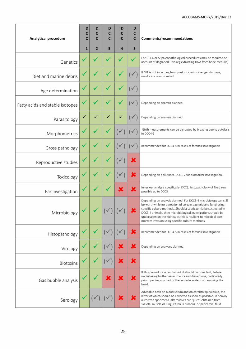
Effects of decomposition code on investigative tests
The recommended suite of tissue sampling for subsequent analysis depends on the
carcass DCC and is summarised in Table 1. See tissue sampling and storage section for
collection information and further procedures. Decisions upon sample collection will differ,
depending upon the analyses planned, laboratories involved or research questions posed.
Table 1 should therefore be seen as guidelines and not a proscriptive protocol.
Table 1 below: Recommendation for tissue sampling considering carcass DCC. Shading: green ✓
indicates the process is of potential use in carcasses of the indicated DCC; grey (✓) indicates that
there may be limitations and red indicates the procedure is not recommended/very unreliable, due
to post mortem autolysis.
ACCOBAMS-MOP7/2019/Doc 33
25
Analytical procedure
DCC 1
DCC 2
DCC 3
DCC 4
DCC 5
Comments/recommendations
Genetics ✓ ✓ ✓ ✓ ✓ For DCC4 or 5: paleopathological procedures may be required on account of degraded DNA (eg extracting DNA from bone medulla)
Diet and marine debris ✓ ✓ ✓ ✓ (✓) If GIT is not intact, eg from post mortem scavenger damage, results are compromised
Age determination ✓ ✓ ✓ ✓ (✓)
Fatty acids and stable isotopes ✓ ✓ ✓ ✓ (✓) Depending on analysis planned
Parasitology ✓ ✓ ✓ ✓ (✓) Depending on analysis planned
Morphometrics ✓ ✓ ✓ (✓) (✓) Girth measurements can be disrupted by bloating due to autolysis in DCC4-5
Gross pathology ✓ ✓ ✓ (✓) (✓) Recommended for DCC4-5 in cases of forensic investigation
Reproductive studies ✓ ✓ ✓ (✓)
Toxicology ✓ ✓ ✓ (✓) Depending on pollutants. DCC1-2 for biomarker investigation.
Ear investigation ✓ ✓ ✓ Inner ear analysis specifically: DCC1, histopathology of fixed ears possible up to DCC3
Microbiology ✓ ✓ (✓) (✓)
Depending on analysis planned. For DCC3-4 microbiology can still be worthwhile for detection of certain bacteria and fungi using specific culture methods. Should a septicaemia be suspected in DCC3-4 animals, then microbiological investigations should be undertaken on the kidney, as this is resilient to microbial post mortem invasion using specific culture methods.
Histopathology ✓ ✓ (✓) (✓) Recommended for DCC4-5 in cases of forensic investigation
Virology ✓ ✓ (✓) Depending on analyses planned.
Biotoxins ✓ ✓ (✓)
Gas bubble analysis ✓ ✓
If this procedure is conducted: it should be done first, before undertaking further assessments and dissections, particularly prior opening any part of the vascular system or removing the head.
Serology ✓ (✓) (✓)
Advisable both on blood serum and on cerebro-spinal fluid, the latter of which should be collected as soon as possible. In heavily autolyzed specimens, alternatives are “juice” obtained from skeletal muscle or lung, vitreous humour or pericardial fluid
ACCOBAMS-MOP7/2019/Doc 33
26
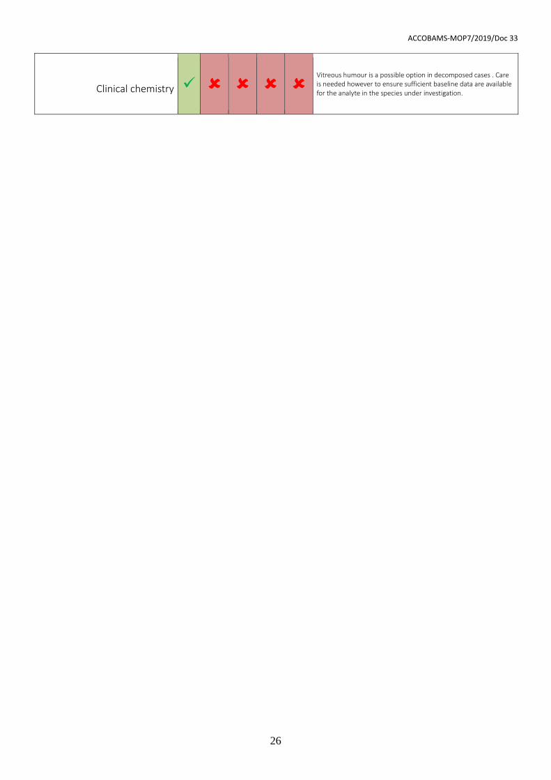
Clinical chemistry ✓
Vitreous humour is a possible option in decomposed cases . Care is needed however to ensure sufficient baseline data are available for the analyte in the species under investigation.
ACCOBAMS-MOP7/2019/Doc 33
27
Tier One: Basic morphometrics, external examination and basic tissue sampling
Data collection and photographs
Scientific value is optimised by careful documentation of systematically collected data and
use of non-ambigious terminology. The use of standardised data sheets and forms is
recommended for field work. In addition to written observations, photographic and video
records of carcasses and the surrounding environment can capture important details such
as the pattern of a mass stranding, traces of predators/scavengers and any markings,
scars or injuries which would disappear soon after death or following carcase removal. In
cases where there are no evident marks, it is still important to take photographs as soon
as possible following arrival on site. Digital pictures and videos can be extremely important
in evaluating human interaction. When photographing/filming wounds suspected to have
been caused by propellers, images should be taken with the objective placed
perpendicular to the axis of the lesions’ surface.
Images support the descriptions of the post mortem report and, in tier three investigations,
aid the pathologist in identifying the sampling area and to connect macroscopic
observations with microscopic evidence. Photographic documentation should include a
general body overview and detailed pictures of main distinctive features. As a minimum, it
is recommended to take lateral overviews of the whole body (both sides), genital slit
region, the head with exposed teeth or baleen, and a cranio-caudal ‘skyline’ image
outlining the silhouette of the epaxial muscles. For those species included in photo-ID
catalogues, additional pictures of identifying characteristics (e.g. of colour patterns and
dorsal fin or fluke) should be taken. Rare species or specimens are especially valuable
and require extra measurements to ensure a complete body of data. The entire carcass
removal to a suitable laboratory or museum for study or preservation should be attempted.
It is recommended to present a case label and ruler/scale bar in images. The label should
(ideally) include the animal identification number, the date of the stranding, the species
and investigating organization together with detail of the lesion/body part. When taking
close-ups, images should also be taken from a wider angle to allow a viewer to
contextualise the image. Care should be taken to minimise shadows, reflections and glare
ACCOBAMS-MOP7/2019/Doc 33
28
and exclude fingers or instruments from the shot. The ruler or label should not occlude
important areas. Particularly noteworthy lesions or features should also be photographed
without any scale or label for potential publication. If the tissue or organ have been
removed from the carcass it is good practice to place on a absorbent background which
minimises blood in the field of view.
Given the storage requirements of high-resolution digital media, it is advised to consider
archiving to a secure location, e.g. cloud- based storage.
Body measurements
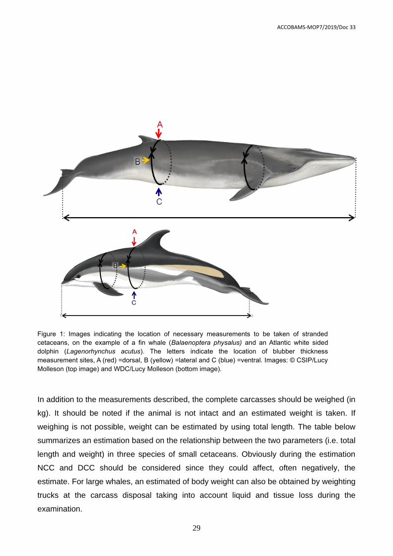
Two principle measurements should be taken: total body length and girth. Measure the
length by placing the animal on its belly (if possible), holding a measuring tape or ruler in a
straight line next to the carcass parallel to the longitudinal body axis. Measuring the
distance between the notch (if present) in the tail fluke and the tip of the rostrum (Figure
1). Measure the girth for DCC1-3 carcasses, in cm, by placing the measuring tape around
the carcass immediately cranial to the dorsal fin without compressing the body. A second
girth measurement may be taken at the level of the axilla, immediately caudal to the
pectoral fin (Figure 1). If it is not possible to take an encircling measurement (e.g. if dealing
with a large whale), take a half girth measurement and double it. Note if the carcass is
bloated, incomplete or otherwise if measurements estimates may not be reliable.
To measure blubber thickness, incise the blubber dorsoventrally along the girth
measurement line at the level of the cranial insertion of the dorsal fin. The blubber
thickness is measured (in mm) at three locations: dorsal, lateral and ventral, as presented
in Figure 1. Make sure to cut perpendicular to the surface of the skin. The epidermal
thickness is not routinely measured. Additional measurements can be taken accordingly,
see Annex 2 for a more extensive measurement collection, which could be applicable e.g.
when dealing with rare species.
Freezing carcases prior to post mortem examination
In circumstances where there is no immediate capacity to undertake a post mortem
investigation, freezing of the carcass is a possible alternative. Chilling the carcass at 0-4°C
for up till 5-10 days (this accounts for small cetaceans) is preferable to freezing, due to
artefacts unavoidably induced by the freeze-thaw process. Such artefacts can mask or
obliterate indications of pathology hence freezing carcases should only be employed
when there is no possibility of examining the carcass fresh or storing it chilled. This should
be clearly stated in the post mortem report.
ACCOBAMS-MOP7/2019/Doc 33
29
In addition to the measurements described, the complete carcasses should be weighed (in
kg). It should be noted if the animal is not intact and an estimated weight is taken. If
weighing is not possible, weight can be estimated by using total length. The table below
summarizes an estimation based on the relationship between the two parameters (i.e. total
length and weight) in three species of small cetaceans. Obviously during the estimation
NCC and DCC should be considered since they could affect, often negatively, the
estimate. For large whales, an estimated of body weight can also be obtained by weighting
trucks at the carcass disposal taking into account liquid and tissue loss during the
examination.
Figure 1: Images indicating the location of necessary measurements to be taken of stranded
cetaceans, on the example of a fin whale (Balaenoptera physalus) and an Atlantic white sided
dolphin (Lagenorhynchus acutus). The letters indicate the location of blubber thickness
measurement sites, A (red) =dorsal, B (yellow) =lateral and C (blue) =ventral. Images: © CSIP/Lucy
Molleson (top image) and WDC/Lucy Molleson (bottom image).
ACCOBAMS-MOP7/2019/Doc 33
30
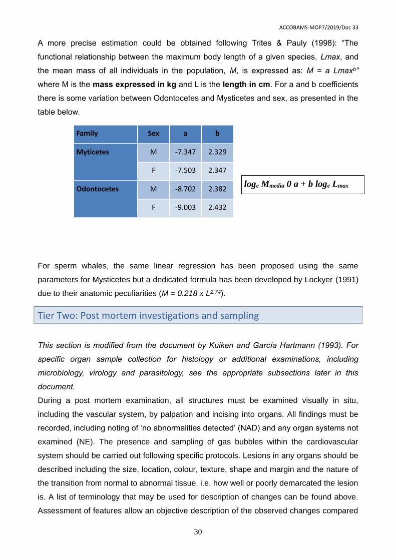
A more precise estimation could be obtained following Trites & Pauly (1998): “The
functional relationship between the maximum body length of a given species, Lmax, and
the mean mass of all individuals in the population, M, is expressed as: M = a Lmaxb”
where M is the mass expressed in kg and L is the length in cm. For a and b coefficients
there is some variation between Odontocetes and Mysticetes and sex, as presented in the
table below.
Family Sex a b
Myticetes M -7.347 2.329
F -7.503 2.347
Odontocetes M -8.702 2.382
F -9.003 2.432
For sperm whales, the same linear regression has been proposed using the same
parameters for Mysticetes but a dedicated formula has been developed by Lockyer (1991)
due to their anatomic peculiarities (M = 0.218 x L2.74).
Tier Two: Post mortem investigations and sampling
This section is modified from the document by Kuiken and García Hartmann (1993). For
specific organ sample collection for histology or additional examinations, including
microbiology, virology and parasitology, see the appropriate subsections later in this
document.
During a post mortem examination, all structures must be examined visually in situ,
including the vascular system, by palpation and incising into organs. All findings must be
recorded, including noting of ‘no abnormalities detected’ (NAD) and any organ systems not
examined (NE). The presence and sampling of gas bubbles within the cardiovascular
system should be carried out following specific protocols. Lesions in any organs should be
described including the size, location, colour, texture, shape and margin and the nature of
the transition from normal to abnormal tissue, i.e. how well or poorly demarcated the lesion
is. A list of terminology that may be used for description of changes can be found above.
Assessment of features allow an objective description of the observed changes compared
loge Mmedia 0 a + b loge Lmax
ACCOBAMS-MOP7/2019/Doc 33
31
to normal anatomical conditions. In case of inexperienced personnel, this approach is
quite simple and along with pictures taken during examination, it could allow advice of
skilled experts.
Photographs of lesions should be taken with a ruler or scale bar as mentioned above.
Representative tissue samples should be placed into fixatives (e.g. 10% neutral buffered
formalin, the most commonly and widely used fixative) for histological examination,
ensuring inclusion of the transition from normal to abnormal tissue and also include tissue
samples with no gross lesions. Samples should be collected for additional testing and
stored appropriately according to the suspected aetiology of any lesion.
Procedures for dissecting and examining carcasses depend on the size and species, but it
is recommended to follow the outlines as reported below. Firstly, procedures as described
in tier one should be conducted. Tier two can be seen as a follow-up process and outlines
are summarised below. Gas examination (and possible subsequent sampling, DCC1-2)
and ears collection (for inner ear analysis, DCC1), as well as samples for microbiological
and virological analyses need to be taken as early as possible to avoid artefacts due to
sectioning, decomposition or contamination and microbial genome degradation by
proteolysis. Furthermore, the brain (and, more in general, the central nervous system), the
ears, the pancreas, the thyroid gland and the liver should be fixed in 10% neutral buffered
formalin as soon as possible for histopathological investigations, due to the rapid post
mortem autolysis affecting these tissues. Care should be taken to prevent cross-
contamination with enteric micro-organisms. Examination of the gastrointestinal tract
should therefore be performed last, unless there is gross pathology in the GIT, when it is
recommended to remove in its entirety and investigate on a separate table or area so that
samples can be taken as soon as possible.
External examination
Photographs, body measurements and carcass condition/state of decomposition are taken
as described in Tier One. Examine the animal for external lesions (including signs of intra-
and inter- species interactions, as well as anthropogenic interactions), taking note of any
penetrating wound, and ectoparasites and sample appropriately. Ectoparasites are most
likely to be found in or near the body openings (including wounds), in crevices or adjacent
to and on the fins and flukes. Take a 2 cm2 piece of full thickness skin, where possible
excluding the blubber layer, and/or muscle for DNA studies. Further samples for skin,
blubber and muscles can be obtained for several post mortem examinations as
ecotoxicological studies, histopathology and stable isotopes analyses. Table 2 and 3 in the
ACCOBAMS-MOP7/2019/Doc 33
32
paragraph for sampling procedures will detail more deeply the possible analyses and
samples conservation.
Examine the oral cavity (including teeth or baleen, tongue, gingiva and lymphatic tissue),
the eyes (for bubbles, evidence of intraocular haemorrhage, asymmetry or swelling),
blowhole, anus, genital slit and mammary slits (when present) for lesions, discolorations
and discharges. Press the skin in the area cranial to the mammary slits in a caudal
direction to express any content present in the mammary glands. If liquid can be extruded
take a sample for other analyses such as toxicology (see sampling procedures). Record
the volume, colour and consistency of liquid. Any abnormalities should also be sampled to
identify aetiological agents.
Nutritional condition state
Assessing the body condition state is an important metric as it provides an indicator for the
ante mortem health of the animal. It should be judged based on the blubber thickness, lipid
composition and back muscle mass. The physiological blubber thickness is difficult to
assess in isolation as it is naturally influenced by a range of factors, including species,
season, region, sex, age, reproductive status and environmental temperature. An
emaciated animal, however, will have lost both fat reserves and muscle mass; this is most
notable in the blubber and lumbar muscles dorsal to the spine. To judge an abnormal
blubber thickness, experience in assessment of the species in relation to its environment is
needed as blubber thickness is physiologically varying strongly according to season, age
and sex. In addition, percentages of lipids can be measured in the blubber layer and could
be an informative descriptor of nutritional condition in fresh cases (DCC1-2).
Based on the state of blubber and skeletal muscle the NCC can be characterised as:
Very good: the animal’s outlining on a cranial perspective is convex; round
appearance caudal to the skull and lateral to the dorsal fin visible; subcutaneous-,
pleural and other visceral fat present; blubber layers are thick.
Good: the animal’s outlining on a cranial perspective is convex; no hollow
appearance caudal to the skull and lateral to the dorsal fin visible; possibly some
subcutaneous-, pleural and other visceral fat present.
Suboptimal: the animal’s outline on a cranial perspective is not fully round; a slight
hollow appearance caudal to the skull and lateral to the dorsal fin is visible (slightly
hollow or almost flat); no internal fat is observed.
ACCOBAMS-MOP7/2019/Doc 33
33
Poor: the animal’s outline on a cranial perspective shows moderate concavity, and
outline of lateral aspects of the vertebrae; a hollow appearance caudal to the skull
and lateral to the dorsal fin is visible; scapula’s can be observed sticking out.
Emaciated: the animal’s outlining on a cranial perspective is very concave and the
lateral aspects of the vertebrae are easily palpable; an extremely hollow
appearance caudal to the skull and lateral to the dorsal fin is visible; scapula can be
observed sticking out; blubber layers are minimal (in small odontocetes <1 cm).
It is recommended to assess NCC for cases in DCC1-3. Post mortem changes will hamper
reliable assessment of nutritional condition in cases in DCC4-5.
Subcutaneous examination
Measure the blubber thickness (as described in Tier one). Record the colour of the blubber
(e.g. white, yellow, pink). Pay attention to the melon and the acoustic fat bodies externally
and internally to the lower jaw. Make multiple incisions into the tissue to check for
haemorrhages. Take samples of blubber and muscle (see sampling procedures).
If possible, position the animal in right lateral recumbency; make a mid-line ventral incision
from the symphysis of the mandible to a short distance posterior of the anus circumventing
the umbilical region, genital slit and anus. From the posterior end of this ventral incision,
make a second incision almost to the dorsal mid-line. Using the line of weakness between
tissue planes, separate skin and blubber from the underlying muscle to remove the
integument from the upper side. Stripping of the blubber layer can also be done strip by
strip in larger animals. During this phase, pay attention to the subcutaneous veins in order
to detect and quantify any evidences of gas bubbles according to specific protocols
(Bernaldo de Quirós et al. 2012). Examine the blubber layer as extensive as possible by
cutting strips and note the colour, presence of any discoloration (e.g. haemorrhages). The
presence of any parasites or lesions in the blubber should be recorded and a
representative sample should be collected (see sampling procedures). Cestoda parasites
may appear as white cysts of less than 1 cm in diameter, often in the ano-genital region or
the dorsal aspect of the chest wall; nematodes may also be found in the subcutaneous
tissue as a result of larval migration patterns. Also examine the subcutaneous tissue for
the presence of bruises and haemorrhages, oedema and/or hyperaemia and different
discoloration.
ACCOBAMS-MOP7/2019/Doc 33
34
Locate the pre-scapular lymph node, cranial to the pectoral fin, and sample for
histopathology as well as for virology and other microbiological investigations.
In females, incise the mammary gland and record the presence of liquid, parasites or
gross lesions and collect/sample these.
The following section describes the key points to note whilst undertaking a post mortem
investigation for cetaceans examined in right lateral recumbency, i.e. by removing the left
flank wall. It is possible that protocols will be adjusted during post mortem examination
depending on circumstances or findings, so the following is intended to serve as a
recommendation and aide-memoire to existing protocols.
Visualisation of internal organs
When opening the body cavities, note any abnormal liquid or lesions and make sure that
such are carefully assessed to be able to establish the origin/aetiology. The anatomical
position of the organs should be verified, paying attention to any displacement, ruptures or
herniation evident. Collect any free fluid in the thoracic and abdominal cavity.
Presence of gas bubbles (in particular in the thorax, mediastinum and peri-renal location)
in the mesenteric veins and lumbo-sacral plexus should be evaluated and quantified
according to specific protocols (Bernaldo de Quirós et al. 2012).
If possible open before the abdominal cavity prior to the thoracic cavity in order to observe
the proper position of the diaphragm and asses the presence of gas in the chest
(pneumothorax). After removing the left abdominal wall, and a gross evaluation of pre-
existing punctures to the thoracic wall, physiological negative pressure can be confirmed
by incising the diaphragm and observing a small caudal displacement accompanied by the
sound of air influx. Collect any free fluid in the thoracic and abdominal cavity for cytology
and microbiology. Collect a representative sample of any parasites (e.g. cysts in
abdominal wall or under the peritoneum).
Examination of abdominal organs (except GIT)
Care should be taken to avoid contamination of the carcass with gastro-intestinal tract
(GIT) contents: preferably by tying off the oesophagus and rectum to prevent leakage of
content and removing the stomachs, mesenteric lymph nodes with intestines, pancreas
and spleen. Pancreas should be removed, macroscopically examined, checked for
parasites, and fixed in 10% neutral buffered formalin as soon as possible due to rapid
decomposition (chemical/enzymatic post mortem autolysis). Assess the spleen and
ACCOBAMS-MOP7/2019/Doc 33
35
sample splenic tissue for any microbiological and virological examination. Note any
accessory spleens and presence of any gas bubbles in the mesenteric vasculature or
spleen. Examination of the GIT can be left to the end of the post mortem examination or
perform it in a completely separate area to prevent contamination of other tissues with the
enteric microbiota. Sample spleen for microbiology, virology and histopathology.
Examine the urinary bladder in situ, and sample any urine present with a syringe, noting
the volume, colour, turbidity and nature of any contents present. This sample could be
useful for microbiology. Freeze urine for biotoxins examination at -20°C. If uroliths are
detected these can be frozen at -20°C prior to analysis.
In females, remove the entire reproductive tract and open the vagina and uterus
examining and sampling any stones, mucosal alternation or discharge. The reproductive
history of cetaceans can be informed by analysis of ovarian tissue. Record the presence
of any corpora lutea and/or albicantia or follicles on each ovary and then store the ovaries
in 10% neutral buffered formalin, with some transverse incisions to allow proper fixation. If
unfamiliar with the techniques to assess ovarian scarring, assessment of scars can be
done at a later stage. Ensure to differentiate left from right ovary during fixation, e.g. by
making a small transvers incision in one side. Additional sampling of the reproductive tract
of females for histology and microbiology should be conducted when abnormalities are
observed.
In males, remove the testes, check for symmetry and weigh them, noting if epididymal
tissue is included in the weight, and measure the long axis length. Testes size is correlated
to sexual maturity and activity. Make cross-sectional incisions at 1 cm intervals to
examine. Store the testes appropriately for reproductive studies. Examine the genital
opening, penis, epididymis and accessory glands. Additional sampling for histology,
virology and microbiology should be conducted if abnormalities are observed.
In pregnant females, where the foetus can be easily palpated, photograph the uterus with
a scalebar in the frame or measure the uterus. Note which uterine horn carries the foetus
and the foetal position to identify possible dystocia. Note if the cervix is dilated. Describe
the consistency and turbidity of the amniotic fluid and sample it for microbiological analysis
if required. Twins are very unusual. When the foetus is of sufficient size to examine the
individual organs, a post mortem and tissue sampling (especially including for toxicological
and virological tests) should be performed. Foetal stomach contents may be collected bot
bacteriological culture. If the foetus is too small for a full post mortem examination, the
whole foetus and its placenta can be collected for histology, or frozen for virology,
ACCOBAMS-MOP7/2019/Doc 33
36
microbiology and toxicology, or fixed and kept as a museum collection. Always note the
sex, weight, length and if the DCC of the foetus more advanced than the dam.
Remove and examine the adrenal glands, make a transverse section and measure the
cortico:medulla ratio directly or photograph the cross section through the mid-adrenal
gland with an appropriate scale bar. Collect a cross-sectional slice approximately 0,5 cm
thick of the centre of both adrenal glands for histological examination. Slice and
investigate the adrenal glands for presence of cysts, nodules and/or other abnormalities.
Before removing the kidneys from the body cavity look for any anomalous gas evidence
around them in the perirenal space and in the associated vasculature. Remove them from
the body and incise the kidneys longitudinally and check for parasites in the renal
vasculature. Sample 1 cm3 of tissue from each kidney for histological examination (if no
gross lesions), ensuring to collect at least one entire renule, and for toxicology and
microbiology. Further samples should be obtain fresh or frozen for microbiology and
contaminants. Assess the urethra for any abnormalities and sample as appropriate.
Remove the liver, examine all surfaces and make multiple incisions into the parenchyma.
Record the colour and consistency and distribution of any abnormalities Record any
nodules and other abnormalities. Examine the bile ducts for abnormalities and parasites
(note that cetaceans do not have a gall bladder). Take frozen samples for ecotoxicological
examination, virology, microbiology, parasitology and stable isotopes analyses. Sample for
histopathology from two regions of the liver and from any gross lesions.
Examination of GIT
In order to avoid any possible contamination from the GIT content tie up both ends before
extracting it from the abdominal cavity. If micro- and/or nano-plastic studies are
considered, reduce possible environmental contamination through particulates in the air,
tools, clothes or other plastic objects. Consider possible sampling for microbiology as first
step that should be carried out as soon as possible before any other procedure.
Locate and examine the mesenteric lymph nodes and sample a 0,5 cm thick cross-
sectional slice from halfway along its length for histological examination.
Separate the stomachs from the intestine by ligating the GIT after the ampulla
duodenalis. Note that cetaceans generally have three stomach compartments, but there is
species-specific variation in compartment numbers. In general, the following three
compartments are present: the forestomach (cardiac), the main stomach (fundic or
glandular) and the pyloric stomach.
ACCOBAMS-MOP7/2019/Doc 33
37
Record the presence and amount of any ingested prey species, digesta, otoliths (these
can be stored frozen), and parasites. Take note of evidences of marine litter by recording
and weighting the presence and type of any macro-plastics or other ingested marine
debris in the stomachs and retain for future studies. A better evaluation for marine litter
ingestion should be carried out by washing gastric surface and stomach contents using
proper mesh size that can help in separating diet and parasites from foreign bodies. A
size of at least 1 mm is recommended in order to separate macro and meso-plastic which
be later classified using the MSFD master list (TC D10 MSFD, 2019). Any macroparasite
should be also recorded and a representative sample should be collected from each
stomach compartment. Describe any gross lesions, including the distribution and size of
any ulcerations for each separate stomach compartment. Ideally the fluid resulting from
the washing should be preserved to be further filtered using meshes till at least of
minimum 500 μm size for diet studies, 250 μm for marine litter and adult parasites (DCC 1-
3).
Open the intestine throughout the entire length. Check for lesions, foreign bodies and/or
parasite presence, and record and sample these. Note the colour and consistency of the
content. In very fresh cases, samples of intestinal content could be taken for
microbiota/microbiome studies otherwise it should be analyzed as the stomach for marine
litter and parasitological examination. A sample of stomach contents and faecal material
could be collected for parasitological studies and algal biotoxins. Several section of
intestinal should be opened and fixed in formalin
If GIT examination cannot be undertaken immediatly, collect the entire GIT (including all
content, like fish bones, otoliths and all other food or non-food remains for additional
studies into prey and/or marine debris ingestion) and freeze at -20’C for subsequent gross
pathology, diet, parasitology, debris and biotoxins analysis. Specific protocols exist for the
collection of samples for micro- and nano-plastic ingestions.
Examination of neck region, and thoracic organs
As stated above, before opening the thorax, asses the presence of negative pressure in
the chest. Open it by cutting the ribs and check for possible bones fractures. Locate the
larynx and note its position. Check for foreign bodies in the oral cavity. Note the presence
and colour of any foam and/or obstruction (e.g. foreign bodies). Free the larynx from the
sphincter muscle that holds it in place, and, whilst pulling the tongue caudally incise along
the neck and free the trachea. Check for foreign bodies in the tracheal lumen. Free the
heart and lungs from the thoracic cavity checking for adhesions between lungs and
ACCOBAMS-MOP7/2019/Doc 33
38
thoracic wall/diaphragm and the presence of any anomalous gas presence in the
mediastinum or in the visceral pleura (bubbles). Note and sample any free fluids in the
pericardial sac and thoracic cavity. Pericardial, pulmonary and muscle fluids can be
used as a substitute for serum: for cases in moderate decomposition condition or when
serum cannot be sampled, collect fluid from the pericardial sac prior to opening the
pericardial sac, e.g. using a syringe, avoiding contamination with blood. Fluid from lungs
can also be collected.
Examine the ribcage for fractures or bruising and, ventral to the thoracic spine, the rete
mirabilis for evidence of haemorrhage.
Examine the surface of the tongue. Note the presence of neonatal papillae and incise into
the tongue muscle for examination. Examine the lymphatic tissue of the tonsillar ring and
sample any abnormalities for histology.
Open the oesophagus longitudinally and check for lesions, foreign bodies, food remains
and/or parasite presence; record and collect these. Examine and fix the thyroid for
histology and freeze a section if required.
Open the larynx, trachea and major bronchi longitudinally and check and record for
lesions, foreign bodies, including sand silt or mud, foam, fluid, aspirated ingesta. Observe
and sample for histology and virology the tonsils at the passage between the larynx and
trachea.
Note any profound asymmetry in the size or congestion between either lung. Make
multiple incisions into the parenchyma of both lungs. Note level and distribution of any
parasite burden in pulmonary parenchyma and take a representative sample. Collect
swabs for microbiology from any evident pathological changes. A minimum of two pieces
of each lung, one from the hilus (tracheal bifurcation) and one from near the surface
including pleura should be taken for histological examination. Care should be taken to
ensure buoyant samples are fully immersed in formalin, for example by placing tissue
paper on the surface. The samples should include part of the major bronchial tree.
Further frozen samples should be collected for ecotoxicological examinations (i.e.
biomarkers and/or contaminants), virology and microbiology. For neonates, small pieces of
lung can be dropped in water to test floatation to determine if the lungs ever inflated
Open all major branches of the pulmonary blood vessels and examine and record the
presence of parasites. Examine the bronchial and pulmonary associated lymph nodes.
The latter can be found approximately halfway along the ventral edge of each lung. Cut a
ACCOBAMS-MOP7/2019/Doc 33
39
1 cm thick cross-sectional central slice of the left pulmonary associated lymph node,
including a small amount of the adjacent lung tissue attached, for histological examination,
microbiology and virology.
Examine the structure and extent of the thymus and sample as required. Age-related
thymic atrophy is normal but histopathological assessment can be useful to assess
potential immunotoxic pollutant-related or any other pathology. Note the relative degree of
atrophy and whether any cysts are present.
Collect any blood present in the heart ventricles (or elsewhere) and centrifuge at 1,000-
1,500 rounds/8-10 minutes for sero-epidemiological and/or virological investigations. With
advanced time after death or the freezing process will make it difficult to obtain serum .
Nevertheless, it is very important to collect and archive blood for different purposes (incl.
toxicology, parasitology, algae toxicology, PCR).
Before separating the heart from the lungs, check the heart for any evident gross
malformation and for gas bubbles within the coronary veins and score them; then cut
through the major blood vessels at the base of the heart. Examine the myocardium by
cutting into the right and left papillary muscles and the septal myocardium. Check for
evidence of petechial haemorrhages in the epicardium and myocardium. Open the left and
right ventricles, atria and thoracic aorta for examination and record and collect a
representative sample of any parasites present. Check to ensure no defects/ holes in the
septum. Rinse the heart and weigh it. Sample heart tissue and include a piece of both
ventricle walls and atrioventricular septum, including papillary muscles, for histological
examination. Record any gross defects and then sample for histological examination. In
case of neonates, assess whether there are any abnormalities in the foetal/neonatal
cardiac anatomy (e.g. foramen ovale, patent ductus arteriosus).
In case an animal’s total body weight could not be established, methodologies based on
the relationship between the heart weight and total body weight can be used to get an
estimation: log W = (log H + 2.2) / 0.984, with H = heart weight and W = body weight, both
in kg (see: Brody 1945).
Examine the lumen and walls of veins and arteries looking for thrombi and parasitic
infections, in particular in fin and beaked whales, or other lesions. If necessary sample
them for further investigations. Remember to look for gas bubbles and to score them
according to the previously cited protocol.
ACCOBAMS-MOP7/2019/Doc 33
40
Examination of the head
The eyes should be examined (check for hyphema) and collected (histology) with optical
nerve attached.
Each ear bone should be removed carefully as soon as possible. Note any signs of
bleeding or trauma in the surrounding tissue and external ear canal. Examine the ear
sinuses, the Eustachian tube, the nasal sacs and sinus cavities. After ear extraction and
parasite collection, rinse each ear cavity with water to assess the severity of the parasitic
burden. Use forceps to collect a representative sample of any parasites present. In
carcasses in DCC1-3, ears (tympano-periotic complex) should be removed for
examination of infectious diseases, degenerative and traumatic changes and further
microbiological, parasitological, virological and histological investigations of the middle and
inner ear as well as surrounding tissue should be conducted to allow further understanding
of the health of the ear apparatus as well aetiology of lesions.. With electron microscopy
(EM) and immunofluorescence techniques, DCC1 inner ears can be assessed for potential
degeneration and death of hair cells and associated innervation, especially in relation to
acoustic trauma. The inner ear should be fixed following these steps by order:
1) removal of the stapes by tissue forceps,
2) Perforation of the round and oval window membranes with a needle
3) Slow perfusion of fixative into the structures of the inner ear (10% neutral buffered
formalin or 4% paraformaldehyde, pH 7-7.4) with minimal pressure through one window
until the fixative seeps through the other window, using a soft catheter (or the tip of a
plastic pipette) and a 1ml syringe.
See more details on the perfusion protocol in Morell and André (2009) and Raverty et al.
(2018). Then, the ears should be immersed in the fixative solution. Whilst unsuitable for
ultrastructure imaging, the ears of carcasses in DCC2-3 should also be collected for
histological assessment to investigate hearing damage.
Examine the melon through serial sections to assess any trauma and/or inflammatory
lesions. Include upper airways assessing presence of foreign bodies, parasites or
discharge. As already stated, look for any haemorrhages in the acoustic fat bodies (i.e.
internal and external). For odontocetes, examine the teeth. Note tooth wear, integrity of
dentition and state of gingiva, number and position of teeth. Remove at least 4 intact teeth
(if present) from the middle of the lower jaw for age determination. Check for swelling and
fractures in or around the jaws or other changes in the jaw articulations. For mysticetes,
ACCOBAMS-MOP7/2019/Doc 33
41
examine the baleens for gross lesions, parasites and trauma, and collect up to six baleen
plates ensuring each is removed as close to the gum line as possible. Teeth and baleen
can be used for life history and stable isotope analysis either dried and stored at room
temperature, or frozen, depending onto preferences of laboratories involved.
Sample the cerebrospinal fluid (CSF) before removing the head and opening the skull to
examine the brain. In detail, remove the overlying soft tissue at the back of the head and
neck, to gain access to the atlanto-occipital joint (foramen magnum). Then, to obtain a
sample suitable for all type of analyses (microbiological, cytological, sero-epidemiological
and biomolecular) flame the site using a butane torch for one or two seconds. It is
suggested to use a syringe with a sterile needle of appropriate length (i.e. 1.3 x 88mm) or
a sterile Pasteur pipette. Sample the spinal cord for histopathological, microbiological and
biomolecular analyses.
Except for sperm whales, the skull can be opened by sawing a vertical cut parallel to the
transverse dorsal ridge (visible and palpable on dorsum of the skull) paying attention to not
cut the meningeal and cerebral tissues. Two further cuts should be made in the horizontal
plane through the occipital condyles, making sure to leave the posterior portion of the
condyles on the skull. Both cuts should be extended until they meet each other. The
separated piece of skull can then be removed using a chisel, flat bladed screwdriver
and/or a ‘skull cracker’. Care should be taken to avoid human exposure to aerosols during
this process.
Remove the brain from the skull and make an incision through the brain and check for
ventricular dilation. Note the colour and turbidity of any CSF and the structure of the
choroid plexuses Cut the whole brain longitudinally by means of a sagittal, paramedian
section, thereby collecting the smallest portion for virological, microbiological and
ecotoxicological investigations examinations and the largest portion of it for
histopathological examination (Annex 3). For neuroanatomical studies, place the
remaining whole brain, including the pituitary gland which can be found caudoventrally to
the optic chiasm within the cranium. Once fixed in 10% buffered formalin, make multiple
slices into the brain tissue. The sampled brain should be processed using large tissue
cassettes (if available), to allow full lesion profiling throughout the sample. Skulls can be
retained for morphometric studies.
ACCOBAMS-MOP7/2019/Doc 33
42
Examination of the skeletal system and rete mirabilis
A representative sample of different muscle locations and diaphragm should be taken for
histology, toxicology, parasitology etc. All bones should be investigated macroscopically
for changes (e.g. fractures, inflammations, degenerations). Samples from humerus, lumbal
vertebrates, ribs and lesions should be preserved for histology and infectious
investigations as well toxicology (frozen or formalin for histology). Humerus could be also
retain for diatoms investigations (Rubini et al., 2018) to support any diagnosis of drowning.
Rete mirabilis should be taken for histology.
ACCOBAMS-MOP7/2019/Doc 33
43
Tissue sampling procedures, storage and analysis
Morphometric studies and imaging
If the capacity exists, assessing carcasses by Computed Tomography (CT), DMX (bone
density) and/or Magnetic Resonance Imaging (MRI) prior to post mortem examination will
allow imaging and evaluation of a range of morphometrics and identification of potential
pathologies. CT-scanning will allow visualisation of skeletal anatomy and lesions (e.g.
trauma, shot injuries), assay bone density, assess joints and inter-vertebral discs. MRI
allows the assessment of internal anatomy (including position of organs), all soft tissues
and many potential pathologies. In addition, the collection and maceration of skeletal
material, or the entire skeleton is of value for stable isotope analysis, morphometric studies
or accession to museum collections. Definitive diagnosis of cause of death should
however take preference over maintaining skeletal integrity for museum collections, and
histology samples should be taken of any bone abnormalities that may be present in an
animal.
Tissue sampling and storage
The nature and storage requirements for samples taken for subsequent analysis depend
on the requirements of the testing laboratory, and it is advised to check local requirements
and protocols prior to sampling. As stated in Paragraph 5, national CITES permits should
be assessed before collecting, preserving and exchanging tissues sampled from stranded
cetaceans.
In general however, the following principles apply:
Swabs or tissues stored for subsequent microbiological/virological assay should be
collected as soon as practical, as aseptically as possible and with an appropriate
cold chain between sampling and testing. If it is not possible to perform
microbiological analyses immediately, the use of an appropriate transport medium
(depending on the pathogen) or freezing (in particular for virology) could be an
option. Some viruses or bacteria could request specific sampling and/or preserving
procedures.
Tissues for microscopic examinations should be fixed in 10% neutral buffered
formalin. Possibly a commercial solution to ensure standardised quality (i.e. pH,
ACCOBAMS-MOP7/2019/Doc 33
44
formalin concentration) should be used. If not available, characteristics of the
preservative should be regularly checked. Samples should not be thicker than 1 cm
and the ratio sample:formalin should be 1:10 and a plastic box with proper volume
and dimension should be used. Formalin should be changed after 24 hours
(especially for fatty tissues such as brain) and samples should be kept less than 72
hours in formalin for immunohistochemical analyses against infectious pathogens
and/or selected tissue (bio)markers. Histological samples that can be processed are
generally between 3-5mm thick.
Samples for subsequent assay for persistent organic pollutants should be wrapped
in aluminium foil to prevent sample contamination from plastic in the storage
containers. For heavy metals plastic bags could be used. Sampled tissues should
then be frozen at -20 °C or colder.
Most samples destined for DNA molecular testing can be stored frozen or in 70%
ethanol; however, samples collected for other genomic, metabolomics or proteomic
work are likely to be more labile and require specific transport and storage media.
Tissues stored for cell culture in an appropriate medium (depending on the tissue)
should be collected as soon as practical and as aseptically as possible, with
appropriate conservation between sampling and testing.
A summary of the minimal samples set to be collected during cetaceans’ post mortem
investigations is included at the end of the document.
Labelling of samples
Samples from each animal are examined by a number of people, often in different
laboratories, under different conditions, and therefore it is vital that they are properly
labelled for traceability, also taking into account long-lasting labelling and labelling in
freezers (e.g. up to -80°C). This requires that:
Every animal should be assigned a unique reference number;
Every sample/ sample container should have a firmly attached label;
Permanent ink or pencil should be used to write labels;
Labels should include the unique reference number and tissue sample identifier.
It should be noted however that permanent ink could come off due to freezing/defrosting or
when submerged in formalin. Overlying these with transparent tape may prevents this.
ACCOBAMS-MOP7/2019/Doc 33
45
Tissue and biological specimens’ archive
To ensure availability of tissues for future studies, it is recommended to store samples in a
sample archive. The creation of an official tissue bank for the stranding network is strongly
recommended since it could preserve different tissues and fluid, and it could obtain CITES
accreditation supporting exchanges of samples. An effective sample archive is very
important when working with rare species, so that a larger sample size can be built up over
time, but also as new pathogens may be detected in the future. With availability of a range
of samples from multiple individuals and species, long-term medical, ecological and
population studies can be facilitated.
It strongly depends upon the available facilities and request by the local laboratories
conducting further analysis, but in general, all tissues and body fluids should be sampled
and stored at -80°C preferably and/ or at -20°C. Formalin-fixed tissues should be stored
for a limited period only in proper cabinets and paraffin embedding is highly
recommended.
As a minimum, we recommend to sample at least: lung, liver and spleen for
microbiological assessment (at -20°C, or -80°C or colder for ecotoxicology/biomarkers)
and brain, lung, kidney and spleen for virology and microbiology (-80°C or colder).
Additionally, we recommend storage of blood and/or serum, CSF and other possible fluids
collected at -80°C (or colder) as a minimum.
Age determination
Teeth collection: A longitudinal cross section through odontocete cetacean teeth reveals
annual growth rings which can be used to determine age. To ensure all rings are visible on
cross section, a minimum of four whole, undamaged or least worn teeth need to be
collected from each individual for repeated estimates if necessary. Preferentially take the
teeth from the middle of the lower jaw as these are usually the straightest and make age
determination more accurate. From the harbour porpoise, teeth can be extracted easily by
inserting a sharp knife or scalpel either side of the gums in-between the teeth and the
connective tissue. In species such as the bottlenose dolphin, teeth can be loosened by
initially levering the space between the tooth and the socket with a sharp pointed
instrument. Dental elevators are useful tools for extracting teeth. If extracting of teeth is not
possible, a piece of mandible containing at least 4 teeth should be collected. In baleen
whales, age could be estimated analysing baleen plates or earplugs.
ACCOBAMS-MOP7/2019/Doc 33
46
Storage: Loose teeth, jaw sections, baleen and earplug can be stored frozen. The
temperature at which they are frozen is not critical. Alternatively, for short term storage,
they can be fixed in 70% ethanol. They should not be stored dry as they may dehydrate
which may lead to cracking of the teeth.
Diet analysis, marine litter and micro- /nano-plastics
The best practice is to sample the entire GIT (unopened) from the oesophagus to the
rectum, to ensure all content is collected. If this is not feasible the single parts (i.e.
oesophagus, gastric chambers and intestine) could be separated maintaining them close
by using strings or cable ties. The GIT is then placed on a non-plastic surface or grid for
microbiological sampling, gross examination of ingesta and digesta, pathological changes,
parasites and marine debris. Ingesta and digesta should as well as the GIT walls should
be washed and filtered through different mesh size to separate macro and meso plastic
(useful samples up to 1 mm, micro-plastic (up to 50 µm if possible), diet (up to 500 µm)
and parasites (up to 250 µm). The remaining fluid could be also preserved for nano-plastic
investigations.
If not possible examining the GIT immediately, the whole GIT or single parts could be
stored frozen closed for subsequent analysis for the presence of macro- and microplastics,
dietary analysis and parasitology which can be undertaken concurrently. Samples for
microplastic analysis may also require samples taken from the air, gloves, clothes, water
pipes and all the other plastic materials used during sampling and analyses at the time of
sampling to act as a negative control. The temperature at which the GIT is frozen is not
critical.
Item characterisation: Material should be categorised as rope, net, floats, monofilament,
braided line, hooks, packaging, cigarette butts, plastics, and other anthropogenic material.
Size, shape (image analysis of digital photographs), mass, volume, and polymer type if
plastic (e.g., Raman spectroscopy or Fourier transform infrared spectroscopy, FT-IR)
should all be recorded, and all evidence should be identified as to source using
established techniques.
Marine Litter will be later classified using the MSFD master list (TG D10 MSFD, 2019).
ACCOBAMS-MOP7/2019/Doc 33
47
Genomic (DNA) studies and RT-PCR ecotoxicological investigations
DNA is most readily recovered from skin or muscle even if other tissues could be used.
These samples can be stored frozen at -20°C or in 70% ethanol. Facilities permitting,
storage of duplicate samples in RNAlater at -80°C enables more advanced genetic
sequencing techniques to be employed. Use RNAlater® Solution with fresh tissue only; do
not freeze tissues before immersion in RNAlater® Solution. Before immersion in
RNAlater® Solution, cut large tissue samples to ≤0.5 cm in any single dimension. Place
the fresh tissue in 5–10 volumes of RNAlater® Solution. Most samples in RNAlater®
Solution can be stored at room temperature for 1 week without compromising RNA quality,
or at –20°C or –80°C indefinitely. Do not freeze samples in RNAlater® Solution
immediately; at 4°C overnight (to allow the solution to thoroughly penetrate the tissue),
remove supernatant, then move to –20°C or –80°C for long-term storage.
Reproduction studies
Female gonads: Both ovaries should be placed, whole, in 10% neutral-buffered formalin
or other fixatives if available or preferable. It is vital to differentiate between the left and the
right ovary; this can be done by using separate containers, or, if storage does not allow
this, by making a small incision in the left ovary before collecting them together in a
container.
Male gonads: Each testis should be weighed separately after removal of the epididymis.
Subsequently, place a cross-sectional slice approximately 1 cm thick from midway along
the organ’s length in 10% neutral-buffered formalin or other fixatives if available or
preferable. Fix at ratio of 1 volume tissue: 10 volumes fixative and once fixed, after about
24 hours, the samples can be stored in a smaller volume of fixative.
Histopathology and immunohistochemistry samples
Collection: In grossly normal organs take a random cross-sectional sample of 1 cm unless
specified otherwise. Please note that samples that are processed for histology are
generally 3-5mm thick. All gross lesions should be sampled to include an adjacent piece of
normal tissue, if present. Always use a sharp scalpel to take the samples. Careful handling
of tissues for histology is important to avoid squeeze artefacts, in particularly in soft and
delicate tissues as brain, spinal cord, pancreas, lymphoid organs and GIT mucosa.
ACCOBAMS-MOP7/2019/Doc 33
48
Individually labelled containers/permeable bags (e.g. tea bags) should be used to aid
differentiation when multiple samples are taken from macro-microscopically similar organs
(e.g. lymphoid tissues) and to be able to identify samples and lesions after fixation.
Fixation: 10% neutral-buffered formalin (4% of formaldehyde) is the most used fixative.
Formalin is classified by the EU as a carcinogen and the necessary precautions to avoid
direct contact and exposure must be taken. Other fixatives are now on the market to
ensure human safety, but there is lack of experience in regards to their use for tissues
examined by immunohistochemistry or in-situ hybridization. Formalin will only penetrate
approximately 1 cm/day in any direction (tissue depending), so perfuse or slice samples no
larger than 1-1.5 cm in diameter to avoid or slow down autolysis of the inner tissue
portions. Use formalin at a ratio of 1 volume of tissue: 10 volumes of fixative. The tissues
should remain in this fixative for a minimum of 24-48 hours (except the brain, see below) at
room temperature. Longer time could affect immunohistochemical studies. If tissues have
a high blood content, the fixative should be changed each 24 hours until it is no longer
discoloured. Additionally it is recommended to inject formalin in eyes and intestine, to
ensure proper fixation.
The brain should be placed in 10x as much formalin as tissue when fixing the largest
portion of brain (see Annex 3), and should remain in this fluid for at least a week. To allow
faster fixation, several transverse/coronal incisions should be made through the frontal
lobes to expose the lateral ventricles. Another possibility is to freeze the brain for one hour
to make easier coronal complete cuts of 1 cm; slices should be then separated by paper
sheets and put in 10% neutral-buffered formalin. This facilitates a more rapid penetration
of fixative into all brain areas. In really fresh cases, ensure that the brain has cooled to
near room temperature prior to immersion in fixative to avoid artefacts such as dark
neurons and contraction of arterioles.
Storage: Once fixation has taken place, the samples may be stored in a smaller volume of
10% neutral buffered formalin. If immunohistochemistry may be required, store tissues in
70% ethanol. If the time to processing is in excess of three months: change to formol
saline at this point to avoid saponification. To minimise fixation artefacts it is recommended
that neural tissue samples are processed within three months. Samples stored for years
could limit diagnostic value of immunohistochemistry. Store the histology samples at room
temperature and consider national legislation on workers safety related to the
management and storage of formalin. If it is necessary to preserve formalin fixed samples
for long periods, paraffin embedding is the better option.
ACCOBAMS-MOP7/2019/Doc 33
49
Microbiology
Sampling for microbiological diagnostics should be made in collaboration with the
laboratory undertaking the processing of the samples. Preferably, microbiological
examination should be carried out on a minimum of tissues including samples of lung,
liver, spleen, kidney, brain, CSF and any gross lesions of suspected microbiological
aetiology.
Collection: Ideally, samples should be taken using an aseptic technique whenever
possible. Tissue samples (solid or liquid) or swabs, such as nylon flocked swabs, are both
appropriate for microbiological examination. Each has advantages and disadvantages and
the choice depends on the preference of the person performing the microbiological
analysis and also the target microorganism(s) e.g. for the diagnosis of neurobrucellosis by
Brucella ceti.
For a solid tissue sample, cut a block approximately 4x4x4 cm using a sterile scalpel and
forceps and place this into a sterile container. Samples should be kept at 0-4°C until
processed, which should be carried out preferably on the same day or at least within 24
hours after collection. If analysis is not possible <24 hours, samples could be frozen at -
70°C, or preferable at -20 °C until further processed, to allow the subsequent growth of
fastidious organisms.
For swabs, sear the surface of the organ (e.g. with a hot blade), then incise with a sterile
scalpel and place a sterile swab into the incision without contaminating it with surrounding
tissue. Then, directly apply the sample onto culture medium or insert the swab into a tube
containing transport medium and store at 4°C until processing. Swabs should be
processed within 24 hours. If this is not possible, samples of solid tissue are preferred.
For diagnosis of neurobrucellosis by Brucella ceti, CSF is the sample of choice, collected
collected aseptically with a syringe with a sterile needle of appropriate lenght (i.e. 1.3 x
88mm) or a sterile Pasteur pipette..
Processing: This should be undertaken after consultation with the
pathologist/microbiologist, as different media and methods are employed dependant on
the suspected target microorganisms. Many pathogens require selective growth media or
specialised containment due to their zoonotic potential, for example Brucella spp. or
Mycobacterium spp..
ACCOBAMS-MOP7/2019/Doc 33
50
Virology
Collection: The choice of sampling for virology diagnostics should be made in collaboration
with the laboratory undertaking the processing of the samples, taking into account the
target virus, the (clinical and) gross lesions of the stranded animal(s) and the state of
decomposition of the carcass. The tissues listed in these guidelines- lung, spleen, brain
and kidney - are a good range of samples required for several known viruses, e.g.
morbillivirus, influenzavirus, poxvirus, and herpesvirus detection. Any lesions of suspected
viral aetiology should be sampled similarly. For virus isolation, samples of 1x1x1 cm from
the relevant organs should be collected aseptically and placed into sterile containers with
viral transport medium (VTM).
Storage: If samples are collected for molecular detection of pathogens by PCR and are
submitted to the laboratory within 24 hours of sampling, they should be stored at 0-4°C,
otherwise they should be frozen at -80°C before being submitted to analysis. If freezing at
-80°C, or even at -20°C, is not possible, then the samples can be placed in RNAlater and
stored at -20°C (or even at room temperature for up to two weeks). Frozen samples can
be transported worldwide on dry ice (solid CO2) or using biofreeze bottles. For situations
where large number of samples are being collected in the field or transported distance, it is
advised to use an IATA approved LN2 Vapour/Dry shipper.
Sero-epidemiological investigations against viral and other infectious agents require a
blood sample of at least 7 ml collected and centrifuged to obtain serum. Serum should be
separated from whole blood as soon as possible and stored at -80°C. Even if the serum
shows some evidence of erythrocytic haemolysis, it can still be of diagnostic value. Where
a blood sample is unobtainable, a sample of pericardial fluid (PCF) and/or vitreous humour
and/or lung and/or muscle tissue juices can be suitable substitutes, with special emphasis
on consistently autolysed individuals. Furthermore, in well-preserved animals it is highly
recommended to collect as soon as possible during the post mortem examination cerebro-
spinal fluid (CSF) samples, which may allow an ad hoc search of antibodies against a
range of neurotropic pathogens (e.g. Cetacean morbillivirus, herpesvirus, West Nile virus,
Brucella spp., Toxoplasma gondii, etc.).
Parasitology
Parasite specimens should be isolated from the organs and cleaned from blood, dietary
residues and adhering tissue in tap water (e.g. with a soft paint brush). Then preserve the
ACCOBAMS-MOP7/2019/Doc 33
51
specimen in water in case of immediate analyses otherwise in 70% ethanol, or alcohol with
10% glycerine, or freeze it.
If the total number (intensity) of parasites cannot be recorded, a semi-quantitative
assessment of the level of parasitic infection into none = no parasites visible, mild,
moderate or severe infestation should be recorded for each organ. Documenting infection
and associated lesions through photographs and histopathology is recommended.
Furthermore, the macroscopically detectable severity of lesions associated with parasites
should be recorded (no lesion, mild, moderate, severe) and the tissue sampled in 10%
neutral-buffered formalin for histopathological examination.
Toxicology
Collection: The choice of sampling for toxicological assessment should be made in
collaboration with the laboratory undertaking the processing of the samples. It is
recommended to duplicate samples of blubber, muscle, liver, kidneys and brain and collect
these for persistent organic pollutant (POP), plastic additives and trace elements and/or
fatty acids analysis. Samples collected for POP analysis should only come into contact
with stainless steel, aluminium, glass or Teflon and are most conveniently stored in
aluminium foil (shiny side out; do not use recycled foil as this might contain plastic
particles) with an additional sample in plastic.
For lactating females, a milk sample should be collected. This must be stored in a glass
container. If the container has a plastic cap, it is recommended to cover the opening with
aluminium foil (shiny side out) to keep the sample from contacting the (plastic) cap, as this
could invalidate the results of POP analysis.
Samples of brain, muscle, liver and kidney can be sampled for trace element analysis.
These samples should not come into contact with any metals other than stainless steel
and thus can be stored in plastic containers. Bone and/or blood samples are also suitable
for heavy metal analysis.
If a foetus is present but too small for full post mortem examination, the whole foetus and
(parts of) its placenta can be wrapped in aluminium foil (shiny side out) for POP analysis.
For foetuses that can be dissected, if is recommended to conduct a full post mortem
investigation with subsequent tissue sampling as done for other cases.
Storage: The minimal size of samples for trace elements and organochlorine analysis is
10g of solid tissue or 10 ml for milk. Samples should be stored frozen at -20°C until
ACCOBAMS-MOP7/2019/Doc 33
52
analysis. For DCC1-2, samples should be stored at -80°C in liquid nitrogen or RNA later
for Real-Time (RT) PCR.
Biotoxins
Collection: Biotoxin samples collection is highly recommended particularly in case of
unusual mortality events, mass mortalities and when a harmful algal bloom (HAB) is
suspected in the surrounding area and/or the live animal exhibited neurological symptoms.
Biotoxin samples include tissues and fluids such as: liver, kidney, brain, muscle, serum,
aqueous humour, stomach contents, intestinal contents, faeces, urine. Tissue samples can
be stored in plastic bags. Stomach and intestinal contents, faeces and urine can be
collected in appropriate sized vials, usually 10-20 ml. Five to ten ml of urine and one to two
ml of aqueous humour should be collected using sterile syringes and needles and stored
in appropriate sized vials.
Storage: samples should be stored at -80°C unless being shipped immediately on dry ice.
Suggested sampling protocols
The following tables contain a recommended suite of samples to be taken- obviously this
should be modified according to the scientific and policy questions being asked of
individual strandings networks. Table 2 is arranged by diagnostic investigation, and table
3 by organ system.
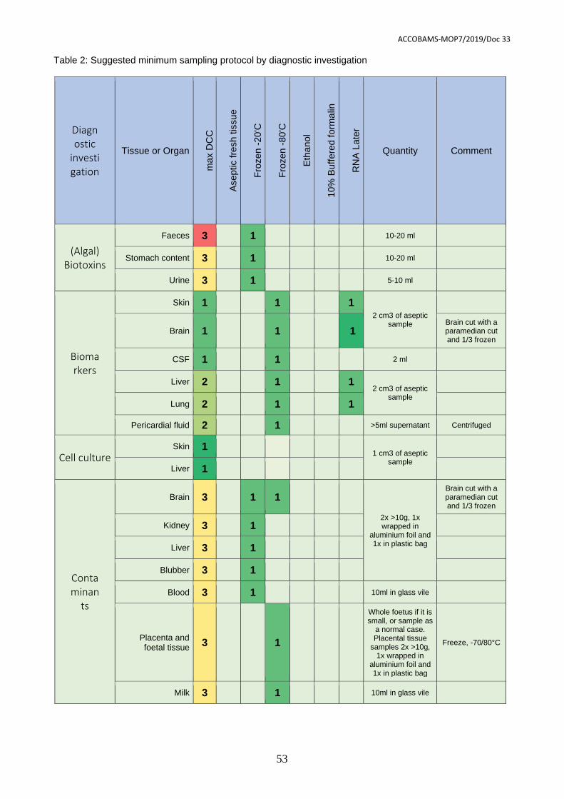
ACCOBAMS-MOP7/2019/Doc 33
53
Table 2: Suggested minimum sampling protocol by diagnostic investigation
Diagnostic
investigation
Tissue or Organ
max D
CC
Aseptic fre
sh t
issue
Fro
zen -
20'C
Fro
zen -
80'C
Eth
ano
l
10%
Buff
ere
d f
orm
alin
RN
A L
ate
r
Quantity Comment
(Algal) Biotoxins
Faeces 3 1 10-20 ml
Stomach content 3 1 10-20 ml
Urine 3 1 5-10 ml
Biomarkers
Skin 1 1 1 2 cm3 of aseptic
sample
Brain 1 1 1 Brain cut with a paramedian cut and 1/3 frozen
CSF 1 1 2 ml
Liver 2 1 1 2 cm3 of aseptic
sample
Lung 2 1 1
Pericardial fluid 2 1 >5ml supernatant Centrifuged
Cell culture Skin 1
1 cm3 of aseptic sample
Liver 1
Contaminan
ts
Brain 3 1 1
2x >10g, 1x wrapped in
aluminium foil and 1x in plastic bag
Brain cut with a paramedian cut and 1/3 frozen
Kidney 3 1
Liver 3 1
Blubber 3 1
Blood 3 1 10ml in glass vile
Placenta and foetal tissue
3 1
Whole foetus if it is small, or sample as
a normal case. Placental tissue
samples 2x >10g, 1x wrapped in
aluminium foil and 1x in plastic bag
Freeze, -70/80°C
Milk 3 1 10ml in glass vile
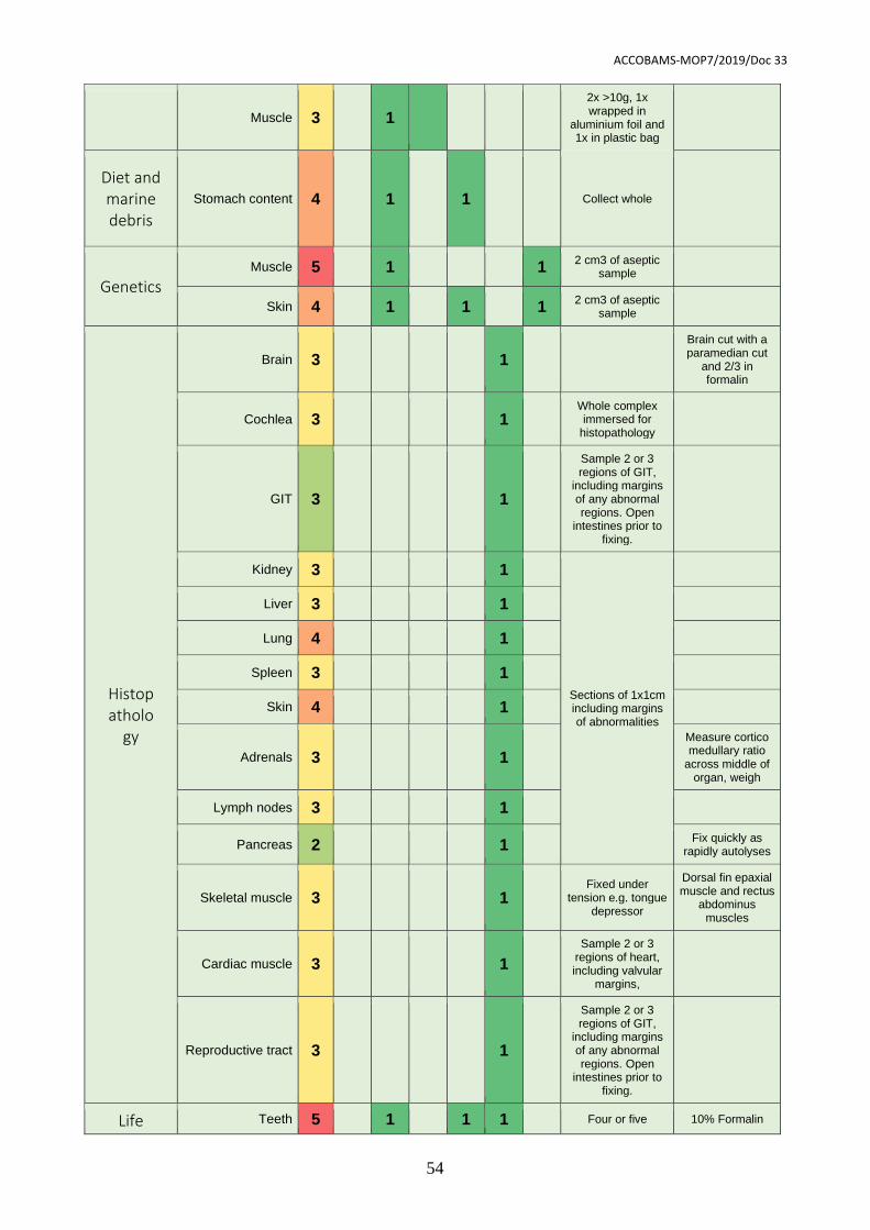
ACCOBAMS-MOP7/2019/Doc 33
54
Muscle 3 1
2x >10g, 1x wrapped in
aluminium foil and 1x in plastic bag
Diet and marine debris
Stomach content 4 1 1 Collect whole
Genetics Muscle 5 1 1
2 cm3 of aseptic sample
Skin 4 1 1 1 2 cm3 of aseptic
sample
Histopatholo
gy
Brain 3 1
Brain cut with a paramedian cut
and 2/3 in formalin
Cochlea 3 1 Whole complex immersed for
histopathology
GIT 3 1
Sample 2 or 3 regions of GIT,
including margins of any abnormal regions. Open
intestines prior to fixing.
Kidney 3 1
Sections of 1x1cm including margins of abnormalities
Liver 3 1
Lung 4 1
Spleen 3 1
Skin 4 1
Adrenals 3 1
Measure cortico medullary ratio
across middle of organ, weigh
Lymph nodes 3 1
Pancreas 2 1 Fix quickly as
rapidly autolyses
Skeletal muscle 3 1 Fixed under
tension e.g. tongue depressor
Dorsal fin epaxial muscle and rectus
abdominus muscles
Cardiac muscle 3 1
Sample 2 or 3 regions of heart, including valvular
margins,
Reproductive tract 3 1
Sample 2 or 3 regions of GIT,
including margins of any abnormal regions. Open
intestines prior to fixing.
Life Teeth 5 1 1 1 Four or five 10% Formalin
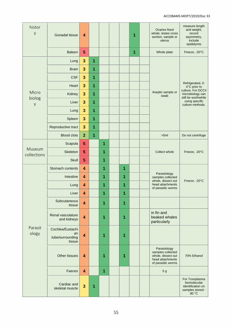
ACCOBAMS-MOP7/2019/Doc 33
55
history
Gonadal tissue 4 1
Ovaries fixed whole, testes cross section, sample or
uterus
measure length and weight,
record asymmetry,
include epididymis
Baleen 5 1 Whole plate Freeze, -20°C
Microbiolog
y
Lung 3 1
Aseptic sample or swab
Refrigerated, 0-4°C prior to
culture. For DCC4 microbiology can still be worthwhile
using specific culture methods.
Brain 3 1
CSF 3 1
Heart 3 1
Kidney 3 1
Liver 3 1
Lung 3 1
Spleen 3 1
Reproductive tract 3 1
Blood clots 2 1 >5ml Do not centrifuge
Museum collections
Scapula 5 1
Collect whole Freeze, -20°C Skeleton 5 1
Skull 5 1
Parasitology
Stomach contents 4 1 1 Parasitology
samples collected whole, dissect out head attachments of parasitic worms
Freeze, -20°C
Intestine 4 1 1
Lung 4 1 1
Liver 4 1 1
Subcutaneous tissue
4 1 1
Renal vasculature and kidneys
4 1 1 in fin and beaked whales particularly
Cochlea/Eustachian
tube/surrounding tissue
4 1 1
Other tissues 4 1 1
Parasitology samples collected whole, dissect out head attachments of parasitic worms
70% Ethanol
Faeces 4 1 5 g
Cardiac and skeletal muscle
3 1
For Toxoplasma biomolecular
identification on samples stored -
80 °C
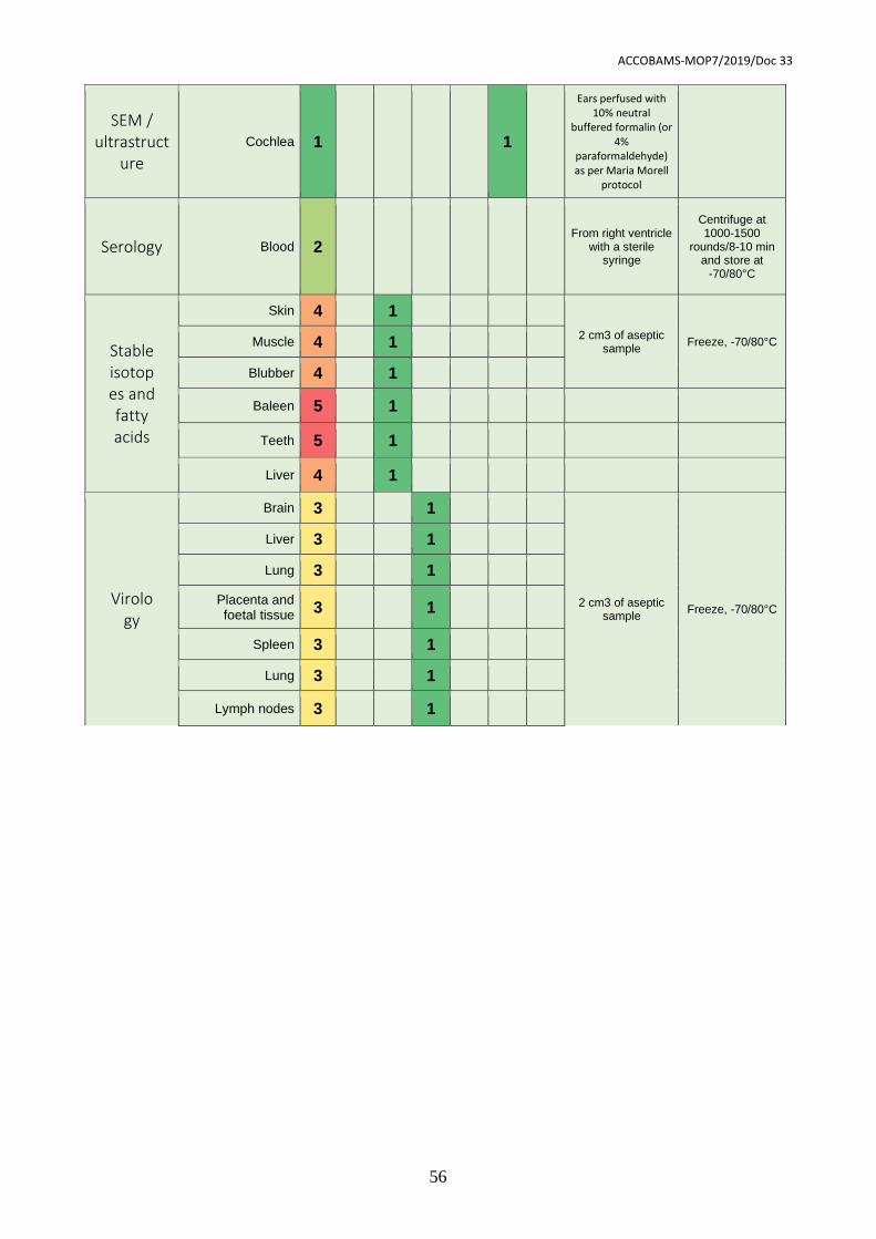
ACCOBAMS-MOP7/2019/Doc 33
56
SEM / ultrastruct
ure
Cochlea 1 1
Ears perfused with 10% neutral
buffered formalin (or 4%
paraformaldehyde) as per Maria Morell
protocol
Serology Blood 2 From right ventricle
with a sterile syringe
Centrifuge at 1000-1500
rounds/8-10 min and store at
-70/80°C
Stable isotopes and fatty acids
Skin 4 1
2 cm3 of aseptic sample
Freeze, -70/80°C Muscle 4 1
Blubber 4 1
Baleen 5 1
Teeth 5 1
Liver 4 1
Virology
Brain 3 1
2 cm3 of aseptic sample
Freeze, -70/80°C
Liver 3 1
Lung 3 1
Placenta and foetal tissue
3 1
Spleen 3 1
Lung 3 1
Lymph nodes 3 1
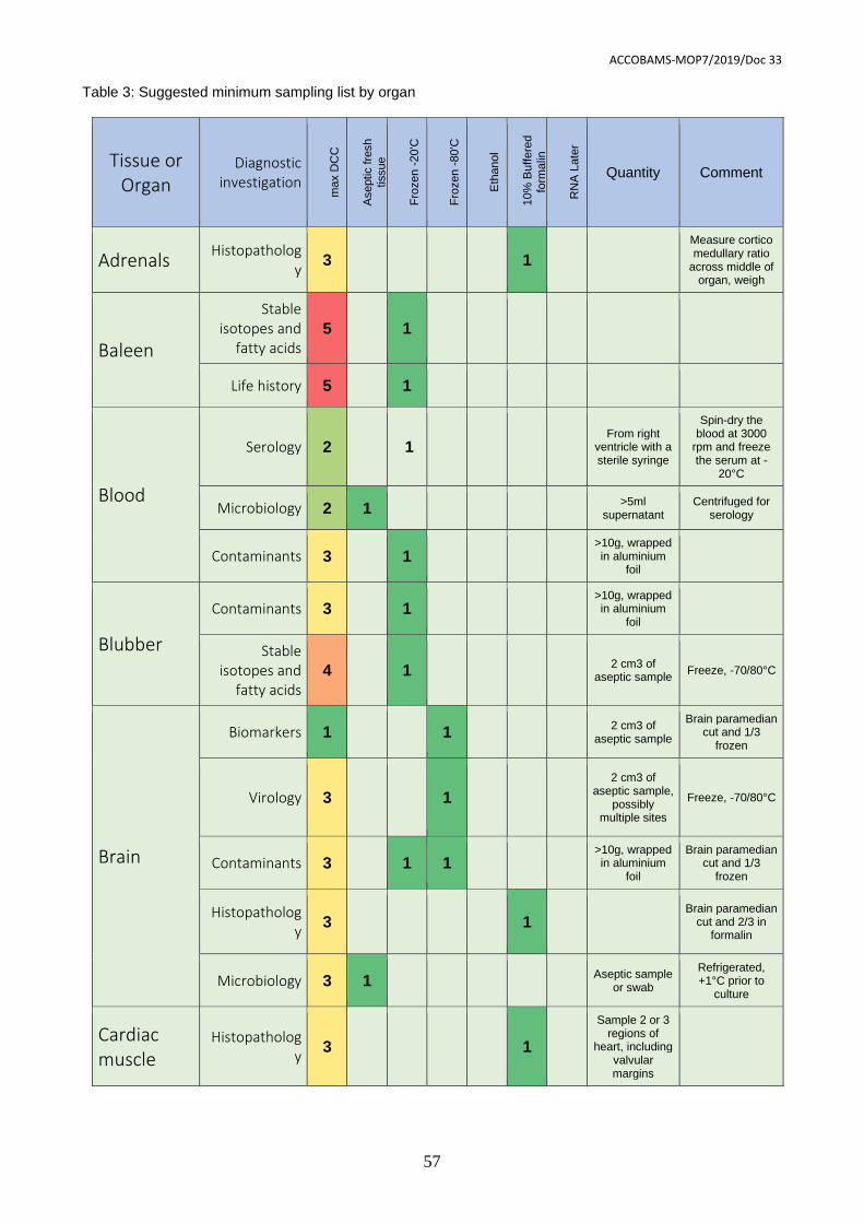
ACCOBAMS-MOP7/2019/Doc 33
57
Table 3: Suggested minimum sampling list by organ
Tissue or Organ
Diagnostic investigation
ma
x D
CC
Aseptic f
resh
tissue
Fro
zen -
20'C
Fro
zen -
80'C
Eth
anol
10%
Buffere
d
form
alin
RN
A L
ate
r
Quantity Comment
Adrenals Histopatholog
y 3 1
Measure cortico medullary ratio
across middle of organ, weigh
Baleen
Stable isotopes and
fatty acids 5 1
Life history 5 1
Blood
Serology 2 1 From right
ventricle with a sterile syringe
Spin-dry the blood at 3000
rpm and freeze the serum at -
20°C
Microbiology 2 1 >5ml
supernatant Centrifuged for
serology
Contaminants 3 1 >10g, wrapped in aluminium
foil
Blubber
Contaminants 3 1 >10g, wrapped in aluminium
foil
Stable isotopes and
fatty acids 4 1
2 cm3 of aseptic sample
Freeze, -70/80°C
Brain
Biomarkers 1 1 2 cm3 of
aseptic sample
Brain paramedian cut and 1/3
frozen
Virology 3 1
2 cm3 of aseptic sample,
possibly multiple sites
Freeze, -70/80°C
Contaminants 3 1 1 >10g, wrapped in aluminium
foil
Brain paramedian cut and 1/3
frozen
Histopathology
3 1 Brain paramedian
cut and 2/3 in formalin
Microbiology 3 1 Aseptic sample
or swab
Refrigerated, +1°C prior to
culture
Cardiac muscle
Histopathology
3 1
Sample 2 or 3 regions of
heart, including valvular margins
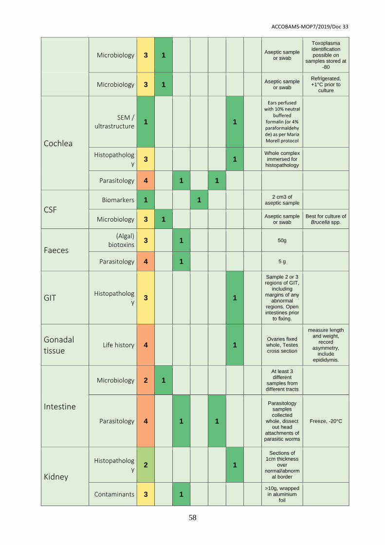
ACCOBAMS-MOP7/2019/Doc 33
58
Microbiology 3 1 Aseptic sample
or swab
Toxoplasma identification possible on
samples stored at -80
Microbiology 3 1 Aseptic sample
or swab
Refrigerated, +1°C prior to
culture
Cochlea
SEM / ultrastructure
1 1
Ears perfused with 10% neutral
buffered formalin (or 4% paraformaldehyde) as per Maria Morell protocol
Histopathology
3 1 Whole complex immersed for
histopathology
Parasitology 4 1 1
CSF Biomarkers 1 1
2 cm3 of aseptic sample
Microbiology 3 1 Aseptic sample
or swab Best for culture of
Brucella spp.
Faeces
(Algal) biotoxins
3 1 50g
Parasitology 4 1 5 g
GIT Histopatholog
y 3 1
Sample 2 or 3 regions of GIT,
including margins of any
abnormal regions. Open intestines prior
to fixing.
Gonadal tissue
Life history 4 1 Ovaries fixed whole, Testes cross section
measure length and weight,
record asymmetry,
include epididymis.
Intestine
Microbiology 2 1
At least 3 different
samples from different tracts
Parasitology 4 1 1
Parasitology samples collected
whole, dissect out head
attachments of parasitic worms
Freeze, -20°C
Kidney
Histopathology
2 1
Sections of 1cm thickness
over normal/abnorm
al border
Contaminants 3 1 >10g, wrapped in aluminium
foil
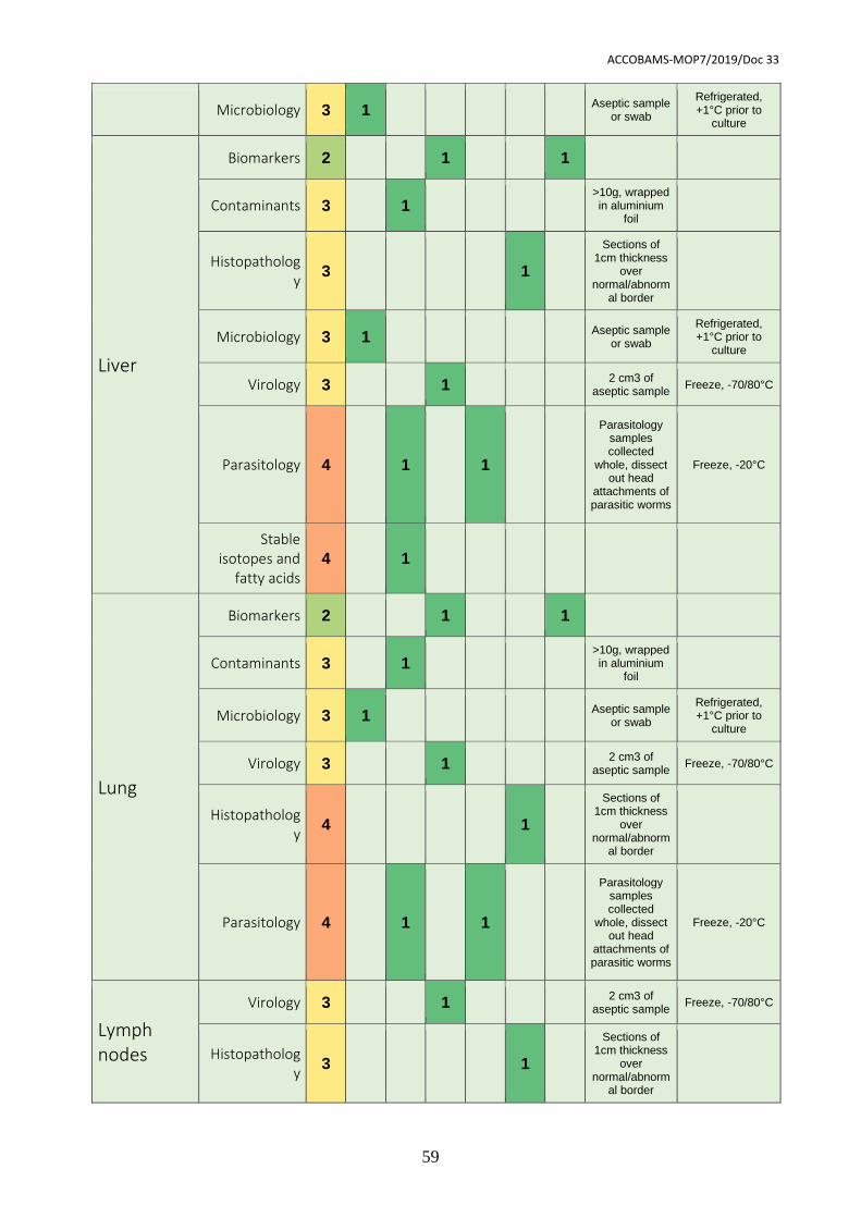
ACCOBAMS-MOP7/2019/Doc 33
59
Microbiology 3 1 Aseptic sample
or swab
Refrigerated, +1°C prior to
culture
Liver
Biomarkers 2 1 1
Contaminants 3 1 >10g, wrapped in aluminium
foil
Histopathology
3 1
Sections of 1cm thickness
over normal/abnorm
al border
Microbiology 3 1 Aseptic sample
or swab
Refrigerated, +1°C prior to
culture
Virology 3 1 2 cm3 of
aseptic sample Freeze, -70/80°C
Parasitology 4 1 1
Parasitology samples collected
whole, dissect out head
attachments of parasitic worms
Freeze, -20°C
Stable isotopes and
fatty acids 4 1
Lung
Biomarkers 2 1 1
Contaminants 3 1 >10g, wrapped in aluminium
foil
Microbiology 3 1 Aseptic sample
or swab
Refrigerated, +1°C prior to
culture
Virology 3 1 2 cm3 of
aseptic sample Freeze, -70/80°C
Histopathology
4 1
Sections of 1cm thickness
over normal/abnorm
al border
Parasitology 4 1 1
Parasitology samples collected
whole, dissect out head
attachments of parasitic worms
Freeze, -20°C
Lymph nodes
Virology 3 1 2 cm3 of
aseptic sample Freeze, -70/80°C
Histopathology
3 1
Sections of 1cm thickness
over normal/abnorm
al border
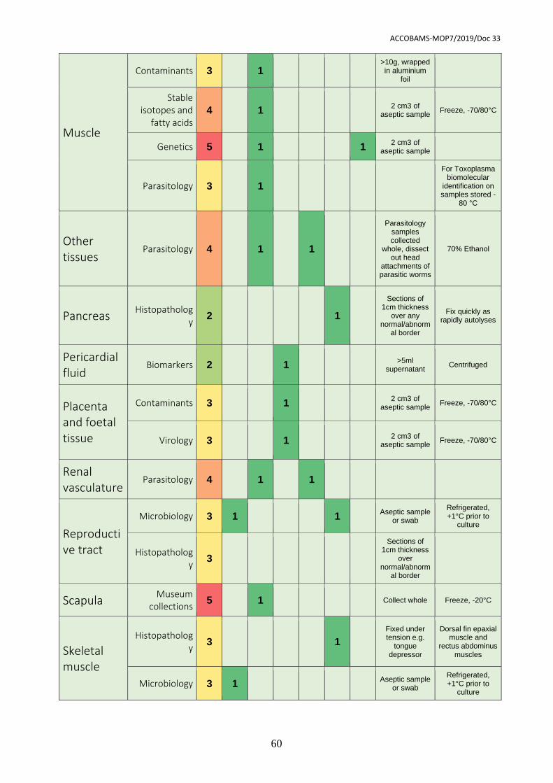
ACCOBAMS-MOP7/2019/Doc 33
60
Muscle
Contaminants 3 1 >10g, wrapped in aluminium
foil
Stable isotopes and
fatty acids 4 1
2 cm3 of aseptic sample
Freeze, -70/80°C
Genetics 5 1 1 2 cm3 of
aseptic sample
Parasitology 3 1
For Toxoplasma biomolecular
identification on samples stored -
80 °C
Other tissues
Parasitology 4 1 1
Parasitology samples collected
whole, dissect out head
attachments of parasitic worms
70% Ethanol
Pancreas Histopatholog
y 2 1
Sections of 1cm thickness
over any normal/abnorm
al border
Fix quickly as rapidly autolyses
Pericardial fluid
Biomarkers 2 1 >5ml
supernatant Centrifuged
Placenta and foetal tissue
Contaminants 3 1 2 cm3 of
aseptic sample Freeze, -70/80°C
Virology 3 1 2 cm3 of
aseptic sample Freeze, -70/80°C
Renal vasculature
Parasitology 4 1 1
Reproductive tract
Microbiology 3 1 1 Aseptic sample
or swab
Refrigerated, +1°C prior to
culture
Histopathology
3
Sections of 1cm thickness
over normal/abnorm
al border
Scapula Museum
collections 5 1 Collect whole Freeze, -20°C
Skeletal muscle
Histopathology
3 1
Fixed under tension e.g.
tongue depressor
Dorsal fin epaxial muscle and
rectus abdominus muscles
Microbiology 3 1 Aseptic sample
or swab
Refrigerated, +1°C prior to
culture
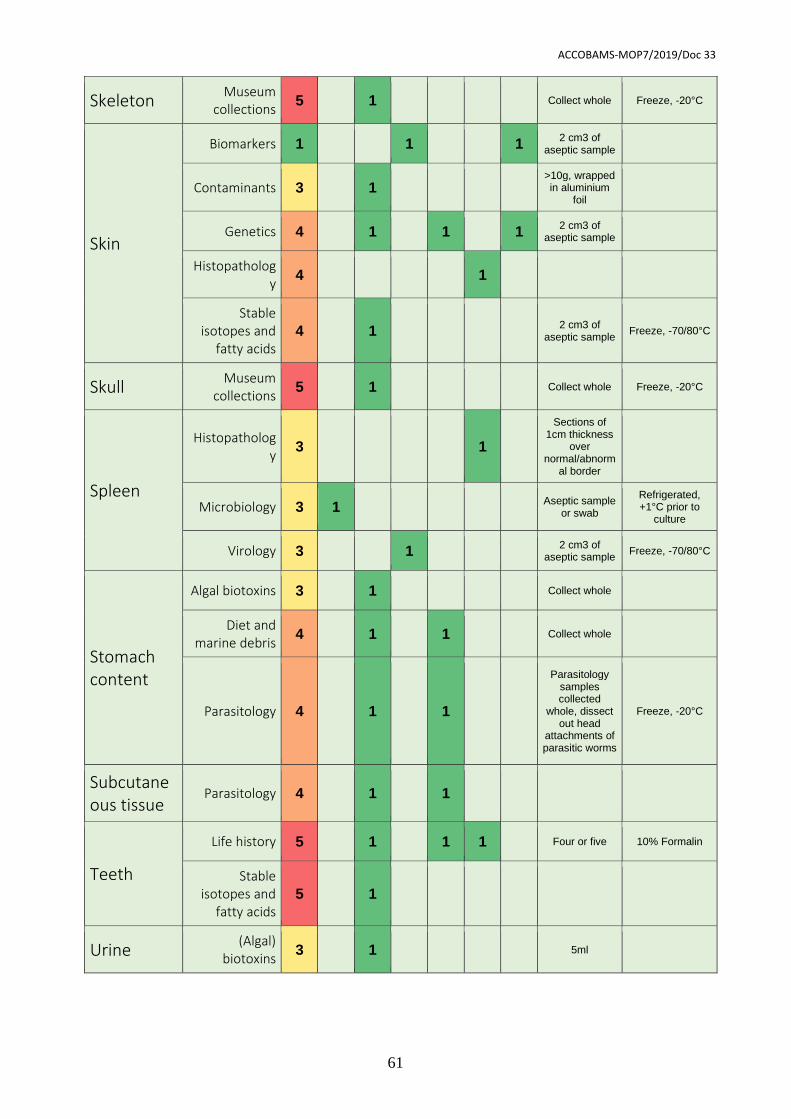
ACCOBAMS-MOP7/2019/Doc 33
61
Skeleton Museum
collections 5 1 Collect whole Freeze, -20°C
Skin
Biomarkers 1 1 1 2 cm3 of
aseptic sample
Contaminants 3 1 >10g, wrapped in aluminium
foil
Genetics 4 1 1 1 2 cm3 of
aseptic sample
Histopathology
4 1
Stable isotopes and
fatty acids 4 1
2 cm3 of aseptic sample
Freeze, -70/80°C
Skull Museum
collections 5 1 Collect whole Freeze, -20°C
Spleen
Histopathology
3 1
Sections of 1cm thickness
over normal/abnorm
al border
Microbiology 3 1 Aseptic sample
or swab
Refrigerated, +1°C prior to
culture
Virology 3 1 2 cm3 of
aseptic sample Freeze, -70/80°C
Stomach content
Algal biotoxins 3 1 Collect whole
Diet and marine debris
4 1 1 Collect whole
Parasitology 4 1 1
Parasitology samples collected
whole, dissect out head
attachments of parasitic worms
Freeze, -20°C
Subcutaneous tissue
Parasitology 4 1 1
Teeth
Life history 5 1 1 1 Four or five 10% Formalin
Stable isotopes and
fatty acids 5 1
Urine (Algal)
biotoxins 3 1 5ml

ACCOBAMS-MOP7/2019/Doc 33
62
ACCOBAMS-MOP7/2019/Doc 33
63
Acknowledgements
This publication arose from several previous workshops, meetings and expert elicitation
rounds. This work was originally initiated in 1991 by Thijs Kuiken and Manuel García-
Hartmann, who organised the first cetacean pathology workshop as previously published
by the European Cetacean Society (1993). Approximately 25 years later, similar
workshops were organised by ACCOBAMS (Report of the ACCOBANS/PELAGOS
Workshop on Cetacean Live Strandings, Monaco 2014) and during other ECS
conferences, including that in Malta in 2015, for which we acknowledge the main organizer
Thierry Jauniaux, and in Funchal, Madeira in 2016.
The final document as presented here was completed with support of ACCOBAMS,
ASCOBANS and the IWC Strandings initiative.
ACCOBAMS-MOP7/2019/Doc 33
64
Literature
ACCOBAMS Resolution 6.22 - Cetaceans Live Strandings. Annex II Common Best
Practices for a basic post-mortem examination of stranded cetaceans.
Arbelo, M., de los Monteros, A. E., Herráez, P., Andrada, M., Sierra, E., Rodríguez, F., ... &
Fernández, A. (2013). Pathology and causes of death of stranded cetaceans in the Canary
Islands (1999− 2005). Diseases of aquatic organisms, 103(2), 87-99.
Brody, S. (1945). Bioenergetics and Growth, Reinhold Publishing, New York (1945), pp.
484-663.
Bernaldo de Quirós, Y., González-Díaz, Ó., Arbelo, M., Andrada, M., & Fernández, A.
(2012). Protocol for gas sampling and analysis in stranded marine mammals. Protocol
exchange.
Díaz-Delgado, J., Fernandez, A., Sierra, E., Sacchini, S., Andrada, M., Vela, A. I., ... &
Arbelo, M. (2018). Pathologic findings and causes of death of stranded cetaceans in the
Canary Islands (2006-2012). PloS one, 13(10), e0204444.
Dierauf, L., & Gulland, F. M. (Eds.). (2001). CRC handbook of marine mammal medicine:
health, disease, and rehabilitation. CRC press.
Di Guardo, G., Di Francesco, C. E., Eleni, C., Cocumelli, C., Scholl, F., Casalone, C., ... &
Leonardi, L. (2013). Morbillivirus infection in cetaceans stranded along the Italian
coastline: pathological, immunohistochemical and biomolecular findings. Research in
Veterinary Science, 94(1), 132-137.
Gulland, F. M., & Hall, A. J. (2007). Is marine mammal health deteriorating? Trends in the
global reporting of marine mammal disease. EcoHealth, 4(2), 135-150.
Hunt, T. D., Ziccardi, M. H., Gulland, F. M., Yochem, P. K., Hird, D. W., Rowles, T., &
Mazet, J. A. (2008). Health risks for marine mammal workers. Diseases of aquatic
organisms, 81(1), 81-92.
Jauniaux, T., Petitjean, D., Brenez, C., Borrens, M., Brosens, L., Haelters, J., ... &
Coignoul, F. (2002). Post-mortem findings and causes of death of harbour porpoises
(Phocoena phocoena) stranded from 1990 to 2000 along the coastlines of Belgium and
Northern France. Journal of Comparative Pathology, 126(4), 243-253.
ACCOBAMS-MOP7/2019/Doc 33
65
Jepson, P. D., Deaville, R., Barber, J. L., Aguilar, À., Borrell, A., Murphy, S., ... &
Cunningham, A. A. (2016). PCB pollution continues to impact populations of orcas and
other dolphins in European waters. Scientific Reports, 6, 18573.
Kuiken, T., & García Hartmann, M. (1993). Cetacean pathology: dissection techniques and
tissue sampling. ECS Newsletter 17. Special Issue, 1-43.
Lane, E. P., De Wet, M., Thompson, P., Siebert, U., Wohlsein, P., & Plön, S. (2014). A
systematic health assessment of Indian ocean bottlenose (Tursiops aduncus) and Indo-
Pacific humpback (Sousa plumbea) dolphins incidentally caught in shark nets off the
KwaZulu-Natal coast, South Africa. PloS One, 9(9), e107038.
Leeney, R. H., Amies, R., Broderick, A. C., Witt, M. J., Loveridge, J., Doyle, J., & Godley,
B. J. (2008). Spatio-temporal analysis of cetacean strandings and bycatch in a UK
fisheries hotspot. Biodiversity and Conservation, 17(10), 2323.
Lockyer, C. (1991). Body composition of the sperm whale, Physeter catodon, with special
reference to the possible functions of fat depots. Marine Research Institute.
Mazzariol S., Cozzi B., Centelleghe C. (2015). Handbook for Cetaceans’ Strandings.
Available at: http://www.netcet.eu/dissemination/item/132-handbook-for-
cetaceans%E2%80%99-strandings
Morell, M., André, M. (2009). Cetacean ear extraction and fixation protocol. Available at:
http://www.zoology.ubc.ca/files/Ear_extraction_and_fixation_protocol_UBC.pdf
Peltier, H., Authier, M., Deaville, R., Dabin, W., Jepson, P. D., Van Canneyt, O., ... &
Ridoux, V. (2016). Small cetacean bycatch as estimated from stranding schemes: The
common dolphin case in the northeast Atlantic. Environmental Science & Policy, 63, 7-18.
Peltier, H., Baagøe, H. J., Camphuysen, K. C., Czeck, R., Dabin, W., Daniel, P., ... &
Jepson, P. D. (2013). The stranding anomaly as population indicator: the case of harbour
porpoise Phocoena phocoena in North-Western Europe. PLoS One, 8(4), e62180.
Peltier, H., Dabin, W., Daniel, P., Van Canneyt, O., Dorémus, G., Huon, M., & Ridoux, V.
(2012). The significance of stranding data as indicators of cetacean populations at sea:
Modelling the drift of cetacean carcasses. Ecological Indicators, 18, 278-290.
Plön, S., de Wet, M., Lane, E., Wohlsein, P., Siebert, U., & Thompson, P. (2015). A
standardized necropsy protocol for health investigations of small cetaceans in southern
Africa. African Journal of Wildlife Research, 45(3), 332-342.
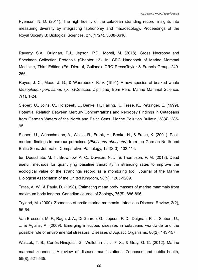
ACCOBAMS-MOP7/2019/Doc 33
66
Pyenson, N. D. (2011). The high fidelity of the cetacean stranding record: insights into
measuring diversity by integrating taphonomy and macroecology. Proceedings of the
Royal Society B: Biological Sciences, 278(1724), 3608-3616.
Raverty, S.A., Duignan, P.J., Jepson, P.D., Morell, M. (2018). Gross Necropsy and
Specimen Collection Protocols (Chapter 13). In: CRC Handbook of Marine Mammal
Medicine, Third Edition (Ed. Dierauf, Gulland). CRC Press/Taylor & Francis Group, 249-
266.
Reyes, J. C., Mead, J. G., & Waerebeek, K. V. (1991). A new species of beaked whale
Mesoplodon peruvianus sp. n.(Cetacea: Ziphiidae) from Peru. Marine Mammal Science,
7(1), 1-24.
Siebert, U., Joiris, C., Holsbeek, L., Benke, H., Failing, K., Frese, K., Petzinger, E. (1999).
Potential Relation Between Mercury Concentrations and Necropsy Findings in Cetaceans
from German Waters of the North and Baltic Seas. Marine Pollution Bulletin, 38(4), 285-
95.
Siebert, U., Wünschmann, A., Weiss, R., Frank, H., Benke, H., & Frese, K. (2001). Post-
mortem findings in harbour porpoises (Phocoena phocoena) from the German North and
Baltic Seas. Journal of Comparative Pathology, 124(2-3), 102-114.
ten Doeschate, M. T., Brownlow, A. C., Davison, N. J., & Thompson, P. M. (2018). Dead
useful; methods for quantifying baseline variability in stranding rates to improve the
ecological value of the strandings record as a monitoring tool. Journal of the Marine
Biological Association of the United Kingdom, 98(5), 1205-1209.
Trites, A. W., & Pauly, D. (1998). Estimating mean body masses of marine mammals from
maximum body lengths. Canadian Journal of Zoology, 76(5), 886-896.
Tryland, M. (2000). Zoonoses of arctic marine mammals. Infectious Disease Review, 2(2),
55-64.
Van Bressem, M. F., Raga, J. A., Di Guardo, G., Jepson, P. D., Duignan, P. J., Siebert, U.,
... & Aguilar, A. (2009). Emerging infectious diseases in cetaceans worldwide and the
possible role of environmental stressors. Diseases of Aquatic Organisms, 86(2), 143-157.
Waltzek, T. B., Cortés‐Hinojosa, G., Wellehan Jr, J. F. X., & Gray, G. C. (2012). Marine
mammal zoonoses: A review of disease manifestations. Zoonoses and public health,
59(8), 521-535.

ACCOBAMS-MOP7/2019/Doc 33
67
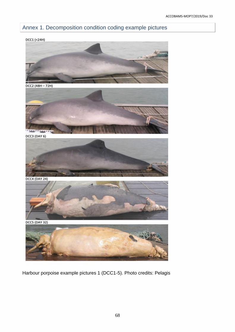
ACCOBAMS-MOP7/2019/Doc 33
68
Annex 1. Decomposition condition coding example pictures
Harbour porpoise example pictures 1 (DCC1-5). Photo credits: Pelagis
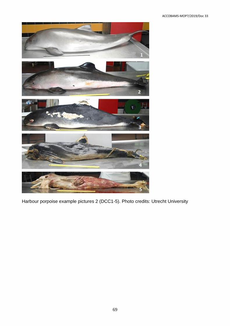
ACCOBAMS-MOP7/2019/Doc 33
69
Harbour porpoise example pictures 2 (DCC1-5). Photo credits: Utrecht University
ACCOBAMS-MOP7/2019/Doc 33
70
DCC1:
DCC2:
DCC3:
Common dolphin example pictures (DCC1-3). Photo credits: CSIP, James Barnett
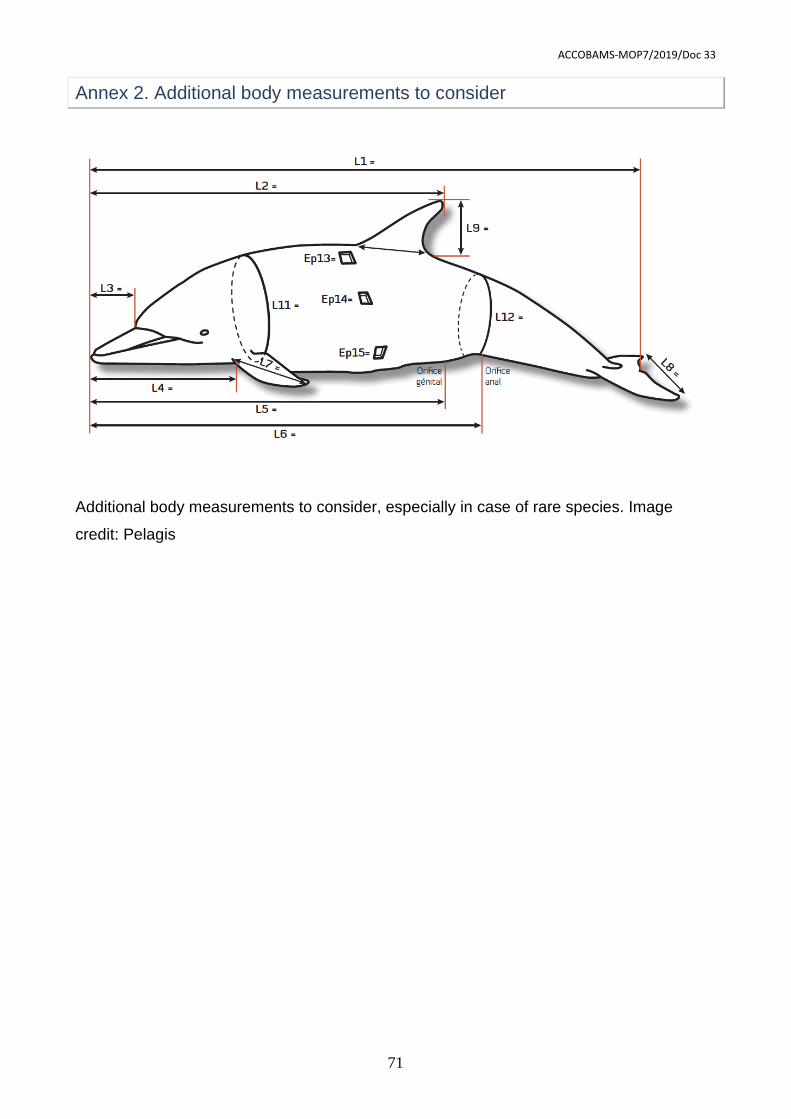
ACCOBAMS-MOP7/2019/Doc 33
71
Annex 2. Additional body measurements to consider
Additional body measurements to consider, especially in case of rare species. Image
credit: Pelagis
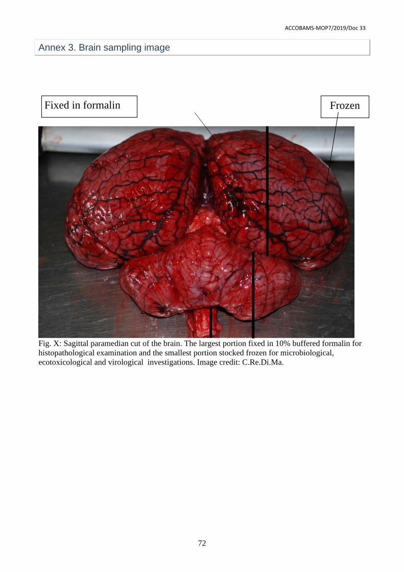
ACCOBAMS-MOP7/2019/Doc 33
72
Annex 3. Brain sampling image
Fig. X: Sagittal paramedian cut of the brain. The largest portion fixed in 10% buffered formalin for
histopathological examination and the smallest portion stocked frozen for microbiological,
ecotoxicological and virological investigations. Image credit: C.Re.Di.Ma.
Fixed in formalin Frozen