belgian journal of paediatrics - bvk-sbp
TRANSCRIPT
BELGISCHE VERENIGING VOOR KINDERGENEESKUNDE SOCIÉTÉ BELGE DE PÉDIATRIE
2017 - Volume 19 - number 1 - March
VV.U./E.R. S. Cadranel (ULB), M. Raes (KUL)
Gasthuisberg, Herestraat 49, 3000 Leuven
E-mail: [email protected]
QUARTERLYISSN 2466-8907
Belgian Journalof Paediatrics
BJP
Articles Les apports graisseux chez la femme enceinte et allaitante.
State of the Art Nieuwe therapieën voor mucoviscidose.
Made In Belgium A poor start to life? Perinatal predictors of cardiovascular and renal health in childhood.
Cochrane Corner Pediatrics Introduction. Immediate or early skin-to-skin contact between mothers and newborns can be recommended to promote successful breastfeeding and the infant’s overall wellbeing.
Activities of Paediatric Societies
Jaarverslag 2016 - Rapport annuel 2016.
Abstracts of the 45th annual congress
Allergology Cardiology Dermatology Emergency Medicine Endocrinology Gastroenterology General Paediatrics Genetics Immunology/Infectious Diseases Infectiology Metabolic Diseases Neonatology Nephrology Neurology Oncology - Hematology Pulmonology Rheumatology
Publication of the Belgian Society of Paediatrics
Belgische Vereniging voor KindergeneeskundeSociété Belge de Pédiatrie
“Grenzen in pediatrie“ - “Frontières en pédiatrie”
Antwerpen Hilton Hotel23-24 Maart - Mars 2017
BVK/SBP Congres 2017
Kuns
tena
ar -
Arti
ste:
: Ka
atje
Ver
mei
ren

R A C E C A D O T R I L
Vermindert het volume van de stoelgang snel
Verkort de duur van de diarree signifi cant
Uitstekend veiligheids- en tolerantieprofi el
1ste antisecretoire behandeling van diarree vanaf de leeftijd van 3 maanden
Vermindert het volume van de stoelgang snel Vermindert het volume van de stoelgang snel Vermindert het volume van de stoelgang snel Vermindert het volume van de stoelgang snel
Verkort de duur van de diarree signifi cant
Uitstekend veiligheids- en tolerantieprofi el Uitstekend veiligheids- en tolerantieprofi el
behandeling van diarreebehandeling van diarreebehandeling van diarreebehandeling van diarreebehandeling van diarreevanaf de leeftijd van 3 maandenvanaf de leeftijd van 3 maandenvanaf de leeftijd van 3 maandenvanaf de leeftijd van 3 maandenvanaf de leeftijd van 3 maanden
Tiorfi x Baby - 10 mg en Tiorfi x Junior - 30 mg voor kinderen. Een geneesmiddel op voorschrift.
Tiorfi x 100 mg voor volwassenen. Een geneesmiddel vrij verkrijgbaar in de apotheek.
NY
/TF/
15/0
00
9 -
10
/20
15
NAAM VAN HET GENEESMIDDEL: Tiorfi x Baby 10 mg granulaat voor orale suspensie. Tiorfi x Junior 30 mg granulaat voor orale suspensie. Tiorfi x 100 mg harde capsules. KWALITA-
TIEVE EN KWANTITATIEVE SAMENSTELLING: 10 mg: Elk zakje bevat 10 mg racecadotril en 966,5 mg sucrose. 30 mg: Elk zakje bevat 30 mg racecadotril en 2,9 g sucrose. 100 mg:
Elke capsule bevat 100 mg racecadotril en 41 mg lactosemonohydraat. FARMACEUTISCHE VORM: 10 en 30 mg: Granulaat voor orale suspensie. Wit poeder met een kenmerkende
abrikozengeur. 100 mg: Harde capsule. Ivoorkleurige capsules (grootte 2) die een wit gekleurd poeder met zwavelgeur bevatten. THERAPEUTISCHE INDICATIES: 10 en 30 mg:
Aanvullende symptomatische behandeling van acute diarree bij zuigelingen (ouder dan 3 maanden) en kinderen samen met orale rehydratatie en de gebruikelijke ondersteunende
maatregelen, als die op zichzelf niet volstaan om de klinische aandoening onder controle te krijgen en wanneer een oorzakelijke behandeling niet mogelijk is. Als een oorzakelijke
behandeling mogelijk is, kan racecadotril toegediend worden als een aanvullende behandeling. 100 mg: Tiorfi x is geïndiceerd voor de symptomatische behandeling van acute diar-
ree bij volwassenen wanneer de oorzakelijke behandeling niet mogelijk is. Als een oorzakelijke behandeling mogelijk is, kan racecadotril toegediend worden als een aanvullende
behandeling. DOSERING EN WIJZE VAN TOEDIENING: 10 en 30 mg: Tiorfi x Baby en Tiorfi x Junior worden oraal toegediend samen met orale rehydratatie. Tiorfi x Baby is bedoeld
voor kinderen < 13 kg. Tiorfi x Junior is bedoeld voor kinderen ≥13 kg. De aanbevolen dosis hangt af van het lichaamsgewicht: 1,5 mg/kg per dosis (overeenkomend met 1 tot 2 zakjes),
driemaal daags op regelmatige tijdstippen. Bij kinderen minder dan 9 kg: één zakje van 10 mg 3 maal per dag. Bij kinderen van 9 kg tot 13 kg: twee zakjes van 10 mg 3 maal per dag.
Bij kinderen van 13 kg tot 27 kg: één zakje van 30 mg 3 maal per dag. Bij kinderen van meer dan 27 kg: twee zakjes van 30 mg 3 maal per dag. In de klinische studies bij kinderen
bedroeg de behandelingsduur 5 dagen. De behandeling moet worden voortgezet tot er twee normale stoelgangen worden waargenomen. De behandeling mag niet langer duren
dan 7 dagen. Langdurige behandeling met racecadotril is niet aanbevolen. Er werden geen klinische studies uitgevoerd bij zuigelingen jonger dan 3 maanden. Speciale populaties:
Er zijn geen studies uitgevoerd bij zuigelingen of kinderen met nierinsu¦ ciëntie of leverinsu¦ ciëntie. Voorzichtigheid is geboden bij patiënten met lever- of nierinsu¦ ciëntie. Het
granulaat kan worden toegevoegd aan voedsel, gedispergeerd in een glas water of in de zuigfl es. Het moet goed worden gemengd en onmiddellijk worden toegediend. 100 mg:
Enkel voor volwassenen: Aanvankelijk één capsule, ongeacht het uur van de dag. Daarna één capsule driemaal daags bij voorkeur vóór de hoofdmaaltijden. De behandeling moet
worden voorgezet tot er twee normale stoelgangen worden waargenomen. De behandeling mag niet langer duren dan 7 dagen. Speciale populaties: Ouderen: de dosering hoeft
niet te worden aangepast bij ouderen. Voorzichtigheid is geboden bij patiënten met lever- of nierinsu¦ ciëntie. CONTRA-INDICATIES: Overgevoeligheid voor de werkzame stof of
voor een van de hulpstoª en. Tiorfi x Baby en Tiorfi x Junior bevatten sucrose. Patiënten met zeldzame erfelijke aandoeningen als fructose-intolerantie, glucose-galactose malab-
sorptie of sucrase-isomaltase insu¦ ciëntie dienen dit geneesmiddel niet te gebruiken. BIJWERKINGEN: 10 en 30 mg: Er zijn gegevens van klinische studies beschikbaar over 860
pediatrische patiënten met acute diarree die werden behandeld met racecadotril en over 411 kinderen behandeld met placebo. 100 mg: Er zijn gegevens van klinische studies be-
schikbaar over 2,193 volwassen patiënten met acute diarree die werden behandeld met racecadotril en 282 die werden behandeld met placebo. De volgende bijwerkingen zijn vaker
opgetreden met racecadotril dan met de placebo of werden gerapporteerd tijdens de postmarketingbewaking. De frequentie van bijwerkingen wordt volgens de volgende conventie
gedefi nieerd: zeer vaak (≥ 1/10), vaak (≥ 1/100 tot < 1/10), soms (≥ 1/1.000 tot < 1/100), zelden (≥ 1/10.000 tot < 1/1.000), zeer zelden (< 1/10.000), niet bekend (kan niet worden geraamd op
grond van de beschikbare gegevens). 10 en 30 mg: Infecties en parasitaire aandoeningen Soms: tonsillitis.Huid- en onderhuidaandoeningen Soms: uitslag, erytheem. Niet bekend:
erythema multiforme; oedeem van de tong, het gezicht, de lippen of het ooglid; angio oedeem, urticaria, erythema nodosum, papuleuze uitslag, prurigo, pruritus. 100 mg: Zenuw-
stelselaandoeningen Vaak: hoofdpijn. Huid- en onderhuidaandoeningen Soms: uitslag, erytheem. Niet bekend: erythema multiforme, oedeem van de tong, het gelaat, de lippen of
het ooglid; angio oedeem, urticaria, erythema nodosum, papuleuze uitslag, prurigo, pruritus, toxische huideruptie. Melding van vermoedelijke bijwerkingen. Het is belangrijk om na
toelating van het geneesmiddel vermoedelijke bijwerkingen te melden. Op deze wijze kan de verhouding tussen voordelen en risico’s van het geneesmiddel voortdurend worden
gevolgd. Beroepsbeoefenaren in de gezondheidszorg wordt verzocht alle vermoedelijke bijwerkingen te melden via het nationale meldsysteem: België - Federaal agentschap voor
geneesmiddelen en gezondheidsproducten. Afdeling Vigilantie EUROSTATION II - Victor Hortaplein, 40/ 40 - B-1060 Brussel - Website: www.fagg.be - e-mail: adversedrugreactions@
fagg-afmps.be AARD EN INHOUD VAN DE VERPAKKING: 10 en 30 mg: Zakjes van gelast papier/aluminium/polyethyleen. Verpakkingsgrootten met 16 zakjes. 100 mg: PVC-PVDC/
Aluminium blisterverpakking. Verpakkingsgrootten met 20 harde capsules. HOUDER VAN DE VERGUNNING VOOR HET IN DE HANDEL BRENGEN Bioprojet Europe Ltd., 29 Earlsford
Terrace, Dublin-2, Ierland. Vertegenwoordiger voor correspondentie en inlichtingen: Takeda Belgium, Gentsesteenweg 615, 1080 Brussel. NUMMER VAN DE VERGUNNING VOOR
HET IN DE HANDEL BRENGEN: 10 mg: BE400723 - 30 mg: BE400732 - 100 mg: BE400741 AFLEVERINGSWIJZE: 10 en 30 mg: Op medisch voorschrift. 100
mg: Geneesmiddel niet op medisch voorschrift. DATUM VAN HERZIENING VAN DE TEKST: 09/2015 Datum van goedkeuring van de tekst: 07/2015
10 mg: € 16,50
30 mg: € 16,50
100 mg: € 16,50
Tiorfix_AD_magazPed_A4_16-09-2015.indd 1 18/09/15 10:33
3
FOUNDING EDITOR
L. Corbeel
REDACTEURS EN CHEF - EDITEURS RESPONSABLES HOOFDREDACTEURS - VERANTWOORDELIJK UITGEVERS
S. Cadranel M. Raes
CO-REDACTEURS
N. Francotte M. Wojciechowski
UNIVERSITÉS-UNIVERSITEITEN
G. Buyse (UZL) J. De Schepper (UZB) P. Lepage (ULB) V. Schmitz (ULG) J. Vande Walle (UZG) S. Verhulst (UZA)
SPECIALITES - SPECIALISMEN
Cardiologie M. Gewillig Endocrinologie J. De Schepper Gastroenterologie I. Hoffman Hemato-Oncologie A. Uyttebroeck Immunologie I. Meyts Soins intensifs, Intensieve zorgen D. Biarent Neurologie L. De Meirleir Neonatologie B. Van Overmeire (C. Lecart) Nephrologie J. Vande Walle (E. Levtchenko) Pneumologie J. Hellinckx Reumatologie-Autoimmuunziekten, Rhumatologie-Maladies autoimmunes C. Wouters
VERENIGINGEN – GROUPEMENTS
V.V.K Ann De Guchtenaere G.B.P.F P. Bauche
BUREAU DE LA SOCIETE BELGE DE PEDIATRIE BUREAU VAN DE BELGISCHE VERENIGING VOOR KINDERGENEESKUNDE
VOORZITTER A. MALFROOT PRÉSIDENTE VICE-VOORZITTER C. VAN GEET VICE-PRÉSIDENTE SCHATBEWAARDER D. DE WOLF TRÉSORIER SECRETARIS C. VERMYLEN / M. RAES SECRÉTAIRES PAST-PRESIDENT P. LEPAGE PAST-PRESIDENT PARTNERSHIP S. CADRANEL PARTNERSHIP
Redactieraad / Comité de rédaction

Les bons choixcommencent tôt
Faire de l’eau la première des boissons,c’est déjà bien.
Boire Spa Reine, c’est opterpour la pureté d’une eau unique,idéale dès le plus jeune âge,et même avant…
A la vie
5
BELGISCHE VERENIGING VOOR KINDERGENEESKUNDE SOCIÉTÉ BELGE DE PÉDIATRIE
• Editorial 7
• President's Address 9
• Articles Les apports graisseux chez la femme enceinte et allaitante. 11 J-P. Langhendries
• State of the Art
Nieuwe therapieën voor mucoviscidose. 13 K. De Boeck
• Made in Belgium
A poor start to life? Perinatal predictors of cardiovascular and renal health in childhood. 17 A. Raaijmakers
• Cochrane Corner Pediatrics
Introduction. 20 Cebam, Cochrane Belgium
Immediate or early skin-to-skin contact between mothers and newborns can be recommended to promote successful breastfeeding and the infant’s overall wellbeing. 21 B. Avau, T. Bekkering, F. Cools
• Activities of Paediatric Societies Jaarverslag 2016 College en Academie Pediatrie. 22
Rapport annuel 2016 Collège et Academie de Pediatrie. 23
• 45th Congress of the Belgian Society of Paediatrics The Congress President’s address. 24
• Abstracts 25
1. Allergology 27
2. Cardiology 29 - 31
3. Dermatology 32
4. Emergency Medicine 33 - 34
5. Endocrinology 35 - 39
6. Gastroenterology 41 - 45
7. General Paediatrics 47 - 54
8. Genetics 55 - 58
9. Immunology/Infectious Diseases 59
10. Infectiology 61 - 68
11. Metabolic Diseases 69
12. Neonatology 71 - 77
13. Nephrology 79 - 82
14. Neurology 83 - 85
15. Onco-Hematology 87 - 91
16. Pulmonology 92 - 96
17. Rheumatology 97 - 98
• Authors 99
Contents

We care for children BELGISCHE VERENIGING VOOR KINDERGENEESKUNDE
SOCIETE BELGE DE PEDIATRIE
BELGISCHE VERENIGING VOOR KINDERGENEESKUNDE
SOCIETE BELGE DE PEDIATRIE
BELGISCHE VERENIGINGVOOR KINDERGENEESKUNDESOCIÉTÉ BELGE DE PÉDIATRIE
LA SBP REMERCIE SES PARTENAIRESPOUR LEUR SOUTIENDE BVK BEDANKT ZIJN PARTNERS VOOR HUN STEUN“Grenzen in pediatrie“
“Frontières en pédiatrie”
steème
BELGISCHE VERENIGINGVOOR KINDERGENEESKUNDESOCIÉTÉ BELGE DE PÉDIATRIE
LA SBP REMERCIE SES PARTENAIRESPOUR LEUR SOUTIENDE BVK BEDANKT ZIJN PARTNERS VOOR HUN STEUN“Grenzen in pediatrie“
“Frontières en pédiatrie”
steème
DE BVK BEDANKT ZIJN PARTNERS VOOR HUN STEUN
LA SBP REMERCIE SES PARTENAIRES POUR LEUR SOUTIEN
BELGISCHE VERENIGINGVOOR KINDERGENEESKUNDESOCIÉTÉ BELGE DE PÉDIATRIE
LA SBP REMERCIE SES PARTENAIRESPOUR LEUR SOUTIENDE BVK BEDANKT ZIJN PARTNERS VOOR HUN STEUN“Grenzen in pediatrie“
“Frontières en pédiatrie”
steème
BELGISCHE VERENIGINGVOOR KINDERGENEESKUNDESOCIÉTÉ BELGE DE PÉDIATRIE
LA SBP REMERCIE SES PARTENAIRESPOUR LEUR SOUTIENDE BVK BEDANKT ZIJN PARTNERS VOOR HUN STEUN“Grenzen in pediatrie“
“Frontières en pédiatrie”
steème
BELGISCHE VERENIGINGVOOR KINDERGENEESKUNDESOCIÉTÉ BELGE DE PÉDIATRIE
LA SBP REMERCIE SES PARTENAIRESPOUR LEUR SOUTIENDE BVK BEDANKT ZIJN PARTNERS VOOR HUN STEUN“Grenzen in pediatrie“
“Frontières en pédiatrie”
steème
BELGISCHE VERENIGINGVOOR KINDERGENEESKUNDESOCIÉTÉ BELGE DE PÉDIATRIE
LA SBP REMERCIE SES PARTENAIRESPOUR LEUR SOUTIENDE BVK BEDANKT ZIJN PARTNERS VOOR HUN STEUN“Grenzen in pediatrie“
“Frontières en pédiatrie”
steème
BELGISCHE VERENIGINGVOOR KINDERGENEESKUNDESOCIÉTÉ BELGE DE PÉDIATRIE
LA SBP REMERCIE SES PARTENAIRESPOUR LEUR SOUTIENDE BVK BEDANKT ZIJN PARTNERS VOOR HUN STEUN“Grenzen in pediatrie“
“Frontières en pédiatrie”
steème
BELGISCHE VERENIGINGVOOR KINDERGENEESKUNDESOCIÉTÉ BELGE DE PÉDIATRIE
LA SBP REMERCIE SES PARTENAIRESPOUR LEUR SOUTIENDE BVK BEDANKT ZIJN PARTNERS VOOR HUN STEUN“Grenzen in pediatrie“
“Frontières en pédiatrie”
steème
7
The first issue of the quarterly BJP due for March coincides with the Annual Congress of our Belgian Society of Paediatrics. It is a tradition to host the abstracts of papers presented during the Congress. Which, this year will be held in Antwerp’s Hilton with an interesting theme: “Frontiers in Paediatrics” elaborated by its president, our co-editor Marek Wojciechowski and his team. A number of abstracts similar to the record number of last year will be published this year. However this indisputable success is a two-edged blade. On the one hand it means that paediatricians and, even more important, young colleagues training in the diverse centers of our country, are keen to participate. But, on the other hand it considerably reduces the space left for other publications. Therefore our editorial board will analyse the possibility to publish the abstracts of the congress as a supplement of the BJP in order to avoid the occupation of most of the space of this first issue of the year. This matter needs a thorough reflexion for decision of the board of direction of the Belgian Society of Paediatrics.
As a consequence we had to delay, until the June issue, the publication of the theme “Learning Disabilities” which was aready introduced with an article by V. Delvenne (BJP 2016;18: 305-7).The first and only original article of the present issue “Les apports graisseux chez la femme enceinte et allaitante” by J.P Langhendries and colleagues is a useful complimentary development of the round table on “Fat and Child’s brain” (BJP 2016; 18: 239-48). Under the section “State of the art” we publish an interesting well illustrated article “Nieuwe therapieën voor mucoviscidose” by C. De Boeck. This section is important because it shows the more recent progresses in very diverse fields of paediatric specialties in our country.In another of our traditional sections, made in Belgium (MIB) the article “A poor start to life? Perinatal predictors of cardiovascular and renal health in childhood” by A. Raaijmakers and colleagues is a summary of her PhD thesis.This section is also important since it enhances the communication between researchers of our faculties. Therefore we extend an invitation to all the Belgian university centers to inform us in due time of the PhD theses on paediatrics, neonatology or child health presented in their faculties.
With the article “Immediate or early skin-to-skin contact between mothers and newborns can be recommended to promote successful breastfeeding and the infant’s overall wellbeing.” by B. Avau, T. Bekkering, F. Cools we inaugurate a new section “Cochrane corner” in collaboration with the Centre for Evidence-Based Medicine (Cebam) an independent, multidisciplinary and interuniversity centre which encourages healthcare practitioners to implement Evidence-Based Medicine (EBM) in their daily practice. EBM means that decision-making is based on the best available scientific evidence. Cebam is the Belgian representative of Cochrane, an international network of researchers, healthcare practitioners, patients, policy makers and other people with an interest in healthcare that guarantees high quality and trusted evidence. This new section will be regularly implemented in each quarterly issue of the BJP. We remind you that the annual membership fee of the Belgian Society of Paediatrics gives free access to Cebam. This is another good reason to be part of the BVK-SBP.Log in to our website www.bvksbp.be which is being restructurated in its professional but also public divisions. Hopefully this reniewed website will harbour our numerous specialties of Paediatrics and build a solid bridge with our paediatric regional institutions VVK and GBPF.
Rendez vous in Antwerp to join the Annual Congress of our Belgian Society of Paediatrics.
Samy Cadranel and Marc Raes, chief editors.
Uw vragen of commentaarVos questions ou commentaires
Comité de rédaction - Redactieraad M. Raes - S. Cadranel
Gasthuisberg - Kindergeneeskunde
Herestraat 49 - 3000 Leuven E-mail [email protected]
BELGISCHE VERENIGING VOOR KINDERGENEESKUNDE SOCIÉTÉ BELGE DE PÉDIATRIE
Editorial

9
President's Address
Dear Colleagues and Friends,
I am delighted to welcome you to Antwerp for the 45th edition of the Annual Congress of the Belgian Society of Paediatrics. Antwerp with its vibrant atmosphere of this metropolis, is a perfect location to host our Congress as it already did in the past. The congress' president, Mark Wojciechowski and his team have a range of interesting sessions in store for you, innovating and reflecting on ethical and intercultural aspects. We all have to face in our daily paediatric practice with these problems linked to our changing society with refugees, poverty and imported diseases.
High-quality science is also integrated in the congress program, in the field of neonatology, paediatric intensive care medicine and all other paediatric specialties. A session will be dedicated to the Belgian Paediatric Clinical Research Network (BPCRN) and another to the young investigators and their applications for the BVK-SBP scientific grant. There will also be an array of educational sessions and workshops related to the theme of frontiers in paediatrics.
Enjoy the remarkable program.
Anne MalfrootBVK-SBP president

R A C E C A D O T R I L
Diminue rapidement la production de selles
Réduit signifi cativement la durée de la diarrhée
Excellent profi l de sécurité et de tolérance
1er traitement antisécrétoire de la diarrhée à partir de 3 mois
Tiorfi x Baby - 10 mg et Tiorfi x Junior - 30 mg Pour enfants. Médicament sur prescription
Tiorfi x 100 mg pour adultes. Médicament en vente libre
10 mg: € 16,50
30 mg: € 16,50
100 mg: € 16,50
NY
/TF/
15/0
00
9 -
10
/20
15
DENOMINATION DU MEDICAMENT Tiorfi x Baby 10 mg granulés pour suspension buvable. Tiorfi x Junior 30 mg granulés pour suspension buvable. Tiorfi x 100 mg gélules. COMPOSITION
QUALITATIVE ET QUANTITATIVE: 10 mg: Chaque sachet contient 10 mg racécadotril et 966,5 mg sucrose. 30 mg: Chaque sachet contient 30 mg racécadotril et 2,9 g sucrose. 100 mg:
Chaque gélule contient 100 mg racécadotril et 41 mg de lactose monohydrate. FORME PHARMACEUTIQUE: 10 et 30 mg: Granulés pour suspension buvable. Poudre blanche à l’odeur
caractéristique d’abricot. 100 mg: Gélule de couleur ivoire (taille 2) contenant une poudre blanche, à l’odeur de soufre. INDICATIONS THERAPEUTIQUES: 10 et 30 mg: Traitement sympto-
matique adjuvant de la diarrhée aiguë chez les nourrissons (âgés de plus de 3 mois) et les enfants, en association avec une réhydratation orale et les mesures de soutien habituelles, dans
le cas où elles ne su¤ sent pas à elles seules à contrôler l’a¦ ection clinique, et si on ne peut pas remédier à la cause de la diarrhée. Le racécadotril peut être administré comme médica-
tion complémentaire si le traitement de la cause est possible. 100 mg: Tiorfi x est indiqué pour le traitement symptomatique de la diarrhée aiguë chez les adultes dans le cas où elles ne
su¤ sent pas à elles seules à contrôler l’a¦ ection clinique, et si on ne peut pas remédier à la cause de la diarrhée. Le racécadotril peut être administré comme médication complémentaire
si le traitement de la cause est possible. POSOLOGIE ET MODE D’ADMINISTRATION: 10 et 30 mg: Tiorfi x Baby et Tiorfi x Junior sont administrés par voie orale en association avec une
réhydratation orale. Tiorfi x Baby est destiné aux enfants de poids < 13 kg. Tiorfi x Junior est destiné aux enfants de poids ≥13 kg. La dose recommandée dépend du poids corporel: 1,5 mg/
kg par prise, (correspondant à 1 ou 2 sachets), trois fois par jour, à des heures régulières. Chez les enfants de moins de 9 kg: un sachet de 10 mg 3 fois par jour. Chez les enfants de 9 kg à
13 kg: deux sachets de 10 mg 3 fois par jour. Chez les enfants de 13 à 27 kg: un sachet de 30 mg 3 fois par jour. Chez les enfants de plus de 27 kg: deux sachets de 30 mg 3 fois par jour. La
durée du traitement dans les essais cliniques chez les enfants était de 5 jours. Le traitement doit se poursuivre jusqu’à ce que deux selles normales peuvent être observées. Le traitement
ne devra pas être poursuivi au delà de 7 jours. Le traitement au long cours par le racécadotril est déconseillé. Il n’existe pas d’études cliniques chez les nourrissons de moins de 3 mois.
Populations particulières: Il n’existe pas d’études chez les nourrissons et les enfants sou¦ rant d’insu¤ sance rénale ou hépatique. La prudence est de mise chez les patients insu¤ sants
hépatiques ou rénaux. Les granulés peuvent être ajoutés à la nourriture, dissous dans un verre d’eau ou dans un biberon. Le tout doit être bien mélangé et immédiatement administré. 100
mg: Seulement pour adultes: Une gélule d’emblée quelque soit le moment de la journée. Ensuite une gélule trois fois par jour de préférence avant les repas principaux. Le traitement doit
être poursuivi jusqu’à ce que deux selles normales sont observées. Le traitement ne devrait pas durer plus de 7 jours. Populations particulières: Personnes âgées: la posologie ne doit pas
être ajustée pour les personnes âgées. La prudence est de mise chez les patients insu¤ sants hépatiques ou rénaux. CONTRE-INDICATIONS: Hypersensibilité à la substance active ou à
l’un des excipients. Tiorfi x Baby et Tiorfi x Junior contiennent du sucrose. Ces médicaments sont contre-indiqués chez les patients présentant une intolérance au fructose, un syndrome
de malabsorption du glucose et du galactose ou un défi cit en sucrase/isomaltase (maladies héréditaires rares). EFFETS INDESIRABLES: 10 et 30 mg: Les données disponibles émanent
d’études cliniques incluant 860 enfants atteints de diarrhée aiguë traités par racécadotril et 411 enfants traités par placebo. 100 mg: Les données disponibles émanent d’études cliniques
incluant 2193 patients atteints de diarrhée aiguë adultes traités par racécadotril et 282 patients traités par placebo. Les e¦ ets indésirables suivants ont été observés plus fréquemment
avec racécadotril qu’avec le placebo, ou ont été rapportés après la mise sur le marché. La fréquence des e¦ ets indésirables est défi nie selon la convention suivante: très fréquent (≥1/10),
fréquent (≥1/100, <1/10), peu fréquent (≥1/1 000, <1/100), rare (≥1/10 000, <1/1 000), très rare (<1/10 000), fréquence indéterminée (ne peut être estimée sur la base des données disponibles). 10
et 30 mg: Infections et infestations Peu fréquent: amygdalite. A¦ ections de la peau et du tissu sous-cutané. Peu fréquent: éruption cutanée, érythème. Fréquence indéterminée: érythème
polymorphe, œdème de la langue, du visage, des lèvres ou de la paupière, angio-œdème, urticaire, érythème noueux, éruption cutanée papuleuse, prurigo, prurit. 100 mg: A¦ ections du
système nerveux. Fréquent: mal de tête. A¦ ections de la peau et du tissu sous-cutané. Peu fréquent: éruption cutanée, érythème. Fréquence indéterminée: érythème polymorphe, œdème
de la langue, du visage, des lèvres ou de la paupière, angio-œdème, urticaire, érythème noueux, éruption cutanée papuleuse, prurigo, prurit, nécrolyse épidermique toxique. Déclaration
des e¦ ets indésirables suspectés. La déclaration des e¦ ets indésirables suspectés après autorisation du médicament est importante. Elle permet une surveillance continue du rapport
bénéfi ce/risque du médicament. Les professionnels de santé déclarent tout e¦ et indésirable suspecté via le système national de déclaration: Belgique. Agence fédérale des médicaments
et des produits de santé Division Vigilance - EUROSTATION II - Place Victor Horta, 40/ 40 - B-1060 Bruxelles - Site internet: www.afmps.be - e-mail: [email protected]
Luxembourg/Luxemburg - Direction de la Santé - Division de la Pharmacie et des Médicaments - Villa Louvigny - Allée Marconi - L-2120 Luxembourg - Site internet: http://www.ms.public.
lu/fr/activites/pharmacie-medicament/index.html NATURE ET CONTENU DE L’EMBALLAGE EXTERIEUR: 10 et 30 mg: Sachets de papier/aluminium/polyéthylène thermosoudés. Emballages
de 16 sachets. 100 mg: PVC-PVDC/ Aluminium plaquettes. Emballages de 20 gélules. TITULAIRE DE L’AUTORISATION DE MISE SUR LE MARCHE: Bioprojet Europe Ltd., 29 Earlsford Terrace,
Dublin-2, Irlande Représentant local: Takeda Belgium, Chaussée de Gand 615, 1080 Bruxelles NUMERO D’AUTORISATION DE MISE SUR LE MARCHE: 10 mg:
BE400723, 30 mg: BE400732, 100 mg: BE400741 MODE DE DELIVRANCE: 10 et 30 mg: Médicament soumis à prescription médicale 100 mg: Délivrance libre
DATE DE MISE A JOUR DU TEXTE: 09/2015. Date d’approbation: 07/2015.
Tiorfix_AD_magazPed_A4_16-09-2015.indd 2 18/09/15 10:32
11
Articles
Les apports graisseux chez la femme enceinte et allaitante.
J-P. Langhendries ¹ MD, Pharmed, A. Xhonneux ² RD, F. Martin ² RD, A. Borbarnac ² MD, C. Dadoumont ² MD.
¹ Département de pédiatrie du CHC, NICU, ² Unité de nutrition pédiatrique CHC-Site St Vincent, B-4000 LIEGE.
1. Une alimentation contenant suffisamment d'acides gras oméga 3 et 6 (AAL et AL) chez la femme enceinte, peut-elle contribuer à entraîner une plus forte concentration en DHA chez le foetus et/ou chez le prématuré?L’acide docosahexaénoïque (DHA, C22 :6 : n-3) est un des acides gras polyinsaturés à longue chaîne (AGPI-LC) tout comme le sont aussi l’acide arachidonique (ARA, C 20 :4 : n-6) et l’acide eicosapentaénoïque (EPA, C 20 : 5 : n-3). Le DHA est synthétisé à partir de l’acide alpha-linolénique (AAL; C 18 :3, n-3), un des acides gras polyinsaturés (AGPI) essentiels avec l’acide linoléique (AL; C 18 :2, n-6). Cette transformation synthétique de l’AAL en DHA est très faible, inférieur à 1 %. Elle se réalise au niveau hépatique, essentiellement au travers d’une série d’élongations et de désaturations. Le foie fœtal étant encore immature dans ses capacités à satisfaire ce type de transformation, la diète maternelle influence globalement les apports fœtaux en AGPI-LC, particulièrement celle de DHA. Il existe d’ailleurs un transport actif placentaire largement privilégié pour le DHA en comparaison d’autre acides gras comme l’AL. Il est préférable de recommander à la mère la consommation préformée de DHA plutôt que celle de son précurseur, l’AAL. Pour favoriser l’utilisation optimale de DHA par le cerveau fœtal, durant la phase intense de myélinisation qui débute dès la seconde moitié de la grossesse, les consensus actuels sont de recommander chez la femme enceinte une consommation de 200 mg/jour de DHA au minimum, idéalement 400 mg/j. Cela peut être rencontré en consommant du poisson gras deux fois par semaine comme bonne source de n-3 AGPI-LC, mais aussi en variant les espèces, ceci dans le but de diminuer la contamination variable de certaines d’entre elles avec des métaux lourds 1, 2.
Certaines études se rapportant à ce type de supplémentation semblent avoir montré un effet bénéfique, avec une diminution du risque de prématurité, un léger allongement de la grossesse et une prise de poids un peu meilleure 3-5. Une méta-analyse récente des études sur l’effet cognitif et visuel chez l’enfant né suite à une diète enrichie en AGPI-LC oméga-3 chez la mère enceinte n’a cependant pas été conclusive 6. Elle n’a toutefois pas montré non plus d’effets délétères liés à cette supplémentation. Une autre étude très récente s’est intéressée aux variations épigénétiques potentielles, c’est-à-dire aux variations de l’expression génique du fœtus, que pourraient provoquer de plus fortes doses de DHA administrées à la mère enceinte 7.
Dans cette étude, une supplémentation journalière de 800 mg de DHA chez la femme enceinte, n’a pas montré de différences significatives sur le degré de méthylation de l’épigénome de l’enfant à la naissance et à 5 ans 7. Il faut toutefois signaler que de très petites modifications modérées de méthylation à la naissance, certes non significatives mais persistantes à 5 ans, semblaient plus
marquées chez les garçons nés du groupe de femmes enceintes supplémentées par ces fortes doses de DHA. Ceci souligne l’impact possible que de fortes doses pourraient avoir sur l’épigénome fœtal 7. Il ne parait donc pas prudent à ce stade de nos connaissances de dépasser un apport journalier de 400 mg de DHA. D’autres études doivent d’abord confirmer l’innocuité absolue de ces plus fortes doses de DHA sur le foetus. Dans une autre étude récente, une augmentation précoce et significative du contenu en DHA dans les membranes des globules rouges d’enfants nés prématurément, entre 24 et 32 semaines d’âge gestationnel, semble être un reflet intéressant de l’augmentation des apports durant la grossesse 8. Des taux plus élevés de DHA, associés à des valeurs basses d’AL, ont été corrélés à un risque plus faible d’hémorragie intra-ventriculaire, une meilleure microstructure de la substance blanche cérébrale évaluée par résonnance magnétique, et un développement langagier et moteur plus performant entre 30 et 36 mois d’âge postnatal 8.
2. Existe-t-il des lignes directrices spécifiques à propos du contenu en acides gras oméga-3 et 6 du régime de la maman pendant la grossesse et l'allaitement? Si oui, quelles sont-elles?Le lait de femme apporte une proportion riche et heureusement assez constante de lipides, quelle que soit son alimentation. Sa concentration en DHA peut cependant varier. Cela dépend de la consommation en AGPI de la mère allaitante. Classiquement, l’apport recommandé en AGPI doit être d’au moins 5 % de l’apport énergétique total, dont 1 % de celui-ci issu d’acides gras n-3 avec un minimum de 200 mg de DHA. Toutefois, des études expérimentales récentes sur un modèle animal tentent à suggérer que la consommation d’AGPI devrait rester inférieure à 3 % de l’énergie totale et, cela, afin de permettre cette synthèse endogène optimale de DHA à partir de l’AAL 9. Certaines études montrent toutefois que les apports du lait de femme en AAL et, de façon plus générale en AGPI-LC, peuvent être insuffisants. C’est particulièrement vrai lorsque la femme allaitante en consomme en insuffisance. En effet, dans les enquêtes, seulement 13 % de la population belge consomme du poisson deux fois par semaine. Il est donc proposé avec insistance aux femmes allaitantes de suivre les mêmes recommandations que les femmes enceintes, étant donné le prolongement de l’intense myélinisation cérébrale de l’enfant dans les premiers mois de vie postnatale. Les mères allaitantes dont la consommation de DHA est enrichie ont une teneur en DHA de leur lait sensiblement plus élevée 10. La teneur optimale en AAL, ce précurseur du DHA, peut se rencontrer en privilégiant les huiles de colza et de noix 2.
Ces dernières huiles permettent aussi un apport suffisant en AL sans être excessif. En effet, un excès d’apport en AL dans le lait maternel a été incriminé dans le risque de voir se développer une obésité chez l’enfant. Même si
12
Articles
cette dernière notion n’a pas été formellement prouvée et reste discutée, les huiles de maïs, d’arachide, de pépins de raisin et de tournesol ne devraient certainement pas être les plus recommandées chez la femme allaitante 2. De façon plus générale, c’est aussi un argument pour promouvoir le maintien d’un bon rapport d’acides gras oméga 6/oméga 3 chez la femme enceinte et celle allaitante, idéalement de 4/1. Les régimes végétariens qui ne comportent pas de poissons, pas d’œufs ou d’algues sont pauvres en DHA et EPA. Certaines recommandations vont dans le sens de préconiser la consommation en micro-algues dans ces régimes car leur richesse en DHA est élevée. L’association américaine de diététique a publié des guidelines bien utiles pour optimiser les apports en graisses dans les différents régimes végétariens 11. Le Conseil Supérieur de la Santé (CSS) a cependant pris position à ce niveau et a publié des recommandations quant au risque de contamination par l’arsenic organique ou inorganique dans les compléments alimentaires en général 12. La consommation d’algues Hijiki est à éviter. La consommation des autres algues alimentaires ne peut dépasser 7g (une demi-cuillère à café de produit déshydraté) par jour. Le CSS n’est pas favorable à la recommandation d’une consommation de ces micro-algues chez la femme enceinte 12.
Conclusions et recommandations plus pratiques: - A peu de détails près, les recommandations pour les femmes enceintes et les
mères allaitantes peuvent être considérées comme identiques; - un apport en AGPI représentant 5 % de l’énergie totale journalière, s’il reste
recommandé classiquement, ne devrait pas être trop souvent dépassé, dans le but de favoriser la synthèse endogène de DHA à partir de l’AAL; 1 % de l’apport énergétique total devrait être sous forme d’acide gras oméga 3, parmi lesquels au moins 200 mg de DHA, idéalement 400 mg/j durant ces périodes et suivant certaines recommandations;
- sur la base de nos connaissances actuelles d’un possible impact sur l’épigénome fœtal que pourraient avoir des doses nettement plus élevées de supplémentation en DHA durant la grossesse, il ne paraît pas prudent de dépasser la limite journalière de 400 mg de DHA recommandée actuellement ;
- la consommation de poisson gras deux fois par semaine comme bonne source de n-3 AGPI-LC peut aider à la rencontre de cet objectif ; pour éviter un apport excessif en métaux lourds, il est conseillé de varier les espèces et d’éviter les poissons de fin de chaîne alimentaire, le thon notamment ;
- pour les personnes qui ne consomment pas de poissons, les œufs sont une bonne source de DHA, faut-il le rappeler ; un œuf classique contient 45 mg de DHA (avec un rapport oméga 6/oméga 3 moyen variant entre 6 et 10 ; les œufs enrichis en oméga 3 contiennent en moyenne 75 mg de DHA avec un rapport oméga 6/oméga 3 voisin de 1.5 ;
- les huiles de colza et de noix devraient être largement privilégiées en ces périodes, en raison de leur teneur élevée en AAL, le précurseur du DHA ;
- un apport excessif en AL n’est pas recommandé et on préconisera le bon maintien d’un rapport oméga 6/oméga 3 de 4/1 pendant ces périodes particulières ;
- en cas de régime d’exclusion de produits animaux, les micro-algues sont une source significative de DHA ; pour le CSS, la consommation des algues alimentaires ne peut dépasser 7 g (une demi-cuillère à café de produit déshydraté) par jour ; en outre, le CSS ne conseille pas leur consommation chez la femme enceinte en raison de leur teneur plus élevée en arsenic inorganique.
- une alimentation saine, variée et équilibrée peut satisfaire aux recommandations proposées pour optimiser l’apport des graisses alimentaires, et cela dans la majorité des cas. Des compléments alimentaires peuvent parfois être justifiés mais il est nécessaire d’en contrôler leur teneur médicalement afin d’éviter des surdosages ou une ingestion excessive de certains contaminants. C’est particulièrement vrai au cours de la grossesse où le fœtus peut être plus exposé à la toxicité des métaux lourds.
1. Koletzko B, Lien E, Agostini C et al. The roles of long-chain polyunsaturated fatty accids in pregnancy, lactation and infancy: review of current knowledge and consensus recommandations. J Perinat Med 2008; 36: 5-14.
2. Briend A, Legrand P, Boquet et al. Lipid intake in children under 3 years of age in France. A position paper by the Committee on Nutrition of the French Society of Paediatrics. Arch Pédiatr 2014; 21: 424-438.
3. Horvath A, Koletzko B, Szajewska H. Effect of supplementation of women in high-risk pregnancies with long-chain polyunsaturated fatty acids on pregnancy outcomes and growth measures at birth: a meta-analysis of randomized controlled trials. Br J Nutr 2007; 98: 253-259.
4. Koletzko B, Cetin I, Brenna J et al. Dietary fat intakes for pregnant and lactating women. Br J Nutr 2007; 98: 873-877.
5. Szajewska H, Horvath A, Koletzko B. Effect of n-3 long-chain polyunsaturated fatty acid supplementation of women with low-risk pregnancies on pregnancy outcomes and growth measures at birth: a meta-analysis of randomized controlled trials. Am J Clin Nutr 2006; 83: 1337-1444.
6. Gould JF, Smithers LG, Makrides M. The effect of maternal omega-3 (n-3) LCPUFA supplementation on early childhood cognitive and visual development: a systematic review and meta-analysis of randomized controlled trials. Am J Clin Nutr 2013; 97: 531-544.
7. van Dijk SJ, Zhou J, Peters TJ et al. Effect of prenatal DHA supplementation on the infant epigenome: results from a randomized controlled trial. Clin Epigenetics 2016; 8: 114-126.
8. Tam EWY, Chau V, Barkovich J et al. Early postnatal docosahexaenoic acid levels and improved preterm brain development. Pediatr Res 2016; 79: 723-730.
9. Gibson RA, Neumann MA, lien EL et al. Docohexaenoic acid synthesis from alpha-linolenic acid is inhibited by diets high in polyunsaturated fatty acids. Prostaglandins Leukot Essent Fatty Acids 2013; 88: 139-146.
10. Fidler N, Sauerwald T, Pohl A ET AL. Docohexaenoic acid transfer into human milk after dietary supplementation; a randomized clinical trial. J lipid Res 2000; 41:1376-1383.
11. Craig WJ, Mangels AR. American Dietetic Association (ADA) position paper: vegetarian diets. J Am Diet Ass 2009; 109: 1266-1282.
12. Avis du Conseil Supérieur de la Santé (CSS) http://www.health.belgium.be/fr/avis-9149-arsenic.
REFERENCES:
13
State of the Art
Nieuwe therapieën voor mucoviscidose.K De Boeck.
KU Leuven, mucoreferentiecentrum Leuven.
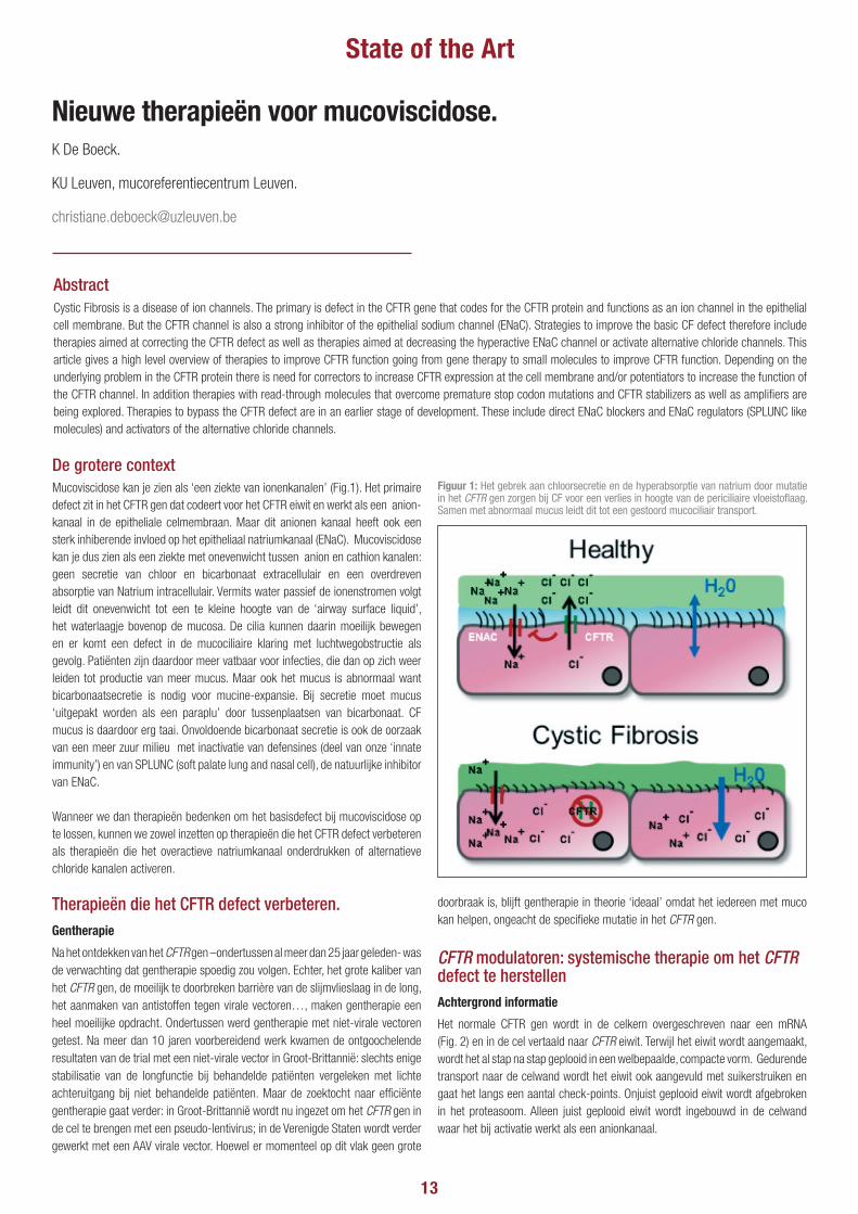
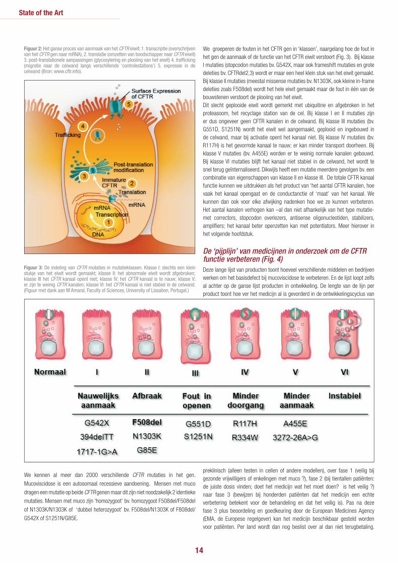
Mucoviscidose kan je zien als ‘een ziekte van ionenkanalen’ (Fig.1). Het primaire defect zit in het CFTR gen dat codeert voor het CFTR eiwit en werkt als een anion-kanaal in de epitheliale celmembraan. Maar dit anionen kanaal heeft ook een sterk inhiberende invloed op het epitheliaal natriumkanaal (ENaC). Mucoviscidose kan je dus zien als een ziekte met onevenwicht tussen anion en cathion kanalen: geen secretie van chloor en bicarbonaat extracellulair en een overdreven absorptie van Natrium intracellulair. Vermits water passief de ionenstromen volgt leidt dit onevenwicht tot een te kleine hoogte van de ‘airway surface liquid’, het waterlaagje bovenop de mucosa. De cilia kunnen daarin moeilijk bewegen en er komt een defect in de mucociliaire klaring met luchtwegobstructie als gevolg. Patiënten zijn daardoor meer vatbaar voor infecties, die dan op zich weer leiden tot productie van meer mucus. Maar ook het mucus is abnormaal want bicarbonaatsecretie is nodig voor mucine-expansie. Bij secretie moet mucus ‘uitgepakt worden als een paraplu’ door tussenplaatsen van bicarbonaat. CF mucus is daardoor erg taai. Onvoldoende bicarbonaat secretie is ook de oorzaak van een meer zuur milieu met inactivatie van defensines (deel van onze ‘innate immunity’) en van SPLUNC (soft palate lung and nasal cell), de natuurlijke inhibitor van ENaC.
Wanneer we dan therapieën bedenken om het basisdefect bij mucoviscidose op te lossen, kunnen we zowel inzetten op therapieën die het CFTR defect verbeteren als therapieën die het overactieve natriumkanaal onderdrukken of alternatieve chloride kanalen activeren.
AbstractCystic Fibrosis is a disease of ion channels. The primary is defect in the CFTR gene that codes for the CFTR protein and functions as an ion channel in the epithelial cell membrane. But the CFTR channel is also a strong inhibitor of the epithelial sodium channel (ENaC). Strategies to improve the basic CF defect therefore include therapies aimed at correcting the CFTR defect as well as therapies aimed at decreasing the hyperactive ENaC channel or activate alternative chloride channels. This article gives a high level overview of therapies to improve CFTR function going from gene therapy to small molecules to improve CFTR function. Depending on the underlying problem in the CFTR protein there is need for correctors to increase CFTR expression at the cell membrane and/or potentiators to increase the function of the CFTR channel. In addition therapies with read-through molecules that overcome premature stop codon mutations and CFTR stabilizers as well as amplifiers are being explored. Therapies to bypass the CFTR defect are in an earlier stage of development. These include direct ENaC blockers and ENaC regulators (SPLUNC like molecules) and activators of the alternative chloride channels.
De grotere contextFiguur 1: Het gebrek aan chloorsecretie en de hyperabsorptie van natrium door mutatie in het CFTR gen zorgen bij CF voor een verlies in hoogte van de periciliaire vloeistoflaag. Samen met abnormaal mucus leidt dit tot een gestoord mucociliair transport.
Therapieën die het CFTR defect verbeteren.Gentherapie
Na het ontdekken van het CFTR gen –ondertussen al meer dan 25 jaar geleden- was de verwachting dat gentherapie spoedig zou volgen. Echter, het grote kaliber van het CFTR gen, de moeilijk te doorbreken barrière van de slijmvlieslaag in de long, het aanmaken van antistoffen tegen virale vectoren…, maken gentherapie een heel moeilijke opdracht. Ondertussen werd gentherapie met niet-virale vectoren getest. Na meer dan 10 jaren voorbereidend werk kwamen de ontgoochelende resultaten van de trial met een niet-virale vector in Groot-Brittannië: slechts enige stabilisatie van de longfunctie bij behandelde patiënten vergeleken met lichte achteruitgang bij niet behandelde patiënten. Maar de zoektocht naar efficiënte gentherapie gaat verder: in Groot-Brittannië wordt nu ingezet om het CFTR gen in de cel te brengen met een pseudo-lentivirus; in de Verenigde Staten wordt verder gewerkt met een AAV virale vector. Hoewel er momenteel op dit vlak geen grote
doorbraak is, blijft gentherapie in theorie ‘ideaal’ omdat het iedereen met muco kan helpen, ongeacht de specifieke mutatie in het CFTR gen.
CFTR modulatoren: systemische therapie om het CFTR defect te herstellenAchtergrond informatie
Het normale CFTR gen wordt in de celkern overgeschreven naar een mRNA (Fig. 2) en in de cel vertaald naar CFTR eiwit. Terwijl het eiwit wordt aangemaakt, wordt het al stap na stap geplooid in een welbepaalde, compacte vorm. Gedurende transport naar de celwand wordt het eiwit ook aangevuld met suikerstruiken en gaat het langs een aantal check-points. Onjuist geplooid eiwit wordt afgebroken in het proteasoom. Alleen juist geplooid eiwit wordt ingebouwd in de celwand waar het bij activatie werkt als een anionkanaal.
14
State of the Art
We kennen al meer dan 2000 verschillende CFTR mutaties in het gen.
Mucoviscidose is een autosomaal recessieve aandoening. Mensen met muco
dragen een mutatie op beide CFTR genen maar dit zijn niet noodzakelijk 2 identieke
mutaties. Mensen met muco zijn ‘homozygoot’ bv. homozygoot F508del/F508del
of N1303K/N1303K of ‘dubbel heterozygoot’ bv. F508del/N1303K of F808del/
G542X of S1251N/G85E.
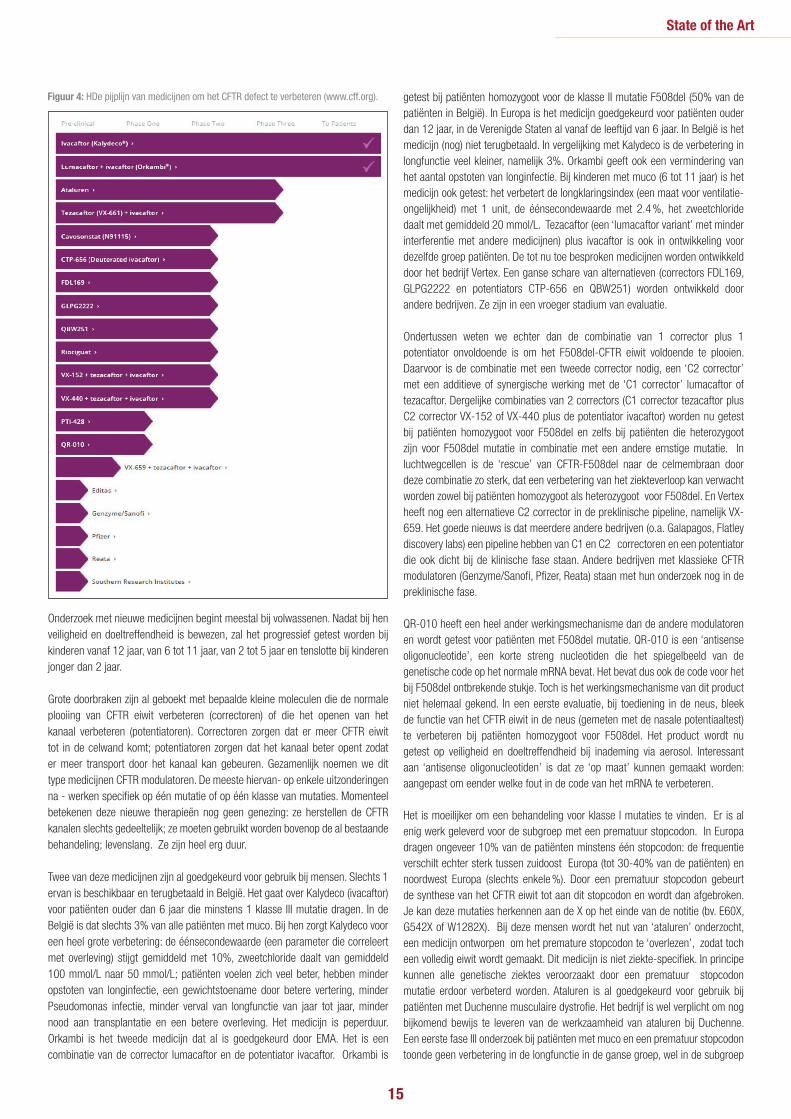
We groeperen de fouten in het CFTR gen in ‘klassen’, naargelang hoe de fout in het gen de aanmaak of de functie van het CFTR eiwit verstoort (Fig. 3). Bij klasse I mutaties (stopcodon mutaties bv. G542X, maar ook frameshift mutaties en grote deleties bv. CFTRdel2,3) wordt er maar een heel klein stuk van het eiwit gemaakt. Bij klasse II mutaties (meestal missense mutaties bv. N1303K, ook kleine in-frame deleties zoals F508del) wordt het hele eiwit gemaakt maar de fout in één van de bouwstenen verstoort de plooiing van het eiwit.Dit slecht geplooide eiwit wordt gemerkt met ubiquitine en afgebroken in het proteasoom, het recyclage station van de cel. Bij klasse I en II mutaties zijn er dus ongeveer geen CFTR kanalen in de celwand. Bij klasse III mutaties (bv. G551D, S1251N) wordt het eiwit wel aangemaakt, geplooid en ingebouwd in de celwand, maar bij activatie opent het kanaal niet. Bij klasse IV mutaties (bv. R117H) is het gevormde kanaal te nauw; er kan minder transport doorheen. Bij klasse V mutaties (bv. A455E) worden er te weinig normale kanalen gebouwd. Bij klasse VI mutaties blijft het kanaal niet stabiel in de celwand, het wordt te snel terug geïnternaliseerd. Dikwijls heeft een mutatie meerdere gevolgen bv. een combinatie van eigenschappen van klasse II en klasse III. De totale CFTR kanaal functie kunnen we uitdrukken als het product van ‘het aantal CFTR kanalen, hoe vaak het kanaal opengaat en de conductanctie of ‘maat’ van het kanaal. We kunnen dan ook voor elke afwijking nadenken hoe we ze kunnen verbeteren. Het aantal kanalen verhogen kan –al dan niet afhankelijk van het type mutatie- met correctors, stopcodon overlezers, antisense oligonucleotiden, stabilizers, amplifiers; het kanaal beter openzetten kan met potentiators. Meer hierover in het volgende hoofdstuk.
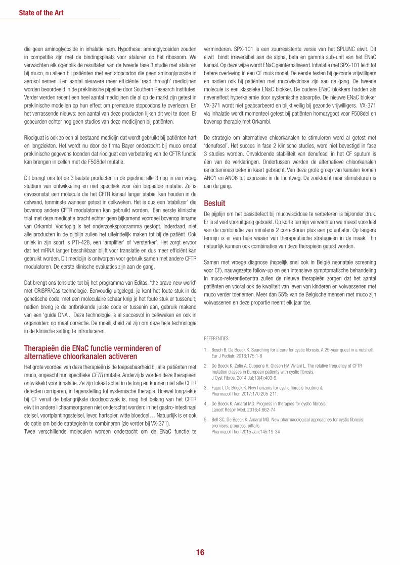
De ‘pijplijn’ van medicijnen in onderzoek om de CFTR functie verbeteren (Fig. 4)Deze lange lijst van producten toont hoeveel verschillende middelen en bedrijven werken om het basisdefect bij mucoviscidose te verbeteren. En de lijst loopt zelfs al achter op de ganse lijst producten in ontwikkeling. De lengte van de lijn per product toont hoe ver het medicijn al is gevorderd in de ontwikkelingscyclus van
preklinisch (alleen testen in cellen of andere modellen), over fase 1 (veilig bij gezonde vrijwilligers of enkelingen met muco ?), fase 2 (bij tientallen patiënten: de juiste dosis vinden; doet het medicijn wat het moet doen? is het veilig ?) naar fase 3 (bewijzen bij honderden patiënten dat het medicijn een echte verbetering betekent voor de behandeling en dat het veilig is). Pas na deze fase 3 plus beoordeling en goedkeuring door de European Medicines Agency (EMA, de Europese regelgever) kan het medicijn beschikbaar gesteld worden voor patiënten. Per land wordt dan nog beslist over al dan niet terugbetaling.
Figuur 2: Het ganse proces van aanmaak van het CFTR eiwit: 1. transcriptie (overschrijven van het CFTR gen naar mRNA), 2. translatie (omzetten van boodschapper naar CFTR eiwit) 3. post-translationele aanpassingen (glycosylering en plooiing van het eiwit) 4. trafficking (migratie naar de celwand langs verschillende ‘controlestations’) 5. expressie in de celwand (Bron: www.cftr.info).
Figuur 3: De indeling van CFTR mutaties in mutatieklassen. Klasse I: slechts een klein stukje van het eiwit wordt gemaakt; klasse II: het abnormale eiwit wordt afgebroken; klasse III het CFTR kanaal opent niet; klasse IV: het CFTR kanaal is te nauw; klasse V: er zijn te weinig CFTR kanalen; klasse VI: het CFTR kanaal is niet stabiel in de celwand. (Figuur met dank aan M Amaral, Faculty of Sciences, University of Lissabon, Portugal.)
15
State of the Art
Figuur 4: HDe pijplijn van medicijnen om het CFTR defect te verbeteren (www.cff.org).
Onderzoek met nieuwe medicijnen begint meestal bij volwassenen. Nadat bij hen veiligheid en doeltreffendheid is bewezen, zal het progressief getest worden bij kinderen vanaf 12 jaar, van 6 tot 11 jaar, van 2 tot 5 jaar en tenslotte bij kinderen jonger dan 2 jaar.
Grote doorbraken zijn al geboekt met bepaalde kleine moleculen die de normale plooiing van CFTR eiwit verbeteren (correctoren) of die het openen van het kanaal verbeteren (potentiatoren). Correctoren zorgen dat er meer CFTR eiwit tot in de celwand komt; potentiatoren zorgen dat het kanaal beter opent zodat er meer transport door het kanaal kan gebeuren. Gezamenlijk noemen we dit type medicijnen CFTR modulatoren. De meeste hiervan- op enkele uitzonderingen na - werken specifiek op één mutatie of op één klasse van mutaties. Momenteel betekenen deze nieuwe therapieën nog geen genezing: ze herstellen de CFTR kanalen slechts gedeeltelijk; ze moeten gebruikt worden bovenop de al bestaande behandeling; levenslang. Ze zijn heel erg duur.
Twee van deze medicijnen zijn al goedgekeurd voor gebruik bij mensen. Slechts 1 ervan is beschikbaar en terugbetaald in België. Het gaat over Kalydeco (ivacaftor) voor patiënten ouder dan 6 jaar die minstens 1 klasse III mutatie dragen. In de België is dat slechts 3% van alle patiënten met muco. Bij hen zorgt Kalydeco voor een heel grote verbetering: de éénsecondewaarde (een parameter die correleert met overleving) stijgt gemiddeld met 10%, zweetchloride daalt van gemiddeld 100 mmol/L naar 50 mmol/L; patiënten voelen zich veel beter, hebben minder opstoten van longinfectie, een gewichtstoename door betere vertering, minder Pseudomonas infectie, minder verval van longfunctie van jaar tot jaar, minder nood aan transplantatie en een betere overleving. Het medicijn is peperduur. Orkambi is het tweede medicijn dat al is goedgekeurd door EMA. Het is een combinatie van de corrector lumacaftor en de potentiator ivacaftor. Orkambi is
getest bij patiënten homozygoot voor de klasse II mutatie F508del (50% van de patiënten in België). In Europa is het medicijn goedgekeurd voor patiënten ouder dan 12 jaar, in de Verenigde Staten al vanaf de leeftijd van 6 jaar. In België is het medicijn (nog) niet terugbetaald. In vergelijking met Kalydeco is de verbetering in longfunctie veel kleiner, namelijk 3%. Orkambi geeft ook een vermindering van het aantal opstoten van longinfectie. Bij kinderen met muco (6 tot 11 jaar) is het medicijn ook getest: het verbetert de longklaringsindex (een maat voor ventilatie-ongelijkheid) met 1 unit, de éénsecondewaarde met 2.4 %, het zweetchloride daalt met gemiddeld 20 mmol/L. Tezacaftor (een ‘lumacaftor variant’ met minder interferentie met andere medicijnen) plus ivacaftor is ook in ontwikkeling voor dezelfde groep patiënten. De tot nu toe besproken medicijnen worden ontwikkeld door het bedrijf Vertex. Een ganse schare van alternatieven (correctors FDL169, GLPG2222 en potentiators CTP-656 en QBW251) worden ontwikkeld door andere bedrijven. Ze zijn in een vroeger stadium van evaluatie.
Ondertussen weten we echter dan de combinatie van 1 corrector plus 1 potentiator onvoldoende is om het F508del-CFTR eiwit voldoende te plooien. Daarvoor is de combinatie met een tweede corrector nodig, een ‘C2 corrector’ met een additieve of synergische werking met de ‘C1 corrector’ lumacaftor of tezacaftor. Dergelijke combinaties van 2 correctors (C1 corrector tezacaftor plus C2 corrector VX-152 of VX-440 plus de potentiator ivacaftor) worden nu getest bij patiënten homozygoot voor F508del en zelfs bij patiënten die heterozygoot zijn voor F508del mutatie in combinatie met een andere ernstige mutatie. In luchtwegcellen is de ‘rescue’ van CFTR-F508del naar de celmembraan door deze combinatie zo sterk, dat een verbetering van het ziekteverloop kan verwacht worden zowel bij patiënten homozygoot als heterozygoot voor F508del. En Vertex heeft nog een alternatieve C2 corrector in de preklinische pipeline, namelijk VX-659. Het goede nieuws is dat meerdere andere bedrijven (o.a. Galapagos, Flatley discovery labs) een pipeline hebben van C1 en C2 correctoren en een potentiator die ook dicht bij de klinische fase staan. Andere bedrijven met klassieke CFTR modulatoren (Genzyme/Sanofi, Pfizer, Reata) staan met hun onderzoek nog in de preklinische fase.
QR-010 heeft een heel ander werkingsmechanisme dan de andere modulatoren en wordt getest voor patiënten met F508del mutatie. QR-010 is een ‘antisense oligonucleotide’, een korte streng nucleotiden die het spiegelbeeld van de genetische code op het normale mRNA bevat. Het bevat dus ook de code voor het bij F508del ontbrekende stukje. Toch is het werkingsmechanisme van dit product niet helemaal gekend. In een eerste evaluatie, bij toediening in de neus, bleek de functie van het CFTR eiwit in de neus (gemeten met de nasale potentiaaltest) te verbeteren bij patiënten homozygoot voor F508del. Het product wordt nu getest op veiligheid en doeltreffendheid bij inademing via aerosol. Interessant aan ‘antisense oligonucleotiden’ is dat ze ‘op maat’ kunnen gemaakt worden: aangepast om eender welke fout in de code van het mRNA te verbeteren.
Het is moeilijker om een behandeling voor klasse I mutaties te vinden. Er is al enig werk geleverd voor de subgroep met een prematuur stopcodon. In Europa dragen ongeveer 10% van de patiënten minstens één stopcodon: de frequentie verschilt echter sterk tussen zuidoost Europa (tot 30-40% van de patiënten) en noordwest Europa (slechts enkele %). Door een prematuur stopcodon gebeurt de synthese van het CFTR eiwit tot aan dit stopcodon en wordt dan afgebroken. Je kan deze mutaties herkennen aan de X op het einde van de notitie (bv. E60X, G542X of W1282X). Bij deze mensen wordt het nut van ‘ataluren’ onderzocht, een medicijn ontworpen om het premature stopcodon te ‘overlezen’, zodat toch een volledig eiwit wordt gemaakt. Dit medicijn is niet ziekte-specifiek. In principe kunnen alle genetische ziektes veroorzaakt door een prematuur stopcodon mutatie erdoor verbeterd worden. Ataluren is al goedgekeurd voor gebruik bij patiënten met Duchenne musculaire dystrofie. Het bedrijf is wel verplicht om nog bijkomend bewijs te leveren van de werkzaamheid van ataluren bij Duchenne. Een eerste fase III onderzoek bij patiënten met muco en een prematuur stopcodon toonde geen verbetering in de longfunctie in de ganse groep, wel in de subgroep
16
die geen aminoglycoside in inhalatie nam. Hypothese: aminoglycosiden zouden in competitie zijn met de bindingsplaats voor ataluren op het ribosoom. We verwachten elk ogenblik de resultaten van de tweede fase 3 studie met ataluren bij muco, nu alleen bij patiënten met een stopcodon die geen aminoglycoside in aerosol nemen. Een aantal nieuwere meer efficiënte ‘read through’ medicijnen worden beoordeeld in de preklinische pipeline door Southern Research Institutes. Verder werden recent een heel aantal medicijnen die al op de markt zijn getest in preklinische modellen op hun effect om premature stopcodons te overlezen. En het verrassende nieuws: een aantal van deze producten lijken dit wel te doen. Er gebeurden echter nog geen studies van deze medicijnen bij patiënten.
Riociguat is ook zo een al bestaand medicijn dat wordt gebruikt bij patiënten hart en longziekten. Het wordt nu door de firma Bayer onderzocht bij muco omdat preklinische gegevens toonden dat riociguat een verbetering van de CFTR functie kan brengen in cellen met de F508del mutatie.
Dit brengt ons tot de 3 laatste producten in de pipeline: alle 3 nog in een vroeg stadium van ontwikkeling en niet specifiek voor één bepaalde mutatie. Zo is cavosonstat een molecule die het CFTR kanaal langer stabiel kan houden in de celwand, tenminste wanneer getest in celkweken. Het is dus een ‘stabilizer’ die bovenop andere CFTR modulatoren kan gebruikt worden. Een eerste klinische trial met deze medicatie bracht echter geen bijkomend voordeel bovenop inname van Orkambi. Voorlopig is het onderzoeksprogramma gestopt. Inderdaad, niet alle producten in de pijplijn zullen het uiteindelijk maken tot bij de patiënt. Ook uniek in zijn soort is PTI-428, een ‘amplifier’ of ‘versterker’. Het zorgt ervoor dat het mRNA langer beschikbaar blijft voor translatie en dus meer efficiënt kan gebruikt worden. Dit medicijn is ontworpen voor gebruik samen met andere CFTR modulatoren. De eerste klinische evaluaties zijn aan de gang.
Dat brengt ons tenslotte tot bij het programma van Editas, ‘the brave new world’ met CRISPR/Cas technologie. Eenvoudig uitgelegd: je kent het foute stuk in de genetische code; met een moleculaire schaar knip je het foute stuk er tussenuit; nadien breng je de ontbrekende juiste code er tussenin aan, gebruik makend van een ‘guide DNA’. Deze technologie is al succesvol in celkweken en ook in organoiden: op maat correctie. De moeilijkheid zal zijn om deze hele technologie in de klinische setting te introduceren.
Therapieën die ENaC functie verminderen of alternatieve chloorkanalen activerenHet grote voordeel van deze therapieën is de toepasbaarheid bij alle patiënten met muco, ongeacht hun specifieke CFTR mutatie. Anderzijds worden deze therapieën ontwikkeld voor inhalatie. Ze zijn lokaal actief in de long en kunnen niet alle CFTR defecten corrigeren, in tegenstelling tot systemische therapie. Hoewel longziekte bij CF veruit de belangrijkste doodsoorzaak is, mag het belang van het CFTR eiwit in andere lichaamsorganen niet onderschat worden: in het gastro-intestinaal stelsel, voortplantingsstelsel, lever, hartspier, witte bloedcel… Natuurlijk is er ook de optie om beide strategieën te combineren (zie verder bij VX-371).Twee verschillende moleculen worden onderzocht om de ENaC functie te
State of the Art
1. Bosch B, De Boeck K. Searching for a cure for cystic fibrosis. A 25-year quest in a nutshell. Eur J Pediatr. 2016;175:1-8
2. De Boeck K, Zolin A, Cuppens H, Olesen HV, Viviani L. The relative frequency of CFTR mutation classes in European patients with cystic fibrosis. J Cyst Fibros. 2014 Jul;13(4):403-9.
3. Fajac I, De Boeck K. New horizons for cystic fibrosis treatment. Pharmacol Ther. 2017;170:205-211.
4. De Boeck K, Amaral MD. Progress in therapies for cystic fibrosis. Lancet Respir Med. 2016;4:662-74
5. Bell SC, De Boeck K, Amaral MD. New pharmacological approaches for cystic fibrosis: promises, progress, pitfalls. Pharmacol Ther. 2015 Jan;145:19-34
REFERENTIES:
verminderen. SPX-101 is een zuurresistente versie van het SPLUNC eiwit. Dit eiwit bindt irreversibel aan de alpha, beta en gamma sub-unit van het ENaC kanaal. Op deze wijze wordt ENaC geïnternaliseerd. Inhalatie met SPX-101 leidt tot betere overleving in een CF muis model. De eerste testen bij gezonde vrijwilligers en nadien ook bij patiënten met mucoviscidose zijn aan de gang. De tweede molecule is een klassieke ENaC blokker. De oudere ENaC blokkers hadden als neveneffect hyperkalemie door systemische absorptie. De nieuwe ENaC blokker VX-371 wordt niet geabsorbeerd en blijkt veilig bij gezonde vrijwilligers. VX-371 via inhalatie wordt momenteel getest bij patiënten homozygoot voor F508del en bovenop therapie met Orkambi.
De strategie om alternatieve chloorkanalen te stimuleren werd al getest met ‘denufosol’. Het succes in fase 2 klinische studies, werd niet bevestigd in fase 3 studies worden. Onvoldoende stabiliteit van denufosol in het CF sputum is één van de verklaringen. Ondertussen werden de alternatieve chloorkanalen (anoctamines) beter in kaart gebracht. Van deze grote groep van kanalen komen ANO1 en ANO6 tot expressie in de luchtweg. De zoektocht naar stimulatoren is aan de gang.
BesluitDe pijplijn om het basisdefect bij mucoviscidose te verbeteren is bijzonder druk. Er is al veel vooruitgang geboekt. Op korte termijn verwachten we meest voordeel van de combinatie van minstens 2 correctoren plus een potentiator. Op langere termijn is er een hele waaier van therapeutische strategieën in de maak. En natuurlijk kunnen ook combinaties van deze therapieën getest worden.
Samen met vroege diagnose (hopelijk snel ook in België neonatale screening voor CF), nauwgezette follow-up en een intensieve symptomatische behandeling in muco-referentiecentra zullen de nieuwe therapieën zorgen dat het aantal patiënten en vooral ook de kwaliteit van leven van kinderen en volwassenen met muco verder toenemen. Meer dan 55% van de Belgische mensen met muco zijn volwassenen en deze proportie neemt elk jaar toe.
17
A poor start to life? Perinatal predictors of cardiovascular and renal health in childhood.Summary of PhD thesis of Anke Raaijmakers, presented on 26th of January 2017, Leuven, Belgium.
A. Raaijmakers, E. Levtchenko, L. van den Heuvel, J.A. Staessen, K. Allegaert.
Author Affiliations: Department of Pediatrics, University Hospitals Leuven, Leuven, Belgium (A.R., E.L.); KU Leuven. Department of Development and Regeneration, University of Leuven, Leuven, Belgium (A.R., E.L., B.v.d.H., K.A.). Research Unit Hypertension and Cardiovascular Epidemiology, KU Leuven Department of Cardiovascular Sciences, University of Leuven, Leuven, Belgium (J.A.S.). R&D Group VitaK, Maastricht University, Maastricht, The Netherlands (J.A.S.). Intensive Care and Department of Pediatric Surgery, Erasmus Medical Center Sophia Children’s Hospital, Rotterdam, The Netherlands (K.A.). Department of Pediatrics, Division of Neonatology, Erasmus MC Sophia Children's Hospital, Rotterdam, The Netherlands (K.A.).
Made In Belgium
KeywordsCAKUT, HNF1B, ELBW, Extremely Low Birth Weight, microcirculation, hypertension
The aims of this PhD–project were to a) determine screening criteria for HNF1B analysis in patients with congenital anomalies of kidney and urinary tract (CAKUT), b) generate an infrastructure to facilitate longitudinal follow–up of a birth cohort of former extremely low birth weight (ELBW, birth weight <1000 grams) neonates and subsequent data pooling with other cohorts and c) use this infrastructure to study the consequences of preterm birth and investigate risk factors or biomarkers to quantify problems in young adolescence ¹.
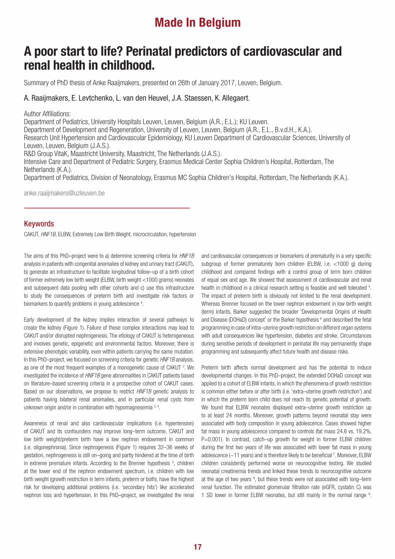
Early development of the kidney implies interaction of several pathways to create the kidney (Figure 1). Failure of these complex interactions may lead to CAKUT and/or disrupted nephrogenesis. The etiology of CAKUT is heterogeneous and involves genetic, epigenetic and environmental factors. Moreover, there is extensive phenotypic variability, even within patients carrying the same mutation. In this PhD–project, we focused on screening criteria for genetic HNF1B analysis, as one of the most frequent examples of a monogenetic cause of CAKUT 2. We investigated the incidence of HNF1B gene abnormalities in CAKUT patients based on literature–based screening criteria in a prospective cohort of CAKUT cases. Based on our observations, we propose to restrict HNF1B genetic analysis to patients having bilateral renal anomalies, and in particular renal cysts from unknown origin and/or in combination with hypomagnesemia 3, 4.
Awareness of renal and also cardiovascular implications (i.e. hypertension) of CAKUT and its confounders may improve long–term outcome. CAKUT and low birth weight/preterm birth have a low nephron endowment in common (i.e. oligonephronia). Since nephrogenesis (Figure 1) requires 32–36 weeks of gestation, nephrogenesis is still on–going and partly hindered at the time of birth in extreme premature infants. According to the Brenner hypothesis 5, children at the lower end of the nephron endowment spectrum, i.e. children with low birth weight (growth restriction in term infants, preterm or both), have the highest risk for developing additional problems (i.e. ‘secondary hits’) like accelerated nephron loss and hypertension. In this PhD–project, we investigated the renal
and cardiovascular consequences or biomarkers of prematurity in a very specific subgroup of former prematurely born children (ELBW, i.e. <1000 g) during childhood and compared findings with a control group of term born children of equal sex and age. We showed that assessment of cardiovascular and renal health in childhood in a clinical research setting is feasible and well tolerated ¹. The impact of preterm birth is obviously not limited to the renal development. Whereas Brenner focused on the lower nephron endowment in low birth weight (term) infants, Barker suggested the broader ‘Developmental Origins of Health and Disease (DOHaD) concept’ or the Barker hypothesis 6 and described the fetal programming in case of intra–uterine growth restriction on different organ systems with adult consequences like hypertension, diabetes and stroke. Circumstances during sensitive periods of development in perinatal life may permanently shape programming and subsequently affect future health and disease risks.
Preterm birth affects normal development and has the potential to induce developmental changes. In this PhD–project, the extended DOHaD concept was applied to a cohort of ELBW infants, in which the phenomena of growth restriction is common either before or after birth (i.e. ‘extra–uterine growth restriction’) and in which the preterm born child does not reach its genetic potential of growth. We found that ELBW neonates displayed extra–uterine growth restriction up to at least 24 months. Moreover, growth patterns beyond neonatal stay were associated with body composition in young adolescence. Cases showed higher fat mass in young adolescence compared to controls (fat mass 24.6 vs. 19.2%, P=0.001). In contrast, catch–up growth for weight in former ELBW children during the first two years of life was associated with lower fat mass in young adolescence (~11 years) and is therefore likely to be beneficial 7. Moreover, ELBW children consistently performed worse on neurocognitive testing. We studied neonatal creatinemia trends and linked these trends to neurocognitive outcome at the age of two years 8, but these trends were not associated with long–term renal function. The estimated glomerular filtration rate (eGFR, cystatin C) was 1 SD lower in former ELBW neonates, but still mainly in the normal range 9.
18
1. Raaijmakers A, Petit T, Gu Y, Zhang Z, Wei F, Cools B, et al. Design and feasibility of 'PREMATurity as predictor of children's Cardiovascular-renal Health' (PREMATCH): A pilot study. Blood pressure. 2015;24(5):275-83.
2. Vivante A, Kohl S, Hwang DY, Dworschak GC, Hildebrandt F. Single-gene causes of congenital anomalies of the kidney and urinary tract (CAKUT) in humans. Pediatric nephrology (Berlin, Germany). 2014;29(4):695-704.
3. Raaijmakers A, Corveleyn A, Devriendt K, van Tienoven TP, Allegaert K, Van Dyck M, et al. Criteria for HNF1B analysis in patients with congenital abnormalities of kidney and urinary tract. Nephrology, dialysis, transplantation : official publication of the European Dialysis and Transplant Association - European Renal Association. 2015;30(5):835-42.
4. Raaijmakers A, Mekahli D, Levtchenko EN. Simplified screening criteria for HNF1B analysis. Kidney international. 2015;87(6):1258-9.
5. Brenner BM, Garcia DL, Anderson S. Glomeruli and blood pressure. Less of one, more the other? American journal of hypertension. 1988;1(4 Pt 1):335-47.
6. Barker DJ, Osmond C, Golding J, Kuh D, Wadsworth ME. Growth in utero, blood pressure in childhood and adult life, and mortality from cardiovascular disease. BMJ (Clinical research ed). 1989;298(6673):564-7.
7. Catch–up growth in the first two years of life in Extremely Low Birth Weight (ELBW) infants is associated with lower body fat in young adolescence. PLoS One. 2017 Mar 9;12(3):e0173349.
8. Raaijmakers A, Ortibus E, van Tienoven TP, Vanhole C, Levtchenko E, Allegaert K. Neonatal creatinemia trends as biomarker of subsequent cognitive outcome in extremely low birth weight neonates. Early human development. 2015;91(6):367-72.
9. Raaijmakers A, Zhang ZY; Claessens, J; Cauwenberghs, N; van Tienoven, TP; Wei, FF; Jacobs, L; Levtchenko, E; Pauwels, S; Kuznetsova, T; Allegaert, K; Staessen, JA. Does Extremely Low Birth Weight Predispose to Low-Renin Hypertension? Hypertension. 2017 Mar;69(3):443-449.
REFERENCES:
Made in Belgium
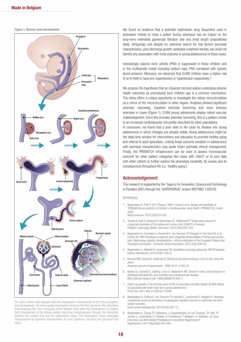
The early ureteric bud interacts with the metanephric mesenchyme to form the pronephric duct (pronephros). The more caudal mesonephric ducts are the first ‘glomeruli–like’ structures (mesonephros) that form transiently active filtration units while the metanephros is created. Early development of the kidney implies branching morphogenesis through the interaction between the ureteric bud and the metanephric mass. The metanephric mass undergoes mesenchymal–to–epithelial transformation to form nephrons, including the glomeruli and tubuli.
We found no evidence that a potential nephrotoxic drug (ibuprofen) used in premature infants to close a patent ductus arteriosus has an impact on the long–term estimated glomerular filtration rate and renal length (unpublished data). Intriguingly and despite an extensive search for risk factors (perinatal characteristics, post discharge growth, postnatal creatinine trends), we could not identify any association with renal outcome in young adolescence in these cases.
Interestingly, plasma renin activity (PRA) is suppressed in these children and in the multivariate model including sodium load, PRA correlated with systolic blood pressure. Moreover, we observed that ELBW children have a higher risk (5 to 9–fold) to have pre–hypertension or hypertension respectively 9.
We propose the hypothesis that an impaired microcirculation underlying adverse health outcomes as prematurely born children age is a common mechanism. The retina offers a unique opportunity to investigate the retinal microcirculation as a mirror of the microcirculation in other organs. Analyses showed significant arteriolar narrowing, impaired arteriolar branching and more tortuous arterioles in cases (Figure 1). ELBW young adolescents display retinal vascular maldevelopment. Since this includes arteriolar narrowing, this is a pattern similar to an increased cardiovascular risk profile described for other populations. In conclusion, we found that a poor start to life casts its shadow into young adolescence in which changes are already visible. Young adolescence might be the ideal time window for interventions and education to promote healthy aging and referral to adult specialists. Linking these outcome variables in adolescence with perinatal characteristics may guide future perinatal clinical management. Finally, the PREMATCH infrastructure can be used to assess microvascular outcome for other patient categories like cases with CAKUT or to pool data with other cohorts to further explore the phenotype variability, its causes and its consequences throughout life (i.e. ‘healthy aging’).
AcknowledgementThis research is supported by the “Agency for Innovation, Science and Technology in Flanders (IWT) through the “SAFEPEDRUG” project (IWT/SBO 120033).
Figure 1. Normal renal development

IN 2016 WAREN JULLIE MEER DAN 2000 GECONECTEERD OP
WWW.BELGIANMASTERCLASS.BE
Bijscholingen in pediatrie? Die vindt u opwww.belgianmasterclass.be
1. Ethics & economy2. Respiratory pediatrics3. Highlights from the Hot Topics in Neonatology 20134. Highlights from the Hot Topics in Neonatology 20125. Seasonality of respiratory syncytial virus (RSV) in Belgium 2016
Prof Anne Malfroot
Voorzitster van de Belgische Verenigingvoor Kindergeneeskunde
Prof Bart Van Overmeire
Vice-voorzitter van de Belgische Vereniging voor Neonatologie
Bijscholingen in pediatrie? Die vindt u opwww.belgianmasterclass.be
GO FOR HOME EDUCATION
BELGISCHE VERENIGINGVOOR KINDERGENEESKUNDESOCIÉTÉ BELGE DE PÉDIATRIE
Abbv
ie S
A/N
V - B
EGEN
150
051
- Dec
embe
r 201
6
Met de steun van:
G.B.N. - B.V.N.Groupement Belge de Néonatologie
Belgische Vereniging voor Neonatologie
20
Cochrane Corner Pediatrics
Introduction.
By Cebam, Cochrane Belgium.
http://belgium.cochrane.org
Cebam wants to introduce you to Evidence-Based Medicine by providing this monthly column. The Belgian Centre for Evidence-Based Medicine (Cebam) is an independent, multidisciplinary and in-teruniversity centre. Cebam encourages healthcare practitioners to implement Evidence-Based Medicine (EBM) in their daily practice. EBM means that decision-making is based on the best avail-able scientific evidence.
CochraneCochrane is an international network of researches, healthcare practitioners, patients, policy mak-ers and other people with an interest in healthcare. Cochrane guarantees high quality and trusted evidence. Cebam is the Belgian representative of Cochrane.Cochrane helps healthcare practitioners to make informed decisions for their patients. This means deciding to give a certain treatment because it improves the patients wellbeing. But it also means not giving a certain treatment (anymore) because the harms outweigh the benefits. This decision is based on a systematic overview of the best available scientific studies: the systematic review.
Cochrane Systematic ReviewsA systematic review is a method to collect the results from well-conducted studies about a certain clinical question. If possible, the results from different studies are being combined (meta-analysis). The advantage of this method is that the amount of patients increases. For this reason, the effects of a treatment can be estimated more precisely than when this would be done with individual studies. The conclusions of Cochrane reviews are more and more expressed through the
GRADE method. This approach divides the strength of evidence into 4 categories:• High: We have a lot of confidence that the real effect is close to the estimate.
Further re-search will most likely not change our confidence in the estimate.• Moderate: The confidence is moderate. Further research will probably have
an important impact on our confidence in the estimate and might change the estimate.
• Low: Our confidence is low. Further research will very likely have an important impact on our confidence in the estimate and will probably change the estimate.
• Very low: Any estimate of the effect is uncertain.
What is in it for me as a medical doctor?Systematic reviews are one of the sources of EBM for healthcare practitioners, as these give you an overview of the available knowledge concerning a well-defined clinical question. After reading, you would have to translate the conclusion of the review to your own daily practice. Would you take the same decisions in practice? Therefore you need to take your own experience into ac-count, in addition to the patients’ specific situation and preferences. This means ‘working evidence-based’. If your practical decisions diverge from the evidence from the literature, it is nec-essary to find out why this is the case. Importantly, there might be a very good reason for this. This process is very instructive. Therefore, as an appetizer, we provide you with a weekly column, summarizing an interesting Cochrane review that has been published last month.
21
Cochrane Corner Pediatrics
Context:Mothers and their babies are often separated at birth, where newborn babies can be held wrapped or dressed in their mother’s arms, placed in cribs or under warmers. This review, assessed as up-to-date in December 2015, investigates whether immediate (within 10 min) or early (between 10 min and 24 h) skin-to-skin contact, placing the baby naked, prone on the mother’s bare chest, improves the baby’s transition to the outside world, including chances of successfully breastfeeding the baby.
Clinical question:Is immediate or early skin-to-skin contact (SSC) with the mother following birth, beneficial for the initiation of breastfeeding and infant wellbeing in healthy newborn babies?
Immediate or early skin-to-skin contact between mothers and newborns can be recommended to promote successful breastfeeding and the infant’s overall wellbeing.B. Avau, T. Bekkering, F. Cools.
In collaboration with Cebam, Cochrane Belgium.
http://belgium.cochrane.org
This review found 38 studies, involving 3472 women. The majority investigated mothers delivering healthy, full-term babies, but 8 studies concerned women giving birth by caesarean section and 6 studied women that gave late preterm birth (≥ 35 weeks). SSC mothers had a higher chance of successfully breastfeeding their babies 1 to 4 months after birth (670 per 1000 for SSC, 95% confidence interval: 579 – 773, versus 541 per 1000 for standard contact; 887 mothers). Furthermore, mothers who gave SSC to their babies breastfed their babies on average for a longer period than mothers who gave standard contact (mean difference: 64 days, 95%CI: 38 – 90; 264 mothers). SSC mothers were also more likely to exclusively breastfeed their babies at 1 month or 6 weeks to 6 months after birth. A few studies investigated the babies’ blood glucose levels, 75 – 180 min after birth, and body temperatures, 90 – 150 min after birth. They found that blood glucose was on average higher in babies receiving SSC, compared to those receiving standard contact (MD: 10 mg/dL; 95%CI: 8 – 13; 144 mothers), a clinically significant result, while body temperature was similar (MD: 0,3 °C; 95%CI: 0,13 – 0,47; 558 mothers).
Remarks:The level of evidence presented in this review is moderate (for successfully breastfeeding, exclusively breastfeeding at 1 month or 6 weeks to 6 months after birth) to low (for duration of breastfeeding, blood glucose levels, body temperature). Evidence was downgraded because of a risk of bias due to a lack
Summary of the results:
1. Moore ER, Bergman N, Anderson GC, Medley N. Early skin-to-skin contact for mothers and their healthy newborn infants. Cochrane Database Syst Rev. 2016 Nov 25;11:CD003519
REFERENCE:
of blinding (all outcomes), small sample sizes (blood glucose levels) and a high variability in the results (body temperature). Further limitations in these studies include the fact that SSC was defined and measured in various ways, and varying levels of support for breastfeeding for the women in the standard care groups.
Conclusion:This review indicates that immediate or early SSC with the mother following birth seems to be beneficial for successfully breastfeeding newborn babies and can also have a limited effect on physiological parameters such as blood glucose. No conclusions could be made for subgroups, such as women giving birth via a caesarean section or women giving late preterm birth, due to the small sample sizes. Furthermore, no difference was found comparing immediate versus early SSC or for different lengths of contact time for SSC.
Implications for practice:Immediate or early SSC after birth should be normal practice where possible, as it can promote successful breastfeeding and the newborn’s overall well-being.
22
Activities of Paediatric Societies
De samenstelling van de academie wijzigde in 2016.Als GBPF leden zetelen nu: P. Philippet, M. Michel en C. Versteeg.De 2 laatste collega’s vervangen P. Vincke en M. Alexander.Voor de VVK zetelen A. De Guchtenaere, M. Azou en A. Rochtus.Voor de BVK: D. De Wolf, C. Van Geet en G. Casimir. M. Pletincx, D. Barillari en Y. Vandenplas vertegenwoordigen de Beroepsvereniging en tot slot zijn de diensthoofden van de universitaire diensten vertegenwoordigd door C. Vermylen, S. Vandaele en JP Misson.Het nieuwe bestuur van de academie wordt uitgemaakt door voorzitter: Y. Vandenplasondervoorzitter: M. Pletincx, schatbewaarder: P. Philippet en secretaris: S. Vandaele.
In het college zetelen de volgende leden: A. Malfroot, D. Barillari, C. Van Geet, C. Vermylen, F. Ravet, H. Van Hauthem, J. Evrard, M. Pletincx, P. Alliet en Y. Vandenplas.C Van Geet is voorzitter, M Pletincx ondervoorzitter en D. Barillari secretaris. Door aflopende mandaten dient de samenstelling van het college gewijzigd te worden in 2016. Kandidaten werden voorgedragen maar de minister heeft nog geen nieuwe leden aangeduid.
Aangezien de academie alle geledingen van de kindergeneeskunde vertegenwoordigt en het college het officieel adviesorgaan is voor de minister van Volksgezondheid, is het logisch dat beide organen nauw samenwerken. In 2016 werden zo verschillende topics gemeenschappelijk aangepakt.
Jaarverslag 2016 College en Academie Pediatrie.
C. Van Geet (voorzitter College) en Y. Vandenplas (voorzitter Academie).
Er vonden in 2016 6 vergaderingen van de Academie plaats en 6 vergaderingen van het College.
De academie Kindergeneeskunde en het College Kindergeneeskunde hebben het laatste jaar opnieuw stevig samengewerkt.
Er is een uitstekend uitgewerkte basistekst van de VVK over de huidige pediatrie. De academie wil op basis van deze tekst verder gaan en een toekomstvisie ontwerpen voor de pediatrie. Hierin komt de positionering tov urgentisten, eerste lijn en specialisten aanbod. Op Europees niveau is de visie van de eerste lijn land per land verschillend. De inbreng van de VVK-jongeren en GBPF-jongeren in dit project wordt verwacht.
Rond deze toekomstvisie op de pediatrie worden werkgroepen opgericht met één collega per deelnemende vereniging binnen de academie en één rapporteur per groep. Een bijzondere overkoepelende vergadering van de academie over dit onderwerp moet finaal leiden tot een consensus tekst. De samenstelling van de 3 werkgroepen is als volgt: eerste lijn met preventieve geneeskunde: D. Barillari, C. Versteegh, A. DeGuchtenaere, Y. Vandenplas.tweede lijn: M Pletincx, M Michel, A Covents, C. Van Geet.terde lijn met opleiding assistenten : S Vandaele, C Vermylen, P Philippet, M. Azou.
2.Erkenning subspecialiteitenDit onderwerp kwam aan bod op de jaarlijkse vergadering van de Belgische Vereniging Kindergeneeskunde, gepaard gaand met een persconferentie. De Academie en het College Pediatrie werden uitgenodigd op het kabinet van Minister
De Block op 12 oktober waarbij vele discussiepunten werden verhelderd. De erkenning van de subspecialiteiten binnen de kindergeneeskunde blijkt echter géén prioriteit te zijn. Het kabinet wenst dit te koppelen aan de "opleiding tot algemeen kinderarts".
3. Zorgprogramma pediatrie De vraag tot vernietiging van het KB zorgprogramma Pediatrie op basis van verschillende argumenten, is ontvankelijk verklaard door de Auditeur . Ondertussen is het KB zorgprogramma Pediatrie vernietigd en valt men de facto terug op het oude KB. De vernieuwde visie van de programmatie van de medische zorg in België in zorgnetwerken zal hopelijk de concentratie en de kwaliteitsoptimalisatie van de zorg voor het zieke kind kunnen bewerkstelligen.
4. Rond specifieke opdrachten die het college krijgt van de minister is in 2016 verder nagedacht over kwaliteitsindicatoren in de kindergeneeskunde alsook over multidisciplinair handboek.Een studie over de nood aan psychopedagogische omkadering van de afdelingen pediatrie is afgerond.
1.Toekomst van de kindergeneeskunde
23
Activities of Paediatric Societies
La composition de l'académie a changé en 2016.Les membres de la GBPF sont maintenant: P. Philippet, M. Michel et C. Versteeg.Ces deux derniers collègues remplacent P.Vincke . et M. Alexander.La VVK est composé de A. Guchtenaere, M. Azou et A. Rochtus.Pour La BVK: D. Wolf, C. Van Geet et G. Casimir. M. Pletincx, D. Barillari et Y. Vanden Plas représentent l'Association professionnelle et enfin les chefs des services universitaires sont C. Vermylen, S. Vandaele et JP Misson. Le nouveau conseil d'administration de l'académie se compose du Président Y. Vanden Plas du Vice-président M. Pletincx, du Trésorier P. Philippet et du ( de la ? ) secrétaire S. Vandaele.
Le collège compte les membres suivants: A. Malfroot, D. Barillari, C. Van Geet, C. Vermylen, F. Ravet, H. Van Hauthem, J. Evrard, M. Pletincx, P. Alliet et Y. Vanden Plas.C. Van Geet est Présidente, M. Pletincx, vice-président et D. Barillari, secrétaire.Vu l’échéance en 2016 de certains mandats du collège, sa composition doit être modifiée. Les candidats ont été proposés mais le ministre n'a pas encore ratifié officiellement les nouveaux membres.
Comme l'Académie représente tous les niveaux de la pédiatrie et que le collège est l'organisme consultatif officiel pour le ministre de la Santé, il est logique que les deux organismes travaillent en collaboration. Ainsi, en 2016, quelques sujets ont été traités en commun.
Rapport annuel 2016 Collège et Academie de Pediatrie.
C. Van Geet (président) et Y. Vanden Plas (président Academie).
En 2016 se sont tenues 6 réunions de l’académie et 6 réunions du collège.
L'Académie de pédiatrie et le Collège de Pédiatrie ont à nouveau bien travaillé ensemble l’année passée.
il existe un excellent texte de base élaboré par la SBP sur la situation actuelle de la pédiatrie. L'Académie veut aller plus loin sur lbase de ce texte et concevoir une vision d’avenir pour la pédiatrie. Cela concerne la position des urgentistes, la première ligne et les spécialistes. Au niveau européen, la vision de la première ligne diffère dans chaque pays. La contribution des jeunes VVK et GBPF au niveau de ce projet sera prise en compte.Pour avancer sur ce projet, des groupes de travail sont organisés avec la participation d’un collègue par association participant à l’Académie et 1 rapporteur par groupe. Une réunion de coordination doit finalement aboutir à un texte final de consensus.La composition des 3 groupes de travail est la suivante:première ligne avec la médecine préventive: D. Barillari, C. Versteegh, A. DeGuchtenaere et Y. Vanden Plas. deuxième ligne: M Pletincx, M Michel, A Covents, C. Van Geet.troisième ligne avec les assistants en formation: S. Vandaele, C. Vermylen, P. Philippet et M. Azou.
2. Reconnaissance des surspécialités.Ce sujet a été discuté lors de la réunion annuelle de la Société Belge de Pédiatrie, suivie d’une conférence de presse. L'Académie et le Collège de pédiatrie ont
été invités au cabinet du Ministre De Block le 12 octobre où de nombreuses questions ont été clarifiées. Cependant, la reconnaissance des surspécialités en pédiatrie ne serait pas une priorité. Le gouvernement voudrait lier ce sujet à “la formation du pédiatre généraliste".
3. Programme pédiatrique de soins.La demande pour annuler le programme pédiatrique de soins AR sur la base d’arguments divers, a été déclarée comme irrecevable par l’auditeur. Entretemps le programme a été annulé et, de facto, nous retombons sur l'ancien AR. La vision renouvellée du programme de soins médicaux en soins pourra, espérons-nous, répartir la Belgique dans les réseaux de concentration et optimizer la qualité des soins de l'enfant malade.
4. Les demandes spécifiques que le collège a reçues du ministre sur les indicateurs de qualité en pédiatrie ainsi que sur le manuel multidisciplinaire ont été approfondies en.Une étude sur la nécessité d'un cadre psycho-pédagogique des départements pédiatriques a été réalisée.
1.l’avenir de la pédiatrie
24
Dear colleagues
Welcome at the 45th annual congress of the Belgian Society of Paediatrics. With the general theme ‘frontiers in
paediatrics’ we want to explore the interfaces, limits and boundary lines that we face today, that impress us, for which it
may be difficult to form an opinion. Food for heart and brain. We hope that the congress will push boundaries, broaden
your horizon, let you reflect on social and ethical issues and increase your medical knowledge.
We thank the many persons who have worked hard to organize this congress.
On behalf of the Belgian Society of Paediatrics and the organizing committee,
Prof. dr. Anne Malfroot Prof. dr. Mark WojciechowskiPresident of the Belgian Society for Paediatrics Congress president
The Congress President’s address
45th Congress of the Belgian Society of Paediatrics
25
ABSTRACTS
INDEX
N° Subspeciality Pages Abstracts
1 Allergology P 35 ALLERGOLOGY A01 to A04
2 Cardiology P 37 to 39 CARDIOLOGY C01 to C09
3 Dermatology P 41 DERMATOLOGY D01 to D04
4 Emergency Medicine P 43 to P 44 EMERGENCY MEDICINE J01 to J06
5 Endocrinology P 45 to P 49 ENDOCRINOLOGY E01 to E15
6 Gastroenterology P 51 to P 55 GASTROENTEROLOGY H03 to H18
7 General Paediatrics P 57 to P 64 GENERAL PAEDIATRICS GP01 to GP25
8 Genetics P 65 to P 68 GENETICS G01 to G12
9 Immunology/Infectious Diseases P 69 IMMUNOLOGY/INFECTIOUS DISEASES L01 to L03
10 Infectiology P 71 to P 78 INFECTIOLOGY I01 to I26
11 Metabolic Diseases P 79 METABOLIC DISEASES M01 to M03
12 Neonatology P 81 to P 87 NEONATOLOGY K01 to K21
13 Nephrology P 89 to P 92 NEPHROLOGY U01 to U14
14 Neurology P 93 to P 95 NEUROLOGY B01 to B07
15 Onco-Hematology P 97 to P 101 ONCO-HEMATOLOGY O01 to O18
16 Pulmonology P 103 to P 107 PULMONOLOGY P01 to P17
17 Rheumatology P 109 to P 110 RHEUMATOLOGY R01 to R07
45ème Congrès Annuel de la Société Belge de Pédiatrie 2017
45ste Jaarlijks Congres van de Belgische Vereniging voor Kindergeneeskunde 2017

PROTECT THEIRECZEMA-PRONE SKIN
FROM BIRTH ON
1 IN 5 CHILDRENAFFECTED*
EACH SKIN TYPE DESERVES ITS OWN MUSTELA CARE
MUSTELA®, IT ALL STARTS WITH SKIN
* Watson 2011, Isaac 1998** Respondents perceived the products to be effective for reducing itchy skin. Clinical study by self-assessment, conducted under dermatologic control.
60 YEARS OF DERMATOLOGICAL RESEARCH
NORMAL SKIN ECZEMA-PRONE SKINDRY SKIN
OUR COMMITMENTS
PHENOXYETHANOL- PHTHALATEFRAGRANCE - PARABENPRIORITY GIVEN TO INGREDIENTS OF
NATURAL ORIGIN
NEW FORMULAS
R E L I E V E SITCHING **
AGREE
ECZEMA-PRONE SKIN
MUSTELA®, IT ALL STARTS WITH SKIN
pa-journal-A4-UK.indd 1 1/09/16 11:20
27
1. ALLERGOLOGY
Allergology A01 SenSitizAtion to non-Specific lipid trAnSfer proteinS (nS-ltp) in children And AdultS in A north-weStern europeAn country.A. VAN GASSE 1.1 University Hospital Antwerp (UZA), department of paediatrics, Antwerpen, Belgium, Paediatrics.
Introduction: Non-specific lipid transfer proteins (ns-LTP) are important allergen components in plant foods and pollen. Epidemiologic and clinical data are mainly coming from the Mediterranean area.
Aim: To study the prevalence and clinics of IgE reactivity to ns-LTPs in (pre)school children and adults with symptoms of pollen and/or plant food allergy in a north-western European country.
Methods: Patients with pollen and/or plant food allergy were included according to a definite history and tested for specific IgE (sIgE) reactivity to 6 ns-LTPs (rAra h 9 from peanut (Arachis hypogeae), rCor a 8 from hazelnut (Corylus avellana), rMal d 3 from apple (Malus domesticus) and rPru p 3 from peach (Prunus persica), nArt v 3 from mugwort (Artemisia vulgaris) and rPar j 2 from wall pellitory (Parietaria judaica)). Clinical significance of sIgE reactivity to Pru p 3, the ns-LTP of peach, was assessed by basophil activation test (BAT).
Results: In 177/718 (25%, 95%CI 22-28) patients with symptoms of pollen and/or food allergy a positive sIgE to minimally one ns-LTP was demonstrated (ns-LTP+ patients). No significant differences in sIgE reactivity to ns-LTP(s) were observed between the 3 different clinical phenotypes, nor between sexes or different age groups. Except, in children at preschool age in whom sIgE reactivity to ns-LTP was more frequent in those with only plant food allergy (47%, 95%CI 30-64) as compared to the other 2 clinical phenotypes (< 15%) (P<0.05). Pru p 3 was recognized in 86% (95%CI 80-90) of ns-LTP+ patients. However, as for the other ns-LTPs, in most of the cases sIgE to Pru p 3 was clinically irrelevant. BAT with rPru p 3 was positive in 4/4 patients with generalized reactions and in 2/7 patients with an oral allergy syndrome (OAS) to peach. BAT was negative in 8/8 Pru p 3+ patients tolerating peach.
Conclusions: Specific IgE reactivity to ns-LTPs is prevalent in children and adults with pollen and/or plant food allergic symptoms from our region. Ns-LTP+ patients do generally not react to the respective plant food(s). Functional evaluation by BAT might help to elucidate the clinical relevance of a positive ns-LTP IgE result.
Allergology A02 where there’S Smoke there’S fire: cAnnAbiS Allergy through pASSive expoSure.I. DECUYPER ¹, M. FABER ², V. SABATO ³, C. BRIDTS 4, M. HAGENDORENS ¹, H. RIHS 5, L. DE CLERCK ³, D. EBO ³.
¹ Universitair Ziekenhuis Antwerpen, Edegem, Belgium, Pediatrie, Immunologie-Allergologie-Reumatologie, ² Universitair Ziekenhuis Antwerpen, Edegem, Belgium, Dermatologie, ³ Universitair Ziekenhuis Antwerpen, Edegem, Belgium, Immunologie-Allergologie-Reumatologie, 4 Universiteit Antwerpen, Antwerpen, Belgium, Immunologie-Allergologie-Reumatologie, 5 IPA-Institute for Prevention and Occupational Medicine, German Social Accident Insurance, Ruhr University Bochum, Bochum, Germany, Prevention and Occupational Medicine.
Results: Marihuana allergy seems on the rise and has been attributed to a sensitization to Can s 3, a non-specific lipid protein (ns-LTP) from Cannabis sativa. Sensitization and elicitation of allergic symptoms seem to occur mainly through direct exposure. We report one child and one adult in whom allergy could have resulted from passive exposure to cannabis. History was taken and skin prick tests with cannabis extracts rich in ns-LTP were performed. Total and specific IgE´s were quantified for industrial hemp and recombinant (r), or native (n) components: rCan s 3 (Cannabis sativa), rPru p 3 (Prunus persica), bromelain, rBet v 1 and rBet v 2 (Betula verrucosa), rPhl p 1 and rPhl p 5b (Phleum pratensis), nArt v 1 and nArt v 3 (Artemisia vulgaris). Finally, basophil activation tests were performed with rCan s 3. Both patients denied any previous active cannabis exposure, but clearly described repeated passive exposure to cannabis smoke. They presented with respiratory and/or cutaneous allergic symptoms upon cannabis smoke and also reported severe allergic symptoms to different plant-foods. One patient, currently 16 years old, reported symptoms to cannabis smoke since the age of 8 years old. All patients showed positive skin prick test results for cannabis, sIgE reactivity to rCan s 3 and basophil activation upon stimulation with rCan s 3. Although it is difficult to demonstrate with certainty, we believe that our case histories suggest that cannabis allergy might result from passive exposure and may occur both in children and adults.
Allergology A03 legume Allergy: croSS-reActivity in A flemiSh peA Allergic populAtion.I. DECUYPER ¹, Y. VAN LOOCKE ², E. GOOSSENS ², C. MERTENS ³, M. HAGENDORENS ¹, D. EBO 4. 1 University Hospital Antwerp (UZA), department of paediatrics, Antwerpen, Belgium, Pediatrie, Immunologie-Allergologie-Reumatologie, ² Universiteit Antwerpen, Antwerpen, Belgium, Geneeskunde, ³ Universiteit Antwerpen, Antwerpen, Belgium, Immunologie-Allergologie-Reumatologie, 4 UZA, Edegem, Belgium, Immunologie-Allergologie-Reumatologie.
Introduction: Prevalence of legume allergies varies geographically and often correlates with dietary habits. In spite op peanut allergy being to most prevalent food allergy worldwide, southern European studies report lentil allergy to be the most prevalent legume allergy in their allergic population. Furthermore, they suggest frequent cross-reactivity mainly with chickpea and pea and rarely with peanut or soy. These studies also show that most legume allergic patients are children.
Aim: To evaluate allergenic serologic and clinical cross-reactivity in pea allergic patients in Flanders.
Methods: Patients with a compelling history of pea-allergy documented by positive pea-specific (s)IgE (>0,10 KUA/L, FEIA ImmunoCAP) were included. History was complemented by a standardized questionnaire on food allergy, more specifically inquiring about contact with other legumes (peanut, soy, lentil and chickpea) and the subsequent occurrence of allergic symptoms. Total and sIgE were measured to chickpea, lentil, soy and peanut (ImmunoCAP).
Results: 17 patients were included, 9 of which were children (mean age: 6 years) and 8 adults (mean age: 32 years). The most frequent reported symptoms on contact with pea comprised an oral allergy syndrome (71%). 82% of patients also reported allergic symptoms to at least one other legume confirmed by a corresponding positive sIgE. 41% even reported allergic symptoms to two or more other legumes confirmed by corresponding positive sIgE’s. Peanuts were reported as most frequent clinically cross-reactive legume, followed by soy, lentil and finally chickpea. However, only 24% of patients reported to have had regular contact with all five questioned legumes.
Conclusions: It appears that clinical cross-reactivity among legume allergy is common in our population. In contrary to the southern Europe studies, cross-reactivity with other legumes than chickpea/lentil such as peanut and soy seem to play a role as well. Larger studies combining history with IgE measurement and oral provocation tests are necessary to confirm these findings and further elucidate the cross-reactivity profiles.
Allergology A04 AnticonvulSAnt hyperSenSitivity Syndrome: A cASe report.R. PROOST ¹, D. BOSHOFF ², K. VANDEN DRIESSCHE ³.
¹ Virga Jesse Hospital, Hasselt, Belgium, Pediatrics, ² Virga Jesse Hospital, Hasselt, Belgium, Pediatrics, cardiology, ³ Virga Jesse Hospital, Hasselt, Belgium, Pediatrics, infectiology.
Results: Introduction Hypersensitivity reactions to anticonvulsants are often severe and possibly life threatening. Delayed diagnosis should be avoided. In light of a recent case, diagnosis of Anticonvulsant Hypersensitivity Syndrome (ACHSS), possible risk factors and treatment guidelines are discussed. Case presentation A 12-year old girl presented because of fever and rash. The girl was known with petits mals and mild developmental delay because of a 20pter13 chromosomal deletion. For a short period of time she had been treated with valproate in the past. She was started on lamotrigine 3 prior to the first appearance of symptoms, because of a first grand mal. Besides fever and rash, she complained of sore throat and malaise. The rash was scarlatiniform. Lab showed leukopenia with normal formula and mild trombopenia. CRP was 5.6 mg/L. The lamotrigine was switched to levetiracetam because a drug reaction was suspected. There was no resolution of fever nor rash, instead symptoms only worsened. In the meantime diagnoses of scarlatina, Epstein-Barr and erythema infectiosum were considered. Symptoms shortly improved but she revisited our emergency department 3 weeks after the initial appearance of symptoms. She had a diffuse scarlet, scaly dermatitis, hepatosplenomegaly, diffuse adenopathies and signs of strawberry tongue and conjunctivitis. Lab showed some leukocytosis with atypical lymphocytes, CRP was 4.9 mg/L. Furthermore there was a high ferritine of 350 μg/L and elevated liver enzymes. She was started on broad-spectrum antibiotics for possible Streptococcal/Staphylococcal toxic shock syndrome and was treated with IVIG IV for possible Kawasaki disease. Echocardiogram showed no coronaritis or other cardiac abnormalities. Because of low inflammatory parameters, negative cultures and insufficient improvement after IVIG IV, the diagnosis of Anticonvulsant Hypersensitivity Syndrome (ACHSS) after treatment with lamotrigine was made. The patient was started on methylprednisolone 2 mg/kg/day. The patient’s liver tests worsened after initial start of treatment and developed to fulminant hepatitis with ascites. After 48 hours considerable clinical improvement was seen and liver tests started to decline. Four days later the patient was discharged without rash with tapering of the corticosteroids. There was reappearance of the rash a week after discharge and methylprednisolone was again augmented and slower tapering was planned. A month after discharge there were no symptoms and liver enzymes normalized. Discussion Anticonvulsant hypersensitivity syndrome (ACHSS) is also known as Drug Reaction with Eosinophilia and Systemic Symptoms (DRESS). Antiepileptic drugs (AED), and more specific AEDs of the aromatic group (eg. lamotrigine, phenytoin, phenobarbital, …) are especially causative in these drug reactions. The formation of toxic metabolites (arene oxides) and formation neoantigens after altering of cytochrome P450 enzyme are etiological hypotheses. Certain HLA alleles have been reported to have high association with adverse reactions to AEDs, specific alleles giving higher incidence of reactions to lamotrigine. Most common clinical findings in ACHSS are morbilliform rash, hematological abnormalities, adenopathies and visceral involvement, most frequently hepatitis. Although eosinophilia is common, it is not required for diagnosis. Different scoring systems for aiding an often difficult diagnosis exist. Treatment of ACHSS/DRESS consists of immediate cessation of the causative drug and administration of systemic corticosteroids, preferably prednisolone or methylprednisolone at a minimum dose of 1 mg/kg/day. Slow tapering of corticosteroids over 3 to 6 months might be necessary to avoid relapse. Conclusion After initiation of AEDs and especially when starting AEDs of the aromatic drug class, strict follow up is required to diagnose and treat possible ACHSS/DRESS because of often severe visceral damage. Treatment exists of systemic corticosteroids and after recovery, slow tapering of corticosteroids is important to prevent relapse.
Bexsero® :premier vaccin contre le méningocoque de
sérogroupe B.
Le seul indiqué dès l’âge de 2 mois.1,2
premier vaccin contre le méningocoque de
sérogroupe B
Le seul indiqué dès l’âge de 2 mois.
Se référer à la notice la plus récente pour la mise à jour des effets secondaires possibles et des précautions.RÉSUMÉ DES CARACTÉRISTIQUES DU PRODUIT Veuillez vous référer au Résumé des Caractéristiques du Produit pour une information complète concernant l’usage de ce médicament Ce médicament fait l’objet d’une surveillance supplémentaire qui permettra l’identi� cation rapide de nouvelles informations relatives à la sécurité. Les professionnels de la santé déclarent tout effet indésirable suspecté. Voir rubrique « Effets Indésirables » pour les modalités de déclaration des effets indésirables. DÉNOMINATION DU MÉDICAMENT Bexsero suspension injectable en seringue préremplie Vaccin méningococcique groupe B (ADNr, composant, adsorbé) EU/1/12/812/001 Classe pharmacothérapeutique : vaccins méningococciques, Code ATC : J07AH09 COMPOSITION QUALITATIVE ET QUANTITATIVE Une dose (0,5 ml) contient : Protéine de fusion recombinante NHBA de Neisseria meningitidis groupe B 1, 2, 3 50 microgrammes Protéine recombinante NadA de Neisseria meningitidis groupe B 1, 2, 3 50 microgrammes Protéine de fusion recombinante fHbp de Neisseria meningitidis groupe B 1, 2, 3 50 microgrammes Vésicules de membrane externe (OMV) de Neisseria meningitidis groupe B, souche NZ98/254 mesurée en tant que proportion de l’ensemble des protéines contenant l’antigène PorA P1.4 2 25 microgrammes 1 produite dans des cellules d’E. coli par la technique de l’ADN recombinant 2 adsorbée sur hydroxyde d’aluminium (0,5 mg Al³+) 3 NHBA (antigène de liaison à l’héparine de Neisseria), NadA (adhésine A de Neisseria), fHbp (protéine de liaison du facteur H) INDICATIONS THÉRAPEUTIQUES Bexsero est indiqué pour l’immunisation active des sujets à partir de l’âge de 2 mois contre l’infection invasive méningococcique causée par Neisseria meningitidis de groupe B. L’impact de l’infection invasive à différentes tranches d’âge ainsi que la variabilité épidémiologique des antigènes des souches du groupe B dans différentes zones géographiques doivent être pris en compte lors de la vaccination. Voir rubrique 5.1 du RCP complet pour plus d’informations sur la protection contre les souches spéci� ques au groupe B. Ce vaccin doit être utilisé conformément aux recommandations of� cielles. POSOLOGIE ET MODE D’ADMINISTRATION Posologie
Tableau 1. Résumé de la posologie
Tranche d’âge Primovaccination Intervalles entre les doses de primovaccination Rappel
Nourrissons de 2 à 5 mois Trois doses de 0,5 ml chacune, avec une première dose administrée à l’âge de 2 mois a 1 mois minimum Oui, une dose entre 12 et 15 mois b, c
Nourrissons non vaccinés de 6 à 11 mois Deux doses de 0,5 ml chacune 2 mois minimumOui, une dose au cours de la deuxième année avec un
intervalle d’au moins 2 mois entre la primovaccination et la dose de rappel c
Enfants non vaccinés de 12 à 23 mois Deux doses de 0,5 ml chacune 2 mois minimum Oui, une dose avec un intervalle de 12 à 23 mois entre la primovaccination et la dose de rappel c
Enfants de 2 à 10 ans Deux doses de 0,5 ml chacune 2 mois minimum Besoin non établi d
Adolescents (à partir de 11 ans) et adultes* Deux doses de 0,5 ml chacune 1 mois minimum Besoin non établi d
a La première dose doit être administrée à l’âge de 2 mois. La sécurité et l’ef� cacité de Bexsero chez les nourrissons de moins de 8 semaines n’ont pas encore été établies. Aucune donnée n’est disponible. b En cas de retard, la dose de rappel ne doit pas être administrée au-delà de 24 mois. c La nécessité et le moment d’administration d’une dose de rappel n’ont pas encore été déterminés. d Voir rubrique 5.1 du RCP complet * Il n’existe aucune donnée chez les adultes de plus de 50 ans Mode d’administration Le vaccin est administré par une injection intramusculaire profonde, de préférence dans la face antérolatérale de la cuisse chez le nourrisson ou dans la région du muscle deltoïde du haut du bras chez les sujets plus âgés. Des sites d’injection distincts doivent être utilisés si plusieurs vaccins sont administrés simultanément. Le vaccin ne doit pas être injecté par voie intraveineuse, sous-cutanée ni intradermique et ne doit pas être mélangé avec d’autres vaccins dans la même seringue. Pour les instructions concernant la manipulation du vaccin avant administration, voir la rubrique 6.6 du RCP complet CONTRE-INDICATIONS Hypersensibilité aux substances actives ou à l’un des excipients. MISES EN GARDE SPÉCIALES ET PRÉCAUTIONS D’EMPLOI Comme pour les autres vaccins l’administration de Bexsero doit être reportée chez des sujets souffrant de maladie fébrile sévère aiguë. Toutefois, la présence d’une infection mineure, telle qu’un rhume, ne doit pas entraîner le report de la vaccination. Ne pas injecter par voie intravasculaire. Comme pour tout vaccin injectable, un traitement médical approprié et une surveillance adéquate doivent toujours être disponibles en cas de réaction anaphylactique consécutive à l’administration du vaccin. Des réactions en rapport avec l’anxiété, y compris des réactions vaso-vagales (syncope), de l’hyperventilation ou des réactions en rapport avec le stress peuvent survenir lors de la vaccination comme réaction psychogène à l’injection avec une aiguille (voir rubrique « Effets indésirables »). Il est important que des mesures soient mises en place a� n d’éviter toute blessure en cas d’évanouissement. Ce vaccin ne doit pas être administré aux patients ayant une thrombocytopénie ou tout autre trouble de la coagulation qui serait une contre-indication à une injection par voie intramusculaire, à moins que le béné� ce potentiel ne soit clairement supérieur aux risques inhérents à l’administration. Comme tout vaccin, la vaccination avec Bexsero peut ne pas protéger tous les sujets vaccinés. Il n’est pas attendu que Bexsero assure une protection contre la totalité des souches de méningocoque B en circulation. Comme pour de nombreux vaccins, les professionnels de santé doivent savoir qu’une élévation de la température corporelle peut survenir suite à la vaccination des nourrissons et des enfants (de moins de 2 ans). L’administration d’antipyrétiques à titre prophylactique pendant et juste après la vaccination peut réduire l’incidence et la sévérité des réactions fébriles post-vaccinales. Un traitement antipyrétique doit être mis en place conformément aux recommandations locales chez les nourrissons et les enfants (de moins de 2 ans). Les personnes dont la réponse immunitaire est altérée soit par la prise d’un traitement immunosuppresseur, une anomalie génétique ou par d’autres causes, peuvent avoir une réponse en anticorps réduite après vaccination. Des données d’immunogénicité sont disponibles chez les patients présentant un dé� cit en complément, une asplénie ou une dysfonction splénique. Il n’existe aucune donnée sur l’utilisation de Bexsero chez les sujets de plus de 50 ans et il existe des données limitées chez les patients atteints de maladies chroniques. Le risque potentiel d’apnée et la nécessité d’une surveillance respiratoire pendant 48 à 72 heures doivent soigneusement être pris en compte lors de l’administration des doses de primovaccination chez des grands prématurés (nés à 28 semaines de grossesse ou moins), en particulier chez ceux ayant des antécédents d’immaturité respiratoire. En raison du béné� ce élevé de la vaccination chez ces nourrissons, l’administration ne doit pas être suspendue ou reportée. Le capuchon de la seringue peut contenir du latex de caoutchouc naturel. Bien que le risque de développer des réactions allergiques soit très faible, les professionnels de santé doivent évaluer le rapport béné� ces/risques avant d’administrer ce vaccin à des sujets présentant des antécédents connus d’hypersensibilité au latex. La kanamycine est utilisée au début du procédé de fabrication et est éliminée au cours des étapes ultérieures de la fabrication. Les taux de kanamycine éventuellement détectables dans le vaccin � nal sont inférieurs à 0,01 microgramme par dose. L’innocuité de Bexsero chez les sujets sensibles à la kanamycine n’a pas été établie. EFFETS INDÉSIRABLES Résumé du pro� l de sécurité La sécurité de Bexsero a été évaluée lors de 14 études, dont 10 essais cliniques randomisés contrôlés portant sur 8776 sujets (âgés de 2 mois minimum) ayant reçu au moins une dose de Bexsero. Parmi les sujets vaccinés par Bexsero, 5849 étaient des nourrissons et des enfants (de moins de 2 ans), 250 étaient des enfants (entre 2 et 10 ans) et 2677 étaient des adolescents et des adultes. Parmi les nourrissons ayant reçu les doses de primovaccination de Bexsero, 3285 ont reçu une dose de rappel au cours de leur deuxième année de vie. En outre, les données d’une étude ultérieure portant sur 207 enfants exposés à Bexsero ont été évaluées. Chez les nourrissons et les enfants (de moins de 2 ans), les réactions indésirables locales et systémiques les plus fréquemment observées lors des essais cliniques étaient : sensibilité et érythème au site d’injection, � èvre et irritabilité. Dans les études cliniques menées chez les nourrissons vaccinés à 2, 4 et 6 mois, la � èvre (≥ 38 °C) était rapportée chez 69% à 79 % des sujets lorsque Bexsero était co-administré avec des vaccins de routine (contenant les antigènes suivants : pneumococcique heptavalent conjugué, diphtérie, tétanos, coqueluche acellulaire, hépatite B, poliomyélite inactivée et Haemophilus in� uenzae de type b), contre 44% à 59 % des sujets recevant les vaccins de routine seuls. Une utilisation plus fréquente d’antipyrétiques était également rapportée chez les nourrissons vaccinés avec Bexsero et des vaccins de routine. Lorsque Bexsero était administré seul, la fréquence de la � èvre était similaire à celle associée aux vaccins de routine administrés aux nourrissons pendant les essais cliniques. Les cas de � èvre suivaient généralement un schéma prévisible, se résolvant généralement le lendemain de la vaccination. Chez les adolescents et les adultes, les réactions indésirables locales et systémiques les plus fréquemment observées étaient : douleur au point d’injection, malaise et céphalée. Aucune augmentation de l’incidence ou de la sévérité des réactions indésirables n’a été constatée avec les doses successives du schéma de vaccination. Liste tabulée des effets indésirables Les effets indésirables (consécutifs à la primovaccination ou à la dose de rappel) considérés comme étant au moins probablement liés à la vaccination ont été classés par fréquence. Les fréquences sont dé� nies comme suit : Très fréquent : (≥ 1/10) Fréquent : (≥ 1/100 à < 1/10) Peu fréquent : (≥ 1/1 000 à < 1/100) Rare : (≥ 1/10 000 à < 1/1 000) Très rare : (< 1/10 000) Fréquence indéterminée : (ne peut être estimée sur la base des données disponibles) Dans chaque groupe de fréquence, les effets indésirables sont présentés par ordre de sévérité décroissante. Outre les événements rapportés lors des essais cliniques, les réactions spontanées rapportées dans le monde pour Bexsero depuis sa commercialisation sont décrites dans la liste ci dessous. Comme ces réactions ont été rapportées volontairement à partir d’une population de taille inconnue, il n’est pas toujours possible d’estimer de façon � able leur fréquence. Ces réactions sont, en conséquence, listées avec une fréquence indéterminée. Nourrissons et enfants (jusqu’à l’âge de 10 ans) Affections du système immunitaire Fréquence indéterminée : réactions allergiques (y compris réactions anaphylactiques) Troubles du métabolisme et de la nutrition Très fréquent : troubles alimentaires Affections du système nerveux Très fréquent : somnolence, pleurs inhabituels, céphalée Peu fréquent : convulsions (y compris convulsions fébriles) Fréquence indéterminée : épisode d’hypotonie-hyporéactivité Affections vasculaires Peu fréquent : pâleur (rare après le rappel) Rare : syndrome de Kawasaki Affections gastro-intestinales Très fréquent : diarrhée, vomissements (peu fréquents après le rappel) Affections de la peau et du tissu sous-cutané Très fréquent : rash (enfants âgés de 12 à 23 mois) (peu fréquent après le rappel) Fréquent : rash (nourrissons et enfants âgés de 2 à 10 ans) Peu fréquent : eczéma Rare : urticaire Affections musculo-squelettiques et systémiques Très fréquent : arthralgies Troubles généraux et anomalies au site d’administration Très fréquent : � èvre (≥ 38 °C), sensibilité au niveau du site d’injection (y compris sensibilité sévère au site d’injection dé� nie par des pleurs lors d’un mouvement du membre ayant reçu l’injection), érythème au site d’injection, gon� ement du site d’injection, induration au site d’injection, irritabilité Peu fréquent : � èvre (≥ 40 °C) Fréquence indéterminée : vésicules au point d’injection ou autour du site d’injection Adolescents (à partir de 11 ans) et adultes Affections du système immunitaire Fréquence indéterminée : réactions allergiques (y compris réactions anaphylactiques) Affections du système nerveux Très fréquent : céphalée Fréquence indéterminée : syncope ou réaction vaso-vagale à l’injection Affections gastro-intestinales Très fréquent : nausées Affections musculo-squelettiques et systémiques Très fréquent : myalgies, arthralgies Troubles généraux et anomalies au site d’administration Très fréquent : douleur au point d’injection (y compris douleur sévère au point d’injection dé� nie par une incapacité à mener à bien des activités quotidiennes normales), gon� ement du site d’injection, induration au point d’injection, érythème au site d’injection, malaise. Fréquence indéterminée : � èvre, vésicules au point d’injection ou autour du site d’injection Déclaration des effets indésirables suspectés La déclaration des effets indésirables suspectés après autorisation du médicament est importante. Elle permet une surveillance continue du rapport béné� ce/risque du médicament. Les professionnels de santé déclarent tout effet indésirable suspecté via le système national de déclaration : Belgique Agence fédérale des médicaments et des produits de santé Division Vigilance EUROSTATION II Place Victor Horta, 40/40 B-1060 Bruxelles Site internet: www.afmps.be e-mail: [email protected] Luxembourg Direction de la Santé – Division de la Pharmacie et des Médicaments Villa Louvigny – Allée Marconi L-2120 Luxembourg Site internet: http://www.ms.public.lu/fr/activites/pharmacie-medicament/index.html TITULAIRE DE L’AUTORISATION DE MISE SUR LE MARCHÉ GSK Vaccines S.r.l., Via Fiorentina 1, 53100 Sienne, Italie DATE D’APPROBATION DU TEXTE 02/2017 (v03) MODE DE DELIVRANCE Sur prescription médicale
« Les éléments indiqués blue foncé font partie d’une mise à jour de la notice patient. Celle-ci indique également des effets secondaires possibles tels que : moins réactif que la normale ou manque de sensibilisation, et pâleur ou coloration bleutée de la peau chez les jeunes enfants. »1. Bexsero SMPC 2. Medini D, Stella M, Wassil J, Vaccine 2015; 33; 2629-2636 Février 2017 - BE/BEX/0011/16
Prix public Belgique 86,52€Prix public Luxembourg 84,07€
2017-058_GSKBEX_Pub_A4_FR_BAT.indd 1 8/03/17 08:46
29
2. CARDIOLOGY
cArdiology c01 long-term outcome After the roSS procedure in children.M. PEERS DE NIEUWBURGH ¹, C. VÔ ¹, J. RUBAY ², A. PONCELET ², G. DE BECO ², T. SLUYSMANS ¹.1 Cliniques universitaires Saint-Luc, Woluwe-Saint-Lambert, Belgium, Cardiologie pédiatrique, ² Cliniques universitaires Saint-Luc, Woluwe-Saint-Lambert, Belgium, Chirurgie Cardiaque.
Introduction: The Ross procedure consists in transplanting the pulmonary valve in aortic position as a substitute for the deficient aortic valve. A homograft (or heterograft) is placed in pulmonary position. The advantages are the growth potential of the autograft, the low risk of endocarditis and the absence of anticoagulant treatment. The main complications are the dilation of the ascending aorta and the degeneration of the pulmonary homograft.
Aim: We evaluate the long-term survival, the need for re-intervention and the dilation of the ascending aorta after the Ross procedure in children.
Methods: A retrospective study was conducted on the 57 children who underwent the Ross procedure between 1991 and 2015 in Cliniques St-Luc. The Kaplan-Meier method was used to study the survival of the cohort. A cross-sectional analysis of echocardiographic measurements, converted into Z-scores, was performed.
Results: The mean age at Ross was 9.9 years (IQR 25-75: 5-14,6 years, range 2 days to 17 years) and the mean follow-up was 14 years. Amongst the children studied, 28% had associated lesions (Shone’s syndrome), 44% had a bicuspid valve, 96% were operated by the same surgeon. The overall survival rate was 93% at 1 year, 91% at 5 years, 89% at 10 years, and 86% at 20 years. The early mortality was 7%, mainly observed in Shone’s syndrome and in patients (n=12) requiring a Ross-Konno operation (50% mortality). Late mortality was 5%. The survival rate without re-intervention on the autograft was 100% at 5 years, 95% at 10 years and 76% at 20 years (valvular plasty n=1, ascending aorta prosthesis n=3, Tirone David n=4). The most frequent re-interventions concerned the pulmonary homograft (n=43, 67%). The aorta was abnormally dilated (aortic root, sino-tubular junction and ascending aorta: median> Z-score +2) but no increase in dilation was observed during follow-up, with mean and median values similar at 1, 5, 10, 15 and 20 years. Five patients (8.8%) had endocarditis on the right tract (4 homograft, 1 Contegra). Four patients had to undergo heart transplant surgery (n=2) or mitral valve surgery (n=2). There was no aortic stenosis nor significant aortic regurgitation ( >2/4) observed in 100% of the patients at 5 years, 97% at 15 years, 95% at 20 years. The left ventricular function remained stable and the NYHA status normal (NYHA I 83%, NYHA II 17%).
Conclusions: Our results confirm a more favorable outcome in children after the Ross procedure than with other techniques (mechanical valve, homograft). An enlargement of the aortic root and the ascending aorta was observed pre-existing to the operation without significant increase of the dilation during the follow-up. The majority of re-interventions were done on the pulmonary homograft. The incidence of pulmonary valve endocarditis rate was significant. Long-term follow-up of those patients is mandatory.
cArdiology c02 mAgnetic reSonAnce imAging (mri) evAluAtion of normAl biventriculAr function in heAlthy children from 0 to 18 yeArS of Age.C. VÔ ¹, C. DE FAYS ², S. MONIOTTE ².
¹ Cliniques universitaires Saint-Luc, Woluwe-Saint-Lambert, Belgium, Paediatric cardiology, ² Cliniques Universitaires Saint-Luc, , Belgium, Paediatric cardiology.
Introduction: Cardiac magnetic resonance imaging (cMRI) has emerged in the past decades as the gold standard technique to evaluate cardiac function. The excellent temporal resolution, along with a very good blood-myocardium contrast, as well as the non-invasive and the radiation-free character of the technique are only some of the numerous advantages offered by MRI. In addition, the assessment of cardiac function is an important part of almost every routine cardiac clinical examination. To date, only few studies measured the biventricular function, volumes and masses is small groups of healthy children, using various acquisition techniques. This lack of normative data is a limiting factor in the adequate use of cMRI and many decision-taking processes.
Aim: The aim of this study was to establish normative paediatric values for biventricular volumes, masses and function in MRI from healthy children between 0 and 18 years.
Methods: We retrospectively studied short-axis cMRI performed at 1.5T in 158 children aged from 18 days to 18 years, all having a normal anatomy and function as assessed by echocardiograms and no cardiovascular history. We measured end-systolic and end-diastolic volumes and masses for the right and left ventricles, as previously well described in the literature. For each parameter, the mean value and standard deviations were determined, for each gender, and age group (0-6, 7-12, 13-18 years old).
Results: Mean values for biventricular end-systolic, end-diastolic volumes, ejection fractions and masses were indexed to body surface area (BSA), size and weight. We found good correlations between age and BSA and size-indexed left and right ventricles, as well as BSA and weight-indexed biventricular masses. By contrast, biventricular ejection fraction was stable throughout the age groups. Although ejection fraction was not different in boys and girls, left ventricular masses were significantly higher in boys, including when indexed to BSA and weight.
Conclusions: We determined normative biventricular volumic, functional and mass values assessed by 1.5T cMRI in a healthy paediatric population. Our values were in line with previously published studies. Our data will be used in the future to determine percentile curves for indexed values. This is helpful to take clinical decisions in patient management, especially in the patient operated for congenital heart diseases.
cArdiology c03 totAl AbSence of pericArdium in A child with repetitive lower reSpirAtory trAct infectionS.R. KINUANI ¹, M. SEGHAYE ¹, J. SACRÉ ¹, P. BRUYÈRE ², B. DARON ³. 1 University Hospital Liège, NDB, , Belgium, Department of Pediatrics, ² University Hospital Liège, NDB, , Belgium, Department of Medical Imaging, ³ Regional Hospital Center East Belgium, Verviers, , Belgium, Department of Pediatrics.
Results: Total absence of the pericardium is uncommon with an incidence of 1/10.000. As it is usually asymptomatic, its diagnosis is commonly missed. We report on the case of a young girl with frequent lower respiratory tract infections in whom total absence of pericardium was diagnosed. A 8 years old girl was referred for cardiological advice because of an enlarged heart at the chest X-Ray that was performed in the context of a pneumonia. The girl had been explored previously for non-allergic bronchial asthma and repetitive pulmonary infections. The personal history revealed transient hypogammaglobulinemia at the age of 9 months that required immunoglobulin substitution and also a gastro-esophageal reflux. Clinical examination was normal but hyperactivity of the precordium, especially of the right ventricle and a 2/6 systolic murmur over the pulmonary artery. ECG showed right axis deviation, first degree AV-Block and incomplete right bundle branch block. Echocardiography showed axial deviation of the heart, enlarged right ventricle and pulmonary artery. The right pulmonary veins appeared enlarged whereas the left ones were not seen with sufficient precision. The atrial septum was intact. An angio-multidetector CT scan was performed in order to exclude left partial pulmonary venous return. This examination allowed to exclude the diagnosis. Left pulmonary veins were shown to drain into the left atrium but their caliber was smaller than the right ones. The right pulmonary artery was larger than the left one (15 mm versus 9 mm). Hypermobility of the whole heart was suggestive for the absence of pericardium. The diagnosis of total absence of pericardium was then confirmed at cardiac MRI in supine and left lateral position. The thoracic angio-scan also showed the presence of a relative hypoplasic left lung partially compressed by the enlarged heart with hypoventilation of the lower lobe and of a bronchiectasia. The immunological exploration confirmed a hypogammaglobulinemia. A substitutive treatment was given. The patient was treated symptomatically as yet. A fixation of the heart by cardiopexia was discussed but not proposed as therapeutic option owing to the fact that total absence of pericardium are generally asymptomatic and to the presence of co-morbidities such as the hypogammaglobulinemia. The bronchiectasia could be considered a consequence of repetitive infections of the lower left lobe secondary to left lung compression by the heart displaced in the left hemithorax. Our case illustrates a rare cause of pericardial anomaly related to repetitive lower respiratory tract infections and hypogammaglobulinemia. At the best of our knowledge, a such association has not been described previously.
cArdiology c04 normAl cArdiopulmonAry exerciSe teSting pArAmeterS in A pAediAtric cohort. ApplicAtion to overweight pAtientS And pAtientS operAted for congenitAl heA.C. VÔ ¹, F. FRANCESCHINI ¹, S. MONIOTTE ².1 Cliniques universitaires Saint-Luc, Woluwe-Saint-Lambert, Belgium, Paediatric cardiology, ² Cliniques Universitaires Saint-Luc, , Belgium, Paediatric cardiology.
Introduction: Cardiopulmonary exercise testing (CPET) provides information on a patient’s capacity for dynamic exercise and can detect cardiovascular abnormalities that are not necessarily obvious at rest. Indications for CPET are wide : exercise intolerance, identification of exercise-induced arrhythmias, detection of functional capacity impairment in the children with congenital heart diseases, … Determination of normal parameters in healthy children are essential to interpret data collected in sick patients.
Aim: The aim of this study was to obtain normative paediatric values for maximal CPET. The database was then used to compare patients operated for transposition of the great arteries (D-TGA) and single ventricle (SV) with healthy children by case-control matching.
Methods: : In a retrospective study, we studied 432 healthy children (288 boys and 144 girls) from 5 to 18 years of age who performed a maximal CPET on a treadmill. 33 of these children had an BMI above P97 and were analyzed as the “Obese patients”. 24 children (16 boys and 8 girls) operated for D-TGA and 13 children operated for SV were compared to healthy children. All these children performed a maximal CPET on a treadmill following a similar modified Bruce protocol.
Results: Significant differences were observed between boys and girls for maximal oxygen uptake (peak VO2), maximal heart rate (peak HR), anaerobic threshold (AT) and maximal respiratory exchange ratio (RER max), while the maximal workload was not significantly different between genders. In girls, we found a postpubertal decrease of peak VO2 when normalized for weight, while it remained stable in boys. VO2peak was strongly correlated with age, weight, height and BSA, but appeared to be overestimated in obese children when expressed in ml/kg/min, and underestimated when normalized to weight. Children with corrected D-TGA tend to have lower exercise capacity than healthy children but the differences observed did not reach significance. As expected, children palliated for SV (Fontan operation) had a significant lower peak HR and VO2.
Conclusions: This study gives normative values for age, sex and anthropometric CPET parameters. We also describe equations predicting these values that could be used as reference when testing children on a treadmill. Peak VO2 normalized to weight was often used in published studies. However, BSA offered a better correlation to predict peak VO2. Compared to studies using a cycloergometer, CPET performed on a treadmill also seemed to favor a more maximal exercise and were possible in younger children. We think our date will be helpful in clinical decision processes faced on a daily basis in patient management, particularly after surgery for congenital heart diseases.
Bexsero® :premier vaccin contre le méningocoque de
sérogroupe B.
Le seul indiqué dès l’âge de 2 mois.1,2
premier vaccin contre le méningocoque de
sérogroupe B
Le seul indiqué dès l’âge de 2 mois.
Se référer à la notice la plus récente pour la mise à jour des effets secondaires possibles et des précautions.RÉSUMÉ DES CARACTÉRISTIQUES DU PRODUIT Veuillez vous référer au Résumé des Caractéristiques du Produit pour une information complète concernant l’usage de ce médicament Ce médicament fait l’objet d’une surveillance supplémentaire qui permettra l’identi� cation rapide de nouvelles informations relatives à la sécurité. Les professionnels de la santé déclarent tout effet indésirable suspecté. Voir rubrique « Effets Indésirables » pour les modalités de déclaration des effets indésirables. DÉNOMINATION DU MÉDICAMENT Bexsero suspension injectable en seringue préremplie Vaccin méningococcique groupe B (ADNr, composant, adsorbé) EU/1/12/812/001 Classe pharmacothérapeutique : vaccins méningococciques, Code ATC : J07AH09 COMPOSITION QUALITATIVE ET QUANTITATIVE Une dose (0,5 ml) contient : Protéine de fusion recombinante NHBA de Neisseria meningitidis groupe B 1, 2, 3 50 microgrammes Protéine recombinante NadA de Neisseria meningitidis groupe B 1, 2, 3 50 microgrammes Protéine de fusion recombinante fHbp de Neisseria meningitidis groupe B 1, 2, 3 50 microgrammes Vésicules de membrane externe (OMV) de Neisseria meningitidis groupe B, souche NZ98/254 mesurée en tant que proportion de l’ensemble des protéines contenant l’antigène PorA P1.4 2 25 microgrammes 1 produite dans des cellules d’E. coli par la technique de l’ADN recombinant 2 adsorbée sur hydroxyde d’aluminium (0,5 mg Al³+) 3 NHBA (antigène de liaison à l’héparine de Neisseria), NadA (adhésine A de Neisseria), fHbp (protéine de liaison du facteur H) INDICATIONS THÉRAPEUTIQUES Bexsero est indiqué pour l’immunisation active des sujets à partir de l’âge de 2 mois contre l’infection invasive méningococcique causée par Neisseria meningitidis de groupe B. L’impact de l’infection invasive à différentes tranches d’âge ainsi que la variabilité épidémiologique des antigènes des souches du groupe B dans différentes zones géographiques doivent être pris en compte lors de la vaccination. Voir rubrique 5.1 du RCP complet pour plus d’informations sur la protection contre les souches spéci� ques au groupe B. Ce vaccin doit être utilisé conformément aux recommandations of� cielles. POSOLOGIE ET MODE D’ADMINISTRATION Posologie
Tableau 1. Résumé de la posologie
Tranche d’âge Primovaccination Intervalles entre les doses de primovaccination Rappel
Nourrissons de 2 à 5 mois Trois doses de 0,5 ml chacune, avec une première dose administrée à l’âge de 2 mois a 1 mois minimum Oui, une dose entre 12 et 15 mois b, c
Nourrissons non vaccinés de 6 à 11 mois Deux doses de 0,5 ml chacune 2 mois minimumOui, une dose au cours de la deuxième année avec un
intervalle d’au moins 2 mois entre la primovaccination et la dose de rappel c
Enfants non vaccinés de 12 à 23 mois Deux doses de 0,5 ml chacune 2 mois minimum Oui, une dose avec un intervalle de 12 à 23 mois entre la primovaccination et la dose de rappel c
Enfants de 2 à 10 ans Deux doses de 0,5 ml chacune 2 mois minimum Besoin non établi d
Adolescents (à partir de 11 ans) et adultes* Deux doses de 0,5 ml chacune 1 mois minimum Besoin non établi d
a La première dose doit être administrée à l’âge de 2 mois. La sécurité et l’ef� cacité de Bexsero chez les nourrissons de moins de 8 semaines n’ont pas encore été établies. Aucune donnée n’est disponible. b En cas de retard, la dose de rappel ne doit pas être administrée au-delà de 24 mois. c La nécessité et le moment d’administration d’une dose de rappel n’ont pas encore été déterminés. d Voir rubrique 5.1 du RCP complet * Il n’existe aucune donnée chez les adultes de plus de 50 ans Mode d’administration Le vaccin est administré par une injection intramusculaire profonde, de préférence dans la face antérolatérale de la cuisse chez le nourrisson ou dans la région du muscle deltoïde du haut du bras chez les sujets plus âgés. Des sites d’injection distincts doivent être utilisés si plusieurs vaccins sont administrés simultanément. Le vaccin ne doit pas être injecté par voie intraveineuse, sous-cutanée ni intradermique et ne doit pas être mélangé avec d’autres vaccins dans la même seringue. Pour les instructions concernant la manipulation du vaccin avant administration, voir la rubrique 6.6 du RCP complet CONTRE-INDICATIONS Hypersensibilité aux substances actives ou à l’un des excipients. MISES EN GARDE SPÉCIALES ET PRÉCAUTIONS D’EMPLOI Comme pour les autres vaccins l’administration de Bexsero doit être reportée chez des sujets souffrant de maladie fébrile sévère aiguë. Toutefois, la présence d’une infection mineure, telle qu’un rhume, ne doit pas entraîner le report de la vaccination. Ne pas injecter par voie intravasculaire. Comme pour tout vaccin injectable, un traitement médical approprié et une surveillance adéquate doivent toujours être disponibles en cas de réaction anaphylactique consécutive à l’administration du vaccin. Des réactions en rapport avec l’anxiété, y compris des réactions vaso-vagales (syncope), de l’hyperventilation ou des réactions en rapport avec le stress peuvent survenir lors de la vaccination comme réaction psychogène à l’injection avec une aiguille (voir rubrique « Effets indésirables »). Il est important que des mesures soient mises en place a� n d’éviter toute blessure en cas d’évanouissement. Ce vaccin ne doit pas être administré aux patients ayant une thrombocytopénie ou tout autre trouble de la coagulation qui serait une contre-indication à une injection par voie intramusculaire, à moins que le béné� ce potentiel ne soit clairement supérieur aux risques inhérents à l’administration. Comme tout vaccin, la vaccination avec Bexsero peut ne pas protéger tous les sujets vaccinés. Il n’est pas attendu que Bexsero assure une protection contre la totalité des souches de méningocoque B en circulation. Comme pour de nombreux vaccins, les professionnels de santé doivent savoir qu’une élévation de la température corporelle peut survenir suite à la vaccination des nourrissons et des enfants (de moins de 2 ans). L’administration d’antipyrétiques à titre prophylactique pendant et juste après la vaccination peut réduire l’incidence et la sévérité des réactions fébriles post-vaccinales. Un traitement antipyrétique doit être mis en place conformément aux recommandations locales chez les nourrissons et les enfants (de moins de 2 ans). Les personnes dont la réponse immunitaire est altérée soit par la prise d’un traitement immunosuppresseur, une anomalie génétique ou par d’autres causes, peuvent avoir une réponse en anticorps réduite après vaccination. Des données d’immunogénicité sont disponibles chez les patients présentant un dé� cit en complément, une asplénie ou une dysfonction splénique. Il n’existe aucune donnée sur l’utilisation de Bexsero chez les sujets de plus de 50 ans et il existe des données limitées chez les patients atteints de maladies chroniques. Le risque potentiel d’apnée et la nécessité d’une surveillance respiratoire pendant 48 à 72 heures doivent soigneusement être pris en compte lors de l’administration des doses de primovaccination chez des grands prématurés (nés à 28 semaines de grossesse ou moins), en particulier chez ceux ayant des antécédents d’immaturité respiratoire. En raison du béné� ce élevé de la vaccination chez ces nourrissons, l’administration ne doit pas être suspendue ou reportée. Le capuchon de la seringue peut contenir du latex de caoutchouc naturel. Bien que le risque de développer des réactions allergiques soit très faible, les professionnels de santé doivent évaluer le rapport béné� ces/risques avant d’administrer ce vaccin à des sujets présentant des antécédents connus d’hypersensibilité au latex. La kanamycine est utilisée au début du procédé de fabrication et est éliminée au cours des étapes ultérieures de la fabrication. Les taux de kanamycine éventuellement détectables dans le vaccin � nal sont inférieurs à 0,01 microgramme par dose. L’innocuité de Bexsero chez les sujets sensibles à la kanamycine n’a pas été établie. EFFETS INDÉSIRABLES Résumé du pro� l de sécurité La sécurité de Bexsero a été évaluée lors de 14 études, dont 10 essais cliniques randomisés contrôlés portant sur 8776 sujets (âgés de 2 mois minimum) ayant reçu au moins une dose de Bexsero. Parmi les sujets vaccinés par Bexsero, 5849 étaient des nourrissons et des enfants (de moins de 2 ans), 250 étaient des enfants (entre 2 et 10 ans) et 2677 étaient des adolescents et des adultes. Parmi les nourrissons ayant reçu les doses de primovaccination de Bexsero, 3285 ont reçu une dose de rappel au cours de leur deuxième année de vie. En outre, les données d’une étude ultérieure portant sur 207 enfants exposés à Bexsero ont été évaluées. Chez les nourrissons et les enfants (de moins de 2 ans), les réactions indésirables locales et systémiques les plus fréquemment observées lors des essais cliniques étaient : sensibilité et érythème au site d’injection, � èvre et irritabilité. Dans les études cliniques menées chez les nourrissons vaccinés à 2, 4 et 6 mois, la � èvre (≥ 38 °C) était rapportée chez 69% à 79 % des sujets lorsque Bexsero était co-administré avec des vaccins de routine (contenant les antigènes suivants : pneumococcique heptavalent conjugué, diphtérie, tétanos, coqueluche acellulaire, hépatite B, poliomyélite inactivée et Haemophilus in� uenzae de type b), contre 44% à 59 % des sujets recevant les vaccins de routine seuls. Une utilisation plus fréquente d’antipyrétiques était également rapportée chez les nourrissons vaccinés avec Bexsero et des vaccins de routine. Lorsque Bexsero était administré seul, la fréquence de la � èvre était similaire à celle associée aux vaccins de routine administrés aux nourrissons pendant les essais cliniques. Les cas de � èvre suivaient généralement un schéma prévisible, se résolvant généralement le lendemain de la vaccination. Chez les adolescents et les adultes, les réactions indésirables locales et systémiques les plus fréquemment observées étaient : douleur au point d’injection, malaise et céphalée. Aucune augmentation de l’incidence ou de la sévérité des réactions indésirables n’a été constatée avec les doses successives du schéma de vaccination. Liste tabulée des effets indésirables Les effets indésirables (consécutifs à la primovaccination ou à la dose de rappel) considérés comme étant au moins probablement liés à la vaccination ont été classés par fréquence. Les fréquences sont dé� nies comme suit : Très fréquent : (≥ 1/10) Fréquent : (≥ 1/100 à < 1/10) Peu fréquent : (≥ 1/1 000 à < 1/100) Rare : (≥ 1/10 000 à < 1/1 000) Très rare : (< 1/10 000) Fréquence indéterminée : (ne peut être estimée sur la base des données disponibles) Dans chaque groupe de fréquence, les effets indésirables sont présentés par ordre de sévérité décroissante. Outre les événements rapportés lors des essais cliniques, les réactions spontanées rapportées dans le monde pour Bexsero depuis sa commercialisation sont décrites dans la liste ci dessous. Comme ces réactions ont été rapportées volontairement à partir d’une population de taille inconnue, il n’est pas toujours possible d’estimer de façon � able leur fréquence. Ces réactions sont, en conséquence, listées avec une fréquence indéterminée. Nourrissons et enfants (jusqu’à l’âge de 10 ans) Affections du système immunitaire Fréquence indéterminée : réactions allergiques (y compris réactions anaphylactiques) Troubles du métabolisme et de la nutrition Très fréquent : troubles alimentaires Affections du système nerveux Très fréquent : somnolence, pleurs inhabituels, céphalée Peu fréquent : convulsions (y compris convulsions fébriles) Fréquence indéterminée : épisode d’hypotonie-hyporéactivité Affections vasculaires Peu fréquent : pâleur (rare après le rappel) Rare : syndrome de Kawasaki Affections gastro-intestinales Très fréquent : diarrhée, vomissements (peu fréquents après le rappel) Affections de la peau et du tissu sous-cutané Très fréquent : rash (enfants âgés de 12 à 23 mois) (peu fréquent après le rappel) Fréquent : rash (nourrissons et enfants âgés de 2 à 10 ans) Peu fréquent : eczéma Rare : urticaire Affections musculo-squelettiques et systémiques Très fréquent : arthralgies Troubles généraux et anomalies au site d’administration Très fréquent : � èvre (≥ 38 °C), sensibilité au niveau du site d’injection (y compris sensibilité sévère au site d’injection dé� nie par des pleurs lors d’un mouvement du membre ayant reçu l’injection), érythème au site d’injection, gon� ement du site d’injection, induration au site d’injection, irritabilité Peu fréquent : � èvre (≥ 40 °C) Fréquence indéterminée : vésicules au point d’injection ou autour du site d’injection Adolescents (à partir de 11 ans) et adultes Affections du système immunitaire Fréquence indéterminée : réactions allergiques (y compris réactions anaphylactiques) Affections du système nerveux Très fréquent : céphalée Fréquence indéterminée : syncope ou réaction vaso-vagale à l’injection Affections gastro-intestinales Très fréquent : nausées Affections musculo-squelettiques et systémiques Très fréquent : myalgies, arthralgies Troubles généraux et anomalies au site d’administration Très fréquent : douleur au point d’injection (y compris douleur sévère au point d’injection dé� nie par une incapacité à mener à bien des activités quotidiennes normales), gon� ement du site d’injection, induration au point d’injection, érythème au site d’injection, malaise. Fréquence indéterminée : � èvre, vésicules au point d’injection ou autour du site d’injection Déclaration des effets indésirables suspectés La déclaration des effets indésirables suspectés après autorisation du médicament est importante. Elle permet une surveillance continue du rapport béné� ce/risque du médicament. Les professionnels de santé déclarent tout effet indésirable suspecté via le système national de déclaration : Belgique Agence fédérale des médicaments et des produits de santé Division Vigilance EUROSTATION II Place Victor Horta, 40/40 B-1060 Bruxelles Site internet: www.afmps.be e-mail: [email protected] Luxembourg Direction de la Santé – Division de la Pharmacie et des Médicaments Villa Louvigny – Allée Marconi L-2120 Luxembourg Site internet: http://www.ms.public.lu/fr/activites/pharmacie-medicament/index.html TITULAIRE DE L’AUTORISATION DE MISE SUR LE MARCHÉ GSK Vaccines S.r.l., Via Fiorentina 1, 53100 Sienne, Italie DATE D’APPROBATION DU TEXTE 02/2017 (v03) MODE DE DELIVRANCE Sur prescription médicale
« Les éléments indiqués blue foncé font partie d’une mise à jour de la notice patient. Celle-ci indique également des effets secondaires possibles tels que : moins réactif que la normale ou manque de sensibilisation, et pâleur ou coloration bleutée de la peau chez les jeunes enfants. »1. Bexsero SMPC 2. Medini D, Stella M, Wassil J, Vaccine 2015; 33; 2629-2636 Février 2017 - BE/BEX/0011/16
Prix public Belgique 86,52€Prix public Luxembourg 84,07€
2017-058_GSKBEX_Pub_A4_FR_BAT.indd 1 8/03/17 08:46
30
Abstracts
cArdiology c05 when bAby’S heArt reveAlS mum’S diSeASe: diAgnoSiS of Sjögren’S diSeASe in A womAn whoSe fetuS ShowS complete Atrio-ventriculAr block.H. ANDRÉ ¹, C. VAN LINTHOUT ², M. GEWILIG ³, L. DE CATTE 4, O. MALAISE 5, M. SEGHAYE ¹.
¹ CHU Sart Tilman, Liège, Belgium, Pediatrics, ² CHU Sart Tilman, Liège, Belgium, Gynecology and Obstetrics, ³ University Hospitals Leuven, Leuven, Belgium, Pediatric Cardiology, 4 University Hospitals Leuven, Leuven, Belgium, Gynecology and Obstetrics, 5 CHU Sart Tilman, Liège, Belgium, Rhumatology.
Results: Introduction: This is the case of a newborn in whom grade 3 atrioventricular block (AVB) diagnosis during fetal life had led to maternal diagnosis of lupus erythematous, or more specifically to Sjögren's disease. Case report: During the follow-up of a pregnancy without any particularities, fetal sinus bradycardia was suspected at 24 weeks of gestation. Repeated controls revealed that it was in fact a complete atrio-ventricular block with a heart rate of about 60/minute, without signs of low cardiac output. The mother’s etiological assessment raised positive results for rheumatologic markers for anti-SSA and anti-SSB. The mother was therefore referred to the rheumatologist at 25 weeks 4/7. The anamnesis revealed that she complained about eye dryness, migraine arthralgia during pregnancy, sun allergy and chest pain which may a posteriori correspond to a small pleurisy. The diagnosis of Sjögren's syndrome was made in this context. Corticotherapy was no more envisaged at this stage because AVB had evolved for more than 4 weeks. Nevertheless, at 26 weeks the heart rate was 56/minute and beginning hydrops was observed, justifying the use of corticosteroid therapy (also used for pulmonary maturation) and Salbutamol 30mg/d. Within 48 hours, hydrops regression was observed, heart rate increasing slightly up to 65/minute. Afterward the patient was closely followed-up. At 30 weeks 5/7, the reappearance of the hydrops was noticed, that did not respond to the increase of Salbutamol dosage (50mg/d) and to a new cure of corticosteroids. The caesarean was therefore performed at 31 weeks 4/7. The newborn was on invasive ventilation from birth on due to hyaline membrane disease and the presence of pleural effusion. A cardiac ultrasound performed immediately after birth showed a good left ventricular function. There was few ascites. An external pacemaker was placed without any significant complications but a pericardial effusion that needed to be drained on D14 and D15. Respiratory distress due to pulmonary overload justified the introduction of diuretics. On D42 the patient received an internal pacemaker (VVI) at a frequency of 110/ minute. Afterward the evolution was simple with a progressive food empowerment. The child returned at home at 39 weeks and 4 days. At 3 months, the patient is in stable hemodynamic condition and is free from any medical treatment. Discussion: Prenatal diagnosis of complete AVB in the fetus is uncommon (1/200.000) and is usually associated with an immunological disease in the mother. Indeed, anti-SSA / SSB antibodies are frequently found in maternal blood. These autoantibodies that pass the placental barrier are responsible for myocardial involvement and for cellular destruction of the atrioventricular node between 16 and 24 weeks. However, the pathogenesis of congenital AVB remains poorly understood. AVB is the most severe complication seen in children of women with anti-SSA antibodies. Unfortunately, there is no treatment that allows reversibility in advanced 3rd degree AVB. On the contrary, in the case of a second-degree AVB block or in the case of hydrops, ascites or pleural effusion, the use of fluorinated glucocorticoids could improve the situation or even allow transient or permanent AVB. Conclusion: The detection of fetal bradycardia must lead to testing an immunological cause, in particular the presence of anti-SSA and anti-SSB antibodies in the mother. The latter is asymptomatic at the time of diagnosis in the majority of cases. The management of the fetus with complete AVB is generally supportive given the late diagnosis. The neonate is generally treated by pacemaker implantation. It is not uncommon that premature birth is provoked in case of fetal compromise. In this situation, complications of prematurity are additional unfavorable factors to the outcome.
cArdiology c06 Stridor poSt-cArdiAc Surgery.E. HOORNAERT ¹. 1 University Hospital Saint-Luc, , Belgium, pédiatrie.
Introduction: Congenital heart diseases are one of the major causes of malformation in new-borns (0.8%), leading to surgical repair in one third of the cases. A complication of heart surgery -and particularly surgery affecting the aortic arch such as patent ductus arteriosus ligation (1 to 10 % according to studies) and Norwood intervention- is laryngeal stridor. Laryngeal stridor is a high pitched sound audible during inhalation when there is a narrowing of the upper respiratory tract. Narrowing is most of the time localised in the glottic or subglottic area. Narrowing of the glottic region of the larynx is mainly due to unilateral or bilateral vocal cord paralysis. Vocal cord paralysis may have various aetiologies. Among them, the two most common aetiologies involve a direct trauma of the recurrent laryngeal nerve during surgery and mechanical lesions induced by intubation. These lesions localised on the vocal cords can have several aspects: oedema, ulcerations, granulomas. In some cases, a prolonged sedation or post-surgical neurologic events may also explain vocal cord paralysis. In the subglottic area, narrowing is mainly due to direct lesions related to prolonged or traumatic intubation. Laryngeal oedema is most of the time found in this area. Beside oedema, other lesions such as stenosis, pseudo-membranes or ulcerations can be found.
Aim: Morbidity due to laryngeal stridor is important, therefore, it is important to sensitize doctors to its aetiologies and to its treatment to assure the best management.
Methods: : We report the cases of two newborns, of respectively 5 and 8 days old at the moment of the intervention for their congenital heart disease. Both of them presented a laryngeal stridor succeeding surgery. The first patient benefited from a Norwood intervention (first stage) for a left heart hypoplasia and presented a left vocal cord paralysis
whereas the second patient benefited from a Blalock shunt and a Rashkind intervention for a complex congenital heart disease (unique ventricle defect) and presented a right vocal cord paralysis, along with a tracheal stenosis. The vocal cord paralysis of the first patient as well as his laryngeal stridor spontaneously resolved. Indeed, check-up by fibronasopharyngolaryngoscopy after three months revealed spontaneous reversion of his vocal cord paralysis. As for the second patient, right vocal cord paralysis, laryngeal stridor, tachypnea and chest indrawing still remain today, two months after his surgery. Only the tracheal stenosis resolved spontaneously two months after surgery.
Results: /.Conclusions: Laryngeal stridor is a common and sometimes severe complication of heart surgery. It induces important post-surgical morbidities such as persistent airway symptoms, feeding difficulties, a longer hospitalisation and ICU stay… Initial treatment of a laryngeal stridor appearing after extubation involves administration of adrenalin by nebulization, followed by corticosteroids, and sometimes reintubation. As a matter of fact, corticosteroids and adrenalin are used as therapeutic treatment for laryngeal oedema. In cases of vocal cord paralysis diagnosed by fibronasopharyngolaryngoscopy, the most widely accepted management is to wait and see, as spontaneous reversion is often seen.
cArdiology c07 SeptAl hypertrophy And myocArdiAl cell dAmAge in A neonAte born from A mother with pregeStAtionAl diAbetiS.L. ZAMBELLI ¹, V. BALTHASAR ², N. FARHAT ³, A. PARENT 4, M. SEGHAYE ³.
¹ Centre Hospitalier Universitaire de Liège, Liège, Belgium, Pediatrics , ² Centre Hospitalier Universitaire de Liège, Liège, Belgium, Pediatrics, ³ Centre Hospitalier Universitaire de Liège, Liège, Belgium, Pediatrics, Cardiology, 4 Centre Hospitalier Universitaire de Liège, Liège, Belgium, Pediatrics, Endocrinology.
Results: Case report: A male neonate was born at 36 weeks of gestation by caesarean section scheduled for macrosomia. The young mother (aged 19) with a low socio-economic status had a badly controlled type 1 diabetes and hypothyroidism treated by L-Thyroxin. The glycated haemoglobin (HbA1c) was 11% at diagnosis of pregnancy made at 5 weeks of gestation. After an attempt of re-equilibration (insulin, insulin detemir), the mother decided to stop controlling her glucose values but continued to inject insulin. At 31 weeks of gestation, she presented premature contractions treated by tocolysis medication; lung maturation was done by betamethason. At birth, Apgar score was 9/10. Weight was 4175g (p99), size 51 cm (p96) and HC 35 mm (p88). The neonate developed respiratory distress due to wet lung and was put on nCPAP for less than 24 hours. Early hypoglycaemias (minimal value: 33 mg/dL) were corrected by a continuous glucose 10% solution infusion until day 3. Echocardiography performed after birth showed hypertrophy of the anterior wall of the right ventricle and of the interventricular septum. The aortic valve was slightly dysplasic without any sub-valvular or vavular stenosis. Insuline Like Growth Factor-1 β (IGF-1) measured at H2 was in normal range: 55mg/L (nl: 15-150). Ultrasensitive Troponin value at H6 was slightly elevated: 65 ng/L (nl: <14 ng/L). ECG was normal. The neonate had an uneventful evolution and was discharged home after 2 weeks due to the social situation. Discussion: Septal hypertrophy belongs to the congenital heart diseases that occur in 30-40% of the newborns in case of uncontrolled maternal diabetes. It is a disproportionate thickening of the interventricular septum and might be responsible for diastolic dysfunction of the myocardium. Its exact pathogenesis is unclear but foetal hyperinsulinism secondary to maternal hyperglycaemia in the 3rd trimester seems to be responsible for cardiomyocyte hyperplasia and -hypertrophy. In this respect, IGF1 might be a central mediator. Indeed, it is increased in cord blood of neonates born from mothers with pregestational diabetes, who present with hypertrophic cardiomyopathy. In our patient, however, IGF-1 level was in normal range. In contrast, troponin T level was increased, as a sign for transient myocardial cell damage the origin of which is unclear at this stage. Aortic valve dysplasia was not associated with a significant stenosis, thus cannot explain myocardial hypertrophy or damage. It might be considered a consequence of early gestational metabolic overload. Septum hypertrophy is mostly asymptomatic but can be related to significant left ventricular outflow tract obstruction, with systolic and/or diastolic dysfunction of the left ventricle. However, the evolution is generally favourable with a regression of hypertrophy within weeks to months. Conclusion: Any type of maternal diabetes exposes the fetus to a risk of heart disease. Its strict balancing is essential for maintaining the homeostasis of the foetus, in particular to avoid exaggerated growth of septum cardiomyocytes that might be mediated by IGF-1 and myocardial cell damage expressed by troponin T release.
31
Abstracts
cArdiology c08 primAry pulmonAry hypertenSion in children: A Severe diSeASe not to be miSSed.T. DUTILLEUX ¹, Z. DEPUIS ¹, N. FARHAT ¹, B. DARON ², M. SEGHAYE ³. 1 Centre Hospitalier Universitaire de Liège, Liège, Belgium, Pediatrics, ² CH Peltzer-La Tourelle, Verviers, Belgium, Pediatrics , ³ Centre Hospitalier Universitaire de Liège, Liège, Belgium, Pediatrics.
Results: Introduction Primary pulmonary hypertension (PPH) in children is a severe and uncommon disease with an estimated incidence of 0,7 cases per million. PPH underlies complex mechanisms that are not fully understood yet. However, due to significant progress in the comprehension of its pathophysiology, prognosis of PPH has been improved. Case report A 7 years old boy was referred in the cardiological outpatient clinic because of cardiac murmur. His familial history was uneventful as well as his personal history until the beginning of his schooling 2 years ago. From this time on, parents observed limitation of his exercise tolerance as the only clinical sign. At clinical examination the boy was eutrophic. Blood pressure and SaO2 were normal. Precordium was hyperactive at palpation. Cardiac auscultation revealed intense B2 and a diastolic murmur over the pulmonary artery. There were no signs of cardiac insufficiency. Pulmonary examination was normal. ECG showed right ventricular (RV) hypertrophy. Echocardiography confirmed clinical suspicion of pulmonary hypertension with enlarged right atrium and RV that was also hypertrophied and enlarged pulmonary artery. Flow velocity of the tricuspid regurgitation allowed to estimate systolic RV pressure at 80 mmHg. There was pulmonary regurgitation grade II. Cardiac defect was excluded. Examinations to exclude auto-immune process, pro-thrombotic disease, obstructive apnea, pulmonary emboli and interstitial pulmonary disease were all normal. A microcytic anemia due to minor β-thalassemia was however detected. BNP level were slightly elevated (142 ng/L). BMPR2 mutation was excluded. The 6 minutes walking test gave a result of 320 meters (SaO2 at the end of the test: 90%) Cardiac catheterization confirmed elevated pulmonary vascular resistances that decreased slightly on 100% oxygen and NO inhalation. Treatment by the phosphodiesterase 5 inhibitor Sildenafil and the endothelin receptor antagonist Bosentan was introduced. The boy is in stable condition with, however, persistant elevated pulmonary pressure (estimated RV systolic pressure: 70 mmHg) at 6 months follow up. Conclusion Primary pulmonary hypertension in children is very uncommon but the diagnosis must be discussed in cases of dyspnea, syncope at exercise or even exercise intolerance, as it was the case in our patient. Clinical examination is often contributive. ECG shows RV hypertrophy and echocardiography allows confirming the diagnosis and estimating systolic RV pressure. Cardiac catheterization is necessary to calculate pulmonary vascular resistances and to test pulmonary vasoreactivity. This latter is mandatory for the treatment choice.
cArdiology c09 cheSt pAin mAy unrAvel importAnt underlying cArdiAc pAthology in AdoleScentS.K. STABENOW ¹, A. BENATAR ². 1 UZ, Brussels, Belgium, Pediatrie, ² UZBrussel, Brussel, Belgium, Pediatric cardiology.
Results: INTRODUCTION Chest pain is a frequent complaint in adolescents often benign in nature. We describe a patient with an uncommon though potentially lethal cause of chest pain. CASE REPORT A 14-year-old girl presented with a one month history of occasional episodes of chest discomfort – on and off ½ hour duration, no radiation to neck or arm. Personal and family history was non – contributory. Physical examination revealed an obese girl, BMI 34,3 kg/m², normotensive, normal pulses. Apex slightly displaced with left ventricular hypertrophy. No gallop or click. Left parasternal area: 2 over 6 ejection systolic murmur. Remainder normal. Electrocardiogram showed LVH, infero-lateral ST depression. Echocardiogram revealed a hypertrophic LV predominantly affecting the septum from base to apex, Left ventricular outflow obstruction (LVOT) instantaneous ΔP 50 mm Hg, systolic anterior motion anterior mitral leaflet giving rise to 1 over 4 mitral insufficiency. Magnetic resonance imaging confirmed the diagnosis of hypertrophic cardiomyopathy (HCM) with furthermore late gadolinium enhancement. Graded exercise testing confirmed an increase in blood pressure at maximum exercise. 24 hour Holter monitoring displayed no arrhythmias. Genetic micro-array identified a de novo frameshift mutation of the MYBPC3 gene which codes for a truncated myosin-binding protein C. Treatment with high – dose beta blockers has failed to reduce the gradient substantially. DISCUSSION Cardiac causes of chest pain in adolescents, such as encountered with HCM, should not go undetected. Detailed history taking, thorough clinical examination and an ECG ought readily lead to the diagnosis. Echocardiography and cardiovascular MRI will be confirmatory. HCM is a disease of the heart muscle sarcomere leading to left ventricular hypertrophy, myocyte disarray and diastolic dysfunction. The causes of chest pain include myocardial ischaemia due to microvascular dysfunction, increased LV wall stress and LVO obstruction. It is an important cause of arrhythmic sudden death, heart failure -due to LVO obstruction or systolic dysfunction- and atrial fibrillation with risk of stroke The estimated prevalence of HCM in the general population is 1 in 500. It may be caused by dominant mutations in the genes that encode the sarcomere or the adjacent Z-disc. Mutations in the genes encoding beta-myosin heavy chain (MYH7) and myosin-binding protein C (MYBPC3) account for the majority of cases and tend to have more severe hypertrophy, microvascular dysfunction and myocardial fibrosis. Mutations in the MYBPC3 gene, encoding cardiac myosin binding protein, occur in 40-50% of all cases of HCM, are generally inherited in an autosomal dominant fashion, but may be sporadic. Treatment strategies include restriction of physical exercise, beta blockers to relieve LVOT obstruction, anti-arrhythmic and rate-controlling drugs, surgical septal myectomy, percutaneous alcohol septal ablation and pacemaker or ICD implantation. CONCLUSION Chest pain may unravel important underlying cardiac pathology in adolescents. We were struck by the degree of hypertrophy and LVOT obstruction this patient with HCM presented with at such young age.
32
3. DERMATOLOGY
dermAtology d01 StevenS-johnSon Syndrome induced by mycoplASmA pneumoniAe And by cArbAmAzepine: compArAtive cASe report.M. LEBOUTTE ¹, B. DE BONT ², J. PAPADOPOULOS ³, T. METZ 4, D. SANCHEZ MOLINA 5, J. VERMEULEN 5.1 UCL St-Luc, Brussels, Belgium, Pediatric, ² CH Jolimont, Haine-Saint-Paul, Belgium., , Belgium, Head of Pediatric Unit, ³ CH Jolimont, Haine-Saint-Paul, Belgium., , Belgium, Head of Pediatric Intensive Care Unit, 4 CH Jolimont, Haine-Saint-Paul, Belgium., , Belgium, Head of Dermatology Unit, 5 CH Jolimont, Haine-Saint-Paul, Belgium., , Belgium, Pediatric.
Results: Stevens-Johnson syndrome (SJS) is a rare life-threatening disease, most often caused by medication. Both skin and mucosal presentation might occur. We report a first case of a 11-year-old girl with fever, bilateral conjunctivitis, cheilitis and an extended maculopapular rash on the hands. A treatment by carbamazepine had been started 3 days before for her recent discovered epilepsy. Serology and blood analyses where all normal except for the C-reactive protein: 87mg/dL, then 173 mg/dL. The carbamazepine was stopped and replaced by keppra. Clobetasol propionate and mouthwashes were started as supportive therapy. Whereas the cheilitis improved rapidly, the rash extended on the contrary. A conjunctivitis with bloody tears appeared. We report a second case of a 10-year-old girl with persistent cough, high fever, ocular pain, face swelling, vesicular rash on the body (chest, upper arms, and hands), anorexia, loss of weight and an alteration of general condition. She was first treated by penicillin, then clarythromycin, fluticasone propionate and ibuprofen at home. She was admitted at our pediatric department 10 days after disease onset. Diagnostic evaluation including, nasal aspiration, urinary sample, throat swab, blood culture, general blood analysis, abdominal and cardiac ultrasound pictures were all normal. Serology was positive for a recent mycoplasma pneumoniae (MP) infection. Cutaneous swab showed a methicillin-sensitive staphylococcus aureus. Skin biopsy showed a bullous dermatose subepidermal with keratinocytic necrosis compatible with a SJS. Fibroscopy showed fibrin around the epiglottis and from the basis of the mouth to the piriform sinus. Supportive therapy included fluid maintenance, medrol, clobetasol propionate, intravenous immunoglobulin, cetirizine, respiratory physiotherapy and local mouthwashes. Respiratory deterioration and food difficulties required a transfer to the intensive care unit for nasogastric feeding, non-invasive ventilation (NIV) and nalbuphine. She recovered after 22 days of stay at our pediatric department. Mycoplasma pneumoniae infection might cause a SJS especially in childhood. According to the literature our second case of SJS caused by MP presented with more and severe mucous lesions (mucositis), while our first case of SJS caused by medication presented principally with skin lesions. The duration of hospitalization depends mostly on the severity of mucous lesions. The diagnosis is principally clinical and can be confirmed by serology. MP can be treated by 7 to 14 days of oral clarithromycin or by 5 days oral azithromycin. In Europe, 15% of MP are resistant to macrolide. In this case and after 8 years old, 7 to 14 days of tetracycline or fluoroquinolone might be used in second intention. In conclusion, SJS is most often caused by medication but some virus or bacteria might also cause this syndrome, especially in childhood. MP is a common cause and must be excluded, especially in case of mucosal involvement.
dermAtology d02 lymphomAtoid pApuloSiS in two children: About A mimicking troubleSome Skin condition.C. DRAGUET ¹, V. BERNIER ¹, A. SERVAIS ², I. THEATE ³, C. CHANTRAIN 4, P. PHILIPPET 4.
¹ CHC, Liège, Belgium, Department of Pediatrics, sector of Pediatric Dermatology, ² CHC, Liège, Belgium, Department of Pathological Anatomy and Cytology, ³ The Institute of Pathology and Genetics, Gosselies, Belgium, , 4 CHC, Liège, Belgium, Department of Pediatrics, sector of Pediatric Hematology-Oncology.
Results: Lymphomatoid Papulosis (LyP) is a primary cutaneous CD30+ T-cell lymphoproliferative disorder, uncommon in children and considered as a benign reactional condition. It appears as long-lasting or recurrent necrotic and crusted papulo-nodules on the skin surface. We report two histologically distinct cases including a diagnosis of type D LyP, rarely described in children. The first patient is a 15-year-old Caucasian boy presenting with a 6-week history of a non-pruritic, non-febrile eruption of large necrotic papules and nodules involving the face, trunk, extremities and buttocks. A skin biopsy confirmed the histological diagnosis of type A LyP and topical fluticasone was applied on active lesions. After three months of treatment, the lesions had healed leaving some scars. The second patient is a 9-year-old Caucasian boy presenting with a 5-week history of a non-pruritic, non-febrile flare of crusted papules and small nodules involving the trunk, extremities and buttocks. A diagnosis of type D LyP was confirmed by the histology. At the two-month follow-up consultation, all these lesions had faded. LyP appears as flares that spontaneously regress within 4 to 8 weeks and run a benign course. Its prevalence is probably underestimated because of spontaneous resolution. It may also be misdiagnosed as chickenpox or papular urticaria. Lifelong progression to lymphoma has been described. Histology identifies seven subtypes differing in cytological and architectural findings. Type A LyP is the most common subtype encountered in children. On the contrary, type D LyP is very rare and histologically characterized by a coexpression of CD8 and CD30 in T-lymphocytes and associated with marked epidermotropism. All these histological findings were present in our second case. To our knowledge, only four type D LyP pediatric cases have been described in the literature so far. First-line treatment generally comprises therapeutic abstention or topical corticosteroids. The lesions often heal spontaneously. These reports emphasize the mimicking clinical presentation of LyP, discuss the challenging histological features and focus on need for close follow-up, due to possible progression to lymphoma.
dermAtology d03 dArk hAiry tongue in infAnt: A cASe report.C. RICHARD ¹, F. DE MEYER ¹, S. VAN STEIRTEGHEM ¹, A. CAILLEAUX ¹. 1 CHU Ambroise Paré MONS, Mons, Belgium, Pediatric.
Introduction: Dark hairy tongue (DHT) is a common condition in adults older than 40 years old. Rarely, it can develop in infants with congenital lingual melanotic macules or congenital melanocytic naevi. To our knowledge, only 2 cases of DHT have been published.
Aim: We herein describe an extremely rare case of DHT in a young infant (2 months old boy).
Methods: : A healthy 2-month-old boy presented with a dark pigmented lesion on the tongue and painfull feeding despite a local treatment with miconazole. He was born full term after a normal pregnancy with a normal birth weight and got a normal weight gain since birth. He’s feeded with first formula milk and he did not receive any medication Physical examination revealed a black lesion covering the tongue and clinical signs for regurgitation. Blood count, renal and liver functions and immunoglobulin levels were normal, without inflammation. No allergy to cow’s milk was found. Bacterial and fungi analyses remain negative.
Results: After a careful examination of the tongue, dermatologists confirmed the diagnosis of DHT. A local treatment by daily manual scrapping with pineapple juice was realized and a second treatment by miconazole topic. Gastroesohageal relux was confirmed by PH metrics leading to omeprazole treatment (10 mg daily) and appropriated formula milk. Situation improved progressively with lesion involving. Actually, lesion is completely resolved with no recurrences. Due to this favorable evolution, we didn’t perform biopsy for a complete histopathology study.
Conclusions: We herein report an illustrative case of a DHT. This is an unusual benign disease frequently observed in adult population but extremely rare in infants. Precise etiology is unknown. Several diseases associated with pigmented of the oral mucosa have to be ruled out by medical and family history. Surgical treatment and biopsies could be proposed in refractory cases.
dermAtology d04 voluminouS leSion of the ScAlp: diAgnoSiS And treAtment of A Surinfected kerion in An eight-yeAr-old boy.M. VANDENBROUCKE ¹, M. TOOULOU ², M. MIYAMOTO ³, A. FAIK 4, E. KOURANI 5, F. VERMEULEN 5.
¹ Erasme Hospital, Anderlecht, Belgium, Department of Pediatrics, ² Erasme Hospital, Brussels, Belgium, Department of Plastic, Reconstructive and Aesthetic Surgery, ³ Erasme Hospital, Brussels, Belgium, Department of Dermatology , 4 MediSina, Brussels, Belgium, General Practice , 5 Erasme Hospital, Brussels, Belgium, Department of Pediatrics.
Results: : Introduction: Tinea capitis, with its five major clinical variants, is the most frequent fungal infection seen in the pediatric population. Our case represents a diagnostic and therapeutic challenge. Case report: An eight-year-old boy, of Syrian origin, born in Belgium and previously healthy was admitted for a one-month history of rash on the scalp. Prior treatment included topical betamethasone, without clinical improvement. Thereafter, his general practitioner (GP) discontinued local corticotherapy. She collected samples for culture and initiated treatment by flucloxacilline in combination with itraconazole oraly and sodium fusidate localy, for a provisional diagnosis of tinea capitis. The adherence to antifungal medication was poor. One week later, the child was admitted because lesion grew up and fever onset, despite the treatment. On physical examination, he presented a painful, raised, ill-defined area of 5 cm in diameter with suppurative discharge in the vertex area of the scalp, surrounded by tiny pustules. It was associated with facial papular rash and cervical, subcentimetric, mobile and tender lymphadenopathies. The remaining examination was normal. Blood analysis revealed moderate inflammation with no evidence of immunodeficiency. Initial diagnosis of abscess was surgically drained. Simultaneously, empiric intravenous antibiotic was started. Antifungal treatment was added in a second phase for a suspicion of superinfected tinea capitis. However, osteitis was excluded by cerebral computed tomography. Secondarily, the initial samples collected by GP yielded Microsporum Audouinii variant Langeronii. The initial treatment was shifted to oral Terbinafine in addition to local Terbinafine for a new satellite lesion, with prompt clinical improvement. Survey cultures (bacterial and fungal) remained negative during hospitalization. Discussion: This case was tricky due to the delay in diagnosis and compliance failure. Kerion is often misdiagnosed. The pitfall is to consider it as a bacterial infection or an abscess. Microsporum Audouinii is an ectotrix antropophillic dermatophyte, which is registered as the most frequent dermatophyte in Belgium, due to immigration. Usually, clinical presentation is a non-inflammatory tinea capitis but, in the absence of appropriate treatment, it led to kerion. Griseofulvin is the gold standard treatment, which is unavailable in Belgium. Terbinafine is an alternative. Surgery could be avoided with an earlier diagnosis. Conclusion: Tinea capitis can be difficult to diagnose. Delay in diagnosis and inappropriate treatment can lead to extension of the lesion, kerion, scarring and alopecia.
33
4. EMERCENCY MEDICINE
emercency medicine j01 cAtheter in bronchiolitiS : SyStemAtic or not ?S. REDANT ¹, L. ORAZIETTI ¹, S. MOUREY ¹, M. ILUNGA NDAYA ¹, N. STERN ¹, C. HAGGENMACHER ¹, D. BIARENT ¹ .1 HUDERF, Brussels, Belgium, Emergency.
Introduction: Acute bronchiolitis is the most frequent cause of respiratory distress in paediatric emergency medicine. The management requires an ABC evaluation. The risk of cardiovascular degradation is frequently over-evaluated and induces an excess in peripheral catheterization. Recent multicentric studies reinforces the benefit of the nasogastric tube for hydratation in bronchiolitis.
Aim: The aim of the study was to assess the necessity of the placement of a catheter in acute bronchiolitis.
Methods: : We conducted a prospective observational study in patients under 18 months of age, who required an hospitalization for bronchiolitis. We surveyed the number of peripheral catheterization and their use in rapid sequence of intubation, adrenaline for cardiovascular reanimation (CPR) and secondary pediatric intensive care unit (PICU) admission.
Results: From October 1st 2014 to March 31st 2015, 192 children were hospitalized for acute Bronchiolitis, among which 30 were excluded (3 for direct admission to the PICU, 20 being transferred from other hospitals and 7 for comorbidities). In total we included 162 consecutive patients. Among them, peripheral catheterization was performed in 35 in the emergency room. We compared the two populations with (pop A, n=35) and without (pop B, n=127) catheter. There were no significant differences in age, oxygen saturation, heart rate, CRP, neutrophil levels and number of nebulization at admission. Pop A compared to pop B had significant higher temperature (38.1 +/- 0.9 vs 37.6+/0.7°C) and respiratory rate (64+/- 13 vs 59 +/- 17). There was no association between catheter placement and a reported decrease of food intake (<60%), low oxygen saturation (<94% or <90%), oxygen administration or secondary PICU admission. An association was found between catheter placement and the presence of radiological condensation together with antibiotics administration (p<0.001). 13 patients were secondarly transferred in the PICU and included four children from pop A (NS). No association was found between secondary admission and decreased food intake, low saturation, oxygen delivery, nebulization, or catheter placement at admission (NS). Except for antibiotics administration, catheters placed in the ED were used in the ward in 1 patient for hydratation and in PICU in 1 patient after failure of VNI.
Conclusions: In our series, there were no life-threatening event that requires immediate venous access for CPR. The management of a child with bronchiolitis should be the management of the respiratory insufficiency in witch the placement of a vascular access is not the first concern. Most of the children with bronchiolitis can be treated with non invasive methods (feeding and ventilation). When intubation is required, the first step remained bag and valve mask ventilation which is life saving and a vascular access will be usefull only at that stage but will not be lifesaving.
emercency medicine j02 impAct of poSitive blood culture on therApeuticS And diAgnoSiS in emergency medecine.M. NUNEZ VALDERMA ¹, S. REDANT ², P. LUBITASSY ³, C. HAGGENMACHER ², D. DE BELS 4, D. BIARENT ².
¹ HUDERF, Brussels, Belgium, Emergency, ² HUDERF, Brussels, Belgium, Emergency, ³ HUDERF, Brussels, Belgium, Microbiology, 4 CHU Brugmann, , Belgium, Intensive Care.
Introduction: Blood culture (BC) is the gold standard in the detection of bacteremia. The rate of positive BC taken in the emergency department is very low, among which many are contaminants.
Aim: To study the impact of positive blood culture on therapeutics and diagnosis.
Methods: : Three years of positive BC (2015, 2014, 2013) were retrospectively studied, collected and analysed according to their filed qualification of false positives (FP) and true positives (TP). Anamnestic, clinical, diagnostic and therapeutic data were collected.
Results: 6.8% (n=376) positive blood cultures: 0.8% (n=44) TP and 6.0% (n=332) FP. The levels of temperature, heart rate, CRP were significantly different between the 2 groups. The most frequent germs of TP were streptococcus pneumoniae, streptococcus pyogenes and staphylococcus aureus. The most frequent germs of FP were coagulase negative streptococcus, micrococcus species and streptococcus viridans. The mean duration of antibiotics was significantly longer in the population of true positives. 13 (9.8%) of the 132 patients who had been sent home were called back due to a false positive BC and had to return to the emergency room in order to be reassessed, redo a BC and control BC with an additional cost. Of these, 5 were hospitalized and 4 of them received antibiotic therapy which was subsequently unnecessary. Of the 5537 HC taken in 3 years, 5 permitted the diagnosis, 1 confirmed the right choice of antibiotic therapy, 2 allowed diminishing the spectrum and duration of antibiotic therapy. With a BC efficiency of 0.8%, only 0.14% influenced diagnosis or treatment.
Conclusions: The blood culture have in their majority no direct diagnostic or therapeutic interest in the ED. Blood culture should be done when a systemic antibiotic therapy is required by the clinical/paraclinical exams but routine BC is a human and economic burden.
emercency medicine j03 the influence of religiouS eventS on A public europeAn pediAtric emergency depArtment AttendAnce.S. BENSLIMAN ¹, M. RACHDI ¹, M. MASSIN ², D. DE BELS ³, S. REDANT ¹. 1 HUDERF, Brussels, Belgium, Emergency, ² HUDERF, Brussels, Belgium, Cardiology, ³ CHU Brugmann, , Belgium, Intensive Care.
Introduction: Forecasting the number of patients attending the emergency room (ER) is an essential part of improving resources allocation. Previous studies reported that ER patient flow might be modulated by exterior factors such as moon phases or sport events such as US Super Bowl. As religious events might also influence the attendance, studies that evaluate the workload associated to such events might help decision-making when it comes to staff worktime planning.
Aim: Determine if there are changes in pediatric ER attendance around some of the main religious events in a public children hospital in Brussels looking at two Islamic Aïds and Christmas Eve.
Methods: : We conducted a retrospective study during a three-year period, totalizing surgical and pediatric ER admissions based on an empiric timeline established on usual ER organization within our hospital. We defined three observation periods: 8h-20h, 20h-24h and 0h-12h and compared the event day with the seventh day before and the seventh day after to keep the same day of the week to ease comparison. Results were collected on an excel spreadsheet and statistical analysis was carried out using the program Prism 6, GraphPad (GraphPad Software, La Jolla, California USA). An ANOVA test was carried out to compare ED attendance during the event day and the control days. A d’Agostino-Pearson test was carried out to assess the population’s normal distribution. Statistical significance was defined as p < 0.05.
Results: Regarding Aïd-Al-Fitr, which celebrates the end of Ramadan, pediatric admissions are decreased by 44% between 8h-20h and increased by 42% between 00h-12h. Regarding Aid Al Adha, which is celebrated with a sheep sacrifice, traumatology admissions were reduced by 18% from 8h-20h and pediatric admissions were 27% higher for the same time period. On Christmas Eve, traumatology admissions decreased by 38% from 8h-20h, by 47% fewer from 0h-12h and pediatric admissions increased by 47% from 0h-12h. However, no statistically significant difference was obtained when between the event day and the control days. Discussion Aïd-Al-Fitr being celebrated during the day, it appears that it might influence the attendance pattern since we observed a decreased in day-time admissions, a time of family reunion, and an increase in evening admissions, which matches with the end of the celebration. Aïd Al Adha, also celebrated during the day, didn’t seem to have the same influence. This might be related to the fact that it is less celebrated in Europe than Aïd-Al-Fitr (which is traditionally completed at home in Muslim countries but is forbidden within Belgian houses by law). Moreover, Aïds are not holidays in Europe, which diminish the celebration. On Christmas Eve, traumatology admissions are decreased, but in contrast with the literature, we found more admissions during evening. Finally, the lack of statistical significance might be related to the size of the population who celebrates these events as the population sample is not solely Muslim or Christian. Moreover, the observance within each community is unknown.
Conclusions: Our results do not support the hypothesis that religious events influence pediatric ER attendance. Religious events are not an effective prediction tool when it comes to ER attendance within our hospital.
34
Abstracts
emercency medicine j04 the influence of lunAr phASeS on pediAtric emergency AdmiSSion: the end of A myth ?A. TUBBING ¹, S. REDANT ¹.
¹ HUDERF, Brussels, Belgium, Emergency.
Introduction: The concept of an effect of the lunar phases on human health and activity, an idea still widely accepted by patients and certain doctors, is a notion that has been around for centuries. Since first proposed in ancient Egypt and Babylon, many investigations and studies have been led to demonstrate and understand this curious phenomenon. Multiple clinical studies have been led regarding several medical problems whose incidence is thought to be related to the phases of the moon, such as stroke, aortic dissection, ureteral calculus, heart attack, and sudden death, however a no statistically significant results have been obtained. Psycho-somatization, higher stress levels and behavioral problems, such as suicide and violent crime were also thought to be linked to the phases of the moon, but again, no statistical proof have been achieved. A significant difference of 4.9min/night between the full moon and the new moon has been demonstrated in children, with shorted sleep cycles reported during the full moon.
Aim: Founded on existing literature, the hypothesis of higher pediatric emergency department frequentation during the full moon was emitted, based on child sleep disturbances as well as higher parental stress levels. The aim of the study was to quantify and compare the frequentation of our pediatric emergency department during the 4 moon phases.
Methods: : A retrospective study was carried out in the pediatric emergency department of our hospital. Surgical and pediatric consultations during 24 hours were totaled for each of the 24 first hours of the 4 moon phases : new moon, first quarter, full moon and last quarter. An ANOVA test was carried out to compare the frequentation during the 4 different moon phases. A d’Agostino-Pearson test was carried out to assess the normal distribution of the population. Statistical significance was defined as p < 0.05.
Results: From July 26th 2014 to July 27th 2016, 10319 patients were included in the study during the observation cycle corresponding to 25 lunar cycles. An average number of children in each cycle was calculated. The higher number of consultations was observed during the full moon : average of 76+/-34 patients in internal medicine pediatric consultation and 30+/-9 in surgical consultation. The lower number of consultations was observed during the new moon phase : average of 70+/-31 patients in pediatric internal medicine consultation and 28+/-11 in surgical consultation. We observe a rise in frequentation of 8.6% between the new moon and the full moon for internal medicine consultation and a 4.6% rise for surgical consultation. No statistically significant difference was obtain when statistical analysis was carried out between the full moon and the new moon phase. Variations in lunar light are thought to have a stimulating effect on retinal receptors and melatonin production, leading to a modification in the level of awareness, sleep quality, and behavioral patterns. At full moon, the illuminance is 250 times greater than a moonless clear night sky. This hypothesis could suggest as higher level of attentiveness and concern on the part of the parents in respect to the condition of their child, and thereby increase the likelihood of them seeking a medical consultation.
Conclusions: In concordance with studies conducted in adult emergency departments, this studying including 10319 patients failed to demonstrate a significant lunar influence on the frequentation of the pediatric emergency department.
emercency medicine j05 ASymptomAtic high level AtrioventriculAr bloc in children.A. MABROUK ¹, S. REDANT ¹, B. VIVIANE ¹, S. IRIDA ¹, A. STEFANIDIS ¹, M. MASSIN ¹ .1 HUDERF, Brussels, Belgium, Emergency.
Results: Introduction: Complex pediatric rhythm disorders pose a diagnostic and therapeutic challenge for emergency practitioners. We hereby present a complex case with a favorable result. Observation : A 10-year-old patient native of Somalia was referred to the emergency department by a pediatrician for irregular bradycardia at 45 / min. In his medical history, he has been showing intolerance to the effort for several years. There was no evidence of chest pain, palpitations, syncope, fever or recent travel, or any family history of heart disease. The parameters when he was admitted were: TA 106/56mmHg, RC 45 ', SpO2: 100%. The clinical examination was normal. The ECG showed a sinus rhythm with variable atrioventricular conduction. At the emergency department a stress test was conducted and showed at the beginning of the effort a disappearance of the disorders of the auriculo-ventricular conduction with the presence of a limit PQ space. At maximal effort, heart rate was 193 beats / min with strictly normal atrioventricular conduction. In post-exertion, with the patient still in an adrenergic context, atrioventricular conduction remained normal for several minutes before the reappearance of a variable atrioventricular block. The infectious investigation (including Lyme disease), thyroid testing, rheumatoid factor, and systemic lupus erythematosus were negative. Cardiac ultrasound was usual.
The rhythm Holter showed normal atrioventricular conduction phases and multiple phases of atrioventricular block variable, sometimes grade I, sometimes grade II Wenckebach and sometimes grade II of type Mobitz. There were no complete atrioventricular block phases, no tachyarrhythmia, and no ventricular pauses. Unfortunately because of the illegal status of the patient in the country, he didn’t come back for the monitoring, and thus the cardiac MRI, the conduction of a family cardiological check-up, and the search for maternal autoantibodies anti SSA / Ro52 could not be performed. Discussion : Patients with a high grade atrioventricular block have an increased risk for heart failure, syncope, or even sudden cardiac death. The acute treatment of atrioventricular block depends on its degree of severity and clinical tolerance. The strategy is based on the removal of any risk factors and correction of ionic disorders - sometimes the administration of atropine or isoprenaline. Acute treatment of AV block depends on clinical tolerance. Transient cardiac pacemaker is indicated in situations of symptomatic and reversible bradycardias or pending the placement of a permanent pacemaker when indicated. Conclusion: We are in front of a variable atrio-ventricular block disappearing in an adrenergic context. These observations argue in favor of an atrioventricular block of idiopathic nodal seat, of which evolution is possibly benign. In a large multi-center study, the long-term results of an isolated AV block were favorable, regardless of the patient's age at diagnosis. Emergency management is limited to assessment of rest and exercise hemodynamic tolerance, but assessment done by a specialist is needed to find a secondary cause and long-term follow-up.
emercency medicine j06 gAStrointeStinAl bleeding with hemorrhAgic Shock in children: do not forget pAtient hiStory !M. SOLOWIANIUK ¹, E. BECQUET ¹, A. NIZEYIMANA ¹, P. JAMBLIN ², M. SEGHAYE ¹.1 CHU Liege, Liège, Belgium, Pédiatrie, ² CHR La Citadelle, , Belgium, radiologie.
Results: Solowianiuk M. 1, Bequet E. 1 , Jamblin P.², Nizeyimana A. 1 , Seghaye M.-C. 1 1 Dept. of Pediatrics, University Hospital Liège, NDB, Liège, Belgium ² Dept. of radiology, Regional Hospital Center, CHR, Liège, Belgium. Introduction: Hemorrhagic shock is uncommon in children. We report on the case of a child who presented an hemorrhagic shock secondary to gastrointestinal bleeding of unknown etiology. Missing medical history of the patient led to delayed diagnosis. Case report: A 4-year-old boy, ex-extremely preterm baby of 26 weeks of gestationnal age, was presented in the emergency room for bloody vomiting in a context of non-steroidal anti-inflammatory drug intake. He was pale, apathic, in a state of malnutrition with tachycardia, low blood pressure and hypothermia. The blood sample showed a hemoglobin concentration of 2,4g/dL. He required volume expansion and blood transfusion to improve parameters and clinical condition. He was transferred to the intensive care unit where treatment consisted of intravenous somatostatin and proton pump inhibitors. Abdominal ultrasound showed a juxta duodenal hematoma and ascites. Albumin and furosemide were started. During hospitalization, he developed a splenomegaly. Gastroscopy was performed and esophageal varices were seen. Retrospectively, prematurity coming back in mind, the medical history of the child has been reconstructed and revealed that he had developed a portal vein thrombosis consecutive to a venous umbilical catheter. In light of that fact, Doppler ultrasound demonstrated a portal cavernoma, responsible for portal hypertension. The ligation of the esophageal varices was performed. A surgical solution with by-pass to relieve portal hypertension is currently discussed. Discussion: Portal cavernoma, takes an important place in the etiology of gastrointestinal bleeding due to esophageal varices among children. Mechanism of portal cavernoma is a cavernous transformation of the portal vein, characterized by replacement of the normal single channel portal vein by numerous tortuous veins, consecutive to portal vein thrombosis. Several causes are implicated: congenital anomalies of vitelline veins, local inflammation or vascular damage (such as omphalitis, umbilical vein catheterization or abdominal surgery) and systemic prothrombotic state. In pediatric patients, morbidity and mortality are low because of the relatively benign and slow course of development. However, complications related to portal hypertension might be serious, among others: ascites (20%), splenomegaly, hypersplenism (30%), portal biliopathy and failure to thrive (50%). Most common presentation in children is a well-tolerated acute variceal bleeding, occurring usually in the first or second decade of life. Diagnosis may be supported by liver function (often normal), endoscopy, ultrasound, MRI and CT. Early administration of vasoconstrictor agents (somatostatin) and endoscopic variceal ligation are treatments of choice for variceal bleeding. The surgical procedure may be considered in case of recurrences of varices, requiring iterative ligations. Selective decompressive shunts are mostly preferred. Mesenteric-left portal vein bypass with interposition of a jugular venous allograft (Meso-Rex bypass) is often used in children because of a lower complication rate. Trans-jugular intrahepatic porto-systemic shunt (TIPS) is sometimes an option. In end-stage complications or biliopathy, liver transplantation may be indicated. Conclusion: This case report reminds us one significant etiology of gastrointestinal bleeding in children , the central importance of patient medical history and the risks of portal catheterization in neonates. Prognosis of portal cavernoma is good in childhood, despite of a high prevalence of variceal bleeding, thanks to an effective conservative treatment.
35
5. ENDOCRINOLOGY
endocrinology e01 treAtment of lAte-pubertAl mAle to femAle trAnSgender AdoleScentS with cyproterone AcetAte to SuppreSS endogenouS AndrogenS.L. TACK ¹, R. HEYSE ², M. CRAEN ¹, K. DHONDT ³, H. VANDEN BOSSCHE ², J. LARIDAEN ², M. COOLS ¹.1 Ghent University Hospital, Gent, Belgium, Department of Pediatrics, Division of Pediatric Endocrinology, Pediatrics and Genetics Research Unit, ² Ghent University Hospital, Gent, Belgium, Department of Pediatrics, Division of Child Psychology, ³ Ghent University Hospital, Gent, Belgium, Department of Pediatrics, Division of Pediatric Neurology and Metabolism.
Introduction: Cyproterone acetate (CA) is an anti-androgenic progestin, commonly used in adult trans women to suppress endogenous androgens, often in combination with estrogens to induce feminisation. No data are available in transgender male to female adolescent girls (further referred to as trans girls), who may benefit from this treatment in situations where Gonadotropin Releasing Hormone Analogues are not the treatment of choice and/or are not reimbursed for this indication as is the case in Belgium.
Aim: To assess the (side) effects and biochemical / hormonal changes of CA alone and in combination with estrogens in trans girls.
Methods: Retrospective analysis of clinical and biochemical data in twenty-seven trans girls who presented at Tanner stage G4, treated with CA in monotherapy during at least six months (mean 12 months) and later in combination with incremental doses of estrogens (CA+E) (mean 16 months). Statistical analysis of data included a paired/unpaired Student's T test or a Wilcoxon signed-ranks/Mann-Whitney U test as appropriate.
Results: Physical changes included a decrease of facial and non-facial hair growth in over half (55,6%) of the adolescents during CA. After addition of estrogens, 71,4% reported a decrease of facial and non-facial hair growth. One third experienced breast development under CA (Tanner stage B2-3) which increased to Tanner B3-4 in 76.2% during CA+E. However, breast volume was limited in most cases, and most trans girls expressed a desire for later breast augmentation surgery. Reported side effects during both CA and CA+E were breast tenderness, emotionality, fatigue and flushes. No relevant weight changes were observed. Nearly all patients had (almost) reached adult height at the start of treatment. Main safety parameters changed as follows: hemoglobin and hematocrit decreased and liver enzymes transiently and modestly increased during CA. Triglycerides and cholesterol levels slightly decreased during CA, but returned to baseline during CA+E; glucose metabolism was unaffected. None of the changes in safety parameters necessitated the stop of treatment. No venous thrombosis events were documented. Important hormonal changes included a decrease in gonadotropins during CA+E and in total and free testosterone throughout treatment. Prolactin levels increased during CA and restored during CA+E. SHBG, and DHEAS levels reached a nadir under CA and restored after addition of estrogens. Breast development or volume during CA did not correlate with (increased) prolactin levels, or (decreased) testosterone levels.
Conclusions: Treatment with CA in late-pubertal trans girls was overall safe and well-tolerated and induced mild clinical and biochemical feminizing changes. Rapid further feminization was observed with increasing doses of E. We conclude that CA is a valuable alternative treatment choice in late-pubertal adolescents who seek medically induced male to female gender transition.
endocrinology e02 bone development And body compoSition in trAnSgender AdoleScentS treAted with progeStinS: A proSpective Study.L. TACK ¹, M. CRAEN ¹, B. LAPAUW ², S. GOEMAERE ³, K. TOYE (3), J. KAUFMAN ², S. VANDEWALLE ², H. ZMIERCZAK ³, M. COOLS ¹.
¹ Ghent University Hospital, Gent, Belgium, Department of Pediatrics, Division of Pediatric Endocrinology, Pediatrics and Genetics Research Unit, ² Ghent University Hospital, Gent, Belgium, Department of Internal Medicine, Division of Endocrinology, ³ Ghent University Hospital, Gent, Belgium, Department of Internal Medicine, Division of Osteoporosis and Metabolic Bone Diseases.
Introduction: According to international protocols, endogenous pubertal hormones can be suppressed with Gonadotropin Releasing Hormone Analogues (GnRHa) in adolescents with gender dysphoria. GnRHa are expensive and not reimbursed for this indication in Belgium. In addition, GnRHa have been shown to interfere with bone mass accrual, especially in male-to-female adolescents. In adolescents with advanced pubertal development, psychological relief can be attained by using progestins, which are considerably cheaper and easier to administrate than GnRHa. Little is known about the physical changes induced by progestins and their impact on bone development.
Aim: We aimed to assess if the pro-androgenic progestin Lynestrenol (L), and the anti-androgenic progestin Cyproterone Acetate (CA), used in female-to-male and male-to-female adolescents respectively, alter body composition and negatively affect bone health.
Methods: Prospective analysis of data acquired between 2011 and April 2016 of 48 transgender adolescents (33 natal females (FtM), 15 natal males (MtF)). The following investigations were performed at the start of treatment with progestins and before association with cross-sex hormones: dual x-ray absorptiometry (DXA) scans of the spine, hip and whole body; peripheral quantitative computed tomography (pQCT) scans of the tibia and radius; venous blood sampling; anthropometric and grip strength measurements. FtM (mean age 16 years 2 months) were treated with L for a median duration of 9 months; MtF (mean age 16 years 4 months) were treated with CA for a median duration of 7 months. Vitamin D supplementation was advised in all patients. Statistical analysis included: Shapiro-Wilk, paired Student-T or Wilcoxon signed-ranks test as appropriate.
Results: In FtM, lean mass and grip strength increased, fat mass did not change. Waist and hip circumference both increased resulting in a stable waist-hip ratio (WHR). Overall, L induced masculinizing changes in body composition and grip strength. Bone mineral content and density (BMC/BMD) increased at all sites; Z-scores of BMD did not change, reflecting physiological bone mass accrual in FtM adolescents. In MtF, lean mass decreased, and fat mass increased, with an overall stable weight evolution. Muscle strength did not change over the observed period, which contrasts the physiological increase seen in healthy males in population-based studies. The WHR decreased. Overall, CA induced feminizing changes in body composition and muscle strength. Bone area and BMC, but not BMD increased. BMD Z-scores decreased, indicating impaired bone mass accrual during treatment with CA as compared to age-matched male controls. Vitamin D levels increased due to supplementation.
Conclusions: Treatment with pro- and anti-androgenic progestins during 9 and 7 months respectively induce changes in body composition and grip strength towards the desired sex in FtM and MtF transgender adolescents. Bone mass accrual was unaffected in FtM adolescents who received L but was clearly stunted in MtF adolescents receiving CA, similar to GnRHa treatment. Increased awareness of bone health, prophylactic measures such as Vitamin D supplementation and weight baring exercises, and prospective BMD assessment are warranted, especially in MtF adolescents who receive medical treatment for gender transition.
endocrinology e03 evAluAtion of the freeStyle libre flASh glucoSe monitoring SyStem in children And AdoleScentS with type 1 diAbeteS.R. ZEEVAERT ¹, I. GYS ¹, A. OP 'T EYNDT ¹, E. BEVILACQUA ¹, A. WIJNANDS ¹, P. DECLERCQ ², G. MASSA ¹. 1 Jessa Hospital, Hasselt, Belgium, Department of Pediatric Endocrinology and Diabetology, ² Jessa Hospital, Hasselt, Belgium, Clinical Laboratory.
Introduction: The FreeStyle® Libre Flash Glucose Monitoring System (FSLFGMS, Abbott) measures glucose concentrations in the interstitial fluid for up to 14 days. It has been approved for use in children aged > 4 yrs in January 2016. Experience in children is still limited.
Aim: To evaluate the accuracy and usability of the FSLFGMS in children with type 1 DM.
Methods: 67 children with type 1 DM (35 girls), aged 5.0-17.6.9 yrs, were included. Subjects wore a sensor on the back of their upper arm. For the first 14 days they regularly measured capillary bloodglucose (BG) with their usual BG meter (Accu-Chek® Mobile (ACM), Roche (n = 24), Contour® Next Link (CNL), Bayer (n = 26); OneTouch® Verio® IQ (OTV), LifeScan (n = 17)) followed by a sensor glucose scanning. FSLFGMS readings were compared to BG measurements by consensus error grid analysis; the mean absolute difference (MAD) and the mean absolute relative difference (MARD) were calculated. After 14 days subjects were asked to fill in a questionnaire on the usability of the FSLFGMS.
Results: 2648 FSLFGMS readings were paired with BG results. FSLFGMS were higher than BG (163 ± 97 mg/dl vs 156 ± 84 mg/dl; p < 0.001). FSLFGMS readings were highly correlated with BG (r = 0.938; p < 0.001). 80.2% of the data pairs were in zone A (= no effect on clinical action) and 18.5% were in zone B (= altered clinical action with little or no effect on clinical outcome) of the consensus error grid. Overall MAD was – 9.0 mg/dl. MAD varied with BG meter: CNL -14.3 mg/dl, ACM -9.5 mg/dl, OTV +4.1 mg/dl (p < 0.001). Overall MARD was 17.2%. MARD was not different between BG meter: CNL 18.5%, ACM 16.4%, OTV 16.5% (p = 0.089). 39 patients (58%) reported sensor problems, mainly early detachment of the sensor. Nonetheless, the usability questionnaire indicated high levels of satisfaction.
Conclusions: Results showed a good agreement between the FSLFGMS readings and capillary BG measurements in children. FSLFGMS overestimated BG creating a higher risk for hypoglycaemia. The wearing of the sensor requires special attention. Further studies in children are imperative in order to optimize the use of the FSLFGMS in the paediatric population.
36
Abstracts
endocrinology e04 endocrine lAte effectS of neurobAStomA therApy - the 20-yeAr experience of A Single center.C. GEURTEN ¹, M. GEURTEN ², C. HOYOUX ³, M. LEBRETHON 4.1 CHR La Citadelle, , Belgium, Pediatrics, ² université de Liège, Liège, Belgium, Neuropsychology, ³ CHR La Citadelle, , Belgium, Pediatric Hemato-Oncology, 4 CHU Liege, Liège, Belgium, Pediatric Endocrinology.
Introduction: Neuroblastoma is a childhood neoplasia affecting extracranial neuroectodermal tissue and accounting for about 10% of solid childhood malignancies. It is characterised by a survival rate approaching 70%, leading to the emergence of a population of adult survivors having been exposed to adverse effects of cytotoxic drugs or therapy. Late effects of childhood cancer therapy are being increasingly recognised, and adequate management of those require appropriate awareness and knowledge of them. Patients treated for neurobastoma are exposed to surgery, chemotherapy, radiotherapy or MIBG therapy. All those treatments potentially affect the endocrine and gonadal system.
Aim: Our work consists in a retrospective review of endocrine and gonadal late effects arising in survivors treated for neuroblastoma in childhood.
Methods: We retrospectively reviewed medical files of 50 patients (M/F = 27:23) having being treated for neurobastoma between 1994 and 2016. Collected data consisted in age, height, and weight at diagnosis as well as age, height, and weight at the time of last endocrine follow-up. Standard deviation values were computed for those parameters to enable comparison. Biological value as TSH, T4, FSH, LH, ACTH, IGF-1 were recorded at the time of last follow-up. Incidence of late effect in our sample was compared to incidence of neurobastoma therapy late effects in larger series.
Results: Patients included in study were between 0 and 190 months old at diagnosis (mean: 27 months) and between 1 and 25 years-old at follow-up (mean: 11.6 years). Twenty-six patients were treated with chemotherapy, eleven of them underwent radiation therapy and five of them were treated with MIBG therapy. Five patients died before reaching end of therapy. Four patients developed gonadal dysfunction. Seven children required treatment for hypothyroidism, among which five patients had benefited from MIBG therapy. Other non-endocrine complications included partial deafness (n=4), hypogammaglobulinemia (n=3), alopecia (n=2), neuropathy (n=1), osteopenia (n=1). Statistical analysis through ANOVA with repeated measures revealed there was no effect of chemotherapy on weight but that chemotherapy had a significant impact on height after treatment (p<.05, mean of height standard deviation at diagnosis = . 0.9 and at follow up = -.59). Moreover, regression analyses indicated that the effect of chemotherapy on patients’ growth rate depended on age at diagnosis (p=.016).
Conclusions: Treatment for childhood malignancies exposes children to late effects affecting, among other, the endocrine system. In children treated for neurobastoma, hypothyroidism, gonadal failure and altered growth appear as the main complications. A close follow-up of survivors is thus highly appropriate.
endocrinology e05 diAbetic ketoAcidoSiS At onSet of type 1 diAbeteS in children: role of genetic And immunologicAl mArkerS. A twenty-yeAr experience in A belgiAn centre.A. VICINANZA ¹, A. MESSAAOUI ¹, S. TENOUTASSE ¹, H. DORCHY ¹.
¹ Queen Fabiola Children's University Hospital, Brussels, Belgium, Diabetology.
Introduction: Diabetic ketoacidosis (DKA) is the leading cause of morbidity and mortality in children with type 1 diabetes (T1D). Well-established risk factors for DKA at diagnosis include: younger age (< 2 years old), ethnic minority, lower Body Mass Index (BMI), delayed treatment as well as lower socioeconomic status. Little is currently known about the association between genetic and immunological markers and the risk of DKA at T1D onset.
Aim: The ultimate aim of this study was to create a multivariable model foreseeing the onset of DKA in newly diagnosed patients in a multi-ethnic environment like in Belgium.
Methods: This retrospective study included 532 T1D paediatric patients (age < 18 yr at diagnosis) recruited at our hospital, from January 1995 to December 2014. Demographic, clinical, biochemical as well as genetic and immunological data at diagnosis have been collected. DKA and its degrees of severity were defined according to the criteria of the International Society for Paediatric and Adolescent Diabetes (ISPAD, 2014). Genetic risk categories (susceptible, neutral and protective genotypes) for developing T1D were defined according to the results reported by the Belgian Diabetes Registry. Univariate, bivariate and multivariate statistical analyses, including logistic regression models, were applied in order to investigate risk factors related to DKA and its severity at diagnosis.
Results: The mean age at diagnosis of the total population was 7.8 yr (range 0.2-17.5). Overall 42% of patients presented DKA at diagnosis with a male-to-female ratio of 1:1. Severe form of DKA was observed in almost 29% of children with DKA at T1D onset. As expected, children presenting with DKA at diagnosis had higher glycaemia and haemoglobin A1c and lower random C-peptide level and BMI z-score. No deaths linked to DKA nor cerebral oedemas occurred in patients with DKA at diagnosis over 20 years. This study outlined the major risk of DKA at T1D onset for younger children (< 3 yr of age) and for those belonging to ethnic minorities. Moreover, children below 3 yr had 1.9-fold increased risk of severe DKA at diagnosis compared to older ones. Children carrying neutral genotypes had a 1.5-fold
increased risk of DKA at diagnosis than those with susceptible or protective genotypes (95% CI 1.006-2.369, p = 0.047), a paradoxal observation not previously reported. Interestingly, neutral genotypes were more frequent in ethnic minorities and these parameters were both independently predictive of DKA at T1D onset. When autoantibody variables were introduced in the adjusted model, only solitary positive Insulinoma-Associated Protein 2 Antibody (IA-2A) increased the risk of the occurrence of DKA at diagnosis independently of its severity (p = 0.025). The proposed multivariable model could help to predict the probability of DKA in approximately 70% of newly diagnosed cases.
Conclusions: To our knowledge, this was the first reported implication of IA-2A positivity and neutral genotypes predisposing to DKA at diagnosis regardless of its severity. Earlier diagnosis through genetic and immunologic screening of high-risk children could decrease DKA incidence at diabetes onset.
endocrinology e06 AuxologicAl And hormonAl findingS in eArly onSet obeSe children with mutAtionS in the melAnocortin 4 receptor gene: firSt reSultS.K. VAN DE MAELE ¹, J. DE SCHEPPER ¹, J. VANBESIEN ¹, S. VERHEYDEN ¹, B. RUTTEMAN ¹, M. DE RADEMAEKER ², I. GIES ¹. 1 University Hospital Brussels, Vrije Universiteit Brussel, , Belgium, Pediatric endocrinology, 2 University Hospital Brussels, Vrije Universiteit Brussel, , Belgium, Medical genetics.
Introduction: Heterozygous mutations in the Melanocortin 4 Receptor (MC4R) gene have been reported in 0.5 to 5.8 % of subjects with severe obesity starting in childhood. Hyperphagia, accelerated linear growth and increased lean and bone mass are hallmarks of this genetic form of obesity.
Aim: To describe the auxological and hormonal characteristics of children with a heterozygous MC4R gene mutation.
Methods: Hormonal and anthropometric data of 5 children with severe obesity and a heterozygous MC4R gene mutation or variant were retrieved from the electronic medical records. MC4R gene mutation screening was performed because of a dominant familial hyperphagic pattern of obesity with childhood onset. In four (two siblings) subjects the loss-of-function mutation was located at a well-known hot spot mutation site in the amino acid sequence of the MC4R locus. The mutation was confirmed in the (most) obese parent.
Results: Age at referral was between 1.9 and 7.5 years. Birth length varied between 48 to 52 cm (-1 to +1 SDS) and birth weight was between 2.9 to 3.6 kg (-1.25 to +0.6 SDS). Hyperphagia from infancy was noted by the parents in all children. The 4 children with a confirmed pathogenic MC4R mutation presented with a body weight and body length above the 97th growth percentile at the age of 2 years, which was not the case in the subject with the variant. The observed accelerated linear growth pattern appeared independent of the parental BMI. At the moment of referral, median (range) body weight was +3.8 SDS (+2.4 to +4.1 SDS), height +2.1 SDS (-0.67 to +2.69 SDS), BMI +3.3 SDS (+1.85 to +3.78 SDS). Fat percentage through skinfold measurements ranged from 25.9 to 36.5%. Hormonal investigations showed normal serum insulin-like growth factor 1 (IGF-1) values in most patients, with the exception of an elevated value (283 µg/L) in one patient. Basal growth hormone level was normal in this particular patient and its height velocity was normal. Despite normal fasting glucose levels in all patients, serum insulin was elevated in 4 of them. Sex hormone-binding globulin, lipid profile, thyroid stimulating hormone and free thyroxine concentration were normal in all patients.
Conclusions: Obese children diagnosed with MC4R mutations may already present with a tall stature at the age of two years, probably related to an excessive caloric intake and/or hyperinsulinemia, given their normal IGF-1 status. Early diagnosis of this disorder is wanted since small-molecule MC4R-agonists might be excellent candidate drugs for this disorder.
endocrinology e07 ovAriAn fAilure in clASSicAl gAlActoSemiA: An experience of four cASeS.F. ULGIATI ¹, E. BOROS ¹, S. TENOUTASSE ¹, C. HEINRICHS ¹, C. DE LAET ¹, C. BRACHET ¹.1 ULB-Hôpital Universitaire des Enfants Reine Fabiola, Brussels, Belgium, Pediatric Endocrinology.
Introduction: Classical galactosaemia is an inherited inborn error of the major galactose metabolism pathway, caused by galactose-1- phosphate uridyltransferase (GALT) deficiency. Ovarian dysfunction is an important long term complication occurring in 80- 90% of affected females . Both the pathogenesis of ovarian damage and its time of onset are still unclear. Ovarian insufficiency is not clinically evident until puberty but the detection of abnormally low serum level of anti-müllerian hormone (AMH) in addition to elevated FSH levels in affected girls younger than 2 years suggests that it could begin in early life. Galactose metabolism has an important role in ovarian tissue. As far as GALT is concerned, expression and activity are highest in the liver, followed by the ovary, cerebellum and kidney. Possible damage mechanisms include direct toxicity of galactose and its metabolites, glycosylation abnormalities of FSH and its receptor and increased activation of ovarian apoptotic processes. Another possible mechanism which has been proposed is an epigenetic alteration involving ovarian follicles development genes.
37
Abstracts
Aim: The aim of this abstract is to present four patients of pubertal age (older than 10years) affected by classical galactosemia.
Methods: Retrospective study of four patients, aged respectively 12, 15, 16, 25 years, with classical galactosemia, who were seen in the Metabolic and Endocrinology outpatient clinics of the HUDERF Hospital in Brussels, from April 2006 to January 2016.
Results: In all patients galactosemia was detected in neonatal period and a strict galactose free diet was started before 14th day of life in patients 1, 2 and 3 and on 22nd day of life in patient 4. Direct DNA sequencing showed an homozygosity for the pQ188R mutation in patients 1 and 3, patient 2 presented a compound heterozygosity for pC126Y and pF171S mutations, while no DNA sequencing was asked for patient 4. The first three patients were referred to the endocrinology unit at the age of 12 and the fourth patient at the age of 15. At first visit, their pubertal development was evaluated at Tanner stage M3P2, M3P2, M3P4 and M1P2 respectively. None had had menarche. Hormonal assessment showed a high basal plasma FSH level in patients 1, 3 and 4 (respectively: 100,1 U/L, 56 U/L and 140 U/L) associated to low or undetectable estradiol levels (respectively: <5 ng/L, 22ng/L et 9,4 ng/L) and undetectable AMH level in patient 1, leading to the diagnosis of ovarian failure. Instead, hormonal assessment was not suggestive for ovarian failure in patient 2, showing a normal FSH plasma level (7.2 U/L), as well as estradiol level (16 ng/L). Patients 1 and 4 underwent pelvic ultrasonography which showed the absence of any pubertal sign. Bone age, according to the Greulich and Pyle method, was delayed relative to chronological age in patients 1 and 4 while there was no discordance in patients 2 and 3. At that time, galactose 1-phosphate erythrocyte levels were low in patients 1, 2 and 4 reflecting good adherence to dietary restriction, while they were at the upper limit of the normal range in patient 2 attesting non-optimal adherence. Spontaneous menarche occurred in patient 2 and 3 respectively at the age of 12,5 years and 13 years. Patient 2 had, since then, regular menstrual cycles, while patient 3 presented secondary amenorrhea from the age of 14. Hormonal replacement therapy was proposed to patients 1, 3 and 4 in order to complete pubertal development and prevent oestrogen deprivation consequences.
Conclusions: Premature ovarian failure is a progressive long term complication of classical galactosemia which doesn’t seem to be prevented by restricted galactose diet and which appears to be more frequent in patients with a homozygosity for Q188R mutation. Several mechanisms have been considered to explain its pathogenesis but further studies are needed to clearly elucidate the causal mechanism as well as to identify an etiological treatment. Despite elevated FSH, partial breast development as well as menarche may occur spontaneously. Long term estrogen therapy should however be considered to complete pubertal development and prevent estrogen deprivation consequences.
endocrinology e08 evolution of body mASS index in children with type 1 diAbeteS mellituS.M. DE KEUKELAERE ¹.
¹ KU, Leuven, Belgium, pediatrics.
Introduction: The prevalence of childhood overweight and obesity has risen during the last 30 years. Not only children with type 2 diabetes, but also those with type 1 diabetes (T1D) are overweight and obese. In children with type 1 diabetes, obesity has been linked with an increased cardiovascular risk.
Aim: A better understanding of the evolution of weight patterns in the years after diagnosis of T1D, may be important to identify those children with a risk for excess weight gain. Identification of these subgroups might lead to intervention strategies to decrease excess weight gain.
Methods: We retrospectively analyzed data of all children with type 1 diabetes followed at the department of Pediatric Endocrinology in the University Hospital Leuven (UZ Leuven) and diagnosed between June 1991 and February 2015. Data as age, sex, BMI and tanner score were extracted. A total of 390 subjects were included in the database and more than 6000 BMI measurements were analyzed. The longitudinal BMI SDS measurement were analyzed using linear mixed models.
Results: Standardized BMI (BMI SDS) using all data (n=6088) was 0.3, with a deviation of 0.95. Seventeen % of the male patients and 19% of the female patients were overweight or obese. An increase in BMI SDS was seen as a function of (1) time since diagnosis and (2) age, both being independent predictors. Data of boys and girls were compared and a significant stronger relation between BMI SDS and time since diagnosis as well as with age, was seen in girls.
Conclusions: These data suggest an important increase in BMI in children with type 1 diabetes, especially in girls. Given the increased risk of metabolic syndrome and other complications in overweight children, special attention is needed to prevent this evolution.
endocrinology e09 low/medium doSeS of inhAled corticoSteroidS cAn cAuSe AdrenAl inSufficiency.J. FUDVOYE ¹, M. LEBRETHON ¹, A. PARENT ¹. 1 CHU Liege, Liège, Belgium, Pediatric endocrinology/diabetology
Results: We report the cases of three children treated with inhaled corticosteroids (ICS) for asthma presenting adrenal suppression (AS) associated with features of cortisol deficiency, including non-specific symptoms (fatigue, growth retardation,…) but also life-threatening conditions (hypoglycemia, coma). ICS are commonly prescribed to asthmatic children. Until recently, a dose lower than 500 µg/day of fluticasone proprionate was considered to be safe regarding the risk of AS. However, recent literature reported children who presented adrenal insufficiency when using 500 µg or less/day of Fluticasone Proprionate. Case 1: A five year-old boy was referred to the endocrine clinic for growth evaluation. He presented recurrent respiratory infections treated by Fluticasone, 500 µg daily. Patient’s growth parameters demonstrated short stature (-2.7 SD for height) and decreased growth velocity. Glucagon stimulation testing showed undetectable cortisol (<0.1 µg/dl) with morning ACTH very low (3.1 pg/mL). Because of the ACTH deficiency, hydrocortisone therapy was initiated. Brain nuclear magnetic resonance was normal. After reducing the dose of ICS, the dose of hydrocortisone was slowly decreased with progressive elevation of cortisol and ACTH on blood samples collected at 8 am (ACTH: 33.8 pg/mL et Cortisol à 8 µg/dl). An ACTH stimulation test showed a good response in cortisol (16.5 µg/dL after one hour). Case 2: A five year-old girl presented growth retardation. She was receiving 1 inhalation, twice daily, of a fixed combination of Salmeterol 25 µg/fluticasone 250 µg for persistent asthma. Patient’s growth parameters demonstrated relatively short stature (-1.6 SD) and a decreased growth velocity (-2.9 SD). Glucagon stimulation test showed a very low basal cortisol at 5.2 µg/l with ACTH < 3.8 ng/l with cortisol reaching a maximum of 107.3 µg/l. Treatment with Seretide was then discontinued and clinical examination 6 months later revealed a significant increase in growth velocity (9.2 cm/year). Case 3: A 4.5 year-old boy presented severe hypoglycemia (10 mg/dl) with coma. He had been found unconscious in his bed in the morning. He had fever and low appetite the previous day. Glucose infusion was given with rapid clinical improvement. Cortisol level collected during hypoglycemia was 13,8 µg/dl. Insuline was undetectable. Clinical examination was without particularity except palor. Glucagon stimulation test showed low basal cortisol at 2 µg/L and low ACTH at 4.2 ng/L without elevation of cortisol (13.2 µg/L). Hydrocortisone therapy was started with an initial dose of 10 mg/m2 of body surface area. Brain nuclear magnetic resonance was normal. The boy was treated for persistent asthma with 2 inhalations, twice daily, of a fixed combination of Salmeterol 25µg/fluticasone 125 µg. Doses of Seretide are currently being decreased and adrenal function is recovering. Sensitivity to AS associated with ICS is difficult to predict in children. This sensitivity might involve genetic factors which are still unknown. Millions of children receive long term ICS to control asthma, however, clinically symptomatic AS is uncommon. This condition can lead to potential serious complications, especially in stress conditions. In order to prevent adrenal insufficiency, the minimal required dose of ICS should be defined and at risk children should be identified.
endocrinology e10 vAriAble clinicAl And hormonAl preSentAtion of Allgrove Syndrome.D. KLINK ¹, E. DE GREEF ², M. VAN OORT ³, J. VANBESIEN 4, J. DE SCHEPPER 4.1 ZNA Koningin Paola Kinderziekenhuis, Antwerp, Belgium, Pediatric Endocrinology and Diabetes, ² University Hospital Brussels, Vrije Universiteit Brussel, , Belgium, Pediatric Gastroenterology, ³ ZNA Koningin Paola Kinderziekenhuis, Antwerp, Belgium, Pediatrics, 4 University Hospital Brussels, Vrije Universiteit Brussel, , Belgium, Pediatric Endocrinology and Diabetes.
Results: Background: Allgrove syndrome, or triple "A" syndrome (3A syndrome), is a rare autosomal recessive syndrome caused by mutations in the AAAS gene with variable phenotype, and an estimated prevalence of 1 per 1,000,000 individuals. Its cardinal symptoms are achalasia, alacrimia and adrenocorticotropic hormone (ACTH) insensitivity. Aim : Our objective is to illustrate the variable clinical and hormonal presentation during childhood by describing two cases with a genetic confirmed 3A syndrome. Material and Methods : Measurements of ACTH and adrenal hormones, as well as Sanger sequencing of the AAAS gene were performed Results : Case 1 A boy, diagnosed with Charcot-Marie-Tooth type 1A at age 2, presented with hypoglycemic seizures and hyperpigmentation at age 3. Primary adrenal insufficiency was confirmed with decreased serum cortisol level (26 nmol/l) and an elevated ACTH level (> 1250 pg/ml) at the critical sample. DHEAS and aldosteron concentrations were normal at <15 µg/dl and 143.7 pg/ml, respectively. Alacrimia and a hypernasal voice had been present since birth. The sequencing of the AAAS gene detected 2 known heterozygous mutations: c.1331+G>A and c.1024 C>T. Case 2 A 4.7 year old girl presented with achalasia and poor lacrimation, which was confirmed by the Schirmer test. Subsequent sequencing of the AAAS gene detected a homozygous splice mutation in exon 1 (G14fs).The testing of parents confirmed their heterozygous carrier status. At time of diagnosis baseline hormonal investigations revealed normal levels of ACTH, cortisol, renin, aldosterone, but an unmeasurable DHEAS concentration. A Synacthen test showed a normal cortisol response, but an absent DHEA response. During follow-up until the present age of 10.5 years, ACTH levels consistently remained in the upper normal range with normal cortisol and low DHEAS concentrations , reflecting a relative ACTH insensitivity. Conclusion : Adrenal insufficiency is not always present at initial diagnosis in children with triple “A “syndrome, whereas alacrimia is an early sign to be looked for in case of adrenal insufficiency or achalasia.
38
Abstracts
endocrinology e11 bone heAlth index by bonexpert in boyS And AdoleScentS with klinefelter Syndrome.R. DE WOLF ¹, I. GIES ², C. ERNST ³, M. DE RADEMAEKER 4, F. RAYMACKERS 5, J. DE SCHEPPER ².
¹ UZ Brussel, Jette, Belgium, Pediatrics, ² UZ Brussel, Jette, Belgium, Pediatrics/ Klinefelter Clinic, ³ UZ Brussel, Jette, Belgium, Radiology, 4 UZ Brussel, Jette, Belgium, Medical Genetics/ Klinefelter Clinic, 5 UZ Brussel, Jette, Belgium, Klinefelter Clinic.
Introduction: Although adult men with Klinefelter syndrome are known to be at higher risk for osteoporosis, studies regarding bone status during childhood and adolescence are very rare.
Aim: The aim was to evaluate retrospectively the bone mineral status in pediatric KS patients at the level of the metacarpal bones of the left hand using an automated radiogrammatic method (BoneXpert).
Methods: : In 19 ( 6 prepubertal) KS boys and adolescents bone age using the Greulich and Pyle method as well as the Bone Health Index (BHI), which describes bone mass as a function of the cortical thickness of three metacarpals and the metacarpal width and length, were calculated by the program from the most recent X ray of the left hand and wrist. The bone age was expressed as a z-score for gender and chronological age and BHI as a z-score for gender and bone age. Height and BMI z score were transformed into SDS values based on a reference data set for Flemish children.
Results: Median (range) age, body height SDS and BMI SDS were respectively 13.5 ( 4.8-16.8) years, 1.10 (-1.32-2.28) and 0.70 (-2.27-2.78). In total 8/13 KS adolescents had a high (> 15 mIU/L) FSH concentration, but only 2 had a decreased testosterone concentration. Median (range) bone age SDS and BHI SDS were respectively 0.04 ( -1.92-2.67) and -0.64( -2.93-1.10). Overall, 6/19 KS patients had a low (z-score ≤ −1.0) bone mineral status. No significant correlations between BHI SDS and age, bone age, bone age SDS, height SDS, BMI SDS and FSH concentrations were found in the studied population.
Conclusions: KS boys and adolescents have a normal bone maturation, but a slightly decreased bone mass, irrespective of pubertal status, and gonadal function, adiposity and tallness. Preventive measures, such as sufficient calcium and vitamin D intake, should be encouraged in KS children and adolescents with a low bone mass.
endocrinology e12 Severe hypotoniA, developmentAl delAy And fAilure to thrive: think thyroid.A. EL AMOURI ¹, L. DE MEIRLEIR ², J. DE SCHEPPER ³. 1 UZ Brussel, Jette, Belgium, General pediatrics, ² UZ Brussel, Jette, Belgium, Pediatric Neurology, ³ UZ Brussel, Jette, Belgium, Pediatric Endocrinology.
Introduction: Entry of T3 through the blood-brain barrier is facilitated by specific transmembrane transporters, mainly the monocarboxylate transporter 8 (MCT8). Impairment of this thyroid hormone transporter irreversibly leads to the distinct X-linked intellectual disability syndrome, called the Allan-Herndon-Dudley syndrome (AHDS).
Aim: We report a male infant with an initial presentation of hypotonia, severe intellectual disability and severe failure to thrive, related to an inactivating MCT8 mutation to illustrate the particular clinical phenotype of AHDS.
Methods: Brain MRI, metabolic investigations, thyroid function tests and Sanger sequencing of the SLC16A2 gene were performed.
Results: Thyroid function tests were performed in a 12-month-old boy, second-born to healthy, nonconsanguinous parents after an uneventful pregnancy. He was conceived through IVF and born at 40 weeks of gestation with a birth weight of 3300 g (25th centile). His mother is currently under chronic levothyroxine substitution after a past history of partial thyroidectomy for a thyroid nodule, discovered after the pregnancy. Family history revealed a maternal uncle, deceased at the age of 9 years with the diagnosis of cerebral palsy. Developmental delay was observed in the first months of life. Physical examination at the age of 4 months showed no dysmorphic features nor neurocutaneous stigmata except from a broad nasal bridge. Initial neurological findings included hypotonia with an important head lag, evolving into a varying neurological pattern of severe hypotonia, alternating with dystonia and dyskinesia and finally quadriparesis. Subsequently, he showed failure to thrive with his weight gradually deflecting towards the 3rd centile. Insufficient oral skills and dysphagia led to dependence of nocturnal tube feeding by the age of 10 months and a gastrostomy by the age of 13 months. Brain magnetic resonance imaging at the age of 5 months showed no white matter abnormalities and was repeated at the age of 10 months, showing an arrest in the myelination at 2-4 months. Extensive metabolic and neurologic testing after the first MRI were noncontributory, but did not include thyroid function tests. After the second MRI, X-linked disorders with delay in myelination lead to the diagnosis of MCT8 impairment, revealing the pathognomonic thyroid profile with a normal TSH (3.1 mIU/L), an elevated fT3 (12 ng/L) and a low fT4 (9.8 ng/L). Genetic analysis confirmed a known pathogenic mutation (c.289delCC, pGln97fs) of the SLC16A2 gene and the mother was a hemizygous carrier. Treatment with a T3 analog Triac was started to restore the thyroid hormone deficiency in the brain (phase 2 trial).
Conclusions: This case report illustrates that the diagnosis of AHDS can be challenging. Pediatricians should be aware of the key biological finding: an elevated free T3 level due to deficient T3 entry into neurons. Obtaining a thyroid hormone profile is an easy and inexpensive screening method and should be part of the routine work-up in male infants with failure to thrive, severe hypotonia and developmental delay, even with normal congenital hypothyroidism screening.
endocrinology e13 novel mutAtionS in the ghrh receptor in two SiblingS with iSolAted complete growth hormone deficiency.F. ULGIATI ¹, E. BOROS ², C. HEINRICHS ², C. BRACHET ². 1 ULB-Hôpital Universitaire des Enfants Reine Fabiola, Brussels, Belgium, Endocrinologie Pediatrique, ² ULB-Hôpital Universitaire des Enfants Reine Fabiola, Brussels, Belgium, Pediatric Endocrinology.
Results: Introduction : Idiopathic isolated complete growth hormone deficiency (GHD) is usually sporadic and may result from environmental cerebral insults or developmental anomalies. However, 3-30% of the cases are familial forms. Familial IGHD can be inherited through several modes of transmission. It can result from genetic defects in genes encoding the GH, the GH secretagogue receptor, GH-releasing hormone (GHRH) synthesis, secretion, or action. It has been estimated that the mutations in the human GHRH receptor (GHRHR) gene cause approximately 10% of autosomal recessive familial IGHD cases. The human GHRHR gene is located on the short arm of chromosome 7, and is mostly expressed in the anterior pituitary gland. More than 20 mutations for GHRHR have been reported in patients with IGHD; homozygous and compound heterozygous mutations lead to a loss of GHRHR function. Aim: the aim of this abstract is to present the case of two siblings with IGHD due to a compound heterozygosity for two novel GHRHR gene mutations. Clinical case: We describe two siblings, aged respectively 4 and 2 years, who were referred to the Endocrinology outpatient clinic of the HUDERF in Brussels, for evaluation of short stature. They were born from non-consanguineous parents. Their father was Spanish and measured 179.6 cm (+0.4 SDS) and their mother was Belgian, measuring 175.4 cm (+1,9 SDS). Patient 1: was a female, referred at the age of 7 months for growth deceleration. She was born by spontaneous vaginal delivery at 38 weeks of gestation. Birth weight was 3,700 Kg (+1.3 SDS) and length was 51 cm ( +1.5 SDS). Prolonged jaundice was present in neonatal period which was treated by phototherapy. No history of neonatal hypoglycemia was reported. She was breastfed until the age of 5 months. Slowdown in statural and ponderal growth was evidenced from the age of 3 months according to Cole’s growth charts. At 7 months clinical examination revealed a depressed nasal bridge, in the absence of frontal bossing and height was 60.4 cm(-3,3 SDS). Biochemical analysis revealed an IGF1 level at the lower limit of normal for her age (59 ng/ml), normal basal cortisol level and normal thyroid function tests. Glucagon test showed twice a poor GH response (GH peak response of respectively 2.6 ng/ ml and 1.3 ng/ml) accompanied by a normal cortisol response while TRH and LHRH test were normal. Magnetic resonance showed an anterior pituitary hypoplasia (2,7 mm of pituitary height). Patient 2: was a male, referred at the age of 7 months for evaluation of short stature. He was born by spontaneous vaginal delivery at 39 weeks of gestation. Birth weight was 4,440 Kg ( +2,03 SDS) and length was 53 cm (+1.39 SDS). Neither jaundice nor hypoglycemia were present in neonatal period. At 7 months clinical examination revealed a micropenis (3 X 1,1 cm) in the absence of facial dysmorphism and height was 64,5 cm (-2,4 SDS). Biochemical analysis revealed a low insulin growth factor (IGF1) level for his age (15 ng/ml), a prolactin level at the upper limit of the normal range (14 ng/ml) and normal thyroid function tests. Glucagon test showed twice a poor GH response (GH peak response of respectively 1,58 et 1,24 ng/ ml) accompanied by a normal cortisol response while TRH and LHRH test were normal. Magnetic resonance showed a mild hypoplasia of anterior pituitary gland (3 mm of pituitary height). Both siblings presenting isolated GHD without frontal bossing, a candidate gene approach was used and genetic investigation revealed a compound heterozygosity of c.271dupG and c.674_677delinsGCTGTTGGCAGAAG in the GHRHR gene. As soon as IGHD diagnosis was established, GH treatment was started at a dose of 25 µg/Kg/day, followed by a clear catch-up growth. Conclusion: GHRHR mutation is responsible for 10% of isolated familial GHD. In the absence of frontal bossing, it is the first molecular diagnosis to look for. Here, we report 2 novel mutations, both frameshift in nature. Their deleterious character is most probable.
39
Abstracts
endocrinology e14 ovAriAn fibrothecomA with Androgenic mAnifeStAtionS in A girl with centrAl precociouS puberty.L. MCHIRGUI ¹, C. NAVARRO MORENO ¹, M. GÉRARD ², P. JADOUL ³, D. DIMITRIU 4, E. MARBAIX 5, V. BEAULOYE ¹. 1 Cliniques Universitaires St Luc, Brussels, Belgium, Endocrinologie pédiatrique, ² Hôpital Princesse Paola 21 rue du Vivier 6900 Marche-en-Famenne, Limelette (Ottignies-Louvain-la-Neuve), Belgium, Gynecologie, ³ Cliniques Universitaires St Luc, Brussels, Belgium, Gynécologie-Andrologie, 4 UCL, Cliniques Universitaires Saint-Luc, Woluwe-Saint-Lambert, Belgium, Radiologie pédiatrique, 5 Cliniques Universitaires St Luc, Brussels, Belgium, Anatomie pathologique.
Results: Introduction: Fibrothecomas are uncommon benign ovarian tumours (1% of all ovarian neoplasms), occurring typically in postmenopausal women, they are rare under 30 years (10%). Fibrothecomas are usually hormonally inactive, but can be estrogenic or rarely androgenic (11%).We report the unusual case of an androgenic fibrothecoma observed in a 9-years-old premenarchal girl. Clinical case: A 9 years and 9 months-old African girl presented a breast development at 6 years and 6 months-old, increased growth velocity since the age of 7 years and 6 months, and pubic hair at 8 years-old. She had increased transpiration odour since 6 months ago. She displayed acne and vaginal secretions of 5 months of evolution. Since 3 months, she had developed axillar hair, deep voice and enlarged clitoris. She did not have any neurological symptoms, or abdominal pain. She measured 159.2 cm (+3.3 DS, delta TH DS: +2.5DS). Her BMI was 23.4 kg/m² (+2.2 DS). Tanner score showed an advanced puberty development: A3, P5, B4. Clinical examination revealed an enlarged clitoris of 9 mm with no signs of sexual ambiguity. Bone age was advanced of 3 years (Greulich and Pyle method). A GnRH test showed a maximum LH response of 28.3 IU/l, compatible with a central precocious puberty. The IGF-1 was increased: 484.7 ng/ml (N: 91-423 ng/ml). But lab analysis also displayed a hyperandrogenism with a total testosterone 4.97 nmol/L (N: 0.45-1.1 nmol/L), 17-hydroxyprogesterone 517 ng/dl (N: 18-230 ng/dL), DHEAS 6.2 µmol/l (N: 2.8-12.6 µmol/L), androstenedione 10.74 ng/ml (0.47-2.08 ng/mL). Thyroid function, cortisol, anti-mullerian hormone and inhibin B were normal. An ACTH test and the study of the CYP21A2 gene excluded an adrenal hyperandrogenism. MRI of the pituitary and adrenal ultrasound were normal. Pelvic ultrasound showed a right-sided heterogeneous ovarian mass. The pelvic MRI revealed a well-defined tissue mass of 5.3 x 5.3 x 5.4 cm in the right ovary. She had a right ovariectomy highlighting a voluminous mass with a not suspect aspect. Histological examination diagnosed a fibrothecoma with lutein cells in the stroma and revealed in many cells an immunohistochemically labelling for calretinin. After surgery, the androgen levels returned to normal, followed by menarche 6 weeks later. Conclusions: Fibrothecomas are tumours which are rarely hormonally active and even more rarely androgenic. They occur typically in postmenopausal women, due to the ovarian hyperstimulation resulting from the lack of negative feedback on the pituitary, and are very infrequent in women under thirty years of age. We report the youngest case described in the literature of a fibrothecoma in a context of ovarian hyperstimulation following a precocious puberty with the unusual association of androgenic secretion.
endocrinology e15 vAginAl blood loSS in A two yeAr-old girl.I. SOMERS ¹, H. DOTREMONT ¹, M. DEN BRINKER ¹, A. FRANCE ¹. 1 University Hospital Antwerp (UZA), department of paediatrics, Antwerpen, Belgium, Pediatric Endocrinology .
Results: Introduction: Vaginal bleeding in a two-year-old girl has an extended differential diagnosis from benign causes to severe disease. Aim: With this case we discuss the extended differential diagnosis of vaginal bleeding in a young girl, its difficulties and the need for further follow-up. Methods & Results = Case Report: A two-year-old girl presented with recent vaginal bleeding during 7 days. She is the first child of nonconsanguineous parents with a history of feeding difficulties, short stature (-4,2 SD) and transiently suspected renal tubular acidosis, which was not withheld. Clinically she was prepubertal with normal inspection of external genitalia. Blood examination showed normal FSH and LH (2,7 and <0,2 U/L, respectively), estradiol (11 ng/l) was measurable but not elevated. Abdominal ultrasound showed a cyst on the left ovary. At this time, she was diagnosed with a transient follicular cyst; which was thought to be the cause of a transient mount in estrogen levels. However, she developed breast stage M3, her growth accelerated (stature -2,7 SD), and she continued having vaginal bleedings every two to three months. No skin, bone or other abnormalities were found. At this time estradiol levels were elevated (138 ng/l) without measurable gonadotropins. Tumor markers (beta HCG <1,0 U/L, AFP 1,8 mcg/l, CEA 0,4 mcg/l) were negative. Gonadotropin-releasing hormone testing excluded true/gonadotropin-dependent precocious puberty (FSH peak 1,0 U/L; LH peak 0,6 U/L). Ultrasound revealed a pubertal sized uterus (length 42 mm) with presence of a corpus luteum on the left ovary. Bone age was normal for her actual age. Extended XR’s (skull, pelvis, lower limb) showed fibrous bone dysplasia. Extended biochemical evaluation revealed only elevated liver enzymes, in particular alkaline phosphatase (710 U/L) but also ASAT (39 U/L), ALAT (46 U/L)) GGT (294 U/L). All these results guided us towards the diagnosis of a gonadotropin independent precocious puberty due to McCune Albright syndrome, confirmed genetically (GNAS mutation p.Arg201Cys, C.601C>T in mosaic state). Conclusion: The most common cause of vaginal bleeding in an infant girl is a benign follicular cyst. Other diagnoses are true/gonadotropin-dependent precocious puberty, and pseudo/gonadotropin-independent precocious puberty, foreign body, sexual abuse, or tumors of the genital tract. This case shows the gradual development of gonadotropin-independent precocious puberty, initially mimicking a solitary ovarian cyst. As symptoms progressed, clinical and endocrine re-evaluation showed a gonadotropin-independency. An active search for bone abnormalities suggested the diagnosis of McCune Albright syndrome; with at this time also hepatic involvement. Therefore, in case of an initial benign diagnosis one should counsel patients for possible alarm symptoms, plan follow-up consultations and re-evaluate when symptoms do not fit the initial diagnosis.

41
6. GASTROENTEROLOGY
gAStroenterology h01 AntibioticS induce remiSSion in pediAtric pSc-Aih overlAp Syndrome Allowing corticoSteroid-free therApy.P. SAMBON ¹, S. VARMA ¹, M. KOMUTA ², P. CLAPUYT ³, E. SOKAL ¹.1 Université Catholique de Louvain, Cliniques universitaires St Luc, Woluwe-Saint-Lambert, Belgium, Pediatric Gastroenterology and Hepatology, ² Université Catholique de Louvain, Cliniques universitaires St Luc, Woluwe-Saint-Lambert, Belgium, Service of anatomical pathology, ³ Université Catholique de Louvain, Cliniques universitaires St Luc, Woluwe-Saint-Lambert, Belgium, Pediatric Radiology Unit.
Introduction: Concomitant presence of autoimmune hepatitis (AIH) and primary sclerosing cholangitis (PSC) is labelled as AIH-PSC overlap syndrome or autoimmune sclerosing cholangitis (ASC). Treatment of AIH with corticosteroids and azathioprine; and of the PSC component with ursodeoxycholic acid (UDCA) is the standard practice. Antibiotics are increasingly being shown to have benefit in PSC but their role in paediatric ASC is not well evaluated.
Aim: We investigated the response to oral antibiotics as initial or subsequent therapy in children with ASC.
Methods: Patients diagnosed with ASC on basis of biochemical, liver biopsy and radiology findings were included. They received metronidazole or vancomycin for 14 days [10-220] either at diagnosis (i.e. initial therapy) or during their maintenance period. When antibiotics were administered as initial therapy, steroid free induction regime was adopted. In children during the maintenance phase antibiotics were administered if they had not achieved biochemical remission with their standard treatment of steroids, azathioprine and UDCA. The outcome parameters to assess the efficacy of antibiotics were achievement of biochemical remission and additionally steroid avoidance when given in the initial therapy.
Results: Ten children with ASC were included, of which 6 received oral antibiotics (4 metronidazole, 2 vancomycin) at diagnosis and 4 received metronidazole during the maintenance period. All patients showed a significant decrease in their AST (-55%, p=0,005), ALT (-84%, p=0,003) and GGT (-53%, p=0,003), without significant difference across the two groups. All six children in the initial therapy group did not need corticosteroids and continued to be in remission until last follow up duration of 400 days [216-888]. Among the four children administered antibiotics in the maintenance phase, two showed biochemical remission and steroids could be tapered; while two did not show any benefit. There was transient biochemical relapse after stopping antibiotics in one responder, for which they were restarted and continued until last follow up while continuing to be in remission.
Conclusions: We demonstrate the benefit of antibiotics in ASC by achieving steroid free treatment when given at diagnosis as induction regime. When given in the maintenance phase they assist in achieving long term biochemical remission in an otherwise uncontrolled ASC.
gAStroenterology h02 conSumption of bAnAnA And Stool compoSition in toddlerS.T. DEVREKER ¹, K. HUYSENTRYUT ², B. DANAU ³, Y. VANDENPLAS ².
¹ Universitair kinderziekenhuis Brussel, Brussel, Belgium, Kinder gastro-enterologie, ² Universitair kinderziekenhuis Brussel, Brussel, Belgium, Kindergastro-enterologie, ³ Erasmus Hoge School Brussels, Belgium, Belgium, Dieetleer.
Introduction: The incidence of constipation is 1-30 % in toddlers and children. Although banana is considered to increase stool consistency, and therefore aggravate constipation, not any study has been undertaken till now on the impact of banana on stool consistency.
Aim: Evaluate the effect of banana consumption on stool consistency in 1 - 2.5 year old toddlers.
Methods: 23 parents of the hospital day care center signed the informed consent. During a one-week run-in period (normal intake), the staff of the day-care center made photos of each stool. During the two weeks intervention period, the dietary intake was the same plus one extra banana per day. Again, the staff took a picture from each defecation. All photos were made anonymous. Nine health care providers (pediatric gastro nurses, dieticians and doctors) scored each photo according to volume and consistency, unaware of the photo was from the run-in or intervention period.
Results: Evaluation of stool volume and consistency during the study: Volume Week 1 Week 2 Week 3 > 50 % diaper 46 % 27 % 19 % 25-50 % diaper 43 % 40 % 67 % < 25 % diaper 11 % 33 % 14 % Consistency Hard 11 % 20 % 0 % Formed 30 % 53 % 81 % Soft 53 % 27 % 11 % Watery 6 % 0% 8 % No parent spontaneously complained about increased stool consistency during the consumption of banana.
Conclusions: In contrast to parental (and many medical) belief, daily consumption of a banana does not induce hard stools, and even seems to soften the stools.
gAStroenterology h03 protein-loSing enteropAthy After liver trAnSplAntAtion: A cASe SerieS of five pAtientS.O. PERDAENS ¹, L. BESSALAH ², S. VARMA ², E. SOKAL ² . 1 Cliniques Universitaires Saint-Luc, Université Catholique de Louvain, Belgium, Pédiatrie, ² Cliniques Universitaires Saint-Luc, Université Catholique de Louvain, Belgium, Pédiatrie.
Introduction: Protein-losing enteropathy (PLE) is an uncommon cause of diarrhea and hypoalbuminemia, due to abnormally increased intestinal loss of proteins. Loss of albumin can reach 60% of its turnover, while normally it does not exceed 10%. Concomitantly there can be malabsorption of long chain triglycerides (LCT) and fat-soluble vitamins (FSV). Symptoms are unspecific: diarrhea, edema, ascites, pleural and pericardial effusion (because of (b/o) hypoproteinemia), recurrent infections (b/o hypogammaglobulinemia and T-lymphocyte leakage), thrombo-embolic events (b/o deficiency in anticoagulant proteins), weight gain (b/o fluid retention) or weight loss (b/o malabsorption) and growth retardation. Many etiologies can cause PLE, it can also occur after orthotopic liver transplantation (OLT).
Aim: To determine a strategy of investigation for PLE after LT.
Methods: At Cliniques universitaires Saint-Luc in Brussels, PLE was diagnosed in five children following liver transplantation (over a total of 1032 OLT). Based on Braamskamp et al., we established a systematic approach of PLE. When PLE is clinically and biologically suspected, fecal α-1-antitrypsin (A1AT) loss is increased and can be measured by 24-hours stool collection. Clearance above 24ml/24h, in absence of diarrhea, or 56ml/24h, in presence of diarrhea, is suggestive of PLE. Alternatively, scintigraphy confirms PLE by showing an increased fecal excretion of radio-isotopes (e.g. Tc-99m Human Serum Albumin, In-111 transferrin) and the site of leakage. Further investigations are necessary to find the underlying etiology.
Results: Child 1 with progressive familial intrahepatic cholestasis type 1 (PFIC1) developed one month (mo) after OLT (performed at 3years(yrs)3mo) severe diarrhea and dehydration, with partial benefit of cholestyramine. The child needed repeatedly albumin and plasma infusions and FSV supplementation. Its situation remained unchanged for 2yrs. Child 2 with PFIC1 presented 3mo post OLT (performed at 2yrs10mo) with diarrhea and vomiting, dehydration and ionic disorders. Three children underwent OLT at 1yr5mo, 2yrs9mo and 9mo for decompensated cirrhosis secondary to biliary atresia (BA) after failure of Kasaï procedure. Child 3 developed diarrhea, hypoalbuminemia and iron deficiency anaemia 4yrs2mo post OLT. Child 4 presented after 3yrs3mo with diarrhea, facial and scrotal edema. Child 5 suffered 3mo later of diarrhea and intermittent fever. Blood samples showed hypoproteinemia. Levels of gammaglobulins, vitamin A and D were low, except for child 5, for whom gammaglobulins were normal, vitamins A and D were not measured. Transferrin-In111 scintigraphy proved PLE (not done for child 5). Liver biopsies showed moderate fibrosis. Child 1 underwent external, followed by internal biliary diversion which normalized PLE. For child 2 PLE was controlled following ursodeoxycholic acid (UDCA) treatment, with cholestyramine, iron and FSV supplementation. Food allergies were thought to cause PLE in child 3, as only radioallergosorbent tests appeared pathologic, but PLE persisted for 4yrs, despite a restricted diet. A sus-hepatic stenosis was subsequently demonstrated, and treated by balloon venoplasty with resolution of PLE. For child 4, intestinal lymphangiectasia (IL) were seen by enteroscopy. Hepatic vein catheterization was normal. The child was on a restrictive diet with supplementation of MCT, calcium and vitamin D and received repeatedly LCT (b/o skin problems), albumin and gammaglobulins infusions. Six years after OLT, symptoms were still present and no mechanical, vascular or immunological explanation was found. There was a moderate improvement under Somatostatin. External biliary diversion was then performed, with a current improvement of his PLE, and marked decrease of albumin infusion requirements. For child 5, intestinal biopsies were suggestive of posttransplant lymphoproliferative disease (PTLD), with positive EBV PCR. It was treated by methylprednisolone, albumin infusions and Rituximab, and PLE resolved concomitantly to its PTLD.
Conclusions: PLE is an uncommon complication after OLT. PFIC1 patients might be prone to this complication, attributed to restoration of normal bile flow in a bowel unable to process bile acid transport. Biliary diversion corrected PLE in one PFIC1 and one BA patient. A second PFIC patient responded to UDCA and cholestyramine. Other etiologies included sus-hepatic vein stenosis and PTLD, and in both cases, PLE resolved after treatment of the underlying condition.
42
Abstracts
gAStroenterology h04 SAfety of A new Synbiotic StArter formulA.I. XINIAS ¹, A. ANALITIS ², A. MAVROUDI ¹, I. ROILIDES ¹, M. LYKOGEORGOU ¹, V. DELIVORGIA ¹, V. MILINGOS ¹, M. MYLONOPOULOU ¹, Y. VANDENPLAS ³.
¹ Hippocration Hospital, Thessaloniki, Greece, 3rd Paediatric Department, ² National and Kapodistrian University of Athens, Athens, Greece, Department of Hygiene, Epidemiology and Medical Statistics, Faculty of Medicine, ³ UZ Brussel, Jette, Belgium, Kidz Health castle.
Introduction: Breastfeeding is the best way to feed all infants, but not all infants can be (exclusively) breastfed. Infant formula is the second choice infant feeding.
Aim: The aim was to test the safety of a new starter formula.
Methods: The new synbiotic infant formula, supplemented with Bifidobacterium lactis and fructo-oligosaccharides, was tested in 280 heatly term-born infants during 3 months.
Results: The median age at inclusion was 0.89 months. The evolution of all anthropometric parameters was within normal range. Weight evolution was in accordance with the WHO growth charts for exclusive breastfed infants, on P50 for girls and on P25 for boys. Box-plots for weight-for-length and BMI-for-age z-scores evolved from - 1 at inclusion to 0 after 3 months. Regurgitation occurred in 63 % of all infants after one month being fed with the study formula, thus at a mean age of almost two months, decreasing to 41.6 % at the age of three months and 20 % at the age of four months. The monthy decrease is statistically significant. Daily regurgitation decreased from 10.9 % (1 month intervention) over 3.0 % (2 months intervention) to 0.7 % (3 months intervention). Only very few infants (~ 1 %) regurgitated significant volumes (p<0.001). After one month intervention, 15.6 % of the infants presented daily crying episodes, decreasing to 3.7 and 0.7 after 2 and 3 months intervention (all p < 0.001). Nighttime crying decreased from 38 to 4 % during the three months intervention. The percent of infants with unsoothable crying after one month intervention (median age 1.89 month) was 26.3 % of the babies that cried, but only 16 % of the total number of infants with information available on crying incidence. After two and three months intervention, the incidence of unsoothable crying decreased to 5 and 0.01 %, respectively (median postnatal age 3.89 and 4.89 months) (p<0.001). The percent of infants that cried for three hours or more decreased from 10.5 % after one month to 2.2 % after two months. No infant cried during three hours or more after three months intervention (mean calendar age 4.89 months) (p<0.001). No serious adverse event related to the study product was reported. The incidence of constipation, regurgitation, infantile crying and colic was clinically and statistically significantly lower than the reported incidence in literature for a similar age.
Conclusions: The new synbiotic infant formula was safe and well tolerated. Functional gastro-intestinal manifestations were lower than reported in literature.
gAStroenterology h05 implementAtion of the StrongkidS Screening tool At the univerSity hoSpitAl of Antwerp.L. VAN ZITTEREN ¹, V. DEMEULEMEESTER ¹, N. MOES ², E. VANDEVIJVER ².1 University Hospital Antwerp, , Belgium, Pediatrics, ² University Hospital Antwerp, , Belgium, Pediatric Gastro-enterology.
Introduction: Acute and chronic malnutrition in hospitalised children in Belgium is an insufficiently recognized problem. Prevention of malnutrition in children with a high(er) risk also seems important. The availability of the STRONGkids screening tool, a validated tool in Belgium, might be an answer.
Aim: The aim of this research is to evaluate the implementation of the STRONGkids screening tool.
Methods: Prospective observational research. All hospitalised children of the University Hospital of Antwerp between one month and 16 years old were included during two 2-month periods in 2015-2016. The children had to be admitted for at least 24 hours. A STRONGkids score ranging from 0 to 5 was noted in the patient file. Eventually, 548 children were included. Every last three days of months 1, 2, 3, 6, 9 and 12 after implementation were evaluated.
Results: The STRONGkids score was performed in 61.3% of the children. Nutritional interventions were only undertaken in 9 of 14 high risk cases. There was barely any registration of data in months 3 and 12. Evaluation days of these months were public holidays and weekend days. 82.6% of the scores were filled in by medical students, of which 67% seemed incorrect.
Conclusions: On moments of increased workload the screening for malnutrition is not performed consequently. This shows a lack of awareness amongst caregivers, and also they seem to think it is time consuming. Additional training of the nutritional status is needed. There is a need for a performance-indicator instead of the current quality-indicator in Belgium.
gAStroenterology h06 pAtient-control croSS-over Study between two different SyStemS (colotip And periSteen) for colon enemAS in children, preliminAry reSultS.S. VANDE VELDE ¹, K. VAN RENTERGHEM ², R. DE BRUYNE ¹, M. VAN WINCKEL ¹, S. VAN BIERVLIET ¹.
¹ UZ Gent, Gent, Belgium, kindergastro-enterologie, ² UZ Gent, Gent, Belgium, pediatric surgery.
Introduction: Colon enemas is used especially in children with spina bifida, anorectal malformations and Hirschsprung’s disease for the treatment of constipation and/ or fecal incontinence. In Belgium there are 2 different systems available: colotip ® (CT) or peristeen ® (PT). CT is a much cheaper system for colon enemas, but it is not clear if this system is less efficient or less tolerated in children then PT.
Aim: The aim of the study is a patient-control cross-over study for 2 different colon enema systems.
Methods: Children using colotip® for colon enemas were asked to participate after informed consent. A questionnaire and 2 week diary was filled with CT. Patients switched to peristeen® and filled a questionnaire and 2 week diary. Non parametric statistics by spss statistics 24® were used.
Results: 7 patients are enrolled (median 9 years, 3 girls) with consent. 3 patients had spina bifida, 2 had Hirschsprung’s and 2 had anorectal malformations. Timing and frequency during the week of the enema did not change between CT and PT. There was no statistical difference found between CT and PT for continence, time on toilet, quantity of water used, being self-reliable and Visual Analog Scale (VAS). Patients scored the 2 systems by a VAS score, where 1 means completely unsatisfied with the system and 10 means completely satisfied with the colon enema system. The results not being significant can be due to the small sample group. After using PT system: 1 patient became continent who was not with CT, 1 patient used 0.5L less water, 2 patients spent 10 minutes less time on the toilet and 1 patients became self-reliant and 4 patients can perform the colon enema with help, compared to not self-reliant with CT. The median VAS score for CT is 6, the median VAS score for PT is 8. Statistical there was a positive trend for a better VAS score and better self-reliance for PT compared to CT. All patients and parents preferred using PT, compared to CT.
Conclusions: Comparing colotip enema system to peristeen in children, peristeen is preferred. There is yet no statistical difference for various of parameters, probably due to the small group. A positive trend was found for a better VAS score and better self-reliance with PT, compared to CT. Enrollment will continue.
gAStroenterology h07 Pautoimmune pancreatitis in children: Working Guidelines for Diagnosis and Management.I. SCHEERS ¹, J. PALERMO ², S. FREEDMAN ³, M. WILSCHANSKI 4, U. SHAH 5, M. ABU-EL-HAIJA ², B. BARTH 6, D. FISHMAN 7, C. GARIEPY 8, M. GIEFER 9, M. HEYMAN ¹0, R. HIMES 7, S. HUSAIN ¹¹, T. LIN ², Q. LIU ¹², M. LOWE ¹¹, M. MASCARENHAS ¹³, V. MORINVILLE ¹4, C. OOI ¹5, E. PERITO ¹0, D. PICCOLI ¹³, J. POHL ¹6, S. SCHWARZENBERG ¹7, D. TROENDLE 6, S. WERLIN ¹8, B. ZIMMERMAN ¹9, A. UC 20, T. GONSKA 21. 1 Cliniques Universitaires St Luc, Brussels, Belgium, Pediatric Gastroenterology, ² Cincinnati Children's Hospital Medical Center, Cincinnati, United States (the), Pediatric gastroenterology, ³ Harvard Medical School, Beth Israel Deaconess Medical Center, Boston, United States (the), Pediatric gastroenterology, 4 Hadassah-Hebrew University Hospital, Jerusalem, Israel, Pediatric gastroenterology, 5 Harvard Medical School, Massachusetts General Hospital for Children, Boston, United States (the), Pediatric gastroenterology, 6 University of Texas Southwestern Medical School, Dallas, United States (the), Pediatric gastroenterology, 7 Baylor College of Medicine, Houston, United States (the), Pediatric gastroenterology, 8 Nationwide Children’s hospital, Columbus, United States (the), Pediatric gastroenterology, 9 Seattle Children's Hospital, Seattle, United States (the), Pediatric gastroenterology, 10 University of California at San Francisco, San Francisco, United States (the), Pediatric gastroenterology, 11 Children's Hospital of Pittsburg of UPMC, Pittsburg, United States (the), Pediatric gastroenterology, 12 Keck School of Medicine, University of Southern California, Children's Hospital Los Angeles, Los Angeles, United States (the), Pediatric gastroenterology, 13 The Children's Hospital of Philadelphia, Philadelphia, United States (the), Pediatric gastroenterology, 14 Montreal Children’s Hospital, McGill University, Montreal, Canada, Pediatric gastroenterology, 15 Discipline of Paediatrics, School of Women’s and Children’s Health, Medicine, University of New South Wales and Sydney Children’s Hospital Randwick, Sydney, Australia, Pediatric gastroenterology, 16 University of Utah, Salt Lake City, Salt lake City, United States (the), Pediatric gastroenterology, 17 University of Minnesota Masonic Children’s Hospital, Minneapolis, Minneapolis, United States (the), Pediatric gastroenterology, 18 Medical College of Wisconsin, Milwaukee, Milwaukee, United States (the), Pediatric gastroenterology, 19 University of Iowa Carver College of Medicine, Iowa City, Iowa City, United States (the), Statistics, 20 University of Iowa Carver College of Medicine, Iowa City, Iowa City, United States (the), Pediatric gastroenterology, 21 Hospital for Children Toronto, Toronto, Canada, Pediatric gastroenterology.
Introduction: Autoimmune pancreatitis (AIP) is an increasingly recognized disease entity, but data in children are limited. Pediatric gastroenterologists relied on adult AIP guidelines but disease presentation and outcome of AIP in children might differ from the adult experience.
43
Abstracts
Aim: We aim to develop a working definition and diagnostic approach for AIP in children.
Methods: Clinical data, imaging, histology, and treatment modalities were collected using 2 different approaches: (1) a systematic literature search and (2) children with an AIP diagnosis from the largest multicenter study of chronic pancreatitis in children (INSPPIRE) and from Cliniques St-Luc (CUSL). We then sought expert opinion from pediatric pancreatologists.
Results: We identified 44 AIP cases, 26 from literature review, 14 from the INSPPIRE and 4 from CUSL cohort. The median age at diagnosis was 13.2 years. Abdominal pain (39/44, 87%) and/or obstructive jaundice (20/44, 45%) were the most reported symptoms at diagnosis. Elevated IgG4 levels was seen in only 8/38 (21%). Cross-sectional imaging was abnormal in all children mainly showing hypointense global or focal gland enlargement (35/43, 81%), irregularity of the main pancreatic duct (29/43, 67%) and common bile duct stricture (25/43, 58%). Lymphoplasmacytic inflammation, pancreas fibrosis and ductal granulocyte infiltration were the main histologic findings (18/25, 72%). Children with AIP had a prompt clinical response to steroids. Complications of AIP included impaired exocrine (4/25,16%) and/or endocrine (3/27,11%) function.
Conclusions: AIP in children is a distinct subtype of pancreatitis. Based on these observations, we established working guidelines to help identification and management of children with AIP and pave the way for future studies.
gAStroenterology h08 unexplAined gAStro-inteStinAl complAintS: croSSing borderS.J. DE VRIEZE ¹, E. PEETERS ², S. VAN GIJLSWIJK ³, D. TROUET 4, E. VAN DE VIJVER 5.
¹ University Hospital Antwerp (UZA), department of paediatrics, Antwerpen, Belgium, Paediatrics, ² ZNA Koningin Paola Kinderziekenhuis, Antwerp, Belgium, Paediatrics, Paediatric gastroenterology, ³ Zuwe Hofpoort Ziekenhuis, Woerden, Netherlands (the), Paediatrics, Paediatric gastroenterology, 4 University Hospital Antwerp (UZA), department of paediatrics, Antwerpen, Belgium, Paediatric nephrology, 5 University Hospital Antwerp (UZA), department of paediatric gastroenterology, Antwerpen, Belgium, Paediatric gastroenterology.
Results: Introduction: In clinical practice patients with various gastro-intestinal symptoms are presented to us on a daily base. Sometimes, the diagnosis is clear-cut, but in many patients further investigations are needed. Often the etiology remains unclear, even after a thorough work-up. These are labeled as unexplained gastro-intestinal complaints. Aim: In some cases a broader differential diagnosis outside the field of the gastroenterologist can bring the clue to the correct diagnosis to a symptom that otherwise would have been falsely diagnosed as an unexplained gastrointestinal complaint. We illustrate this with 3 case-reports in which a thorough history eventually offered the clue to the right diagnosis. Methods: We describe 3 cases of children who were presented to the gastroenterologist with gastrointestinal complaints, but in whom diagnoses were made in the field of pediatric nephrology. Results: The first child was suffering from abdominal pain for over 3 years. A thorough work-up couldn’t direct us to the right diagnosis, until we noticed he had back and flank pain during supine examination. This was the clue that lead to further investigation in the form of a MRI , showing us the diagnosis of pelvic-ureteric junction obstruction. After laparoscopic pyeloplasty the pain resolved and has not returned. An abdominal ultrasound earlier during a pain-episode might have shown us this diagnosis earlier. The second child presented with therapy-resistent constipation. Blood examination, allergytesting, gastroscopy, colonoscopy, rectal biopsies, colontransit- time were all normal. Later on she presented also with feeding difficulties, failure to thrive and fatigue. Blood and urine examination with fractional excretion of ions revealed a renal tubular acidosis. Shortly after treatment with potassiumcitrate she showed good appetite, growth and resolvement of the constipation. The third child presented with recurrent vomiting and failure to thrive. Blood examination showed persistent hypokalemic hypochloremic alkalosis. The fractional excretion of ions directed us to the diagnosis of Bartter syndrome. The combination of failure to thrive, delayed motoric development, therapy-resistent constipation and vomiting gave the clue to the correct diagnosis. Treatment with indomethacin, potassiumphosphate and magnesiumoxide gave rapid clinical en biochemical response with an impressive catch-up growth and improved motor development. Conclusion: In patients with unexplained abdominal complaints, a thorough history and broad differential diagnosis, not limited to the field of the gastroenterologist is essential. This should include inquiering for urinary complaints, intermittent flank pain, relation with foods or drinking, polydipsia, polyuria, recurrent vomiting and therapy-resistent constipation. Clues in history can direct us to (rare) nephrologic diagnoses. Non-invasive examinations such as a urine sedimentation, biochemical en fractional excretion of ions and abdominal ultrasound can help us towards the diagnosis of nephrologic disorders and should be considered early on in the work up.
gAStroenterology h09 the predictive vAlue of colon trAnSit time And AnorectAl mAnometry in the ApproAch of fecAl continence in children with SpinA bifidA.C. DAEZE ¹, S. VANDE VELDE ², S. VAN BIERVLIET ², M. VAN WINCKEL ², R. DEBRUYNE ², A. DE GUCHTENAERE ³.
¹ University Hospital Gent, , Belgium, Pediatrics, ² University Hospital Gent, , Belgium, Pediatrics, Gastroenterology, ³ Zeepreventorium De Haan, De Haan, Belgium, Pediatrics.
Introduction: Only a minority of children with spina bifida (SB) achieve spontaneously fecal continence. Despite adequate bowel management, a substantial part of them still suffers from fecal incontinence, which is associated with major psychological problems. At the present, there is no standard protocol to predict whether these children will achieve continence spontaneously, nor what the designated treatment is for bowel management.
Aim: The aim of the current study is to analyze colon transit time (CTT) and anorectal manometry (ARM) in children with SB as a predictor of spontaneous fecal continence.
Methods: SB patients (2.5-7 years old), followed at the SBRC at the Ghent University Hospital, who underwent a CTT study and/or ARM before starting bowel management, were asked to participate in this retrospective study. A standardized questionnaire about the presence of constipation or fecal incontinence was completed. Normal values for CTT were based on the results of a study by Vande Velde. Sixteen age and sex-matched controls were selected from this population. Normal values for ARM were based on the results of a study by Kumar et al. Total and segmental CTT was measured using a 6-day method, as described by Abrahamsson. ARM was performed in non-sedated children with a water-perfused latex-free catheter. Ethical approval was obtained.
Results: Twenty-two patients were studied. They all had a CTT study, and seventeen had agreed to ARM. 10/22 patients (45.5%) suffered from constipation, according to Rome III criteria. 5/22 patients (22.7%) became spontaneously continent, 10/22 (45.5%) became pseudo continent with bowel management, and 7/22 (31.8%) remained incontinent. SB patients had a significant longer CTT compared to healthy controls (p=0,001), which was mainly due to difference in the left CTT (p=0,037) and the rectosigmoidal CTT (p=0,007). As expected, constipated SB patients had a significant longer CTT than non-constipated SB patients (p=0,000). There was also a significant difference in CTT according to continence status (p=0,001). In the group with an abnormal CTT study, 10 patients had undergone ARM, which was abnormal in 6 cases and normal in 4 cases. None of these patients developed continence spontaneously. In case of a normal CTT study (10 patients), 7 had undergone ARM. Four children had a normal resting pressure, they all gained continence spontaneously. The three children with abnormal resting pressure rested incontinent. Comparing the resting pressure in the spontaneous continence group with the incontinent patients, there was no significant difference (p=0,156), but this could be due to the small group number of spontaneous continent children.
Conclusions: This study confirms that, in case of a normal CTT and normal resting pressure, spontaneous continence is to be expected. If CTT is abnormal, independent of the result of ARM, there will be need for bowel management in order to develop pseudo-continence. In these cases, ARM is not a designated examination.
gAStroenterology h10 hAemAtocheziA cAuSed by eoSinophilic proctocolitiS in A newborn before Any orAl feeding : Allergic or new trAnSient entity?M. DEBUF ¹, T. CLAEYS ², J. STALENS ³, L. CORNETTE 4.
¹ UCL, Cliniques Universitaires Saint-Luc, Woluwe-Saint-Lambert, Belgium, Pediatrics, ² AZ Sint-Jan Brugge-Oostende, Brugge, Belgium, Pediatric gastroenterology, ³ Centre Hospitalier de Wallonie Picarde, Tournai, Belgium, Pediatrics, 4 AZ Sint-Jan Brugge-Oostende, Brugge, Belgium, Neonatology.
Results: Introduction : Haematochezia is a frequent symptom in early infancy. However, it occurs very rarely within the immediate neonatal period, especially before any oral intake. We here report a newborn that presented with haematochezia immediately after birth. A rectosigmoidoscopy revealed a severe inflammation associated with diffuse eosinophilic infiltration on biopsy. The clinical outcome of the patient was favourable after introduction of an amino acid formula diet. Reintroduction of standard formula milk at the age of three months was successful. Aim : To discuss on a possible non-allergic aetiology of some neonatal haematochezia by healthy newborns. Methods : We discuss the possible aetiology of "congenital" eosinophilic inflammation of the distal colon. Results : Multiple case series show that haematochezia in the very young healthy infant mostly improves spontaneously or does not reoccur after a food challenge test. The term "neonatal transient eosinophilic colitis" is postulated for this clinical entity. A food protein induced allergic proctocolitis (FPIAP) can only be diagnosed after a provocation test, showing recurrence of rectal bleeding or another unambiguous adverse reaction. Because of the congenital presentation of haematochezia, we initially considered our case to be a non-classical potentially severe type of FPIAP, possibly by in utero sensitization. Surprisingly, at the age of three months, a challenge with cow's milk formula was well tolerated, excluding cow's milk protein allergy as the cause for this eosinophilic proctocolitis. Hence, either the child reacted to another food protein (egg or soy) through in utero sensitization, or maybe there was no food allergy at all, and we observed a "neonatal transient eosinophilic colitis". Our case is the first newborn in Europe that fits this diagnosis, presenting with haematochezia before the first feed, with upon investigation blood eosinophilia (up to 32%), as well as eosinophilic infiltrate into the gut tissue. So far, only two similar Japanese cases have been described. These infants improved spontaneously after being exclusively parenterally fed during a few days, and tolerated normal breast milk later on. Lymphoid hyperplasia within the gut as well as eosinophilia are two characteristic findings of haematochezia in early infacy, whether in FPIAP or neonatal transient eosinophilic colitis. Both entities present with early rectal bleeding and both involve eosinophils. We hypothesize that different chemical mediators are released in these entities, based on a different pathophysiological process. Indeed, eosinophils can release several inflammatory mediators. It is unclear at this stage which chemical mediator released by these eosinophils causes either a benign transient inflammation versus FPIAP. Conclusion : If a well-looking newborn infant presents with haematochezia before its first oral feed, FPIAP is a probable cause, but this diagnosis needs to be confirmed by an abnormal oral challenge test once the haematochezia has disappeared. If such challenge cannot demonstrate an allergic origin, like in our case, then the aetiology of this haematochezia is not FPIAP but rather a neonatal transient eosinophilic colitis. We believe that this colitis occurs more frequently than previously thought. Further research is needed to clarify the different pathophysiological roles of eosinophils in early haematochezia.
44
Abstracts
gAStroenterology h11 A Synbiotic pArtiAl whey hydrolySAte with reduced lActoSe decreASeS infAntile colic And improveS quAlity of life Scoring; A rAndomized open pilot Stud.I. XINIAS ¹, A. ANALITIS ², A. MAVROUDI ³, I. ROILIDES 4, M. LYKOGEORGOU 4, V. DELIVORGIA 5, V. MILINGOS 4, M. MYLONOPOULOU 4, Y. VANDENPLAS 6.
¹ Hippocration Hospital, Thessaloniki, Greece, 3rd Pediatric Department, ² National and Kapodistrian University of Athens, Athens, Greece, Department of Hygiene, Epidemiology and Medical Statistics, Faculty of Medicine, ³ Hippocration Hospital, Thessaloniki, Greece, 3rd Pediatric Department, 4 Hippocration Hospital, Thessaloniki, Greece, 3rd Paediatric Department, 5 Hippocration Hospital, Thessaloniki, Greece, 3rd paediatric Department, 6 UZ Brussel, Jette, Belgium, Kidz Health castle.
Introduction: Infantile colic is a frequent reason for parents to consult a paediatrician.
Aim: The aim was to evaluate the efficacy of a formula containing partially hydrolyzed whey protein, reduced lactose, Bifidobacterium lactis (B lactis) and galacto-oligosaccharides (GOS)in formula fed infants presenting with infantile colic (IC), defined as episodes of unsoothable crying for more than three hours per day during more than three days a week without organic cause.
Methods: The efficacy of the study formula was evaluated during one month in 40 term born infants (mean age: 1.0 ± 0.4 months; range 20 days-3 months) presenting with IC. Twenty full-term formula fed infants with IC which were not offered any dietary intervention served as control. All parents completed the parent form of a Quality of Life (QoL) questionnaire, assessing the burden of IC on family life. Parents were also asked to report stool frequency and composition, and to rate the benefit of the formula at the follow-up visit.
Results: The infants in the control group were slightly older than these in the intervention group. Crying time did not differ at inclusion. The test formula reduced crying time from 3.2 hours (P25-75 3.1 - 3.3) to 0.5 hours (0.3 - 0.8). Although the decrease in crying time was also significant in the control group (from 3.5 (3.2-4.5) to 2.0 (2.2-3.0), the difference in decreased crying time between the intervention and control group was statistically significant (2.7 versus 1.2 hours, or 1.5 hours/day less crying in the intervention group). Stool composition became looser only in the intervention group. Defecation frequency showed no statistically significant change in either group. Median scores of the QoL questionnaire obtained at inclusion, decreased after one month in both the intervention and the control group for the majority of the variables indicating improvement. More specifically, in the intervention group median scores improved significantly for all parameters. In the control group, all parameters improved as well except for the parent-child interaction and QoL related social interaction with friends and relatives. The median score changes for all the 7 QoL questionnaire variables between inclusion and after the intervention was significantly greater in the treatment group compared to the control group. The median score regarding the benefit of intervention versus no intervention was also statistically significant lower, i.e. better, in the intervention group.
Conclusions: The test-formula significantly reduced the duration of crying episodes as well as the burden of the overall QoL.
gAStroenterology h12 prevAlence of mAlnutrition in hoSpitAliSed children At the univerSity hoSpitAl of Antwerp.V. DEMEULEMEESTER ¹, L. VAN ZITTEREN ¹, N. MOES ², E. VANDEVIJVER ².
¹ University Hospital Antwerp, , Belgium, Pediatrics, ² University Hospital Antwerp, , Belgium, Pediatric Gastro-enterology.
Introduction: Acute and chronic malnutrition in hospitalised children in Belgium is an insufficiently recognized problem. Because of the short and long term consequences, it is important to detect and treat this problem in a timely manner.
Aim: The aim of this research is to evaluate the prevalence of malnutrition using standard deviation-scores.
Methods: Prospective observational research. All children hospitalised at the Pediatric Ward and Pediatric Intensive Care Unit (PICU) of the University Hospital of Antwerp between 28 days and 16 years old were included during two 2-month periods (June 2015 - July 2015 and December 2015 - January 2016). The children had to be admitted for at least 24 hours. Weight and length were measured and noted down in the patient record. Eventually, 548 children were included.
Results: The prevalence of malnutrition at admission was 13.8%. 14.8% suffered from acute malnutrition and 17.1% had chronic malnutrition. Weight and length were measured in 83.0% of the patients (PICU and Pediatric Ward respectively, 77.0% and 83.8%). For only 70.3% of these patients admitted at the Pediatric Ward, data was plotted in a growth chart.
Conclusions: The results of acute and chronic malnutrition are higher than the findings of similar research in Belgium and The Netherlands. In conclusion, our data shows that not only malnutrition in hospitalised children remains a problem, but also the poor registration of our biometric data. More awareness of this issue is needed as a start for a standardised screening assessment in order to set a proper treatment.
gAStroenterology h13 lAte onSet SmAll inteStine StenoSiS After blunt AbdominAl trAumA: A pediAtric cASe report of An unkown entity.N. LECOQ ¹, D. TUERLINCKX ², F. SMETS 3, C. DE MAGNÉE 4, S. MONSEUR 5, R. MENTEN 6, A. MOURIN 7.
¹ CHU Diant Godinne, Yvoir, Belgium, Pediatrie, ² CHU Dinant -godinne, Dinant, Belgium, Pédiatrie, ³ UCL, Cliniques Universitaires Saint-Luc, Woluwe-Saint-Lambert, Belgium, Gastroenterologie pédiatrique, 4 UCL, Cliniques Universitaires Saint-Luc, Woluwe-Saint-Lambert, Belgium, chirurgie pédiatrique abdominale, 5 CHU Dinant -godinne, Dinant, Belgium, Radiologie, 6 UCL, Cliniques Universitaires Saint-Luc, Woluwe-Saint-Lambert, Belgium, radiologie, 7 UCL, Cliniques Universitaires Saint-Luc, Woluwe-Saint-Lambert, Belgium, anatomopathologie.
Results: Introduction - Late onset intestinal stenosis and subocclusion after blunt abdominal trauma is a rare entity. Only a few cases have been reported in the literature. The symptoms may occur months to years after the abdominal trauma. Various mechanisms are described such as localized intestinal ischemia, small perforation of the bowel or mesenteric vascular injury. We report a case of a late onset small intestine stenosis 2 months after a blunt abdominal trauma. Case report - A 7 years old girl was involved in a high velocity car crash, she was seat-belted. Immediately after the car crash, she complained of acute abdominal pain. The CT scan showed intraperitoneal liquid of undetermined origin. A diagnosis of digestive ileus and mesenteric contusion was made. The child was transferred to the intensive care unit for observation. A week later, the symptoms had disappeared and she was discharged. A month after the car crash, she was admitted to the emergency room for chronic abdominal pain and acute vomiting. The ultrasonography and the CT scan showed ileitis and inflammatory intestinal stenosis of 15 centimeters in right iliac fossa. Laboratory tests showed no signs of inflammatory disease. After antalgic treatment, the girl was no longer painful. Two weeks later, the girl was admitted again in the pediatric department for major abdominal pain, vomiting, constipation and weight loss. The ultrasonography showed intestinal subocclusion. After failure of conservative treatment, a laparotomy was performed. Two stenotic segments of the small intestine were removed. Histopathological examination revealed ischemic necrosis of the small bowel’s segments. She completely recovered and remained asymptomatic for the 2 next months. Discussion and conclusion – Subocclusion and intestinal stenosis must be considered in differential diagnostic of chronic pain with or without vomitting after a blunt abdominal trauma. Ignorance of these late onset sequelae may induce delay of treatment. Patients with no signs of intestinal inflammatory disease and history of abdominal trauma should be followed regularly. Given the few cases described, there are no guidelines for imaging follow-up and treatment. CT scan seems to be the most frequently used imaging to confirm the ultrasonographic findings. The treatment is the same as the occlusion’s treatment.
gAStroenterology h14 Acute necrotizing biliAry pAncreAtitiS in An obeSe child.F. BLONDEL ¹, T. VAN GOETHEM ¹, S. BALBEUR ¹, F. VERNA ¹, C. MEUNIER ¹, R. TAMBUCCI ², I. SCHEERS ¹.
¹ Cliniques Universitaires St Luc, Woluwe-Saint-Lambert, Belgium, Pediatric gastroenterology, ² Cliniques Universitaires St Luc, Woluwe-Saint-Lambert, Belgium, Pediatric surgery.
Results: Introduction Acute necrotizing pancreatitis is a rare but severe condition that occurs in less than 1% of children with pancreatitis. It can cause partial or complete destruction of the pancreatic tissue, leading to high morbidity and mortality. The main etiologies of pancreatitis in children include genetic mutations, trauma and gallstones. It has been shown that obesity worsen pancreatitis prognosis. Gallstones are uncommon in children. Risk factors include total parenteral nutrition, ceftriaxone, obesity, spherocytosis and Down’s syndrome; however 50% are idiopathic. With the rising prevalence of obesity in Western countries, the aim of this case is to highlight obesity as an uncommon but important risk factor for gallstones and severe pancreatitis. Case report A 10-year-old girl was admitted for severe abdominal pain and vomiting lasting for 3 days. She was overweight (BMI: 28,5kg/m², +2,9DS), but had otherwise, no relevant medical history. Her mother and maternal grandmother had a history of cholecystectomy. She had epigastric and right hypochondria sensitivity at clinical examination. Biochemical investigations showed increased liver enzymes (SGPT: 92UI/L), lipase (5509: UI/L) and cholestasis (GGT: 171UI/L, total bilirubin: 3,9mg/dl of whom 3,1mg/dl direct bilirubin). Abdominal ultrasound evidenced acute cholecystitis without dilatation of the biliary duct or gallstones. Ascites was present, but the pancreas couldn’t be explored. Pancreatitis was most likely due to gallstone migration. The patient was treated with pain medication, intravenous fluids and antibiotics. There was no gallstone visible in the common bile duct on endoscopic ultrasound (EUS) which precluded a sphincterotomy. The girl’s clinical condition progressively worsened during hospitalization. Abdominal CT-scan performed at day 7 revealed extensive necrotizing pancreatitis, with a compound solid and liquid collection (10x7,5cm) in the pancreas body. MRCP confirmed the CT findings and ruled out residual gallstones in the choledochus. EUS guided pseudocyst drainage was performed and 2 cyst-gastrostomy drains were left in place. As performing cholecystectomy in the context of severe acute pancreatitis was at high risk, it was decided to postpone the intervention. The patient’s condition slowly improved and she was discharged 13 days later. Three days later, the patient had to be rehospitalized for severe abdominal pain, increased lipase, transaminases and GGT. Abdominal ultrasound showed hyperechoic material in the papillary region suggestive of biliary sludge. Sphincterotomy and biliary drainage were performed by endoscopic retrograde pancreatography (ERCP). EUS confirmed residual gallstones in the bladder prompting semi-urgent cholecystectomy. In summary, this 10-year-old obese girl was hospitalized for 40 days with severe necrotizing pancreatitis requiring naso-jejunal nutrition, intensive pain management and endoscopic
45
Abstracts
pseudocyst drainage. Conclusion The gastrointestinal consequences of obesity in children are not sufficiently recognized by paediatricians as they are less frequent than in adults. We here report the case of a child who presented dramatic biliopancreatic complications of obesity which will impact her long-term well-being.
gAStroenterology h15 conStipAtion AS firSt Symptom for rAre tumourS: A cASe of inflAmmAtory myofibroblAStic tumour preSenting AS perSiStent fAecAl impAction.A. OUALD CHAIB ¹, T. DEVREKER ¹, K. HUYSENTRUYT ¹, K. VANDERLINDEN ², Y. VANDENPLAS ¹.
¹ UZ Brussel, Jette, Belgium, Pediatrics, [2] UZ Brussel, Jette, Belgium, Pediatric surgery.
Results: Introduction: According to the European and North American guidelines, constipation, without red flags, should be treated with macrogol without complementary investigations. When adequate treatment fails , care givers have to exclude an underlying cause. Case: A 14-year old boy, without noticeable antecedents, was referred to our hospital for further investigations because of chronic functional constipation (Rome IV criteria) with only very slight improvement after adequate disimpaction and maintenance treatment with macrogol. He still presented overflow diarrhoea, hard bowel movement and abdominal aches. Also decreased appetite and weight loss were observed. An ultrasound was performed, showing a mass localized in or surrounding the ascending colon. On computed tomography, an ileo-ileo-caecal intussusception was seen with an intraluminal space occupying lesion. Explorative laparotomy was performed and the intussusception was reduced. The ileo-ileal part with the inner-lying mass was not reducible, and was resected. The patient recovered well from surgery and could leave the hospital one week later. The pathology report of the resected mass showed an inflammatory myofibroblastic tumour (IMT) without involvement of lymph nodes. According to recent literature, IMT is a rare benign or locally aggressive neoplasm, which can occur at various locations, such as the lungs, liver and gastrointestinal tract. The treatment consists of a radical surgical resection. The clinical benefit of chemotherapy has not been demonstrated. Conclusion: most cases of constipation and faecal impaction are functional and can be treated at home with macrogol. In recurrent or persistent cases, further investigation is mandatory to exclude rare and severe causes, as was the case in this patient.
gAStroenterology h16 A unexpected diAgnoSiS of recurrent vomiting in A teenAger cAucASiAn girl.A. QUIROS MIRAMONTES ¹, A. NEBBIOSO ¹, N. VITALI ¹, E. DE GENDT ², K. TAZI ¹, T. BALFROID ¹, L. P-Q ¹.
¹ ULB - Hôpital d'Ixelles, Brussels, Belgium, Pediatrie, ² ULB - Hôpital d'Ixelles, Brussels, Belgium, Gastro enterologie.
Results: Vomiting is a very common symptom observed in children. It is not disease specific and of limited value in the differentiation of upper gastrointestinal tract. Therefore, this is a challenge to practitioners to do an early appropriate diagnosis especially when the patient is still in good health. We report hereafter the case of a patient infected by Helicobacter pylori and associated with recurrent episodes of vomiting. A 14-year-old Caucasian girl was hospitalized in our pediatric department for headache and recurrent vomiting for two months. There was neither weight loss, nor long-term use of medication, nor severe illnesses on her medical history. Physical examination was normal. All the complementary investigations were normal including blood and stool tests, cerebral MRI and videonystagmography. Finally, a gastroscopy was performed because of the persistence of recurrent vomiting. There was no peptic ulcer and mild signs of gastric inflammation. Histopathological examination on the biopsy specimen showed signs of chronic and acute gastritis on the antrum and the body of the stomach. Neither atrophy nor metaplasia was detected. The immunostaining revealed the presence of Helicobacter Pylori in the same areas of the biopsy. A good recovery was obtained within 2 weeks, following the first-line treatment regimen with Amoxicilline, Clarythromycine and Omeprazole. Helicobacter pylori is a spiral shaped gram-negative bacterium that inhabits the gastrointestinal tissues and affects in varying frequency children and adults worldwide. This organism is present in approximately 50% of the world populations, and is the most common cause of chronic gastritis. However 90% of the patients are asymptomatic. Infection rate in developed countries is around 20–40% while it is up to 30-70% in developing countries. Factors that influence the incidence of Helicobacter pylori infection include low socioeconomic status, poverty, inadequate hygiene in childcare and a genetic predisposition. Although it is mainly acquired in childhood and most of the infected individuals are initially asymptomatic, Helicobacter pylori infection is clinically important nowadays because it is widely recognized as a key pathogen in a variety of gastro- intestinal diseases. Additionally, it is also associated causally with a diverse spectrum of extragastric disorders such as iron deficiency anemia, chronic idiopathic thrombocytopenic purpura, growth retardation, allergic disorders and diabetes mellitus. For these reasons, the diagnosis and the treatment of H. pylori infection in children are of great importance. In conclusion, this case underlines the heterogeneous disease’s spectrum of Helicobacter pylori infection. It is not associated with specific symptomatology in children. In case of recurrent vomiting, one should consider this possible diagnosis.
gAStroenterology h17 fulminAnt hepAtic fAilure in A 3-month child due to pArAcetAmol treAtment.C. MORGENTHAL ¹, A. CROCHELET ¹, E. BEQUET ¹, M. SEGHAYE ¹.
¹ CHU Liege, Liège, Belgium, Pédiatrie.
Results: Introduction Acetaminophen remains a major cause of overdose in children worldwide. It is available in a variety of oral, rectal and intravenous formulations. Hepatotoxicity occurs when a large portion of the toxic, highly reactive, N-acetyl-p-benzeoquinoneimine is produced after acetaminophen overdose. Risk factors include age, pattern of ingestion, nutritional status, concomitant use of CYP2E1-inducing drugs, comorbidities and genetic background. Diagnosis of chronic acetaminophen intoxication is often difficult because symptoms are insidious in the beginning and often nonspecific. However, they must lead to the realization of laboratory tests that will provide a diagnosis. The treatment is based on N-acetylcysteine administration, intravenously or orally. The recommended duration of treatment depends on clinical and biological evolution. Liver transplant is rarely required (only in case of terminal liver deficiency). Mortality is rare in children and adolescents. Clinical case A healthy 3-month child was presented in the emergency for unconsciousness (Glasgow score 9/15). He presented fever since 2 days with respiratory congestion and vomiting. His hemodynamic parameters were stable except for tachycardia. The pulmonary auscultation showed signs of bronchiolitis. The abdominal examination revealed a hepatomegaly. The laboratory tests revealed major hepatic cytolysis (ASAT 10 443 U/L, ALAT 4810 U/L), haemostasis disorders (TP 114 sec, INR > 6.5, factor V < 5%), hypoglycaemia (35mg/dL) and lactic acidosis (pH 7.19, EB -16.9 mmol/L, lactates 1499mg/L). The renal function was normal. Acetaminophen was above toxic values (39.6 mg/L). Hepatotropic virus serologies were negatives. There is no evidence of vascular or tumor pathology. When questioning the mother, it was concluded that the patient probably received toxic acetaminophen doses, but the exactly amount remains unknown (> 100mg/kg/day). He has also received a dose of ibuprofen (10mg/kg). The patient was transferred to paediatric intensive care unit for treatment and support. He has received plasma, vitamin K and intravenous N-acetylcysteine for 3 days. Due to grade III encephalopathy and severe hepatic failure, the patient was registered on the waiting list for urgent liver transplant. Fortunately, the liver function had gradually improved. Acetaminophen returned to non-toxic values after 36 hours. The metabolic assessment carried out shows a suspicion of mucopolysaccharidosis (type 1). However, the alteration of liver function renders this result uninterpretable. A control will be provided later. Conclusion Acetaminophen is the most widely used analgesic or antipyretic over the world. Parents are not always aware of posology or side effects of this non-inoffensive drug. It is our duty to provide them the administration advices in order to avoid the occurrence of serious cases of acetaminophen poisoning, mainly in young children.
gAStroenterology h18 inflAmmAtory bowel diSeASe And congenitAl chloride diArrheA: Slc26A3, the common denominAtor?N. ELLOUZE ¹, E. BEQUET ², S. BULK ³, J. DEFLANDRE 4.
¹ CHR Citadelle, Liège, Belgium, Pédiatrie, ² CHR Citadelle, Liège, Belgium, Gastro-entérologie pédiatrique, ³ CHR Citadelle, Liège, Belgium, Génétique , 4 CHR Citadelle, Liège, Belgium, Gastro-entérologie.
Results: Congenital chloride diarrhea is an extremely rare autosomal recessive disease due to bi allelic mutation of the gene SLC26A3. This gene is expressed in colon and small intestine epithelia. The mutation of SLC26A3 entails a decrease of chlorine absorption in the terminal ileum and in the colon, resulting in aqueous stools with high concentration of chlorine and metabolic alkalosis. We report the medical story of two brothers who suffered from congenital chloride diarrhea (confirmed by DNA sequence analysis showing a homozygous mutation of the SLC26A3 gene) and who both developed during adolescence an inflammatory bowel disease mimicking a Crohn's disease. To our knowledge, literature does not report any association between congenital chloride diarrhea and Crohn’s disease. However, a decrease of SLC26A3 gene expression has been already mentioned in pathogenesis of ulcerative hemorrhagic colitis. Indeed, the decrease of SLC26A3 promoter activity by the IFN-γ could contribute to the intestinal inflammatory process. This unusual clinical association of inflammatory bowel disease mimicking a Crohn’s disease and congenital chloride diarrhea raises questions over a common genetic origin.

C
M
J
CM
MJ
CJ
CMJ
N
Journal du pédiatre_Page 1_Probiotical ORS_décembre_2016-11-14[vectorisé].pdf 1 14/11/2016 17:13:43
47
7. GENERAL PAEDIATRICS
generAl pAediAtricS gp01 doeS ’ringlAnd’ improve mortAlity And pediAtric lung function in Antwerp? heAlth impAct ASSeSSment of A predicted Air quAlity chAnge.D. VAN BRUSSELEN ¹, W. ARRAZOLA ², T. NAWROT ³, B. NEMERY 4, D. AVONTS 5.1 Ghent University, Ghent, Belgium, Department of Primary Health Care, ² Agentschap Zorg en Gezondheid, 1030, Belgium, Vlaamse Vereniging voor Respiratoire Gezondheid, 3 University Hasselt, Hasselt, Belgium, Centre for Environmental Sciences, 4 KU Leuven, , Belgium, Department of Public Health and Primary Health Care, 5 Ghent University, Ghent, Belgium, Department of Primary Health Care.
Introduction: The Antwerp ring road has a traffic density of 300,000 vehicles per day and borders the city center. The ‘Ringland project’ aims to change the current ‘open air ring road’ into a ‘filtered tunneled ring road’, putting the entire urban ring road into a tunnel and thus filtering air pollu- tion. We conducted a health impact assessment (HIA) to quantify the possible benefit of a ‘filtered tunneled ring road’, as compared to the ‘open air ring road’ scenario, on air quality and its long-term health effects.
Aim: Health Impact Assessment of a Predicted Air Quality Change by Moving Traffic from an Urban Ring Road into a Tunnel. The Case of Antwerp, Belgium.
Methods: We modeled the change in annual ambient PM2.5 and NO2 concentrations by covering 15 kilometers of the Antwerp ring road in high resolution grids using the RIO-IFDM street can- yon model. The exposure-response coefficients used were derived from a literature review: all-cause mortality, life expectancy, cardiopulmonary diseases and childhood Forced Vital Capacity development (FVC).
Results: Our model predicts changes between -1.5 and +2 μg/m3 in PM2.5 within a 1,500 meter radius around the ring road, for the ‘filtered tunneled ring road’ scenario as compared to an ‘open air ring road’. These estimated annual changes were plotted against the population exposed to these differences. The calculated change of PM2.5 is associated with an expected annual decrease of 21 deaths (95% CI 7 to 41). This corresponds with 11.5 deaths avoided per 100,000 inhabitants (95% CI 3.9–23) in the first 500 meters around the ring road every year. Of 356 schools in a 1,500 meter perimeter around the ring road changes between -10 NO2 and + 0.17 μg/m3 were found, corresponding to FVC improvement of between 3 and 64ml among school-age children.
Conclusions: The expected change in PM2,5 and NO2 by covering the entire urban ring road in Antwerp is associated with considerable health gains for the approximate 352,000 inhabitants living in a 1,500 meter perimeter around the current open air ring road.
generAl pAediAtricS gp02 conSumption of energy drinkS in belgiAn SecondAry SchoolS.T. DEVREKER ¹, K. HUYSENTRYUT ¹, P. SPRUYT ², Y. VANDENPLAS ³.
¹ Universitair kinderziekenhuis Brussel, Brussel, Belgium, Kinder gastro-enterologie, ² Erasmus Hoge School Brussels, Belgium, Belgium, Dieetleer, ³ universitair kinderziekenhuis Brussel, Brussel, Belgium, Kindergastro-enterologie.
Introduction: Energy drinks (ED) contain guarana, taurine and a high amount of carbohydrates and caffeine. Children and adolescents are populations at risk for adverse effects.
Aim: Evaluate the consumption of ED in a population of Belgian 11-19 year-old school children and inquire for the motives for ED consumption.
Methods: An anonymous questionnaire was sent to 1260 children and adolescents. Descriptive analyses were performed using R, χ²-testing was done to determine differences in proportions.
Results: The mean (SD) age of the 1252 respondents (47.5% male) was 10.5 (1.7) years; 729 attended general (ASO), 225 technical (TSO), 190 vocational/art (BSO) and 97 special secondary education (BuSO). ED were consumed by 58.2% of the population, with 189 (15.2%) consuming ED’s at a regular or daily basis. The proportion of children that consumed ED differed according to the type of school (lowest in ASO, 49.7% and highest in BSO, 75.8%; p<0.001), presence/awareness of rules forbidding ED at school (p=0.002) and sex (higher in boys (65.4%) vs girls (51.8%), p<0.001). Only 29.5% of the respondents were aware of a formal prohibition of ED at their school. The mean (SD) age of consumers was higher (p<0.001) than that of not-consumers (15.3 (1.7) vs 14.7 (1.6) years). ED’s were acquired at home in 15.7% of the total population. The most common reasons among consumers for ED consumption were good taste (554, 76.4%), stay awake/have more energy (263, 36.3%) and better concentration (65, 9.0%). Reasons among not-consumers not to consume ED were no need for ED (370, 71.2%), unhealthy (230, 44.2%) and sportive personality (102, 19.6%).
Conclusions: More than half of the teenagers consume ED, with 15% consuming ED’s at a regular or daily basis. Motives for drinking ED are the taste, optimizing concentration and stay awake at school, while motives for not drinking ED are the unhealthy aspect, not feeling the need and being sportive. Information campaigns, with an active role for schools, are needed to inform teenagers for the potential adverse effects of ED’s.
generAl pAediAtricS gp03 the difficultieS of Setting up breAStfeeding, A mAjor cAuSe of eArly weAning in wAlloniA: dAtA from A regionAl immunizAtion Survey.E. ROBERT ¹, B. SWENNEN ². 1 ULB Hôpital Erasme, Brussels, Belgium, Ecole de santé publique, ² Université Libre de Bruxelles Faculté de Médecine, Brussels, Belgium, School of public health, Research Center of Health Policy and Systems - International Health.
Introduction: The recommendation since 2001 for World Health Organization (WHO) has been for mothers to exclusively breastfeed their baby for the first 6 months and to continue to breastfed up to 2 years of life. In the French speaking Community of Belgium (Wallonia), there are still no regular standardized surveys on breastfeeding (BF), and no specific studies have been conducted on the duration of BF and its associated factors. It was decided to introduce a 20-question breastfeeding module, based on WHO definitions, in the vaccination coverage survey in Wallonia in 2012 and 2015. Belgium’s BF rates are among the lowest in Europe.
Aim: To provide the prevalence of BF in maternity ward, measure the duration of the exclusive breastfeeding (EBF) and describe the determinants of these practices in Wallonia.
Methods: A two-stage cluster sampling was drawn from the population of children aged 18-24 months living in Wallonia in 2015. 55 clusters of children were randomly drawn lots from the randomly selected population of 47 municipalities. A face-to-face questionnaire was administrated by trained investigators at the children's homes. Factors associated with EBF to maternity ward were identified by bivariate analysis and logistic regression. Cox’s proportional hazards model was used to identify factors associated with the risk of discontinuation EBF.
Results: In 2015, anamnestic data were collected from 562 mothers. The survey response rate was 78.8%. In Wallonia, the proportion of women breastfeeding at birth was 81.7% (78.5-84.3) with 74.0% (70.3-77.6) exclusively. At discharge from the maternity unit these results were respectively 76.8% (73.3-80.3) and 68.9% (64.5-72.4). The most strongly associated factor with EBF was the partner’s attitude. At this moment, 40% of mothers declared they had not been adequately informed about BF. Less than 60% said they were informed about the benefits of BF for their child and more than 40% for themselves. At 3 and 6 months of age, 45.8% and 11.5% of children were exclusively breastfed for 50.4% and 25.9% breastfed (mixed or completed). Nearly 24% of mothers gave 6 months as a response to the ideal duration of EBF. The lower durations of the EBF are independently associated with existence of difficulties in implementation of BF and unsupportive partner’s attitude. Of those who had difficulties in initiating BF, 74.7% of mothers were exclusively breastfeeding at discharge of the maternity ward for 91.6% of those who had not experienced difficulties. However a total of nearly 53% of mothers declared having encountered difficulties during the implementation of breastfeeding. The existence of difficulties (crack, engorgement … and including the perception of lack of milk) is the main cause of weaning before the child reached 3 months of age. The median duration of EBF is 6 weeks lower for mothers experiencing difficulties. During their first pregnancy, future mothers do not know that BF is a practice that can prove difficult. Retrospectively, only 35.1% of the women were satisfied with their duration of any breastfeeding.
Conclusions: During pregnancy informing mothers of possible difficulties should be systematically considered. Moreover, correct management of difficulties by professionals at the maternity unit and later, will positively determine the future of breastfeeding. Consideration by stakeholders of mothers' difficulties, their ability to help them and to reveal their pain are essential, since the experience of the first child will greatly influence the probability of breastfeeding the following children. The results in Wallonia confirm lower breastfeeding rates and durations than in most high-income countries. We hope that the management of breastfeeding is sufficiently considered in the pilot projects in the context of "deliveries with shortened hospital stay" so that once the shortening is effective, the care of the parturient is successful. However, pediatricians, like most health care providers, are currently insufficiently trained in breastfeeding and in particular with regard to the management of difficulties encountered by mothers.
48
Abstracts
generAl pAediAtricS gp04 impAct of pneumococcAl conjugAte vAccine on the incidence of pleurAl effuSion in children : 2000-2014.E. SURGUN ¹, S. BLUMENTAL ², C. RADULESCU ³, G. MASCART 4, P. SMEESTERS ², P. SMEESTERS ², P. LEPAGE ².1 Hôpital Civil Marie Curie, Lodelinsart, Belgium, Pédiatrie, ² ULB-Hôpital Universitaire des Enfants Reine Fabiola, Brussels, Belgium, Infectiology, ³ CHU Brugmann, , Belgium, Résumé clinique minimum, 4 CHU Brugmann, , Belgium, Biologie Clinique.
Introduction: Community-acquired pneumococcal pneumonia is frequently complicated by parapneumonic pleural effusion. Empyema is the most severe stage associated with an important morbi-mortality. From 2011 (January and September in Flanders and Wallonia-Brussels Federation respectively), the heptavalent pneumococcal conjugate vaccine (PCV7) was replaced by 13-valent pneumococcal conjugate vaccine (PCV13) containing six additional serotypes. Assessment of the impact of these vaccines requires active monitoring.
Aim: Our aim was to assess vaccine impact on the incidence of pleural effusions. We therefore compared the temporal evolution of pneumococcal pleural effusions in children before and during each of the PCVs era in a Brussels pediatric tertiary hospital. Recently both regions have started the implementation of the PCV10 vaccine; this will certainly influence pleural effusions epidemiology and will deserve further investigations.
Methods: All children aged 0 to 16 years presenting with a pleural effusion during a community-acquired pneumonia episode and hospitalized at the Hôpital Universitaire Des Enfants Reine Fabiola between January 2000 and December 2014 were included. Demographic, clinical and biological data were retrospectively collected. Patients were grouped according to three pre-defined periods « pre-PCV », « PCV7 », « PCV13 ». Streptococcus pneumoniae was identified on biological samples (blood culture, pleural culture and / or Polymerase Chain Reaction (PCR) on pleural samples). Pneumococcal serotypes data were available from isolates of patients with community-acquired pneumonia complicated by pleural effusion between January 2005 and December 2014. Incidence rates were calculated with 95% confidence intervals using the total number of pneumonia admissions as the denominator. The analyzes were carried out by vaccination periods, years and age groups.
Results: Among 370 children hospitalized with pleural effusion complicating community-acquired pneumonia, Streptococcus pneumoniae was detected in 120 samples (blood cultures : 58, pleural liquid cultures: 15, blood + pleural liquid cultures : 8, and PCR : 39). During the study period, the overall incidence of parapneumonic effusions increased by 28% reaching an incidence of 78.3‰ during the « PCV13 » period (p=0.01). The incidence of pleural effusions with proved pneumococcal infection was 28.5‰ during the « PCV13 » period, corresponding to a 60 % increase from the « pre-PCV » period (p<0.0001), particularly among children aged under 2 years (p=0.03) and 2 to 5 years (p=0.001). This incidence was 34.2 ‰ during the « PCV7 » period (16.7% increase as compared to the « PCV13 » period). The analysis of pneumococcal serotypes demonstrated a predominance of serotype 1 during the three vaccination periods; its incidence increased by 63.4% between the « pre-PCV » and « PCV13 » period. Serotype 19A was the second most common isolated serotype.
Conclusions: Our study suggests an increased incidence of total and pneumococcal – related pleural effusions in children with community-acquired pneumonia in Brussels during the vaccine era compared with the non-vaccine period. A moderate decrease was however observed during the « PCV13 » period. Serotype 1 was the most frequently identified pneumococcal serotype.
generAl pAediAtricS gp05 the belgiAn pAediAtric SurveillAnce unit 'pediSurv': more thAn counting cASeS.M. SABBE ¹, T. GRAMMENS ¹, E. MENDES DA COSTA ¹, A. LITZROTH ¹, S. QUOILIN ¹.
¹ Scientific Institute of public health, Brussels, Belgium, Epidemiology of infectious diseases.
Introduction: In several countries, Paediatric Surveillance Units have been established to facilitate surveillance for a range of conditions. In Belgium, “PediSurv” (Pediatric diseases Surveillance) was set up in 2002 to contribute to the polio-free certification and measles elimination goal of the World Health Organisation (WHO). Currently measles, mumps, acute flaccid paralysis (AFP), invasive pneumococcal disease (IPD), congenital rubella syndrome (CRS) and Haemolytic Uremic Syndrome (HUS) are included. Ad hoc studies included pandemic H1N1 influenza (2009) and varicella hospitalisations (2011-2012).
Aim: We provide an overview of the results from this surveillance network and the importance to public health.
Methods: All Belgian paediatricians (and GPs working in Brussels) can participate in this network. Subscription is possible through the website https://www.wiv-isp.be/pedisurv/. Participation is voluntary and requests a monthly reporting. Zero notification and reporting are easily done by clicking a link in a monthly email reminder. Case reporting is done through a secure website or, in hospitals, through the application HD4DP ‘healthdata for data providers” (available for PediSurv in the course of 2017).
Results: In 2016, 518 clinicians participated (329 paediatricians and 189 GP’s). In 2016, we received 4031 zero notifications and 106 case notifications. During 2003-2015, there were annually about 5600 zero notifications and 290 case notifications. There was a large variation in measles notifications, from 1 (in 2015) to 716 (in 2011) suspected measles cases. Using complementary data sources such as mandatory reports and data from the National reference Centre for measles, we demonstrated that measles incidence remained above the elimination threshold of < 1 per million. Non-polio AFP rate ranged from 0.06 to 0.50, meaning the surveillance system is missing cases. Incidence of IPD in children
< 2 year decreased significantly (-35%) after introduction of the 7-valent pneumococcal conjugated vaccine (PCV) and estimated at 104/100.000 in 2009. After introduction of the 13-valent PCV, an additional decrease of 42% was noted. In 2015 incidence was estimated at 59/100.000. Vaccine serotypes almost completely disappeared. During 2009-2015, a total of 110 HUS cases were reported and highlighted the predominance of STEC O157 as the principal aetiology. Mumps notifications varied from 6 (in 2016) to 88 (in 2005), however the mumps outbreak of 2012-2013 mainly affecting adolescents and adults was not captured through Pedisurv. During the influenza A(H1N1) 2009 pandemic, we demonstrated that hospitalization rate was highest among children < 1 year. Virological surveillance showed there was only influenza A(H1N1) circulation and no other influenza types. According to the 2011-2012 varicella study, there were 552 children hospitalised with varicella, 65% had one or more complications, 49% were bacterial superinfections and 10% neurological disorders.
Conclusions: This surveillance plays an important role in informing vaccination policy, monitoring of vaccine-preventable diseases and progress in different elimination goals. Data are also used at international level by ECDC and WHO. Resources: PediSurv surveillance: https://epidemio.wiv-isp.be/ID/Surveillance/Pages/pedisurv.aspx https://www.wiv-isp.be/pedisurv/.
generAl pAediAtricS gp06 compliAnce with internAtionAl vAccine recommendAtionS in belgiAn expAtriAte children.I. DECUYPER ¹, M. WOJCIECHOWSKI ¹. 1 UZA, Edegem, Belgium, Pediatrie.
Introduction: Expatriate families often live in countries with high prevalence of infectious diseases and poor health care facilities. It seems obvious that as much preventive measures as possible should be taken, including immunizations. Vaccine recommendations in Belgium are issued by travel clinics and health services.
Aim: To compare the given recommendations to the international recommendations as issued by the CDC (https://wwwnc.cdc.gov/travel/) and verify if these recommendations are implemented by the families.
Methods: Vaccine data of Belgian expat children seen at the institute of tropical medicine between January 2008 and December 2016 were compared to the international recommendations as issued by the CDC. Data of the last consultation were withheld in case of multiple consultations.
Results: 134 children of 84 families were seen in 185 consultations. 77 families are living in Africa (25 in West-Africa, 35 in Central Africa, 16 in East-Africa, 1 in South-Africa), 3 families in the Arabian peninsula, 2 families in Southeast Asia, 2 families in South America. In addition to routine primary vaccination (including hepatitis B), immunization against hepatitis A, typhoid fever and rabies is recommended by the CDC for all countries where our patients live; vaccination against yellow fever and meningococcus ACW135Y is recommended according to the local prevalence (which for yellow fever is broader than the mandatory vaccination rules). Complete data were obtained from 116 children. 15 (12,9%) of the children did not complete their primary immunizations. 11 children (9,5%) were completely vaccinated against hepatitis A, 28 (24%) against typhoid fever and 10 (8,6%) against rabies. 52/85 (61,2%) children for whom yellow fever vaccine is recommended, were actually immunized. 37/43 (86,1%) children for whom meningococcal ACW135Y is recommended were vaccinated (22 unconjugated vaccine, 15 conjugated).
Conclusions: A large number of expatriate children is incompletely vaccinated according to international recommendations as issued by the CDC and some parents are dealing nonchalantly with advice. Strategies for more effective implementation of the recommendations should be developed.
generAl pAediAtricS gp07 vAlidAtion of A urine colour chArt for the evAluAtion of hydrAtion in Sporting AdoleScentS.T. DEVREKER ¹, K. HUYSENTRYUT ², S. GROEBBENS ³, Y. VANDENPLAS ².
¹ Universitair kinderziekenhuis Brussel, Brussel, Belgium, Kinder gastro-enterologie, ² Universitair kinderziekenhuis Brussel, Brussel, Belgium, Kindergastro-enterologie, ³ Erasmus Hoge School Brussels, Belgium, , Belgium, Dieetleer.
Introduction: Hydration is essential in sport practise. Different techniques exist to estimate hydration status. The most accurate techniques require laboratory testing and are not possible to use in a sport setting. Therefore, we wanted to know if a simple and easy to use urine colour chart (UCC) would provide useful data in sporting adolescents.
Aim: The UCC was validated for adults in 1994. This is the first study to try to validate the UCC in adolescents by comparing the result of the UCC to the urine specific density, measured by a refractometer which is considered the golden standard.
Methods: 37 sporting adolescents participated : 18 swimmers, 13 water polo players and 6 long distance runners. 74 samples were collected as urine was collected before and after exercise. Every sample was analysed for urine specific density, measured by a refractometer (Kern Optics ORD 1PM Digital urine refractometer ORD 1PM). Next , the UCC (Hydration For Health - H4H) was used to evaluate the state of hydration: the urine, in a complete transparent recipient, was hold before a white background and given a value between 1 (light yellow) and 8 (dark brown) by two different observers. Hydration is optimal with values 1 or 2 and there is a moderate dehydration with 3 and 4, a significant dehydration with 5 and 6 and an extreme dehydration above 6.
Results: The results of both observers for the UCC were identic. Results of the refractometer showed that 64% of the participants started their physical exercise dehydrated compared to
49
Abstracts
62% with the UCC. Overall results, before and after exercise, in all different sport disciplines, showed an excellent Pearson correlation of 0.909. Every value of the UCC could also be related to a urine specific density value: value 1 = 1,0043 ± 0,0021; value 2 = 1,0145 ± 0,0048; value 3 = 1,0212 ± 0,0037; value 4 = 1,0257 ± 0,0042; value 5 = 1,0300 ± 0,0004; value 6 = 1,0369 ± 0,0009. An additional and important result of our study was that the result obtained by the UCC strongly motivated sporting adolescents in having a better hydration status for the next exercise.
Conclusions: This work shows an excellent correlation between the UCC and the urine specific density measured by refractometer, and provides coaches and medical staff with an easy to use tool for evaluation of hydration in young sporters on the field.
generAl pAediAtricS gp08 pAediAtric preSSure dependent poStcricoid mASS: A report of 2 cASeS.K. KAIRET ¹, E. OORTS ², A. BOUDEWYNS ². 1 ANTWERP UNIVERSITY HOSPITAL, Edegem, Belgium, Paediatrics, ² ANTWERP UNIVERSITY HOSPITAL, Edegem, Belgium, Otorhinolaryngology and Head & Neck Surgery.
Results: INTRODUCTION A pressure dependent postcricoid mass (PDPCM) is a vascular-appearing mass in the postcricoid region. It should be differentiated from other types of postcricoid masses, such as hemangiomas and vascular malformations. Pathognomonic is the pressure dependent character. PDPCMs represent an anatomic variant of a venous plexus in the postcricoid region. PDPCM as a clinical entity was only recently reported by Haugen et al. Here we present 2 cases of PDPCMs. AIM To identify and further describe PDPCM as a clinical entity. METHODS Description of 2 cases that presented at the ENT and pediatrics department of the Antwerp University Hospital. RESULTS Case 1 A 2-month-old boy was referred due to stridor present since some weeks after birth. A few weeks after birth, the parents noticed continuously noisy breathing, with increasing intensity after feeding. At the end of feeding intermittent coughing and choking were observed. There was no snoring and no apneas. There were no clinical signs of gastro-oesophageal reflux. Clinical examination showed a stridor and suprasternal retraction. Awake flexible laryngoscopy revealed a large cystic blue mass at the posterior glottis and mild laryngomalacia. Contrast-enhanced CT-examination could not reveal any pharyngeal or laryngeal masses, nor could a subsequent contrast-enhanced MRI. A MR-angiography showed no evidence for an arteriovenous malformation. Postcricoid hemangioma was suspected. Treatment was initiated with propranolol. Follow-up at 3 months did not show change in appearance on awake flexible endoscopy. In cases of hemangioma, a prompt involution after onset of treatment within the first 48 hours is expected and the initial diagnosis was questioned. Airway endoscopy was performed during spontaneous breathing after induction with an inhalational agent (sevoflurane). Endoscopy confirmed mild laryngomalacia, but no mass was observed. The patient was then placed in Trendelenburg position and sevoflurane was stopped. While waking up and coughing, a round mass ballooned in the postcricoid region. Gastroscopy was normal. Based on the unaltered aspect of this mass after treatment and findings during panendoscopy, diagnosis of PDPCM was made. Expectant conservative management was instituted. The infant is now 1 year old and asymptomatic. Case 2 A 5-month-old boy was referred because of apneic epsidoses. Since 7 weeks old, feeding became less tolerated with noisy drinking. At a peripheral hospital Omeprazole was started with clinical suspicion of gastro-oesophageal reflux. Clinical examination was unremarkable with the exception of noisy breathing. Awake flexible laryngoscopy revealed a bluish mass at the posterior glottis, ballooning when crying. Airway endoscopy, performed during spontaneous breathing, showed intermittent mild laryngomalacia. There were no abnormalities in the subglottic region or trachea. Polysomnography was normal. No imaging studies were performed. Expectant conservative management was initiated. CONCLUSION We described two cases of PDPCM. This represents an anatomic variant of a venous plexus in the postcricoid region. It is important to differentiate PDPCMs from other types of laryngeal masses. A unique and typical feature of PDPCM is the pressure-dependent behaviour. In most cases, no treatment is required and watchful waiting policy may be proposed.
generAl pAediAtricS gp09 Acute hemorrhAgic edemA of infAncy: A drAmAtic preSentAtion with A benign courSe.T. VAN DER HEGGEN ¹, E. DHONT ², P. SCHELSTRAETE ³, J. COLPAERT 4. 1 University Hospital Gent, , Belgium, Pediatrics, ² Universitair Ziekenhuis Gent, Gent, Belgium, Pediatric intensive care, ³ Universitair Ziekenhuis Gent, Gent, Belgium, Pediatrics - pneumology, 4 AZ Groeninge, Kortrijk, Belgium, Pediatrics.
Results: Introduction Acute hemorrhagic edema of infancy (AHEI) is an uncommon type of leukocytoclastic vasculitis affecting young children under two years of age. It is a benign condition that usually resolves spontaneously in a few weeks, without any sequelae or a need for active therapy. Nevertheless, as the onset is often dramatic with an impressive purpuric rash on the extremities and face, its presentation can easily be confused with more serious conditions (e.g. meningococcemia) that require immediate treatment. Case report An 11-month-old male infant was admitted to a pediatric department with low grade fever, productive cough, rhinorrhoea and decreased appetite. He was diagnosed with a viral bronchitis, and treated supportively with antipyretics and intravenous fluid therapy. On the second day of admission, a single small purpuric lesion on his left ankle was seen. In the following hours, the patient rapidly developed multiple annular hemorrhagic lesions on his hands, feet and both ears. Simultaneously , all extremities showed a progressive, nonpitting dorsal edema. Broad-spectrum antibiotics were initiated because of suspicion of meningococcal sepsis. However, the patient remained nontoxic and hemodynamically stable; he was alert but irritable. Complete blood count showed a leucocytosis, mild thrombocytosis and a C-reactive protein of 54 mg/L. Liver and kidney function tests, urine
analysis and blood culture were normal. The patient was transferred to the pediatric ICU because of ongoing gradual increase in swelling and pain in both hands, with a prolonged capillary refill time, indicating compromised limb perfusion. An urgent fasciotomy of both hands was performed by two parallel dorsal incisions. Nasopharyngeal aspirate was positive for adeno-, boca- and parainfluenza virus. Diagnosis of AHEI was confirmed by a skin biopsy, which showed a leukocytoclastic vasculitis. Besides intravenous pain medication, no further therapy was initiated. After two days of ongoing progression of the skin lesions in both size and number , and development of a bilateral conjunctivitis, a gradual improvement of the hemorrhagic rash and the patient’s general condition was seen. He was discharged from hospital thirteen days after admission. Discussion Our patient presented with the classic triad of clinical findings, consistent with the criteria for diagnosis of AHEI: low grade fever, a rapidly progressing purpuric rash starting on the ears and limbs, with accompanying nonpitting and occasionally indurating tender acral edema. Despite the acuteness and rapid progression of the disease, it is important to note that the patient remains in a good general condition in the majority of cases. There is absence of any visceral involvement. Need for a fasciotomy, as in our case, is rare and has only been reported once in literature. AHEI is believed to be an immune-complex mediated disease, as approximately 75% of cases are preceded by infections (mostly urinary and respiratory tract infections), drug exposure or active immunization. In our patient, we consider adenovirus as the causative agent. Due to its self-limiting behavior, conservative management is the standard approach, however systemic steroids are suggested by some authors in severe cases. Conclusion AHEI is an uncommon type of leukocytoclastic vasculitis, with an often dramatic clinical picture but a benign course. The acuteness of the skin lesions, in contrast with the good general condition of the patient, is the key feature to differentiate it from other worrying diagnoses, like meningococcemia.
generAl pAediAtricS gp10 vAccinAtion coverAge for children in wAlloniA in 2015: Some progreSS Since 2012 but better following of the progrAmme recommendAtion Are needed.E. ROBERT ¹, B. SWENNEN ². 1 ULB Hôpital Erasme, Brussels, Belgium, Ecole de santé publique, ² Université Libre de Bruxelles Faculté de Médecine, Brussels, Belgium, School of public health, Research Center of Health Policy and Systems - International Health.
Introduction: Since 1989, immunization coverage surveys have been carried out every three years in the French community of Belgium (Wallonia). In Belgium, the recommended vaccine doses (4 hexavalent, 1 RRO, 1 Meningococcus C, 3 Pneumococcus) are free of charge. The 2 or 3 doses of vaccine against Rotavirus must be paid by the parents (11,80 € per dose in 2013).
Aim: To evaluate the vaccination coverage for children aged 18-24 months.
Methods: The cross-sectional survey is based on an expanded program on immunization (EPI) two-stage sampling study with probability of drawing proportional to the size of the commune. 55 clusters of children were randomly drawn lots from the randomly selected population of 47 municipalities. A face-to-face questionnaire has been administrated by trained investigators at the children's homes on the basis of vaccination documents.
Results: In 2015, 562 questionnaires were carried out. The survey response rate is 78.8%. The results are reported by vaccine (%): Hexa1: 98.8, Hexa4: 92.3; RRO: 95.6, MenC 91.2, Pneumo1: 97.6, Pneumo3: 92.9. Coverage dropped between the first and last dose of vaccine: by 6,5% for Hexavalent and 4.7% for the Pneumococcus. Since 2012, coverage remained stable for all vaccines except for Pneumo 3 which increased by 3.7% (p <0.05) and complete Rotavirus by 7% (p <0.005). Between 2012 and 2015, the recommended complete vaccine schedule (without Rotavirus = Hexa4 + RRO + MenC + Pneumo3) increased from 81.8% to 86.4% (p<0.05) and from 70.1% to 79.3% (p<0.005) (total schedule with including Rotavirus). For the first doses of Hexa and Pneumo, 65% of children initialize their schedule at the recommended age (between 8 and 10 weeks of life). 25% of children have been vaccinated between 10 and 12 weeks of life and 9% started with more than one month delay (> 12 weeks of life). For doses of Hexa3 and Pn2, the proportion of children have been vaccinated for which the schedule is respected (between 16-18 weeks) was less than 22%. Almost 40% of children have received doses between 18 and 20 weeks and nearly 38% after 20 weeks. Simultaneous vaccination at the same session has been respected for Hexa1 / Pneumo1, by 91.5% of children, by 85.2% for Hexa3 / Pneumo2, 69.4% for RRO / Pneumo3 and 66.7% for Hexa4 / MenC. Since 2012, simultaneous vaccination is statistically (p <0.005) increasing respectively by (%) 5.1, 7.4, 10.0, 11.1 for each combination. As in 2012, the simultaneity is more often respected for the children followed at the Office de la Naissance et de l’Enfance (ONE) than in private medicine. As for the Hexa4 coverage, maternal education and the number of household incomes influence the quality of follow-up (vaccination initiated and completed). In other words, the children of the most insecure mothers have lower immunization quality schedule
Conclusions: The initiation of immunization of children is excellent in Wallonia. However, the herd immunity threshold of 95% is not reached for either Pertussis or Hib due to the loss of coverage for the last doses of the vaccine schedule. Otherwise, cumulative scheduling delays by children postpone the timing of protection from invasive diseases (Hib and Pneumo) and Pertussis. For the third dose of Hexavalent and the second dose of Pneumococcus, nearly 38% receive these doses at an age greater than 20 weeks. This percentage is the same as that calculated in 2012. Since 2012, RRO coverage is close to achieving the WHO goals for measles elimination. Vaccine loss is lower in ONE and the simultaneous injections are greater. The influence of financial and educational precariousness of mothers influencing negatively the completion of the vaccination schedule (Hexa4). Since 2012 there has been a strengthening of vaccinations in the second year of life and a better adhesion of professionals to the simultaneous administration of vaccines. Efforts are yet to be made to increase this simultaneity, which is one of the means to increase the comprehensive coverage and reach the herd immunity thresholds.
50
Abstracts
generAl pAediAtricS gp11 proSpective multicentric evAluAtion of the reSponSe to initiAl hepAtitiS b viruS vAccinAtion And revAccinAtion in children with coeliAc diSeASe.T. ROUSSEFF ¹, S. VANDE VELDE ², R. DE BRUYNE ², M. VAN WINCKEL ², P. SCHELSTRAET ³, S. VAN BIERVLIET ².
¹ UZ Gent, Gent, Belgium, Pediatrics, ² UZ Gent, Gent, Belgium, Paediatric Gastroenterology and nutrition, ³ UZ Gent, Gent, Belgium, Paediatric pneumology and infectious diseases.
Introduction: Coeliac disease (CD) is an autoimmune disease characterized by immune mediated inflammatory damage of the small intestinal mucosa, precipitated by the ingestion of gluten-containing foods. Nonresponse following Hepatitis B virus (HBV) vaccine in a healthy population is 4-10% and can partially be explained by genetic predisposition, especially Human leukocyte antigen (HLA) DQ2 and DQ8 alleles seem to play a primary role. It is known that more than 95% of coeliac patients possess these HLA genotypes. Consequently, a lower immunisation rate after HBV vaccine is seen in coeliac patients.
Aim: The aim of this study is to prospectively map the responses to HBV vaccine in children with CD. We also investigated if there is a relationship between the patients’ responses to HBV vaccination and the dietary compliance.
Methods: At the moment of annual follow-up, we performed a blood analysis and measured the antihepatitis B surface antibodies (antiHBs AB) in children with CD followed at the paediatric gastroenterology department of the university hospital of Ghent and Antwerp, between 2015 and 2016. Subjects with antiHBs AB <10 U/L were considered non-responders. Non-responders were advised to take a single intramuscular HBV vaccine booster. Response was checked at the next annual appointment. Compliance to gluten free diet (GFD) and CD activity were monitored as usual, using serum anti-transglutaminase antibody levels (a-TG AB). The results were compared to the 4-10% non-response reported in literature.
Results: 91 children with CD were included of which 29% (n=26) were male. The mean age at diagnosis of CD was 6.0 years (range 1–16 years) and 9.3 years (range 3-17 years) at measurement of antiHBs AB. Of the 38 (42%) responders to vaccination, 25 (66%) showed low response (10- 100 IU/L), 11 (29%) intermediate response (100-1000IU/L) and 2 (5%) a high response (>1000 IU/L). More than half of the patients were non-responders (53 (58%)). Until now, for only 14/53 (26%) non-responders, antiHBs AB were available after intramuscular revaccination. Of those 57% (n=8) acquired immunity after a single HBV booster. The a-TG AB were still positive in 20/91 (22%) CD patients. The a-TG AB ranged from 11-392 U/mL (normal value <7U/mL). Twelve of them (60%) were non-responders. Control antiHBs AB titre after booster vaccination was available for 3/12. At control all had normal a-TG AB and 2/3 became responders.
Conclusions: Non-responsiveness to HBV vaccination was more frequently found in children with CD compared to the literature reported non-response. Since more than half of the CD patients have an insufficient response to HBV vaccination this should be checked. A single booster injection was able to induce a response in more than 50% of patients. Furthermore, compliance to the prescribed GFD may possibly improve the immune response to HBV vaccination in children with CD.
generAl pAediAtricS gp12 knowledge And AwAreneSS of the generAl public About the toxicity of over-the-counter Selling drugS.A. STEFANIDIS ¹, V. DESECK ¹, A. TUBBING ¹, M. MASSIN ², S. REDANT ³. 1 HUDERF, Brussels, Belgium, emergency, ² HUDERF, Brussels, Belgium, cardiology, ³ HUDERF, Brussels, Belgium, Emergency.
Introduction: Paracetamol, ibuprofen and dimetindene maleate are the 3 non-prescriptions drugs that are most commonly given by parents. However, the toxicity of this first-line pharmacotherapy is often unknown to the general public. The most common example is for the paracetamol who is, in case of overdose, a cause of hepatic failure that can lead to liver transplantation. This demonstrates the importance of information provided by primary care physicians such as general practitioners and pediatricians.
Aim: The objective of this study was to assess parents’ patient knowledge about potential toxicities and appropriate doses of 3 non-prescription medications, and evaluate their ability to recognize products containing those drugs.
Methods: We conducted a three-month observational descriptive study using a 14-item closed-type anonymous questionnaire with multiple choice questions, which we distributed to parents in the waiting room of a pediatric emergency department. We collected 142 surveys in which we compared the use of the 3 most common over-the-counter selling drugs with domperidone, a drug that is no longer available for free-sale, amoxicillin, a widely prescribed antibiotic, and diazepam, a prescription drug with well-known toxicity.
Results: Only 17.6% of the parents were aware that high dose of paracetamol can cause toxicity, against 17.6% for ibuprofen and 49.2% for diazepam. A large part of the participants (67.5%) consider paracetamol with low or inexistent toxicity. For both ibuprofen and domperidone, about half of the parents attribute them a low harmfulness (respectively 45.6% for ibuprofen and 42.8% for domperidone), against 33.7% for amoxicillin and 10.5% for diazepam. In case of overdosing, 49.2% of the parents consider that the first thing to do is calling poison control center. Only 28.1% think that an ambulance should be called, and 19% would go to the emergency. The other things to do are: induce vomiting for 24.6%, drink milk for 6.3%. 76.7% of the participants think that the first-line treatment given in the emergencies is a gastric suction. For the good use of paracetamol, only 36% of the participants know the correct daily dosage. A large majority (62.6%) think that diazepam can cause death, while 23.2% attributes a lethal potential to paracetamol. There
is a frequent confusion between paracetamol and ibuprofen. People think that there is paracetamol in Perdophen® (22.5%), and Brufen® (19.7%). And some don’t know that there is paracetamol in Panadol® (60.6%) and Perdolan® (27.5%). The majority believe that the stomach is the organ most affected by the toxicity of these drugs, except for diazepam which is the most toxic to the central nervous system.
Conclusions: In conclusion, this study demonstrates that parents are misinformed about the good use and potential toxicity of over-the-counter drugs. They should be informed by primary care physicians about the correct uses and dosages, and be aware of the risks they may incur to their children in case of unintentional overdose. It would ensure a better safety of the consumption of these free sale medications.
generAl pAediAtricS gp13 diAgnoSiS of Acute AppendicitiS in children younger thAn 6 yeArS: Still A chAllenge.J. HUGO ¹, K. NYAMUGABO ², M. SEGHAYE ², B. DESPRECHINS ³. 1 Université de Liège, Liège, Belgium, Médecine, ² Université de Liège, Liège, Belgium, Pédiatrics, ³ université de Liège, Liège, Belgium, Medical Imaging.
Introduction: Diagnosis of appendicitis in young children might be challenging due to several reasons, among others the non-typical clinical presentation and the confusing history. While appendicitis is uncommon in children younger than 6 years (5/10.000), its misdiagnosis causes treatment delay and is related to more frequent (up to 57%) and severe abdominal and extra-abdominal complications than in older children.
Aim: Our aim was to define the mode of presentation of acute appendicitis in children < 6 years.
Methods: In our retrospective study 24 patients were selected , (12 females and 12 males), mean age: 46.3 months (range 15 to 70 months), admitted in our institution between January 2012 and October 2016 in whom acute appendicitis was finally diagnosed were enrolled in the study and their files analyzed.
Results: Medical history was not contributive but most patients showed recent symptoms of acute gastroenteritis The more frequent first symptom patients or parents reported on was abdominal pain (n=14; 58%). Two of them complained about chronic abdominal pain (lasting for a minimum of 2 months). Abdominal pain was located in the right fossa iliaca in 7 out of 14 patients (50%). The second more frequent first symptom was vomiting in 6 (25%) and fever in 4 (16%) patients, respectively. In the whole cohort, 8 patients (33%) had a temperature higher than 38°C. Independently of the first symptom reported, at clinical examination, 11 patients complained about diffuse abdominal pain and 11 about localized pain in the right fossa iliaca with abdominal guarding and psoas sign in 3 patients. Two patients could not be examined because of persistent crying. In 12 patients out of the cohort (50%), biological examination showed an inflammatory reaction with hyperleukocytosis and elevated CRP-level and echography signs of appendicitis. In 7 cases (29%) echography alone was evocative, whereas in 6 cases (25%) blood analysis alone led to suspect the diagnosis. In 6 patients (25%), abdominal CT-scan was necessary to diagnose appendicitis. The duration between primary presentation and diagnosis ranged from 5 hours to six days. All patients were operated and following pre- and post-operative complications were noticed: appendicular perforation (n=17; 70,8%), plastrons (n=2), appendicular abscesses (n=7). In 2 cases, abscess formation occurred after surgery that had to be drained.
Conclusions: This retrospective study confirms that diagnosis of acute appendicitis in children < 6 years is challenging with time span ranging from 5 hours to 6 days. Only 60% of the patients complain about abdominal pain and if pain is present, it is located in the right fossa iliaca in only 50 %. Biological tests and imaging by echography show limitations as there are together consistent with the diagnosis in only 50% of the patients and alone in about 25%, respectively. Abdominal CT-scan was necessary to confirm the putative diagnosis in 25% of the cases. Cautious approach is therefore mandatory in all young children in whom the diagnosis of appendicitis cannot be excluded with certitude. Providing an algorithm for decision-making in this age group seems to be necessary to enhance diagnosis performances.
generAl pAediAtricS gp14 Short-StAy mAternity progrAm: whAt iS the experience After one yeAr of implementAtion in the chc hoSpitAl, SAint-vincent Site?C. CAUWE ¹, A. MARGUGLIO ², L. BEAUPORT ², A. FRANÇOIS ², J. LANGHENDRIES ², S. SMEETS ², V. THIRY (3), L. TOMME ³, A. VERVOORT ², P. MATON ². 1 UCL, Cliniques Universitaires Saint-Luc, Woluwe-Saint-Lambert, Belgium, Pediatrics, ² CHC, Site Saint-Vincent, Liège, Belgium, Neonatology, ³ CHC, Site Saint-Vincent, Liège, Belgium, Pediatrics.
Introduction: The Ministry of Public Health decided to shorten the length of stay in the maternity in 2016 : starting at the time of the mother’s admission, the length of stay should not exceed 72 hours for a vaginal delivery and 96 hours for a caesarean section. At this time, each hospital decides a global program to follow these early discharged babies. In the Saint-Vincent clinical hospital, this short-stay program started on January 1st 2016. The system instituted includes some rules imposed by the hospital itself in order to optimize the follow-up: each patient should be seen by a health practitioner by 5 to 10 days (midwife, ONE, pediatrician or general practitioner) and a phone contact should be realized on the 10th day to evaluate the situation. In order to provide an answer to the typical questions and problems encountered in the first days of life, a short-stay consultation has been organized. This consultation allows the families to see a pediatrician quickly if the maternity pediatrician or the midwife deems it necessary.
51
Abstracts
Aim: The purpose of this presentation is a retrospective evaluation of our system after a year. The different questions discussed will be: • Practical organization: Is the follow-up organized adequately at discharge? By whom? Who addresses the patients in short-stay consultations? Are the consultations exclusively reserved to the short-stay patients? • Efficiency: Is the consultation accessible? Are patients compliant? What is the delay? Does the use of the consultation evolve with time? • Utility: Did the follow-up enable us to detect all the severe diseases? Were there any exceptions - (jaundice requiring hospitalization, nuclear icterus, Aorta coarctation, dehydration requiring hospitalization)? Is there a change in the alimentation at discharge (maternal feeding, artificial feeding or mixt)?
Methods: Data used were collected by the midwives for each delivery in the hospital; we retrospectively analyzed the data of the 3565 births between January 1st 2015 and December 31st 2015 and compared it to the 3578 births between January 1st 2016 and December 31st 2016. Pediatricians collected the data about the consultations and a retrospective analysis of the informatic medical files of the patients offered the information necessary regarding potential missed diseases.
Results: During one year, 566 consultations have been analyzed on our site. Among these, 350 were effective short-stay consultations. In the application of the imposed timing, we achieve a clear progress during the year 2016, with an improvement of 38% and 18% for vaginal delivery and cesarean section respectively. A further analysis of this retrospective will be exposed.
Conclusions: We propose here a whole new organization of care around maternity and birth. More than ever, multi-disciplinarity and communication between health practitioners are essential. After a one-year practice, we think that the short-stay system seems to be feasible if we provide an exemplary organization.
generAl pAediAtricS gp15 kAwASAki diSeASe in two SiblingS of A Sub-SAhArAn fAmily.T. BALFROID ¹, N. VITALI ¹, L. YI ¹, G. RONDIA ², P. LÊ ¹. 1 ULB - Hôpital d'Ixelles, Brussels, Belgium, Pediatric, ² ULB - Hôpital d'Ixelles, Brussels, Belgium, Cardiology.
Results: Kawasaki disease (KD) is an acute systemic vasculitis of the small and medium-sized vessels that predominantly affects children under 5 years old. It is characterized by high fever for at least 5 days associated with cervical lymphadenopathy and mucocutaneous tissues alterations. Its major complication is the development of coronary arteries aneurysms. The underlying aetiology is still unknown but it is supposed to be a complex interplay of genetic susceptibility and/or an activation of the immune system triggered by an infection. We report hereby the case of one Cameroonian family with 2 KD-affected siblings. The first case concerned a girl of 8 months who developed high fever since 9 days, cough, bilateral non-purulent conjunctivitis, cheilitis and an impaired general status. Laboratory features showed 8.9 g/dL haemoglobin, 501.000/µL platelet cells, 7.530/µL neutrophils, 100 mm/h sedimentation rate and 133 mEq/L of natremia. Furthermore, the two-dimensional echocardiography performed at that time revealed diffuse aneurysm formations (5 mm) of the coronary arteries. KD was suspected. Intravenous immunoglobulin (IVIG) and high-dose aspirin were started with the resolution of the fever on the next day. Her younger brother was diagnosed with KD 18 months later at 11 months old. He presented high fever since 3 days followed by bilateral non-purulent conjunctivitis, polymorphous generalized rash, strawberry tong, cervical lymphadenopathies and desquamation of the hands and feet. He experienced peeling of the chin and bottom as well as swelling of the hands and cheilitis during the hospitalization. IVIG and high-dose aspirin were given at the 7th day from the initial fever with prompt defervescence. The echocardiography at this time didn’t show any sign of cardiac complication. Unfortunately, the child developed aneurysm of his coronary arteries 20 days after the discharge from hospital. KD occurs worldwide with the highest incident in Northeast Asia, particularly in Japan and Korea. The incidence rates of KD were about 265 and 217 cases per 100,000 children under 5 years of age respectively. In contrast, in the United States, the estimated figure was much lower from 19 to 25 per 100,000. In Japan, siblings’ cases are well recognised. The risk is increased by 2 when the parents are affected with KD. It goes up to 10-fold in siblings of an index case. There is also evidence of the familial occurrence of KD in North America population. However, the accurate incidence is not yet known. Furthermore, studies from Japan also suggest a higher risk of developing coronary artery lesions with families with multiple affected members. Although KD was reported in Africa since the end of the ‘70s, its incidence remains rare. To our knowledge, this is the third report of siblings affected with KD in the Sub-Saharan population. Further genetic researches are needed to understand the physiopathology of KD. They could therefore lead to an improvement of its management including diagnosis, treatment and counselling.
generAl pAediAtricS gp16 hyperthyroïdie et triSomie 21 : une ASSociAtion peu connue – à propoS de 4 pAtientS.C. GEERTS ¹, P. LYSY ², G. DEMBOUR ³. 1 Clinique Saint- Pierre, Ottignies, Belgium, Pediatrics, ² UCL, Cliniques Universitaires Saint-Luc, Woluwe-Saint-Lambert, Belgium, Endocrinology, ³ Clinique Saint- Pierre, Ottignies, Belgium, consultation multidisciplinaire de trisomie 21.
Results: Les patients atteints de trisomie 21 présentent une susceptibilité accrue aux dysfonctions thyroïdiennes. Si le syndrome de Down est reconnu chez les praticiens comme fortement associé au risque d’hypothyroïdie (retrouvée chez près de 50% des patients), l’hyperthyroïdie est plus rare, malgré une prévalence supérieure à celle de la population générale: 6.5/1000 vs 1-2/1000, respectivement. Ces cas d’hyperthyroïdie se déclarent, en pédiatrie, principalement sous la forme de la maladie de Graves-Basedow. Les caractéristiques épidémiologiques de l’hyperthyroïdie chez les patients porteurs de trisomie 21 différent de celles de la population générale sur plusieurs points: un diagnostic plus précoce, l’absence de prédominance féminine et l’association plus fréquente à d’autres pathologies auto-immunes. Dans cet article, nous présenterons les cas de quatre patients atteints du syndrome de Down qui ont développé une maladie de Graves-Basedow aux âges respectifs de 4, 8, 12 et 20 ans. A part une patiente asymptomatique, les autres patients ont présenté : une tachycardie, des tremblements, une alopécie, des diarrhées et une perte pondérale. Aucun patient ne présentait de goitre ni d’exophtalmie à l’examen clinique. Les bilans biologiques étaient tous semblables avec une TSH effondrée et une T4 libre supérieure à la normale, avec présence d’anticorps anti-thyroglobuline, anti-TPO et anti-récepteurs à la TSH. Les échographies ont montré des glandes thyroïdiennes soit normales soit augmentées de volume. La seule scintigraphie réalisée était normale. Trois patients ont été traités par anti-thyroïdiens de synthèse et ont développé une hypothyroïdie dans les 2 à 7 mois qui ont suivi l’initiation de la thérapie. Un patient a reçu dans un premier temps de l’Iode 131 puis du metimazol et du propranolol en raison d’une tachycardie persistante. Une patiente (asymptomatique) n’a pas reçu de traitement initial, vu la normalisation de la T4 libre au contrôle biologique ultérieur. Elle a reçu de la L-thyroxine à titre stabilisateur. Conclusion : le syndrome de Down peut être associé à des formes précoces et potentiellement plus sévères de Graves-Basedow de présentation clinique variable. Il est recommandé d’évaluer la présence d’anticorps anti-récepteurs à la TSH dans le screening thyroïdien en cas de symptomatologie évocatrice d’hyperthyroïdie ou lors de dosages thyroïdiens perturbés avec TSH supprimée.
generAl pAediAtricS gp17 An AtypicAl cASe of A 2 yeAr old boy with Acute kidney injury, A rAce AgAinSt time.W. KEENSWIJK ¹, J. VANDE WALLE ¹. 1 Ghent University, Ghent, Belgium, Pediatrics.
Results: An interesting report of a 2-year old boy with persistent spiking fever and signs of acute kidney injury (AKI) is presented. Initially he had presented with high fever, respiratory symptoms, vomiting, abdominal pain and cervical lymphadenopathy. He was treated with oral amoxicillin and Ibuprofen for a suspected upper respiratory tract infection but had to be admitted to a local hospital 3 days later because of persistent fever. Bacterial sepsis was suspected and antibiotics were switched to intravenous amoxicillin-clavulanate but without improvement. His condition worsened and high spiking fever persisted even though repeated cultures remained negative. An elevated serum creatinine and BUN were also noted and suspicion of a tubulointerstitial nephritis secondary to sepsis arose. Upon presentation an ill looking tachycardic child was noted with a normal blood pressure, normal oxygen saturation. Additionally bilateral conjunctivitis, periorbital edema, a hyperemic pharynx, dry cracked lips with blood crusts and cervical lymphadenopathy were also noted. Several petechiae were noticeable on his shoulders and axillary pits. His laboratory results showed slight anemia and thrombopenia with leukocytosis with significantly elevated C-reactive protein (282 mg/L). Elevated serum Creatinine (2.0 mg/dl) and Blood Urea Nitrogen (103 mg/dl) was seen but electrolytes were within normal range. In addition increased coagulation times and slightly increased liver enzymes were seen. Renal ultrasound showed enlarged swollen kidneys with increased corticomedullary differentiation suggestive of renal vasculitis. Although in the differential diagnosis, septic AKI was initially suspected, Kawasaki disease (KD) was eventually considered as the working diagnosis, leading to IV Immunoglobulin administration even though the patient at the time did not completely fulfil the criteria. Fever subsided and renal function recovered soon thereafter. He developed peeling of fingertips of hands with severe thrombocytosis in the second week and with this fulfilled the criteria of complete KD. Kawasaki disease presenting with renal impairment is very rare and only few reports have described this. Untreated up to 25% of patients with Kawasaki disease develop coronary artery abnormalities while this decreases to 5% with treatment within the first 10 days. We present this case aiming to increase awareness amongst clinicians concerning AKI as a potential manifestation of Kawasaki disease. In case of incomplete or atypical KD timely treatment to prevent the potentially fatal complications of this disease is warranted since the risk of complications is at least as high as in the complete form. In addition this case also illustrates the potential added value of early ultrasound imaging in differentiating causes of AKI.
52
Abstracts
generAl pAediAtricS gp18 An AdoleScent with Severe AbdominAl pAin, A unexpected outcome.W. KEENSWIJK ¹, D. VAN DE PUTTE ². 1 Ghent University, Ghent, Belgium, Pediatrics, ² Ghent University, Ghent, Belgium, Gastrointestinal surgery.
Results: A 14-year old girl African girl was referred to the pediatric department with suspicion of an urine tract infection. Abdominal pain had started 3 days earlier and initially was intermittent, diffuse and cramping. At presentation it had become continuous, progressively increasing in intensity and accompanied by vomiting up to 3 times a day. She reported the tendency to move around constantly in search of a comfortable position.There was no fever, diarrhea nor dysuria and she regularly passed stool. Her medical history was unremarkable. On clinical examination a sick looking girl was noted with normal vital signs. Auscultation of heart and lungs didn’t reveal abnormalities. A non-distended abdomen with reduced abdominal peristalsis was noted in addition to diffuse tenderness at the slightest touch hampering deep palpation. No muscle guarding was noted however. The intra-abdominal organs could not be palpated and there was no vaginal discharge. Her laboratory results showed: hemoglobin 8.6 g/dl, MCV 67.3 fl, leukocytes 10.2 x 10³/µl, platelets 394 x 10³/µl, serum iron 20 µg/dl, CRP 32.2mg/dl, serum creatinine 0.76 mg/dl and BUN 20 mg/dl. In addition normal coagulation times, glucose, electrolytes and liver enzymes were noted. Her urinalysis showed microscopic leukocyturia and hematuria with mild proteinuria (0.39 g/l). Pregnancy testing was negative. To exclude appendicitis, an ultrasound of the abdomen was done which showed some free abdominal fluid but the appendix could not be identified. Other abdominal organs including the kidneys and gallbladder did not show stones or other abnormalities. To exclude a surgical origin a computed tomography (CT) of the abdomen with oral contrast was performed. The CT revealed the presence of free abdominal fluid. There were also signs of small bowel obstruction with a positive small bowel feces sign. An acute appendicitis with perforation and secondary small bowel obstruction was suspected and the surgeon was consulted. At laparoscopy the appendix was normal but a severe distension of the distal ileum was seen and conversion to a laparotomy was necessary. On enterotomy a significant amount of intraluminal spongy material was discovered and subsequently removed. Post-surgery she recovered well and admitted to having swallowed large quantities of spongy material taken from her mattress. She had recently started this odd practice but also noted that she had regularly done this between the ages of 3 and 7 years. Since her laboratory results showed significant iron deficiency anemia the diagnosis of Pica was subsequently made. Pica is a pattern of eating non-food materials ranging from clay and hair to animal feces. These unusual cravings can be linked to mental disorders such as obsessive compulsive behavior but are also strongly associated with iron deficiency which in turn is associated with poverty and malnutrition. Young children and adults can present with this behavior but it is more often seen during pregnancy in developing countries. She was started on iron supplementation and the psychiatrist was consulted.
generAl pAediAtricS gp19 An infAnt with A Sudden onSet loSS of viSibility And nyStAgmuS; A cASe report.S. VERMANING AND P. AERSENS ¹. 1 Jessa Hospital, Hasselt, Belgium, Pediatrie.
Results: Introduction A four month old infant was referred to our hospital with a sudden onset loss of visibility and hearing, pale pupils with a nystagmus. First clinical exam showed us a normal cardiac, pulmonic and abdominal evaluation. Both eyes contained signs of cataract. He did not seem to react on any visual or auditory stimuli. Intermittently there was a diffuse nystagmus. The nystagmus was often directed vertically with a pendular waveform and could not be provoked by movement of the head. Aim Cataract is common in children and can have huge impact on a child’s further life when treatment is delayed. There is a diverse aetiology of infantile cataract. Cataract can be classified according to underlying pathology and/or the morphology an location of cataract in the lens. In view of a clinical case, we reviewed the literature for narrowing the differential diagnosis. Literature also guides us in starting treatments, with the ultimate goal of recovering visual acuity. Methods We used online databases such as pubmed, uptodate, medline, web of science and pediatrical and ophtalmologic books. Results and discussion One-third of the cataract cases is hereditary, one-third is associated with underling pathology and the last third is idiopathic. While waiting for microarray results, underlying pathologies were futher investigated. Cataract is seen in craniofacial, apical and chromosomal syndromes and in metabolic-, skeletal-, muscular-, dermatological-, renal, ocular and central nervous system disorders. It can also be caused by an intrauterine infection. Our patient had a normal physical apperance, no clinical signs of a syndrome. Blood examination showed no signs of intrauterine infections, renal, skeletal or muscular disorders. The cranial sonography revealed no underlying pathology. MRI revealed nonspecific limited T2 hyperintense parenchyma changes right periventricular, partly correlation with signal loss on the flair sequence. The clinical relevance of these results remain unclear. EEG shows a normal background activity according to age. Photic driving was seen occipital during intermittent light stimulation. Extensive auditory examination showed a normal hearing. The reduced hearing at the time of the presentation was due to an otitis. Eye exam by an ophthalmologist confirmed bilateral white cataract, with only a slight visual reaction on light stimuli. Infantile nystagmus can be caused secondary due to visual deprivation due to cataract. Metabolic screening is still ongoing. Conclusion The sudden onset loss of visibility and hearing appears to be caused by congenital infantile cataract, presumably hereditary cataract. For recovering visual acuity lens replacement must be performed as soon as possible.
generAl pAediAtricS gp20 Acute AbdominAl pAin reveAling hemAtocolpoS in A thirteen-yeAr-old AdoleScent.P. TSHIMANGA ¹, A. BLECIC ¹, K. NYAMUGABO ¹, B. DESPRECHINS ², M. SEGHAYE ¹. 1 CHU Liege, Liège, Belgium, Pediatrics, ² CHU Liege, Liège, Belgium, Medical Imaging.
Results: Introduction Hematocolpos is the gradual accumulation of menstrual blood in the uterus due to an imperforation of the hymen. It can manifest by a primary amenorrhea, cyclic pelvic pain or a pelvic mass. We report the case of a thirteen years old adolescent in whom hematocolpos was discovered by ultrasonography while she was explored for abdominal pain. Case report A 13-year-old girl attended the emergency room for abdominal pain lasting for 4 days. The pain was localized in the supra-pubic abdominal region. In addition, she presented constipation and has had dysuria for the last 2 months. She also described a feeling of pelvic pressure during miction. Similar pain had occurred few weeks ago. The history of the patient was marked by an allergic asthma and a pleuresy 6 years ago. She was premenarchal. At clinical examination, heart rate and blood pressure were normal. She was afebrile. The physical exam showed a healthy adolescent female with signs of both thelarche and adrenarche. Cardiopulmonary-, ENT- and neurological examinations were normal. Her abdomen was soft and non-tender but a supra-pubic mass was noticed at palpation. Blood tests were normal as were urinary sediment and -culture. Abdominal ultrasound showed a pelvic mass of 18 x 14 x 9 cm with well-defined contours, and an echogenic liquid content. The mass repressed the bladder anteriorly and seemed to develop at the expense of the uterine cavity. It was a hematocolpos. The patient was taken to the operating theater by the gynecologists for crucial hymenectomy and drainage of approximately 1400 ml hematic fluid. The patient had no postoperative complications and was discharged without symptoms. Hematocolpos may be associated with malformations of the urinary tract or the uterus. Therefore, a pelvic abdomino MRI was performed one week later that was totally normal. Discussion Hematocolpos is defined as the accumulation of menstrual blood in the vagina instead of its expulsion during the periods. The principal cause of hematocolpos is imperforated hymen. Its incidence is 0.1%. Usually, the diagnosis is not made before puberty. Mainly, it presents by cyclic lower pelvic pain, pelvic mass and primary amenorrhea when the girl starts her menstruation. The diagnosis is made by ultrasound. In the case of hymeneal imperforation, once the diagnosis is confirmed, the treatment consists of an incision of the hymen. MRI also has a privileged place in the exploration of a hematocolpos. First, it serves to properly set the diagnosis and secondly, to exclude uterine and / or urogenital malformations. It has to be noticed that if not managed in time, hematocolpos may have a devastating sequelae in form of extensive endometriosis Conclusion The diagnosis of hematocolpos should always be considered in the differential diagnosis of lower pelvic pain in a young adolescent girl. Ultrasound is the exam of choice for the diagnosis of hematocolpos. A MRI of the pelvis should be performed to confirm the clinical diagnosis and to exclude the presence of possible coexisting anatomic variations of the urological tract or uterus malformation.
generAl pAediAtricS gp21 evAluAtion of A progrAm to promote A heAlthy life Style in elementAry SchoolS. evAluAtion of dietAry hAbitS.S. MORTAJI ¹, F. CHAMAS ², P. STASSART ³, M. CLOES 4, M. LEBRETHON 5. 1 CHU du Sart-Tilman, , Belgium, Département de Pédiatrie, ² Echevinat de la Santé et de la Jeunesse de la Ville de Liège, , Belgium, ³ Echevinat de l'instruction publique de la ville de Liège, Liège, Belgium, 4 Service d'intervention et gestion en activités physiques et sportives, liège, Belgium, Sciences de la motricité, 5 CHU du Sart-Tilman, Belgium, Département de Pédiatrie. Site N-D des Bruyères.
Introduction: One in 5 children is overweight or obese in Europe, with Belgian studies revealing a proportion of 20 to 25% of children overweight and 5 to 8% of obese children. Therefore, it is important to act on the prevention of obesity by promoting a healthy lifestyle in schools. The "Echevinat de la Santé et de la Jeunesse de la Ville de Liège" implemented a project "Get moving at school for better health” with the goal to progressively implement in schools a program promoting physical activity and a program promoting healthy eating.
Aim: The first step was to gain better knowledge of the dietary habits of young children and the second step was to identify the beneficial effect of the project on these habits.
Methods: The population is composed of children aged 6 and 7, in first and second years primary schools. Nine schools have agreed to participate in this project with 7 schools acting as experimental and 2 schools as control. The questionnaires have been developed to acquire a better knowledge of children’s dietary habits during a school week. The questionnaires were completed in class in the morning and in the afternoon during a school week (5 days). They essentially consist of pictograms to facilitate the children's response. They were collected during two periods, one at the beginning of the school year (October 2015) and one at the end of the school year (May 2016). The experimental schools received support in order to promote physical activity and healthy eating during the school year. A medical check-up was organized for children at the beginning and at the end of the year in order to collect anthropometric data (weight, height, BMI).
Results: The questionnaires were proposed to 532 children. For the purposes of the data analysis we only selected 360 usable questionnaires having more than 50% response rate. In our population 23% of children were overweight/obese. Overall, 58% of children drank a maximum of one sweet drink per day but water consumption seems insufficient with less than 3 times per day for 89% of them. 72% of children had a regular breakfast (4 to 5 times a week). However dairy products were consumed only 1 or 2 times/day by 39% of children. Vegetables and fruits were consumed 1 or 2 times/day by 8 and 21% of children respectively. 55% of them ate fries twice a week. Fish consumption was quite low, only 21% of children ate fish once a week. Finally, 55% of them ate sweets more than 7 times per week. The comparisons between the 2015 and 2016 questionnaires are preliminary but
53
Abstracts
there is significant increase in vegetables and fruits consumption in experimental schools compared to control schools.
Conclusions: The first results show the interest of organizing a program to promote a healthy lifestyle in elementary schools. The findings show that the eating habits of young people are unsatisfactory and need to be improved.
generAl pAediAtricS gp22 'pediSurv' pAediAtric SurveillAnce of uncommon infectiouS diSeASeS: trAnSition to innovAtive dAtA collection with heAlthdAtA.be.M. SABBE ¹, T. GRAMMENS ¹, E. MENDES DA COSTA ¹, S. QUOILIN ¹, J. VAN BUSSEL ². 1 Scientific Institute of public health, Brussels, Belgium, Epidemiology of infectious diseases, ² Scientific Institute of public health, Brussels, Belgium, Healthdata.be.
Introduction: In Belgium, a network “PediSurv” was set up in 2002 within the Scientific Institute of Public Health (WIV-ISP) to contribute to polio-free certification and measles elimination goal of the World Health Organization. Participation is voluntary with reporting of cases of acute flaccid paralysis, measles, mumps, invasive pneumococcal disease, congenital rubella syndrome and haemolytic uremic syndrome.
Aim: TWe provide an overview of the transition in data collection from a web-based platform to the new national scientific data collection platform Healthdata.be.
Methods: Since the start of PediSurv, data collection was paper- and web-based, and since 2011 almost exclusively web-based. In 2015 a new service, Healthdata.be, was set up within the WIV-ISP to facilitate data exchange between healthcare professionals and researchers. Main driver was reduction of registration burden of clinicians, caused by a diverse landscape of patient registries in Belgium. Healthdata.be was appointed as the coordinating party of the Action point 18 on patient registries of the national Action plan eHealth 2013-2018. The new data collection works according to the ‘only once’ principle, facilitates re-use of data, and guarantees respect of patient privacy and medical confidentiality. In the start-up phase (2014-2017), 42 existing patient registries managed by WIV-ISP and RIZIV-INAMI will migrate to this new national data collection platform.
Results: A data collection application, HD4DP (“healthdata for data providers”) was developed for registration of medical information within hospitals and laboratories. The application enables automatic import of existing information from clinical or administrative applications and facilitates completion of missing information as well as validation of all collected information. Since July 2016, this open source software is installed in every hospital in Belgium and can be accessed through a clinical application or web browser. When data are sent, patient information is pseudonymised and medical information is securely sent via the eHealth box of a trusted third party (national eHealth-platform). Researchers at WIV-ISP only receive medical information and no patient identifiers. Pedisurv received authorization from the Privacy commission to collect data through Healthdata.be. In 2017, participants of Pedisurv working in a hospital will receive instructions how to access HD4DP. Extramural paediatricians and GP’s will use another version of the application, available in second half of 2017. In anticipation of this extramural version, data collection has to be conducted both through Healthdata.be (intramural) and the existing web-based tool (extramural).
Conclusions: Direct benefit of this transition for paediatricians participating to PediSurv may not be observed immediately since most information is not readily available for extraction and still has to be filled in. However, the increased security of data transfer is an absolute improvement. Moreover, in future, linkage with other registries such as the National Reference centers for microbiology will be possible. Resources PediSurv surveillance: https://epidemio.wiv-isp.be/ID/Surveillance/Pages/pedisurv.aspx Healthdata.be: https://healthdata.wiv-isp.be/ Action plan eHealth 2013-2018: http://www.plan-egezondheid.be/ or http://www.plan-esante.be/.
generAl pAediAtricS gp23 A rAre complicAtion of Severe AnorexiA nervoSA; SubcutAneouS emphySemA.W. KAPPERS ¹, M. DOCX ², T. CRIJNEN ³, K. VERELLEN ³, K. MAES 4. 1 ZNA Koningin Paola Kinderziekenhuis, Antwerp, Belgium, General Paediatrics, ² ZNA Koningin Paola Kinderziekenhuis, Antwerp, Belgium, Chronic Paediatric Diseases and Nephrology, ³ ZNA Middelheim, Antwerpen, Belgium, Paediatric Intensive Care Unit, [4] ZNA Universitaire Kinder- en Jeugd Psychiatrie , Antwerpen, Belgium, Child and Adolescent Psychiatry.
Results: Aim This abstract will provide an insight in an extremely rare complication of anorexia nervosa: spontaneous pneumomediastinum with subcutaneous emphysema. Introduction Anorexia nervosa is a common psychiatric disorder, resulting in a diversity of somatic symptoms. The most common somatic symptoms include hypothermia, bradycardia, muscle weakness, electrolyte disorders and hormonal disbalance. Nonetheless, spontaneous pneumomediastinum with subcutaneous emphysema is also a known rare complication of severe anorexia nervosa. By presenting the following case, we will give you a good perspective on this subject. Case report We present a 14 year old restrictive anorectic male, with a severe weight loss (>50%) over the previous 9 months. At admission his body mass index (BMI) was 10.7 kg/m² (body mass divided by the square of the body height). Symptoms included severe muscle weakness making him almost bedridden, liver insufficiency (AST > 3750 U/L, ALT > 5000 U/L, gamma-GT 169 U/L), prerenal and renal insufficiency, bone marrow depression with trilinear abnormalities (thrombopenia (53 x 10.9/L), leukopenia (4 x 10.9/L) and low reticulocytes (6,2 x 10.9/L)) and prolonged QTc-syndrome (456 ms). Because of his critical condition he was admitted to the Pediatric Intensive care Unit (PICU). He complained of headache and neck pain. There were signs of subcutaneous emphysema
in the neck and especially in the supraclavicular fossae. Chest radiograph and chest computed tomography (CT) scan confirmed the presence of subcutaneous emphysema on both sides of the shoulders, most prominently the right. Also subcutaneous emphysema around the lower cervical paravertebral muscles on the right side, pneumomediastinum, pneumopericardium and subcutaneous emphysema up to the thoracic inlet. Results Conservative treatment was started with complete bedrest in the beginning and slow oral refeeding with a regimen of 15 kcal/kg/day, progressively increased. During hospitalisation there were no respiratory problems and there was a gradually spontaneous improvement, with the gain of weight. After 3 months of refeeding, there was a complete resolution of the emphysema. Conclusion Anorexia nervosa associated with diffuse soft tissue emphysema and a pneumomediastinum is an extremely rare complication in severe undernourished patients. We speculate that the severe state of malnutrition contributes to the risk of tissue emphysema and spontaneous pneumomediastinum. It is important to distinguish between a spontaneous or traumatic air leakage, like oesophageal rupture due to forced vomiting. The latter can give mediastinitis leading to severe morbidity and mortality. In our case there were no indications for traumata or vomiting, so spontaneous air leakage was most likely. The hypothesis for spontaneous air leakage is as follows: due to the extreme malnutrition there is subclinical alveolar leakage by weakening of the alveolar wall and regression of the connective tissue. From there the air can proceed to the subcutaneous tissue, the epidural region, the pericard and/or peritoneal space. This physiological mechanism is also known as the Macklin effect. In these patients a minimal increase of intra-alveolar pressure during their daily activities like coughing, lifting or a choking incident can become the cause of these air leaks. In most cases it is a benign condition which disappears after refeeding. However, because of some potential life-threatening events, like cardiovascular collapse or sudden death, hospitalization in an intensive care unit with monitoring and intensive nutritional rehabilitation is necessary.
generAl pAediAtricS gp24 Acute hemorrhAgic edemA of infAncy : A cASe report.Z. DEPUIS ¹, J. FRERE ¹, C. GEURTEN ¹, J. MISSON ¹, A. BREVER ¹. 1 CHR Citadelle, Liège, Belgium, Pédiatrie.
Results: Introduction: Acute hemorrhagic edema of infancy (AHEI) is a rare acute cutaneous leukocytoclastic vasculitis affecting children under 24 months of age. It is a relatively benign condition that can be confused, in its first presentation, with meningococcemia, Henoch-Schönlein purpura, urticaria, erythema multiform, idiopathic thrombocytopenia or even child abuse, among others. We report a case of AHEI and discuss about the characteristics of this distinct clinical entity. Case report: A 7-month-old Angolan male was admitted to the pediatric emergency department for a rash combined with fever, cough and nasal congestion progressing for the last 48 hours. He was born in Belgium, had a history of pneumoniae at the age of 3 weeks and had a sickle cell trait. Initial assessment showed a well-appearing infant who was afebrile, with normal hemodynamic and respiratory parameters. On clinical examination, erythematous and purpuric lesions involving the face, ears and uppers members were observed. Some had an annular form or violaceous discoloration. Both hands and forearms were edematous. There were no joints swelling. He also suffered from a rhinopharyngitis and bronchiolitis. Blood tests showed a normal white blood cells count and a negative C-reactive protein; the platelets count, coagulation profile and renal function were normal too. Urinalysis was normal, without hematuria or proteinuria. Naso-pharyngeal aspiration was positive for the respiratory syncytial virus, and additional blood serology showed an acute coinfection with parvovirus B19. We made the presumptive diagnosis of AHEI associated with upper respiratory tract infection, and the patient was monitored at hospital. Initial treatment was supportive. First, the purpuric eruption extended to the trunk and inferior members, then, a progressive and spontaneous resolution of all clinical findings occurred within 5 days. They were no renal or digestive complications. His clinical course was excellent at 2 months follow-up, without recurrence. Conclusion: AHEI is a rare and self-limited cutaneous vasculitis, probably mediated by an immune complex hypersensitivity related to recent infection or more rarely to medication use. The clinical manifestations of AHEI are often very impressive. Purpuric cutaneous lesions and edema predominate on face and extremities, most of the time without mucosal involvement. Beside the fever and skin lesions, children are nontoxic in appearance. There are no laboratory tests which can provide diagnostic criteria. Recovery is spontaneous within days or weeks, without visceral damage. Characteristics of AHEI are interesting to recognize for the differential diagnosis of a child presenting with a purpuric rash, which ranges from acute life-threatening illness to benign condition.
54
Abstracts
generAl pAediAtricS gp25 A cASe report of A child with purpurA, Severe AbdominAl pAin And hemAtocheziA, An AtypicAl cASe of henoch Shönlein purpurA.W. KEENSWIJK ¹, K. VAN RENTERGHEM ², J. VANDE WALLE ¹. 1 Ghent University, Ghent, Belgium, Pediatrics, ² Ghent University, Ghent, Belgium, Gastrointestinal Surgery.
Results: A 5-year old Caucasian boy presented with sudden onset of diffuse purpuric and petechial skin lesions without fever at the pediatric department. The following days he developed diffuse abdominal pain with bloody stools (4-6x/day). He also complained of scrotal pain and joint pain (knee, ankles). His previous medical history was unrevealing. Family history was negative for auto immune or gastro intestinal diseases. He had not taken medication besides paracetamol. Clinical examination revealed an alert but tachycardic boy with a blood pressure of 130/90. Pharyngeal apthous lesions with slight facial edema and edema of the hands were noticed. Diffuse purpuric and petechial lesions of the face, arms, legs, buttocks and abdomen were present. Auscultation of lungs and heart was normal. The abdomen was diffusely painful on palpation without rebound tenderness or guarding and without signs of organ (liver, spleen) enlargement. An enlarged and painful left scrotum was seen. His laboratory results showed: Hb 10.8 (reference 11-14) g/dl Leukocytes 23.8 (reference 6-14) x 10³/µL Platelets 693 (reference150-450) x 10³/µL CRP 57.5 (reference < 0.5) mg/L BUN 24 (reference 12-42) mg/dl Serum Creatinine 0.29 (reference 0.32-0.59) mg/dl Total Protein 49 (reference 62-78) g/L Albumin 25 (reference 35-52) g/L. Electrolytes, coagulation times, liver enzymes and lipase were within normal range but urinalysis showed nephrotic range proteinuria (3.6 g/L reference 0-0.2)), hematuria (4000/µl, reference 0-25) and leukocuyturia (370/µL reference 0-25). Blood and urine cultures remained negative. Abdominal ultrasound showed no abnormalities of liver, kidneys, spleen and with adequate persistalsis of the intestines. Ultrasound of the scrotum showed diffuse enlargement and hypervascularisation of left epididymis consistent with epididymitis. Henoch Shonlein Purpura (HSP) was suspected. Hematochezia persisted and two days after admission he suddenly developed severe colicky abdominal pain with reduced peristalsis. An abdominal x-ray was performed revealing an ileus. An abdominal ultrasound showed a “target sign” (outer hypoechoic ring from the edematous walls of the intussuscipiens around an echo-dense center of the intussuceptum) confirming iliocecal intussusception while no subdiaphragmatic free gas was seen on abdominal x-ray. Laparascopy however did reveal thickened small intestinal loops with extensive linear hemorrhages on the outer intestinal surface. He received IV albumin, furosemide, ACE inhibition (lisinopril) and eventually corticosteroids were started. HSP is an acute immune complex-mediated, leukocytoclastic vasculitis characterized by purpura (without thrombocytopenia), abdominal pain, glomerulonephritis, arthritis and infrequently scrotal (orchitis, epididymitis) and central nervous system involvement. Intussusception in children with HSP is rare but has been described and usually presents as abdominal pain, nausea or vomiting, bloody stool, and (transient) ileus. Corticosteroids reduced the mean time to resolution of abdominal symptoms and decrease the risk of developing persistent renal disease in children with HSP. Outcome in case of intussusception is variable ranging from spontaneous resolution to intestinal infarction requiring resection.
55
8. GENETICS
geneticS g01 pdgfrβ mutAtionS cAuSe infAntile myofibromAtoSiS.F. ARTS ¹, S. DA SILVA DARCHEN ², R. SCIOT ³, B. BRICHARD 4, M. RENARD 5, L. NOEL ¹, A. VELGHE ¹, C. GALANT 6, M. DEBIEC-RYCHTER 7, A. VAN DAMME 4, M. VIKKULA 8, R. HELAERS 8, N. LIMAYE 8, H. POIREL 8), J. DEMOULIN ¹.1 de Duve Institute, Université catholique de Louvain, , Belgium, Experimental medicine unit, ² Cliniques Universitaires Saint-Luc, Université Catholique de Louvain, , Belgium, Pédiatrie, ³ University Hospital Leuven and KULeuven, , Belgium, Department of Pathology, 4 Cliniques Universitaires Saint-Luc, Université Catholique de Louvain, , Belgium, Department of Pediatric Hematology and Oncology, 5 University Hospital Leuven, KU Leuven, , Belgium, Department of Pediatric Hematology and Oncology, 6 Cliniques Universitaires Saint-Luc, Université Catholique de Louvain, , Belgium, Department of Pathology, 7 University Hospital Leuven and KULeuven, , Belgium, Department of Human Genetics, 8 de Duve Institute, Université catholique de Louvain, , Belgium, Human Molecular Genetics.
Introduction: Infantile myofibromatosis, one of the most prevalent benign tumors of soft tissue of childhood, is characterized by the presence of nodules in the skin, subcutaneous soft tissues, bones or viscera. Multifocal nodules with visceral lesions are associated with a poor prognosis. Recently, germline and somatic heterozygous mutations in the platelet-derived growth factor receptor β (PDGFRB) have been associated with the familial form of infantile myofibromatosis and two cases of overgrowth syndrome. These mutations had not been characterized.
Aim: The aim of this project was to characterize these new PDGFRB mutants functionally.
Methods: First, we expressed three PDGFRB mutants associated with familial infantile myofibromatosis (R561C, P660T and N666K) in Ba/F3 cells. In the second part of this project, we sequenced PDGFRB in 16 patients with sporadic myofibromatosis or solitary myofibroma, which are much more frequent than familial cases, by targeted deep sequencing (Ion Torrent technology). Finally, we tested the sensitivity of the mutants to tyrosine kinase inhibitors.
Results: In the first part of the study, we noticed in the Ba/F3 cells that, in the absence of ligand, the R561C and N666K mutants activated pathways that are activated by the wild-type receptor only in response to PDGF. The P660T mutant showed no difference with the wild-type receptor, suggesting that it might represent a polymorphic variant unrelated to the disease. Moreover, both activated mutants, namely R561C and N666K, were able to transform NIH3T3 and Ba/F3 cells. The mutant identified in an overgrowth syndrome, P584R, was also constitutively active. The sequencing experiments showed gain-of-function mutations in the coding sequence of PDGFRB were identified in seven patients, six of whom had the sporadic multicentric form of the disease. Mutations were located in the transmembrane, juxtamembrane and kinase domains of the receptor. Moreover, we were able to show that most mutated receptors were inhibited by imatinib, suggesting a treatment for severe myofibromatosis. One mutant was resistant to imatinib but remained sensitive to dasatinib and ponatinib.
Conclusions: In conclusion, the PDGFRB mutations previously identified in familial infantile myofibromatosis and overgrowth syndrome activate the receptor in the absence of ligand. In addition, we identified gain-of-function PDGFRB mutations in 75% (6/8) of multifocal infantile myofibromatosis cases, shedding light on the mechanism of disease development, which is reminiscent of multifocal venous malformations induced by TIE2 mutations. Our results provide a genetic test to facilitate diagnosis, and preclinical data for development of molecular therapies.
geneticS g02 cApillAry mAlformAtion – ArteriovenouS mAlformAtion: compAriSon of clinicAl mAnifeStAtionS between pAtientS with And without rASA1 mutAtionS.E. PAIRET ¹, M. AMYERE ², R. HELAERS ², N. REVENCU ³, M. VIKKULA ².
¹ UCL St-Luc, Brussels, Belgium, Center for Human Genetics, ² de Duve Institute, Université catholique de Louvain, , Belgium, Human Molecular Genetics, ³ Cliniques Universitaires Saint-Luc, Université Catholique de Louvain, , Belgium, Center for Human Genetics.
Introduction: Capillary Malformation – Arteriovenous Malformation (CM-AVM) is an autosomal dominant disorder characterized by multifocal capillary malformations (CMs), often pink, small, and surrounded by a white halo, and a high risk for fast-flow vascular malformations. This disorder has high penetrance (98.5%) and variable expressivity, even inside the same family. In addition to CMs, around one third of the patients have fast-flow vascular malformations such as arteriovenous malformations (AVMs), arteriovenous fistulas (AVFs), or Parkes Weber Syndrome. Around half of the patients with CM-AVM have RASA1 heterozygous loss-of-function mutations.
Aim: The aim of this project was to collect and compare the clinical manifestations of patients without or with a RASA1 mutation.
Methods: One hundred and twenty-nine index patients and 64 relatives without a RASA1 mutation were included as part of a national and international cohort. Their clinical files were reviewed and improved clinical questionnaires were sent out to patients’ referring doctors. The article entitled “RASA1 mutations and associated phenotypes in 68 families with capillary malformation-arteriovenous malformation” written by Revencu, Boon, et al. in 2013 was used to describe the phenotype of CM-AVM patients with a mutation in RASA1. Clinical photos of patients without a mutation in RASA1 were reviewed. FileMakerPro and IBM SPSS Statistics program were used to sort and statistically compare the clinical manifestations of patients with or without a RASA1 mutation. In parallel to this clinical project, an on-going research program led by Prof. M. Vikkula was aiming to identify other genes responsible for the CM-AVM phenotype. Genome-wide linkage study, whole exome sequencing, and massive parallel sequencing were performed in families without a mutation in RASA1 to identify a new gene, GENE2, responsible for CM-AVM2. Expression studies of selected variants demonstrated loss of protein function. GENE2 was identified in 54 index patients and 56 relatives of the original cohort. The clinical differences between CM-AVM patients with a mutation in RASA1 (“CM-AVM1”) and those with a mutation in GENE2(“CM-AVM2”) were the focus of this study.
Results: A new entity, CM-AVM2, has been identified in patients with a mutation in GENE2. CM-AVM2 is similar to CM-AVM1, but also has strong similarities with Hereditary Haemorrhagic Telangiectasia (HHT), as some patients have telangiectasia (15%) and epistaxis (7%). Bier spots were observed in 12 per cent of CM-AVM2. Twenty per cent of CM-AVM2 patients had a fast-flow lesion compared to 31.5% of CM-AVM1 patients. The distribution of fast-flow lesions resembles more that of CM-AVM1 than HHT.
Conclusions: The identification of the new gene GENE2 as responsible for CM-AVM2 allows stratification of the CM-AVM disorder genetically and phenotypically. The new clinical entity described helps orient doctors to the gene involved in CM-AVM, guides genetic counselling, and gives patients the associated relative risks for potentially dangerous fast-flow lesions. One can hope that this newly identified gene will also help better understand the physiopathology of vascular anomalies and angiogenesis and help identify possible therapeutic targets.
56
Abstracts
geneticS g03 congenitAl contrActurAl ArAchnodActyly: delineAtion of clinicAl diAgnoStic criteriA.I. MEERSCHAUT ¹, S. DE CONINCK ², S. GARCIA MINAUR ³, J. OOSTERWIJK 4, R. IGBOKWE 5, M. SURI 6, A. BAYAT 7, G. JONES 8, C. DALI 7, S. LYNCH 9, E. BLAIR 10, A. COLLINS 11, V. LAITINEN 12, E. THOMAS 13, A. MALE 14, I. STOLTE-DIJKSTRA 4, K. KEYMOLEN 15, L. CHERYL 16, D. YADAV 17, F. MCKENZIE 18, S. BERLAND 19, P. WILLEMS ²0, A. TOPA ²¹, F. PETIT ²², A. DESTREE ²³, J. DE BACKER ², P. COUCKE ², A. DE PAEPE ², S. SYMOENS ², B. CALLEWAERT ². 1 University Hospital Ghent, Ghent, Belgium, Department of Pediatrics, ² University Hospital Ghent, Ghent, Belgium, Center for Medical Genetics, ³ University Hospital La Paz, Madrid, Spain, Clinical Genetics Department, 4 University Medical Center Groningen, Groningen, Netherlands (the), Department of Genetics, 5 Birmingham Women's NHS Foundation Trust, Birmingham, United Kingdom (the), West Midlands Clinical Genetics Unit, 6 Nottingham University Hospitals NHS Trust, Nottingham, United Kingdom (the), Nottingham Clinical Genetics Service, 7 Copenhagen University Hospital, Copenhagen, Denmark, Clinical Genetics Clinical, Kennedy Center, 8 University Hospitals of Leicester NHS Trust, Leicester, United Kingdom (the), Clinical Genetics Department, 9 Our Lady's Children & Temple street Hospitals, Dublin, Ireland, Department of Clinical Genetics, 10 Oxford University Hospitals NHS Trust Churchill Hospital, Oxford, United Kingdom (the), Churchill Hospital, 11 University Hospital Southampton NHS Foundation Trust Princess Anne Hospital, Southampton, United Kingdom (the), Wessex Clinical Genetics Service, 12 Fimlab Laboratories, Tampere, Finland, Fimlab Laboratories, 13 Borough Wing Guy's Hospital Great Maze Pond, London, United Kingdom (the), Genetics Clinic, 14 Great Ormond street hospital, London, United Kingdom (the), Clinical Genetics Department, 15 Brussels University Hospital, Brussels, Belgium, Center for Medical Genetics, 16 NHS Greater Glasgow & Clyde, Glasgow, United Kingdom (the), Clinical Genetics Department, 17 Jawaharlal Institute of Postgraduate Medical Education and Research, Pondicherry, India, Department of Pediatrics, 18 Genetics Services of Western Australia, Subiaco, Australia, Genetics Services of Western Australia, 19 Haukeland University Hospital, Bergen, Norway, Center for Medical Genetics and Molecular Medicine, 20 Gendia, Antwerp, Belgium, Gendia, 21 Sahlgrenska University Hospital, Göteborg, Sweden, Department of Clinical Genetics, 22 CHU de Lille, Lille, France, Department for Clinical Genetics, 23 Institute for Pathology and Genetics, Gosselies, Belgium, Center for Human Genetics.
Introduction: Congenital contractural arachnodactyly (CCA) is an autosomal dominant connective tissue disorder manifesting joint contractures, arachnodactyly, crumpled ears, and scoliosis as main features. Many of the features improve with age. Its rarity and substantial overlap with other conditions including Bethlem myopathy, Marfan syndrome and distal arthrogryposes make the diagnosis challenging, though important for clinical management. CCA is caused by mutations in FBN2.
Aim: We performed a comprehensive clinical and molecular assessment in a large cohort of CCA patients to delineate clinical diagnostic criteria and guide molecular analyses for FBN2.
Methods: FBN2 analysis using either Sanger Sequencing or PCR-based next-generation sequencing was performed in 122 clinically well-characterized probands. We collected data on 10 main clinical characteristics, and determined the sensitivity, specificity, and positive and negative predictive value to find an FBN2 mutation for each feature, in order to establish a weighted clinical scoring system on 20 points.
Results: Fifty-five probands harbored an FBN2 mutation (mutation uptake rate 45%). All but 2 mutations were located in the neonatal region (exons 22-36) with half of them altering or producing cysteines in the cbEGF-like domains. Seventeen patients had a large intragenic deletion, or a splice site mutations predicted to result in exon deletions. Logistic regression analysis revealed a significantly higher clinical score in FBN2+ versus FBN2- patients (mean resp. 11.6 vs 7.7 ; p < 0,001). ROC curve analysis revealed that a clinical score of 11 or more yields a sensitivity of 54.5% and a specificity of 90.5% to find an FBN2 mutation. A score of 6 or less is unlikely to be associated with an FBN2 mutation, unless in adult patients. In addition, review of cardiovascular features revealed non- or slowly progressive aortic root dilatation in five FBN2+ patients and six FBN2- patients. Though aortic root dilatation is a non-discriminative feature, its occurrence in CCA does warrant echocardiographic follow-up.
Conclusions: Our clinical criteria for CCA, based upon a clinical score on 20 points, are helpful to guide molecular analyses. Especially in patients with a score between 7 and 11, FBN2 analysis is warranted. As some patients with a score above 11 do not harbor an FBN2 defect, molecular heterogeneity is likely.
geneticS g04 the 22q11.2 deletion Syndrome: deScription of the populAtion Affected by the 22q11.2 deletion Syndrome At the cliniqueS univerSitAireS SAint-luc.V. ANTOONS ¹, C. BARREA ². 1 Cliniques universitaires Saint-Luc, Woluwe-Saint-Lambert, Belgium, Pediatrics, ² Cliniques Universitaires Saint-Luc, , Belgium, Pediatrics.
Introduction: The 22q11.2 deletion syndrome is a polymalformative disorder of genetic origin. The main affections of this syndrome are cardiac malformation, hypocalcaemia, immune system disorders, feeding problems, developmental delays, learning disorders and psychiatric diseases. Children with the 22q11.2 deletion syndrome have a very
heterogeneous phenotypic profile. This variability makes the care difficult for the different practitioners because the health priorities vary according to the severity of the various attacks and the age of the child.
Aim: The scope of this work was to refine the knowledge of this syndrome in order to evaluate and improve the service offered during the multidisciplinary consultations at the Cliniques universitaires Saint-Luc.
Methods: The first part of this work consists in a review and a synthesis of the literature. In the second part an elaborated analysis of the pediatric population of the Cliniques universitaires Saint-Luc affected by the 22q11.2 deletion syndrome was proposed to assess the severity of their disease and their treatment. This study was carried out in a retrospective manner. Our cohort is composed of 83 patients under 19 years (45 females, 38 males) from different pediatric departments. Information was collected about the diagnostic and genetic aspects, the different anomalies, the symptoms and the claims of each child of the cohort. The information was then compared to the data found in the literature.
Results: Some similarity is observed between our data and those of the literature. The frequency of cardiac abnormalities (89%) is similar to that found in the literature. The same observation was made for hypocalcaemia (14-30%), palate anomalies (53%), velopharyngeal insufficiency (61%), language problems (71%) and behavioral disorders (45%). On the other hand, the frequency of gastro-intestinal tract abnormalities is particularly high in our cohort (75% vs. 36% Litt.).
Conclusions: Such results differences about the gastro-intestinal tract abnormalities may be explained by the lack of information concerning this field in the literature reviewed. Given that, the intervention of a gastroenterologist during the multidisciplinary consultations was suggested. As few information about psychiatric disorders screening and management was available in our cohort, a psychologist was invited to join more systematically the consultations. Based on this study, a systematic and complete evaluation at diagnosis of the 22q11.2 deletion syndrome and during the follow-up is of main importance. Therefore, an evaluation and follow-up gridline for each child affected by this disease is adviced. This gridline should serve as a basis for specialists and should be adapted in function of the specificities of each patient. Secondly, it was observed that children with the 22q11.2 deletion syndrome didn’t have regular biological controls. Consequently, such controls were integrated in the gridline.
geneticS g05 A two-generAtion fAmily with myhre Syndrome identified through whole exome Sequencing.I. MEERSCHAUT ¹, S. JANSSENS ², W. STEYAERT ², J. PANZER ³, F. PLASSCHAERT 4, K. BONTE 5, T. DE BACKER 6, P. COUCKE ², D. DE WOLF ³, B. CALLEWAERT ². 1 University Hospital Ghent, Ghent, Belgium, Department of Pediatrics, ² University Hospital Ghent, Ghent, Belgium, Center for Medical Genetics, [3] University Hospital Ghent, Ghent, Belgium, Department of Pediatric Cardiology, 4 University Hospital Ghent, Ghent, Belgium, Department of Orthopaedic Surgery, 5 University Hospital Ghent, Ghent, Belgium, Department of Head, Neck and Maxillofacial Surgery, 6 University Hospital Ghent, Ghent, Belgium, Department of Cardiology.
Results: Introduction: Myhre syndrome (MYHRS) (MIM 139210) is a rare autosomal dominant disorder characterized by mental retardation, dysmorphic facial features, muscular pseudohypertrophy, a thickened skin and skeletal anomalies (mainly short stature, broad ribs, flattened vertebrae, and brachydactyly). Other features, such as congenital heart defects (CHD) and laryngeal complications have been reported. MYHRS is caused by recurrent missense mutations in SMAD4 (MIM 600993). SMAD4 encodes the common SMAD protein required for most transcriptional responses to transforming growth factor-beta (TGFB) and bone morphogenic proteins (BMP) signaling. Perturbation of these pathways are known to result in multiple developmental defects, often with connective tissue involvement. Aim: We report on a two-generation family with MYRHS identified through whole exome sequencing (WES). Methods: We provide clinical information of the proband and her two affected children, as well as the results of the complete genetic workup, which finally resulted in the diagnosis of MYRHS in this family. Results: The in the meantime fifty years old proband had history of a CHD (either a patent ductus arteriosus or aortic coarctation), vertebral segmentation defects causing severe thoracic scoliosis, and a progressive tracheal stenosis requiring tracheostomy. She had repeated miscarriages. Her first live-born child, eleven years old at last evaluation, was diagnosed with a muscular ventricular septum defect, coarctatio aortae for which coarctectomy was performed, hemivertebrae T5-7, and a hypoplastic twelfth rib on the right side. The second life born, a boy aged six at last evaluation, was diagnosed with tetralogy of Fallot, a butterfly vertebra at level T6, and a plexus cyst on echography of the brain. All three patients showed similar craniofacial dysmorphic features, with small low set ears, short palpebral fissures, and a prominent chin. All had bilateral clinodactyly of the second toe and a rather firm skin that was not thickened or stiff. The proband and her daughter had a limited range of motion of the shoulders. Despite karyotyping, array comparative genome hybridization, JAG1 analysis to exclude Alagille syndrome, and RASopathy associated genes (PTPN11, SOS1, RAF1, KRAS, BRAF, MEK1, MEK2 and HRAS), the cause remained elusive. In a next step, WES in the proband revealed the presence of a heterozygous c.1486C>T missense mutation in exon 12 of SMAD4 (NM_005359), which has previously been reported as a pathogenic mutation for MYHRS. It results in an Arginine to Cysteine substitution at position 496, which is a highly conserved residue in the MH2 domain involved in transcriptional activation. The mutation was confirmed with Sanger sequencing and was also present in both children. Conclusion: We report the first familial occurrence of MYHRS in a two-generation family and report on the variability of the clinical hallmarks including the thick skin, short stature and pseudomuscular build, all being absent in this family. The laryngeal stenosis in combination with heart and skeletal defects should trigger the diagnosis in milder cases.
57
Abstracts
geneticS g06 igf1 hAploinSufficiency in A kindred with Short StAture.W. STAELS ¹, M. COOLS ¹, A. DHEEDENE ², B. MENTEN ², B. CALLEWAERT ², J. DE SCHEPPER ¹. 1 Ghent University Hospital, Gent, Belgium, Pediatric and Adolescent Endocrinology and Diabetology, ² Ghent University Hospital, Gent, Belgium, Pediatrics and Medical Genetics.
Introduction: Mutations in the IGF1 gene are rare genetic causes of short stature. Short stature associated with IGF1 haploinsufficiency has been reported in only 4 patients.
Aim: We present a young child with complete heterozygous deletion of the IGF1 gene to complement the clinical and hormonal presentation of IGF1 haploinsufficiency.
Methods: Comparative genome hybridization using the Agilent 180k array platform.
Results: A 4-year-old girl presented with short stature of familial maternal origin. Her history showed prenatal growth retardation (birth length -3.7 SDS, birth weight -3.8 SDS and birth head circumference -2.6 SDS at 40 weeks of gestation) without postnatal catch-up growth. She suffered from a mild delay in language and motor skill development. Her appetite was poor. Physical examination revealed a short stature (88 cm or -3.6 SDS), short arm span (84 cm), proportional microcephaly (44 cm or -3.8 SDS), a subtle deformity of the wrists and clinodactyly of the fifth fingers. Bone age was delayed by 1.8 years. The proband had normal IGF1 (74 µg/L, ref. 29-219) and IGFBP3 (4000 µg/L, ref. 1078-4327) levels. Familial history was significant for short stature affecting at least 4 other family members over 4 generations with female adult heights of 146-147cm, and male height of 157cm. Routine karyotyping and FISH analysis with CEPX/CEPY probes ruled out Turner syndrome. Copy number aberrations of SHOX and the PAR1 region were excluded by qPCR. Comparative genome hybridization revealed a 1.5Mb deletion in 12q23.1, including IGF1, and segregating in all affected family members. After one week of growth hormone (50 µg/kg/day), IGF1 increased by 38% (102 µg/L, ref. 29-219) and IGFBP3 by 28% (5133 µg/L, ref. 1078-4327).
Conclusions: We provide further evidence that IGF1 haploinsufficiency associates with familial short stature. In contrast to biallelic IGF1 loss-of-function mutations, neuromotor development is (nearly) normal and IGF1 response to GH is within normal limits.
geneticS g07 cogitAtion Around An AtypicAl cASe: bicytopeniA And Short StAture.E. THIELEMANS ¹, A. VAN UYTVANCK ², A. BUSSON ², L. DEDEKEN ³, C. DE LAET 4, L. DEDECKEN 5, G. SMITS ², D. MICHEZ 6. 1 ULB Faculty of Medicine, Anderlecht, Belgium, Pédiatrie, ² ULB-Hôpital Universitaire des Enfants Reine Fabiola, Brussels, Belgium, Genetics, ³ ULB-Hôpital Universitaire des Enfants Reine Fabiola, Brussels, Belgium, Hematology, 4 ULB-Hôpital Universitaire des Enfants Reine Fabiola, Brussels, Belgium, Metabolic Disorders, 5 ULB-Hôpital Universitaire des Enfants Reine Fabiola, Brussels, Belgium, Onco-Hematology, 6 CH Epicura site d'Hornu, Boussu, Belgium, Neonatology.
Results: Aim: During the last decade, diagnosis of atypical pediatric cases has been massively meliorated by genomics approaches (CGH-array or Massive Parallel Sequencing). We present here a demonstrative case. Methods: Clinical description and CGH-array testing. Results: A 3-month old girl showed up at our pediatrics clinic with severe aregenerative anemia (Hb 5.2 g/dL) and moderate neutropenia. She was born at term with intrauterine growth restriction. Her birth weight and size were just below P3 (-2.5 SD), with a head circumference at P10 (-1.6 SD). In the following months, she presented no catch-up but for the head circumference at P50 (0 SD) and suffered frequently from vomiting and diarrhea. At six-month of age, she was diagnosed with failure-to-thrive (weight and height close to -3SD) and a feeding tube was put in place. Standard metabolic evaluation returned normal. A Silver-Russel Syndrome was suspected. Hematologically, despite regular red cells transfusions from 3-month of age, her anemia persisted with Hb levels ranging from 7.9 to 9.1 g/dL. The neutropenia fluctuated without recurrent or atypical infections. The hematologist suggested to genetically evaluating for Diamond-Blackfan anemia or Chromosome-breakage Syndromes. At 7-month old, she was referred to the genetics clinic. On the physical examination, she was found with fine red hair, pale translucent skin, macrocrania, proeminent forehead, eyebrows inter-space widening and triangular face. The body was found symmetric and eumorphic (but for mild 5th finger clinodactyly). A diagnosis of Silver-Russel Syndrome according to the 2016 Netchine-Harbison score was given (five criteria on six). Considering the peculiar and severe hematological symptoms, the SRS genetic assessment was started with a CGH-array (and not with a 11p15 methylation analysis). The CGH-array demonstrated a 15q25.2 deletion of 1.6 Mb, from RPS17L to EFTUD1B, corresponding a rare recurrent deletion syndrome for which 11 patients have been described. RPS17L, a ribosomal protein, is found mutated in 1-2 % of patients presenting with Diamond-Blackfan anemia, explaining the hematological symptoms. Conclusions: We describe an atypical case of a Silver-Russel Syndrome patient with bicytopenia. In front of the atypical presentation, genomic tests were prescribed first. A rare 15q25.2 deletion syndrome was diagnosed. Clinical follow-up, affected genes roles and literature review will be presented.
geneticS g08 lAryngo -trAcheomAlAciA, reSpirAtory diStreSS And feeding problemS in An infAnt with 3q22.3-q25.1 deletion.C. BRASSET ¹, F. DEBRAY ², P. PHILIPPET ³, T. CARICHON ³. 1 Université catholique de Louvain (UCL), Brussels, Belgium, Etudiante 3ème master en
médecine, [2] CHC Clinique de l'Esperance, Liège, Belgium, Service de génétique et des maladies métaboliques, [3] CHC Clinique de l'Esperance, Liège, Belgium, Département de pédiatrie.
Introduction: Deletions on chromosome 3q22.3-q25.1 are very rare. Only a few cases have been reported in the literature with partial deletions on the long arm of chromosome 3. They were associated with dysmorphic features, congenital malformations, developmental delay and intellectual disabilities.
Aim: To describe morphological abnormalities and possible complications in an infant with a large 3q22.3-q25.1 chromosomic deletion.
Methods: We performed CGH array for genetic analysis; spine and cranial X-rays; cardiac, cranial and abdominal ultrasound, electrocardiography and fiber optic endoscopy of the airways.
Results: A 3-months-old boy, the first child of non-consanguineous parents, born at 40.3 weeks gestation, presented at birth with bilateral congenital talipes equinovarus and facial dysmorphisms. The talipes equinovarus was managed with orthopedic correction a few days after birth. Dysmorphological examination revealed telecanthus, ptosis, epicanthus inversus, low-set ears and pointed helix. He had a small head with 32 centimeters of cranial perimeter. Investigations showed a decrease convexity on the frontal bone but spine X-rays, electrocardiography and all the ultrasounds were normal , including the cranial one. It seems that far the patient doesn’t have any intern abnormalities. The CGH array showed a 12 Mb de novo deletion of chromosome 3q22.3-q25.1 which includes the FOXL2 gene. This gene is associated with the blepharophimosis, ptosis and epicanthus inversus syndrome (OMIM #110100). This deletion could explain facial dysmorphic features in our case. A 77 kb microdeletion on chromosome 4q22.1, including the PKD2 gene had also been showed, which could lead to adult-onset polycystic kidney disease. Renal ultrasound didn’t show any cyst in our patient. At the age of 2 months, the patient presented fever, feeding difficulties and breath distress. His was 54.5 centimeters height and 3.95 kilograms weight. Cardiac and neurological status remained correct. We diagnosed a bronchitis revealing a laryngo-tracheomalacia. Despite oxygen, amoxicillin and nasogastric tube feeding, poor evolution leaded to intensive cares with respiratory support (CPAP) and nasogastric tube feeding. Multidisciplinar and genetic follow-up are scheduled for optimal management of this complex case.
Conclusions: A large deletion on the long arm of chromosome 3 causes facial and ears dysmorphic features. Our patient doesn’t have any internal malformation excepting laryngo-tracheomalacia. It can explain the difficult managing of breathing and feeding in the course of an upper airways infection. A close follow-up is mandatory, especially regarding neurodevelopmental issues, neurological status, malformations and possible complications.
geneticS g09 microduplicAtion of 15q26.3 not including igf1r AS A novel genetic cAuSe of infAntile overgrowth.CD. LIBEERT ¹, B. CALLEWAERT ², J. DE SCHEPPER ¹. 1 UZ Gent, Gent, Belgium, Pediatrics, ² UZ Gent, Gent, Belgium, Medical Genetics.
Introduction: Overgrowth syndromes are characterized by proportionate tall stature and macrocephaly, and may be associated with obesity, congenital malformations, and/or intellectual disability . Over the last decade several pathogenic copy number variations, have been identified in patients with overgrowth, including microduplication of 15q26.3. Most 15q26.3 duplications include the insulin-like growth factor 1 receptor (IGF1R), explaining the accelerated growth. Recently, a microduplication of 15q26.3 not including IGF1R was reported in two families showing overgrowth and variable intellectual disability (Leffler M. et al., Eur J Med Gen, 2016).
Aim: To report the clinical findings in a third case with a small microduplication of 15q26.3 not including the IGF1R region.
Methods: Comparative genome hybridization was done using an Agilent 180k microarray platform. NSD1 was analyzed by Sanger sequencing of the exons 2-23 including the splice junctions. Duplications or deletions within NSD1 were evaluated by MLPA.
Results: This female was two-years old at the time of genetic evaluation for accelerated growth. She was the first child of a healthy unrelated couple. Mother's and father's height was 182 cm. The family history was significant for Beckwith-Wiedemann syndrome in a maternal nephew due to hypermethylation of H19. The patient was born at 36 weeks of gestation by normal delivery. Birth weight was 3200 g (> 97th centile), birth length 52 cm (> 97th centile) and head circumference 35.5 cm (97th centile). She had transient feeding problems, necessitating nasogastric feeding in the first week of life. She presented with delayed motor development. Diagnosis of Sotos syndrome was suspected, but NSD1 analysis was normal. Accelerated linear growth was observed after the first 3 months with a normal weight increase. Teeth erupted prematurely. At the age of 2 years, her length was 96 cm (> 97 th centile), weight 16 kg (97th centile), OFC 51 cm (97th centile) and armspan 96 cm. She showed a high forehead, mild synophris, epicanthal folds, deep-set eyes, a flat nasal bridge, mid face flattening, a prominent chin, and tapering fingers. Ultrasound of the abdomen and heart were normal. Bone maturation and serum IGF-1 were normal. ArrayCGH analysis showed a de novo 1.6 Mb duplication of 15q26.3 (arr 15q26.3q26.3 (98386658-99753407)x3). Speech delay and mild coordination and motor problems were noted later on, whereas height and weight progressed normally but above the 97th percentile. At the age of 4 year, mitral valve insufficiency developed, necessitating a surgical repair.
Conclusions: The previous and this case report highlight the possible involvement of the distal region of 15q in infantile overgrowth, independent of IFGR1 duplication. Delayed speech development , tall stature and macrocephaly with a prominent chin appear constant findings, while cardiac manifestations may be variably present.
58
Abstracts
geneticS g10 1q21.1 copy number vAriAntS.B. AMAN ¹, M. SYS ², B. DIMITROV ³, K. KEYMOLEN ³, A. VAN DEN BOGAERT 4. 1 UZ Brussel, Jette, Belgium, Pediatrics, ² UZ Brussel, Jette, Belgium, Centre for Medical Genetics - Child Psychiatry, ³ UZ Brussel, Jette, Belgium, Centre for Medical Genetics - Pediatrics, 4 UZ Brussel, Jette, Belgium, Cytogenetics Laboratory.
Introduction: Array-Comparative Genomic Hybridization (array-CGH) has become a first-tier test in the diagnostic evaluation of children with developmental delay. Since the implementation of array-CGH in the clinical practice, small changes in the amount of DNA, so-called copy number variants (CNV's) are sometimes detected. The clinical implications of these CNV's is often unclear. We present a subset of three patients with a 1q21.1 microduplication and three patients with a 1q21.1 microdeletion, referred to the department of genetics.
Aim: TWe tried to find a common phenotype for patients presenting with a 1q21.1 CNV, which could be implemented in the screening and clinical follow-up of these patients.
Methods: Six patients showed a 1q21.1 CNV by array-CGH. The literature was reviewed using PubMed. The patients were re-invited to the consultation in order to discuss the results obtained by array-CGH. The counseling of the patients included the clinical presentation to be expected and was based upon the data found in the literature. A segregation analysis in parents/siblings was proposed.
Results: Review of literature showed that patients with 1q21.1 CNV's more frequently have lower intelligence and a delayed motor- and/or language development, microcephaly (in case of a deletion) or macrocephaly (in case of a duplication). Other neuropsychiatric symptoms include autism spectrum disorders (ASD), seizures and behavioral problems. A range of comorbidities (cardiac, ophthalmic, auditory problems) are linked with 1q21.1 CNV's. The patients in our population presented similar neuropsychiatric findings, deviations in head circumference and severity of developmental delay as those described in literature. Other comorbidities tend to be diverse and unspecific. The most frequent observed comorbidities include hearing loss and vision problems. Segregation analysis showed that a large number of the parents of these children are also carrier of these CNV's and are without (obvious) symptoms. This suggests, in accordance with literature, that 1q21.1 CNV's should be seen as predisposing factors but they cannot be considered as a distinct syndrome.
Conclusions: 1q21.1 CNV's are predisposing factors for developmental delay, microcephaly or macrocephaly, and various neuropsychiatric symptoms. In the follow-up of these children we ask vigilance for hypotonia, feeding problems, behavioral problems/ADHD/ASD and other psychiatric disorders (schizophrenia). In case of the detection of a 1q21.1 CNV, we advise referral to an ophthalmologist, cardiologist and ENT specialist in order to exclude associated comorbidities. Genetic counseling of these patients and their families is difficult, given the wide range of symptoms and the variation in severity. This complexity gives also rise to ethical problems regarding whether or not performing pre-implantation genetic diagnosis (PGD) or invasive prenatal diagnosis in families in whom a 1q21.1 CNV was detected in a proband and an (asymptomatic) parent.
geneticS g11 mAlignAnt infAntile oSteopetroSiS: SometimeS A chAllenge to mAke the diAgnoSiS in An eArly StAge.B. AMN. ALDERS ¹, M. MEUWISSEN ², G. MORTIER ³. 1 UZA, Edegem, Belgium, Pediatrie, ² Antwerp University Hospital, Edegem, Belgium, Medical genetics, ³ ANTWERP UNIVERSITY HOSPITAL, Edegem, Belgium, Medical genetics.
Results: A 15-week-old boy was referred to the paediatric department because of failure to thrive, a bulging fontanel and saccadic eye movements. He is the second child of non-consanguineous Caucasian parents. He was born at term and was admitted to the neonatal unit shortly after birth because of fever and irritability. He was treated for a suspected infection and after a few days he was discharged. During the next weeks the parents noted abnormal eye movements and persistent rhinorrhea but did not seek medical attention. Clinical examination on admission revealed macrocephaly and dolichocephaly with a large open fontanel, frontal bossing, and micro- and retrognathia. Neurological examination showed axial hypotonia with head lag, peripheral hypertonia with clenched fists, and saccadic eye movements with intermittent nystagmus. Hepatosplenomegaly was absent. First laboratory investigations showed a normal blood count, high alkaline phosphatases with an elevated bone fraction and a low phosphate with a normal calcium level. In addition, the MRI of the brain showed no abnormalities. A funduscopic examination and the visual evocated potentials were normal. Brainstem evoked response audiometry was also normal. A skeletal survey showed mild sclerosis and metaphyseal irregularities, not specific for a particular bone disorder.. At the age of 18 weeks he was admitted again with a respiratory syncytial virus infection. On admission hepatosplenomegaly was noted for the first time. Laboratory examinations showed an anemia and thrombocytopenia. Despite the phosphate suppletion there was still a hypophosphatemia and in addition a hypocalcemia. He received adequate blood transfusions and was treated with supportive care. Radiographs where repeated and now showed a clear pattern of cortical sclerosis affecting the diaphyses and metaphyses of the long bones, suggesting the diagnosis of malignant infantile osteopetrosis. The additional findings of hepatosplenomegaly, anemia and thrombocytopenia supported this diagnosis. The patient was referred for a bone marrow transplantation. Molecular analysis revealed compound heterozygostiy for two single nucleotide changes in the
TCIRG1 gene (c.716+1G>T and c.1555-2A>C) that both alter the splicing of the mRNA product, hereby confirming the diagnosis of autosomal recessive osteopetrosis with infantile onset. The parents were heterozygous carriers of one of both mutations. Infantile malignant osteopetrosis is a rare metabolic bone disease. The overall incidence is estimated at 1 in 250 000 births. It is a result from the failure of osteoclasts to resorb immature bone, which leads to abnormal bone marrow cavity formation and bone marrow failure causing hepatosplenomegaly because of compensatory extramedullary hematopoiesis. Impaired bone remodeling also causes narrowing of the cranial nerve foramina, resulting in cranial nerve compression and visual and auditory impairment. Infantile malignant osteopetrosis is an autosomal recessive disease. Mutations in the TCIRG1 gene, as found in this case, are responsible for 50% of the affected individuals. The diagnosis is based on clinical and radiographic evaluation and if possible molecularly confirmed. It has a poor prognosis since most untreated children die in the first life decade as a complication of bone marrow suppression. In most cases a bone marrow transplantation is indicated. It can alleviate many features of the disease but needs to be performed early to minimize irreversible damage. With this report we want to emphasize the difficulties in making the diagnosis at an early stage and also want to illustrate the rapid progression of the bone changes. Recognizing the disease early is an important challenge since the prognosis improves if bone marrow transplant can be performed as soon as possible after the first signs and symptoms become apparent.
geneticS g12 hypotoniA And developmentAl delAy, An AtypicAl initiAl preSentAtion of purine nucleoSide phoSphorylASe (pnp) deficiency.T. CORBISIER ¹, N. DEMONCEAU ², N. BENOIT ³, I. MAYSTADT 4. 1 IPG, Charleroi, Belgium, Pediatrie, ² CHC Clinique de l'Esperance, Liège, Belgium, Neuropediatrics, ³ IPG, Charleroi, Belgium, Genetics - Biomolecular Department, 4 IPG, Charleroi, Belgium, Genetics.
Results: Here we report on a 4-year-old girl, who is the first child of healthy and non-consanguineous caucasian parents. Antenatal and birth history was unremarkable. She was referred for genetic advise at the age of 20 months for a motor development delay. She acquired head control between 12 and 14 months after 3 months of intensive physiotherapy, used the thumb-finger grasp at 24 months, and independently walked at 3 ½ years. She was able to pronounce around thirty words at 30 months. She has an excellent social contact. Her medical files revealed that she experienced recurrent upper and lower respiratory tract and gastro intestinal infections in the two first years, with spontaneous improvement.. She was hospitalised at 2 ½ years for a second urinary tract infection, a severe herpetic stomatitis and a surinfected herpetic panaris. She was noted lymphopenic (total lymphocyte count: 290 / μL) but regarding the good clinical evolution, that was attributed to the viral illness. At 4 years, she was treated for a knee’s cellulitis with a good therapeutic response to an oral antibiotic treatment. At the examination, she had axial hypotonia, with normal osteotendineous reflexes but slightly increased muscle tone in both lower limbs. No ataxia was noted but the balance was unstable. She had no particular dysmorphic signs. She had folliculitis at the back. The cerebral and medullar MRI, standard metabolic investigations, cardiac ultrasounds, and pelvis X-Ray did not show abnormalities. First genetic analyses were negative (CGH micro arrays, SPG3 and SPG4-related spatic paraplegias, Prader-Willi Syndrome, Spinal Muscular Atrophy, Steinert Myotonic Dystrophy , Dejerine Sottas syndrome, and Ullrich /Bethlem myopathy). An exome sequencing was performed in Nijmegen (Netherlands) and showed that the patient is compound heterozygous for two mutations in the purine nucleoside phosphorylase (PNP) gene: c.172C>T p.(Arg58*) and c.700C>T p.(Arg234*). This result and the heterozygous status of the parents were confirmed by Sanger sequencing. These two mutations are clearly pathogenic as they create a premature stop codon and have been previously reported in PNP deficiency patients. The diagnosis was then confirmed by immunologic and targeted metabolic testing (urinary purines analysis, PNP activity measure,...). The role of purine nucleoside phosphorylase (PNP) enzyme in the degradation and salvage pathway of purine nucleoside is to convert (deoxy) inosine to hypoxanthine and (deoxy) guanosine to guanine. PNP deficiency leads to an accumulation of deoxyguanosine triphosphate (dGTP) which is toxic for the T cells. Severe immunodeficiency is the most common presenting symptom of this syndrome. The patients have a profound T cell deficiency with variable B cell functions, leading to repeated infections with viral illnesses often being the cause of death before 30 years. Up to two thirds of patients have neurologic involvement, unrelated to the infections. Spastic diplegia or tetraplegia, hypotonia, ataxia, behavioural difficulties, development delay and varying degrees of intellectual disability have been described. The only therapeutic option is a bone narrow or hematopoietic stem cell transplantation to restore immunologic function. That treatment could prevent but not reverse the central nervous damage, underlying the importance of early diagnosis and treatment. This case report illustrates that neurologic symptoms may be the foreground clinical presentation of PNP deficiency. It also emphasizes the contribution of the next-generation sequencing techniques to obtain an early and important diagnosis for patients presenting with atypical phenotypes of known syndromes
59
9. IMMUNOLOGY/INFECTIOUS DISEASES
immunology l01 eArly-onSet common vAriAble immunodeficiency AS A cArdinAl mAnifeStAtion chAllengeS the diAgnoSiS of wiedemAnn-Steiner And roifmAn SyndromeS.D. BOGAERT ¹, M. DULLAERS ², H. KUEHN ³, B. LEROY 4, J. NIEMELA ³, H. DE WILDE 5, S. DE SCHRYVER 6, M. DE BRUYNE 7, F. COPPIETERS 7, F. DE BAETS 8, S. ROSENZWEIG ³, E. DE BAERE 9, F. HAERYNCK 8.1 Ghent University Hospital, Gent, Belgium, Department of Pediatric Immunology and Pulmonology, ² Ghent University, Ghent, Belgium, Department of Pulmonary Medicine, ³ National Institutes of Health (NIH), Bethesda, United States (the), Clinical Center, Department of Laboratory Medicine, 4 Ghent University Hospital, Ghent, Belgium, Department of Ophthalmology, 5 Ghent University Hospital, Ghent, Belgium, Department of Pediatric Cardiology, 6 Montreal Children’s Hospital, McGill University, Montreal, Canada, Department of Pediatric Allergy and Immunology, 7 Ghent University, Ghent, Belgium, Center for Medical Genetics, 8 Ghent University Hospital, Ghent, Belgium, Department of Pediatric Immunology and Pulmonology, 9 Ghent University Hospital, Ghent, Belgium, Center for Medical Genetics.
Introduction: Common variable immunodeficiency (CVID) is one of the most prevalent primary immunodeficiencies, characterized by decreased serum immunoglobulin (Ig) G, decreased IgA and/or IgM, poor antibody responses to vaccines, and exclusion of defined causes of hypogammaglobulinemia. Patients have an increased susceptibility to infections, mainly of the respiratory tract. Patients may also develop complications related to a disrupted immune homeostasis such as autoimmunity or lymphoproliferation. Age of onset is variable; about 25% of CVID patients are diagnosed in childhood. Syndromic primary immunodeficiencies are rare genetic disorders that affect both the immune system and other organ systems. More often, the immune defect is not the major clinical problem and is sometimes only recognized after a diagnosis has been made based on extra-immunological abnormalities. Here, we report two sibling pairs with syndromic primary immunodeficiencies that exceptionally presented as early-onset CVID while extra-immunological characteristics were less obvious.
Aim: We aimed to unravel the underlying genetic syndromic disorder in two sibling pairs presenting with CVID in early childhood.
Methods: Genetic evaluation included cytogenetic analyses, whole exome sequencing and Sanger sequencing. The KMT2A splice site mutation identified was confirmed on cDNA derived from patients’ peripheral blood mononuclear cells. Flow cytometry was used for immunophenotyping of peripheral blood mononuclear cells.
Results: Two unrelated sibling pairs presenting with recurrent respiratory tract infections and antibody deficiency since infancy were diagnosed with early-onset CVID. With time, however, additional clinical features not typically associated with CVID gradually became more prominent, including skeletal and organ anomalies and mild facial dysmorphism. As this raised suspicion of an underlying syndromic disorder, extensive genetic assessment was performed. Whole exome sequencing revealed KMT2A-associated Wiedemann-Steiner syndrome in one sibling pair and their mother. In the other sibling pair, targeted testing of the known disease gene for Roifman syndrome (RNU4ATAC) provided a definite diagnosis. Because of the important immunodeficiency, detailed phenotyping of B and T lymphocyte subsets was done. This showed decreased circulating follicular helper T cells in all patients. Furthermore, the Roifman syndrome patients had markedly reduced B cell BAFF-R expression.
Conclusions: This study demonstrates that CVID can be the principal clinical presentation of Wiedemann-Steiner and Roifman syndromes in early childhood, which is exceptional and has not been previously reported. Moreover, the diagnosis of early-onset CVID diverted attention away from the initially less evident extra-immunological features. These elements combined significantly delayed accurate diagnosis in the here-reported patients, hampering patient follow-up and screening. With this report, we want to underline the importance of proactively examining pediatric CVID patients for extra-immunological syndromic manifestations, including evaluation by a clinical geneticist and thorough genetic testing, to establish an accurate diagnosis. Furthermore, this study extends the genetic and immunophenotypical spectrum of Wiedemann-Steiner and Roifman syndromes and highlights potential directions for future pathophysiological research.
immunology l02 A peculiAr cASe of A 9 monthS old boy with A pneumoniA which wouldn't heAl.S. DAELEMANS ¹, J. VAN DER WERFF TEN BOSCH ², X. BOSSUYT ³, Y. VANDENPLAS ¹.
¹ Universitair Ziekenhuis Brussel, Vrije Universiteit Brussel (VUB), , Belgium, Department of Pediatrics, ² Universitair Ziekenhuis Brussel, Vrije Universiteit Brussel (VUB), , Belgium, Department of Pediatric Immunology, ³ Universitair Ziekenhuis Leuven, Katholieke Universiteit Leuven (KUL), , Belgium, Department of Microbiology and Immunology.
Results: A 9 months old boy was admitted because of high spiking fever for 6 days. Laboratory investigations revealed high inflammatory parameters and a chest X ray was suggestive for pneumonia. The child was treated with various sequential different antibiotics with none or only transient effect. Cultures from separate 2 broncho-alveolar lavages remained negative for viruses, bacteria, mycobacteria and fungi. Because the situation of the patient only got worse and he didn't react to antibiotics as he should have done, an immunodeficiency was suspected. Based on the images and the clinical presentation, a rhodamine staining was performed to check for chronic granulomatous disease, which was confirmed. Genetic testing for the CYBB gene revealed a mutation. After careful deliberation, a lung biopsy was done, revealing the presence of Burkholderia Multivorance. The patient was treated accordingly with Meropenem, Trimethoprim/Sulfmethoxazole and Ceftazidim and the clinical situation improved markedly. He is now awaiting stemcell transplantation. Conclusion: This case highlights the importance to consider a primary immunodeficiency (PID) in any child with an abnormal infection. In this case, the child was proven to have X-linked CGD, a disorder of the neutrophils leading to impaired phagocytosis and severe opportunistic infections with bacteria such as Serratia or Burkholderia species as wel as fungi. Thinking of PID early will improve the prognosis of the patient.
immunology l03 mAcrophAge ActivAtion Syndrome SecondAry to A cmv infection in A neonAte.L. ADOUANE ¹, J. PIERART ², S. TRIBOLET ³, E. HENRION 4, B. FLORKIN 5.
¹ Université de Liège, Liège, Belgium, Etudiante en Master 4, ² CHR Citadelle, Liège, Belgium, Pédiatrie-Néonatologie, ³ CHR Citadelle, Liège, Belgium, Assistante en pédiatrie, 3ème année, 4 CHR, Namur, Belgium, Pédiatrie-Néonatologie, 5 CHR Citadelle, Liège, Belgium, Immuno-rhumatopédiatrie.
Results: Introduction Macrophage activation syndrome (MAS) is a life-threatening systemic disorder which is thought to be caused by the activation and uncontrolled proliferation of T lymphocytes and well-differentiated macrophages, leading to cytokine overproduction. It usually associates clinical signs (fever, hepatosplenomegaly…), biological signs (cytopenia, liver insufficiency…) and cytological signs such as widespread hemophagocytosis. Its pathogenesis is often linked to an immunodeficiency disposition and a viral infection trigger. Aim and method We describe a case of a very premature infant born at 28 weeks and 2 days of gestation who developed at 50th days of life a MAS. Etiological checkup excluded a familial hemophagocytic lymphohystiocytosis but showed a congenital cytomegalovirus infection. From this case-report, we reviewed the physiopathology of macrophage activation syndrome, the investigations for diagnosis and the treatment. Case report An ELBW boy, born at 28 weeks from a consanguineous family, with an uneventful pregnancy, was admitted in NICU. At day 10, he develops necrotizing enterocolitis with Enterobacter cloacae septic choc treated by multiple platelets transfusions and surgery. At day 47, he develops an important hepatosplenomegaly, with elevated serum liver enzymes and ferritin, hypofibrinogenemia, recurring thrombocytopenia, anemia and elevated acute phase reactants. Microbiologic as well as metabolic workup is done. The only significant result was positive CMV PCR on blood and urine. A CMV PCR was realized on the surgical intestinal biopsy performed day 14 and will return positive, confirming a congenital CMV infection. As the clinical and biological features were compatible for a MAS, a bone marrow aspirate is done and supports the diagnosis with definite activation and uncontrolled proliferation of T8 lymphocytes. He receives high IV dose steroids, cyclosporine and ganciclovir. Evolution is good with easy tapering of immunosuppressive drugs and ongoing treatment with (Val)ganciclovir Conclusion Macrophage activation syndrome is a rare and potentially fatal systemic immunologic disorder. It generally occurs in patients who encounters specific pathogens in the context of preexisting congenital or acquired immune deficiencies. It is associated with a reported 20-30% mortality rates with a much better prognosis in secondary disease. The earlier recognition of its clinical features and treatment are critical. In our patient, immunological workup showed no indication of a familial HLH. Genetic analysis for homozygocy showed that all known genes for FHLH or secondary HLH to metabolic diseases were heterozygous. X-linked forms were excluded. We concluded that CMV was probably the causative agent of secondary macrophage activation syndrome. Further follow-up comforts this idea.

C
M
J
CM
MJ
CJ
CMJ
N
Journal du pédiatre_Page 2_Probiotical D_décembre_2017-03-07[vectorisé].pdf 1 7/03/2017 14:11:16
61
10. INFECTIOLOGY
infectiology i01 multidrug-reSiStAnt tuberculoSiS in children in belgium: A country-wide retroSpective Study.K. SALMON ¹, G. GROENEN 2, S. BLUMENTAL 3, F. LEBRUN 4, P. SCHELSTRAETE 5, S. VAN STEIJN 6, F. MOUCHET ¹.1 ULB Saint-Pierre, Brussels, Belgium, Pediatrics, ² BELTA-TBnet, Belgium Lung and Tuberculosis Association, Brussels, Belgium, Brussels, Belgium, ³ ULB-Hôpital Universitaire des Enfants Reine Fabiola, Brussels, Belgium, Pediatric Infectious Disease Unit, 4 CHC Montegnée/Rocourt Hospital, Liège, Belgium, Pediatrics, 5 Universitair Ziekenhuis Gent, Gent, Belgium, Pediatric Pulmonology and Cystic Fibrosis Centre, 6 ZNA Koningin Paola Kinderziekenhuis, Antwerp, Belgium, Pediatrics.
Introduction: Multidrug-resistant (MDR) tuberculosis (TB) represents a public health concern. Treatment is long and requires expensive and potentially toxic drug regimens. Published information on morbidity and mortality in children in low incidence areas is scarce.
Aim: To describe the epidemiology, clinical presentation and outcome of pediatric MDR-TB.
Methods: This retrospective multicentric study included all children ≤15 years with confirmed drug-resistant TB (DR-TB) treated in Belgium over a 16-year period (2001-2016). Patients were identified from a national database (BELTA-TBnet registry) and data were abstracted retrospectively from medical records.
Results: Out of 249 cases of MDR-TB recorded in Belgium, 9 were children (3,6%), representing 0,8% of all pediatric TB diseases in Belgium. Mean age at diagnosis was 7,8 years (range 1,3-14,6). There were 5 boys and 4 girls. All children had a foreign origin and no child was infected with Human Immunodeficiency Virus. Three children (30%) had a history of treated TB in the 2 years preceding the current diagnosis. Five patients presented with pulmonary and 4 with extrapulmonary disease (military, meningitis, sub-cutaneous and osteoarticular TB). Second-line treatment was started from the outset in the 3 suspected TB relapse cases. The child with a miliary disease received straightway MDR regimen after reception of Xpert® MTB/RIF result as did the patient with osteoarticular disease, based on his contact index case drug susceptibility testing (DST) result. All treatments were subsequently adapted according to individual DST results. The oldest patient had an extensively drug-resistant (XDR) strain and the youngest one a pre-XDR one (resistant to fluoroquinolones). Different regimens were used. Eight children received 4 to 6 drugs during 12 to 24 months. The XDR case received 7 drugs for 22 months combined with elective lung resection. All children received an injectable agent (amikacin or capreomycin) and pyrazinamide. Fluoroquinolone was given to 7 children. Oral bacteriostatic agents such as ethionamide or prothionamide were given to 5, cycloserine to 4 and para-aminosalicylic acid to 2 children. Linezolid was given to 3 as initial second-line drug and as replacement therapy for one patient after having stopped amikacin because of side effects. Amoxicillin-clavulanic acid, meropenem and clarithromycin were added as rescue drugs for the XDR case. One child (11% of this group) died from meningitis with massive intracranial haemorrhage, after a week of second-line treatment and before complete DST results were obtained. Seven children completed treatment, out of these 5 developed sensorineural hearing loss and one a peripheral neuropathy. The last case still under treatment does not currently present any side effect.
Conclusions: DR-TB is a rare condition in children in Belgium and calls for early suspicion to optimize outcome. Previous TB disease and contact with infectious MDR cases are highly suggestive of DR-TB. Treatment is challenging, long lasting, and is associated with high rates of adverse reactions. It requires appropriate drug regimens based on the international recommendations and the prevention and active management of adverse reactions.
infectiology i02 SurgicAl exciSion for diAgnoSiS And treAtment of cervicofAciAl grAnulomAtouS lymphAdenitiS in children: retroSpective Study with long term follow-up.Q. NEVEN ¹, S. SCHMITZ ², M. HAINAUT ³, E. ANDRÉ 4, D. VAN DER LINDEN 5.
¹ Université catholique de Louvain (UCL), Brussels, Belgium, Faculty of medicine, ² University Hospital Saint-Luc, Belgium, Department of Head and Neck Surgery, ³ Saint-Pierre University Hospital, , Belgium, Department of pediatrics, 4 University Hospital Saint-Luc, , Belgium, Department of Microbiology, 5 University Hospital Saint-Luc, , Belgium, Paediatric Infectious Diseases, General Paediatrics.
Introduction: Granulomatous cervicofacial lymphadenitis in children can be of noninfectious or infectious origin. Among the later, cat-scratch disease (CSD) and non-tuberculous mycobacterial (NTM) infections are the most frequent etiologies. Children with both types of infections show painless unilateral neck node(s), typically in the submandibular or preauricular areas. Over a period of weeks or months and in the absence of effective therapy the lesions usually progress to liquefaction and fistula formation can occur. The optimal treatment remains debatable. Although complete surgical excision of the enlarged lymph nodes has long been considered the treatment of choice of NTM lymphadenitis, recent studies have reported successful use of antibiotic treatment or wait-and-see approach.
Aim: To review early and late outcome following various surgical procedures and compare clinical characteristics of patients based on final diagnosis.
Methods: We performed a retrospective multicenter study of all children who underwent surgical excision of granulomatous lymph nodes in the cervicofacial area from January 1, 2000 to March 1, 2016 at St Luc University Hospital – Brussels and St Pierre University Hospital – Brussels. Data were extracted from all electronic medical records and completed by calling or writing to parents and/or general practitioners. The included patients were divided into three groups according to final diagnoses: the first two included children with
proven NTM and CSD lymphadenitis, respectively, and the third group consisted of children without a definitive diagnosis.
Results: Forty patients fulfilled inclusion criteria and were included in this study. The median age at first symptoms was 3.7 years (13 months to 14 years). The male-to-female ratio was 1.1. The median time between the onset of symptoms and the first surgical procedure was 2.7 months (range 1 to 9 months). The primary surgical procedure consisted in total excision (n=27), incision/drainage (n=9) or incomplete excision (n=4). None of the patients who had a primary complete excision went on to another surgery, but a further operation was performed in 54% who had an initial procedure other than complete excision. Mean follow-up was 5.5 years (6 months - 15.3 years). At long term follow-up, all patients were healthy without evidence of recurrent or new lesions. Early facial nerve palsy occurred in 8 children (20%) but most of them were transient with full recovery in all but 2 patients (5%). 25 patients fit with diagnosis of NTM infection, 6 with CSD while diagnosis remained uncertain in 9 patients. Clinical findings were similar within NTM- and CSD-groups.
Conclusions: We confirm good clinical and functional long-term outcome of patients with granulomatous cervicofacial lymphadenitis treated by surgery. We advocate early intervention with complete excision which leads to a quick resolution with good esthetic result, reduce need for additional surgery and allows to rule out lymphoma of other malignancies.
infectiology i03 immunogenicity of vAccinAtion AgAinSt hepAtitiS b At birth Among newbornS of motherS infected with hepAtitiS b viruS.A. REVERCEZ ¹, P. LEPAGE ².
¹ HUDERF, Brussels, Belgium, pédiatrie , ² HUDERF, Brussels, Belgium, Pédiatrie générale, infectiologie.
Introduction: Hepatitis B is a global health problem. Prevention of perinatal transmission plays a key role. Indeed, this transmission mode is half of chronic HBV infections in the world.
Aim: The aim of this study is to evaluate the immunogenicity of vaccination and the injection of immunoglobulins against hepatitis B among newborns of mothers positive HBs Ag in a Brussels hospital.
Methods: We retrospectively reviewed the data of 187 children born to mothers infected with hepatitis B, 2010 and 2014 at the Brugmann hospital. A prospective study was conducted, we contacted the parents of children who had not been serology to achieve it.
Results: We observed 100% protection against hepatitis B with a antiHbs rate ≥ 10 mIU / mL, which means that none of the infants has been contaminated. But we found that only 42.8% of children are followed correctly. A control blood test must be performed from one month after the last dose of vaccine against hepatitis B. Recognized vertical transmission risk factors are the presence of HBeAg and high viral load; 80.2% of HBsAg positive mothers were tested for HBeAg, but only 23.6% were tested for viral load. In terms of the geographical origin of the mothers, mostly women from Africa are observed (60.2% of patients).
Conclusions: The vertical transmission prevention measures must be strictly implemented, as well as screening of HBsAg in pregnant women to take care of properly newborns. The results of our study confirm the excellent efficacy of the combination HBIG and vaccine at birth.
62
Abstracts
infectiology i04 epidemiology of herpeS zoSter infection: retroSpective Study in A tertiAry hoSpitAl in bruSSelS (2011-2014).M. VANDENBROUCKE ¹, S. BLUMENTAL ², P. LEPAGE ³.
¹ Erasme Hospital, Anderlecht, Belgium, Department of Pediatrics, ² HUDERF, Brussels, Belgium, Infectious Diseases Unit , ³ HUDERF, Brussels, Belgium, Infectious Diseases Unit.
Introduction: Varicella Zoster Virus (VZV) causes Varicella and Herpes Zoster. Varicella is considered as a benign disease. However, in 2 to 6% of cases complications occur. These complications are more severe and common in immunocompromised patients, pregnant women, adults and adolescents, but they can affect healthy children and subsequently increase the burden of the disease.
Aim: The aim of the study was to assess the burden of VZV diseases in a tertiary hospital during a 4-year period in a country without VZV universal mass vaccination.
Methods: This study followed the one-year (2011-2012) national survey on Varicella paediatric hospitalisation in Belgium published by Blumental et al. (1) From 1 January 2011 to 31 December 2014, we carried out a retrospective monocentric study in the Hôpital Universitaire des Enfants Reine Fabiola in Brussels. Inclusion criteria were children from 0 to 18 years hospitalised with a clinical diagnosis of Varicella or Herpes Zoster. Clinical and epidemiological data were recorded from medical files.
Results: 83 children with Varicella and 7 with Herpes Zoster were included. Among Varicella cases 92% were < 5 years old. 28% had an underlying condition and none of them were vaccinated against Varicella. The median duration of hospitalisation was 3 days; most hospitalisations occurred from May to July. 28% of patients were treated with acyclovir, 53% received antibiotics. 5% were admitted to Paediatric Intensive Care Unit and 3 patients underwent surgery. Complications were found in 79% of patients, ¾ before 5 years of age. The most common types of complications were skin superinfections (41%), neurological (18%, especially febrile seizures), pulmonary (11%) and Ear Nose and Throat complications (10%). Concerning Herpes Zoster hospitalisations, the median age was 4.5 years and the median duration of hospitalisation 6 days. They all received IV acyclovir, 71% had underlying conditions and their clinical outcome was good. We observed no death and no severe long-term sequelae for both Varicella and Herpes Zoster among our cohort. Only cutaneous scars were described.
Conclusions: As shown in this study, VZV diseases complications are often underestimated and can be severe. Complications were similar to other European studies. We observed that most hospitalised cases occurred in young children with no underlying condition. References (1) Blumental S, Sabbe M, Lepage P and the Belgian Group for Varicella. Varicella paediatric hospitalisations in Belgium: A one-year national survey. Archives of Diseases in Childhood 2016;101:16-22.
infectiology i05 vAricellA-gerelAteerde hoSpitAliSAtieS in een tertiAir pediAtriSch centrum in belgie: overzicht over 10 jAAr.M. VAN DE VOORDE ¹, P. SCHELSTRAETE ², J. WILLEMS ³.
¹ Ghent University, Ghent, Belgium, Pediatrics, ² Ghent University, Ghent, Belgium, Pediatric pneumology and infectious diseases, ³ Ghent University, Ghent, Belgium, Intensive care unit.
Introduction: Varicella is a widespread infectious disease, caused by the varicella zoster virus (VZV). It occurs most frequently in childhood. Varicella is generally considered as an unpleasant but innocent disease. However, the disease can have a serious course, sometimes even with mortal consequences, both in healthy and immunocompromised children.
Aim: This study aims to improve the knowledge about hospitalisations, complications, sequellae and mortality of varicella-infections on the basis of varicella-related hospitalisations in our tertiary pediatric population over 10 years.
Methods: Using the database of the university hospital Ghent, we analysed retrospectively all the pediatric patients that were hospitalised with main or secondary diagnosis of varicella between January 2005 en December 2014. The database contained patients hospitalised in the pediatric intensive care unit, the specialised pediatric ward and the pediatric hemato-oncology ward. Patients were included regardless their medical history or immune status.
Results: Eighty-nine patients varying between 4 months and 12 years of age were included. The median age was 2 years. Thirty of the eighty-nine patients (33%) were immunocompromised, 47/89 (53%) had an important medical history. Bacterial surinfection of the skin and soft tissue was the most frequent complication, occurring in 38/89 patients (42,7%), followed by respiratory complications in 20/89 patients (22,5%), sepsis and septic shock in 18/89 patients (20,2%), neurologic complications in 14/89 patients (15,7%), hematologic complications in 10/89 patients (11,2%), gastro-intestinal complications in 5/89 patients (5,6%) and cardiac complications in 2/89 patients (2,2%). Thirty-two of the eighty-nine patient (36%) had more than 1 complication and 4/89 patients (4,5%) died as consequence of their varicella infection. Two of them had no medical history, the other two had psychomotor retardation. Streptococcus pyogenes was is 13/89 patients (14,6%) and Stafylococcus aureus in 8/89 patients (9,0%) the cause of bacterial surinfection. Thirty-six patients had sequellae of their varicella-infection.
Conclusions: These data show the serious course varicella infections can have, with drastic complications and fatality, not only in immunocompromised children and children with a medical history, but also in previously healthy children. This epidemiologic information about hospitalisations, complications, sequellae and mortality as a consequence of varicella infections in children is essential for developing an adequate vaccination strategy.
infectiology i06 recurrent tuberculoSiS in A 19-month-old child: relApSe or reinfection?L. HAJSELOVA ¹, C. ADLER ¹, V. MATHYS ², F. MOUCHET ¹.
¹ Saint-Pierre University Hospital, , Belgium, Pediatrics, ² The Scientific Institute of Public Health, , Belgium, Infectious disease.
Results: Introduction: A recurrence of tuberculosis is a second episode of tuberculosis occurring after a first episode considered cured. Recurrence due to reinfection may be expected to be a constant risk over time. By contrast, relapse is more likely to occur soon after completion of the first episode. Identification of the cause of recurrence requires the ability to differentiate the strains involved in both episodes. Case Report: A 2-month-old boy, born in Belgium from Guinean parents arrived 4 years earlier, presented with a 4 weeks history of recurrent pyrexia. His weight and length were under third percentile, temperature was 38.5°C and he had moderate tachypnea. Admission chest radiograph (CXR) demonstrated diffuse bilateral miliary pattern. Gastric aspirate and bronchial washing cultures identified fully sensitive M. tb. Two months of intensive therapy with isoniazid, rifampicin, pyrazinamid and ethambutol was started and continuation therapy with isoniazid and rifampicin was administred for 9 months. Methylprednisolone was added for the first month and tapered off for the next 2 months. The child has been followed up at monthly intervals on ambulatory care. At the end of treatment, general examination revealed normal as CXR. Family screening demonstrated that both parents were infected by M.tb and preventive isoniazid therapy was prescribed. The index case was not identified. Eight months later, both parents presented with smear positive sputum. They were not adherent to preventive therapy. The asymptomatic 19-month-old child was referred to exclude TB disease. His weight was on the 25th percentile and length was on the 90th percentile. CXR showed left upper lobe consolidation. Tuberculin skin test was positive and Interferon-gamma release assays indicated a cellular immune response to M. tb. Polymerase chain reaction revealed positive in bronchoalveolar lavage aspirate. Xpert® MTB/RIF assay confirmed the presence of M.tb. and did not identify resistance to rifampicin. Daily conventional treatment was readministered. Immunologic investigations revealed normal lymphocyte counts and subsets, normal T-cell response after PHA stimulation, and normal age-adjusted serum immunoglobulins. The interleukin-12 and the interferon gamma pathways were normal. HIV serology was normal. M.tb strains isolated from the four specimens (father, mother and child’s both episodes) were submitted to genotyping and revealed fully matching genotypes between the mother’s strain and the child’s first episode strain and between the father’s strain and the child’s second episode. Discussion: We were able to prove that the child presented two successive TB diseases caused by different strains of M.tb., one having infected his mother and the second his father. This confirms that the second event was a reinfection. The first infection appeared not to have protected against a second episode. The risk of TB disease persisted because this child remained in a social environment where he was reexposed to M.tb. highlighting the importance of contact investigation and social network analysis. Appropriate treatment of latent tuberculous infection in both parents would have avoided transmission of M.tb. and reinfection in this child.
infectiology i07 brucelloSiS in A young boy with prolonged fever And involvement of the reticuloendotheliAl SyStem.N. LAVAL ¹, A. BIVER ², I. DE LA FUENTE GARCIA ².
¹ UCL St Luc, Woluwe-Saint-Lambert, Belgium, pédiatrie, ² Centre Hospitalier de Luxembourg, Luxembourg, Luxembourg, pédiatrie.
Results: Brucellosis is a zoonosis that is transmissible to humans existing worldwide but especially prevalent around the Mediterranea, the Arabian Peninsula, Mexico and Central and South America. Humans contract the disease by direct contact with infected animals, their carcasses or their milk. In Europe, this disease remains rare, but should be considered in the differential diagnosis of prolonged fever, especially with a history of travel in endemic areas or consumption of unpasteurized milk products. Boy (3y), born in Luxembourg from Capverdian parents, complaining of high fever since 7days, arthralgia, fatigue and occasional abdominal pain. Five weeks before admission, he was hospitalized because of high 12 days fever and light cough. Clinical examination showed multiple lymphadenopathies and hepatomegaly. Chest X-ray showed a pneumopathy. Microbiological workout showed positive Mycoplasma Pneumoniae IgM and a positive PCR for enterorhinovirus on a nasal swab. He was discharged with Azythromycin and improved clinically. He was readmitted 5 weeks later. He looked pale and lethargic, had slight hepatomegaly and painful palpation of the left sacroiliac region. Routine lab showed inflammation (WBC 17 170/ml, CRP 121 mg/dl, ESR 116mm/h). Multiple serologies (HIV, Bartonella, Leishmaniosis, Parvovirus, Q fever, Echinococcosis, Brucellosis (Rose-Bengal and Elisa), CMV, and EBV) remained negative. Tuberculin skin test was negative. Abdominal US revealed multiple micronodular lesions of the spleen, heterogeneous liver enhancement and multiple small sized lymph nodes; MRI and total body PET scan confirmed these lesions. Bone marrow aspiration showed positive PCR for Brucella. All blood and bone marrow cultures stayed negative. There was no history of travel to an endemic area nor consumption of unpasteurized milk products. The boy also suffers from pseudo-vitaminD deficient rickets (Thr409IIe mutation). No association between these two conditions were found in the literature. The child was treated with Rifampicin and Trimethoprim-Sulfomethoxazole for 56 days. Resurgence of fever at day 11 of treatment motivated the addition of Gentamycin for 14 days. At the end of treatment there were no clinical, biological or radiological abnormalities left. At 5 months of follow-up the patient remains asymptomatic. Until now, the source of infection remains unknown despite a Ministry of Health investigation. Brucellosis is extremely rare in Luxembourg, our
63
Abstracts
case being the only pediatric reported case of Brucellosis since 2001. Although Brucellosis remains a rare cause of prolonged fever in our countries, it should nevertheless be excluded if presence of the classic triad of fever, arthralgia and hepatomegaly/ or splenomegaly even in patients without clear epidemiologic risk factors. PCR (blood, bone marrow, lymph nodes) seems to be the diagnostic tool with highest sensitivity compared to serology and blood cultures. Prolonged treatment in children below 8 y of age with Rifampicin and TMP-SMX may give unsatisfactory results and tritherapy with addition of Gentamycin should be considered in patients with systemic inflammation.
infectiology i08 the evolution of eScherichiA coli reSiStAnce to commonly uSed AntibioticS in belgiAn febrile children with urinAry trAct infection.E. HENNAUT ¹, H. DUONG ¹, B. CHIODINI ¹, B. ADAMS ¹, K. LOLIN ¹, G. MASCART ², P. LEPAGE ³, K. WISSING 4, K. ISMAILI ¹.
¹ ULB-Hôpital Universitaire des Enfants Reine Fabiola, Brussels, Belgium, nephrology, ² ULB Brugmann, Brussels, Belgium, microbiology, ³ ULB-Hôpital Universitaire des Enfants Reine Fabiola, Brussels, Belgium, infectious disease, 4 UZ, Brussels, Belgium, nephrology.
Introduction: Antibiotic resistance of Gram-negative bacteria causing urinary tract infection has increased dramatically in recent years.
Aim: The aim of this study was to examine the changing pattern of causative organisms involved in febrile urinary tract infection within a Belgian pediatric population and to evaluate the changing resistance patterns of commonly used antibiotics, in particular for Escherichia coli.
Methods: We prospectively collected data on 394 children treated for urinary tract infection during two periods in a pediatric hospital in Brussels. (period 1: 2006-2008, 198 children and period 2: 2013-2015, 196 children). Antibiotic resistance was analyzed within the positive urine isolates.
Results: E. coli caused 86% and 88% of the infections in the first and second period, respectively. The resistance rates of E. coli to ampicillin and co-trimoxazole were high in both periods, but did not increase over time: 63% versus 59% and 40% versus 33%, respectively. The resistance rates of E. coli to amikacin, temocillin and cephalosporins of second and third generation remained low and stable (0%, 0% and 3% for period 1, versus 0%, 2% and 4% for period 2). No major change was observed in the resistance rates to ciprofloxacin and amoxi-clavulanate (4% and 7% for period 1, versus 3% and 11%, for period 2).
Conclusions: Over a 10-year period, the susceptibility of E. coli responsible for febrile UTI in Belgian children has remained stable.
infectiology i09 tuberculoSiS Screening for ASylum-Seeking children in 2015.M. ROELANTS DU VIVIER ¹, F. MOUCHETI ¹.
¹ CHU Saint-Pierre, Brussels, Belgium, Pediatrie.
Introduction: Following the ongoing conflicts and crises in several countries public health systems in Europe are establishing screening procedures for infectious diseases, mainly tuberculosis (TB), likely to accompany the refugees. This work is centered on the population of asylum seekers, because migrants are a high risk population and migrant children are more affected by it, because they have a higher risk of developing the active disease and to a more serious degree.
Aim: To describe demographic characteristics, type of screening, clinical presentation and bacteriological findings of pediatric TB among refugees.
Methods: The work is based on the retrospective observation and analysis of the data from tuberculosis screening system in asylum seeking children and adolescents under 17 years in Belgium. Patients were identified from file review at the Foreigners Office, in collaboration with FARES and VRGT. Data were abstracted from medical records from hospitals where children are diagnosed and treated.
Results: 38,720 asylum seekers were registered in Belgium during 2015, with no particular registration of children. Indirect data from Foreigners Office showed that 3,314 children under 5 years and 3,099 unaccompanied minors requested asylum. 13 cases of tuberculosis were detected in children and adolescents. The median of age was 16 years (1-16); the detection was 7 cases by passive screening (complaints) and the remaining 6 by active screening (chest X ray). 11 cases (84,6% of patients) presented pulmonary TB and 2 cases (15,4%) extra pulmonary form, from which all are adenitis. For those who presented with symptoms, the most common was cough for more than two weeks in 11 cases (84%) . All Chest X-rays revealed nonspecific TB pulmonary involvements; Chest CT-Scan was performed in 45%. Laboratory findings; only 2 cases presented with direct smear positive. TB was confirmed by culture of expectoration in 4 cases, gastric washing in 2 cases and bronchoalveolar lavages in 3 cases, of which one was already positive on expectoration and biopsy in 3 cases. TB was diagnosed as probable in two cases (cultures negatives). Drug susceptibility testing demonstrated isoniazid resistance in one case, the others were fully sensitive.
Conclusions: Although there is a shortage of accurate records of migrant children and hence gaps in the detection of this population due to administrative problems; an increase in cases of tuberculosis has been seen. Screening is done mostly on a passive basis. The characteristics of tuberculosis cases show a presence of mainly pulmonary tuberculosis, with very varied and non-specific clinical and radiological signs. Confirmation of the diagnosis by laboratory and complementary tests was very difficult in this population.
infectiology i10 An exceptionAl complicAtion of ScArlet fever: invASive group A StreptococcAl infection with StreptococcAl toxic Shock Syndrome.H. WARNIER ¹, Z. DEPUIS ¹, K. NYAMUGABO ¹, M. SEGHAYE ¹.
¹ CHU Liege, Liège, Belgium, Pediatrie.
Results: Introduction: Group A Streptococcus (GAS) causes common infectious diseases in children such as pharyngitis, impetigo and scarlet fever. More exceptionally, it is responsible for group A streptococcal invasive infections (IGAS) with significant mortality and morbidity. We report the case of an eight-year old girl having developed osteomyelitis and streptococcal toxic shock syndrome (STSS) in the course of a classical scarlet fever. Case report : The patient consulted for a generalized rash with a 48h lasting fever, loss of appetite, global discomfort and pain in the right knee. Initial physical examination revealed erythematous pharyngitis, scarlet fever rash, mild jaundice and moderate signs of dehydration. After a few hours we noticed the apparition of a petechial rash and hemodynamic instability with tachycardia and low blood pressure. Laboratory & imaging findings showed first elevated CRP-value (96 mg/L), hepatic cytolysis and hyperbilirubinemia. Low platelet count (130.000/ml) and abnormal coagulation tests (INR: 1,7) appeared after 24h. White Blood Cells count increased after 48h (15.780/ml) as did CRP (130 mg/L), and platelet decreased to 89 000/ml after 36h. Abdominal ultrasonography showed inflammation of the gall bladder wall. Magnetic Resonance Imaging (MRI) revealed osteomyelitis in the distal part of the right femur. Culture of throat swab and blood culture were both positive for a GAS, allowing us to diagnose IGAS with primary pharyngeal focus, right femur osteomyelitis, that filled the criteria of STSS. Initial management of hemodynamic instability included multiple IV fluids challenges and maintenance by high flow IV infusion with close monitoring in Intensive Care Unit during 48h. Antibiotic treatment primarily consisted of ampicilline that was shifted to cefotaxime/vancomycine due to the signs of toxi-infectious shock and finally adapted, according to the antibiogram, in a combination of ampicillin, amoxicillin-clavulanic acid and clindamycin. Total duration of IV antibiotics was 15 days and then the child was allowed to go back home with oral amoxicilline for 5 more weeks. Coagulation tests and liver tests normalized without any treatment. Thrombocytosis at day 15 (1.12x10⁶) justified salicylate treatment. Clinical evolution was favorable with resolution of fever after 36h of treatment and hemodynamic stabilization after 72h. Biological inflammatory syndrome decreased slowly (CRP became negative after 14 days and sedimentation rate after 50 days) and platelet count came back to a normal range after 2 weeks of salicylate treatment. However MRI controls (at week 3 and week 12) showed progressive lytic lesions of the femur. The orthopedic treatment is currently still ongoing. Conclusion : Common GAS infections of children can, in rare cases, quickly progress to an invasive and potentially lethal disease. Since predictive criteria of IGAS are missing, our case reminds us to perform careful patient examination in order to promptly recognize patients evolving towards IGAS and to start aggressive treatment.
64
Abstracts
infectiology i11 Spectrum of clinicAl mAnifeStAtionS due to mycoplASmA pneumoniAe infectionS in children.C. DENIS ¹, S. MAGOTTEAUX ², M. SEGHAYE ³, K. NYAMUGABO MUNYERE NKANA ³.
¹ Université de Liège, Liège, Belgium, Pediatrics , ² université de Liège, Liège, Belgium, Pediatrics, ³ CHU Liege, Liège, Belgium, Pediatrics.
Introduction: Mycoplasma pneumoniae is a common respiratory pathogen in children, with a wide range of clinical manifestations, including upper and lower respiratory tract infections but also non-respiratory manifestations that tend to be overlooked. This 2-year retrospective study focuses on the spectrum of clinical manifestations and the frequency of these in the pediatric population in our hospital.
Aim: The objective of this study was to determine the spectrum of clinical manifestations of Mycoplasma pneumoniae infections in the pediatric population in our hospital.
Methods: A 2-year retrospective study was performed from September 2014 to November 2016. It included 133 children under the age of 16 years with positive titers of IgM against Mycoplasma pneumoniae.
Results: Among the 133 cases studied, 93 children (69.9%) presented a lower respiratory infection, 7 (5.3%) had upper respiratory tract infections, 17 (12.8%) had non-respiratory infections and 16 (12.0%) had Mycoplasma antibodies whose link to the clinical manifestations remained unclear. Concerning the lower respiratory infections, 48 (36.1%) suffered from acute bronchitis, 34 (25.6%) from pneumonia, and 11 (8.3%) from bronchiolitis. Upper respiratory tract manifestations were present in 7 (5.3%) patients (6 influenza-like illness et 1 tonsillitis with cervical adenitis). Extra-respiratory manifestations varied, 6 of these (4.5%) were neurological (2 de novo epilepsy, 3 febrile convulsions et 1 post-infection ataxia), 5 (3.8%) were cutaneous (4 polymorph erythema, 1 toxic shock syndrome-like), hematological in 2 (1.5%) (1 Henoch-Schönlein and 1 thrombocytopenia with petechiae), digestive in 2 (1.5%) (2 pancreatitis) and osteo-articular in 2 (1.5%) (1 knee arthritis and 1 multifocal osteomyelitis). Among the patients with Mycoplasma antibodies without any clear link to the clinical manifestation, 2 (1.5%) had unexplained fever, 2 (1.5%) presented an unexplained syncope and 12 had nonspecific manifestations (urinary tract infection, sialadenitis, paludism, group A streptococcus tonsillitis etc.). Although Mycoplasma pneumoniae infections are less frequent in children under 5 years, the clinical manifestations are often more severe. Indeed, most children in our study were aged 4 and under (69.9%), 25.6% were between 5 and 9, and 4.5% were 10 and above. The frequency of pulmonary infections for which hospitalization was necessary was higher in children aged 4 and under (77.4%). On the other hand, non-respiratory infections were more evenly spread throughout the age groups, with 58.8% of the children aged 4 and under, and 41.2% aged 5 and above. Moreover, of the 133 cases studied, 28 (21.2%) presented asthma or bronchial hyperreactivity (known or newly diagnosed), and 24 of these patients (85.7%) presented a lower respiratory tract infection, showing a certain predisposition of these patients to pulmonary manifestations.
Conclusions: In our series, Mycoplasma pneumoniae infections occurred in any age group, and were more severe under the age of 5. The spectrum of clinical manifestations varied, ranging from respiratory to non-respiratory manifestations. The latter are not that rare. Therefore, Mycoplasma pneumoniae infections should be considered as a potential diagnosis in children with atypical symptoms. Finally, the presence of asthma or bronchial hyperreactivity predisposes children to acute respiratory tract infections by this pathogen.
infectiology i12 Acute hAemAtogenouS oSteomyelitiS: About 2 children with unuSuAl preSentAtionS.H. DUMONCEAU ¹, C. ROMAN ¹, C. FRICX ¹, J. MERGEN ², F. VERMEULEN ¹.
¹ Erasme Hospital, Université Libre de Bruxelles, Brussels, Belgium, Department of Pediatrics, ² CHU de Charleroi, Hôpital Marie Curie, Charleroi, Belgium, Department of Pediatrics.
Results: Introduction: Haematogenous osteomyelitis is an uncommon infection that mainly affects long bones metaphysis. Femur and tibia are the most common localisations whereas pelvis is less usual. Presenting features can be moderate, nonspecific or confusing especially when localisation is uncommon. We describe two cases of pelvic haematogenous osteomyelitis with tricky clinical manifestations. First case: A 7-year-old boy developed fever and increasing diffuse pain in the left groin radiating to the iliac fossa 3 days after a minor trauma. The physical examination showed a limping. Multiple investigations revealed a leucocytosis with neutrophils and an increasing C-Reactive Protein (CRP), normal hip ultrasonography (US) and X-ray. Abdominal US and computerized tomography (CT) showed fluid in the left iliac fossa with no identified etiology. An intravenous antibiotic therapy was started immediately after one blood culture returned positive for Staphylococcus aureus. On day five, bone scan and Magnetic Resonance Imaging (MRI) proved an osteomyelitis of the ischiopubic symphysis complicated by abscesses in obturator muscles. Due to adequate antibiotherapy, the evolution was rapidly beneficial. Oral antibiotic replaced intravenous antibiotherapy on day ten, for four more weeks. The disease resolved without sequelae. Second case: An 8-year-old girl presented a recent history of fever and acute thigh pain localized in the left groin radiating to the perineal area without trauma history. She also had pollakiura. The physical examination showed obesity, tachycardia and a discreet limping. A blood test revealed the same inflammatory syndrome and two iterative urine tests showed leucocyturia. Urinary tract US, Hip US and X-ray were normal. Empiric intravenous antibiotherapy was started and shifted to an adequate intravenous antibiotherapy after identification of Staphylococcus aureus in two blood cultures. On day seven, bone scan and pelvic MRI proved an osteomyelitis of the left iliopubic, ischiopubic rami and pubic complicated by abscesses in obturator muscles. On day sixteen, oral antibiotic replaced intravenous antibiotherapy for four more weeks, followed by a full recover without sequelae.
Discussion: Haematogenous osteomyelitis concerns 2 to 13/100000 children in developed countries. This diagnostic should be evocated when a child has pain, fever or a reduced range of movement. Localized signs or symptoms can appear later. Pelvis osteomyelitis is an uncommon localization. Therefore it could be confused with digestive or urinary infections among others. In our two cases, initial presentations were tricky and osteomyelitis was not the first diagnosis raised. Blood and radiological tests are needed to diagnose osteomyelitis. White blood count (WBC), CRP and Erythrocyte Sediment Rate (ESR) are usually increased. Staphylococcus aureus is the most common organism isolated. MRI is the best exam to diagnose osteomyelitis and identify local complications. The treatment must be started promptly with parenteral antibiotics to avoid potentially devastating morbidity. A change to oral therapy is made once clinical and biological responses are achieved. Antibiotics therapy duration range from 3 to 6 weeks, depending on initial presentation and complications.
Conclusion: As a delay in diagnosis and effective therapy can lead to potentially devastating morbidity, initial presenting features of osteomyelitis should be recognized, knowing that the initial presentation can be tricky.
infectiology i13 neonAtAl herpeS Simplex meningoencephAlitiS: A cASe report.K. ROMAEN ¹, C. LAMBRECHTS ², A. ESMANS ³, I. VLEMINCX ¹, P. VANWALLEGHEM 4, D. HAENTJENS ¹.
¹ Monica, Deurne, Belgium, Department of pediatrics, ² University of Antwerp, Antwerp, Belgium, Faculty of medicine, ³ monica, Deurne, Belgium, Department of gynaecology, 4 monica, Deurne, Belgium, Department of neurology.
Results: Introduction. Neonatal herpes simplex virus (HSV) infections of the central nervous system (CNS) remain a significant cause of serious morbidity and mortality. It is estimated that 1 neonate in 3200-10000 live births has HSV infection. Approximately one-third of neonatal HSV disease involves the CNS. These neonates usually present in the second or third week of life up to six weeks. There are three ways of acquisition: intrauterine 5%, perinatal 85% and postnatal 10%. With antiviral therapy, mortality from CNS disease is 6% but neurological morbidity is 70%. Case report. A 27-day-old female infant presented at the emergency department with an one-day- history of focal epileptic seizures and drinking less. The mother reported no fever, no skin lesions, no cough, no runny nose and no vomiting or diarrhea. The baby was irritable and painful to manipulation. The patient was not given any medications except vitamin D daily. Her medical history reveals that she weighed 3315 g at birth and she was born via spontaneous vaginal delivery at 40 weeks pregnancy. There was no documentation of prolonged rupture of the membranes. The first month of life was unremarkable until now. Her mother reported a history of a primary genital herpes simplex infection with lesions 1,5 years ago. During pregnancy or at time of delivery there were no herpetic lesions. There was no contact with orolabial HSV. The patient was admitted to the pediatric care unit and a sepsis workup was performed. She was empirically treated with amoxicillin, cefotaxim and acyclovir. The focal epileptic seizures were treated with levetiracetam. Initial laboratory investigations showed a normal leukocyte count, normal C-reactive protein level (CRP) and normal liver and kidney function. Cerebrospinal fluid (CSF) studies demonstrated 36 white blood cells/µl, with 90% mononuclear cells, 2 red blood cells/µl, glucose of 36 mg/dl and protein of 1,01 g/l. Bacterial studies of the blood, CSF and urine were negative. Polymerase chain reaction (PCR) for HSV of the CSF was positive for HSV type 2. Magnetic Resonance Imaging (MRI) of the head showed multiple lesions, mainly marked in the left temporal lobe. The diagnosis of neonatal herpes simplex meningoencephalitis was made. Repeat HSV PCR from CSF on day 21 of treatment was negative. Upon discharge, she was started on oral acyclovir suppressive therapy for 6 months and levetiracetam will be continued for approximately 2 years. Neurological development up to now goes well, she has been seizure-free. New MRI and Brainstem evoked response audiometry (BERA) will be performed at the age of 6 months. Discussion. Most mothers of newborns with perinatally-acquired HSV infection lack a history of clinically evident genital herpes during pregnancy. Although recurrent genital herpes is associated with a very low risk of neonatal herpes (0-3%), recurrent herpes which is commonly asymptomatic at time of delivery, may cause CNS disease. Viral shedding can occur in the absence of maternal symptoms and lesions. Guidelines advise to consider suppressive antiviral therapy from 36 weeks of gestation when there are genital HSV lesions anytime during pregnancy. There is no evidence that HSV seropositive women without active genital lesions during pregnancy benefit from suppressive therapy.
infectiology i14 SpondylodiScitiS in young children: think About it !V. KAISIN ¹, S. SCHRODER ², P. PHILIPPET ¹, C. GENIN ³.
¹ CHC Clinique de l'Esperance, Liège, Belgium, Pediatrics, ² CHC Clinique de l'Esperance, Liège, Belgium, Orthopedic surgery, ³ CHC Clinique de l'Esperance, Liège, Belgium, Pediatrics, infectiology.
Results: We report two cases of spondylodiscitis in children under 2 years of age. The first case is a seven-month-old boy presenting with incoercible cries since 13 days. The infant was described as apathetic and tired. He would not stand on his feet, cried when in the sitting position, stopped crawling on all fours, and mobilised his upper limbs more than his bottom limbs. All these complaints were confirmed during the physical examination. There was no pain during passive mobilisation of the hips and legs nor during the palpation of the sacroiliac joints or the vertebrae. There was no notion of trauma, fever, gastrointestinal tract disturbance, nor respiratory or urinary tract anomalies. The initial blood sample showed a mild inflammatory syndrome (CRP 11.0 mg/L, erythrocyte sedimentation rate (ESR) 45 mm/h), an inversion of the white blood cell formula, and a slight anaemia (haemoglobin (Hb) 9.9 g/dl). A whole body scintigraphy revealed a signal on the L4-L5 discus, which
65
Abstracts
was confirmed by the spinal radiography bringing to the final diagnosis of spondylodiscitis. No pathogen was identified; nevertheless, the evolution was favorable with an intravenous antibiotherapy (oxacillin and cefotaxime), which was switched after 10 days to oral antibiotics (cefuroxime axetil) for 3 weeks. An MRI was planned as part of the follow-up and showed a reorganisation of the L4-L5 disc with decrease in its height as well as modification of the vertebrae signal on both sides of the disc. The second case, which is very similar to the first, concerns a fifteen-month-old boy also presenting with incoercible cries since 10 days. The child was described with apathy and fever since three days. The mobilisation of the pelvis and the vertebrae was painful with a child crying when standing or sitting. The first blood test showed an inflammatory syndrome (CRP 13.1 mg/L, ESR 39 mm/h) and anaemia (Hb 9.4 g/dl). A hyper-fixation of the 3rd and 4th lumbar vertebrae was discovered at the full body scintigraphy. The lumbar spine radiography showed a pinching of the L3-L4 disc, leading to the diagnosis of spondylodiscitis. With 11 days of IV antibiotherapy (oxacillin and cefotaxime), as well as analgesia and bed rest, favourable clinical and biological evolution was observed. The treatment was then continued orally (amoxicillin and clavulanic acid) for one month. Due to vague and aspecific symptoms, the diagnosis of spondylodiscitis at a young age is difficult but one not to miss. Even if spondylodiscitis is a rare situation (2-4 % of osteo-articular infections in children), it is important to stress that an adequate and prompt therapy is crucial in avoiding short- and long-term consequences.
infectiology i15 hepAtoSplenic cAt ScrAtch diSeASe occurring SimultAneouSly in two SiblingS.M. KLEINSCHMIDT ¹, S. AWADA ¹, C. FRICX ¹, A. MASSEZ ², F. JACOBS ³, F. VERMEULEN ¹.
¹ ULB Hôpital Erasme, Brussels, Belgium, Pediatrics, ² ULB Hôpital Erasme, Brussels, Belgium, Radiology, ³ ULB Hôpital Erasme, Brussels, Belgium, Infectiology.
Introduction: Cat scratch disease (CSD) is a self-limited cause of lymphadenitis in children following the scratch or bite from a cat or kitten transmitting the gram-negative intracellular bacillus Bartonella henselae. An atypical presentation with liver and spleen lesions is rarely reported in an immunocompetent child.
Aim: We report about two siblings with simultaneously occurring systemic Bartonellosis and provide a literature review.
Methods: Case 1: A previously healthy nine-year-old girl, with unremarkable personal and familial medical history, was referred to the emergency department for an epigastric pain evolving since three weeks associated with prolonged fever, malaise, fatigue, anorexia and weight loss. Blood test showed a high inflammatory syndrome (C-reactive protein 140 mg/L) and an erythrocyte sedimentation rate of 38 mm/h. Abdominal ultrasonography (US) and computed tomography revealed four hypodense hepatic lesions, and one hypovascular splenic lesion as well as large adenopathy located in the hepatic and periportal hilum. Multiple hepatic granulomata evoke several differential diagnoses, among which: acute leukemia, lymphoma, bacterial and mycobacterial infections, histoplasmosis and metastatic disease. Systemic CSD was the first hypothesis since a more detailed anamnesis showed a history of scratch by a young kitten. Positive serology of Bartonella henselae confirmed the diagnosis. Case 2: Her seven–year-old sister presented with abdominal epigastric pain, anorexia and weight loss. Abdominal US showed moderate splenomegaly. Blood test revealed neutropenia (200/µL) as well as a low inflammatory syndrome (C-reactive protein 26 mg/L). As her sister, she had been exposed to the kitten. Serology for Bartonella henselae was positive. Clinical and biological improvement in both girls was noticed after five days of antimicrobial treatment with persistent radiological lesions. Clinical, biological and radiological follow-up is still in progress.
Results: Discussion: Atypical clinical presentations of CSD include a broad spectrum of clinical syndromes ranging from prolonged fever of unknown origin to hepatosplenic, ocular and neurological manifestations. Management of hepatosplenic CSD has no consensus guidelines in the literature as most patients with the commonest forms of the disease recover spontaneously. Conform to literature, we used a combination of Rifampin and Azithromycin per os for both girls during four weeks.
Conclusions: Systemic CSD without previous lymphadenitis is rare but should be considered in patients with prolonged fever of unknown origin and/or abdominal pain and a history of cat contact. An early diagnosis and an appropriate treatment are important to prevent complications. A prospective, controlled study of appropriate antimicrobial therapy (type and duration) is still needed.
infectiology i16 criticAl pertuSSiS illneSS in infAntS: the tip of the iceberg !C. CHRISTIAENS ¹, P. PHILIPPET ², I. LOECKX ³.
¹ CHC Clinique de l'Esperance, Liège, Belgium, Pédiatrie, ² CHC Clinique de l'Esperance, Liège, Belgium, Département de pédiatrie , ³ CHC Clinique de l'Esperance, Liège, Belgium, Dépatement de pédiatrie, secteurs de cardiologie et de soins intensifs pédiatriques.
Introduction: Whooping cough is a coccobacillus infection mainly due to Bordetella Pertussis and occasionally to Bordetella Parapertussis or Holmesii. Each year, 16 million people are affected and more than 195000 of them will die, especially infants. Airborne transmission is the rule and occurs usually in the close family. Decrease of vaccine-induced immunity with time is responsible of non-recognized slightly symptomatic forms in adults, facilitating the contagiousness leading to recrudescence of the illness in unvaccinated infants. Polymerase chain reaction on sample from the nasopharynx allows the diagnosis. Critical pertussis illness is defined as a disease requiring hospitalization in intensive care unit or leading to death. Risks factors are hyperleukocytosis, hyponatremia with oliguria, and age less than 3 months old.
Aim: /Methods: We illustrate 3 different complications of Bordetella infection : severe pulmonary hypertension, endocrine complication and neurological disorder. - A one-month old girl is admitted in the pediatric intensive care unit (PICU) for apathy and tachypnea. She rapidly worsens with refractory hypoxemia despite aggressive invasive respiratory support, and cardiogenic shock requiring fluid therapy and inotropes. Heart ultrasound confirms pulmonary hypertension with right heart failure. Therapy consisted on pulmonary vasodilators (inhaled NO, prostaglandin, Milrinone) and inotropic support. The child’s condition progressively improves, and she can be weaned of respiratory support after 10 days. Heart ultrasound was nearly normal at discharge. - A one-month old boy is admitted in the PICU for bradyapnea syndrome requiring invasive respiratory support. Severe hyponatremia (Na 110 mmol/L) due to a syndrome of inappropriate anti-diuretic hormone secretion appears on day 4, responding to classic treatment. Two days later, he presents severe hypoglycemia due to transient hyperinsulinism, requiring high intravenous glucose and continuous enteral feeding. - A three-weeks old boy is admitted in the PICU for dyspnea and anorexia requiring invasive respiratory support during three weeks, presenting repeated absence seizures with bilateral fronto-temporal abnormalities on electro-encephalogram four weeks after the diagnosis. Clinical neurologic examination and brain MRI are normal. Anti-comitial treatment with phenytoin and phenobarbital allows to control the situation.
Results: Different virulence factors including numerous toxins, adhesion factors and surface structures are incriminated in the pathogenicity. Primary step is adherence of the bacteria to respiratory epithelial cell’s cilia, and thus bypass of host defenses. Cytotoxins inhibit ciliary clearance and affect cellular signaling, causing lympho-leukocytosis and resulting in: - Endothelial dysfunction and leukosequestration responsible of acute vasoconstriction and pulmonary vascular diameter reduction, leading to pulmonary hypertension - Pertussis toxin may induce excessive secretion of insulin and ADH - Encephalopathy is explained by hypoxia induced by coughing paroxysm but also by direct effect of bacterial toxins.
Conclusions: Bordetella infection can presents with various severe complications. Respiratory manifestations sometimes are only the tip of the Bordetella infection’s iceberg ! Antibiotherapy is not sufficient to avoid these complications, specific and adequate treatments are required. Decreasing infection risk by improving vaccination: neonates and infants could be protected against Bordetella by cocoon vaccination (adults of the family and child care givers), and vaccination during pregnancy to transfer high level of pertussis maternal antibodies, contributing to passive protection.
infectiology i17 echinococcuS grAnuloSuS : A cASe report of A globAl economic And heAlth burden.D. MENSCHAERT ¹, A. DARON ², J. FRERE ².
¹ University of Liege, Liège, Belgium, Student, ² CHR Citadelle, Liège, Belgium, Department of Pediatrics.
Results: Introduction Cystic echinococcosis (CE) is a parasitic zoonosis caused by the larval stage of Echinococcus granulosus. The global prevalence is estimated around 1 million people. We present here an atypical neurological form of CE. Clinical case We present the case of a 5-year-old girl who was admitted for seizures. Clinical and radiologic work-up showed a cerebral cyst, diagnosed as a benign cyst. Due to recurrence of seizures, a complementary MRI was performed. The cyst presented then all characteristics of a hydatid cyst due to echinococcus species. Medical history revealed expositional risk factors: of Moroccan origin, she spent several months in the family farm in contact with dogs. Considering the localisation of the cyst, antiparasitic therapy was started in order to reduce cyst size and to facilitate surgery. After a 6-month course of albendazole, curative surgical resection was performed with no adverse effect. Pathology confirmed the diagnosis of infection due to E. granulosus, as did the serology that turned out positive in the meantime. Discussion Echinococcosis is a zoonotic infection caused by the larval stage of tapeworms belonging to the genus Echinococcus. Two major forms occur in human pathology, cystic echinococcosis (CE) caused by E. granulosus and alveolar echinococcosis (AE) caused by E. multilocularis. E. granulosus has a worldwide location, with high endemic areas located in north Africa, South America and Asia. Ingestion of eggs present in contaminated soils, through dog’s feces, infects humans. The larval form of E. granulosus may migrate from the small intestine to any organ. But up to 80% of CE involves the liver and/or lungs. Brain cysts are rarely seen, and represents approximately 0,9% of total CE cases. The diagnosis of CE is suspected by correlating anamnestic features and medical imaging. The World Health Organisation has published a consensus on an image-based, stage-specific approach for choosing the most appropriate treatment option, especially for liver cysts. Currently, there are four different treatment options. Surgical based, drug based, percutaneous treatment and the « wait and see » approach. The annual medical cost concerning human echinococcosis diagnosis and treatment reaches 3 billion dollars. Conclusion Human CE represents a global economic and health burden. There is a serious lack of guidelines concerning the most efficient therapeutical approach. Advances in knowledge and development of new diagnostic and control tools is an urgent need to improve the health and economic outcome of Echinococcus infection.
66
Abstracts
infectiology i18 extreme leucocytoSiS in b. pertuSSiS infection.T. CRIJNEN ¹, P. ROGIERS ¹.
¹ UZNA Middelheim, Antwerpen, Belgium, Paediatric Intensive Care Unit.
Results: We present a case of a 2.5-month old boy who was admitted to our Paediatric Intensive Care Unit after a period of suffocating coughing fits with cyanotic discolouration of the lips. A nasopharyngeal aspirate detected an infection with B. pertussis, confirming the diagnosis of whooping cough. Treatment for the respiratory infection was started with azithromycin orally, and respiratory support with a high flow nasal cannula. Peripheral blood count at admittance showed an extreme leucocytosis of 78,1 x 10E9/L, with 50% neutrophils and 41% lymphocytes in the differentiation. Two days later the leucocyte count was further increased to 93 x 10E9/L. Extreme leucocytosis is a sign of severe clinical whooping cough. It causes blood hyperviscosity resulting in clogging of the smaller vessels. This phenomenon increases the risk of multi organ failure. The treatment for extreme leucocytosis is performing a double exchange blood transfusion (EBT), using 150 mL/kg of packed cells combined with thrombocytes, in order to wash out the excessive amount of leucocytes. Other well-known severe complications of B. pertussis infection are subconjunctival, alveolar or intracerebral haemorrhaging, hypoxaemia, pneumonia, apnoea, pneumothorax, rectal prolapse, rib fractures, encephalopathy with convulsions, and pulmonary hypertension. After having performed an EBT the leucocytes were decreased to 46,5 x 10E9/L, with a stable hemoglobulin level and thrombocyte count. No signs of pulmonary hypertension or cerebral haemorrhaging was found through cardiac and transfontanellar ultrasound examination. The procedure was however complicated by severe electrolyte disturbances, for which intravenous suppletion was initiated. During the following days the boy developed hypoalbuminemia and proteinuria with the formation of peripheral oedemas, fitting a diagnosis of nephrotic syndrome. Furthermore, he developed severe diarrhoea and progressive dyspnoea. Intubation and ventilation was inevitable. A thoracic X-ray showed infiltration of the lungs. Antibiotic treatment with ceftriaxone and amikacin intravenously was started under suspicion of a bacterial superinfection. Since a transfusion reaction or viral infection could also be responsible for the symptoms that were found, further examination was conducted. An infection with cytomegalovirus was proven through polymerase chain reaction on plasma, urine and nasopharyngeal aspirate. Treatment was started with ganciclovir intravenously, after which the boy slowly recovered. No neurological sequelae were detected at the time of discharge. This case clearly shows the significance of extreme leucocytosis in severe B. pertussis infection, and the importance of performing an EBT to reduce complications. On the other hand, executing an EBT will expose patients to other threats like viral infections or transfusion reactions. Screening for these complications is imperative in order to make sure the treatment will not worsen the clinical outcome, instead of improving it.
infectiology i19 miliAry tuberculoSiS And brAin AbSceSS in An AdoleScent under treAtment with An tnf-AlphA blocker.A. LEMMENS ¹, A. MALFROOT ², E. DE GREEF ³, A. GOOSSENS 4, D. HASAERTS 5, Y. VANDENPLAS ¹, E. DE WACHTER ².
¹ UZ Brussel, Brussels, Belgium, Pediatrics, ² UZ Brussel, Brussels, Belgium, Pediatric Pulmonology Clinic, ³ UZ Brussel, Brussels, Belgium, Pediatric Gastroenterology Clinic, 4 UZ Brussel, Brussels, Belgium, Department of Pathology, 5 UZ Brussel, Brussels, Belgium, Pediatric Neurology Department.
Results: Introduction: Tuberculous brain abscess is a rare form of central nervous system tuberculosis (TB) and is life-threatening. Besides, TB disease in immunocompromised hosts is challenging: risk for progression to active TB is much higher in immunosuppressed subjects, even in regions with low TB incidence. It has an atypical presentation leading to a delay in diagnosis, often associated with complex drug interactions, resulting in decreased efficacy. Patients infected with HIV are best studied. Case report: A 14-year old boy known with ulcerative colitis (UC) presented with fever since 1 week. He was treated for UC with Azathioprin, Infliximab (TNF alpha blocker) and Medrol for insufficient disease control. He had headaches and low abdominal pain since 2 days, and persistent high-inflammatory parameters under treatment with broad-spectrum antibiotics. However, no origin of the fever was found after multiple technical investigations. Tuberculosis skin test (TST) was weakly positive with a soft induration of 15 mm, considered as doubtful. CT scan showed multiple pulmonary nodular lesions. PET-CT and bronchoscopy confirmed multiple lesions and adenopathies. TB was confirmed by punction biopsy of one of the glands showing necrotizing granulomas with giant cells, fitting a mycobacterial infection, and interferon-gamma release assay (IGRA) being positive for Mycobacterium tuberculosis. All cultures (bacterial, viral, fungal) remained negative. Twenty-four hours after start of quadri-tuberculous treatment fever disappeared. His course was complicated by episodes of tonic-clonic seizures, explained by the presence of caseating granulomas on cerebral MRI. Tubercular bacilli were recovered from abdominal mesenterial adenopathies, in brain granuloma biopsy, in gastric and broncho-alveolar lavage, confirming the diagnosis of miliary TB, complicated with brain abscesses. Progressive cerebral granuloma formation under optimal therapy suggested disregulation of his immune system. However UC disease relapsed, and immunotherapy was restarted. Clinical recovery was accomplished after 6 months of TB therapy. 10 months after diagnosis, the patient is still under isoniazide and rifampycine and needs a second fecal transplantation for his UC. He had two more epileptic seizures, but imaging of the brain showed a regression in size of the granulomas. We plan to continue TB treatment at least for 18 months. Family screening for tuberculosis (TB) revealed an active pulmonary TB in both his father and brother. His mother only had a latent TB. Conclusion: This case report emphasizes that diagnosis of TB in children under immunosuppression isn't straightforward.
Cutoff values for standard TST are not applicable in these patients, very discrete reactions have to be taken into account, and IGRAs appear to overcome the shortcomings of TST. Complete work-up to define all TB localizations is needed and it remains key to find the index-patient. Treatment is challenging because guidelines for duration of treatment cannot be applied in immunosuppression and drug-drug interaction may complicate the course.
infectiology i20 SternAl oSteomyelitiS in children: A cASe report.V. LETIZIA ¹, C. BRUNELLE ², C. ETOBOU ³, J. STALENS ³, L. COLLARD ³, P. LENOIR ³, J. DUSABÉ 4, J. DUTRIEUX 5.
¹ UCL St-Luc, Brussels, Belgium, Pediatrie, ² Centre Hospitalier de Wallonie Picarde, Tournai, Belgium, Pédiatrie, ³ Centre Hospitalier de Wallonie Picarde, Tournai, Belgium, Pediatrie, 4 Centre Hospitalier de Wallonie Picarde, Tournai, Belgium, Orthopédie, 5 Centre Hospitalier de Wallonie Picarde, Tournai, Belgium, Radiologie et Imagerie médicale.
Results: Introduction: Sternal osteomyelitis is a rare diagnosis in children (<1% of osteomyelitis). This entity should be considered when a child consult for a para-sternal mass in a febrile context. Initial symptoms are often non-specific, leading to delayed diagnosis. Once the infection is established, focal inflammation symptoms appear. The therapeutic management consists of a targeted antibiotic therapy according to the age and the germs responsible of this infection. Subject We report the case of a 15-month-old child admitted to the emergency room for a check-up concerning the sudden appearance of a mass in front of the xyphoid appendix in a context of fever a few hours before. Biology shows an inflammatory syndrome (CRP 49.6 mg/L) without associated leukocytosis. The first radiological assessment is reassuring (standard radiography, ultrasonography and bone scintigraphy). The evolution is marked by a spontaneous decrease in the volume of the mass, followed by a re-increase of volume with appearance of local signs of inflammation (redness, heat). The diagnosis of osteomyelitis is retained after magnetic resonance imaging. Intraveinous antibiotic therapy with Oxacillin (dose 200 / kg / day) is administered for 1 week, followed by an oral relay with Staphycid (100 mg / kg / day) for 5 weeks., followed by an oral antibiotherapy with Staphycid (100 mg/kg/day) for 5 weeks. Discussion The sternal osteomyelitis of the child is a rare entity with initial symptoms that are not very specific and may lead to late diagnosis. The most commonly suspected germ is Staphylococcus aureus regardless of age, but other bacteria may also be the cause. Biological analyzes show an increase of the inflammatory syndrome as well as mild to moderate leukocytosis. Magnetic resonance is the radiological examination of choice in children. From a therapeutic point of view, antibiotherapy directed against Staphylococcus aureus remains the first choice treatment (Oxacilline 100-150 mg/kg/d). This can then be adapted according to the results of blood cultures, articular or osseous punctures. Many studies advocate a rapid oral relay depending on the normalization of CRP and clinical response. Conclusion Osteomyelitis in children is rare but curable. Sternal localization accounts for <1% of cases. Recognition of clinical signs, often subtle, evocative imaging and early treatment by antibiotics with good bone penetration will ensure recovery without sequelae in the majority of cases. This clinical case also recalls the limits of bone scintigraphy in young children.
infectiology i21 pediAtric SubdurAl empyemA AS ent complicAtion: A review of four cASeS.C. THEMELIN ¹, D. AMROM ¹, G. VAN RIJCKEVORSEL ¹, A. BIVER ², I. DE LA FUENTE GARCIA ³, E. SCALAIS ¹.
¹ Centre Hospitalier de Luxembourg, Luxembourg, Luxembourg, Pediatric Neurology, ² Centre Hospitalier de Luxembourg, Luxembourg, Luxembourg, Pediatrics, ³ Centre Hospitalier de Luxembourg, Luxembourg, Luxembourg, Pediatrics.
Introduction: Otorhinolaryngeal infection is common in the pediatrics population. Even if the intracranial complication of sinus infection or mastoid’s infection is rare, the morbidity and the mortality remain important. Subdural empyema is the most frequent intracranial complication of ENT infections.
Aim: The purpose of this paper is to perform a review of four cases of subdural empyema as complication of ENT infection. The aim is to better characterize the clinical presentation, the microbiology, the complications, the surgical and medical management and the outcome. We want to show the diversity of subdural empyema presentation.
Methods: Four cases of subdural empyema in the course of sinogenic infection or mastoiditis where retrospectively studied. Age, gender, past medical history, first clinical exam, present illness history, starting point of the infection, SDE location, microbiology, surgical intervention, medical management, complications of the subdural empyema and outcomes were collected from the medical report.
Results: The mean age of the patient was 12,5 years and the female-to-male rate was 3/1. The most common initial symptoms were fever, headache, nausea and vomiting. At the first medical exam at the Kanner Klinik, three patients had an altered mental status. For three patients, the starting point of the infection was a sinogenic infection and the fourth was a mastoiditis. The SDE location was frontal, hemispheric, interhemispheric or in the posterior fossa. In 50% of the case, more than one germ was found and the most common was streptococcus. The most prominent complication was the intra-cranial hypertension associated or not with meningitides, ventriculitis, encephalitis, venous thrombosis, osteitis and abscess. Three patients had a surgical intervention to wash or drain a part of the subdural empyema, the fourth had a sinogenic drainage. All the patients had large spectrum antibiotherapy. The outcome was a complete neurologic recovery for two patients, one had persistent headache and the last one had a sensitivity loss of the right hand and seizure.
67
Abstracts
Conclusions: First of all, we must remain conscious that the diagnosis remains difficult due to the multiple clinical presentations of subdural empyema. Secondly, a rapid diagnosis associated with a multidisciplinary cooperation and prompt medical and surgical management are essentials and can lead to good outcomes in subdural empyema. Finally, we must be mindful that subdural empyema can lead to other intracranial complications.
infectiology i22 multifocAl oSteomyelitiS due to coxiellA burnetii: cASe report.L. KORNREICH ¹.
¹ Hôpital Saint-Pierre, , Belgium, Pediatrics.
Results: Multifocal osteomyelitis due to Coxiella burnetii: case report Kornreich L. 1, Mouchet F., 1Levy J., 1 de Saint Aubain N3., Busson L. 2, Stevart O1. 1Department of Paediatrics, Saint-Pierre University Hospital, Brussels, Belgium 2Department of Department of Microbiology, Saint-Pierre University Hospital, Brussels, Belgium. 3Department of Pathology, Bordet Institute, Brussels, Belgium Background: Q fever is a bacterial zoonosis caused by Coxiella burnetii and is a rare cause of multifocal osteomyelitis, especially in children. We report a case of multifocal granulomatous bone lesions possibly due to this agent. Case report: A 13-year-old girl presented with a 3-month history of intermittent night pain of the left shoulder. She reported no local trauma and denied fever or any systemic symptoms. Born in Somalia, she arrived in Belgium 4 years earlier and her medical history was unremarkable except for excision at the age of 6. She had a scar on the left arm presumably due to Bacille Calmette-Guérin vaccination. She also mentioned having had contacts with sheep. Physical examination was normal except for a soft moderately enlarged (1.5cm) left axillary lymph node. The complete blood count and C-reactive protein were normal, as erythrocyte sedimentation rate (14mm/h). Tuberculin skin test showed a 12-mm induration, Interferon-gamma release assays revealed immune response to mitogens but negative to M. tb. antigens. X-ray and MRI revealed proximal metaphyseal lesion of the left humeral great trochanter measuring 23 x 30 mm, enhancing strongly after gadolinium injection. Positron emission tomography/computed tomography (PET/CT) revealed the presence of multifocal bone lytic lesions on left humerus, on the 5th lombar vertebra and in the right sacro-iliac region and uptake in left pelvis lymph nodes. Histological examination of humeral bone lesion and of one pelvic node showed the presence of tuberculoid noncaseating inflammatory granulomas. Direct smear, Polymerase chain reaction (PCR) and culture revealed negative for Mycobacterium tuberculosis (M.tb) and for any other bacteria and fungi. Serological titers for Coxiella burnetii revealed: phase I IgM of 1:128, phase I IgG of 1:256, phase II IgM negative and phase II IgG of 1:128. One month later, these titers were as follow: phase I IgM of 1:64, phase I IgG of 1:256, phase II IgM negative and phase II IgG of 1:256. PCR testing for Coxiella burnetii revealed weakly positive on lymph node material. Six months later, she remained symptoms free and a control MRI showed resolution of pelvic abnormality and a decrease of the humeral lesion size. Given the lack of symptoms in an otherwise well adolescent, a decision was made to continue close observation without initiating therapy. Conclusion: Chronic Q fever osteomyelitis is rarely described in children and might be underdiagnosed because of less frequent symptoms than in adult and spontaneous resolution in several cases, as ours. The diagnosis should be considered in cases of multifocal osteomyelitis, especially if granulomatous lesions are demonstrated on histologic analysis of bone specimens. According to the CDC definition, our case corresponded to a clinically compatible case of chronic illness that has laboratory supportive results: antibody titer to Coxiella burnetii phase I IgG antigen ≥1:128 and <1:800 and weakly positive PCR tests.
infectiology i23 A cASe report of A lumboSAcrAl SpondylodiScitiS in A young child.I. PIROTTE ¹, A. TAXHET ¹, B. DESPRESCHINS ², M. SEGHAYE ¹.
¹ Centre Hospitalier Universitaire de Liège, Liège, Belgium, Pédiatrie, ² Centre Hospitalier Universitaire de Liège, Liège, Belgium, Radiologie.
Results: Introduction Infectious spondylodiscitis is uncommon in children. Initial clinical manifestations that are often non-specific, especially in young children, may lead to delayed diagnosis and treatment. This may in turn be responsible for serious spinal deformities and neurological complications. We report on the case of a young child who presented in the emergency department with non-specific clinical signs and in whom infectious spondylodiscitis was finally diagnosed. Case description An 18 months old girl refused to walk since 10 days. She presented fever 3 days ago. There was no trauma. Her neurological and locomotor examinations remained normal except for pain at the extension of the lower limbs. There was no sign of infection or inflammation in the blood analysis. Radiography and echography of the hips were normal. A lumbar MRI performed the day after admission showed a L5-S1 spondylodiscitis with pre- and intra-vertebral abcess. The blood culture and Kingella Kingae PCR on the throat smear were negative. The child evolved well under empirical antibiotherapy for 6 weeks (clindamycin and cefotaxime intravenously for 2 weeks then rifampicin and flucloxacillin orally for 4 weeks). Discussion This case report illustrates the non-specific clinical presentation of infectious spondylodiscitis in young children and the possible absence of biological inflammatory signs in the acute phase. Untreated spondylodiscitis can lead to serious complications such spinal deformities, permanent alteration of spine mobility and neurological complications. For that reason, although uncommon, the diagnosis of spondylodiscitis must be evoked in the case of children complaining about back pain, refusing to walk, showing reduction of their motricity or unexplained irritability. Spine radiograph can remain normal for a long time in the course
of the disease. The method of choice for an early diagnosis is the spinal MRI. The treatment consists of intravenous antibiotic therapy against generally for two weeks, and if the evolution is favorable, antibiotics are continued orally for several weeks. Antibiotherapy is initially empiric and then adapted according to blood culture or disc/vertebral aspiration culture, if done. Staphylococcus aureus is responsible for infectious spondylodiscitis in approximately 80% of the cases. It must be pointed out that, unfortunately, the causative pathogen is rarely identified and that a probabilistic therapy with at least an anti-staphylococcal antibiotic is the recommended attitude. Conclusion Infectious spondylodiscitis in young children is uncommon and its clinical manifestations are non-specific. Due to the severity of its complications, the diagnosis must be discussed soon in all cases of unexplained pain and/or of functional limitation in a young child. MRI is the gold standard for the early diagnosis.
infectiology i24 A cASe of bezold AbSceSS diAgnoSed in A young infAnt.K. TAZI ¹, E. KADZ ¹, J. WIDELEC ², A. MANSBACH ³, T. BALFROID ¹, A. QUIROS ¹, P. LÊ ¹.
¹ ULB - Hôpital d'Ixelles, Brussels, Belgium, Pediatric, ² ULB - Hôpital d'Ixelles, Brussels, Belgium, Radiology, ³ ULB-Hôpital Universitaire des Enfants Reine Fabiola, Brussels, Belgium, ENT.
Results: Acute otitis media (AOM) is a very common disease in children. Thanks to the use of antibiotics, severe complications such as mastoiditis, meningitis, neck and brain abscesses become very rare of about 0.25%. However, this incidence has increased in recent years, which could be explained by the emergence of resistance to antibiotics and a more wait-and-see attitude for AOM. We describe hereby a case of Bezold abscess following complicated AOM. A 6-month-old infant was admitted to our general pediatric department for high fever for 9 days and an impaired general status. Physical examination showed a purulent left ear otorrhea associated with painful swelling of the retro-auricular area and neck stiffness. Biological investigations revealed hyperleucocytosis (15,460/mm3) elevated platelet count (897,000/mm3), high CRP (68.15 mg/L) and ESR (60 mm/h) levels. The consulted otorhinolaryngologist confirmed a sharp acute otitis and the presence in the external auditory canal of an inflammatory polyp. Imaging studies with computed tomography scan revealed fluid collection, erosion in the left mastoid and signs of inflammation of the middle ear. Fluid collection with air foci was also noted in soft tissues adjacent to the left mastoid cavity extending into the neck, suggesting a Bezold abscess. The right middle ear also presented chronic inflammatory changes. The child was treated by daily local aspiration associated with intravenous piperacillin/tazobactam for a total duration of 7 days following the abundant presence of Pseudomonas aeruginosa from the ear pus swab culture. The patient was discharged on the 8th day with a very good recovery. Bezold abscess is a rare complication of mastoiditis. The latter itself is a rare complication of AOM. The incidence of Bezold abscess is estimated at 2 of 1000 to 2 of 100,000 of cases. It affects individuals of all ages, with the peak incidence in older children and adults. Streptococcus pneumoniae and Streptococcus pyogenes appear to be the predominant and most virulent pathogens. Furthermore, the association between Bezold abscess and cholesteatoma as well as chronic otitis media has been described. The diagnostic criteria in the literature are not unanimous and the clinical manifestations vary and include gradual fever, otalgia, otorrhea, protrusion of the auricle, postauricular tenderness, erythema or swelling, torticollis, odynophagia, trismus, irritability in young children or inappetence. In conclusion, early Bezold mastoiditis may be missed due to similar symptoms of a ruptured AOM. In case of signs of mastoiditis associated with tenderness along the sternocleidomastoid muscle and torticollis, Bezold abscess should be suspected and treated aggressively to avoid further complications.
68
Abstracts
infectiology i25 mASS effect’S cerebrAl empyemA induced by pAnSinuSitiS.L. BESSALAH ¹, E. THIELEMANS ², P. LAVOLO ³, J. PAPADOPOULOS 4, M. MAKA 5.
¹ UCL St-Luc, Brussels, Belgium, paediatrics, ² ULB Faculty of Medicine, Anderlecht, Belgium, paediatrics, ³ UCL St-Luc, Brussels, Belgium, paediatrics, 4 Hôpital de Jolimont, Haine-Saint-Paul, Belgium, paediatrics, 5 CH Epicura site d'Hornu, Boussu, Belgium, paediatrics.
Results: Introduction A 10 years old girl was presented to our department in order to investigate headaches associated with apathetic and sleepy behavior in a febrile context. After further investigations it came that the observed sings of central nervous system (CNS) infection were due to a subdural empyema generated by an ascendant complication of a pansinusitis. Such a cause and effect relationship is not so frequent which, in our opinion, in light of the possible potentially harmful complications implies that the case warrant to be reported. Case report A 10 years old girl with nothing relevant in terms of health background but a well solved nephrotic syndrome at the age of 3 years old came in our department with a misleading meningeal syndrome (apathetic and sleepy but without real meningeal sign and no lateralizing) and feverish context for 2 weeks. It could be noticed a bradypsychia but a preserved state of awareness with a Glasgow score at 15. The clinical examination revealed nothing more. CAT-scan was conducted and highlighted a subdural effusion with a compressive mass effect and a left pansinusitis. The patient was then transferred to the intensive care unit in order to realize a surgical drainage and has been plunged into an induced-coma for 2 days. The day after the surgery, the hemodynamic state collapsed because of sepsis (treated by means of Vancomycine 1, 5 g) and postoperative complications. Streptococcus constellatus has been revealed in blood cultures made before surgery as well as in the material collected from the abscess (treated by means of Claforan 1 g and Tiberal 250 mg). In the same time, the neurological state of the patient deteriorated featuring right hemiplegia. MRI scan of the brain showed: multiple purulent collections, brain’s left hemisphere compressed by persistent mass effect. This investigation revealed left posterior parietal cytotoxic parenchymal injury and sagittal sinus septic phlebitis. Following these results, a new surgical intervention was decided: inter-hemispheric drainage and insertion of a left external ventricular tube. There was no positive evolution of the state of awareness three days after the first intervention and moreover, there was a persistent collection on the posterior part of the cerebral falx. Eight days after the first intervention, it was observed a septic degradation. A new MRI scan investigation was done: stable left hemisphere cerebral edema, under pressure collection surrounded by inflammatory shell compressing the neighborhood parieto-occipital parenchyma. Nine days after the first intervention, in view of the clinical stability, a new intervention was decided: drainage of the inter-hemispheric collection. The resulting neurological examination showed a spontaneous eye opening, easy instructions execution, right hemiplegia, aphasia in the process of being resolved as well as hyperreflexia of the lower right member. Right now, the patient still present a residual hemiplegia which will require long revalidation process without any insurance of complete recovering. Discussion: As described in literature, cerebral empyema as a consequence of ENT infections is not so frequent in older persons and usually happens in children and young adult. Sinus aetiology is incriminated in 3 – 11 % far below otologic aetiology (20-34%). The entrance in the disease is increasing quietly within ~ 15 days. Typically, the process begins with fever and headaches while an antibiotic treatment for ENT infection was already running. A unilateral palpebral edema can also be observed. As the infection progresses, neurological symptoms can occur. Nowadays, the most common germs are streptococcus, enterococcus and anaerobic ones. The patient care includes surgical drainage of the abscess and optimized antibiotherapy. Moreover, surgery may help determining germs involved and prevents further complication. Neurological sequelae may occur, e.g. : focal neurologic injury, hydrocephaly, epilepsy. In our case, the observation matches with theory as the reported case involves a child with pansinusitis. The evolution of the symptoms was quiet for 2 weeks in opposition of what we expected from CAT scan investigation. The incriminated germ was streptococcus which is not surprising as discussed above. Let us mention the fact that our patient presented younger a nephrotic syndrome which could eventually lead to immunosuppression state and by the way increasing the possibility of further infections as proposed in literature. The evolution of our patient seems to compromise because of the highlighted residual hemiplegia. Conclusion: Subdural empyema is a complication of paranasal sinusitis, otitis media, or mastoiditis which may endanger a patient's life. To avoid negative prognosis and potential sequelae related to that pathology, it is important to keep in mind the existence of such a cause and effect relationship, then it becomes possible manage the patient care in the best conditions, namely : surgery, optimal antibiotherapy and last but not least a multidisciplinary approach.
infectiology i26 oSteomyelitiS cAuSed by pAnton-vAlentine leucocidin-poSitive StAphylococcuS AureuS: A cASe report.E. BALLIGAND ¹, C. GEERTS ¹, P. VINCKE ², R. ROSSILLON ³, B. BRASSEUR 4, M. MICHEL 5.
¹ Clinique Saint- Pierre, Ottignies, Belgium, Pediatrics, ² Clinique Saint- Pierre, Ottignies, Belgium, Radiology, ³ Clinique Saint- Pierre, Ottignies, Belgium, pediatric orthopaedic surgery, 4 Clinique Saint- Pierre, Ottignies, Belgium, Pediatric infectology, 5 Clinique Saint- Pierre, Ottignies, Belgium, Head departement of pediatrics.
Results: Introduction Panton-Valentine leucocidin-positive Staphylococcus aureus (PVL-SA) is responsible for severe necrotizing infections, among which osteoarticular infections. The clinical picture is often more worrisome than non PVL-SA infections, with a higher rate of morbidity and mortality. Clinical case presentation We report the case of a 8-year old girl presenting with signs of severe soft tissue infection of the leg and sepsis. Work-up showed high inflammatory marker, peripheral blood culture positive for a methicillin-sensitive Staphylococcus aureus (MSSA), and extensive tibial osteomyelitis with subperiosteal and muscular abscesses detected by MRI. With this unusually aggressive picture, PVL was screened by PCR and was positive. There was no sign of endocarditis nor macroscopic deep-vein thrombosis. An initial therapy with oxacillin and clindamycin was started for 6 weeks, associated with two successive orthopedic surgeries. This was followed by and oral treatment with clindamycin and rifampicin, but unfortunately, the clinical and biological parameters deteriorated, and a new course of 5 weeks with the same initial bitherapy and a new surgery were necessary. On the follow-up, the evolution is reassuring. Discussion Osteoarticular PVL-SA infections are associated with a more severe clinical picture and evolution. Successive surgical procedures are frequently needed to control the infection, associated with a prolonged antibiotherapy course. There are no specific guidelines available concerning the best antibiotic choice or the duration of treatment. The most classical antibiotherapy in case of PVL-MSSA (more frequent in our country than PVL-MRSA) is intravenous antistaphylococcal penicillin, associated or not with clindamycin, used for its antitoxin effect. The duration of treatment varies from 2 to 3 months (IV and PO included). Recurrent infections and orthopedic sequelae (growth abnormalities or debilitating surgery) are not rare. MRI is recommended for short and long term follow-up.
69
11. METABOLIC DISEASES
metAbolic diSeASeS m01 Adult-derived humAn liver Stem cellS (AdhlScS) AS A potentiAl treAtment in zellweger Spectrum diSorderS?T. DEMARET ¹, M. GEYSENS ¹, J. RAVAU ¹, F. ANDRÉ ¹, P. LAVOLO ¹, S. LOMMAERT ¹, M. VINCENT ², C. LOMBARD ¹, E. SOKAL ¹.1 Université catholique de Louvain, , Belgium, Laboratory of Pediatric Hepatology and Cell Therapy (PEDI), Institut de Recherche Expérimentale et Clinique (IREC), ² Cliniques Universitaires Saint-Luc, Belgium, Institut des Maladies Rares.
Introduction: Zellweger spectrum disorders (ZSD) are peroxisome biogenesis disorders (PBD) attributable to mutations in PEX genes that lead to metabolites accumulation (phytanic acid, …). Supportive management is currently considered the standard of care, since no treatment has showed clinical benefits. Adult-Derived Human Liver Stem Cells (ADHLSCs) are isolated from the parenchymal fraction of human livers not suitable for transplantation. In contrast to hepatocytes, they resist well to cryopreservation and they are able to proliferate in vitro making them suitable for cell therapy. Clinical trials for urea cycle disorders and Crigler-Najjar syndrome are ongoing with ADHLSCs.
Aim: The goal of our study was to determine whether ADHLSCs would be a suitable candidate for the cell therapy treatment of ZSD.
Methods: We characterized peroxisome biogenesis and function in ADHLSCs isolated from healthy donors and a ZSD patient (PEX1 mutation) used as negative control. Peroxisome biogenesis was evaluated by immunofluorescence staining against PMP70, a peroxisome membrane protein, and catalase, a peroxisome matrix protein. We further transfected cells with a plasmid coding for a green fluorescent protein linked to a peroxisome targeting signal (GFP-SKL) to assess their capacity to import peroxisomal proteins. Finally, peroxisome function was evaluated by a phytanic acid degradation assay.
Results: Immunocytochemistry staining showed a granular colocalized pattern for PMP70 and catalase in ADHLSCs isolated from healthy donors. ADHLSCs from the ZSD patient exhibited a coarse granular pattern for PMP70 and absence of catalase staining (peroxisomal ghosts) meaning a peroxisome biogenesis disorder. GFP-SKL transfection lead to a punctiform or a diffuse green staining pattern in healthy ADHLSCs or peroxisome biogenesis deficient ADHLSCs, respectively. Healthy ADHLSCs degraded phytanic acid at 24.72 ± 7.88 pmol/min*mg protein, a speed comparable to previous studies on hepatocytes by Wanders & Van Roermund (1993).
Conclusions: ADHLSCs isolated from healthy donors contain functional peroxisomes making them potential candidates for the cell therapy treatment of ZSD.
metAbolic diSeASeS m02 A twelve yeAr old boy with epiSodeS of Acute Somnolence And metAbolic AcidoSiS.D. OPHOFF ¹, F. EYSKENS ², T. CRIJNEN ¹.
¹ ZNA Middelheim, Antwerpen, Belgium, Pediatric intensive care unit, ² ZNA Middelheim, Antwerpen, Belgium, Pediatrics - metabolic diseases.
Results: We present a case of a 12-year old boy who came to the emergency department in a somnolent state. He fainted the same morning, before eating or drinking anything. Afterwards he started vomiting. He didn't have fever. His mother told us that he had similar episodes between the age of 0 and 6 years. Several analyses were performed but nothing was found. At the time doctors concluded the episodes were a result of migraine attacks. Physical examination showed a drowsy boy, who reacted adequately when asked questions. Further physical and neurological examination was normal. Lab results showed a metabolic acidosis with a base excess of -8,6 mmol/L and a raised lactate of 6,4 mmol/L. Liver- and kidney function, electrolytes and infection parameters were normal. We admitted the boy to our paediatric intensive care unit for close monitoring. During the first hours of admittance the boy developed a horizontal nystagmus. A cranial CT-scan was performed, which could not demonstrate any abnormalities. To try and correct the metabolic acidosis, intravenous fluid boluses were given, without the intended result. This gave rise to the suspicion of an underlying metabolic disorder. After taking blood samples for metabolic analyses an intravenous dose of thiamine was administered under the suspicion of an intermittent form of maple syrup urine disease (MSUD). A spectacular improvement of the metabolic acidosis and clinical condition was demonstrated several hours afterwards. Blood analyses confirmed the suspected diagnosis of acute intermittent MSUD. MSUD is a rare metabolic disorder caused by dysfunction of the branched-chain alpha-keto acid dehydrogenase (BCKD) complex, resulting in decreased decarboxylation of valine, leucine and iso-leucine. Clinical symptoms are mainly of neurological nature, varying from normal development and intelligence to mental retardation,depending on the residual BCKD enzyme activity. Recently classification of MSUD has been revised into a severe form with neonatal onset caused by little or no residual enzyme activity, and a group of acute intermittent late-onset forms with milder clinical symptoms. In periods of catabolic stress or high protein intake all MSUD patients may develop severe metabolic intoxication and encephalopathy. Symptoms of such crises vary from ataxia and lethargy to convulsions, coma and even death. Diagnosis is made on the basis of measurement of elevated levels of valine, leucine, and iso-leucine in plasma. Confirmation can be obtained by genetic testing. Patients with acute intermittent MSUD may show normal development and will only present symptoms during catabolic stress or high protein intake. Blood analyses in between crises can be normal. Treatment is based on a protein restricted diet, and aggressive treatment of crises. In some patients these crises can be treated with thiamine. It can also be prescribed in order to prevent crises from occurring. With the described treatment patients with acute intermittent MSUD can lead a normal life and have a good prognosis. It is therefore important to think about this disorder in patients with unexplained decreased consciousness and perform blood analyses during periods of metabolic crises.
metAbolic diSeASeS m03 milky Serum in eArly infAncy.M. LÉONARD ¹, M. FAGNARD ², P. PERLOT ³, J. STALENS ², C. DE LAET 4.
¹ Universitair kinderziekenhuis Brussel, Brussel, Belgium, Pédiatrie, ² Centre Hospitalier de Wallonie Picarde, Tournai, Belgium, Pédiatrie, ³ ULB-Hôpital Universitaire des Enfants Reine Fabiola, Brussels, Belgium, Pédiatrie, 4 ULB-Hôpital Universitaire des Enfants Reine Fabiola, Brussels, Belgium, Maladies métaboliques.
Results: We report the case of a 6-week-old female infant who presented with vomiting and altered general condition. The baby was born full term, by eutocic vaginal delivery after an uneventful pregnancy. Her birth weight was 3,080g. She did not present any particular event during the neonatal period and was fully breast-fed. At the age of 6 weeks, after 3 days of poor food intake, the infant started vomiting initially some milk and, later on, bile. She was also less alert. She was then admitted at the emergency department. She was afebrile. The stools were less frequent for a few weeks but appeared normal. The urine appeared normal. Weight gain was poor (100 grams per week). At the emergency room, the infant was eumorphic, well-colored, normally hydrated and tonic. General and abdominal examination was normal, her liver and spleen were not enlarged. An abdominal ultrasound did not show any abnormality. While doing the venipuncture, caregivers were challenged by a creamy, "strawberry milkshake"-like serum. The laboratory results, apart from neutrophilic hyperleukocytosis, were mostly uninterpretable, given the lipemic status of the serum. The ionogram showed pseudohyponatremia of 114 mmol/L, with hypochloremia of 83 mmol/L. The general and neurological conditions rapidly deteriorated. The infant became lethargic with a tender, bloated abdomen and tachycardia. She had a pale and waxy coloration. The abdominal ultrasound was repeated and showed ascites. The child was then fasted and hydrated with a perfusion fluid containing glucose and electrolytes. She was referred to a university center for further assessment. With elevated lipase and isolated ascites, she was found to have pancreatitis due to extreme hypertriglyceridemia of 6694 mg/dL and milder hypercholesterolemia of 579 mg/dL (which means about twice the amount of lipid found in whole milk). Her general condition improved dramatically on perfusion alone with antalgic medication. Acetylcalicylate was given for a few days as antithrombotic prophylaxis. Oral nutrition with total lipid eviction was reintroduced after 72 hours of fasting and was well tolerated. The triglyceride level dropped to 419 mg/dL in a few days. Total cholesterol also decreased by more than half and the hepatic enzymes which could not be analyzed before due to triglyceride interference were normal. Small amounts of lipids (40% of the total energy intake) could be added to the diet in the form of middle chain triglycerides mainly and 8% of long chain triglycerides containing Arachidonic acid (AA) and Docosahexaenoic acid (DHA). The child adapted easily to this new diet, gained weight and was discharged after 10 days of hospitalization with a stable level of triglycerides which remained under 1000 mg/dL. Both parents were tested for hyperlipidemia, and though the mother had a normal lipidogram, the father had mild hypertriglyceridemia (189 mg/dL) with a normal cholesterol level and insulin resistance (fasting blood glucose of 114 mg/dL). Discussion The diagnosis of autosomal recessive familial hypertriglyceridemia was made, probably due to a lipoprotein lipase deficiency. Genetic testing is currently pending. In this disease, the triglycerides are stocked in the chylomicrons and unable to be unloaded, resulting in a high risk of pancreatitis, before one year of age in one in four affected children. The only treatment available consists of a lipid poor diet, especially avoiding long chain triglyceride which need to be transported by chylomicrons. Genic therapies are being developed for this disease, but are not yet available in routine care. The lipoprotein lipase is not produced by the liver and therefore, liver transplantation, which can be offered in other types of hyperlipidemia is not an option in this case. Conclusion A lipemic serum, an otherwise normally developing child should promptly evoke a familial form of hyperlipidemia. Although rarely life-threatening, the occurrence of pancreatitis is a painful complication and can leave sequelae like pseudocysts. It should be prevented vigorously by an adequate diet.

Etendez vos connaissances en pédiatrie surwww.belgianmasterclass.be
Prof Anne Malfroot
Présidente de la SociétéBelge de Pédiatrie
www.belgianmasterclass.be
GO FOR HOME EDUCATION
BELGISCHE VERENIGINGVOOR KINDERGENEESKUNDESOCIÉTÉ BELGE DE PÉDIATRIE
Abbv
ie S
A/N
V - B
EGEN
150
051
- Dec
embe
r 201
6
Soutenu par:
G.B.N. - B.V.N.Groupement Belge de Néonatologie
Belgische Vereniging voor Neonatologie
EN 2016 VOUS AVEZ ÉTÉ PLUS DE 2000 À VOUS ÊTRE CONNECTÉS SUR LE SITE
WWW.BELGIANMASTERCLASS.BE
Prof Bart Van Overmeire
Vice-président du groupementBelge de Néonatologie
1. Ethics & economy2. Respiratory pediatrics3. Highlights from the Hot Topics in Neonatology 20134. Highlights from the Hot Topics in Neonatology 20125. Seasonality of respiratory syncytial virus (RSV) in Belgium 2016
71
12. NEONATOLOGY
neonAtology k01 prolonged rupture of membrAneS before 35 weekS of geStAtion: lAtency And eArly neonAtAl outcome.L. JULIE ¹, M. KALENGA ², J. PIERART ², R. VIELLEVOYE ², C. LEFEBVRE ², V. DE HALLEUX ², C. PIELTAIN ², I. BROUX ², V. RIGO ², T. SENTERRE ², K. DELBECQUE ³.1 University of Liege, Liège, Belgium, Medical School, ² University of Liège, CHU - CHR Citadelle, Liège, Belgium, Neonatal Intensive Care Unit, ³ University of Liege, Liège, Belgium, Department of Pathology.
Introduction: Prolonged preterm rupture of membranes has been associated with neonatal morbidity and mortality for a long while. Most frequently reported complications include sepsis and respiratory problems. It is generally thought that gestational age at birth is to the main predictor of outcome. In terms of latency of membranes rupture before parturition, reported findings are variable and even contradictory. Some have reported an improved neonatal outcome with long latency, including no increase in infection risk beyond 48 hours. These findings have promoted the expectant management strategy after premature rupture of membranes. Clearly, there are differences in patient characteristics and in management protocols between reported studies. Moreover, the continuously improving strategies of care in obstetrics and in neonatology may influence the epidemiology.
Aim: The aim of this study was to evaluate the effect of prolonged latency periods of preterm rupture of membranes and to bring actual information on early neonatal outcome using recent data from our single unit.
Methods: We analyzed record data of singleton newborns less than 35 weeks of gestation admitted from January 2014 to November 2016. Inclusion criteria also comprised histological examination of the placenta that is routinely obtained for all preterm deliveries. Patients with severe congenital malformations were excluded. Early onset sepsis was defined as positive blood culture and/or the presence of clinical and biological features of infection within the first three days of life. To take into account the duration of membranes rupture, patients were divided in 3 groups: group A less than 18 hours; group B between 18 hours and 2 weeks, and group C more than 2 weeks of membranes rupture. Difference within these groups was calculated after adjusting for gestation age and for exposition to prenatal steroid.
Results: Of the 348 singleton preterm infants enrolled, 172 (49.5%) were delivered after spontaneous rupture of fetal membranes. We classified 84 newborns in group A (48.8%), 59 patients in group B (34.3%) and 29 patients in group C (16.9%). Mean gestational age was similar between these groups: 30.2 ±3.1, 29.9 ±3.1 and 30.0 ±3.0 weeks for group A, B and C respectively. There was no significant difference in the proportion of extremely low gestational age newborns (ELGAN) : 28.6%, 39.0 and 34.5% for group A, B and C respectively; p = 0.42). Also, neither birth weight nor the proportion of infants with fetal growth restriction were different. However, use of prenatal steroids was more common is case of prolonged rupture of membranes (69.0%, 88.1% and 100% for group A, B and C respectively; p = 0.0003). After adjusting for gestational age at birth and for prenatal steroids, patients of group B and C showed higher frequency of C reactive protein above 10 mg/dL within the first 3 postnatal days (8.3%, 28.8% and 17.2%) and for early onset sepsis (7.2%, 25.9%, and 32.1%, p = 0.005, for group A, B and C, respectively). Longer latency of rupture did not significantly increase the total incidence of histologic chorioamnionitis (p = 0.11) but was more frequently associated with funisitis (p = 0.048). Moreover, skeletal deformity was most common in group C (17.2%), as compared to group A (2.4%) and group B (1.7%); p = 0.019). No difference was observed in survival, intracranial hemorrhage, need for tracheal ventilation beyond 72 hours, bronchopulmonary dysplasia as well as the time to reach full enteral feeding. Complications were mainly observed in ELGAN.
Conclusions: Prolonged premature rupture of membranes remains a challenging problem in perinatology, with a higher risk of neonatal sepsis despite systematic use of antibiotics before birth. This is associated with an increased incidence of skeletal deformities. It is noteworthy that, when adjusted for gestational age and for prenatal steroids, survival without early severe neurological and respiratory complications were better than frequently reported. However, preterm babies born after prolonged rupture of membranes were exposed to an important prenatal inflammation, as indicated by higher CRP level, more sepsis and funisitis. All these are risk factors for long term morbidity. This observation should be taken into account for the neurodevelopmental and respiratory follow-up programs in our cohort.
neonAtology k02 preliminAry reSultS of A SyStemAtic review on eArly predictive fActorS for inSure fAilure in pretermS with neonAtAl reSpirAtory diStreSS Syndrome.B. DE BISSCHOP ¹, F. DERRIKS ², F. COOLS ².
¹ University Hospital Brussels, Vrije Universiteit Brussel, , Belgium, Pediatrics, ² University Hospital Brussels, Vrije Universiteit Brussel, , Belgium, Neonatology.
Introduction: Early surfactant administration using the INSURE procedure (Intubation-SURfactant-Extubation) combined with nasal CPAP from birth, has become standard management for respiratory distress syndrome in preterm infants. However, not all preterms tolerate the procedure and some require reintubation and mechanical ventilation. Knowledge of early predictive factors for INSURE failure could enable neonatologists to select only those infants for INSURE procedure who have a good chance of success.
Aim: To identify early predictive factors for failure of the INSURE procedure in preterm infants with respiratory distress syndrome.
Methods: MEDLINE, EMBASE and the Cochrane Central Register of Controlled Trials (CENTRAL) were searched (until Dec 2015) for original studies. In addition, reference lists were checked. Restrictions were languages other than English, French, German and Dutch. Original studies comparing INSURE success with INSURE failure in preterm infants with RDS were included. Case reports/series and articles for which only the abstract was available were excluded. Risk of bias was assessed using the SIGN checklists. A pre-specified form was used for data extraction. All steps were performed by 2 reviewers independently.
Results: A total of 516 records were identified. After exclusion of 422 irrelevant records based on title and abstract and another 48 articles based on search restrictions, 46 full-text reports were assessed for eligibility. Finally, 10 studies (836 participants) were included. Nine studies were cohort studies (4 prospective, 5 retrospective) and 1 study was the intervention arm of a randomized trial. Substantial variation existed in study populations, INSURE procedures and outcome definitions. Methodological quality varied from high to unacceptable. Only 4 studies performed a multivariate analysis. Gestational age at birth, birth weight and severity of lung disease appear to be the most consistent individual risk factors for INSURE failure. Data-analysis is still ongoing. If possible, a meta-analysis will be performed.
Conclusions: Lower gestational age at birth, lower birth weight and more severe lung disease at birth seem to independently predict INSURE failure. However, conclusions are limited due to substantial clinical and methodological heterogeneity between studies.
neonAtology k03 phArmAcokineticS of cefAzolin in mAternAl And umbilicAl cord of full-term newbornS.D. MENSCHAERT ¹, A. JANSSEN ¹, V. MISTRETTA ², T. STERGIOPOULOU ¹, M. SEGHAYE ¹.
¹ University Hospital Liège, Liège, Belgium, Department of Pediatrics, ² University Hospital Liège, Liège, Belgium, Department of Toxicology.
Introduction: Neonates from mothers colonized with Group B Streptococcus (GBS) are at risk for vertical transmission, because they might be exposed to GBS during vaginal delivery. Neonatal GBS infection can lead to early- onset sepsis with severe clinical implications. Thus, antibiotic regimens are administered intravenously in mothers offering intrapartum prophylaxis. Penicillin or amoxicillin are the first antibiotic choices. In penicillin- allergic women, without a history of anaphylaxis and respiratory distress, cefazolin can be an alternative choice. However, there is lack of pharmacokinetic data of maternal and neonatal sera that proves that the use of cefazolin can protect effectively the newborns based on the recommendations by Centers and Disease Control and Prevention (CDC), USA. Cefazolin blood concentrations can offer protection against GBS if they exceed the minimum inhibitory concentration, which generally is less than 1 mg/L for cefazolin- susceptible GBS strains.
Aim: Herein, we studied the pharmacokinetics of cefazolin in maternal and umbilical arterial and venous sera for the prevention of neonatal GBS infection based on the guidelines of CDC for the cefazolin dosing and administration time.
Methods: Sixteen healthy pregnant women without multiple pregnancy, between 20 and 41 years old, were recruited. All women were colonized by GBS and gave vaginal delivery. The gestational age was more than 32 weeks. Treatment with cefazolin started with an intravenous infusion of 2 g administered over 30 min, followed by a second infusion after 8 h of 1 g cefazolin over 15 min. Blood samples of 2 ml were collected at time intervals after the start of the infusion at 1, 15 and 30 (for the 2-gr infusion) or 1,7 and 15 min (for the 1-gr infusion) during the first two cefazolin administrations. After each infusion, sampling was scheduled at 3, 10, 16, 60, 120, 180 and 240 mins. Maternal serum was obtained just after the labor, as well. Venous and arterial cordon blood sample was aimed to be retrieved immediately after delivery. Cefazolin concentrations in the sera were determined by a high-pressure liquid chromatography. Statistical analysis was performed using GraphPad Prism, version 6.01 software.
Results: The mean ± SD Cmax of 2 gr cefazolin in mothers was 133,7 ± 19,6 mg/L and was demonstrated by the end of drug administration. There was just one mother, who needed a second dose of 1 gr cefazolin leading to absence of additional data. The mean ± SD Cmin of 2 gr cefazolin was 13,2 ± 7,3 mg/L and was reported at the timepoint of 240 mins. About the same cefazolin concertation 13,1 ± 10 mg/L was demonstrated in mothers just after the delivery. Umbilical arterial and venous serum was obtained from 10 neonates and the mean ± SD cefazolin concentration was 14,9 ± 9,1 and 14,6 ± 9,4 mg/L, respectively.
Conclusions: Cefazolin concentration was almost same in maternal, umbilical arterial and venous sera just after delivery leading to the conclusion that cefazolin passes easily from mother to newborn resulting in same serum concentration. Cefazolin in the maternal, venous and arterial umbilical cord was much higher than 1 mg/L, the threshold of cefazolin susceptibility for GBS. The cefazolin infusion in mothers appears to be adequate for the prevention of GBS disease and can be an efficient antibiotic choice in penicillin- allergic mothers without severe allergic reactions.
Etendez vos connaissances en pédiatrie surwww.belgianmasterclass.be
Prof Anne Malfroot
Présidente de la SociétéBelge de Pédiatrie
www.belgianmasterclass.be
GO FOR HOME EDUCATION
BELGISCHE VERENIGINGVOOR KINDERGENEESKUNDESOCIÉTÉ BELGE DE PÉDIATRIE
Abbv
ie S
A/N
V - B
EGEN
150
051
- Dec
embe
r 201
6
Soutenu par:
G.B.N. - B.V.N.Groupement Belge de Néonatologie
Belgische Vereniging voor Neonatologie
EN 2016 VOUS AVEZ ÉTÉ PLUS DE 2000 À VOUS ÊTRE CONNECTÉS SUR LE SITE
WWW.BELGIANMASTERCLASS.BE
Prof Bart Van Overmeire
Vice-président du groupementBelge de Néonatologie
1. Ethics & economy2. Respiratory pediatrics3. Highlights from the Hot Topics in Neonatology 20134. Highlights from the Hot Topics in Neonatology 20125. Seasonality of respiratory syncytial virus (RSV) in Belgium 2016
72
Abstracts
neonAtology k04 trAnSumbilicAl ArteriAl embolizAtion of hepAtic ArteriovenouS mAlformAtionS in A newborn.T. ROUSSEFF ¹, R. DE BRUYNE ², K. DE COEN ³, K. SMETS ³, W. DE CALUWE 4, A. HEYNEMAN 5, J. PANZER 6, K. VANDEKERCKHOVE 6, L. MATTHYSSENS 7, E. DHONT 8, L. DEFREYNE 9.
¹ UZ Gent, Gent, Belgium, Pediatrics, ² UZ Gent, Gent, Belgium, Pediatric gastroenterology and hepatology, ³ UZ Gent, Gent, Belgium, Neonatology, 4 AZ Sint-Jan Brugge-Oostende, Brugge, Belgium, Neonatology, 5 AZ Sint Vincentius Deinze, Deinze, Belgium, Pediatrics, 6 UZ Gent, Gent, Belgium, Pediatric Cardiology, 7 UZ Gent, Gent, Belgium, Pediatric gastrointestinal surgery, 8 UZ Gent, Gent, Belgium, Pediatric Intensive Care Unit, 9 UZ Gent, Gent, Belgium, Interventional Radiology.
Introduction: Hepatic arteriovenous malformations (AVMs) are rarely encountered congenital lesions. Complications include high-output cardiac failure, embolism and haemorrhage. The pathogenesis of AVMs is not well known, most frequently they are seen in hereditary haemorrhagic teleangiectasia patients, rarely as an isolated disease. We describe the clinical and diagnostic findings and subsequent treatment in a neonate presenting with isolated hepatic AVMs.
Aim: We describe the clinical and diagnostic findings and subsequent treatment in a neonate presenting with isolated hepatic AVMs.
Methods: A male neonate was admitted to NICU after an urgent caesarean section at postmenstrual age of 37 weeks because of a preterminal cardiotocography (CTG). Due to respiratory failure and difficult oxygenation, mechanical NO-assisted ventilation was necessary. Diagnostic work-up revealed profound cardiomegaly on chest X-ray. Echocardiography demonstrated severe pulmonary arterial hypertension and right heart failure. Laboratory examination showed thrombocytopenia (35 000/µL) and coagulopathy and liver failure (prothrombin time 50%, bilirubin 7.5 mg/dL, AST 108 U/L, ALT 93 U/L, gamma-glutamyltransferase 118 U/L and LDH 922 U/L). Color doppler ultrasound of the liver revealed well defined areas with multidirectional high flow and low resistance (mosaic pattern), 4 in the right and 2 in the left lobe, supplied by the hepatic artery and draining into dilated hepatic veins. Flow was reversed in the portal vein. Cerebral ultrasound was normal. Physical examination showed hyper dynamic cor, no teleangiectasias.
Results: After 2 sessions of transarterial embolization, performed via the umbilical artery and with glue, the child clinically improved with recovery of the right heart failure and pulmonary hypertension on echocardiography. Subsequent further spontaneous resolution of the lesions was observed. The patient was easily weaned from ventilation. After one month the baby was discharged from hospital with only some mild cholestasis remaining. Liver ultrasound demonstrated small residual nidi in the right lobe with hepatofugal flow and normal hepatopetal flow in the left lobe and at the liver hilum.
Conclusions: Isolated hepatic AVMs are rare congenital lesions associated with significant morbidity and mortality. We report the case of a newborn with multiple hepatic AVMs presenting with severe heart failure and pulmonary hypertension. We emphasize the importance of a multidisciplinary approach towards diagnosis and treatment. In this case, the patient was successfully treated by percutaneous transarterial embolization performed via the umbilical artery with good recovery of the high-output heart failure.
neonAtology k05 intrA-pArenchymAl infArction in A premAture bAby with A novel col4A1 vAriAnt: A cASe-report.J. VERBRUGGEN ¹, L. CORNETTE ², L. CORNETTE ², L. CORNETTE ².
¹ UZ Gent, Gent, Belgium, Pediatrie, ² AZ St. Jan Brugge AV, Brugge, Belgium, Neonatologie.
Results: Introduction We present a case report of a premature baby born with an extensive intra-parenchymal haemorrhage in the right cerebral hemisphere. The most common form of preterm brain injury is a germinal matrix – intraventricular haemorrhage. This bleeding can cause venous congestion of the deep penetrating medullar veins. Subsequently this can cause an intra-parenchymal infarction. The vast majority of infants develops this lesion after birth. Sequential ultrasound enables the clinician to define the time of onset of the bleeding and to follow the evolution. Studies showed that the bleeding most often occurs within 72 hours after birth. In 10-20% of cases this causes venous congestion with an intra-parenchymal infarction over the next 24-48 hours. In this case the preterm baby is born after a gestational age of 31 weeks and 5 days. The mother was admitted to the hospital because of maternal hypertension and fetal intra-uterine growth restriction. A prenatal ultrasound on admission did not reveal any cerebral abnormality. The infant was born after caesarean section because of foetal distress. After birth endotracheal surfactant was administered by LISA-method and ventilator support by nasal CPAP (continuous positive airway pressure) was initiated. However, an ultrasound at the age of 2 hours revealed an intra-parenchymal lesion within the right hemisphere, without any signs of a germinal matrix haemorrhage. This early presentation of an extensive intra-parenchymal haemorrhage is suggestive for a mutation in COL4A1 or COL4A2. These genes encode respectively for the alpha-1 and alpha-2 subunit of collagen type IV. Collagen type IV is a major structural element of basement membranes. Several cases illustrate an onset during pregnancy, resulting in severe brain destruction, such as hydranencephaly or porencephaly (cavities in the brain). In adults it’s correlated with cerebrovascular accidents, such as stroke or aneurysm. Methods Genetic analysis of the COL4A1 and COL4A2 genes in the patient and parents. Research of the literature for precipitating factors in newborns with cerebral haemorrhage caused by COL4A1 or COL4A2 mutations. Results A new variant in the COL4A1 gene was identified, located in exon 45 (96C>T). Parental genetic analysis is still ongoing. Conclusion There exist an increasing number of cases reporting mutations in COL4A1 and COL4A2. This report describes a new putative variant within COL4A1. Prediction tools show a likely effect on splicing. It is important for the clinician to be aware of its existence, as it is becoming
clear that a wider spectrum of CVA disease may be caused by COL4A1/2 abnormalities than initially considered. Further research is necessary to determine the clinical significance of this new COL4A1 mutation.
neonAtology k06 recurrent lAte onSet group b StreptococcuS diSeASe in A premAture newborn due to infected breASt milk. A cASe report.T. ROUSSEFF ¹, C. DE PRAETER ², L. GOOSSENS ², K. SMETS ², S. VANHAESEBROUCK ².
¹ UZ Gent, Gent, Belgium, Pediatrics, ² UZ Gent, Gent, Belgium, Neonatology.
Results: Streptococcus agalactiae (group B streptococcus ‘GBS’) is the most common cause of neonatal sepsis. Implementation of universal screening for GBS rectovaginal colonization in pregnant women and intrapartum antibiotic prophylaxis after early colonisation has led to a substantial reduction of early onset neonatal sepsis. Late onset GBS (LOGBS) disease (7-89 days after birth) is less commonly seen, with an average incidence of 0.33 per 1000 newborns and has remained unchanged over the past years. Most often LOGBS disease is due to cross infection or delayed infection, rarely it is associated with ingestion of infected breast milk. Infants born prematurely, especially those less than 34 weeks of gestation, are at increased risk. We describe a male premature infant, born at 24 weeks of gestation with a birth weight of 710 gram after premature rupture of the membranes. He was ventilated for 46 days. Maternal vaginal swab showed no GBS. Because of suspicion of choriamnionitis, penicillin and tobramycin were started for 7 days, blood cultures remained negative as well as placental cultures. Minimal enteral feeding was introduced at day 3, expressed breast milk was given. At day 19 respiratory deterioration was noticed, investigations showed a maximal CRP rise till 178 mg/L and positive blood cultures for both GBS and Enterobacter cloacae. He was treated with vancomycin and cefotaxim during one week, meropenem was added the first 3 days. He improved well, control blood cultures were negative. At day 43 he developed a second sepsis, again blood cultures turned positive with GBS. A sputum sample at that time showed both GBS and Enterobacter cloacae. Once more he was treated with vancomycin (for 3 days) and cefotaxim (for a total of 10 days). On day 55, 60, 61 and 62 both frozen and fresh samples of maternal breast milk were cultured, yielding a significant growth of GBS (colony forming units/mL were not determined). No clinical evidence of mastitis was seen. At that time, no other cases of GBS infection were identified at our NICU. Expressed breast milk cannot be pasteurized at our NICU. In agreement with the parents, breast milk was than discarded, and no GBS infection reoccurred. No further breast milk samples were investigated. From day 96 respiratory support was no longer necessary, he drunk well and two weeks later he was discharged from hospital. Transmission of GBS through maternal breast milk is not commonly seen and not widely recognised. It is important to consider breast milk as a potential source in (extreme) prematures with recurrent or late onset GBS disease.
neonAtology k07 doSing vAncomycin in neonAteS, A chAllenge.K. VAN DAMME ¹, K. GUERTI ², L. MAHIEU ³.
¹ University Hospital Antwerp (UZA), department of paediatrics, Antwerpen, Belgium, Neonatology, ² UZA, Edegem, Belgium, Clinical Biology, ³ UZA, Edegem, Belgium, Neonatology.
Introduction: For more than 5 decades vancomycin has been used to treat neonatal late-onset sepsis. However, optimal dosing regimens for vancomycin in neonatal intensive care units (NICU’s) are still a matter of debate and multiple treatment regimens are circulating. Reasons for this are the insufficient knowledge about neonatal pharmacokinetics, the uncertainty about optimal dosing targets and the differences in measuring techniques for target levels. The importance of finding an appropriate dosing regimen is dual: to achieve optimal microbiological activity while minimizing toxicity (nephrotoxicity, ototoxicity). Where 5-10 mg/L used to be the steady-state serum vancomycin trough level goal, at present 10-15 mg/L is the serum trough level thought to be appropriate in case of serious neonatal bacterial infections sensitive to vancomycin. Renal function and body composition are the factors that influence the neonatal pharmacokinetics of vancomycin the most. Since those factors are mostly dependent on the age and weight of the neonate, dosing adjustments are made in function of gestational age, gestational weight and postnatal age.
Aim: In September 2014 the dosing regimen of vancomycin in the NICU at our center was adjusted in function of the most recent literature and the new guidelines concerning optimal serum trough level. We conducted a retrospective observational cohort study to compare the old and new dosing regimens concerning the vancomycin trough levels.
Methods: Data of serum vancomycin trough levels of the NICU patients at our center treated with IV vancomycin between January 2012 and July 2016 were retrospectively collected and analyzed. Patients treated before September 2014 were treated according to the old dosing protocol. Patients from September 2014 were treated according to the new dosing protocol existing in our center. For prematures younger than 29 weeks gestational age (GA) and between 34-35 weeks GA, the new protocol meant a reduction in dose without a change in interval (15 mg/kg instead of 20 mg/kg, Q24 and Q12 respectively). For prematures between 29-33 weeks GA this meant a reduction in dose but with a shortening of the interval (15 mg/kg instead of 20 mg/kg, Q12 instead of Q18). For older neonates the changes depended on the GA (15 mg/kg Q8 in the new protocol vs 10-15-20 mg/kg Q6-8-12 according to GA in the old protocol). Vancomycin trough levels, measured 24 hours after the start of treatment, were compared between the two groups. During the study period vancomycin through levels at our center were measured by a particle-enhanced turbidimetric inhibition immunoassay (Dimension Vista 1500 System; Siemens Healthcare Diagnostics). Levels were compared using chi-square statistics or Kruskal-Wallis test were appropriate with the statistical program Intercooled Stata 9.0.
73
Abstracts
Results: In total 653 patients were included (n=347 old protocol, n=306 new protocol). Optimal vancomycin trough levels were considered between 10-15 mg/L. Median vancomycin trough levels were 8.8 (5.2-13) vs. 8.4 (5.4-11.4) in the old vs. new protocol group, respectively (p=0.12). With the implementation of the new dosing protocol fewer patients had vancomycin trough levels that were too high (15% vs 7%, X²=10.8, p<0.01), but more suboptimal vancomycin trough levels were seen (57% vs. 67%, X²=6.7, p=0.01).
Conclusions: With our new vancomycin dosing protocol, more than 65% of NICU patients treated with vancomycin had a suboptimal trough level (< 10 mg/L). This number was even higher than with the preceding protocol and this may compromise the treatment efficacy of vancomycin. A new dosing protocol was therefore needed to raise the number of patients with therapeutic levels without reaching potentially toxic levels of vancomycin. As a result of this study, a renewed dosing protocol is implemented at our NICU since September 2016, with a loading dose followed by a maintenance dose adjusted for gestational age, gestational weight and postnatal age based on the most recent pharmacokinetic and pharmacodynamic insights. This renewed dosing regimen will be further evaluated and adapted when needed.
neonAtology k08 intereSt of multiSite bActeriAl Screening for the diAgnoSiS of congenitAl liSterioSiS with negAtive blood culture: A cASe report.D. SNYERS ¹, S. THIRION ¹, M. KALENGA ¹, J. PIERART ¹.
¹ CHR Citadelle, Liège, Belgium, Néonatologie.
Results: INTRODUCTION Listeria monocytogenes (LM) is known to cause severe invasive disease in pregnant women and in newborns. Fortunately, despite the wide natural distribution of this pathogen, clinically overt neonatal infections occurs rarely. As a consequence, listeriosis is not always considered for early onset neonatal infection, especially when blood culture is negative. Indeed, identification of listeria in the blood stream by conventional bacteriological methods remains the reference method for diagnosing listeriosis. We report a case of congenital listeriosis with negative blood culture despite typical chorioamnionitis, where the pathogen was cultured in gastric aspirate. CASE REPORT A male baby of 28 weeks of gestational age presented with severe neonatal respiratory distress at birth requiring cardiac resuscitation, tracheal intubation and surfactant administration. The mother was admitted 3 days before with clinically suspected chorioamnionitis and treated by antibiotics. Emergency C-section was performed because of increasing biological features of inflammation and alteration in foetal heart rate. On admission in the neonatal intensive care, the baby also presented fever and a petechial rash. He had biological signs of infection (CRP 103 mg/dl, leukocytes 32 000/mm3 with thrombocytopenia). The pulmonary x-ray picture was compatible with congenital pneumonia. After routine blood sampling including bacteriological analyses, antibiotic treatment was initiated. Neither conventional blood culture, nor screening with polymerase chain reaction could show any pathogen. The culture of cerebrospinal fluid (CSF) also remained negative. However, listeria was identified on conventional culture of gastric fluid obtained at birth and on placental swabs. Moreover, the histological analysis of the placenta showed features of acute and severe chorioamniotitis with multiple abcesses as well as important funisitis. These findings were highly suggestive of listeria infection. COMMENTS Listeria Monocytogenes is a gram positive bacillus that may caused infection by the consumption of contaminated food. For the mother, infection is frequently asymptomatic or manifests as isolated fever and/or flu-like symptoms. Maternal infection may lead to spontaneous abortion, fetal death, premature rupture of membranes, premature labor, chorioamnionitis and neonatal infection. Often severe, LM neonatal infection account for 6% of all sepsis presenting in the first 48 hours of life and is associated with high case fatality rates (25-30%) even with an appropriate and early antibiotic treatment. Thanks to strategies to improve prevention during pregnancy, listeriosis has become a rare disease in many countries and decreasing in the last decades. In Belgium, the incidence of neonatal listeriosis was about 0,8-5,5 every 100,000 live births between 2007 to 2013 but remain an important public health problem by his seriousness. Neonatal listeria infection may be caused by ascending infection, transplacental passage or by inhalation of amniotic fluid. Early onset infection presents usually with bacteraemia (80 %), meningitis (24%), respiratory distress and pneumonia (38%) while late-onset neonatal listeriosis is most commonly associated with meningitis. Laboratory diagnosis requires isolation of LM from normally sterile clinical specimens (blood, CSF, amniotic fluic or placenta). According to the nature of the positive sample, some studies have classified LM infection in four categories, with decreasing mortality rate: invasive infection (blood or cerebrospinal fluid positive), maternal infection (placenta or blood's mother positive), infection with positive gastric liquid and only with positive superficial samples. PCR on CSF have a good sensitivity but the value in the blood is unknown. While blood culture remains gold standard, a negative result doesn’t rule out infection but could be link with a better outcome. CONCLUSION Although the disease is rare, congenital listeriosis still remain a possible challenging condition in neonatal practice as revealed by this case report. This also shows the importance of multisite bacterial culture beside conventional blood and CSF culture, in combination with histological examination of the placenta for the diagnosis of congenital listeriosis.
neonAtology k09 mAnAgement of reSpirAtory diStreSS in pAtientS with pierre robin Sequence: illuStrAtion by A cASe of SuSpected Stickler Syndrome.L. ZAMBELLI ¹, M. KALENGA ², V. BALTHASAR ², B. ISABELLE ², D. VIRGINIE ², L. CAROLINE ², P. CATHERINE ², P. JULIE ²), R. VINCENT ², S. THIBAULT ², V. RENAUD ².
¹ University of Liege, Liège, Belgium, Pediatrics, ² CHR Citadelle, Liège, Belgium, Neonatology.
Results: CASE REPORT A male neonate was referred to our neonatal intensive care unit for management of an unusual respiratory distress. The baby was born at 41+4/7 weeks of gestation by caesarean section for post maturity and failure of induction. The pregnancy was marked by a history of mild oligohydramnios developed in the third trimester. There were no infectious risk factors. At birth, Apgar score was 8/9/8. The neonate was appropriate for gestational age. On physical examination, physicians noted the association of a cleft palate, retrognathia and glossoptosis as well as arachnodactyly and deformities of the feet and hands. Twenty minutes after birth, the patient developed an obstructive respiratory distress acutely managed by a CPAP applied on a laryngeal mask. After transfer, the patient was rapidly weaned of the respiratory support with the help of a Guedel pattern airway. In order to preserve oral feeding, a nasopharyngeal tube (NPA) was then placed with satisfactory results. The patient developed adequate breathing and feeding competences. Polysomnography was performed to confirm the absence of significant infraclinical obstructive syndrome. Parents were educated to NPA management and hospital discharge was authorized at 49 days of life. Diagnosis of Stickler syndrome was proposed based on the association of cranio-facial and limb malformations, abnormal thin and pale retina on ophthalmological examination and history of suspected Elhers-Danlos syndrome in the mother. Array CGH did not detect any chromosome imbalance. Analyse of the COL2A gene is currently under process. DISCUSSION Pierre Robin sequence (PRS) is a congenital disorder that includes micro-retrognathism and glossoptosis. Cleft palate is considered as a common and additional feature. PRS occurs in about 1 to 8500 live births and can be seen either as an isolated sequence or in association with other conditions (50%) such as 22q11.2 deletion and Stickler syndrome. The major complication of PRS is the airway obstruction caused by glossoptosis. Airway endoscopy is essential to confirm the presence of glossoptosis. Polysomnography is the gold standard to quantify airway obstruction and identify comorbidities, such as central apnea. Initial management ideally consists in conservative methods such as lateral/ prone position. If needed, a NPA can be necessary for several months, until growth improves glossoptosis. The main risks related to the NPA are blockage of the tube by secretions and aspiration of stomach contents when the airway is too long. In case of unsatisfactory results, surgical management consists in mandibular distraction osteogenesis (MDO), tracheostomy or tongue lip adhesion (TLA). Increasingly, MDO is the primary surgical intervention for upper airway obstruction with a reported success rate of 91.3% in the prevention of a tracheostomy and 97% in the improvement of obstructive airway symptoms in children with PRS. CONCLUSION Newborn with PRS may experience varying degrees of airway obstruction and feeding difficulties. Multidisciplinary approach including genetic counseling is essential for appropriate diagnosis and management. Conservative treatment with NPA seems to be an efficient and safe method while prospective comparative studies are lacking.
74
Abstracts
neonAtology k10 An unuSuAl cAuSe of reSpirAtory diStreSS in A neonAte : jeune Syndrome.K. VAN MECHELEN ¹, G. MORTIER ², S. VERHULST ¹, L. MAHIEU ³.
¹ University Hospital Antwerp, , Belgium, Paediatrics, ² University Hospital Antwerp, , Belgium, Genetics, ³ University Hospital Antwerp, , Belgium, Neonatology.
Introduction: A full term neonate, born from non-consanguineous African parents, developed respiratory failure several hours after birth. He was intubated and examination showed a small thorax with short ribs. His extremities were of normal proportion and there was no polydactyly nor facial dysmorphism. Weight, length and head circumference were within normal range. A babygram showed bucket handle clavicles, short and horizontally oriented ribs with distal cupping at the costochondral junctions, an abnormal pelvis with small iliac wings and trident acetabulum, and mild shortening of the tubular bones. The spine was normal. The clinical and radiographic phenotype was compatible and even diagnostic for Jeune syndrome.
Aim: To describe a rare cause of respiratory distress in a neonate.
Methods: We present the clinical and radiographic data of a neonate with Jeune syndrome. We review the literature on this topic.
Results: Jeune syndrome or asphyxiating thoracic dystrophy is a rare autosomal recessive skeletal dysplasia with a wide spectrum of manifestations and multi-organ involvement. The diagnosis is based on clinical and radiographic findings. It can be confirmed by genetic analysis but considerable locus heterogeneity often hampers the molecular analysis. Jeune syndrome is considered to be a ciliopathy since mutations in genes coding for components of the ciliary apparatus can cause the disorder. The most important clinical sign is the small narrow thorax with short ribs, which results in lung hypoplasia and life-threatening respiratory complications. Typical radiographic findings are: a narrow bell-shaped thorax with short, horizontally oriented ribs and irregular costochondral junctions, bulbous and irregular anterior end of the ribs, small iliac bones with a trident acetabulum, relatively short and wide long bones, postaxial polydactyly and short middle and distal phalanges, often with cone-shaped epiphyses. Other possible complications occurring later in life include: nephronophthisis, bile duct proliferation, liver fibrosis and cirrhosis, pancreatic exocrine insufficiency, retinitis pigmentosa, subglottic stenosis, dental or heart defects. In our case, there was a left dysplastic kidney. The eye fundus examination, abdominal ultrasound and liver function tests were normal. A bronchoscopy showed hypoplasia of the left main bronchus. CT thorax angiography excluded a vascular malformation. Management of Jeune syndrome is mostly supportive in the neonatal period with (mechanical) ventilation in the most severe cases and aggressive treatment of respiratory infections. The respiratory problems tend to improve with age because of the growth of the chest wall and therefore possible lung expansion. Sometimes an operative correction of the chest wall is performed to improve lung development. The mortality rates are between 60% and 80%. But the prognosis in recent articles seems better due to improved intensive care modalities. After infancy, patients may develop progressive renal failure or other complications, so follow-up is important. In our case, an extubation trial failed at day 6 and the boy was re-intubated. Tracheostomy was placed because of the ventilator-dependency. Currently, the patient can tolerate several hours being off the ventilator.
Conclusions: Rare causes of respiratory distress in neonates should be considered when clinical and/or radiographic evaluation reveal unusual signs such as a small chest and other radiographic anomalies.
neonAtology k11 hemolytic diSeASe of the newborn And neutropeniA: A cASe report, eelen j, AerSSenS p., jeSSA ziekenhuiS (StAdSomvAArt 11, 3500 hASSelt).J. EELEN ¹, P. AERSSENS ².
¹ Jessa Hospital, Hasselt, Belgium, Pediatrics, ² Jessa Hospital, Hasselt, Belgium, Pediatrics - Neurology.
Results: Background: Alloimmune hemolytic disease in the newborn is not uncommon. The main clinical manifestations of hemolytic disease of the newborn are pallor and jaundice as a result of anemia and hyperbilirubinemia. Other hematological findings have been reported, as well for neutropenia, however much less is known about the incidence, pathophysiology and natural course of this association. We report a case with hemolytic disease of the newborn, complicated with neutropenia. Case report: A female girl was born as the second child of a 32 year old woman on a postmenstrual age of 34 weeks and 6 days. Postnatally a positive direct coombs test and anti-D-antibodies were detected and immediately after birth double phototherapy was started. On day 7 she developed a hyperbilirubinemia and an anemia with a hemoglobin level of 8,2 g/dl in the context of a known resus immunisation (anti D, blood group of baby B positive, blood group of mother B negative) for which she
received a blood transfusion. She developed an anemia again on day 13, 20 and 29 and for each she got a blood transfusion. Phototherapy was continued for a total period of 20 days. At the age of 23 days she developed an additional neutropenia untill 330 neutrophils/microlitre gradually rising again to 1050 neutrophils/microlitre 6 days later. Four days later she was discharged from hospital. Ten days later she presented with fever at the emergency department. Again, she had a neutropenia until 100 neutrophils/microlitre. Intraveneus broad spectrum antibiotics were given during three days, blood cultures remained negative. A nasopharyngeal aspirate showed a para-influenza infection. At the age of two months we saw her again for a clinical check-up and neutrophils were spontaneously normalized. Discussion: Starting from this case report we underwent a literature review. There are few studies who report an association between hemolytic disease of the newborn and neutropenia. In one study (Blanco et al) they retrospectiveley studied a case series of 69 newborns with hemolytic disease of the newborn from which 45% developed a neutropenia, independent of the severity of illness, the antibody specificity or the treatment they got. The neutropenia had a benign clinical course without associated infections and recovered sponaneously within 6 months in most cases. Conclusion: Neutropenia is a relatively frequent hematological finding in children with hemolytic disease of the newborn and is independent of severity of illness, antibody specificity or treatment. In most cases there is a benign clinical course, as it was in our case report. However, not much is known about the exact pathophysiology of this association. So we need larger, prospective studies with more attention for the role of anti-neutrophil antibodies, bone marrow examination and the pre- and perinatal risk factors.
neonAtology k12 An unuSuAl etiology of reSpirAtory diStreSS.J. DEELEN ¹, D. GROSSMAN ².
¹ Universitair Ziekenhuis Brussel, Brussels, Brussel, Belgium, Pediatrics, ² Chirec, Clinique Edith Cavell, Brussels, Brussel, Belgium, Pediatrics.
Results: Congenital hypothyroidism (CH) is one of the most common preventable causes of mental retardation. The clinical features of CH are frequently subtle but some neonates may present symptoms from birth. These symptoms may be very varied. In rare cases the presenting symptom is respiratory distress. This can be associated with a genetic defect in the TTF-1/NKX2.1 gene. We present the case of a girl, born to non consanguineous parents, at term after a normal pregnancy. The mother had a history of hypothyroidism treated with L-Thyroxine. The infant was born after a scheduled cesarean section. Neonatal adaptation was unremarkable with a good Apgar score. Her birth weight was 4075 g (97th percentile), length was 50 cm (percentile 50-90) and head circumference was 36.5 cm (percentile 90-97). Quickly after birth, she developed respiratory distress with tachypnea, retractions, grunting and cyanosis. She was admitted to the neonatal intensive care unit and required non invasive respiratory support (CPAP). The following day the respiratory distress persisted with high oxygen requirements. She received a dose of intratracheal surfactant after which the oxygen was slowly reduced. She continued to have intermittently high oxygen requirements for a number of days. She received inotropic support with Dobutamine for the first 5 days of life. The respiratory support was gradually weaned and withdrawn on day 7 of life. Feed tolerance was difficult for the first few days of life and the infant required tube feeds until day 12. Routine newborn screening undertaken on day 4 revealed a high suspicion of congenital hypothyroidism. A repeat sample showed: TSH > 100 mU/l, free T4 5.5 pmol/l and Thyroglobuline 47.9 µg/l. An ultrasound scan and scintigraphy revealed total absence of the thyroid gland. The child was treated with thyroid replacement therapy, oral L-Thyroxine 50 µg. Bone age, confirmed by knee radiograph, was found to correlate with a gestational age of 32 weeks. A blood test on day 15 showed a TSH of 15.47 mU/l and a free T4 of 32.1 pmol/l. The girl was also treated for hyperbilirubinemia. The infant was hypotonic from birth. Following treatment muscle tone and wakefulness improved. Respiratory distress is not a well-known symptom of CH. In 1982, Gatrad described an infant with CH causing respiratory distress. The association of hypothyroidism and respiratory pathology has also been described in a number of animal models. Abnormal intrauterine pulmonary development, including hypoplasia, poor alveolar differentiation, abnormal surfactant production and epithelial cell differentiation are influenced by thyroid hormone. Sometimes respiratory problems in infants with congenital hypothyroidism may be linked to a genetic disorder. Mutations in the thyroid transcription factor 1 (TTF-1) gene NKX2-1 can cause a phenotype called brain-lung-thyroid syndrome. Patients with this syndrome may show several neurological features, pulmonary symptoms and hypothyroidism. The hypothyroidism can be the result of athyreosis, thyroid hypoplasia or an apparently normal gland in situ. TTF-1 is an early marker of lung differentiation and plays an important role in the structural development of the lung and the expression of surfactant protein and many other proteins. Patients with a mutation in the NKX2-1 gene do not necessarily have all symptoms at birth, they may only show respiratory distress. Other neurological and/or thyroid symptoms can appear at an older age. In our patient comparative genomic hybridization did not show any mutation. This case shows that it is possible for a patient with CH to have respiratory symptoms without a mutation in the TTF-1 gene. This case also highlights the importance of exploring possible underlying causes when an infant has an atypical presentation or an unusual clinical course.
75
Abstracts
neonAtology k13 trAnexAmic Acid AS An Antifibrinolytic medicAtion in A neonAte with Severe coAgulAtion diSorder.F. KRECHTING ¹, M. DOCX ¹, S. BARÉ ², L. CEULEMANS ², G. DELANGHE ², A. JESPERS ², H. VAN DE BROEK ².
¹ Queen Paola Children’s Hospital, Antwerpen, Belgium, Paediatrics, ² Queen Paola Children’s Hospital, Antwerpen, Belgium, Neonatology.
Introduction: The anti-coagulative properties of tranexamic acid (TXA) are a direct effect of inhibition of the fibrinolytic system. TXA is a lysine analogue that forms a reversible complex through binding at the lysine sites of plasminogen. It is a competitive inhibitor of plasminogen and thereby inhibits the conversion of plasmine in the fibrinolytic system. As a result, fewer fibrin clots are broken down. The T ½ (half-life) of TXA is 3 hours. There is a renal elimination and a good enteral absorption. The most common side effects are: gastrointestinal problems, thromboembolic disorders and, with high dosages, convulsions.
Aim: TXA is used within the paediatric field to prevent bleeding during cardiovascular surgery and neurosurgery like craniosynostosis correction. With this case, we would like to highlight the use of TXA in untreatable coagulation disorders in a septic very low birth weight (VLBW) male neonate.
Methods: We present a case of a VLBW male neonate with a gestational age of 24 weeks and 6 days. The birth was a vaginal breech delivery. Prenatally one dose of Celestone was given. Biometry: birth weight: 995 grams (P95) height: 35.5 cm (P90-97) and head circumference: 25 cm (P90-97). In the neonatal period he developed a severe bronchopulmonary dysplasia [BPD (grade III-IV)], despite treatment with Surfactant and prenatal Celestone treatment. Cholestasis and liver failure also occurred after prolonged TPN use. He developed an acquired CMV infection for which he was treated with Ganciclovir. After a fulminant sepsis with Klebsiella pneumoniae at the age of 4 weeks, he developed a diffuse intravascular coagulation (DIC). The latter created a severe coagulation disorder with a huge bleeding tendency in our patient. The classical treatment with repeated transfusions of fresh frozen plasma, packed cells and vitamin K administration produced a slight amelioration. The increased bleeding tendency was very obvious on clinical examination with intracranial bleeding and prolonged bleeding from wounds. After a literature study in Pubmed we found the use of TXA in neonatal cardiovascular surgery to prevent huge bleedings. We therefore decided to administer the same dosage to our patient as described in those papers, namely 4 mg/kg TXA intravenously. This led to the normalisation of the clinical observed bleedings and brought about a correction of the biochemical coagulation within several hours, with the APTT (activated partial thromboplastin time) decreasing from > 300 sec at the moment of administration to 34.9 sec 12 hours later (normal range up to 42 sec). The initial INR (international normalised ratio) was > 10.3, and afterward it nearly normalised to 1.42 (normal range up to 1.35).
Results: In this case, we present a neonate with a complicated medical history, an acute bleeding tendency and severe abnormal coagulation tests who was successfully treated with TXA after failure of regular transfusions with fresh frozen plasma and vitamin K treatment. Sufficient information is not available concerning the right dosage of TXA in neonates. Our dose was based on case reports in the literature. According to an in vitro study from 2013 on the effective concentration of TXA for inhibiting fibrinolysis in neonatal plasma, the minimum concentrations of TXA needed to completely prevent fibrinolysis are significantly lower in neonates than in adults (6.5 µg/mL for neonatal plasma and 17.5 µg/mL for adults). This does not yet establish a dose that is effective in neonates.
Conclusions: The use of TXA is worth considering in the treatment of neonates with severe coagulation problems who do not respond to classical standard therapy. Further research is needed.
neonAtology k14 premAture with congenitAl dySerythropoietic AnemiA type 1 preSenting with AnemiA And perSiStent pulmonAry hypertenSion: A cASe report.N. LODEWEYCKX ¹, J. D'HAESE ².
¹ UZA, Edegem, Belgium, Pediatrics, ² AZ Sint-Jan Brugge-Oostende, Brugge, Belgium, Neonatology.
Results: Introduction: Our case concerns a 32 week old neonate, born with a C-section because of fetal distress. Prenatal diagnosis of IUGR and oligohydramnios were made. After birth, it presented with severe anemia, and several blood transfusions were necessary during the first days of admission. Intubation and mechanical ventilation were necessary. Even after starting NO, a severe refractory pulmonary hypertension developed. During the admission, several other complications like unilateral thalamic hemorrhage, spontaneous intestinal perforation and decreased cardiac function developed, leading to death on day 11. A blood smear after birth showed poikylocytosis, anisocytosis and microcytosis . In combination with
the clinical presentation, the diagnosis of CDA type I was made. Blood samples were taken from the patient and parents for genetic confirmation. Congenital Dyserythropoietic Anemia (CDA) type I is part of a group of hereditary disorders of erythropoiesis. It is characterized by macrocytic anemia and megaloblastic erythoïd hyperplasia with dyserythropoiesis on bone marrow. Although it is usually diagnosed in child- or adulthood, some cases present in utero as severe anemia in association with intrauterine growth retardation and hydrops fetalis. During the neonatal period, persistent jaundice and persistent pulmonary hypertension are common symptoms that are difficult to treat. The gene responsible for CDA I is located on chromosome 15, and more than 30 unique mutations have been identified so far. Methods: Genetic analysis of the CDAN-1 gene in the patient and parents. Research of the literature on congenital dyserythropoietic anemia and the genetic mutations responsible. Results: Genetic research confirmed two genetic mutations in our patient. Genetic research on the parental blood samples confirmed that both parents were heterozygote for one mutation. Conclusion: Congenital dyserythropoietic anemia type I is a rare disorder that can present in the neonatal period with severe and life-threatening symptoms. Our case highlights the fact that a correct clinical diagnosis is important, not because of the therapeutical consequences, but to provide the parents proper genetic counseling for future pregnancies.
neonAtology k15 cAndidA luSitAniAe endocArditiS in An extremely low geStAtionAl Age infAnt.C. DE RONGÉ ¹, P. MATON ¹.
¹ CHC Montegnée/Rocourt Hospital, Liège, Belgium, Neonatology.
Results: Introduction Although the incidence of neonatal candidiasis has decreased over the past decade, it remains a major health problem for extremely low birth weight preterm infant because it is associated with significant risk of death and neurodevelopmental impairment. Endocarditis is one of the most serious manifestations of candidiasis, with high mortality and morbidity rates. We report a preterm infant who developed an endocarditis with a rare Candida species. Case report A male infant with a birth weight of 680g was born at 24th 2/7 week of gestation by vaginal birth from the first pregnancy of a 28-year-old mother for endometritis suspicion. His Apgar score was 2/6/7. The baby was admitted in the neonatal intensive care unit for extreme prematurity and respiratory distress syndrome. Early intratracheal surfactant was administered. An umbilical catheter was inserted for parenteral nutrition and antibiotics administration (penicilline-amikacine), as sepsis could not been excluded. Low-dose of hydrocortisone were given to prevent the bronchopulmonary dysplasia of the extremely preterm infant. On the 4th day, intravenous Fluconazole was started (12 mg/kg/day) as Candida lusitaniae had grown from a tracheal aspirate and a skin swab. The child developed on 7th day a digestive perforation with Candida peritonitis and candidemia. Because of clinical and biological instability as well as a new cardiac murmur, an echocardiography was controlled on day 11th and showed a new mitral insufficiency. Central venous catheter was removed. Echocardiography on day 13th revealed a mitral vegetation of 2.5mm. Treatment was completed with Amphotericin B deoxycholate 1 mg/kg/day. Blood culture remained positive for Candida lusitaniae from day 7th to day 13th, confirming diagnosis of Candida endocarditis. Cerebral spinal fluid was sterile and no fungal ball was found at splenic, hepatic and renal echography. Dilated eye examination has not yet been carrying out. Mitral insufficiency and vegetation resolved at day 37th. Amphotericin B treatment was completed to 6 weeks. Discussion Candida endocarditis is a cardiac valve infection due to damaged endothelium during a sepsis. Clinical features in neonates aren’t specifics. They often presents with feeding difficulties, respiratory distress, hypotension and tachycardia with embolic phenomena. Diagnosis is made by a persistent candidemia with valvular vegetation on echocardiography. Extremely low birth weight is the main risk factor for developing invasive candidiasis. Other risks factors include: intubation, steroids, broad-spectrum antibiotics, central venous catheter, parenteral nutrition and vaginal birth. 80-90% of neonatal invasive candidiasis are due to 2 pathogens, C. albicans and C. parapsilosis. Candida lusitaniae is then a rare cause of neonatal candidiasis. Fluconazole prophylaxis for 6 weeks starting the first day of life has been recommended in neonates with birth weights <1000g in the intensive care units with high rate of invasive candidiasis. Oral bovine lactoferrin could also be effective to prevent invasive fungal infections. Adult treatment of Candida endocarditis is based on a combined approach with long course antifungal agents and surgical debridement. For neonates, there is less evidence that the combined therapy is better than antifungal therapy alone. Intravenous Amphotericin B deoxycholate 1 mg/kg/d is recommended as initial therapy. Flucytosine may be added but toxicity is greater in neonates. Step-down therapy with oral Fluconazole 12 mg/kg/d may be used if the patient is stabilized, blood culture remains negative and Candida species susceptible. Conclusion Extremely low-birth-weight infants are at increased risk for developing candidemia due to their immature immune system and epithelial protection. Antifungal prophylaxis is recommended in some of those children. The diagnosis of a candidal infection of any organ system should prompt an examination of other organ involvement. Candidemia and Candida endocarditis treatment differs from adults. Central venous catheter removal is strongly recommended. Early management is essential to decrease the risk of death and neuromorbidity.
76
Abstracts
neonAtology k16 nephrogenic diAbeteS inSipiduS in A preterm neonAte After in utero expoSure to lithium.T. STALS ¹, C. VANHOLE ², A. DEBEER ², E. LEVTCHENKO ³, A. SMITS ².
¹ University Hospitals Leuven, Leuven, Belgium, Department of Pediatrics, ² University Hospitals Leuven, Leuven, Belgium, Neonatal intensive care unit, ³ University Hospitals Leuven, Leuven, Belgium, Department of Pediatric Nephrology.
Introduction: Lithium is used as a mood stabilizer in the treatment of bipolar disorders, also during pregnancy. Being a cation, it is not metabolized and is excreted unchanged by the renal system. During pregnancy, an increased risk in Ebstein’s anomaly in the fetus is documented, and in the postnatal period hypoglycemia, hypothyreoidia, hypotonia, respiratory insufficiency, electrolyte disturbances and nephrogenic diabetes insipidus (NDI) are reported.
Aim: Based on a case-report we aim to illustrate postnatal renal effects in a preterm neonate after in utero exposure to lithium.
Methods: A pregnant woman, diagnosed with a bipolar disorder, was treated with lithium 4 x 42 mg/day. On this dose, she was clinically stable. The pregnancy was complicated with a left-sided prenatally confirmed congenital diaphragmatic hernia (CDH) and polyhydramnion. After premature rupture of membranes at a gestational age of 29 weeks and 6 days, a girl with birth weight 1200 g (p25), length 38 cm (p25) and head circumference 27.5 cm (p25) was delivered. Apgar scores were 6,4 and 5 at 1,5 and 10 minutes respectively. Immediately after birth she was intubated, ventilated and admitted to the neonatal department of the University Hospitals Leuven. Surgical repair of the CDH was performed on postnatal age (PNA) 6 days. On PNA 2 days she presented with polyuria of 12 ml/kg/h and a maximum of 19 mL/kg/h.
Results: Polyuria on PNA 2 days was accompanied by a hypernatremia (150 mmol/L) and relatively low urinary osmolality (166 mmol/kg H2O), suggesting the diagnosis of NDI. In addition to our local protocol of daily fluid requirements, a glucose 5% infusion was started as well as intermittent NaCl 0.9% boluses for replacement of fluid deficits. Fractional excretion of sodium was increased (7.2%). Plasma lithium level of the mother was 0.18 and 0.12 mmol/L on the day of delivery and 0.2 mmol/L 2 days after delivery (therapeutic range 0.4-1 mmol/L). Plasma lithium level in the neonate on PNA 2 days was 0.1 mmol/L. A desmopressin test (single dose of 0.0125 mcg intravenously) on PNA 9 days resulted in a transient increase of urinary osmolality up to 510 mmol/kg H2O excluding primary NDI. Diuresis gradually decreased to a normal value of 4 ml/kg/h on day 23. Glucose 5% infusion was stopped on day 9. Renal function remained normal. Based on the transient nature of the NDI, the in utero exposure to lithium was considered as the cause of NDI.
Conclusions: In a preterm neonate, in utero exposure to lithium can cause severe polyuria and natriuresis. The mechanism of lithium-induced NDI is complex and involves reduced sensitivity to anti-diuretic hormone, decreased aquaporin expression and lithium competition with sodium entry via epithelial sodium channels (ENaC) in collecting duct cells. With this case, we want to raise awareness on the need for postnatal monitoring and clinical observation of - both preterm as well as otherwise healthy - neonates exposed to maternal lithium intake. Even with low maternal lithium exposure, neonatal effects may appear. The presence of natriuresis and the transient character of the disease can help to distinguish primary NDI due to mutations in Aquaporin 2 water channel and Vasopressin-receptor 2 gene from lithium induced polyuria.
neonAtology k17 the Story of A Simple pArAvertebrAl Skin AppendAge.T. BALFROID ¹, M. CASSART ², P. KOUTSANASTASI ¹, K. TAZI ¹, P. LÊ ¹.
¹ ULB - Hôpital d'Ixelles, Brussels, Belgium, Pediatric, ² ULB - Hôpital d'Ixelles, Brussels, Belgium, Radiology.
Results: The tethered cord syndrome is an abnormal spinal cord attachment to adjacent structures first described by Dr HJ Hoffman in 1976. The aetiology includes congenital or acquired origin. The majority of cases are related to spinal dysraphism, including intradural lipomas, lipomyelomeningocele, split cord malformations, dermal sinus tract, and neuroenteric medullaris. It could be suspected by clinical signs or found incidentally by medical imaging. Usual encountered symptoms and signs are pain, neurologic disabilities (sensitive and functional), orthopedic deformities and cutaneous lesions. We report hereafter the case of an early diagnosis associated with a non-specific skin lesion. This is a case of a 40-weeker baby boy, born by iterative caesarean section. He is the third child of a Polish couple with no particular medical history. His brother and sister are in good health. The Apgar score was 10 at 1, 5 and 10 minutes after birth. Birth weight, length and head circumferences were 3.96 kg, 52 cm and 37 cm respectively. The first physical examination was normal except the presence of a tail-like skin protrusion at the level of the sacrum, a half centimetre left from the medial line. He left the hospital on his third day of life and got a spine echography five days later. An image evoking a tethered spinal cord with an intraspinal lipoma has been found. An MRI was realized soon after and revealed a small communication and an extension of the lipoma to a diastasis of the posterior arc of S2. Clinical presentation of the tethered spinal cord syndrome include neuro-orthopaedic syndrome, cutaneous lombo-sacral and sphincters disorders. Neuro-orthopaedic complications result in anomalies of osteo-tendinous reflexes, walking disorders, dismorphy of one or more feet, amyotrophy, anomalies of curvature of the spine or diffuse pain in the anoperineal region. In the cutaneous lombosacral presentation, dermal sinus, angioma, fauna tail or pedicle tumor, as in our case, can be found. Those two presentations are rather easy to notice, but the most common manifestations are sphincter disorders, which are present in 80 percents of the cases. The urinary troubles are more frequent than the stercoral troubles. The apparition of
the symptoms is slow and insidious and can begin at all ages. The treatment is surgical but indication for children is still discussed. Nowadays the debate is in favor of a systematic and early intervention in reason of potential risk of neurologic deficit. This case illustrates the necessity to perform an advanced imaging work-up in presence of a cutaneous abnormality in the lumbosacral region because of the risk of underlying spinal cord involvement. The early diagnosis of tethered spinal cord reduces the risk of developing neurologic impairment. It is also important to ensure the follow up of diagnosed patients who, even after surgical treatment, may have recurrences of the pathology.
neonAtology k18 An unuSuAl preSentAtion of reduced motility of the Superior member in A newborn.K. TAZI ¹, E. CHEVALIER ¹, M. CASSART ², A. NOBRE MACHADO ¹, T. BALFROID ¹, A. QUIROS ¹, P. LÊ ¹.
¹ ULB - Hôpital d'Ixelles, Brussels, Belgium, Pediatric, ² ULB - Hôpital d'Ixelles, Brussels, Belgium, Radiology.
Results: Distal humerus epiphysiolysis is a very rare injury in newborns. It is usually the result of a traumatic delivery or battered child syndrome. Because of its rare occurrence, data on this subject in literature is very poor. It represents one of the usual missed causes of pediatric traumas on initial presentation. We report hereafter the case of an atypical fracture occurred to a preterm baby. A boy with an estimated gestational age of 35 weeks was born in our hospital by vaginal delivery from a non-consanguinity couple. The mother is originating from Turkey and the father from a Sub-Saharan area. No particular events were reported during the pregnancy. However, perpartum antibioprophylaxia of group B streptococcus was administered to the mother after a recent positive screening and for prematurity. No visible trauma was observed during the delivery. The newborn was admitted in the neonatal care unit one hour after birth for respiratory distress controlled with an nCPAP. Clinical examination revealed reduced mobility associated with stiffness and pain in extension/pronation of the upper right arm. Despite this clinical point, a catheter was placed in this arm due to perfusion difficulties. On the 3rd day, an edema at the opposite side of the right elbow was noted. Radiography findings didn’t show any anomalies excluding the swelling of the soft tissues compatible with a cellulitis. Repeated ultrasonography examination at the 8th day of life in better condition showed distal humeral epiphysiolysis and posterior displacement of the radius and ulna. The suffering arm was immobilized for ten days, associated with passive physiotherapy and continued painkillers. No open or closed reduction was done. Distal humeral epiphysiolysis in the newborn is a rare clinical entity with an incidence of about 1:35,000 births. It is usually the result of birth trauma, typically secondary to an abnormal presentation. Clinical manifestations combine swelling and tenderness of the elbow associated with pseudoparalysis of the arm. The differential diagnosis includes neonatal brachial plexus palsy, dislocation of the elbow or of the radial head and fracture of the lateral humeral condyle. No clear guidelines for the management of this injury in the neonates are well established. It generally consists of an early open or closed reduction (before five days of life) and/or an immobilization. The prognosis is unanimously favorable. This entity is often misdiagnosed clinically and radiographically in the early stage. Ultrasonography, a readily available and non-invasive technique, can be used to evaluate the non-ossified epiphysis of the infants’ elbow to demonstrate dislocations, fractures, and physeal separations. It constitutes therefore an important diagnosis tool in such cases.
neonAtology k19 A mASS not So hArmleSS.S. TRIBOLET ¹, C. HOYOUX ², L. BOON ³, C. CHERUY 4, M. DEMARCHE 5, P. JAMBLIN 6, A. ROBERTI 7, I. BROUX ¹.
¹ UCHR Citadelle, Liège, Belgium, Néonatologie, ² CHR Citadelle, Liège, Belgium, Onco-hématologie pédiatrique, ³ UCL, Cliniques Universitaires Saint-Luc, Woluwe-Saint-Lambert, Belgium, Centre des Malformations Vasculaires, Service de Chirurgie Plastique, 4 CHA Libramont, Libramont, Belgium, Gynécologie, 5 CHR Citadelle, Liège, Belgium, Chirurgie pédiatrique, 6 CHR Citadelle, Liège, Belgium, Radiologie, 7 CHR Citadelle, Liège, Belgium, ORL.
Results: A mass is discovered in a male fetus of 38 weeks of gestation. Nothing was visible by previous ultrasound and fast-growing appeared afterwards. The vascular tumor stood in the right side of the neck and the face, and seemed to be non-compressive. A C-section was done 3 days later to optimize neonatal care. At birth, the neck mass looked like a hemangioma. The newborn was breathing spontaneously but presented severe hypoxemia. Direct laryngoscopy revealed upper airway left shift secondary to the laryngeal structures invasion by the hemorrhagic mass. A laryngeal mask was placed in order to ensure adequate ventilation until intubation was performed under fibroscopic control. An umbilical venous catheter was sterilely inserted. Skin petechiae and heavy bleeding at the catheter site appeared quickly. Platelets and red blood cells were transfused in emergency. Blood test revealed moderate anemia (Hb 10.4g/dl), profound thrombocytopenia (11000/mm³) and coagulation disorders (hypofibrinogenemia, D-Dimer level >4000). The diagnosis of Kasabach-Merritt phenomenon was then emitted. Propranolol, Corticosteroids and Vincristine were the first line treatment initiated on day one. Platelets transfusions were given daily when less than 20000/mm3 until the 8th day of life, without any increasing of their rate. Sirolimus was added during the second week and seemed sooner more efficient, with normal platelets count obtained in 1 week and visible tumor involution. Extubation was performed after fibroscopic control during the 5th week of life. Propanol, Vincristine and Methylprednisolone were progressively discontinued and the lesion continued to regress. Sirolimus is still running and will be continued for months. In conclusion, a fast-growing mass which is high located should always been anticipated. The delivery should be programmed in a materno-fetal intensive care unit center. Kasabach-Merritt is a life-
77
Abstracts
threatening phenomenon characterized by severe thrombocytopenia and consumptive coagulopathy. It is a common complication of a rapidly enlarging rare vascular tumor known as kaposiform hemangioendothelioma. The clinical presentation and the laboratory findings will be discussed, as well as the treatment. To this day, there is no consensus regarding the treatment but emerging drugs as sirolimus seem to be efficient. Platelets transfusions should be avoided, considering the risk to worsen the situation and enlarge the mass.
neonAtology k20 meningoencephAlitiS herpetic with SepticemiA And metAbolic troubleS in neonAte.P. LAVOLO ¹, L. BESSALAH ², E. THIELEMANS ², M. MAKA ¹.
¹ CH Epicura site d'Hornu, Boussu, Belgium, Pédiatrie, ² CH Epicura site d'Hornu, Boussu, Belgium, Pediatrie.
Results: Case presentation – This case is about a baby, born at thirty four weeks and five days for spontaneous labor, with an apgar score of 4/6/8 and developed a respiratory distress at birth. He was admitted in the neonatal service for respiratory support and placement of an umbilical venous catheter for hypotension at 35/21 (26). The maternal vaginal smear was unknown so an empirical antibiotherapy was started with ampicillin (100mg twice) and amikacin (29mg once). Total parenteral nutrition was started on the first day because dirty residues in his stomach were observed. He kept a polypnea despite the non-invasive ventilation and regularly presented episodes of bradycardia and desaturation. On the third day of life, he developed jaundice. An epicutano-cava catheter has been set up for the parenteral nutrition. The day after he developed a neck swelling , we suspected an extravasation. For this reason, he was transferred to neonatal center care. He still have respiratory distress so a chest x-ray was performed and showed bilateral infiltrates. At the fifth day, we took the decison to intubate for increased acidemia, dyspnea and lethargy. On the folowing days, we noted a progressive development of metabolic alkalosis, hyponatremia, hypocalcaemia and an episode of fever the eighth day (start cefotaxime and vancomycin). On the tenth day he was extubated with relay by b-pap but the day after, a vesicle appeared on the right lateral side of the chest and he presented a sudden desaturation and bradycardia due to pulmonary hemorrhage. Treatment by acyclovir was started and the PCR for herpes simplex 1 returned positive afterwards. Neurologically, a transfontanellar ultrasound was performed regularly from the sixth day (day when metabolic alkalosis appeared). Periventricular hyperechogenicity appeared on the seventh day and continued to evolve afterwards. The MRI showed right temporal and parietal lesion and left fronto-parietal lesion which is compatible with meningoencephalitis herpetic. Background - Neonatal HSV infection incidence is estimated to 1/3200 – 1/10000. It remains a clinical challenge. It has been three distinct periods of acquisition: intrauterine, perinatal and postnatal. Eighty five percent occurs during perinatal period whereas intrauterine HSV infection occurs rarely. There are risk factors as the mode of delivery, the duration of ruptured membranes, the type of maternal HSV infection (primary versus recurrent) and the use of fetal scalp monitors. Most mothers neonates had not history of HSV infection or other risk factors. Neonatal HSV may be classified into three categories: localized skin, eye and mouth (45%); meningoencephalitis (one-third) with or without skin, eye and mouth involvement; disseminated disease (one-fourth). Manifestations generally occur between the first and third week of life but may occur at any time during the first six weeks of life. Skin vesicles may appear late in the course of disseminated disease but between 60 – 70 % have skin vesicles during the disease course. Neonates with disseminated disease have involvement of liver, lungs (pneumonia and hemorrhagic pneumonitis), central nervous system (60 – 75 % of cases), heart, adrenal glands, bone marrow and coagulation system, kidney, gastrointestinal tract and to finish, skin and mucosal lesions. Newborns with disseminated disease often present in the first week of life with nonspecific signs and symptoms of neonatal sepsis, e.g. respiratory distress, apnea, irritability, lethargy, hepatomegaly etc. The diagnosis is often delayed until the second week of life like this baby. Mortality exceeds eighty percent in untreated disseminated neonatal HSV. The diagnosis may be established by PCR (detection of HSV-DNA), culture and detection of HSV antigens using direct immunofluorescence assays. Neuroimaging may be normal early in the course of meningoencephalitis. In addition to the classic temporal lobe destructive lesions, imaging abnormalities may be multifocal or limited to the brainstem or cerebellum. Discussion - In our case, the mother had not history of HSV infection. The newborn did not developed neurological signs except the lethargy. He did not convulsed and the electroencephalogram remained normal. The major symptom was the respiratory distress due to the herpetic pneumonia. The metabolic alkalosis is correlated with hepatic involvement on herpes infection. It became normal after introduction of enteral alimentation. We have to think on HSV infection when we have a newborn with a sepsis-like picture, meningoencephalitis, progressive pneumonitis or hepatitis and keep in mind that all organs can be affected.
neonAtology k21 recurrent group b StreptococcAl diSeASe due to contAminAted breASt milk. A cASe report.C. JONARD ¹, M. MELCHIOR ², M. TONIOLO ¹, D. TUERLINCKX ¹, D. TUERLINCKX ¹.
¹ CHU Dinant -godinne, Dinant, Belgium, Pédiatrie, ² Clinique Sainte-Elisabeth, Namur, Belgium, Pédiatrie.
Introduction: Group B Streptococcus (GBS) is the main cause of neonatal sepsis and meningitis by newborns with no underlying disease. Contrary to early-onset sepsis, incidence of late-onset sepsis remains unchanged despite antibiotic prophylaxis and the latter might depend on environmental factors although sometimes the transmission is unclear. Recurrent invasive group B streptococcal disease, which was first reported in the 1970s, is a rare manifestation of pediatric GBS infections.
Aim: This report presents a case of recurrent GBS disease, discusses the origins of these recurrences and highlights the importance of identifying a shared source.
Methods: A girl was the second child of healthy unrelated parents. She was born after induction for macrosomia at 38 weeks of gestation, with a weight of 3640g. Maternal GBS colonization was positive. Unfortunately, no antibiotic prophylaxis was administered during labour. The rupture of amniotic membranes was less than 12 hours. At birth, a single nuchal cord encirclement was observed. The girl needed stimulation and ventilation with good reactivity (Apgar 3/7/9). Three hours after birth, she presented respiratory distress and fever. She was admitted in neonatology and antibiotics were started after bacteriological sampling. A diagnosis of invasive infection with Streptococcus B was confirmed (positive blood culture for GBS serotype 3). Newborn was breastfed. She showed at first a good clinical and biological evolution. At 7 days, the level of C-reactive protein increased together with relapse of fever, reason to suspect a deeper infection site. Brain and heart ultrasounds, an MRI of the head, chest x-rays, urine and blood cultures were conducted and results were negative. The girl was discharged after 16 days of intravenous antimicrobial therapy. At 7 weeks of age, she was presented in the emergency room with fever (38.1°C) and irritability. Blood culture showed once more a GBS serotype 3. Breast milk culture was positive for GBS with the same serotype (Belgian Reference Laboratory for Group B Streptococci, University Hospital of Liege). She was discharged after 10 days of intravenous therapy and breastfeeding was stopped. At 6 months of age, the patient remained well and no further infectious complications occurred.
Results: This case presented here is instructive for several reasons. First, it is a rare occurrence of the disease. Secondly, clinical observation shows that the girl was admitted with the same clinical signs, as attested by literature. Finally, our case implicates breast milk as a source of GBS infection, like several reports on this issue have shown. In this case, the GBS serotype was the same in all samples including breast milk but recurrent GBS infection does not necessarily imply relapse with the same strain. In such cases, following investigations should be performed to exclude a source of infection: cultures of oropharynx, faeces, urine from patients and their caretakers and culture of maternal milk in breastfeeding infants. Only a few studies have evaluated breast milk contamination: a rate of 10-40% of all samples analysed in neonatology center (Superior Health Council of Belgium, 2016) were contaminated with an incidence of 3.5% GBS positive (Kubín et al., 1987. William J Olver et al., 2000). If these analyses searching an external source of recontamination are negative, it is necessary to look for a deep focus (an endocarditis, osteomyelitis, abscesses, an infection thrombus, etc.) and immunodeficiency.
Conclusions: GBS infections are a common cause of early and late-onset neonatal sepsis but recurrences are rare. In the case of recurrences, it is essential to exclude a source of infection. Sometimes, breast milk contamination can be incriminated and breastfeeding stopped.
79
13. NEPHROLOGY
nephrology u01 eStimAted gfr bArely elucidAteS the complexity of the uremic Syndrome in childhood.E. SNAUWAERT ¹, W. VAN BIESEN ², A. RAES ¹, J. VANDE WALLE ¹, S. ELOOT ².1 Ghent University Hospital, Ghent, Belgium, Paediatric Nephrology, ² Ghent University Hospital, Ghent, Belgium, Nephrology.
Introduction: Chronic kidney disease (CKD) in childhood is characterized by the retention of multiple compounds that contribute to the uremic syndrome, which affects multiple systems in the body, all leading to a severely diminished quality of life and lifespan. Without any doubt, the uremic syndrome is barely elucidated by the retention of current markers (e.g. urea, creatinine) followed in clinical practice.
Aim: Therefore, this study aimed to describe the concentrations of several uremic toxins, other than urea and creatinine, in non-dialysis CKD children, and to compare them to healthy children. Furthermore, we evaluated whether estimated Glomerular Filtration Rate (eGFR) is representative for various uremic toxin concentrations in CKD children.
Methods: In 50 healthy children and 57 non-dialysis CKD children, serum concentrations were determined of different small solutes [uric acid (UA), phosphate, calcium, urea, creatinine, symmetric dimethylarginine (SDMA), and asymmetric dimethylarginine (ADMA)], middle molecules [β2microglobuline (β2M), complement factor D (CfD), and fibroblast growth factor 23 (FGF23)], and protein-bound solutes [p-cresylglucuronide (pCG), hippuric acid (HA), indole acetic acid (IAA), IS (indoxyl sulfate), pCS, (p-cresylsulfate) and 3-carboxy-4-methyl-5-propyl-furanpropionic acid (CMPF)], using ELISA, UPLC, and standard laboratory techniques. Uremic toxin concentrations of the non-dialysis CKD group were expressed as a z-score, relative to the healthy control group. For uremic toxins that vary according to age, z-scores (i.e. number of standard deviations SD) relative to age-matched values were calculated. Pearson or Spearman’s rho correlation coefficients (rs), as appropriate, were calculated to correlate uremic toxins with eGFR.
Results: With the exception of pCG and ADMA, all evaluated uremic toxin concentrations were significantly increased in the non-dialysis CKD group compared to healthy children, with z-scores ranging in the overall non-dialysis CKD group between 1.7 for total HA to 11.1 for SDMA (all p<0.05). Although no significantly elevated concentrations were found in the overall non-dialysis group for ADMA and total pCG, significantly increased concentrations were found in children with CKD stage 5 for ADMA (z-score 3.5; p<0.05) and with stage 4-5 for pCG (z-score 3.8-6.6; p<0.05). Beside pCG and ADMA, also total HA tend to increase late in the progress of CKD: 22.2 SD elevated concentrations were found during CKD stage 5, although normal during the early stages of CKD. In contrast, SDMA, CfD, β2M, and CMPF concentrations were elevated already early in the course of CKD: z-scores of 1.6 to 4.8 during CKD stage 2 (all p<0.05). eGFR correlated poorly (rs<0.5) with several uremic toxins, such as pCS (rs= -0.5), pCG (rs= -0.4), CMPF (rs= -0.4) and ADMA (rs= -0.3). In contrast, a strong correlation with eGFR was found for creatinine (rs= -1.0), CfD (rs= -0.9), β2M (rs= -0.9), SDMA (rs= -0.9), and FGF23 (rs= -0.8).
Conclusions: The uremic syndrome is characterized by an accumulation of multiple uremic toxins in childhood, each toxin demonstrating a particular retention profile according to the different CKD stages. We found that eGFR poorly reflected the concentrations of several uremic toxins, especially the protein-bound uremic toxins. Therefore, eGFR cannot be considered representative for evaluating accumulation of solutes in the course of CKD.
nephrology u02 blood ureA nitrogen to Serum creAtinine rAtio iS An AccurAte predictor of diSeASe courSe And outcome in diArrheA –ASSociAted hemolytic uremic Syndrome.W. KEENSWIJK ¹, J. VANMASSENHOVE ², A. RAES ¹, E. DHONT ³, J. VANDE WALLE ¹.
¹ Ghent University, Ghent, Belgium, Pediatrics, ² Ghent University, Ghent, Belgium, Internal medicine, ³ Ghent University, Ghent, Belgium, Pediatric Intensive Care.
Introduction: Diarrhea-associated Hemolytic Uremic Syndrome (D+HUS) is a common thrombotic microangiopathy associated with significant morbidity and mortality in children. Early identification of risk factors predicting a complicated course and poor outcome can aid in early and appropriate treatment and development of new treatment strategies.
Aim: This study aims to establish the predictive value of BUN-to-serum Creatinine ratio at admission, in addition to clinical, laboratory and imaging parameters in predicting the clinical course and outcome in pediatric D+HUS.
Methods: Records were searched for patients aged 1 month to 18 years, admitted between 1st January 2008 and 1st January 2015 with D+HUS at the Ghent University Hospital. A complicated disease course was defined as developing one or more of the following complications: neurological dysfunction, pancreatitis, cardiac or pulmonary involvement, hemodynamic instability and hematologic complications whilst a poor outcome was defined by either death or development of Chronic Kidney Disease (CKD). Children with D+HUS with a complicated course and/or poor outcome were compared to those with an uncomplicated disease course with regard to clinical, biochemical and imaging parameters.
Results: Thirty four children were included from which11 were identified with a complicated disease course/ poor outcome. No children died and risk of a complicated course/ poor outcome in D+HUS was strongly associated with oliguria (p=0.000006) and hypertension (p=0.00003) within the first 24 hours of presentation. In addition, higher serum Creatinine (p=0.000006) and sLDH (p=0.02) with lower BUN-to-serum creatinine ratio (p=0.000007), estimated Creatinine Clearance (p=0.004), and pRifle classification as “F” (p= 0.001) at admission were all significantly associated with development of subsequent complications. A BUN-to-sCreatinine ratio≤ 40 at admission was a strong and accurate predictor of a complicated disease course and poor outcome.
Conclusions: In addition to clinical features, a BUN-to-serum creatinine ratio at admission
can be used in D+HUS to accurately identify children at risk for a complicated course and poor outcome which can aid in timely recognition and specific therapeutic measures.
nephrology u04 iS it SAfe to treAt febrile children with urinAry trAct infection by AmbulAtory orAl AntibioticS ?E. HENNAUT ¹, H. DUONG ¹, B. CHIODINI ¹, B. ADAMS ¹, K. LOLIN ¹, S. BLUMENTAL ², K. WISSING ³, K. ISMAILI ¹.
¹ ULB-Hôpital Universitaire des Enfants Reine Fabiola, Brussels, Belgium, nephrology, ² ULB-Hôpital Universitaire des Enfants Reine Fabiola, Brussels, Belgium, infectious disease, ³ UZ, Brussels, Belgium, nephrology.
Introduction: Urinary tract infection is one of the most common infections diagnosed in pediatric patients. Early treatment with appropriate antibiotics is essential to reduce the risk of renal scarring which is associated with the development of long term complications. In almost all cases of urinary tract infection, empirical antimicrobial treatment is initiated before the laboratory results of urine culture are available. Nonetheless, there is no consensus on the most effective antibiotic regimen or route of administration. In the past, most authorities recommended initiating antibiotic therapy by parenteral route. However, in the last decade American and European guidelines started to propose initial treatment with oral antibiotics for children older than two or three months of age.
Aim: Prospective cohort study conducted to assess the safety and efficacy of ambulatory oral antibiotic treatment of febrile urinary tract infection in children in terms of resolution of fever, antibiotics tolerance, bacterial resistance , and loss to ambulatory follow-up.
Methods: Two-year prospective single-center evaluation of the local protocol of oral ambulatory treatment of children presenting a first episode of febrile urinary tract infection in a large academic pediatric hospital.
Results: From October 2013 to October 2015, 82 children were treated ambulatory with cefuroxime-axetil. The median age was of 8 months. Sixty-five patients (79%) were girls. Seventy-nine children (96%) the protocol-defined follow up visit after 2 days of treatment and 3 patients (4%) with ambulatory treatment were lost to follow-up. Seventeen patients started on oral antibiotics (21%) had to be hospitalized and switched to intravenous treatment for the following reasons: vomiting in 9 patients, persistent fever in 6 patients, bacteremia and antibiotic multiresistance in 1 patient each. Six children (8%) presented recurrent UTI after a median of 5 months of follow-up. Gram-negative bacteria were isolated in 81 (99%) children. All these pathogens except for two were sensitive to cefuroxime-axetil : a gram-positive strain (Streptococcus faecalis), and an extended-spectrum beta-lactamases (ESBL) producing Enterobacteriaceae.
Conclusions: Ambulatory oral treatment in children with febrile urinary tract infection is safe and efficient if patients can be reevaluated 48h after initiation of oral antibiotics, as 21% of patients require to be to IV treatment.
nephrology u05 diAgnoStic ApproAch in Alport Syndrome: renAl biopSy verSuS genetic teSting.D. DINNEWETH ¹, J. VANDE WALLE ¹, E. LEVTCHENKO ², K. VAN HOECK ³, J. LOMBET 4, B. CALLEWAERT 5, A. PRYTULA ¹.
¹ Ghent University Hospital, Ghent, Belgium, Department of Pediatric Nephrology, ² University Hospitals Leuven, Leuven, Belgium, Department of Pediatric Nephrology, ³ Antwerp University Hospital, Edegem, Belgium, Department of Pediatric Nephrology, 4 CHC Clinique de l'Esperance, Liège, Belgium, Department of Pediatric Nephrology, 5 Ghent University Hospital, Ghent, Belgium, Center of Medical Genetics.
Introduction: The diagnosis of Alport syndrome/thin basement membrane disease (AS/TBMN) is based mainly on the clinical presentation, family history and renal biopsy and/or skin biopsy. Increasing availability and technical improvement of genetic testing for COL4A5, COL4A4 and COL4A3 may render the diagnosis of AS earlier, which is important to slow down the progression to end-stage renal disease.
Aim: The aim of the study was twofold: (1) to evaluate the time delay from initial presentation to diagnosis and to assess the diagnostic work-up and clinical course; and (2) to evaluate the diagnostic value of renal biopsy and genetic testing.
Methods: We conducted a retrospective analysis of 49 patients from four Belgian paediatric nephrology centres. The collected data were obtained between June 1995 and October 2015.
Results: (1) The initial differential diagnosis for haematuria was often broad, resulting in an extensive diagnostic workup. In patients who had both renal biopsy and genetic testing (n=17), the median time to diagnosis was 2,5 years (range: 0-12). The estimated glomerular filtration rate (eGFR) levels in boys (n=20, median age: 16 years, range 1-22 years) were lower than in healthy Belgian children, while eGFR was less compromised in girls (n=28, median age: 13 years, range 3-30 years). There was no significant difference in proteinuria and eGFR levels between affected boys and girls at all time points. For the extrarenal symptoms, hearing loss was significantly more prevalent in autosomal recessive AS and boys with X-linked AS compared to girls with X-linked AS (p=0,001). No differences were recorded for eye impairment (p=0,116). (2) Genetic testing had a higher diagnostic value (sensitivity 94,1%) for early diagnosis in patients with AS than renal biopsy (sensitivity 76,5%) .
Conclusions: Genetic testing has a higher diagnostic yield in AS, while renal biopsy allows a prompt exclusion of other glomerulopathies. In order to shorten the delay in the diagnosis of AS/TBMN, we aim to make an algorithm (diagnostic approach and follow-up) for patients with AS/TBMN.
80
Abstracts
nephrology u06 urinAry derived renAl progenitor cellS of cyStinoSiS pAtientS: the key to kidney regenerAtion ?F. OLIVEIRA ARCOLINO ¹, K. VEYS ², M. ABD ELMONEM ¹, L. VAN DEN HEUVEL ¹, E. LEVTCHENKO ².
¹ KU Leuven, , Belgium, Development and Regeneration, ² UZ Leuven Gasthuisberg, Leuven, Belgium, Pediatrics.
Introduction: Cystinosis is a rare, autosomal recessive inherited lysosomal storage disorder characterized by cystine accumulation and crystal formation in all body tissues. In infantile nephropathic cystinosis, a generalized proximal tubular dysfunction (renal Fanconi syndrome) develops in infancy, and if left untreated progressive glomerular damage evolves into end stage renal disease by the end of the first decade of life. We have recently shown that cystinosis patients void an excessive number of podocytes and proximal tubular cells in urine.
Aim: We hypothesized that in compensation for increased podocyte and renal proximal tubular cell loss, ongoing regeneration might happen, which could be reflected by the presence of kidney progenitor cells in the urine of cystinosis patients.
Methods: Kidney progenitor cells were isolated and cultured from fresh urine samples of infantile nephropathic cystinosis patients (n = 8, age range 4-15 years old), and healthy control subjects (n = 10, age range 4-12 years old). None of the cystinosis patients had undergone renal transplantation. The total number of cells and the number of kidney progenitor cells in urine were quantified using qPCR analysis of mRNA extracted from these cells. The expression of vimentin was correlated to calibration curves derived from known numbers of adult kidney progenitor cells and normalized to volume of urine. The urinary derived kidney progenitor cells were characterized by qPCR, FACS and immunofluorescence analysis. In addition, cystinotic kidney progenitor cells were differentiated to functional podocytes, shown by qPCR, Western Blotting and Immunofluorescence analysis.
Results: We demonstrated a significant increased excretion of kidney progenitor cells in the urine of cystinosis patients (progenituria), while in healthy control subjects none of these progenitors were found in urine. FACS analysis showed that the progenitor cells isolated from cystinotic urine expressed mesenchymal stem cell proteins as CD73, CD44, CD105, CD29, did not express hematopoietic stem cell markers, and were positive for the kidney proteins CD24 and CD133. The cells were positive for nephron progenitor markers, such as Vimentin, PAX2 and CITED1 and were able to differentiate into functional podocytes.
Conclusions: We have demonstrated the presence of kidney progenitor cells in the urine of infantile nephropathic cystinotic patients. This data points suggest an attempt for enhanced tissue regeneration as a compensation for the increased cell loss. Urinary derived cystinotic kidney progenitor cells might serve as a future therapeutic tool in regenerative medicine once the correction of the genetic defect and consequent restoration of the phenotype have been proven successful.
nephrology u07 nephrogenic diAbeteS inSipiduS in A femAle toddler.M. DOCX ¹, B. LOEYS ², J. VANDE WALLE ³.
¹ ZNA Koningin Paola Kinderziekenhuis, Antwerp, Belgium, Department of Pediatrics, Chronic Pediatric Diseases and Nephrology, ² University of Antwerp, Antwerp, Belgium, Center of Medical Genetics, ³ University Hospital Gent, , Belgium, Department of Pediatric Nephrology and Rheumatology, Ghent University Hospital, Ghent Belgium.
Results: Introduction Nephrogenic Diabetes Insipidus ( NDI) is a rare disease and is characterized by the production of hypoosmolar urine ( > 50-60 ml/kg) ( age: 0-10 year) despite concurrent hypovolemia, due to the inability of renal collecting tubules ( CT) to absorb water in response to antidiuretic hormone ( ADH). In most cases ( 90%), inherited NDI is an X-linked disease ( OMIM 304800), caused by loss of function mutations in the AVPR2 gene ( Vasopressin V2 receptor; OMIM 300538), located on chromosome Xq28. In extreme rare occasions, the diseases caused by mutations in the AQP2 gene ( Aquaporine 2; OMIM: 107777) located on chromosome 12q13.12. Mutations in the AQP2 gene leads to autosomal NDI ( OMIM 125800), which can appear in a recessive ( 9%) or dominant pattern ( 1%). Aim We report a novel AQP2 gene mutation in a Syrian Female toddler. Case Report A Syrian family including five family members ( father,mother, brother and sister and proband) was investigated. The proband was first admitted to our hospital at the age of 4 9/12 year, presenting with polydipsia, polyuria and signs of failure to thrive. Testing of the proband upon admission revealed a low spot urine osmolality of 134 mmosm/kg ( normal: 300-900 mmosm/kg) and a normal plasma osmolality of 282 mmosm/kg ( normal: 275-295 mmosm/kg). The tentative diagnosis of nephrogenic diabetes insipidus was retained and DNA analysis was performed. Meanwhile the patient was treated with a combination of indomethacin and amiloridehydrochloride/hydrochlorothiazide. With this treatment there was an improvement in the general condition with improvement of the urine output and normalization of the laboratory results. Results Presence of a homozygous variant c 490 A>C ( pIIe164 leu) in AQP2 gene. Testing of both parents ( consanguinity) gave the same variant , but both were heterozygous. Both parents and the 2 other children have no clinical symptoms. Conclusion DNA sequence analysis revealed a novel mutation in the AQP2 gene ( To our knowledge not yet described). We suggest in nephrogenic diabetes insipidus that adequate diagnosis and treatment is very important for improving the prognosis of these children. Further suggestions are DNA analysis for treatment and early detection in NDI in other siblings.
nephrology u08 trAnSient AntenAtAl bArtter Syndrome preSenting with hyperkAlemiA in A novel mutAtion in the kcnj1 gene involving romk.Q. BERTRAND ¹, E. LEVTCHENKO ¹.
¹ UZ Leuven, Leuven, Belgium, Pediatrie.
Results: Introduction Antenatal Bartter Syndrome is an inherited disorder caused by renal salt wasting in the thick ascending loop of Henle and characterized by fetal polyuria, polyhydramnios, hyperreninism and hyperaldosteronism with normal or low blood pressure. Antenatal Bartter Syndrome type I is caused by bi-allelic mutations in the SLC1A1 gene encoding the sodium-potassium-chloride cotransporter NKCC2. Type II syndrome is due to mutations in the KCNJ1 gene encoding the renal outer medullary potassium channel ROMK. Even if Bartter Syndromes are defined as hypokalemic salt losing tubulopathies, transient tendency to hyperkalemia has already been described as early neonatal complication. This tendency can be met with mutation involving ROMK. In a peculiar subgroups of patients with severe antenatal Bartter syndrome the disease gradually disappears during the first months of life. Recently the underlying genetic defect of this transient Bartter syndrome has been clarified and involves a defect in the gene MAGED2 on X chromosome, that regulates expression of NKCC2 and of a distal tubule sodium-chloride co-transporter NCC in the hypoxic fetal renal medulla. We report a case of transient antenatal Bartter Syndrome with severe polyhydramnios and a premature delivery caused by a novel homozygous mutation in the KCNJ1 gene. Case The patient is the second child of non-consanguineous Turkish parents. The mother’s first pregnancy wasn’t complicated and her 2.5 year old son is healthy. The current pregnancy was complicated by polyhydramnios starting from the second trimester requiring amniotic fluid drainage at GA of 26, 27 and 29 weeks. Uncontroleld polyhydramnios led to a premature delivery at 31 weeks of pregnancy (birth weight: 2050g). After birth the child had moderate polyuria (urine output 4.5 to 6.2 ml/kg/day) and was hyponatremic (127 mmol/L) and hyperkalemic (6.7 mmol/L). Maximal hyperkalemia was observed on day 6 and minimal hyponatremia on day 5. Sodium excretion was excessive (fractional sodium excretion 6%) with the presence of hyperreninism (>18,5 microg/L/h) and hyperaldosteronism (9050 ng/L). Sodium and fluid supplementation were started after birth but could be stopped at the age of 5 weeks as electrolyte disturbances and polyuria completely disappeared. The diagnosis of transient antenatal Bartter syndrome was made, however, the molecular analysis of the MAGED2 gene showed no anomalies. Subsequent molecular diagnosis revealed a novel homozygous variant c.718G>A ( p.Glu240Lys) in the KCNJ1 gene that was indicated as pathogenetic. At 17 months, weight and length were still following the 10-25th percentile, and there are no obvious electrolyte disturbances. Renal ultrasound showed a persistent nephrocalcinosis characteristic for the antenatal Bartter syndrome. Conclusion Although the pathogenenicity of the found novel variant still has to be confirmed by the functional studies, it is likely to cause transient antenatal Bartter syndrome in our patient. It can be speculated that the found missense variant has higher functional consequences prenatally and directly after births, but allows sufficient sodium reabsorption in the thick ascending loop of Henle after neonatal period. This observation indicates that KCNJ1 should be analyzed in patients with transient Bartter syndrome not having mutations in MAGED2.
nephrology u09 A 16-month old girl with hypovolemiA And hyperkAlAemiA, A cASe of AtypicAl hemolytic uremic Syndrome.E. DEHAENE ¹, W. KEENSWIJK ¹, J. VANDE WALLE ¹.
¹ Ghent University, Ghent, Belgium, Pediatrics.
Results: A 16-month old girl was admitted with fever and vomiting without diarrhea since 4 days. The general practitioner, consulted on day 1 of the symptoms, prescribed motilium suppositories. On day 2 the parents consulted the emergency department (ED) because of deterioration with drowsiness. Although the ED staff noticed no clinical signs of dehydration, the patient was discharged with oral rehydration sollution (ORS). Thereafter the patient continued non billious vomiting. The ED was consulted again on day 4. The patient was very drowsy and there had been anuria > 24 hours. The history revealed no recent travelling and none of the relatives were sick. Blood analysis showed hyperkalemia (7.8 mmol/L) as well as elevated creatinine (6.5 mg/dL) and blood urea levels (450 mg/dL). The patient was transferred to the Paediatric Intensive Care Unit (PICU) of an academic center. At admission a pale and sick child was seen with poor circulation of extremities with normal respiratory rate and oxygen saturation. Blood pressure was 120/80 mmHg and pulse rate 110 beats/minute. There were periods of non reactivity. Pupillary light reflexes were normal. Nuchal richidity was absent and no petechiae were observed. No other abnormalities were observed. Laboratory testing revealed hypochromic anemia ( Hemoglobin 8,2 g/dL), elevated schistocytes (3.8%), thrombopenia (123 000/µL), hyponatremia (132 mmol/L), hyperkalemia (7.8 mmol/L), low chloride (93 mmol/L), elevated urea (244 mg/dL) and creatinine (4.46 mg/dL), elevated liver enzymes (AST 106 U/L, ALT 252 U/L), high LDH (2670 U/L), normal CK levels ( 71 U/L) and high CRP (80,8 mg/L). Renal ultrasound showed bilateral swollen hyperdense kidneys with increased corticomedullary differentiation. Hemolytic uremic syndrome was suspected based on the combination of AKI, microangiopathic hemolytic anemia with increased schistocytes, thrombopenia, low C3 (46 mg/dL) and C4 (0,08 g/L), low factor H levels (26 mg/dL), a high C3d/C3 index (2.4), normal factor I (5.0 mg/dL) and normal ADAMTS13 levels (49 IU/dL) in absence of diarrhea. In the differential diagnosis for Acute Kidney Injury (AKI), we could exclude
81
Abstracts
based on this data, acute tubular necrosis, pre-existent renal disease, Streptococcal toxic shock syndrome and diarrhea-associated hemolytic uremic syndrome. Diagnosis of atypical hemolytic uremic syndrome (aHUS) was set. A panel of 24 genes involved in aHUS was sequenced after multiplex PCR amplification. There was no detectable mutation. To correct for the hyperkalemia nebulized salbutamol, IV sodium bicarbonate and calcium gluconate were given. Continuous venovenous hemodialysis (CVVHD) was initiated for severe hyperkalemia, uraemia and hypervolemia with anuria. For the complement mediated aHUS eculizumab was administered, with fast response. Conclusion and lessons to learn: 1) Atypical hemolytic uremic syndrome is a rare disorder characterized by AKI, schistocytes and complement-activation in absence of verotoxin associated diarrhea. 2) Because of the risk of hyperkaliemia, ORS should never be given in patients without diarrhea-associated dehydration and without monitoring the diuresis.
nephrology u10 how cAn we improve the cAre for children with bedwetting? updAte of the multidiSciplinAry ghent-model.L. DOSSCHE ¹, A. SPINOIT ², C. RENSON ³, C. VAN HERZEELE ³, E. VAN LAECKE ², A. RAES ¹, J. VANDE WALLE ¹.
¹ Ghent University Hospital, Ghent, Belgium, Pediatric Nephrology, ² Ghent University Hospital, Ghent, Belgium, Pediatric Urology, ³ Ghent University Hospital, Ghent, Belgium, Pediatric Urology/Pediatric Nephrology.
Introduction: Enuresis or bedwetting is intermittent urine loss during sleep in children older than 5 years of age. It is a frequent condition since approximately 10% of 7-years old children are bedwetting at least one night per month. In most of the cases, bedwetting can be explained by a mismatch between one or more of the following three systems: bladder-overactivity/volume, an inability to arouse to bladder sensations and/or a lack of vasopressin release during sleep leading to nocturnal polyuria. Nowadays, the need to treat bedwetting is undeniable since multiple studies showed the negative psychological and social impact on the child and their family when left untreated. However, the pathophysiology is quite complex and heterogeneous, therefore a good diagnostic strategy is crucial in order to define the appropriate treatment for the right patient. Moreover, more complex and therapy-resistant cases ask for a multidisciplinary approach in a specialized center. The Ghent University Hospital has a reference-center with a team consisting of pediatric nephrologists/urologists, psychologists and physiotherapists, working closely together in the treatment of these children. In order to optimize the systematic multidisciplinary approach and update this with the most recent guidelines/studies, a need of revision of the local protocol was identified.
Aim: Development of an up-to-date local protocol conform with international standards (ICCS = International Children’s Continence Society) in order to increase the quality and standardization of the tertiary care for children with enuresis.
Methods: The first step was a survey of relevant literature and guidelines published until June 2016, in order to be able to evaluate the local approach and existing diagnostic tool (written questionnaire). In order to identify the needs, multiple team-meetings were organized to set the goals for this project and to discuss the content of the protocol.
Results: The local protocol, including multiple flowcharts to visualize the step-by-step approach was developed and discussed by the multidisciplinary team. Additionally, to improve diagnostic approach, the existing written questionnaire was updated and incorporated as a standardized diagnostic screening-tool in the electronic patient file. Moreover, the existing voiding diaries were evaluated. Finally, the “voiding class” was established, where once a week a physiotherapist and urologist give urotherapeutic advice and background information to groups of maximum 6 children and their parents.
Conclusions: : Conform with international guidelines (ICCS), a multi-disciplinary uniform diagnostic approach in children with enuresis is the gold standard, of which adequate history is the cornerstone of the evaluation. It is crucial to identify possible bladder dysfunction, since this needs a total different therapeutic approach. Moreover, missed comorbidities, for example fecal incontinence/constipation or behavioral problems, are often related to therapy-resistance. By adapting the international ICCS-screening stool and incorporation in the electronic patient hospital file, we standardized our local approach. Additionally, a home-based voiding dairy is implemented with registration during at least two full days and 7 consecutive nights of quantity and quality of fluid intake , voiding volumes and diaper weights in the morning. In order to improve motivation and thereby treatment results, it is important to actively involve the child. The voiding class increases the knowledge of the underlying causes and possible solutions of both the children and their parents and offers uniform urotherapeutic advice. Moreover, by organizing this in small groups, it diminishes taboo and possibly improves self-esteem by meeting peers with the same problem. Despite the multiple gaps in knowledge, a step-by-step therapeutic approach depending on the underlying pathophysiological mechanism seems the most logical choice. The updated local protocol including flowcharts was created to standardize the care of children with enuresis. The future prospective of this project is the validation of the voiding class and improve the tools for home registration by the use of apps/electronic-online voiding diaries instead of paper versions.
nephrology u11 A 4-yeAr-old boy preSenting with perSiStent urinAry incontinence.W. KEENSWIJK ¹, J. VANDE WALLE ¹.
¹ Ghent University, Ghent, Belgium, Pediatrics.
Results: A 4-year-old boy was referred to the nephrologist with daytime urinary incontinence and suspicion of an overactive bladder. At the age of 17 months (approximately
3 years earlier) he had been referred to the pediatric endocrinologist because of polyuria and polydipsia in order to exclude diabetes insipidus. Repeated water deprivation tests and a magnetic resonance imaging scan of the brain had been normal. Diabetes (DI) was excluded, and primary polydipsia was thought to be most likely since diabetes mellitus also had been excluded. At the current presentation, he drank up to 3 L a day and quite often had wet diapers. He also seemed to pass stools infrequently and with difficulty. Curiously his grandmother had similar symptoms of polyuria and polydipsia since childhood and had been diagnosed with primary polydipsia. The physical examination of our pediatric patient was normal. In the differential diagnosis we included diabetes insipidus but also contemplated other possibilities, such as polydipsia, nephronophthisis, tubulopathies and hypercalciuria. Laboratory results including urinalysis and an ultrasound of the kidney did not show any abnormalities, making a tubulopathy or hypercalciuria unlikely. A desmopressin test by the intravenous route came back normal (with low normal maximum urinary concentration), pointing to another cause than diabetes insipidus. Genetic testing for nephronophthisis came back negative but was positive for a missense mutation in the X-linked AVPR2 gene (p.Arg104Cys) associated with partial nephrogenic diabetes insipidus. He was started on daily desmopressin.Within 3 days the urinary incontinence resolved as did the polyuria and faecal incontinence. His grandmother was referred to the geneticist and eventually the adult nephrologist. Arginine vasopressin (AVP) is an endogenous hormone produced by the neurohypothalamus. It binds to the V2 receptor in the basolateral membrane of the collecting ducts to induce water retention by the kidney, thereby counterbalancing dehydration and hypernatremia. AVP causes this through insertion of the aquaporin-2 water channels in the luminal membrane of tubular cells in collecting ducts. DI is a well-established cause of polyuria and polydipsia. It is divided into the much more frequent central type, caused by a lack of AVP, and the nephrogenic type, caused by AVP resistance. This AVP resistance can be caused by a mutation in the X-linked AVRP2 gene (90 % of cases), which encodes the V2 receptor, or a mutation in the AQP2 gene (10 % of cases), which encodes the aquaporin−2 water channels. The very rare AVPR2 mutation in our case caused partial DI through decreased availability of the V2 receptor in the collecting ducts but with compensatory increased affinity for arginine vasopressine (AVP) explaining the overall partial DI fenotype. This case highlights the importance of being thorough when confronted with a difficult diagnosis. It also emphasizes that a test result does not necessarily equate to the presence or absence of a condition since the test with 100 % sensitivity and specificity has yet to be discovered.
nephrology u12 A young infAnt preSenting with pAllor And poor feeding: it’S time to Add AtypicAl hemolytic uremic Syndrome to the equAtion.T. FRANÇOIS ¹, W. KEENSWIJK ², E. DHONT ¹, J. VAN DE WALLE ³.
¹ UZ Gent, Gent, Belgium, Intensieve Zorgen Pediatrie, ² Universitair Ziekenhuis Gent, Gent, Belgium, Pediatrische Nefrologie, ³ UZ Gent, Gent, Belgium, Pediatrische Nefrologie.
Results: Introduction Atypical hemolytic uremic syndrome (aHUS) is characterized by a triad of thrombocytopenia, hemolytic anemia and acute kidney injury, caused by a dysregulation of the alternative complement system. Until recently, plasma exchange was the only available treatment option associated with technical challenges and variable efficacy. Eculizumab, a monoclonal humanized anti-C5 antibody, is currently the first-line treatment. Case Report Our case concerns a 2.5-month old girl, born at term following an uncomplicated pregnancy, presenting with pallor, anorexia and somnolence without fever, vomiting nor diarrhea. Clinical examination revealed a pale infant with mild tachycardia, tachypnea without respiratory distress, and a blood pressure of 103/79 mmHg. A blood sample showed a hemolytic anemia (Hb5g/dL, LDH 1820 U/L) with normal platelet count, renal dysfunction (Creatinine 1.08mg/dL, BUN 84mg/dL), hypoalbuminemia and elevated transaminases. The indirect Coombs test was negative and an erythrocyte transfusion was administered. Her condition deteriorated with development of hypertension and fluid overload secondary to oliguria with ensuing respiratory and circulatory failure. She was intubated, ventilated and her circulation was restored. Hemolysis (Hb 8.4g/dL, LDH 2478U/L, schistocytes 74‰) was confirmed, with thrombocytopenia (74 x10³/µL) and acute kidney injury with elevated serum creatinine, proteinuria and hematuria. Our differential diagnosis included Sepsis, Cobalamin C deficiency, ADAMTS13 deficiency and atypical/typical HUS. Tests for STEC-HUS were negative, homocysteine and ADAMTS13 levels were normal. Low complement levels (CH50, factor C3, C4, I, H, B) were reported, using adult normal values, but elevated ratios of C3d/C3 and Bb/B indicated an activation of the complement system and aHUS was strongly suspected. She received multiple blood and platelet transfusions, and hemodialysis was started. After immunization against N. meningitidis combined with amoxicillin prophylaxis, Eculizumab was administered on day 4 of admission with immediate hemodynamic improvement and rise of the platelet count. Hemodialysis was discontinued after 8 days. After 13 days, Eculizumab was repeated, and the thrombocytopenia and anemia fully recovered. Serum creatinine returned to normal but proteinuria, hematuria and hypertension persisted. Discussion We describe an infant with an atypical presentation of aHUS. Because of an ongoing decline in renal function combined with hemolytic anemia, progressive thrombocytopenia, and low complement levels, timely diagnosis of aHUS was done and therapy with Eculizumab was initiated, resulting in improvement of renal and hematological parameters. When suspecting aHUS, even without the full triad at first presentation, further investigations (ADAMTS13 activity, STEC) should be done to differentiate between the type of thrombotic microangiopathy. With a normal ADAMTS13 activity and no evidence for Shigatoxin-producing E coli, treatment with Eculizumab should be given to halt the primary process of inflammation and reduce the risk of potentially fatal complications. Conclusion Patients with aHUS have a variable presentation and fast-progressive evolution towards end-organ damage. It is necessary to recognize the symptoms and to screen for the disease even when the triad is not complete. Treatment with Eculizumab should be started as soon as possible. It can be used safely, even in young infants, if the precautions against meningococcal infection are taken.
82
Abstracts
nephrology u13 miS-interpretAtion of AldoSterone levelS in 2 pAtientS with congenitAl iSolAted hypoAldoSteroniSm.E. VAN HOYWEGHEN ¹, A. DE CLERCQ ², P. BOLLEN ², M. RAYYAN ³, Q. BERNARD 4, D. RYMEN 5, W. VAN DE CASSEYE 5, E. LEVTCHENKO 6.
¹ ZOL, Genk, Belgium, Pediatrics, ² AZ Vesalius Tongeren, Tongeren, Belgium, pediatrics, ³ UZ Leuven Gasthuisberg, Leuven, Belgium, neonatology, 4 UZ Leuven Gasthuisberg, Leuven, Belgium, pediatrics, 5 ZOL Genk, Genk, Belgium, pediatrics, 6 UZ Leuven Gasthuisberg, Leuven, Belgium, pediatric nephrology.
Results: Introduction: Congenital isolated hypoaldosteronism is a rare inherited disorder, that is transmitted as an autosomal recessive trait. The clinical presentation is typical of an aldosterone deficiency; affected infants have hypovolemia, salt wasting and failure to thrive. The common defect in this disorder is the decreased activity of the terminal enzyme in the aldosterone biosynthetic pathway, aldosterone synthase ( encoded by CYP11B2 gene). These children have low aldosterone concentrations and high plasma renin activity (PRA), but the diagnosis is challenging due to difficulties in interpretation of hormone levels. Aim: we present two case reports of children with initial misdiagnosis of congenital isolated hypoaldosteronism. Methods: Case 1. A female, born at GA of 28 weeks, with a birth weight of 1140 g presented in the neonatal period with hyponatremia ( 113 mmol/l) and hyperkalemia (6-7 mmol/l), aldosterone level was 406 pg/ml (ref for 28 weeks: 50-6350), PRA: 23,88 ng/ml/u=135 pmol/ml/u ( ref for 28 weeks 5-12). She had normal levels of 17 OH progesterone, DHEAS and cortisol. Initially the diagnosis of pseudo-hypoaldosteronism was made and high sodium substitution was started: 25 meq/kg/d. At the age of 3 months , she had a pneumonia and was admitted at ICU. The revision of blood tests revealed that aldosterone level at birth was too low for the degree of hyponatremia. We started suppletion with mineralocorticosteroid hormone (fludrocortisone) and sodium substitution could be gradually stopped. Three months later, she presented with hypertension and food refusal. PRA was 0.2 µg/l/u, aldosterone 94 ng/ml. Fludrocortisone dose was reduced and blood pressure normalized. Genetic testing for CYP11B2 is pending. Case 2. A male born from consanguineous parents at 40 weeks GA with a birthweight of 3030 g presented at the age of 3 weeks with failure to thrive , hyponatremia (122 mmol/l), hyperkalemia (5.39 mmol/l), aldosterone level of126 ng/l (50-2000 for the age of 1 week)), PRA >111microgram/l/u. (0.82-1.4 for the age 1-3 months), normal cortisol, 17- OH progesterone and DHEAS. Likewise, he was initially diagnosed with a pseudo-hypoaldosteronism because of the normal aldosterone level and sodium substitution was started. After the revision of the diagnosis fludrocortisone was initiated and sodium substitution could be stopped after 8 weeks. Genetic testing is pending. Discussion & Conclusion: Both patients presented with hyponatremia, hyperkalemia and aldosterone levels within the low-normal range for age, but too low for the degree of severe hyponatremia, extra-cellular volume contraction and hyperkalemia. The confusion between pseudo-hypoaldosteronism (characterized by elevated aldosterone and PRA) and hypoaldosteronism (low or low-normal aldosterone and elevated PRA) led to delayed treatment with fludrocortisone. The dose of fludrocortisone should be carefully monitored as high doses required shortly after birth should be gradually tapered because of the maturation of mineralocorticoid receptor in the distal nephron. Overdosing causes sodium retention and hypertension.
nephrology u14 "hinmAn-Allen Syndrome leAding to eArly chronic renAl fAilure : About two cASeS".S. MARTIN ¹, J. MAQUET ², L. COLLARD ², P. PHILIPPET ³, M. GHUYSEN ².
¹ CHC Clinique de l'Esperance, Liège, Belgium, Département de Pédiatrie, ² CHC Clinique de l'Esperance, Liège, Belgium, Département de Pédiatrie, Secteur Néphrologie, ³ CHC Clinique de l'Esperance, Liège, Belgium, Département de Pédiatrie.
Results: Introduction : Hinman-Allen syndrome or non-neurogenic neurogenic bladder (NNNB) is a severe bladder voiding dysfunction of neuropsychological origin characterized by functional bladder outlet obstruction in the absence of neurologic deficit or lower urinary tract malformation. Both lower and upper urinary tract deterioration are observed with eventual renal failure. This secondary bladder dysfunction mimicking neurologic disease is thought to be an acquired condition occurring after the age of toilet training in early or late childhood. Cases report : We report two cases of NNNB. Patient A, born in 1997, is followed since 2005. At 7 years-old, he presented with chronic urinary retention and incontinence by overflow associated with severe chronic constipation and faecal incontinence. Renal ultrasound showed severe hydronephrosis and distended bladder with thickened walls. Ureterocystography excluded a sub-vesical obstacle and neurogenic bladder was suspected without any underlying neurogenic abnormality (lumbosacral column X-Ray, anal manometry, cerebral and spinal MRI were normal). At diagnosis, serum creatinine and estimated Glomerular Filtration Rate (GFR) were normal. At that time, clean voiding by intermittent catheterization was proposed but refused by the patient’s family. Moreover, the patient kept on categorically refusing any attempt to approach his uro-genital area. The patient was lost on follow-up until 2012, when blood tests accidentally revealed stage 3 renal failure. The patient had developed growth retardation and hypertension. Suprapubic catheterization was the only option since the patient refused to perform intermittent self-catheterization. In 2016 chromium-51-EDTA-clearance was 27 ml/min/1.73m², corresponding to stage 4 renal failure. Genetic analysis did not reveal any mutation in the genes involved in Ochoa’syndrome. Patient B, born in 2007 and living in a precarious socio-economic situation, presented in 2015 a first urinary tract infection in a context of enuresia, chronic constipation and hypertension. Biological explorations confirmed chronic renal failure (chromium-51-EDTA-clearance at 28 ml/min/1.73m2) anaemia, and metabolic acidosis. Further explorations showed bilateral ureterohydronephrosis with grade V bilateral vesicoureteral reflux (VUR). Bladder retention and urinary tract infection episodes combined to the recurrence of VUR after surgical treatment led to the diagnosis of NNNB, spinal MRI being normal. Suprapubic catheterization was urgently performed for acute urinary retention and could not be removed as the patient fearly refused intermittent catheterization. Following hypnosis, intensive physiotherapy, alpha blocker medication, prolonged family removal and psychotherapy, spontaneous voiding recovered, allowing removal of the suprapubic catheter. Under close monitoring, bladder emptying remains satisfactory until now, without recurrence of urinary tract infections, although residual volume remains significant.
Conclusions: Hinman-Allen syndrome is a severe bladder dysfunction mimicking neurologic disease due to psychosocial issues that can lead to end-stage renal failure and is responsible of significant morbidity. - Diagnosis is difficult, often delayed and based on clinical presentation and radiographic manifestations with neurogenic bladder in absence of underlying neurogenic abnormality. - Early management is crucial in order to preserve renal function. - Collaborating with the patient and his/her family is essential, yet very time-consuming.
83
14. NEUROLOGY
neurology b01 rAnbp2 mutAtion iS linked to recurrent Acute necrotizing encephAlitiS.A. ROCHTUS ¹, A. VAN RHIJN ², K. JANSEN ³, P. VAN DE WIJDEVEN ¹.1 Rehabilitation Centre for Children and Adolescents, Pulderbos, Belgium, Pediatrics, ² Rehabilitation Centre for Children and Adolescents, Pulderbos, Belgium, Physical Medicine, ³ University Hospitals Leuven, Leuven, Belgium, Pediatrics.
Results: Introduction Acute necrotizing encephalopathy (ANE) is a rapidly progressive encephalopathy in otherwise healthy children, associated with common viral infections such as influenza A. Most ANE is sporadic and non-recurrent, however we present the case of a patient with ANE after influenza A infection with a missense mutation in RANBP2, a susceptibility allele for familial and recurrent cases of ANE. Case report A previously healthy 2-year-old boy was admitted with a three-day history of fever, lethargy and vomiting with a presumed viral infection. He presented with focal seizures, wherefore lorazepam and levetiracetam were administered. Initial investigations revealed an elevated cerebrospinal fluid (CSF) protein level but no other abnormalities, wherefore therapy with intravenous antibiotics and acyclovir was started. Rapidly, he became encephalopathic and developed brain stem and pyramidal signs and was transferred to the Pediatric Intensive Care Unit. Brain T2-weighted MRI revealed symmetric lesions in both cerebellar hemispheres, in the cerebellar vermis, the mesencephalon, amygdalae, hippocampi, thalami, putamina and external capsules. Based on these findings a differential diagnosis was made of ANE and acute disseminated encephalomyelitis and the patient was started on high dose corticosteroids for five days and oseltamivir. The next day, Influenza A became positive, and after significant progression of the lesions on brain MRI a few days later, the diagnosis of ANE became clear. Genetic analysis revealed a pathogenic variant c.1754C>T (p.Thr585Met) in the RANBP2 gene. No other cases of encephalitis were reported in the family. Mutational analysis of his family is ongoing. Discussion ANE was initially described in 1995 as a rapidly progressive encephalopathy associated with hyperpyrexia, raised CSF protein and symmetrical necrotic lesions in the thalami, pons and brainstem. There is an association with viral infections, particularly influenza A and parainfluenza. The absence of CSF pleocytosis can help distinguish ANE from other encephalopathies like ADEM. ANE has significant morbidity and mortality. Most cases occur sporadic and non recurrent. However, a missense mutation in nuclear pore protein Ran Binding Protein 2 (RANBP2) has been identified in familial and recurrent ANE, referred to as ANE1. RANBP2 protein is located on the cytoplasmic surface of the nuclear pore and has numerous roles like intracellular trafficking of mitochondria, energy production, and lipid peroxidation. Over 60 patients have been described so far, including familial cases and patients with de novo mutations. ANE1 is inherited as autosomal dominant, but shows incomplete penetrance. Four heterozygous missense variants have been described. Our patient has the most common variant c.1754C>T (p.Thr585Met). Associated phenotypes range from asymptomatic carrier to severe episodes of encephalopathy. Evidence regarding optimal therapy is limited; however, immunomodulatory therapy might be associated with improved outcome. Conclusion ANE1 should be considered in the differential diagnosis of any patient with ANE and (1) family history of neurologic symptoms occurring in the context of infection, (2) recurrent encephalopathy, or (3) MRI changes of ANE that affect the external capsule, limbic structures or temporal lobe. Future studies are essential to unravel how RANBP2 predisposes to central nervous system dysfunction, to address prevention of ANE1.
neurology b02 eArly-onSet AbSence epilepSy AS A primAry Symptom of the glucoSe trAnSporter type 1 deficiency Syndrome: cASe report.A. BLAUEN ¹, L. DE MEIRLEIR ², G. VAN RIJCKEVORSEL ³, D. AMRON ³, D. LEDERER 4, A. BIVER 5, I. DE LA FUENTE 5, E. SCALAIS ³.
¹ UCL St Luc, Woluwe-Saint-Lambert, Belgium, Pediatry, ² VUB, Brussels, Belgium, Neurology, ³ Centre Hospitalier de Luxembourg, Luxembourg, Luxembourg, Neurology, 4 Institute for Pathology and Genetics, Gosselies, Belgium, Genetics, 5 Centre Hospitalier de Luxembourg, Luxembourg, Luxembourg, Pediatry.
Introduction: Glut1 deficiency syndrome (GLUT1 DS) is a rare genetic disorder, due to SLC2A1 mutation. It’s characterized by a low level of functional Glut1, a protein that is required for glucose to cross the blood-brain barrier, glucose being the main source of fuel for brain metabolism. Glut1 DS usually presents in its classical phenotype as early-onset epileptic encephalopathy with a triad including severe epilepsy (onset usually before 12 months), a complex movement disorder and developmental delay. In later childhood, seizures can be of various types and frequency, usually refractory to anticonvulsants. Up to 10% of patients with early-onset absence epilepsy may have GLUT1 DS. Seizures can be also exacerbated by fasting. GLUT1DS may occur as isolated early-onset absence epilepsy, isolated movement disorder without epilepsy. In adults, few clinical signs have also been reported. The diagnosis is based on a lumbar puncture in the fasting state with hypoglycorrhachia without hypoglycaemia and confirmed by SLC2A1 mutation. The treatment consists in a ketogenic diet (KD) or the modified Atkins diet to provide an alternative fuel for brain metabolism. This treatment is efficient for seizure and movement disorder. It is important to establish the diagnosis as soon as possible, the neurologic outcome, being influenced by the age at which KD is initiated and the usual antiepileptic drugs (AED) being seldom efficient.
Aim: We present a case of Glut1DS characterized by early-onset absence epilepsy. The aim is to present a case of a rare genetic disorder.
Methods: We present the case of a 4-year-old boy with a Glut1 DS.
Results: Our patient initially presented complex partial seizures that began at the age of 13 months with normal developmental milestones and normal head circumference. His EEG showed bilateral fronto – temporal spike discharges and his brain MRI was normal. Initially, seizures were not very well controlled by valproate. During his follow-up, seizures became partially controlled by levetiracetam, but worsening before meals. In contrast, he had a good neurodevelopmental evolution. Both blood and urine metabolic screening were negative. At the age of 4 years, DNA analysis, by next generating sequencing, disclosed a mutation (Gly314Ser) in SLC2A1. A lumbar puncture has confirmed a hypoglycorrhachia without hypoglycaemia and a low lactate rate in the CSF. A KD was proposed but the compliance was bad. So, an alternative therapy with low glycemic index diet and Glycosade were initiated a few days ago, based on a previous case report (Almuqbil 2015). The aim is to stabilize the blood glucose level to provide a steady state glucose transport into the brain to prevent seizures, and the advantage is that this diet is easier to apply. The patient will be evaluated regularly to see the effect of this new diet.
Conclusions: Glut1 DS is a rare genetic disorder usually characterized in his classical form by severe epilepsy, a complex movement disorder and developmental delay. We confirmed that it may also occur as an early-onset absence epilepsy without mental retardation. As previously published, the diagnosis is based on the lumbar puncture (hypoglycorrhachia without hypoglycaemia) and confirmed by SLC2A1 mutation. KD diet by supplying ketone bodies may restore brain energy metabolism in patients with GLUT1 DS. Alternatively, low glucose index diet has recently been shown to be an alternative fuel to the brain in patients with difficult compliance to KD.
neurology b03 A different cAuSe of heAdAche in childhood: cAvernomA of the brAinStem.E. KOURANI ¹, M. KLEINSCHMIDT ¹, C. FRICX ¹, M. BRUNEAU ², F. VERMEULEN ¹.
¹ ULB Hôpital Erasme, Brussels, Belgium, pediatrics, ² ULB Hôpital Erasme, Brussels, Belgium, Neurosurgery.
Results: Introduction: Cavernous malformations of the brainstem present a unique diagnostic and therapeutic challenge. They represent 1.7 to 18% of all vascular malformations in children. Isolated headaches occur in 2.8% of patients and hemorrhage is a common manifestation in children. Case report: This report is about an 8 year old boy of Brazilian origin, known to have chronic sinusitis and multiple allergies, who presented with aggravation of chronic headaches, vomiting, diplopia, facial paralysis and gait instability of one week duration. A cerebral Computed Tomography (CT) scan and Magnetic Resonance Imaging (MRI) demonstrated a large, well defined, cystic hemorrhagic lesion of the pons with a significant mass effect in the adjacent brainstem. Our multidisciplinary team suggested a cavernous malformation and a conservative strategy was conducted primarily because of the delicate location. After a progressive neurological degradation over the next few days due to a recurrence of the hemorrhage, confirmed on brain MRI, surgical treatment was decided allowing a successful resection by an anterior transpetrosal approach. Postoperatively the patient was discharged home with minimal and improving neurological deficits. The follow-up MRI confirmed the complete excision. Discussion: This case showed that brainstem cavernomas may present with headaches and gradual neurological symptoms due to bleeding events. Because of their location near critical neurological structures, they become quickly symptomatic even with small hemorrhages. The radiological diagnosis is often evident. The treatment of this kind of cavernous malformation represents a challenging task. After an observational attitude considering the high risks of surgery, additional hemorrhage favored surgical intervention. Although cavernous malformation resection is curative, further cavernomas may develop, justifying long term follow up.
Conclusions: Brainstem cavernomas can become symptomatic even with small hemorrhages. A careful observation can be recommended in some conditions because of the problematic location. However, progressive neurological deficits or recurrent hemorrhages may favor the surgical resection in the first week after the neurological event.
84
Abstracts
neurology b04 trAnSverSe myelitiS complicAted by A Syrinx formAtion, ASSociAted with AntigAnglioSide And Anti SulfAtide AntibodieS in A 12-yeAr-old girl.A. BLAUEN ¹, G. VAN RIJCKEVORSEL ², D. AMRON ², A. BIVER ³, I. DE LA FUENTE ³, E. SCALAIS ².
¹ UCL St Luc, Woluwe-Saint-Lambert, Belgium, Pediatry, ² Centre Hospitalier de Luxembourg, Luxembourg, Luxembourg, Neurology, ³ Centre Hospitalier de Luxembourg, Luxembourg, Luxembourg, General pediatry.
Introduction: Acute transverse myelitis (ATM) is a rare disease in children, its incidence is estimated at 2/million children/year. It’s an acquired spinal cord demyelinating disorder, that can occur as an independent entity or on a continuum of neuro inflammatory disorders that includes ADEM, neuromyelitis optica or multiple sclerosis. It is usually characterized by back pain, motor and sensory deficits or bladder/bowel dysfunction. The diagnostic criteria include an inflammation of the spinal cord defined by a cerebrospinal fluid pleocytosis or a high T2 signal intensity or an enhancement on MRI and a progression to nadir in less than 21 days. The physiopathology of the isolated idiopathic form remains unclear, it’s an overlap of an immune and inflammatory reaction. The treatment consists in immunomodulating therapy. The prognosis is variable, children have a better outcome than adults, with one-half making a complete recovery by 2 years.
Aim: We present a case of transverse myelitis associated with antiganglioside and anti sulfatide antibodies complicated by a syrinx formation to support the overlap of an immune response and an inflammatory phenomena that induces some necrosis and a cavity. To our knowledge, necrosis has already been reported but it’s the first case complicated by a syrinx formation.
Methods: We present the case of a 12-year-old girl with an acute transverse myelitis.
Results: The patient presented a cervical pain, a bilateral paraesthesia of the ulnar nerve area, an alteration of the abdominal sensitivity, a bilateral distal upper limbs paresis and complained about an incapacity to run. The cerebral and spinal MRI ruled out a tumor, a compressive or ischemic phenomena, and highlighted a myelopathy characterized by a high T2 signal intensity involving the anterior gray matter extending from C6 to T1. The CSF analysis didn’t show any pleocytosis, nor oligoclonal bands, and the culture and the PCR were negative. The infectious serologies were all negative too. The auto-immune serologies were positive for anti gangliosides and anti sufatides antibodies. Electromyography revealed recent axonal damage of the nerve from the root C8-T1. Intravenous Immunoglobulins were given. During the follow up, at 8 months, a control MRI revealed a syrinx formation on the anterior gray matter extending from C6 to T1. At 1.5 years, the evolution was good, she kept only a distal bilateral paresis of the upper limbs.
Conclusions: Acute transverse myelitis is a rare acquired demyelinating disorder in children, that is characterized by back pain, motor and sensory deficits or bladder/bowel dysfunction. It’s an exclusion diagnostic, so an urgent MRI is required to rule out a tumor, a compressive or ischemic phenomena. The diagnosis is based on a CSF pleocytosis or/and an enhancement or a high T2 signal on MRI. The physiopathology consists in an immune para infectious and an inflammatory process that can lead to necrosis and seldom to an intramedullary cavity. The treatment is an immunomodulating therapy . The prognosis is variable, with one-half making a complete recovery by 2 years.
neurology b05 AtAxiA in A non wAlking child At the emergency depArtment.V. BALTUS ¹, S. REDANT ¹, R. SANTALUCIA ², A. TOUIL ², G. BOITIOS ³, D. BIARENT 4.
¹ Brugmann Hospital, Belgium, Urgences pédiatriques, ² Brugmann Hospital, Belgium, Paediatric neurology, ³ Brugmann Hospital, Belgium, general paediatrics, 4 Brugmann Hospital, Belgium, Paediatric Intensive care unit.
Results: Ataxia in a non walking child at the Emergency Department V. Baltus1- S. Redant1- R. Santalucia² - A. Touil²– G. Boitios³ - D. Biarent1 Emergency department , Neurology, Radiology, Hôpital Universitaire des Enfants Reine Fabiola, Université Libre de Bruxelles, Brussels, Belgium Introduction Ataxia can be defined as a disturbance in the smooth, accurate coordination of movements. In children it is most commonly manifested as an unsteady gait. Common conditions causing acute ataxia in children are acute cerebellar ataxia, toxic ingestion, trauma, migraine syndrome, labyrinthitis or Guillain-Barré syndrome. Life threatening causes of acute ataxia like tumors, infection, stroke and intracranial hemorrhage are fortunately uncommon. Case report A 16 month old boy is admitted to the emergency department for weakness and disorder of balance progressing since two
weeks. He refused to stand up, wanted to be carried most of the time and had a mild truncal oscillation while sitting. The past history was unremarkable, walk was not yet acquired and the child was immunized. On examination, the vital signs were normal for his age, GCS was normal, the child falled on the left side while crawling on all fours and had a positive Romberg test. The rest of the examination was normal. The patient was referred to ENT and ophthalmology. The ENT examination was normal. The funduscopic examination showed a bilateral papilledema. The subsequent CT scan of the brain showed a tumor in the posterior fossa which was confirmed on the cerebral MRI. Solumedrol was given at 2 mg/kg/day. A wide subtotal excision of the tumor was done on the following day. The anatomopathological examination showed a pilocytic astrocytoma. Subsequently, 7 days after the neurosurgery, the patient developed signs of raised intracranial pressure. The repeated CT scan of the brain showed a hydrocephalus which needed to be drained by a ventriculo-peritoneal shunt. On discharge the patient kept some neurological disorders but with a favorable evolution. Discussion Ataxia is usually the result of cerebellar dysfunction. However, disturbances at many levels of the nervous system can also affect coordination. Acute ataxia is an uncommon presenting complaint in children. Although many causes of ataxia are benign, patients with life-threatening processes, such as mass lesions and central nervous system infection, must be quickly identified. Patients with life-threatening conditions often have worrisome signs of symptoms. Historical features, specific physical findings, and selected ancillary studies can identify most causes of ataxia, particularly those that are serious and require stabilization and intervention. Posterior fossa tumors may present with slowly progressive ataxia and symptoms of raised intracranial pressure. However, the absence of raised intracranial pressure does not exclude a brain tumor. Neuroimaging should be obtained in patients with acute ataxia who have altered GCS, focal neurologic signs, cranial neuropathies, marked asymmetry of ataxia or a history of trauma. MRI is the imaging modality of choice for patients with acute ataxia but may be difficult to obtain emergently. Conclusion We presented a case of acute ataxia, in a 16 month old non walking child, due to mass lesion with compression of the cervelet. The absence of clinical raised intracranial pressure symptoms, like in our case report, does not exclude the diagnosis of brain tumor.
neurology b06 Self limited fAmiliAl infAntile epilepSy.A. VAN ROEST ¹, B. CEULEMANS ¹.
¹ Universitair Ziekenhuis Antwerpen, Edegem, Belgium, Kinderneurologie.
Results: Benign epilepsy during infancy is a common entity. Together with the neonatal type, it describes a spectrum of epilepsy with onset from birth to 12 months of age. Characteristic is the good outcome. It is important to distinguish this type from the non-benign epilepsies to provide adjusted investigations and therapy. Here, we describe the incidence, clinical and EEG characteristics, diagnostic criteria and genetics of self limited familial infantile epilepsy. First report of infantile partial seizures dates from 1963 by Fukuyama. Later on, Watanabe and Vigevano described series of infants with clusters of complex partial seizures with or without secondary generalization. Striking was the familial occurrence of the epilepsy. In his recent classification, the International League Against Epilepsy (ILAE) classified this type as 'self limited familial and non-familial infantile epilepsy' (SFIE and SFNIE). The incidence is 22 cases per 100 000. The familial differs only from the non-familial form by a positive family history. Before onset there is a normal development and absence of underlying disorders or neurological abnormalities. The onset is in the first year of life and peaks at 4-6 months of age. Seizures last about 20 seconds to 3 minutes each, and occur in clusters with about 5-10 seizures a day for a period of 1-3 days. Clusters may reoccur in 1-3 months in 1/3 of the patients. A seizure is characterized by a psychomotor arrest, cyanosis, decreased responsiveness, head and eye deviation, automatisms, tonic contraction and bilateral clonic jerks. They often alternating from one side to another, and sometimes progressing to hemiclonic or bilateral convulsions. Interictal EEG between clusters is normal. During a cluster, interictal EEG is usually normal either, but can show lateralized occipito-parietal delta waves and spikes. Ictal EEG shows fast activity originating in the occipito-parietal area, which can spread to both hemispheres. The seizure onset may vary from lobe to lobe or from hemisphere to hemisphere in different seizures in the same patient. Neuroimaging is typically normal and not mandatory if the presentation is typical, familial history positive and if electroclinical diagnosis is normal. Genetic tests are not required but can support diagnosis in doubtful cases. A number of gene mutations have been associated with SF(N)IE, including PRRT2 which accounts for more than half of the cases. Other mutations are linked with the infantile type too, but rather with the neonatal or neonatal-infantile type of self-limited familial epilepsy. Treatment with antiepileptic medication is not indicated because of the self-limiting and benign character. Nevertheless, treatment is usually started because of persistence of seizures during a cluster without certainty about diagnosis. Medication can be stopped when diagnosis is confirmed. Majority of infants have a normal neurodevelopment without epilepsy in later life. A minority of individuals may have epilepsy in later life. Patients with self limited infantile epilepsy related with PRRT2 may develop paroxysmal kinesiogenic dyskinesia in later life.
85
Abstracts
neurology b07 diAgnoSiS of Acute diSSeminAted in A girl with clinicAl SignS of meningoencephAlitiS : the role of mri.A. BLECIC ¹, K. NYAMUGABO ², B. DESPRECHINS ³, J. DUBRU ², M. SEGHAYE ².
¹ CHU Liege, Liège, Belgium, Pediatrics, ² CHU Liege, Liège, Belgium, Pediatrics, ³ CHU Liege, Liège, Belgium, Medical Imaging.
Results: Acute disseminated encephalomyelitis (ADEM) is an inflammatory demyelinating disorder of the central nervous system. It tipically follows a minor infection or vaccination with a latency period of 2-30 days an is thought to be immune-mediated. ADEM is clinically characterized by the acute onset of focal neurological signs and encephalopathy. Cerebrospinal fluid analysis (CSF) usually shows lymphocytic pleocytosis but, unlike viral or bacterial encephalitis, no evidence of direct central nervous system infection is found. The cerebral magnetic resonance imaging is essential to ADEM diagnosis. High doses intravenous steroids are proposed for therapy. Outcome of ADEM patients is usually favorable but recurrent or multiphasic forms have been reported. We described a 4 year-old girl seen in the pediatric out-clinic for fever associated with impaired neurological including somnolence and ataxia. The girl had been operated for a tetralogy of Fallot as she was an infant, with good result. On clinical examination there was a stiff neck, somnolence and obsession as well as ataxia. The rest of the clinical examination was normal. Given that the clinical exam was evocative of a meningo- encephalitis, blood tests, blood culture, cerebral CT scan to exclude intracranial hypertension and lumbar puncture were performed. The CSF analysis showed a leukocytosis with predominant neutrophils. Treatment with Cefotaxim and Acyclovir was then instituted and the clinical neurological examination improved 24 hours after the start of treatment. A cerebral MRI was performed at day 5 in the course of the disease, showing diffuse plaques of demyelination in the parietal and occipital regions. This result and the negativity of the exploration for viral meningo-encephalitic infection led to ascertain the diagnosis of ADEM. After a second lumbar puncture to confirm the absence of viral infection, high-dose corticosteroids (10- 30 mg / kg / day) during 3 days where then started that were well tolerated. Our case depicts a typical presentation of ADEM with acute onset of focal neurological signs and encephalopathy. ADEM is more frequent in children than in adults. It has usually of good prognosis, but some patients require intensive care because of severe encephalopathy, coma, seizures or tetraplegia. ADEM is a rare cause of encephalitis. The initial clinical picture mimics severe CNS infection. Therefore, it is recommended to confirm the absence of viral genome (especially human herpes virus) in CSF before starting corticosteroids. The diagnosis of ADEM should be systematically evoked before a picture of unexplained acute encephalitis. MRI is the gold standard for ADEM diagnosis.
Bexsero®:het eerste vaccin tegen
meningokokken van serogroep B.
Het enige geïndiceerd vanaf 2 maanden.1,2
het eerste vaccin tegen meningokokken van
serogroep B
Het enige geïndiceerd vanaf 2 maanden.
Vaccin tegen meningokokken van groep B(rDNA, component, geadsorbeerd)
Gelieve de laatste versie van de Samenvatting van de Productkenmerken te raadplegen voor de meest recente informatie over mogelijke bijwerkingen en voorzorgen bij gebruik.
VERKORTE SAMENVATTING VAN DE PRODUCTKENMERKEN Gelieve de Samenvatting van de Produktkenmerken te raadplegen voor de volledige informatie over het gebruik van dit geneesmiddel. Dit geneesmiddel is onderworpen aan aanvullende monitoring. Daardoor kan snel nieuwe veiligheidsinformatie worden vastgesteld. Beroepsbeoefenaren in de gezondheidszorg wordt verzocht alle vermoedelijke bijwerkingen te melden. Zie rubriek “Bijwerkingen” voor het rapporteren van bijwerkingen. NAAM VAN HET GENEESMIDDEL Bexsero suspensie voor injectie in voorgevulde spuit Meningokokken groep B-vaccin (rDNA, component, geadsorbeerd) EU/1/12/812/001 Farmacotherapeutische categorie: meningokokkenvaccins, ATC-code: J07AH09 KWALITATIEVE EN KWANTITATIEVE SAMENSTELLING Een dosis (0,5 ml) bevat: Recombinant Neisseria meningitidis groep B NHBA-fusie-eiwit 1, 2, 3 50 microgram Recombinant Neisseria meningitidis groep B NadA-eiwit 1, 2, 3 50 microgram Recombinant Neisseria meningitidis groep B fHbp-fusie-eiwit 1, 2, 3 50 microgram Buitenmembraanvesikels (BMV) van Neisseria meningitidis groep B-stam NZ98/254, gemeten als hoeveelheid totaal eiwit dat PorA P1.4 bevat 2 25 microgram 1Geproduceerd in E. coli-cellen door recombinant-DNA-technologie 2Geadsorbeerd aan aluminiumhydroxide (0,5 mg Al³+) 3NHBA (Neisseria heparine-bindend antigeen), NadA (Neisseria-adhesine A), fHbp (factor H-bindend eiwit) THERAPEUTISCHE INDICATIES Bexsero is geïndiceerd voor de actieve immunisatie van personen van 2 maanden en ouder tegen invasieve meningokokkenziekte veroorzaakt door Neisseria meningitidis groep B. Bij het vaccineren moet rekening worden gehouden met het effect van invasieve ziekte bij verschillende leeftijdsgroepen, evenals met de variabiliteit van de epidemiologie van antigenen voor groep B-stammen in verschillende geogra� sche gebieden. Zie rubriek 5.1 van de volledige SPK voor informatie over bescherming tegen speci� eke groep B-stammen. Dit vaccin dient te worden gebruikt in overeenstemming met of� ciële aanbevelingen. DOSERING EN WIJZE VAN TOEDIENING Dosering
Tabel 1. Samenvatting van de dosering
Leeftijdsgroep Primaire immunisatie Intervallen tussen primaire doses Booster
Zuigelingen van 2 tot en met 5 maanden Drie doses, elk van 0,5 ml, de eerste dosis toegediend op een leeftijd van 2 maanden a Niet minder dan 1 maand Ja, één dosis tussen 12 en 15 maanden b, c
Niet-gevaccineerde zuigelingen van 6 tot en met 11 maanden Twee doses, elk van 0,5 ml Niet minder dan 2 maanden Ja, één dosis in het tweede levensjaar met een interval van minimaal 2 maanden tussen de
primaire serie en de boosterdosis c
Niet-gevaccineerde kinderen van 12 tot en met 23 maanden Twee doses, elk van 0,5 ml Niet minder dan 2 maanden Ja, één dosis met een interval van 12 tot en met 23 maanden tussen de primaire serie en
de boosterdosis c
Kinderen van 2 tot en met 10 jaar Twee doses, elk van 0,5 ml Niet minder dan 2 maanden Noodzaak niet vastgesteld d
Adolescenten (11 jaar of ouder) en volwassenen* Twee doses, elk van 0,5 ml Niet minder dan 1 maand Noodzaak niet vastgesteld d
a De eerste dosis moet worden gegeven op de leeftijd van 2 maanden. De veiligheid en werkzaamheid van Bexsero bij zuigelingen jonger dan 8 weken zijn nog niet vastgesteld. Er zijn geen gegevens beschikbaar. b In geval van uitstel mag de booster niet later dan 24 maanden worden gegeven. c De noodzaak voor een boosterdosis na dit vaccinatieschema is niet vastgesteld d Zie rubriek 5.1 van de volledige SPK. * Gegevens over volwassenen ouder dan 50 jaar ontbreken. Wijze van toediening Het vaccin wordt toegediend via een diepe intramusculaire injectie, bij voorkeur in het anterolaterale gedeelte van de dij bij zuigelingen, of in de streek van de deltaspier van de bovenarm bij oudere personen. Als meer dan één vaccin tegelijk wordt toegediend, moeten afzonderlijke injectieplaatsen worden gebruikt. Het vaccin mag niet intraveneus, subcutaan of intradermaal worden toegediend, en mag niet worden gemengd met andere vaccins in dezelfde spuit. Voor instructies over het hanteren van het vaccin voorafgaand aan toediening, zie rubriek 6.6 van de volledige SPK. CONTRA-INDICATIES Overgevoeligheid voor de werkzame stof(fen) of voor een van de hulpstof(fen). BIJZONDERE WAARSCHUWINGEN EN VOORZORGEN BIJ GEBRUIK Zoals dat voor alle vaccins geldt, dient ook toediening van Bexsero te worden uitgesteld bij personen die lijden aan een acute, ernstige, met koorts gepaard gaande ziekte. De aanwezigheid van een lichte infectie, zoals verkoudheid, mag echter niet leiden tot uitstel van vaccinatie. Niet intravasculair injecteren. Zoals dat voor alle injecteerbare vaccins geldt, dienen passende medische behandeling en toezicht altijd direct beschikbaar te zijn voor het geval zich na toediening van het vaccin een anafylactische reactie voordoet. Reacties die verband houden met angst, waaronder vasovagale reacties (syncope), hyperventilatie of stressgerelateerde reacties, kunnen in relatie met vaccinatie voorkomen als psychogene reactie op de naaldinjectie (zie rubriek “Bijwerkingen”). Het is belangrijk dat er passende procedures zijn om letsel als gevolg van � auwvallen te voorkomen. Dit vaccin mag niet worden toegediend aan personen met trombocytopenie of een bloedstollingsstoornis die een contra-indicatie voor intramusculaire injectie vormt, tenzij het mogelijke voordeel duidelijk opweegt tegen het risico van toediening. Zoals dat voor alle vaccins geldt, beschermt vaccinatie met Bexsero mogelijk niet alle gevaccineerden. Bexsero wordt niet geacht bescherming te bieden tegen alle circulerende meningokokken B-stammen. Zoals dat voor veel vaccins geldt, moet het medisch personeel zich ervan bewust zijn dat een temperatuursstijging kan optreden na vaccinatie van zuigelingen en kinderen (jonger dan 2 jaar). Profylactische toediening van antipyretica gelijktijdig met en meteen na vaccinatie kan de incidentie en intensiteit van koortsreacties na vaccinatie verminderen. Antipyretische medicatie dient te worden gestart volgens de lokale richtlijnen bij zuigelingen en kinderen (jonger dan 2 jaar). Individuen met een immunode� ciëntie, door het gebruik van immunosupressieve therapie, een genetische stoornis, of door een andere oorzaak, kunnen een verlaagde antilichaamrespons hebben bij actieve immunisatie. Immunogeniciteitsgegevens zijn beschikbaar van individuen met complement de� ciëntie, asplenie of miltdisfuncties. Er zijn geen gegevens over het gebruik van Bexsero bij personen ouder dan 50 jaar en beperkte gegevens bij patiënten met chronische medische aandoeningen. Wanneer de primaire immunisatieserie aan zeer premature zuigelingen (geboren na ≤ 28 weken zwangerschap) wordt toegediend, moet rekening worden gehouden met een potentieel risico op apneu en de noodzaak van controle van de ademhaling gedurende 48-72 uur, vooral bij zuigelingen met een voorgeschiedenis van onvolgroeide longen. Aangezien het voordeel van vaccinatie groot is bij deze groep zuigelingen, moet vaccinatie niet worden onthouden of uitgesteld. De dop van de injectiespuit bevat mogelijk natuurlijk rubber (latex). Hoewel het risico op het ontwikkelen van allergische reacties zeer klein is, moet het medisch personeel de voor- en nadelen goed afwegen voordat dit vaccin wordt toegediend aan personen met een bekende voorgeschiedenis van overgevoeligheid voor latex. Kanamycine wordt aan het begin van het productieproces gebruikt en wordt in latere productiestadia verwijderd. Indien aanwezig, bedraagt het kanamycineniveau in het uiteindelijke vaccin minder dan 0,01 microgram per dosis. Veilig gebruik van Bexsero bij personen die gevoelig zijn voor kanamycine is niet vastgesteld. BIJWERKINGEN Overzicht van het veiligheidspro� el De veiligheid van Bexsero is geëvalueerd in 14 onderzoeken, inclusief 10 gerandomiseerde gecontroleerde klinische studies met 8.776 proefpersonen (vanaf de leeftijd van 2 maanden) die minimaal één dosis Bexsero toegediend kregen. Van de personen die Bexsero toegediend kregen, waren 5.849 zuigelingen en kinderen (jonger dan 2 jaar), 250 kinderen (van 2 tot 10 jaar) en 2.677 adolescenten en volwassenen. Van de proefpersonen die de primaire immunisatieserie voor zuigelingen van Bexsero toegediend kregen, kregen 3.285 een boosterdosis in het tweede levensjaar. Gegevens van nog eens 207 kinderen die Bexsero toegediend kregen in een later onderzoek, zijn eveneens geëvalueerd. De meest voorkomende lokale en systemische bijwerkingen bij zuigelingen en kinderen (jonger dan 2 jaar) die in klinische studies zijn waargenomen, waren gevoeligheid en erytheem op de injectieplaats, koorts en prikkelbaarheid. In klinische onderzoeken bij zuigelingen gevaccineerd op de leeftijd van 2, 4 en 6 maanden, is bij 69% tot 79% van de proefpersonen melding gemaakt van koorts (≥ 38°C) wanneer Bexsero gelijktijdig werd toegediend met standaardvaccins (die de volgende antigenen bevatten: 7-valent pneumokokkenconjugaat, difterie, tetanus, acellulair pertussis, hepatitis B, geïnactiveerde poliomyelitis en Haemophilus in� uenzae type b) in vergelijking met 44% tot 59% van de proefpersonen die alleen de standaardvaccins kregen toegediend. Bij zuigelingen die Bexsero en standaardvaccins toegediend kregen, is ook vaker melding gemaakt van het gebruik van antipyretica. Wanneer alleen Bexsero werd toegediend, kwam koorts bij zuigelingen even vaak voor als bij standaardzuigelingenvaccins die tijdens klinische studies werden toegediend. Eventuele koorts volgde in het algemeen een voorspelbaar patroon, waarbij de meeste koortsgevallen de dag na de vaccinatie over waren. De meest voorkomende lokale en systemische bijwerkingen waargenomen bij adolescenten en volwassenen waren pijn op de injectieplaats, malaise en hoofdpijn. Er is geen toename waargenomen in de incidentie of ernst van bijwerkingen bij opeenvolgende doses in de vaccinatiereeks. Tabel met bijwerkingen Bijwerkingen (na primaire immunisatie of boosterdosis) die ten minste als mogelijk gerelateerd aan de vaccinatie kunnen worden beschouwd, zijn naar frequentie ingedeeld. De frequentie is als volgt geclassi� ceerd: Zeer vaak: (≥1/10) Vaak: (≥1/100, <1/10) Soms: (≥1/1.000, <1/100) Zelden: (≥1/10.000, <1/1.000) Zeer zelden: (<1/10.000) Niet bekend: (kan met de beschikbare gegevens niet worden bepaald) De bijwerkingen worden binnen elke frequentiegroep gerangschikt in a� opende volgorde van ernst. Naast de meldingen uit klinische onderzoeken, zijn ook de wereldwijd ontvangen vrijwillige meldingen over bijwerkingen van Bexsero sinds de introductie op de markt in de volgende lijst opgenomen. Aangezien deze bijwerkingen vrijwillig zijn gemeld door een populatie van onbekende omvang, is het niet altijd mogelijk om een betrouwbare schatting van de frequentie te geven en worden ze daarom hier vermeld met de frequentie Niet bekend. Zuigelingen en kinderen (tot en met 10 jaar) Immuunsysteemaandoeningen Niet bekend: allergische reacties (waaronder anafylactische reacties) Voedings- en stofwisselingsstoornissen Zeer vaak: eetstoornissen Zenuwstelselaandoeningen Zeer vaak: slaperigheid, ongewoon huilen, hoofdpijn Soms: insulten (inclusief febriele insulten) Niet bekend: hypotoon – hyporesponsieve episode Bloedvataandoeningen Soms: bleekheid (zelden na booster) Zelden: ziekte van Kawasaki Maagdarmstelselaandoeningen Zeer vaak: diarree, braken (soms na booster) Huid- en onderhuidaandoeningen Zeer vaak: huiduitslag (kinderen van 12 tot en met 23 maanden) (soms na booster) Vaak: huiduitslag (zuigelingen en kinderen van 2 tot en met 10 jaar) Soms: eczeem Zelden: urticaria Skeletspierstelsel- en bindweefselaandoeningen Zeer vaak: artralgie Algemene aandoeningen en toedieningsplaatsstoornissen Zeer vaak: koorts (≥38°C), gevoeligheid op de injectieplaats (inclusief ernstige gevoeligheid op de injectieplaats, gede� nieerd als huilen wanneer geïnjecteerde ledemaat wordt bewogen), erytheem op de injectieplaats, zwelling op de injectieplaats, verharding op de injectieplaats, prikkelbaarheid Soms: koorts (≥40°C) Niet bekend: blaren op of rondom de injectieplaats Adolescenten (van 11 jaar en ouder) en volwassenen Immuunsysteemaandoeningen Niet bekend: allergische reacties (waaronder anafylactische reacties) Zenuwstelselaandoeningen Zeer vaak: hoofdpijn Niet bekend: syncope of vasovagale reacties op injectie Maagdarmstelselaandoeningen Zeer vaak: misselijkheid Skeletspierstelsel- en bindweefselaandoeningen Zeer vaak: myalgie, artralgie Algemene aandoeningen en toedieningsplaatsstoornissen Zeer vaak: pijn op de injectieplaats (inclusief ernstige pijn op de injectieplaats, gede� nieerd als niet in staat normale dagelijkse activiteiten uit te voeren), zwelling op de injectieplaats, verharding op de injectieplaats, erytheem op de injectieplaats, malaise Niet bekend: koorts, blaren op of rondom de injectieplaats Melding van vermoedelijke bijwerkingen Het is belangrijk om na toelating van het geneesmiddel vermoedelijke bijwerkingen te melden. Op deze wijze kan de verhouding tussen voordelen en risico’s van het geneesmiddel voortdurend worden gevolgd. Beroepsbeoefenaren in de gezondheidszorg wordt verzocht alle vermoedelijke bijwerkingen te melden via het nationale meldsysteem: België Federaal agentschap voor geneesmiddelen en gezondheidsproducten Afdeling Vigilantie EUROSTATION II Victor Hortaplein, 40/40 B-1060 Brussel Website: www.fagg.be e-mail: [email protected] Luxemburg Direction de la Santé – Division de la Pharmacie et des Médicaments Villa Louvigny – Allée Marconi L-2120 Luxembourg Site internet: http://www.ms.public.lu/fr/activites/pharmacie-medicament/index.html HOUDER VAN DE VERGUNNING VOOR HET IN DE HANDEL BRENGEN GSK Vaccines S.r.l, Via Fiorentina 1, 53100 Siena, Italië DATUM VAN DE GOEDKEURING VAN DE TEKST 02/2017 (v03) AFLEVERINGSWIJZE Op medisch voorschrift
De elementen in donkerblauw maken deel uit van een update van de patiëntenbijsluiter met vermelding van mogelijke nevenwerkingen zoals: minder reactief dan gebruikelijk of gebrek aan besef, en bleekheid of blauwachtige huidverkleuring bij jonge kinderen.
1. Bexsero SMPC 2. Medini D, Stella M, Wassil J, Vaccine 2015; 33; 2629-2636 Februari 2017 - BE/BEX/0011/16
Publieksprijs België 86,52€
2017-058_GSKBEX_Pub_A4_NL_BAT.indd 1 8/03/17 08:39
87
15. ONCO-HEMATOLOGY
neurology o01 potentiAl neurotoxicity of non-cnS-directed chemotherApy during childhood: A neuroimAging Study of brAin Structure And function.A. C. SLEURS ¹, S. DEPREZ ², G. VERCRUYSSE ¹, I. ELENS ³, R. PEETERS ², S. SUNAERT ², H. SEGERS ¹, V. LABARQUE ¹, M. RENARD ¹, J. LEMIERE ¹, A. UYTTEBROECK ¹.1 UZ Leuven Gasthuisberg, Leuven, Belgium, Pediatric Hemato-Oncology, ² UZ Leuven Gasthuisberg, Leuven, Belgium, Radiology, ³ KULeuven, Leuven, Belgium, Biological Psychology.
Introduction: Chemotherapy has been associated with neurocognitive sequelae in patients with leukemia and brain tumors. However, for survivors of childhood solid non-CNS tumors treated with multiagent chemotherapy, evidence remains limited.
Aim: This study aims to explore structural and functional brain connectivity for attention-related networks and white matter (WM) integrity in survivors of solid non-CNS tumors.
Methods: We acquired magnetic resonance (MR) brain images of survivors of solid non-CNS tumors (n=25) and healthy age-matched controls (n=25). Patients were treated with chemotherapy only (no radiotherapy). MR-scans included functional imaging during rest, i.e. resting state fMRI (Rs-fMRI) and Diffusion Weighted Imaging (DWI), which characterizes the WM structure. (1) For Rs-fMRI, functional networks were calculated by using independent component analysis. We used the neuroimaging tools SPM8 and FSL. Attention-related networks were selected for group comparisons, being the default mode network (DMN), and the left and right frontoparietal network (FPN). (2) For DWI, we compared Fractional Anisotropy (FA), which indicates the main direction of diffusion of water molecules. By using ExploreDTI and FSL, FA-maps were compared between survivors and controls within the WM. Analyses were multiple-voxel and Bonferroni-corrected. Clusters larger than 30 voxels were retained.
Results: First, lower functional connectivity was found in survivors between the DMN and the left inferior parietal lobule (IPL), which is located close to the DMN and within the FPN (p<.05, clustersize: 147 voxels). Second, we observed higher FA in controls compared to survivors in central WM regions, including the right splenium of the corpus callosum, the anterior and posterior internal capsule, and the cingulum (p<.05, clustersize: 30-101 voxels).
Conclusions: This neuroimaging study shows lower functional connectivity in attention-related networks and reduced structural connectivity in central WM regions for survivors of childhood solid tumors. Future research is necessary to identify the relationship with cognitive functioning, and specific biomarkers.
neurology o02 SucceSSful trAnSition from pAediAtric to Adult cAre in Sickle cell diSeASe: An experience in A belgiAn center.T. VANDERGRAESEN ¹, L. DEDEKEN ¹, A. EFIRA ², P. LE ¹, S. HUYBRECHTS ¹, S. DIALLO ¹, C. DEVALCK ¹, C. HEIJMANS ¹, M. AZERAD ³.
¹ HUDERF, Brussels, Belgium, hemato-oncology, ² Brugmann Hospital, Belgium, hematogy, ³ Brugmann Hospital, Belgium, hematology.
Introduction: Despite reduction of mortality during childhood, young adults with SCD are at higher risk of morbidity and mortality after transition from paediatric to adult care, mainly in the US.
Aim: To evaluate our transition system, we have studied the outcome of patients beyond the transition to adult care and looked if it was associated with an increase in vaso-occlusive crisis (VOC) and acute chest syndromes (ACS), the occurrence of progressive organ damage and changes in hydroxyurea prescription.
Methods: This study focus on sickle cell disease patients, born after January 1, 1980 and who have been transitioned from pediatrics (Hôpital Universitaire des Enfants Reine Fabiola, HUDERF) to adult care (CHU Brugmann), followed for minimum 2 years at HUDERF and having transitioned at least 2 years before December 2015. Demographic data, genotype, disease modify therapies, biological data, clinical data, acute clinical event, 6 minutes-walk test, cardiac echography) were collected retrospectively. Patients who underwent a hematopoietic stem cell transplantation were excluded from the analysis. Student test was used to analyse continuous variable and Wilcoxon regression for paired sample. Mc Nemar’s test was used for comparison of paired nominal data and Chi-square test when data missed.
Results: Thirty eight patients (11 males) were included and made the transition at a median age of 20.1 years (range: 18.0-26.8). All patients were HbSS. The median age at diagnosis was 0.8 year (range: 0-7.95). The median duration of the follow-up before the transition was 15.95 years (range: 3.3 – 25.2) and after transition was 4.7 years (range: 2.2-5.8 years), accounting for 552.15 and 159.61 patient-years (PY) of follow-up respectively. Two patients died, 5.2 years and 0.6 year after the transition respectively. The death rate after transition was of 1.25/100 PY. Four women have delivered after a median gestation of 37.1 weeks (range: 35.9-38.7 weeks). The median new-born weight was 2.9 kg (range: 2.4-3.4 kg). The rate of disease modifying therapies remains stable after the transition. 33 patients were treated with hydroxyurea before transition compared to 35 after transition. Four patients were on chronic transfusion program before and 6 after transition. Hematological data were not statistically different at time of transition when compared to last follow-up, suggesting stable compliance to therapy but creatinine level increased significantly. VOC and ACS rate did not increased after transition as well as hospitalisation rate and days of hospitalisation. Tricuspid regurgitation velocity (TRV) increased significantly over the time but no patient developed pulmonary hypertension. The 6-minutes walk distance decreased significantly. Systolic and diastolic blood pressure increased significantly between the time of transition
and the last follow-up. Regarding the educational status (8 data missing), 21 (70%) patients were able to reach graduate school and only 4 were not able to finish high school.
Conclusions: Our data provide useful information on the outcome during the years after transition. No increase in hospitalizations, VOC or ACS was observed. This is very different from the data provided by Blinder et al. One of the reason could be that comparing to the US situation, the Belgian Health Care System provide access and quality of healthcare even in underprivileged population. The prescription rate of HU remained unchanged and the compliance to treatment evaluated by hematological parameters remained stable. Nevertheless, changes in cardio-vascular parameters and creatinine levels suggest that despite the well-standardized follow-up for young adult patient with sickle cell disease, progressive organ damage leading to renal and cardio-vascular disease might not be prevented. However, compared to the literature, we have found less high level of TRV (9.7% vs. 24-45%), no patient with pulmonary hypertension (compared to 6-10%). In addition, our patients seems to have a better 6-minutes walk distance even in the childhood cohort (paediatrics data: 562m vs. 491m in Waltz study; adults data : 490m vs. 398m). Additional data from a larger number of patients as well as a longer period of follow-up are required to confirm the efficacy of our transition system.
neurology o03 impAct of hemAtopoietic Stem cell trAnSplAntAtion on dentAl development.C. DHOOGE ¹, L. RUYSSINCK ², K. TOULOUSE ², V. BORDON ³, R. CAUWELS 4.
¹ Ghent University Hospital, Ghent, Belgium, ped hemato/oncologie en SCT, ² Ghent University, Ghent, Belgium, ³ Ghent University Hospital, Ghent, Belgium, pediatric hemato/oncology and SCT, 4 Ghent University Hospital, Ghent, Belgium, department of dentistry.
Introduction: Pediatric patients treated with a hematopoietic stem cell transplantation (HSCT) often suffer from late side effects caused by the treatment.
Aim: The aim of this study is to investigate the late effects of a HSCT on dental development. In addition, patients and parents awareness on this topic was investigated.
Methods: 42 young adults treated and followed at the Ghent University Hospital who were under the age of 12 y at the time of HSCT were examined clinically and radiographically (Planmeca ProMax® 2D). Transplants (11 autologous/31 allogeneic) were done for malignant disease in 34 pts. Eight patients received a HSCT for a nonmalignant disease. Twelve patients underwent a conditioning regimen with total body irradiation (TBI), 21 patients with busulfan and 9 patients with other chemotherapeutic agents. 16 patients were < 3 y, 9 patients were 3–6 years and 17 patients were > 6 years at HSCT. Every patient was evaluated on dental agenesis, microdontia and root-crown ratio. Patients and their parents were asked about their knowledge and interest for dental screening at the follow-up clinic using a questionnaire.
Results: Overall, the prevalence of agenesis and microdontia of one or more dental elements is respectively 51,3% and 46,2% in our study population. 76,3% of patients have a strongly aberrant rootcrown ratio of at least one element. Patients treated < 3 years of age show significantly more microdontia (76,9%; p <0,001) as well as agenesis (92,3%; p < 0,001) compared to patients treated at an older age. The first premolar of the mandible is the most vulnerable element for agenesis as well as for microdontia. More microdontia is found in patients treated with a busulfan conditioning regimen compared to the other conditioning regimens (68,4% versus 25%). Patients older than 6 years, treated with busulfan have statististically more microdontia compared to patients > 6 y treated with TBI conditioning regimen (p=0,044). There was no difference of the conditioning regimens on agenesis nor on root-crown ratio. Almost all patients/parents find it important to receive information about the dental late effects of a HSCT and are interested in dental screening at the follow-up clinic.
Conclusions: Treatment with HSCT has an explicit negative impact on dental development. The degree of this effect depends on age at HSCT and used conditioning-regimen. Dental follow-up of these patients is essential and should be incorporated in the follow-up program.
Bexsero®:het eerste vaccin tegen
meningokokken van serogroep B.
Het enige geïndiceerd vanaf 2 maanden.1,2
het eerste vaccin tegen meningokokken van
serogroep B
Het enige geïndiceerd vanaf 2 maanden.
Vaccin tegen meningokokken van groep B(rDNA, component, geadsorbeerd)
Gelieve de laatste versie van de Samenvatting van de Productkenmerken te raadplegen voor de meest recente informatie over mogelijke bijwerkingen en voorzorgen bij gebruik.
VERKORTE SAMENVATTING VAN DE PRODUCTKENMERKEN Gelieve de Samenvatting van de Produktkenmerken te raadplegen voor de volledige informatie over het gebruik van dit geneesmiddel. Dit geneesmiddel is onderworpen aan aanvullende monitoring. Daardoor kan snel nieuwe veiligheidsinformatie worden vastgesteld. Beroepsbeoefenaren in de gezondheidszorg wordt verzocht alle vermoedelijke bijwerkingen te melden. Zie rubriek “Bijwerkingen” voor het rapporteren van bijwerkingen. NAAM VAN HET GENEESMIDDEL Bexsero suspensie voor injectie in voorgevulde spuit Meningokokken groep B-vaccin (rDNA, component, geadsorbeerd) EU/1/12/812/001 Farmacotherapeutische categorie: meningokokkenvaccins, ATC-code: J07AH09 KWALITATIEVE EN KWANTITATIEVE SAMENSTELLING Een dosis (0,5 ml) bevat: Recombinant Neisseria meningitidis groep B NHBA-fusie-eiwit 1, 2, 3 50 microgram Recombinant Neisseria meningitidis groep B NadA-eiwit 1, 2, 3 50 microgram Recombinant Neisseria meningitidis groep B fHbp-fusie-eiwit 1, 2, 3 50 microgram Buitenmembraanvesikels (BMV) van Neisseria meningitidis groep B-stam NZ98/254, gemeten als hoeveelheid totaal eiwit dat PorA P1.4 bevat 2 25 microgram 1Geproduceerd in E. coli-cellen door recombinant-DNA-technologie 2Geadsorbeerd aan aluminiumhydroxide (0,5 mg Al³+) 3NHBA (Neisseria heparine-bindend antigeen), NadA (Neisseria-adhesine A), fHbp (factor H-bindend eiwit) THERAPEUTISCHE INDICATIES Bexsero is geïndiceerd voor de actieve immunisatie van personen van 2 maanden en ouder tegen invasieve meningokokkenziekte veroorzaakt door Neisseria meningitidis groep B. Bij het vaccineren moet rekening worden gehouden met het effect van invasieve ziekte bij verschillende leeftijdsgroepen, evenals met de variabiliteit van de epidemiologie van antigenen voor groep B-stammen in verschillende geogra� sche gebieden. Zie rubriek 5.1 van de volledige SPK voor informatie over bescherming tegen speci� eke groep B-stammen. Dit vaccin dient te worden gebruikt in overeenstemming met of� ciële aanbevelingen. DOSERING EN WIJZE VAN TOEDIENING Dosering
Tabel 1. Samenvatting van de dosering
Leeftijdsgroep Primaire immunisatie Intervallen tussen primaire doses Booster
Zuigelingen van 2 tot en met 5 maanden Drie doses, elk van 0,5 ml, de eerste dosis toegediend op een leeftijd van 2 maanden a Niet minder dan 1 maand Ja, één dosis tussen 12 en 15 maanden b, c
Niet-gevaccineerde zuigelingen van 6 tot en met 11 maanden Twee doses, elk van 0,5 ml Niet minder dan 2 maanden Ja, één dosis in het tweede levensjaar met een interval van minimaal 2 maanden tussen de
primaire serie en de boosterdosis c
Niet-gevaccineerde kinderen van 12 tot en met 23 maanden Twee doses, elk van 0,5 ml Niet minder dan 2 maanden Ja, één dosis met een interval van 12 tot en met 23 maanden tussen de primaire serie en
de boosterdosis c
Kinderen van 2 tot en met 10 jaar Twee doses, elk van 0,5 ml Niet minder dan 2 maanden Noodzaak niet vastgesteld d
Adolescenten (11 jaar of ouder) en volwassenen* Twee doses, elk van 0,5 ml Niet minder dan 1 maand Noodzaak niet vastgesteld d
a De eerste dosis moet worden gegeven op de leeftijd van 2 maanden. De veiligheid en werkzaamheid van Bexsero bij zuigelingen jonger dan 8 weken zijn nog niet vastgesteld. Er zijn geen gegevens beschikbaar. b In geval van uitstel mag de booster niet later dan 24 maanden worden gegeven. c De noodzaak voor een boosterdosis na dit vaccinatieschema is niet vastgesteld d Zie rubriek 5.1 van de volledige SPK. * Gegevens over volwassenen ouder dan 50 jaar ontbreken. Wijze van toediening Het vaccin wordt toegediend via een diepe intramusculaire injectie, bij voorkeur in het anterolaterale gedeelte van de dij bij zuigelingen, of in de streek van de deltaspier van de bovenarm bij oudere personen. Als meer dan één vaccin tegelijk wordt toegediend, moeten afzonderlijke injectieplaatsen worden gebruikt. Het vaccin mag niet intraveneus, subcutaan of intradermaal worden toegediend, en mag niet worden gemengd met andere vaccins in dezelfde spuit. Voor instructies over het hanteren van het vaccin voorafgaand aan toediening, zie rubriek 6.6 van de volledige SPK. CONTRA-INDICATIES Overgevoeligheid voor de werkzame stof(fen) of voor een van de hulpstof(fen). BIJZONDERE WAARSCHUWINGEN EN VOORZORGEN BIJ GEBRUIK Zoals dat voor alle vaccins geldt, dient ook toediening van Bexsero te worden uitgesteld bij personen die lijden aan een acute, ernstige, met koorts gepaard gaande ziekte. De aanwezigheid van een lichte infectie, zoals verkoudheid, mag echter niet leiden tot uitstel van vaccinatie. Niet intravasculair injecteren. Zoals dat voor alle injecteerbare vaccins geldt, dienen passende medische behandeling en toezicht altijd direct beschikbaar te zijn voor het geval zich na toediening van het vaccin een anafylactische reactie voordoet. Reacties die verband houden met angst, waaronder vasovagale reacties (syncope), hyperventilatie of stressgerelateerde reacties, kunnen in relatie met vaccinatie voorkomen als psychogene reactie op de naaldinjectie (zie rubriek “Bijwerkingen”). Het is belangrijk dat er passende procedures zijn om letsel als gevolg van � auwvallen te voorkomen. Dit vaccin mag niet worden toegediend aan personen met trombocytopenie of een bloedstollingsstoornis die een contra-indicatie voor intramusculaire injectie vormt, tenzij het mogelijke voordeel duidelijk opweegt tegen het risico van toediening. Zoals dat voor alle vaccins geldt, beschermt vaccinatie met Bexsero mogelijk niet alle gevaccineerden. Bexsero wordt niet geacht bescherming te bieden tegen alle circulerende meningokokken B-stammen. Zoals dat voor veel vaccins geldt, moet het medisch personeel zich ervan bewust zijn dat een temperatuursstijging kan optreden na vaccinatie van zuigelingen en kinderen (jonger dan 2 jaar). Profylactische toediening van antipyretica gelijktijdig met en meteen na vaccinatie kan de incidentie en intensiteit van koortsreacties na vaccinatie verminderen. Antipyretische medicatie dient te worden gestart volgens de lokale richtlijnen bij zuigelingen en kinderen (jonger dan 2 jaar). Individuen met een immunode� ciëntie, door het gebruik van immunosupressieve therapie, een genetische stoornis, of door een andere oorzaak, kunnen een verlaagde antilichaamrespons hebben bij actieve immunisatie. Immunogeniciteitsgegevens zijn beschikbaar van individuen met complement de� ciëntie, asplenie of miltdisfuncties. Er zijn geen gegevens over het gebruik van Bexsero bij personen ouder dan 50 jaar en beperkte gegevens bij patiënten met chronische medische aandoeningen. Wanneer de primaire immunisatieserie aan zeer premature zuigelingen (geboren na ≤ 28 weken zwangerschap) wordt toegediend, moet rekening worden gehouden met een potentieel risico op apneu en de noodzaak van controle van de ademhaling gedurende 48-72 uur, vooral bij zuigelingen met een voorgeschiedenis van onvolgroeide longen. Aangezien het voordeel van vaccinatie groot is bij deze groep zuigelingen, moet vaccinatie niet worden onthouden of uitgesteld. De dop van de injectiespuit bevat mogelijk natuurlijk rubber (latex). Hoewel het risico op het ontwikkelen van allergische reacties zeer klein is, moet het medisch personeel de voor- en nadelen goed afwegen voordat dit vaccin wordt toegediend aan personen met een bekende voorgeschiedenis van overgevoeligheid voor latex. Kanamycine wordt aan het begin van het productieproces gebruikt en wordt in latere productiestadia verwijderd. Indien aanwezig, bedraagt het kanamycineniveau in het uiteindelijke vaccin minder dan 0,01 microgram per dosis. Veilig gebruik van Bexsero bij personen die gevoelig zijn voor kanamycine is niet vastgesteld. BIJWERKINGEN Overzicht van het veiligheidspro� el De veiligheid van Bexsero is geëvalueerd in 14 onderzoeken, inclusief 10 gerandomiseerde gecontroleerde klinische studies met 8.776 proefpersonen (vanaf de leeftijd van 2 maanden) die minimaal één dosis Bexsero toegediend kregen. Van de personen die Bexsero toegediend kregen, waren 5.849 zuigelingen en kinderen (jonger dan 2 jaar), 250 kinderen (van 2 tot 10 jaar) en 2.677 adolescenten en volwassenen. Van de proefpersonen die de primaire immunisatieserie voor zuigelingen van Bexsero toegediend kregen, kregen 3.285 een boosterdosis in het tweede levensjaar. Gegevens van nog eens 207 kinderen die Bexsero toegediend kregen in een later onderzoek, zijn eveneens geëvalueerd. De meest voorkomende lokale en systemische bijwerkingen bij zuigelingen en kinderen (jonger dan 2 jaar) die in klinische studies zijn waargenomen, waren gevoeligheid en erytheem op de injectieplaats, koorts en prikkelbaarheid. In klinische onderzoeken bij zuigelingen gevaccineerd op de leeftijd van 2, 4 en 6 maanden, is bij 69% tot 79% van de proefpersonen melding gemaakt van koorts (≥ 38°C) wanneer Bexsero gelijktijdig werd toegediend met standaardvaccins (die de volgende antigenen bevatten: 7-valent pneumokokkenconjugaat, difterie, tetanus, acellulair pertussis, hepatitis B, geïnactiveerde poliomyelitis en Haemophilus in� uenzae type b) in vergelijking met 44% tot 59% van de proefpersonen die alleen de standaardvaccins kregen toegediend. Bij zuigelingen die Bexsero en standaardvaccins toegediend kregen, is ook vaker melding gemaakt van het gebruik van antipyretica. Wanneer alleen Bexsero werd toegediend, kwam koorts bij zuigelingen even vaak voor als bij standaardzuigelingenvaccins die tijdens klinische studies werden toegediend. Eventuele koorts volgde in het algemeen een voorspelbaar patroon, waarbij de meeste koortsgevallen de dag na de vaccinatie over waren. De meest voorkomende lokale en systemische bijwerkingen waargenomen bij adolescenten en volwassenen waren pijn op de injectieplaats, malaise en hoofdpijn. Er is geen toename waargenomen in de incidentie of ernst van bijwerkingen bij opeenvolgende doses in de vaccinatiereeks. Tabel met bijwerkingen Bijwerkingen (na primaire immunisatie of boosterdosis) die ten minste als mogelijk gerelateerd aan de vaccinatie kunnen worden beschouwd, zijn naar frequentie ingedeeld. De frequentie is als volgt geclassi� ceerd: Zeer vaak: (≥1/10) Vaak: (≥1/100, <1/10) Soms: (≥1/1.000, <1/100) Zelden: (≥1/10.000, <1/1.000) Zeer zelden: (<1/10.000) Niet bekend: (kan met de beschikbare gegevens niet worden bepaald) De bijwerkingen worden binnen elke frequentiegroep gerangschikt in a� opende volgorde van ernst. Naast de meldingen uit klinische onderzoeken, zijn ook de wereldwijd ontvangen vrijwillige meldingen over bijwerkingen van Bexsero sinds de introductie op de markt in de volgende lijst opgenomen. Aangezien deze bijwerkingen vrijwillig zijn gemeld door een populatie van onbekende omvang, is het niet altijd mogelijk om een betrouwbare schatting van de frequentie te geven en worden ze daarom hier vermeld met de frequentie Niet bekend. Zuigelingen en kinderen (tot en met 10 jaar) Immuunsysteemaandoeningen Niet bekend: allergische reacties (waaronder anafylactische reacties) Voedings- en stofwisselingsstoornissen Zeer vaak: eetstoornissen Zenuwstelselaandoeningen Zeer vaak: slaperigheid, ongewoon huilen, hoofdpijn Soms: insulten (inclusief febriele insulten) Niet bekend: hypotoon – hyporesponsieve episode Bloedvataandoeningen Soms: bleekheid (zelden na booster) Zelden: ziekte van Kawasaki Maagdarmstelselaandoeningen Zeer vaak: diarree, braken (soms na booster) Huid- en onderhuidaandoeningen Zeer vaak: huiduitslag (kinderen van 12 tot en met 23 maanden) (soms na booster) Vaak: huiduitslag (zuigelingen en kinderen van 2 tot en met 10 jaar) Soms: eczeem Zelden: urticaria Skeletspierstelsel- en bindweefselaandoeningen Zeer vaak: artralgie Algemene aandoeningen en toedieningsplaatsstoornissen Zeer vaak: koorts (≥38°C), gevoeligheid op de injectieplaats (inclusief ernstige gevoeligheid op de injectieplaats, gede� nieerd als huilen wanneer geïnjecteerde ledemaat wordt bewogen), erytheem op de injectieplaats, zwelling op de injectieplaats, verharding op de injectieplaats, prikkelbaarheid Soms: koorts (≥40°C) Niet bekend: blaren op of rondom de injectieplaats Adolescenten (van 11 jaar en ouder) en volwassenen Immuunsysteemaandoeningen Niet bekend: allergische reacties (waaronder anafylactische reacties) Zenuwstelselaandoeningen Zeer vaak: hoofdpijn Niet bekend: syncope of vasovagale reacties op injectie Maagdarmstelselaandoeningen Zeer vaak: misselijkheid Skeletspierstelsel- en bindweefselaandoeningen Zeer vaak: myalgie, artralgie Algemene aandoeningen en toedieningsplaatsstoornissen Zeer vaak: pijn op de injectieplaats (inclusief ernstige pijn op de injectieplaats, gede� nieerd als niet in staat normale dagelijkse activiteiten uit te voeren), zwelling op de injectieplaats, verharding op de injectieplaats, erytheem op de injectieplaats, malaise Niet bekend: koorts, blaren op of rondom de injectieplaats Melding van vermoedelijke bijwerkingen Het is belangrijk om na toelating van het geneesmiddel vermoedelijke bijwerkingen te melden. Op deze wijze kan de verhouding tussen voordelen en risico’s van het geneesmiddel voortdurend worden gevolgd. Beroepsbeoefenaren in de gezondheidszorg wordt verzocht alle vermoedelijke bijwerkingen te melden via het nationale meldsysteem: België Federaal agentschap voor geneesmiddelen en gezondheidsproducten Afdeling Vigilantie EUROSTATION II Victor Hortaplein, 40/40 B-1060 Brussel Website: www.fagg.be e-mail: [email protected] Luxemburg Direction de la Santé – Division de la Pharmacie et des Médicaments Villa Louvigny – Allée Marconi L-2120 Luxembourg Site internet: http://www.ms.public.lu/fr/activites/pharmacie-medicament/index.html HOUDER VAN DE VERGUNNING VOOR HET IN DE HANDEL BRENGEN GSK Vaccines S.r.l, Via Fiorentina 1, 53100 Siena, Italië DATUM VAN DE GOEDKEURING VAN DE TEKST 02/2017 (v03) AFLEVERINGSWIJZE Op medisch voorschrift
De elementen in donkerblauw maken deel uit van een update van de patiëntenbijsluiter met vermelding van mogelijke nevenwerkingen zoals: minder reactief dan gebruikelijk of gebrek aan besef, en bleekheid of blauwachtige huidverkleuring bij jonge kinderen.
1. Bexsero SMPC 2. Medini D, Stella M, Wassil J, Vaccine 2015; 33; 2629-2636 Februari 2017 - BE/BEX/0011/16
Publieksprijs België 86,52€
2017-058_GSKBEX_Pub_A4_NL_BAT.indd 1 8/03/17 08:39
88
Abstracts
neurology o04 primAry intrAcrAniAl tumorS diAgnoSed during the firSt yeAr of life: A retroSpective Study of belgiAn cASeS between 2001 And 2014.J. DEREME ¹, E. SARIBAN ², S. SCHIFFLERS ³, L. WILLEMS 4, S. JACOBS 5, K. NORGA 6, A. VAN DAMME 7.
¹ Université catholique de Louvain (UCL), Brussels, Belgium, Student, ² HUDERF, Brussels, Belgium, Hemato-oncology, ³ CHC Clinique de l'Esperance, Liège, Belgium, Hemato-oncology, 4 UZ Gent, Gent, Belgium, Hemato-oncology, 5 UZ Leuven, Leuven, Belgium, Hemato-oncology, 6 UZ Antwerpen, Antwerp, Belgium, Hemato-oncology, 7 Cliniques Universitaires Saint-Luc, , Belgium, Hemato-oncology.
Introduction: Brain tumors are the second most common cause of cancer in children after acute lymphoblastic leukaemia. They account for approximately 20% off all tumors. For children younger than 1 year, this pathology represents 15% of all cancers. Studies describing brain tumours in this age group are limited and some have revealed differences when compared to the older paediatric population (e.g. clinical presentation, topographic distribution or prognosis). Most studies are too small to attain statistical significance.
Aim: The aim of this work was to describe brain tumors in children under 1 year of age in Belgium and to define how they may differ from older children. We wanted to analyse various parameters such as the clinical presentation at diagnosis, the distribution and the frequency of histologic subtypes, the intracranial localisations, overall global survival and sequellae.
Methods: Retrospective analysis inclusion criteria were intracranial primary neoplasms diagnosed during the first year of life between 2001 and 2014. Cranial nerve tumors and glandular tissues tumors were included. Metastasis, vascular malformations, spinal tumors and hamartomas were excluded. We performed a retrospective analsysis of data collected from all Belgian paediatric hemato-oncology institutions: The Institut Roi Albert (UCL), the Hopital Universitaire des enfants Reine Fabiola (HUDERF), the Clinique de l’Espérance (CHC Liège), the Universitair Ziekenhuis Gent (UZG), the Universitair Ziekenhuis Leuven (KUL), Universitair Ziekenhuis van Antwerpen (UZA).
Results: We identified 60 patients who met the inclusion criteria. Median age at diagnosis was 7 months and 15% of the tumors were metastatic at diagnosis. The male to female ratio was 1:1. The median follow-up time was 86.19 months. The median prediagnosis interval was 28 days, and varied according to presenting symptoms. The most frequent tumour type was pilocytic astrocytoma (25%), ATRT (11.6%) and glioblastoma (8.3%). Signs of intracranial hypertension were present in 64.3% of cases. The most common signs were vomiting (43.3%), hydrocephalus (41.7%), bulging fontanelles (26.7%). Anatomical localisation corresponded to clinical presentation. The most common was the supratentorial space (53%). Other localisations were central, posterior fossa tumors and the brainstem with respectively 21%, 19% and 7%. We obtained a 5y-OS of 66,6%. This survival varied significantly with the histologic type, histological grade, and completeness of surgical resection. However, age, localisation, age at diagnosis, prediagnosis symptom interval and primary dissemination did not demonstrate significant difference for the overall survival rate.
Conclusions: Conclusion: Infant brain tumours are rare, thus making statistical analysis difficult. We described only 60 patients in 6 institutions in 14 years. This is the first time the Belgian experience of infant brain tumors is described and this series is one of the largest series described internationally. Nevertheless, larger patient groups are needed to establish specific diagnostic and therapeutic guidelines.
neurology o05 metronomic treAtmentS in pediAtric neuro-oncology, A new therApeutic AlternAtive?A. FOHN ¹, C. HOYOUX ², M. DRESSE ², B. FLORKIN ², P. FORGET ², C. PIETTE ².
¹ University of Liege, Liège, Belgium, Student - Master 4 Medicine, ² Service Universitaire de Pédiatrie du CHR de la Citadelle, Liège, Belgium, Hémato-oncologie.
Results: Metronomic chemotherapy is the daily or weekly administration of low doses of chemotherapy without prolonged interruption between treatment cycles. This type of treatment is currently used for palliative purposes. We present the clinical course of two patients with brain tumours in the University Department of Pediatric Hemato-Oncology of the CHR Citadelle. Following tumor progression after several treatment lines, metronomic chemotherapy was introduced for palliative purpose and surprisingly led to prolonged complete remission. Based on these two clinical cases, we illustrate the rationality of metronomic treatments and question their place in pediatric neuro-oncology. The first patient developed a metastatic medulloblastoma while he was 4 years old. As first-line treatment, he received for 12 months the HIT protocol, which combines chemotherapy and radiotherapy. The result was a stability of the lesions. The lesions were also stable with the second line of maintenance chemotherapy with Temozolomide. Treatment with metronomic chemotherapy was initiated with an oral administration of Temozolomide at 50mg/m²/day from October 2012 to February 2014, together with intrathecal injections of Etoposide 0.5mg once/day for 5 days over 28 days alternating with Cytosar Liposomal (Depocyte®) 35mg once/day one day over 28 days from October 2012 to November 2013. In total, the patient received Temozolomide for 17 months and intrathecal chemotherapy with Etoposide and Cytosar Liposomal for 12 months. The introduction of this treatment led to a stabilization of the disease and then to complete remission for three years. The second patient presented a metastatic pinealoblastoma while he was 9 years old, in a context of predisposition to tumors. He received a first regular chemotherapy followed by radiotherapy but the disease remained stable. Treatment with metronomic chemotherapy was initiated in September 2015 with intravenous administration of Avastin® 400mg once every 15 days, combined with daily oral administration of Célécoxib (Celebrex®) 200mg in
the morning and 100mg in the evening, Fenofibrate 160mg and Thalidomide 100mg. Apart from Thalidomide stopped in June 2016, the patient still receives the other three molecules. This treatment has led to a complete remission for 18 months. The analysis of these two cases illustrates the effectiveness of metronomic chemotherapy after using several treatment lines, with less toxic effects. Indeed, the administered dose is clearly lower than the doses of regular chemotherapy, which reduces the toxicity of the molecules. This low daily dose can be administered at home, improving compliance with treatment. The absence of interruptions between administrations, usually present in conventional chemotherapy, improves the efficiency of molecules by preventing tumor cell replication. In addition, the molecules also target the vascular compartment, specifically the endothelial cells. These are more vulnerable to low doses of chemotherapy agents than healthy endothelial cells and are the unique targets. Endothelial cells are genetically more stable than tumor cells, what induces less resistance to the molecules used in metronomic chemotherapy. The introduction of this type of treatment, as the first therapeutic alternative in the medical treatment, should be the subject of future studies to measure its effectiveness.
neurology o06 long-term follow up of quAlity of life After pediAtric Stem cell trAnSplAntAtion: compAring reSultS on the pedSql pAtientS And pArentS.P. DE VOS ¹, S. VAN LANCKER ¹, V. BORDON ¹, C. DHOOGE ¹.
¹ University Hospital Gent, , Belgium, Pediatric Hemato-oncology & Stem Cell Transplantation.
Introduction: Allogeneic stem cell transplantation(SCT) proved to increase survival rate in life-threatening diseases as cancer in pediatric patients. In consequence patients and parents face new and major challenges on return to ordinary life. As part of the awareness of the possible impact of allogeneic transplant on quality of life (QOL) we started in 2010 with a multidisciplinary outpatient clinic for long-term follow-up of patients(pts) from at least 1 year post SCT. In this study we wanted to find out if there is a difference in QOL measured with the Pediatric Quality of Life Questionnaire(PedsQL), both in terms of overall QOL as in terms of fatigue, comparing data from patiënts and parents.
Aim: In the literature there is evidence for using information not only from patients, but also from so-called 'proxi', mostly parents. As in an earlier study we found that parents have a pretty good picture of the physical condition and academic performance of their children, but have a significant different perception of the emotional and social well-being of their child and also tend to underestimate fatigue, we wanted to explore if this was confirmed by new data.
Methods: We used the PedsQL to measure the impact of disease burden in patients and parents. Average time after SCT was 1 year. We used both the generic module and the fatigue module. From the age 5-7 both children(if able to) and parents filled in the questionnaires, for preschool children only parents. The PedsQL has values 0 to 100, where high values mean high levels of QOL. Some data are missing mostly for reasons as other culture, mental retardation,age. The results relate to data from 40 patients and their parents for the PedsQL generic module and 39 patients and their parents for the PedsQL fatigue module filled in between november 2015 and oktober 2016.
Results: We found consistently very small (not significant) differences in the results of patients and their parents both in the general as in the fatigue module . We can only find that parents tend to underestimate the emotional well-being (mean patients higher QOL) of their children and underestimate (mean patients lower QOL) fatigue in the field of sleep.
Conclusions: Even if we found some differences in perception on the emotional QOL(underestimation by parents) and concerning fatigue (overestimation QOL by parents) , the hypothesis that these differences are significant is not confirmed, neither for the general PedsQL, nor for the fatigue module of the PedsQL.
neurology o07 multiple epStein-bArr viruS ASSociAted Smooth muScle SArcomAS of the gut in A child treAted for Acute lymphoblAStic leukemiA.Q. NEVEN ¹, C. DE MAGNEE ², C. GALANT ³, S. DUPONT 4, B. BRICHARD 4, A. VAN DAMME 4, M. DE VILLE DE GOYET 4, C. BOULANGER 4.
¹ Université catholique de Louvain (UCL), Brussels, Belgium, Faculty of medicine, ² University Hospital Saint-Luc, , Belgium, Pediatric Surgery, ³ University Hospital Saint-Luc, Belgium, Anatomopathology, 4 University Hospital Saint-Luc, Belgium, Pediatric Haematology and Oncology.
Results: Introduction Epstein Barr virus-smooth muscle tumors (EBV-SMT) are rare and present typically in immunocompromised patients. Complete surgical resection represents the main therapeutic approach, in particular in progressive tumors. Reduction of immunosuppressant dose in organ transplant patients or improvement of the immune status is also crucial in disease control. There is currently no EBV-targeted therapy for EBV-SMT but sirolimus could be a potential alternative treatment. Case description A 7-year- old boy with a history of low-risk acute lymphoblastic leukemia (ALL) treated according to the French ALL protocol for low risk patients (Fralle 2000 A group) began to complain of abdominal pain towards the end of maintenance therapy. Abdominal ultrasound showed two ileal intussusceptions. As one of them was no longer reducible, a laparotomy was performed and showed more than ten polyps in the small intestine which were not resected in order to avoid extensive small bowel resection. One week later, a second laparotomy was necessary because of symptom recurrence with relapse ileo-ileal invagination. Histologic examination revealed intestinal smooth muscle sarcomas associated with EBV and a PET-CT scan showed multiple active lesions in the small bowel.
89
Abstracts
The EBV PCR in blood was also positive. Of note, the immune status of the patient was normal at that time and EBV serology revealed positive IgG. Treatment with sirolimus was initiated (2 mg/m 2 /day) and maintained for seven months. The abdominal symptoms subsided while the EBV load decreased progressively and completely disappeared after two months. Two and a half years after the surgeries, the child remains in very good general condition, without abdominal symptoms and in complete remission. Discussion Although generally described in children with acquired immunodeficiency virus (HIV/AIDS), following organ transplantation or in congenital immunodeficiency patients, EBV-SMT may arise even after moderate transient immunosuppression. An association with ALL is possible although rarely described. Therapeutic strategies mainly depend on tumor localisation and etiology of immunodeficiency. The mainstay of treatment includes complete surgical resection and the reduction of immunosuppresive drugs. The mTor pathway plays a role in regulating cell growth and proliferation and its deregulation is associated with various human cancers. It has been hypothesised that the m-TOR pathway is implicated in EBV-SMT. Conclusion We confirm successful use of Sirolimus for treatment of EBV-SMT, even if it is not clear if it was solely responsible for the clinical improvement of our patient. A more profound understanding of the physiopathogenesis of EBV-SMTs is warranted to define new treatment options and further improve prognosis.
neurology o08 refrActory immune thrombocytopenic purpurA in A child with vitAmin d deficiency.F. BARO ¹, J. BASTIN ¹, S. SCHIFFLERS ², N. FRANCOTTE ², P. PHILIPPET ², C. CHANTRAIN ².
¹ CHC Clinique de l'Esperance, Liège, Belgium, Pediatrics, ² CHC Clinique de l'Esperance, Liège, Belgium, Pediatric Hematology Oncology.
Results: Immune thrombocytopenic purpura (ITP) is an isolated thrombocytopenia consisting in a premature platelet destruction mediated by self-reacting antibodies and an impaired platelet production. In children, this disorder is often primary or idiopathic. It is frequently associated to banal infection or immunization and, in most of the cases, it resolves spontaneously within 6 months. However, several factors have been associated to persistent or chronic ITP. Here, we report the clinical history of a 8-year-old boy who presented with severe ITP occurring one week after an upper respiratory tract infection. The child did not respond to intravenous immunoglobulins (IVIG) therapy. The initial biological workup excluded an immune deficiency and an inflammatory disease. The bone marrow examination, performed prior to corticotherapy, showed a slight hyperplasia of the megakaryocytic lineage suitable for the diagnosis of ITP. Treatment with methylprednisolone (4mg/kg/d x5 days) and pulses with dexamethasone (40mg/kg/days x 5 days) failed to increase the level of blood platelets. Genetic analyses ruled out an inherited thrombocytopenia and a breath test excluded a Helicobacter Pylori infection. The child was treated with mycophenolate mofetil (600mg/m2 twice a day, started on day 23) and dapsone (1mg/kg/day started on day 38). While the levels of iron, vitamin B12 and folic acid were normal, a severe vitamin D deficiency was identified. Supplementation of vitamin D was initiated on day 33 after diagnosis. Unexpectedly, the thrombocytosis improved rapidly on day 40 and remained normal after the discontinuation of mycophenolate and dapsone. On day 175, the child developed a relapse of thrombocytopenia that was, one more time, associated to a decreased level of plasmatic vitamin D. IVIG allowed a transient improvement of thrombocytosis. After 4 IVIG courses, treatment with dapsone combined with oral vitamin D supplementation was resumed and resulted in a prolonged normalization of thrombocytosis. This observation suggests a role of vitamin D in the evolution of ITP. This is consistent with the high incidence of vitamin D deficiency reported in patients with several auto-immune disorders. We review the potential mechanisms sustaining the influence of vitamin D on ITP and we discuss the possible interest of vitamin D dosage and supplementation in ITP.
neurology o09 "levenmetleff": webSite for Support And heAlthcAre communicAtion with AdoleScentS in long-term follow up After pediAtric Stem cell trAnSplAntAtion.P. DE VOS ¹.
¹ University Hospital Gent, Belgium, Pediatric Hemato-oncology & Stem Cell Transplantation.
Introduction: Even if most patients report a good overall quality of life during long-term follow up, they also express the need to have relevant peer support. Which means to meet and talk to other young people who have similar experiences with disease and treatment with stem cell transplantation. Peer support can be offered by supportgroups or by using the internet. Today the internet and the social media are a natural mean of communication for adolescents. There are numerous benefits including anonimity and speed. Even if several digital platforms for patient encounter and information after stem cell transplantation exist, they were limited to adults. A website for this particular group was missing. A gouvermental project allowed to realise a website for adolescents age 12+. The name is 'Levenmetleff', which means 'Living with Late Effects, Facts and Fables', but 'lef' also means 'courage'.
Aim: As a significant proportion of the patients mention emotional difficulties (anxiety, worries, lonelyness) and the need for reliable information and healthy lifestyle education we wanted to realise the possibility of peer support without the limitation in time or space by offering a website to chat, to discuss, to find information about relevant subjects(medical and psychological) and to communicatie with the professionals. In addition we wanted to offer information and a forum to discuss also accesible to parents, family, friends and professionals (health care and other e.g. teachers), use online quality of life questionnaires and finally have the possibility of an online consultation.This in a secure digital environment to guarantee medical confidentiality and limited exposure to sensitive information. Refering to literature the use of avatars in online communication with adolescents is supported. Therefore this facility was added. Lastly a newsletter provides up-to-date information quickly.
Methods: Preliminary patients and their parents invited for consultation were informed about the project and asked for their opinion about necessary content.They were also asked to complete a survey designed for this purpose. Parallel to this proces several web design agencies were contacted and a choice was made. Criteria were financial, fascination for the topic and their technical capabilities.The design, programming and operalization of all the functionalities was a three year proces. The website is focused on three groups of users: patients, professionals and 'other' (family, friends, teachers,...).There is also a link to the social media. Access rights are granted according to the type of user. Finally the website was adapted to use on tablet and smartphone. In june 2016 'Levenmetleff' went online.
Results: The first users - mainly patients and parents - provided positive feedback and suggestions to improve the website. There is need for further content and the intention is to work on this within the future. The aim is to continuously evaluate both in terms of ease of use of the functionalities and of the content . Security and anonimity is guaranteed by the obligation to register and obtain approvement to create an account.
Conclusions: Updating relevant content and achieving the target population is the biggest challenge.
neurology o10 mAnAgement of A vip Secreting neuroblAStomA in A 13 monthS old child: chemotherApy verSuS Surgery.S. DAHALANI ¹, S. BALBEUR ², G. LEVY ²), B. BRICHARD ², A. VAN DAMME ².
¹ Cliniques Universitaires Saint-Luc, Université Catholique de Louvain, Belgium, Pediatrics, ² Cliniques Universitaires Saint-Luc, Belgium, Pediatric Hematology and Oncology.
Introduction: Neuroblastoma is the most frequent solid cancer during the first year of life. Two percent of neuroblastomas regress spontaneously, but most frequently they progress and need therapeutic management. We describe a 13 month-old girl with a vaso-active intestinal peptide (VIP) - secreting neuroblastoma.
Aim: Neuroblastoma management depends on histological and biomolecular parameters, as well on staging. VIP-secreting neuroblastoma has a particular presentation and complications and therefore requires a specific management. The aim of this case report is to discuss the effectiveness of chemotherapy in VIP-secreting neuroblastoma.
Methods: The patient is a 13 month-old girl, presenting with a bloated abdomen and a profuse refractory diarrhea, leading to dehydration and profound hypokalemia. Additional examinations revealed the presence of a right retroperitoneal mass. Biochemical explorations were suggestive of neuroblastoma (NSE, VIP and urinary Dopamine). Tumor biopsy confirmed neuroblastoma on the basis of anatomopathological and cytogenetic examination. Treatment was started according to the LINES protocol.
Results: According to the neuroblastoma management guidelines, VIP secreting neuroblastomas respond poorly to chemotherapy, and should therefore be directly treated by surgery. In this case, the patient had image-defined risk factors, increasing the risks of a surgical intervention. Therefore, chemotherapy was initiated according to the LINES protocol in order to reduce tumor, and then remove the tumor in a second step. The patient received two courses of Carboplatine and VP16, and then two courses of CADO. Neither was effective in reducing the tumor size or the symptoms. Eventually, complete surgical resection was performed, despite persistence of the image-defined risk factors, leading to rapid resolution of the symptomatology.
Conclusions: This VIP-secreting neurobalastoma was refractory to chemotherapy, as far as tumour size and intensity of diarrhea is concerned. Surgery, although technically challenging, was very effective in improving the child’s condition. In VIP-secreting neuroblastoma, initial surgery should be contemplated even when surgical risk factors are present.
90
Abstracts
neurology o12 SpinAl cord compreSSion due to ewing SArcomA.R. DE WOLF ¹, M. PLETINCX ², T. SEKHARA ³, J. LEURIS ².
¹ University Hospital Brussels, Vrije Universiteit Brussel, Belgium, Pediatrics, ² CHIREC - Ste-Anne St-Remi, Anderlecht, Belgium, Pediatrics, ³ CHIREC - Ste-Anne St-Remi, Anderlecht, Belgium, Neurology.
Results: Introduction Ewing sarcoma is the second most common malignant bone tumour in the paediatric patient. It can also originate from soft tissue. The incidence is highest during the second decade of life. An incidence of 15% of the latter type in diagnoses is reported under the age of ten years. Case history A 20 month old girl was referred after consulting a paediatrician to the emergency ward to “exclude meningitis”, since she had presented with fever and neck stiffness. Further investigation of the patient’s history showed the presence of paroxysmal nocturnal pains of unknow origin, as well as a refusal to walk. The medical history of both patient and family was negative. Additional physical examination revealed a symmetrical decreased strength in the lower limbs, absence of patellar and ankle jerks, and a distended bladder. Initial blood sample findings and a CT-scan of the brain were normal. A lumbar puncture was performed and showed no signs of meningitis apart a highly elevated protein count (2822 mg/dL). MRI-imaging of the medulla and pelvic region showed a retroperitoneal mass of 3 x 3 cm at the L1-L2 vertebrae invading the epidural space and compressing the cauda equina. Several metastatic lesions were seen in the patient’s lungs. The patient was transferred to a tertiary care centre for urgent decompression and further oncological management. Biopsy and pathological analysis allowed the tumour to be classified as an Ewing sarcoma. Conclusion ‘Red flag’ symptoms associated with spinal cord compression are rare in children but need immediate management.
neurology o13 A 15 yeAr old girl with A mASS in the breASt. cASe report of iSolAted extrAmedullAry relApSe of Acute lymphoblAStic leukemiA in An AdoleScent femAle.T. VAN GENECHTEN ¹, K. NORGA ¹, J. VERLOOY ¹, M. WOJCIECHOWSKI ¹, V. MONDELAERS ².
¹ University Hospital Antwerp (UZA), department of paediatrics, Antwerpen, Belgium, ² Ghent University Hospital, Gent, Belgium, Department of paediatrics.
Results: Introduction Acute lymphoblastic leukemia (ALL) is by far the most common malignancy in childhood. With gradual improvements of risk-adapted chemotherapy and better supportive care, overall survival is around 85-90% in the developed world. Relapsed ALL however stays the fifth most common childhood malignancy and its outcome is only steadily improving, but most who relapse, have poor prognosis. Site of relapse and duration since attaining first complete remission (CR) are the most important prognostic factors in predicting event free survival and overall survival. Isolated bone marrow (BM) relapse has a worse prognosis than combined extramedullary (EM) and BM relapse, which, in turn, is worse than isolated EM relapse. EM relapse mostly occurs in the so called sanctuary sites being the central nervous system (CNS) and testes. Isolated relapse in other sites remains rare in children, but relapses in skin, iris, gastric and hepatic tissue, ovary, kidney, heart, thoracic wall, bones, lymph nodes, and breast have been described. Aim Focussing on isolated extramedullary relapse in paediatric B-ALL, specifically isolated relapse of the breast. Stressing clinical evaluation of the breast in follow-up of a patient with B-ALL in CR. Methods Pubmed search using keywords: acute lymphoblastic/lymphocytic leukemia, relapse, isolated extramedullary relapse, isolated extramedullary relapse of the breast, relapse after hematopoietic stem cell transplantation, Outcome, t(4;11)(q21;q23) acute lymphoblastic leukemia. Results/case report We present the case of a 14 year old girl who initially presented at the emergency ward with increasing fatigue, swollen joints and weight loss. She had various hematomas, petechiae and pitting oedema of the lower extremities. There was hepatomegaly, but neither lymphadenopathy nor splenomegaly at time of diagnosis. Peripheral blood smear showed hyperleukocytosis with 65% blasts. She was diagnosed as having Pro-B ALL, and was initially treated following the EORTC 58081 protocol. Due to her age above ten years, the hyperleukocytosis (347x109/l) at time of diagnosis and the chromosomal translocation (t(4; 11)(q21; q23)) present in the bone marrow blasts and correlating with poor prognosis she was allocated to the very high risk (VHR) treatment group. Translocation t(4; 11)(q21; q23) is one of the most common specific chromosomal translocations in ALL, occurring in 2% of childhood ALL. First CR was reached on day 35 after completion of induction chemotherapy. At six months after diagnosis she received an allogeneic hematopoietic stem cell transplantation (HSCT) from a matched unrelated donor. There was no significant graft versus host disease and morphologic and molecular examination of the bone marrow showed continued complete remission. Six months after HSCT our patient presents with a growing mass in the right breast. NMR mammography showed a large hyper intense mass, fine needle aspiration confirmed the diagnosis of extramedullary relapse of the pro-B-ALL. Screening of the CNS and BM showed no signs of relapse at that time. As a (very) early isolated EM relapse she was classified as S2 according to BFM criteria or as intermediate risk according to the COG classification, and she was treated according to the HR arm of IntReALL 2010. Subsequently maintenance chemotherapy according to LSA2L2 was initiated. A literature survey conducted by K. Todo et al in 2006 found 10 other cases of isolated ALL relapse in the breast. In four cases relapse occurred after HSCT, as it did in our patient. The median time of relapse was 24 months, but relapse occurred up until six years after first CR. They suggest that the breast is a preferred relapse site in adolescent women, and should therefore be routinely examined for leukemic involvement during follow-up. In analogy to so called sanctuary sites, that are protected from systemic chemotherapy by biological barriers (e.g. blood-brain barrier for CNS and lower temperature in testicles), there are some organs or sites that are so called immune privileged (the cornea, anterior chamber of the eye, the CNS, the testicles, the ovaries, the adrenal cortex, a pregnant uterus, hair follicles), that are not sufficiently accessible for the immunoreactive donor cells, with a reduced graft-
versus-leukemia effect after allogenic HSCT. The breast has not until now been regarded as an immune privileged site. The genetic translocation found in our patient has no known predestination for any specific drug resistance. The reason for isolated relapse of the breast in ALL remains unclear. Conclusion: Although rare, isolated relapse of B-ALL in the breast is a sporadic sign of treatment failure in B-ALL. Considering poor outcome in case of relapse, first line protocols should be optimised further to prevent relapse altogether. Reasons for treatment failure should be further evaluated and salvage therapy improved according to these findings. Meanwhile, awareness of possible isolated EM sites is vital for the paediatric oncologist. Physical examination of the breast should be a routine investigation in follow-up of paediatric patients treated for ALL.
neurology o14 Severe hypercAlcemiA, eArly Sign of A childhood Acute lymphoblAStic leukemiA/lymphomA.A. JANSSEN ¹, J. FRÈRE ², B. FLORKIN ², C. HOYOUX ², R. NICOLESCU ².
¹ CHR La Citadelle, Belgium, Pediatrie, ² CHR La Citadelle, , Belgium, Pédiatrie.
Results: Aim Drawing attention to metabolic disorders which, apart from their individual gravity, may be indicative of malignant pathologies and highlight difficulties of acute lymphoblastic leukemia (ALL) diagnosis. Introduction Hypercalcemia is defined by a total calcium concentration higher than 2.6 mmol/l and is related to protein concentration, pH and should always be confirmed by another sample. Total and ionized calcium can help to confirm diagnosis when there are disturbances of acid-base balance or albumin level. The symptoms are polymorphous (renal, gastrointestinal, neurological, cardiac, musculoskeletal or nonexistent) and can be misleading or overlooked. Acute lymphoblastic leukemia is the most common malignant tumor of childhood. Some pediatric lymphomas, called acute lymphoblastic lymphomas share many similarities with ALL, and therefore are treated the same way. The most frequent symptoms are fever, bone pain, loss of weight, pallor, bleeding, hepatosplenomegaly and lymphadenopathy. Hypercalcemia is known to be a complication of ALL but remains uncommon in childhood malignancy. Its incidence is estimated at an average of 0.4 to 1.3% compared to 20-30% for adult malignancies. Case report The patient is a 3-year-old boy hospitalized in ICU for hypokalemia with pernicious vomiting for less than 24 hours and abdominal pain. His past medical history was uneventful, excepted chronic constipation. Physical exam was normal outside arterial hypertension. Initial blood biology revealed metabolic alkalosis, hypokalemia (2.7mEq/l), total hypercalcemia (at 4.57, confirmed at 4.16 mmol/l), with normal –complete blood count (CBC) and differential, as well as LDH. Exploration: • hypovitaminosis D, normal PTH, decreased alkaline phosphatase and negative PTHrp • Renal ultrasound: large hyperechoic kidneys with cortical microcystic formations and hyperechogenic processes in the hepatic parenchyma • Chest and abdomen radiography: multiple small peripheral lytic lesions at the level of the two iliac wings as well as branches of the obturator frames, in femoral heads, proximal femurs and ribs. • Normal cardiac ultrasound. First hypothesis was a tumoral process with leukemic renal infiltration but CBC, uric acid, marrow aspiration (including flow cytometry) and bone trephine biopsy were all normal. Second hypothesis was hypophosphatasia (OMIM #241510) but, despite a low level of alkaline phosphatase, phosphoetanolamine (urinary and plasma) and pyridoxal-5-phosphate were normal. Patient showed rapid clinical improvement after treatment with intravenous hyperhydration, furosemide and pamidronate despite of pancreatitis, hypertension and short QT. 3weeks later, he was readmitted with same symptoms (nausea, vomiting and anorexia) and recurrence of hypercalcemia associated with femoral fractures. Renal biopsy and surgical bone biopsy were non-contributive but 53% of lymphoblasts were present in marrow aspiration. Immunophenotyping (or flow cytometry) was compatible with a B-cell acute lymphoblastic leukemia. Conclusion Major hypercalcemia (> 3.5 mmol / l) can be life-threatening and should be treated urgently with hyperhydration and biphosphonates when necessary. The two major causes of hypercalcemia in the general population are malignant affections and primary hyperparathyroidism but differential diagnosis is broad. It remains an uncommon complication in children’s malignancies. While normal PTH can exclude hyperparathyroidism, normal marrow aspiration and bone biopsy do not exclude LLA diagnosis and must be repeated if necessary. In this case, hypercalcemia was the first sign of a B-LLA associated with osteolytic lesions and non-specific gastrointestinal symptoms. We want to emphasize that any hypercalcemia (even of accidental discovery) must be explored.
neurology o15 ewSr1-creb3l3 fuSion: A cASe report About A So fAr never-diAgnoSed fuSion in A ScleroSing epithelioid fibroSArcomA.G. LEVY ¹, L. LIBBRECHT ², M. DEBIEC-RYCHTER ³, A. VAN DAMM ¹, B. BRICHARD ¹.
¹ Cliniques Universitaires Saint-Luc, Belgium, Pediatric Hematology and oncology, ² Cliniques Universitaires Saint-Luc, Belgium, Anatomopathology, ³ UZ Leuven, Leuven, Belgium, Center for Human Heredity.
Results: Sclerosing epithelioid fibrosarcoma (SEF) is a rare, slow growing sarcoma of deep soft tissue. It occurs mainly in young adults but can be seen in adolescents. It happens with a peak incidence between ages 30 and 40 and no gender preference. It is localized mainly in the extremities but is abdominal in 11%. Prognostic is poor and lies around 40 percent at 3 years. It is mainly linked to the location and operability, as SEF are highly chemotherapy-resistant. Histological diagnosis is difficult and SEF remains an exclusion diagnosis. Among others, advances in immunohistochemistry recently showed that SEF expresses MUC4 as does low grade fibromyxoid sarcoma (LGFMS). Both tumors seem indeed related, often coexist within the same mass and exhibit great morphologic overlap. Genetically, both SEF and LGFMS tumors harbor FUS-CREB3L2 and EWSR1-CREB3L1 fusions. EWSR1-gene rearrangements are frequent in pure SEF and have been described with either CRE3L1 or CREB3L2. FUS-CREB3L2 rearrangement on the other side is more rarely positive in SEF than in LGFMS. We present here the case of an 18-year-old boy
91
Abstracts
with complaints of deterioration of general status, appetite and weight loss, mild diarrhea and asthenia for three weeks. Initial imaging check-up with abdominal ultrasonography, CT-scan and 18-fluorodeoxyglucose-PET-scan showed an intra-abdominal mass with ascites and multiple disseminated liver metastases. The abdominal mass developed from the mesenteric root and entangled with the jejunal vessels. It was heterogeneous, partially necrotic and calcified. There was no other lesion. Biopsy of the abdominal tumor showed a small-cell tumor with abundant sclerosis. The morphological picuture was one of a SEF with cells harboring diffuse granular cytoplasmic MUC4-positivity. FISH revealed EWSR1-translocation with a polysomy of EWS 5’-end (up to 5 copies) in 25% of the cells. There was however no evidence for CREB3L1- or CREB3L2-rearrangement. Molecular karyotyping on DNA extracted from tumor tissues showed loss of chromosome 10p, subclonal losses of chromosomes 10q and 13, chromotrypsis of chromosome 15. Moreover it showed gains of 19p13.3 and 22q12 regions. The gains of 19p13.3 harbored a break within CREB3L3¬-gene and the gains of 22q12 a break within EWSR1-gene. Confirmation of the EWSR1-CREB3L3 fusion is being confirmed by RT-PCR. We thus concluded that the tumor was a SEF with a so far never-described EWSR1-CREB3L3 fusion.
neurology o16 Auto-immunity in Sickle cell diSeASe.M. BALDÉ ¹, T. DUTILLEUX ², P. LEMOINE-TREMBLAY ³, P. PHILIPPET 4.
¹ UCL St Luc, Woluwe-Saint-Lambert, Belgium, 3ème année de master, [2] université de Liège, Liège, Belgium, 3ème année de master, [3] université de Liège, Liège, Belgium, 4ème année de master, [4] CHC Clinique de l'Esperance, Liège, Belgium, hémato-oncologie pédiatrique.
Introduction: Sickle cell disease (SCD) is known to present a lot of possible complications including inflammatory disorders enhanced by molecular interactions between SS red blood cells and the endothelium. Auto-immune manifestations has been described more frequently in this population of patients, without a clear explanation.
Aim: NeuDescribe an association between SCD and auto-immune diseases while understanding clinical challenges.
Methods: Case reports : A 10 years old girl suffering SCD is admitted in our emergency department because of widespread pain. The initial clinical shape of vaso-occlusive event was complicated by an inflammatory storm (high fever, very high sediment rate and ferritininemia, polyclonal hypergammaglobulinemia) with loss of weight, general health condition alteration, diffuse reactional adenopathies (confirmed by biopsy), and severe hepatitis and pancreatitis. Viral and bacterial researches remain uncontributive, but auto-immunity was confirmed by ANF 1/3200, antibodies anti-DNA and ASCA positive, and complement’s consumption. The situation improved only by corticotherapy. It is notable that, some years ago, her sister, also SCD, suffered from an auto-immune dacryoadenitis.
Results: Based on the literature, we discuss the association between SCD and auto-immunity, rising to diagnostic and therapeutic challenges. A few series describe these associations, mainly in adults, with rheumatoid arthritis as the most frequent. Systemic lupus erythematosus (SLE) could be seen with symptoms mimicking SCD complications. Use of corticoids or other immunosuppressive treatments could be the cause of more infectious or vaso-occlusive events.
Conclusions: The first issue is to make distinction between symptoms caused by systemic problems from SCD or from auto-immune disease. Clinicians has to integrate the screening of auto-immune pathologies into their clinical approach of SCD, in order to adapt the management.
neurology o17 pAroxySmAl nocturnAl hemoglobinuriA (pnh): A pitfAll in children.N. MERINDOL ¹, C. CHANTRAIN ², S. SCHIFFLERS ², P. PHILIPPET ², N. FRANCOTTE ².
¹ CHC Clinique de l'Esperance, Liège, Belgium, Pediatrie, ² CHC Clinique de l'Esperance, Liège, Belgium, Département de pédiatrie, secteur d’hémato-oncologie pédiatrique.
Introduction: We report the case of a 14 year-old girl with thrombocytopenia (60 000/mm3) identified during an endocrine workup for overweight. The child only complained of moderate fatigue. Progressively, the patient developped leucopenia with neutropenia, anemia with high reticulocytes rate and negative Coombs and an increase of LDH (>1.5 x normal rate). Bone marrow sample showed rare cytological and phenotypical anomalies, but PNH (GPI deficient) clones were observed at the flow cytometry. With this diagnosis, and after four blood transfusions, a treatment by Eculizumab is engaged after meningococcal vaccination.
Aim: /Methods: /Results: Paroxysmal nocturnal hemoglobinuria (PNH) is a rare disease in childhood : only 10% of cases are discovered before the age of 21. PNH is a clonal cell disease caused by mutations in the PIG-A gene on the X chromosome. This deficit enables, notably, the fixation of protein membrane CD55 and CD59 which protect red cells against complement mediated haemolysis. Their absence on the bone marrow cells surface generates a cellular lysis. All the lineages may be affected resulting in cytopenia. Red blood cells are the most frequently affected, including haemolytic anaemia. As a consequence, the main symptoms are : fatigue, dyspnea, and dark-coloured urine, the latter being more rare in childhood. Complement-mediated intra-vascular haemolysis increases incidence of thromboembolism. Thrombosis is the first mortality cause in this disease : the Incidence seems similar to adults or a bit lower in the paediatric series. The research of a PNH is indicated facing any haemolytic anaemia with a negative Coombs, any aplastic or refractory anaemia, and in case of unexplained thrombosis associated with cytopenia or haemolysis. Blood cells
analysis, LDH, bilirubin, haptoglobin and other haemolysis markers, flow cytometry and bone marrow analysis are the keys of the diagnosis. If supportive treatments are available (blood transfusions, anticoagulation agents), the only curative treatment was lastly peripheral blood stem cells transplant. Nevertheless, a treatment by Eculizimab, monoclonal antibody against the complement protein 5 (C 5) that interrupts the complement cascade is now available, allowing a significant decrease in disease morbidity and co-morbidities without requiring transplantation.
Conclusions: PNH is a very rare disease in children with a high risk of delayed diagnosis. It’s often associated with bone marrow failure. Given the low incidence of PNH, especially in paediatrics, and the variability of the clinical presentations, the delays of diagnostic are long and the evolution of the disease is hard to determine. Nevertheless, thanks to the clinical, diagnostic and therapeutic advances, the patient care process has been significantly improved. Over the last decade, a treatment based on human monoclonal antibodies (Eculizumab) is available, resulting in an important reduction of symptoms and complications.
neurology o18 rAre benign AbdominAl tumorS: cyStic lymphAngiomAS in A pediAtric populAtion.J. OSSIEUR ¹, B. DE MOERLOOSE ², V. MONDELAERS ², E. VANDECRUYS ², G. LAUREYS ², L. WILLEMS ².
¹ UZ Gent, Gent, Belgium, Pediatrie, ² UZ Gent, Gent, Belgium, Onco-Hematology.
Introduction: : Lymphangiomas are rare but benign congenital malformations, resulting from a multifocal lymphatic proliferation. They are found most frequently in the head, neck and axilla, but can occur in a variety of anatomic locations. Intra-abdominal cystic lymphangiomas are considered a rare type of lymphangiomas (1/20 000 in children) and are most frequently found in the mesentery, followed by the omentum, mesocolon and retroperitoneum. They can involve multiple organs including the intestine, liver, and pancreas. Patients often present with vague symptoms (abdominal pain, vomiting, diarrhea, constipation, dysuria, abdominal distention, … ), which makes them difficult to diagnose. However, intestinal obstruction, or even hemorrhage can be the presenting symptom in children with abdominal cystic lymphangioma as well.
Aim: We recently diagnosed a girl with an abdominal cystic lymphangioma. Following this, we aimed to look into the patients that were diagnosed with the same condition in our center during the past decade and to compare this with findings in international literature.
Methods: In our electronic patient database we searched for ‘abdominal cystic lymphangioma’ between 2005 and 2016. We found 6 cases, of which 5 will be described (the sixth case had insufficient data to collect). We compared our findings with literature collected on PubMed.
Results: A 6-year-old girl visits the pediatrician because her parents are worried about her abdominal circumference, in an otherwise healthy girl. The unusually large abdominal circumference was always referred to as ‘baby fat’. On clinical examination, we find a very large abdominal circumference that is firm but not hard on palpation. Liver and spleen are difficult to evaluate. There are no enlarged cervical, axillar or inguinal glands. Blood tests were completely normal, but a mass is discovered on ultrasound. This is confirmed by MRI, which shows a voluminous cystic mass in the abdominal cavity with an important mass effect on the surrounding organs. Both history and imaging are suggestive of a lymphangioma. The tumor was surgically resected and histological findings confirmed a cystic lymphangioma. In consequence of this diagnosis, we searched in our electronic patient database and found 5 other cases of intra-abdominal cystic lymphangiomas. All patients presented with abdominal distention. Other symptoms varied but all were vague. In none of the patients a specific symptom was present.
Conclusions: Cystic lymphangiomas represent 5% of benign tumors in infants. Intra-abdominal cystic lymphangiomas are very rare but benign congenital malformations of multifocal lymphatic proliferation. These masses can mimic other pathologies, making the diagnosis difficult. Patients usually present with vague complaints of abdominal pain or distention. Often, the children don’t seem very ill. This was also seen in our patients, who all presented with abdominal distention and vague symptoms. A good clinical examination and ultrasound can detect a (large) mass that needs further examination. Diagnosis of a cystic lymfangioma can be suspected based on radiological findings (ultrasound, CT, MRI). Therapy ranges between surgery, sclerotherapy and fine-needle aspiration. In these 5 cases, the tumor was removed surgically. Laparoscopic removal of the mass is the gold standard, with low morbidity rates. If the tumor is completely resected, the prognosis is excellent. If the resection was incomplete (partial resection), careful follow-up is required because a local relapse is possible. In one of our cases, a partial resection was necessary, but in the years following diagnosis, no relapse was seen on imaging.
92
16. PULMONOLOGY
pulmonology p01 chArActerizing treAtment outcome in children with down Syndrome And obStructive Sleep ApneA by functionAl reSpirAtory imAging.M. SLAATS ¹, D. LOTERMAN ², C. VAN HOLSBEKE ², W. VOS ², K. VAN HOORENBEECK ³, J. DE BACKER ², W. DE BACKER 4, M. WOJCIECHOWSKI 5, A. BOUDEWYNS 6, S. VERHULST ³.1 University Hospital Antwerp (UZA), department of paediatrics, Antwerpen, Belgium, Pediatrics, ² Fluidda, , Belgium, Biomedical engineering, ³ University Hospital Antwerp (UZA), department of paediatrics, Antwerpen, Belgium, Pediatric pulmonology, 4 UZA, Edegem, Belgium, Pulmonlogy, 5 UZA, Edegem, Belgium, Pediatrics, 6 UZA, Edegem, Belgium, Otorhinolaryngology, head and neck surgery.
Introduction: Each year approximately 3,000 to 5,000 children are born with Down syndrome (DS) worldwide. Down syndrome (DS) is associated with a high prevalence of sleep disturbances including sleep-disordered breathing (SDB). SDB consists of a spectrum of breathing abnormalities during sleep ranging from primary snoring to obstructive sleep apnea (OSA). Prevalence of OSA ranges between 24-79% in children with Down syndrome (DS). The complexity of the pathogenesis of OSA in these children is illustrated by a high incidence of residual OSA after adenotonsillectomy (AT) (up to 50%) and by a frequent need for additional treatment. Functional respiratory imaging (FRI) is an innovative technique to calculate flow patterns, upper airway (UA) volumes and resistance/conductance. No studies have used FRI to characterize the UA in children with DS and OSA yet.
Aim: The aim of this study was to investigate whether functional respiratory imaging (FRI) could characterize treatment outcome in children with DS and OSA.
Methods: Caucasian children with DS diagnosed with OSA by polysomnography were prospectively included. All children got a thorough evaluation and an ultra-low dose computed tomography scan of the UA. The UA tract was extracted from the scan and combined with computational fluid dynamics for FRI. Decisions on the need and type of surgery were based upon findings during drug-induced sleep endoscopy. Results were evaluated by a control polysomnography (PSG) after surgery.
Results: Twenty-nine children were included: 14 boys, mean age of 7.6±5.2 years, mean body mass z-score of 0.7±1.2 and obstructive apnea-hypopnea index (oAHI) of 11.1 events/hour (3.1-70). Two children had a history of AT, one had undergone tonsillectomy. Seven children were diagnosed with moderate OSA, 16 with severe OSA. DISE-directed surgery consisted of tonsillectomy (n=8), AT (n=13) and adenoidectomy in one patient. Three children only received medical treatment with orthodontics. After treatment, 25 children underwent a second PSG in which 17 children had persistent OSA (oAHI>2/hr), however, 15 patients had a > 50% decrease in oAHI. Children with > 50% decrease in oAHI were significantly younger (4.1 vs 13.4 years), had a lower conductance of the airway (0.0 vs 6.2) and less tongue base collapse (27% vs 70%). Older Children (>8 years old) had more tongue base collapse than younger children (25% vs 78%).
Conclusions: This is the first study characterizing treatment outcome in DS children with OSA by FRI. Treatment outcome in younger children is significantly better, which is why it is important to screen for OSA and treat children at younger age (<8 years). More tongue base obstruction is seen in older children (>8 years), which is associated with insufficient response to (adeno)tonsillectomy and other surgical treatments are needed.
pulmonology p02 impAct of tube feeding on nutritionAl StAtuS in children And AdultS with cyStic fibroSiS (cf).D. LIBEERT ¹, S. WANYAMA ², D. DECLERCQ ³, M. THOMAS ², F. DE BAETS 4, S. VAN BIERVLIET 5.
¹ UZ Gent, Gent, Belgium, Pediatrics, ² Scientific Institute of public health, Brussels, Belgium, Belgian CF registry, ³ UZ Gent, Gent, Belgium, CF Centre dietetics, 4 UZ Gent, Gent, Belgium, Pediatric pulmonology, 5 UZ Gent, Gent, Belgium, Pediatric gastroenterology and nutrition.
Introduction: There is a known association between nutritional status and pulmonary function in CF patients. Therefore, tube feeding (TF) is often used to maintain nutritional status. It is often used as last resource when other interventions fail. However, the long-term results on nutritional status are not yet clear.
Aim: To evaluate the long-term effect of TF on nutritional status in CF patients.
Methods: This was a registry based, retrospective longitudinal study using data on all patients included in the Belgian CF registry (BCFR) between 2000 and 2013. The registry is a one-point registration of changes made during the preceding year. Cases were defined as patients using tube feeding (TFCF). Index year is the year with the first recording of TF. On index year, cases were compared with 2 control patients matched for age, gender, pancreatic status and genotype class (CoCF) recruited from the BCFR. Results are given as median and interquartile range (IQR).
Results: 113 TFCF were enrolled and 223 CoCF were selected. At index, TFCF displayed a worse nutritional status than CoCF, which was reflected in a smaller stature (height z-score: -1.1 (-1.9- -0.5) vs. -0.4 (-1.2- 0.2)(P<0.0001)) and a worse BMI (BMI z score: -1.5 (-2.4- -0.7) vs. -0.4 (-1.1- 0.2)(P<0.0001)). Evaluating the BMI in TFCF, a significant increase is observed by the third year post index (P= 0.0012)(BMI z-score: -1yr: -1.8 (-2.5- -1); index: -1.5 (-2.4- -0.7); +3yrs: -1.1 (-2-0.3)). However, they did not normalize their BMI and remained significantly thinner than CoCF at every timepoint (P<0.0001). Looking at the trend, TFCF displayed a decline in BMI prior to index year. After introduction of TF, a significant slope difference (P<0.02) was noted towards an annual adjusted increase of 0.08SD, whereas CoCF also had a stable but more modest annual increase of 0.04SD. The
stature of TFCF-children remained at a lower level compared to CoCF-children and did not change over time (height z-score: -1yr: -0.7 (-1.9- -0.4); index: -1.1 (-1.9- -0.5); +3 yrs: -1 (-1.9- -0.4)(NS)). At index year, there was no significant difference in the presence of CF related diabetes (CFRD). In the 3 years post-index, TFCF cases developed more frequently CFRD (n=13 (11.5%), n=11(4.9%)(P<0.03)). However, this difference balances out over the following years (28,3% vs 20,6% (p=0,1147)).
Conclusions: Tube feeding restored BMI towards the original curve, which was still lower than the CoCF curve. However, it did not improve growth. In the years after initiation of tube feeding, significantly more TFCF patients develop CFRD. These results suggest that some CF patients experience a persistent nutritional decline due to an early pre-diabetic stage. This observation argues for strict glycemic control in all patients starting with ETF.
pulmonology p03 impAct of tube feeding on pulmonAry function in children And AdultS with cyStic fibroSiS (cf).D. LIBEERT ¹, S. WANYAMA ², D. DECLERCQ ³, M. THOMAS ², F. DE BAETS 4, S. VAN BIERVLIET 5.
¹ UZ Gent, Gent, Belgium, pediatrics, ² Scientific Institute of public health, Brussels, Belgium, Belgian CF registry, ³ UZ Gent, Gent, Belgium, CF Centre dietetics, 4 UZ Gent, Gent, Belgium, Pediatric pulmonology, 5 UZ Gent, Gent, Belgium, Pediatric gastroenterology and nutrition.
Introduction: There is a known association between nutritional status and pulmonary function in CF patients. Therefore, tube feeding (TF) is often used to maintain nutritional status and improve pulmonary function. It is often used as last resource when other interventions fail. However, the long-term results on pulmonary function outcomes are not yet clear.
Aim: Evaluation of long-term effect of TF on pulmonary function in CF patients.
Methods: This was a registry based, retrospective longitudinal study using data on all patients included in the Belgian CF registry (BCFR) between 2000 and 2013. The registry is a one-point registration of changes made during the preceding year and is entered by the CF centers. Cases were defined as patients using tube feeding (TFCF). Index year is the year with the first recording of TF. On index year, cases were compared with 2 control patients matched for age, gender, pancreatic status and genotype class (CoCF) recruited from the BCFR. Results are given as median and interquartile range (IQR).
Results: 113 TFCF were enrolled and 223 CoCF were selected. The forced expiratory volume in 1 second (FEV1%) at index of TFCF was 51.4 (32.7-73.1) vs. 77.7 (65.6-94.3) in CoCF (p<0.0001). No significant changes were observed when comparing FEV1% in TFCF during 3 years post-index (FEV1%: -1y: 49.1% (33.2-73.2); Index: 51.4% (32.7-73.1); +3yrs: 63.7% (42.3-85.3)). However, the rapid decline of pulmonary function in TFCF prior to index (FEV1%: -1.52%/year (p=0.0017) stopped and stabilized at a lower level after introduction of TF (slope difference P<0.05). In contrast, CoCF showed a stable deterioration (FEV1% -0.48%/year) (P<0.0001) throughout the observation period. TFCF had significantly more hospitalization days during the index year compared to CoCF (30d (10-54) vs. 10d (0-15)(P<0.0001)). Hospitalization increased significantly towards the index year (P=0.0025) and decreased afterwards (P<0.0001) (-1y: 14d (3-33); index: 30d (10-61); +3yrs: 15d (0-45)). The IV AB treatment also decreased after introduction of TF (-1y: 13d (0-46); Index: 6d (0-24); +3yrs: 0d (0-28). However, at every evaluation point, TFCF were more frequently hospitalized and received significantly more intravenous antibiotics (IV AB) than CoCF. In the 3-year period post-index, there were significantly more transplantations (n=19 (16.8%) vs. n=6 (2.7%)(P<0.0001)) and deaths (n=10 (8.9%) vs. n=4 (1.8%)(P< 0.007)) in TFCF compared to CoCF.
Conclusions: The pulmonary function stabilized at a lower level after initiation of TF, but no further improvement was observed. The increase in hospitalization days and IV antibiotic treatment prior to index is reversed after introduction of TF. The irreversible pulmonary function loss might imply that we react too late in addressing nutritional deficiencies.
93
Abstracts
pulmonology p04 the long-term outcome of A vASculAr ring: Single center experience.A. DEPYPERE ¹, M. BOON ², M. PROESMANS ², B. COOLS ³.
¹ Sint-Vincentiusziekenhuis, Antwerpen, Belgium, Pediatrics , ² UZ Leuven Gasthuisberg, Leuven, Belgium, Pediatric pulmonology, ³ UZ Leuven Gasthuisberg, Leuven, Belgium, Pediatric Cardiology.
Introduction: Not a lot is known on the long-term outcome of children with a vascular ring.
Aim: The aim of this study is to report our approach to vascular rings and in particular to document the long-term outcome of the patients treated in our center.
Methods: A retrospective review was conducted of all patients born between 1980-2013 and diagnosed with a complete vascular ring in our center. By excluding patients with absence of 2 years of follow-up, a total of 54 patients was obtained. Patients were divided in 3 major subgroups based on the anatomy of their vascular ring and in 2 groups based on therapeutic intervention. Age and methods of diagnosis, type of surgery, postoperative complications and long-term outcome were reviewed. The c2 test was used for statistical analysis.
Results: 37/53 (70%) of the vascular rings were diagnosed before the age of 2, most often by using a combination of echocardiography, x-ray and CT. Afterwards 44 of the 54 patients were surgically treated with no procedural related deaths. The median follow-up was 8 years. After 2 years complete improvement of symptoms was obtained in 10/51 (20%), partial improvement in 23/51 (45%), and no improvement in 18/51 (28%). After 10 years, 40 patients of the study population were free of complaints.
Conclusions: Surgical treatment of a congenital vascular ring is safe and mostly indicated in patients with a double aortic arch. Conservative treatment is a good option for patients with little symptoms. At long-time scale the outcome of a considerable amount of patients is still complicated with residual symptoms.
pulmonology p05 influence of rAce on the upper AirwAy in A belgiAn pediAtric populAtion with obStructive Sleep ApneA.M. SLAATS ¹, W. VOS ², C. VAN HOLSBEKE ³, J. DE BACKER ³, D. LOTERMAN ², W. DE BACKER 4, A. BOUDEWYNS 5, S. VERHULST 6.
¹ University Hospital Antwerp (UZA), department of paediatrics, Antwerpen, Belgium, Pediatrics, ² Fluidda, , Belgium, Biomedical engineering, ³ Fluidda, Belgium, biomedical engineering, 4 UZA, Edegem, Belgium, Pulmonlogy, 5 UZA, Edegem, Belgium, Otorhinolaryngology, head and neck surgery, 6 University Hospital Antwerp (UZA), department of paediatrics, Antwerpen, Belgium, Pediatric pulmonology.
Introduction: Obstructive sleep apnea(OSA) occurs in up to 4% of children in the general pediatric population. OSA is a multifactorial condition. American studies have indicated that for instance Afro-American children have a higher rate of persistent OSA after adenotonsillectomy. However, the influence of race has not been investigated in European studiesr.
Aim: The aim of this study was to investigate possible differences in upper airway (UA) anatomy of different races in a Belgium pediatric population with obstructive sleep apnea (OSA).
Methods: All non-obese and obese children diagnosed with OSA at our center were retrospective included from a database of 129 children. All subjects underwent an evaluation in terms of history, clinical examination and an ultra-low dose CT-scan of the UA. 3-D reconstructions were built from these images and were further analyzed by computational fluid dynamics. Decisions on the need and type of surgery were based upon findings during drug-induced sedation endoscopy and a control polysomnography (PSG) was performed.
Results: Thirty-two of 129 children(25%) were non-Caucasian children; 8 had a Middle East (ME) origin, 7 from Morocco, 15 had an African origin and 2 from Asia. Children with UA surgery before their first sleep study were excluded. Children from Asia and Morocco were also excluded because of their small number. Caucasian children were compared with similar African children and ME children based on age, weight and gender. Characteristics of the African children (n=9): age:4.1±4.0 years, BMI z-score:0.9±2.0 and obstructive apnea-hypopnea index (oAHI):29.7 (10.5-48) events/hour Characteristics of the children from ME origin (n=7): age:5.7±2.9 years, BMI z-score:2.2±1.6 and oAHI:19.6 (4.9-37). There were no differences in baseline demographics and clinical characteristics with the matched Caucasian children. African children had a lower volume in the area of the tongue base compared to Caucasian children (440 vs 999 mm³, p=0.03), there was no difference with the group of the ME. Persistent OSA was found in 22% in the Caucasian group, 38% in the African group and 40% in the ME group (p=0.5). No differences were observed in the imaging parameters of children with or without persistent OSA. There were no significant correlations with the oAHI after treatment and baseline imaging parameters in the African group. There was a positive correlation between the volume of the tongue base and oAHI after treatment in the ME group (r=0.8, p=0.01), these children had epiglottis collapse at endoscopy.
Conclusions: This is the first study that compared the UA of children with OSA in different races in Europe. This study implies that persistent OSA is correlated with the zone below the tongue base in children from ME. African children have more severe OSA and more obstruction in the tongue base area. Further studies are needed to confirm these data.
pulmonology p06 chArActeriSticS At bASeline of tube fed cyStic fibroSiS (cf) pAtientS with mAtched controlS: A regiStry Study.D. LIBEERT ¹, S. WANYAMA ², D. DECLERCQ ³, M. THOMAS ², F. DE BAETS 4, S. VAN BIERVLIET 5.
¹ UZ Gent, Gent, Belgium, Pediatrics, ² Scientific Institute of public health, Brussels, Belgium, Belgian CF registry, ³ UZ Gent, Gent, Belgium, CF Centre dietetics, 4 UZ Gent, Gent, Belgium, Pediatric pulmonology, 5 UZ Gent, Gent, Belgium, Pediatric gastroenterology and nutrition.
Introduction: CF patients often struggle with malnutrition due to pancreatic insufficiency and pulmonary infections. There is a known association between nutritional status and pulmonary function in CF patients. Therefore, tube feeding (TF) is often used to maintain nutritional status. It is often used as last resource when nutritional advice and supplements failed. However, the long-term results are not yet clear.
Aim: To evaluate the differences in characteristics at initiation of TF compared to matched controls.
Methods: This was a registry based, retrospective longitudinal study using data on all patients included in the Belgian CF registry (BCFR) between 2000 and 2013. The registry is a one-point registration of changes made during the preceding year and is entered by the CF centers. Cases were defined as patients using tube feeding (TFCF). Index year was the year with the first recording of TF. On index year, cases were compared with 2 control patients matched for age, gender, pancreatic status and genotype class (CoCF) recruited from the BCFR. Results are given as median and interquartile range (IQR).
Results: From the 1482 patients in the registry, 113 TFCF were enrolled and 223 CoCF were selected. Three included controls dropped out due to death during the follow-up period. Median age at introduction was 10.3 years (1.3-18.4) in TFCF and 9.9 (2.1-18.6) in CoCF. The duration of TF was 2 yrs (1-5 yrs). TFCF patients were diagnosed earlier (0.2 yrs (0-1.2) vs 0.3 yrs (0.1-2)(P<0.02)). As one would expect, their nutritional status was significantly worse (BMI z-score -1.5 (-2.4 - -0.7) vs. -0.4 (-1.1- 0.2) (P<0.0001)). They were also significantly shorter (height z-score -1.1 (-1.9- -0.5) vs. -0.4 (-1.2 – 0.4)(P<0.0001)). Their pulmonary function was significantly worse (FEV1%: 51.4% (32.7-73.1) vs. 82.7% (65.6-94.3)(P<0.0001)). Even at first entry in the BCFR, pulmonary function and nutritional status was significantly worse in the cases eventually receiving TF (FEV1%: 63.0% (43.1- 81.0) vs. 78.8% (65.6-95.4)(P<0.0001))(BMI z-score -1.3 (-2.1- -0.5) vs. -0.5 (-1.3-0.2)(P<0.0001)). They were hospitalized more frequently during index year (30d (10-61) vs 1d (0-12)(P<0.0001)). There was no difference in prevalence of chronic pseudomonas or MRSA infection. However, TFCF were more frequently colonized by Burkholderia Cepacia complex (n=4 (3.5%) vs. n=1 (0.4%)(P<0.05)). No difference in the presence of CF related diabetes was discerned.
Conclusions: At index year, patients starting with tube feeding had a significantly worse nutritional and pulmonary status leading to an increased hospitalization rate compared to matched controls. Noteworthy is the fact they already had a worse pulmonary and nutritional status at first entry in the registry.
pulmonology p07 Acquired diAphrAgmAtic herniA in children – SymptomS And diAgnoSiS.C. DE BRUYNE ¹, V. SOMVILLE ¹, M. MELCHIOR ², E. GUEULETTE ³.
¹ Clinique Sainte-Elisabeth, Namur, Belgium, Pediatrics, ² Clinique Sainte-Elisabeth, Namur, Belgium, Neonatology, ³ Clinique Sainte-Elisabeth, Namur, Belgium, Pneumo-pediatrics.
Results: Introduction: Congenital diaphragmatic hernia is due to a diaphragm defect occuring in 1/2000 to 1/4000 births, with acute respiratory symptoms among newborns. In 5 to 25%, the herniation is “acquired” and does not occur until later in life, past neonatal period. The diagnosis is made after abdominal organ(s) pass through a congenital diaphragmatic defect that was obstructed by the spleen or the liver. This assumption is supported by normal previous chest-RX, but factors involved in the late herniation remain unknown. Thanks to a late-presenting congenital diaphragmatic hernia clinical case, we reviewed what we tought were the most relevant articles, to highlight the clinical signs that should make us suspect this pathology, but also to define the characteristics and the radiological examinations leading to the diagnosis. Case report: We report a 22-weeks old boy, born premature at 32 weeks and 4 days of gestational age following a threat of preterm labor escaping tocolysis. He has been re-hospitalized in our pediatric service for a worsening trailing broncholitis, with major respiratory distress requiring oxygenotherapy. His medical history reveals a good adaptation at birth, the necessity of ventilatory help with a CPAP therapy during his first 20 hours of life for transient tachypnea of the newborn, a phototherapy treated neonatal jaundice, normal premature radiologic check-up (transfontanellar and abdominal ultrasounds, chest X-ray) and some upper respiratory tract infections. Our patient presented symptoms of broncholitis for a few days, with fever, vomiting after feeding and diarrhea. The biological test showed no important abnormalities (mild lymphocytic hyperleukocytis and CRP level). The immune analysis of the nasopharyngeal aspiration was positive for the respiratory syncitial virus. Due to the lasting symptoms and the lack of improvement, a chest X-ray was performed, which showed an anterior-right side diaphragmatic hernia. An abdominal ultrasound and Doppler confirmed a Morgagni hernia, with no acute ischemia bowel signs. Discussion: There is a wide spectrum of clinical signs in acquired diaphragmatic hernia in children, classified in four main variants. 1- respiratory symptoms (respiratory distress, tachypnea, cyanosis, cough, wheeze, fever), 2- gastro-intestinal symptoms (abdominal pain, regurgitations, vomiting, anorexia, constipation), 3- both respiratory and gastro-intestinal symptoms and 4- asymptomatic (accidental diagnosis). Diagnosis is achived earlier with children presenting respiratory symptoms compared to gastro-intestinal manifestations, however, this difference is not statistically relevant. Some authors managed to demonstrate
94
Abstracts
that right-side hernias were mainly associated with respiratory symptoms while the full spectrum of symptoms were observed in left-sided episodes. In most cases, the diagnosis is achieved with chest radiography. However some chest X-ray can be misinterpreted as pneumonia, pneumothorax or pleural effusion. These incorrect interpretations may be avoided by placing a naso-gastric probe before the radiology exam (it enables stomach herniation to be identified more easily), or chest ultrasound to confirm the possible pleural effusion. Misdiagnosis can lead to complications when the hernia is treated with chest tube insertion in order to treat pneumothorax or pleural effusion. Gastro-intestinal perforation or hepatic bleeding may then be iatrogenically induced. In our clinical case, the diagnosis was a Morgagni hernia (anterior), less frequent than posterior hernias (Bochdalek). Indeed, embryologically, the diaphragm closes from front to back which explains the higher incidence of posterior deficits. Authors do not fully agree with the associated malformations. In a review by the Congenital Diaphragmatic Hernia Study Group, associated anomalies (cardiac pathologies, chromosomal anomalies and gastro-oesophageal reflux) were find only in 15,2% of the cases. Another (Munevver Hosgor et al.) reports a stronger association, with an 80% incidence of wide spectrum associated anomalies. However, this study includes only twenty patients. Conclusion: Late-presenting diaphragmatic hernia is a rare congenital pathology and most of the available information derive from small cohorts of patients or case reports. The wide range of clinical manifestations explains the diagnostic's challenges. Futhermore, radiographic images are not always easy to interpret and can potentially lead to misdiagnosis associated with a high rate of complications. A differential diagnosis must be considered in presence of a child with unexplained respiratory distress, unspecific gastro-intestinal symptoms or both. Chest X-ray and ultrasound should therefore be part of the child's evaluation.
pulmonology p08 wrong wAy! lAryngeAl cleft: A cASe report.A. DE MUL ¹, A. MORNAND ¹, V. MC LIN ¹, H. CAO VAN ¹.
¹ HUG, Hôpital Cantonal de Genève, Genève, Switzerland, Pédiatrie.
Results: A laryngeal cleft is a rare malformation characterized by an abnormal posterior communication between larynx and esophagus (1/10 000 to 1/20 000 living births). The diagnosis is often delayed especially in minor forms. The most frequent clinical symptoms are swallowing disorders with aspiration, cyanosis during feeding and chronic cough. A 3-day-old baby, born at term of an uncomplicated pregnancy, is admitted for brief desaturation events during feeding. He presents frequent regurgitations and a weak cry. The physical examination is normal. The direct flexible laryngoscopy shows a posterior laryngeal hyperemia suggestive of gastroesophageal reflux disease. A proton pump inhibitor treatment is introduced. Unfortunately, the patient develops respiratory distress and requires oxygen therapy. The nasogastric tube insertion procedure is complicated by a pneumothorax. The modified barium study reveals aspiration. A laryngologic, pneumologic and gastro intestinal endoscopy under general anesthesia is performed and reveals a type II laryngeal cleft, a tracheobronchomalacia and abnormal lung segmentation. An endoscopic repair with CO₂ laser is successfully performed with complete recovery. This case report aims to raise the awareness of this underdiagnosed laryngeal malformation. The triple endoscopic examination under general anesthesia is the gold standard for diagnosis. A multidisciplinary approach is mandatory because laryngeal cleft is often associated with other congenital abnormalities, mostly involving gastro-intestinal and tracheo-broncho-pulmonary tracts, and syndromic disorders. The prognosis depends on depth of the cleft, any associated malformations and an early diagnosis avoiding aspiration-related complications.
pulmonology p09 An unuSuAl cAuSe of AtelectASiS And chronic inflAmmAtion in the lung.S. DAELEMANS ¹, E. DE WACHTER ², C. ERNST ³, D. SMETS 4, A. CASTEELS 5, Y. VANDENPLAS ¹, A. MALFROOT ².
¹ Universitair Ziekenhuis Brussel, Vrije Universiteit Brussel (VUB), Belgium, Department of Pediatrics, ² Universitair Ziekenhuis Brussel, Vrije Universiteit Brussel (VUB), , Belgium, Department of Pediatric Pulmonology and Infectiology, ³ Universitair Ziekenhuis Brussel, Vrije Universiteit Brussel (VUB), Belgium, Department of Radiology, 4 Universitair Ziekenhuis Brussel, Vrije Universiteit Brussel (VUB), Belgium, Department of Thoracic Surgery, 5 Algemeen Stedelijk Ziekenhuis Aalst, Belgium, Department of Pediatrics.
Results: A 4-year-old girl presented with fever and a pneumonia of the right middle lobe (RML). She was treated with antibiotics (cefuroxim) with a rapid clinical improvement, but with a persistent atelectasis of the RML, despite physiotherapy. A few weeks later, fever relapsed, demonstrating again a pneumonia in the RML. A bronchoscopy was performed, showing mucus plugging in the RML, which was removed subsequently. No anatomical abnormalities were seen. Intrapulmonary percussion ventilation physiotherapy was started. However, a third episode of cough, wheezing and fever was seen after 2 weeks, showing again the same radiographic abnormalities. Biochemical markers of inflammation were low. A second bronchoscopy showed inflamed mucosa with purulent secretions in the right bronchial tree, especially the right middle and upper lobe. A broncho-alveolar lavage revealed H. influenzae and M. catarrhalis. A CT scan was performed to exclude bronchiectasis. Instead, an important exostosis of the costal side of the anterolateral arch of the fourth right rib was seen, compressing a part of the RML. A 3D-reconstruction of the CT-images could precisely demonstrate the aspect of this shortened and deformed rib. There was no history of thoracic trauma, suggesting this is a congenital rib malformation. To avoid further complications, endoscopic surgery for partial rib removal is planned in the near future. Conclusion: Congenital rib malformations are a rare cause of compression atelectasis and subsequent chronic inflammation of the lung. This case emphasizes the importance to also consider extrapulmonary anatomical anomalies in cases of relapsing lung problems in the same lobe.
pulmonology p10 recurrent Stridor in A 9-month-old boy due to congenitAl trAcheobronchiAl StenoSiS.T. MANGODT ¹, L. VERSTRAETE ², K. SANDU ³, K. VAN HOORENBEECK 4, A. BOUDEWYNS 5.
¹ University of Antwerp, Antwerp, Belgium, Pediatrics, ² Heilig-Hartziekenhuis Lier, Lier, Belgium, Pediatrics, ³ Lausanne University Hospital, Lausanne, Switzerland, Airway Unit of the Department of Otolaryngology, Head and Neck Surgery, 4 University Hospital Antwerp, , Belgium, Pediatrics, 5 University Hospital Antwerp, , Belgium, Otolaryngology, Head and Neck Surgery.
Introduction: A 9-month-old boy presented with an exacerbation of biphasic stridor during a lower airway infection. He is part of a twin, otherwise pregnancy and birth were uneventful. His growth pattern was normal. Since birth, the infant was found to have a noisy breathing pattern, particularly during agitation. A clinical diagnosis of laryngomalacia was made but this was never confirmed endoscopically. Because of persistent respiratory symptoms after adequate treatment of the acute infection (viral bronchopneumonia), the patient was referred to a tertiary center for further examinations. A complete endoscopic upper and lower airway examination was performed during spontaneous breathing under general anesthesia. This revealed a right sided tracheal bronchus (also known as ‘pig bronchus’) and an abnormal appearance of the distal trachea with a severe narrowing of the airway lumen that could not be passed by a pediatric 2.8 mm bronchoscope. Computerized tomography with three-dimensional reconstructions demonstrated a distal, long-segment tracheal stenosis. The right upper lobe bronchus origin was located proximal to the stenotic segment. There were no associated cardiovascular anomalies. Based upon the diagnostic work-up, the diagnosis of a Grillo type 3 tracheal stenosis was established. The patient was referred to the Airway Unit of the University Hospital in Lausanne (CH) for surgical correction through slide tracheoplasty. A cervico-sternotomy approach with temporary ECMO ventilation was used. Peroperatively, the stenotic segment was found to be 16mm in length encompassing six tracheal rings. The procedure was uneventful and after a recovery period of three weeks, the patient was able to return home. He is currently in good health and requires no medical treatment or respiratory physiotherapy.
Aim: To discuss the aspects of congenital tracheal stenosis relevant for referring pediatricians.
Methods: Review of literature accessible through PubMed.
Results: Congenital tracheal stenosis (CTS) is a rare but potentially life-threatening developmental anomaly of the trachea characterized by a narrowed lumen, most often as a consequence of complete tracheal rings. Symptom severity will depend upon the location, length and degree of the stenosis and on associated cardiopulmonary abnormalities. In mild cases, episodes of intermittent respiratory deterioration may easily be mistaken for pseudo croup. In more severe cases, the diagnosis is usually established in the neonatal period in an infant presenting with severe respiratory insufficiency. Therefore, a timely diagnosis and treatment are mandatory. However, due to the rarity of the condition, the variety of clinical presentation and the possibility of associated cardiopulmonary abnormalities, this constitutes a challenge for the physician. In our patient, diagnosis was delayed because respiratory symptoms were considered mild and attributed to the most common cause of inspiratory stridor: congenital laryngomalacia. The diagnosis can be established by means of a complete upper and lower airway endoscopic examination complemented by a contrast-enhanced CT scan of the thorax including three-dimensional reconstructions. Echocardiography is required to exclude associated cardiovascular anomalies. CTS requires a multidisciplinary treatment approach integrating the expertise of cardiothoracic surgeons, ENT surgeons, pediatric cardiologists and pulmonologists along with a pediatric anesthesiology team. In mild, short segment CTS a conservative approach can be justified as a normal tracheal diameter may be reached by the age of nine years. However, close monitoring of these patients is mandatory and a large subgroup will ultimately require surgery because of deterioration of respiratory symptoms. The gold standard treatment consists of surgical correction through slide tracheoplasty. This procedure has the highest survival rates (up to 88%) with lower mortality and postoperative complications than other surgical techniques.
Conclusions: Congenital tracheal stenosis is a rare but potentially life-threating condition. Its clinical presentation varies and there is a known association with a broad range of other cardiopulmonary abnormalities. Timely diagnosis and treatment based upon a multidisciplinary approach is a prerequisite to prevent morbidity and mortality. Slide tracheoplasty is the preferred surgical technique. A complete upper and lower airway endoscopy is the first step towards the diagnosis and should be performed in all infants and children with recurrent or protracted respiratory symptoms in the absence of a respiratory infection.
95
Abstracts
pulmonology p11 interStitiAl lung diSeASe in infAncy _ when to think About it And whAt Should be the diAgnoStic ApproAch?L. DIERICKX ¹, F. DE BAETS ².
¹ University Hospital Gent, , Belgium, Paediatrics, ² Ghent University, Ghent, Belgium, Pulmonology.
Results: A 2-month-old infant was hospitalised in a regional hospital because of a clinical bronchiolitis with need of oxygen support by nasal cannula with a flow of 3L/min. PCR showed an adenovirus infection. There were no signs of a bacterial infection. Chest X-ray couldn’t identify any infiltrates or pleural effusion. During hospitalization oxygen support could be reduced to a flow of 0.3L/min. When the treating physicians tried to stop the oxygen support, the child went directly into partial respiratory failure with desaturations till 75%. The hypoxemia recovered with minimal oxygen supply. During the hospitalization the mother of the child repeated several times that the tachypnea registered in hospital accompanied by clinical signs of respiratory distress was already present since birth. Because of this atypical clinical presentation an ultrasound of the heart and a sweat test was performed to exclude respectively underlining cardiologic diseases and cystic fibrosis. Both tests were negative. The paediatrician suspected an interstitial lung disease and referred the patient to the university hospital Ghent. At admission the child had clinical signs of respiratory distress and hypoxia. During hospitalisation difficulties in feeding were noticed. Looking to weight curves, one notices a term child with a low birth weight that didn’t improve after birth. Therefore nasogastric tube feeding was started. A high resolution computed tomography scan (HRCT) of the chest showed an impressive image of diffuse ground glass opacities. The HRCT was followed by a bronchoalveolar lavage (BAL) and a lung biopsy. A BAL was done to exclude infectious causes and specific disorders respectively Pneumocystis jirovecii infection, alveolar proteinosis and alveolar haemorrhage. Cytological evaluation of the BAL couldn’t identify any specific abnormalities. Lung biopsy showed a chronic pneumonitis of infancy with an image of pneumocysts type II with surfactant and myeline complexes. Since our patient is an infant beyond the neonatal period a mutation SFTC or ABCA3 mutation is suspected. To search for a underlying mutation in order to give genetic counselling genetic testing is going on. In the context of an interstitial lung disease in infancy also a immunological screening was done. This showed repeatedly low levels of immunoglobulin G and high levels of immunoglobulin M. Immunological disease that could explain an interstitial lung disease in children are a severe combined immunodeficiency, hyper IgM syndrome and in very exceptional cases a hypogammaglobulinemia. Interstitial lung disease relies on atypical infections such as Pneumocystis jirovecii. In this case the BAL was negative for P. jirovecii. Because of the repeated hypogammaglobulinemia, additional diagnostic testing is still going on. Meanwhile immunoglobulin suppletion has been started. Due to the lack of controlled trials of any therapeutic intervention in children with interstitial lung disease therapeutic approach is based upon indirect evidence as case reports and clinical experience. A therapy was started with corticoids at 2mg/kg/day, because of lack of effect therapy with hydroxychloroquine was initiated (5mg/kg 2 times a day).
pulmonology p12 dySphAgiA, chronic reSpirAtory congeStion And AbnormAlitieS of the Aortic Arch.M. ROUSSEAU ¹, I. LOECKX ², C. BARREA ³, A. PONCELET 4, P. PHILIPPET 5, M. THIMMESCH 6.
¹ CHC Clinique de l'Esperance, Liège, Belgium, Pediatrie, ² CHC Clinique de l'Esperance, Liège, Belgium, pédiatrie , secteur de cardiologie pédiatrique, ³ St-Luc University Hospital UCL, Brussels, Belgium, secteur cardiologie pédiatrique et chirurgie cardiaque., 4 St-Luc University Hospital UCL, Brussels, Belgium, secteur cardiologie pédiatrique et chirurgie cardiaque, 5 CHC Clinique de l'Esperance, Liège, Belgium, Département de pédiatrie, 6 CHC Clinique de l'Esperance, Liège, Belgium, pédiatrie, secteur de pneumologie pédiatrique.
Introduction: Anomalies of the aortic arch constitute 15 to 20% of congenital heart defects. They are classified according to the presence/absence of a vascular ring. Symptomatology depends on compression of the aerodigestive structures which can occur at different moments after birth. Concerning the respiratory tract, there can be stridor, hoarse cough or repeated respiratory infections. At the digestive level, eating disorders and dysphagia may be present. Some patients are asymptomatic. We describe a clinical case with a delayed diagnosis.
Aim: /Methods: /Results: A 3 years old boy was presented to the pneumopediatric consultation for repeated bronchitis since the first months of life with bad response to treatments. In addition he presents dietary difficulties since the introduction of solids, at the age of 4 months, with repeated vomiting and dysphagia for solids. A normal chest radiograph, negative allergy tests, normal immune balance, normal abdominal ultrasound, negative lactose test, normal barium esophageal transit, normal gastroscopy and normal brain MRI were recorded. During the clinical examination, the child presents with a stridor after an effort in the waiting room. Control of the chest x-ray shows an printing on the trachea. Assessment is completed by an injected thoracic CT and reveals a right aortic arch associated with a retro-esophageal subclavian artery (arteria lusoria), responsible for the compression of the trachea and esophagus. Heart morphology is normal at the ultrasound. Bronchoscopy shows a narrowing of the lower third segment of the trachea and control gastroscopy confirms the presence of an obstruction in the lower third part of the esophagus. The first step curative treatment consists in a surgical section of the arterial ligament, without post-operative complication. However, after 3 months of follow up, the child remains symptomatic and esophageal barium transit control shows the persistence of the esophageal compression. A surgical re-implantation of the arteria lusoria is planned.
Conclusions: When respiratory symptoms doesn’t respond to medicated therapy, an aortic arch abnormality should be part of the differential diagnosis. The trachea should be well observed on the thoracic radiograph. Assessment will be completed by cardiac ultrasound, CT angiography or MRI with contrast, and barium esophageal transit. Bronchoscopy may also be proposed to evaluate the degree of compression. Genetic research should be proposed to exclude a genetic syndrome (22q11). Surgical indication is discussed on a case-by-case basis, usually reserved for severe symptoms.
pulmonology p13 uSe of inhAled AntibioticS (iAb) in non cf chronic lower reSpirAtory infectionS.S. DAELEMANS ¹, E. DE WACHTER ², F. COOLS ³, Y. VANDENPLAS ¹, A. MALFROOT ².
¹ Universitair Ziekenhuis Brussel, Vrije Universiteit Brussel (VUB), Belgium, Department of Pediatrics, ² Universitair Ziekenhuis Brussel, Vrije Universiteit Brussel (VUB), Belgium, Department of Pediatric Pulmonology and Infectiology, ³ Universitair Ziekenhuis Brussel, Vrije Universiteit Brussel (VUB), , Belgium, Department of Neonatology.
Introduction: Based on the experience in CF, there is a growing interest in the use of IAb in non CF lower respiratory tract infections. Studies have been performed mostly in adults with non-CF bronchiectasis, ventilator-associated pneumonia, COPD, mycobacterial disease, and other infections; applying Ab formulations for inhalation as well as "off-label" use of injectable Ab for inhalation. Also, the use of IAb for eradicating organisms such as P. aeruginosa in non-CF bronchiectasis is under investigation.
Aim: To investigate tolerance and efficacy of IAb treated non-CF infants and children.
Methods: Retrospective review of data of all IAb treatment courses in young non CF subjects in our pediatric department.
Results: Eight non CF subjects (2 boys -age 0-15 years –median 71 months) with chronic colonization of pseudomonas or other resistant species were included. Underlying medical condition: extreme prematurity (GA 24-25 w) with O2 dependent BPD (3 in whom 2 with pulmonary hypertension, 1 with a tracheocanula and 2 on CPAP or BiPap), Down syndrome and GA 34 weeks and severe pulmonary hypertension, O2 dependency and tracheocanula (1), congenital oesophagal atresia with tracheafistula and recurrent aspiration pneumonias with bronchiectasis (1) polymalformative syndrome with mental retardation and intractable respiratory infection (2 adolescents), bronchiectasis after PCP pneumonia in T-cel lymphoma (1 adolescent). Reasons to start IAb were: abcense of response to repetitive IV antibiotic courses, practical issues with IV acces, inceased ventilatory conditions, recurrent acute exacerbations. 75% of treatments were started in hospital; 2 adolescents started IAb in outpatient consultation. Age at start of IAb varied from 4 months to 15 years of age; duration of treatment from 14 days to several months. Used IAb were colomycin, amikacin, gentamycin, ceftazidim; in mono or bitherapy, based on sensitivity testing. Doses and preparation were extrapolated from CF guidelines, including use of salbutamol before IAb to avoid bronchial hyperreactivity. Inhalation was well tolerated and safe. Eradication was successful and resulted in clinical improvement, decreased ventilator conditions and less acute exacerbation during a 4 to several months follow-up.
Conclusions: Data on the use of IAb in prematures and young children are scarce. These pilot data show that treatment with IAb in selected non CF subjects with severe respiratory condition is safe and effective. It is feasible in non-hospitalized children and avoids the need for parenteral administration. IAb in non CF is nowadays not formally indicated and more scientific evidence from ongoing clinical trials is awaited.
pulmonology p14 A cASe report of An uncommon cAuSe of inhAlAtion burn.L. PEETERS ¹, E. DE WACHTER ¹, Y. VANDENPLAS ², A. MALFROOT ¹.
¹ UZ Brussel, Brussels, Belgium, Pediatrics, Pediatric Pulmonology Clinic, ² UZ Brussel, Brussels, Belgium, Pediatrics.
Results: Introduction: Most inhalation burns are caused by smoke inhalation. We present the case of an uncommon inhalation burn, due to an accidental inhalation of a burning firecracker. Case report: A 14-year old girl inhaled accidentally a piece of a burning firecracker. She presented with dysphagia , coughing and a sour throat. Fibroscopy of the larynx did not show any specific lesions. However, a circumferential burn lesion of a length of 5 cm was found in the subglottic region, narrowing the cranial part of the trachea. Prophylactic Augmentin was administered for 1 week. Pulmonary function remained normal. A follow up of fibroscopy showed a progressive healing with a small narrowing of the trachea, but without any evidence for a stricture, over a period of 5 weeks. Discussion Inhalation burns are well known after inhalation of smoke from a fire or from chemical origin. They mostly result in a pulmonary complication. In our case, only local effects were seen. Inhalation injury is known to cause a high risk of morbidity and mortality rate. This is a case of true dry heat in the lower respiratory tract with a rapid and positive evolution without stricture after healing.
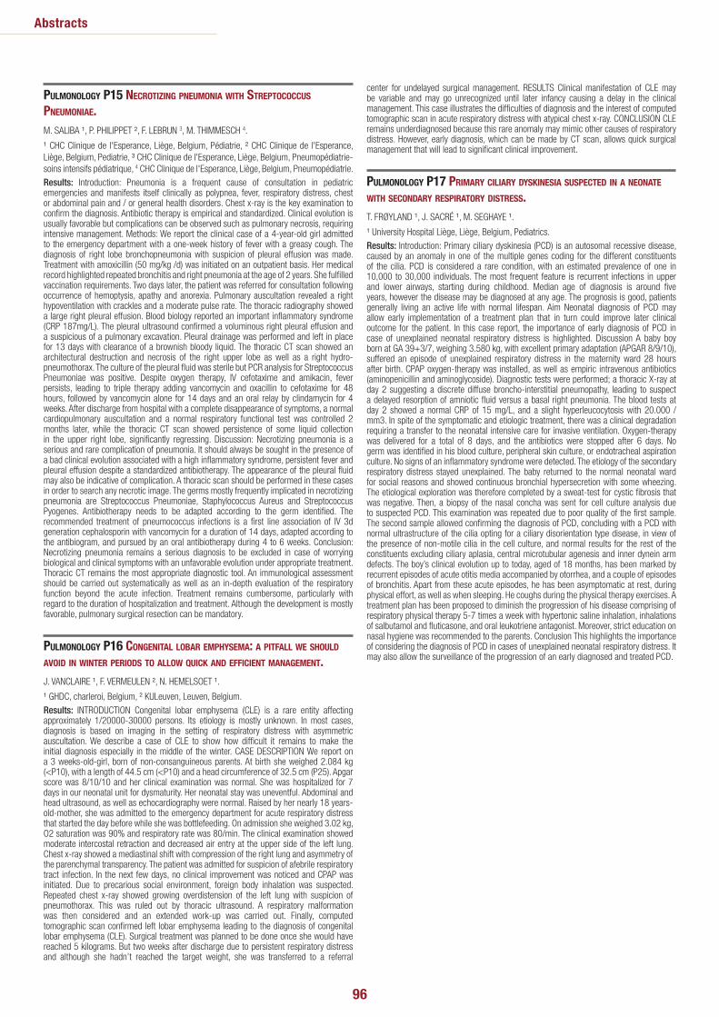
96
Abstracts
pulmonology p15 necrotizing pneumoniA with StreptococcuS pneumoniAe.M. SALIBA ¹, P. PHILIPPET ², F. LEBRUN 3, M. THIMMESCH 4.
¹ CHC Clinique de l'Esperance, Liège, Belgium, Pédiatrie, ² CHC Clinique de l'Esperance, Liège, Belgium, Pediatrie, ³ CHC Clinique de l'Esperance, Liège, Belgium, Pneumopédiatrie- soins intensifs pédiatrique, 4 CHC Clinique de l'Esperance, Liège, Belgium, Pneumopédiatrie.
Results: Introduction: Pneumonia is a frequent cause of consultation in pediatric emergencies and manifests itself clinically as polypnea, fever, respiratory distress, chest or abdominal pain and / or general health disorders. Chest x-ray is the key examination to confirm the diagnosis. Antibiotic therapy is empirical and standardized. Clinical evolution is usually favorable but complications can be observed such as pulmonary necrosis, requiring intensive management. Methods: We report the clinical case of a 4-year-old girl admitted to the emergency department with a one-week history of fever with a greasy cough. The diagnosis of right lobe bronchopneumonia with suspicion of pleural effusion was made. Treatment with amoxicillin (50 mg/kg /d) was initiated on an outpatient basis. Her medical record highlighted repeated bronchitis and right pneumonia at the age of 2 years. She fulfilled vaccination requirements. Two days later, the patient was referred for consultation following occurrence of hemoptysis, apathy and anorexia. Pulmonary auscultation revealed a right hypoventilation with crackles and a moderate pulse rate. The thoracic radiography showed a large right pleural effusion. Blood biology reported an important inflammatory syndrome (CRP 187mg/L). The pleural ultrasound confirmed a voluminous right pleural effusion and a suspicious of a pulmonary excavation. Pleural drainage was performed and left in place for 13 days with clearance of a brownish bloody liquid. The thoracic CT scan showed an architectural destruction and necrosis of the right upper lobe as well as a right hydro-pneumothorax. The culture of the pleural fluid was sterile but PCR analysis for Streptococcus Pneumoniae was positive. Despite oxygen therapy, IV cefotaxime and amikacin, fever persists, leading to triple therapy adding vancomycin and oxacillin to cefotaxime for 48 hours, followed by vancomycin alone for 14 days and an oral relay by clindamycin for 4 weeks. After discharge from hospital with a complete disappearance of symptoms, a normal cardiopulmonary auscultation and a normal respiratory functional test was controlled 2 months later, while the thoracic CT scan showed persistence of some liquid collection in the upper right lobe, significantly regressing. Discussion: Necrotizing pneumonia is a serious and rare complication of pneumonia. It should always be sought in the presence of a bad clinical evolution associated with a high inflammatory syndrome, persistent fever and pleural effusion despite a standardized antibiotherapy. The appearance of the pleural fluid may also be indicative of complication. A thoracic scan should be performed in these cases in order to search any necrotic image. The germs mostly frequently implicated in necrotizing pneumonia are Streptococcus Pneumoniae, Staphylococcus Aureus and Streptococcus Pyogenes. Antibiotherapy needs to be adapted according to the germ identified. The recommended treatment of pneumococcus infections is a first line association of IV 3d generation cephalosporin with vancomycin for a duration of 14 days, adapted according to the antibiogram, and pursued by an oral antibiotherapy during 4 to 6 weeks. Conclusion: Necrotizing pneumonia remains a serious diagnosis to be excluded in case of worrying biological and clinical symptoms with an unfavorable evolution under appropriate treatment. Thoracic CT remains the most appropriate diagnostic tool. An immunological assessment should be carried out systematically as well as an in-depth evaluation of the respiratory function beyond the acute infection. Treatment remains cumbersome, particularly with regard to the duration of hospitalization and treatment. Although the development is mostly favorable, pulmonary surgical resection can be mandatory.
pulmonology p16 congenitAl lobAr emphySemA: A pitfAll we Should Avoid in winter periodS to Allow quick And efficient mAnAgement.J. VANCLAIRE ¹, F. VERMEULEN ², N. HEMELSOET ¹.
¹ GHDC, charleroi, Belgium, ² KULeuven, Leuven, Belgium.
Results: INTRODUCTION Congenital lobar emphysema (CLE) is a rare entity affecting approximately 1/20000-30000 persons. Its etiology is mostly unknown. In most cases, diagnosis is based on imaging in the setting of respiratory distress with asymmetric auscultation. We describe a case of CLE to show how difficult it remains to make the initial diagnosis especially in the middle of the winter. CASE DESCRIPTION We report on a 3 weeks-old-girl, born of non-consanguineous parents. At birth she weighed 2.084 kg (<P10), with a length of 44.5 cm (<P10) and a head circumference of 32.5 cm (P25). Apgar score was 8/10/10 and her clinical examination was normal. She was hospitalized for 7 days in our neonatal unit for dysmaturity. Her neonatal stay was uneventful. Abdominal and head ultrasound, as well as echocardiography were normal. Raised by her nearly 18 years-old-mother, she was admitted to the emergency department for acute respiratory distress that started the day before while she was bottlefeeding. On admission she weighed 3.02 kg, O2 saturation was 90% and respiratory rate was 80/min. The clinical examination showed moderate intercostal retraction and decreased air entry at the upper side of the left lung. Chest x-ray showed a mediastinal shift with compression of the right lung and asymmetry of the parenchymal transparency. The patient was admitted for suspicion of afebrile respiratory tract infection. In the next few days, no clinical improvement was noticed and CPAP was initiated. Due to precarious social environment, foreign body inhalation was suspected. Repeated chest x-ray showed growing overdistension of the left lung with suspicion of pneumothorax. This was ruled out by thoracic ultrasound. A respiratory malformation was then considered and an extended work-up was carried out. Finally, computed tomographic scan confirmed left lobar emphysema leading to the diagnosis of congenital lobar emphysema (CLE). Surgical treatment was planned to be done once she would have reached 5 kilograms. But two weeks after discharge due to persistent respiratory distress and although she hadn’t reached the target weight, she was transferred to a referral
center for undelayed surgical management. RESULTS Clinical manifestation of CLE may be variable and may go unrecognized until later infancy causing a delay in the clinical management. This case illustrates the difficulties of diagnosis and the interest of computed tomographic scan in acute respiratory distress with atypical chest x-ray. CONCLUSION CLE remains underdiagnosed because this rare anomaly may mimic other causes of respiratory distress. However, early diagnosis, which can be made by CT scan, allows quick surgical management that will lead to significant clinical improvement.
pulmonology p17 primAry ciliAry dySkineSiA SuSpected in A neonAte with SecondAry reSpirAtory diStreSS.T. FRØYLAND ¹, J. SACRÉ ¹, M. SEGHAYE ¹.
¹ University Hospital Liège, Liège, Belgium, Pediatrics.
Results: Introduction: Primary ciliary dyskinesia (PCD) is an autosomal recessive disease, caused by an anomaly in one of the multiple genes coding for the different constituents of the cilia. PCD is considered a rare condition, with an estimated prevalence of one in 10,000 to 30,000 individuals. The most frequent feature is recurrent infections in upper and lower airways, starting during childhood. Median age of diagnosis is around five years, however the disease may be diagnosed at any age. The prognosis is good, patients generally living an active life with normal lifespan. Aim Neonatal diagnosis of PCD may allow early implementation of a treatment plan that in turn could improve later clinical outcome for the patient. In this case report, the importance of early diagnosis of PCD in case of unexplained neonatal respiratory distress is highlighted. Discussion A baby boy born at GA 39+3/7, weighing 3.580 kg, with excellent primary adaptation (APGAR 8/9/10), suffered an episode of unexplained respiratory distress in the maternity ward 28 hours after birth. CPAP oxygen-therapy was installed, as well as empiric intravenous antibiotics (aminopenicillin and aminoglycoside). Diagnostic tests were performed; a thoracic X-ray at day 2 suggesting a discrete diffuse broncho-interstitial pneumopathy, leading to suspect a delayed resorption of amniotic fluid versus a basal right pneumonia. The blood tests at day 2 showed a normal CRP of 15 mg/L, and a slight hyperleucocytosis with 20.000 /mm3. In spite of the symptomatic and etiologic treatment, there was a clinical degradation requiring a transfer to the neonatal intensive care for invasive ventilation. Oxygen-therapy was delivered for a total of 8 days, and the antibiotics were stopped after 6 days. No germ was identified in his blood culture, peripheral skin culture, or endotracheal aspiration culture. No signs of an inflammatory syndrome were detected. The etiology of the secondary respiratory distress stayed unexplained. The baby returned to the normal neonatal ward for social reasons and showed continuous bronchial hypersecretion with some wheezing. The etiological exploration was therefore completed by a sweat-test for cystic fibrosis that was negative. Then, a biopsy of the nasal concha was sent for cell culture analysis due to suspected PCD. This examination was repeated due to poor quality of the first sample. The second sample allowed confirming the diagnosis of PCD, concluding with a PCD with normal ultrastructure of the cilia opting for a ciliary disorientation type disease, in view of the presence of non-motile cilia in the cell culture, and normal results for the rest of the constituents excluding ciliary aplasia, central microtubular agenesis and inner dynein arm defects. The boy’s clinical evolution up to today, aged of 18 months, has been marked by recurrent episodes of acute otitis media accompanied by otorrhea, and a couple of episodes of bronchitis. Apart from these acute episodes, he has been asymptomatic at rest, during physical effort, as well as when sleeping. He coughs during the physical therapy exercises. A treatment plan has been proposed to diminish the progression of his disease comprising of respiratory physical therapy 5-7 times a week with hypertonic saline inhalation, inhalations of salbutamol and fluticasone, and oral leukotriene antagonist. Moreover, strict education on nasal hygiene was recommended to the parents. Conclusion This highlights the importance of considering the diagnosis of PCD in cases of unexplained neonatal respiratory distress. It may also allow the surveillance of the progression of an early diagnosed and treated PCD.
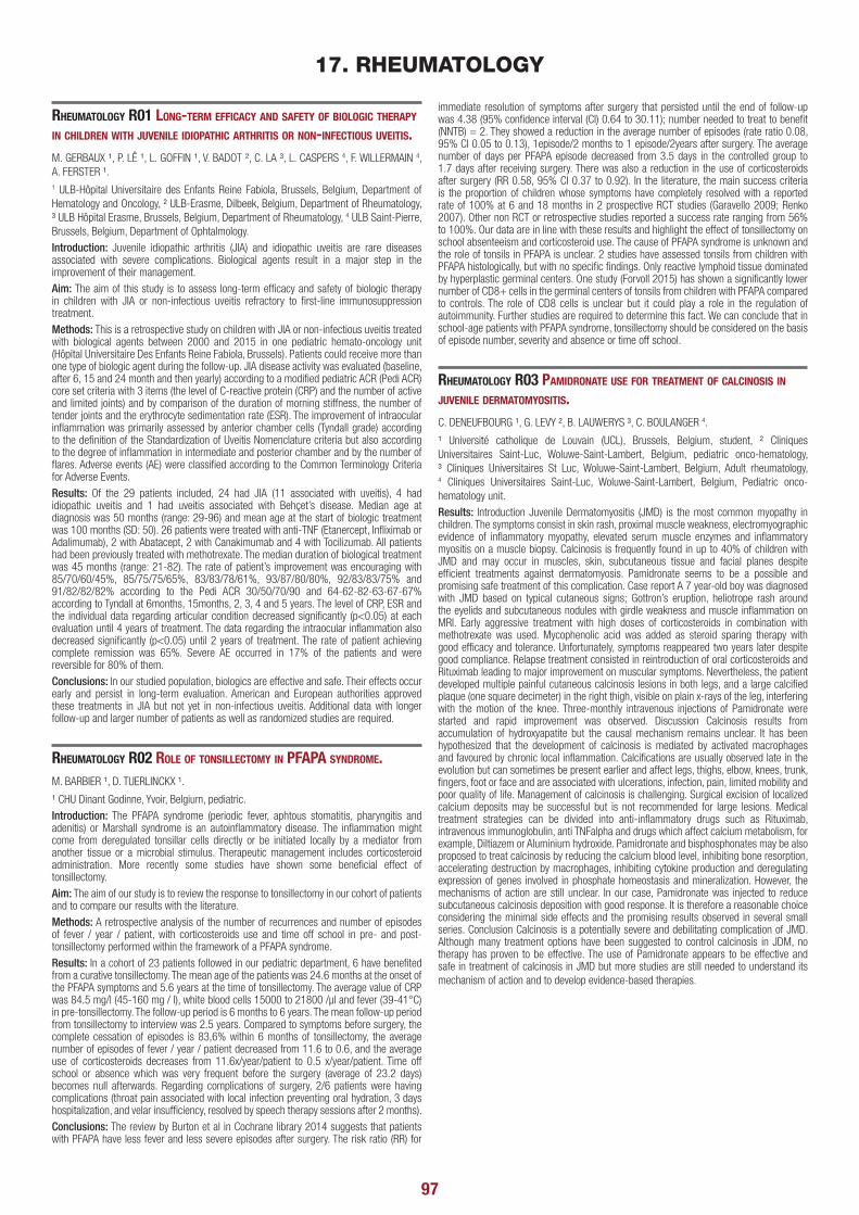
97
17. RHEUMATOLOGY
rheumAtology r01 long-term efficAcy And SAfety of biologic therApy in children with juvenile idiopAthic ArthritiS or non-infectiouS uveitiS.M. GERBAUX ¹, P. LÊ ¹, L. GOFFIN ¹, V. BADOT ², C. LA ³, L. CASPERS 4, F. WILLERMAIN 4, A. FERSTER ¹.1 ULB-Hôpital Universitaire des Enfants Reine Fabiola, Brussels, Belgium, Department of Hematology and Oncology, ² ULB-Erasme, Dilbeek, Belgium, Department of Rheumatology, ³ ULB Hôpital Erasme, Brussels, Belgium, Department of Rheumatology, 4 ULB Saint-Pierre, Brussels, Belgium, Department of Ophtalmology.
Introduction: Juvenile idiopathic arthritis (JIA) and idiopathic uveitis are rare diseases associated with severe complications. Biological agents result in a major step in the improvement of their management.
Aim: The aim of this study is to assess long-term efficacy and safety of biologic therapy in children with JIA or non-infectious uveitis refractory to first-line immunosuppression treatment.
Methods: This is a retrospective study on children with JIA or non-infectious uveitis treated with biological agents between 2000 and 2015 in one pediatric hemato-oncology unit (Hôpital Universitaire Des Enfants Reine Fabiola, Brussels). Patients could receive more than one type of biologic agent during the follow-up. JIA disease activity was evaluated (baseline, after 6, 15 and 24 month and then yearly) according to a modified pediatric ACR (Pedi ACR) core set criteria with 3 items (the level of C-reactive protein (CRP) and the number of active and limited joints) and by comparison of the duration of morning stiffness, the number of tender joints and the erythrocyte sedimentation rate (ESR). The improvement of intraocular inflammation was primarily assessed by anterior chamber cells (Tyndall grade) according to the definition of the Standardization of Uveitis Nomenclature criteria but also according to the degree of inflammation in intermediate and posterior chamber and by the number of flares. Adverse events (AE) were classified according to the Common Terminology Criteria for Adverse Events.
Results: Of the 29 patients included, 24 had JIA (11 associated with uveitis), 4 had idiopathic uveitis and 1 had uveitis associated with Behçet’s disease. Median age at diagnosis was 50 months (range: 29-96) and mean age at the start of biologic treatment was 100 months (SD: 50). 26 patients were treated with anti-TNF (Etanercept, Infliximab or Adalimumab), 2 with Abatacept, 2 with Canakimumab and 4 with Tocilizumab. All patients had been previously treated with methotrexate. The median duration of biological treatment was 45 months (range: 21-82). The rate of patient’s improvement was encouraging with 85/70/60/45%, 85/75/75/65%, 83/83/78/61%, 93/87/80/80%, 92/83/83/75% and 91/82/82/82% according to the Pedi ACR 30/50/70/90 and 64-62-82-63-67-67% according to Tyndall at 6months, 15months, 2, 3, 4 and 5 years. The level of CRP, ESR and the individual data regarding articular condition decreased significantly (p<0.05) at each evaluation until 4 years of treatment. The data regarding the intraocular inflammation also decreased significantly (p<0.05) until 2 years of treatment. The rate of patient achieving complete remission was 65%. Severe AE occurred in 17% of the patients and were reversible for 80% of them.
Conclusions: In our studied population, biologics are effective and safe. Their effects occur early and persist in long-term evaluation. American and European authorities approved these treatments in JIA but not yet in non-infectious uveitis. Additional data with longer follow-up and larger number of patients as well as randomized studies are required.
rheumAtology r02 role of tonSillectomy in pfApA Syndrome.M. BARBIER ¹, D. TUERLINCKX ¹.
¹ CHU Dinant Godinne, Yvoir, Belgium, pediatric.
Introduction: The PFAPA syndrome (periodic fever, aphtous stomatitis, pharyngitis and adenitis) or Marshall syndrome is an autoinflammatory disease. The inflammation might come from deregulated tonsillar cells directly or be initiated locally by a mediator from another tissue or a microbial stimulus. Therapeutic management includes corticosteroid administration. More recently some studies have shown some beneficial effect of tonsillectomy.
Aim: The aim of our study is to review the response to tonsillectomy in our cohort of patients and to compare our results with the literature.
Methods: A retrospective analysis of the number of recurrences and number of episodes of fever / year / patient, with corticosteroids use and time off school in pre- and post-tonsillectomy performed within the framework of a PFAPA syndrome.
Results: In a cohort of 23 patients followed in our pediatric department, 6 have benefited from a curative tonsillectomy. The mean age of the patients was 24.6 months at the onset of the PFAPA symptoms and 5.6 years at the time of tonsillectomy. The average value of CRP was 84.5 mg/l (45-160 mg / l), white blood cells 15000 to 21800 /μl and fever (39-41°C) in pre-tonsillectomy. The follow-up period is 6 months to 6 years. The mean follow-up period from tonsillectomy to interview was 2.5 years. Compared to symptoms before surgery, the complete cessation of episodes is 83,6% within 6 months of tonsillectomy, the average number of episodes of fever / year / patient decreased from 11.6 to 0.6, and the average use of corticosteroids decreases from 11.6x/year/patient to 0.5 x/year/patient. Time off school or absence which was very frequent before the surgery (average of 23.2 days) becomes null afterwards. Regarding complications of surgery, 2/6 patients were having complications (throat pain associated with local infection preventing oral hydration, 3 days hospitalization, and velar insufficiency, resolved by speech therapy sessions after 2 months).
Conclusions: The review by Burton et al in Cochrane library 2014 suggests that patients with PFAPA have less fever and less severe episodes after surgery. The risk ratio (RR) for
immediate resolution of symptoms after surgery that persisted until the end of follow-up was 4.38 (95% confidence interval (CI) 0.64 to 30.11); number needed to treat to benefit (NNTB) = 2. They showed a reduction in the average number of episodes (rate ratio 0.08, 95% CI 0.05 to 0.13), 1episode/2 months to 1 episode/2years after surgery. The average number of days per PFAPA episode decreased from 3.5 days in the controlled group to 1.7 days after receiving surgery. There was also a reduction in the use of corticosteroids after surgery (RR 0.58, 95% CI 0.37 to 0.92). In the literature, the main success criteria is the proportion of children whose symptoms have completely resolved with a reported rate of 100% at 6 and 18 months in 2 prospective RCT studies (Garavello 2009; Renko 2007). Other non RCT or retrospective studies reported a success rate ranging from 56% to 100%. Our data are in line with these results and highlight the effect of tonsillectomy on school absenteeism and corticosteroid use. The cause of PFAPA syndrome is unknown and the role of tonsils in PFAPA is unclear. 2 studies have assessed tonsils from children with PFAPA histologically, but with no specific findings. Only reactive lymphoid tissue dominated by hyperplastic germinal centers. One study (Forvoll 2015) has shown a significantly lower number of CD8+ cells in the germinal centers of tonsils from children with PFAPA compared to controls. The role of CD8 cells is unclear but it could play a role in the regulation of autoimmunity. Further studies are required to determine this fact. We can conclude that in school-age patients with PFAPA syndrome, tonsillectomy should be considered on the basis of episode number, severity and absence or time off school.
rheumAtology r03 pAmidronAte uSe for treAtment of cAlcinoSiS in juvenile dermAtomyoSitiS.C. DENEUFBOURG ¹, G. LEVY ², B. LAUWERYS ³, C. BOULANGER 4.
¹ Université catholique de Louvain (UCL), Brussels, Belgium, student, ² Cliniques Universitaires Saint-Luc, Woluwe-Saint-Lambert, Belgium, pediatric onco-hematology, ³ Cliniques Universitaires St Luc, Woluwe-Saint-Lambert, Belgium, Adult rheumatology, 4 Cliniques Universitaires Saint-Luc, Woluwe-Saint-Lambert, Belgium, Pediatric onco-hematology unit.
Results: Introduction Juvenile Dermatomyositis (JMD) is the most common myopathy in children. The symptoms consist in skin rash, proximal muscle weakness, electromyographic evidence of inflammatory myopathy, elevated serum muscle enzymes and inflammatory myositis on a muscle biopsy. Calcinosis is frequently found in up to 40% of children with JMD and may occur in muscles, skin, subcutaneous tissue and facial planes despite efficient treatments against dermatomyosis. Pamidronate seems to be a possible and promising safe treatment of this complication. Case report A 7 year-old boy was diagnosed with JMD based on typical cutaneous signs; Gottron’s eruption, heliotrope rash around the eyelids and subcutaneous nodules with girdle weakness and muscle inflammation on MRI. Early aggressive treatment with high doses of corticosteroids in combination with methotrexate was used. Mycophenolic acid was added as steroid sparing therapy with good efficacy and tolerance. Unfortunately, symptoms reappeared two years later despite good compliance. Relapse treatment consisted in reintroduction of oral corticosteroids and Rituximab leading to major improvement on muscular symptoms. Nevertheless, the patient developed multiple painful cutaneous calcinosis lesions in both legs, and a large calcified plaque (one square decimeter) in the right thigh, visible on plain x-rays of the leg, interfering with the motion of the knee. Three-monthly intravenous injections of Pamidronate were started and rapid improvement was observed. Discussion Calcinosis results from accumulation of hydroxyapatite but the causal mechanism remains unclear. It has been hypothesized that the development of calcinosis is mediated by activated macrophages and favoured by chronic local inflammation. Calcifications are usually observed late in the evolution but can sometimes be present earlier and affect legs, thighs, elbow, knees, trunk, fingers, foot or face and are associated with ulcerations, infection, pain, limited mobility and poor quality of life. Management of calcinosis is challenging. Surgical excision of localized calcium deposits may be successful but is not recommended for large lesions. Medical treatment strategies can be divided into anti-inflammatory drugs such as Rituximab, intravenous immunoglobulin, anti TNFalpha and drugs which affect calcium metabolism, for example, Diltiazem or Aluminium hydroxide. Pamidronate and bisphosphonates may be also proposed to treat calcinosis by reducing the calcium blood level, inhibiting bone resorption, accelerating destruction by macrophages, inhibiting cytokine production and deregulating expression of genes involved in phosphate homeostasis and mineralization. However, the mechanisms of action are still unclear. In our case, Pamidronate was injected to reduce subcutaneous calcinosis deposition with good response. It is therefore a reasonable choice considering the minimal side effects and the promising results observed in several small series. Conclusion Calcinosis is a potentially severe and debilitating complication of JMD. Although many treatment options have been suggested to control calcinosis in JDM, no therapy has proven to be effective. The use of Pamidronate appears to be effective and safe in treatment of calcinosis in JMD but more studies are still needed to understand its mechanism of action and to develop evidence-based therapies.
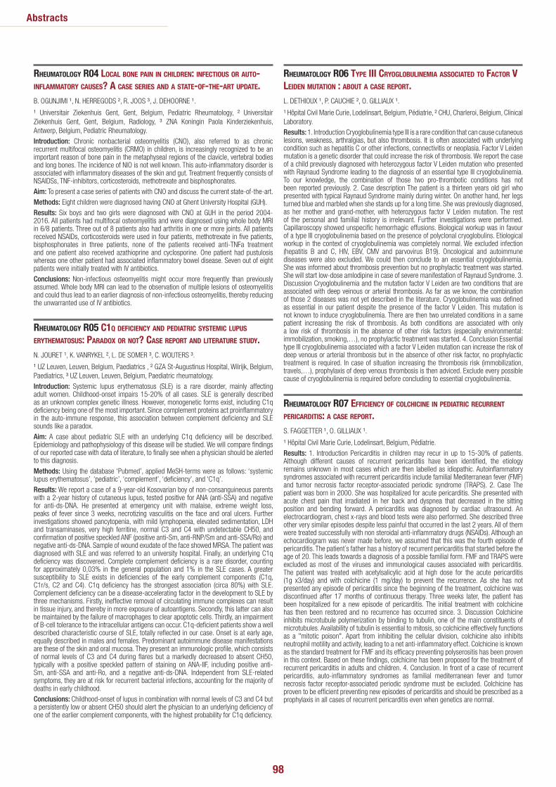
98
Abstracts
rheumAtology r04 locAl bone pAin in children: infectiouS or Auto-inflAmmAtory cAuSeS? A cASe SerieS And A StAte-of-the-Art updAte.B. OGUNJIMI ¹, N. HERREGODS ², R. JOOS ³, J. DEHOORNE ¹.
¹ Universitair Ziekenhuis Gent, Gent, Belgium, Pediatric Rheumatology, ² Universitair Ziekenhuis Gent, Gent, Belgium, Radiology, ³ ZNA Koningin Paola Kinderziekenhuis, Antwerp, Belgium, Pediatric Rheumatology.
Introduction: Chronic nonbacterial osteomyelitis (CNO), also referred to as chronic recurrent multifocal osteomyelitis (CRMO) in children, is increasingly recognized to be an important reason of bone pain in the metaphyseal regions of the clavicle, vertebral bodies and long bones. The incidence of NIO is not well known. This auto-inflammatory disorder is associated with inflammatory diseases of the skin and gut. Treatment frequently consists of NSAIDSs, TNF-inhibitors, corticosteroids, methotrexate and bisphosphonates.
Aim: To present a case series of patients with CNO and discuss the current state-of-the-art.
Methods: Eight children were diagnosed having CNO at Ghent University Hospital (GUH).
Results: Six boys and two girls were diagnosed with CNO at GUH in the period 2004-2016. All patients had multifocal osteomyelitis and were diagnosed using whole body MRI in 6/8 patients. Three out of 8 patients also had arthritis in one or more joints. All patients received NSAIDs, corticosteroids were used in four patients, methotrexate in five patients, bisphosphonates in three patients, none of the patients received anti-TNFa treatment and one patient also received azathioprine and cyclosporine. One patient had pustulosis whereas one other patient had associated inflammatory bowel disease. Seven out of eight patients were initially treated with IV antibiotics.
Conclusions: Non-infectious osteomyelitis might occur more frequently than previously assumed. Whole body MRI can lead to the observation of multiple lesions of osteomyelitis and could thus lead to an earlier diagnosis of non-infectious osteomyelitis, thereby reducing the unwarranted use of IV antibiotics.
rheumAtology r05 c1q deficiency And pediAtric SyStemic lupuS erythemAtoSuS: pArAdox or not? cASe report And literAture Study.N. JOURET ¹, K. VANRYKEL ², L. DE SOMER ³, C. WOUTERS ³.
¹ UZ Leuven, Leuven, Belgium, Paediatrics , ² GZA St-Augustinus Hospital, Wilrijk, Belgium, Paediatrics, ³ UZ Leuven, Leuven, Belgium, Paediatric rheumatology.
Introduction: Systemic lupus erythematosus (SLE) is a rare disorder, mainly affecting adult women. Childhood-onset impairs 15-20% of all cases. SLE is generally described as an unknown complex genetic illness. However, monogenetic forms exist, including C1q deficiency being one of the most important. Since complement proteins act proinflammatory in the auto-immune response, this association between complement deficiency and SLE sounds like a paradox.
Aim: A case about pediatric SLE with an underlying C1q deficiency will be described. Epidemiology and pathophysiology of this disease will be studied. We will compare findings of our reported case with data of literature, to finally see when a physician should be alerted to this diagnosis.
Methods: Using the database ‘Pubmed’, applied MeSH-terms were as follows: ‘systemic lupus erythematosus’, ‘pediatric’, ‘complement’, ‘deficiency’, and ‘C1q’.
Results: We report a case of a 9-year-old Kosovarian boy of non-consanguineous parents with a 2-year history of cutaneous lupus, tested positive for ANA (anti-SSA) and negative for anti-ds-DNA. He presented at emergency unit with malaise, extreme weight loss, peaks of fever since 3 weeks, necrotizing vasculitis on the face and oral ulcers. Further investigations showed pancytopenia, with mild lymphopenia, elevated sedimentation, LDH and transaminases, very high ferritine, normal C3 and C4 with undetectable CH50, and confirmation of positive speckled ANF (positive anti-Sm, anti-RNP/Sm and anti-SSA/Ro) and negative anti-ds-DNA. Sample of wound exudate of the face showed MRSA. The patient was diagnosed with SLE and was referred to an university hospital. Finally, an underlying C1q deficiency was discovered. Complete complement deficiency is a rare disorder, counting for approximately 0,03% in the general population and 1% in the SLE cases. A greater susceptibility to SLE exists in deficiencies of the early complement components (C1q, C1r/s, C2 and C4). C1q deficiency has the strongest association (circa 80%) with SLE. Complement deficiency can be a disease-accelerating factor in the development to SLE by three mechanisms. Firstly, ineffective removal of circulating immune complexes can result in tissue injury, and thereby in more exposure of autoantigens. Secondly, this latter can also be maintained by the failure of macrophages to clear apoptotic cells. Thirdly, an impairment of B-cell tolerance to the intracellular antigens can occur. C1q-deficient patients show a well described characteristic course of SLE, totally reflected in our case. Onset is at early age, equally described in males and females. Predominant autoimmune disease manifestations are these of the skin and oral mucosa. They present an immunologic profile, which consists of normal levels of C3 and C4 during flares but a markedly decreased to absent CH50, typically with a positive speckled pattern of staining on ANA-IIF, including positive anti-Sm, anti-SSA and anti-Ro, and a negative anti-ds-DNA. Independent from SLE-related symptoms, they are at risk for recurrent bacterial infections, accounting for the majority of deaths in early childhood.
Conclusions: Childhood-onset of lupus in combination with normal levels of C3 and C4 but a persistently low or absent CH50 should alert the physician to an underlying deficiency of one of the earlier complement components, with the highest probability for C1q deficiency.
rheumAtology r06 type iii cryoglobulinemiA ASSociAted to fActor v leiden mutAtion : About A cASe report.L. DETHIOUX ¹, P. CAUCHIE ², O. GILLIAUX ¹.
¹ Hôpital Civil Marie Curie, Lodelinsart, Belgium, Pédiatrie, ² CHU, Charleroi, Belgium, Clinical Laboratory.
Results: 1. Introduction Cryoglobulinemia type III is a rare condition that can cause cutaneous lesions, weakness, arthralgias, but also thrombosis. It is often associated with underlying condition such as hepatitis C or other infections, connectivitis or neoplasia. Factor V Leiden mutation is a genetic disorder that could increase the risk of thrombosis. We report the case of a child previously diagnosed with heterozygous factor V Leiden mutation who presented with Raynaud Syndrome leading to the diagnosis of an essential type III cryoglobulinemia. To our knowledge, the combination of those two pro-thrombotic conditions has not been reported previously. 2. Case description The patient is a thirteen years old girl who presented with typical Raynaud Syndrome mainly during winter. On another hand, her legs turned blue and marbled when she stands up for a long time. She was previously diagnosed, as her mother and grand-mother, with heterozygous factor V Leiden mutation. The rest of the personal and familial history is irrelevant. Further investigations were performed. Capillaroscopy showed unspecific hemorrhagic effusions. Biological workup was in favour of a type III cryoglobulinemia based on the presence of polyclonal cryoglobulins. Etiological workup in the context of cryoglobulinemia was completely normal. We excluded infection (hepatitis B and C, HIV, EBV, CMV and parvovirus B19). Oncological and autoimmune diseases were also excluded. We could then conclude to an essential cryoglobulinemia. She was informed about thrombosis prevention but no prophylactic treatment was started. She will start low-dose amlodipine in case of severe manifestation of Raynaud Syndrome. 3. Discussion Cryoglobulinemia and the mutation factor V Leiden are two conditions that are associated with deep veinous or arterial thrombosis. As far as we know, the combination of those 2 diseases was not yet described in the literature. Cryoglobulinemia was defined as essential in our patient despite the presence of the factor V Leiden. This mutation is not known to induce cryoglobulinemia. There are then two unrelated conditions in a same patient increasing the risk of thrombosis. As both conditions are associated with only a low risk of thrombosis in the absence of other risk factors (especially environmental: immobilization, smoking,…), no prophylactic treatment was started. 4. Conclusion Essential type III cryoglobulinemia associated with a factor V Leiden mutation can increase the risk of deep venous or arterial thrombosis but in the absence of other risk factor, no prophylactic treatment is required. In case of situation increasing the thrombosis risk (immobilization, travels,…), prophylaxis of deep venous thrombosis is then adviced. Exclude every possible cause of cryoglobulinemia is required before concluding to essential cryoglobulinemia.
rheumAtology r07 efficiency of colchicine in pediAtric recurrent pericArditiS: A cASe report.S. FAGGETTER ¹, O. GILLIAUX ¹.
¹ Hôpital Civil Marie Curie, Lodelinsart, Belgium, Pédiatrie.
Results: 1. Introduction Pericarditis in children may recur in up to 15-30% of patients. Although different causes of recurrent pericarditis have been identified, the etiology remains unknown in most cases which are then labelled as idiopathic. Autoinflammatory syndromes associated with recurrent pericarditis include familial Mediterranean fever (FMF) and tumor necrosis factor receptor-associated periodic syndrome (TRAPS). 2. Case The patient was born in 2000. She was hospitalized for acute pericarditis. She presented with acute chest pain that irradiated in her back and dyspnea that decreased in the sitting position and bending forward. A pericarditis was diagnosed by cardiac ultrasound. An electrocardiogram, chest x-rays and blood tests were also performed. She described three other very similar episodes despite less painful that occurred in the last 2 years. All of them were treated successfully with non steroidal anti-inflammatory drugs (NSAIDs). Although an echocardiogram was never made before, we assumed that this was the fourth episode of pericarditis. The patient´s father has a history of recurrent pericarditis that started before the age of 20. This leads towards a diagnosis of a possible familial form. FMF and TRAPS were excluded as most of the viruses and immunological causes associated with pericarditis. The patient was treated with acetylsalicylic acid at high dose for the acute pericarditis (1g x3/day) and with colchicine (1 mg/day) to prevent the recurrence. As she has not presented any episode of pericarditis since the beginning of the treatment, colchicine was discontinued after 17 months of continuous therapy. Three weeks later, the patient has been hospitalized for a new episode of pericarditis. The initial treatment with colchicine has then been restored and no recurrence has occurred since. 3. Discussion Colchicine inhibits microtubule polymerization by binding to tubulin, one of the main constituents of microtubules. Availability of tubulin is essential to mitosis, so colchicine effectively functions as a "mitotic poison". Apart from inhibiting the cellular division, colchicine also inhibits neutrophil motility and activity, leading to a net anti-inflammatory effect. Colchicine is known as the standard treatment for FMF and its efficacy preventing polyserositis has been proven in this context. Based on these findings, colchicine has been proposed for the treatment of recurrent pericarditis in adults and children. 4. Conclusion. In front of a case of recurrent pericarditis, auto-inflammatory syndromes as familial mediterranean fever and tumor necrosis factor receptor-associated periodic syndrome must be excluded. Colchicine has proven to be efficient preventing new episodes of pericarditis and should be prescribed as a prophylaxis in all cases of recurrent pericarditis even when genetics are normal.
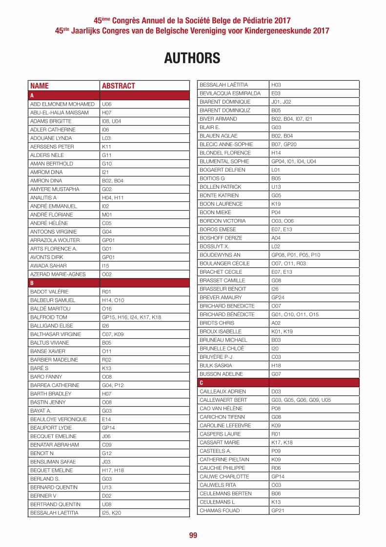
99
45ème Congrès Annuel de la Société Belge de Pédiatrie 201745ste Jaarlijks Congres van de Belgische Vereniging voor Kindergeneeskunde 2017
AUTHORS
NAME ABSTRACTAABD ELMONEM MOHAMED U06
ABU-EL-HAIJA MAISSAM H07
ADAMS BRIGITTE I08, U04
ADLER CATHERINE I06
ADOUANE LYNDA L03
AERSSENS PETER K11
ALDERS NELE G11
AMAN BERTHOLD G10
AMROM DINA I21
AMRON DINA B02, B04
AMYERE MUSTAPHA G02
ANALITIS A H04, H11
ANDRÉ EMMANUEL I02
ANDRÉ FLORIANE M01
ANDRÉ HÉLÈNE C05
ANTOONS VIRGINIE G04
ARRAZOLA WOUTER GP01
ARTS FLORENCE A. G01
AVONTS DIRK GP01
AWADA SAHAR I15
AZERAD MARIE-AGNES O02
B
BADOT VALÉRIE R01
BALBEUR SAMUEL H14, O10
BALDÉ MARITOU O16
BALFROID TOM GP15, H16, I24, K17, K18
BALLIGAND ELISE I26
BALTHASAR VIRGINIE C07, K09
BALTUS VIVIANE B05
BANSE XAVIER O11
BARBIER MADELINE R02
BARÉ S K13
BARO FANNY O08
BARREA CATHERINE G04, P12
BARTH BRADLEY H07
BASTIN JENNY O08
BAYAT A. G03
BEAULOYE VERONIQUE E14
BEAUPORT LYDIE GP14
BECQUET EMELINE J06
BENATAR ABRAHAM C09
BENOIT N G12
BENSLIMAN SAFAE J03
BEQUET EMELINE H17, H18
BERLAND S. G03
BERNARD QUENTIN U13
BERNIER V D02
BERTRAND QUENTIN U08
BESSALAH LAETITIA I25, K20
BESSALAH LAËTITIA H03
BEVILACQUA ESMIRALDA E03
BIARENT DOMINIQUE J01, J02
BIARENT DOMINIQUZ B05
BIVER ARMAND B02, B04, I07, I21
BLAIR E. G03
BLAUEN AGLAE B02, B04
BLECIC ANNE-SOPHIE B07, GP20
BLONDEL FLORENCE H14
BLUMENTAL SOPHIE GP04, I01, I04, U04
BOGAERT DELFIEN L01
BOITIOS G B05
BOLLEN PATRICK U13
BONTE KATRIEN G05
BOON LAURENCE K19
BOON MIEKE P04
BORDON VICTORIA O03, O06
BOROS EMESE E07, E13
BOSHOFF DERIZE A04
BOSSUYT X. L02
BOUDEWYNS AN GP08, P01, P05, P10
BOULANGER CÉCILE O07, O11, R03
BRACHET CECILE E07, E13
BRASSET CAMILLE G08
BRASSEUR BENOIT I26
BREVER AMAURY GP24
BRICHARD BENEDICTE O07
BRICHARD BÉNÉDICTE G01, O10, O11, O15
BRIDTS CHRIS A02
BROUX ISABELLE K01, K19
BRUNEAU MICHAEL B03
BRUNELLE CHLOÉ I20
BRUYÈRE P-J C03
BULK SASKIA H18
BUSSON ADELINE G07
C
CAILLEAUX ADRIEN D03
CALLEWAERT BERT G03, G05, G06, G09, U05
CAO VAN HÉLÈNE P08
CARICHON TIFENN G08
CAROLINE LEFEBVRE K09
CASPERS LAURE R01
CASSART MARIE K17, K18
CASTEELS A. P09
CATHERINE PIELTAIN K09
CAUCHIE PHILIPPE R06
CAUWE CHARLOTTE GP14
CAUWELS RITA O03
CEULEMANS BERTEN B06
CEULEMANS L K13
CHAMAS FOUAD GP21
Authors
100
CHANTRAIN C D02
CHANTRAIN CHRISTOPHE O08, O17
CHERUY CHARLOTTE K19
CHERYL L. G03
CHEVALIER ELSA K18
CHIODINI BENEDETTA I08, U04
CHRISTIAENS CHRISTELLE I16
CLAEYS TANIA H10
CLAPUYT PHILIPPE H01
CLOES MARC GP21
COLAIACOVO MARIE-LAURE
O11
COLLARD LAURE U14
COLLARD LAURENCE I20
COLLINS A. G03
COLPAERT JOHAN GP09
COOLS BJORN P04
COOLS F. P13
COOLS FILIP K02
COOLS MARTINE E01, E02, G06
COPPIETERS FRAUKE L01
CORBISIER TIPHAINE G12
CORNETTE LUC H10, K05, K05, K05
COUCKE PAUL G03, G05
CRAEN MARGARITA E01, E02
CRIJNEN THOMAS GP23, M02
CRIJNEN THOMAS EDUARD MARIA
I18
CROCHELET ANNE-SOPHIE H17
D
DA SILVA DARCHEN SOPHIE G01
DAELEMANS S. L02, P09, P13
DAEZE CHARLOTTE H09
DAHALANI SALIM O10
DALI C.I. G03
DANAU BIRGIT H02
DARON AURORE I17
DARON B C03
DARON BENOÎT C08
DE BACKER JAN P01, P05
DE BACKER JULIE G03
DE BACKER TINE G05
DE BACKER WILFRIED P01, P05
DE BAERE ELFRIDE L01
DE BAETS FRANS L01, P02, P03, P06, P11
DE BECO GEOFFREY C01
DE BELS DAVID J02, J03
DE BISSCHOP BARBARA K02
DE BONT BAUDOUIN D01
DE BRUYNE CORALIE P07
DE BRUYNE MARIEKE L01
DE BRUYNE RUTH GP11, H06, K04
DE CALUWE WIM K04
DE CATTE L C05
DE CLERCK LUC A02
DE CLERCQ AN U13
DE COEN KRIS K04
DE CONINCK SHANA G03
DE FAYS CHARLOTTE C02
DE GENDT ETIENNE H16
DE GREEF ELISABETH E10, I19
DE GUCHTENAERE ANN H09
DE HALLEUX VIRGINIE K01
DE KEUKELAERE MARIJKE E08
DE LA FUENTE ISABEL B02, B04
DE LA FUENTE GARCIA ISABEL
I07, I21
DE LAET CORINNE E07, G07, M03
DE MAGNEE CATHERINE O07
DE MAGNÉE CATHERINE H13
DE MEIRLEIR LINDA B02, E12
DE MEYER FRANÇOISE D03
DE MOERLOOSE BARBARA O18
DE MUL AURÉLIE P08
DE PAEPE ANNE G03
DE PRAETER CLAUDINE K06
DE RADEMAEKER MARJAN E06, E11
DE RONGÉ CAROLINE K15
DE SCHEPPER JEAN E06, E10, E11, E12, G06, G09
DE SCHRYVER SARAH L01
DE SOMER LIEN R05
DE VILLE DE GOYET MAËLLE
O07
DE VOS PATRICIA O06, O09
DE VRIEZE JELENA H08
DE WACHTER E. P09, P13
DE WACHTER ELKE I19, P14
DE WILDE HANS L01
DE WOLF DANIEL G05
DE WOLF RIK E11, O12
DEBEER ANNE K16
DEBIEC-RYCHTER M O15
DEBIEC-RYCHTER MARIA G01
DEBRAY FRANÇOIS-GUILLAUME
G08
DEBRUYNE RUTH H09
DEBUF MARIE-JULIE H10
DECLERCQ DIMITRI P02, P03, P06
DECLERCQ PETER E03
DECUYPER INE A02, A03, GP06
DEDECKEN LAURENCE G07
DEDEKEN LAURENCE G07, O02
DEELEN JE K12
DEFLANDRE JACQUES H18
DEFREYNE LUC K04
DEHAENE ESTELLE U09
DEHOORNE JOKE R04
DELANGHE G K13
DELBECQUE KATTY K01
DELIVORGIA V H04, H11
DEMARCHE MARTINE K19
Authors
101
DEMARET TANGUY M01
DEMBOUR GUY GP16
DEMEULEMEESTER VERONIQUE
H05, H12
DEMONCEAU N G12
DEMOULIN JEAN-BAPTISTE G01
DEN BRINKER MARIEKE E15
DENEUFBOURG CHARLOTTE
R03
DENIS CHLOÉ I11
DEPREZ SABINE O01
DEPUIS ZOE I10
DEPUIS ZOÉ C08, GP24
DEPYPERE ANOUK P04
DEREME JULIEN O04
DERRIKS FRANK K02
DESECK VIRGINIE GP12
DESPRECHINS BRIGITTE B07, GP13, GP20
DESPRESCHINS BRIGITTE I23
DESTREE A. G03
DETHIOUX LORRAINE R06
DEVALCK CHRISTINE O02
DEVREKER THIERRY GP02, GP07, H02, H15
D'HAESE JAMES K14
DHEEDENE ANNELIES G06
DHONDT KARLIEN E01
DHONT EVELYN GP09, K04, U02
DHONT EVELYNE U12
DHOOGE CATHARINA O03, O06
DIALLO SAFIATOU O02
DIERICKX LAURE P11
DIMITRIU DANA E14
DIMITROV BOYAN G10
DINNEWETH DELPHINE U05
DOCQUIER PIERRE-LOUIS O11
DOCX M K13
DOCX MARTINE GP23, U07
DORCHY HARRY E05
DOSSCHE LIEN U10
DOTREMONT HILDE E15
DRAGUET CATHERINE D02
DRESSE MARIE-FRANÇOISE O05
DUBRU JEAN-MARIE B07
DULLAERS MELISSA L01
DUMONCEAU HÉLÈNE I12
DUONG HONG PHUOC I08, U04
DUPONT SOPHIE O07
DUSABÉ JEAN-PAUL I20
DUTILLEUX TANGUY C08, O16
DUTRIEUX JEAN-LUC I20
E
EBO DIDIER A02, A03
EELEN JULIE K11
EFIRA ANDRE O02
EL AMOURI AMINA E12
ELENS IRIS O01
ELLOUZE NESRINE H18
ELOOT SUNNY U01
ERNST C. P09
ERNST CAROLINE E11
ESMANS ANJA I13
ETOBOU CHARLES I20
EYSKENS FRANÇOIS M02
F
FABER MARGARETHA A02
FAGGETTER SARAH R07
FAGNARD MARIE M03
FAIK AICHA D04
FARHAT NESRINE C07, C08
FERSTER ALINA R01
FISHMAN DOUGLAS H07
FLORKIN BENOIT O14
FLORKIN BENOÎT L03, O05
FOHN ANAÏS O05
FORGET PATRICIA O05
FRANCE ANNICK E15
FRANCESCHINI FLORENCE C04
FRANÇOIS ANNE GP14
FRANÇOIS TINE U12
FRANCOTTE NADINE O08, O17
FREEDMAN STEVEN H07
FRERE JULIE GP24, I17
FRÈRE JULIE O14
FRICX CHRISTOPHE B03, I12, I15
FRØYLAND TRINE P17
FUDVOYE JULIE E09
G
GALANT CHRISTINE G01, O07
GARCIA MINAUR SIXTO G03
GARIEPY CHERYL H07
GEERTS CHLOÉ GP16, I26
GENIN CAROLINE I14
GÉRARD MICHEL E14
GERBAUX MARGAUX R01
GEURTEN CLAIRE E04, GP24
GEURTEN MARIE E04
GEWILIG M C05
GEYSENS MATHILDE M01
GHUYSEN MARIE-SOPHIE U14
GIEFER MATTHEW H07
GIES INGE E06, E11
GILLIAUX OLIVIER R06, R07
GOEMAERE STEFAN E02
GOFFIN LAURENCE R01
GONSKA TANJA H07
GOOSSENS ANIETA I19
GOOSSENS EVY A03
GOOSSENS LINDE K06
GRAMMENS TINE GP05, GP22
GROEBBENS STEEN GP07
Authors
102
GROENEN GUIDO I01
GROSSMAN D K12
GUERTI KHADIJA K07
GUEULETTE EMMANUELLE P07
GYS INGE E03
H
HAENTJENS DOMINIQUE I13
HAERYNCK FILOMEEN L01
HAGENDORENS MARGO A02, A03
HAGGENMACHER CAROLINE
J01, J02
HAINAUT MARC I02
HAJSELOVA LUCIA I06
HASAERTS DANIELLE I19
HEIJMANS CATHERINE O02
HEINRICHS CLAUDINE E07, E13
HELAERS RAPHAEL G01, G02
HEMELSOET N P16
HENNAUT ELISE I08, U04
HENRION ELISABETH L03
HERREGODS NELE R04
HEYMAN MELVIN H07
HEYNEMAN ANN K04
HEYSE ROBIN E01
HIMES RYAN H07
HOORNAERT EMMY C06
HOYOUX CLAIRE E04, K19, O05, O14
HUGO JULIE GP13
HUSAIN SOHAIL Z. H07
HUYBRECHTS SOPHIE O02
HUYSENTRUYT KOEN H15
HUYSENTRYUT KOEN GP02, GP07, H02
I
IGBOKWE R. G03
ILUNGA NDAYA MYRIAM J01
IRIDA SHARRA J05
ISABELLE BROUX K09
ISMAILI KHALID I08, U04
J
JACOBS FRÉDÉRIQUE I15
JACOBS SANDRA O04
JADOUL PASCALE E14
JAMBLIN PAUL J06, K19
JANSEN KATRIEN B01
JANSSEN AURELIE O14
JANSSEN AURÉLIE K03
JANSSENS SANDRA G05
JESPERS A K13
JONARD CHARLOTTE K21
JONES G. G03
JOOS RIK R04
JOURET NATHALIE R05
JULIE LÉONARD K01
JULIE PIERART K09
K
KADZ EMMANUELLE I24
KAIRET KOEN GP08
KAISIN VIOLETTE I14
KALENGA MASENDU K01, K08, K09
KAPPERS WILLEMIJN GP23
KAUFMAN JEAN-MARC E02
KEENSWIJK WERNERGP17, GP18, GP25, U02, U09, U11, U12
KEYMOLEN K. G03
KEYMOLEN KATHELIJN G10
KINUANI RACHEL C03
KLEINSCHMIDT MICHAELA B03, I15
KLINK DANIEL E10
KOMUTA MINA H01
KORNREICH LAURE I22
KOURANI EHSAN B03, D04
KOUTSANASTASI PERSA K17
KRECHTING FLOORTJE K13
KUEHN HYE SUN L01
L
LA CÉLINE R01
LABARQUE VEERLE O01
LAITINEN V. G03
LAMBRECHTS CHIARA I13
LANGHENDRIES JEAN-PAUL GP14
LAPAUW BRUNO E02
LARIDAEN JOLIEN E01
LAUREYS GENEVIÈVE O18
LAUWERYS BERNARD R03
LAVAL NANCY I07
LAVOLO PAULINE I25, K20, M01
LE PHU QUOC O02
LÊ PHU QUO GP15, K17
LÊ PHU-QUOC K18, R01
LÊ P-Q I24
LEBOUTTE MARIE-EMELINE D01
LEBRETHON MARIE CHRISTINE
E09
LEBRETHON MARIE-CHRISTINE
E04
LEBRETHON MC MARIE-CHRISTINE
GP21
LEBRUN FREDERIC I01
LEBRUN FRÉDERIC P15
LECOQ NATHALIE H13
LEDERER DAMIEN B02
LEFEBVRE CAROLINE K01
LEMIERE JURGEN O01
LEMMENS AN-SOFIE I19
LEMOINE-TREMBLAY PIERRE-LUC
O16
LENOIR PASCAL I20
LÉONARD MARIE M03
LEPAGE PHILIPPE GP04, I03, I04, I08
LEROY BART L01
LETIZIA VEGA I20
Authors
103
LEURIS JULIE O12
LEVTCHENKO ELENA K16, U05, U06, U08, U13
LEVY GABRIEL O10, O15, R03
LIBBRECHT LOUIS O15
LIBEERT DENIS G09, P02, P03, P06
LIMAYE NISHA G01
LIN TOM K. H07
LITZROTH AMBER GP05
LIU QUIN H07
LODEWEYCKX NIELS K14
LOECKX ISABELLE I16, P12
LOEYS BART U07
LOLIN KSENIJA I08, U04
LOMBARD CATHERINE M01
LOMBET JACQUES U05
LOMMAERT SYLVIE M01
LOTERMAN DIETER P01, P05
LOWE MARK E. H07
LUBITASSY PRECIEUX J02
LYKOGEORGOU M H04, H11
LYNCH S. G03
LYSY PHILIPPE GP16
M
MABROUK ASMAA J05
MAES KATRIEN GP23
MAGOTTEAUX SAMIA I11
MAHIEU LUDO K07, K10
MAKA MICHEL I25, K20
MALAISE OLIVIER C05
MALE A. G03
MALFROOT A. P09, P13
MALFROOT ANNE I19, P14
MANGODT THOMAS P10
MANSBACH ANNE-LAURE I24
MAQUET JULIE U14
MARBAIX ETIENNE E14
MARGUGLIO ARNAUD GP14
MARTIN SOPHIE U14
MASCARENHAS MARIA H07
MASCART GEORGES GP04, I08
MASSA GUY E03
MASSEZ ANNE I15
MASSIN MARTIAL GP12, J03, J05
MATHYS VANESSA I06
MATON PIERRE GP14, K15
MATTHYSSENS LUCAS K04
MAVROUDI A H04, H11
MAYSTADT I G12
MC LIN VALÉRIE P08
MCHIRGUI LEILA E14
MCKENZIE F. G03
MEERSCHAUT ILSE G03, G05
MELCHIOR MARIE K21, P07
MENDES DA COSTA ELISE GP05, GP22
MENSCHAERT DENIS I17, K03
MENTEN BJORN G06
MENTEN RENAUD H13, O11
MERGEN JULIEN I12
MERINDOL NINON O17
MERTENS CHRISTEL A03
MESSAAOUI ANISSA E05
METZ TANGUY D01
MEUNIER COLOMBINE H14
MEUWISSEN MARIJE G11
MICHEL MARIANNE I26
MICHEZ DELPHINE G07
MILINGOS V H04, H11
MISSON JEAN-PAUL GP24
MISTRETTA VIRGINIE K03
MIYAMOTO MAKIKO D04
MOES NICOLETTE H05, H12
MONDELAERS VEERLE O13, O18
MONIOTTE STÉPHANE C02, C04
MONSEUR SANDRINE H13
MORGENTHAL CHARLINE H17
MORINVILLE VERONIQUE H07
MORNAND ANNE P08
MORTAJI SOUFIANE GP21
MORTIER GEERT G11, K10
MOUCHET FRANÇOISE I01, I06, I09
MOUREY STÉPHANIE J01
MOURIN ANNE H13
MYLONOPOULOU M H04, H11
N
NAVARRO MORENO CONSTANZA
E14
NAWROT TIM GP01
NEBBIOSO ANDREA H16
NEMERY BEN GP01
NEVEN QUENTIN I02, O07
NICOLESCU RAMONA O14
NIEMELA JULIE L01
NIZEYIMANA AGATHE J06
NOBRE MACHADO ALEXANDRA
K18
NOEL LAURA A. G01
NORGA KOEN O04, O13
NUNEZ VALDERMA MAIA J02
NYAMUGABO KINDJA B07, GP13, GP20, I10
NYAMUGABO MUNYERE NKANA KINDJA
I11
O
OGUNJIMI BENSON R04
OLIVEIRA ARCOLINO FANNY U06
OOI CHEE Y. H07
OORTS ELINE GP08
OOSTERWIJK J.C. G03
OP 'T EYNDT ANNIEK E03
OPHOFF DAPHNÉE M02
ORAZIETTI LAURENCE J01
OSSIEUR JULIE O18
Authors
104
OUALD CHAIB ABDELHALIM H15
P
PAIRET ELEONORE G02
PALERMO JOSEPH J. H07
PANZER JOSEPH G05, K04
PAPADOPOULOS JEAN D01, I25
PARENT ANNE-SIMONE C07, E09
PEERS DE NIEUWBURGH MAUREEN
C01
PEETERS ELLEN H08
PEETERS LINDE P14
PEETERS RONALD O01
PERDAENS OCÉANE H03
PERITO EMILY H07
PERLOT PASCALE M03
PETIT F. G03
PHILIPPET P D02
PHILIPPET PIERREG08, I14, I16, O08, O16, O17, P12, P15, U14
PICCOLI DAVID A. H07
PIELTAIN CATHERINE K01
PIERART JULIE K01, K08, L03
PIETTE CAROLINE O05
PIROTTE ISABELLE I23
PLASSCHAERT FRANK G05
PLETINCX MICHEL O12
POHL JOHN H07
POIREL HÉLÈNE A. G01
PONCELET ALAIN C01, O11, P12
P-Q LE H16
PROESMANS MARIJKE P04
PROOST RENEE A04
PRYTULA AGNIESZKA U05
Q
QUIROS ADRIAN I24, K18
QUIROS MIRAMONTES ADRIAN
H16
QUOILIN SOPHIE GP05, GP22
R
RACHDI MAJDA J03
RADULESCU CRISTINA EMANUELA
GP04
RAES ANN U01, U02, U10
RAVAU JOACHIM M01
RAYMACKERS FREDDY E11
RAYYAN MAISSA U13
REDANT SÉBASTIEN GP12, J01, J02, J03, J04, J05
REDANT SEBATIEN B05
REDING RAYMOND O11
RENARD MARLEEN G01, O01
RENAUD VIELLEVOYE K09
RENSON CATHERINE U10
REVENCU NICOLE G02
REVERCEZ AUDREY I03
RICHARD CLAIRE D03
RIGO VINCENT K01
RIHS HANS-PETER A02
ROBERT EMMANUELLE GP03, GP10
ROBERTI ALEXANDRA K19
ROCHTUS ANNE B01
ROELANTS DU VIVIER MALIKA
I09
ROGIERS PETER I18
ROILIDES I H04, H11
ROMAEN KATRIEN I13
ROMAN CÉLINE I12
RONDIA GILLES GP15
ROSENZWEIG SERGIO L01
ROSSILLON RENAUD I26
ROUSSEAU MARIE-CÉCILE P12
ROUSSEFF THAIS GP11, K04, K06
RUBAY JEAN C01
RUTTEMAN BART E06
RUYSSINCK LAURE O03
RYMEN DAISY U13
S
SABATO VITO A02
SABBE MARTINE GP05, GP22
SACRÉ JEAN-PAUL P17
SACRÉ J-P C03
SALIBA MARIANE P15
SALMON KELLY I01
SAMBON PAULINE H01
SANCHEZ MOLINA DIANA D01
SANDU KISHORE P10
SANTALUCIA ROBERTO B05
SARIBAN ERIC O04
SCALAIS EMMANUEL B02, B04, I21
SCHEERS ISABELLE H07, H14
SCHELSTRAET PETRA GP11
SCHELSTRAETE PETRA GP09, I01, I05
SCHIFFLERS STEFAN O08, O17
SCHIFFLERS STEPHAN O04
SCHMITZ SANDRA I02
SCHRODER SILVIA I14
SCHWARZENBERG SARAH J.
H07
SCIOT RAF G01
SEGERS HEIDI O01
SEGHAYE MARIE-CHRISTINE
B07, C05, C07, C08, GP13, GP20, H17, I10, I11, I23, J06, K03, P17
SEGHAYE M-C C03
SEKHARA TAYEB O12
SENTERRE THIBAULT K01
SERVAIS A D02
SHAH UZMA H07
SLAATS MONIQUE P01, P05
SLEURS CHARLOTTE O01
SLUYSMANS THIERRY C01
SMEESTERS PIERRE GP04, GP04
Authors
105
SMEETS SOPHIE GP14
SMETS D. P09
SMETS FRANÇOISE H13
SMETS KOENRAAD K04, K06
SMITS ANNE K16
SMITS GUILLAUME G07
SNAUWAERT EVELIEN U01
SNYERS DIANE K08
SOKAL ETIENNE H01, H03, M01
SOLOWIANIUK MARIE J06
SOMERS INES E15
SOMVILLE VIOLAINE P07
SPINOIT ANNE-FRANÇOISE U10
SPRUYT PETER GP02
STABENOW KATHARINA C09
STAELS WILLEM G06
STALENS JEAN-PHILIPPE H10, I20
STALENS J-P M03
STALS TINNEKE K16
STASSART PIERRE GP21
STEFANIDIS ATHANASIA GP12, J05
STERGIOPOULOU THEODOULI
K03
STERN NORA J01
STEYAERT WOUTER G05
STOLTE-DIJKSTRA I. G03
SUNAERT STEFAN O01
SURGUN EBRU GP04
SURI M. G03
SWENNEN BÉATRICE GP03, GP10
SYMOENS SOFIE G03
SYS MELISSA G10
T
TACK LLOYD E01, E02
TAMBUCCI ROBERTO H14
TAXHET ARNAUD I23
TAZI KAOUTAR H16, I24, K17, K18
TENOUTASSE SYLVIE E05, E07
THEATE I D02
THEMELIN CÉLINE I21
THIBAULT SENTERRE K09
THIELEMANS ELISE G07, I25, K20
THIMMESCH MATHIEU P12
THIMMESCH MATTHIEU P15
THIRION SOPHIE K08
THIRY VÉRONIQUE GP14
THOMAS E. G03
THOMAS MURIEL P02, P03, P06
TOMME LARISSA GP14
TONIOLO MARIKA K21
TOOULOU MONIKA D04
TOPA A. G03
TOUIL AMINA B05
TOULOUSE KAAT O03
TOYE KAATJE E02
TRIBOLET SOPHIE K19, L03
TROENDLE DAVID H07
TROUET DOMINIQUE H08
TSHIMANGA PRISCA GP20
TUBBING ALICE GP12, J04
TUERLINCKX DAVID H13, K21, K21, R02
U
UC ALIYE H07
ULGIATI FIORENZA E07, E13
UYTTEBROECK ANNE O01
V
VAN BIERVLIET STEPHANIE GP11, H06, H09, P02, P03, P06
VAN BIESEN WIM U01
VAN BRUSSELEN DAAN GP01
VAN BUSSEL JOHAN GP22
VAN DAMM AN O15
VAN DAMME AN G01, O04, O07, O10, O11
VAN DAMME KARLIJN K07
VAN DE BROEK H K13
VAN DE CASSEYE WILLY U13
VAN DE MAELE KAROLIEN E06
VAN DE PUTTE DIRK GP18
VAN DE VIJVER ELS H08
VAN DE VOORDE MARLIES I05
VAN DE WALLE JOHAN U12
VAN DE WIJDEVEN PATRICIA B01
VAN DEN BOGAERT ANN G10
VAN DEN HEUVEL LAMBERTUS
U06
VAN DER HEGGEN TATJANA GP09
VAN DER LINDEN DIMITRI I02
VAN DER WERFF TEN BOSCH J.
L02
VAN GASSE ATHINA A01
VAN GENECHTEN TOON O13
VAN GIJLSWIJK SANDRA H08
VAN GOETHEM TANIA H14
VAN HERZEELE CHARLOTTE
U10
VAN HOECK KOEN U05
VAN HOLSBEKE CEDRIC P01, P05
VAN HOORENBEECK KIM P01, P10
VAN HOYWEGHEN ELKE U13
VAN LAECKE ERIK U10
VAN LANCKER SOPHIE O06
VAN LINTHOUT CHRISTINE C05
VAN LOOCKE YANNICK A03
VAN MECHELEN KAREN K10
VAN OORT MARK E10
VAN RENTERGHEM KATRIEN
GP25, H06
VAN RHIJN ANNEMIEK B01
VAN RIJCKEVORSEL GERMAINE
B02, B04, I21
VAN ROEST AALT B06
VAN STEIJN SUSANNE I01
VAN STEIRTEGHEM SAMUEL
D03
Authors
106
VAN UYTVANCK ALEXANDRA
G07
VAN WINCKEL MYRIAM GP11, H06, H09
VAN ZITTEREN LEONARDA MARIA
H05, H12
VANBESIEN JESSE E06, E10
VANCLAIRE JULIE P16
VANDE VELDE SASKIA GP11, H06, H09
VANDE WALLE JOHANGP17, GP25, U01, U02, U05, U07, U09, U10, U11
VANDECRUYS ELS O18
VANDEKERCKHOVE KRISTOF
K04
VANDEN BOSSCHE HEIDI E01
VANDEN DRIESSCHE KOEN A04
VANDENBROUCKE MAUREEN
D04, I04
VANDENPLAS Y. L02, P09, P13
VANDENPLAS YVANGP02, GP07, H02, H04, H11, H15, I19, P14
VANDERGRAESEN TRACY O02
VANDERLINDEN KIM H15
VANDEVIJVER ELS H05, H12
VANDEWALLE SARA E02
VANHAESEBROUCK SOPHIE K06
VANHOLE CHRISTINE K16
VANMASSENHOVE JILL U02
VANRYKEL KATRIEN R05
VANWALLEGHEM PHYLLIS I13
VARMA SHARAT H01, H03
VELGHE AMÉLIE I. G01
VERBRUGGEN JOERY K05
VERCRUYSSE GERTRUI O01
VERELLEN KAREN GP23
VERHEYDEN STEPHANIE E06
VERHULST STIJN K10, P01, P05
VERLOOY JORIS O13
VERMANING AND P. AERSENS SIETSKE
GP19
VERMEULEN F P16
VERMEULEN FRANCOISE B03, I15
VERMEULEN FRANÇOISE D04, I12
VERMEULEN JOELLE D01
VERNA FEDERICO H14
VERSTRAETE LIEVE P10
VERVOORT ANNE GP14
VEYS KOENRAAD U06
VICINANZA ALFREDO E05
VIELLEVOYE RENAUD K01
VIKKULA MIIKKA G01, G02
VINCENT MARIE-FRANÇOISE
M01
VINCENT RIGO K09
VINCKE PIERRE I26
VIRGINIE DE HALLEUX K09
VITALI NANCY GP15, H16
VIVIANE BALTUS J05
VLEMINCX ILSE I13
VÔ CHRISTOPHE C01, C02, C04
VOS WIM P01, P05
W
WANYAMA SIMEON P02, P03, P06
WARNIER HELENE I10
WERLIN STEVEN H07
WIDELEC JACQUES I24
WIJNANDS ANNE E03
WILLEMS JEF I05
WILLEMS LEEN O04, O18
WILLEMS P. G03
WILLERMAIN FRANÇOIS R01
WILSCHANSKI MICHAEL H07
WISSING KARL MARTIN I08, U04
WOJCIECHOWSKI MAREK O13, P01
WOJCIECHOWSKI MARK GP06
WOUTERS CARINE R05
X
XINIAS I H04, H11
Y
YADAV D. G03
YI LAURENT GP15
Z
ZAMBELLI LAURA C07, K09
ZEEVAERT RENATE E03
ZIMMERMAN BRIDGET H07
Jong geleerdis oud gedaan
Hoofdzakelijk water drinken, dat is alvast een goed begin!
Spa Reine drinken, dat is kiezen voor de zuiverheid van een uniek water, ideaal van jongs af aan en zelfs al vóór de geboorte…
Op het leven

Jong geleerdis oud gedaan
Hoofdzakelijk water drinken, dat is alvast een goed begin!
Spa Reine drinken, dat is kiezen voor de zuiverheid van een uniek water, ideaal van jongs af aan en zelfs al vóór de geboorte…
Op het leven

JEADV2014(2)
PU
BLI
CATIE
EFFECT OP XEROSE EN GEBRUIK OP LANGE TERMIJN
PEDIATR DERMATOL.
2016(3)
NIEUW
ING
EDIEND
VOORKOMT OPSTOTEN, VERMINDERT HET CORTICOSTEROÏDEGEBRUIK
• 2 internationale multicentrische klinische studies
• 586 gerandomiseerde patiënten
NIEUWE KLINISCHE GEGEVENS IN ATOPISCHE DERMATITIS
1. Source: Internal calculations based on IMS Health Database retail audit of drugs and food supplements in Belgium, ATOPIE MARKET, full year 2015
2. Boralevi F., Saint Aroman M., Delarue A., Raudsepp, H., Kaszuba A., Bylaite M., Tiplica GS. Long-term emollient therapy improves xerosis in children with atopic dermatitis. J Eur Acad Dermatol Venereol. 2014; 28(11): 1456-62.
3. G.S. Tiplica, A. Kaszuba, L. Malinauskiene, P. Konno, F. Boralevi, E. Garrigue, M. Saint-aroman, A. Delarue. The regular use of emollients prevents flares in children with atopic dermatitis and reduces corticosteroid consumption: a randomized controlled study. Submitted to Pediatric Dermatology.
INVOORSCHRIFTEN VAN DE DERMATOLOOG(1)
N°1ONS ENGAGEMENT: ALLE VORMEN VAN
XEROSE DOELTREFFEND BEHANDELEN
SKIN EXPERTISE
IN OUR DNA
Medisch hulpmiddel
Essentiële ingrediënten
20 jaar ervaringin de behandeling van een droge huid
Voortdurende innovatieklinisch bewezen doeltreffendheid
Aanvaardbare prijs voor langdurig gebruik
ONS ENGAGEMENT: ALLE VORMEN VAN XEROSE DOELTREFFEND BEHANDELEN
E V I D E N C E M A K E S U S
3 ESSENTIËLE
INGREDIËNTEN
BEWEZEN DOELTREFFENDHEID
BEWEZEN DOELTREFFENDHEID
2015
0313
- 0
9/20
15
15380_Annnonce_Presse_Dexeryl_NL.indd 1 15/09/15 14:35
3 ESSENTIËLE
INGREDIËNTEN
BEWEZEN DOELTREFFENDHEID
2016
0507
– 1
2/20
16