bat guano virome: predominance of dietary viruses from insects and plants plus novel mammalian...
TRANSCRIPT
JOURNAL OF VIROLOGY, July 2010, p. 6955–6965 Vol. 84, No. 140022-538X/10/$12.00 doi:10.1128/JVI.00501-10Copyright © 2010, American Society for Microbiology. All Rights Reserved.
Bat Guano Virome: Predominance of Dietary Viruses from Insectsand Plants plus Novel Mammalian Viruses�
Linlin Li,1,2 Joseph G. Victoria,1,2 Chunlin Wang,3 Morris Jones,4 Gary M. Fellers,5Thomas H. Kunz,6 and Eric Delwart1,2*
Blood Systems Research Institute, San Francisco, California1; Department of Laboratory Medicine, University of California,San Francisco, California2; Stanford Genome Technology Center, Stanford, California3; Clinical Investigation Facility,
David Grant USAF Medical Center, Travis Air Force Base, California4; U.S. Geological Survey,Western Ecological Research Center, Point Reyes, California5; and Center for Ecology and
Conservation Biology, Department of Biology, Boston University, Boston, Massachusetts6
Received 5 March 2010/Accepted 30 April 2010
Bats are hosts to a variety of viruses capable of zoonotic transmissions. Because of increased contactbetween bats, humans, and other animal species, the possibility exists for further cross-species transmis-sions and ensuing disease outbreaks. We describe here full and partial viral genomes identified usingmetagenomics in the guano of bats from California and Texas. A total of 34% and 58% of 390,000 sequencereads from bat guano in California and Texas, respectively, were related to eukaryotic viruses, and thelargest proportion of those infect insects, reflecting the diet of these insectivorous bats, including membersof the viral families Dicistroviridae, Iflaviridae, Tetraviridae, and Nodaviridae and the subfamily Densoviri-nae. The second largest proportion of virus-related sequences infects plants and fungi, likely reflecting thediet of ingested insects, including members of the viral families Luteoviridae, Secoviridae, Tymoviridae, andPartitiviridae and the genus Sobemovirus. Bat guano viruses related to those infecting mammals comprisedthe third largest group, including members of the viral families Parvoviridae, Circoviridae, Picornaviridae,Adenoviridae, Poxviridae, Astroviridae, and Coronaviridae. No close relative of known human viral pathogenswas identified in these bat populations. Phylogenetic analysis was used to clarify the relationship to knownviral taxa of novel sequences detected in bat guano samples, showing that some guano viral sequences falloutside existing taxonomic groups. This initial characterization of the bat guano virome, the first meta-genomic analysis of viruses in wild mammals using second-generation sequencing, therefore showed thepresence of previously unidentified viral species, genera, and possibly families. Viral metagenomics is auseful tool for genetically characterizing viruses present in animals with the known capability of direct orindirect viral zoonosis to humans.
Bats belong to one of the most diverse, abundant, and widelydistributed group of mammals. More than 1,100 bat speciesbelong to the order of Chiroptera, representing approximately20% of all mammalian species (54). Most bat species feed oninsects and other arthropods, while others feed on fruit nectar,bird or mammal blood, and small vertebrates such as fish,frogs, mice, and birds (30). Of the 47 species of bats reportedin the United States, most of them are insectivorous (http://www.batcon.org/).
Bats are considered the natural reservoir of a large variety ofzoonotic viruses causing serious human diseases such as lyssa-viruses, henipaviruses, severe acute respiratory syndrome coro-navirus, and Ebola virus (6, 38, 46, 59, 63, 65). Characteristicsof bats, including their genetic diversity, broad geological dis-tribution, gregarious habits, high population density, migratoryhabits, and long life span (30, 58), likely endow them with theability to host diverse viruses, some of which are also able toinfect humans and other mammals (41, 63).
More than 80 virus species have been isolated or detected inbats using nucleic acid-based methods (6, 38, 59, 65). Viruses
that have been recently discovered in bats include astroviruses,adeno-associated viruses (AAVs), adenoviruses, herpesviruses,and polyomavirus (8, 9, 13, 31, 32, 35, 37, 39, 40, 42, 61, 62, 68).For example, it was recently reported that a newly identifiedadenovirus isolated from bat guano was capable of infectingvarious vertebrate cell lines, including those of humans, mon-keys, dogs, and pigs (35). With increasing human populationsin previously wild areas, contact of bats with humans and withwild and domestic animals has increased, providing greateropportunities for cross-species transmissions of potentiallypathogenic bat viruses. To better understand the range ofviruses carried by bats, we undertook an initial character-ization of the guano viromes of several common bat speciesin the United States.
The development of massively parallel sequencing technol-ogy makes is possible to reveal uncultured viral assemblageswithin biological or environmental samples (11, 28). To date,this approach has been used to characterize viruses in equinefeces (7), human blood (5), tissue (14), human feces (3, 4, 15,45, 60, 67), and human respiratory secretions (64), which inturn has facilitated the discovery of many novel viruses (18, 20,25, 33, 47, 50). In the present study, we analyzed the virusespresent in guano from several bat species in California andTexas, using sequence-independent PCR amplification, pyro-sequencing, and sequence similarity searches.
* Corresponding author. Mailing address: Blood Systems ResearchInstitute. 270 Masonic Ave., San Francisco, CA 94118. Phone: (415)923-5763 Fax: (415) 567-5899. E-mail: [email protected].
� Published ahead of print on 12 May 2010.
6955
MATERIALS AND METHODS
Collection of bat guano. All of the bat species sampled are insectivorous.Plastic sheets were laid down on flat surfaces beneath bat roosts. Freshly pro-duced bat guano was then collected 1 day later and stored at �80°C. Sampleswere collected from a bat roost near San Saba, TX, on two occasions that were3 days apart during the summer of 2008. The roost (TM) was occupied mostly byTadarida brasiliensis (Brazilian free-tailed bat). Three other species present insmaller numbers, Myotis velifer (Cave myotis), Nycticeus humeralis (evening bat),and Perimyotis subflavus (tricolored bat), are also known to share the roost andmay have also been sampled.
Guano samples from northern California were collected from five differentroosts at Point Reyes National Seashore. One roost (GF-4) was occupied byAntrozous pallidus (pallid bat), and the other four roosts (GF-3, -5, -6, and -7)were occupied by Myotis spp. and/or Tadarida brasiliensis (Table 1).
Sample preparation and viral nucleic acid extraction. Bat guano was pro-cessed as previously described (60). Briefly, groups of 12 bat guano pellets fromthe same roosts were resuspended by vigorous vortexing in Hank’s bufferedsaline solution (Gibco BRL) and cleared of debris by low-speed centrifugation(5 min at 11,000 � g). A total of 500 �l of guano supernatant was filtered througha 0.45-�m filter (Millipore) to remove bacterium-sized particles. The viral par-ticles containing filtrate were digested with a mixture of DNases and RNase toremove unprotected nucleic acids (i.e., those not in viral capsids) (1). Viralnucleic acids were then extracted using the QIAamp viral RNA minikit (Qiagen).
DNA and RNA library construction and pyrosequencing. Viral nucleic acidlibraries were constructed by random PCR amplification as previously described(60). Both a RNA virus-only and DNA plus RNA virus sequence-independentamplifications were performed and then pooled prior to sequencing. For RNAvirus-only amplification, an aliquot of the extracted viral nucleic acid collectedfrom each pool of 12 guano pellets was treated with DNase (Ambion) to removeviral DNA. A total of 100 pmol of primer, consisting of an arbitrarily designed20-base oligonucleotide followed by a randomized octamer sequence at the 3�end, was then used in a reverse transcription (RT) reaction (Moloney murineleukemia virus reverse transcriptase; Promega). For the RNA plus DNA virusamplification, the DNase step prior to RT was excluded. A single round of DNAsynthesis was then performed using Klenow fragment polymerase (New EnglandBiolabs), followed by PCR amplification of double-stranded DNA using a primerconsisting of only the 20-base fixed portion of the random primer.
A total of 37 distinct random primers (containing different 20-base fixedsequences) were applied to guano collected from the 6 bat roosts (Table 1). Thenumber of primers assigned per roost was based on the number of guano pelletscollected. Viral nucleic acids were therefore amplified from 37 pools of 12 guanopellets per pool, with each pellet presumed to be from a different animal. Guanoobtained from up to 96 bats in the Texas roost (8 primers) and up to 348 bats inthe 5 California roosts (29 primers) was analyzed (Table 1). To further improveviral nucleic acid sampling within each pool, the random PCR amplificationswere performed in duplicate, starting with the Klenow-treated products, result-ing in four PCRs per original pool (2 viral RNA-only inputs and 2 viral RNA plusDNA inputs). The DNA obtained from these four PCRs was mixed and purified,and the DNA concentration was measured. Equal amounts of DNA from eachof the 37 different pools were then mixed together and run on a 2% agarose gel,and DNA fragments from the 500- to 1,000-bp region were excised and purified.The DNA was then sequenced on a single pyrosequencing gasket using GS FLXTitanium reagents (Roche).
A subset of random primer sequences used was previously published (60). Theother random primers were designed by generating random sequences usingPrimo (http://www.changbioscience.com/primo/primor.html) that were then an-
alyzed by BLASTn to remove those primers likely to bind to human and bacterialsequences.
Bioinformatics. The pyrosequencing reads were grouped in 37 bins, accordingto their unique sequence tags (the 20 fixed bases of the random PCR primer).The fixed primer sequences plus eight additional downstream nucleotides (en-coded by the 3� NNNNNNNN part of the random primers), were then trimmedfrom each read. Trimmed reads within each sequence bin were then assembledby Sequencher software (Gene Codes), with a criterion of 95% identity orgreater over at least 35 bp. Contigs were therefore assembled using sequencesfrom at most 12 animals. When overlapping sequences in contigs containedmutations (due to pyrosequencing error or because multiple viral variants fromdifferent animals in the same pool were sequenced), the consensus sequence wasused. The assembled sequence contigs and singlets greater than 100 bp were thencompared to the GenBank nonredundant nucleotide and protein databases usingBLASTn and BLASTx, respectively. Using BLAST searches, sequences wereclassified as likely originating from a eukaryotic virus, bacteria, phage, oreukaryote or deemed unclassifiable based on the taxonomic origin of the best-hitsequence. An E value of 0.001 was used as the cutoff value for significant hits.
Phylogenetic analysis. Reference viral sequences from different viral familieswere obtained from GenBank. Amino acid sequence alignments were generatedusing ClustalW and implemented in MEGA 4.1 with the default settings (29).Aligned sequences were trimmed to match the genomic regions of the viralsequences obtained in our study and phylogenetic trees generated by MEGA4,using neighbor-joining with amino acid p distances and 1,000 bootstrap repli-cates. The GenBank accession numbers of the viral sequences used in thephylogenetic analyses are shown in the trees.
Nucleotide sequence accession numbers. Trimmed and binned sequence readsand contigs of metagenomes from bat guano in California and Texas have beendeposited in the GenBank sequence reads archive under accession numberSRA012669. Sequences from the genomes described in more detail can be foundunder GenBank accession numbers HM228873 to HM228895 and HM234168 toHM234169.
RESULTS
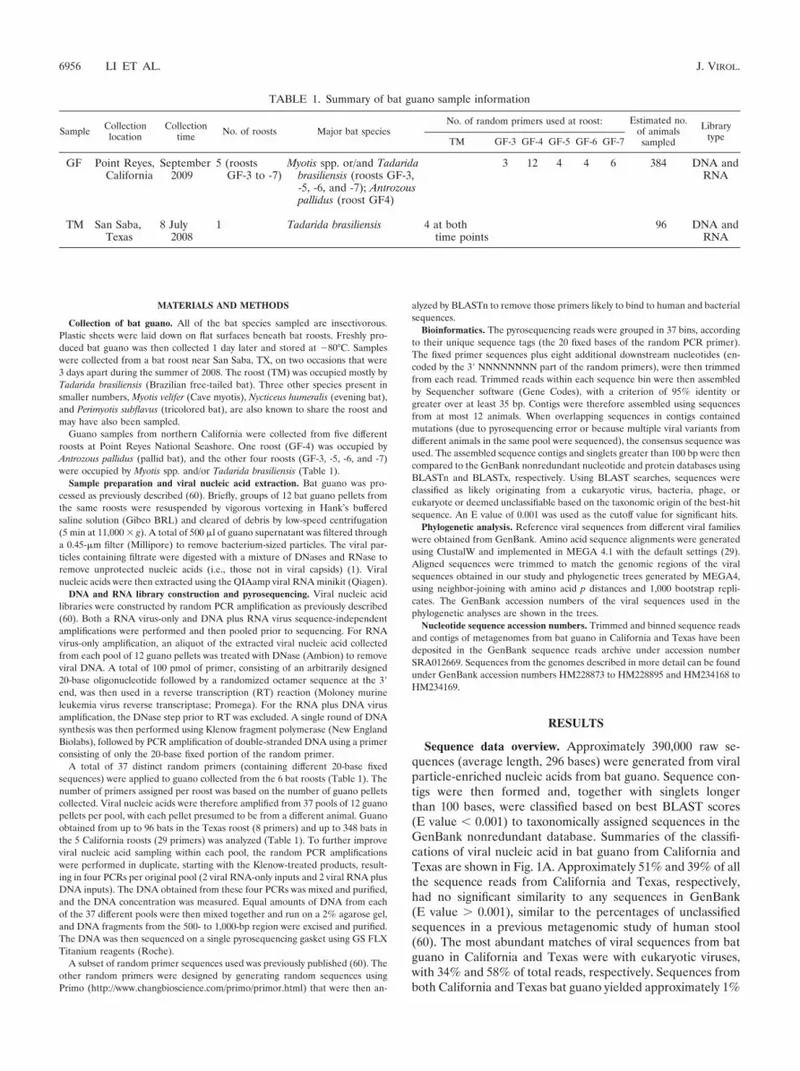
Sequence data overview. Approximately 390,000 raw se-quences (average length, 296 bases) were generated from viralparticle-enriched nucleic acids from bat guano. Sequence con-tigs were then formed and, together with singlets longerthan 100 bases, were classified based on best BLAST scores(E value � 0.001) to taxonomically assigned sequences in theGenBank nonredundant database. Summaries of the classifi-cations of viral nucleic acid in bat guano from California andTexas are shown in Fig. 1A. Approximately 51% and 39% of allthe sequence reads from California and Texas, respectively,had no significant similarity to any sequences in GenBank(E value � 0.001), similar to the percentages of unclassifiedsequences in a previous metagenomic study of human stool(60). The most abundant matches of viral sequences from batguano in California and Texas were with eukaryotic viruses,with 34% and 58% of total reads, respectively. Sequences fromboth California and Texas bat guano yielded approximately 1%
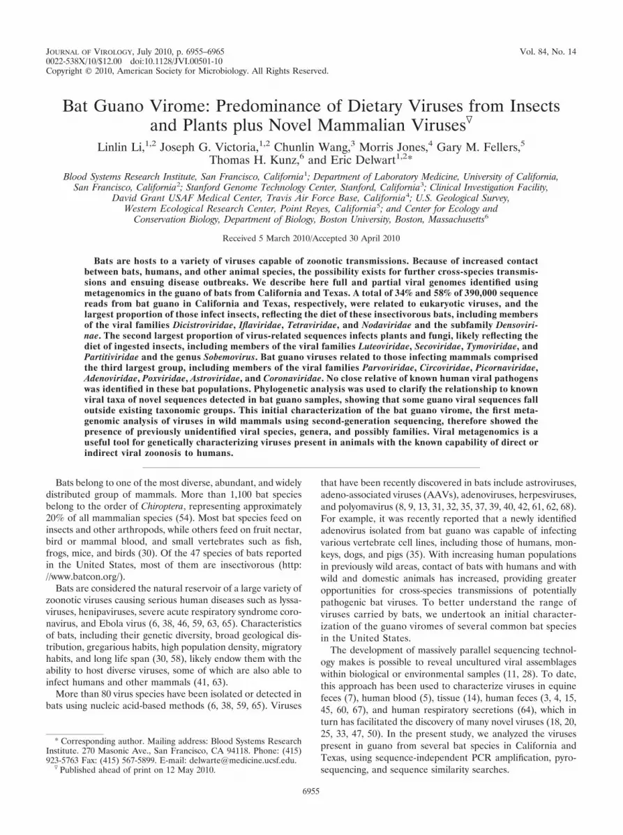
TABLE 1. Summary of bat guano sample information
Sample Collectionlocation
Collectiontime No. of roosts Major bat species
No. of random primers used at roost: Estimated no.of animalssampled
LibrarytypeTM GF-3 GF-4 GF-5 GF-6 GF-7
GF Point Reyes,California
September2009
5 (roostsGF-3 to -7)
Myotis spp. or/and Tadaridabrasiliensis (roosts GF-3,-5, -6, and -7); Antrozouspallidus (roost GF4)
3 12 4 4 6 384 DNA andRNA
TM San Saba,Texas
8 July2008
1 Tadarida brasiliensis 4 at bothtime points
96 DNA andRNA
6956 LI ET AL. J. VIROL.

FIG. 1. Sequence classification for California and Texas bat guano-derived sequences based on BLASTx (E value � 0.001). (A) Percentagesof sequences with similarity to those of eukaryotes, bacteria, phages, and eukaryotic virus in GenBank and to unclassifiable sequences. (B) Per-centages of most abundant eukaryotic viral matches classified by viral families. Plant viruses are highlighted in green, insect viruses are highlightedin red, and mammalian viruses are not highlighted. (C) Eukaryotic viral families in California roost GF-3 to -7 and in one Texas roost at twocollection time points (TM1-4 and TM5-8).
6957
of eukaryote sequences, indicating the DNase and RNasetreatment was largely effective in removing non-capsid-pro-tected bat host nucleic acids.
Phages in bat guano. Based on prior studies, phages com-posed a significant fraction of human and equine fecal viralpopulations (3, 4, 7, 60). The levels of phage sequences in thefeces of South Asian children with nonpolio acute flaccid pa-ralysis and health contacts processed in the same manner wereapproximately 16% and 12% of total reads, respectively (60).In our study, the sequences with similarities to phages made up4% and 0.1% of sequences in bat guano from California andTexas, respectively. Among the phages in bat guano samplesfrom California, the majority belonged to the families Sipho-viridae (67%) and Microviridae (28%), consistent with earlierviral metagenomic studies of which siphophages were the mostabundant phages in human and equine feces (3, 4, 7). The mostabundant sequence matches were to c2-like Lactococcusphages, T1-like enterobacterium phages, Chlamydia phage 3,and Spiroplasma phage 4 (data not shown).
Eukaryotic virus population in bat guano. Many previouslycharacterized and highly divergent eukaryotic viral sequenceswere detected in bat guano. The families of eukaryotic virusesthat were found, based on their most significant BLASTxmatches, are shown in Fig. 1B. Sequences of DNA virusesinfecting eukaryotes made up a smaller fraction (approxi-mately 10%) than eukaryotic RNA viral sequences in bothCalifornia and Texas bat metagenomes. The DNA viruses weredominated by single-stranded DNA (ssDNA) viruses, includ-ing animal viruses from the families Parvoviridae and Circoviri-dae and plant viruses from the family Geminiviridae. Most ofthe proteins encoded by ssDNA eukaryotic virus-like se-quences showed less than 60% amino acid identities to knownviral protein, suggesting the presence of numerous novel viralspecies in bats. Sequences related to the newly discoveredCyclovirus genus in the family Circoviridae, commonly found inthe tissues of hoofed farm animals and chickens as well as inhuman and wild chimpanzee feces, were also detected (33),showing that these viruses also exist in wild bats. Single-stranded RNA viruses belonged largely to the families Dicis-troviridae, Nodaviridae, and Picornaviridae. Double-strandedRNA viral sequences in the family Partitiviridae (18%) werealso detected in the bat guano from California.
Guano collected from bats in California had a more diverseviral composition than guano collected from bats in Texas,which may reflect the multiple roosts and bats species sampledin California. The most common eukaryotic viral families var-ied greatly between each of the five California roosts sampled(Fig. 1C). The guano viromes of the GF-3, -5, and -6 roostswere dominated by plant viruses, whereas the GF-4 roost, theonly one with pallid bats, was richest in insect dicistroviruses,and the GF-7 roost had a more diversified virus profile. Theviral compositions of the two guano samplings from the sameTexas roost were highly distinct, with the earlier collectiondominated by dicistroviruses and the later one by plant virusleutoviruses and tymoviruses (Fig. 1C, TM1-4 and TM5-8).
Insect viruses. The largest fraction of the bat guano viromewas related to insect viruses from the family Dicistroviridae,consisting of 29% of California viral sequences and 61% ofTexas viral sequences, likely reflecting the insect-based diet ofthe bat species analyzed. Viral sequences related to viruses
from Iflaviridae, Tetraviridae, Alphanodavirus, and Densovirinaewere also detected. Most of the viruses were novel, sharing lessthan 60% amino acid (aa) similarity to known viral proteins,while some shared high (�90%) amino acid similarity withknown insect viruses.
Viral sequences similar to those of Kashmir bee virus (12)and acute bee paralysis virus (19) were very abundant in batguano from California and Texas. Sequences covered �70% ofthe complete genomes of these viruses (GenBank accessionnumbers HM228885 to HM228895). The translated full-lengthstructural proteins shared 98% similarity with Kashmir beevirus and 97% aa similarity with acute bee paralysis virus,indicating that the viruses found in bat guano were variants ofKashmir bee virus and acute bee paralysis virus rather thannew viral species. Because the sampled bats are nocturnal, it isunlikely that they feed on diurnal bees. Kashmir bee virus andacute bee paralysis virus may therefore also infect nocturnalbees or other insect hosts, or the bat species studied may havepreviously unknown dietary activities.
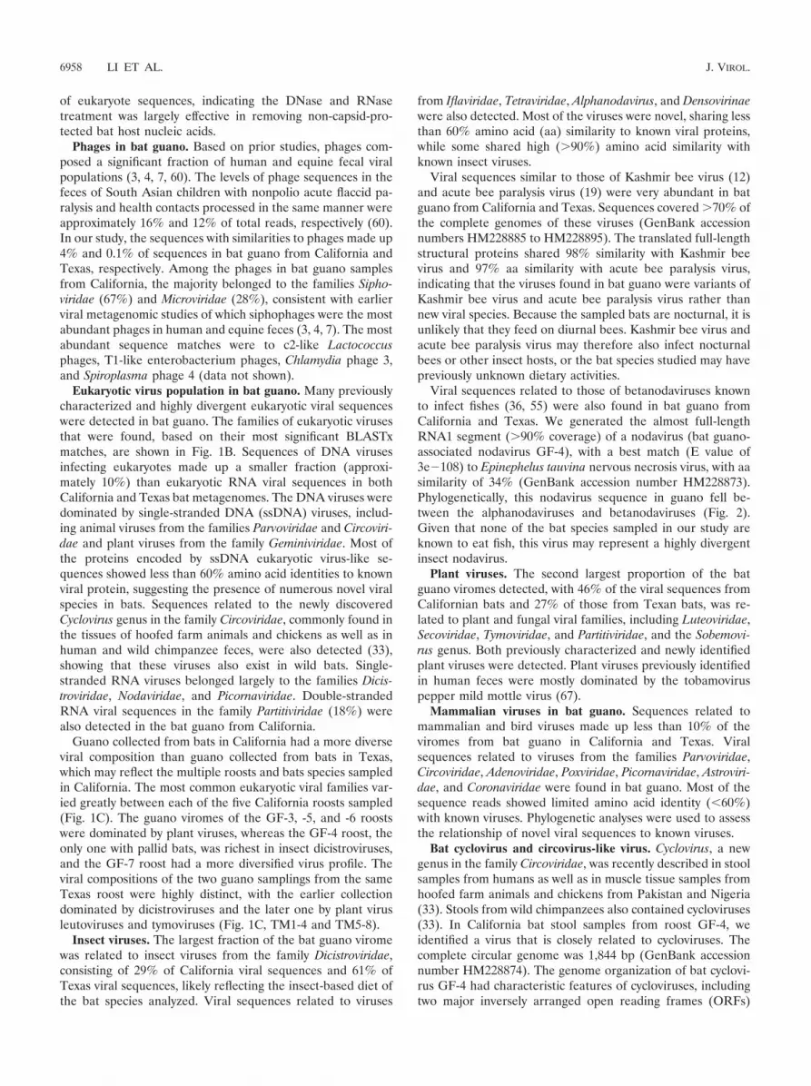
Viral sequences related to those of betanodaviruses knownto infect fishes (36, 55) were also found in bat guano fromCalifornia and Texas. We generated the almost full-lengthRNA1 segment (�90% coverage) of a nodavirus (bat guano-associated nodavirus GF-4), with a best match (E value of3e�108) to Epinephelus tauvina nervous necrosis virus, with aasimilarity of 34% (GenBank accession number HM228873).Phylogenetically, this nodavirus sequence in guano fell be-tween the alphanodaviruses and betanodaviruses (Fig. 2).Given that none of the bat species sampled in our study areknown to eat fish, this virus may represent a highly divergentinsect nodavirus.
Plant viruses. The second largest proportion of the batguano viromes detected, with 46% of the viral sequences fromCalifornian bats and 27% of those from Texan bats, was re-lated to plant and fungal viral families, including Luteoviridae,Secoviridae, Tymoviridae, and Partitiviridae, and the Sobemovi-rus genus. Both previously characterized and newly identifiedplant viruses were detected. Plant viruses previously identifiedin human feces were mostly dominated by the tobamoviruspepper mild mottle virus (67).
Mammalian viruses in bat guano. Sequences related tomammalian and bird viruses made up less than 10% of theviromes from bat guano in California and Texas. Viralsequences related to viruses from the families Parvoviridae,Circoviridae, Adenoviridae, Poxviridae, Picornaviridae, Astroviri-dae, and Coronaviridae were found in bat guano. Most of thesequence reads showed limited amino acid identity (�60%)with known viruses. Phylogenetic analyses were used to assessthe relationship of novel viral sequences to known viruses.
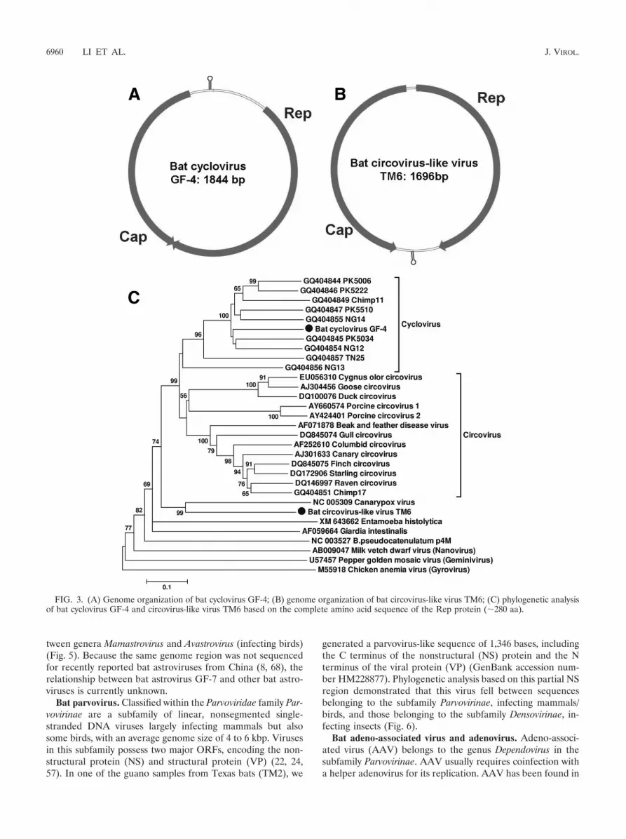
Bat cyclovirus and circovirus-like virus. Cyclovirus, a newgenus in the family Circoviridae, was recently described in stoolsamples from humans as well as in muscle tissue samples fromhoofed farm animals and chickens from Pakistan and Nigeria(33). Stools from wild chimpanzees also contained cycloviruses(33). In California bat stool samples from roost GF-4, weidentified a virus that is closely related to cycloviruses. Thecomplete circular genome was 1,844 bp (GenBank accessionnumber HM228874). The genome organization of bat cyclovi-rus GF-4 had characteristic features of cycloviruses, includingtwo major inversely arranged open reading frames (ORFs)
6958 LI ET AL. J. VIROL.
encoding the putative replication-associated protein (Rep; 281aa) and capsid protein (Cap; 227 aa). A characteristic potentialstem-loop structure with a conserved nonanucleotide motif(5�-TAATACTAT-3�) was also found in the 5� intergenic re-gion (between the start codons of the two major ORFs) (Fig.3A). The putative Rep proteins of the bat cyclovirus GF-4 had45% to 68% aa similarity to cycloviruses found in human andchimpanzee feces and 39% to 43% similarity to the Rep pro-teins of porcine and avian circoviruses (data not shown).
In Texas bat stool sample TM6, we found a small, circularDNA virus with a full-genome size of 1,696 bp (bat circovirus-like virus TM6) (GenBank accession number HM228876). Thevirus had two major ORFs arranged in opposite directions,with Rep at 264 aa and Cap at 226 aa, and two noncodingintergenic regions (Fig. 3B). The stem-loop structure also hadthe cyclovirus-conserved nonanucleotide motif (5�-TAATACTAT-3�) but was instead located at the 3� intergenic region(between the stop codons of the two major ORFs).
A phylogenetic analysis of the complete Rep protein of batcyclovirus GF-4 and bat circovirus-like virus TM6, includingcycloviruses, circoviruses, chicken anemia virus (CAV), andnon-Circoviridae Rep proteins from the plant Nanovirus milkvetch dwarf virus, Geminivirus pepper golden mosaic virus,canarypox virus, Bifidobacterium pseudocatenulatum plasmidpM4, Giardia intestinalis, and Entamoeba histolytica was per-formed (Fig. 3C). Examination of the phylogenetic treeshowed that bat cyclovirus GF-4 grouped with known cyclovi-ruses, forming a distinct species of cyclovirus. Bat circovirus-like virus TM6 fell outside the Circovirus and Cyclovirus clades,grouping with canarypox virus. While most closely related tothe replicase sequence of canarypox virus, these two proteinsshowed only 40% aa similarity.
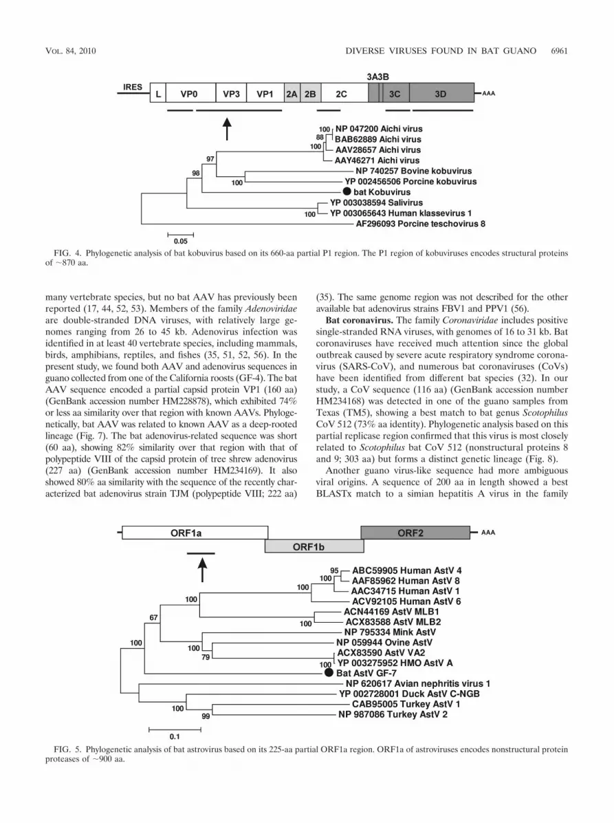
Bat kobuvirus. Kobuvirus, a genus in the family Picornaviri-dae, currently contains three species: Aichi virus, bovine kobu-virus, and porcine kobuvirus. Kobuvirus-related viruses namedsalivirus and klassevirus have been recently described in hu-man stool samples (20, 21, 34). Aichi virus and salivirus have
been associated with human gastroenteritis, while bovinekobuvirus and porcine kobuvirus are associated with bovineand porcine diarrhea, respectively (26, 27, 48, 66). In one of theTexan sets of guano samples (TM7), we found approximately400 reads which assembled into 5 contigs covering more than60% of the viral genome of a virus closely related to kobuvi-ruses (GenBank accession numbers HM228880 toHM228884). BLASTx searches showed that these contigsshared 39% to 59% aa similarity to kobuviruses. We tentativelynamed this virus bat kobuvirus. According to the InternationalCommittee on Taxonomy of Viruses (ICTV) (http://www.picornastudygroup.com/definitions/genus_definition.htm), themembers of a picornavirus genus should share �40%, �40%,and �50% aa similarity in their P1, P2, and P3 regions, re-spectively. The largest contig (1,998 bp) covered about 70% ofthe P1 region and shared 46% aa similarity with the closestmatch, human Aichi virus. Bat kobuvirus therefore appears to bea new viral species within the genus Kobuvirus (Fig. 4). Phyloge-netic analysis using the contig (1,335 bp) covering more than 80%of the 3-D region produces a similar tree topology (data notshown).
Bat astrovirus. The family Astroviridae includes positive sin-gle-stranded RNA viruses, with genomes of 6.4 to 7.3 kb,encoding nonstructural proteins with ORF1a and ORF1b andstructural protein with ORF2 (43). Astroviruses (AstV) havebeen identified in a variety of mammals and birds, includinghumans, cattle, pigs, sheep, mink, dogs, cats, mice, bats, chick-ens, and turkeys. In the California bat roost GF-7, we detecteda highly divergent astrovirus-like sequence (677 bp) (GenBankaccession number HM228876). The translated amino acid se-quence most closely matched the serine protease region of thenewly characterized human HMOAstV-A viral genome (34%similarity) (16, 23). Phylogenetic analysis based on this regionyielded a tree topology that was congruent with those of anal-yses using other genome regions (8, 49, 68). The tree showedthat the bat astrovirus GF-7 sequence fell in a basal positionrelative to other mamastroviruses (infecting mammals) be-
FIG. 2. Phylogenetic analysis of bat guano-associated nodavirus GF-4 based on its 950-aa RNA-dependent RNA polymerase (RdRp) region.RNA1 of nodaviruses encodes the RdRp protein of �1,000 aa.
VOL. 84, 2010 DIVERSE VIRUSES FOUND IN BAT GUANO 6959
tween genera Mamastrovirus and Avastrovirus (infecting birds)(Fig. 5). Because the same genome region was not sequencedfor recently reported bat astroviruses from China (8, 68), therelationship between bat astrovirus GF-7 and other bat astro-viruses is currently unknown.
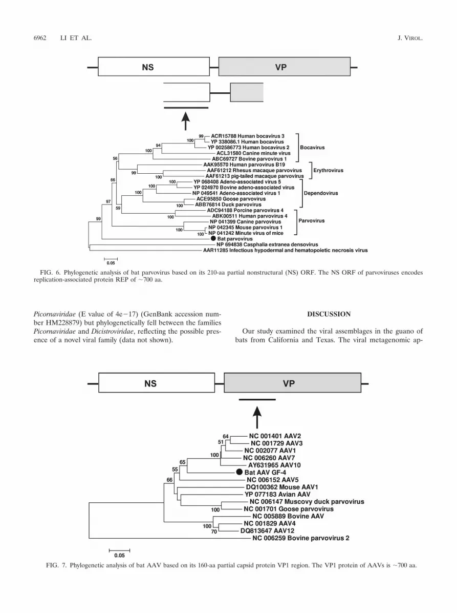
Bat parvovirus. Classified within the Parvoviridae family Par-vovirinae are a subfamily of linear, nonsegmented single-stranded DNA viruses largely infecting mammals but alsosome birds, with an average genome size of 4 to 6 kbp. Virusesin this subfamily possess two major ORFs, encoding the non-structural protein (NS) and structural protein (VP) (22, 24,57). In one of the guano samples from Texas bats (TM2), we
generated a parvovirus-like sequence of 1,346 bases, includingthe C terminus of the nonstructural (NS) protein and the Nterminus of the viral protein (VP) (GenBank accession num-ber HM228877). Phylogenetic analysis based on this partial NSregion demonstrated that this virus fell between sequencesbelonging to the subfamily Parvovirinae, infecting mammals/birds, and those belonging to the subfamily Densovirinae, in-fecting insects (Fig. 6).
Bat adeno-associated virus and adenovirus. Adeno-associ-ated virus (AAV) belongs to the genus Dependovirus in thesubfamily Parvovirinae. AAV usually requires coinfection witha helper adenovirus for its replication. AAV has been found in
FIG. 3. (A) Genome organization of bat cyclovirus GF-4; (B) genome organization of bat circovirus-like virus TM6; (C) phylogenetic analysisof bat cyclovirus GF-4 and circovirus-like virus TM6 based on the complete amino acid sequence of the Rep protein (�280 aa).
6960 LI ET AL. J. VIROL.
many vertebrate species, but no bat AAV has previously beenreported (17, 44, 52, 53). Members of the family Adenoviridaeare double-stranded DNA viruses, with relatively large ge-nomes ranging from 26 to 45 kb. Adenovirus infection wasidentified in at least 40 vertebrate species, including mammals,birds, amphibians, reptiles, and fishes (35, 51, 52, 56). In thepresent study, we found both AAV and adenovirus sequences inguano collected from one of the California roosts (GF-4). The batAAV sequence encoded a partial capsid protein VP1 (160 aa)(GenBank accession number HM228878), which exhibited 74%or less aa similarity over that region with known AAVs. Phyloge-netically, bat AAV was related to known AAV as a deep-rootedlineage (Fig. 7). The bat adenovirus-related sequence was short(60 aa), showing 82% similarity over that region with that ofpolypeptide VIII of the capsid protein of tree shrew adenovirus(227 aa) (GenBank accession number HM234169). It alsoshowed 80% aa similarity with the sequence of the recently char-acterized bat adenovirus strain TJM (polypeptide VIII; 222 aa)
(35). The same genome region was not described for the otheravailable bat adenovirus strains FBV1 and PPV1 (56).
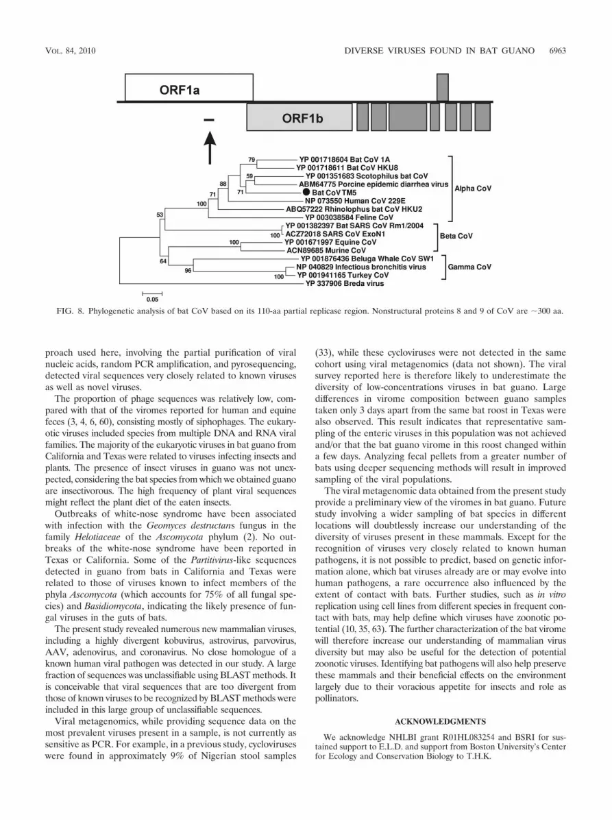
Bat coronavirus. The family Coronaviridae includes positivesingle-stranded RNA viruses, with genomes of 16 to 31 kb. Batcoronaviruses have received much attention since the globaloutbreak caused by severe acute respiratory syndrome corona-virus (SARS-CoV), and numerous bat coronaviruses (CoVs)have been identified from different bat species (32). In ourstudy, a CoV sequence (116 aa) (GenBank accession numberHM234168) was detected in one of the guano samples fromTexas (TM5), showing a best match to bat genus ScotophilusCoV 512 (73% aa identity). Phylogenetic analysis based on thispartial replicase region confirmed that this virus is most closelyrelated to Scotophilus bat CoV 512 (nonstructural proteins 8and 9; 303 aa) but forms a distinct genetic lineage (Fig. 8).
Another guano virus-like sequence had more ambiguousviral origins. A sequence of 200 aa in length showed a bestBLASTx match to a simian hepatitis A virus in the family
FIG. 4. Phylogenetic analysis of bat kobuvirus based on its 660-aa partial P1 region. The P1 region of kobuviruses encodes structural proteinsof �870 aa.
FIG. 5. Phylogenetic analysis of bat astrovirus based on its 225-aa partial ORF1a region. ORF1a of astroviruses encodes nonstructural proteinproteases of �900 aa.
VOL. 84, 2010 DIVERSE VIRUSES FOUND IN BAT GUANO 6961
Picornaviridae (E value of 4e�17) (GenBank accession num-ber HM228879) but phylogenetically fell between the familiesPicornaviridae and Dicistroviridae, reflecting the possible pres-ence of a novel viral family (data not shown).
DISCUSSION
Our study examined the viral assemblages in the guano ofbats from California and Texas. The viral metagenomic ap-
FIG. 6. Phylogenetic analysis of bat parvovirus based on its 210-aa partial nonstructural (NS) ORF. The NS ORF of parvoviruses encodesreplication-associated protein REP of �700 aa.
FIG. 7. Phylogenetic analysis of bat AAV based on its 160-aa partial capsid protein VP1 region. The VP1 protein of AAVs is �700 aa.
6962 LI ET AL. J. VIROL.
proach used here, involving the partial purification of viralnucleic acids, random PCR amplification, and pyrosequencing,detected viral sequences very closely related to known virusesas well as novel viruses.
The proportion of phage sequences was relatively low, com-pared with that of the viromes reported for human and equinefeces (3, 4, 6, 60), consisting mostly of siphophages. The eukary-otic viruses included species from multiple DNA and RNA viralfamilies. The majority of the eukaryotic viruses in bat guano fromCalifornia and Texas were related to viruses infecting insects andplants. The presence of insect viruses in guano was not unex-pected, considering the bat species from which we obtained guanoare insectivorous. The high frequency of plant viral sequencesmight reflect the plant diet of the eaten insects.
Outbreaks of white-nose syndrome have been associatedwith infection with the Geomyces destructans fungus in thefamily Helotiaceae of the Ascomycota phylum (2). No out-breaks of the white-nose syndrome have been reported inTexas or California. Some of the Partitivirus-like sequencesdetected in guano from bats in California and Texas wererelated to those of viruses known to infect members of thephyla Ascomycota (which accounts for 75% of all fungal spe-cies) and Basidiomycota, indicating the likely presence of fun-gal viruses in the guts of bats.
The present study revealed numerous new mammalian viruses,including a highly divergent kobuvirus, astrovirus, parvovirus,AAV, adenovirus, and coronavirus. No close homologue of aknown human viral pathogen was detected in our study. A largefraction of sequences was unclassifiable using BLAST methods. Itis conceivable that viral sequences that are too divergent fromthose of known viruses to be recognized by BLAST methods wereincluded in this large group of unclassifiable sequences.
Viral metagenomics, while providing sequence data on themost prevalent viruses present in a sample, is not currently assensitive as PCR. For example, in a previous study, cycloviruseswere found in approximately 9% of Nigerian stool samples
(33), while these cycloviruses were not detected in the samecohort using viral metagenomics (data not shown). The viralsurvey reported here is therefore likely to underestimate thediversity of low-concentrations viruses in bat guano. Largedifferences in virome composition between guano samplestaken only 3 days apart from the same bat roost in Texas werealso observed. This result indicates that representative sam-pling of the enteric viruses in this population was not achievedand/or that the bat guano virome in this roost changed withina few days. Analyzing fecal pellets from a greater number ofbats using deeper sequencing methods will result in improvedsampling of the viral populations.
The viral metagenomic data obtained from the present studyprovide a preliminary view of the viromes in bat guano. Futurestudy involving a wider sampling of bat species in differentlocations will doubtlessly increase our understanding of thediversity of viruses present in these mammals. Except for therecognition of viruses very closely related to known humanpathogens, it is not possible to predict, based on genetic infor-mation alone, which bat viruses already are or may evolve intohuman pathogens, a rare occurrence also influenced by theextent of contact with bats. Further studies, such as in vitroreplication using cell lines from different species in frequent con-tact with bats, may help define which viruses have zoonotic po-tential (10, 35, 63). The further characterization of the bat viromewill therefore increase our understanding of mammalian virusdiversity but may also be useful for the detection of potentialzoonotic viruses. Identifying bat pathogens will also help preservethese mammals and their beneficial effects on the environmentlargely due to their voracious appetite for insects and role aspollinators.
ACKNOWLEDGMENTS
We acknowledge NHLBI grant R01HL083254 and BSRI for sus-tained support to E.L.D. and support from Boston University’s Centerfor Ecology and Conservation Biology to T.H.K.
FIG. 8. Phylogenetic analysis of bat CoV based on its 110-aa partial replicase region. Nonstructural proteins 8 and 9 of CoV are �300 aa.
VOL. 84, 2010 DIVERSE VIRUSES FOUND IN BAT GUANO 6963
The use of trade, product, or firm names is for descriptive purposesalone and does not imply endorsement by the U.S. Government.
REFERENCES
1. Allander, T., S. U. Emerson, R. E. Engle, R. H. Purcell, and J. Bukh. 2001.A virus discovery method incorporating DNase treatment and its applicationto the identification of two bovine parvovirus species. Proc. Natl. Acad. Sci.U. S. A. 98:11609–11614.
2. Blehert, D. S., A. C. Hicks, M. Behr, C. U. Meteyer, B. M. Berlowski-Zier,E. L. Buckles, J. T. Coleman, S. R. Darling, A. Gargas, R. Niver, J. C.Okoniewski, R. J. Rudd, and W. B. Stone. 2009. Bat white-nose syndrome: anemerging fungal pathogen? Science 323:227.
3. Breitbart, M., M. Haynes, S. Kelley, F. Angly, R. A. Edwards, B. Felts, J. M.Mahaffy, J. Mueller, J. Nulton, S. Rayhawk, B. Rodriguez-Brito, P. Salamon,and F. Rohwer. 2008. Viral diversity and dynamics in an infant gut. Res.Microbiol. 159:367–373.
4. Breitbart, M., I. Hewson, B. Felts, J. M. Mahaffy, J. Nulton, P. Salamon, andF. Rohwer. 2003. Metagenomic analyses of an uncultured viral communityfrom human feces. J. Bacteriol. 185:6220–6223.
5. Breitbart, M., and F. Rohwer. 2005. Method for discovering novel DNAviruses in blood using viral particle selection and shotgun sequencing. Bio-techniques 39:729–736.
6. Calisher, C. H., J. E. Childs, H. E. Field, K. V. Holmes, and T. Schountz.2006. Bats: important reservoir hosts of emerging viruses. Clin. Microbiol.Rev. 19:531–545.
7. Cann, A. J., S. E. Fandrich, and S. Heaphy. 2005. Analysis of the viruspopulation present in equine faeces indicates the presence of hundreds ofuncharacterized virus genomes. Virus Genes 30:151–156.
8. Chu, D. K., L. L. Poon, Y. Guan, and J. S. Peiris. 2008. Novel astroviruses ininsectivorous bats. J. Virol. 82:9107–9114.
9. Chua, K. B., G. Crameri, A. Hyatt, M. Yu, M. R. Tompang, J. Rosli, J.McEachern, S. Crameri, V. Kumarasamy, B. T. Eaton, and L. F. Wang. 2007.A previously unknown reovirus of bat origin is associated with an acuterespiratory disease in humans. Proc. Natl. Acad. Sci. U. S. A. 104:11424–11429.
10. Crameri, G., S. Todd, S. Grimley, J. A. McEachern, G. A. Marsh, C. Smith,M. Tachedjian, C. De Jong, E. R. Virtue, M. Yu, D. Bulach, J. P. Liu, W. P.Michalski, D. Middleton, H. E. Field, and L. F. Wang. 2009. Establishment,immortalisation and characterisation of pteropid bat cell lines. PLoS One4:e8266.
11. Delwart, E. L. 2007. Viral metagenomics. Rev. Med. Virol. 17:115–131.12. de Miranda, J. R., M. Drebot, S. Tyler, M. Shen, C. E. Cameron, D. B. Stoltz,
and S. M. Camazine. 2004. Complete nucleotide sequence of Kashmir beevirus and comparison with acute bee paralysis virus. J. Gen. Virol. 85:2263–2270.
13. Drexler, J. F., V. M. Corman, F. Gloza-Rausch, A. Seebens, A. Annan, A.Ipsen, T. Kruppa, M. A. Muller, E. K. Kalko, Y. Adu-Sarkodie, S. Oppong,and C. Drosten. 2009. Henipavirus RNA in African bats. PLoS One 4:e6367.
14. Feng, H., M. Shuda, Y. Chang, and P. S. Moore. 2008. Clonal integration ofa polyomavirus in human Merkel cell carcinoma. Science 319:1096–1100.
15. Finkbeiner, S. R., A. F. Allred, P. I. Tarr, E. J. Klein, C. D. Kirkwood, andD. Wang. 2008. Metagenomic analysis of human diarrhea: viral detection anddiscovery. PLoS Pathog. 4:e1000011.
16. Finkbeiner, S. R., L. R. Holtz, Y. Jiang, P. Rajendran, C. J. Franz, G. Zhao,G. Kang, and D. Wang. 2009. Human stool contains a previously unrecog-nized diversity of novel astroviruses. Virol. J. 6:161.
17. Gao, G., M. R. Alvira, S. Somanathan, Y. Lu, L. H. Vandenberghe, J. J. Rux,R. Calcedo, J. Sanmiguel, Z. Abbas, and J. M. Wilson. 2003. Adeno-associ-ated viruses undergo substantial evolution in primates during natural infec-tions. Proc. Natl. Acad. Sci. U. S. A. 100:6081–6086.
18. Gaynor, A. M., M. D. Nissen, D. M. Whiley, I. M. Mackay, S. B. Lambert, G.Wu, D. C. Brennan, G. A. Storch, T. P. Sloots, and D. Wang. 2007. Identi-fication of a novel polyomavirus from patients with acute respiratory tractinfections. PLoS Pathog. 3:e64.
19. Govan, V. A., N. Leat, M. Allsopp, and S. Davison. 2000. Analysis of thecomplete genome sequence of acute bee paralysis virus shows that it belongsto the novel group of insect-infecting RNA viruses. Virology 277:457–463.
20. Greninger, A. L., C. Runckel, C. Y. Chiu, T. Haggerty, J. Parsonnet, D.Ganem, and J. L. DeRisi. 2009. The complete genome of klassevirus—anovel picornavirus in pediatric stool. Virol. J. 6:82.
21. Holtz, L. R., S. R. Finkbeiner, G. Zhao, C. D. Kirkwood, R. Girones, J. M.Pipas, and D. Wang. 2009. Klassevirus 1, a previously undescribed memberof the family Picornaviridae, is globally widespread. Virol. J. 6:86.
22. Jones, M. S., A. Kapoor, V. V. Lukashov, P. Simmonds, F. Hecht, and E.Delwart. 2005. New DNA viruses identified in patients with acute viralinfection syndrome. J. Virol. 79:8230–8236.
23. Kapoor, A., L. Li, J. Victoria, B. Oderinde, C. Mason, P. Pandey, S. Z. Zaidi,and E. Delwart. 2009. Multiple novel astrovirus species in human stool.J. Gen. Virol. 90:2965–2972.
24. Kapoor, A., E. Slikas, P. Simmonds, T. Chieochansin, A. Naeem, S. Shaukat,M. M. Alam, S. Sharif, M. Angez, S. Zaidi, and E. Delwart. 2009. A newlyidentified bocavirus species in human stool. J. Infect. Dis. 199:196–200.
25. Kapoor, A., J. Victoria, P. Simmonds, E. Slikas, T. Chieochansin, A. Naeem,S. Shaukat, S. Sharif, M. M. Alam, M. Angez, C. Wang, R. W. Shafer, S.Zaidi, and E. Delwart. 2008. A highly prevalent and genetically diversifiedPicornaviridae genus in South Asian children. Proc. Natl. Acad. Sci. U. S. A.105:20482–20487.
26. Khamrin, P., N. Maneekarn, A. Kongkaew, S. Kongkaew, S. Okitsu, and H.Ushijima. 2009. Porcine kobuvirus in piglets, Thailand. Emerg. Infect. Dis.15:2075–2076.
27. Khamrin, P., N. Maneekarn, S. Peerakome, S. Okitsu, M. Mizuguchi, and H.Ushijima. 2008. Bovine kobuviruses from cattle with diarrhea. Emerg. Infect.Dis. 14:985–986.
28. Kristensen, D. M., A. R. Mushegian, V. V. Dolja, and E. V. Koonin. 2010.New dimensions of the virus world discovered through metagenomics.Trends Microbiol. 18:11–19.
29. Kumar, S., M. Nei, J. Dudley, and K. Tamura. 2008. MEGA: a biologist-centric software for evolutionary analysis of DNA and protein sequences.Brief Bioinform. 9:299–306.
30. Kunz, T. H., and M. B. Fenton. 2003. Bat Ecology. University of ChicagoPress, Chicago, IL.
31. Kuzmin, I. V., M. Niezgoda, R. Franka, B. Agwanda, W. Markotter, J. C.Beagley, O. Y. Urazova, R. F. Breiman, and C. E. Rupprecht. 2008. Possibleemergence of West Caucasian bat virus in Africa. Emerg. Infect. Dis. 14:1887–1889.
32. Lau, S. K., K. S. Li, Y. Huang, C.-T. Shek, H. Tse, M. Wang, G. K. Choi, H.Xu, C. S. Lam, R. Guo, K.-H. Chan, B.-J. Zheng, P. C. Woo, and K.-Y. Yuen.2010. Ecoepidemiology and complete genome comparison of differentstrains of severe acute respiratory syndrome-related Rhinolophus bat coro-navirus in China reveal bats as a reservoir for acute, self-limiting infectionthat allows recombination events. J. Virol. 84:2808–2819.
33. Li, L., A. Kapoor, B. Slikas, O. S. Bamidele, C. Wang, S. Shaukat, M. A.Masroor, M. L. Wilson, J. B. Ndjango, M. Peeters, N. D. Gross-Camp, M. N.Muller, B. H. Hahn, N. D. Wolfe, H. Triki, J. Bartkus, S. Z. Zaidi, and E.Delwart. 2010. Multiple diverse circoviruses infect farm animals and arecommonly found in human and chimpanzee feces. J. Virol. 84:1674–1682.
34. Li, L., J. Victoria, A. Kapoor, O. Blinkova, C. Wang, F. Babrzadeh, C. J.Mason, P. Pandey, H. Triki, O. Bahri, B. S. Oderinde, M. M. Baba, D. N.Bukbuk, J. M. Besser, J. M. Bartkus, and E. L. Delwart. 2009. A novelpicornavirus associated with gastroenteritis. J. Virol. 83:12002–12006.
35. Li, Y., X. Ge, H. Zhang, P. Zhou, Y. Zhu, Y. Zhang, J. Yuan, L.-F. Wang, andZ. Shi. 2010. Host range, prevalence, and genetic diversity of adenoviruses inbats. J. Virol. 84:3889–3897.
36. Liu, C., J. Zhang, F. Yi, J. Wang, X. Wang, H. Jiang, J. Xu, and Y. Hu. 2006.Isolation and RNA1 nucleotide sequence determination of a new insectnodavirus from Pieris rapae larvae in Wuhan city, China. Virus Res. 120:28–35.
37. Luby, S. P., E. S. Gurley, and M. J. Hossain. 2009. Transmission of humaninfection with Nipah virus. Clin. Infect. Dis. 49:1743–1748.
38. Mackenzie, J. S. 2005. Emerging zoonotic encephalitis viruses: lessons fromSoutheast Asia and Oceania. J. Neurovirol. 11:434–440.
39. Maeda, K., E. Hondo, J. Terakawa, Y. Kiso, N. Nakaichi, D. Endoh, K.Sakai, S. Morikawa, and T. Mizutani. 2008. Isolation of novel adenovirusfrom fruit bat (Pteropus dasymallus yayeyamae). Emerg. Infect. Dis. 14:347–349.
40. McKnight, C. A., A. G. Wise, R. K. Maes, C. Howe, A. Rector, M. Van Ranst,and M. Kiupel. 2006. Papillomavirus-associated basosquamous carcinoma inan Egyptian fruit bat (Rousettus aegyptiacus). J. Zoo Wildl. Med. 37:193–196.
41. Messenger, S. L., C. E. Rupprecht, and J. S. Smith. 2003. Bats, emergingvirus infections, and the rabies paradigm, p. 622–679. In T. H. Kunz andM. B. Fenton (ed.), Bat ecology. University of Chicago Press, Chicago, IL.
42. Misra, V., T. Dumonceaux, J. Dubois, C. Willis, S. Nadin-Davis, A. Severini,A. Wandeler, R. Lindsay, and H. Artsob. 2009. Detection of polyoma andcorona viruses in bats of Canada. J. Gen. Virol. 90:2015–2022.
43. Monroe, S., M. Carter, J. Hermann, D. Mitchell, and A. Sanchez-Fau-quier. 2005. Astroviridae, p. 859–864. In C. Fauquet, M. Mayo, J. Ma-niloff, U. Desselberger, and L. Ball (ed.), Virus taxonomy: eighth reportof the International Committee on Taxonomy of Viruses. AcademicPress, San Diego, CA.
44. Mori, S., L. Wang, T. Takeuchi, and T. Kanda. 2004. Two novel adeno-associated viruses from cynomolgus monkey: pseudotyping characterizationof capsid protein. Virology 330:375–383.
45. Nakamura, S., C. S. Yang, N. Sakon, M. Ueda, T. Tougan, A. Yamashita, N.Goto, K. Takahashi, T. Yasunaga, K. Ikuta, T. Mizutani, Y. Okamoto, M.Tagami, R. Morita, N. Maeda, J. Kawai, Y. Hayashizaki, Y. Nagai, T. Horii,T. Iida, and T. Nakaya. 2009. Direct metagenomic detection of viral patho-gens in nasal and fecal specimens using an unbiased high-throughput se-quencing approach. PLoS One 4:e4219.
46. Omatsu, T., S. Watanabe, H. Akashi, and Y. Yoshikawa. 2007. Biologicalcharacters of bats in relation to natural reservoir of emerging viruses. Comp.Immunol. Microbiol. Infect. Dis. 30:357–374.
47. Quan, P. L., S. Junglen, A. Tashmukhamedova, S. Conlan, S. K. Hutchi-son, A. Kurth, H. Ellerbrok, M. Egholm, T. Briese, F. H. Leendertz, and
6964 LI ET AL. J. VIROL.
W. I. Lipkin. 2010. Moussa virus: a new member of the Rhabdoviridaefamily isolated from Culex decens mosquitoes in Cote d’Ivoire. VirusRes. 147:17–24.
48. Reuter, G., A. Boldizsar, and P. Pankovics. 2009. Complete nucleotide andamino acid sequences and genetic organization of porcine kobuvirus, amember of a new species in the genus Kobuvirus, family Picornaviridae.Arch. Virol. 154:101–108.
49. Rivera, R., H. H. Nollens, S. Venn-Watson, F. M. Gulland, and J. F. Welle-han, Jr. 2010. Characterization of phylogenetically diverse astroviruses ofmarine mammals. J. Gen. Virol. 91:166–173.
50. Rosario, K., S. Duffy, and M. Breitbart. 2009. Diverse circovirus-like genomearchitectures revealed by environmental metagenomics. J. Gen. Virol. 90:2418–2424.
51. Roy, S., L. H. Vandenberghe, S. Kryazhimskiy, R. Grant, R. Calcedo, X.Yuan, M. Keough, A. Sandhu, Q. Wang, C. A. Medina-Jaszek, J. B. Plotkin,and J. M. Wilson. 2009. Isolation and characterization of adenoviruses per-sistently shed from the gastrointestinal tract of non-human primates. PLoSPathog. 5:e1000503.
52. Schmidt, M., H. Katano, I. Bossis, and J. A. Chiorini. 2004. Cloning andcharacterization of a bovine adeno-associated virus. J. Virol. 78:6509–6516.
53. Schmidt, M., A. Voutetakis, S. Afione, C. Zheng, D. Mandikian, and J. A.Chiorini. 2008. Adeno-associated virus type 12 (AAV12): a novel AAVserotype with sialic acid-and heparan sulfate proteoglycan-independenttransduction activity. J. Virol. 82:1399–1406.
54. Simmons, N. B. 2005. Chiropttera, p. 312–529. In D. E. Wilson and D. M.Reeder (ed.), Mammal species of the world: a taxonomic reference. Smith-sonian Institution Press, Washington, DC.
55. Skliris, G. P., J. V. Krondiris, D. C. Sideris, A. P. Shinn, W. G. Starkey, andR. H. Richards. 2001. Phylogenetic and antigenic characterization of new fishnodavirus isolates from Europe and Asia. Virus Res. 75:59–67.
56. Sonntag, M., K. Muhldorfer, S. Speck, G. Wibbelt, and A. Kurth. 2009. Newadenovirus in bats, Germany. Emerg. Infect. Dis. 15:2052–2055.
57. Tattersall, P., M. Bergoin, M. Bloom, K. Brown, R. Linden, and P.Tijssen. 2005. Paroviridae, p. 353–369. In C. Fauquet, M. Mayo, J. Ma-niloff, U. Desselberger, and L. Ball (ed.), Virus taxonomy: eighth reportof the International Committee on Taxonomy of Viruses. AcademicPress, San Diego, CA.
58. Teeling, E. C., M. S. Springer, O. Madsen, P. Bates, S. J. O’Brien, and W. J.Murphy. 2005. A molecular phylogeny for bats illuminates biogeography andthe fossil record. Science 307:580–584.
59. van der Poel, W. H., P. H. Lina, and J. A. Kramps. 2006. Public healthawareness of emerging zoonotic viruses of bats: a European perspective.Vector Borne Zoonotic Dis. 6:315–324.
60. Victoria, J. G., A. Kapoor, L. Li, O. Blinkova, B. Slikas, C. Wang, A. Naeem,S. Zaidi, and E. Delwart. 2009. Metagenomic analyses of viruses in stoolsamples from children with acute flaccid paralysis. J. Virol. 83:4642–4651.
61. Wang, L. F., E. Hansson, M. Yu, K. B. Chua, N. Mathe, G. Crameri, B. K.Rima, J. Moreno-Lopez, and B. T. Eaton. 2007. Full-length genome se-quence and genetic relationship of two paramyxoviruses isolated from batand pigs in the Americas. Arch. Virol. 152:1259–1271.
62. Watanabe, S., N. Ueda, K. Iha, J. S. Masangkay, H. Fujii, P. Alviola, T.Mizutani, K. Maeda, D. Yamane, A. Walid, K. Kato, S. Kyuwa, Y. Tohya, Y.Yoshikawa, and H. Akashi. 2009. Detection of a new bat gammaherpesvirusin the Philippines. Virus Genes 39:90–93.
63. Wibbelt, G., S. Speck, and H. Field. 2009. Methods for assessing diseases inbats, p. 775–794. In T. H. Kunz and S. Parsons (ed.), Ecological and behav-ioral methods for the study of bats. Johns Hopkins University Press, Balti-more, MD.
64. Willner, D., M. Furlan, M. Haynes, R. Schmieder, F. E. Angly, J. Silva, S.Tammadoni, B. Nosrat, D. Conrad, and F. Rohwer. 2009. Metagenomicanalysis of respiratory tract DNA viral communities in cystic fibrosis andnon-cystic fibrosis individuals. PLoS One 4:e7370.
65. Wong, S., S. Lau, P. Woo, and K. Y. Yuen. 2007. Bats as a continuing sourceof emerging infections in humans. Rev. Med. Virol. 17:67–91.
66. Yamashita, T., M. Ito, Y. Kabashima, H. Tsuzuki, A. Fujiura, and K. Sakae.2003. Isolation and characterization of a new species of kobuvirus associatedwith cattle. J. Gen. Virol. 84:3069–3077.
67. Zhang, T., M. Breitbart, W. H. Lee, J. Q. Run, C. L. Wei, S. W. Soh, M. L.Hibberd, E. T. Liu, F. Rohwer, and Y. Ruan. 2006. RNA viral community inhuman feces: prevalence of plant pathogenic viruses. PLoS Biol. 4:e3.
68. Zhu, H. C., D. K. Chu, W. Liu, B. Q. Dong, S. Y. Zhang, J. X. Zhang, L. F.Li, D. Vijaykrishna, G. J. Smith, H. L. Chen, L. L. Poon, J. S. Peiris, and Y.Guan. 2009. Detection of diverse astroviruses from bats in China. J. Gen.Virol. 90:883–887.
VOL. 84, 2010 DIVERSE VIRUSES FOUND IN BAT GUANO 6965