atp synthesis, mitochondrial function, and steroid biosynthesis in rodent primary and tumor leydig...
TRANSCRIPT

BIOLOGY OF REPRODUCTION 84, 976–985 (2011)Published online before print 12 January 2011.DOI 10.1095/biolreprod.110.087460
ATP Synthesis, Mitochondrial Function, and Steroid Biosynthesis in Rodent Primaryand Tumor Leydig Cells1
Andrew S. Midzak,2,3 Haolin Chen,3 Miguel A. Aon,4 Vassilios Papadopoulos,5,6,7,8 and Barry R. Zirkin3
Department of Biochemistry and Molecular Biology,3 Division of Reproductive Biology, Johns Hopkins BloombergSchool of Public Health, Baltimore, MarylandInstitute of Molecular Cardiobiology,4 Department of Medicine, Johns Hopkins University, Baltimore, MarylandThe Research Institute of the McGill University Health Centre5 and Departments of Medicine,6 Biochemistry,7
and Pharmacology & Therapeutics,8 McGill University, Montreal, Quebec, Canada
ABSTRACT
Previous studies in MA-10 tumor Leydig cells demonstratedthat disruption of the mitochondrial electron-transport chain(ETC), membrane potential (DW
m), or ATP synthesis indepen-
dently inhibited steroidogenesis. In contrast, studies of primaryLeydig cells indicated that the ETC, DW
m, and ATP synthesis
cooperatively affected steroidogenesis. These results suggestsignificant differences between the two systems and call intoquestion the extent to which results from tumor Leydig cellsrelate to primary cells. Thus, to further understand thesimilarities and differences between the two systems as well asthe impact of ATP disruption on steroidogenesis, we performedcomparative studies of MA-10 and primary Leydig cells undersimilar conditions of mitochondrial disruption. We show thatmitochondrial ATP synthesis is critical for steroidogenesis inboth primary and tumor Leydig cells. However, in strikingcontrast to primary cells, perturbation of DW
min MA-10 cells
did not substantially decrease cellular ATP content, a perplexingfinding because DW
mpowers the mitochondrial ATP synthase.
Further studies revealed that a significant proportion of cellularATP in MA-10 cells derives from glycolysis. In contrast, primarycells appear to be almost completely dependent on mitochon-drial respiration for their energy provision. Inhibitor studies alsosuggested that the MA-10 ETC is impaired. This work under-scores the importance of mitochondrial ATP for hormone-stimulated steroid production in both MA-10 and primary Leydigcells while indicating that caution must be exercised inextrapolating data from tumor cells to primary tissue.
ATP, glycolysis, Leydig cells, mitochondria, respiration, steroidhormones/steroid hormone receptors, testosterone
INTRODUCTION
Synthesis of testosterone in mammalian males is performedalmost exclusively by testicular Leydig cells. Acute synthesisof testosterone is stimulated by the binding of circulating
luteinizing hormone (LH) to high-affinity receptors on theLeydig cell plasma membrane, which results in the formationof 30,50-cAMP [1]. LH, through cAMP, promotes the transferof cholesterol to the inner mitochondrial membrane, where it ismetabolized into pregnenolone (P5) via the P450 cholesterolside-chain cleavage enzyme (CYP11A1). This is the primarypoint of postreceptor control of steroidogenesis, becausecholesterol does not freely diffuse across the mitochondrialintermembrane space. Several proteins, including the steroido-genic acute regulatory (STAR) protein [2] and translocatorprotein (TSPO) [3], are critical for this mitochondrialcholesterol transfer, operating as components of a larger‘‘cholesterol transfer’’ complex recently named the trans-duceosome [4, 5]. Upon transfer to the mitochondrial matrix,cholesterol is metabolized to P5 by CYP11A1. Subsequently,P5 is metabolized by the 3b-hydroxysteroid dehydrogenase/isomerase (3bHSD) enzyme in the endoplasmic reticulum (ER)to progesterone (P4), which is converted to androstenedione bythe ER cytochrome P450 17a-hydroxylase/17,20-lyase(CYP17). Androstenedione is finally metabolized in the ERby 17b-hydroxysteroid dehydrogenase (17bHSD) to yield theandrogen testosterone [6].
In addition to its central role in cholesterol transport andmetabolism in steroidogenic cells, the mitochondria are bestknown for their role in the synthesis of ATP. In many cells,mitochondrial ATP synthesis provides the bulk of cellular ATPthrough oxidative phosphorylation, a process in whichelectrons flow from electron donors (NADH and FADH
2)
generated by mitochondrial metabolic processes to the terminalelectron acceptor, oxygen. Electron transfer occurs along aseries of mitochondrial polypeptide complexes—complex I(NADH dehydrogenase), complex III (cytochrome c reduc-tase), and complex IV (cytochrome c oxidase) [7, 8]—and iselectrochemically coupled to the translocation of protons acrossthe inner mitochondrial membrane, generating a proton-motiveforce composed of an electrical gradient (DW
m) and an H
þ
gradient (DpH). The mitochondrial membrane potential (DWm
)is utilized by the mitochondria for numerous processes,including the powering of mitochondrial ATP synthase [7, 8].
In addition to its production by oxidative phosphorylation,ATP is synthesized by cytosolic glycolysis [9]. Thoughglycolysis produces much less ATP per cycle than oxidativephosphorylation [9], it nonetheless plays an important role insome mammalian cells. For example, spermatozoa containrespiring mitochondria, but glycolytically derived ATP appearsto be the primary energy source for sperm motility [10, 11].Whether this also is true of the somatic cells in the testisremains an open question.
In previous studies using MA-10 mouse tumor Leydig cells,inhibition of mitochondrial electron transport with antimycin A
1Supported by NIA grant R37-AG21092 (B.R.Z.), NIEHS Training GrantES07141 (A.S.M.), and NIH grants RO1-HL091923-01 (M.A.A.) andR01-ES03495 (V.P.).2Correspondence and current address: Andrew S. Midzak, TheResearch Institute of the McGill University Health Centre andDepartment of Medicine, Rm A10.110, Montreal General Hospital,1650 Cedar Ave., Montreal, Quebec H3G 1A4, Canada.FAX: 514 937 6961; e-mail: [email protected]
Received: 30 July 2010.First decision: 31 August 2010.Accepted: 7 January 2011.� 2011 by the Society for the Study of Reproduction, Inc.eISSN: 1529-7268 http://www.biolreprod.orgISSN: 0006-3363
976
Dow
nloaded from w
ww
.biolreprod.org.

and of mitochochondrial ATP synthesis with oligomycinsuppressed cAMP-stimulated steroid (P4) synthesis [12]. Thesestudies suggested that the energetic state of the mitochondria ofthe MA-10 cells is critically involved in the regulation ofsteroidogenesis. Building on these findings with tumor cells,we examined the effects of mitochondrial electron-transportchain (ETC) inhibition in primary Leydig cells with the ETCcomplex III inhibitor myxothiazol [13]. Myxothiazol inhibitedcAMP and testosterone synthesis in response to LH as well asthe activities of the downstream steroidogenic enzymes3bHSD, CYP17, and 17bHSD [13]. Collectively, these studiesdemonstrated that mitochondrial disruption inhibits steroidbiosynthesis at multiple steps in the steroidogenic pathway.These studies did not address the relative contributions ofparticular mitochondrial energetic functions—electron trans-port, DW
m, and ATP synthesis—to the control of steroidogen-
esis. Knowledge of these contributions is important for ourmechanistic understanding of steroid synthesis and metabo-lism.
Many previous studies of cellular energetics in relationshipto Leydig cell steroidogenesis have utilized hormone-respon-sive MA-10 mouse tumor Leydig cells as a model system [12,14, 15]. The extent to which findings with these cells can beextrapolated to primary cells is uncertain, however, becausefast-growing tumor cell types, such as MA-10 cells, typicallydisplay markedly modified energy metabolism in comparisonto cells freshly isolated from their tissue of origin [9, 16, 17]. Amajor objective of the present study was to critically comparethe relationship between mitochondrial metabolism and steroidsynthesis in primary and tumor Leydig cells. To this end,relationships among DW
m, cellular ATP levels, sources of ATP
synthesis, and steroidogenesis were analyzed in primaryLeydig cells freshly isolated from rat testes in comparison toMA-10 tumor Leydig cells. We report that that primary Leydigcell ATP levels were highly sensitive to DW
mdisruption,
whereas MA-10 cells derived a significant proportion of theircellular ATP from glycolysis. Additionally, differences inmitochondrial ETC function were observed between the twocell types. However, both cell types were highly dependent onmitochondrial ATP for their steroidogenic function. Thepresent results, taken together, extend our knowledge regardingenergetic regulation of steroidogenesis and point to a centralrole of mitochondrial-derived ATP in steroidogenesis. Theresults also indicate, however, that given the importantdifferences between MA-10 and primary cells, caution mustbe exercised before extrapolating data obtained with tumor toprimary Leydig cells.
MATERIALS AND METHODS
Materials
Rotenone, antimycin A, sodium cyanide, carbonyl cyanide m-chlorophenylhydrazone (CCCP), oligomycin, 2-deoxyglucose (2-DG), dibutyryl cAMP(dbcAMP), and 22(R)-hydroxycholesterol (HC) were obtained from Sigma-Aldrich. P5, P4, androstenedione, and testosterone were purchased fromSteraloids. Tetramethylrhodamine methyl ester (TMRM) and tetramethylrhod-amine ethyl ester (TMRE) were purchased from Molecular Probes. Type IVcollagenase was obtained from Worthington. Testosterone and P4 antibodieswere obtained from MP Biomedicals. Bovine LH (USDA-bLH-B-6) wasprovided by the U.S. Department of Agriculture Animal Hormone Program.
Animals
Brown Norway rats (age, 4 mo) were obtained from Harlan SpragueDawley through the National Institute on Aging and housed in the animalfacilities of the Johns Hopkins Bloomberg School of Public Health (228C,14L:10D) with access to feed and water ad libitum. Animal handling and care
were in accordance with protocols approved by the Institutional Animal Careand Use Committee of the Johns Hopkins University.
Leydig Cell Isolation and Culture of Tumor Leydig Cells
Leydig cells were isolated from 4-mo-old Brown Norway rats as previouslydescribed [18]. In brief, the testicular artery was cannulated and perfused withtype IV collagenase (1 mg/ml) in dissociation buffer (M199 medium with 2.2 g/Lof Hepes, 0.1% bovine serum albumin, 25 mg/L of trypsin inhibitor, and 0.7 g/Lof sodium bicarbonate [pH 7.4]). Testes were then decapsulated, and dissociationwas continued at 348C at a lower concentration (0.25 mg/ml) of collagenase, withlow-speed shaking (90 cycles/min). Seminiferous tubules were removed byfiltration through nylon mesh (pore size, 100 lm). The remaining fraction wascentrifuged at 250 3 g and the pellet resuspended in 55% isotonic Percoll. ThePercoll suspension was centrifuged for 1 h at 27 000 3 g, and Leydig cells with adensity of 1.07 g/ml and greater were harvested for subsequent experimentation.Leydig cell purity, as determined by their staining for 3bHSD activity [19], wasgreater than 95% in all experiments. The viability of the cells, as assessed bytrypan blue exclusion, was greater than 90%.
The MA-10 mouse tumor Leydig cells, derived from mouse Leydig tumors[20], were a gift from Dr. Mario Ascoli (Department of Pharmacology, CarverCollege of Medicine, The University of Iowa, Iowa City, IA). Cells werecultured in 75-cm2 cell culture flasks (Dow Corning Corp.) and were grown inWaymouth complete medium MB 752/1 (Invitrogen) containing 15% horseserum (Invitrogen) at 348C in 5% CO
2in a humidified incubator.
Two-Photon Laser-Scanning Microscopy
The cationic potentiometric fluorescent dye TMRM was used to monitorchanges in DW
musing two-photon laser-scanning fluorescence microscopy.
The large potential gradient across the inner mitochondrial membrane results inthe accumulation of TMRE within the matrix compartment according to itsNerst potential. Cells were loaded with 100 nM TMRM by adding dye to mediaand allowing uptake for 5 min at 348C. Two-photon microscopy was performedas previously described [21]. Briefly, images were recorded using a two-photonlaser-scanning microscope (Bio-Rad MRC-1024MP) with excitation at 740 nm(Tsunami Ti:Sa laser; Spectra Physics). Because of the overlap in the crosssections for two-photon excitation of the two fluorophores of interest (NADHand TMRM), this wavelength permitted simultaneous recording of theNAD(P)H:NAD(P)þ redox potential and DW
m. The red emission of TMRE
was collected at 605 6 25 nm, and NADH emission was collected as the totalfluorescence at less than 490 nm. At 2-min intervals, 512- 3 512-pixel, 8-bit,gray-scale images of the emission channels were collected simultaneously andstored. The average power from the Ti:Sa laser was 1000 mW, and the pulsebandwidth was approximately 12 nm, corresponding to a pulse duration of lessthan 60 fsec at a repetition rate of 80 MHz. This excitation was attenuated bythe optical system and by a combination of neutral-density filters such that theaverage intensity at the focal plane was less than 10 mW. Images were analyzedand color added offline using ImageJ software (Wayne Rasband, NationalInstitutes of Health; http://rsbweb.nih.gov/ij/).
Fluorescent Microplate Reading of DWm
Fluorescent microplate readings of mitochondrial DWm
were performed aspreviously described [22] with modifications. Briefly, primary or MA-10Leydig cells were plated in 96-well plates (50 000 cells/well) and allowed toadhere for 1 h before incubating the cells in phenol red M199 containing 100nM of the potentiometric dye TMRE for 30 min at 348C. Following loading, thecells were washed with PBS and exposed to various concentrations of theenergy toxins used in the present study, suspended in phenol red-free M199, for15 min at 348C. Following this incubation period, medium was removed, andthe cells were washed three times with PBS before leaving the cells suspendedin PBS after the last wash. TMRE fluorescence was read immediately using afluorescence microplate reader (Bio-Rad) with an excitation wavelength of 530nm and an emission wavelength of 590 nm.
ATP Assay
Cellular ATP levels were assessed using the Promega luminescent cellviability assay (Promega G7570; Fisher Scientific). Purified primary or MA-10tumor cells (3.3 3 104 cells/well) were plated in 96-well luminescence assayplates (Costar, Fisher Scientific). Treatment media were prepared in 100 ll ofphenol red-free M199 medium (Invitrogen). Treatment groups were analyzed inreplicates of three to four. After incubation at 348C, cells were brought to roomtemperature, and an equal volume of Promega Cell Titer-Glo substrate (amixture of Cell-Glo reagent and buffer) was added to the wells. Cells were
ATP PRODUCTION IN PRIMARY AND TUMOR LEYDIG CELLS 977
Dow
nloaded from w
ww
.biolreprod.org.

incubated at room temperature on a shaker for 2 min, followed by an 8-min
standing incubation at room temperature to enable cell lysis. Samples were
analyzed for overall luminescence using a fluorescence microplate reader (Bio-Rad). Cellular ATP levels were determined according to standard curves of
freshly prepared ATP solutions (Sigma-Aldrich).
Testosterone and P4 Radioimmunoassay
To assess the time and dose responses of primary and tumor Leydig cells to
CCCP, oligomycin, 2-DG, rotenone, and antimycin A, the purified primary or
MA-10 tumor Leydig cells (1 3 105 cells/well) were incubated in 96-wellFalcon culture plates with LH (100 ng/ml) or dbcAMP (1 mM) in the presence
of increasing concentrations of CCCP (0–10 lM), oligomycin (0–10 lg/ml), 2-
DG (0–100 mM), antimycin A (0–10 lM), or rotenone (0–10 lM) for 0–2 h. At
the end of incubation, the media were collected for testosterone analysis(primary cell cultures) or P4 analysis (MA-10 cell cultures) by radioimmuno-
assay (RIA). Briefly, rabbit antitestosterone polyclonal antibodies were utilized
as per the manufacturer’s instructions (MP Biomedicals). The sensitivity of the
antibody was 10 pg testosterone/tube, with inter- and intra-assay coefficients ofvariation of 14.0% and 13.7%, respectively. For analysis of P4, rabbit anti-P4
monoclonal antibodies were utilized as per the manufacturer’s instructions (MP
Biomedicals). The sensitivity of the antibody was 15 pg P4/tube, with inter- and
intra-assay coefficients of variation of 8.6% and 7.4%, respectively. To assayLH signaling and the steroidogenic enzymes 3bHSD, CYP17, and 17bHSD in
primary cells, the cells were incubated with LH (100 ng/ml), dbcAMP (1 mM),
HC (20 lM), P5 (10 lM), P4 (10 lM), or androstenedione (5 lM) for 2 h. Atthe end of the incubation, media were collected for testosterone measurementsby RIA.
Statistical Analysis
Experiments were performed in triplicate unless otherwise stated. Each
experiment contained a minimum of three replicates. The data are reported asthe mean 6 SEM of three (or more) independent experiments. As appropriate,statistical analysis was performed by Student t-test (comparison of two datasets) or one-way ANOVA, with the latter followed by the Student-Newman-Keuls test of multiple data sets if P , 0.05 by ANOVA, using the Prism 4.02software package from GraphPad, Inc.
RESULTS
Leydig Cell Response to Mitochondrial Depolarization
The effect of the mitochondrial protonophore and respira-tion uncoupler CCCP on DW
mwas examined in primary rat
Leydig cells in real time using two-photon microscopy. Cellswere loaded with the potentiometric fluorescent dye TMRM,and the relative fluorescence intensity derived from images ofsingle Leydig cells was quantified over time (Fig. 1, A, B, and
FIG. 1. Two-photon microscopy (TPM) ofmitochondrial DW
mand cellular NAD(P)H
levels in LH-stimulated and in LH- andCCCP-stimulated primary Leydig cells. A)TPM image of TMRM fluorescence fromtwo Leydig cells and corresponding timetraces of TMRM fluorescence in the samecells exposed to LH (100 ng/ml) and to LHplus 1 lM CCCP (arrow). B) TPM image ofNAD(P)H autofluorescence from the sameLeydig cells as in A and corresponding timetraces of NAD(P)H autofluorescence in thesame cells exposed to LH (100 ng/ml) and toLH and 1 lM CCCP (arrow). C) TPMreadings (mean 6 SEM) of TMRM fluores-cence of Leydig cells exposed to increasingconcentrations of CCCP (0–1 lM) (summaryof two experiments, n ¼ 20 cells). D) TPMreadings (mean 6 SEM) of NAD(P)H auto-fluorescence of Leydig cells exposed toincreasing concentrations of CCCP (0–1lM) (summary of two experiments, n ¼ 20cells). *P , 0.001 vs. cells incubatedwithout CCCP.
978 MIDZAK ET AL.
Dow
nloaded from w
ww
.biolreprod.org.

associated time traces). The dose-dependent effect of CCCPexposure in this experiment was obtained by comparingrelative fluorescence of cells before and after exposure toCCCP for 5 min, a time at which the fluorescence stabilized.As shown in Figure 1C, mitochondria maintained their DW
mat
0.01 lM CCCP, but a decline was observed in the meanintracellular fluorescence of TMRM at doses of 0.1 lM andgreater, indicative of loss of the dye from the mitochondriabecause of depolarization. Studies in brain mitochondria havedemonstrated that dissipation of the DW
mresults in mitochon-
drial oxidation of NAD(P)H and decrease in autofluorescence[23]. As shown in Figure 1, B and D, in conjunction with lossof DW
m, cellular levels of NAD(P)H autofluorescence in the
Leydig cells decreased correspondingly.Plate-based assays were used to assess DW
mand ATP in
relationship to the steroidogenic capacity of primary and tumorLeydig cells. Figure 2 shows the effect of treatment of MA-10and primary Leydig cells with increasing concentrations ofCCCP on DW
m(Fig. 2, A and B), intracellular ATP content
(Fig. 2, C and D), and steroid production (Fig. 2, E and F). TheDW
min both MA-10 and primary Leydig cells, as measured by
the fluorescent dye TMRE, was highly responsive to CCCP,with significant decreases seen at 0.05–10 lM CCCP and
median inhibitory concentrations (IC50
) of 0.08 6 0.09 and0.15 6 0.16 lM CCCP for primary and MA-10 cells,respectively (Fig. 2, A and B). Mitochondrial depolarizationwas not accompanied by changes in trypan blue dye exclusionin either cell type, indicating that the effects seen were not aconsequence of generalized toxicity. In the case of MA-10cells, treatment with increasing concentrations of CCCP causeda significant, but modest (;30%), reduction in ATP levels atconcentrations above 1 lM (Fig. 2D). In striking contrast, asubstantial decrease was observed in the cellular ATP contentin primary Leydig cells exposed to increasing concentrations ofCCCP, with inhibition of approximately 90% seen at greaterthan 1 lM (Fig. 2C). IC
50values for ATP inhibition of 1.56 6
0.14 and 0.97 6 0.54 lM were observed for primary and MA-10 cells, respectively (Fig. 2, C and D). Similar treatment ofboth MA-10 tumor and primary Leydig cells with increasingconcentrations of CCCP resulted in significantly decreasedsteroidogenic capacity (P4 production in the case of MA-10cells; testosterone production in the case of primary Leydigcells), with IC
50values of 5.20 6 1.66 and 3.87 6 1.51 lM for
primary and MA-10 cells, respectively (Fig. 2, E and F).Notably, the CCCP concentration that elicited significantchanges in ATP levels by MA-10 and primary Leydig cells
FIG. 2. Effect of CCCP on LH-stimulatedprimary and MA-10 tumor Leydig cellmitochondrial DW
m, intracellular ATP con-
tent, and steroid production. A and B)Fluorescence plate reader assay of DW
min
primary (A) and MA-10 tumor (B) Leydigcultures incubated with increasing doses ofCCCP (0–10 lM) in the presence ofmaximally stimulating LH (100 ng/ml) for15 min. C and D) ATP levels of primary (C)and MA-10 tumor (D) Leydig culturesincubated with increasing doses of CCCP(0–10 lM) in the presence of maximallystimulating LH (100 ng/ml) for 2 h. E and F)Steroid production by primary (E) and MA-10 tumor (F) Leydig cultures were incubatedwith increasing doses of CCCP (0–10 lM) inthe presence of maximally stimulating LH(100 ng/ml) for 2 h. Points shown representthe mean 6 SEM of three to four experi-ments, with three replicates per experiment.*P , 0.01 vs. cells incubated without CCCP,#P , 0.05 vs. corresponding primary or MA-10 cells.
ATP PRODUCTION IN PRIMARY AND TUMOR LEYDIG CELLS 979
Dow
nloaded from w
ww
.biolreprod.org.

(1 lM) was nearly an order of magnitude greater than theconcentration found to alter DW
m(0.1 lM) (Fig. 2, A and B).
Even higher CCCP concentrations were required to signifi-cantly inhibit steroid production (Fig. 2, E and F).
ATP Synthesis and Steroidogenesis in Tumor Versus PrimaryLeydig Cells
The observations that DWm
depolarizing concentrations ofCCCP elicited only modest changes in MA-10 ATP levels cellsbut severely depleted levels in primary Leydig cells, whereassteroidogenesis was reduced in both cell types, suggested thatthe roles of DW
mdepolarization and intracellular ATP levels on
steroidogenesis might differ between the two cell types. Wehypothesized that differences may exist in the importance ofglycolysis versus mitochondrial ATP production for MA-10and primary Leydig cells. To test this, the effects of culturingMA-10 versus primary Leydig cells with the ATP synthaseinhibitor oligomycin on ATP levels and steroid productionwere assessed. Culturing the cells with oligomycin had noeffect on DW
min either cell type (Fig. 3A). As seen in Figure
3B, oligomycin treatment significantly decreased ATP levels inboth MA-10 and primary Leydig cells. However, whereasprimary cells only retained 6.4% 6 4.2% of their cellular ATPupon CCCP exposure, MA-10 cells retained 67.9% 6 11.9%(Fig. 3B). These results suggest that whereas the primary cellsderive nearly all of their cellular ATP from mitochondrialrespiration, the MA-10 cells derive a substantial proportion oftheir cellular ATP from an oligomycin-insensitive, nonmito-chondrial pool. Steroid production in response to LH wasaffected similarly in the two cell types (Fig. 3C).
To further compare ATP production in MA-10 and primaryLeydig cells, the respective abilities of these cell types torespond to acute inhibition of glycolysis was tested byculturing the cells with increasing concentrations of 2-DG(0–100 mM). Used this way, 2-DG can serve as a competitiveinhibitor of glycolysis [24]. 2-DG did not affect DW
min either
cell type (Fig. 4, A and B). However, as seen in Figure 4, C andD, 2-DG inhibited both MA-10 and primary Leydig cell ATPlevels, with the levels in MA-10 cells inhibited at far lower
concentrations (IC50
: primary cells, 69.94 6 2.60 mM; MA-10cells, 1.27 6 1.33 mM). Though 2-DG inhibited hormone-driven testosterone synthesis in primary cells, it did so only atvery high concentration, with an IC
50of 47.63 6 7.26 mM. In
contrast, MA-10 cell P4 production was far more sensitive to 2-DG, with an IC
50of 2.76 6 1.50 mM (Fig. 4, E and F).
Altered ETC Dynamics and Steroidogenesis in MA-10Versus Primary Leydig Cells
To compare the effect of inhibition of electron transport onMA-10 and primary cells, the cells were cultured in thepresence of rotenone, a complex I electron-transport inhibitor,or antimycin A, a complex III electron-transport inhibitor.Steroid production, intracellular ATP content, and DW
mwere
measured. As seen in Figure 5, probing cellular response withthe complex I inhibitor rotenone significantly reduced DW
m,
ATP levels, and testosterone production in primary Leydigcells but not in MA-10 cells. The primary cells wereexquisitely sensitive to complex I inhibition, with maximalinhibition of mitochondrial parameters and testosteronesynthesis seen at low nanomolar concentrations of rotenone(data not shown). Treatment with the complex III inhibitorantimycin A resulted in reduced DW
m(Fig. 5A) and ATP
content (Fig. 5B) in both primary and MA-10 cells. Consistentwith the critical role played by mitochondrial electron transportin steroid biosynthesis [12, 13], antimycin A also significantlyinhibited steroid production by both cell types (Fig. 5C).Cellular ATP and hormone-mediated steroid synthesis weremore severely affected in primary cells than in MA-10 cells(Fig. 2, B and C), which is consistent with the hypotheses thatmitochondrial energetics play a critical role in steroidogenesisand that primary cells are more dependent on mitochondrialATP synthesis for cellular ATP compared with MA-10 cells.
Energetic Control of Steroidogenic Pathway
Though MA-10 and primary cells are both hormone-responsive, steroidogenic Leydig cells, the two cell typesproduce different steroids as their final product. Thus, whereas
FIG. 3. Effect of oligomycin on LH-stimulated mitochondrial DWm
, intracellular ATP content, and steroid production in primary and MA-10 tumor Leydigcells. Black bars correspond to controls; gray bars correspond to oligomycin treatment. Data are presented as a percentage of control values. A) DW
min
primary and MA-10 tumor Leydig cultures incubated with oligomycin (1 lg/ml) in the presence of maximally stimulating LH (100 ng/ml) for 15 min. B)ATP levels of primary and MA-10 tumor Leydig cultures incubated with oligomycin (1 lg/ml) in the presence of maximally stimulating LH (100 ng/ml) for2 h. C) Steroid production by primary and MA-10 tumor Leydig cultures incubated with oligomycin (1 lg/ml) in the presence of maximally stimulating LH(100 ng/ml) for 2 h. Values shown represent the mean 6 SEM of three experiments, with three replicates per experiment. *P , 0.001 vs. cells incubatedwithout oligomycin, #P , 0.05 vs. corresponding primary or MA-10 cells.
980 MIDZAK ET AL.
Dow
nloaded from w
ww
.biolreprod.org.
primary cells produce testosterone, MA-10 cells synthesize P4as their final product, as a consequence of reduced CYP17expression. This renders MA-10 cells an inappropriate modelto study the later steps of testosterone synthesis. Therefore, toexamine the hypothesis that cellular ATP is involved in thepostmitochondrial steps of steroid production, primary Leydigcells were cultured in the presence of maximally inhibitingconcentrations of CCCP, a mitochondrial uncoupler, oroligomycin, an ATP synthase inhibitor (Fig. 6A), or with 2-DG, an inhibitor of glycolysis, to deplete cellular ATP by twodifferent methods (Fig. 6C). These cells also were culturedwith stimulators of steroidogenesis (LH or dbcAMP) orsubstrates of the steroidogenic enzymes (HC for CYP11A1,P5 for 3bHSD, P4 for CYP17, or androstenedione for17bHSD), and the ability of cells to synthesize testosteronewas measured (Fig. 6, B and D). Inhibition of mitochondrialATP synthesis reduced activity at all steps of steroidogenesis,with no difference in trypan blue dye exclusion. As seen inFigure 6B, the steps before mitochondrial cholesterol transportwere affected more than were those after cholesterol transport.A more modest decrease in ATP with 2-DG (Fig. 6A) onlyaffected mitochondrial cholesterol transport, leaving down-stream activities unaffected (Fig. 6D).
DISCUSSION
The mitochondrial DWm
is a central component ofmitochondrial metabolism, providing the driving force foroxidative phosphorylation and the import of proteins andmetabolites [7, 8, 25, 26]. Previous investigations of theregulation of steroidogenesis have demonstrated agents thatdisrupt DW
m, such as the protonophore CCCP, inhibit steroid
formation by MA-10 tumor Leydig cells, the mitochondriafrom those cells, adrenal tumor cells, and nonsteroidogenicCOS-1 cells transfected with CYP11A1 and STAR constructs[12, 14, 27]. Studies of the interplay between DW
mand
mitochondrial steroid metabolism have indicated that inaddition to DW
m, electron flux through the mitochondrial
ETC [12] and the presence of ATP [28] are also critical forsteroid formation in MA-10 cells.
Similar results have been obtained using primary Leydigcells. We recently showed that perturbation of the mitochon-drial ETC with myxothiazol resulted in suppression of humanchorionic gonadotropin/LH-stimulated testosterone formationand reduced intracellular ATP [13]. We also noted reductionsin cAMP production and in the activities of 3bHSD, CYP17,and 17bHSD [13]. Because ATP is required for several steps in
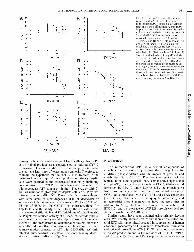
FIG. 4. Effect of 2-DG on LH-stimulatedprimary and MA-10 tumor Leydig cellmitochondrial DW
m, intracellular ATP con-
tent, and steroid production. A and B) DWm
in primary (A) and MA-10 tumor (B) Leydigcultures incubated with increasing doses of2-DG (0–100 mM) in the presence ofmaximally stimulating LH (100 ng/ml) for15 min. C and D) ATP levels in primary (C)and MA-10 tumor (D) Leydig culturesincubated with increasing doses of 2-DG(0–100 mM) in the presence of maximallystimulating LH (100 ng/ml) for 2 h. E and F)Steroid production by primary (E) and MA-10 tumor (F) Leydig cultures incubated withincreasing doses of 2-DG (0–100 mM) inthe presence of maximally stimulating LH(100 ng/ml) for 2 h. Points shown representthe mean 6 SEM of three experiments, withthree replicates per experiment. *P , 0.01vs. cells incubated with CCCP, #P , 0.05 vs.corresponding primary or MA-10 cells.
ATP PRODUCTION IN PRIMARY AND TUMOR LEYDIG CELLS 981
Dow
nloaded from w
ww
.biolreprod.org.

steroidogenesis, the results of these studies and of those onMA-10 cells suggest that myxothiazol might exert its effectsthrough reduced ATP production and, thus, point to theimportance of both mitochondrial ETC fidelity and ATP forsteroidogenesis. However, results obtained on the interrela-tionships among ATP synthesis, DW
m, and steroidogenesis
have not all been consistent. For example, it has been reportedthat dissipating DW
min MA-10 cells with CCCP inhibited
steroid formation but had no significant effect on cellular ATPlevels [12]. Such results call into question whether DW
mplays
a role in steroidogenesis beyond mitochondrial ATP synthesis,especially in light of findings that cholesterol transport in yeastis not affected by DW
mdisruption [29].
Studies of mitochondrial energetics in relationship to steroidformation have been conducted using both primary and tumorLeydig cells. The extent to which results obtained from thetumor cells can be extrapolated to primary cells has not beentested critically, however, so it seemed possible to us that someof the apparently conflicting data in the literature might be aconsequence of differences in the cell types used. Weaddressed this herein by conducting comparative studies on
Leydig cell mitochondrial energetics and steroid synthesisusing both primary and tumor Leydig cells. As had been shownby others in MA-10 cells [12, 14, 27], we found that CCCPdepolarized mitochondria in primary rat as well as mousetumor Leydig cells. The IC
50values of DW
mdepolarization
were not significantly different between the cell types.Surprisingly, the concentrations of CCCP necessary todepolarize the mitochondria, (IC
50, ;0.1 lM in both cell
types) were significantly less than those required to affect LH-stimulated testosterone synthesis. Moreover, exposure ofprimary Leydig cells to CCCP and elicitation of DW
mdepolarization was accompanied by a substantial reduction incellular ATP levels, suggesting that primary Leydig cell ATP isderived almost entirely from mitochondrial oxidative phos-phorylation. When cellular ATP responses to CCCP exposurewere assessed, however, a striking difference was observedbetween the two cell types. In contrast to results with primarycells and in keeping with previous findings [12], CCCPtreatment of MA-10 cells resulted in DW
mdepolarization but
not in a substantial decrease of cellular ATP levels in MA-10Leydig cells.
The large difference between the cell types regardingcellular ATP levels raised the possibility that the tumor cellsderive a majority of their ATP from glycolysis rather thanoxidative phosphorylation. To investigate this possibility, theeffect of the ATP synthase inhibitor oligomycin on Leydig cellparameters was assessed. As expected, oligomycin did notsignificantly affect DW
m[30]; rather, it potently inhibited
testosterone and P4 synthesis in both primary and tumorLeydig cells. Oligomycin significantly inhibited cellular ATPlevels in both cell types, but the degree of inhibition wasmarkedly different. Inhibition of mitochondrial ATP synthesisresulted in almost complete depletion of cellular ATP levels inprimary but not in tumor Leydig cells, as has been reported byothers [12, 15]. Indeed, inhibition of mitochondrial ATPsynthesis in MA-10 cells resulted in only 40%–50% reductionsin cellular ATP levels. The existence of a large, oligomycin-insensitive pool supports the hypothesis that tumor Leydig cellsderive ATP from sources other than, or in addition to,mitochondrial respiration.
To assess the glucose dependence of tumor and primaryLeydig cells directly, cells were cultured with the non-hydrolyzable glucose analog 2-DG, which serves as acompetitive inhibitor of the glycolytic pathway [24]. We noteddose-dependent inhibition of glucose metabolism and steroidformation in both primary and MA-10 tumor cells. However,steroid production was more significantly affected in the tumorLeydig cells than in the primary cells, with maximal inhibitionat lower glucose concentrations. Importantly, MA-10 cell ATPlevels were significantly more sensitive to glycolytic inhibitionthan were primary cell ATP levels.
The energetic differences between primary and tumorLeydig cells were not limited to glucose metabolism and ATPproduction. Though primary Leydig cells were highlysensitive to the complex I inhibitor rotenone, tumor Leydigcells were surprisingly refractory to ETC inhibition byrotenone. Because both cell types were strongly affected byinhibition of complex III by antimycin A and complex IV byNaCN (data not shown), these findings suggest that the tumorcells possess an impaired mitochondrial ETC at the level ofcomplex I. Mutational and functional changes in complex Ihave been observed in other transformed and tumor cells [31,32], though the precise alterations are heterogeneous. Thismay be an important consideration for reproductive toxicologystudies, because rotenone and other complex I inhibitors arecurrently utilized in pesticides [33, 34] and have been used to
FIG. 5. Effect of ETC inhibition on LH-stimulated mitochondrial DWm
,intracellular ATP content, and steroid production in primary and MA-10tumor Leydig cells. Black bars correspond to controls; gray barscorrespond to rotenone or antimycin A treatment. Data are presented asa percentage of control values. A) DW
min primary and MA-10 tumor
Leydig cultures incubated with rotenone (0.1 lM) or antimycin A (1 lM) inthe presence of maximally stimulating LH (100 ng/ml) for 15 min. B) ATPlevels in primary and MA-10 tumor Leydig cultures incubated withrotenone (0.1 lM) or antimycin A (1 lM) in the presence of maximallystimulating LH (100 ng/ml) for 2 h. C) Steroid production by primary andMA-10 tumor Leydig cells incubated with rotenone (0.1 lM) or antimycinA (1 lM) in the presence of maximally stimulating LH (100 ng/ml) for 2 h.Points shown represent the mean 6 SEM of three experiments, with threereplicates per experiment. *P , 0.001 vs. cells incubated without ETCinhibitors, #P , 0.05 vs. corresponding primary or MA-10 cells.
982 MIDZAK ET AL.
Dow
nloaded from w
ww
.biolreprod.org.

examine complex I failure in models of Parkinson disease[35]. Importantly, exposure of rodent models has beendemonstrated to decrease circulating testosterone levels [35].Toxicological studies of complex I inhibitors would returnfalse negatives if performed in assays using MA-10 tumorLeydig cells.
The data presented herein make it apparent that theseemingly inconsistent findings in the literature regarding thecontribution of mitochondrial energetics to steroidogenesislikely are a consequence of significant, intrinsic differencesbetween primary and tumor Leydig cells, as summarized inFigure 7. Both primary and MA-10 Leydig cells utilize
FIG. 6. Effect of mitochondrial disruptionand glycolytic inhibition on metabolic fluxthrough steroidogenic pathway in primaryLeydig cells. A) ATP levels in cells incubat-ed with 1 lg/ml of oligomycin or 5 lMCCCP in the presence of either LH (100 ng/ml), dbcAMP (1 mM), HC (20 lM), P5 (10lM), P4 (10 lM), or androstenedione (5 lM)for 2 h. B) Testosterone production in cellstreated as in A. After 2 h, medium wascollected and testosterone levels assessedby RIA. Values are presented as the per-centage of testosterone production withoutinhibitor. C) ATP levels in cells incubatedwith 100 mM 2-DG in the presence ofeither LH (100 ng/ml), dbcAMP (1 mM), HC(20 lM), P5 (10 lM), P4 (10 lM), orandrostenedione (5 lM) for 2 h. D) Testos-terone production in cells treated as in C.After 2 h, medium was collected andtestosterone levels assessed by RIA. Valuesare presented as the percent of testosteroneproduction without inhibitor. Lowercaseletters designate groups that are statisticallysignificant from each other (P , 0.05).
FIG. 7. Model of Leydig cell ATP utiliza-tion. Hormone-responsive primary (A) andtumor (B) Leydig cells critically utilizemitochondrial ATP for mitochondrial cho-lesterol transport (upper orange arrow).However, the tumorigenic transition hasresulted in a larger glycolytic contributionto cellular ATP (gradient arrow above).Consequently, tumor Leydig cells utilize agreater proportion of glycolytic ATP (B; bluearrow) for cholesterol transport than doprimary cells (A; blue arrow). Primary cellsalso utilize mitochondria-derived ATP forenzymatic reactions taking place in the ER(A; lower orange arrow), which are missingin tumor Leydig cells (B).
ATP PRODUCTION IN PRIMARY AND TUMOR LEYDIG CELLS 983
Dow
nloaded from w
ww
.biolreprod.org.
mitochondrial ATP for mitochondrial cholesterol transport.However, the tumorigenic transition in MA-10 cells hasresulted in a larger glycolytic contribution to cellular ATP.As a consequence, MA-10 cells utilize a greater proportion ofglycolytic ATP for cholesterol transport than do primary cells,though mitochondria-derived ATP remains absolutely criticalin these cells. Primary cells also utilize mitochondria-derivedATP for enzymatic reactions taking place in the ER, aspreviously demonstrated in other primary cell studies [36].These reactions are missing in tumor Leydig cells. Theglycolytic changes and dysfunctional complex I activity inMA-10 cells provide uncertainty regarding the extent to whichstudies of mitochondrial energetics and regulation of steroido-genesis in these transformed cells relate to the regulation ofsteroidogenesis in primary Leydig cells. Regardless, thesefindings demonstrate that steroidogenesis, especially mito-chondrial cholesterol import, is exquisitely sensitive to ATPsupply and demand, not to DW
mor ETC per se. The relative
importance of this ATP demand, whether for phosphorylationof key steroidogenic components such as STAR [37–39], themitochondrial transport of fatty acids [15], or unknownfunctions related to assembly of the transduceosome [4, 5],remains an area of active investigation.
ACKNOWLEDGMENT
We thank Dr. M. Ascoli (University of Iowa, Iowa City, IA) forproviding the MA-10 Leydig cells.
REFERENCES
1. Puett D, Li Y, DeMars G, Angelova K, Fanelli F. A functionaltransmembrane complex: the luteinizing hormone receptor with boundligand and G protein. Mol Cell Endocrinol 2007; 260–262:126–136.
2. Miller WL. Steroidogenic acute regulatory protein (StAR), a novelmitochondrial cholesterol transporter. Biochim Biophys Acta 2007; 1771:663–676.
3. Papadopoulos V, Baraldi M, Guilarte TR, Knudsen TB, Lacapere JJ,Lindemann P, Norenberg MD, Nutt D, Weizman A, Zhang MR, GavishM. Translocator protein (18 kDa): new nomenclature for the peripheral-type benzodiazepine receptor based on its structure and molecularfunction. Trends Pharmacol Sci 2006; 27:402–409.
4. Rone MB, Fan J, Papadopoulos V. Cholesterol transport in steroidbiosynthesis: role of protein-protein interactions and implications indisease states. Biochim Biophys Acta 2009; 1791:646–658.
5. Liu J, Rone MB, Papadopoulos V. Protein-protein interactions mediatemitochondrial cholesterol transport and steroid biosynthesis. J Biol Chem2006; 281:38879–38893.
6. Payne AH, Hales DB. Overview of steroidogenic enzymes in the pathwayfrom cholesterol to active steroid hormones. Endocr Rev 2004; 25:947–970.
7. Kadenbach B, Ramzan R, Wen L, Vogt S. New extension of the MitchellTheory for oxidative phosphorylation in mitochondria of living organisms.Biochim Biophys Acta 2010; 1800:205–212.
8. Wittig I, Schagger H. Supramolecular organization of ATP synthase andrespiratory chain in mitochondrial membranes. Biochim Biophys Acta2009; 1787:672–680.
9. Vander Heiden MG, Cantley LC, Thompson CB. Understanding theWarburg Effect: the metabolic requirements of cell proliferation. Science2009; 324:1029–1033.
10. Nascimento JM, Shi LZ, Tam J, Chandsawangbhuwana C, Durrant B,Botvinick EL, Berns MW. Comparison of glycolysis and oxidativephosphorylation as energy sources for mammalian sperm motility, usingthe combination of fluorescence imaging, laser tweezers, and real-timeautomated tracking and trapping. J Cell Physiol 2008; 217:745–751.
11. Lin CY, Hung PH, VandeVoort CA, Miller MG. 1H NMR to investigatemetabolism and energy supply in rhesus macaque sperm. Reprod Toxicol2009; 28:75–80.
12. Allen JA, Shankara T, Janus P, Buck S, Diemer T, Hales KH, Hales DB.Energized, polarized, and actively respiring mitochondria are required foracute Leydig cell steroidogenesis. Endocrinology 2006; 147:3924–3935.
13. Midzak AS, Liu J, Zirkin BR, Chen H. Effect of myxothiazol on Leydigcell steroidogenesis: inhibition of luteinizing hormone-mediated testoster-
one synthesis but stimulation of basal steroidogenesis. Endocrinology2007; 148:2583–2590.
14. King SR, Liu Z, Soh J, Eimerl S, Orly J, Stocco DM. Effects of disruptionof the mitochondrial electrochemical gradient on steroidogenesis and thesteroidogenic acute regulatory (StAR) protein. J Steroid Biochem MolBiol 1999; 69:143–154.
15. Duarte A, Castillo AF, Castilla R, Maloberti P, Paz C, Podesta EJ, CornejoMaciel F. An arachidonic acid generation/export system involved in theregulation of cholesterol transport in mitochondria of steroidogenic cells.FEBS Lett 2007; 581:4023–4028.
16. Moreno-Sanchez R, Rodrıguez-Enrıquez S, Saavedra E, Marın-HernandezA, Gallardo-Perez JC. The bioenergetics of cancer: is glycolysis the mainATP supplier in all tumor cells? Biofactors 2009; 35:209–225.
17. Warburg O. On the origin of cancer cells. Science 1956; 123:309–314.18. Klinefelter GR, Hall PF, Ewing LL. Effect of luteinizing hormone
deprivation in situ on steroidogenesis of rat Leydig cells purified by amultistep procedure. Biol Reprod 1987; 36:769–783.
19. Payne AH, Downing JR, Wong KL. Luteinizing hormone receptors andtestosterone synthesis in two distinct populations of Leydig cells.Endocrinology 1980; 106:1424–1429.
20. Ascoli M. Characterization of several clonal lines of cultured Leydigtumor cells: gonadotropin receptors and steroidogenic responses. Endo-crinology 1981; 108:88–95.
21. Aon MA, Cortassa S, Marban E, O’Rourke B. Synchronized whole celloscillations in mitochondrial metabolism triggered by a local release ofreactive oxygen species in cardiac myocytes. J Biol Chem 2003; 278:44735–44744.
22. Huang SG. Development of a high-throughput screening assay formitochondrial membrane potential in living cells. J Biomol Screen 2002;7:383–389.
23. Starkov AA, Fiskum G. Regulation of brain mitochondrial H2O
2
production by membrane potential and NAD(P)H redox state. J Neuro-chem 2003; 86:1101–1107.
24. Zhu Z, Jiang W, McGinley JN, Thompson HJ. 2-Deoxyglucose as anenergy restriction mimetic agent: effects on mammary carcinogenesis andon mammary tumor cell growth in vitro. Cancer Res 2005; 65:7023–7030.
25. Daum G, Vance JE. Import of lipids into mitochondria. Prog Lipid Res1997; 36:103–130.
26. Mokranjac D, Neupert W. Energetics of protein translocation intomitochondria. Biochim Biophys Acta 2008; 1777:758–762.
27. Artemenko IP, Zhao D, Hales DB, Hales KH, Jefcoate CR. Mitochondrialprocessing of newly synthesized steroidogenic acute regulatory protein(StAR), but not total StAR, mediates cholesterol transfer to cytochromeP450 side chain cleavage enzyme in adrenal cells. J Biol Chem 2001; 276:46583–46596.
28. King SR, Stocco DM. ATP and a mitochondrial electrochemical gradientare required for functional activity of the steroidogenic acute regulatory(StAR) protein in isolated mitochondria. Endocr Res 1996; 22:505–514.
29. Tuller G, Daum G. Import of sterols into mitochondria of the yeastSaccharomyces cerevisiae. FEBS Lett 1995; 372:29–32.
30. Weber J, Senior AE. ATP synthesis driven by proton transport in F1F0-ATP synthase. FEBS Lett 2003; 545:61–70.
31. Gasparre G, Porcelli AM, Bonora E, Pennisi LF, Toller M, Iommarini L,Ghelli A, Moretti M, Betts CM, Martinelli GN, Ceroni AR, Curcio F, et al.Disruptive mitochondrial DNA mutations in complex I subunits aremarkers of oncocytic phenotype in thyroid tumors. Proc Natl Acad SciU S A 2007; 104:9001–9006.
32. Baracca A, Chiaradonna F, Sgarbi G, Solaini G, Alberghina L, Lenaz G.Mitochondrial complex I decrease is responsible for bioenergeticdysfunction in K-ras transformed cells. Biochim Biophys Acta 2010;1797:314–323.
33. Lummen P. Complex I inhibitors as insecticides and acaricides. BiochimBiophys Acta 1998; 1364:287–296.
34. Cicchetti F, Drouin-Ouellet J, Gross RE. Environmental toxins andParkinson’s disease: what have we learned from pesticide-induced animalmodels? Trends Pharmacol Sci 2009, 30:475–483.
35. Alam M, Schmidt WJ. Mitochondrial complex I inhibition depletes plasmatestosterone in the rotenone model of Parkinson’s disease. Physiol Behav2004; 83:395–400.
36. Khanum A, Buczko E, Dufau ML. Essential role of adenosine triphosphatein activation of 17beta-hydroxysteroid dehydrogenase in rat Leydig cells.Endocrinology 1997; 138:1612–1620.
37. Arakane F, King SR, Du Y, Kallen CB, Walsh LP, Watari H, Stocco DM,Strauss JF III. Phosphorylation of steroidogenic acute regulatory protein(StAR) modulates its steroidogenic activity. J Biol Chem 1997; 272:32656–32662.
38. Jo Y, King SR, Khan SA, Stocco DM. Involvement of protein kinase C
984 MIDZAK ET AL.
Dow
nloaded from w
ww
.biolreprod.org.

and cyclic adenosine 30,50-monophosphate-dependent kinase in steroido-genic acute regulatory protein expression and steroid biosynthesis inLeydig cells. Biol Reprod 2005; 73:244–255.
39. Poderoso C, Converso DP, Maloberti P, Duarte A, Neuman I, Galli S,
Maciel FC, Paz C, Carreras MC, Poderoso JJ, Podesta EJ. A mitochondrialkinase complex is essential to mediate an ERK1/2-dependent phosphor-ylation of a key regulatory protein in steroid biosynthesis. PLoS ONE2008; 3:e1443.
ATP PRODUCTION IN PRIMARY AND TUMOR LEYDIG CELLS 985
Dow
nloaded from w
ww
.biolreprod.org.