apoptosis as a mechanism for removal of mutated cells of saccharomyces cerevisiae: the role of grx2...
TRANSCRIPT
Available online at www.sciencedirect.com
1780 (2008) 160–166www.elsevier.com/locate/bbagen
Biochimica et Biophysica Acta
Apoptosis as a mechanism for removal of mutated cells of Saccharomycescerevisiae: The role of Grx2 under cadmium exposure
Débora Silva Gomes a, Marcos Dias Pereira a, Anita Dolly Panek a,Leonardo Rodrigues Andrade b, Elis Cristina Araújo Eleutherio a,⁎
a Departamento de Bioquímica, Instituto de Química, UFRJ, 21941-909, Rio de Janeiro, RJ, Brazilb Departamento de Histologia e Embriologia, Instituto de Ciências Biomédicas, UFRJ, Rio de Janeiro, RJ, Brazil
Received 21 June 2007; received in revised form 24 August 2007; accepted 11 September 2007Available online 29 September 2007
Abstract
Cadmium is a strong mutagen that acts by inhibiting DNA mismatch repair, while its toxic effect seems to be related to an indirect oxidativestress that involves glutathione (GSH) mobilization. Among the roles of GSH is the protection of proteins against oxidative damage, by formingreversible mixed disulfides with cysteine residues, a process known as protein glutathionylation and catalyzed by glutaredoxins (Grx). In thiscurrent study, Saccharomyces cerevisiae cells deficient in GRX2, growing in 80 μM CdSO4, showed high mitochondrial mutagenic rate,determined by frequency of mutants that had lost mitochondrial function (petite mutants), high tolerance and lower apoptosis induction. Themutant strain also showed decreased levels of glutathionylated-protein after cadmium exposure, which might difficult the signaling to apoptosis,leading to increased mutagenic rates. Taken together, these results suggest that Grx2 is involved with the apoptotic death induced by cadmium, aform of cellular suicide that might lead of removal of mutated cells.© 2007 Elsevier B.V. All rights reserved.
Keywords: Glutathionylation; Grx2; Petite; Apoptosis; Cadmium; Saccharomyces cerevisiae
1. Introduction
Cadmium is a nonessential metal, although in some marineorganisms, cadmium, cobalt and zinc can functionally substitutefor one another to maintain optimal growth rates [1,2]. Cadmiumis one of the most toxic heavy metal, classified by IARC (Inter-national Agency for Research on Cancer) as human carcinogen[3]. It inhibits DNA mismatch repair (MMR) pathway throughthe inactivation of the ATPase activity of the MSH2–MSH6heterodimer [4], resulting in extreme mutability [5]. In addition,chronic exposure to non-lethal concentrations (in the range of10–100 μM) also induces mutants possessing mitochondrialdysfunction [5]. Mutations caused by damage to mitochondrialDNA are associated with a variety of human diseases, includingcancers [6].
Cadmium also induces apoptosis [7], which are ordered andcharacterized by mitochondrial dysfunction, release of cyto-
⁎ Corresponding author. Tel./fax: +55 21 2562 7735.E-mail address: [email protected] (E.C.A. Eleutherio).
0304-4165/$ - see front matter © 2007 Elsevier B.V. All rights reserved.doi:10.1016/j.bbagen.2007.09.014
chrome c to the cytoplasm, activation of caspase-9 and hydro-lysis of specific cellular proteins; however, the precise pathwayremains poorly understood. Apoptotic cell death induced byCd2+ has been related to accumulation of glutathionylated pro-teins – GSH-protein – formation of mixed disulfides betweenglutathione and proteins [8]. Since cadmium is not a directoxidant, it seems that GSH-protein accumulation is a result ofthe effect of cadmium to glutaredoxin, the expected catalyst ofprotein glutathionylation/deglutathionylation [9]. According toliterature, protein glutathionylation seems to represent a mecha-nism of redox regulation [10], in a fashion similar to thatmediated by protein phosphorylation. The alterations in cellularsulfhydryls may be the major determining factor for the path ofcell death in response to this heavy metal [11].
Glutaredoxins (Grxs) are thiol-disulfide that catalyze theglutathione-dependent reduction of protein disulfides and gluta-thione–protein mixed disulfides via two distinct mechanisms.The dithiol mechanism involves two cysteine residues in theactive site, whereas the monothiol mechanism relies only on themore N-terminal active site cysteine [12].
161D.S. Gomes et al. / Biochimica et Biophysica Acta 1780 (2008) 160–166
Three Grx subfamilies have been distinguished in the yeastSaccharomyces cerevisiae: the dithiolic proteins Grx1 andGrx2; Grx3, Grx4, and Grx5 are monothiolic members of thesecond subgroup, lacking the more C-terminal active site cys-teine; Grx3 and Grx4 possess an additional thioredoxin (Trx)domain and are constituents of the third subfamily [13]. Thesefive Grxs also differ in regard to their subcellular localization.Grx1 is cytosolic, Grx3 and Grx4 are nuclear, Grx5 is mito-chondrial, and Grx2 has a dual localization in the cytosol andmitochondria [13]. Grx5 is essential for the functional assemblyof iron–sulfur centers [14] and Grx3 and Grx4 need their Trxdomain for nuclear targeting and for Grx-like activity [15]. Thedithiolic Grx1 and Grx2 play distinct roles during different stressconditions [16]. Usually, different isoforms in different com-partments play different roles. Although the dithiolic Grx2 is alsomitochondrial, it cannot compensate the loss of the monothiolicGrx5, indicating distinct roles [14]. The double mutant grx2grx5and the triple mutant grx3grx4grx5 were non-viable [17].
In mammals, two Grxs have been identified, the cytosolicGrx1 and Grx2, which are involved in important processes suchas cellular differentiation and regulation of transcription factors[for review see [18]]. The mammalian Grx2 is present in twoisoforms derived from alternative first exons: Grx2a is targetedto mitochondria, whereas Grx2b is localized in the nucleus [19].Despite high similarity, mammalian Grx1 and Grx2 have somedifferences: Grx2 has a CSYC active site motif, instead of theCPYC motif of the mammalian cytosolic and Escherichia coli[20]. Moreover, Grx2 lacks one of the conserved non-active sitecysteine residues encountered in Grx1, consequently less easilyinactivated by oxidants and GSSG [19].
In this study, the involvement of Grx1 and Grx2 in themutagenesis and apoptotic cell death caused by cadmium, usingSaccharomyces cerevisiae as experimental model of eukaryoticorganism, was investigated. The use of this yeast for identifi-cation of the molecular mechanisms of heavy metals toxicity isespecially attractive because of the easy genetic manipulationand availability of the complete Saccharomyces cerevisiae ge-nomic sequence.
Although Saccharomyces cerevisiae has five Grx isoforms,we choose to study Grx1 and Grx2 due to the followingreasons: (i) Grx1 and Grx2 from yeasts share 40–52% identityand 61–76% similarity with Grxs from bacterial and mam-malian species [16]; (ii) the synthesis of yeast Grx2 is inducedin response to Cd2+ [21], evidencing the importance of thisprotein in the protection against this heavy metal; (iii) Grx1 iscytosolic, and Grx2 has a dual localization in the cytosol andmitochondria [13], a focus of reactive oxygen species – ROS –production and apoptosis; iv) mammalian Grx2, ortholog toyeast Grx2, has been associated with mitochondrial redoxhomeostasis during oxidative stress-induced apoptosis [22].
2. Materials and methods
2.1. Saccharomyces cerevisiae strains and growth conditions
The control strain BY4741 (MATa his3Δ1 leu2Δ0 met15Δ0 ura3Δ0) and itsisogenic mutants grx1Δ and grx2Δ, harboring the genes GRX1 and GRX2
interrupted by the gene KanMX4 gene,were acquired from Euroscarf, Frankfurt,Germany. Cells were grown in liquid YPD medium containing 1% yeast extract,2% glucose, 2% peptone, using an orbital shaker at 28 °C and 160 rpm with theratio of flask volume/medium of 5/1.
2.2. Cadmium stress
Cells were grown up to middle exponential phase (1 mg dry weight/mL=106 cells/mL). Thereafter, cells were re-inoculated into fresh medium (initialcell concentration was 105 cells/mL) in the presence or absence of 80 μMCdSO4 and incubated at 28 °C/160 rpm for 48 h.
2.3. Determination of glutathione and GSH-protein
Reduced (GSH) and oxidized (GSSG) forms of glutathione were determinedspectrophotometrically, in neutralized trichloroacetic acid (10% TCA) extracts,as previously described [23]. To release GSH bound to proteins and determineglutathionylated protein (GSH-protein), the pellets from the acid extraction weretreated with 1% sodium borohydride [24].
2.4. Lipid peroxidation
Cells were centrifuged at 2000×g for 2 min and washed twice with distilledMillipore purified water. The pellets were resuspended in 0.5 mL of 10% TCA(w/v) and 1.5 g of glass bead was added. The samples were lysed by six cyclesof 20 s agitation on a vortex followed by 20 s on ice. Extracts were centrifugedat 2000×g for 3 min and the supernatant mixed with 0.1 mL of 0.1 M EDTAand 0.6 mL of 1% (w/v) thiobarbituric acid in 0.05 M NaOH. The reactionmixture was incubated in a boiling water bath for 15 min and, after cooling, theabsorbance was measured at 532 nm [25].
2.5. Cadmium absorption
Analyses of the capacity of Saccharomyces cerevisiae cells to absorbcadmium were determined by using atomic absorption spectrophotometry, aspreviously described [23,26]. For measuring residual cadmium present in themedium, 5 mL aliquots were centrifuged, the supernatant was collected andsubjected to atomic absorption spectrophotometry — Atomic Absorption Spec-trometer, Perkin Elmer 3100. Cadmium absorption was calculated by deter-mining the difference in metal content between the control medium without cells(initial concentration) and the test medium containing cells. Percentages ofcadmium accumulation were calculated through the equation:
Absorption ð%Þ ¼ ½ðinitial concentrationÞ � ðfinal concentrationÞ= ðinitial concentrationÞ� � 100
The limit of detection for cadmium as measured by atomic absorption is5 nM.
2.6. Cell viability and mutagenesis
Tolerance against Cd2+ stress was analyzed by plating on solidified YPDmedium, after proper dilution. Cells were also plated on YPGly (1% yeastextract, 4% glycerol, 2% peptone and 2% agar), which was used to test strainsfor the petite phenotype (mitochondrial mutagenesis). Plates were done intriplicates. Colonies were counted after incubation at 28 °C for at least 72 h.Viability was determined by plating stressed and nonstressed cells on YPD;tolerance was expressed as percentage of survival. The mutagenic effect ofcadmium was measured by also plating stressed cells on YPGly [5].
Survival ð%Þ ¼ ½ðnumber of cells=mL after Cd stressÞ= ðnumber of cells=mL before Cd stressÞ� � 100
Mutation rate ð%Þ¼ ½ðnumber of cells=mL after Cd stress plated on YPDÞ
� ðnumber of cells=mL after Cd stress plated on YPGlyÞ= ðnumber of cells=mL after Cd stress plated on YPDÞ� � 100:
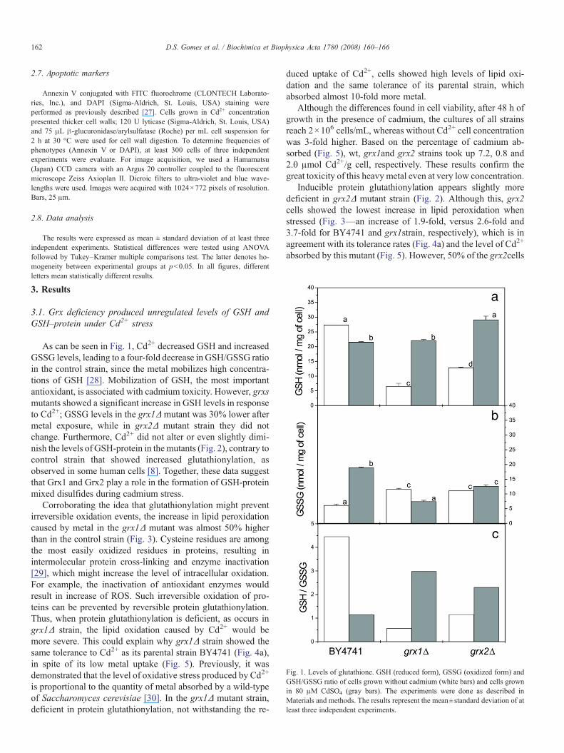
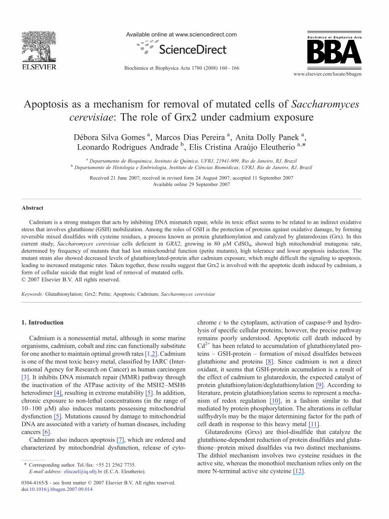
Fig. 1. Levels of glutathione. GSH (reduced form), GSSG (oxidized form) andGSH/GSSG ratio of cells grown without cadmium (white bars) and cells grownin 80 μM CdSO4 (gray bars). The experiments were done as described inMaterials and methods. The results represent the mean±standard deviation of atleast three independent experiments.
162 D.S. Gomes et al. / Biochimica et Biophysica Acta 1780 (2008) 160–166
2.7. Apoptotic markers
Annexin V conjugated with FITC fluorochrome (CLONTECH Laborato-ries, Inc.), and DAPI (Sigma-Aldrich, St. Louis, USA) staining wereperformed as previously described [27]. Cells grown in Cd2+ concentrationpresented thicker cell walls; 120 U lyticase (Sigma-Aldrich, St. Louis, USA)and 75 μL β-glucuronidase/arylsulfatase (Roche) per mL cell suspension for2 h at 30 °C were used for cell wall digestion. To determine frequencies ofphenotypes (Annexin V or DAPI), at least 300 cells of three independentexperiments were evaluate. For image acquisition, we used a Hamamatsu(Japan) CCD camera with an Argus 20 controller coupled to the fluorescentmicroscope Zeiss Axioplan II. Dicroic filters to ultra-violet and blue wave-lengths were used. Images were acquired with 1024×772 pixels of resolution.Bars, 25 μm.
2.8. Data analysis
The results were expressed as mean ± standard deviation of at least threeindependent experiments. Statistical differences were tested using ANOVAfollowed by Tukey–Kramer multiple comparisons test. The latter denotes ho-mogeneity between experimental groups at pb0.05. In all figures, differentletters mean statistically different results.
3. Results
3.1. Grx deficiency produced unregulated levels of GSH andGSH–protein under Cd2+ stress
As can be seen in Fig. 1, Cd2+ decreased GSH and increasedGSSG levels, leading to a four-fold decrease in GSH/GSSG ratioin the control strain, since the metal mobilizes high concentra-tions of GSH [28]. Mobilization of GSH, the most importantantioxidant, is associated with cadmium toxicity. However, grxsmutants showed a significant increase in GSH levels in responseto Cd2+; GSSG levels in the grx1Δ mutant was 30% lower aftermetal exposure, while in grx2Δ mutant strain they did notchange. Furthermore, Cd2+ did not alter or even slightly dimi-nish the levels of GSH-protein in the mutants (Fig. 2), contrary tocontrol strain that showed increased glutathionylation, asobserved in some human cells [8]. Together, these data suggestthat Grx1 and Grx2 play a role in the formation of GSH-proteinmixed disulfides during cadmium stress.
Corroborating the idea that glutathionylation might preventirreversible oxidation events, the increase in lipid peroxidationcaused by metal in the grx1Δ mutant was almost 50% higherthan in the control strain (Fig. 3). Cysteine residues are amongthe most easily oxidized residues in proteins, resulting inintermolecular protein cross-linking and enzyme inactivation[29], which might increase the level of intracellular oxidation.For example, the inactivation of antioxidant enzymes wouldresult in increase of ROS. Such irreversible oxidation of pro-teins can be prevented by reversible protein glutathionylation.Thus, when protein glutathionylation is deficient, as occurs ingrx1Δ strain, the lipid oxidation caused by Cd2+ would bemore severe. This could explain why grx1Δ strain showed thesame tolerance to Cd2+ as its parental strain BY4741 (Fig. 4a),in spite of its low metal uptake (Fig. 5). Previously, it wasdemonstrated that the level of oxidative stress produced by Cd2+
is proportional to the quantity of metal absorbed by a wild-typeof Saccharomyces cerevisiae [30]. In the grx1Δ mutant strain,deficient in protein glutathionylation, not withstanding the re-
duced uptake of Cd2+, cells showed high levels of lipid oxi-dation and the same tolerance of its parental strain, whichabsorbed almost 10-fold more metal.
Although the differences found in cell viability, after 48 h ofgrowth in the presence of cadmium, the cultures of all strainsreach 2×10⁶ cells/mL, whereas without Cd2+ cell concentrationwas 3-fold higher. Based on the percentage of cadmium ab-sorbed (Fig. 5), wt, grx1and grx2 strains took up 7.2, 0.8 and2.0 μmol Cd2+/g cell, respectively. These results confirm thegreat toxicity of this heavy metal even at very low concentration.
Inducible protein glutathionylation appears slightly moredeficient in grx2Δ mutant strain (Fig. 2). Although this, grx2cells showed the lowest increase in lipid peroxidation whenstressed (Fig. 3—an increase of 1.9-fold, versus 2.6-fold and3.7-fold for BY4741 and grx1strain, respectively), which is inagreement with its tolerance rates (Fig. 4a) and the level of Cd2+
absorbed by this mutant (Fig. 5). However, 50% of the grx2cells
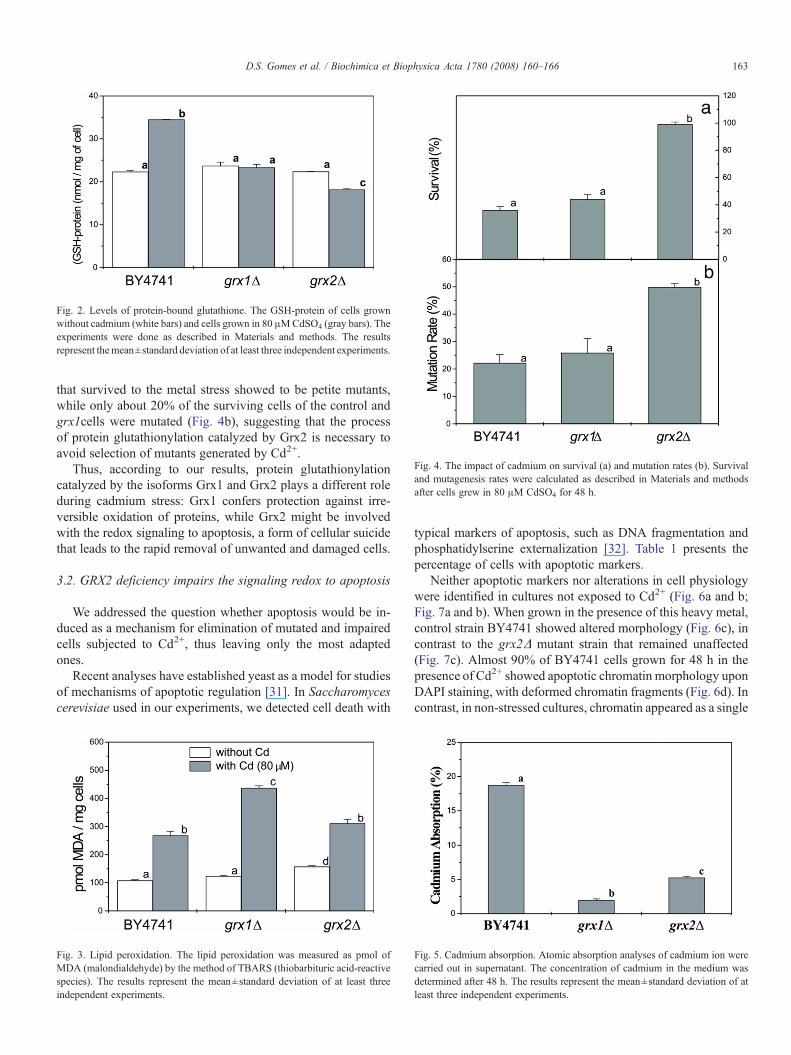
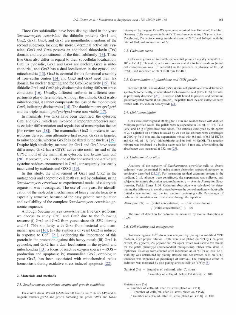
Fig. 4. The impact of cadmium on survival (a) and mutation rates (b). Survivaland mutagenesis rates were calculated as described in Materials and methodsafter cells grew in 80 μM CdSO4 for 48 h.
Fig. 2. Levels of protein-bound glutathione. The GSH-protein of cells grownwithout cadmium (white bars) and cells grown in 80 μMCdSO4 (gray bars). Theexperiments were done as described in Materials and methods. The resultsrepresent themean±standard deviation of at least three independent experiments.
163D.S. Gomes et al. / Biochimica et Biophysica Acta 1780 (2008) 160–166
that survived to the metal stress showed to be petite mutants,while only about 20% of the surviving cells of the control andgrx1cells were mutated (Fig. 4b), suggesting that the processof protein glutathionylation catalyzed by Grx2 is necessary toavoid selection of mutants generated by Cd2+.
Thus, according to our results, protein glutathionylationcatalyzed by the isoforms Grx1 and Grx2 plays a different roleduring cadmium stress: Grx1 confers protection against irre-versible oxidation of proteins, while Grx2 might be involvedwith the redox signaling to apoptosis, a form of cellular suicidethat leads to the rapid removal of unwanted and damaged cells.
3.2. GRX2 deficiency impairs the signaling redox to apoptosis
We addressed the question whether apoptosis would be in-duced as a mechanism for elimination of mutated and impairedcells subjected to Cd2+, thus leaving only the most adaptedones.
Recent analyses have established yeast as a model for studiesof mechanisms of apoptotic regulation [31]. In Saccharomycescerevisiae used in our experiments, we detected cell death with
Fig. 3. Lipid peroxidation. The lipid peroxidation was measured as pmol ofMDA (malondialdehyde) by the method of TBARS (thiobarbituric acid-reactivespecies). The results represent the mean±standard deviation of at least threeindependent experiments.
typical markers of apoptosis, such as DNA fragmentation andphosphatidylserine externalization [32]. Table 1 presents thepercentage of cells with apoptotic markers.
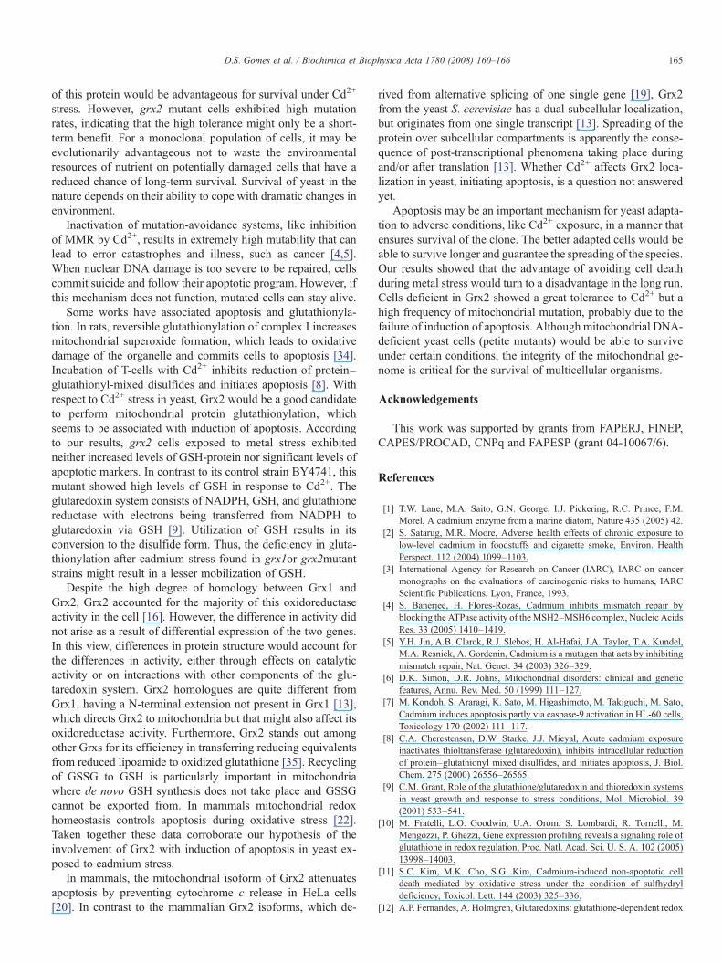
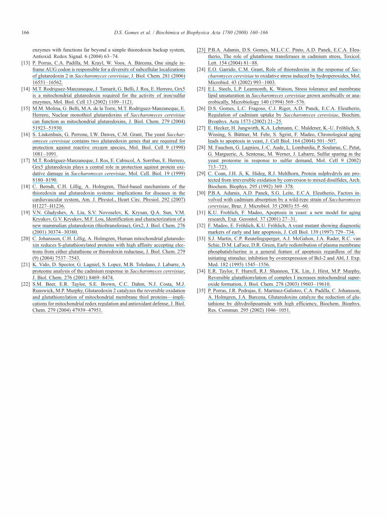
Neither apoptotic markers nor alterations in cell physiologywere identified in cultures not exposed to Cd2+ (Fig. 6a and b;Fig. 7a and b). When grown in the presence of this heavy metal,control strain BY4741 showed altered morphology (Fig. 6c), incontrast to the grx2Δ mutant strain that remained unaffected(Fig. 7c). Almost 90% of BY4741 cells grown for 48 h in thepresence of Cd2+ showed apoptotic chromatin morphology uponDAPI staining, with deformed chromatin fragments (Fig. 6d). Incontrast, in non-stressed cultures, chromatin appeared as a single
Fig. 5. Cadmium absorption. Atomic absorption analyses of cadmium ion werecarried out in supernatant. The concentration of cadmium in the medium wasdetermined after 48 h. The results represent the mean±standard deviation of atleast three independent experiments.
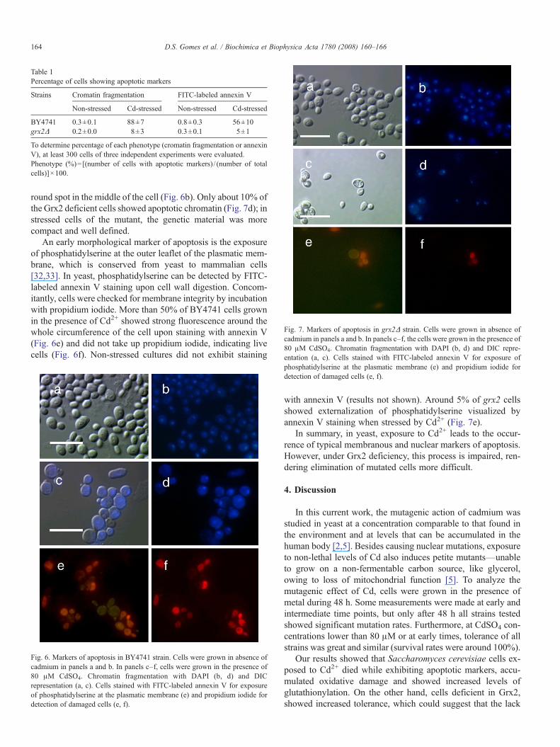
Table 1Percentage of cells showing apoptotic markers
Strains Cromatin fragmentation FITC-labeled annexin V
Non-stressed Cd-stressed Non-stressed Cd-stressed
BY4741 0.3±0.1 88±7 0.8±0.3 56±10grx2Δ 0.2±0.0 8±3 0.3±0.1 5±1
To determine percentage of each phenotype (cromatin fragmentation or annexinV), at least 300 cells of three independent experiments were evaluated.Phenotype (%)=[(number of cells with apoptotic markers) / (number of totalcells)]×100.
Fig. 7. Markers of apoptosis in grx2Δ strain. Cells were grown in absence ofcadmium in panels a and b. In panels c–f, the cells were grown in the presence of80 μM CdSO4. Chromatin fragmentation with DAPI (b, d) and DIC repre-entation (a, c). Cells stained with FITC-labeled annexin V for exposure of
164 D.S. Gomes et al. / Biochimica et Biophysica Acta 1780 (2008) 160–166
round spot in the middle of the cell (Fig. 6b). Only about 10% ofthe Grx2 deficient cells showed apoptotic chromatin (Fig. 7d); instressed cells of the mutant, the genetic material was morecompact and well defined.
An early morphological marker of apoptosis is the exposureof phosphatidylserine at the outer leaflet of the plasmatic mem-brane, which is conserved from yeast to mammalian cells[32,33]. In yeast, phosphatidylserine can be detected by FITC-labeled annexin V staining upon cell wall digestion. Concom-itantly, cells were checked for membrane integrity by incubationwith propidium iodide. More than 50% of BY4741 cells grownin the presence of Cd2+ showed strong fluorescence around thewhole circumference of the cell upon staining with annexin V(Fig. 6e) and did not take up propidium iodide, indicating livecells (Fig. 6f). Non-stressed cultures did not exhibit staining
phosphatidylserine at the plasmatic membrane (e) and propidium iodide fordetection of damaged cells (e, f).
Fig. 6. Markers of apoptosis in BY4741 strain. Cells were grown in absence ofcadmium in panels a and b. In panels c–f, cells were grown in the presence of80 μM CdSO4. Chromatin fragmentation with DAPI (b, d) and DICrepresentation (a, c). Cells stained with FITC-labeled annexin V for exposureof phosphatidylserine at the plasmatic membrane (e) and propidium iodide fordetection of damaged cells (e, f).
with annexin V (results not shown). Around 5% of grx2 cellsshowed externalization of phosphatidylserine visualized byannexin V staining when stressed by Cd2+ (Fig. 7e).
In summary, in yeast, exposure to Cd2+ leads to the occur-rence of typical membranous and nuclear markers of apoptosis.However, under Grx2 deficiency, this process is impaired, ren-dering elimination of mutated cells more difficult.
4. Discussion
In this current work, the mutagenic action of cadmium wasstudied in yeast at a concentration comparable to that found inthe environment and at levels that can be accumulated in thehuman body [2,5]. Besides causing nuclear mutations, exposureto non-lethal levels of Cd also induces petite mutants—unableto grow on a non-fermentable carbon source, like glycerol,owing to loss of mitochondrial function [5]. To analyze themutagenic effect of Cd, cells were grown in the presence ofmetal during 48 h. Some measurements were made at early andintermediate time points, but only after 48 h all strains testedshowed significant mutation rates. Furthermore, at CdSO4 con-centrations lower than 80 μM or at early times, tolerance of allstrains was great and similar (survival rates were around 100%).
Our results showed that Saccharomyces cerevisiae cells ex-posed to Cd2+ died while exhibiting apoptotic markers, accu-mulated oxidative damage and showed increased levels ofglutathionylation. On the other hand, cells deficient in Grx2,showed increased tolerance, which could suggest that the lack
165D.S. Gomes et al. / Biochimica et Biophysica Acta 1780 (2008) 160–166
of this protein would be advantageous for survival under Cd2+
stress. However, grx2 mutant cells exhibited high mutationrates, indicating that the high tolerance might only be a short-term benefit. For a monoclonal population of cells, it may beevolutionarily advantageous not to waste the environmentalresources of nutrient on potentially damaged cells that have areduced chance of long-term survival. Survival of yeast in thenature depends on their ability to cope with dramatic changes inenvironment.
Inactivation of mutation-avoidance systems, like inhibitionof MMR by Cd2+, results in extremely high mutability that canlead to error catastrophes and illness, such as cancer [4,5].When nuclear DNA damage is too severe to be repaired, cellscommit suicide and follow their apoptotic program. However, ifthis mechanism does not function, mutated cells can stay alive.
Some works have associated apoptosis and glutathionyla-tion. In rats, reversible glutathionylation of complex I increasesmitochondrial superoxide formation, which leads to oxidativedamage of the organelle and commits cells to apoptosis [34].Incubation of T-cells with Cd2+ inhibits reduction of protein–glutathionyl-mixed disulfides and initiates apoptosis [8]. Withrespect to Cd2+ stress in yeast, Grx2 would be a good candidateto perform mitochondrial protein glutathionylation, whichseems to be associated with induction of apoptosis. Accordingto our results, grx2 cells exposed to metal stress exhibitedneither increased levels of GSH-protein nor significant levels ofapoptotic markers. In contrast to its control strain BY4741, thismutant showed high levels of GSH in response to Cd2+. Theglutaredoxin system consists of NADPH, GSH, and glutathionereductase with electrons being transferred from NADPH toglutaredoxin via GSH [9]. Utilization of GSH results in itsconversion to the disulfide form. Thus, the deficiency in gluta-thionylation after cadmium stress found in grx1or grx2mutantstrains might result in a lesser mobilization of GSH.
Despite the high degree of homology between Grx1 andGrx2, Grx2 accounted for the majority of this oxidoreductaseactivity in the cell [16]. However, the difference in activity didnot arise as a result of differential expression of the two genes.In this view, differences in protein structure would account forthe differences in activity, either through effects on catalyticactivity or on interactions with other components of the glu-taredoxin system. Grx2 homologues are quite different fromGrx1, having a N-terminal extension not present in Grx1 [13],which directs Grx2 to mitochondria but that might also affect itsoxidoreductase activity. Furthermore, Grx2 stands out amongother Grxs for its efficiency in transferring reducing equivalentsfrom reduced lipoamide to oxidized glutathione [35]. Recyclingof GSSG to GSH is particularly important in mitochondriawhere de novo GSH synthesis does not take place and GSSGcannot be exported from. In mammals mitochondrial redoxhomeostasis controls apoptosis during oxidative stress [22].Taken together these data corroborate our hypothesis of theinvolvement of Grx2 with induction of apoptosis in yeast ex-posed to cadmium stress.
In mammals, the mitochondrial isoform of Grx2 attenuatesapoptosis by preventing cytochrome c release in HeLa cells[20]. In contrast to the mammalian Grx2 isoforms, which de-
rived from alternative splicing of one single gene [19], Grx2from the yeast S. cerevisiae has a dual subcellular localization,but originates from one single transcript [13]. Spreading of theprotein over subcellular compartments is apparently the conse-quence of post-transcriptional phenomena taking place duringand/or after translation [13]. Whether Cd2+ affects Grx2 loca-lization in yeast, initiating apoptosis, is a question not answeredyet.
Apoptosis may be an important mechanism for yeast adapta-tion to adverse conditions, like Cd2+ exposure, in a manner thatensures survival of the clone. The better adapted cells would beable to survive longer and guarantee the spreading of the species.Our results showed that the advantage of avoiding cell deathduring metal stress would turn to a disadvantage in the long run.Cells deficient in Grx2 showed a great tolerance to Cd2+ but ahigh frequency of mitochondrial mutation, probably due to thefailure of induction of apoptosis. Although mitochondrial DNA-deficient yeast cells (petite mutants) would be able to surviveunder certain conditions, the integrity of the mitochondrial ge-nome is critical for the survival of multicellular organisms.
Acknowledgements
This work was supported by grants from FAPERJ, FINEP,CAPES/PROCAD, CNPq and FAPESP (grant 04-10067/6).
References
[1] T.W. Lane, M.A. Saito, G.N. George, I.J. Pickering, R.C. Prince, F.M.Morel, A cadmium enzyme from a marine diatom, Nature 435 (2005) 42.
[2] S. Satarug, M.R. Moore, Adverse health effects of chronic exposure tolow-level cadmium in foodstuffs and cigarette smoke, Environ. HealthPerspect. 112 (2004) 1099–1103.
[3] International Agency for Research on Cancer (IARC), IARC on cancermonographs on the evaluations of carcinogenic risks to humans, IARCScientific Publications, Lyon, France, 1993.
[4] S. Banerjee, H. Flores-Rozas, Cadmium inhibits mismatch repair byblocking the ATPase activity of theMSH2–MSH6 complex, Nucleic AcidsRes. 33 (2005) 1410–1419.
[5] Y.H. Jin, A.B. Clarck, R.J. Slebos, H. Al-Hafai, J.A. Taylor, T.A. Kundel,M.A. Resnick, A. Gordenin, Cadmium is a mutagen that acts by inhibitingmismatch repair, Nat. Genet. 34 (2003) 326–329.
[6] D.K. Simon, D.R. Johns, Mitochondrial disorders: clinical and geneticfeatures, Annu. Rev. Med. 50 (1999) 111–127.
[7] M. Kondoh, S. Araragi, K. Sato, M. Higashimoto, M. Takiguchi, M. Sato,Cadmium induces apoptosis partly via caspase-9 activation in HL-60 cells,Toxicology 170 (2002) 111–117.
[8] C.A. Cherestensen, D.W. Starke, J.J. Mieyal, Acute cadmium exposureinactivates thioltransferase (glutaredoxin), inhibits intracellular reductionof protein–glutathionyl mixed disulfides, and initiates apoptosis, J. Biol.Chem. 275 (2000) 26556–26565.
[9] C.M. Grant, Role of the glutathione/glutaredoxin and thioredoxin systemsin yeast growth and response to stress conditions, Mol. Microbiol. 39(2001) 533–541.
[10] M. Fratelli, L.O. Goodwin, U.A. Orom, S. Lombardi, R. Tornelli, M.Mengozzi, P. Ghezzi, Gene expression profiling reveals a signaling role ofglutathione in redox regulation, Proc. Natl. Acad. Sci. U. S. A. 102 (2005)13998–14003.
[11] S.C. Kim, M.K. Cho, S.G. Kim, Cadmium-induced non-apoptotic celldeath mediated by oxidative stress under the condition of sulfhydryldeficiency, Toxicol. Lett. 144 (2003) 325–336.
[12] A.P. Fernandes, A. Holmgren, Glutaredoxins: glutathione-dependent redox
166 D.S. Gomes et al. / Biochimica et Biophysica Acta 1780 (2008) 160–166
enzymes with functions far beyond a simple thioredoxin backup system,Antioxid. Redox Signal. 6 (2004) 63–74.
[13] P. Porras, C.A. Padilla, M. Krayl, W. Voos, A. Bárcena, One single in-frame AUG codon is responsible for a diversity of subcellular localizationsof glutaredoxin 2 in Saccharomyces cerevisiae, J. Biol. Chem. 281 (2006)16551–16562.
[14] M.T. Rodriguez-Manzaneque, J. Tamarit, G. Belli, J. Ros, E. Herrero, Grx5is a mitochondrial glutaredoxin required for the activity of iron/sulfurenzymes, Mol. Biol. Cell 13 (2002) 1109–1121.
[15] M.M. Molina, G. Belli, M.A. de la Torre, M.T. Rodriguez-Manzaneque, E.Herrero, Nuclear monothiol glutaredoxins of Saccharomyces cerevisiaecan function as mitochondrial glutaredoxins, J. Biol. Chem. 279 (2004)51923–51930.
[16] S. Liukenhuis, G. Perrone, I.W. Dawes, C.M. Grant, The yeast Sacchar-omyces cerevisiae contains two glutaredoxin genes that are required forprotection against reactive oxygen species, Mol. Biol. Cell 9 (1998)1081–1091.
[17] M.T. Rodríguez-Manzaneque, J. Ros, E. Cabiscol, A. Sorribas, E. Herrero,Grx5 glutaredoxin plays a central role in protection against protein oxi-dative damage in Saccharomyces cerevisiae, Mol. Cell. Biol. 19 (1999)8180–8190.
[18] C. Berndt, C.H. Lillig, A. Holmgren, Thiol-based mechanisms of thethioredoxin and glutaredoxin systems: implications for diseases in thecardiovascular system, Am. J. Physiol., Heart Circ. Physiol. 292 (2007)H1227–H1236.
[19] V.N. Gladyshev, A. Liu, S.V. Novoselov, K. Krysan, Q.A. Sun, V.M.Kryukov, G.V. Kryukov, M.F. Lou, Identification and characterization of anew mammalian glutaredoxin (thioltransferase), Grx2, J. Biol. Chem. 276(2001) 30374–30380.
[20] C. Johansson, C.H. Lillig, A. Holmgren, Human mitochondrial glutaredo-xin reduces S-glutathionylated proteins with high affinity accepting elec-trons from either glutathione or thioredoxin reductase, J. Biol. Chem. 279(9) (2004) 7537–7543.
[21] K. Vido, D. Spector, G. Lagniel, S. Lopez, M.B. Toledano, J. Labarre, Aproteome analysis of the cadmium response in Saccharomyces cerevisiae,J. Biol. Chem. 276 (2001) 8469–8474.
[22] S.M. Beer, E.R. Taylor, S.E. Brown, C.C. Dahm, N.J. Costa, M.J.Runswick, M.P. Murphy, Glutaredoxin 2 catalyzes the reversible oxidationand glutathionylation of mitochondrial membrane thiol proteins—impli-cations for mitochondrial redox regulation and antioxidant defense, J. Biol.Chem. 279 (2004) 47939–47951.
[23] P.B.A. Adamis, D.S. Gomes, M.L.C.C. Pinto, A.D. Panek, E.C.A. Eleu-therio, The role of glutathione transferases in cadmium stress, Toxicol.Lett. 154 (2004) 81–88.
[24] E.O. Garrido, C.M. Grant, Role of thioredoxins in the response of Sac-charomyces cerevisiae to oxidative stress induced by hydroperoxides, Mol.Microbiol. 43 (2002) 993–1003.
[25] E.L. Steels, L.P. Learmonth, K. Watson, Stress tolerance and membranelipid unsaturation in Saccharomyces cerevisiae grown aerobically or ana-erobically, Microbiology 140 (1994) 569–576.
[26] D.S. Gomes, L.C. Fragoso, C.J. Riger, A.D. Panek, E.C.A. Eleutherio,Regulation of cadmium uptake by Saccharomyces cerevisiae, Biochim.Byophys. Acta 1573 (2002) 21–25.
[27] E. Hecker, H. Jungwirth, K.A. Lehmann, C. Maldener, K.-U. Fröhlich, S.Wissing, S. Büttner, M. Fehr, S. Sgrist, F. Madeo, Chronological agingleads to apoptosis in yeast, J. Cell Biol. 164 (2004) 501–507.
[28] M. Fauchon, G. Lagnieu, J.-C. Aude, L. Lombardia, P. Soularue, C. Petat,G. Marguerie, A. Sentenac, M. Werner, J. Labarre, Sulfur sparing in theyeast proteome in response to sulfur demand, Mol. Cell 9 (2002)713–723.
[29] C. Coan, J.H. Ji, K. Hideg, R.J. Mehlhorn, Protein sulphydryls are pro-tected from irreversible oxidation by conversion to mixed disulfides, Arch.Biochem. Biophys. 295 (1992) 369–378.
[30] P.B.A. Adamis, A.D. Panek, S.G. Leite, E.C.A. Eleutherio, Factors in-volved with cadmium absorption by a wild-type strain of Saccharomycescerevisiae, Braz. J. Microbiol. 35 (2003) 55–60.
[31] K.U. Frohlich, F. Madeo, Apoptosis in yeast: a new model for agingresearch, Exp. Gerontol. 37 (2001) 27–31.
[32] F. Madeo, E. Fröhlich, K.U. Fröhlich, A yeast mutant showing diagnosticmarkers of early and late apoptosis, J. Cell Biol. 139 (1997) 729–734.
[33] S.J. Martin, C.P. Reutelingsperger, A.J. McGahon, J.A. Rader, R.C. vanSchie, D.M. LaFace, D.R. Green, Early redistribution of plasma membranephosphatidylserine is a general feature of apoptosis regardless of theinitiating stimulus: inhibition by overexpression of Bcl-2 and Abl, J. Exp.Med. 182 (1995) 1545–1556.
[34] E.R. Taylor, F. Hurrell, R.J. Shannon, T.K. Lin, J. Hirst, M.P. Murphy,Reversible glutathionylation of complex I increases mitochondrial super-oxide formation, J. Biol. Chem. 278 (2003) 19603–19610.
[35] P. Porras, J.R. Pedrajas, E. Martinez-Galisteo, C.A. Padilla, C. Johansson,A. Holmgren, J.A. Barcena, Glutaredoxins catalyze the reduction of glu-tathione by dihydrolipoamide with high efficiency, Biochem. Biophys.Res. Commun. 295 (2002) 1046–1051.