functional analysis of the saccharomyces cerevisiae dup240
TRANSCRIPT
Microbiology (2002), 148, 2111–2123 Printed in Great Britain
Functional analysis of the Saccharomycescerevisiae DUP240 multigene family revealsmembrane-associated proteins that are notessential for cell viability
Re! my Poirey,1,2 Laurence Despons2, Ve! ronique Leh,2
Maria-Jose Lafuente,1† Serge Potier,2 Jean-Luc Souciet2
and Jean-Claude Jauniaux1
Author for correspondence: Jean-Claude Jauniaux. Tel : 49 6221 42 49 71. Fax: 49 6221 42 52 49 71.e-mail : j.jauniaux!dkfz.de
1 AngewandteTumorvirologie, AbteilungF0100 and VirologieApplique! e a' l’Oncologie(Unite! INSERM 375),DeutschesKrebsforschungszentrum,P. 1011949, D-69009Heidelberg, Germany
2 Laboratoire de Ge! ne! tiqueet Microbiologie, UPRES-A7010 ULP/CNRS, Institut deBotanique, 28 rue Goethe,F-67083 Strasbourg cedex,France
The DUP240 gene family of Saccharomyces cerevisiae is composed of 10members. They encode proteins of about 240 amino acids which contain twopredicted transmembrane domains. Database searches identified only onehomologue in the closely related species Saccharomyces bayanus, indicatingthat the DUP240 genes encode proteins specific to Saccharomyces sensu stricto.The short-flanking homology PCR gene-replacement strategy with a variety ofselective markers for replacements, and classical genetic methods, were usedto generate strains deleted for all 10 DUP240 genes. All of the knock-outstrains were viable and had similar growth kinetics to the wild-type. Two-hybrid screens, hSos1p fusions and GFP fusions were carried out; the resultsindicated that the Dup240 proteins are membrane associated, and that some ofthem are concentrated around the plasma membrane.
Keywords : gene disruption, membrane protein, gene tandem repeats, two-hybridsystem, yeast
INTRODUCTION
Multigene families have been found in all sequencedorganisms. In Arabidopsis thaliana gene duplication isas high as 65% (Arabidopsis Genome Initiative, 2000).These paralogous genes have similar sequences. Thereare numerous, more or less ancient, examples that reflectdifferent situations: tRNA genes dispersed throughoutthe genome; rRNAgenes gathered in clusters ; dispersionand lower duplication levels for the genes encodingribosomal proteins. The duplication phenomenon isobserved for genes with known and unknown functions.Studying these multigenic families should provide usefulinformation about the functions of the different copiesand also about how they are involved in genomedynamics.
Saccharomyces cerevisiae S288C potentially encodes5651 ORFs (Malpertuy et al., 2000). Of these, 914
.................................................................................................................................................
†Present address: Centro de Biologia Molecular Severo Ochoa, Universi-dad Autonoma, Canto Blanco, 28049, Madrid, Spain.
Abbreviation: GFP, green fluorescent protein.
(16±2%) belong to two-gene families and 1544 (27±3%)belong to multigene families with between three andover 20 members (Blandin et al., 2000). In some cases,there are physiological reasons for the presence ofmultigene families. For example the cytoplasmic andmitochondrialmethionyl-tRNA synthetases are encodedby two related but different nuclear genes, resulting indifferent localizations (Schneller et al., 1978). The genesencoding alcohol dehydrogenase (ADHI and ADHII)are very similar, but ADHI catalyses the formation ofethanol from acetaldehyde whereas ADHII catalyses theformation of acetaldehyde from ethanol (Johnston &Carlson, 1992). Another well-studied case is the CUP1locus, which encodes a copper- or cadmium-chelatingmetallothionein expressed after induction with metals.The reference strain, S288C, contains only two copies ofCUP1, but copper- or cadmium-resistant strains cancontain up to 15 copies (Karin et al., 1984). Theexpression of most hexose transporters is tightly regu-lated by glucose concentration, starvation, osmoticpressure and the physiological state of the cells. In fact,to abolish glucose consumption and transport activitycompletely, all 18 members of the hexose transporterfamily, HXT1–17, GAL2 and three members of the
0002-5399 # 2002 SGM 2111
R. Poirey and others
.................................................................................................................................................................................................................................................................................................................
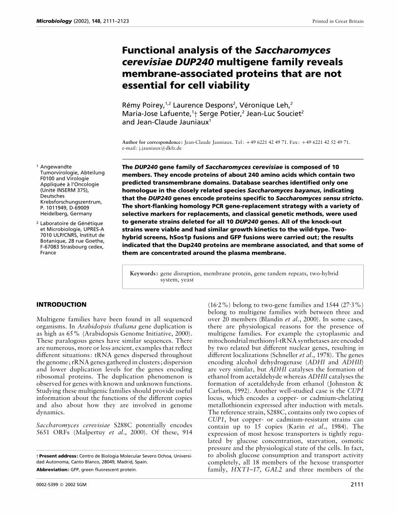
Fig. 1. Map of the genetic organization, chromosome localization and predicted amino acid sequence identities of the 10DUP240 ORFs of S. cerevisiae. The ORFs are represented by rectangles, their orientation with respect to the centromeresby arrows. The percentages show the level of amino acid identities between the indicated ORF products.
maltose transporter family (AGT1, YDL247w andYJR160c) have to be deleted (Wieczorke et al., 1999).
In comparison with these established examples and withthe exception of the PAU and OSBP families (Rachidiet al., 2000; Beh et al., 2001), the large gene familiesidentified by sequencing programmes remain mostlyunstudied. This is the case for the DUP240 gene family.This family is particularly interesting because of itsunusually high copy number (10), the high level ofnucleotide identity between some of its members and thespecific chromosomal organization of the members (Fig.1). The YAR027w, YAR028w, YAR029w, YAR031wand YAR033w ORFs are arranged as tandem repeats onchromosome I and the YGL051w and YGL053w ORFsare arranged as tandem repeats on chromosome VII.The nucleotide sequences of YAR033w and YAR031ware 98% identical to those of YGL051w and YGL053w(Feuermann et al., 1997). Most of the correspondingproteins are approximately 240 aa long. Moreover,these ORFs appear to be specific for the Saccharomycessensu stricto group (Bon et al., 2000).
We carried out a functional analysis of these genes to getsome insight into the role of the proteins which theyencode. We replaced all 10 members of the DUP240family and screened for associated phenotypes using thecriteria defined by the EUROFAN network (Dujon,1998; Oliver et al., 1998). Green fluorescent protein(GFP) fusion fluorescence was subsequently used tostudy the subcellular localization of the different ORFproducts. Activation of the RAS signalling pathwaythrough hSos1p–Dup fusions and the classical GAL4-based nuclear two-hybrid system were used to determinewhether some of the Dup240 proteins were localizedat the plasma membrane and to search for partnersthat interact with the products of the YGL051w andYGL053w genes. Our results indicated that the DUP240
gene family encodes membrane-associated proteins thatare not essential for cell viability.
METHODS
Yeast strains and media. The Saccharomyces cerevisiae strainsused in this study are listed in Table 1. Cells were grown onYPD [1% yeast extract, 2% peptone, 2% glucose (dextrose)and 2% agar], YPG (1% yeast extract, 2% peptone, 2%, v}v,glycerol and 2% agar) and SD [6±7 g yeast nitrogen base l−",2% glucose (dextrose) and 2% agar] supplemented with theamino acids and bases not required for the selection. The samemedia without agar were used for liquid cultures. Syntheticcomplete glucose medium (SC) was used for the two-hybridselections (Sherman et al., 1986). Sporulation of diploidsrequired growth on YPD overnight at 30 °C prior to transferonto sporulation plates (1% potassium acetate and 2% agar)for 3–5 days at 30 °C.
PCR-mediated gene replacements. The primers used for PCR-mediated gene disruption consisted of 40–60 nt specific tosequences just upstream or downstream from the ORF to bedeleted followed at the 3« end of the primers by 18–20 baseshomologous to the upstream or downstream flanking regionof a selectable marker gene located in a yeast integratingvector (Table 2). PCR conditions were: 94 °C for 2 min,(94 °C for 30 s, 54 °C for 30 s, 72 °C for 2 min) ¬30 cycles,72 °C for 10 min. The specific PCR products were treated withphenol}chloroform prior to ethanol precipitation. Usually1–5 µg amplified DNA was used to transform S. cerevisiae.High-efficiency transformations were performed by themethod described by Gietz et al. (1995). Correct ORF re-placements were checked by PCR analysis. Oligonucleotideswere designed to bind outside the target loci (A1 and A4),within the target loci (A2 and A3) or within the marker genes(M2 and M3). In diploid yeast transformants with correctlyintegrated markers, one of the two copies of the target locuswas replaced by the marker module. In PCR experiments withgenomic yeast DNA and using either A1, A2 and M2 or M3,A3 and A4, the correct integration of the marker DNA was
2112
Functional analysis of the DUP240 gene family
Table 1. S. cerevisiae strains
Strain Genotype Source*
SFY526 MATa trp1-901 leu2-3,112 ura3-52 his3-200 gal4∆ gal80∆ lys2-801 ade2-101
URA3 : :GAL1–lacZ can
Bartel et al. (1993b)
PJ69-4a MATa trp1-901 leu2-3,112 ura3-52 his3-200 gal4∆ gal80∆ LYS2 : :GAL1–HIS3 GAL2–ADE2
met2 : :GAL7–lacZ
James et al. (1996)
PJ69-4α MATα trp1-901 leu2-3,112 ura3-52 his3-200 gal4∆ gal80∆ LYS2 : :GAL1–HIS3 GAL2–ADE2
met2 : :GAL7–lacZ
P. James
CDC25H MATa ade2-801 cdc25-2 his3-200 leu2-3,112 trp1-901 ura3-52 Petitjean et al. (1990)
(All subsequent strains are isogenic to S288C)
BY4709 MATα ura3∆0 ATCC 200872
FY1679 MATa}MATα his3∆200}HIS3 leu2∆1}LEU2 trp1∆63}TRP1 ura3∆52}ura3∆52 ATCC 96604
FYBL1-17B MATa his3∆200 trp1∆63 ura3∆851 B. Dujon
FYBL1-23D MATα his3∆200 trp1∆63 ura3∆851 B. Dujon
LD073 MATa YGL053w–YGL051w∆ : :kan his3∆200 trp1∆63 ura3∆851 This study
LD076 MATα YAR027w–YAR033w∆ : :HIS3 his3∆200 trp1∆63 ura3∆851 This study
LD078 MATa YAR027w–YAR033w∆ : :HIS3 YGL053w–YGL051w∆ : :kan his3∆200 trp1∆63
ura3∆851
This study
BY4734 MATα his3∆200 leu2∆0 met15∆0 trp1∆63 ura3∆0 ATCC 200896
LD084 MATα YGL053w–YGL051w∆ : :kan his3∆200 leu2∆0 met15∆0 trp1∆63 ura3∆ This study
LD099 MATa YAR027w–YAR033w∆ : :HIS3 YGL053w–YGL051w∆ : :kan his3∆200 leu2∆0 met15∆0
trp1∆63 ura3∆
This study
LD100 MATα YAR027w–YAR033w∆ : :HIS3 YGL053w–YGL051w∆ : :kan his3∆200 leu2∆0 met15∆0
trp1∆63 ura3∆
This study
LD102 MATa YAR023c∆ : :MET15 YAR027w–YAR033w∆ : :HIS3 YGL053w–YGL051w∆ : :kan
his3∆200 leu2∆0 met15∆0 trp1∆63 ura3∆
This study
LD103 MATα YAR023c∆ : :MET15 YAR027w–YAR033w∆ : :HIS3 YGL053w–YGL051w∆ : :kan
his3∆200 leu2∆0 met15∆0 trp1∆63 ura3∆
This study
LD107 MATa YAR023c∆ : :MET15 YAR027w–YAR033w∆ : :HIS3 YGL053W–YGL051w∆ : :kan
YHL044w∆ : :LEU2 his3∆200 leu2∆0 met15∆0 trp1∆63 ura3∆
This study
LD108 MATα YAR023c∆ : :MET15 YAR027w–YAR033w∆ : :HIS3 YGL053w–YGL051w∆ : :kan
YHL044w∆ : :LEU2 his3∆200 leu2∆0 met15∆0 trp1∆63 ura3∆
This study
LD115 MATa YAR023c∆ : :MET15 YAR027w–YAR033w∆ : :HIS3 YCR007c∆ : :TRP1
YGL053w–YGL051w∆ : :kan YHL044w∆ : :LEU2 his3∆200 leu2∆0 met15∆0 trp1∆63 ura3∆
This study
LD114 MATα YAR023c∆ : :MET15 YAR027w–YAR033w∆ : :HIS3 YCR007c∆ : :TRP1
YGL053w–YGL051w∆ : :kan YHL044w∆ : :LEU2 his3∆200 leu2∆0 met15∆0 trp1∆63 ura3∆
This study
*ATCC, American Type Culture Collection, Manassas, VA, USA; P. James, University of Wisconsin, Madison, WI, USA; B. Dujon,Institut Pasteur, Paris, France.
confirmed by the appearance of two PCR products of thepredicted length: one characteristic for the wild-type allele(A1-A2 or A3-A4 amplification product) and a secondfragment characteristic for the mutated allele (A1-M2 or M3-A4 amplification product). Incorrect transformants yieldedonly the amplification products of the wild-type allele.
DNA preparations and Escherichia coli transformation. Bac-terial plasmid DNA preparations, bacterial transformationsand DNA manipulations were carried out according tostandard protocols (Sambrook et al., 1989). Yeast genomicDNA and plasmid DNA were purified according to themethod described by Hoffman & Winston (1987).
Cloning of a PCR product in a linearized plasmid byhomologous recombination in yeast. Plasmidswere linearizedby two different endonucleases and dephosphorylated withcalf intestine phosphatase (Boehringer). The sequences to becloned were amplified by PCR (Saiki et al., 1985) fromgenomic DNA of yeast strain FY1679 with oligonucleotide
primers having 20 additional nucleotides at the 5« ends. Theseends were homologous to the ends of the linearized plasmidand used for cloning by homologous recombination in thisplasmid (Muhlrad et al., 1992). The PCR conditions were:94 °C for 1±5 min, (94 °C for 30 s, 48 °C for 2 min, 70 °C for2±5 min) ¬10 cycles, (94 °C for 30 s, 60 °C for 2 min, 70 °C for2±5 min) ¬35 cycles, 70 °C for 10 min. The PCR product andthe linearized plasmid were treated with phenol}chloroformprior to ethanol precipitation. High-efficiency transformationswere performed according to the method described by Gietz etal. (1995). For cloning in pGBT9 (Bartel et al., 1993a), pSOS(Stratagene) and pGRU1 (see below), the vector was linearizedby EcoRI and SalI, NcoI and MluI, and EcoRI and SalI,respectively. The PCR product and the corresponding linear-ized plasmid were introduced into the yeast cells simul-taneously. The oligonucleotide sequences are given in Table 2.We ensured that there were no errors in the junctions betweenthe plasmid and the inserts for pGBT9-YGL051w and pGBT9-YGL053w by sequencing.
2113
R. Poirey and others
Table 2. Oligonucleotides used as primers to generate PCR products
Name Oligonucleotides used for PCR-based ORF disruptions Plasmid† ORF replacement‡
Sequence (5«!3«)*
53S1 CATAACAACCTCCAAAACCATATAATAACCTTACACAAGA-
CAAGA
pFA6a-kanMX4 YGL053w–YGL051w∆ : :kan
TATCAATTCAACATGCGTACGCTGCAGGTCGAC pFA6a-HIS3MX6 YAR027w–YAR033w∆ : :HIS3
51S2 CTGATTATATATCATACTCTAGTTTATGTTCGCTTTACGT-
ATGGCAG
pFA6a-kanMX4 YGL053w–YGL051w∆ : :kan
TGTCTCTAAGCTAATCGATGAATTCGAGCTCG pFA6a-HIS3MX6 YAR027w–YAR033w∆ : :HIS3
23S1 TGCGTTCTTTTTATACCAATATATTAGATACGTAAACTC-
TACTCAAGATTGTACTGAGAGTGCAC
pRS401 YAR023c∆ : :MET15
23S2 CAGAGTTATTTGCTTTACGAAATTGTACGCGCCAAGTATA-
TAATGCTGTGCGGTATTTCACACCG
pRS401 YAR023c∆ : :MET15
07S1 CGCTCAGTATAAGTGCTAAATAAAATTATCAAGATTTATA-
TTTCAAAGGAGAGGGCCAAGAGGGA
pRS404 YCR007c∆ : :TRP1
07S2 GTCTATACCATATCAAGACAAGAAACAAAACTCCGTATCT-
GCATGGGCAAGTGCACAAACAATAC
pRS404 YCR007c∆ : :TRP1
44S1 TATAAAGACAAATTCAAAAGCAAGTGAGGGCCCGCTAAGG-
CTATGAAGGCCGTTTCTGACAGAG
pRS405 YHL044w∆ : :LEU2
44S2 GCGTTATAAAAAACTAAAGTAGAACCTAGAAATACCTTCC-
AATCAATCGCACAGAATCAAATTCG
pRS405 YHL044w∆ : :LEU2
Name Oligonucleotides used for cloning by recombination Plasmid§ Insert
name
Sequence (5«!3«)§
51P1 ACCAAAGGTCAAAGACAGTTGACTGTATCGATGCAGACCCCTCTAGAAAG pGBT9 YGL051w
51P2 TAAGAAATTCGCCCGGAATTAGCTTGGCTGCTATTCCGTCTTTTTAAGAAGC
53P1 ACCAAAGGTCAAAGACAGTTGACTGTATCGATGCAAACCCCTTCAGAAAATAC pGBT9 YGL053w
53P2 TAAGAAATTCGCCCGGAATTAGCTTGGCTGCTAAAAAAACTCATCGACACC
29SOS1 ATTAGTTATAGTAGGATCCCCATGAATAAATATCTATTTGACC pSOS YAR029w
29SOS2 CGCGGCGGCCGCGAGCTCACTACAATATCCGCTGTCTTGG
31SOS1 ATTAGTTATAGTAGGATCCCCATGTCGCCTCAATACCATTT pSOS YAR031w
31SOS2 CGCGGCGGCCGCGAGCTCACTAAAAAAACTCATCGACACC
33SOS1 ATTAGTTATAGTAGGATCCCCATGCAGACCCCTCCAGAAAG pSOS YAR033w
33SOS2 CGCGGCGGCCGCGAGCTCACTATTCCGTCTTTTTAAGAAGC
51SOS1 ATTAGTTATAGTAGGATCCCCATGCAGACCCCTCTAGAAAG pSOS YGL051w
51SOS2 CGCGGCGGCCGCGAGCTCACTATTCCGTCTTTTTAAGAAGC
53SOS1 ATTAGTTATAGTAGGATCCCCATGCAAACCCCTTCAGAAAAT pSOS YGL053w
53SOS2 CGCGGCGGCCGCGAGCTCACTAAAAAAACTCATCGACACC
adhGRUf GCGTTGGCCGATTCATTCCCGTTGCTTGCATGCAACTTC pGRU1 Padh1
adhGRUr CGGAGCTTGCATGCCTGCAGCGGCCGCATCTTTCAGGAGGCTTGCTTCT
29ADHf GAAGCAAGCCTCCTGAAAGATGAATAAATATCTATTTGACC pGRU1-Padh1 YAR029w
29ADHr CGGAGCTTGCATGCCTGCAGCAATATCCGCTGTCTTGGAA
31ADHf GAAGCAAGCCTCCTGAAAGATGTCGCCTCAATACCATTTT pGRU1-Padh1 YAR031w
31ADHr CGGAGCTTGCATGCCTGCAGAAAAAACTCATCGACACCAGG
33ADHf GAAGCAAGCCTCCTGAAAGATGCAGACCCCTCCAGAAAG pGRU1-Padh1 YAR033w
33ADHr CGGAGCTTGCATGCCTGCAGTTCCGTCTTTTTAAGAAGCGC
51ADHf GAAGCAAGCCTCCTGAAAGATGCAGACCCCTCTAGAAAG pGRU1-Padh1 YGL051w
51ADHr CGGAGCTTGCATGCCTGCAGTTCCGTCTTTTTAAGAAGCGC
53ADHf GAAGCAAGCCTCCTGAAAGATGCAAACCCCTTCAGAAAAT pGRU1-Padh1 YGL053w
53ADHr CGGAGCTTGCATGCCTGCAGAAAAAACTCATCGACACCAGG
44ADHf GAAGCAAGCCTCCTGAAAGATGAGTTCAGAATTATTAATATC pGRU1-Padh1 YHL044w
44ADHr CGGAGCTTGCATGCCTGCAGAACTGTGACCCCCATCTCTG
*The nucleotides at the 5« end are homologous to the genomic DNA region just upstream or downstream from the gene to be replaced.The initiator or stop codon is shown in bold. The underlined nucleotides are homologous to the plasmid sequence upstream ordownstream from the selectable marker gene.
2114
Functional analysis of the DUP240 gene family
Determination of Dup240–GFP localization in vivo. The PCR-amplified DUP240 ORFs were cloned in-frame at the C-terminal end of the green fluorescent protein-S65T (GFP-S65T) gene of pGRU1 (Michel Aigle, IBGC, Bordeaux, France)and of pGRU1-Padh1. Transformed yeast cells were selectedin SD medium without uracil because URA3 was used as themarker gene for selection of pGRU1. Dup240–GFP andcontrol GFP were excited with a 488 nm laser and viewedunder a Leica confocal microscope equipped with a ¬63objective.
Activation of the RAS signalling pathway through hSos1p–Dup240 fusion. We modified the cytoplasmic two-hybridsystem (Aronheim et al., 1997) marketed by Stratagene(CytoTrap) so that it would test for the presence of a hybridprotein at the plasma membrane. This system uses the S.cerevisiae mutant CDC25H, containing a thermosensitivemutation of the CDC25 gene product (Petitjean et al., 1990).This gene is homologous to the human hSOS1 gene (Chardinet al., 1993) and encodes a GDP}GTP exchange factor. Bystimulating the exchange of GDP associated with the Ras1 andRas2 proteins for GTP, Cdc25p stimulates the signallingpathway involving these proteins. hSos1p is cytoplasmic in thesystem. The cdc25H mutation prevents cell growth at 37 °Cwhile growth is normal at 25 °C. Cell growth can be restoredby targeting hSos1p to the plasma membrane (Aronheim et al.,1994). To test whether Dup240 proteins can target hSos1p tothe plasma membrane we constructed hSos1p–Dup240 fusionproteins by introducing PCR-amplified DUP240 ORFs intothe pSOS vector (CytoTrap) in-frame at the 5« end of thehSOS1 gene. We then tested the ability of the resultingconstructions to restore the growth of the CDC25H yeaststrain (CytoTrap) at 37 °C. Drops of liquid control andtransformed yeast cultures were deposited onto YPD platesand incubated for 2–3 days at 37 °C to evaluate growth.
Two-hybrid screen strategy. Two-hybrid screens were carriedout using a system based on that described by Fields & Song(1989). We used the Gal4pAD-yeast genomic library (mech-anically sheared genomic DNA fragments with a mean size of800 bp inserted into the pACTII vector which bears themarker gene LEU2 ; A. Ramne & P. Sunnerhagen, http :}}www.mips.biochem.mpg.de}proj}eurofan}eurofanj1}b5}index.html). This library was introduced (Georgakopouloset al., 2001) into the haploid yeast PJ69-4a (Table 1).This strain contains three markers (HIS3, ADE2 and lacZ)controlled by three different GAL promoters (GAL1, GAL2and GAL7, respectively), which can each be activated inthe two-hybrid system (James et al., 1996). This library wascomposed of 2±1¬10( independent transformed yeast cells. Itwas amplified in liquid culture and aliquot vials containing6±5¬10) yeast cells were stored at ®80 °C. A mating strategyinspired by Bendixen et al. (1994) and modified by Fromont-Racine et al. (1997) was used to obtain a wide range of diploidscontaining both bait and target plasmids. High matingefficiency enabled us to test over 5¬10( interactions perexperiment. The haploid yeast PJ69-4α (Table 1) was trans-formed with the bait cloned by homologous recombination
†The pFA6a series are reporter}marker plasmids (pFA, plasmids for Functional Analysis ; Wach et al., 1994). Information on thesequences and availability of the pRS400 series of vectors is given in Brachmann et al. (1998).
‡The ORF replacement cassette amplified with both 53S1 and 51S2 oligonucleotides allowed the replacement of either the region ofchromosome VII between the YGL053w ORF and the YGL051w ORF or the region of chromosome I between the YAR027w ORF andthe YAR033w ORF.
§The sequence corresponding to the insert is underlined; the rest of the sequence is homologous to the plasmid and was used forhomologous recombination in yeast.
and fused with the Gal4pBD (Gal4p-DNA binding domain) ofthe pGBT9 vector bearing the marker gene TRP1. For eachscreen, one vial containing 6±5¬10) transformed PJ69-4a wasmixed with 10* PJ69-4α cells. Cells were concentrated ontofilters and incubated on rich medium for 4±5 h at 30 °C prior tocollection. The cells were diluted and spread onto SC®Leu,SC®Trp and SC®Leu®Trp plates to count the number ofparental cells and the number of diploids. The rest of the cellsuspension was spread onto 24 SC®Leu®Trp®His plates(24 cm¬24 cm) containing 2 mM 3-aminotriazole. The plateswere incubated at 30 °C for 4 days. His-positive clones weresubjected to a second selection on SC®Leu®Trp®adenineplates. Plasmids were rescued in E. coli HB101. Insert junc-tions with Gal4pAD were sequenced and precisely identifiedin the yeast genome using the MIPS (Munich InformationCentre for protein sequences ; http :}}mips.gsf.de) YeastGenome Database (MYGD) or the Saccharomyces GenomeDatabase from Stanford University (http:}}genome-www.stanford.edu}Saccharomyces).
β-Galactosidase filter assays to test the two-hybrid inter-actions in another genetic background. Yeast strain SFY526was transformed by the LiAc method (Gietz et al., 1995)simultaneously with two plasmids which gave a two-hybridinteraction in PJ69-4 and spread onto SD®Leu®Trp agar in150 mm diameter Petri plates. After 2 days at 30 °C theseplates were replicated onto 125 mm diameter filters, whichwere incubated on YPD medium plates for 1 day at 30 °C.The filters were frozen quickly in liquid nitrogen and putonto plates containing, in a total of 3±5 ml: 58±5 µl 2%X-Gal in dimethylformamide, 9±5 µl β-mercaptoethanol, 16±1 gNa
#HPO
%.7H
#O l−", 5±5 g NaH
#PO
%.H
#O l−", 0±75 g KCl l−"
and 0±246 g MgSO%\7H
#O l−" at pH 7±0. The time required for
yeasts to stain blue was measured at room temperature(22 °C).
RESULTS
In silico analysis
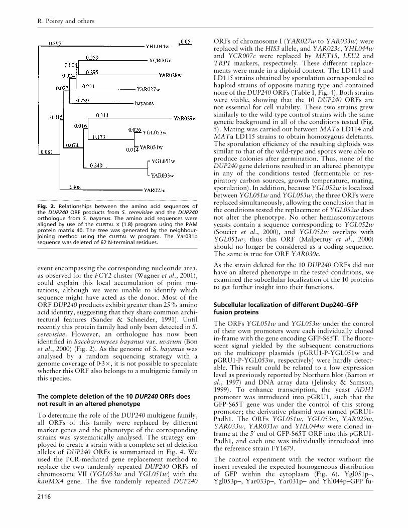
The 10 DUP240 ORFs constitute a large multigenefamily in the S. cerevisiae S288C strain. The relation-ships between the 10 encoded proteins are depicted inFig. 2. The alignment of the protein sequences (Fig. 3)revealed five well-conserved domains: C1 (amino acids1–44), C2 (amino acids 109–150), C3 (amino acids206–224), H1 (amino acids 51–67) and H2 (amino acids78–94). H1 and H2 are hydrophobic domains andbecause of their length they are predicted to be trans-membrane domains (Klein et al., 1985). The ORFproducts Yar033p and Ygl051p have a very high level ofidentity (96±6%), with only eight amino acid differencesin a total of 234. Furthermore, six of these differenceswere within the 17 residues of the second predictedtransmembrane domain (Fig. 3), but they did not affectthe hydrophobicity of the domain. A genetic conversion
2115
R. Poirey and others
.................................................................................................................................................
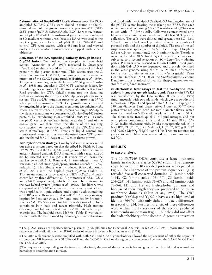
Fig. 2. Relationships between the amino acid sequences ofthe DUP240 ORF products from S. cerevisiae and the DUP240orthologue from S. bayanus. The amino acid sequences werealigned by use of the CLUSTAL X (1.8) program using the PAMprotein matrix 40. The tree was generated by the neighbour-joining method using the CLUSTAL W program. The Yar031psequence was deleted of 62 N-terminal residues.
event encompassing the corresponding nucleotide area,as observed for the FCY2 cluster (Wagner et al., 2001),could explain this local accumulation of point mu-tations, although we were unable to identify whichsequence might have acted as the donor. Most of theORF DUP240 products exhibit greater than 25% aminoacid identity, suggesting that they share common archi-tectural features (Sander & Schneider, 1991). Untilrecently this protein family had only been detected in S.cerevisiae. However, an orthologue has now beenidentified in Saccharomyces bayanus var. uvarum (Bonet al., 2000) (Fig. 2). As the genome of S. bayanus wasanalysed by a random sequencing strategy with agenome coverage of 0±3¬, it is not possible to speculatewhether this ORF also belongs to a multigenic family inthis species.
The complete deletion of the 10 DUP240 ORFs doesnot result in an altered phenotype
To determine the role of the DUP240 multigene family,all ORFs of this family were replaced by differentmarker genes and the phenotype of the correspondingstrains was systematically analysed. The strategy em-ployed to create a strain with a complete set of deletionalleles of DUP240 ORFs is summarized in Fig. 4. Weused the PCR-mediated gene replacement method toreplace the two tandemly repeated DUP240 ORFs ofchromosome VII (YGL053w and YGL051w) with thekanMX4 gene. The five tandemly repeated DUP240
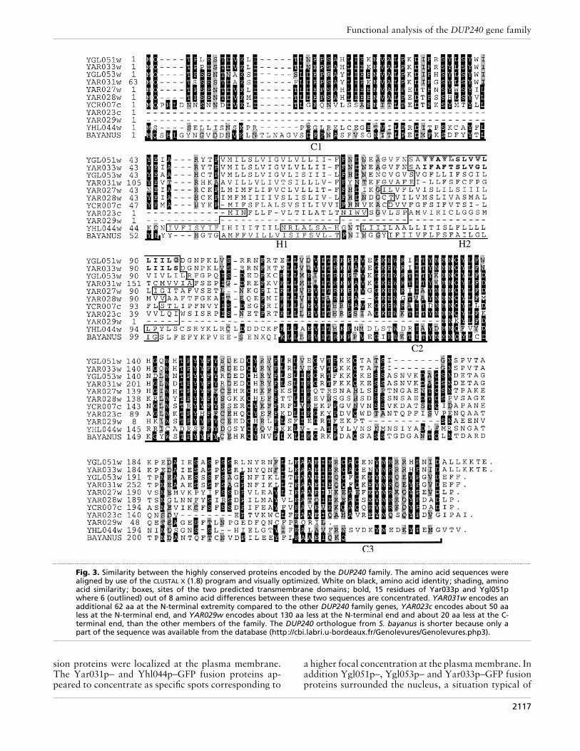
ORFs of chromosome I (YAR027w to YAR033w) werereplaced with the HIS3 allele, and YAR023c, YHL044wand YCR007c were replaced by MET15, LEU2 andTRP1 markers, respectively. These different replace-ments were made in a diploid context. The LD114 andLD115 strains obtained by sporulation corresponded tohaploid strains of opposite mating type and containednone of the DUP240 ORFs (Table 1, Fig. 4). Both strainswere viable, showing that the 10 DUP240 ORFs arenot essential for cell viability. These two strains grewsimilarly to the wild-type control strains with the samegenetic background in all of the conditions tested (Fig.5). Mating was carried out between MATα LD114 andMATa LD115 strains to obtain homozygous deletants.The sporulation efficiency of the resulting diploids wassimilar to that of the wild-type and spores were able toproduce colonies after germination. Thus, none of theDUP240 gene deletions resulted in an altered phenotypein any of the conditions tested (fermentable or res-piratory carbon sources, growth temperature, mating,sporulation). In addition, because YGL052w is localizedbetween YGL051w and YGL053w, the three ORFs werereplaced simultaneously, allowing the conclusion that inthe conditions tested the replacement of YGL052w doesnot alter the phenotype. No other hemiascomycetousyeasts contain a sequence corresponding to YGL052w(Souciet et al., 2000), and YGL052w overlaps withYGL051w ; thus this ORF (Malpertuy et al., 2000)should no longer be considered as a coding sequence.The same is true for ORF YAR030c.
As the strain deleted for the 10 DUP240 ORFs did nothave an altered phenotype in the tested conditions, weexamined the subcellular localization of the 10 proteinsto get further insight into their functions.
Subcellular localization of different Dup240–GFPfusion proteins
The ORFs YGL051w and YGL053w under the controlof their own promoters were each individually clonedin-frame with the gene encoding GFP-S65T. The fluore-scent signal yielded by the subsequent constructionson the multicopy plasmids (pGRU1-P-YGL051w andpGRU1-P-YGL053w, respectively) were hardly detect-able. This result could be related to a low expressionlevel as previously reported by Northern blot (Barton etal., 1997) and DNA array data (Jelinsky & Samson,1999). To enhance transcription, the yeast ADH1promoter was introduced into pGRU1, such that theGFP-S65T gene was under the control of this strongpromoter ; the derivative plasmid was named pGRU1-Padh1. The ORFs YGL051w, YGL053w, YAR029w,YAR033w, YAR031w and YHL044w were cloned in-frame at the 5« end of GFP-S65T ORF into this pGRU1-Padh1, and each one was individually introduced intothe reference strain FY1679.
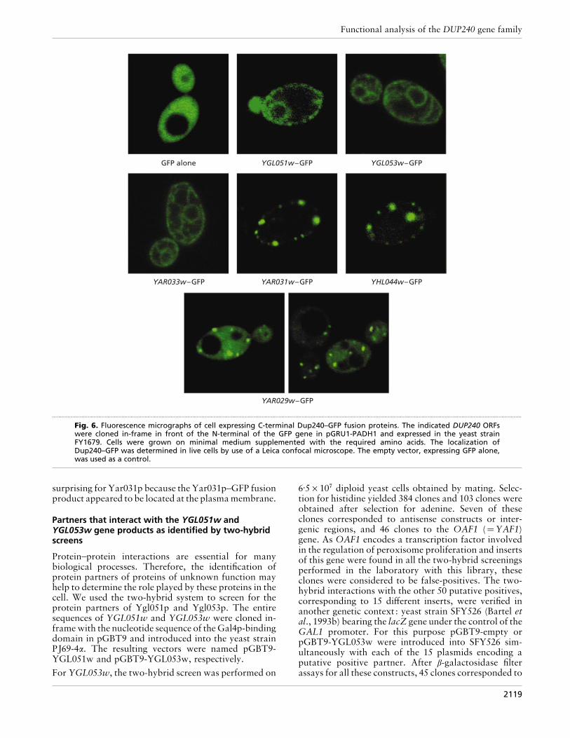
The control experiment with the vector without theinsert revealed the expected homogeneous distributionof GFP within the cytoplasm (Fig. 6). Ygl051p–,Ygl053p–, Yar033p–, Yar031p– and Yhl044p–GFP fu-
2116
Functional analysis of the DUP240 gene family
.................................................................................................................................................................................................................................................................................................................
Fig. 3. Similarity between the highly conserved proteins encoded by the DUP240 family. The amino acid sequences werealigned by use of the CLUSTAL X (1.8) program and visually optimized. White on black, amino acid identity; shading, aminoacid similarity ; boxes, sites of the two predicted transmembrane domains; bold, 15 residues of Yar033p and Ygl051pwhere 6 (outlined) out of 8 amino acid differences between these two sequences are concentrated. YAR031w encodes anadditional 62 aa at the N-terminal extremity compared to the other DUP240 family genes, YAR023c encodes about 50 aaless at the N-terminal end, and YAR029w encodes about 130 aa less at the N-terminal end and about 20 aa less at the C-terminal end, than the other members of the family. The DUP240 orthologue from S. bayanus is shorter because only apart of the sequence was available from the database (http://cbi.labri.u-bordeaux.fr/Genolevures/Genolevures.php3).
sion proteins were localized at the plasma membrane.The Yar031p– and Yhl044p–GFP fusion proteins ap-peared to concentrate as specific spots corresponding to
a higher focal concentration at the plasma membrane. Inaddition Ygl051p–, Ygl053p– and Yar033p–GFP fusionproteins surrounded the nucleus, a situation typical of
2117
R. Poirey and others
.................................................................................................................................................
Fig. 4. Strategy used to construct the strains deleted for the 10DUP240 ORFs. Lines with arrows indicate that the identifiedallele was introduced by PCR-mediated gene disruption. Linesconnecting two strains define a mating between the twostrains and lines without arrows indicate that a spore resultedfrom the mating. In some instances the genotypes of the sporesare indicated. Strains LD114 and LD115 are deleted for all 10DUP240 ORFs.
an endoplasmic reticulum localization. Moreover,Ygl053p– and Yar033p–GFP fusion proteins appearedto accumulate into additional membranes that couldcorrespond to the Golgi apparatus. Conversely, theYar029p–GFP fusion protein showed a similar dis-tribution to GFP alone, but with additional spotscorresponding to a higher focal concentration not linkedto the plasmamembrane. This could be becauseYar029pis the shortest member of the Dup240 family and theonly one that lacks the predicted transmembrane do-main. The localization of the Dup240 protein at theplasma membrane was further addressed by studyingthe hSos1p–Dup240 fusion RAS signalling pathwayactivation system.
A new strategy to test the localization of Ygl051p,Ygl053p and Yar033p at the plasma membrane level :hSos1p–Dup240 fusion
We modified the cytoplasmic two-hybrid system de-veloped by Aronheim et al. (1994) so that it could beused to detect proteins localized at the plasma mem-
.................................................................................................................................................
Fig. 5. The 10 DUP240 ORFs are not essential for cell viability.The LD114 mutant strain (lower rows of each tenfold dilutionseries of an overnight cell suspension) grew similarly to theBY4709 control strain (upper rows) on complete glucosemedium (top panel), complete glycerol medium (middle panel)and minimal medium (bottom panel) after 2–5 days at 30 °C.The same results were obtained at 15 °C and 37 °C (data notshown).
brane. This system is based on the activation of the Rassignalling pathway. The growth of the cdc25H mutant(strain CDC25H) at the restrictive temperature requiresthe targeting of the tested hSos1p–Dup240 fusion proteinto the plasma membrane. The YAR029w, YAR031w,YAR033w, YGL051w and YGL053w ORFs were clonedin-frame with the hSOS1 gene, resulting in pSOS-YAR029w, pSOS-YAR031w, pSOS-YAR033w, pSOS-YGL051w and pSOS-YGL053w, respectively. Theseconstructs were introduced into the yeast strainCDC25H. The constructions containing the YAR033w,YGL051w and YGL053w ORFs were able to restore thegrowth of the yeast cells. The correlation between theloss of the plasmid and the inability to grow at therestrictive temperature was verified. Growth was thustruly due to the presence of the coding sequences ofone or other of these three ORFs, indicating that thecorresponding fusion proteins were targeted to theplasma membrane.
This result can be explained by at least two mechanisms.Firstly, these bait proteins could interact physically withendogenous membrane protein targets, allowing the co-localization of the hSos1p protein at the plasma mem-brane. hSos1p could then fulfil its deoxyriboguanosineexchange function. Alternatively, the bait proteinsYar033p, Ygl051p and Ygl053p may themselves bemembrane proteins. This would localize the fusedhSos1p protein at the plasma membrane, allowing it tofulfil its role as an enzyme.
The fusion proteins hSos1p–Yar029p and hSos1p–Yar031p were unable to restore the growth of the yeastCDC25H strain. These results were expected forYar029p (see preceding section on GFP fusions) but were
2118
Functional analysis of the DUP240 gene family
GFP alone YGL051w–GFP YGL053w–GFP
YAR031w–GFP YHL044w–GFP
YAR029w–GFP
YAR033w–GFP
.................................................................................................................................................................................................................................................................................................................
Fig. 6. Fluorescence micrographs of cell expressing C-terminal Dup240–GFP fusion proteins. The indicated DUP240 ORFswere cloned in-frame in front of the N-terminal of the GFP gene in pGRU1-PADH1 and expressed in the yeast strainFY1679. Cells were grown on minimal medium supplemented with the required amino acids. The localization ofDup240–GFP was determined in live cells by use of a Leica confocal microscope. The empty vector, expressing GFP alone,was used as a control.
surprising for Yar031p because the Yar031p–GFP fusionproduct appeared to be located at the plasma membrane.
Partners that interact with the YGL051w andYGL053w gene products as identified by two-hybridscreens
Protein–protein interactions are essential for manybiological processes. Therefore, the identification ofprotein partners of proteins of unknown function mayhelp to determine the role played by these proteins in thecell. We used the two-hybrid system to screen for theprotein partners of Ygl051p and Ygl053p. The entiresequences of YGL051w and YGL053w were cloned in-frame with the nucleotide sequence of the Gal4p-bindingdomain in pGBT9 and introduced into the yeast strainPJ69-4α. The resulting vectors were named pGBT9-YGL051w and pGBT9-YGL053w, respectively.
For YGL053w, the two-hybrid screen was performed on
6±5¬10( diploid yeast cells obtained by mating. Selec-tion for histidine yielded 384 clones and 103 clones wereobtained after selection for adenine. Seven of theseclones corresponded to antisense constructs or inter-genic regions, and 46 clones to the OAF1 (¯YAF1)gene. As OAF1 encodes a transcription factor involvedin the regulation of peroxisome proliferation and insertsof this gene were found in all the two-hybrid screeningsperformed in the laboratory with this library, theseclones were considered to be false-positives. The two-hybrid interactions with the other 50 putative positives,corresponding to 15 different inserts, were verified inanother genetic context : yeast strain SFY526 (Bartel etal., 1993b) bearing the lacZ gene under the control of theGAL1 promoter. For this purpose pGBT9-empty orpGBT9-YGL053w were introduced into SFY526 sim-ultaneously with each of the 15 plasmids encoding aputative positive partner. After β-galactosidase filterassays for all these constructs, 45 clones corresponded to
2119
R. Poirey and others
.................................................................................................................................................
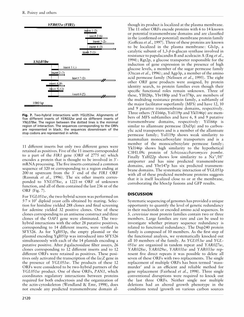
Fig. 7. Two-hybrid interactions with YGL053w. Alignments offive different inserts of YER032w and six different inserts ofYNL078w. The region between the dotted lines is the minimalregion of interaction. The sequences corresponding to the ORFsare represented in black; the sequences downstream of thestop codons are represented in white.
11 different inserts but only two different genes wereretained as positives. Five of the 11 inserts correspondedto a part of the FIR1 gene (ORF of 2775 nt) whichencodes a protein that is thought to be involved in 3«-mRNA processing. The five inserts contained a commonsequence of 320 nt corresponding to a region ending at200 nt upstream from the 3« end of the FIR1 ORF(Russnak et al., 1996). The six other inserts corres-ponded to YNL078w, a 1221 nt ORF of unknownfunction, and all of them contained the last 236 nt of theORF (Fig. 7).
For YGL051w, the two-hybrid screen was performed on5±7¬10( diploid yeast cells obtained by mating. Selec-tion for histidine yielded 288 clones and final screeningfor adenine yielded 32 positive clones. One of theseclones corresponding to an antisense construct and threeclones of the OAF1 gene were eliminated. The two-hybrid interactions with the other 28 putative positives,corresponding to 14 different inserts, were verified inSFY526. As for Ygl053p, the empty plasmid or theplasmid encoding Ygl051p was introduced into SFY526simultaneously with each of the 14 plamids encoding aputative positive. After β-galactosidase filter assays, 26clones corresponding to 12 different inserts and to 12different ORFs were retained as positives. These posi-tives only activated the transcription of the lacZ gene inthe presence of YGL051w. The products of these 12ORFs were considered to be two-hybrid partners of theYGL051w product. One of these ORFs, PAN1, whichcoordinates regulatory interactions between proteinsrequired for both endocytosis and the organization ofthe actin-cytoskeleton (Wendland & Emr, 1998), doesnot encode any predicted transmembrane domain al-
though its product is localized at the plasma membrane.The 11 other ORFs encode proteins with 6 to 14 knownor potential transmembrane domains and are classifiedin the (confirmed or potential) membrane protein family(Goffeau et al., 1997). Three of these proteins are knownto be localized in the plasma membrane: Gls1p, acatalytic subunit of 1,3-β--glucan synthase involved inresistance to papulacandin B and aculeacin A (Eng et al.,1994) ; Rgt2p, a glucose transporter responsible for theinduction of gene expression in the presence of highglucose levels, a member of the sugar permease family(Ozcan et al., 1996) ; and Agp3p, a member of the aminoacid permease family (Nelissen et al., 1995). The eightother ORF gene products were assigned, by proteinidentity search, to protein families even though theirspecific functional roles remain unknown. Three ofthese, Yll028p, Ybr180p and Yor378p, are members ofthe multidrug resistance protein family, a subfamily ofthe major facilitator superfamily (MFS) and have 12, 10and 9 putative transmembrane domains, respectively.Three others (Yil166p, Yol119p and Ykl146p) are mem-bers of MFS subfamilies and have 6, 8 and 9 putativetransmembrane domains, respectively : Yil166p issimilar to allantoate permease (Dal5p) and to carbox-ylic acid transporters and is a member of the allantoatepermease family ; Yol119p shows weak similarity tomammalian monocarboxylate transporters and is amember of the monocarboxylate permease family ;Ykl146p shows high similarity to the hypotheticalC3H1.09c protein of Schizosaccharomyces pombe.Finally Yal022p shows low similarity to a Na+}H+
antiporter and has nine predicted transmembranedomains, and Ybr147p has six predicted transmem-brane domains. The systematic interaction of YGL051pwith all of these predicted membrane proteins suggeststhat it is itself localized close to or at the membrane,corroborating the hSos1p fusions and GFP results.
DISCUSSION
Systematic sequencing of genomes has provided a uniqueopportunity to quantify the level of genetic redundancyin their nucleotide or encoded amino acid sequences. InS. cerevisiae most protein families contain two or threemembers. Large families are rare and can be used toinvestigate whether primary sequence redundancy isrelated to functional redundancy. The Dup240 proteinfamily is composed of 10 members. As the first step ofthe functional analysis, we systematically knocked outall 10 members of the family. As YGL053w and YGL-051w are organized in tandem repeat and YAR027w,YAR028w, YAR029w, YAR031w and YAR033w rep-resent five direct repeats it was possible to delete allseven of these ORFs with two replacements. The singlereplacement of multiple ORFs has been termed ‘mass-murder’ and is an efficient and reliable method forgene replacement (Fairhead et al., 1998). Three singleconventional disruptions were required to knock outthe last three ORFs. Neither single nor multipledeletions had an altered growth phenotype in theconditions tested (growth on various carbon sources
2120
Functional analysis of the DUP240 gene family
at different temperatures, mating and sporulationassays). A similar situation has been reported for theAAD1–7 family, which encodes seven homologues tothe arylalcohol dehydrogenase of the lignin-degradingfungus Phanerochaete chrysosporium (Delneri et al.,1999). However, the concurrent knock-out of 20 trans-porter genes was shown to be required to block glucoseconsumption and transport activity in S. cerevisiae(Wieczorke et al., 1999), and only the elimination of theentire OSBP gene family (OSH1–7) produces a lethalphenotype (Beh et al., 2001).
To learn more about the function of this multigenefamily we studied the subcellular localizations of thegene products and searched for interacting partners.Fluorescent staining of proteins Ygl051p, Ygl053p,Yar031p and Yar033p fused to GFP revealed that theywere localized on the yeast plasma membrane and thatYgl051p, Ygl053p and Yar033p were also located in theendoplasmic reticulum. Takahashi et al. (2000) reportedthat YAR027p fused to GFP was localized at the nuclearenvelope and plasma membrane regions. The membranelocalization was further supported by the in silicoanalysis, which suggested that all of the proteins codedby the DUP240 ORFs (except Yar029p, which has nopredicted transmembrane domain) possess two 17 aahydrophobic predicted transmembrane domains. Theactivation of the RAS signalling pathway by hSos1p–Dup240 fusions confirmed the plasma membrane locali-zation, showing that Ygl051p, Ygl053p and Yar033pwere able to target activation in this system, eitherdirectly because they are integral membrane proteins, orindirectly by interacting with associated membraneproteins. Yar029p is much shorter than the otherDup240 proteins and Yar029p–GFP was locatedthroughout the cytoplasm, as was GFP alone. Thisfinding is consistent with the inability of Yar029p totarget hSos1p to the RAS signalling pathway. TheYar031p protein contains 62 extra amino acids at its N-terminus compared to Ygl053p. The C-terminal parts ofthese two ORFs are nevertheless highly conserved:100% identity for the first 93 C-terminal amino acids,and only five differences among the 132 C-terminalamino acids. The identity of the N-terminal domain ofYgl053p and the central region of Yar031p is just 41±7%(48 out of 115 amino acids). When fused to hSos1p,Yar031p was unable to activate the RAS signallingpathway despite its apparent localization as indicated byGFP fusion at the plasma membrane and the presence oftwo predicted transmembrane domains. This discrep-ancy might result from the fact that GFP-fused Yar031palways seems to form aggregates at the membrane, andmay consequently be unable to target hSos1p correctlyfor RAS activation.
The Ygl051p and Ygl053p partners identified by screen-ing of the two-hybrid yeast library were in differentclasses. The product of the ORF YGL051w fused to thebinding domain of Gal4p interacted with many integralmembrane proteins and many proteins of unknownfunction predicted to have six or more transmembranedomains. This finding further suggests that Ygl051p
is located at the plasma membrane. The two-hybridpartners of Ygl053p, Fir1p and Ynl078p do not appearto be plasma membrane proteins, as they lack predictedtransmembrane domains; however, this finding does notexclude a plasma membrane localization for Ygl053p.The common sequence from the different insertsencoding parts of Fir1p or Ynl078p should encodethe domains from Fir1p and Ynl078p required forinteraction with the Ygl053p protein. Recently, high-throughput two-hybrid screens revealed that Yar031pand Apg12p interact, and that Ygl051p and Yar033pinteract (Uetz et al., 2000). We were unable to replicatethis finding (data not shown). Apg12p is involved inautophagy and the targeting of proteins to the vacuole.Most free Apg12p seems to be associated with theendoplasmic reticulum (Mizushima et al., 1998). Two-hybrid interactions have also been reported betweenYar027p and Cks1p, an essential, physically associated,component that interactswith the protein kinaseCdc28p(Hadwiger et al., 1989), between Yar027p and Yar030p,between Ygl053p and Ylr065p, and between Yhl044pand Ykr035p (Ito et al., 2000). The Yar030p, Ylr065pand Ykr035p proteins have unknown functions butpossess two and five putative transmembrane domains,respectively, so these proteins are themselves predictedto be membrane associated. The proteins coded byYGL051w, YGL053w, YAR031w and YAR033w arepredicted to have structural and}or functional roles atmembranes.
Our data suggest that we are dealing with a gene familyspecific to the genus Saccharomyces sensu stricto. Thisgene family belongs to the set of ascomycete-specificgenes, a class of genes that tend to be more sensitive toevolutionary divergence than the average (Malpertuy etal., 2000). Even without specific information on theprecise function of the proteins encoded by this genefamily, we have shown that when amino acid divergenceis detectable, the changes often affect a limited numberof amino acids localized within a specific domain of theprotein. The proteins encoded by this gene family, withthe exception of Yar029p, appear to be located atmembranes. We have evidence that they have specializedfunctions because when they were fused to GFP they didnot show identical fluorescence patterns and when fusedto hSos1p they did not show identical RAS activationproperties. In addition, Ygl051p and Ygl053p bothappear to interact with a specific set of non-redundantproteins. Thus, our data suggest that the 10 Dup240members do not have identical functions.
ACKNOWLEDGEMENTS
We are grateful to P. Sunnerhagen and A. Ramne for providinga yeast genomic two-hybrid library, to D. Alexandraki forproviding yeast transformed with this library, to M. Aigle forproviding pGRU1-S65TGFP and help with the interpretationof fluorescence data, and to P. James and S. Fields forproviding strains PJ69-4a and PJ69-4α. We are also grateful toJ. Rommelaere for his kind support throughout this work.This research was supported in part by a contract within theframework of the EUROFAN project of the EuropeanCommission (BIO4-CT95-0080) to J.C.J. and by a European
2121
R. Poirey and others
Commission Marie Curie Predoctoral Research Trainingfellowship BIO4-CT98-5047 to R.P.
REFERENCES
Arabidopsis Genome Initiative (2000). Analysis of the genomesequence of the flowering plant Arabidopsis thaliana. Nature 408,796–815.
Aronheim, A., Engelberg, D., Li, N., al-Alawi, N., Schlessinger, J.& Karin, M. (1994). Membrane targeting of the nucleotideexchange factor Sos is sufficient for activating the Ras signalingpathway. Cell 78, 949–961.
Aronheim, A., Zandi, E., Hennemann, H., Elledge, S. J. & Karin, M.(1997). Isolation of an AP-1 repressor by a novel method fordetecting protein-protein interactions. Mol Cell Biol 17, 3094–3102.
Bartel, P. L., Chien, C. T., Sternglanz, R. & Fields, S. (1993a). Usingthe two-hybrid system to detect protein-protein interactions. InCellular Interactions in Development : a Practical Approach, pp.153–179. Edited by D. A. Hartley. Oxford: Oxford UniversityPress.
Bartel, P. L., Chien, C. T., Sternglanz, R. & Fields, S. (1993b).Elimination of false positives that arise in using the two-hybridsystem. Biotechniques 14, 920–924.
Barton, A. B., Bussey, H., Storms, R. K. & Kaback, D. B. (1997).Molecular cloning of chromosome I DNA from Saccharomycescerevisiae : characterization of the 54 kb right terminal CDC15-FLO1-PHO11 region. Yeast 13, 1251–1263.
Beh, C. T., Cool, L., Phillips, J. & Rine, J. (2001). Overlappingfunctions of the yeast oxysterol-binding protein homologues.Genetics 157, 1117–1140.
Bendixen, C., Gangloff, S. & Rothstein, R. (1994). A yeast mating-selection scheme for detection of protein-protein interactions.Nucleic Acids Res 22, 1778–1779.
Blandin, G., Durrens, P., Tekaia, F. & 19 other authors (2000).Genomic exploration of the hemiascomycetous yeasts. 4. Thegenome of Saccharomyces cerevisiae revisited. FEBS Lett 487,31–36.
Bon, E., Neuveglise, C., Casaregola, S., Artiguenave, F., Wincker,P., Aigle, M. & Durrens, P. (2000). Genomic exploration ofthe hemiascomycetous yeasts. 5. Saccharomyces bayanus var.uvarum. FEBS Lett 487, 37–41.
Brachmann, C. B., Davies, A., Cost, G. J., Caputo, E., Li, J., Hieter,P. & Boeke, J. D. (1998). Designer deletion strains derived fromSaccharomyces cerevisiae S288C: a useful set of strains andplasmids for PCR-mediated gene disruption and other applica-tions. Yeast 14, 115–132.
Chardin, P., Camonis, J. H., Gale, N. W., van Aelst, L., Schlessinger,J., Wigler, M. H. & Bar-Sagi, D. (1993). Human Sos1: a guaninenucleotide exchange factor for Ras that binds to Grb2. Science260, 1338–1343.
Delneri, D., Gardner, D. C., Bruschi, C. V. & Oliver, S. G. (1999).Disruption of seven hypothetical aryl alcohol dehydrogenasegenes from Saccharomyces cerevisiae and construction of amultiple knock-out strain. Yeast 15, 1681–1689.
Dujon, B. (1998). European Functional Analysis Network (EURO-FAN) and the functional analysis of the Saccharomyces cerevisiaegenome. Electrophoresis 19, 617–624.
Eng, W. K., Faucette, L., McLaughlin, M. M., Cafferkey, R., Koltin,Y., Morris, R. A., Young, P. R., Johnson, R. K. & Livi, G. P. (1994).The yeast FKS1 gene encodes a novel membrane protein,mutations in which confer FK506 and cyclosporin A hypersen-sitivity and calcineurin-dependent growth. Gene 151, 61–71.
Fairhead, C., Thierry, A., Denis, F., Eck, M. & Dujon, B. (1998).‘Mass-murder ’ of ORFs from three regions of chromosome XIfrom Saccharomyces cerevisiae. Gene 223, 33–46.
Feuermann, M., de Montigny, J., Potier, S. & Souciet, J. L. (1997).The characterization of two new clusters of duplicated genessuggests a ‘Lego’ organization of the yeast Saccharomycescerevisiae chromosomes. Yeast 13, 861–869.
Fields, S. & Song, O. (1989). A novel genetic system to detectprotein-protein interactions. Nature 340, 245–246.
Fromont-Racine, M., Rain, J. C. & Legrain, P. (1997). Toward afunctional analysis of the yeast genome through exhaustive two-hybrid screens. Nat Genet 16, 277–282.
Georgakopoulos, T., Koutroubas, G., Vakonakis, I., Tzermia, M.,Prokova, V., Voutsina, A. & Alexandraki, D. (2001). Functionalanalysis of the Saccharomyces cerevisiae YFR021w}YGR223c}YPL100w ORF family suggests relations to mitochondrial}peroxisomal functions and amino acid signalling pathways. Yeast18, 1155–1171.
Gietz, R. D., Schiestl, R. H., Willems, A. R. & Woods, R. A. (1995).Studies on the transformation of intact yeast cells by the LiAc}SS-DNA}PEG procedure. Yeast 11, 355–360.
Goffeau, A., Park, J., Paulsen, I. T., Jonniaux, J. L., Dinh, T.,Mordant, P. & Saier, M. H. (1997). Multidrug-resistant transportproteins in yeast : complete inventory and phylogenetic charac-terization of yeast open reading frames with the major facilitatorsuperfamily. Yeast 13, 43–54.
Hadwiger, J. A., Wittenberg, C., Mendenhall, M. D. & Reed, S. I.(1989). The Saccharomyces cerevisiae CKS1 gene, a homolog ofthe Schizosaccharomyces pombe suc1+ gene, encodes a subunit ofthe Cdc28 protein kinase complex. Mol Cell Biol 9, 2034–2041.
Hoffman, C. S. & Winston, F. (1987). A ten-minute DNApreparation from yeast efficiently releases autonomous plasmidsfor transformation of Escherichia coli. Gene 57, 267–272.
Ito, T., Tashiro, K., Muta, S., Ozawa, R., Chiba, T., Nishizawa, M.,Yamamoto, K., Kuhara, S. & Sakaki, Y. (2000). Toward a protein-protein interaction map of the budding yeast : a comprehensivesystem to examine two-hybrid interactions in all possiblecombinations between the yeast proteins. Proc Natl Acad SciU SA 97, 1143–1147.
James, P., Halladay, J. & Craig, E. A. (1996). Genomic libraries anda host strain designed for highly efficient two-hybrid selection inyeast. Genetics 144, 1425–1436.
Jelinsky, S. A. & Samson, L. D. (1999). Global response ofSaccharomyces cerevisiae to an alkylating agent. Proc Natl AcadSci U SA 96, 1486–1491.
Johnston, S. A. & Carlson, M. (1992). Regulation of carbon andphosphate utilization. In The Molecular and Cellular Biology ofthe Yeast Saccharomyces, Gene Expression, pp. 193–281. Editedby E. W. Jones, J. R. Pringle & J. R. Broach. Cold Spring Har-bor, NY: Cold Spring Harbor Laboratory.
Karin, M., Najarian, R., Haslinger, A., Valenzuela, P., Welch, J. &Fogel, S. (1984). Primary structure and transcription of anamplified genetic locus: the CUP1 locus of yeast. Proc Natl AcadSci U SA 81, 337–341.
Klein, P., Kanehisa, M. & DeLisi, C. (1985). The detection andclassification of membrane-spanning proteins. Biochim BiophysActa 815, 468–476.
Malpertuy, A., Tekaia, F., Casaregola, S. & 21 other authors(2000). Genomic exploration of the hemiascomycetous yeasts. 19.Ascomycetes-specific genes. FEBS Lett 487, 113–121.
Mizushima, N., Noda, T., Yoshimori, T., Tanaka, Y., Ishii, T.,George, M. D., Klionsky, D. J., Ohsumi, M. & Ohsumi, Y. (1998). A
2122
Functional analysis of the DUP240 gene family
protein conjugation system essential for autophagy. Nature 395,395–398.
Muhlrad, D., Hunter, R. & Parker, R. (1992). A rapid method forlocalized mutagenesis of yeast genes. Yeast 8, 79–82.
Nelissen, B., Mordant, P., Jonniaux, J. L., De Wachter, R. &Goffeau, A. (1995). Phylogenetic classification of the majorsuperfamily of membrane transport facilitators, as deduced fromyeast genome sequencing. FEBS Lett 377, 232–236.
Oliver, S. G., Winson, M. K., Kell, D. B. & Baganz, F. (1998).Systematic functional analysis of the yeast genome. TrendsBiotechnol 16, 373–378.
Ozcan, S., Dover, J., Rosenwald, A. G., Wolfl, S. & Johnston, M.(1996). Two glucose transporters in Saccharomyces cerevisiae areglucose sensors that generate a signal for induction of geneexpression. Proc Natl Acad Sci U SA 93, 12428–12432.
Petitjean, A., Hilger, F. & Tatchell, K. (1990). Comparison ofthermosensitive alleles of the CDC25 gene involved in the cAMPmetabolism of Saccharomyces cerevisiae. Genetics 124, 797–806.
Rachidi, N., Martinez, M. J., Barre, P. & Blondin, B. (2000).Saccharomyces cerevisiae PAU genes are induced by anaerobiosis.Mol Microbiol 35, 1421–1430.
Russnak, R., Pereira, S. & Platt, T. (1996). RNA binding analysis ofyeast REF2 and its two-hybrid interaction with a new geneproduct, FIR1. Gene Expr 6, 241–258.
Saiki, R. K., Scharf, S., Faloona, F., Mullis, K. B., Horn, G. T., Erlich,H. A. & Arnheim, N. (1985). Enzymatic amplification of beta-globin genomic sequences and restriction site analysis fordiagnosis of sickle cell anemia. Science 230, 1350–1354.
Sambrook, J., Fritsch, E. F. & Maniatis, T. (1989). MolecularCloning: a Laboratory Manual, 2nd edn. Cold Spring Harbor,NY: Cold Spring Harbor Laboratory.
Sander, C. & Schneider, R. (1991). Database of homology-derivedprotein structures and the structural meaning of sequencealignment. Proteins 9, 56–68.
Schneller, J. M., Schneider, C. & Stahl, A. J. (1978). Distinctnuclear genes for yeast mitochondrial and cytoplasmic methionyl-tRNA synthetases.Biochem BiophysRes Commun 85, 1392–1399.
Sherman, F., Fink, G. R. & Hicks, J. B. (1986). Methods in YeastGenetics. Cold Spring Harbor, NY: Cold Spring Harbor Lab-oratory.
Souciet, J., Aigle, M., Artiguenave, F. & 21 other authors (2000).Genomic exploration of the hemiascomycetous yeasts. 1. A set ofyeast species for molecular evolution studies. FEBS Lett 487, 3–12.
Takahashi, Y., Mizoi, J., Toh, E. A. & Kikuchi, Y. (2000). YeastUlp1, an Smt3-specific protease, associates with nucleoporins. JBiochem 128, 723–725.
Uetz, P., Giot, L., Cagney, G. & 17 other authors (2000). Acomprehensive analysis of protein-protein interactions in Sac-charomyces cerevisiae. Nature 403, 623–627.
Wach, A., Brachat, A., Pohlmann, R. & Philippsen, P. (1994). Newheterologous modules for classical or PCR-based gene disruptionsin Saccharomyces cerevisiae. Yeast 10, 1793–1808.
Wagner, R., Straub, M. L., Souciet, J. L., Potier, S. & de Montigny,J. (2001). New plasmid system to select for Saccharomycescerevisiae purine-cytosine permease affinity mutants. J Bacteriol183, 4386–4388.
Wendland, B. & Emr, S. D. (1998). Pan1p, yeast Eps15, functionsas a multivalent adaptor that coordinates protein-protein inter-actions essential for endocytosis. J Cell Biol 141, 71–84.
Wieczorke, R., Krampe, S., Weierstall, T., Freidel, K., Hollenberg,C. P. & Boles, E. (1999). Concurrent knock-out of at least 20transporter genes is required to block uptake of hexoses inSaccharomyces cerevisiae. FEBS Lett 464, 123–128.
.................................................................................................................................................
Received 5 December 2001; revised 4 March 2002; accepted 20 March2002.
2123