anaesthesia, pain & intensive care
TRANSCRIPT
ISSN 1607-8322ISSN 2220-5799 (Online)
ANAESTHESIA, PAIN& INTENSIVE CARE
An International Journal of Anesthesiology, PainManagement, Intensive Care & Resuscitation
Vol. 16, No. 3 www.apicareonline.com Sep-Dec 2012
StatisticianIrum Abid
Editorial Board:Arshad Taqi (Pakistan)Dario Galante (Italy)Fazal Hameed (Pakistan)Gauhar Afshan (Pakistan)Joseph D. Tobias (USA)Khalid Bashir (Pakistan)Koji Sumikawa (Japan)Mohamad Said Ahmad Maani Takrouri (Saudi Arabia)Pramila Bajaj (India)Rana Altaf Ahmad (Pakistan)Rashed A. Hasan (USA)Saeeda Hayder (Pakistan)Saeid Safari (Iran)Shahab Naqvi (Pakistan)S. K. Malhotra (India)Waqas Ahmed Qazi (Pakistan)
Associate Editors:Zahid Akhtar RaoAssociate Prof. of Anaesthesia, Bahria Univ. Medical & DentalCollege, Karachi (Pakistan)[email protected]
Fares ChedidConsultant NeonatologistTawam Hospital, Al Ain, Abu Dhabi (UAE)[email protected]
Assistant Editors:Pranav Bansal (India)[email protected]
Waqas Ashraf Ch. (UK)[email protected]
Muhammad Faisal Khan (KSA)[email protected]
Amer Majeed (UK)[email protected]
Patron:Brig. M. Salim. SI(M)Professor of Anesthesiology President [email protected]
Editor-in-Chief:Tariq Hayat KhanConsultant Anesthesiologist & Pain SpecialistVice President [email protected]
Editors:Samina IsmailAssociate Prof. of Anaesthesia, AKUH [email protected]
Said AbuhasnaChairman, Dept. of Critical Care Medicine, Chief Intensive Care Unit, Tawam Hospital, Al Ain, Abu Dhabi (UAE)[email protected]
ANAESTHESIA, PAIN& INTENSIVE CARE
An International Journal of Anesthesiology, Pain Management, Intensive Care & Resuscitation
Vol. 16 No. 3 September - December 2012
Published by: Dr. Tariq Hayat Khan
‘Anaesthesia, Pain & Intensive Care’ is indexed by PakMediNet, Medlip. Index Medicus (EMR), EBSCO, Index Copernicus, Embase, EMCare and UDL (Malaysia). Indexation by Medline, CINAHL, ExtraMed and others pending.
Listed with: National Library of Medicine CatalogueNLM ID: 101313795 [Serial]
Registered by Pakistan Medical & Dental Council (PM&DC). Recognized by Higher Education Commission (HEC)Permission granted by District Magistrate Islamabad for publication.
General Information: The journal is published thrice a year in the months of April, August and December. Please direct inquiries regarding subscriptions, single copies and back issues, changes of addresses, and other correspondence to the Publications Office. Advertising inquiries should also be sent to the same address. See us at FACE BOOK, Twitter, Linked in and MedpediaAll articles represent the opinions of the authors and do not reflect official policy of the journal. All rights are reserved to the publisher. No part of the journal may be reproduced or transmitted in any form or by any means, electronic or mechanical, including photocopying, regarding, or via any retrieval system, without written permission from the publisher.
Subscription Rates: The rates for a one-year subscription of the Journal are Pak Rupees 1000 for subscribers in Pakistan, Pak Rupees 2000 for institutions in Pakistan, Pak Rupees 2000 for subscribers from SAARC countries and Pak Rupees 4000 from elsewhere.
Regional Office (Pakistan): Dept of Anaesthesia and Pain Management, AKUH, Karachi.Ph: +92-21-34864631/4331
Regional Office (UAE): Dept of Anaesthesia and Critical Care, Tawam Hospital, P.O. Box 152858, Al-Ain, Abu Dhabi (UAE).
Head Office: ‘APICARE’60-A, Nazim-ud-Din Road, F-8/4, Islamabad (Pakistan)E-mail: [email protected], [email protected] Ph: +92-321-5149709
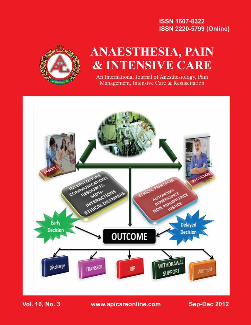
Cover Picture: Medical ethics in ICUPicture credits: Dr. Anwar Ul Haq, Consultant Anaesthetist, Midland Regional Hospital, Tullamore (Ireland)
EDITORIAL VIEWS
Terminating the ventilatory support: an ethical dilemmaLjiljana Kojikj
Euthanasia: is it really a bad idea?Dr Arshad Taqi
The introduction of bispectral index (BIS) in anesthesia practiceDario Galante, Matteo Melchionda
SPECIAL ARTICLE
Medical ethics in ICU patients: conflicts and their resolution Anwar ul Haq
ORIGINAL ARTICLES
A clinical prospective, randomized study to compare intrathecal isobaric bupivacaine – fentanyl and isobaric ropivacaine – fentanyl for lower abdominal and lower limb surgeriesSangeeta Varun, Mridul Srivastava, Indubala Maurya, Rakesh Garg, Vipin Dhama, Yogesh Kumar Manik
Paraphenylene diamine poisoning: Our experience at PMC Hospital NawabshahBashir Ahmed Khuhro, Muhammad Saleh Khaskheli, Abrar Ali Shaikh
A comparison of hemodynamic changes during laryngoscopy and endotracheal intubation by using three modalities of anesthesia inductionLahsaee Masoud, Kamalipour Hamid, Ajeli Zahra, Kamali Karmella
A prospective study of factors predicting postoperative pulmonary complications (PPC) in patients undergoing non-cardiothoracic surgery under general anaesthsia in a developing countryKaleem Ullah Toori, Jahangir Sarwar Khan, Ali Zohair Nomani, Syed Waqar Hussain, Saad Hashmi
ANAESTH, PAIN & INTENSIVE CARE; SEP-DEC 2012 Vol. 16 No. 3
CONTENTS
The effect of pregabalin and gabapentin on preoperative anxiety and sedation: a double blind study Anju Ghai, Monika Gupta, Neha Rana, Raman Wadhera
The incidence of postoperative delirium in elderly patients undergoing urologic surgeryHaxhire Gani, Pirro Prifti, Majlinda Naco, Rudin Domi, Vjollca Beqiri, Durata Torba, Rajmonda Tare
Comparison of different doses of clonidine as an adjuvant to intrathecal bupivacaine for spinal anesthesia and postoperative analgesia in patients undergoing caesarian section Shah Bhavini Bhushan, Joshi Smita Suresh, Shidhaye Ramchandra Vinayak, J. N. Lakhe
SHORT COMMUNICATION
Two point fixation of endotracheal tube in submentotracheal intubation during craniomaxillofacial surgeries-our experience!Dheeraj Kapoor, Anand Gupta, Deepak Thapa, Jasveer Singh
CASE REPORTS
Bilateral transversus abdominis plane (TAP) catheters for postoperative analgesia in a child with spinal dysraphismDane Yuratich, Tarun Bhalla, Venkata R. Jayanthi, Joseph D. Tobias
Ethical Dilemma in multiple co-morbid respiratory failure patient: Patient autonomy against family wishes?Anwar ul Haq
Successful intubation in a child with Lowe syndrome using fiberscope and Glidescope® Dheeraj Kapoor, Nitin Ahuja, Meghana Srivastava
Elective use of high frequency oscillatory ventilation with
223257
262
266
273
276
283
287
280
230
232
237
243
247
252
226
ANAESTH, PAIN & INTENSIVE CARE; SEP-DEC 2012 Vol. 16 No. 3
CONTENTS
transcutaneous carbon dioxide monitoring during thoracoscopic diaphragmatic hernia repairMichelle LeRiger, Arlyne Thung, Karen Diefenbach, Edward Shepherd, Erin Wishloff, Joseph D. Tobias
Massive subcutaneous emphysema secondary to rigid bronchoscopy in a childAnju Ghai, Meenu Goyal, Raman Wadhera, Manish Kumar Goel
Anesthetic management of a missed pheochromocytoma during exploratory laparotomyChhaya Suryawanshi, Thatte , V. R. R. Chari, Manisha Sapate, Anuja A. Goyal
A rare case of pedunculated tonsilar mass in a child Habib Md Reazaul Karim, Jayanta K Mitra, Mohammad Yunus, Vanlalhmangaihi Hmar, Amit Goyal
Persistent status epilepticus due to bupropion intoxication Cevdet Duger, Ahmet Cemil Isbir, Kenan Kaygusuz, Iclal Ozdemir Kol, Sinan Gursoy, Caner Mimaroglu
REVIEW ARTICLES
Euthanasia: protecting ‘right to die’ by denying ‘right to live’
Intravenous paracetamol in pediatrics: A global perspectiveMuzammil Irshad, Mehjabeen Malik, Aamir Furqan
LETTERS TO EDITOR
Sacral bulge after double epidural space localization efforts in pediatric patientsUday Ambi
293
320
315
322
323
324
325
325
296
299
302
305
311
315
317
318
Misconnected epidural infusion into central line: A perfect recipe for disasterRudrashish Haldar and Prakhar Gyanesh
TRENDS & TECHNOLOGy
CLINIQUIZ
Poisoning Pranav Bansal, Gaurav Jain, Mahipal Singh, Meenu Agarwal
Management of severe asthma in ICUMuhammad Faisal Khan
CALENDAR OF EVENTS
OBITUARIES
Dr. Wajahat Shahab Malik, MCPS
Prof. Dr Rahat Sultan Sahibzada
CLINIPICS
Neonatal mattersProfessor Naeem,
TEF with 13 pairs of ribs and anomalous 2nd lumbar vertebraUday Ambi, Navya C N, Ramesh Hatti
The ‘APICARE’ agrees to accept manuscripts prepared in ac-cordance with the ‘Uniform Requirement for Submission of Manuscripts Submitted to Biomedical Journals’ published in the British Medical Joumal 1991 ;302:334N1, All material submitted for publication should be sent exclusively to the ‘APICARE’.
ETHICAL CONSIDERATIONS: If tables, illustrations or photographs, which have been already published, are included, a letter of permission for their re-publication should be obtained from author(s) as well as the editor of the journal where it was previously printed. Permission to reproduce photographs of pa-tients whose identity is not disguised should be sent with the manuscript otherwise the eyes will be blackened out.
MATERIAL FOR PUBLICATION: The material submitted for publication may be in the form of an Original Research, a Re-view Article, a Case Report, Recent Advances, New Techniques, Debates, Book Review/CDs on Clinical/Medical Education, or a Letter to the Editor. Original articles should normally report original research of relevance to clinical anaesthesiology, pain management, intensive care or resuscitation, and may appear ei-ther as a paper or as short communications. The paper should be of about 2000 words, with no more than six tables or illus-trations, short communications should be of about 600 words, with one table or illustration and no more than five references. Clinical case reports and brief or negative research findings may appear in this section. Review article should consist of structured overview of some relatively narrow topic providing background, recent development with reference to original literature.
Letters should normally not exceed 400 words, have no more than 10 references, and be signed by all of the authors. An article based on dissertation submitted as part of the requirement for a fellowship of the postgraduate degree awarding medical institu-tions can be sent for publication after the Research and Training Monitoring Cell (RTMC) have approved it. Editorials are writ-ten by invitation.
Each manuscript should include a title page (containing e-mail address, fax and phone numbers of the corresponding author), abstract, text, acknowledgments, references, tables, and legends.
Each component should begin on a new page, in the following sequence: title page: abstract and keywords: text; acknowledg-ments; references; tables (each table, complete with title and footnotes, on a separate page); and legends for illustrations.
The manuscript should be typed in double spacing on 8 ½″ x 11″ (21.5cm x 28.0cm) white bond paper with one-inch (2.5cm) mar-gin on both sides. It should not exceed 20 pages, excluding tables and references. There should be no more than 40 references in an Original Article, and no more than 100 in a Review Article.
TABLES AND ILLUSTRATIONS: Tables and illustrations should be submitted separately, and legends to illustrations should be typed on a separate sheet. Each table should have a title and be typed in double space without horizontal and verti-cal lines on an 8 ½″ x 11″ (21.5 x 28.0 cm) paper. Tables should be numbered consecutively with Roman numerals in the order they are mentioned in the text. Page number should be in the upper right corner, if abbreviations are used, they should be explained in footnotes. When graphs, scatter grams, or histograms are sub-mitted, the numerical data on which they are based should be supplied.
S.I. UNITS: System Intemational (Sl) Unit measurements should be used. All drugs must be mentioned in generic form. The com-mercial name may, however, be mentioned with in brackets.
FIGURES AND PHOTOGRAPHS: Should be sent only when data cannot be expressed in any other form. These must be unmounted, glossy prints in sharp focus, 5″ x 7″, (12.7 x 17.3
cm) in size. These may be in black and white or in colour. Nega-tives, transparencies, and X-ray films should not be submitted. The number of the figure, the name of the author(s) should be printed on the back of each figure/photograph, and must be cit-ed in the text in consecutive order. Legends must be typed on a separate sheet of paper. Legends for photographs should indicate the magnification, internal scale and the method of staining. Pho-tographs in published articles will not be returned.
REFERENCES: Should be numbered in the order in which they are cited in the text. At the end of the article, the full list of references should give the names and initials of all authors (unless there are more than six when only the first six should be given followed by et al). The authors’ names are followed by the title of the article; the title of the journal abbreviated according to the style of the Index Medicus. e.g.: Hall, RR. The heading of tissues by CO2 laser. Br J. Surg 1971; 58:222-225. Reference to books should give the names of editors, place of publication, publisher, and year.
PEER REVIEW: Every paper will be reviewed by a member of the editorial committee, and one or more external reviewers. Statistical analysis wail be examined by a statistician.
ABSTRACT: Abstracts of onginal article should be prepared with a structured format. The elements addressed could include objectives, design, setting, patients or other participants, inter-ventions, and outcome measures, the results, and conclusions. Please label each section clearly with the appropriate subheading. Review article, Case report and other require a short, unstruc-tured abstract Commentaries do not require abstract. A list of key words should be given consisting of MeSH terms.
INTRODUCTION: This should include the purpose of the ar-ticle. The rationale for the study or observation should be sum-marised; only strictly pertinent references should be cited; the subject should not be extensively reviewed Data or conclusions from the work being reported should not be presented.
METHODOLOGY: The selection of the observational or ex-perimental subjects (patients or experimental animals, including controls) should be described clearly in the methods and the ap-paratus used should be identified (with the manufacturers name and address in parentheses), and procedures described in suffi-cient detail to allow other workers to reproduce the results Ref-erences to established methods should be given, including statis-tical methods; references and brief descriptions for methods that have been published but are not wall known should be provided; new or substantially modified methods should be described, giv-ing reasons for using them, and evaluating their limitations. All drugs and chemicals used should be identified precisely, includ-ing generic name(s), dose(s), and route(s) of administration.
RESULTS: These should be presented in a logical sequence in the text, tables, and illustrations. All the data in the tables or illustrations should not be repeated in the text; only important observations should be emphasized or summarised.
DISCUSSION: The authors comments on the results, sup-ported with contemporary references, including arguments and analysis of identical work done by other workers. A summary is not required. Brief acknowledgment may be made at the end.
ONLINE SUBMISSION: The manuscripts maybe submitted online via our website www.apicareonline.com or sent as e-mail attachments to the head office or one of the regional offices. Pictures must be sent separately as JPEG or in TIFF format. Submission of scanned copies of the tables, diagrams, Graphs, and Pictures is not acceptable. Model submission letter maybe downloaded from the website, duly filled in, signed by all of the authors and must be submitted along with the manuscript.
Instructions for authors
ANAESTH, PAIN & INTENSIVE CARE; VOL 16(3) SEP-DEC 2012 223
EDITORIAL VIEW
Terminating the ventilatory support: an ethical dilemmaLjiljana Kojikj*
*ICU, University Children’s Hospital. Skopje, Macedonia (FYROM)
Correspondence: Ljiljana Kojikj, MD; ICU, University Children’s Hospital, Skopje, Macedonia (FYROM); Tel: + 389 3 229 156; Fax: + 389 3 229 156; E-mail: [email protected]
ABSTRACTIntensive care physicians in modern set ups frequently have to face a dilemma in which they have to vote for a choice to sustain or to withdraw ventilatory treatment in terminally sick patients. The rapidly developing science of organ transplantation has given birth to many new questions, some of which still remain unanswered. Although most of the main religions have somehow endorsed organ harvesting from these patients to sustain the life of some other sick persons, and although many countries have clear guidelines authenticated by the legislation, clinicians in many countries still have to answer these questions based upon their experience and other factors. Many of them refuse to accept the option of terminating life supporting treatment including ventilatory therapy. In this editorial the later viewpoint has been discussed by the esteemed author.
Key words: Ventilator support withdrawal; Infant; Newborn; Brain death; Organ transplantation
Citation: Kojikj L. Terminating the ventilatory support: an ethical dilemma. Anaesth Pain & Intensive Care 2012;16(3):223-225
Almost in every moment of every day of their professional life, intensive care physicians have to make choices. Choices like what diagnostic procedure to perform so as to save valuable time for their patient; what other specialists to consult in order to establish the right diagnosis and what treatment to apply to someone who is at the verge of life and death. Should an intensive care physician be encumbered with a choice to sustain or to withdraw treatment? This is the point at which intensive care medicine is seen to be colliding with ethics, philosophy and the religion. The dilemma regarding ethical issues has been resolved by the respective state legislatives in most of the countries, but not all.
The advances in medical technology, especially pertaining to critical care medicine, along with aggressive resuscitation protocols have expanded the possibilities for dying people to survive. Thus, the line in between life and death has been blurred. Unfortunately, sometimes it is at the cost of significant mental and physical handicap. The patient is virtually trapped in the intensive care unit in a weird but abnormal blend of life and death, not being able to participate in human life activities, and not being allowed to die either. Before the advances in medical technology, determination of death was easy: a patient was dead when cessation of
breathing and heart beat was confirmed. No one ever tried to reverse death. However, in the age of organ transplantation, the practice of recovery of viable organs from otherwise dead humans has changed the things dramatically.1
The historical evolution of the concept of death from a cardiorespiratory failure to a brain failure was established in 1968, when the Harvard criteria equated irreversible coma and apnea (i.e., brain death) with human death and later, when the Uniform Determination of Death Act was enacted permitting organ procurement from heart beating donors. Since then, clinical studies have defined a spectrum of states of impaired consciousness in human beings: coma, minimally conscious state, vegetative state and brain death.2
The USA and EU countries have very precise legal definitions and guidelines for almost all the situations regarding withholding of ventilatory support to the person considered to be brain dead. There have been precise protocols for both adult patients and for children and neonates as well.3-8
The practice of withholding life support (ventilator support in most of the cases), in order to harvest organs for transplantation, is tolerated by the four major word religions as well: The Orthodox Church
224 ANAESTH, PAIN & INTENSIVE CARE; VOL 16(3) SEP-DEC 2012
terminating the ventilatory support
permits transplantation from one man to another and transplantation is strongly recommended from the standpoint of Christian morality. These attitudes are accepted and respected by the Roman Catholic Church, Reformers, Judaism and Islam as well.9-11
Studies and systematic reviews of literature for ventilator support withdrawal, trying to elucidate approach to withdrawing ventilator support, equally reveal a great deal of diversity between the studies on both criteria for ventilator support withdrawal as well as the technique (extubation or no extubation, premedication or no premedication etc.) itself.12,13 In other words, what practice reveals are differences, not only between institutions in the same country or state, but between the different profiles of doctors in the same institution (for example anesthesiologists and surgeons on one side and pediatricians and internists on the other) and even between doctors in the same department of an institution. There has been high level of diversity in life support withdrawal practice between doctors and nurses of the same hospital as well.14,15
On this occasion, what has been well established practice for almost half a century, and has undoubtedly saved many lives shall not be discussed. Instead, it is contemplation on the other aspects of the problem that are arising some serious skepticism over ventilator support withdrawal in a brain dead, all the more so if it serves noble purpose of organ harvesting for transplantation.
Researches reveal that withholding life support legislation is well defined in the countries with developed transplantology.1 As the organ transplantation in the Republic of Macedonia is not developed, there is no legal possibility for withdrawal of ventilatory support. On the contrary, there is clear criminal sanction against the physician who will engage himself in cessation of ventilatory support, defined both as “murder with noble motives”, “grave body injury” and “not giving help”.16
And finally, there are physicians debating on redefining brain death, pointing out that the absence of brain stem function can hardly be assessed with bedside techniques.17,18 It superimposes the question what exactly ‘brain death’ is and have we been misunderstanding the ‘brain death’ concept? Or even worse: have we been making misapplication of it? A study reported that many highly regarded hospitals in the U.S. routinely diagnose ‘brain death’ without following the guidelines proclaimed in 1995 by the American Academy of Neurology (AAN). Researchers at the Massachusetts General Hospital surveyed the top 50 neurology and neurosurgery departments nationwide; 82 percent responded. Results showed that ‘adherence to the AAN guidelines varied widely’, resulting in major differences in practice, which may have consequences
for the determination of death and commencement of transplant procedures. Apnea testing was ignored by 27 percent.19 Not checking for spontaneous respirations might be worrying indeed.
Commenting on this Particular survey, the editor-in-chief of the Journal of American Physicians and Surgeons, Dr. Lawrence Huntoon, posted online: “the survey indicates a high likelihood that some patients are being ‘harvested’ in some hospitals before they are dead! In hospitals with aggressive transplant programs (hospitals make a huge amount of money on transplant cases), making sure a patient is dead before going to the ‘harvesting suite’ may be viewed as a ‘minor technicality/impediment’.20
Even if it is in order to save human life by procuring organs for transplantation, to me, somehow it seems to be unacceptable.
Fifteen years ago, there was a case of 15 months old toddler in the ICU at the University Children’s Hospital in Skopje. The child was comatosed as a result of battered child syndrome. After there were no brain stem reflexes and three consecutive EEG recordings showed no electrical activity, the head of department and whole of the ICU team were thinking of withdrawing ventilatory support. That was the first time my unit faced the slippery ethical issues of ventilatory support withdrawal. It was also an opportunity for the whole of the team to consult the existing legislation of the Republic of Macedonia regarding this matter. The analysis of the Criminal Code of Republic of Macedonia made it clear that in Macedonian legislation there was no option to withdraw ventilator, or any kind of life support without being accused for at least three criminal acts according to the Criminal Code of Republic of Macedonia: ‘murder with noble motives’, ‘grave body injury’ and ‘not giving help’.16
So that even if the dilemma exists for an intensive care physician in my country whether to withhold ventilatory support or not, it is resolved by the law. Fifteen years later, the law has underwent many changes, but not in the part regarding this issue.
My point of view on this issue has somewhat evolved over the years. At that time I thought that my country’s legislation regarding this matter is very primitive and needed upgrading according to the EU and USA laws. Back then I was about to start an initiative to form an Ethical Committee in order to define clearly conditions in which life support will be withheld. However, after gaining years of experience, and after seeing many controversial papers regarding this issue, I don’t think so now.
Would I withdraw ventilator support?
The rationale for withdrawal of ventilatory support might be when it is considered that the infant has
ANAESTH, PAIN & INTENSIVE CARE; VOL 16(3) SEP-DEC 2012 225
editorial view
already entered the process of dying, or where the continuation of assisted ventilation might well allow the infant to survive, but at an expense of severe neurodevelopmental disability.
The arguments “FOR” are mainly related to the issue of the so called ‘quality of life’. This in particular means that an infant might well survive as a result of continuing ventilatory support, but the quality of life is seriously called into question. In other words, it means the infant will not be able to participate in human experience and it will leave him or her forever dependent on a caregiver for everyday living because of substantial neurodevelopmental or physical handicap.
I clearly vote: “AGAINST”. The arguments I consider important are the following:
First and foremost, ‘quality of life’ is a matter of subjective perception.
Second, we can hardly be certain about the extent of any predicted handicap, especially in infants and neonates.
Third, the infant cannot take part in the decision making.
And last, but not least, no one has the right to ‘act like God’ and take life upon his own judgment whether
death or survival with severe handicap is the better of the two.
The only thing that is certain almost half a century after the Harvard Ad Hoc Committee, is the imprecision of the medical science in outlining the ‘brain death’ and its exact clinical, biological and electrophysiological hallmarks. The imprecision in the determination of states of impaired consciousness (including brain death) have not been revealed to the general public nor have they been broadly debated by the community, both medical and religious. Obtaining organs for transplantation from heart-beating patients with impaired consciousness is actually a concealed practice of physician-assisted death. Therefore it violates both the criminal laws and central principles of medical deontology based upon the ‘do-no-harm’ principle. Society must decide if assisted death is permissible, legal and acceptable.21
If, even after fifty years, there are still uncertainties about terminating ventilatory support to brain dead patients, should acting out on compulsion and terminating ventilatory support in a person, who is not brain dead yet, be approved?
Definitely NO!
1. Hammer MD, Crippen D. Brain Death and Withdrawal of Support. Surg Clin North Am. 2006;86(6):1541-51. [Medline]
2. A definition of irreversible coma. Special communication: Report of the Ad Hoc Committee of the Harvard Medical School to Examine the Definition of Brain Death. JAMA. 1968 Aug 5;205(6):337-40. [Medline]
3. Ethics Advisory Committee Membership. Withholding or Withdrawing Life Sustaining Treatment in Children: A Framework for Practice. Second Edition, May 2004
4. American Academy of Pediatrics Committee on Fetus and Newborn, Bell EF. Non-initiation or Withdrawal of Intensive Care for High-Risk Newborns. Pediatrics. 2007 Feb;119(2):401-3. [Medline]
5. University of Michigan Health System. Guidelines for Terminating Life Sustaining Treatment. Revised 4/23/09. Available http://www.med.umich.edu/adultethics/Witholding_Life_Sustaining_Tx.pdf (Accessed on 21 November 2012)
6. Truog RD, Campbell ML, Curtis JR, Haas CE, Luce JM, Rubenfeld GD, et al. Recommendations for end-of-life care in the intensive care unit: A consensus statement by the American Academy of Critical Care Medicine. Crit Care Med 2008;36(3):953-964
7. The Ethics Committee of the Society of
Critical Care Medicine. Crit Care Med 2001;12(29):2332-2348 [Medline]
8. A code of practice for the diagnosis and confirmation of death. Academy of Medical Royal Colleges, published in October 2008. (available online at: http://www.ics.ac.uk/professional/code_of_practice_08)
9. Vuković M, Moljević N, Katanić N, Krivokuća D, Vuković V, Milosević Z. Cadaveric organ transplantation and religion. Med Pregl 2010 Jul-Aug;63(7-8):575-8. [Medline]
10. Rady MY, Verheijde JL. Islam and end-of-life organ donation. Asking the right questions. Saudi Med J. 2009 Jul;30(7):882-6. [Medline]
11. Bruzzone P. Religious aspects of organ transplantation. Transplant Proc. 2008 May;40(4):1064-7 [Medline]
12. Campbell ML. How to withdraw mechanical ventilation: A systematic review of the literature. AACN Adv Crit Care 2007;18:397–403 [Medline]
13. Rubenfeld GD. Principles and practice of withdrawing life-sustaining treatments. Crit Care Clin. 2004 Jul;20(3):435-51, ix. [Medline]
14. Frick S, Uehlinger DE, Zuercher Zenklusen RM. Medical Futility: Predicting Outcome of Intensive Care Unit Patients by Nurses and Doctors-A Prospective Comparative Study, Crit Care Med 2003;31:456-460. [Medline]
15. Kwiecinski, Maureen (2006) “To Be or Not to Be, Should Doctors Decide? Ethical n Legal Aspects of Medical Futility Policies, Marquette Elder’s Advisor: Vol. 7: Iss. 2, Article 7.
16. Службен весник на Република Македонија Бр.37/96. Чл. 124, 131, 135 и 136. (Criminal Code of Republic of Macedonia No.37/96. Art.124, 131, 135 and 136. Available online: http://unpan1.un.org/intradoc/groups/public/documents/untc/unpan016120.pdf
17. Karakatsanis, K.G. Brain death: Should it be reconsidered? Spinal Cord 2008;46(6):396–401. [Medline]
18. Joffe, A.R. The ethics of donation and transplantation: Are definitions of death being distorted for organ transplantation? Philos Ethics Humanit Med 2007;2(1):18. [Medline]
19 Greer DM, Varelas PN, Haque S. Wijdicks EFM. Variability of brain death determination guidelines in leading US neurologic institutions. Neurology 2008;70(4):284-289. [Medline]
20. h t t p : / / w w w . a a p s o n l i n e . o r g /newsoftheday/0010, posted online 7 January 2008
21. Verheijde Jl., Rady M., McGregor JL Brain death, states of impaired consciousness, and physician-assisted death for end-of-life organ donation and transplantation. Med Health Care Philos 2009;12:409–421 [Medline]
REFERENCES
226 ANAESTH, PAIN & INTENSIVE CARE; VOL 16(3) SEP-DEC 2012
EDITORIAL VIEW
Euthanasia: is it really a bad idea?Dr Arshad Taqi
Consultant Anesthesiologist
Correspondence: Dr Arshad Taqi, Kaul Associates, Hameed Latif Hospital, Lahore (Pakistan); E-mail: [email protected]
ABSTRACT‘Euthanasia’ or ‘mercy killing’ is a deliberate intervention undertaken with the express intention of ending a life, to relieve intractable suffering. The debate in favor of or against it is nothing new, but emanates from the days of Socrates, Plato and Hippocrates. Medical advances in the vital organ function support and treatments during later part of the twentieth century, and organ harvesting for transplantation have added newer dimension to this subject; whereas, religious teachings may not favor individual wishes. Financial and social cost of sustaining life of a incurable patient may force us to take unpopular decisions. The debate about euthanasia continues and is likely to continue for the times to come.
Key words: Euthanasia; Active euthanasia; Cardiorespiratory failure; Holy Quran; Critical care; End of life decisions
Citation: Taqi A. Euthanasia: is it really a bad idea? Anaesth Pain & Intensive Care 2012;16(3):226-229
Encyclopaedia Britannica defines ‘euthanasia’ or ‘mercy killing’ as an ‘act or practice of painlessly putting to death person suffering from painful and incurable disease or incapacitating physical disorder OR allowing them to die by withholding treatment OR withdrawing artificial life support measures’.1 House of Lords of Britain defines it as “a deliberate intervention undertaken with the express intention of ending a life, to relieve intractable suffering”. Most of the controversies surrounding euthanasia debate emanate from different definitions.
The idea of ending the life in order to relieve a person of suffering has been debated since ancient time; Socrates and Plato supported it while Hippocrates seems to have opposed it when he wrote these words “I will not prescribe a deadly drug to please someone, nor give advice that may cause his death”.2 The debate was initiated in the modern times in nineteenth century when John Warren advocated using morphine to relieve the suffering of death; knowing that this may hasten death itself. The recommendation emphasized on relief of suffering and did not mention hastening of death. The movement advocating active measures to hasten death by using means like chloroform started on both sides of the Atlantic in late nineteenth century; the moves to earn a legal status have not succeeded in most of the countries. Practice of involuntary euthanasia by the Nazis involved killing children with serious disabilities during World War-II added a fresh, abhor-
able dimension to this philosophy.
Advances in the technologies to support vital organ function and treatments during later part of the twentieth century, for what were once considered incurable diseases, have added fresh dimensions to end of life debate. The definition of euthanasia would now include measures to withhold or withdraw the interventions aimed at extending the life of a patient who has little hope of a meaningful recovery. It would be appropriate to understand different types of euthanasia before its place in today’s healthcare set up is discussed.
Depending on the patient’s consent, euthanasia may be voluntary, non-voluntary or involuntary. Patient’s consent qualifies the practice as voluntary; it may be non-voluntary when the patient has not consented and involuntary when the act is carried out against the patient’s will.
Euthanasia may be active or passive; passive practices include withholding or withdrawal of measures that are necessary for continuation of life (artificial ventilation, dialysis, antibiotics, inotropes); active euthanasia involves the use of lethal substances or forces with an intent to kill. Active euthanasia is still considered homicide, although it is not punishable in Netherlands and Denmark if certain conditions are met. Passive euthanasia and assisted suicide are legal is United States of America.
ANAESTH, PAIN & INTENSIVE CARE; VOL 16(3) SEP-DEC 2012 227
Doctrine of double effect: when one’s otherwise legitimate act (relieving severe pain) will also cause an effect one will normally be obliged to avoid (respiratory depression).3
Central to this debate is suffering of the patient and motive of the care provider, which should be alleviation of suffering in a case of terminal illness.
Let us get the perspective right
Conceded that there are strong arguments against active euthanasia; it is difficult to define a point when the patient is justified in demanding an end to his life. There would be a question mark on the rationality of a decision reached by a person in extreme agony. This is the reason these requests are not given a blanket approval in the countries where the practice has been legalized. More important than any other consideration is the explicit disapproval of taking one’s own life in our faith. All divine religions; Judaism, Christianity and Islam explicitly prohibit taking one’s life. Islam, of all the religions, addresses the issue of life and death in greatest detail; life is considered a sacred trust from Allah and man has no right to terminate it. This debate on euthanasia would have been a non-starter from Islamic perspective if modern technologies and approaches to healthcare had not given a fresh dimension to the concept of life and death.
The dilemma of defining death
Introduction of artificial ventilation and circulatory arrest have redefined the concept of death, which was synonymous with cessation of breathing or circulation; as a matter of fact they were not mutually exclusive, cessation of one would naturally lead to the end of the other. Mechanical ventilation has enabled patients to live without the ability to breathe, this would include brain stem dead patients. Death has been redefined in terms of cessation of circulation; this definition does not encompass the situations where circulatory arrest is induced as therapeutic measure during cardiac or neurosurgical procedures. “Moment of death” was easier to define when people dropped dead due to cardiorespiratory failure; now we understand death as a process rather than a moment. Loss of consciousness with intact circulation and respiration; loss of consciousness and respiration; absence of pulses with cardiac electric activity intact; and loss of cardiac electrical activity are but stages that lead to loss of capacity to maintain body temperature and setting in of rigor mortis, which are certain signs of death. The process can be halted or even reversed spontaneously or with support during the stages where death actually occurs. Cardiac activity is known to have occurred spontaneously within 4-5 minutes of cardiac arrest and
after a much greater interval with cardiac life support. Labeling any one of these events as a marker of death of an individual is fraught with the risk of declaring some of the patients dead prematurely.
Brain stem death or widespread brain death was defined as a marker of termination of life in order to reach decisions regarding termination of life support or organ retrieval. Concept of brain stem death as a marker of death was first proposed at Harvard Medical School; brain stem dead people have, however survived on ventilator for extended periods. Loss of consciousness and respiratory drive notwithstanding, these patients have the capacity to carry out normal biological functions like wound healing, growth to puberty and beyond, getting pregnant and delivering normal babies.4 President’s commission on Bioethics in the US expressed their reservations on equating brain stem death with death of the individual as this did not automatically result in “loss of integrative function of whole body or failure of cardiovascular functions of the living organism”. They proposed the term “total brain failure”, which is “diagnostically distinct from all other injuries” instead. Not a great help in determining when to take a patient off the ventilator or retrieve the organs for donation.5 Agreeing with this report would mean that decisions based on brain death criteria could have resulted in the death of patients who were “not really dead”.6
End of life decisions and organ procurement in Islam
The relationship between man and his body have been made clear in Islam; they are determined by the following guiding principles
Value of human life where killing a soul is tantamount to killing the whole of humanity and saving a soul is like saving the whole of humanity.
Equality of humans; every life is as precious as the other.
The donor of life is God and the determinant of death is God. No man or authority has the right to decide the fate or end of a human life (aside of applying criminal laws).
For the purpose of organ donation a person is considered legally dead and all the Sharia’s (Islamic Law) principles can be applied when one of the following signs is established:
Complete stoppage of the heart and breathing, which are decided to be irreversible by doctors.
Complete stoppage of all vital functions of the brain which are decided to be irreversible by doctors and the brain has started to degenerate. Under these
editorial view
228 ANAESTH, PAIN & INTENSIVE CARE; VOL 16(3) SEP-DEC 2012
Euthanasia
circumstances it is justified to disconnect life supporting systems even though some organs continue to function automatically (e.g. the heart) under the effect of the supporting devices.7
These principles were used to issue a religious decree (Fatwa No. 5) in favor of retrieving the organs from brain dead patients during the conference of Islamic Jurists held in 1986 in Amman, Jordan. Following verse from Holy Quran is cited in justifying organ procurement from dying patients; “Whosoever killeth a human being for other than manslaughter or corruption on earth, it shall be as if he has killed all mankind. And whosoever saveth the life of one, it shall be as if he saved the life of all mankind’ (Holy Quran 5:32)”
The principle of greater good is applied here to justify terminating the life of a dying patient in order to save another. Ironically, the same verse is cited while denying the withdrawal or withholding of treatment in terminally ill patients. Whereas, sanctity of life is one of the cardinal principles of Islam, it explicitly forbids taking one’s own or any other life except in the dispensation of justice under very specific conditions. Following verses from Quran forbid taking one’s own life or the life of those who are under one’s care.
And do not with your own hands cast yourselves into destruction (Holy Quran 2:195).
Nor kill(or destroy)yourselves: For verily God hath been to you most merciful. (Holy Quran 4:29)
And slay not your children for fear of want. We shall provide for them and for you.Lo! Their slaying is a great sin. (Holy Quran 17:31)
Following verses emphasize the time of death is preordained
“Every soul shall have a taste of death. (Holy Quran 3-185)
Truly thou wilt die (one day), and truly they (too) will die (one day) (Holy Quran 3:185)
Nor can a soul die except by God’s leave, the term being fixed as by writing (Holy Quran 39:42)
Allah takes away the souls upon their death; and of those who do not die during their sleep, those on whom He has passed the decree of death He keeps with Him and the rest He restores for a term ordained. Verily in this are signs for those who reflect. (Quran3:145)”
Financial cost of treating terminal conditions
Cost of treating malignancies has more than doubled during last 20 years, this is largely due to development of new drugs and diagnostic imaging technologies. Three factors are operating in this exponential increase in the cost. Firstly these drugs are recent developments
that are largely carrying a patent, hence a premium on the price. The cost of production of these drugs is also high partly due to increasing cost of clinical trials and approvals and also since most of these drugs are biologics with a higher cost of production as compared to traditional therapeutic agents. There also is the issue of supply and demand as most of these drugs are in limited supply without the competitive market mechanisms. Secondly, these drugs are usually prescribed when first line therapies fail; this is a desperate situation for the patients and families who would generally agree to pay whatever it takes to give themselves a chance. Thirdly, the increased cost is due to over-utilization of care; trying off label treatments or therapies with dubious benefits.8 It has been suggested that a substantial portion of the total cost of cancer care is for treatment delivered in the last months, weeks or days of life. Much of this care is of little to no therapeutic benefit and potentially inconsistent with patients’ wishes.9 The cost of care is largely borne by the patients and families in our society, mostly by stretching their resources. Approximately 25% of healthcare money is spent providing care for the last year of life; 20% of the patients die in critical care units in US. Major share of this money is spent on gaining a few extra days or months of life instead of making the last days comfortable.
Social cost of terminal care
There is little scientific data on the social cost of caring for terminally ill patients in our society. It is common knowledge that the families are primary care providers during terminal stages of illness in our society. Data from societies with similar social fabric has shown that although “you should care for your dear ones” was an idea ingrained, this often is enforced by the norms of the community; families adhere to accepted norms about continuity of care under the threat of gossip and social stigma.10 Critical care is another area where therapies aimed at prolonging life (or illness) are may result in “post intensive care syndrome family”.11 Some of the children move to other cities in order to escape from caring their parents.12 The pain is a lot less once the families resign to the fact that treatment is futile and agree to palliative care.
Deterrents to End of life decisions
In terminal illnesses there comes a point in time when active measures to cure the disease are not only futile, they prolong the patient’s agony. It should be within the patient’s rights to determine whether treatments aiming to prolong life should be continued or be substituted by those aiming to provide comfort and deal with the discomfort. Our society is, however, not based on individualism; the family has a key role to
ANAESTH, PAIN & INTENSIVE CARE; VOL 16(3) SEP-DEC 2012 229
play in these decisions. These decisions are influenced by faith and social pressures. Some of the factors that influence these decisions include.13
Family has a central role in deciding the course of treatment. The patients are often kept ignorant about the nature and severity of their illnesses.
Doctors do not inform the patients or families about the severity and extent of their illness. The concept of statistical probability of surviving and average predicted survival with or without treatment is not discussed.
The patients and families want the doctors to “try their best” and leave the rest to destiny.
Presence of parents is considered a source of blessing, serving them is a source reward in the hereafter. The thought of “letting them go” would be heretic.
No one has a right to terminate a person’s life according to religious injunction. Life is considered a gift from Allah and suffering helps shed the sins.
Why not let them go in comfort and with dignity
Advances in medical knowledge and healthcare systems have increased average life span of humans, it has also introduced therapies to treat and cure illnesses. The patients get cured from debilitating illnesses and go on to live, long and meaningful lives. There comes a time
when body is no longer able to cope with stresses of age or overwhelming illness; the survival in these terminal conditions is measured in very short time spans; from days to months. The cost of treating these conditions, financial and psychological, is overwhelming for patients, families and the society. The patients are undergoing intense suffering to gain a few more days of life, the families are paying a heavy price to keep them alive to fulfill their social and religious obligation, the society is allocates precious resources on treating patients who need comfort more than cure.
The patients have the right to be informed about the extent and severity of illness and probable life expectancy; as they are the best judges of their own pain and suffering. Why not give them the right to determine whether they wish to be treated or made comfortable? In case of patients with obtunded consciousness the families act as their surrogates; they need to consider the patient’s best wishes instead of societal pressures while making these decisions. Religious scholars had the courage to issue a decree admitting the concept of brain for the purpose of organ donation for “greater good of the society”. Let us hope they consider the futility of prolonging lives with ventilators or futile courses of exotic therapies.
1. Euthanasia law in Encyclopaedia Britannica. iPad edition; accessed on 21 November 2012
2. Mystakidu Kyariaki (2005). The evolution of Euthanasia and its perceptions in Greek culture and Civilization”. Perspectives in Biology and Medicine. Cited in Wikipedia iPad edition “History of Euthanasia. Downloaded on 20/11/2012
3. Summer Theologiane. Pars Secunda prima pars (copy bt Peter Schoffer 1471) Cited in Wikipedia. Accessed on 20/11/2012
4. Beecher H, Ad Hoc Committee of the Harvard Medical School to Examine the Definition of Brain-death. A definition of irrersible coma. Special Communication: Report of Ad Hoc Committee of the Harvard Medical School to examine the definition of Brain-death. JAMA 1968; 205: 337-340 [Medline]
5. President’s Comission for the study of Ethical Problems in Medicine and Biomedical and Behavioural Research. Defining Death: A
report on the Medcial, Legal and Ethical issues in the determination of death. Washington, DC: Government Printing Office 1981. Available from http://www. bioethics.gov/reports/past-communication/index.html
6. Rady MY, Varheidja JL. Islam and end of life organ donation. Saudi Med J 2009; 30(7): 882-886 [Medline]
7. Hassaballah AM. Minisymposium. Definition of death, organ donation and interruption of treatment in Islam. Nephrol Dial Transplant. 1996 Jun;11(6):964-5. [Medline]
8. Sorenson C. Valuing end of life care in the United States: the case of new cancer drugs. Health Econ Policy Law. 2012 Oct;7(4):411-30. [Medline]
9. Lubitz JD, Riley GF. ‘Trends in Medicare payments in the last year of life. N Engl J Med 1993; 328(15): 1092–1096. [Medline]
10. de Graaff F, Francke AL: Home care for terminally ill Turks and Moroccans and their families in the Netherlands: carers’
experiences and factors influencing ease of access and use of services. Int J Nurs Stud. 2003 Nov;40(8):797-805. [Medline]
11. Wiederman CJ, Lehner GR, Joannide M. From persistence to palliation: limiting active treatment in the ICU. Curr Opin Crit Care. 2012 Dec;18(6):693-9. [Medline]
12. de Graaff FM, Mistiaen P, Devillé WLJM, Anneke L Francke AL. Perspectives on care and communication involving incurably ill Turkish and Moroccan patients, relatives and professionals: a systematic literature review. BMC Palliative Care 2012, 11:17 doi:10.1186/1472-684X-11-17 Available at http://www.biomedcentral.com/1472-684X/11/17 (Accessed on 10 December 2012).
13. Russell H S, Gafford J. Cultural Diversity at the End of Life: Issues and Guidelines for Family Physicians. Am Fam Physician 2005; 71(3): 515-22 [Medline]
REFERENCES
editorial view
230 ANAESTH, PAIN & INTENSIVE CARE; VOL 16(3) SEP-DEC 2012
EDITORIAL VIEW
The introduction of bispectral index (BIS) in anesthesia practiceDario Galante, MD, Matteo Melchionda, MD
University Department of Anesthesia and Intensive Care University Hospital Ospedali Riuniti, Foggia, Italy
Correspondance: Dario Galante, MD, University Department of Anesthesia and Intensive Care, University Hospital Ospedali Riuniti, Foggia (Italy); E-mail: [email protected]
SUMMARYDuring every surgical procedure we must keep the anesthetic level at an appropriate level so that the patient will neither feel pain nor remember the operation. Yet this anesthetic depth must be balanced against the negative effects and consequences of excess anesthetic and the associated potential for delayed wake up. A wide range of monitoring devices allows us to to avoid the risks of pain, unwanted movements, hemodynamic changes as well as awareness. During the past few years processed EEG signals have become available that help gauge the depth of anesthesia by generating a score linked to EEG activity, which becomes depressed as anesthesia deepens. The bispectral index (BIS) represents one of these innovative methods of monitoring in anesthesia, even if more studies are still needed to make it more precise, especially in pediatric patients and neonates where reliability has yet to be well established.
Key words: Bispectral index; Neurological monitoring; Awareness; Depth of anesthesia
Citation: Galante D, Melchionda M. The introduction of bispectral index (BIS) in anesthesia practice. Anaesth Pain & Intensive Care 2012;16(3):235-236
The Bispectral IndexTM (Aspect Medical Systems Inc., Newton, Mass) is a complex EEG parameter that combines power spectrum analysis, time domain analysis and it values their changes over time. The analysis was introduced by geophysics in 1960 to study the motion of the ocean changes in atmospheric pressure and the seismic activity. Following its scope has been extended to the study of electrophysiology in particular the coupling of the frequencies waking and sleeping. In 1996 BIS was approved by the Food and Drug Administration as a measure of the depth of anesthesia induced by sedatives and hypnotics.1
In October 2005, the American Society of Anesthesiologists adopted the “Practice Advisory for Intraoperative Awareness and Brain Function Monitoring”. The approval of this advisory by the ASA signals a heightened concern regarding intraoperative awareness and establishes an important role for brain monitors within anesthesia practice.2
BIS is calculated using a combination of three key elements in his analysis:
a) It fragments the EEG signal captured by sensor second by second and it identifies the artifacts
b) It calculates the index of the state of sedation due to changes induced by anesthetics by an algorithm
c) It obtains the value that is recorded by means of a sensor placed on the patient’s forehead
BIS-index is a number between 0 (absence of brain activity, EEG isoelectric), and 100 (patient awake). An optimal value for the maintenance of the anesthesia should be between 40 to 60.
The results regarding the sensitivity and specificity of the values obtained by BIS-index are conflicting.
In 2002, Bergman IJ studied 8372 incidents and he reported to the Anesthetic Incident Monitoring Study: there were 81 cases in which peri-operative recall was consistent with awareness and he concluded that an objective central nervous system depth of anesthesia monitor may have prevented 42 of these incidents.3
Zhang et al. confirmed, in a recent study performed on 5228 patients during total intravenous anesthesia, that BIS-guided TIVA (between 40–60) decreased the risk of awareness compared with routine TIVA and it concludes that the main reason for awareness was light anesthesia.4
ANAESTH, PAIN & INTENSIVE CARE; VOL 16(3) SEP-DEC 2012 231
editorial view
1. Halliburton JR. Awareness during general anesthesia: new technology for an old problem. CRNA 1998 May;9(2):39-43.
2. American Society of Anesthesiologists Task Force on Intraoperative Awareness. Practice Advisory for Intraoperative Awareness and Brain Function Monitoring. Anesthesiology 2006 Apr;104(4):847-64. [Medline]
3. Bergman IJ, Kluger MT, Short TG. Awareness during general anaesthesia: a review of 81 cases from the Anaesthetic Incident Monitoring Study. Department of Anaesthesia, Auckland Hospital, Auckland, New Zealand. Anaesthesia 2002
Jun;57(6):549-56. [Medline]4. Zhang C, Xu L, Ma YQ, Sun YX, Li YH, Zhang
L, et al. Bispectral index monitoring prevent awareness during total intravenous anesthesia: a prospective, randomized, double-blinded, multi-center controlled trial. Chin Med J (Engl) 2011;124(22):3664-3669. [Medline]
5. Avidan MS, Jacobsohn E, Glick D, Burnside BA, Zhang L, Villafranca A, et al. Prevention of intraoperative awareness in a high-risk surgical population. N Engl J Med 2011 Aug 18;365(7):591-600. [Medline]
6. Sammartino M, Volpe B, Sbaraglia
F, Garra R, D’Addessi A. Capnography and the bispectral index-their role in pediatric sedation: a brief review. Int J Pediatr 2010;2010:828347. Epub 2010 Oct 3. [Pubmed] [Free full article]
7. Klopman MA. Cost-effectiveness of bispectral index monitoring. Curr Opin Anaesthesiol 2011 Apr;24(2):177-81. [Medline]
8. Gao JD, Zhao YJ, Xu CS, Zhao J, Huang YG, Wang TL, et al. Evaluation of entropy for monitoring the depth of anesthesia compared with bispectral index: a multicenter clinical trial. Chin Med J (Engl) 2012;125(8):1389-1392 [Medline]
REFERENCES
Avidan et al. suggested in a recent study on 6041 patients (high risk for awareness) that a protocol based on the bispectral index (BIS) is superior to a control protocol that evaluates the agent concentration (ETAC) to prevent episodes of awareness and he concludes that the superiority of the BIS protocol was not established although it is slightly higher to ETAC.5
Sammartino et al evaluated the possibility to improve the monitoring during pediatric sedation with BIS. BIS monitoring reduces the anesthetic dose, the time of opening eyes and time to discharge from the hospital. Standard monitoring in pediatric sedation, e.g. ECG, pulse oximetry and noninvasive blood pressure, isn’t sufficient during sedation; BIS monitoring and capnography should be recommended for the prevention of complications during sedation in children. They concluded that the data were, however, still insufficient.6
The Department of Anesthesiology, Emory University School of Medicine, Atlanta, analyzed the effectiveness cost of using BIS-monitoring. They concluded that its use justified in every general anesthetic because it reduces anesthetic drugs, decreases time to extubation, decreases incidence of nausea and vomiting and decreases intraoperative awareness.7
Another system of neurological monitoring is Entropy (GE HealthcareTM). The Entropy module allows to obtain numerous informations of the cerebral activity of the patient thanks to the concept of spectrum (the figure obtained is the sum of energy of each individual activity) and it describes the irregularity of the signal and not the predictability of the same. The module describes an entropy SE (state entropy) and RE (reaction entropy). BIS shows the limit of working with fixed windows set of 30 or 15 s compared to entropy that allows to use different windows and it can, therefore,
with greater sensitivity discriminate EEG and EMG signals. Bispectral entropies, State Entropy (SE) and Response Entropy (RE), are processed EEG and FEMG variables which have been shown to correlate with the amount of certain anesthetic agents administered to the patient. Entropy may be used as an aid in adjusting the anesthesia according to individual needs. E-Entropy is available with anesthesia monitor and compact anesthesia monitor using software L-ANE03(A) or later.
A recent multicenter trial confirmed that the values of RE and SE during target-controlled infusion (TCI) with propofol are similar. The Entropy evaluates the degree of sedation than BIS at time point of unconsciousness. After the elimination of myoelectric activation, all values of RE, SE and BIS decreased significantly but the cardiovascular system is more sensitive to noxious stimuli of RE, SE and BIS to do!8
The neurological monitoring reveals numerous advantages for the patient and it should be used as routine monitoring of anesthesia and it must support and be helped by good hemodynamic and ventilatory monitoring techniques. By providing us a possibility of being able to ensure the patient a ‘customized drug dose’ and then an optimal sedation, it has its advantages in intra and post-operative period. For every new and experimental technique the presence of an experienced operator is necessary, who, on the basis of good clinical practice, could lead a good anesthetic practice for the health of the patient. It’s time that every operating room complex has at least one BIS monitor that is used to impart hands-on training to anesthetists, monitor selected difficult cases and to gain valuable information about the dose-effect relationship of volatile anesthetic agents as well as intravenous narcotics, hypnotics and sedatives.
232 ANAESTH, PAIN & INTENSIVE CARE; VOL 16(3) SEP-DEC 2012
SPECIAL ARTICLE
Medical ethics in ICU patients: conflicts and their resolution Anwar ul Haq, MBBS, MCPS, MS, FCARCSI, MSc (Professionalism in Anesthesia)*
*Consultant Anesthetist Midland Regional Hospital Tullamore (Ireland)
Correspondence: Dr. Anwar ul Haq, Consultant Anesthetist, Midland Regional Hospital Tullamore (Ireland); E-mail; [email protected]
ABSTRACTThe patients admitted to an ICU are special in many respects; they may have one or more than one organ failure, old age or an irreversible or a terminal illness. The cost of standardized intensive care is high and many families find it impossible to sustain the cost of prolonged intensive care of their near and dear ones. Difficult decisions may have to be taken by the patient, families or the treating physician. This is the point when medical ethics get involved into it. This special article addresses some of the dilemmas related to ethical issues.
Key words: Intensive care; Intensive care units; Quality of life; Comorbidities; Ethical Dilemmas; Autonomy; Beneficence; Maleficence; Nonmaleficence; Bioethics; DNR; End of life decisions
Citation: Haq AU. Medical ethics in ICU patients: conflicts and their resolution. Anaesth Pain & Intensive Care 2012;16(3):232-236
INTRODUCTION The practice of medicine is rooted in a covenant of trust among patients, healthcare professionals, and society. The ethics of medicine must seek to balance the healthcare professional’s responsibility to each patient and the professional, collective obligation to all who need medical care.
Critically ill patients admitted to intensive care units of tertiary care, county care and public hospitals with different background of their terminal illness, poor or good quality of life, more than one organ failure, psychiatric & psychological illness including dementia, geriatric patients with multiple co morbidities, their socioeconomic background, patient wishes and belonging to difficult families in terms of understanding medical issues and decision making. The critically ill patients develop acute illness which needs immediate medical rescue for example sepsis, shock, pre arrest condition, multi organ failure and major trauma, poisoning or cardiopulmonary arrest along with above mentioned background seeks admission in ICU and later on develop certain ethical dilemmas.
Pre admission decision making process to avoid ethical dilemmas in ICU
Patients admits in ICU with acute illness mentioned
above with irrespective of background of his or her terminal illness , co morbidities, psychiatric illness including dementia, geriatric age, socioeconomic background, poor quality of life and his or her wishes including difficult families through either accidents & emergency , wards, operation theatre or transfer from other hospitals and outcome of these patients either withdrawal or withholding treatment, discharge, transfer to other hospital, or death after staying in critical care unit with all supportive treatment. During all this, the physician may come across different ethical dilemmas. It is imperative to make an early decision rather to make late decision to avoid ethical dilemmas.
But who makes early decision or delayed among the healthcare providers regarding patients or takes responsibility by having effective communication about decision making process.
The principles of bioethics (autonomy, beneficence, malefiecience, and justice) have conflicts among themselves where end of life care issues arise. The limits of medical sciences influence decisions towards end of life care issue. The philosophical, religious and cultural beliefs also play a role in the end of life care towards withhold or withdrawal of the management of critically ill patients; hence face ethical dilemmas.1-4
ANAESTH, PAIN & INTENSIVE CARE; VOL 16(3) SEP-DEC 2012 233
special article
ETHICAL DILEMMAS : THE POSSIBLE ENTITIES ?
There are some conflicts developed among various bioethical principles lead to ethical dilemmas. There are certain possible entities which become the part of these ethical conflicts specially patients admitting in critical care units including ; early or late decisions regarding admissions in ICU , multidisciplinary team conflicts , incompetent or inappropriate patients , surrogate decision makers and their nomination, Informed consent issues regarding procedural interventions in intensive care, withdrawal or with holding supportive care issues in critically ill patients , communication issues , advance directive of critically ill patients and finally end of life Issues. Research, payer’s interests, dual obligations, patients’ wishes and family interests also contributes these conflicts and affects ethical principles. They all contribute in the development of these dilemmas and think about their resolution.4
CONFLICTS OF ETHICAL PRINCIPLES & ETHICAL DILEMMAS
Conflicts arise between various principles and important other entities described as follows;
Autonomy Vs Beneficence: In the United States a consensus has been achieved that Autonomy takes precedence over other considerations: (therefore the patient refusing potentially life-sustaining therapies must be allowed to die if cardiac arrest or other life-threatening conditions should occur). The legal and ethical limits of patient autonomy have not been well defined. Many “ethical dilemmas” involving the withdrawal or withholding of life support can be traced to this issue.5-8
Feinberg notes that autonomy minimally requires the ability to decide for the self free from the control of others and with sufficient level of understanding as to provide for meaningful choice.9 To be autonomous requires a person to have the capacity to deliberate a course of action, and to put that plan into action. This creates problems in the delivery of health care, especially when patients are comatose, incompetent (whether due to age or to mental ability) specially in intensive care setting.6,9
The practice of beneficence is challenged by the respect for autonomy. It is not possible to act without the permission of a free moral agent without that patient’s consent. Patient’s autonomy determines good is a personal decision, and the good that a patient may determine can often differ from that of his or her physician or caregiver. Beneficence therefore must overlap in part with autonomy; patients wish to be provided various levels of information, and may wish
to select a particular direction for their care because in their view that is the greatest good. Because this may differ from the physician’s perspective, a tension is created.6,8
Autonomy vs Non maleficence: The principle compels the physician to consider the harm an intervention may cause to a patient and weigh that harm against the potential for benefit. The principle of nonmaleficence requires that persons refrain from providing interventions, which in their judgment, are likely to be of more harm than benefit. Ethical dilemmas may arise between non maleficence and autonomy when patients request interventions which are without benefit and are harmful or dangerous. Nonmaleficence is a right of the physician (or other health care provider) to refuse to participate in practices which are judged to be harmful to the patient.8 However, patient’s wishes or autonomy should prevail.
Non-abandonment vs Nonmaleficence: Non-Abandonment is core of medical ethics. Judgments made regarding the appropriateness of a specific intervention are not always unanimous, and it may not be possible for the patient, family, and physician to reach a consensus regarding particular therapy.8 Physician is obliged to refrain providing inappropriate treatment to patients. However, physician must not abandon the patient Physician helps patient or surrogate decision maker to understand the issue. If fails: morally wrong to continue the proposed care plan. It is imperative that he or she attempt to find another physician willing to continue care of the patient. However till then, he or she must continue care until other physician take over.10
Disclosure and beneficence: There are two ethical guidelines to be observed in regard to disclosure: appropriate degree of information and humane behavior. Because most patients or relatives including surrogate decision maker do not have backgrounds in medicine. Physicians should disclose information in a way that is meaningful to patients on their own terms. Some medical information is easier to disclose than others. When disclosing hurtful news, it is important that physicians communicate with patients , relatives or surrogate decision maker in humane and respectful ways in ICU setting considering principle of beneficence.8 The moral doctrine of diagnosis disclosure is derived from a respect for the patient’s autonomy as well as the patient’s beneficence. These two goals are not necessarily incompatible, but they often lead to different decisions about what information needs to be shared with patients.11
Bioethics vs Legal Obligations : Law and Medical Ethics are disciplines with frequent areas of overlap,
234 ANAESTH, PAIN & INTENSIVE CARE; VOL 16(3) SEP-DEC 2012
medical ethics in ICU patients
yet each discipline has unique parameters and a distinct focus.12 Medical ethics and the law are not the same, but often help define each other . Breach of ethical obligation may not necessarily mean breach of law. Breach of ethical obligation may be used to prove medical malpractice or medical negligence.8 In intensive care setting different ethical issues arises regarding patient’s autonomy, beneficence, non-maleficence, non abandonment, disclosure, communication issues and end of life issues may lead to legal obligation on certain aspects.
Communications & ethics in intensive care setting
Multiple reports suggest that clinicians’ communication in the ICU is inadequate.13,14 Nurses and physicians underestimate the information needs of ICU patients and their families and frequently lack the skills to communicate complex medical information or to address a family’s emotional needs.15 Attempts to communicate are often ineffective, half of family members fail to understand even basic information about the patient’s diagnosis, prognosis, or treatment. As a result, anxiety and confusion among family members are widespread.16-18 Health care professionals can help patients and families greatly by redirecting their focus toward achievable goals.
Ethical Conflicts Resolution
Most conflicts involve issues of autonomy and beneficence principles. The patient’s right to refuse therapy must be protected, recognizing that most patients are concerned about their families and do not wish to have family members undergo unnecessary anguish. Physicians should be sensitive to such family concerns, but in the end, it is the patient’s wishes that must prevail.5,6,8 In principle, families do not have the right to reverse patients’ advance decisions when the patient loses consciousness . Physicians may concede to the family’s demands for aggressive therapy after the patient loses decision-making capacity due to reasons in case of withdrawal or withholding treatment when end of life issue arises.
The principle of non abandonment is also important when the patient requests an intervention or refuses a therapy (such as CPR) and the physician does not agree. Patients may refuse treatment for reasons that seem irrational to health care professionals, frequently on the basis of fear or misinformation. The health care professionals must remain engaged and supportive of the patient even though this conflict exists. So affective communications & discussions among multidisciplinary teams of physicians caring patient, that provide information and allay fears can resolve many such problems.19,20
Conflicts over the withholding or withdrawing of life support
Conflicts over the withholding or withdrawing of life support may occur among any of a number of interested parties, including patients, families, health care professionals, hospitals, the state, and other “third parties”.8
Most conflicts can be avoided by considering and setting the goals of therapy in intensive care and to consider both the principles that underlie ethical decisions and the quality of communications among the relevant parties.8
Goals of therapy early after ICU admissions
Within first 2 to 3 days after ICU admission, the ICU team should discuss current therapy and its goals with surrogate. They should ask the surrogate if he thinks that the patient would want the current ICU treatment and plan and should routinely check with surrogate and family that the patient would want level of interventions that automatically comes with ICU admission.
DNAR (End of life issue)
DNAR decisions can become ethical dilemmas and are implemented on the assumption that cardiopulmonary arrest will be a spontaneous event that is the culmination of the dying process in a patient who has a terminal illness or a poor quality of life. These decisions arose out of the realisation that resuscitation, including cardiopulmonary resuscitation, is inappropriate in such cases: as it has a poor outcome and is against the wishes of patients and families.
The AAGBI Joint Statement provides a framework for the decision making process in the formation, consequences and implications of a DNAR decision. In the implementation of a DNAR decision the patient, proxy decision maker or senior clinician in charge of the patient are indicating that it is in the patient’s best interests to die naturally without resuscitative interventions that would be considered unnecessary and undignified.
If the patient is not competent to make their own decisions, and has not appointed a proxy decision maker or made an advance decision, then the senior clinician in charge of the patient’s care must make the decision, based on the patient’s best interests.8
Consent issues in intensive care patients
AAGBI laid down the principles of consent which are as relevant to patients in ICU as they are to general population. The specific problem for many ICU patients is the fact that many of them may lack
ANAESTH, PAIN & INTENSIVE CARE; VOL 16(3) SEP-DEC 2012 235
special article
competence either because of disease or sedation. The provisions of MCA 2005 are particularly relevant to ICU patients.
Patients in ICU should not be considered to lack the competence to decide about their medical treatment merely because they are gravely ill, receiving sedative drugs or lack ability to communicate orally. These patients should be allowed to indicate their consent and where possible written documentation of consent discussions should be recorded. Exceptions do occur when emergent, life-saving procedures are required (e.g., endotracheal intubation) and usually there is “blanket consent” for routine ICU procedures (e.g., central lines).21,22 It is the responsibility of individual units and institutions to establish guidelines for which procedures require formal written consent.23
Checklist for surrogate decision maker (SDM)
Surrogate Decision Maker can be member of family or any nominated by patient when patient is competent has very important role in decision making process of incapacitated patient. In section 9.2.4 of AAGBI guidelines on consent issues of any setting where another individual is providing substituted judgment for an incapacitated patient he or she will need to act against the following checklist of requirements.21
Advance decisions (‘advance directives’, ‘living wills’)
AAGBI in section 8.1-4 gives guidelines about advance directives which help to avoid certain ethical issues regarding advance directives of geriatric , psychiatric , and patients having multiple co morbidities. Many Jehovah’s Witnesses carry with them an Advanced Decision forbidding the administration of blood or blood components. Advanced Decisions are legally binding on healthcare workers if they are made voluntarily
by a competent, adequately informed patient, who expresses an explicit refusal of treatment under certain defined circumstances. When a situation falls fully within the terms of the Advanced Decision, clinicians should respect the terms unless there is evidence that the patient may have changed his or her mind since signing it. Advance Decisions cannot authorize doctors to do anything outside the law, or compel them to carry out a specific form of treatment.21
CONCLUSIONS & RECOMMENDATIONS There is no clear cut answers for these arisen ethical issues or dilemmas in intensive care patients as so many factors and personals involve in this including primary team of physicians, multidisciplinary teams in healthcare involves, other healthcare providers , patient , his or her wishes , surrogate decision makers and difficult families sometimes. However one should set certain goals of therapy, interventions, decisions regarding fate of patient critical illness the sooner possible to avoid on developing ethical issues producing complex ethical dilemmas. The better communication among the teams members involved in the patient care in ICU and also effective and meaningful communications with patient if competent , appointed surrogate decision maker and other family members regarding patient’s further treatment, interventions and decisions regarding withholding or withdrawal of supportive measures is important. The institute or effective training bodies should develop Ethical codes and guidelines and should try to resolve of ethical conflicts developed earliest the possible by certain set goals and affective communication among concerned parties involved.
236 ANAESTH, PAIN & INTENSIVE CARE; VOL 16(3) SEP-DEC 2012
medical ethics in ICU patients
1. Prendergast TJ, Claessens MT, Luce JM. A national survey of end-of-life care for critically ill patients. Am J Respir Crit Care Med. 1998 Oct;158(4):1163-7.
2. Fisher M. Ethical issues in the intensive care unit. Curr Opin Crit Care 2004;10:292.
3. Manara AR, Pittman JA, Braddon FE. Reasons for withdrawing treatment in patients receiving intensive care. Anaesthesia 1998;53:523.
4. Sprung CL, Cohen SL, Sjokvist P, et al. End-of-life practices in European intensive care units: the Ethicus Study. JAMA 2003;290:790.
5. Beauchamp TL, Childress JF. Principles of biomedical ethics, 5 th ed. New York City, NY: Oxford University Press;2001
6. Lawrence D.J. The Four Principles of Biomedical Ethics: A Foundation for Current Bioethical Debate. Journal of Chiropractic Humanities 2007;35-36.
7. Thomasma DC. A philosophy of a clinically-based medical ethics. J Med Ethics 1980;6:190-196.
8. Kellum JA &Dacey MJ: Ethics in the intensive care unit: Informed consent; withholding and withdrawal of life support; and requests for futile therapies ; up to date .com 2011.
9. Feinberg J. Harm to self. In: The moral limits of criminal law. New York, NY:Oxford University Press; 1986.
10. Quill TE, Cassel CK. Nonabandonment: a central obligation for physicians. Ann Intern Med 1995;122:368.
11. Figueroa G. Diagnosis disclosure: the place of beneficence, autonomy and veracity in medical ethics; Rev Med Chil. 1998;126(5):569-76.
12. Lisa A and Vincler JD. Law and Medical Ethics; ETHICS IN MEDICINE. University of Washington School of Medicine. ©1998
13. McDonagh JR, Elliott TB, Engelberg RA, et al. Family satisfaction with family conferences about end-of-life care in the intensive care unit: increased proportion of family speech is associated with increased satisfaction. Crit Care Med 2004;32:1484.
14. Arnold RM, Kellum J. Moral justifications for surrogate decision making in the intensive care unit: implications and limitations. Crit Care Med 2003;31:S347.
15. Kutner JS, Steiner JF, Corbett KK, et al. Information needs in terminal illness. Soc Sci Med 1999;48:1341.
16. Azoulay E, Chevret S, Leleu G, et al. Half the families of intensive care unit patients
experience inadequate communication with physicians. Crit Care Med 2000;28:3044.
17. Levy MM. End-of-life care in the intensive care unit: can we do better? Crit Care Med 2001;29:N56.
18. Kellum, JA, Devita, M, Lombard, JJ, Grenvik, A. Ethical and Legal Considerations. In: Weil, MH, Tang, W (Eds), CPR: Resuscitation of the Arrested Heart, Chapter 18, WB Saunders, Philadelphia, 1998.
19. Quill TE, Brody H. Physician recommendations and patient autonomy: finding a balance between physician power and patient choice. Ann Intern Med 1996;125:763.
20. Prendergast TJ. Resolving conflicts surrounding end-of-life care. New Horiz 1997;5:62.
21. General Medical Council. Withholding and withdrawing life prolonging treatments. Good practice in decision-making. 2002 http://www.gmcuk.org/guidance/library .
22. Davis N, Pohlman A, Gehlbach B, et al. Improving the process of informed consent in the critically ill. JAMA 2003;289:1963.
23. Schweickert W, Hall J. Informed consent in the intensive care unit: ensuring understanding in a complex environment. Curr Opin Crit Care 2005;11:624.
REFERENCES
Anesthesia AphorismsCollected by Mark Lema and published in the January and July issues of ASA Newsletter see 2002• If you can’t manage the surgeon, you have no business managing the anesthetic.• Friends come and go, but enemies accumulate.• You can either lead the disease or let the disease lead you.• There is a direct relationship between the number of tattoos and the propofol dose.• There is an inverse relationship between the number of tattoos and the tolerance to regional anesthesia.• There is an inverse relationship between a surgeon’s ability and the frequency that he/she asks for more
muscle relaxant.• There is no condition that cannot be made worse by surgery (and/or anesthesia).• It’seasiertodoitrightthefirsttimethantodoitover.• Beware of colleagues with no sense of humor—they are not very bright and will blame you for their
errors.• Sickpeopledie!(useinplaceofself-flagellationwhenanegativeoutcomeoccurs).• Everypatientisa“preop”—it’sjustamatteroffiguringoutforwhat!• Practice is the best of all instructors.• Numbers are tools, not rules.• If you can feel a pulse, don’t panic.• Fibrillation is a sign of life.• The better you are, the luckier you become.• Be wary of patients whose risk exceeds their ejection fraction. Treat the patient, not the monitor.
ANAESTH, PAIN & INTENSIVE CARE; VOL 16(3) SEP-DEC 2012 237
ORIGINAL ARTICLE
A clinical prospective, randomized study to compare intrathecal isobaric bupivacaine – fentanyl and isobaric ropivacaine – fentanyl for lower abdominal and lower limb surgeriesSangeeta Varun, MD1, Mridul Srivastava, MD 2, Indubala Maurya, MD, DNB, MNAMS, PGCCHM, FCCS 3, Rakesh Garg4, Vipin Dhama, MD5, Yogesh Kumar Manik, MD6
1- Ex Resident, 2- Prof and Head, 3- Senior Resident, 4-, Assistant ProfessorPGIMER and Dr RML Hospital, New Delhi (India)5- Assistant Professor, 6- Lecturer.Department of Anaesthesiology, Lala Lajpat Rai Memorial Medical College, Meerut, Uttar Pradesh (India).
Correspondence: Dr Indubala Maurya, D-15, Medical College Campus, Lala Lajpat Rai Memorial Medical College, Meerut (U.P.) 250004 (India); Phone: +919267210982; E-mail: [email protected].
ABSTRACTIntroduction: Opioids have been used intrathecally as adjuvant to bupivacaine and ropivacaine for improvement in quality and extending the duration of spinal blockade. We hypothesized that intrathecal ropivacaine provides similar anaesthesia with lesser motor blockade as compared to bupivacaine. So, we conducted this prospective, randomized, double blind study with an aim of comparing the effect of isobaric bupivacaine with fentanyl to isobaric ropivacaine with fentanyl with regards to sensory blockade, motor blockade and quality of analgesia in postoperative period.
Methodology: After ethical committee approval and consent, 100 patients, aged 18 to 60 years, undergoing lower abdomen and lower limb surgery were included in the study. The patients were randomly divided into two groups: Group I received 3 ml 0.5% isobaric bupivacaine plus 20 µg fentanyl. Group II received 3 ml 0.5% isobaric ropivacaine plus 20 µg fentanyl. The subarachnoid block was administered in sitting position in L3-L4 inter vertebral space and the study drugs were given at a rate of 0.2 ml/second. The patient was placed in supine position till maximum effect was achieved. The parameters observed included time of onset of sensory blockade, extent of sensory blockade, degree of motor blockade and duration of analgesia. The heart rate, blood pressure, oxygen saturation and respiratory rate were recorded. All the parameters were recorded just after giving spinal anaesthesia, at 5 minute intervals till 15 minutes, then at 15 minute intervals till 180 minutes. Bradycardia and hypotension was treated with inj. atropine, crystalloid solutions and inj. ephedrine IV. Inj. tramadol 1mg/kg was administered as a rescuer analgesic if the patient's VAS score was >3. Any side effects were recorded.
Results: The demographic parameters, duration of surgery and the types of surgery were comparable in the two groups. The time taken to achieve T10, T8 and T6 level of sensory block was significantly more (p<0.05) in Group II as compared to Group I, but time to sensory block level was comparable (p=0.981). Mean time taken to achieve maximum grade of motor blockade was lesser in Group I as compared to Group II (p<0.001). The sensory block regression to S2 was faster in Group II as compared to Group I (p=0.025). The motor recovery was comparable in the two groups (p=0.264). The duration of analgesia was prolonged in Group I as compared to Group II (p=0.027). The mean pulse rate was comparable in the two groups (p >0.05). The mean arterial blood pressure (MAP) was comparable (p>0.05) except between 10 min to 30 min intervals where MAP was relatively lower in group I (p<0.05). The episodes of hypotension was higher in Group I (p=0.001).
Conclusion: We conclude that intrathecal administration of ropivacaine-fentanyl has faster onset and regression of sensory block, delayed onset but comparable regression of motor block and shorter duration of analgesia as compared to intrathecal bupivacaine-fentanyl.
Keywords: Subarachnoid block; Isobaric; Bupivacaine; Ropivacaine; Fentanyl; Sensory block; Motor block
Citation: Varun S, Srivastava M, Maurya I, Garg R, Dhama V, Manik YK. A clinical prospective, randomized study to compare intrathecal isobaric bupivacaine – fentanyl and isobaric ropivacaine – fentanyl for lower abdominal and lower limb surgeries. Anaesth Pain & Intensive Care 2012;16(3):237-242
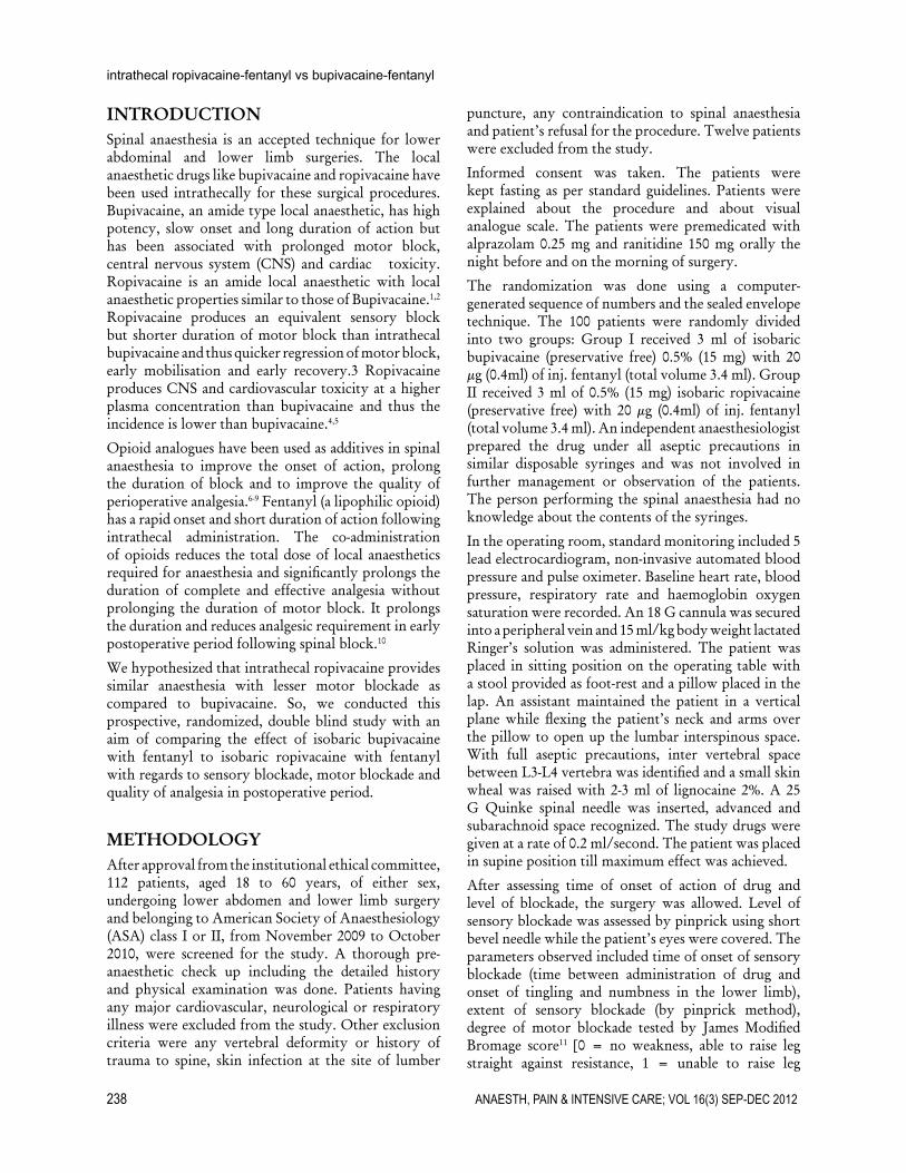
238 ANAESTH, PAIN & INTENSIVE CARE; VOL 16(3) SEP-DEC 2012
intrathecal ropivacaine-fentanyl vs bupivacaine-fentanyl
INTRODUCTIONSpinal anaesthesia is an accepted technique for lower abdominal and lower limb surgeries. The local anaesthetic drugs like bupivacaine and ropivacaine have been used intrathecally for these surgical procedures. Bupivacaine, an amide type local anaesthetic, has high potency, slow onset and long duration of action but has been associated with prolonged motor block, central nervous system (CNS) and cardiac toxicity. Ropivacaine is an amide local anaesthetic with local anaesthetic properties similar to those of Bupivacaine.1,2
Ropivacaine produces an equivalent sensory block but shorter duration of motor block than intrathecal bupivacaine and thus quicker regression of motor block, early mobilisation and early recovery.3 Ropivacaine produces CNS and cardiovascular toxicity at a higher plasma concentration than bupivacaine and thus the incidence is lower than bupivacaine.4,5
Opioid analogues have been used as additives in spinal anaesthesia to improve the onset of action, prolong the duration of block and to improve the quality of perioperative analgesia.6-9 Fentanyl (a lipophilic opioid) has a rapid onset and short duration of action following intrathecal administration. The co-administration of opioids reduces the total dose of local anaesthetics required for anaesthesia and significantly prolongs the duration of complete and effective analgesia without prolonging the duration of motor block. It prolongs the duration and reduces analgesic requirement in early postoperative period following spinal block.10
We hypothesized that intrathecal ropivacaine provides similar anaesthesia with lesser motor blockade as compared to bupivacaine. So, we conducted this prospective, randomized, double blind study with an aim of comparing the effect of isobaric bupivacaine with fentanyl to isobaric ropivacaine with fentanyl with regards to sensory blockade, motor blockade and quality of analgesia in postoperative period.
METHODOLOGYAfter approval from the institutional ethical committee, 112 patients, aged 18 to 60 years, of either sex, undergoing lower abdomen and lower limb surgery and belonging to American Society of Anaesthesiology (ASA) class I or II, from November 2009 to October 2010, were screened for the study. A thorough pre-anaesthetic check up including the detailed history and physical examination was done. Patients having any major cardiovascular, neurological or respiratory illness were excluded from the study. Other exclusion criteria were any vertebral deformity or history of trauma to spine, skin infection at the site of lumber
puncture, any contraindication to spinal anaesthesia and patient’s refusal for the procedure. Twelve patients were excluded from the study.
Informed consent was taken. The patients were kept fasting as per standard guidelines. Patients were explained about the procedure and about visual analogue scale. The patients were premedicated with alprazolam 0.25 mg and ranitidine 150 mg orally the night before and on the morning of surgery.
The randomization was done using a computer-generated sequence of numbers and the sealed envelope technique. The 100 patients were randomly divided into two groups: Group I received 3 ml of isobaric bupivacaine (preservative free) 0.5% (15 mg) with 20 µg (0.4ml) of inj. fentanyl (total volume 3.4 ml). Group II received 3 ml of 0.5% (15 mg) isobaric ropivacaine (preservative free) with 20 µg (0.4ml) of inj. fentanyl (total volume 3.4 ml). An independent anaesthesiologist prepared the drug under all aseptic precautions in similar disposable syringes and was not involved in further management or observation of the patients. The person performing the spinal anaesthesia had no knowledge about the contents of the syringes.
In the operating room, standard monitoring included 5 lead electrocardiogram, non-invasive automated blood pressure and pulse oximeter. Baseline heart rate, blood pressure, respiratory rate and haemoglobin oxygen saturation were recorded. An 18 G cannula was secured into a peripheral vein and 15 ml/kg body weight lactated Ringer’s solution was administered. The patient was placed in sitting position on the operating table with a stool provided as foot-rest and a pillow placed in the lap. An assistant maintained the patient in a vertical plane while flexing the patient’s neck and arms over the pillow to open up the lumbar interspinous space. With full aseptic precautions, inter vertebral space between L3-L4 vertebra was identified and a small skin wheal was raised with 2-3 ml of lignocaine 2%. A 25 G Quinke spinal needle was inserted, advanced and subarachnoid space recognized. The study drugs were given at a rate of 0.2 ml/second. The patient was placed in supine position till maximum effect was achieved.
After assessing time of onset of action of drug and level of blockade, the surgery was allowed. Level of sensory blockade was assessed by pinprick using short bevel needle while the patient’s eyes were covered. The parameters observed included time of onset of sensory blockade (time between administration of drug and onset of tingling and numbness in the lower limb), extent of sensory blockade (by pinprick method), degree of motor blockade tested by James Modified Bromage score11 [0 = no weakness, able to raise leg straight against resistance, 1 = unable to raise leg
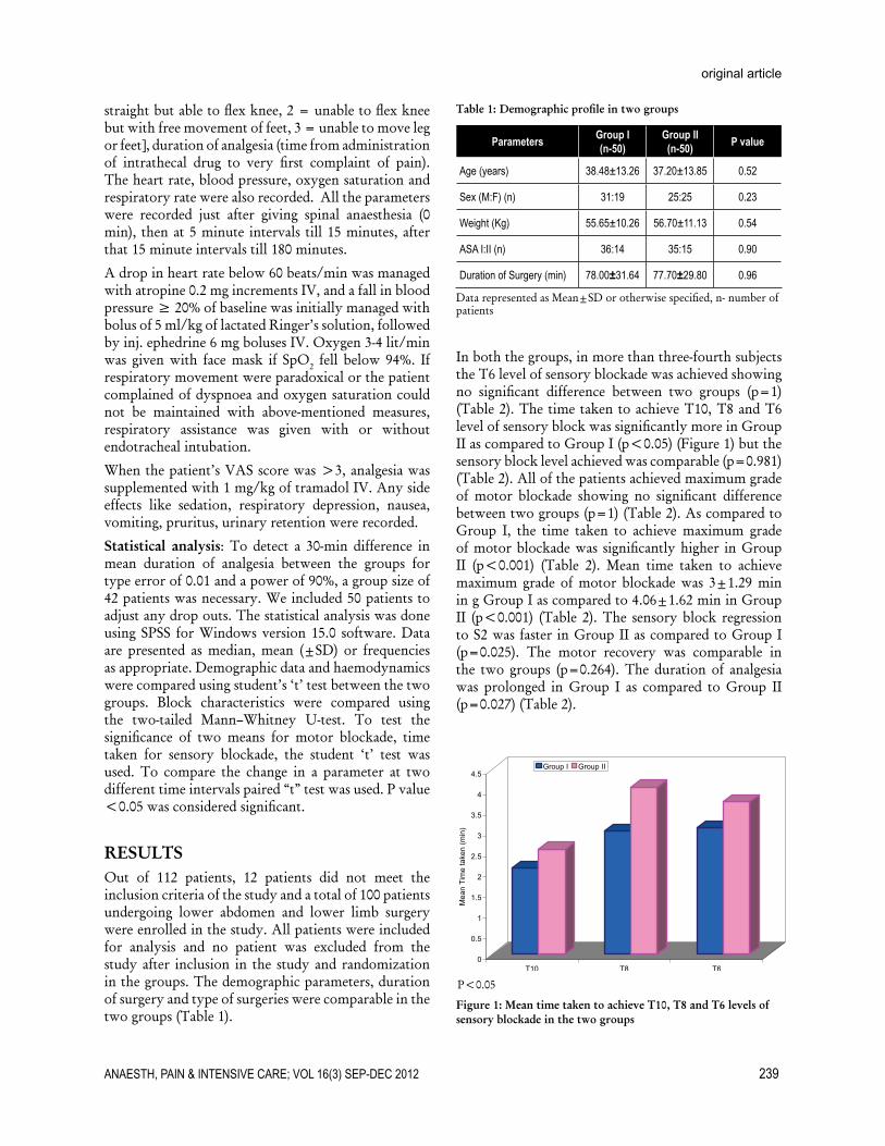
ANAESTH, PAIN & INTENSIVE CARE; VOL 16(3) SEP-DEC 2012 239
original article
straight but able to flex knee, 2 = unable to flex knee but with free movement of feet, 3 = unable to move leg or feet], duration of analgesia (time from administration of intrathecal drug to very first complaint of pain). The heart rate, blood pressure, oxygen saturation and respiratory rate were also recorded. All the parameters were recorded just after giving spinal anaesthesia (0 min), then at 5 minute intervals till 15 minutes, after that 15 minute intervals till 180 minutes.
A drop in heart rate below 60 beats/min was managed with atropine 0.2 mg increments IV, and a fall in blood pressure ≥ 20% of baseline was initially managed with bolus of 5 ml/kg of lactated Ringer’s solution, followed by inj. ephedrine 6 mg boluses IV. Oxygen 3-4 lit/min was given with face mask if SpO2 fell below 94%. If respiratory movement were paradoxical or the patient complained of dyspnoea and oxygen saturation could not be maintained with above-mentioned measures, respiratory assistance was given with or without endotracheal intubation.
When the patient’s VAS score was >3, analgesia was supplemented with 1 mg/kg of tramadol IV. Any side effects like sedation, respiratory depression, nausea, vomiting, pruritus, urinary retention were recorded.
Statistical analysis: To detect a 30-min difference in mean duration of analgesia between the groups for type error of 0.01 and a power of 90%, a group size of 42 patients was necessary. We included 50 patients to adjust any drop outs. The statistical analysis was done using SPSS for Windows version 15.0 software. Data are presented as median, mean (±SD) or frequencies as appropriate. Demographic data and haemodynamics were compared using student’s ‘t’ test between the two groups. Block characteristics were compared using the two-tailed Mann–Whitney U-test. To test the significance of two means for motor blockade, time taken for sensory blockade, the student ‘t’ test was used. To compare the change in a parameter at two different time intervals paired “t” test was used. P value <0.05 was considered significant.
RESULTSOut of 112 patients, 12 patients did not meet the inclusion criteria of the study and a total of 100 patients undergoing lower abdomen and lower limb surgery were enrolled in the study. All patients were included for analysis and no patient was excluded from the study after inclusion in the study and randomization in the groups. The demographic parameters, duration of surgery and type of surgeries were comparable in the two groups (Table 1).
Table 1: Demographic profile in two groups
Parameters Group I (n-50)
Group II (n-50) P value
Age (years) 38.48±13.26 37.20±13.85 0.52
Sex (M:F) (n) 31:19 25:25 0.23
Weight (Kg) 55.65±10.26 56.70±11.13 0.54
ASA I:II (n) 36:14 35:15 0.90
Duration of Surgery (min) 78.00±31.64 77.70±29.80 0.96
DatarepresentedasMean±SDorotherwisespecified,n-numberofpatients
In both the groups, in more than three-fourth subjects the T6 level of sensory blockade was achieved showing no significant difference between two groups (p=1) (Table 2). The time taken to achieve T10, T8 and T6 level of sensory block was significantly more in Group II as compared to Group I (p<0.05) (Figure 1) but the sensory block level achieved was comparable (p=0.981) (Table 2). All of the patients achieved maximum grade of motor blockade showing no significant difference between two groups (p=1) (Table 2). As compared to Group I, the time taken to achieve maximum grade of motor blockade was significantly higher in Group II (p<0.001) (Table 2). Mean time taken to achieve maximum grade of motor blockade was 3±1.29 min in g Group I as compared to 4.06±1.62 min in Group II (p<0.001) (Table 2). The sensory block regression to S2 was faster in Group II as compared to Group I (p=0.025). The motor recovery was comparable in the two groups (p=0.264). The duration of analgesia was prolonged in Group I as compared to Group II (p=0.027) (Table 2).
P<0.05
Figure 1: Mean time taken to achieve T10, T8 and T6 levels of sensory blockade in the two groups
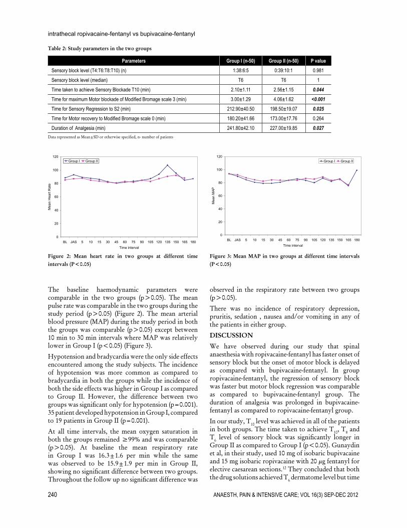
240 ANAESTH, PAIN & INTENSIVE CARE; VOL 16(3) SEP-DEC 2012
intrathecal ropivacaine-fentanyl vs bupivacaine-fentanyl
The baseline haemodynamic parameters were comparable in the two groups (p>0.05). The mean pulse rate was comparable in the two groups during the study period (p>0.05) (Figure 2). The mean arterial blood pressure (MAP) during the study period in both the groups was comparable (p>0.05) except between 10 min to 30 min intervals where MAP was relatively lower in Group I (p<0.05) (Figure 3).
Hypotension and bradycardia were the only side effects encountered among the study subjects. The incidence of hypotension was more common as compared to bradycardia in both the groups while the incidence of both the side effects was higher in Group I as compared to Group II. However, the difference between two groups was significant only for hypotension (p=0.001). 35 patient developed hypotension in Group I, compared to 19 patients in Group II (p=0.001).
At all time intervals, the mean oxygen saturation in both the groups remained ≥99% and was comparable (p>0.05). At baseline the mean respiratory rate in Group I was 16.3±1.6 per min while the same was observed to be 15.9±1.9 per min in Group II, showing no significant difference between two groups. Throughout the follow up no significant difference was
Table 2: Study parameters in the two groups
Parameters Group I (n-50) Group II (n-50) P value
Sensory block level (T4:T6:T8:T10) (n) 1:38:6:5 0:39:10:1 0.981
Sensory block level (median) T6 T6 1
Time taken to achieve Sensory Blockade T10 (min) 2.10±1.11 2.56±1.15 0.044
Time for maximum Motor blockade of Modified Bromage scale 3 (min) 3.00±1.29 4.06±1.62 <0.001
Time for Sensory Regression to S2 (min) 212.90±40.50 198.50±19.07 0.025
Time for Motor recovery to Modified Bromage scale 0 (min) 180.20±41.66 173.00±17.76 0.264
Duration of Analgesia (min) 241.80±42.10 227.00±19.85 0.027DatarepresentedasMean±SDorotherwisespecified,n-numberofpatients
Figure 2: Mean heart rate in two groups at different time intervals (P<0.05)
Figure 3: Mean MAP in two groups at different time intervals (P<0.05)
observed in the respiratory rate between two groups (p>0.05).
There was no incidence of respiratory depression, pruritis, sedation , nausea and/or vomiting in any of the patients in either group.
DISCUSSION
We have observed during our study that spinal anaesthesia with ropivacaine-fentanyl has faster onset of sensory block but the onset of motor block is delayed as compared with bupivacaine-fentanyl. In group ropivacaine-fentanyl, the regression of sensory block was faster but motor block regression was comparable as compared to bupivacaine-fentanyl group. The duration of analgesia was prolonged in bupivacaine-fentanyl as compared to ropivacaine-fentanyl group.
In our study, T10 level was achieved in all of the patients in both groups. The time taken to achieve T10, T8 and T6 level of sensory block was significantly longer in Group II as compared to Group I (p<0.05). Gunaydin et al, in their study, used 10 mg of isobaric bupivacaine and 15 mg isobaric ropivacaine with 20 µg fentanyl for elective caesarean sections.12 They concluded that both the drug solutions achieved T6 dermatome level but time
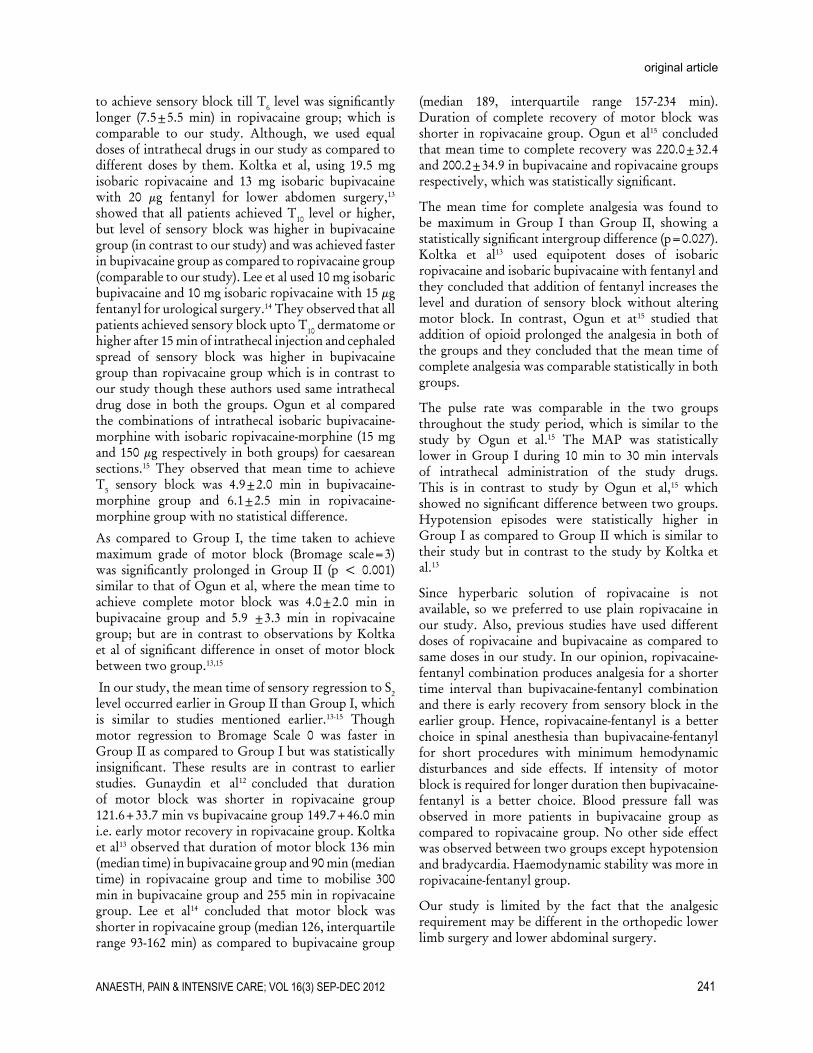
ANAESTH, PAIN & INTENSIVE CARE; VOL 16(3) SEP-DEC 2012 241
original article
to achieve sensory block till T6 level was significantly longer (7.5±5.5 min) in ropivacaine group; which is comparable to our study. Although, we used equal doses of intrathecal drugs in our study as compared to different doses by them. Koltka et al, using 19.5 mg isobaric ropivacaine and 13 mg isobaric bupivacaine with 20 µg fentanyl for lower abdomen surgery,13
showed that all patients achieved T10 level or higher, but level of sensory block was higher in bupivacaine group (in contrast to our study) and was achieved faster in bupivacaine group as compared to ropivacaine group (comparable to our study). Lee et al used 10 mg isobaric bupivacaine and 10 mg isobaric ropivacaine with 15 µg fentanyl for urological surgery.14 They observed that all patients achieved sensory block upto T10 dermatome or higher after 15 min of intrathecal injection and cephaled spread of sensory block was higher in bupivacaine group than ropivacaine group which is in contrast to our study though these authors used same intrathecal drug dose in both the groups. Ogun et al compared the combinations of intrathecal isobaric bupivacaine-morphine with isobaric ropivacaine-morphine (15 mg and 150 µg respectively in both groups) for caesarean sections.15 They observed that mean time to achieve T5 sensory block was 4.9±2.0 min in bupivacaine- morphine group and 6.1±2.5 min in ropivacaine-morphine group with no statistical difference.
As compared to Group I, the time taken to achieve maximum grade of motor block (Bromage scale=3) was significantly prolonged in Group II (p < 0.001) similar to that of Ogun et al, where the mean time to achieve complete motor block was 4.0±2.0 min in bupivacaine group and 5.9 ±3.3 min in ropivacaine group; but are in contrast to observations by Koltka et al of significant difference in onset of motor block between two group.13,15
In our study, the mean time of sensory regression to S2 level occurred earlier in Group II than Group I, which is similar to studies mentioned earlier.13-15 Though motor regression to Bromage Scale 0 was faster in Group II as compared to Group I but was statistically insignificant. These results are in contrast to earlier studies. Gunaydin et al12 concluded that duration of motor block was shorter in ropivacaine group 121.6+33.7 min vs bupivacaine group 149.7+46.0 min i.e. early motor recovery in ropivacaine group. Koltka et al13 observed that duration of motor block 136 min (median time) in bupivacaine group and 90 min (median time) in ropivacaine group and time to mobilise 300 min in bupivacaine group and 255 min in ropivacaine group. Lee et al14 concluded that motor block was shorter in ropivacaine group (median 126, interquartile range 93-162 min) as compared to bupivacaine group
(median 189, interquartile range 157-234 min). Duration of complete recovery of motor block was shorter in ropivacaine group. Ogun et al15 concluded that mean time to complete recovery was 220.0±32.4 and 200.2±34.9 in bupivacaine and ropivacaine groups respectively, which was statistically significant.
The mean time for complete analgesia was found to be maximum in Group I than Group II, showing a statistically significant intergroup difference (p=0.027). Koltka et al13 used equipotent doses of isobaric ropivacaine and isobaric bupivacaine with fentanyl and they concluded that addition of fentanyl increases the level and duration of sensory block without altering motor block. In contrast, Ogun et at15 studied that addition of opioid prolonged the analgesia in both of the groups and they concluded that the mean time of complete analgesia was comparable statistically in both groups.
The pulse rate was comparable in the two groups throughout the study period, which is similar to the study by Ogun et al.15 The MAP was statistically lower in Group I during 10 min to 30 min intervals of intrathecal administration of the study drugs. This is in contrast to study by Ogun et al,15 which showed no significant difference between two groups. Hypotension episodes were statistically higher in Group I as compared to Group II which is similar to their study but in contrast to the study by Koltka et al.13
Since hyperbaric solution of ropivacaine is not available, so we preferred to use plain ropivacaine in our study. Also, previous studies have used different doses of ropivacaine and bupivacaine as compared to same doses in our study. In our opinion, ropivacaine-fentanyl combination produces analgesia for a shorter time interval than bupivacaine-fentanyl combination and there is early recovery from sensory block in the earlier group. Hence, ropivacaine-fentanyl is a better choice in spinal anesthesia than bupivacaine-fentanyl for short procedures with minimum hemodynamic disturbances and side effects. If intensity of motor block is required for longer duration then bupivacaine-fentanyl is a better choice. Blood pressure fall was observed in more patients in bupivacaine group as compared to ropivacaine group. No other side effect was observed between two groups except hypotension and bradycardia. Haemodynamic stability was more in ropivacaine-fentanyl group.
Our study is limited by the fact that the analgesic requirement may be different in the orthopedic lower limb surgery and lower abdominal surgery.
242 ANAESTH, PAIN & INTENSIVE CARE; VOL 16(3) SEP-DEC 2012
intrathecal ropivacaine-fentanyl vs bupivacaine-fentanyl
1. Akerman B, Hellberg IB, Trossvik C. Primary evaluation of the local anaesthetic properties of the amino amide agent Ropivacaine (LEA 103). Acta Anaesthesiol Scand 1988;32:571-8. [Medline]
2. McClure JH. Ropivacaine. Br J Anaesth 1996;76:300-7. [Medline]
3. Gautier P, De Kock M, Huberty L, Demir T, Izydorczic M, Vanderick B. Comparison of the effects of intrathecal ropivacaine, levobupivacaine and bupivacaine for caesarean section. Br J Anaesth 2003;91:684-9. [Medline]
4. Hansen TG. Ropivacaine: a pharmacological review. Expert Rev Neurother 2004;4:781-91. [Medline]
5. McClellan KJ, Faulds D. Ropivacaine: an update of its used in regional anesthesia. Drugs 2000;60:1065-93. [Medline]
6. van Kleef JW, Veering BT, Burm AG. Spinal anesthesia with Ropivacaine: a double blind study of efficacy and safety of and 0.75% solution in patients undergoing minor lower limb surgery. Anesth Analg 1994;78:1125-30. [Medline]
7. Whiteside JB, Burke D, Wildsmith JA. Spinal anaesthesia with Ropivacaine 5 mg ml-1
in glucose 10 mg ml-1 and 50 mg ml-1. Br J Anaesth 2001;86:241-4. [Medline]
8. Bogra J, Arora N, Srivastava P. Synergistic effect of Intrathecal Fentanyl and Bupivacaine in spinal anesthesia. BMC Anesthesiol 2005;5:5. [Medline]
9. Malinovsky JM, Charles F, Kick O, Lepage JY, Malinge M, Cozian A, et al. Intrathecal anesthesia: Ropivacaine versus Bupivacaine. Anesth Analg 2000;91:1457-60. [Medline]
10. Singh H, Yang J, Thornton K, Giesecke AH. Intrathecal fentanyl prolongs sensory bupivacaine spinal block. Can J Anaesth 1995;42:987-91. [Medline]
11. Fettes PD, Hocking G, Peterson MK, Luck JF, Wildsmith JA. Comparison of plain and hyperbaric solutions of ropivacaine for spinal anaesthesia. Br J Anaesth 2005;94:107-11. [Medline]
12. Gunaydin B, Tan ED. Intrathecal hyperbaric or isobaric Bupivacaine and Ropivacaine with Fenatanyl for elective caesarean section. J Matern Fetal Neonatal Med 2012;23:1481-6. [Medline]
13. Koltka K, Uludag E, Senturk M, Yavru A, Karadeniz M, Sengul T, Ozyalcin S. Comparison of equipotent doses of
ropivacaine-fentanyl and bupivacaine-fentanyl in spinal anaesthesia for lower abdominal surgery. Anaesth Intensive Care 2009;37:923-8. [Medline]
14. Lee YY, Ngan Kee WD, Muchhal K, Chan CK. Randomized double blind comparison of Ropivacaine Fentanyl and Bupivacaine Fentanyl for spinal anaesthesia for urological surgery. Acta Anaesthesiol Scand 2005;49:1477-82. [Medline]
15. Oğün CO, Kirgiz EN, Duman A, Okesli S, Akyürek C. Comparison of intrathecal isobaric bupivacaine-morphine and ropivacaine-morphine for Caesarean delivery. Br J Anaesth 2003;90:659–64. [Medline]
16. Nuray C, Berrin G. A comparison of the effects of intrathecal ropivacaine and bupivacaine during cesarean section. Turk J Med Sci 2011;41(2):219-226 [Tubitak]
17. Surjeet Singh, V.P. Singh, Manish Jain, Kumkum Gupta, Bhavna Rastogi, Surbhi Abrol Intrathecal 0.75% Isobaric Ropivacaine Versus 0.5% Heavy Bupivacaine for Elective Cesarean Delivery: A Randomized Controlled Trial . J Pak Med Stud 2012;2:2
REFERENCES
CONCLUSIONWe conclude that intrathecal administration of ropivacaine-fentanyl has faster onset and faster regression of sensory block, delayed onset but comparable regression of motor block and shorter duration of analgesia as compared to intrathecal bupivacaine-fentanyl. The bupivacaine-fentanyl group
is associated with increased episodes of hypotension as compared to ropivacaine-fentanyl combination administered intrathecally.
Financial Support: Departmental sources only
Disclaimers: None
Conflict of Interest: None
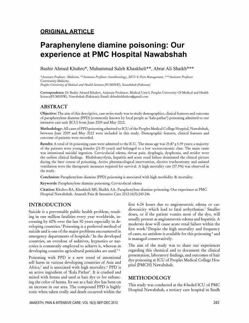
ANAESTH, PAIN & INTENSIVE CARE; VOL 16(3) SEP-DEC 2012 243
ORIGINAL ARTICLE
Paraphenylene diamine poisoning: Our experience at PMC Hospital NawabshahBashir Ahmed Khuhro*, Muhammad Saleh Khaskheli**, Abrar Ali Shaikh****Assistant Professor Medicine, **Assistant Professor Anesthesiology, ,SICU & Pain Management, ***Assistant Professor Community Medicine,Peoples University of Medical and Health Sciences (PUMHSW), Nawabshah (Pakistan)
Correspondence: Dr Bashir Ahmed Khuhro, Assistant Professor, Medical Unit-I, Peoples University Of Medical and Health Science(PUMHSW), Nawabshah (Pakistan); Email: [email protected]
ABSTRACTObjective: The aim of this descriptive, case series study was to study demographics, clinical features and outcome of paraphenylene diamine (PPD) (commonly known by local people as ‘kala pathar’) poisoning admitted to our intensive care unit (ICU) from June 2009 and May 2012.
Methodology: All cases of PPD poisoning admitted to ICU of the Peoples Medical College Hospital, Nawabshah, between June 2009 and May 2012 were included in this study. Demographic features, clinical features and outcome of patients were recorded.
Results: A total of 16 poisoning cases were admitted to the ICU. The mean age was 25.87±5.59 years; a majority of the patients were young females (21-30 years) and belonged to a low socioeconomic class. The main cause was intentional suicidal ingestion. Cervicofacial edema, throat pain, dysphagia, dysphonia, and stridor were the earliest clinical findings. Rhabdomyolysis, hepatitis and acute renal failure dominated the clinical picture during the later course of poisoning. Active pharmacological intervention, elective tracheostomy and assisted ventilation were the therapeutic measures required for survival. A high mortality rate (37.5%) was observed in the study.
Conclusion: Paraphenylene diamine (PPD) poisoning is associated with high morbidity & mortality.
Keywords: Paraphenylene diamine poisoning; Cervicofacial edema
Citation: Khuhro BA, Khaskheli MS, Shaikh AA. Paraphenylene diamine poisoning: Our experience at PMC Hospital Nawabshah. Anaesth Pain & Intensive Care 2012:16(3):243-246
INTRODUCTIONSuicide is a preventable public health problem, result-ing in one million fatalities every year worldwide, in-creasing by 60% over the last 50 years especially in de-veloping countries.1 Poisoning is a preferred method of suicide and is one of the major problems encountered in emergency departments of hospitals.2 In the developed countries, an overdose of sedatives, hypnotics or nar-cotics is commonly employed to achieve it, whereas in developing countries agricultural pesticides are used.3–4
Poisoning with PPD is a new trend of intentional self harm in various developing countries of Asia and Africa,5 and is associated with high mortality.6 PPD is an active ingredient of ‘Kala Pathar’. It is crushed and mixed with henna and used as hair dye or for enhanc-ing the color of henna. Its use as a hair dye has been on an increase in our area. The compound PPD is highly toxic when taken orally and death occurred within the
first 6-24 hours due to angioneurotic edema or car-diotoxicity which lead to fatal arrhythmias.7 Smaller doses, or if the patient vomits most of the dye, will usually present as angioneurotic edema and hepatitis. A moderate dose will cause acute renal failure within the first week.8 Despite the high mortality and frequency of cases, no antidote is available for this poisoning 8 and is managed conservatively.
The aim of the study was to share our experiences regarding this chemical and to document the clinical presentation, laboratory findings, and outcomes of hair dye poisoning at ICU of Peoples Medical College Hos-pital (PMCH) Nawabshah.
METHODOLOGYThis study was conducted at the 8-beded ICU of PMC Hospital Nawabshah, a tertiary care hospital in Sindh
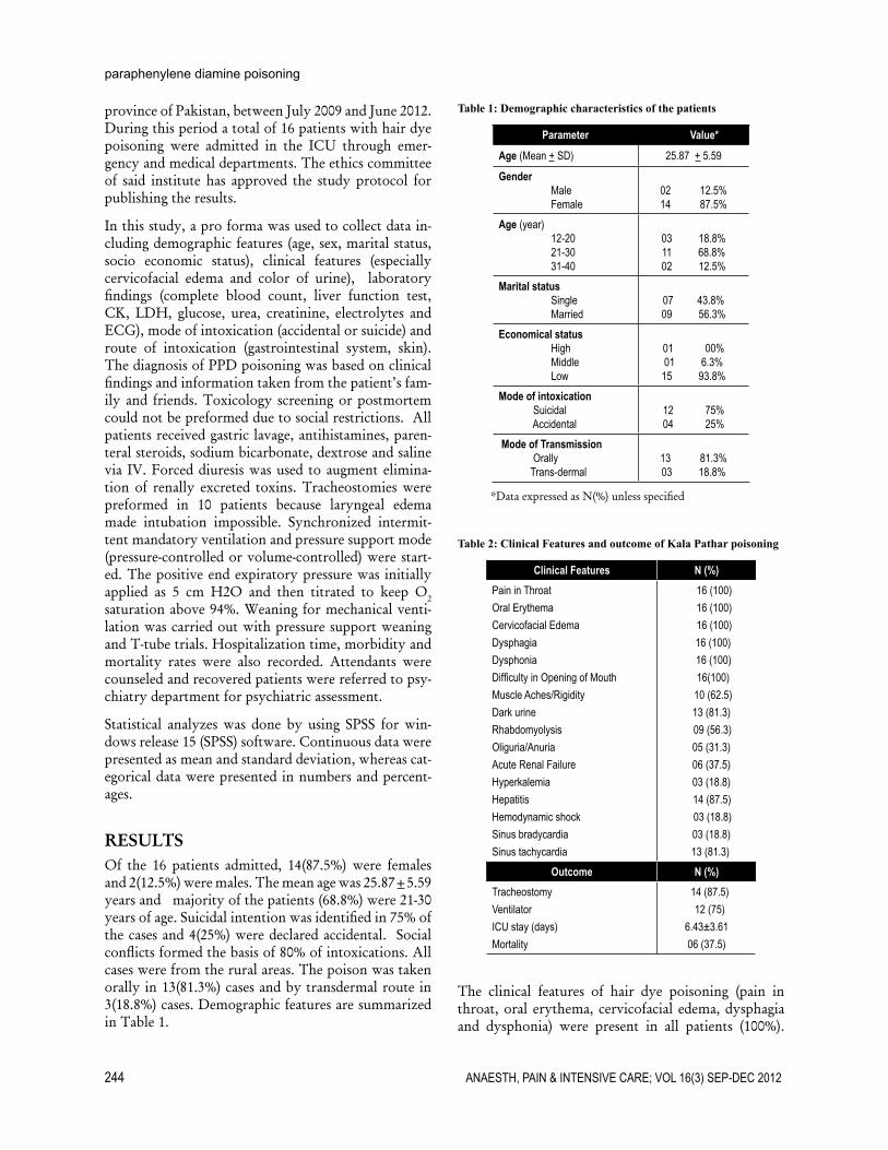
244 ANAESTH, PAIN & INTENSIVE CARE; VOL 16(3) SEP-DEC 2012
paraphenylene diamine poisoning
province of Pakistan, between July 2009 and June 2012. During this period a total of 16 patients with hair dye poisoning were admitted in the ICU through emer-gency and medical departments. The ethics committee of said institute has approved the study protocol for publishing the results.
In this study, a pro forma was used to collect data in-cluding demographic features (age, sex, marital status, socio economic status), clinical features (especially cervicofacial edema and color of urine), laboratory findings (complete blood count, liver function test, CK, LDH, glucose, urea, creatinine, electrolytes and ECG), mode of intoxication (accidental or suicide) and route of intoxication (gastrointestinal system, skin). The diagnosis of PPD poisoning was based on clinical findings and information taken from the patient’s fam-ily and friends. Toxicology screening or postmortem could not be preformed due to social restrictions. All patients received gastric lavage, antihistamines, paren-teral steroids, sodium bicarbonate, dextrose and saline via IV. Forced diuresis was used to augment elimina-tion of renally excreted toxins. Tracheostomies were preformed in 10 patients because laryngeal edema made intubation impossible. Synchronized intermit-tent mandatory ventilation and pressure support mode (pressure-controlled or volume-controlled) were start-ed. The positive end expiratory pressure was initially applied as 5 cm H2O and then titrated to keep O2 saturation above 94%. Weaning for mechanical venti-lation was carried out with pressure support weaning and T-tube trials. Hospitalization time, morbidity and mortality rates were also recorded. Attendants were counseled and recovered patients were referred to psy-chiatry department for psychiatric assessment.
Statistical analyzes was done by using SPSS for win-dows release 15 (SPSS) software. Continuous data were presented as mean and standard deviation, whereas cat-egorical data were presented in numbers and percent-ages.
RESULTSOf the 16 patients admitted, 14(87.5%) were females and 2(12.5%) were males. The mean age was 25.87±5.59 years and majority of the patients (68.8%) were 21-30 years of age. Suicidal intention was identified in 75% of the cases and 4(25%) were declared accidental. Social conflicts formed the basis of 80% of intoxications. All cases were from the rural areas. The poison was taken orally in 13(81.3%) cases and by transdermal route in 3(18.8%) cases. Demographic features are summarized in Table 1.
Table 1: Demographic characteristics of the patients
Parameter Value*
Age (Mean + SD) 25.87 + 5.59
Gender MaleFemale
02 12.5%14 87.5%
Age (year)12-2021-3031-40
03 18.8%11 68.8%02 12.5%
Marital statusSingleMarried
07 43.8%09 56.3%
Economical statusHighMiddleLow
01 00%01 6.3%
15 93.8%Mode of intoxication Suicidal Accidental
12 75%04 25%
Mode of Transmission Orally Trans-dermal
13 81.3%03 18.8%
*Data expressed as N(%) unless specified
Table 2: Clinical Features and outcome of Kala Pathar poisoning
Clinical Features N (%)Pain in ThroatOral ErythemaCervicofacial EdemaDysphagiaDysphoniaDifficulty in Opening of MouthMuscle Aches/RigidityDark urineRhabdomyolysisOliguria/AnuriaAcute Renal FailureHyperkalemiaHepatitisHemodynamic shockSinus bradycardiaSinus tachycardia
16 (100)16 (100)16 (100)16 (100)16 (100)16(100)
10 (62.5)13 (81.3)09 (56.3)05 (31.3)06 (37.5)03 (18.8)14 (87.5)03 (18.8)03 (18.8)13 (81.3)
Outcome N (%)TracheostomyVentilatorICU stay (days)Mortality
14 (87.5)12 (75)
6.43±3.6106 (37.5)
The clinical features of hair dye poisoning (pain in throat, oral erythema, cervicofacial edema, dysphagia and dysphonia) were present in all patients (100%).
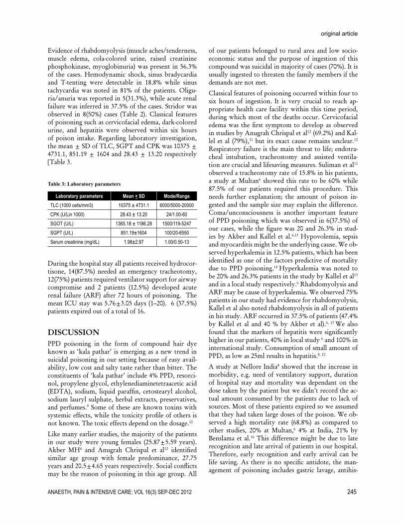
ANAESTH, PAIN & INTENSIVE CARE; VOL 16(3) SEP-DEC 2012 245
original article
Evidence of rhabdomyolysis (muscle aches/tenderness, muscle edema, cola-colored urine, raised creatinine phosphokinase, myoglobinuria) was present in 56.3% of the cases. Hemodynamic shock, sinus bradycardia and T-tenting were detectable in 18.8% while sinus tachycardia was noted in 81% of the patients. Oligu-ria/anuria was reported in 5(31.3%), while acute renal failure was inferred in 37.5% of the cases. Stridor was observed in 8(50%) cases (Table 2). Classical features of poisoning such as cervicofacial edema, dark-colored urine, and hepatitis were observed within six hours of poison intake. Regarding laboratory investigation, the mean ± SD of TLC, SGPT and CPK was 10375 ± 4731.1, 851.19 ± 1604 and 28.43 ± 13.20 respectively [Table 3.
Table 3: Laboratory parameters
Laboratory parameters Mean + SD Mode/RangeTLC (1000 cells/mm3) 10375 ± 4731.1 6000/5000-20000CPK (U/Lin 1000) 28.43 ± 13.20 24/1.00-60SGOT (U/L) 1365.18 ± 1186.28 1500/119-5247SGPT (U/L) 851.19±1604 100/20-6550Serum creatinine (mg/dL) 1.98±2.97 1.00/0.50-13
During the hospital stay all patients received hydrocor-tisone, 14(87.5%) needed an emergency tracheotomy, 12(75%) patients required ventilator support for airway compromise and 2 patients (12.5%) developed acute renal failure (ARF) after 72 hours of poisoning. The mean ICU stay was 5.76±3.05 days (1–20). 6 (37.5%) patients expired out of a total of 16.
DISCUSSIONPPD poisoning in the form of compound hair dye known as ‘kala pathar’ is emerging as a new trend in suicidal poisoning in our setting because of easy avail-ability, low cost and salty taste rather than bitter. The constituents of ‘kala pathar’ include 4% PPD, resorci-nol, propylene glycol, ethylenediaminetetraacetic acid (EDTA), sodium, liquid paraffin, cetostearyl alcohol, sodium lauryl sulphate, herbal extracts, preservatives, and perfumes.9 Some of these are known toxins with systemic effects, while the toxicity profile of others is not known. The toxic effects depend on the dosage.10
Like many earlier studies, the majority of the patients in our study were young females (25.87±5.59 years). Akber MH6 and Anugrah Chrispal et al12 identified similar age group with female predominance, 27.75 years and 20.5±4.65 years respectively. Social conflicts may be the reason of poisoning in this age group. All
of our patients belonged to rural area and low socio-economic status and the purpose of ingestion of this compound was suicidal in majority of cases (70%). It is usually ingested to threaten the family members if the demands are not met.
Classical features of poisoning occurred within four to six hours of ingestion. It is very crucial to reach ap-propriate health care facility within this time period, during which most of the deaths occur. Cervicofacial edema was the first symptom to develop as observed in studies by Anugrah Chrispal et al12 (69.2%) and Kal-lel et al (79%),11 but its exact cause remains unclear.12 Respiratory failure is the main threat to life; endotra-cheal intubation, tracheostomy and assisted ventila-tion are crucial and lifesaving measures. Suliman et al11 observed a tracheostomy rate of 15.8% in his patients, a study at Multan6 showed this rate to be 60% while 87.5% of our patients required this procedure. This needs further explanation; the amount of poison in-gested and the sample size may explain the difference. Coma/unconsciousness is another important feature of PPD poisoning which was observed in 6(37.5%) of our cases, while the figure was 20 and 26.3% in stud-ies by Akber and Kallel et al.6,13 Hypovolemia, sepsis and myocarditis might be the underlying cause. We ob-served hyperkalemia in 12.5% patients, which has been identified as one of the factors predictive of mortality due to PPD poisoning.14 Hyperkalemia was noted to be 20% and 26.3% patients in the study by Kallel et al13
and in a local study respectively.6 Rhabdomyolysis and ARF may be cause of hyperkalemia. We observed 75% patients in our study had evidence for rhabdomyolysis, Kallel et al also noted rhabdomyolysis in all of patients in his study. ARF occurred in 37.5% of patients (47.4% by Kallel et al and 40 % by Akber et al).6, 13 We also found that the markers of hepatitis were significantly higher in our patients, 40% in local study 6 and 100% in international study. Consumption of small amount of PPD, as low as 25ml results in hepatitis.8, 10
A study at Nellore India8 showed that the increase in morbidity, e.g. need of ventilatory support, duration of hospital stay and mortality was dependant on the dose taken by the patient but we didn’t record the ac-tual amount consumed by the patients due to lack of sources. Most of these patients expired so we assumed that they had taken large doses of the poison. We ob-served a high mortality rate (68.8%) as compared to other studies, 20% at Multan,6 4% at India, 21% by Benslama et al.16 This difference might be due to late recognition and late arrival of patients in our hospital. Therefore, early recognition and early arrival can be life saving. As there is no specific antidote, the man-agement of poisoning includes gastric lavage, antihis-
246 ANAESTH, PAIN & INTENSIVE CARE; VOL 16(3) SEP-DEC 2012
paraphenylene diamine poisoning
1. WHO World Suicide Prevention Day 2008; WHO Statement; 2008. Available online:http://www.who.int/mental_health/pre-vention/suicide/wspd_2008_statement.pdf (accessed on 11 July 2012)
2. Lee HL, Lin HJ, Yeh STY, Chi CH, Guo HR. Presentations of patient’s of poisoning and predictor of poisoning -related fatality: find-ings from a hospital-based prospective study. BMC Public Health 2008;8:7.
3. Michel K, Ballinari P, Bille-Brahe U, Bjerke T, Crepet P, De Leo D, et al. Methods used for Para suicide: results of the WHO/EURO Mul-ticenter Study on Para suicide. Soc Psychia-try Psychiatr Epidemiol. 2000;35:156–63.
4. Gunnell D, Ho D, Murray V. Medical manage-ment of deliberate drug overdose: a neglect-ed area for suicide prevention? Emerg Med J 2004;21:35–8.
5. Suliman SM, Homeida M, Aboval OI. Para-phenylene Diamine Induced Acute Tubular Necrosis Following Hair Dye Ingestion. Hu-
man Toxicol 1983;2:633-5.6. Akbar MA, Khaliq SA, Malik NA, Shahzad
A, Tarin SM, Chaudhary GM. Kala pathar (paraphenylene diamin) intoxication; a study at Nishtar hospital Multan. Vol 2, No 4• Octo-ber – December 2010
7. Bowen DAL. A case of phenylenediamine poisoning. Medicine Sci Law 1963;3:216-9.
8. Kondle R,Pathapati RM, Saginela SK, Mal-liboina S, Makineedi VP. Clinical Profile and Outcomes of Hair Dye Poisoning in a Teach-ing Hospital in Nellore. ISRN Emergency Medicine, vol. 2012, Article ID 624253, 5 pages, 2012. doi:10.5402/2012/624253
9. Nohynek GJ, Fautz RF, Benech-Kieffer, Toutain H .Toxicity and human health risk of hair dyes. Food and Chemical Toxicology 2004;42(4):517–543.
10. Hou FQ, Lin XH, Yu YY. Wang TL and Wang GQ. Severe liver injury induced by repeated use of hair dye. Chinese Medical Journal 2009;122 (7):875-77.
11. Suliman SM, Fadlalla M, Naser Mel M, et al. Poisoning with Hair Dye Containing Para-phenyleneDiamine: Ten Years’ Experience. Saudi J Kidney Dis Transpl 1995;6:286-9.
12. Chrispal A, Begum A, Ramya, Zachariah A. Hair Dye Poisoning- an Emerging Problem in the Tropics: an Experience from a Ter-tiary Care Hospital in South India. Trop Doct 2010;40:100-3.
13. Kallel H, Chelly H, Dammak H, Bahloul M, Ksibi H, Hamida CB, et al. Clinical Manifes-tations of Systemic ParaphenyleneDiamine Intoxication. J Nephrol. 2005;18:308.
14. Soni S, Nagarik A, Anuradha GK. Super-vasmol 33 Poisoning- Abstract Presented at 38th Annual Conference of Indian Society of Nephrology;2007
15. Bensalama A, Moutaouakkil S, Mjahed K, El Moknia M, Lahbil D, Fadel H. Syndrome intermediairelorsd’une intoxication aigue par le malathion. Press Med. 1998;27:713-5.
REFERENCES
tamines, parenteral steroids especially hydrocortisone, and alkalinization of the urine.8 Respiratory distress is the major early problem which may require ventila-tory support. Renal support in the form of dialysis is required in acute renal failure,8 but our patients did not need dialysis because they were not fit for dialysis and expired early.
CONCLUSIONParaphenylene diamine (PPD) (Kala Pathar) poisoning
could be a warning to the Asian countries and emerg-ing as alternative to organophosphorus poisoning be-cause of its easy availability, low cost and bitterness. We recommend public awareness regarding this toxin and sale of Kala Pathar should be legally restricted by government.
DISCLOSURENone of the authors received any financial benefit from any source while conducting this study.
ANAESTH, PAIN & INTENSIVE CARE; VOL 16(3) SEP-DEC 2012 247
ORIGINAL ARTICLE
A comparison of hemodynamic changes during laryngoscopy and endotracheal intubation by using three modalities of anesthesia inductionMasoud Lahsaee, MD*, Hamid Kamalipour, MD**, Zahra Ajeli, MD***, Karmella Kamali, MD****
*Assistant Professor of Anesthesiology, Shiraz Anesthesiology and Critical Care Research Center**Professor of Anesthesiology, Laparoscopy Research Center*** Anesthesiologist, Department of Anesthesiology**** Assistant Professor of Radiology, Department of RadiologyShiraz University of Medical Sciences, Shiraz (Iran)
Correspondence: Hamid Kamalipour, MD, Professor of Anesthesia, Department of Anesthesiology, Shiraz University of Medical Sciences, P.O.Box:71345-3168, Shiraz (Iran); Phone: +98-917-111-1112; Fax: +98-711-6284828; E-mail:[email protected]
ABSTRACTBackground: Laryngoscopy and endotracheal intubation is usually associated with hemodynamic changes increases in blood pressure and heart rate. We aimed to compare the hemodynamic effects of combined use of low dose thiopentone and propofol during induction of anesthesia with conventional dose of thiopentone and propofol separately.
Methodology: This trial was conducted prospectively among 90 candidates, American Society of Anaesthesiologists grade-l (ASA-I), scheduled for elective surgery, with an age range of 20-50 years. Selected patients were randomly assigned to three equal groups. Thiopentone was used in group 1, propofol in group 2, and a combination of low dose thiopentone and propofol in group 3 as an induction agent.
Heart rate and blood pressures were measured non-invasively at five different times: prior to the injection of study drugs, three minutes after the last injection of induction drug and immediately before the laryngoscopy and endotracheal intubation, as well as at the first, third and fifth minutes after endotracheal intubation.
Results: The adjusted mean values of systolic blood pressure, diastolic blood pressure, and heart rate were assessed by paired comparisons, by considering the variable of time; all changes were significantly different between Groups 1 and 2. Moreover, changes in systolic and diastolic blood pressures were significantly different between Groups 1 and 3. They were not significant for heart rate. No significant difference was documented between Groups 2 and 3; showing that in these groups the hemodynamic changes were small during drug injection, laryngoscopy, and intubation as well as until five minutes after endotracheal intubation.
Conclusion: The combined use of low dose thiopentone and propofol for anesthetic induction caused less hemodynamic changes than the higher dose of either alone. This modality of anesthesia induction may have clinical importance for the elderly patients as well as those with high blood pressure and heart diseases.
Key words: Thiopental; Propofol; Laryngoscopy; Endotracheal intubation; Anesthesia
Citation: Lahsaee M, Kamalipour H, Ajeli Z, Kamali K. A comparison of hemodynamic changes during laryngoscopy and endotracheal intubation by using three modalities of anesthesia induction. Anaesth Pain & Intensive Care 2012;16(3):247-251
INTRODUCTIONPatients undergoing laryngoscopy and endotracheal in-tubation are known to develop hemodynamic changes such as an increase in blood pressure (BP) and heart rate. In susceptible patients, such changes may lead
to myocardial ischemia or a rise in the intracranial pressure.1-5 On the other hand most commonly used induction agents usually lower BP, while producing a tachycardia. Many studies have been conducted to determine the effects of different types of medications on these hemodynamic changes. Generally, multiple
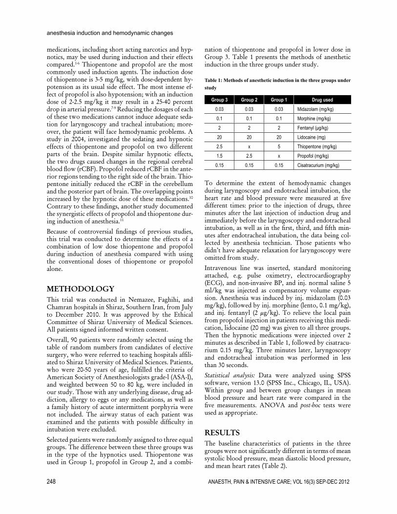
248 ANAESTH, PAIN & INTENSIVE CARE; VOL 16(3) SEP-DEC 2012
anesthesia induction and hemodynamic changes
medications, including short acting narcotics and hyp-notics, may be used during induction and their effects compared.1-6 Thiopentone and propofol are the most commonly used induction agents. The induction dose of thiopentone is 3-5 mg/kg, with dose-dependent hy-potension as its usual side effect. The most intense ef-fect of propofol is also hypotension; with an induction dose of 2-2.5 mg/kg it may result in a 25-40 percent drop in arterial pressure.7-9 Reducing the dosages of each of these two medications cannot induce adequate seda-tion for laryngoscopy and tracheal intubation; more-over, the patient will face hemodynamic problems. A study in 2004, investigated the sedating and hypnotic effects of thiopentone and propofol on two different parts of the brain. Despite similar hypnotic effects, the two drugs caused changes in the regional cerebral blood flow (rCBF). Propofol reduced rCBF in the ante-rior regions tending to the right side of the brain. Thio-pentone initially reduced the rCBF in the cerebellum and the posterior part of brain. The overlapping points increased by the hypnotic dose of these medications.10 Contrary to these findings, another study documented the synergistic effects of propofol and thiopentone dur-ing induction of anesthesia.11
Because of controversial findings of previous studies, this trial was conducted to determine the effects of a combination of low dose thiopentone and propofol during induction of anesthesia compared with using the conventional doses of thiopentone or propofol alone.
METHODOLOGYThis trial was conducted in Nemazee, Faghihi, and Chamran hospitals in Shiraz, Southern Iran, from July to December 2010. It was approved by the Ethical Committee of Shiraz University of Medical Sciences. All patients signed informed written consent.
Overall, 90 patients were randomly selected using the table of random numbers from candidates of elective surgery, who were referred to teaching hospitals affili-ated to Shiraz University of Medical Sciences. Patients, who were 20-50 years of age, fulfilled the criteria of American Society of Anesthesiologists grade-l (ASA-I), and weighted between 50 to 80 kg, were included in our study. Those with any underlying disease, drug ad-diction, allergy to eggs or any medications, as well as a family history of acute intermittent porphyria were not included. The airway status of each patient was examined and the patients with possible difficulty in intubation were excluded.
Selected patients were randomly assigned to three equal groups. The difference between these three groups was in the type of the hypnotics used. Thiopentone was used in Group 1, propofol in Group 2, and a combi-
nation of thiopentone and propofol in lower dose in Group 3. Table 1 presents the methods of anesthetic induction in the three groups under study.
Table 1: Methods of anesthetic induction in the three groups under study
Group 3 Group 2 Group 1 Drug used
0.03 0.03 0.03 Midazolam (mg/kg)
0.1 0.1 0.1 Morphine (mg/kg)
2 2 2 Fentanyl (μg/kg)
20 20 20 Lidocaine (mg)
2.5 x 5 Thiopentone (mg/kg)
1.5 2.5 x Propofol (mg/kg)
0.15 0.15 0.15 Cisatracurium (mg/kg)
To determine the extent of hemodynamic changes during laryngoscopy and endotracheal intubation, the heart rate and blood pressure were measured at five different times: prior to the injection of drugs, three minutes after the last injection of induction drug and immediately before the laryngoscopy and endotracheal intubation, as well as in the first, third, and fifth min-utes after endotracheal intubation, the data being col-lected by anesthesia technician. Those patients who didn’t have adequate relaxation for laryngoscopy were omitted from study.
Intravenous line was inserted, standard monitoring attached, e.g. pulse oximetry, electrocardiography (ECG), and non-invasive BP, and inj. normal saline 5 ml/kg was injected as compensatory volume expan-sion. Anesthesia was induced by inj. midazolam (0.03 mg/kg), followed by inj. morphine (lento, 0.1 mg/kg), and inj. fentanyl (2 μg/kg). To relieve the local pain from propofol injection in patients receiving this medi-cation, lidocaine (20 mg) was given to all three groups. Then the hypnotic medications were injected over 2 minutes as described in Table 1, followed by cisatracu-rium 0.15 mg/kg. Three minutes later, laryngoscopy and endotracheal intubation was performed in less than 30 seconds.
Statistical analysis: Data were analyzed using SPSS software, version 13.0 (SPSS Inc., Chicago, IL, USA). Within group and between group changes in mean blood pressure and heart rate were compared in the five measurements. ANOVA and post-hoc tests were used as appropriate.
RESULTSThe baseline characteristics of patients in the three groups were not significantly different in terms of mean systolic blood pressure, mean diastolic blood pressure, and mean heart rates (Table 2).
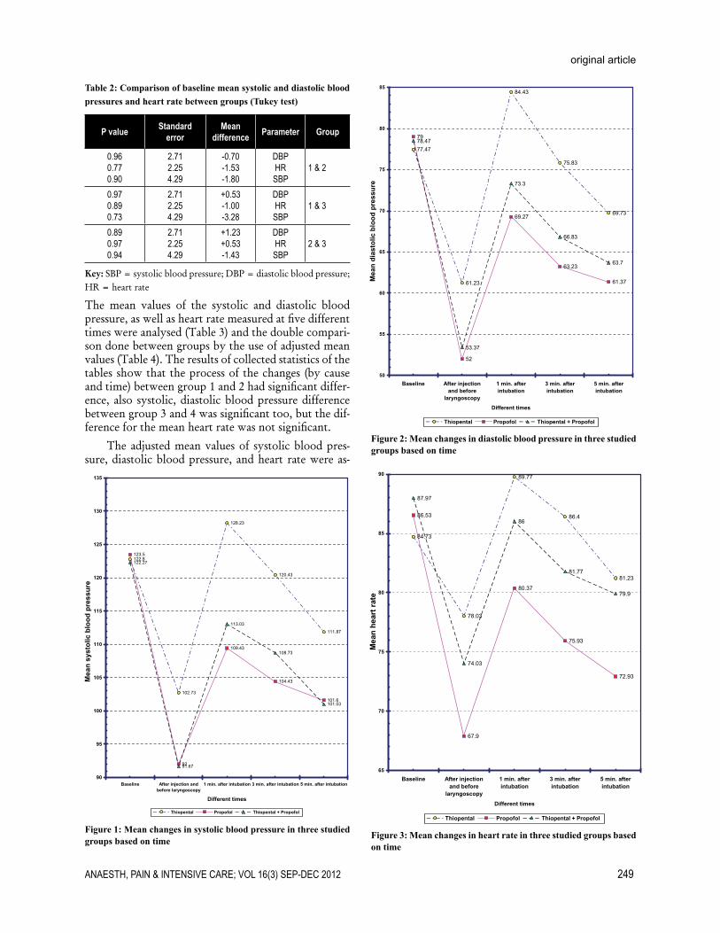
ANAESTH, PAIN & INTENSIVE CARE; VOL 16(3) SEP-DEC 2012 249
original article
Figure 1: Mean changes in systolic blood pressure in three studied groups based on time
Figure 2: Mean changes in diastolic blood pressure in three studied groups based on time
Figure 3: Mean changes in heart rate in three studied groups based on time
Table 2: Comparison of baseline mean systolic and diastolic blood pressures and heart rate between groups (Tukey test)
P value Standard error
Mean difference Parameter Group
0.96 0.770.90
2.712.254.29
-0.70-1.53-1.80
DBPHRSBP
1 & 2
0.970.890.73
2.712.254.29
+0.53-1.00-3.28
DBPHRSBP
1 & 3
0.890.970.94
2.712.254.29
+1.23+0.53-1.43
DBPHRSBP
2 & 3
Key: SBP = systolic blood pressure; DBP = diastolic blood pressure; HR = heart rate
The mean values of the systolic and diastolic blood pressure, as well as heart rate measured at five different times were analysed (Table 3) and the double compari-son done between groups by the use of adjusted mean values (Table 4). The results of collected statistics of the tables show that the process of the changes (by cause and time) between group 1 and 2 had significant differ-ence, also systolic, diastolic blood pressure difference between group 3 and 4 was significant too, but the dif-ference for the mean heart rate was not significant.
The adjusted mean values of systolic blood pres-sure, diastolic blood pressure, and heart rate were as-
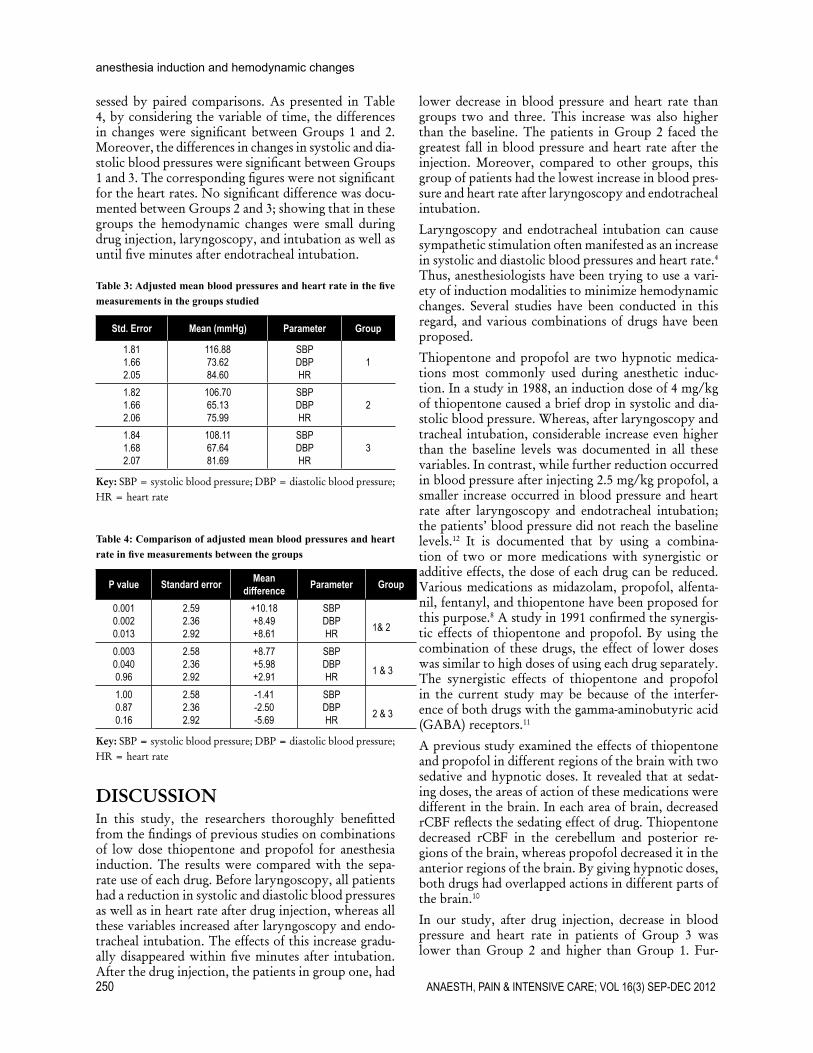
250 ANAESTH, PAIN & INTENSIVE CARE; VOL 16(3) SEP-DEC 2012
anesthesia induction and hemodynamic changes
sessed by paired comparisons. As presented in Table 4, by considering the variable of time, the differences in changes were significant between Groups 1 and 2. Moreover, the differences in changes in systolic and dia-stolic blood pressures were significant between Groups 1 and 3. The corresponding figures were not significant for the heart rates. No significant difference was docu-mented between Groups 2 and 3; showing that in these groups the hemodynamic changes were small during drug injection, laryngoscopy, and intubation as well as until five minutes after endotracheal intubation.
Table 3: Adjusted mean blood pressures and heart rate in the five measurements in the groups studied
Std. Error Mean (mmHg) Parameter Group
1.811.662.05
116.8873.6284.60
SBPDBPHR
1
1.821.662.06
106.7065.1375.99
SBPDBPHR
2
1.841.682.07
108.1167.6481.69
SBPDBPHR
3
Key: SBP = systolic blood pressure; DBP = diastolic blood pressure; HR = heart rate
Table 4: Comparison of adjusted mean blood pressures and heart rate in five measurements between the groups
P value Standard error Mean difference Parameter Group
0.0010.0020.013
2.592.362.92
+10.18+8.49+8.61
SBPDBPHR 1& 2
0.0030.0400.96
2.582.362.92
+8.77+5.98+2.91
SBPDBPHR 1 & 3
1.000.870.16
2.582.362.92
-1.41-2.50-5.69
SBPDBPHR 2 & 3
Key: SBP = systolic blood pressure; DBP = diastolic blood pressure; HR = heart rate
DISCUSSION In this study, the researchers thoroughly benefitted from the findings of previous studies on combinations of low dose thiopentone and propofol for anesthesia induction. The results were compared with the sepa-rate use of each drug. Before laryngoscopy, all patients had a reduction in systolic and diastolic blood pressures as well as in heart rate after drug injection, whereas all these variables increased after laryngoscopy and endo-tracheal intubation. The effects of this increase gradu-ally disappeared within five minutes after intubation. After the drug injection, the patients in group one, had
lower decrease in blood pressure and heart rate than groups two and three. This increase was also higher than the baseline. The patients in Group 2 faced the greatest fall in blood pressure and heart rate after the injection. Moreover, compared to other groups, this group of patients had the lowest increase in blood pres-sure and heart rate after laryngoscopy and endotracheal intubation.
Laryngoscopy and endotracheal intubation can cause sympathetic stimulation often manifested as an increase in systolic and diastolic blood pressures and heart rate.4 Thus, anesthesiologists have been trying to use a vari-ety of induction modalities to minimize hemodynamic changes. Several studies have been conducted in this regard, and various combinations of drugs have been proposed.
Thiopentone and propofol are two hypnotic medica-tions most commonly used during anesthetic induc-tion. In a study in 1988, an induction dose of 4 mg/kg of thiopentone caused a brief drop in systolic and dia-stolic blood pressure. Whereas, after laryngoscopy and tracheal intubation, considerable increase even higher than the baseline levels was documented in all these variables. In contrast, while further reduction occurred in blood pressure after injecting 2.5 mg/kg propofol, a smaller increase occurred in blood pressure and heart rate after laryngoscopy and endotracheal intubation; the patients’ blood pressure did not reach the baseline levels.12 It is documented that by using a combina-tion of two or more medications with synergistic or additive effects, the dose of each drug can be reduced. Various medications as midazolam, propofol, alfenta-nil, fentanyl, and thiopentone have been proposed for this purpose.8 A study in 1991 confirmed the synergis-tic effects of thiopentone and propofol. By using the combination of these drugs, the effect of lower doses was similar to high doses of using each drug separately. The synergistic effects of thiopentone and propofol in the current study may be because of the interfer-ence of both drugs with the gamma-aminobutyric acid (GABA) receptors.11
A previous study examined the effects of thiopentone and propofol in different regions of the brain with two sedative and hypnotic doses. It revealed that at sedat-ing doses, the areas of action of these medications were different in the brain. In each area of brain, decreased rCBF reflects the sedating effect of drug. Thiopentone decreased rCBF in the cerebellum and posterior re-gions of the brain, whereas propofol decreased it in the anterior regions of the brain. By giving hypnotic doses, both drugs had overlapped actions in different parts of the brain.10
In our study, after drug injection, decrease in blood pressure and heart rate in patients of Group 3 was lower than Group 2 and higher than Group 1. Fur-
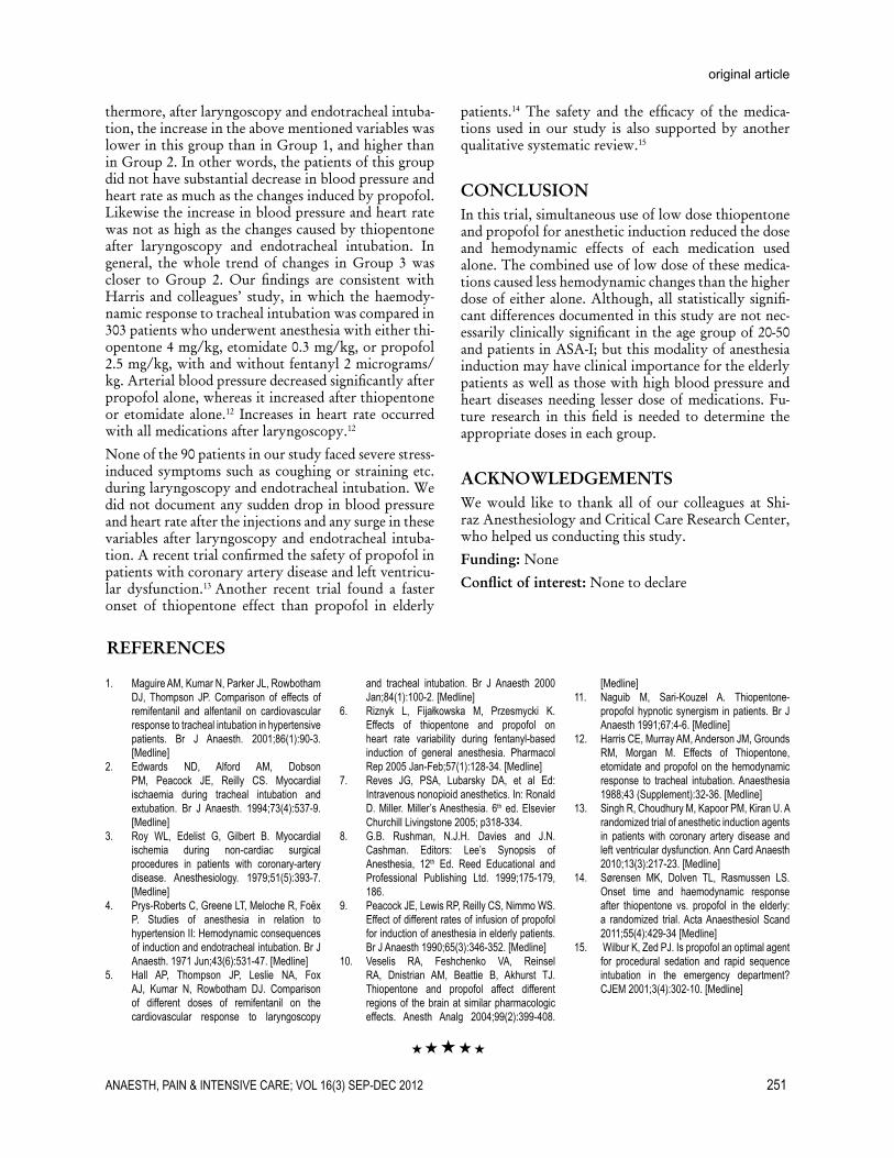
ANAESTH, PAIN & INTENSIVE CARE; VOL 16(3) SEP-DEC 2012 251
original article
thermore, after laryngoscopy and endotracheal intuba-tion, the increase in the above mentioned variables was lower in this group than in Group 1, and higher than in Group 2. In other words, the patients of this group did not have substantial decrease in blood pressure and heart rate as much as the changes induced by propofol. Likewise the increase in blood pressure and heart rate was not as high as the changes caused by thiopentone after laryngoscopy and endotracheal intubation. In general, the whole trend of changes in Group 3 was closer to Group 2. Our findings are consistent with Harris and colleagues’ study, in which the haemody-namic response to tracheal intubation was compared in 303 patients who underwent anesthesia with either thi-opentone 4 mg/kg, etomidate 0.3 mg/kg, or propofol 2.5 mg/kg, with and without fentanyl 2 micrograms/kg. Arterial blood pressure decreased significantly after propofol alone, whereas it increased after thiopentone or etomidate alone.12 Increases in heart rate occurred with all medications after laryngoscopy.12
None of the 90 patients in our study faced severe stress-induced symptoms such as coughing or straining etc. during laryngoscopy and endotracheal intubation. We did not document any sudden drop in blood pressure and heart rate after the injections and any surge in these variables after laryngoscopy and endotracheal intuba-tion. A recent trial confirmed the safety of propofol in patients with coronary artery disease and left ventricu-lar dysfunction.13 Another recent trial found a faster onset of thiopentone effect than propofol in elderly
patients.14 The safety and the efficacy of the medica-tions used in our study is also supported by another qualitative systematic review.15
CONCLUSIONIn this trial, simultaneous use of low dose thiopentone and propofol for anesthetic induction reduced the dose and hemodynamic effects of each medication used alone. The combined use of low dose of these medica-tions caused less hemodynamic changes than the higher dose of either alone. Although, all statistically signifi-cant differences documented in this study are not nec-essarily clinically significant in the age group of 20-50 and patients in ASA-I; but this modality of anesthesia induction may have clinical importance for the elderly patients as well as those with high blood pressure and heart diseases needing lesser dose of medications. Fu-ture research in this field is needed to determine the appropriate doses in each group.
ACKNOWLEDGEMENTSWe would like to thank all of our colleagues at Shi-raz Anesthesiology and Critical Care Research Center, who helped us conducting this study.
Funding: None
Conflict of interest: None to declare
1. Maguire AM, Kumar N, Parker JL, Rowbotham DJ, Thompson JP. Comparison of effects of remifentanil and alfentanil on cardiovascular response to tracheal intubation in hypertensive patients. Br J Anaesth. 2001;86(1):90-3. [Medline]
2. Edwards ND, Alford AM, Dobson PM, Peacock JE, Reilly CS. Myocardial ischaemia during tracheal intubation and extubation. Br J Anaesth. 1994;73(4):537-9. [Medline]
3. Roy WL, Edelist G, Gilbert B. Myocardial ischemia during non-cardiac surgical procedures in patients with coronary-artery disease. Anesthesiology. 1979;51(5):393-7. [Medline]
4. Prys-Roberts C, Greene LT, Meloche R, Foëx P. Studies of anesthesia in relation to hypertension II: Hemodynamic consequences of induction and endotracheal intubation. Br J Anaesth. 1971 Jun;43(6):531-47. [Medline]
5. Hall AP, Thompson JP, Leslie NA, Fox AJ, Kumar N, Rowbotham DJ. Comparison of different doses of remifentanil on the cardiovascular response to laryngoscopy
and tracheal intubation. Br J Anaesth 2000 Jan;84(1):100-2. [Medline]
6. Riznyk L, Fijałkowska M, Przesmycki K. Effects of thiopentone and propofol on heart rate variability during fentanyl-based induction of general anesthesia. Pharmacol Rep 2005 Jan-Feb;57(1):128-34. [Medline]
7. Reves JG, PSA, Lubarsky DA, et al Ed: Intravenous nonopioid anesthetics. In: Ronald D. Miller. Miller’s Anesthesia. 6th ed. Elsevier Churchill Livingstone 2005; p318-334.
8. G.B. Rushman, N.J.H. Davies and J.N. Cashman. Editors: Lee’s Synopsis of Anesthesia, 12th Ed. Reed Educational and Professional Publishing Ltd. 1999;175-179, 186.
9. Peacock JE, Lewis RP, Reilly CS, Nimmo WS. Effect of different rates of infusion of propofol for induction of anesthesia in elderly patients. Br J Anaesth 1990;65(3):346-352. [Medline]
10. Veselis RA, Feshchenko VA, Reinsel RA, Dnistrian AM, Beattie B, Akhurst TJ. Thiopentone and propofol affect different regions of the brain at similar pharmacologic effects. Anesth Analg 2004;99(2):399-408.
[Medline]11. Naguib M, Sari-Kouzel A. Thiopentone-
propofol hypnotic synergism in patients. Br J Anaesth 1991;67:4-6. [Medline]
12. Harris CE, Murray AM, Anderson JM, Grounds RM, Morgan M. Effects of Thiopentone, etomidate and propofol on the hemodynamic response to tracheal intubation. Anaesthesia 1988;43 (Supplement):32-36. [Medline]
13. Singh R, Choudhury M, Kapoor PM, Kiran U. A randomized trial of anesthetic induction agents in patients with coronary artery disease and left ventricular dysfunction. Ann Card Anaesth 2010;13(3):217-23. [Medline]
14. Sørensen MK, Dolven TL, Rasmussen LS. Onset time and haemodynamic response after thiopentone vs. propofol in the elderly: a randomized trial. Acta Anaesthesiol Scand 2011;55(4):429-34 [Medline]
15. Wilbur K, Zed PJ. Is propofol an optimal agent for procedural sedation and rapid sequence intubation in the emergency department? CJEM 2001;3(4):302-10. [Medline]
REFERENCES
252 ANAESTH, PAIN & INTENSIVE CARE; VOL 16(3) SEP-DEC 2012
ORIGINAL ARTICLE
A prospective study of factors predicting postoperative pulmonary complications (PPC) in patients undergoing non-cardiothoracic surgery under general anaesthsia in a developing country
Kaleem Ullah Toori, MRCP (UK), FRCP (Glasgow)*, Jahangir Sarwar Khan, CPS**, Ali Zohair Nomani***, Syed Waqar Hussain***, Saad Hashmi****
* Consultant Pulmonologist, *** PG Trainee, **** House officer, Department of Medicine, KRL General Hospiatl Islamabad (Pakistan)** Associate Professor, of Surgery, Consultant Surgeon, Holy Family Hospital Rawalpindi(Pakistan)
Correspondence: Dr. Kaleem Ullah Toori, Consultant Pulmonologist, KRL General Hospiatl Islamabad-44000 (Pakistan); Phone: +92-3215356040; E-mail: [email protected]
ABSTRACTIntroduction: Post-operative pulmonary complications after non-cardiothoracic surgery are common and can adversely affect morbidity, mortality and length of hospital stay. Knowledge as regards factors predicting post-operative pulmonary complications in our local setting is imperfect.
Aims & Objectives: To study factors predicting post-operative pulmonary complications in developing countries.
Methodology: Data of consecutive 404 patients undergoing non-cardiothoracic surgery under general anesthesia with tracheal intubation was collected prospectively from Jan 2009 to Dec 2010. Chi-square was used for univariate analysis. Multivariate analysis was conducted using forward stepwise logistic regression.
Results: The mean age was 36±18 years with slight male predominence (54% vs 46%). 22% (n=89) were smokers and the mean Body Mass Index was 23±4.5 kg/m2 with 35% (n=141) overweight & obese. 5% (n=20) of subjects had pre-existing chronic lung conditions while 23% (n=92) had non-pulmonary chronic conditions. 70% (n=282) of the surgeries were done electively and the mean duration of anesthesia was 78±44 minutes. The overall post-operative pulmonary complications rate was 8% (n=31) with atelactasis (48%, n=16) followed by bronchospasm (25%, n=8) and pneumonia (16%, n=5) being the commonest complications. The duration of hospital stay was significantly longer (11±9 days, p=0.00) in patients with post-operative pulmonary complications and 29% (n=9) of them required mechanical ventilation. Logistic Regression analysis identified premorbid chronic chest conditions, emergency surgery and prolonged duration of anesthesia as significant predictors of post-operative pulmonary complications while age, gender, Body Mass Index, smoking history and non-pulmonary premorbids were insignificant in this regard.
Conclusion: Post-operative pulmonary complications after non-cardiothoracic surgery are common and lead to increased morbidity and prolonged hospital stay in our setting. We identified pre-existing chest disease, prolonged anesthesia and emergency surgery as significant predictors of post-operative pulmonary complications.
Key words: Postoperative pulmonary complications; Non-cardiothoracic surgery; Body Mass Index; Emergency surgery; Prolonged anesthesia
Citation: Toori KU, Khan JS, Nomani Z, Hussain SW. A prospective study of factors predicting postoperative pulmonary complications (PPC) in patients undergoing non-cardiothoracic surgery under general anaesthsia in a developing country. Anaesth Pain & Intensive Care 2012;16(3):252-256
ANAESTH, PAIN & INTENSIVE CARE; VOL 16(3) SEP-DEC 2012 253
original article
INTRODUCTIONGuiding the patient safely and efficiently through the postoperative course has always been the primary goal and a shared effort of the health care professionals but unfortunately, postoperative pulmonary complications (PPC) frequently defeat this objective yet today.1
Clinically important PPC not only contributes to increased morbidity and mortality;1&2 they are a major factor in driving up total medical expenditures, especially in terms of utilization of intensive care facilities and duration of hospital stay.1,3
The current basis of our understanding of the nature of PPC is weak as only a small number of high-quality studies are available and there is lack of global consensus on a uniform definition of PPC. Also available evidence comprise of studies focusing on specific patients and kinds of surgeries.3 As a consequence, a wide range of incidence of PPC (2% – 40%) has been reported in the literature.4
Majority of investigators include postoperative pneumonia (definite or suspected), respiratory failure (usually defined as the respiratory compromise needing ventilatory support) and bronchospasm as PPC, but analysis of the literature shows that other complications such as unexplained fever, excessive bronchial secretions, productive cough, abnormal breath sounds, atelectasis or hypoxemia may also be included.3 Current evidence based studies have assessed the significance of risk factors like patient’s health status, type of anesthetic, surgical procedure, age, gender, BMI, history of smoking, length of anesthesia, chronic pulmonary disease, non-pulmonary chronic conditions, nutritional status, emergency surgery, intraoperative blood loss, fluid replacement and preoperative ambulatory status as the predictors of PPC.2,3,5 However, yet more control trials need to be done before a uniformity towards these predictors is being established.
The objective of this study is to define the factors predicting postoperative pulmonary complications in our setting and to underpin our understanding of PPC so that appropriate measures can be taken during peri-operative and post-operative periods for better patient handling and use of health care facilities.
METHODOLOGYThis prospective study was done at KRL General Hospital, Islamabad and Holy Family Hospital, Rawalpindi from Jan 2009 to Dec 2010. Data of consecutive 404 patients
undergoing non-cardiothoracic surgery under general anesthesia with tracheal intubation was collected prospectively on a standard performa.Each patient’s demographics along with BMI (Body Mass Index), smoking history, previous chronic pulmonary and non-pulmonary co-morbidities were recorded. Body Mass Index was calculated using standard definition with weight being measured by a standard weight machine in kilograms (Kg) and height with standard scale in meters (m). BMI was taken as the ratio of weight in Kg to square of height in meters. Chronic pulmonary conditions included diagnosed cases of asthma, chronic obstructive pulmonary disease (COPD), previously treated pulmonary tuberculosis and/ or interstitial lung disease (ILD) according to standard protocols. Non-pulmonary co-morbidities included diagnosed cases of Diabtes Mellitus, hypertension, ischemic heart disease, left ventricular failure and/ or chronic renal failure according to standard protocols. The type and duration of surgery, whether done electively or on emergency basis and details of post-operative course were also recorded. Same anesthetic conditions and drugs were used for anesthesia in the whole population group. Note was made of development of any pulmonary complication and whether it required ICU care ± mechanical ventilation.Pulmoary complications included atelectasis, bronchospasm, pneumonia, pleural effusion and Acute respiratory distress syndrome (ARDS). Atelectasis was defined as diminished volume of all or part of lung confirmed on xray chest or computed tomography by consultant radiologist. Bronchospasm was identified clinically and recorded after confirmation by consultant physician. Pneumonia and pleural effsusion were defined in terms of radiological findings confirmed on chest xray by the radiologist. ARDS was recorded on the basis of standard definitons. The total duration of hospital stay was also recorded.
Data were analysed using statistical software SPSS version 17 for Windows (SPSS Inc. Chicago, IL USA) and NCSS 2000 (NCSS, 329 North 1000 East, Kaysville, Utah, 84037). Discrete variables were listed as frequencies and percentages and continuous variables were listed as mean±SD. Chi-square was used for univariate analysis. Multivariate analysis was conducted using forward stepwise logistic regression.
RESULTSThe mean age of was 36 ± 18 years with slight male predominence (54%, n=218). 89 (22%) of the total
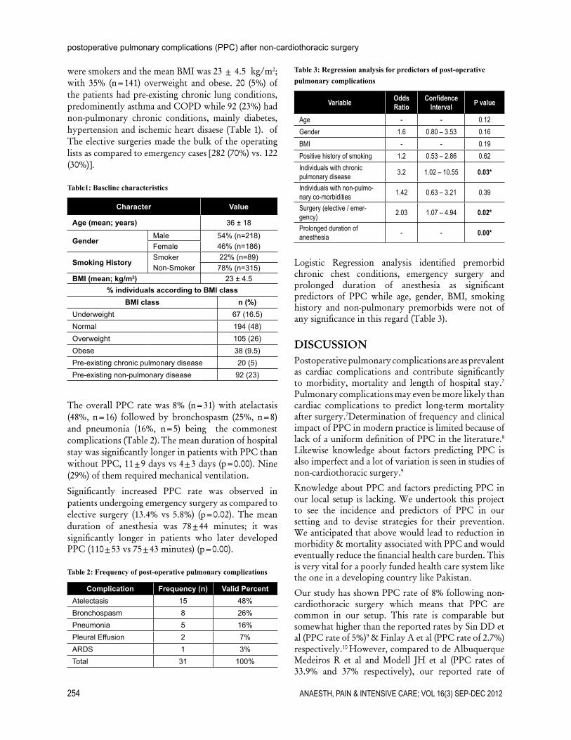
254 ANAESTH, PAIN & INTENSIVE CARE; VOL 16(3) SEP-DEC 2012
postoperative pulmonary complications (PPC) after non-cardiothoracic surgery
were smokers and the mean BMI was 23 ± 4.5 kg/m2; with 35% (n=141) overweight and obese. 20 (5%) of the patients had pre-existing chronic lung conditions, predominently asthma and COPD while 92 (23%) had non-pulmonary chronic conditions, mainly diabetes, hypertension and ischemic heart disaese (Table 1). of The elective surgeries made the bulk of the operating lists as compared to emergency cases [282 (70%) vs. 122 (30%)].
Table1: Baseline characteristics
Character Value
Age (mean; years) 36 ± 18
GenderMale 54% (n=218)Female 46% (n=186)
Smoking HistorySmoker 22% (n=89)Non-Smoker 78% (n=315)
BMI (mean; kg/m2) 23 ± 4.5% individuals according to BMI class
BMI class n (%)Underweight 67 (16.5)Normal 194 (48)Overweight 105 (26)Obese 38 (9.5)Pre-existing chronic pulmonary disease 20 (5)Pre-existing non-pulmonary disease 92 (23)
The overall PPC rate was 8% (n=31) with atelactasis (48%, n=16) followed by bronchospasm (25%, n=8) and pneumonia (16%, n=5) being the commonest complications (Table 2). The mean duration of hospital stay was significantly longer in patients with PPC than without PPC, 11±9 days vs 4±3 days (p=0.00). Nine (29%) of them required mechanical ventilation.
Significantly increased PPC rate was observed in patients undergoing emergency surgery as compared to elective surgery (13.4% vs 5.8%) (p=0.02). The mean duration of anesthesia was 78±44 minutes; it was significantly longer in patients who later developed PPC (110±53 vs 75±43 minutes) (p=0.00).
Table 2: Frequency of post-operative pulmonary complications
Complication Frequency (n) Valid PercentAtelectasis 15 48%Bronchospasm 8 26%Pneumonia 5 16%Pleural Effusion 2 7%ARDS 1 3%Total 31 100%
Table 3: Regression analysis for predictors of post-operative pulmonary complications
Variable Odds Ratio
Confidence Interval P value
Age - - 0.12Gender 1.6 0.80 – 3.53 0.16BMI - - 0.19Positive history of smoking 1.2 0.53 – 2.86 0.62Individuals with chronic pulmonary disease 3.2 1.02 – 10.55 0.03*
Individuals with non-pulmo-nary co-morbidities 1.42 0.63 – 3.21 0.39
Surgery (elective / emer-gency) 2.03 1.07 – 4.94 0.02*
Prolonged duration of anesthesia - - 0.00*
Logistic Regression analysis identified premorbid chronic chest conditions, emergency surgery and prolonged duration of anesthesia as significant predictors of PPC while age, gender, BMI, smoking history and non-pulmonary premorbids were not of any significance in this regard (Table 3).
DISCUSSIONPostoperative pulmonary complications are as prevalent as cardiac complications and contribute significantly to morbidity, mortality and length of hospital stay.7 Pulmonary complications may even be more likely than cardiac complications to predict long-term mortality after surgery.7Determination of frequency and clinical impact of PPC in modern practice is limited because of lack of a uniform definition of PPC in the literature.8 Likewise knowledge about factors predicting PPC is also imperfect and a lot of variation is seen in studies of non-cardiothoracic surgery.9
Knowledge about PPC and factors predicting PPC in our local setup is lacking. We undertook this project to see the incidence and predictors of PPC in our setting and to devise strategies for their prevention. We anticipated that above would lead to reduction in morbidity & mortality associated with PPC and would eventually reduce the financial health care burden. This is very vital for a poorly funded health care system like the one in a developing country like Pakistan.
Our study has shown PPC rate of 8% following non-cardiothoracic surgery which means that PPC are common in our setup. This rate is comparable but somewhat higher than the reported rates by Sin DD et al (PPC rate of 5%)9 & Finlay A et al (PPC rate of 2.7%) respectively.10 However, compared to de Albuquerque Medeiros R et al and Modell JH et al (PPC rates of 33.9% and 37% respectively), our reported rate of
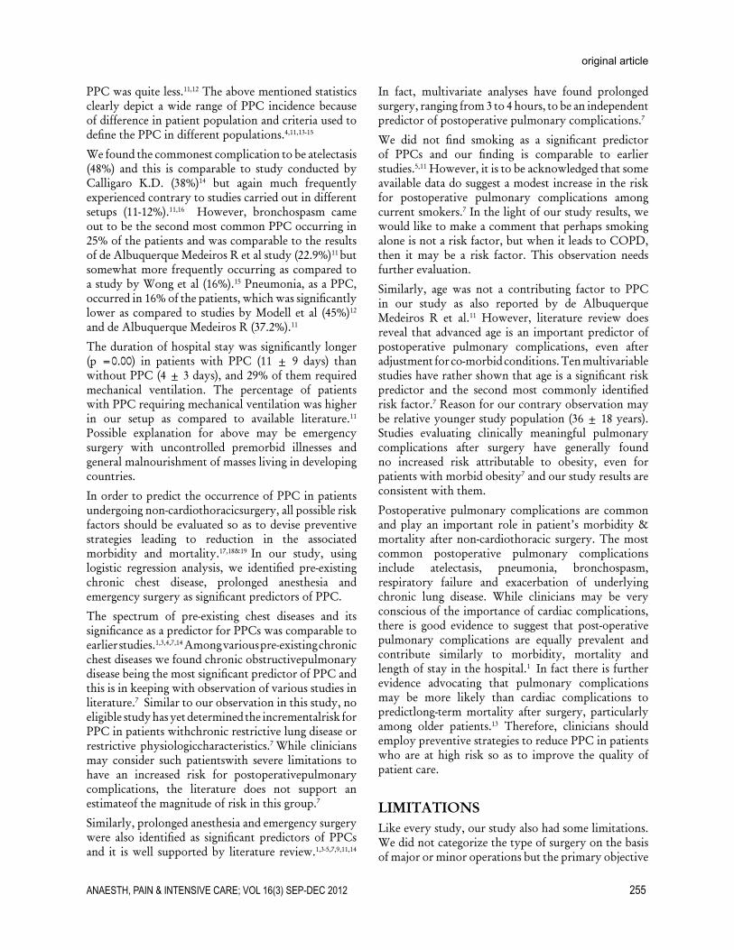
ANAESTH, PAIN & INTENSIVE CARE; VOL 16(3) SEP-DEC 2012 255
original article
PPC was quite less.11,12 The above mentioned statistics clearly depict a wide range of PPC incidence because of difference in patient population and criteria used to define the PPC in different populations.4,11,13-15
We found the commonest complication to be atelectasis (48%) and this is comparable to study conducted by Calligaro K.D. (38%)14 but again much frequently experienced contrary to studies carried out in different setups (11-12%).11,16 However, bronchospasm came out to be the second most common PPC occurring in 25% of the patients and was comparable to the results of de Albuquerque Medeiros R et al study (22.9%)11 but somewhat more frequently occurring as compared to a study by Wong et al (16%).15 Pneumonia, as a PPC, occurred in 16% of the patients, which was significantly lower as compared to studies by Modell et al (45%)12 and de Albuquerque Medeiros R (37.2%).11
The duration of hospital stay was significantly longer (p =0.00) in patients with PPC (11 ± 9 days) than without PPC (4 ± 3 days), and 29% of them required mechanical ventilation. The percentage of patients with PPC requiring mechanical ventilation was higher in our setup as compared to available literature.11
Possible explanation for above may be emergency surgery with uncontrolled premorbid illnesses and general malnourishment of masses living in developing countries.
In order to predict the occurrence of PPC in patients undergoing non-cardiothoracicsurgery, all possible risk factors should be evaluated so as to devise preventive strategies leading to reduction in the associated morbidity and mortality.17,18&19 In our study, using logistic regression analysis, we identified pre-existing chronic chest disease, prolonged anesthesia and emergency surgery as significant predictors of PPC.
The spectrum of pre-existing chest diseases and its significance as a predictor for PPCs was comparable to earlier studies.1,3,4,7,14 Among various pre-existing chronic chest diseases we found chronic obstructivepulmonary disease being the most significant predictor of PPC and this is in keeping with observation of various studies in literature.7 Similar to our observation in this study, no eligible study has yet determined the incrementalrisk for PPC in patients withchronic restrictive lung disease or restrictive physiologiccharacteristics.7 While clinicians may consider such patientswith severe limitations to have an increased risk for postoperativepulmonary complications, the literature does not support an estimateof the magnitude of risk in this group.7
Similarly, prolonged anesthesia and emergency surgery were also identified as significant predictors of PPCs and it is well supported by literature review.1,3-5,7,9,11,14
In fact, multivariate analyses have found prolonged surgery, ranging from 3 to 4 hours, to be an independent predictor of postoperative pulmonary complications.7
We did not find smoking as a significant predictor of PPCs and our finding is comparable to earlier studies.5,11 However, it is to be acknowledged that some available data do suggest a modest increase in the risk for postoperative pulmonary complications among current smokers.7 In the light of our study results, we would like to make a comment that perhaps smoking alone is not a risk factor, but when it leads to COPD, then it may be a risk factor. This observation needs further evaluation.
Similarly, age was not a contributing factor to PPC in our study as also reported by de Albuquerque Medeiros R et al.11 However, literature review does reveal that advanced age is an important predictor of postoperative pulmonary complications, even after adjustment for co-morbid conditions. Ten multivariable studies have rather shown that age is a significant risk predictor and the second most commonly identified risk factor.7 Reason for our contrary observation may be relative younger study population (36 ± 18 years). Studies evaluating clinically meaningful pulmonary complications after surgery have generally found no increased risk attributable to obesity, even for patients with morbid obesity7 and our study results are consistent with them.
Postoperative pulmonary complications are common and play an important role in patient’s morbidity & mortality after non-cardiothoracic surgery. The most common postoperative pulmonary complications include atelectasis, pneumonia, bronchospasm, respiratory failure and exacerbation of underlying chronic lung disease. While clinicians may be very conscious of the importance of cardiac complications, there is good evidence to suggest that post-operative pulmonary complications are equally prevalent and contribute similarly to morbidity, mortality and length of stay in the hospital.1 In fact there is further evidence advocating that pulmonary complications may be more likely than cardiac complications to predictlong-term mortality after surgery, particularly among older patients.13 Therefore, clinicians should employ preventive strategies to reduce PPC in patients who are at high risk so as to improve the quality of patient care.
LIMITATIONSLike every study, our study also had some limitations. We did not categorize the type of surgery on the basis of major or minor operations but the primary objective
256 ANAESTH, PAIN & INTENSIVE CARE; VOL 16(3) SEP-DEC 2012
postoperative pulmonary complications (PPC) after non-cardiothoracic surgery
of the study was to identify modifiable risk factors predictive of PPC for any type of surgery and the study of difference between minor and major surgeries is itself a widely debatable topic. Furthermore, it is pertinent to mention that all the surgeries were almost equally divided between two major surgeons of equal professional expertise, yet the factor of variability of expertise between the two cannot be denied completely.
CONCLUSIONPost-operative pulmonary complications after non-cardiothoracic surgery are common and lead to increased morbidity and prolonged hospital stay in our setting. We identified pre-existing chest disease, prolonged anesthesia and emergency surgery as significant predictors of post-operative pulmonary complications.
1. Braveman J. Post-operative pulmonary complications in the adult patient-causes, effects and therapeutic strategies; Business Briefing; Global Surgery 2004. Available at http://www.touchbriefings.com/pdf/952/braverman_edit[1].pdf. Accessed 10 December 2012).
2. Lawrence VA, Hilsenbeck SG, Mulrow CD, et al. Incidence and hospital stay for cardiac and pulmonary complications after abdominal surgery. J Gen Intern Med. 1995;10:671. [Medline]
3. Canet J, Mazo V. Postoperative pulmonary complications. Minerva Anestesiol 2010;76(2);138-43. [Medline]
4. Rock P, Rich PB. Postoperative pulmonary complications. Curr Opin Anaesthesiol. 2003;16(2):123-31. [Medline]
5. Graybill WS, Frumovitz M, Nick AM, Wei C, Mena GE, Soliman PT, et al. Impact of smoking on perioperative pulmonary and upper respiratory complications after laparoscopic gynecologic surgery. Gynecol Oncol. 2012;125(3):556-60. Epub 2012 Mar 17. [Medline]
6. Canet J, Gallart L, Gomar C, Paluzie G, Vallès J, Castillo J;ARISCAT Group, et al. Prediction of Postoperative Pulmonary Complications in a Population-based Surgical Cohort; on behalf of the ARISCAT Group. Anesthesiology. 2010;113(6):1338-1350. [Medline]
7. Qaseem A, Snow V, Fitterman N, Hornbake ER, Lawrence VA, Smetana GW, et al. Risk Assessment for and Strategies To Reduce Perioperative Pulmonary Complications for Patients Undergoing Noncardiothoracic
Surgery: A Guideline from the American College of Physicians. Ann Intern Med. 2006;144(8):575-80. [Medline]
8. Warner DO. Preventing postoperative pulmonary complications; The role of the Anesthesiologist. Anesthesiology. 2000;92(5):1467-72. [Medline]
9. Lawrence VA, Cornell JE, Smetana GW. Strategies to reduce postoperative pulmonary complications after noncardiothoracic surgery: systematic review for the American College of Physicians. Ann Intern Med. 2006;144(8):596-608. [Medline]
10. McAlister FA,Bertsch K, Man J, Bradley J, Jack M. Incidence of and Risk Factors for Pulmonary Complications after Nonthoracic Surgery. Am J Respir Crit Care Med. 2005;171(5):514-517 [Medline]
11. de Albuquerque Medeiros R, Faresin S, Jardim J. Postoperative lung complications and mortality in patients with mild-to-moderate COPD undergoing elective general surgery. Arch Bronconeumol. 2001;37(5):227-34. [Medline]
12. Modell JH, Moya F. Postoperative pulmonary complications. Incidence and management. Anesth Analg 1966;45(4):432-9. Available at http://www.anesthesia-analgesia.org/content/45/4/432.full.pdf+html?sid= 4e5d4e9a-3f33-48ae-aadb-17266ce36721 (Accessed on 10 December 2012)
13. Fisher BW, Majumdar SR, McAlister FA. Predicting pulmonary complications after nonthoracic surgery: a systematic review of blinded studies. Am J Med. 2002;112(3):219-25. [Medline]
14. Calligaro KD, Azurin DJ, Dougherty MJ, Dandora R, Bajgier SM, Simper S, et al. Pulmonary risk factors of elective abdominal aortic surgery. J Vasc Surg. 1993;18(6):914-20.
15. Wong DH, Weber EC, Schell MJ, Wong AB, Anderson CT, Barker SJ. Factors Associated with Postoperative Pulmonary Complications in Patients with Severe Chronic Obstructive Pulmonary Disease. Anesth Analg. 1995;80:276-84. [Medline]
16. Reinius H, Jonsson L, Gustafsson S, Sundbom M, Duvernoy O, Pelosi P, et al. Prevention of atelectasis in morbidly obese patients during general anesthesia and paralysis: a computerized tomography study. Anesthesiology. 2009;111(5):979-87. [Medline]
17. Arozullah AM, Khuri SF, Henderson WG, Daley J. Participants in the National Veterans Affairs Surgical Quality Improvement Program. Development and validation of a multifactorial risk index for predicting postoperative pneumonia after major noncardiac surgery. Ann Intern Med. 2001;135(10):847-57. [Medline]
18. Roukema JA, Carol EJ, Prins JG. The prevention of pulmonary complications after upper abdominal surgery in patients with noncompromised pulmonary status. Arch Surg. 1988;123:30-34. [Medline]
19. Arozullah AM, Daley J, Henderson WG, Khuri SF. Multifactorial risk index for predicting postoperative respiratory failure in men after major noncardiac surgery. The National Veterans Administration Surgical Quality Improvement Program. Ann Surg. 2000;232(2):242-53. [Medline]
REFERENCES
Acknowledgements: The authors fully acknowledge the efforts of the accompanying medical and paramedical staff at KRL Hospital and Holy Family Hospital for helping them out at every step in the process of completion of this project.
Disclosure: All the authors declare that they do not have any conflict of interest regarding this work.
Research Funding: No grant or fund has been provided for the purpose of this study & the expenses whatsoever for this project have been shared solely by the authors themselves & no one else.
Author contributions: Toori KU, Khan JS, Nomani AZ and Hussain SW designed the study, did data collection & co-ordinated the laboratory work. Toori KU &Khan JS did computer entry. Nomani AZ and Hussain SW did data analysis & wrote manuscript. Toori KU& Nomani AZ revised the manuscript & planned the study.

ANAESTH, PAIN & INTENSIVE CARE; VOL 16(3) SEP-DEC 2012 257
ORIGINAL ARTICLE
The effect of pregabalin and gabapentin on preoperative anxiety and sedation: a double blind study Anju Ghai, MD*, Monika Gupta, MD**, Neha Rana, MD***, Raman Wadhera, MS****
*Professor, ** Senior Resident, ***Assistant ProfessorDepartment of Anaesthesiology ****Professor, Department of ENT Pt B.D.Sharma Postgraduate Institute of Medical Sciences, Rohtak, Haryana (India)
Correspondence: Dr Anju Ghai, 19/9J, Medical Campus, PGIMS, Rohtak, Haryana (India); Phone: +919416974794; E-mail: [email protected]; [email protected]
ABSTRACTBackground: Pregabalin and gabapentin are compounds, which have been alleged to possess anxiolytic, analgesic, and anticonvulsant properties. Both are amino acid derivatives of gamma amino butyric acid. Pregabalin has a similar pharmacological profile to that of gabapentin. It has an amino acid substitution at third position which allows better lipid solubility and diffusion across blood brain barrier, better pharmacokinetic properties and fewer drug interactions due to absence of hepatic metabolism. We hypothesized that premedication with oral pregabalin and gabapentin would produce dose-related reductions in acute (state) anxiety and increase in sedation (sleepiness) before induction of general anaesthesia.
Methodology: 90 women were randomly assigned to receive 300 mg pregabalin and 900 mg gabapentin and placebo 60 minutes prior to surgery. Anxiety and sedation was assessed before administration of drug and 1 hour later. A uniform anaesthetic technique was used in all groups. Parameters including sedation scores and various side effects were assessed.
Results: Demographic variables were comparable. The preinduction anxiety scores were statistically significant from the baseline values in group 1 and 11. The sedation scores were statistically significant 1 hour after the drug. There was statistically significant difference between group I and II (p=0.000), I and III (p=0.000) and II and III(p=0.015). Analysis of sedation scores after surgery were comparable at all time intervals between group I and II. However statistically significant difference was noted between group I and III (p=0.000) and group II and III (p=0.000). A higher percentage of patients in the pregabalin group complained of dizziness and somnolence than the gabapentin and control group.
Conclusion: Preoperative pregabalin (300mg) and gabapentin (900mg) administration 1 hour before surgery led to significant reduction in preoperative anxiety and improves sedation without producing significant side effects.
Keywords: Gabapentin, pregabalin, sedation, anxiety
Citation: Ghai A, Gupta M, Rana N, Wadhera R. The effect of pregabalin and gabapentin on preoperative anxi-ety and sedation: a double blind study. Anaesth Pain & Intensive Care 2012;16(3):257-261
INTRODUCTION Preoperative anxiety remains a problem for many patients during the perioperative period. High anxiety level make the control of postoperative pain more
difficult.1 Although benzodiazepines are effective in reducing perioperative anxiety, but the anxiolytic effect is frequently accompanied by undesirable sedation. GABA analogues are structural analogues
258 ANAESTH, PAIN & INTENSIVE CARE; VOL 16(3) SEP-DEC 2012
the effect of pregabalin and gabapentin
of the inhibitory neurotransmitter gamma amino butyric acid(GABA) and bind in a state dependent manner to the alpha-2 delta subunit of voltage gated Ca channels in over excited presynaptic neurons reducing release of excitatory neurotransmitters, leading to a reduction in levels of anxiety and pain.2 Pregabalin has an amino acid substitution at third position which allows better lipid solubility and diffusion across blood brain barrier, better pharmacokinetic properties and fewer drug interactions. A study in patients with generalized anxiety disorders found that chronic use of pregabalin and gabapentin was significantly more effective than benzodiazepine in improving somatic anxiety symptoms, but its role in acute anxiety needs to be yet evaluated.3,4 Premedication with gabapentin 1200 mg improved preoperative anxiolysis in patients undergoing lumbar discectomy and arthroscopic anterior cruciate ligament repair, where it exerted anxiolysis without exerting anamnesis effects,5,6 though administration of gabapentin 600 mg did not have this effect in patients undergoing total hip arthroplasty.7
Preoperative pregabalin administration (75–300 mg po) increased perioperative sedation in a dose-related fashion, but failed to reduce preoperative anxiety after minor elective procedures.8 The studies have not reached any conclusion due to conflicting results. There has been no study in literature comparing the effect of these two drugs on preoperative anxiety and sedation.
Hence, we planned to compare the effect of pregabalin and gabapentin on preoperative anxiety and sedation. The primary objective of this study was a decrease in the level of acute “state” anxiety before induction of general anaesthesia. The secondary objective was to determine its effect on postoperative sedation and the side effect.
METHODOLOGY With approval of the ethics committee and written informed consent, 90 women belonging to ASA status I-II, aged 35 to 65 years, scheduled for elective abdominal hysterectomy undergoing general anaesthesia were included in the study. Although the wide age range, may affect the level of anxiety in patients, but it was chosen to include all the subjects in that period as the study was time bound. Patients with history of central nervous system disorders, chronic pain, use of regular analgesics, known hypersensitivity to gabapentin or pregabalin, impaired renal function and those with body weight >20% of ideal body weight were excluded from the study.
All patients were visited on the day before surgery. The general physical examination was carried out. Routine
investigations were noted. A linear 0-10cm visual analogue scale (VAS) for anxiety (where 0 denoted no anxiety and 10 was for worst imaginable anxiety) was explained to each patient. The computer generated block randomization schedule was prepared using random number generator to create a list of random numbers by our statistician and was handed over to the hospital pharmacist. To ensure the equal number of patients in each group, block randomization was done. Stat Trek® (stattrek.com) program was used to derive the randomization list. The patients were assigned to one of the following groups.
Group I (n=30) received pregabalin capsules 300mg Group II (n=30) received gabapentin capsules 900mgGroup III (n=30) received placebo capsules
Sample size was decided in consultation with our statistician. The primary end point for this study was a reduction in the patient’s preoperative level of anxiety as assessed using the VAS. Based on a predicted 20% reduction from the patient’s pretreatment (baseline) VAS anxiety score (mean value of 5 and SD of 3), a minimum of 25 subjects were required in each of the three study groups under the assumptions of an ″ level of 0.05 and power of 80%. The study medication was prepared by the hospital pharmacy in identical appearing capsules and was put in 90 numbered envelopes containing 3 capsules each to maintain blinding. The capsules were kept with the pharmacy only and were taken from them when required. The patients, clinical investigators, attending anesthesiologists and nurses in the recovery room who were involved in the patients’ care were all blinded to the content of the study medication. The patients received the study medication 60 minutes before induction of general anaesthesia. No sedative premedication other than the gabapentinoid was given. Anxiety and sedation levels were assessed before administration of drug and 60 minutes later.
Sedation was observed and scored as follows: 0=Alert, conversant, 1=Awake but drowsy, 2=Asleep but arousable, 3=Asleep and not arousable.
On arrival in the operating room, intravenous line was secured with intravenous cannula of appropriate size. Monitoring of non invasive blood pressure (NIBP), heart rate, electrocardiogram and arterial oxygen saturation (SpO2) was carried out and the basal readings were noted. A uniform anaesthetic technique was used in all groups. After preoxygenation for 3 minutes, anaesthesia was induced with propofol 2 mg/kg and intubation of trachea was facilitated with vecuronium bromide 0.1 mg/kg. Haemodynamic parameters were recorded just before injecting propofol, just before tracheal intubation, immediately after tracheal intubation and after 1, 3, 5 and 10 minutes of intubation.
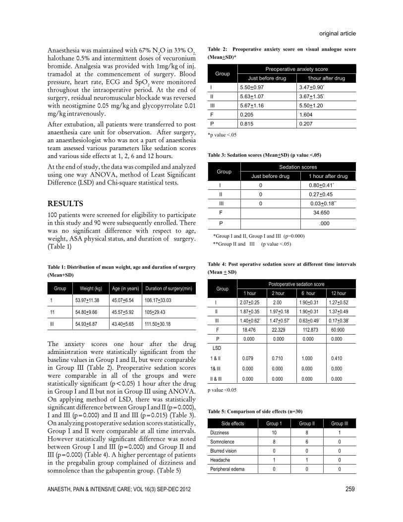
ANAESTH, PAIN & INTENSIVE CARE; VOL 16(3) SEP-DEC 2012 259
original article
Anaesthesia was maintained with 67% N2O in 33% O2, halothane 0.5% and intermittent doses of vecuronium bromide. Analgesia was provided with 1mg/kg of inj. tramadol at the commencement of surgery. Blood pressure, heart rate, ECG and SpO2 were monitored throughout the intraoperative period. At the end of surgery, residual neuromuscular blockade was reversed with neostigmine 0.05 mg/kg and glycopyrrolate 0.01 mg/kg intravenously.
After extubation, all patients were transferred to post anaesthesia care unit for observation. After surgery, an anaesthesiologist who was not a part of anaesthesia team assessed various parameters like sedation scores and various side effects at 1, 2, 6 and 12 hours.
At the end of study, the data was compiled and analyzed using one way ANOVA, method of Least Significant Difference (LSD) and Chi-square statistical tests.
RESULTS100 patients were screened for eligibility to participate in this study and 90 were subsequently enrolled. There was no significant difference with respect to age, weight, ASA physical status, and duration of surgery. (Table 1)
Table 1: Distribution of mean weight, age and duration of surgery (Mean+SD)
Group Weight (kg) Age (in years) Duration of surgery(min)
1 53.97+11.38 45.07+6.54 106.17+33.03
11 54.80+9.66 45.57+5.92 105+29.43
III 54.93+6.87 43.40+5.65 111.50+30.18
The anxiety scores one hour after the drug administration were statistically significant from the baseline values in Group I and II, but were comparable in Group III (Table 2). Preoperative sedation scores were comparable in all of the groups and were statistically significant (p<0.05) 1 hour after the drug in Group I and II but not in Group III using ANOVA. On applying method of LSD, there was statistically significant difference between Group I and II (p=0.000), I and III (p=0.000) and II and III (p=0.015) (Table 3). On analyzing postoperative sedation scores statistically, Group I and II were comparable at all time intervals. However statistically significant difference was noted between Group I and III (p=0.000) and Group II and III (p=0.000) (Table 4). A higher percentage of patients in the pregabalin group complained of dizziness and somnolence than the gabapentin group. (Table 5)
Table 2: Preoperative anxiety score on visual analogue score (Mean+SD)*
GroupPreoperative anxiety score
Just before drug 1hour after drug
I 5.50+0.97 3.47+0.90*
II 5.63+1.07 3.67+1.35*
III 5.67+1.16 5.50+1.20
F 0.205 1.604
P 0.815 0.207
*p value <.05
Table 3: Sedation scores (Mean+SD) (p value <.05)
GroupSedation scores
Just before drug 1 hour after drug
I 0 0.80+0.41*
II 0 0.27+0.45
III 0 0.03+0.18**
F 34.650
P .000
*Group I and II, Group I and III (p=0.000) **Group II and III (p value <.05)
Table 4: Post operative sedation score at different time intervals (Mean + SD)
GroupPostoperative sedation score
1 hour 2 hour 6 hour 12 hour
I 2.07+0.25 2.00 1.90+0.31 1.27+0.52 II 1.87+0.35 1.97+0.18 1.90+0.31 1.37+0.49 III 1.40+0.62* 1.47+0.57* 0.63+0.49* 0.17+0.38*
F 18.476 22.329 112.873 60.900 P 0.000 0.000 0.000 0.000 LSD
1 & II
1& III
II & III
0.079
0.000
0.000
0.710
0.000
0.000
1.000
0.000
0.000
0.410
0.000
0.000
p value <0.05
Table 5: Comparison of side effects (n=30)
Side effects Group 1 Group II Group IIIDizziness 10 8 1Somnolence 8 6 0Blurred vision 0 0 0Headache 1 1 0Peripheral edema 0 0 0
260 ANAESTH, PAIN & INTENSIVE CARE; VOL 16(3) SEP-DEC 2012
the effect of pregabalin and gabapentin
DISCUSSION Patients routinely receive sedatives for reduction of anxiety before surgery at most of the centres. A previous study concluded that benzodiazepines administered before surgery have minimal beneficial effects on the postoperative clinical course of women undergoing abdominal hysterectomy.9
Gabapentin has been reported as an anxiolytic drug in previous studies.3,10,11 For example, it was effective in treating anxiety associated with panic disorders.3,11,12 Recently, de-Paris et al. demonstrated that gabapentin attenuated anxiety associated ‘with simulated public speaking’ in volunteers.12 This disorder may be related to the preoperative anxiety state. The interest in using gabapentin preoperatively to decrease preoperative anxiety is a result of its limited side effects. Moreover, gabapentin seems anxiolytic without exerting amnesic effects.13 Reducing preoperative anxiety with gabapentin may have contributed to the improved postoperative pain and to the reduced morphine use because there is a possible association between preoperative anxiety and postoperative pain .14,15
The efficacy of 300 mg pregabalin has been well proven in previous trials and it has proven efficacious at doses 2-4 fold lower than that of gabapentin.16 Hence, an equipotent dose of gabapentin 900 mg (3 fold higher) was chosen. Our study suggests that a single dose of gabapentin 900 mg and pregabalin 300 mg administered 60 minutes prior to surgery was effective in reducing acute preoperative (state) anxiety and increased levels of sedation before and after surgery. Pregabalin 300 mg is a better anxiolytic and sedative than gabapentin which could be explained due to better alpha 2-delta
ligand binding property.17 Postoperative sedation was comparable with both GABA analogues which can be due to short half life of pregabalin (5-6 hours).18 Dose of 1200 mg of gabapentin given 1-2 hours before surgery has been found to decrease anxiety and postoperative pain after knee surgery.6 But preoperative sedation scores were not assessed in that study. Our results with gabapentin are similar to this study, but at lower dose.
The anxiety findings observed with pregabalin in our study is in contrast while sedation scores are comparable to the findings by White et al, where they studied 75, 150, 300 mg of pregabalin and compared it with placebo. Anxiety levels remained unchanged in their study during the preoperative evaluation period and did not differ among all the groups, though sedation scores were higher with pregabalin 300 mg group at the preinduction assessment and 90 and 120 minutes after surgery compared with 75 mg , 150 mg and placebo.8
Nutt et al evaluated acute onset of anxiolytic activity using a dental anxiety model in 89 patients using 150 mg pregabalin, alprazolam 0.5 mg and a placebo 4 hours before dental procedure and found significant improvement in anxiety and sedation with pregabalin and alprazolam compared with placebo. They suggested that onset of anxiolytic effect after single dose pregabalin occurs within first 3-4 hours.19
CONCLUSIONIn summary, our results indicate that premedication with 900 mg oral gabapentin and 300 mg pregabalin decreases preoperative anxiety, and improves sedation without producing significant side effects.
ANAESTH, PAIN & INTENSIVE CARE; VOL 16(3) SEP-DEC 2012 261
original article
1. Caumo W, Levandovski R, Hidalgo MP. Preoperative anxiolytic effect of melatonin and clonidine on postoperative pain and morphine consumption in patients undergo-ing abdominal hysterectomy: a double-blind, randomized placebo-controlled study. J Pain. 2009 Jan;10(1):100-8. [Medline]
2. Rose MA, Kam PC. Gabapentin: pharmacol-ogy and its use in pain management. Anaes-thesia. 2002;57:451-62 [Medline]
3. Pollack MH, Matthews J, Scott EL. Gabapen-tin as a potential treatment for anxiety disor-ders. Am J Psychiatry. 1998 Jul;155(7):992-3. [Medline]
4. Baldwin DS, Ajel K. Role of pregaba-lin in the treatment of generalized an-xiety disorder. Neuropsychiatr Dis Tre-at. 2007 Apr;3(2):185-91. [Medline]
5. Kiskira O, Kolotoura A, Tsitsopoulos P; karmiri, E; Emexidis Th.; Pouliou, A. The effect of Gabapentin on preoperative anxiety in patients undergoing lumbar discectomy: A-557 European Journal of Anaesthesiology. 2006;23:145
6. Ménigaux C, Adam F, Guignard B, Sessler DI, Chauvin M. Preoperative Gabapentin decreases anxiety and improves early func-tional recovery from knee surgery. Anesth Analg. 2005 May;100(5):1394-9 [Medline]
7. Clarke H, Kay J, Orser BA, Gollish J, Mitsakakis N, Katz J. Gabapentin does not reduce preoperative anxiety when
given prior to total hip arthroplasty. Pain Med. 2010 Jun;11(6):966-71. doi: 10.1111/j.1526-4637.2010.00826.x. [Medline]
8. White PF, Tufanogullari B, Taylor J, Klein K. The effect of Pregabalin on preoperative anxiety and sedation levels: A dose ranging study. Anesth Analg. 2009 Apr;108(4):1140-5. doi: 10.1213/ane.0b013e31818d40ce [Med-line]
9. Kain ZN, Sevarino FB, Rinder C, Pincus S, Alexander GM, Ivy M, Heninger G. Preopera-tive anxiolysis and postoperative recovery in women undergoing abdominal hysterectomy. Anesthesiology. 2001 Mar;94(3):415-22. [Medline]
10. Chouinard G, Beauclair L, Belanger MC. Gabapentin: long-term antianxiety and hypnotic effects in psychiatric patients with comorbid anxiety-related disorders. Can J Psychiatry. 1998 Apr;43(3):305. [Medline]
11. Pande AC, Pollack MH, Crockatt J, Greiner M, Chouinard G, Lydiard RB, et al. Placebo-controlled study of gabapentin treatment of panic disorder. J Clin Psychopharma-col. 2000 Aug;20(4):467-71. [Medline]
12. de-Paris F, Sant’Anna MK, Vianna MR, Barichello T, Busnello JV, Kapczinski F, et al. Effects of gabapentin on anxiety in-duced by simulated public speaking. J Psy-chopharmacol. 2003 Jun;17(2):184-8. [Med-line]
13. de-Paris F, Busnello JV, Vianna MR, Sal-
gueiro JB, Quevedo J, Izquierdo I, et al. The anticonvulsant compound gabapentin pos-sesses anxiolytic but not amnesic effects in rats. Behav Pharmacol. 2000 Apr;11(2):169-73. [Medline]
14. Lynch EP, Lazor MA, Gellis JE, Orav J, Gold-man L, Marcantonio ER. Patient experience of pain after elective noncardiac surgery. Anesth Analg. 1997 Jul;85(1):117-23. [Med-line]
15. Kalkman CJ, Visser K, Moen J, Bonsel GJ, Grobbee DE, Moons KG. Preoperative prediction of severe postoperative pain. Pain. 2003 Oct;105(3):415-23. [Medline]
16. Laurea-Horner BA, Pohl RB. Pregaba-lin: a new anxiolytic. Expert Opin Investig Drugs. 2003 Apr;12(4):663-72. [Medline]
17. Jones D, Sorkin L. Systemic gabapentin and S(+)-3-isobutyl gamma- aminobutyuric acid on the secondary hyperalgesia. Brain Res. 1998 Nov 9;810(1-2):93-9. [Medline]
18. Kavoussi R. Pregabalin: from molecule to medicine. Eur Neuropsychopharmacol. 2006 Jul;16 Suppl 2:S128-33. [Medline]
19. Nutt D, Mandel F, Baldinetti F. Early onset anxiolytic efficacy after a single dose of pregabalin: double-blind, placebo- and active-comparator controlled evaluation using a dental anxiety model. J Psychop-harmacol. 2009 Nov;23(8):867-73. doi: 10.1177/0269881108094722. [Medline]
REFERENCES
Pearls• Prayer is not a “spare wheel” that you pull out when in trouble, but it is a “steering wheel” that directs the
right path throughout the journey.• So why is a Car’s WINDSHIELD so large & the Rear View Mirror so small? Because our PAST is not as
important as our FUTURE. So, Look Ahead and Move on.• Friendship is like a BOOK. It takes a few minutes to burn, but it takes years to write.• All things in life are temporary. If going well, enjoy it, they will not last forever. If going wrong, don’t
worry, they can’t last long either.• Old Friends are Gold! New Friends are Diamond! If you get a Diamond, don’t forget the Gold! Because to
hold a Diamond, you always need a Base of Gold!• Often when we lose hope and think this is the end, GOD smiles from above and says, “Relax, sweetheart,
it’s just a bend, not the end!• When GOD solves your problems, you have faith in HIS abilities; when GOD doesn’t solve your prob-
lems HE has faith in your abilities.• A blind person asked St. Anthony: “Can there be anything worse than losing eye sight?” He replied: “Yes,
losing your vision!”• When you pray for others, God listens to you and blesses them, and sometimes, when you are safe and
happy, remember that someone has prayed for you.• WORRYING does not take away tomorrow’s TROUBLES, it takes away today’s PEACE.• Live simply. Love generously. Care deeply. Speak kindly. Leave the rest to God.
262 ANAESTH, PAIN & INTENSIVE CARE; VOL 16(3) SEP-DEC 2012
ORIGINAL ARTICLE
The incidence of postoperative delirium in elderly patients undergoing urologic surgery Haxhire Gani, Pirro Prifti, Majlinda Naco, Rudin Domi, Vjollca Beqiri, Durata Torba, Rajmonda Tare
Dept. of Anesthesiology & Intensive Care, UHC “Mother Teresa”, Tirana (Albania)
Correspondence: Dr. Haxhire Gani, UHC “Mother Teresa”, Tirana (Albania); Phone: 00355 42 349 395; Cell: 00355 672053588; E-mail: [email protected]
ABSTRACTBackground & objectives: Postoperative delirium is a frequent disorder in sick and elderly patients and has been associated with extended hospital stay and increased cost. The purpose of this study was to assess its etiology and incidence and the effectiveness of routine screening of vague postoperative delirium in the elderly using Confusion Assessment Method (CAM). We aimed to evaluate the risk factors and strategies for prevention and treatment as well as the impact of this psychiatric disorder on postoperative morbidity and mortality. We also assessed the economic impact of this disorder.Methodology: In this prospective, descriptive study, 640 patients, age 65 years or older, were included who underwent surgery at a urology clinic. All patients with a history of psychological problems and treated for these before admission were excluded from the study. Variables noted were: age, use of medications, signs and symptoms, biochemical and clinical balance, hemodynamic profile, and pre, intra and postoperative evaluation.Results: Postoperative delirium (POD) occurred in 166 (26%) out of 640 patients. Incidence was increased with increasing of age from 19% to 31%. POD was present in 27(26%) patients of the 65-70 year age group or in 4.2% of the total patients, in 74 (25%) patients of the 71-75 year age group or in 11.6% of the total patients, in 45 (26%) patients of the 76-80 year age group or in 7.0% of the total patients and in 20 (32%) patients of the >80 year age group or in 3.1% of the total patients. The association with many co-morbid conditions was not significant. Conclusion: The incidence of postoperative delirium with increasing age is significantly high. Further studies are required to relate it with physiologic changes in the brain due to preexistent or concomitant diseases, with blood biochemistry abnormalities, and with hormonal disturbances and with hemodynamic instability. Key word: Postoperative delirium; Confusion Assessment Method (CAM); surgical stress, physiological age.Citation: Gani H, Prifti P, Naco M, Domi R, Beqiri V, Torba D, Tare R. The incidence of postoperative delirium in elderly patients undergoing urologic surgery. Anaesth Pain & Intensive Care 2012;16(3):262-265
INTRODUCTION
According to the American Psychiatric Association, delirium is defined as “a disturbance of consciousness with reduced ability to focus, sustain, or shift in attention, a change in cognition (memory deficit, disorientation, and disordered speech) or the development of clutter perception”. Postoperative delirium (POD) has been associated with increased morbidity and mortality and longer hospital stay. Diagnosis is confirmed with the `gold standard` test, Confusion Assessment Method (CAM), which is administered without any laboratory examinations or radiological tests, in patients who do not have any apparent physical disorders.1,2 With the increase in longevity, there has also been an increase in the number of surgical operations being performed in the elderly.3 These surgical operations have a considerable impact on the psychological status of the elderly. Improvements in surgical techniques, anesthesia, and intensive care units, have made surgery for the elderly
a safe and effective modality of treatment. Despite the improved operative outcomes in the elderly, a large portion of these patients develop postoperative cognitive impairment.
We aimed to assess the etiology and incidence of POD and the effectiveness of routine screening for vague POD in the elderly using CAM. We also aimed to evaluate the risk factors and strategies for prevention and treatment of POD as well as the impact of this psychiatric disorder on postoperative morbidity and mortality.
METHODOLOGYAfter Ethics Committee approval, this prospective, descriptive study was conducted at UHC “Mother Teresa”, Tirana (Albania) January 2010 through October 2011. We included all 640 patients, over 65 years of age, admitted to the urology clinic who underwent
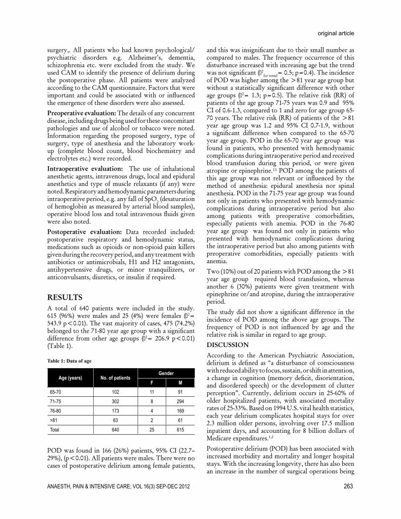
ANAESTH, PAIN & INTENSIVE CARE; VOL 16(3) SEP-DEC 2012 263
original article
surgery,. All patients who had known psychological/psychiatric disorders e.g. Alzheimer’s, dementia, schizophrenia etc. were excluded from the study. We used CAM to identify the presence of delirium during the postoperative phase. All patients were analyzed according to the CAM questionnaire. Factors that were important and could be associated with or influenced the emergence of these disorders were also assessed.
Preoperative evaluation: The details of any concurrent disease, including drugs being used for these concomitant pathologies and use of alcohol or tobacco were noted. Information regarding the proposed surgery, type of surgery, type of anesthesia and the laboratory work-up (complete blood count, blood biochemistry and electrolytes etc.) were recorded.
Intraoperative evaluation: The use of inhalational anesthetic agents, intravenous drugs, local and epidural anesthetics and type of muscle relaxants (if any) were noted. Respiratory and hemodynamic parameters during intraoperative period, e.g. any fall of SpO2 (desaturation of hemoglobin as measured by arterial blood samples), operative blood loss and total intravenous fluids given were also noted.
Postoperative evaluation: Data recorded included: postoperative respiratory and hemodynamic status, medications such as opioids or non-opioid pain killers given during the recovery period, and any treatment with antibiotics or antimicrobials, H1 and H2 antagonists, antihypertensive drugs, or minor tranquilizers, or anticonvulsants, diuretics, or insulin if required.
RESULTSA total of 640 patients were included in the study. 615 (96%) were males and 25 (4%) were females (″2= 543.9 p<0.01). The vast majority of cases, 475 (74.2%) belonged to the 71-80 year age group with a significant difference from other age groups (″2= 206.9 p<0.01) (Table 1).
Table 1: Data of age
Age (years) No. of patientsGender
F M65-70 102 11 91
71-75 302 8 294
76-80 173 4 169
>81 63 2 61
Total 640 25 615
POD was found in 166 (26%) patients, 95% CI (22.7–29%), (p<0.01). All patients were males. There were no cases of postoperative delirium among female patients,
and this was insignificant due to their small number as compared to males. The frequency occurrence of this disturbance increased with increasing age but the trend was not significant (″2
for trend= 0.5; p=0.4). The incidence
of POD was higher among the >81 year age group but without a statistically significant difference with other age groups (″2= 1.3; p=0.5). The relative risk (RR) of patients of the age group 71-75 years was 0.9 and 95% CI of 0.6-1.3, compared to 1 and zero for age group 65-70 years. The relative risk (RR) of patients of the >81 year age group was 1.2 and 95% CI 0.7-1.9, without a significant difference when compared to the 65-70 year age group. POD in the 65-70 year age group was found in patients, who presented with hemodynamic complications during intraoperative period and received blood transfusion during this period, or were given atropine or epinephrine.¹¹ POD among the patients of this age group was not relevant or influenced by the method of anesthesia: epidural anesthesia nor spinal anesthesia. POD in the 71-75 year age group was found not only in patients who presented with hemodynamic complications during intraoperative period but also among patients with preoperative comorbidities, especially patients with anemia. POD in the 76-80 year age group was found not only in patients who presented with hemodynamic complications during the intraoperative period but also among patients with preoperative comorbidities, especially patients with anemia.
Two (10%) out of 20 patients with POD among the >81 year age group required blood transfusion, whereas another 6 (30%) patients were given treatment with epinephrine or/and atropine, during the intraoperative period.
The study did not show a significant difference in the incidence of POD among the above age groups. The frequency of POD is not influenced by age and the relative risk is similar in regard to age group.
DISCUSSION
According to the American Psychiatric Association, delirium is defined as “a disturbance of consciousness with reduced ability to focus, sustain, or shift in attention, a change in cognition (memory deficit, disorientation, and disordered speech) or the development of clutter perception”. Currently, delirium occurs in 25-60% of older hospitalized patients, with associated mortality rates of 25-33%. Based on 1994 U.S. vital health statistics, each year delirium complicates hospital stays for over 2.3 million older persons, involving over 17.5 million inpatient days, and accounting for 8 billion dollars of Medicare expenditures.1,2
Postoperative delirium (POD) has been associated with increased morbidity and mortality and longer hospital stays. With the increasing longevity, there has also been an increase in the number of surgical operations being
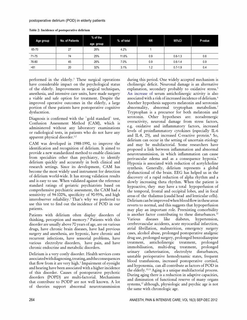
264 ANAESTH, PAIN & INTENSIVE CARE; VOL 16(3) SEP-DEC 2012
postoperative delirium (POD) in elderly patients
performed in the elderly.3 These surgical operations have considerable impact on the psychological status of the elderly. Improvements in surgical techniques, anesthesia, and intensive care units, have made surgery a viable and safe option for treatment. Despite the improved operative outcomes in the elderly, a large portion of these patients have postoperative cognitive dysfucntion.
Diagnosis is confirmed with the `gold standard` test, Confusion Assessment Method (CAM), which is administered without any laboratory examinations or radiological tests, in patients who do not have any apparent physical disorder.1,2
CAM was developed in 1988-1990, to improve the identification and recognition of delirium. It aimed to provide a new standardized method to enable clinicians from specialties other than psychiatry, to identify delirium quickly and accurately in both clinical and research settings. Since its development, CAM has become the most widely used instrument for detection of delirium world-wide. It has strong validation results and is easy to use. When validated against the reference standard ratings of geriatric psychiatrists based on comprehensive psychiatric assessment, the CAM had a sensitivity of 94-100%, specificity of 90-95%, and high interobserver reliability.1 That’s why we preferred to use this test to find out the incidence of POD in our patients.
Patients with delirium often display disorders of thinking, perception and memory.4 Patients with this disorder are usually above 70 years of age, are on various drugs, have chronic brain diseases, have had previous surgery and anesthesia, are hypoxic, have chronic and recurrent infections, have sensorial problems, have various electrolyte disorders, have pain, and have chronic endocrine and metabolic disorders.
Delirium is a very costly disorder. Health services costs associated with diagnosing, treating, and the consequences that flow from it are very high.1 Impairment of eyesight and hearing have been associated with a higher incidence of this disorder. Causes of postoperative psychotic disorders (POPD) are multi-factorial. Mechanisms that contribute to PODP are not well known. A lot of theories support abnormal neurotransmission
during this period. One widely accepted mechanism is cholinergic deficit. Neuronal damage is an alternative explanation, secondary probably to oxidative stress.5
An increase of serum anticholinergic activity is also associated with a risk of increased incidence of delirium.6 Another hypothesis supports melatonin and serotonin abnormality, abnormal tryptophan metabolism.7
Tryptophan is a precursor for both melatonin and serotonin. Other hypotheses are: noradrenergic overactivity, neuronal damage from stress factors, e.g. oxidative and inflammatory factors, increased levels of proinflammatory cytokines (especially IL-6 and IL-8, 25), and increased C-reactive protein.8 So, delirium can occur in the setting of uncertain etiology and may be multifactorial. Some researchers have proposed a link between inflammation and abnormal neurotransmission, in which inflammation can cause perivascular edema and as a consequence hypoxia.9 Hypoxia is associated with reduction of acetylcholine synthesis. Generally, delirium represents a global dysfunctional of the brain. EEG has helped us in the discovery of a rapid reduction of alpha rhythm and a slowly increasing theta rhythm. When the patient is hypoactive, they may have a total hypoperfusion of the temporal, frontal and occipital lobes, and in focal areas of the thalamus (caudal head and lenticular cells). Delirium can be improved when blood flow in these areas reverts to normal, and this suggests that hypoperfusion may play an important role. Preexisting comorbidity is another factor contributing to these disturbances.10 Various diseases like diabetes, hypertension, cerebrovascular accidents, acute myocardial infarction, atrial fibrillation, malnutrition, emergency surgery cases, alcohol abuse, prolonged postoperative analgesic drug use, prolonged surgery, prolonged benzodiazepine treatment, anticholinergic treatment, prolonged immobilisation, multi-drug treatment, prolonged urinary catheterisation, electrolyte disturbances, unstable perioperative hemodynamic states, frequent blood transfusions, increased postoperative cortisol, and hypoxemia, can all contribute as factors of POD in the elderly.10,11 Aging is a unique multifactorial process. During aging there is a reduction in adaptive capacities, and diminution of functional reserve of many organs systems,12 although, physiologic and psychic age is not the same with chronologic age.
Table 2: Incidence of postoperative delirium
Age group No. of Patients% of the
age group% of total RR 95%CI P-value
65-70 27 26% 4.2% 1
71-75 74 25% 11.6% 0.9 0.6-1.3 0.676-80 45 26% 7.0% 0.9 0.6-1.4 0.9>81 20 32% 3.1% 1.2 0.7-1.9 0.4
ANAESTH, PAIN & INTENSIVE CARE; VOL 16(3) SEP-DEC 2012 265
original article
In the elderly there is a generalized reduction of neuron density and a loss of 30% of brain volume, especially after 80 years of age. There is also a diminution of serotonin receptors in the cerebral cortex, diminution of acetylcholine receptors of various areas of brain, a decreased dopamine level to the striate nucleus (neostriate) and substantia nigra, associated with dopamine receptors reduction in the striatum nucleus as well.
In our study, the frequent occurrence of this disturbance increased with increasing age but the trend was not significant (″2
for trend= 0.5; p=0.4). The incidence of POD
was higher among the age group >81 years old but without a statistically significant difference with other age groups (″2= 1.3; p=0.5). The relative risk (RR) of patients of the 71-75 year age group was 0.9 and 95% CI of 0.6-1.3, compared to 1 and zero for 65-70 year age group . The relative risk (RR) of patients of the >81 year age group was 1.2 and 95% CI 0.7-1.9, without a significant difference when compared to the 65-70 year age group . POD in the 65-70 year age group was found in patients, who presented with hemodynamic complications during intraoperative period and received blood transfusion during this period, or were given atropine or epinephrine.¹¹ The type of anesthesia was not relevant or implicated in our study. POD in the 71-75 year age group was found not only in patients who presented with hemodynamic complications during intraoperative period but also among patients with preoperative comorbidities, especially patients with anemia. Hypotension may have caused an adverse
effect on the cerebral functions. POD in the 76-80 year age group was found not only in patients who presented with hemodynamic complications during intraoperative period but also among patients with preoperative comorbidities, especially patients with anemia.
The number of patients with POD associated with blood transfusion and treatment with epinephrine or/ and atropine was seen only in the >81 year age group , but was not significant. Similarly association of other co-morbid conditions in patients with POD was not found statistically significant in our study, similar to other studies.13,14
Analyzing and finding out the factors that cause POPD, understanding of pharmacokinetics and pharmacodynamics, can lead us to treat, and to take measures to prevent this postoperative complication in the elderly. So, prevention and meticulous treatment of these disturbances have direct influence in good performance of post operative condition of the patient.
CONCLUSIONThe incidence of postoperative delirium with increasing age is significantly high. Further studies are required to relate it with physiologic changes in the brain due to preexistent or concomitant diseases with blood biochemistry abnormalities, with hormonal disturbances and with hemodynamic instability.
Conflict of interest: Nil declared.
1. Inouye SK. The Confusion Assessment Method (CAM): Training Manual and Coding Guide. 2003; Yale University School of Medicine. Available at http://www.viha.ca/NR/rdonlyres/0AC07A64-FF24-41E3-BDC5-41CFE4E44F33/0/cam_training_pkg.pdf Accessed on 071212)
2. Marcantonio ER, Flacker JM, Michaels M, Resnick NM. Delirium is independently associated with poor functional recovery after hip fracture. J Am Geriatr Soc. 2000; 48(6): 618-24. [PubMed]
3. Rudolph JL, Jones RN, Rasmussen LS, Silverstein JH, Inouye SK, Marcantonio ER. Independent vascular and cognitive risk factors for postoperative delirium. Am J Med. 2007; 120(9): 807-13. [PubMed]
4. Leslie DL, Marcantonio ER, Zhang Y, Leo-Summers L, Inouye SK. One-year health care costs associated with delirium in the elderly population. Arch Intern Med. 2008 Jan 14; 168(1): 27-32.[PubMed]
5. Karlidag R, Unal S, Sezer OH, Bay Karabulut
A, Battaloğlu B, But A, et al. The role of oxidative stress in postoperative delirium. Gen Hosp Psychiatry. 2006; 28(5): 418-23. [PubMed]
6. Mussi C, Ferrari R, Ascari S, Salvioli G. Importance of serum anticholinergic activity in the assessment of elderly patients with delirium. J Geriatr Psychiatry Neurol. 1999; 12(2): 82-6. [PubMed]
7. Lewis MC, Barnett SR. Postoperative delirium: the tryptophan dysregulation model. Med Hypotheses 2004; 63(3): 402–6. [PubMed]
8. Uchida K, Aoki T, Ishizuka B. Postoperative delirium and plasma melatonin. Med Hypotheses. 2004; 63(3): 402-6. [PubMed]
9. Hála M. Pathophysiology of postoperative delirium: systemic inflammation as a response to surgical trauma causes diffuse microcirculatory impairment. Med Hypotheses. 2007; 68(1): 194-6. [PubMed]
10. `Sieber FE. Postoperative Delirium in the Elderly Surgical Patient. Anesthesiol
Clin. 2009; 27(3): 451-64. [PubMed] 11. Bowman AM. Sleep satisfaction, perceived pain and acute confusion in elderly clients undergoing orthopaedic procedures. J Adv Nurs. 1997; 26(3): 550-64. [PubMed]
12. Inouye SK. Predisposing and precipitating factors for delirium in hospitalized older patients. Dement Geriatr Cogn Disord. 1999; 10(5): 393-400. [PubMed]
13. Large MC, Reichard C, Williams JT, Chang C, Prasad S, Leung Y, Dubeau C, Bales GT, Steinberg GD. Incidence, Risk Factors, and Complications of Postoperative Delirium in Elderly Patients Undergoing Radical Cystectomy. Urology. 2012 Nov 13. pii: S0090-4295(12)01046-1. doi: 10.1016/j.urology.2012.07.086. [Medline]
14. Olin K, Eriksdotter-Jönhagen M, Jansson A, Herrington MK, Kristiansson M, Permert J. Postoperative delirium in elderly patients after major abdominal surgery. Br J Surg. 2005 Dec; 92(12): 1559-64. [Medline]
REFERENCES
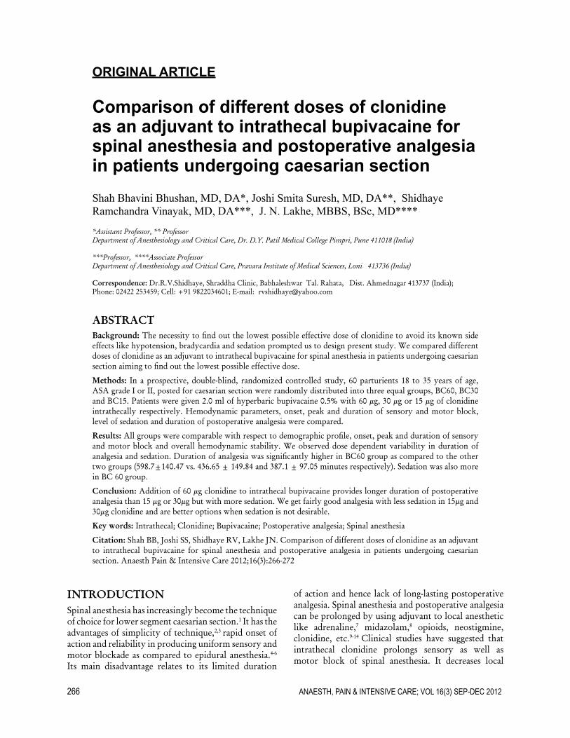
266 ANAESTH, PAIN & INTENSIVE CARE; VOL 16(3) SEP-DEC 2012
ORIGINAL ARTICLE
Comparison of different doses of clonidine as an adjuvant to intrathecal bupivacaine for spinal anesthesia and postoperative analgesia in patients undergoing caesarian section
Shah Bhavini Bhushan, MD, DA*, Joshi Smita Suresh, MD, DA**, Shidhaye Ramchandra Vinayak, MD, DA***, J. N. Lakhe, MBBS, BSc, MD****
*Assistant Professor, ** Professor Department of Anesthesiology and Critical Care, Dr. D.Y. Patil Medical College Pimpri, Pune 411018 (India)
***Professor, ****Associate Professor Department of Anesthesiology and Critical Care, Pravara Institute of Medical Sciences, Loni 413736 (India)
Correspondence: Dr.R.V.Shidhaye, Shraddha Clinic, Babhaleshwar Tal. Rahata, Dist. Ahmednagar 413737 (India); Phone: 02422 253459; Cell: +91 9822034601; E-mail: [email protected]
ABSTRACTBackground: The necessity to find out the lowest possible effective dose of clonidine to avoid its known side effects like hypotension, bradycardia and sedation prompted us to design present study. We compared different doses of clonidine as an adjuvant to intrathecal bupivacaine for spinal anesthesia in patients undergoing caesarian section aiming to find out the lowest possible effective dose.
Methods: In a prospective, double-blind, randomized controlled study, 60 parturients 18 to 35 years of age, ASA grade I or II, posted for caesarian section were randomly distributed into three equal groups, BC60, BC30 and BC15. Patients were given 2.0 ml of hyperbaric bupivacaine 0.5% with 60 μg, 30 μg or 15 μg of clonidine intrathecally respectively. Hemodynamic parameters, onset, peak and duration of sensory and motor block, level of sedation and duration of postoperative analgesia were compared.
Results: All groups were comparable with respect to demographic profile, onset, peak and duration of sensory and motor block and overall hemodynamic stability. We observed dose dependent variability in duration of analgesia and sedation. Duration of analgesia was significantly higher in BC60 group as compared to the other two groups (598.7±140.47 vs. 436.65 ± 149.84 and 387.1 ± 97.05 minutes respectively). Sedation was also more in BC 60 group.
Conclusion: Addition of 60 μg clonidine to intrathecal bupivacaine provides longer duration of postoperative analgesia than 15 μg or 30μg but with more sedation. We get fairly good analgesia with less sedation in 15μg and 30μg clonidine and are better options when sedation is not desirable.
Key words: Intrathecal; Clonidine; Bupivacaine; Postoperative analgesia; Spinal anesthesia
Citation: Shah BB, Joshi SS, Shidhaye RV, Lakhe JN. Comparison of different doses of clonidine as an adjuvant to intrathecal bupivacaine for spinal anesthesia and postoperative analgesia in patients undergoing caesarian section. Anaesth Pain & Intensive Care 2012;16(3):266-272
INTRODUCTIONSpinal anesthesia has increasingly become the technique of choice for lower segment caesarian section.1 It has the advantages of simplicity of technique,2,3 rapid onset of action and reliability in producing uniform sensory and motor blockade as compared to epidural anesthesia.4-6 Its main disadvantage relates to its limited duration
of action and hence lack of long-lasting postoperative analgesia. Spinal anesthesia and postoperative analgesia can be prolonged by using adjuvant to local anesthetic like adrenaline,7 midazolam,8 opioids, neostigmine, clonidine, etc.9-14 Clinical studies have suggested that intrathecal clonidine prolongs sensory as well as motor block of spinal anesthesia. It decreases local
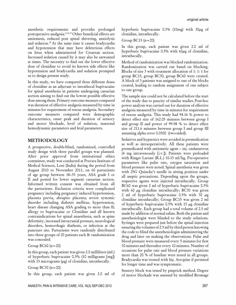
ANAESTH, PAIN & INTENSIVE CARE; VOL 16(3) SEP-DEC 2012 267
original article
anesthetic requirements and provides prolonged postoperative analgesia.9,14-17 Other beneficial effects are antiemesis, reduced post spinal shivering, anxiolysis and sedation.18 At the same time it causes bradycardia and hypotension that may have deleterious effects on fetus when administered for Cesarean section. Increased sedation caused by it may also be unwanted at times. The necessity to find out the lower effective dose of clonidine to avoid its known side effects like hypotension and bradycardia and sedation prompted us to design present study.
In this study, we have compared three different doses of clonidine as an adjuvant to intrathecal bupivacaine for spinal anesthesia in patients undergoing caesarian section aiming to find out the lowest possible effective dose among them. Primary outcome measure compared was duration of effective analgesia measured by time in minutes for requirement of rescue analgesia. Secondary outcome measures compared were demographic characteristics, onset peak and duration of sensory and motor blockade, level of sedation, maternal hemodynamic parameters and fetal parameters.
METHODOLOGYA prospective, double-blind, randomized, controlled study design with three parallel groups was planned. After prior approval from institutional ethics committee, study was conducted at Pravara Institute of Medical Sciences, Loni (India) during the period from August 2010 to November 2011, on 60 parturients of age group between 18–35 years, ASA grade I or II and posted for lower segment caesarian section. Informed written consent was obtained from all the parturients. Exclusion criteria were complicated pregnancy including pregnancy induced hypertension, placenta previa, abruptio placenta; severe systemic disorder including diabetes mellitus, hypertension, heart disease changing ASA grading to more than II; allergy to bupivacaine or Clonidine and all known contraindications for spinal anaesthesia, such as spine deformity, increased intracranial pressure, neurological disorders, hemorrhagic diathesis, or infection at the puncture site. Parturients were randomly distributed into three groups of 20 patients each & randomization was concealed.
Group BC60 (n=20)
In this group, each patient was given 2.0 milliliters (ml.) of hyperbaric bupivacaine 0.5% (10 milligrams [mg]) with 15 micrograms (μg) of clonidine, intrathecally.
Group BC30 (n=20)
In this group, each patient was given 2.0 ml of
hyperbaric bupivacaine 0.5% (10mg) with 30μg of clonidine, intrathecally.
Group BC15 (n=20)
In this group, each patient was given 2.0 ml of hyperbaric bupivacaine 0.5% with 60μg of clonidine, intrathecally.
Method of randomization was blocked randomization. Randomization was carried out based on blocking. Blocks of size 3 with treatment allocation of 1: 1: 1 for group BC15, group BC30, group BC60 were created. A block of 3 patients was assigned to one of the blocks created, leading to random assignment of one subject to one group.
The sample size could not be calculated before the start of the study due to paucity of similar studies. Post-hoc power analysis was carried out for duration of effective analgesia measured by time in minutes for requirement of rescue analgesia. This study had 94.16 % power to detect effect size of 162.05 minutes between group I and group II and power of 99.98 % to detect effect size of 211.6 minutes between group I and group III assuming alpha error 0.0500 (two-sided).
Sedatives and hypnotics were avoided in premedication as well as intraoperatively. All these patients were premedicated with antiemetic agent – inj. ondansetron (4 mg intravenously [i.v.]). Patients were preloaded with Ringer Lactate (R.L.) 10-15 ml/kg. Pre-operative parameters like pulse rate, oxygen saturation and blood pressure were noted. Spinal anesthesia was given with 25G Quincke’s needle in sitting position under all aseptic precautions. Depending upon the groups, respective agents were injected intrathecally. Group BC60 was given 2 ml of hyperbaric bupivacaine 0.5% with 60 μg clonidine intrathecally; BC30 was given 2 ml of hyperbaric bupivacaine 0.5% with 30 μg clonidine intrathecally; Group BC15 was given 2 ml of hyperbaric bupivacaine 0.5% with 15 μg clonidine intrathecally. Each group had a total volume of 2.5 ml made by addition of normal saline. Both the patient and anesthesiologist were blinded to the study solutions. Syringes were prepared just before the spinal injection ensuring the volumes of 2.5 ml by third person knowing the code to blind the anesthesiologist administering the drug and later on making the observations. Pulse and blood pressure were measured every 5 minutes for first 30 minutes and thereafter every 10 minutes. Number of occasions for pulse rate and blood pressure variations more than 20 % of baseline were noted in all groups. Bradycardia was treated with Inj. Atropine if persisted for longer time and was symptomatic.
Sensory block was tested by pinprick method. Degree of motor blockade was assessed by modified Bromage
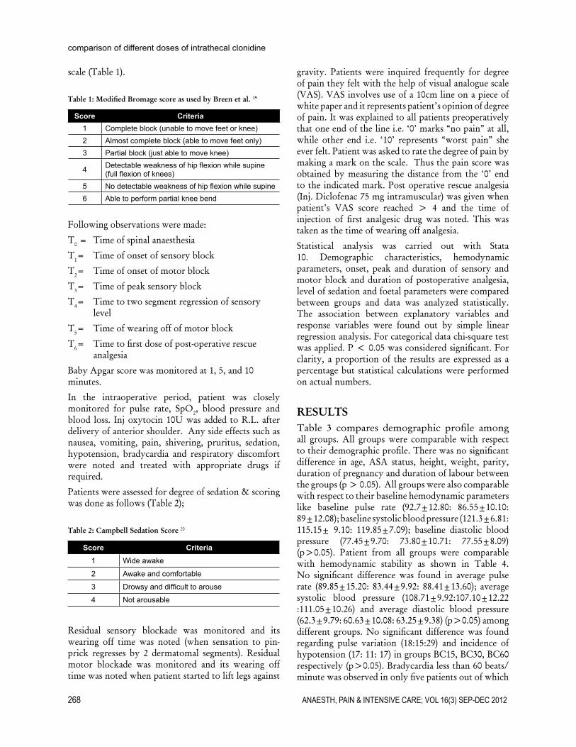
268 ANAESTH, PAIN & INTENSIVE CARE; VOL 16(3) SEP-DEC 2012
comparison of different doses of intrathecal clonidine
scale (Table 1).
Table 1: Modified Bromage score as used by Breen et al. 19
Score Criteria1 Complete block (unable to move feet or knee)2 Almost complete block (able to move feet only)3 Partial block (just able to move knee)
4 Detectable weakness of hip flexion while supine (full flexion of knees)
5 No detectable weakness of hip flexion while supine 6 Able to perform partial knee bend
Following observations were made:
T0 = Time of spinal anaesthesia
T1= Time of onset of sensory block
T2= Time of onset of motor block
T3= Time of peak sensory block
T4= Time to two segment regression of sensory level
T5= Time of wearing off of motor block
T6= Time to first dose of post-operative rescue analgesia
Baby Apgar score was monitored at 1, 5, and 10 minutes.
In the intraoperative period, patient was closely monitored for pulse rate, SpO2, blood pressure and blood loss. Inj oxytocin 10U was added to R.L. after delivery of anterior shoulder. Any side effects such as nausea, vomiting, pain, shivering, pruritus, sedation, hypotension, bradycardia and respiratory discomfort were noted and treated with appropriate drugs if required.
Patients were assessed for degree of sedation & scoring was done as follows (Table 2);
Table 2: Campbell Sedation Score 20
Score Criteria1 Wide awake
2 Awake and comfortable
3 Drowsy and difficult to arouse
4 Not arousable
Residual sensory blockade was monitored and its wearing off time was noted (when sensation to pin-prick regresses by 2 dermatomal segments). Residual motor blockade was monitored and its wearing off time was noted when patient started to lift legs against
gravity. Patients were inquired frequently for degree of pain they felt with the help of visual analogue scale (VAS). VAS involves use of a 10cm line on a piece of white paper and it represents patient’s opinion of degree of pain. It was explained to all patients preoperatively that one end of the line i.e. ‘0’ marks “no pain” at all, while other end i.e. ‘10’ represents “worst pain” she ever felt. Patient was asked to rate the degree of pain by making a mark on the scale. Thus the pain score was obtained by measuring the distance from the ‘0’ end to the indicated mark. Post operative rescue analgesia (Inj. Diclofenac 75 mg intramuscular) was given when patient’s VAS score reached > 4 and the time of injection of first analgesic drug was noted. This was taken as the time of wearing off analgesia.
Statistical analysis was carried out with Stata 10. Demographic characteristics, hemodynamic parameters, onset, peak and duration of sensory and motor block and duration of postoperative analgesia, level of sedation and foetal parameters were compared between groups and data was analyzed statistically. The association between explanatory variables and response variables were found out by simple linear regression analysis. For categorical data chi-square test was applied. P < 0.05 was considered significant. For clarity, a proportion of the results are expressed as a percentage but statistical calculations were performed on actual numbers.
RESULTSTable 3 compares demographic profile among all groups. All groups were comparable with respect to their demographic profile. There was no significant difference in age, ASA status, height, weight, parity, duration of pregnancy and duration of labour between the groups (p > 0.05). All groups were also comparable with respect to their baseline hemodynamic parameters like baseline pulse rate (92.7±12.80: 86.55±10.10: 89±12.08); baseline systolic blood pressure (121.3±6.81: 115.15± 9.10: 119.85±7.09); baseline diastolic blood pressure (77.45±9.70: 73.80±10.71: 77.55±8.09) (p>0.05). Patient from all groups were comparable with hemodynamic stability as shown in Table 4. No significant difference was found in average pulse rate (89.85±15.20: 83.44±9.92: 88.41±13.60); average systolic blood pressure (108.71±9.92:107.10±12.22:111.05±10.26) and average diastolic blood pressure (62.3±9.79: 60.63±10.08: 63.25±9.38) (p>0.05) among different groups. No significant difference was found regarding pulse variation (18:15:29) and incidence of hypotension (17: 11: 17) in groups BC15, BC30, BC60 respectively (p>0.05). Bradycardia less than 60 beats/minute was observed in only five patients out of which
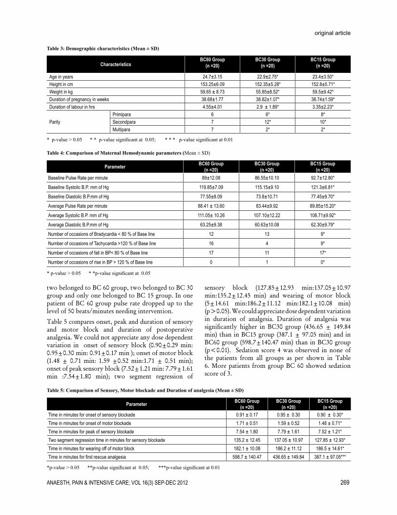
ANAESTH, PAIN & INTENSIVE CARE; VOL 16(3) SEP-DEC 2012 269
original article
Table 3: Demographic characteristics (Mean ± SD)
CharacteristicsBC60 Group
(n =20)BC30 Group
(n =20)BC15 Group
(n =20)
Age in years 24.7±3.15 22.9±2.75* 23.4±3.50*Height in cm 153.25±6.09 152.35±5.28* 152.8±5.71*Weight in kg 59.65 ± 8.73 55.85±8.52* 59.5±9.42*Duration of pregnancy in weeks 38.68±1.77 38.82±1.07* 38.74±1.59*Duration of labour in hrs 4.55±4.01 2.9 ± 1.89* 3.35±2.23*
ParityPrimipara 6 6* 8*Secondpara 7 12* 10*Multipara 7 2* 2*
* p-value > 0.05 * * p-value significant at 0.05; * * * p-value significant at 0.01
Table 4: Comparison of Maternal Hemodynamic parameters (Mean ± SD)
Parameter BC60 Group(n =20)
BC30 Group(n =20)
BC15 Group(n =20)
Baseline Pulse Rate per minute 89±12.08 86.55±10.10 92.7±12.80*
Baseline Systolic B.P. mm of Hg 119.85±7.09 115.15±9.10 121.3±6.81*
Baseline Diastolic B.P.mm of Hg 77.55±8.09 73.8±10.71 77.45±9.70*
Average Pulse Rate per minute 88.41 ± 13.60 83.44±9.92 89.85±15.20*
Average Systolic B.P. mm of Hg 111.05± 10.26 107.10±12.22 108.71±9.92*
Average Diastolic B.P.mm of Hg 63.25±9.38 60.63±10.08 62.30±9.79*
Number of occasions of Bradycardia < 80 % of Base line 12 13 9*
Number of occasions of Tachycardia >120 % of Base line 16 4 9*
Number of occasions of fall in BP< 80 % of Base line 17 11 17*
Number of occasions of rise in BP > 120 % of Base line 0 1 0*
* p-value > 0.05 * *p-value significant at 0.05
two belonged to BC 60 group, two belonged to BC 30 group and only one belonged to BC 15 group. In one patient of BC 60 group pulse rate dropped up to the level of 50 beats/minutes needing intervention.
Table 5 compares onset, peak and duration of sensory and motor block and duration of postoperative analgesia. We could not appreciate any dose dependent variation in onset of sensory block (0.90±0.29 min: 0.95±0.30 min: 0.91±0.17 min ); onset of motor block (1.48 ± 0.71 min: 1.59 ±0.52 min:1.71 ± 0.51 min); onset of peak sensory block (7.52±1.21 min: 7.79±1.61 min :7.54±1.80 min); two segment regression of
sensory block (127.85±12.93 min:137.05±10.97 min:135.2±12.45 min) and wearing of motor block (5±14.61 min:186.2±11.12 min:182.1±10.08 min) (p>0.05). We could appreciate dose dependent variation in duration of analgesia. Duration of analgesia was significantly higher in BC30 group (436.65 ± 149.84 min) than in BC15 group (387.1 ± 97.05 min) and in BC60 group (598.7±140.47 min) than in BC30 group (p<0.01). Sedation score 4 was observed in none of the patients from all groups as per shown in Table 6. More patients from group BC 60 showed sedation score of 3.
Table 5: Comparison of Sensory, Motor blockade and Duration of analgesia (Mean ± SD)
Parameter BC60 Group(n =20)
BC30 Group(n =20)
BC15 Group(n =20)
Time in minutes for onset of sensory blockade 0.91 ± 0.17 0.95 ± 0.30 0.90 ± 0.30*Time in minutes for onset of motor blockade 1.71 ± 0.51 1.59 ± 0.52 1.48 ± 0.71*Time in minutes for peak of sensory blockade 7.54 ± 1.80 7.79 ± 1.61 7.52 ± 1.21*Two segment regression time in minutes for sensory blockade 135.2 ± 12.45 137.05 ± 10.97 127.85 ± 12.93*Time in minutes for wearing off of motor block 182.1 ± 10.08 186.2 ± 11.12 186.5 ± 14.61*Time in minutes for first rescue analgesia 598.7 ± 140.47 436.65 ± 149.84 387.1 ± 97.05***
*p-value > 0.05 **p-value significant at 0.05; ***p-value significant at 0.01
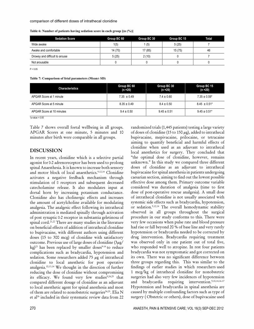
270 ANAESTH, PAIN & INTENSIVE CARE; VOL 16(3) SEP-DEC 2012
comparison of different doses of intrathecal clonidine
Table 7 shows overall foetal wellbeing in all groups. APGAR Scores at one minute, 5 minutes and 10 minutes after birth were comparable in all groups.
DISCUSSIONIn recent years, clonidine which is a selective partial agonist for ″-2 adrenoreceptor has been used to prolong spinal Anaesthesia. It is known to increase both sensory and motor block of local anaesthetics.9,12,14 Clonidine activates a negative feedback mechanism through stimulation of ″ receptors and subsequent decreased catecholamine release. It also modulates input at dorsal horn by increasing potassium conductance. Clonidine also has cholinergic effects and increases the amount of acetylcholine available for modulating analgesia. The analgesic effect following its intrathecal administration is mediated spinally through activation of post synaptic ″-2 receptor in substantia geletinosa of spinal cord.21,22 There are many studies in the literature on beneficial effects of addition of intrathecal clonidine to bupivacaine, with different authors using different doses (15 to 300 mcg) of clonidine with satisfactory outcome. Previous use of large doses of clonidine (3μg/kg)23 has been replaced by smaller doses9-14 to reduce complications such as bradycardia, hypotension and sedation. Some researchers added 75 µg of intrathecal clonidine to local anesthetic for post operative analgesia.10,13,14 We thought in the direction of further reducing the dose of clonidine without compromising its efficacy. We found very few studies9,24,25 that compared different dosage of clonidine as an adjuvant to local anesthetic agent for spinal anesthesia and most of them are related to nonobstetric surgeries24,25. Elia N et al26 included in their systematic review data from 22
randomized trials (1,445 patients) testing a large variety of doses of clonidine (15 to 150 μg), added to intrathecal bupivacaine, mepivacaine, prilocaine, or tetracaine aiming to quantify beneficial and harmful effects of clonidine when used as an adjuvant to intrathecal local anesthetics for surgery. They concluded that “the optimal dose of clonidine, however, remains unknown.” In this study we compared three different doses of clonidine as an adjuvant to intrathecal bupivacaine for spinal anesthesia in patients undergoing caesarian section, aiming to find out the lowest possible effective dose among them. Primary outcome variable considered was duration of analgesia (time to first dose of post-operative rescue analgesia). A small dose of intrathecal clonidine is not usually associated with systemic side effects such as bradycardia, hypotension, or sedation.9,12,14 The overall hemodynamic stability observed in all groups throughout the surgical procedure in our study conforms to this. There were very few occasions when pulse rate and blood pressure had rise or fall beyond 20 % of base line and very rarely hypotension or bradycardia needed to be corrected by drug intervention. Bradycardia requiring treatment was observed only in one patient out of total five, who responded well to atropine. In rest four patients bradycardia was not symptomatic and got corrected on its own. There was no significant difference between three groups regarding this. This was similar to the findings of earlier studies in which researchers used 1 mcg/kg of intrathecal clonidine for nonobstetric surgeries had also very few incidences of hypotension and bradycardia requiring intervention.9,10,14,16.17 Hypotension and bradycardia in spinal anesthesia are caused by multiple confounding factors such as type of surgery ( Obstetric or others), dose of bupivacaine used
Table 6: Number of patients having sedation score in each group [(n (%)]
Sedation Score Group BC 60 Group BC 30 Group BC 15 TotalWide awake 1(5) 1 (5) 5 (25) 7
Awake and comfortable 14 (70) 17 (85) 15 (75) 46
Drowsy and difficult to arouse 5 (25) 2 (10) 0 7
Not arousable 0 0 0 0
P < 0.05
Table 7: Comparison of fetal parameters (Mean± SD)
CharacteristicsGroup BC 60
(n =20)Group BC 30
(n =20)Group BC 15
(n =20)
APGAR Score at 1 minute 7.35 ± 0.49 7.4 ± 0.60 7.35 ± 0.59*
APGAR Score at 5 minute 8.35 ± 0.49 8.4 ± 0.50 8.45 ± 0.51*
APGAR Score at 10 minutes 9.4 ± 0.50 9.45 ± 0.51 9.45 ± 0.51*
*p-value > 0.05
ANAESTH, PAIN & INTENSIVE CARE; VOL 16(3) SEP-DEC 2012 271
original article
, level of sympathetic block, hydration status etc.9,12 Kothari N et al12 who used low doses of clonidine ( 50 μg) showed that incidence of both hypotension and bradycardia was more in bupivacaine group than in bupivacaine with clonidine group. There was no difference in incidence of bradycardia by addition of clonidine. This was because of reducing the dose of bupivacaine from 12.5 mg to 10 mg. Bajwa SJ et al,9 who used 9 mg of bupivacaine also did not observe bradycardia by addition of clonidine even up to 45 μg. So we also might not have observed any significant difference regarding hypotension and bradycardia between three groups due to low doses of clonidine and bupivacaine used. We could not appreciate any dose dependent variation in onset, peak and duration of sensory and motor block. We could appreciate dose dependent variation in duration of analgesia and sedation. Duration of analgesia was significantly higher in BC30 group than in BC15 group and in BC60 group than in BC30 group.(p<0.05) This implies that clonidine prolongs the duration of postoperative analgesia which is higher with increasing dose. This dose dependent variability in duration of analgesia has also been agreed upon by Saxena H et al24 and Strebel
et al25 in nonobstetric surgery and by Bajwa SJ et al 9 in caesarean section surgery. We observed similar dose dependent variability in sedation also. We observed more sedation scores in BC 60 group than in BC 30 than in BC15 group. (p<0.05) Kothari N et al12 also found 35 to 45 % patients drowsy by addition of 50 μg of clonidine to bupivacaine; but Bajwa SJ et al9 did not find any sedation by addition of up to 45 µg of clonidine to bupivacaine. Thus the sedation with clonidine is dose dependent.
CONCLUSIONIn conclusion, intrathecal addition of 60μg clonidine to bupivacaine gives longer duration of postoperative analgesia than 15 μg or 30μg of clonidine but with more sedation. We get fairly good analgesia with less sedation in 15μg and 30μg clonidine and are better options when sedation is not desirable. At the same time when some amount of sedation is acceptable or required, addition of 60μg of clonidine that gives excellent analgesia with negligible hemodynamic complications is a better choice.
272 ANAESTH, PAIN & INTENSIVE CARE; VOL 16(3) SEP-DEC 2012
comparison of different doses of intrathecal clonidine
1. Hunt CO. Spinal anesthesia for obstetrics. International Anaesthesiology Clinics. 1989;27:26-30.
2. Lucy SJ, Naugler MA. Spinal anesthesia for caesarean section (letter). Can J Anaesth. 1991;38:940-1.
3. Morgan P. Spinal anesthesia in obstetrics. Can J Anaesth. 1995;42:1145-63.
4. Covino BG. Rationale for spinal anesthesia. International Anaesthesiology Clinics. 1989;27:8-12.
5. Carter J, Macarthur A. Spinal anesthesia for Caesarean section. Contemporary Anesthesia. 1994;4:11-15.
6. Hunt CO, Dutta S, Hauch M. Perioperative analgesia with subarachnoid fentanyl-bupivacaine. Anaesthesiology. 1987;67.
7. Racle JP, B.A., Poy JY, Gleizal B. Prolongation of isobaric bupivacaine spinal anesthesia with epinephrine and clonidine for hip surgery in the elderly. Anesth Analg. 1987;66:442–6.
8. Prakash S, Joshi N, Gogia AR, Prakash S, Singh R. Analgesic efficacy of two doses of intrathecal midazolam with bupivacaine in patients undergoing cesarean delivery. Reg Anesth Pain Med. 2006 May-Jun;31(3):221-6.
9. Bajwa SJ, Bajwa SK, Kaur J, Singh A, Singh A, Parmar SS. Prevention of hypotension and prolongation of postoperative analgesia in emergency cesarean sections: A randomized study with intrathecal clonidine. Int J Crit Illn Inj Sci. 2012 ;2(2):63-9..
10. Benhamou D, Thorin D, Brichant JF, Dailland P, Milon D, Schneider M. Intrathecal clonidine and fentanyl with hyperbaric bupivacaine improves analgesia during cesarean section. Anesth Analg. 1998;87(3):609-13.
11. Roelants F. The use of neuraxial adjuvant drugs (neostigmine, clonidine) in obstetrics. Curr Opin Anaesthesiol. 2006;19(3):233-7.
12. Kothari N, Bogra J, Chaudhary AK. Evaluation of analgesic effects of intrathecal clonidine along with bupivacaine in cesarean section. Saudi J Anaesth. 2011;5(1):31-5.
13. Lavand’homme PM, Roelants F, Waterloos H, Collet V, De Kock MF. An evaluation of the postoperative antihyperalgesic and analgesic effects of intrathecal clonidine administered during elective cesarean delivery. Anesth Analg. 2008;107(3):948-55.
14. van Tuijl I, van Klei WA, van der Werff DB, Kalkman CJ. The effect of addition of intrathecal clonidine to hyperbaric bupivacaine on postoperative pain and morphine requirements after Caesarean section: a randomized controlled trial. Br J Anaesth. 2006;97(3):365-70.
15. Bonnet F, Buisson VB, Francois Y, Catoire P, Saada M. Effects of oral and subarachnoid clonidine on spinal anesthesia with bupivacaine. Reg Anesth. 1990;15(4):211–4.
16. Sethi BS, Mary Samuel, Deepak Sreevastava. Efficacy of Analgesic Effects of Low Dose Intrathecal Clonidine as Adjuvant to Bupivacaine. Indian J Anaesth. 2007;51(5):415-19.
17. Shah BB, Shidhaye RV, Divekar DS, Panditrao M, Panditrao MM, Suryawanshi C. Effect of addition of Clonidine to Bupivacaine used for patients undergoing spinal anesthesia: A randomized, double blind, controlled study. Sri Lankan Journal of Anaesthesiology. 2011;19(1):17-21.
18. Dobrydnjov I, Axelsson K, Samarütel J, Holmström B. Postoperative pain relief following intrathecal bupivacaine combined
with intrathecal or oral clonidine. Acta Anaesthesiol Scand. 2002;46(7):806-14.
19. Breen TW, Shapiro T, Glass B, Foster-Payne D, Oriol NE. Epidural anesthesia for labor in an ambulatory patient. Anesth Analg. 1993;77:919-24.
20. Campbell DC, Camann WR, Datta S. The addition of bupivacaine to intrathecal sufentanyl for labour analgesia. Anaesth Analg. 1995;81:305-9.
21. Brandt SA, Livingston A. Receptor changes in spinal cord of sheep associated with exposure to chronic pain. Pain. 1990;42:323-9.
22. Reddy SVR, Yaksh TL. Spinal noradrenergic terminal system mediates antinociception. Brain Res.1980;189:391-401.
23. Niemi L. Effects of intrathecal clonidine on duration of bupivacaine spinal anaesthesia, haemodynamics, and postoperative analgesia in patients undergoing knee arthroscopy. Acta Anaesthesiol Scand. 1994;38(7):724-8.
24. H. Saxena, S.K. Singh, S. Ghildiyal: Low Dose Intrathecal Clonidine With Bupivicain Improves Onset And Duration of Block With Haemodynamic Stability. The Internet Journal of Anesthesiology. 2010;23(1). DOI: 10.5580/17df
25. Strebel S, Gurzeler JA, Schneider MC, Aeschbach A, Kindler CH. Small-dose intrathecal clonidine and isobaric bupivacaine for orthopedic surgery: a dose-response study. Anesth Analg. 2004;99(4):1231-8.
26. Elia N, Culebras X, Mazza C, Schiffer E, Tramèr MR. Clonidine as an adjuvant to intrathecal local anesthetics for surgery: systematic review of randomized trials. Reg Anesth Pain Med. 2008;33(2):159-67.
REFERENCES
ANAESTH, PAIN & INTENSIVE CARE; VOL 16(3) SEP-DEC 2012 273
SHORT COMMUNICATION
Two point fixation of endotracheal tube in submentotracheal intubation during craniomaxillofacial surgeries-our experience!
Dheeraj Kapoor, MD, FCCP, FACEE, FCCS*, Anand Gupta, MDS, MFDS RCPS (Glasgow)**, Deepak Thapa, DNB, DA***, Jasveer Singh, MD*
*Assistant Professor (Anesthesiology), **Assistant Professor (Oral and Maxillofacial Surgery), ***Associate Professor Govt. Medical College and Hospital, Chandigarh-160030 (India).
Correspondence: Dr. Dheeraj Kapoor, 1207, Sector 32 B, Chandigarh, (India); Phone: 911724622549, 919646121549; E-mail: [email protected], [email protected]
ABSTRACTAirway management in the craniomaxillofacial trauma surgery may require some modifications of the standard intubation techniques. Nasotracheal intubation is often not an option in panfacial and midfacial injuries due to the probable presence of fractures of base of the skull and associated risk of brain trauma and iatrogenic meningitis. Submental endotracheal intubation may serve as an effective and safe alternative route in these conditions. In standard technique of submentotracheal intubation, the tube is fixed extraorally at the submental incision site with sutures to prevent displacement of the tube during the surgical intervention. But still it leaves a possibility of accidental extubation during the conversion of orotracheal to submental route and vice versa. To counteract this problem we in our institution, fix the tube at two points, one at molar teeth in intraoral region and second at skin surface externally near submental incision site ensuring a secured airway. This procedure has eliminated accidental displacement or extubation in our cases.
Key words: Craniomaxillofacial trauma; Panfacial injuries; Submentotracheal intubation; Tube Fixation technique; Molar teeth; ETT.
Citation: Kapoor D, Gupta A, Thapa D, Singh J. Two point fixation of endotracheal tube in submentotracheal intubation during craniomaxillofacial surgeries-our experience! Anaesth Pain & Intensive Care. 2012:16(3):273-275
INTRODUCTION Airway management in the craniomaxillofacial trauma surgery requires special considerations and modifications of the standard intubation techniques. Nasotracheal intubation is often contra indicated in panfacial and midfacial injuries due to the probable presence of fractures of base of the skull and associated risk of brain trauma and iatrogenic meningitis.1 Midfacial fractures may also cause obstruction to the nasotracheal tube passage and the interference with the surgical reconstruction in that area. These fractures commonly require maxillo-mandibular fixation to restore the dental occlusion, which precludes the use of orotracheal intubation. Surgical tracheostomy may be indicated but is associated with complications such as tracheal stenosis, injury to cervical vessels or the
thyroid gland, and thus should be avoided.1
Submental endotracheal intubation may serve as an effective and safe alternative route in these conditions.2-4
This technique creates a clear intraoperative surgical access, allows maxillo-mandibular fixation without any obstruction and negates the problems associated with conventional intubation through oral and nasal routes in these surgical procedures. Beside craniomaxillofacial trauma surgeries it may also be safely used in many other elective surgical procedures such as LeFort osteotomies, mandibular orthognathic surgery and rhinoplasty procedures.4,6
In standard technique of submentotracheal intubation, the tube is fixed externally at the submental incision site with sutures to prevent displacement of the tube
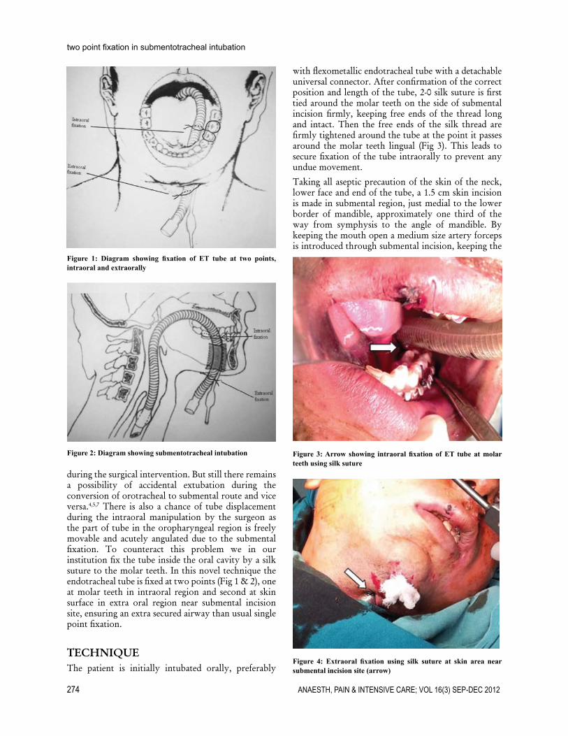
274 ANAESTH, PAIN & INTENSIVE CARE; VOL 16(3) SEP-DEC 2012
two point fixation in submentotracheal intubation
with flexometallic endotracheal tube with a detachable universal connector. After confirmation of the correct position and length of the tube, 2-0 silk suture is first tied around the molar teeth on the side of submental incision firmly, keeping free ends of the thread long and intact. Then the free ends of the silk thread are firmly tightened around the tube at the point it passes around the molar teeth lingual (Fig 3). This leads to secure fixation of the tube intraorally to prevent any undue movement.
Taking all aseptic precaution of the skin of the neck, lower face and end of the tube, a 1.5 cm skin incision is made in submental region, just medial to the lower border of mandible, approximately one third of the way from symphysis to the angle of mandible. By keeping the mouth open a medium size artery forceps is introduced through submental incision, keeping the
Figure 1: Diagram showing fixation of ET tube at two points, intraoral and extraorally
Figure 2: Diagram showing submentotracheal intubation
during the surgical intervention. But still there remains a possibility of accidental extubation during the conversion of orotracheal to submental route and vice versa.4,5,7 There is also a chance of tube displacement during the intraoral manipulation by the surgeon as the part of tube in the oropharyngeal region is freely movable and acutely angulated due to the submental fixation. To counteract this problem we in our institution fix the tube inside the oral cavity by a silk suture to the molar teeth. In this novel technique the endotracheal tube is fixed at two points (Fig 1 & 2), one at molar teeth in intraoral region and second at skin surface in extra oral region near submental incision site, ensuring an extra secured airway than usual single point fixation.
TECHNIQUE The patient is initially intubated orally, preferably
Figure 3: Arrow showing intraoral fixation of ET tube at molar teeth using silk suture
Figure 4: Extraoral fixation using silk suture at skin area near submental incision site (arrow)
ANAESTH, PAIN & INTENSIVE CARE; VOL 16(3) SEP-DEC 2012 275
short communication
direction towards the floor of the mouth staying close to the inner aspect of the mandible to avoid damage of the sublingual gland, submandibular duct and lingual nerve. Then, an incision is made in oral tented mucosa of the floor of the mouth and the artery forceps is dilated after entering the oral cavity to allow easy passage of the proximal end of the tube. We ventilate the patient with 100% oxygen before taking out connector from the tube. The deflated pilot tube cuff is held with artery forceps and is taken out through the submental incision. The tip of the artery forceps is quickly re-inserted through the submental incision to grasp the tracheal tube end to take it out through the same incision. Connector is reattached and patient is ventilated through breathing circuit.
Second fixation of tube is done at the submental incision site using 2.0 silk suture to give secure fixation to prevent displacement of the tube during the surgical intervention (Fig. 4). The fixation of tube with the molar teeth on the lingual side does not hinder any manipulation of jaws during reduction of fracture fragments or intermaxillary fixation.
After completion of the surgical procedure, if extubation is desired, submental fixation point is released and the proximal end of the tube is pulled inside from proximal to the molar fixation point for orotracheal conversion, thus avoiding any displacement or change in position of the tube at the distal end. Submental incision is closed in two layers. Now the molar fixation point is released and the patient trachea is extubated after executing the standard criteria of extubation, ensuring that the patient is fully awake with intact airway reflexes.
DISCUSSIONThe one point fixation technique of endotracheal tube during submental intubation is commonly employed in panfacial trauma patients. There is always a possibility of tube dislodgement during this procedure which can result in catastrophic outcomes. Studies have reported complications like accidental extubation, dislodgement or obstruction of the endotracheal tube (ETT) during
submental intubation.4,5,7 Anesthesiologists often struggle to maintain the optimal position of the tube during the orotracheal to submental conversion and vice versa with one point fixation technique. We improvised securing of the ETT by applying one more fixation point intraorally to keep the tube secured during manipulation of jaws in surgical interventions and during orotracheal/submental ETT conversions. On search of PubMed, we did not found any reference pertaining to the mentioned fixation technique for submental intubation.
This fixation technique reduces the possibility of accidental extubation during orotracheal to submental conversion and vice versa. The force applied to ETT for reverting it back from submental to usual orotracheal one can result in inadvertent dislodgement of the ETT. The molar fixation of the ETT can prevent this potential catastrophe. This simple technique can be used effectively for fixation of ETT intraorally but may have certain limitations. The intraoral part of the tube once fixed cannot be manipulated during the surgical intervention thus sometimes restricting the surgical access. We have practiced this two point fixation technique in 8 patients during the last one year, with desirable outcome. We observed no change in the position of the ETT during the conversion of orotracheal tube to submental route and vice versa. In one patient we had to release the molar fixation during intraoperative period to serve the surgical requirement of extensive intraoral manipulation. No other complications like skin infections or ETT damage was noticed in these patients. Though, we strongly feel that further studies are warranted to evaluate the efficacy of this technique when compared to the conventional one point fixation technique during submental intubation to come out with more definitive outcomes.
Acknowledgement: We are grateful to Dr Ruchita Gupta (Dental Surgeon, Chandigarh) for her important contribution in the artistic work of diagrams of submental tube fixation for better description and understanding.
1. MacInnis E, Baig M. A modified submental approach for oral endotracheal intubation. Int J Oral Maxillofac Surg. 1999;28:344-6. [Medline]
2. Altemir FH. The submental route for endotracheal intubation: a new technique. J Maxillofac Surg. 1986;14:64-5. [Medline]
3. Caron G, Paquin R, Lessard MR, Trepanier CA, LandryPE. Submental endotracheal intubation: an alternative to tracheotomy
in patients with midfacial and panfacial fractures. J Trauma. 2000;48:235-40. [Medline]
4. Amin M, Dill-Russell P, Manisali M, Lee R, Sinton I. Facial fractures and submental tracheal intubation. Anaesthesia. 2002;57:1195-9. [Medline]
5. Jundt JS, Cattano D, Hagberg CA, Wilson JW. Submental intubation: a literature review. Int J Oral Maxillofac Surg. 2012 Jan;41:46-
54. Epub 2011 Sep 17. [Medline]6. Biglioli F, Mortini P, Goisis M, Bardazzi
A, Boari N. Submental orotracheal intubation: An alternative to tracheotomy in transfacial cranial base surgery. Skull Base. 2003;13:189–95. [Medline]
7. Chandu A, Witherow H, Stewart A. Submental intubation in orthognathic surgery: Initial experience. Br J Oral Maxillofac Surg. 2008;46:561–563. [Medline]
REFERENCES
276 ANAESTH, PAIN & INTENSIVE CARE; VOL 16(3) SEP-DEC 2012
CASE REPORT
Bilateral transversus abdominis plane (TAP) catheters for postoperative analgesia in a child with spinal dysraphismDane Yuratich, MD1, Tarun Bhalla, MD1,2, Venkata R. Jayanthi, MD3, Joseph D. Tobias, MD1,2
1. Departments of Anesthesiology, The Ohio State University, Columbus, Ohio2. Department of Anesthesiology & Pain Medicine, Nationwide Children’s Hospital and the Ohio State University, Columbus, Ohio3. Department of Urology, Nationwide Children’s Hospital and the Ohio State University, Columbus, Ohio
Correspondence: Tarun Bhalla, MD, Department of Anesthesiology & Pain Medicine, Nationwide Children’s Hospital, 700 Children’s Drive, Columbus, Ohio 43205 (USA); Ph: (614) 722-4200; Fax: (614) 722-4203; E-mail: [email protected]
ABSTRACTRegional analgesic techniques have become indispensable in the management of adult postoperative pain, and are gaining popularity in the pediatric population. Several case reports have been published describing the use of transversus abdominis plane (TAP) blocks for the provision of analgesia following lower and middle abdominal surgery in the adult population. Although there are several anecdotal reports and a few case series describing TAP blocks in the pediatric population, there are a limited number of reports regarding the use of continuous TAP catheters in the pediatric population. We present our experience with the use of bilateral TAP catheters to provide postoperative analgesia following major abdominal surgery (appendicovesicostomy) in a 5-year old, 17.8 kg pediatric patient with spina bifida. Applications of the technique are discussed and previous reports from the adult and pediatric population regarding the use of TAP catheters are reviewed.
Key words: Regional analgesia; Postoperative pain; Transversus abdominis plane (TAP) blocks; Spina bifida; TAP catheters; Postoperative analgesia
Citation: Yuratich D, Bhalla T, Jayanthi VR, Tobias JD. Bilateral transversus abdominis plane (TAP) catheters for postoperative analgesia in a child with spinal dysraphism. Anaesth Pain & Intensive Care. 2012;16(3):276-279
INTRODUCTIONRegional analgesic techniques have become indispensable in the management of adult postoperative pain. Although there is a greater abundance of literature describing peripheral nerve blockade in adults, the use of regional anesthesia is increasing in neonates and infants1. While caudal analgesia remains the most commonly employed regional technique in the pediatric population, there are specific circumstances that limit its use including patients with spinal dysraphism (meningomyelocele or spina bifida), previous surgical procedures on the bony elements of the spine (laminectomy or spinal fusion with instrumentation), bleeding dyscrasias, and infants with vertebral anatomical abnormalities (VATER).2 The transversus abdominis plane (TAP) block is a peripheral nerve block which can provide sustained abdominal
wall analgesia for lower and middle abdominal surgery and offers an alternative to parenteral opioids in these situations. Although originally described in the adult literature, there are several anecdotal reports of its successful application in the pediatric-aged patient.3-7
The use of regional anesthetic techniques may be especially beneficial in pediatric patients who may be particularly sensitive to the respiratory depressant effects of opioids. One disadvantage of many regional anesthetic techniques is that they provide only a finite duration of analgesia (6-8 hours) when administered via a single injection. To overcome such problems, there is increasing experience with the use of indwelling catheters in peripheral regional anesthetic techniques for the provision of postoperative analgesia. To date, we could find only one other report of the use of continuous TAP catheters in the pediatric population.8
ANAESTH, PAIN & INTENSIVE CARE; VOL 16(3) SEP-DEC 2012 277
case report
We present our experience with the use of bilateral TAP catheters to provide postoperative analgesia following major abdominal surgery in a 5-year old pediatric patient with spinal dysraphism (spina bifida). Applications of the technique are discussed and previous reports from the adult and pediatric population regarding the use of TAP catheters are reviewed.
CASE REPORTInstitutional Review Board approval is not required at Nationwide Children’s Hospital for the presentation of single case reports. A 5-year-old, 17.8 kg child presented for an appendicovesicostomy (Mitrofanoff procedure) in the treatment of neurogenic dysfunction of the urinary bladder. Additional past medical history was significant for spina bifida with hydrocephalus which had required placement of a ventriculoperitoneal shunt. The patient was admitted to the hospital 48 hours prior to the surgical procedure for bowel preparation and intravenous hydration. He was held nil per os for 8 hours and transported to the operating room where routine American Society of Anesthesiologists’ monitors were placed. Anesthesia was induced with propofol and tracheal intubation facilitated by rocuronium. Prior to the start of the surgical procedure, the abdomen was prepped with betadine. Using a linear, high frequency, ultrasound transducer, the three muscle layers of the lateral abdominal wall were visualized bilaterally. With an in-plane approach, with the ultrasound probe placed in a transverse plane in the region of the anterior axillary line, the potential space between the transversus abdominis muscle and the internal oblique muscle was cannulated with an 18 gauge, 2” Tuohy needle. A 20 gauge catheter was advanced 3-4 centimeters beyond the tip of the needle into the potential space after hydro-dissection. Correct needle tip position was confirmed by observing the internal oblique and the transversus abdominis muscles separating from each other with the formation of a black, lens shaped collection of fluid. The needle was withdrawn and the catheter was secured using sterile bio-occlusive dressing. The procedure was repeated on the opposite side. An initial bolus of 5 mL of 0.25% bupivacaine with epinephrine 1:200,000 was administered on each side followed by a continuous infusion of 0.125% bupivacaine at 1 mL/hr on each side. During the 6-7 hour surgical procedure, anesthesia was maintained with isoflurane. Supplemental analgesia included fentanyl (fentanyl 5-6 μg/kg) and hydromorphone (10 μg/kg). At the conclusion of the surgical procedure, residual neuromuscular blockade was reversed and the patient’s trachea was extubated. He was transported to the post-anesthesia care unit (PACU) where he
remained for one hour. He denied pain in the PACU and no opioids were removed. The TAP catheters were removed in the recovery room prior to discharge, as our hospital was still in the process of developing the infrastructure of a peripheral nerve catheter service. The patient was admitted to the inpatient ward and required no intravenous analgesic agents for the initial 9 postoperative hours. His postoperative course was uneventful
DISCUSSIONWhile neuraxial analgesia including caudal epidural analgesia has been the standard alternative to parenteral opioid for the provision of postoperative analgesia in pediatric patients, there is a subset of patients in which caudal analgesia cannot be employed including patients with spinal dysrhaphism. In this population, alterative peripheral techniques of regional anesthesia would be beneficial.6,7
The TAP block may offer an alternative in these situations. The intercostal, subcostal, and first lumbar nerves that contribute to the innervation of the anterior abdominal wall run in a neurovascular plane known as the transverses abdominis plane which is located between the internal oblique muscle and the transversus abdominis muscle. Blockade of these nerves can be achieved with a single injection of local anesthetic administered in this plane. Correct identification of the fascial plane can be facilitated by the use of ultrasound guidance.9 Given that there is bilateral innervation, both sides must be approached to achieve effective analgesia for midline procedures. Performed using ultrasound guidance, this block can be used to provide sustained abdominal wall analgesia and limit the need for postoperative opioid analgesia. The latter may be especially beneficial in the pediatric population with co-morbid conditions, as they are particularly sensitive to the respiratory depressant effects of these medications. In our patient, there was the presence of spina bifida with previous instrumentation to his vertebral column, which was a relative contraindication to neuraxial analgesia. The placement of TAP catheters allowed for the provision of intraoperative analgesia and the continuation of the analgesia throughout the 6-7 hour surgical procedure.
Unlike neuraxial techniques, the TAP block cannot be used instead of general anesthesia. In patients undergoing lower and mid-abdominal surgical procedures, the addition of a TAP block to the general anesthetic technique provides effective postoperative analgesia while decreasing total opioid consumption.4 In a prospective, randomized trial following total
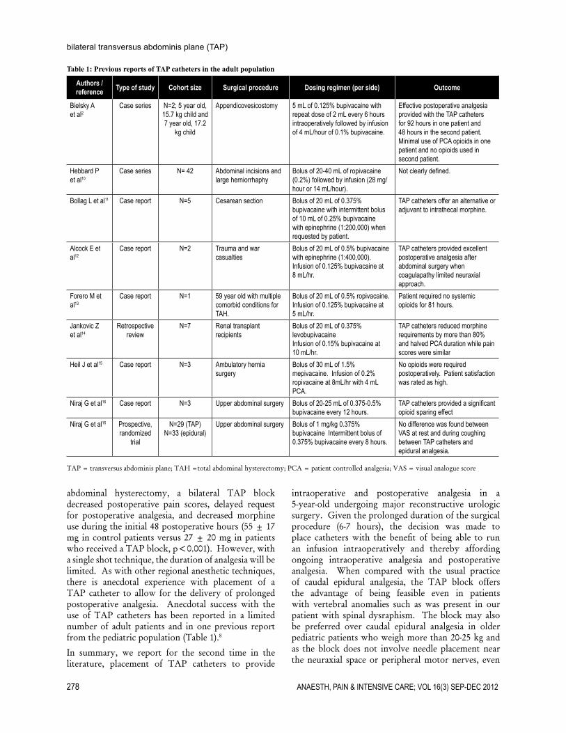
278 ANAESTH, PAIN & INTENSIVE CARE; VOL 16(3) SEP-DEC 2012
bilateral transversus abdominis plane (TAP)
abdominal hysterectomy, a bilateral TAP block decreased postoperative pain scores, delayed request for postoperative analgesia, and decreased morphine use during the initial 48 postoperative hours (55 ± 17 mg in control patients versus 27 ± 20 mg in patients who received a TAP block, p<0.001). However, with a single shot technique, the duration of analgesia will be limited. As with other regional anesthetic techniques, there is anecdotal experience with placement of a TAP catheter to allow for the delivery of prolonged postoperative analgesia. Anecdotal success with the use of TAP catheters has been reported in a limited number of adult patients and in one previous report from the pediatric population (Table 1).8
In summary, we report for the second time in the literature, placement of TAP catheters to provide
Table 1: Previous reports of TAP catheters in the adult population
Authors / reference Type of study Cohort size Surgical procedure Dosing regimen (per side) Outcome
Bielsky A et al2
Case series N=2; 5 year old, 15.7 kg child and 7 year old, 17.2
kg child
Appendicovesicostomy 5 mL of 0.125% bupivacaine with repeat dose of 2 mL every 6 hours intraoperatively followed by infusion of 4 mL/hour of 0.1% bupivacaine.
Effective postoperative analgesia provided with the TAP catheters for 92 hours in one patient and 48 hours in the second patient. Minimal use of PCA opioids in one patient and no opioids used in second patient.
Hebbard P et al10
Case series N= 42 Abdominal incisions and large herniorrhaphy
Bolus of 20-40 mL of ropivacaine (0.2%) followed by infusion (28 mg/hour or 14 mL/hour).
Not clearly defined.
Bollag L et al11 Case report N=5 Cesarean section Bolus of 20 mL of 0.375% bupivacaine with intermittent bolus of 10 mL of 0.25% bupivacaine with epinephrine (1:200,000) when requested by patient.
TAP catheters offer an alternative or adjuvant to intrathecal morphine.
Alcock E et al12
Case report N=2 Trauma and war casualties
Bolus of 20 mL of 0.5% bupivacaine with epinephrine (1:400,000). Infusion of 0.125% bupivacaine at 8 mL/hr.
TAP catheters provided excellent postoperative analgesia after abdominal surgery when coagulapathy limited neuraxial approach.
Forero M et al13
Case report N=1 59 year old with multiple comorbid conditions for TAH.
Bolus of 20 mL of 0.5% ropivacaine. Infusion of 0.125% bupivacaine at 5 mL/hr.
Patient required no systemic opioids for 81 hours.
Jankovic Z et al14
Retrospective review
N=7 Renal transplant recipients
Bolus of 20 mL of 0.375% levobupivacaineInfusion of 0.15% bupivacaine at 10 mL/hr.
TAP catheters reduced morphine requirements by more than 80% and halved PCA duration while pain scores were similar
Heil J et al15 Case report N=3 Ambulatory hernia surgery
Bolus of 30 mL of 1.5% mepivacaine. Infusion of 0.2% ropivacaine at 8mL/hr with 4 mL PCA.
No opioids were required postoperatively. Patient satisfaction was rated as high.
Niraj G et al16 Case report N=3 Upper abdominal surgery Bolus of 20-25 mL of 0.375-0.5% bupivacaine every 12 hours.
TAP catheters provided a significant opioid sparing effect
Niraj G et al16 Prospective, randomized
trial
N=29 (TAP)N=33 (epidural)
Upper abdominal surgery Bolus of 1 mg/kg 0.375% bupivacaine Intermittent bolus of 0.375% bupivacaine every 8 hours.
No difference was found between VAS at rest and during coughing between TAP catheters and epidural analgesia.
TAP = transversus abdominis plane; TAH =total abdominal hysterectomy; PCA = patient controlled analgesia; VAS = visual analogue score
intraoperative and postoperative analgesia in a 5-year-old undergoing major reconstructive urologic surgery. Given the prolonged duration of the surgical procedure (6-7 hours), the decision was made to place catheters with the benefit of being able to run an infusion intraoperatively and thereby affording ongoing intraoperative analgesia and postoperative analgesia. When compared with the usual practice of caudal epidural analgesia, the TAP block offers the advantage of being feasible even in patients with vertebral anomalies such as was present in our patient with spinal dysraphism. The block may also be preferred over caudal epidural analgesia in older pediatric patients who weigh more than 20-25 kg and as the block does not involve needle placement near the neuraxial space or peripheral motor nerves, even
ANAESTH, PAIN & INTENSIVE CARE; VOL 16(3) SEP-DEC 2012 279
case report
in the adult population, the block has been performed following the induction of general anesthesia. Use of a TAP block has also been reported in a patient with an intracranial lesion which would preclude the use of neuraxial blockade due to concerns of increasing intracranial pressure with epidural anesthesia.18
The limited data in the pediatric literature suggest the use of a bolus dose of 0.2-0.3 mL/kg per side of either 0.25% bupivacaine or 0.2% ropivacaine. However, future studies are needed to determine the optimal dosing regimen. As with many other regional anesthetic techniques, the use of ultrasound guidance should be considered to ensure correct needle location and improve the accuracy of the technique.19 In the pediatric population, the most likely serious adverse event is local anesthetic toxicity and attention to volume and concentration is suggested with limitation of the total dose of bupivacaine or ropivacaine to less than 3 mg/kg and limitation of the infusion to less than 0.3 mg/kg/hr. In our choice, we used an infusion of 0.1% bupivacaine at 0.1 mL/kg/hour on each side thereby providing 0.2 mg/kg/hour. The only other adverse event reported in the literature is a single
case of inadvertent trauma to the liver with the blunt regional needle.20 A review by Dario Galante and his colleagues in this issue of ‘Anesthesia, Pain & Intensive Care’ amply describes various aspects of TAP in adult as well as in pediatric patients.21
While continuation of the infusion postoperatively would aid in further decreasing opioid requirements, qualified personnel must be available at all times to manage potential complications. Although our hospital did not have the personnel to manage peripheral nerve catheters postoperatively at the time of this case report, our acute pain and regional anesthesia service have completed the needed administrative and educational components and we now offer the use of continuous peripheral nerve catheters for the treatment of postoperative pain.
CONCLUSIONOur case demonstrates that TAP catheters can be safely used in pediatric patients and offer an alternative when neuraxial analgesia is contraindicated.
Conflict of interests: None
1. Giaufre E, Dalens B, Gombert A. Epidemiology and morbidity of regional anesthesia in children: A 1-year prospective survey of the French Language Society of Pediatric Anesthesiologists. Anesth Analg. 1996;83:904-12.
2. Bielsky A, Efrat R, Suresh S. Postoperative analgesia in neonates after major abdominal surgery: ‘TAP’ our way to success! Pediatr Anesth. 2009;19:541-3.
3. McDonnell JG, Curley G, Carney J, et al. The analgesic efficacy of transversus abdominis plane block after Cesarean delivery: a randomized controlled trial. Anesth Analg. 2008;106:186-91.
4. Carney J, McDonnell JG, Ochana A, et al. The transversus abdominis plane block provides effective postoperative analgesia in patients undergoing total abdominal hysterectomy. Anesth Analg. 2008;107:2056-60.
5. Frederickson M, Seal P, Houghton J. Early experience with the transverses abdominis plane block in children. Pediatr Anesth. 2008;18:891-892.
6. Hardy CA. Transversus abdominis plane block in neonates: is it a good alternative to caudal anesthesia for postoperative analgesia following abdominal surgery? Pediatr Anesth. 2008;19:56.
7. Tobias JD. Preliminary experience with transversus abdominis block in infants and children. Saudi J Anesth. 2009;3:2-6.
8. Taylor L, Birmingham P, Yerkes E, Suresh S.
Children with spinal dysraphism: transversus abdominis plane catheters to the rescue! Pediatric Anesth. 2010;20:951-4.
9. Suresh A, Chan V. Ultrasound guided transversus abdominis plane block in infants, children, and adolescents: a simple procedural guidance for their performance. Pediatric Anesth. 2009;19:296-299.
10. Hebbard P. Ultrasound-guided continuous oblique subcostal transversus abdominis plane blockade. Reg Anesth Pain Med. 2010;35:436-41.
11. Bollag L, Richebe P, Ortner C, Landau R. Transversus abdominis plane catheters for post-cesarean delivery analgesia: a series of five cases. Int J Obstet Anesth (in press).
12. Alcock E, Spencer E, Frazer R, Applegate G, and Buckenmaier C III. Continuous transversus abdominis plane (TAP) block catheters in a combat surgical environment. Pain Med. 2010;11:1426-29.
13. Forero M, Neira VM, Heikkila AJ, Paul JE. Continuous lumbar transversus abdominis plane block may spread to supraumbilical dermatomes. Can J Anaesth. 2011;58:948-51.
14. Jankovic Z, Pollard SG, Nachiappan MM. Continuous transversus abdominis plane blocks for renal transplant recipients. Anesth Analg. 2009;109:1710-11.
15. Heil J, Ilfeld BM, Loland VJ, Sandhu NS, Mariano ER. Ultrasound-guided transversus abdominis plane catheters and ambulatory perineural infusions for outpatient inguinal hernia repair.
Reg Anesth Pain Med. 2010;35:556-8.16. Niraj G, Kelkar A, Fox AJ. Oblique sub-costal
transversus abdominis plane catheters: an alternative to epidural analgesia after upper abdominal surgery. Anesthesia. 2009:1137-40.
17. Niraj G, Kelkar A, Jeyapalan I, Graff-Baker P, Williams O, Darbar A, Maheshwaran A, Powell R. Comparison of analgesic efficacy of subcostal transverses abdominis plane blocks with epidural analgesia following upper abdominal surgery. Anesthesia. 2011;66:465-71.
18. French JLH, McCullough J, Bachra P, Bedforth NM. Transversus abdominis plane block for analgesia after cesarean section in a patient with an intracranial lesion. Int J Obstet Anes. 2009;18:52-54.
19. Tran TMN, Ivanusic JJ, Hebbard P, Barrington MJ. Determination of spread of injectate after ultrasound-guided transverses abdominis plane block: A cadaveric study. Br J Anaesth. 2009;102:123-127.
20. Farooq M, Carey M. A case of liver trauma with a blunt regional anesthesia needle while performing transverses abdominis plane block. Reg Anesth Pain Med. 2008;33:274-275.
21. Galante D, Caruselli M, Dones F, Meola S, Russo G, Pellico G, Caso A, MD6, Lambo M, Donadei F, Mincolelli G. Ultrasound transversus abdominis plane (TAP) block in pediatric patients: not only a regional anesthesia technique for adults. Anaesth Pain & Intensive Care. 2012;16(2): 201-204
REFERENCES
280 ANAESTH, PAIN & INTENSIVE CARE; VOL 16(3) SEP-DEC 2012
CASE REPORT
Ethical Dilemma in multiple co-morbid respiratory failure patient: Patient autonomy against family wishes?
Anwar ul Haq MBBS, MCPS, MS, FCARCSI, MSc (Professionalism Anaesth) Ire
Department of Anesthesiology & ICU, Cork University Hospital, Cork (Ireland)
Correspondence: Dr. Anwar Ul Haq, Consultant Anaesthetist, Midland Regional Hospital Tullamore, Ardon Road, Tullamore (Ireland); E-mail: [email protected].
ABSTRACTAn 82 years old patient with background history of severe COPD, heart failure, multiple co-morbidities and poor quality of life was admitted with pneumonia and subsequently developed acute respiratory distress. There was an obvious conflict of opinion among her family members regarding decision making in her case. The patient time and again insisted against being resuscitated if she ever became seriously ill. However, she did not appoint a proxy decision maker or give an advance directive. This created an ethical dilemma, resulting a clash among the family members as well as her treating physicians concerning the withholding of active treatment and DNR orders in case of cardiorespiratory arrest. In the end the clinicians took lead and, with effective communication with the patient and the family members, made a final decision of withholding treatment in respect of the patient’s dignity and autonomy.
Key words: Ethical dilemma; Patient autonomy; Ethical conflicts; Withholding; DNR; Do not resuscitate orders
Citation: Haq AU. Ethical Dilemma in multiple co-morbid respiratory failure patient: Patient autonomy against family wishes? Anaesth Pain & Intensive Care. 2012;16(3):280-282
INTRODUCTIONWithholding or withdrawal of treatment in a patient with multiple co-morbidities in acute illness setting involving end of life care develop complex ethical dilemmas when conflict arises between principles of ethics specially autonomy, beneficence , nonmaleficence . Patient right of self respect, determination, his or her wishes regarding management or refusal for getting treatment describes patient’s autonomy.1-6 When patient become incapacitated and if surrogate decision maker is not nominated then conflicts arises among the family members for the decision regarding provision of aggressive management or withholding treatment.7 It also create complex matter for clinician in respect to patient’s DNR in case of cardiorespiratory arrest as there is no clear guidelines worldwide.10 A clinician faces ethical issues, dilemmas and their resolution in his day to day practice in acute clinical setting. This interesting case report addresses the various factors which lead to complex ethical dilemma and ultimately the resolution reached by the concerned parties by
effective communication.9
CASE REPORTAn 82 years old patient was admitted to medical ward with lower respiratory tract infection with history of COPD, heart failure, rheumatoid arthritis and mild dementia. Due to multiple comorbidities she had a poor quality of life. Patient went into acute respiratory distress on the third afternoon after admission. Medical registrar-on-call called the anesthesia registrar to review the patient and to discuss the transfer to ICU of one of the tertiary hospitals. The anesthesia registrar assessed the patient to be in acute respiratory distress. She had tachypnoea, tachycardia, SpO2 ranged from 80 to 90%; she was slightly cyanosed, confused, with normal blood pressure but unable to communicate properly. Chest examination revealed bilateral crackles and scattered rhonchi. ABG’s showed PO2 8.2 kpa, PCO2 11.5 kpa, SaO2 88% with pH of 7.21. Patient was on venturi mask with FiO2 60%.
ANAESTH, PAIN & INTENSIVE CARE; VOL 16(3) SEP-DEC 2012 281
case report
Anesthesia registrar started BiPAP. He explained her condition to her younger son and the medical registrar and outlined all future prospects of treatment and their outcome. He also discussed the condition of the patient with the consultant on call in ICU as there was no bed available in e tICU. After being familiarized with the patient’s condition, the consultant intensive care decid-ed to examine the patient in the medical ward. He had discussed with the patient the possibilities of her treat-ment and their outcome earlier and she had agreed not to proceed for aggressive treatment (intubation, venti-lation and in the event of cardiac arrest, resuscitation). But presently the patient was not in full control of her senses and was incapacitated. So the consultant called over the primary clinician and her family members to discuss her fate and further management. The two con-sultants differed in opinion regarding the patient man-agement. Then a conflict arose in between the family members; the patient’s daughter supported her moth-er’s desire not to be aggressively resuscitate in case of serious illness; while the son wanted his mother to go all out in favor of full resuscitation against the wishes of his mother. The patient was incapacitated and had not appointed a surrogate decision maker or given an advance directive. In that way it developed significant ethical dilemmas and resolution of this issue became difficult. After prolonged conversations and exchange of arguments between family members, the clinician in charge of the patient and the ICU consultant, it was decided to withhold the treatment, to honour patient’s autonomy and wishes and to let the nature decide the fate of the patient.
DICUSSIONWe usually come across some difficult ethical dilemmas in our day to day practice in an acute care set up. This ethical dilemma is a result of conflicts between princi-ples of ethics described by Beauchamp and Childress.5
There may be a dispute between patient’s autonomy and family’s wishes, between autonomy and benefi-cence, autonomy and nonmaleficence, withholding of treatment and DNR issues. In our case, all these ethical conflicts intermingled and made this a complex ethical dilemma. The patient had wished not to be given ag-gressive treatment in case she got terminally sickl. But since there was no written advance directive, it gave rise to quarrels between family members. However,
the patients do have a right to self determination and should give informed consent for their medical proce-dural treatment. The patient’s dignity should be main-tained during the whole course of his or her medical management and the ultimate fate of the illness. Indi-vidual self-determination is highly valued, and rightly so. Patients should have the right to accept or refuse treatment. If he chooses to let nature take its course, it should be allowed. It is important to remember that one must respect autonomy as long as we live in har-mony with the first principle of our moral law and the sanctity of life.5,6 Most conflicts involve issues of au-tonomy and beneficence principles. The patient’s right to refuse therapy must be protected, recognizing that most patients are concerned about their families and do not wish to have family members undergo unnecessary burden or hardship. Physicians should be sensitive to such family concerns, but in the end, it is the patient’s wish that must prevail.5-7 In principle, families do not have the right to reverse patients’ advance decisions when the patient loses consciousness and no longer able to make wilful decisions. Physicians may concede to the family’s demands for aggressive therapy after the patient loses decision-making capacity regarding the withdrawal or withholding treatment when end of life issue arises. If the patient is not competent enough to make his own decisions, and has not appointed a surrogate decision maker or made an advance decision, then the senior clinician in charge of the patient’s care must take the decision, based on the patient’s best in-terests (principle of beneficence).5,7 The health care pro-fessionals must remain engaged and supportive of the patient even if a conflict does arise. So affective com-munication and discussions amongst multidisciplinary teams of physicians taking care of the patient, as well as amongst the patient’s family is utmost important; it may provide required information and allay fears to resolve many of the problems.8,9 Resuscitation has the ability to reverse premature death. However it can also prolong terminal illness, increase discomfort and consume resources.10 This might create unwanted dif-ficulty for families. Effective communication resolved the issue in this particular case and ultimately decision reached among physicians and family members was not to go for aggressive resuscitative treatment and to respect the patient’s autonomy as this was in the best interests of the patient.
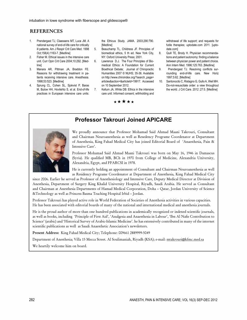
282 ANAESTH, PAIN & INTENSIVE CARE; VOL 16(3) SEP-DEC 2012
intubation in lowe syndrome with fiberscope and glidescope®
1. Prendergast TJ, Claessens MT, Luce JM. A national survey of end-of-life care for critically ill patients. Am J Respir Crit Care Med. 1998 Oct;158(4):1163-7. [Medline]
2. Fisher M. Ethical issues in the intensive care unit. Curr Opin Crit Care 2004;10:292. [Med-line]
3. Manara AR, Pittman JA, Braddon FE. Reasons for withdrawing treatment in pa-tients receiving intensive care. Anesthesia. 1998;53:523. [Medline]
4. Sprung CL, Cohen SL, Sjokvist P, Baras M, Bulow HH, Hovilehto S, et al. End-of-life practices in European intensive care units:
the Ethicus Study. JAMA. 2003;290:790. [Medline]
5. Beauchamp TL, Childress JF. Principles of biomedical ethics, 5 th ed. New York City, NY: Oxford University Press; 2001.
6. Lawrence D.J. The Four Principles of Bio-medical Ethics: A Foundation for Current Bioethical Debate; Journal of Chiropractic Humanities 2007 © NUHS; 35-36. Available on http://www.chiroindex.org/?search_page=articles&action=&articleId=19917. Accessed on 10 September 2012.
7. Kellum JA, White DB: Ethics in the intensive care unit: Informed consent; withholding and
withdrawal of life support; and requests for futile therapies; uptodate.com 2011. [upto-date.com]
8. Quill TE, Brody H. Physician recommenda-tions and patient autonomy: finding a balance between physician power and patient choice. Ann Intern Med. 1996;125:763. [Medline]
9. Prendergast TJ. Resolving conflicts sur-rounding end-of-life care. New Horiz 1997;5:62. [Medline]
10. Santonocito C, Ristagno G, Gullo A, Weil MH. Do-not-resuscitate order: a view throughout the world. J Crit Care. 2012 ;27:5. [Medline]
REFERENCES
Professor Takrouri Joined APICARE We proudly announce that Professor Mohamad Said Ahmad Maani Takrouri, Consultant and Chairman Neuroanesthesia as well as Resedency Programe Coordenator at Department of Anesthesia, King Fahad Medical City has joined Editorial Board of ‘Anaesthesia, Pain & Intensive Care’.
Professor Mohamad Said Ahmad Maani Takrouri was born on May 16, 1946 in Damascus (Syria). He qualified MB, BCh in 1970 from College of Medicine, Alexandria University,Alexandria,Egypt,andFFARCSIin1978.
He is currently holding an appointment of Consultant and Chairman Neuroanesthesia as well as Residency Programe Coordenator at Department of Anesthesia, King Fahad Medical City
since 2006. Earlier he served as Professor of Anesthesiology and Intensive Care, Deputy Medical Director at Division of Anesthesia, Department of Surgery King Khalid University Hospital, Riyadh, Saudi Arabia. He served as Consultant and Chairman at Anesthesia Departments of Hamad Medical Corporation, Doha – Qatar, Jordan University of Science &Technology as well as Princess Basma Teaching Hospital Irbid – Jordan.
Professor Takrouri has played active role in World Federation of Societies of Anesthesia activities in various capacities. He has been associated with editorial boards of many of the national and international medical and anesthesia journals.
Heistheproudauthorofmorethanonehundredpublicationsinacademicallyrecognizedorindexedscientificjournals,aswellasbooks,including‘PrincipleofFirstAid’,‘AnalgesiaandAnaesthesiainLabour’,‘IbnAlNafisContributiontoScience’ (arabic) and ‘Historical Survey of Arabic-Islamic Medicine’. he has extensively contributed in many of the internet scientificpublicationsaswellasSaudiAnaestheticAssociation’snewsletters.
Present Address:KingFahadMedicalCity;Telephone:0096612889999-5049
Department of Anesthesia; Villa 15 Mecca Street. Al Soulimaniah, Riyadh (KSA); e-mail: [email protected]
We heartily welcome him on board.
ANAESTH, PAIN & INTENSIVE CARE; VOL 16(3) SEP-DEC 2012 283
CASE REPORT
Successful intubation in a child with Lowe syndrome using fiberscope and Glidescope® Dheeraj Kapoor, MD, FCCP, FACEE, FCCS*, Nitin Ahuja, MBBS**, Meghana Srivastava, DNB***
*Assistant Professor, **Post Graduate Trainee, ***Senior Resident
Department of Anesthesiology & Intensive Care, Govt. Medical College & Hospital, Chandigarh-160030 (India).
Correspondence: Dheeraj Kapoor, 1207, Sector 32 B, Chandigarh-160030 (India); Phone: 911724622549, 919646121549; E-mail: [email protected]; [email protected]
ABSTRACTWe report the airway management in anticipated difficult airway of a two year old male child with diagnosis of Lowe syndrome, employing two airway management devices. Fiberscope assisted Glidescope® intubation was employed to manage the difficult airway. Lowe syndrome is a rare inherited metabolic disorder with hypotonia, delayed motor and mental milestones, renal dysfunction and hypokalemia. The child had an anticipated difficult airway by virtue of large head circumference with frontal bossing, retrognathia and high arched palate. A careful preanesthetic evaluation and discrete attention to the distinctive components of the syndrome are the essence of successful perioperative management. Airway management in these patients requires standard algorithmic approach to difficult airway with careful selection of ventilation and intubation techniques and aids suitable in these situations to prevent any catastrophes.
Keywords: Lowe syndrome; Oculocerebrorenal syndrome; Glidescope®; Fiberscope; Stylet; Glottic aperture; Optimum external laryngeal maneuver
Citation: Kapoor D, Ahuja N, Srivastava M. Successful intubation in a child with Lowe syndrome using fiberscope and Glidescope®. Anaesth Pain & Intensive Care. 2012;16(3):283-286
INTRODUCTIONOculocerebrorenal syndrome of Lowe (OCRL) is a rare X-linked recessive multi-system disorder.1 The classic triad of this syndrome consists of congenital cataract, neonatal or infantile hypotonia accompanied with subsequent mental impairment and renal tubular dysfunction.1 Renal Fanconi’s syndrome was found to be associated with Lowe syndrome.1 It is signified by low molecular weight proteinuria, proximal renal tubular acidosis, renal phosphate wasting leading to rickets, osteomalacia resulting in pathological frac-tures, bicarbonate wasting, hypercalciuria, aminoaci-duria and hypokalemia.2 Lowe syndrome is caused by mutation of the OCRL gene, located on chromosome Xq24-26 which provides coding for enzyme phosphati-dyl inositol biphosphatase leading to the accumulation of phosphatidyl inositol and mutual disequilibrium of phosphoinositides causing the characteristic clinical
features.2-4,7 Anesthetic management for such patients undergoing surgical procedures can be challenging, due to problems like high intraocular pressure, mental retardation, metabolic abnormalities (due to renal tu-bular dysfunction) and difficult airway. We report the airway management of a two year old male child with Lowe syndrome, posted for congenital cataract surgery having anticipated difficult airway, by employing two airway devices simultaneously including fiberscope and Glidescope® (Verathon Inc. Bothell, WA, USA) for endotracheal intubation.
CASE REPORTA 2-year-old boy, diagnosed with Lowe’s syndrome, was scheduled for bilateral cataract extraction. Preanes-thetic evaluation of the child was done. The child was born out of a full-term normal vaginal delivery in a
284 ANAESTH, PAIN & INTENSIVE CARE; VOL 16(3) SEP-DEC 2012
lowe syndrome using fiberscope and glidescope®
hospital and was observed to have cataract in both eyes at birth. There was no history suggestive of any peri-natal complications in the mother, who stated that the child had delayed motor milestones like neck holding and sitting with support. The child had suffered from recurrent lower respiratory tract infections. There was no history of difficulty in feeding or past medical or surgical treatment. Family history was insignificant.
On physical examination, the child had a large head circumference, 54.1 cm (more than 97th percentile), frontal bossing, severe retrognathia and high arched palate. He had decreased muscle tone and sluggish deep tendon reflexes. His body weight was 8 kg (less than 3rd percentile). He had a skin tag above his left tragus. His vital signs were within normal limits and no other gross abnormality was detected on systemic examination. His blood investigations showed hemoglobin (14gm/dl), hypokalemia and raised alkaline phosphatase (284 IU/L). Albumin was detected on urine analysis. His se-rum sodium, phosphate, magnesium and calcium levels were normal. He was further investigated for suspected congenital abnormalities. Renal function tests, blood sugar, arterial blood gas analysis, chest radiograph and thyroid function test were ordered and the results were found within normal limits. Ultrasonography (USG) of whole abdomen was done to rule out any gross con-genital abnormality with no positive findings.
He was planned for scheduled cataract surgery. Oral potassium supplementation was given preoperatively to correct hypokalemia. On the day of surgery, no pre-medication was given. After taking written pa-rental consent, the child was taken in the operating room (OR). In the OR, continuous electrocardiogram (ECG), non-invasive blood pressure (NIBP) and pulse oximeter (SPO2) were monitored. After preoxygen-ation with 100% oxygen for 3 min, anesthesia was in-duced with sevoflurane (2-4%) in 6 l/min of oxygen. An intravenous access with 22G IV cannula was placed on the dorsum of the left hand. Subsequently, injection glycopyrrolate 50 μg (6μg/kg), fentanyl 15 μg (2μg/kg) were administered intravenously. On confirmation of adequate mask ventilation we planned for visualization of glottic aperture using reusable Glidescope®, using pediatric size blade, in anticipation of difficult intu-bation. We were able to visualize only the posterior region of the glottic aperture (arytenoids and the epi-glottis only, Cormack-Lehane grade 2B) even with ap-plication of optimum external laryngeal manipulation. We introduced the uncuffed endotracheal tube (4.5 mm internal diameter), premounted on the preformed stylet (Gliderite®) supplied with the Glidescope® using the technique described by Ron M Walls,7 but failed to introduce the distal tip of the ETT in front of the glot-
tic aperture which was present too anterior. We than decided to introduce ETT, mounted on a flexible fiber-scope, while visualizing the glottic aperture by Glide-scope®. We mounted the 4.5 mm internal diameter un-cuffed ETT on flexible fiberscope (PENTAX® Europe GmbH, slim Fl-10P2 intubation fiberscope, distal tip diameter 3.4 mm) and introduced it through oral route. The child’s trachea was successfully intubated using this technique. After ensuring the correct placement of the ETT, the child was paralyzed with atracurium besylate 5 mg (0.6mg/kg). Anesthesia was maintained with sevoflurane (0.8-1 MAC) in oxygen and nitrous oxide (50-50%) with intermittent doses of atracurium for muscle relaxation. Intravenous paracetamol 150 mg (20mg/kg) was administered towards the end of surgical intervention. Intra-operative ABG analysis and blood sugar values were unremarkable. Duration of the surgery was about 60 minutes and a total of 50 ml of normal saline was administered. On conclusion of the surgery, once spontaneous breathing returned, anesthesia was reversed with neostigmine (0.4 mg) and glycopyrrolate (80 μg) and extubation of the trachea was performed. Post-operatively, oxygen was given by venturi mask (FiO2 0.5). He was observed in the post-anesthesia care unit (PACU) for 3 hours before being transferred to the ward. The post-operative course was uneventful.
DISCUSSIONOculocerebrorenal syndrome of Lowe or Lowe-Terry-Mac Lachlan syndrome is reported as a multi-system X-linked recessive disorder, mainly affecting males, with prevalence of 1 in 500,000 in general population.2,5 In India, however, the incidence and prevalence of this syndrome is not known.4 The diagnosis is made in in-dividuals showing typical clinical features along with demonstration of reduced activity of inositol poly-phosphate 5-phosphatase enzyme, in cultured skin fi-broblasts.10 Low molecular weight proteinuria may be the most sensitive marker of renal dysfunction occur-ring in this disorder, as it can be seen early in life even in the absence of clinically significant aminoaciduria or any other renal abnormalities.6,11,12
The disease is manifested usually in three stages, where cataracts and glaucoma associated with mental disabil-ity, are evident during the neonatal period.1,2 Fanconi’s type of proximal renal tubular dysfunction occurs till the mid childhood period.8,9 The third phase may be complicated by chronic renal failure during second de-cade of life.2,3,8,9 Clinical features include a prominent forehead (frontal bossing) with thin and sparse hair, short stature, hypotonia, protruding ears, decreased
ANAESTH, PAIN & INTENSIVE CARE; VOL 16(3) SEP-DEC 2012 285
case report
deep tendon reflexes, failure to thrive, maladaptive re-petitive behavior, cryptorchidism, gastroesophogeal re-flux, aspiration pneumonitis, poor cough reflex, hyper-mobile joints, delayed puberty, dental malformations and coagulopathies.8,9,12 Alkaline phosphatase (ALP) is commonly found elevated in Lowe syndrome.6 An-esthesia may be required in these children for various surgical procedures like cataract extraction, strabismus correction, scoliosis reconstruction, herniotomy and orchidopexy.
Our case presented with bilateral congenital cataract, hypotonia, delayed milestones, history of frequent respiratory tract infection and low body weight (fail-ure to thrive). He was diagnosed as Lowe syndrome on basis of these clinical entities along with evidence of proximal renal tubular dysfunction resulting in proteinuria, hypokalemia and raised ALP. We also no-ticed a skin tag over his left tragus, a distinct feature of this syndrome as observed by Scaricaoglu et al.1 We anticipated a difficult airway due to the presence of large head circumference, frontal bossing, retrognathia and high arched palate which is typically seen in this syndrome.2,4,5,8
Our anesthetic concerns in this patient were primarily difficult airway, hypokalemia, hypotonia, positioning and risk of hypoglycemia. Oral potassium supplements and glucose-containing fluids were given pre-operative-ly for management of hypokalemia and for prevention of episodes of hypoglycemia or dehydration. No pre-medication was allowed to prevent exacerbation of ex-isting hypotonia. Airway management in this patient needed special concern in view of distinct craniofacial abnormalities. We anticipated difficult ventilation in this child attributing to large head and retroganthia with propensity of airway obstruction and therefore preferred inhalational induction with sevoflurane, pre-serving the spontaneous breathing of the patient. Af-ter ensuring adequate mask ventilation, we decided for check laryngoscopy with Glidescope® without admin-istering a muscle relaxant. GlideScope® videolaryngo-scope is a contemporary system for tracheal intubation that employs a video camera enclosed into a plastic laryngoscope blade. We selected Glidescope® because it provides a superior view than obtained with a conven-tional laryngoscope.13,24 Scaricaoglu et al encountered a difficult airway due to limited neck extension and in-
tubated their patient with aid of guidewire.1 Pandey R et al reported success to intubate their patient only in the fourth attempt.5 Both the case reports had a child with craniofacial abnormalities similar to this child. We, therefore, did not use conventional laryngoscope in this case to avoid further complications. However, we were only able to visualize the posterior region of the glottic aperture (Cormack-Lehane grade 2B) with the Glidescope® even after applying optimum exter-nal laryngeal manipulation.We tried to maneuver the ETT into the laryngeal inlet using the dedicated sty-let (Gliderite®) by Ronn M Wall technique7 but were unsuccessful due to anteriorly placed larynx. Maneu-vering the ETT towards the vocal cords is a known drawback of the Glidescope® despite using stylet owing to the acute curvature of the blade15 and can even cause airway trauma.16,17 We than planned for using flexible fiberscope to function as a rescue stylet without using its optics, as its distal tip can be easily manipulated. We mounted the ETT on the flexible fiberscope and directed the tip along the curvature of the Glidescope® blade and than through the vocal cords visualized on the Glidescope® screen.
On search of literature we found few reports success-fully employing the same technique in adults, although it has seldom been practiced till now in pediatric group.18-20 Fiberoptic intubation, as a sole airway device could have been the preferred option by many anes-thesiologist in similar cases with difficult airway but it lacks the advantage of panoramic view of glottic aper-ture offered by Glidescope® which averts the problems such as airway trauma caused by the obtrusion of the ETT on the glottic structures during tube passage over the fiberscope.21,22
Lowe syndrome is the rare clinical entity with multi-system involvement. Anesthesia care providers should always have a clinical suspicion of difficult airway in these patients and should delineate a focused plan to manage the airway with multiple airway techniques and devices. In this case, the incorporation of flexible fiberscope as a rescue stylet provided an advantage of superior maneuverability for ETT insertion, along with the Glidescope® empowering better visualization of the glottis, hence avoiding trauma to the glottic structures.
286 ANAESTH, PAIN & INTENSIVE CARE; VOL 16(3) SEP-DEC 2012
lowe syndrome using fiberscope and glidescope®
1. Saricaoglu F, Demirtas F, Aypar U. Preoop-erative and perioperative Management of a patient with Lowe syndrome diagnosed to have Fanconi’s syndrome. Paediatr Anaesth 2004;14:530–532.
2. Loi M. Lowe syndrome. Orphanet J Rare Dis. 2006 May 18;1:16. [Medline]
3. Kim S.M., Lee J.H., Cho S.H., Kim S.H., Chae W.S., Jin H.C. Anesthetic management of a patient with Lowe syndrome accom-panied by end stage renal disease: A case report. Anesth Pain Med. 2012;7:200-202. [KoreaMed]
4. Singh A.R., Singh J.R., Kaur H., Sachdeva G.S., Kaur A., Singh A.D. Lowe Syndrome Oculocerebrorenal Syndrome of Lowe): A Case Report of Two Brothers from India. Int J Hum Genet. 2002;2:243-249.
5. Pandey R, Garg R, Chakravarty C, Darlong V, Punj J, Chandralekha. Lowe’s syndrome with Fanconi syndrome for ocular surgery: perioperative anesthetic considerations. J Clin Anesth. 2010;22:635-7. [Medline]
6. Ramanathan G, Patil S. A case of Lowe syndrome (oculocerebrorenal syndrome): Clinical implications and anaesthetic man-agement. SAJAA. 2009;15:29-31. [AJOL]
7. Walls RM, Samuels-Kalow M, Perkins A. A new maneuver for endotracheal tube inser-tion during difficult GlideScope intubation. J Emerg Med. 2010 Jul;39(1):86-8. Epub 2010 Jan 25. [Medline]
8. Santos M.T.B.R., Watanabe M.M., Manzano
F.S., Lopes C.H., Masiero D. Oculocerebro-renal Lowe syndrome: a literature review and two case reports. Spec Care Dentist. 2007;27:108-111. [Medline]
9. Harrison M, Odell W, Sheehy EC. Dental findings in Lowe syndrome. Pediatr Dent. 1999;21:425-28. [Medline]
10. Zhang X, Hartz PA, Philip E, ET al.Cell lines from kidney proximal tubules of a patient with Lowe syndrome lack OCRL inositol polyphosphatase and accumulate phos-phatidylinositol 4, 5- biphosphatase. J Biol Chem.1998;273:1574- 82. [Medline]
11. Laube GF, Russell-Eggitt IM, van’t Hoff WG. Early proximal tubular dysfunction in Lowe’s syndrome. Arch Dis Child. 2004;89:479–80.
12. Lewis R.A., Nussbaum R.L., Brewer E.D. Lowe syndrome. 2001 [Updated 2012 Feb 23]. In: Pagon R.A., Bird T.D., Dolan C.R., et al., editors. Gene Reviews [Internet]. Seatle (WA): University of Washington, Seatle;1993.
13. Griesdale DE, Liu D, McKinney J, Choi PT. Glidescope® video-laryngoscopy versus direct laryngoscopy for endotracheal intuba-tion: a systematic review and meta-analysis. Can J Anaesth. 2012;59:41-52. Epub 2011 Nov 1.
14. Su YC, Chen CC, Lee YK, Lee JY, Lin KJ. Comparison of video laryngoscopes with direct laryngoscopy for tracheal intubation: a meta-analysis of randomised trials. Eur J Anaesthesiol. 2011;28:788-95.
15. Doyle DJ: The GlideScope video laryngo-scope. Anesthesia. 2005;60:414 –5
16. Cooper RM: Complications associated with the use of the GlideScope videolaryngo-scope. Can J Anaesth. 2007;54:54–7.
17. Hsu WT, Hsu SC, Lee YL, Huang JS, Chen CL: Penetrating injury of the soft palate dur-ing GlideScope intubation. Anesth Analg. 2007;104:1609 –10
18. Moore MS, Wong AB. GlideScope intubation assisted by fiberoptic scope. Anesthesiology. 2007;106:885.
19. Morillas Sendín P, del Olmo Rodríguez C, de Diego Isasa P, Rouco Gil R. Combined use of the Glidescope and a fiberoptic bron-choscope in a case of unexpected difficult intubation. Rev Esp Anestesiol Reanim. 2008;55:454-5.
20. Sharma D, Kim LJ, Ghodke B. Successful airway management with combined use of Glidescope videolaryngoscope and fiberop-tic bronchoscope in a patient with Cowden syndrome. Anesthesiology. 2010;113:253-5.
21. Johnson DM, From AM, Smith RB, From RP, Maktabi MA: Endoscopic study of mechanisms of failure of endotracheal tube advancement into the trachea during awake fiberoptic orotracheal intubation. Anesthesi-ology. 2005;102:910–4.
22. Maktabi MA, Hoffman H, Funk G, From RP: Laryngeal trauma during awake fiberoptic intubation. Anesth Analg. 2002;95:1112–4.
REFERENCES
ANAESTH, PAIN & INTENSIVE CARE; VOL 16(3) SEP-DEC 2012 287
CASE REPORT
Elective use of high frequency oscillatory ventilation with transcutaneous carbon dioxide monitoring during thoracoscopic diaphragmatic hernia repairMichelle LeRiger, MD1, Arlyne Thung, MD1, Karen Diefenbach, MD2, Edward Shepherd, MD3, Erin Wishloff, RRT4, Joseph D. Tobias, MD1
1. Department of Anesthesiology & Pain Medicine2. Department of Pediatric Surgery3. Division of Neonatology and Department of Pediatrics 4. Neonatal Respiratory Services Nationwide Children’s Hospital, Columbus, Ohio (USA)
Correspondence: Michelle LeRiger, MD, Department of Anesthesiology & Pain Medicine, Nationwide Children’s Hospital, 700 Children’s Drive, Columbus, Ohio 43205 (USA); Phone: (614) 722-4200; Fax: (614) 722-4203; E-mail: [email protected]
ABSTRACTThoracoscopy, a minimally invasive technique, for congenital diaphragmatic hernia (CDH) repair has been shown to offer significant advantages versus open procedures. However, positive pressure ventilation during thoracoscopy can be challenging. Minimizing lung movement is important to improve surgical visualization, but one-lung ventilation can be difficult in neonates and infants. Ventilation-perfusion inequalities occur in the lateral decubitus position making it challenging to maintain oxygenation and control hypercarbia from carbon dioxide insufflation. High frequency oscillatory ventilation (HFOV) offers an alternative means of ventilation for such cases. It maintains a constant distending pressure for the alveoli and optimal lung volumes while limiting peak inflating pressures and thus lung over distention. This provides a means of ensuring adequate oxygenation and ventilation with minimal lung movement, allowing adequate intraoperative exposure. During HFOV, continuous monitoring of carbon dioxide (CO2) can be problematic as end-tidal technology is not feasible. As such, we used transcutaneous CO2 (PtcCO2) monitoring in our patient. We present the elective intraoperative institution of HFOV with PtcCO2 monitoring in a 4-day-old infant for thoracoscopic repair of CDH. Previous reports regarding the intraoperative use of HFOV are reviewed and its application in this scenario is discussed. The utility of continuous PtcCO2 monitoring during such cases is presented.
Key words: High frequency oscillatory ventilation (HFOV); Thoracoscopy; Congenital diaphragmatic hernia; Oxygenation; Hypercarbia; Transcutaneous CO2 (PtcCO2) monitoring
Citation: LeRiger M, Thung A, Diefenbach K, Shepherd E, Wishloff E, Tobias JD. Elective use of high frequency oscillatory ventilation with transcutaneous carbon dioxide monitoring during thoracoscopic diaphragmatic hernia repair. Anaesth Pain & Intensive Care. 2012;16(3):287-292
INTRODUCTIONCongenital diaphragmatic hernia (CDH) has an incidence of approximately 1 in 2,000 live births. It results when a portion of the fetal diaphragm tissues fail to fuse, thereby allowing abdominal contents to ascend into the thoracic cavity and interfere with normal lung development. CDH most often manifests as severe respiratory distress in the neonate, a direct
consequence of lung hypoplasia and inadequate pulmonary gas exchange. The initial management of neonatal respiratory failure associated with CDH is directed at ventilation and improving oxygenation.1,2
Currently, surgical repair is delayed until the patient’s physiologic functions have been optimized; the exact timing of the procedure being variable, based on individual patient’s presentation and the institutional expertise. Although traditional surgical management
288 ANAESTH, PAIN & INTENSIVE CARE; VOL 16(3) SEP-DEC 2012
HFOV and CDH repair
of CDH entails a laparotomy or thoracotomy, since 1995, both thoracoscopic and laparoscopic approaches to repair of CDH have been described.3-5 The benefits reported with thoracoscopic techniques include a quicker return to full enteral feeds, shorter duration of postoperative mechanical ventilation, decreased opioid requirements, shorter hospital stay and decreased overall hospital cost.6,7 Additionally, a reduced incidence of chest wall deformities has been reported following thoracoscopic versus open thoracotomy.8-
10 Despite the immediate perioperative and long term benefits, the use of minimally invasive techniques such as thoracoscopy mandates specific changes in anesthetic management including the need for one-lung ventilation (OLV) and concerns regarding CO2 absorption during insufflation.11-13 Although feasible in neonates and small infants, the conduct of OLV may be challenging and difficulties with oxygenation and ventilation are frequently encountered intraoperatively.11-13
High-frequency oscillatory ventilation (HFOV) maintains mean airway pressure and optimal lung volumes while limiting peak inflating pressures and lung overdistention.14,15 Elective use has been suggested in the perioperative care of infants with CDH. Intraoperatively, the technique may be advantageous by limiting lung movement, allowing for thoracoscopic repair of CDH without the need for OLV, and maintaining oxygenation and ventilation. We present the elective intraoperative institution of HFOV in a 4 days old infant for thoracoscopic repair of CDH. Previous reports regarding the intraoperative use of HFOV are reviewed and its application in this scenario discussed.
CASE REPORTInstitutional Review Board approval and the need for parental consent for publication of single case reports are not required by Nationwide Children’s Hospital (Columbus, Ohio). A 4-day-old, 37-week gestational age, 2.84 kg infant was delivered at a referral hospital. Approximately one hour after birth, the infant appeared dusky with increased work of breathing. A chest radiograph revealed a bowel-filled left hemithorax, rightward mediastinal shift, with aeration seen only of the right lower lobe. The infant’s trachea was intubated and he was transported to our hospital. No other co-morbid conditions were noted. Echocardiography revealed a structurally normal heart with right ventricular pressures approximately half that of systemic pressures. Preoperatively, conventional positive pressure mechanical ventilation included synchronized intermittent mechanical ventilation (SIMV) mode with a peak inflating pressure of 21 cmH2O, respiratory rate of 26 breaths per minute, positive end expiratory pressure of 5 cmH2O, inspiratory time of 0.35 seconds,
pressure support of 8 cmH2O and an FiO2 of 0.35. After evaluation and stabilization including repeat echocardiography to verify that pulmonary pressures remained lower than systemic pressures, the patient was scheduled for thoracoscopic repair of the CDH with planned intraoperative HFOV.
The patient was transported to the operating room and standard American Society of Anesthesiologists monitors were placed. An umbilical artery and vein catheter as well as a peripheral intravenous cannula were already in place. A transcutaneous CO2 (TC-CO2) monitor was applied (Sentec AG, Therwil, Switzerland). Anesthesia was induced and maintained with a combination of remifentanil (0.1-0.5 μg/kg/min), dexmedetomidine (0.8 μg/kg/hr) and midazolam (0.1 μg/kg/min). Neuromuscular blockade was provided by intermittent doses of rocuronium. To avoid interference with the surgical procedure from lung movement and facilitate CO2 excretion, the patient was switched to HFOV in the operating room prior to the start of the surgical procedure (3100A HFOV, Sensormedics, San Diego, CA). A neonatal respiratory therapist was present during the case to maintain the HFOV and the transcutaneous CO2 device. Initial HFOV settings included: mean airway pressure (MAP) 12 cmH2O, amplitude (delta P) 20 cmH2O, frequency 6 Hz, and FiO2 of 0.4. There was no change in the patient’s hemodynamic status with the initiation of HFOV. The PtcCO2 readings, arterial blood gas results, ventilator adjustments, and oxygen saturations are outlined in Table 1.
Following the initiation of HFOV, the TC-CO2 was 60 mmHg and the PaCO2 from the arterial blood gas was 42 mmHg (this was after the TC-CO2 was in place for only 5 minutes). The amplitude was increased to 25 cmH2O; however there was no change in the TC-CO2 so the amplitude was further increased to 30 cmH2O and the frequency was decreased to 5 Hz. This resulted in a TC-CO2 value of 53 mmHg and a PaCO2 of 41 mmHg. Throughout this time, the oxygen saturation was 100% with an FiO2 of 0.4. The patient was placed in a right lateral decubitus position and the thoracoscopic procedure was started. Insufflation with CO2 was initiated at a pressure of 2 mmHg. The oxygen saturation decreased from 99% to 92% and the MAP was increased from 12 to 14 mmHg which resulted in an increase of the oxygen saturation to 99-100 percent. When the CO2 insufflation pressure was increased to 4 mmHg to facilitate reduction of the abdominal contents, the amplitude was increased from 30 to 35 mmHg. When the TC-CO2 increased from 40-45 mmHg to 60-65 mmHg the amplitude was increased to 40 cmH2O, resulting in a decrease in TC-CO2 into the 50-55 mmHg range. A subsequent PaCO2 was 44.6 mmHg, so the amplitude was increased to 45 cmH2O. No further changes in ventilation were made. The
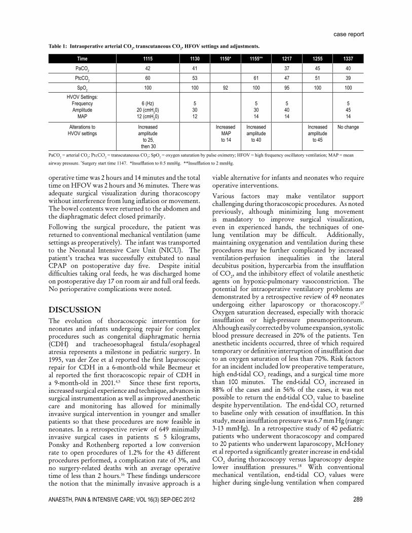
ANAESTH, PAIN & INTENSIVE CARE; VOL 16(3) SEP-DEC 2012 289
case report
operative time was 2 hours and 14 minutes and the total time on HFOV was 2 hours and 36 minutes. There was adequate surgical visualization during thoracoscopy without interference from lung inflation or movement. The bowel contents were returned to the abdomen and the diaphragmatic defect closed primarily.
Following the surgical procedure, the patient was returned to conventional mechanical ventilation (same settings as preoperatively). The infant was transported to the Neonatal Intensive Care Unit (NICU). The patient’s trachea was successfully extubated to nasal CPAP on postoperative day five. Despite initial difficulties taking oral feeds, he was discharged home on postoperative day 17 on room air and full oral feeds. No perioperative complications were noted.
DISCUSSIONThe evolution of thoracoscopic intervention for neonates and infants undergoing repair for complex procedures such as congenital diaphragmatic hernia (CDH) and tracheoesophageal fistula/esophageal atresia represents a milestone in pediatric surgery. In 1995, van der Zee et al reported the first laparoscopic repair for CDH in a 6-month-old while Becmeur et al reported the first thoracoscopic repair of CDH in a 9-month-old in 2001.4,5 Since these first reports, increased surgical experience and technique, advances in surgical instrumentation as well as improved anesthetic care and monitoring has allowed for minimally invasive surgical intervention in younger and smaller patients so that these procedures are now feasible in neonates. In a retrospective review of 649 minimally invasive surgical cases in patients ≤ 5 kilograms, Ponsky and Rothenberg reported a low conversion rate to open procedures of 1.2% for the 43 different procedures performed, a complication rate of 3%, and no surgery-related deaths with an average operative time of less than 2 hours.16 These findings underscore the notion that the minimally invasive approach is a
viable alternative for infants and neonates who require operative interventions.
Various factors may make ventilator support challenging during thoracoscopic procedures. As noted previously, although minimizing lung movement is mandatory to improve surgical visualization, even in experienced hands, the techniques of one-lung ventilation may be difficult. Additionally, maintaining oxygenation and ventilation during these procedures may be further complicated by increased ventilation-perfusion inequalities in the lateral decubitus position, hypercarbia from the insufflation of CO2, and the inhibitory effect of volatile anesthetic agents on hypoxic-pulmonary vasoconstriction. The potential for intraoperative ventilatory problems are demonstrated by a retrospective review of 49 neonates undergoing either laparoscopy or thoracoscopy.17
Oxygen saturation decreased, especially with thoracic insufflation or high-pressure pneumoperitoneum. Although easily corrected by volume expansion, systolic blood pressure decreased in 20% of the patients. Ten anesthetic incidents occurred, three of which required temporary or definitive interruption of insufflation due to an oxygen saturation of less than 70%. Risk factors for an incident included low preoperative temperature, high end-tidal CO2 readings, and a surgical time more than 100 minutes. The end-tidal CO2 increased in 88% of the cases and in 56% of the cases, it was not possible to return the end-tidal CO2 value to baseline despite hyperventilation. The end-tidal CO2 returned to baseline only with cessation of insufflation. In this study, mean insufflation pressure was 6.7 mm Hg (range: 3-13 mmHg). In a retrospective study of 40 pediatric patients who underwent thoracoscopy and compared to 20 patients who underwent laparoscopy, McHoney et al reported a significantly greater increase in end-tidal CO2 during thoracoscopy versus laparoscopy despite lower insufflation pressures.18 With conventional mechanical ventilation, end-tidal CO2 values were higher during single-lung ventilation when compared
Table 1: Intraoperative arterial CO2, transcutaneous CO2, HFOV settings and adjustments.
Time 1115 1130 1150* 1155** 1217 1255 1337
PaCO2 42 41 37 45 40
PtcCO2 60 53 61 47 51 39
SpO2 100 100 92 100 95 100 100HVOV Settings:
FrequencyAmplitude
MAP
6 (Hz)20 (cmH20)12 (cmH20)
530 12
53014
54014
54514
Alterations to HVOV settings
Increased amplitude
to 25, then 30
Increased MAP to 14
Increasedamplitude
to 40
Increasedamplitude
to 45
No change
PaCO2 = arterial CO2; PtcCO2 = transcutaneous CO2; SpO2 = oxygen saturation by pulse oximetry; HFOV = high frequency oscillatory ventilation; MAP = mean
airway pressure. *Surgery start time 1147. *Insufflation to 0.5 mmHg. **Insufflation to 2 mmHg.
290 ANAESTH, PAIN & INTENSIVE CARE; VOL 16(3) SEP-DEC 2012
HFOV and CDH repair
with two-lung ventilation (P = 0.02). The maximum change in the end-tidal CO2 was greater in the younger and smaller patients. The concerns regarding CO2 insufflation in neonates may involve more than ventilator issues. In animal studies, the effects of hypercarbia and CO2 insufflation on the cardiovascular system (mean arterial pressure and cardiac output) have been shown to be more pronounced in neonatal versus adolescent animals.19 With the use of HFOV in our patient, we were able to rapidly control TC-CO2, PaCO2, and oxygen saturation with minor adjustments in the HFOV settings. Control of PaCO2 may be particularly relevant in neonates with CDH given its effects on pulmonary artery pressure in patients at risk for pulmonary hypertension. No clinically significant changes in our patient’s hemodynamic status were noted. Given the limited lung movement produced by HFOV, adequate surgical visualization was obtained with an insufflation pressure of 4 mmHg.
Concurrent with advances in minimally invasive surgical techniques has been the improvement of anesthetic care and monitoring for thoracoscopic procedures. The collaborative practice between the surgeon, anesthesiologist, neonatologist, and respiratory therapist allows the use of low pressure (4-6 mmHg) CO2 insufflation into the operative hemithorax and the necessary adjustments in airway and ventilator management including directed hyperventilation at low tidal volumes using HFOV. HFOV provides a means of ensuring adequate oxygenation and ventilation while minimizing lung movement and interference with surgical visualization.
HFOV was introduced into ICU care as a means of providing ventilation and oxygenation in the treatment of respiratory failure while potentially limiting the risk of barotrauma and volutrauma in patients with acute lung injury. The potential risks are limited by avoiding high peak inflating pressures and tidal volumes. High frequency ventilator techniques rely on respiratory rates greater than 120 breaths per minute (2 Hz) with tidal volumes that are generally less than dead space. Gas exchange is theorized to occur via the process of convection rather than the process of bulk flow as with conventional mechanical ventilation.20 HFOV uses a piston-driven, diaphragm oscillator that provides a constant mean (distending) airway pressure. Superimposed around the MAP are the oscillations provided by the inward and outward movement of the diaphragm. The movement of the diaphragm results in active inspiration and expiration. Active exhalation may decrease gas trapping from impaired exhalation and prevent inadvertent PEEP.21 Tracheal injuries which have been reported with jet ventilatory techniques have not been reported with HFOV.22 Lung volumes are maintained above functional residual capacity
thereby providing a constant distending pressure for alveoli while avoiding high peak inflating pressures. Unlike conventional mechanical ventilation, HFOV is unique in that oxygenation and ventilation are separated with the independent adjustments of MAP and FiO2 for oxygenation and amplitude (delta P), Hz and inspiratory time for ventilation. In general, when caring for patients with acute lung injury, the MAP is set at 2-4 cmH2O above the MAP on conventional ventilation and adjusted up as needed to allow for a decrease of the FiO2 to the desired level. In the ICU setting, the efficacy of the MAP and its effect on alveolar recruitment is assessed by a chest radiograph showing lung expansion of 8-9 ribs. Ventilation is controlled primarily by adjusting the amplitude (delta P) using the power setting on the ventilator. The power knob is adjusted to control the amplitude which is changed in increments of 2-4 cmH2O to provide for adequate chest movement and CO2 removal. Given the low tidal volumes, end tidal CO2 cannot be used for monitoring during HFOV. Our experience as noted in this case and previous reports has suggested the utility of TC-CO2 monitoring with the need for limited arterial blood gas monitoring once a correlation is established.23 Additionally, as the anesthesia machine is not used, the administration of volatile anesthesia is not feasible thereby mandating the use of total intravenous anesthesia (TIVA). In our patient, TIVA included a combination of remifentanil, dexmedetomidine and midazolam.
Various anecdotal experiences have been reported regarding the efficacy of HFOV during open thoracotomy and thoracoscopic procedures (table 2).24-28 Tobias and Burd reported anecdotal experience with the use of HFOV in various clinical scenarios in neonates.24 In infants with CDH, the use of HFOV has become more prevalent as the selected mode of ventilation during surgical repair. In 2000, Bouchut et al. reported the use of HFOV in 22 neonates for open CDH repair.25 No significant differences were noted for any of the studied respiratory parameters (pH, PaO2, PaCO2) recorded at preoperative, perioperative, and postoperative times. The authors noted that the surgeons were satisfied with the more stable operative field with less pulmonary expansion and limited diaphragmatic movements. Currently a European multi-center randomized control trial (VICI-trial) is underway to determine if there is a difference in the incidence of bronchopulmonary dysplasia and/or mortality between neonates treated with HFOV and those treated with conventional mechanical ventilation (CMV) prior to, during and after the open CDH repair.26
The application of HFOV has been used not only for open thoracic procedures but also for thoracoscopic
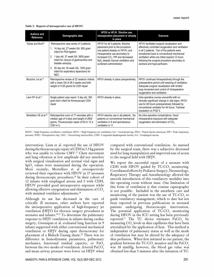
ANAESTH, PAIN & INTENSIVE CARE; VOL 16(3) SEP-DEC 2012 291
case report
intervention. Liem et al. reported the use of HFOV during the thoracoscopic repair of CDH in 3.5 kg patient who was unable to tolerate CMV.27 Chest movement and lung vibration at low amplitude did not interfere with surgical visualization and normal vital signs and SpO2 values were maintained during the operation. More recently, Mortellaro et al retrospectively reviewed their experience with HFOV in 17 neonates during thoracoscopic procedures.28 In their cohort of 12 infants with esophageal atresia and 5 with CDH, HFOV provided good intraoperative exposure while allowing effective oxygenation and elimination of CO2 with minimal ventilator adjustments.28
Although its use has decreased in the care of critically ill neonates, other authors have reported the intraoperative application of high-frequency jet ventilation (HFJV) for thoracic surgery procedures in neonates and infants.29,30 To determine the pulmonary response to HFJV ventilation in infants during cardiac surgery, Greenspan et al. evaluated lung function in 9 infants supported with either conventional mechanical ventilation or HFJV during open thoracotomy for placement of a Blalock-Taussig shunt.29 There was no difference in hemodynamic parameters, pulmonary mechanics, functional residual capacity, or PaO2 between the two modes of ventilation. Arterial PaCO2 and mean airway pressure were lower on HFJV when
compared with conventional ventilation. As assessed by the surgical team, there was a subjective decreased need for lung manipulation and improved ease of access to the surgical field with HFJV.
We report the successful repair of a neonate with CDH with HFOV guided by TC-CO2 monitoring. Coordinated efforts by Pediatric Surgery, Neonatology, Respiratory Therapy and Anesthesiology allowed the smooth introduction of this ventilatory modality into the operating room without issue. One limitation of this form of ventilation is that routine capnography is not possible. Included in the anesthetic care and monitoring of the patient was the use of TC-CO2 to guide ventilatory management, which to date has not been reported in previous publications in neonatal patients undergoing thoracoscopic intervention. The potential application of TC-CO2 monitoring during HFOV in the ICU setting has been previously reported.23 The TC device estimates PaCO2 by measuring CO2 levels in skin capillaries that have been arterialized by the application of heat. This method is independent of pulmonary status as well as the mode of ventilation but may be altered by the adequacy of skin perfusion. When first initiated in our patient, the gradient between the TC-CO2 monitor and the PaCO2 was 18 mmHg; however, the blood gas value was obtained less than 5 minutes after the initiation of TC-
Authors andReference Demographic data
HFOV or HFJV. Elective use, intraoperative placement or already
in placeOutcome
Tobias and Burd24 Retrospective case series of 3 patients.1. 14 day old, 27 weeks GA, 900 gram
infant for PDA ligation.2. 1 day old, 37 week GA, 3600 gram
infant for closure of gastroschisis and bladder extrophy.
3. 30 day old, 30 week GA, 1500 gram infant for exploratory laparotomy for NEC.
HFOV for all 3 patients. Elective placement prior to the procedure, one patient already on HFOV, and intraoperative use secondary to increased CO2, PIP and decreased Sp02 despite manual ventilation and surfactant administration
HFOV facilitated surgical visualization and effectively controlled oxygenation and ventilation in all 3 patients. Two of the patients were transitioned back to conventional mechanical ventilation while one infant expired 12 hours following the surgical procedure secondary to acidosis and hypo-perfusion.
Bouchut J et al.25 Retrospective review of 22 newborn infants with a mean GA of 38.3 weeks and birth weight of 3120 grams for CDH repair.
HFOV already in place preoperatively. HFOV continued intraoperatively through the postoperative period until weaning of ventilation. Adequate surgical visualization with limited lung movement and control of intraoperative oxygenation and ventilation.
Liem NT et al.27 Single patient case report. 5 day old, 350 gram term infant for thoracoscopic CDH repair
HFOV already in place. Intra-operative course uneventful with no clinically significant change in vital signs. HFOV use for 48 hours postoperatively followed by conventional ventilator for 44 hours. Tracheal extubation on POD 5.
Mortellaro VE et al.28 Retrospective room of 17 neonates with a median age of 4 days and weight of 2900 grams. Thoracoscopic repair of EA in 12 d CDH in 5.
HFOV elective use in all patients. Six patients on conventional mechanical ventilation in 6 and spontaneous ventilation in 11.
No intra-operative complications. Good intraoperative exposure with adequate oxygenation and elimination of CO2.
HFOV = High frequency oscillatory ventilation; HFJV = High frequency jet ventilation; GA = Gestational age; PDA = Patent ductus arteriosus; PIP = Peak inspiratory pressure; POD = Postoperative day; NEC = Necrotizing enterocolitis, CDH= Congenital diaphragmatic hernia; EA = Esophageal atresia.
Table 2: Reports of intraoperative use of HFOV.
292 ANAESTH, PAIN & INTENSIVE CARE; VOL 16(3) SEP-DEC 2012
HFOV and CDH repair
CO2 monitoring. At the conclusion of the procedure, the gradient had decreased to 1 mmHg. In the future we would consider placing the TC-CO2 monitor earlier in the care of the patient prior to the initiation of HFOV to allow adequate time for calibration.
In summary, thoracoscopic repair of neonatal conditions such as CDH continues to evolve with increased surgical experience complemented with improvements in technique, instrumentation, anesthetic care, and monitoring. Reported advantages of thoracoscopic versus open thoracic repair include superior surgical visualization, decreased postoperative
pain, decreased hospital stay, and a shorter time to oral feeds. Furthermore, the use of high frequency techniques avoids the need for OLV and is effective in reversing the hypercarbia which frequently accompanies thoracoscopic procedures in neonates and infants. The intraoperative application of HFOV allows for adequate oxygenation and ventilation while maintaining good intraoperative exposure. We propose that TC-CO2 monitoring can be used as a non-invasive measurement of PaCO2, serving as a continuous, non-invasive guide to ventilation during HFOV and facilitating a proactive ventilatory strategy.
1. Robinson PD, Fitzgerald DA. Congenital diaphragmatic hernia. Paediatr Respir Rev. 2007;8:323-334. [Medline]
2. Tsao K, Lally PA, Lally KP. Congenital Diaphragmatic Hernia Study Group. Minimally invasive repair of congenital diaphragmatic hernia. J Pediatr Surg. 2011;46:1158-1164. [Medline]
3. Tsao K, Lally KP. Innovations in the surgical management of congenital diaphragmatic hernia. Clin Perinatol. 2012;39:363-374. [Medline]
4. Van der Zee DC, Max NM. Laparoscopic repair of congenital diaphragmatic hernia in a 6-month old child. Surg Endosc. 1995;9:1001-1003. [Medline]
5. Becmeur F, Jamali RR, Moog R, Keller L, Christmann D, Donato L, et al. Thoracoscopic treatment for delayed presentation of congenital diaphragmatic hernia in the infant. A report of three cases. Surg Endosc. 2001;15:1163-1166. [Medline]
6. Gourlay DM, Cassidy LD, Sato TT, Lal DR, Arca MJ. Beyond feasibility: a comparison of newborns undergoing thoracoscopic and open repair of congenital diaphragmatic hernias. J Pediatr Surg. 2009;44:1702-1707. [Medline]
7. Lobe TE, Rothenbery SS, Waldschmidt J, Stroeder L. Thoracoscopic repair of esophageal atresia in an infant: A surgical first. Pediatr Endosurg Inn Tech. 2000;4:150-156.
8. Durning RP, Scoles PV, Fox OD. Scoliosis after thoracotomy in tracheoesophageal fistula repair. J Bone Joint Surg Am. 1980;62:1156-1158. [Medline]
9. Cherup LL, Siewers RD, Futrell JW. Breast and pectoral muscle maldevelopment after anterolateral and posterolateral thoracotomies in children. Ann Thorac Surg. 1986;41(5):492-497. [Medline]
10. Jaureguizar E, Vazquez J, Murcia J, Diez Pardo JA. Morbid musculoskeletal sequelae for tracheoesophageal fistula. J Pediatr Surg. 1995;20:511-514. [Medline]
11. Tobias JD. Anesthesia for minimally invasive surgery in children. Best Pract Res Clin Anaesthesiol. 2002 Mar;16(1):115-30. [Medline]
12. Tobias JD. Anaesthesia for neonatal thoracic
surgery. Best Pract Res Clin Anaesthesiol. 2004;18:303-320. [Medline]
13. Tobias JD. Thoracoscopy in the pediatric patient. Anesthesiol Clin North America. 2001 Mar;19(1):173-86, viii. [Medline]
14. Stawicki SP, Goyal M, Sarani B. High-frequency oscillatory ventilation (HFOV) and airway pressure release ventilation (APRV): a practical guide. J Intensive Care Med. 2009 Jul-Aug;24(4):215-29. doi: 10.1177/0885066609335728. [Medline]
15. Chiu P, Hedrick HL. Postnatal management and long-term outcome for survivors with congenital diaphragmatic hernia. Prenat Diagn. 2008 Jul;28(7):592-603. doi: 10.1002/pd.2007. [Medline]
16. Ponsky TA, Rothenberg SS. Minimally invasive surgery in infants less than 5kg: Experience of 649 patients. Surg Endosc. 2008 Oct;22(10):2214-9. doi: 10.1007/s00464-008-0025-7. [Medline]
17. Kalfa N, Allal H, Raux O, Lopez M, Forgues D, Guibal MP, et al. tolerance of laparoscopy and thoracoscopy in neonates. Pediatrics. 2005 Dec;116(6):e785-91. [Medline]
18. McHoney M, Mackinlay G, Munro F, Capek A, Aldridge L. Effect of patient weight and anesthetic technique on CO2 excretion during thoracoscopy in children assessed by end-tidal CO2. J Laparoendosc Adv Surg Tech A. 2008 Feb;18(1):147-51. doi: 10.1089/lap.2007.0173. [Medline]
19. Metzelder ML, Kuebler JF, Huber D, Vieten G, Suempelmann R, Ure BM, Osthaus WA. Cardiovascular responses to prolonged carbon dioxide pneumoperitoneum in neonatal versus adolescent pigs. Surg Endosc. 2010 Mar;24(3):670-4. doi: 10.1007/s00464-009-0654-5. [Medline]
20. Slutsky AS. Mechanisms affecting gas transport during high frequency oscillation. Crit Care Med.1984;12:713-717. [Medline]
21. Bancalari A, Gerhardt T, Bancalari E, Suguihara C, Hehre D, Reifenberg L, Goldberg RN. Gas trapping with high-frequency ventilation: jet versus oscillatory ventilation. J Pediatr. 1987 Apr;110(4):617-22. [Medline]
22. Mammel MC, Ophoven JP, Lewallen
PK, Gordon MJ, Sutton MC, Boros SJ. High-frequency ventilation and tracheal injuries. Pediatrics. 1986;77:608-613. [Medline]
23. Berkenbosch JW, Tobias JD. Transcutaneous carbon dioxide monitoring during high frequency oscillatory ventilation in infants and children. Crit Care Med. 2002;30:1024-1027. [Medline]
24. Tobias JD, Burd RS. Anesthetic management and high frequency oscillatory ventilation. Paediatr Anaesth. 2001;11:483-487. [Medline]
25. Bouchut JC, Dubois R, Moussa M, Godard J, Picaud JC, Di Maio M, et al. High frequency oscillatory ventilation during repair of neonatal congenital diaphragmatic hernia. Paediatr Anaesth. 2000;10:377-379. [Medline]
26. van den Hout L, Tibboel D, Vijfhuize S, te Beest H, Hop W, Reiss I; CDH-EURO Consortium. The VICI-trial: high frequency oscillation versus conventional mechanical ventilation in newborns with congenital diaphragmatic hernia: an international multicenter randomized controlled trial. BMC Pediatr. 2011 Nov 2;11:98. doi: 10.1186/1471-2431-11-98. [Medline]
27. Liem NT, Dien TM, Ung NQ. Thoracoscopic repair in the neonatal intensive care unit for congenital diaphragmatic hernia during high-rrequency oscillatory ventilation. J Laparoendosc Adv Surg Tech A. 2010 Feb;20(1):111-4. doi: 10.1089/lap.2008.0412. [Medline]
28. Mortellaro VE, Fike FB, Adibe OO, Juang D, Aguayo P, Ostlie DJ, et al. The use of high-frequency oscillating ventilation to facilitate stability during neonatal thoracoscopic operationsJ Laparoendosc Adv Surg Tech A. 2011 Nov;21(9):877-9. doi: 10.1089/lap.2011.0134. [Medline]
29. Greenspan JS, Davis DA, Russo P, Antunes MJ, Spitzer AR, Wolfson MR High frequency jet ventilation: intraoperative application in infants. Pediatr Pulmonol. 1994;17:155-160. [Medline]
30. Cotter T, Russo P, Tobias JD. Intraoperative jet ventilation during aortic coarctation repair in an infant. J Cardiothorac Vasc Anesth. 2004;18:207-209. [Medline]
REFERENCES
ANAESTH, PAIN & INTENSIVE CARE; VOL 16(3) SEP-DEC 2012 293
CASE REPORT
Massive subcutaneous emphysema secondary to rigid bronchoscopy in a child
Anju Ghai, MD*, Meenu Goyal, MD**, Raman Wadhera, MS***, Manish Kumar Goel, MD**** *Associate Professor, **Assistant Professor, Department of Anesthesiology ***Associate Professor & Head, Department of ENT ****Assistant Professor, Department of Community Medicine Pt. B.D. Sharma Postgraduate Institute of Medical Sciences, Rohtak (India)
Correspondence: Dr. Meenu goel, M.D., Assistant Professor, Department of Anesthesiology, Pt. B.D. Sharma Postgraduate Institute of Medical Sciences, Rohtak (India); Phone: 09416538815; E-mail: [email protected]
ABSTRACT Massive subcutaneous emphysema is a rare complication of interventional bronchoscopy. Complications reported include laryngeal and bronchial spasm, hematorrhea, arrhythmia, airway obstruction, tracheoesophageal fistula, and pneumothorax. Massive subcutaneous emphysema is a rare complication of rigid bronchoscopy. There has been no case report mentioned in the literature till date to the best of our knowledge. We report an eighteen months old female child who developed emphysema involving upper third of her chest, neck and face after rigid bronchoscopy, which increased progressively threatening closure of the airways but was successfully managed conservatively. The details of this case and the management are discussed.
Keywords: Subcutaneous emphysema; Rigid bronchoscopy; Stridor; Bronchodilators; Bronchospasm
Citation: Ghai A, Goyal M, Wadhera R, Goel MK. Massive subcutaneous emphysema secondary to rigid bronchoscopy in a child. Anaesth Pain & Intensive Care 2012;16(3):293-295
INTRODUCTIONBronchoscopy is a widely performed procedure and is generally considered to be safe and effective. Reported rate of severe complications of bronchoscopy and mortality is 0.739% and 0.013% respectively.1 Complications reported include laryngeal and bronchial spasm, hematorrhea, arrhythmia, airway obstruction, trachea-esophageal fistula, and pneumothorax. A case of massive subcutaneous emphysema secondary to rigid bronchoscopy causing a serious risk to airway, which was managed conservatively, is being described here.
CASE REPORT An 18 months old female child presented to the hospital emergency department with history of difficulty in breathing for the previous ten days. She had been on treatment for laryngotracheobronchitis and status asthmatics. There was inadequate relief after treatment with antibiotics and bronchodilators. The respiratory distress had increased for the last four days.
At the time of presentation the child was very sick-looking; her pulse rate was 160 beats/min and respiratory rate was 46/min. She had expiratory stridor with chest indrawing and accessory muscles being used. Auscultation revealed bilateral bronchospasm and decreased air entry on right basal lobe. Chest x-ray showed hyperlucent right lower zone. On enquiring in detail, the mother revealed history of handling of groundnuts by the child when the episode of choking followed by tearing of the eyes started. The child was slightly relieved after a thrust on back, but the difficulty in breathing persisted. An urgent bronchoscopy was planned.
In the operating room, after securing an intravenous line, monitors were attached for SpO2, ECG and heart rate. Oxygen saturation (SpO2) was 90%. Patient was induced with inj. ketamine 40 mg and inj. suxamethonium 10 mg. Rigid bronchoscope size 3.5 was introduced. Inj. atracurium was given. After about ten minutes, a sudden fall in SpO2 and bradycardia was noticed. Bronchoscope was immediately removed by the
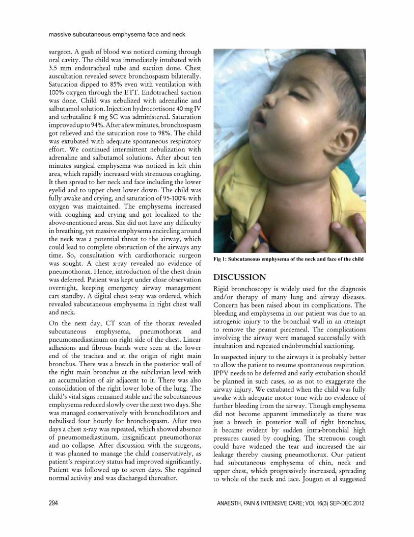
294 ANAESTH, PAIN & INTENSIVE CARE; VOL 16(3) SEP-DEC 2012
massive subcutaneous emphysema face and neck
surgeon. A gush of blood was noticed coming through oral cavity. The child was immediately intubated with 3.5 mm endotracheal tube and suction done. Chest auscultation revealed severe bronchospasm bilaterally. Saturation dipped to 85% even with ventilation with 100% oxygen through the ETT. Endotracheal suction was done. Child was nebulized with adrenaline and salbutamol solution. Injection hydrocortisone 40 mg IV and terbutaline 8 mg SC was administered. Saturation improved up to 94%. After a few minutes, bronchospasm got relieved and the saturation rose to 98%. The child was extubated with adequate spontaneous respiratory effort. We continued intermittent nebulization with adrenaline and salbutamol solutions. After about ten minutes surgical emphysema was noticed in left chin area, which rapidly increased with strenuous coughing. It then spread to her neck and face including the lower eyelid and to upper chest lower down. The child was fully awake and crying, and saturation of 95-100% with oxygen was maintained. The emphysema increased with coughing and crying and got localized to the above-mentioned areas. She did not have any difficulty in breathing, yet massive emphysema encircling around the neck was a potential threat to the airway, which could lead to complete obstruction of the airways any time. So, consultation with cardiothoracic surgeon was sought. A chest x-ray revealed no evidence of pneumothorax. Hence, introduction of the chest drain was deferred. Patient was kept under close observation overnight, keeping emergency airway management cart standby. A digital chest x-ray was ordered, which revealed subcutaneous emphysema in right chest wall and neck.
On the next day, CT scan of the thorax revealed subcutaneous emphysema, pneumothorax and pneumomediastinum on right side of the chest. Linear adhesions and fibrous bands were seen at the lower end of the trachea and at the origin of right main bronchus. There was a breach in the posterior wall of the right main bronchus at the subclavian level with an accumulation of air adjacent to it. There was also consolidation of the right lower lobe of the lung. The child’s vital signs remained stable and the subcutaneous emphysema reduced slowly over the next two days. She was managed conservatively with bronchodilators and nebulised four hourly for bronchospasm. After two days a chest x-ray was repeated, which showed absence of pneumomediastinum, insignificant pneumothorax and no collapse. After discussion with the surgeons, it was planned to manage the child conservatively, as patient’s respiratory status had improved significantly. Patient was followed up to seven days. She regained normal activity and was discharged thereafter.
DISCUSSION
Rigid bronchoscopy is widely used for the diagnosis and/or therapy of many lung and airway diseases. Concern has been raised about its complications. The bleeding and emphysema in our patient was due to an iatrogenic injury to the bronchial wall in an attempt to remove the peanut piecemeal. The complications involving the airway were managed successfully with intubation and repeated endobronchial suctioning.
In suspected injury to the airways it is probably better to allow the patient to resume spontaneous respiration. IPPV needs to be deferred and early extubation should be planned in such cases, so as not to exaggerate the airway injury. We extubated when the child was fully awake with adequate motor tone with no evidence of further bleeding from the airway. Though emphysema did not become apparent immediately as there was just a breech in posterior wall of right bronchus, it became evident by sudden intra-bronchial high pressures caused by coughing. The strenuous cough could have widened the tear and increased the air leakage thereby causing pneumothorax. Our patient had subcutaneous emphysema of chin, neck and upper chest, which progressively increased, spreading to whole of the neck and face. Jougon et al suggested
Fig 1: Subcutaneous emphysema of the neck and face of the child
ANAESTH, PAIN & INTENSIVE CARE; VOL 16(3) SEP-DEC 2012 295
case report
that surgical intervention should be recommended at an early stage and depends on length and depth of the lesion, the degree of subcutaneous emphysema, pneumothorax and/or pneumomediastinum as well as clinical signs suggestive of any mediastinitis.2 In our patient there was no evidence of pneumothorax on chest x-ray. As the choice between conservative and surgical treatment is variable depending upon clinical findings, chest tube drainage was deferred. The absence of pneumothorax could be attributed to the presence of an incomplete breach in the bronchial wall. Bronchodilators, antibiotics and nebulization were the main stay of treatment. The child maintained the saturation throughout and clinical regression of surgical emphysema and respiratory distress was evident in the next 24-48 hours.
Other reported causes of subcutaneous emphysema in the peri-operative period can be trauma to the pharynx, esophagus or trachea from laryngoscopy, intubation, overinflation of endotracheal tube and gastric tube placement.3,4 Surgical emphysema can also be a rare manifestation of foreign body aspiration.5
We conclude that subcutaneous emphysema with extension into soft tissue planes of the supraglottic airway can be a possibility after rigid bronchoscopy and should be kept in mind. Unnoticed, it may rapidly lead to airway compromise and should be recognized promptly to secure the airway promptly before distortion of the airway anatomy makes intubation difficult or even impossible.
1. Jin F, Mu D, Chu D, Fu E, Xie Y, Liu T. Severe Complications of Bronchoscopy. Respiration. 2008;76(4):429-33 [Medline]
2. Marty-Ané CH, Picard E, Jonquet O, Mary H. Membranous tracheal rupture after endotracheal intubation. Ann Thorac Surg. 1995;60:1367–71 [Medline]
3. Levine PA. Hypopharyngeal perforation an untoward complication of endotracheal intubation. Arch Otolaryngol. 1980;106:578–80. [Medline]
4. Gupta RK, Gupta A, Sanghvi B, Parelkar SV, Mishra P. Subcutaneous Emphysema: An Unusual Presentation of Foreign Body
Aspiration. J Bronchology Interv Pulmonol. 2009;16(2):124-6 [Medline]
5. Jougon J, Cantini O, Delcambre F, Minniti A, Velly JF. Esophageal perforation: life threatening complication of endotracheal intubation. Eur J Cardiothorac Surg. 2001;20:7-10 [Medline]
REFERENCES
Prevoznik’s Laws of Anesthesia•Never anesthetize a patient who isn’t there.•Compromise, though not desirable, is permissible with all but patient safety.•Death can be deferred but not defeated.•It is much easier to add (drugs) than to subtract (them).•Never argue with success just because you can’t explain it.•No block ever fails, some just have to be supplemented more than others.•Fifteen minutes spent preoperatively with a patient is worth 15 mg of morphine as a premedicant.•Better to plan than to react!•The less the indications, the greater the complications.•Ability and voice decibel level are inversely proportional.•Everyone eventually gets the reputation they deserve.•There is no such thing as too much I.V. access.•Perfect anesthesia can’t overcome poor surgical technique.•Regional anesthesia is like a lazy cheetah: it can be spotty and may not work; general anesthesia always works.•Planning for emergence begins with the incision.•Airway trumps anything.•Don’t poke a skunk.•Evenablindsquirrelfindsanutonceinawhile.•All substances are poisons; there is none which is not a poison. The difference between a poison and a rem-edy is dosage. –Paracelsus•(1493-1541).•Nothing is as inconspicuous as good anesthesia, nothing so obvious as its absence.•You never have to apologize for a good result.
296 ANAESTH, PAIN & INTENSIVE CARE; VOL 16(3) SEP-DEC 2012
CASE REPORT
Anesthetic management of a missed pheochromocytoma during exploratory laparotomyChhaya Suryawanshi, MD*, W. S. Thatte MD*, V. R. R. Chari, MD*, Manisha Sapate, MD**, Anuja A. Goyal, MBBS***
*Professor **Lecturer *** Chief ResidentDepartment of Anaesthesiology, Dr. D.Y.Patil Medical College, Pune-411018 (India)
Correspondence: Prof. Chhaya Suryawanshi, Department of Anaesthesiology, Pad, Dr. D.Y.Patil Medical College, Pad. Dr. D.Y. Patil Vidyapeeth, Pune-411018, India. Phone: +91-9922888201, E-mail: [email protected]
ABSTRACTPheochromocytomas are highly vascular and catecholamine producing tumours derived from sympathetic or parasympathetic nervous system, and are estimated to occur in 2-8 out of 1 million population per year; about 0.1% of all hypertensives harbour a pheochromocytoma. Patients usually present with signs and symptoms of sympathetic stimulation, e.g. tachycardia and hypertension etc. We present a rare presentation of pheochromocytoma; a patient with undiagnosed abdominal mass posted for exploratory laparotomy diagnosed to be pheochromocytoma only by histopathology postoperatively. This patient developed intraoperative hypertensive crisis and pulmonary oedema but was managed successfully with proper treatment.
Keywords: Pheochromocytoma; Catecholamines; Hypertension; Hypertensive crises
Citation: Suryawanshi C, Thatte WS, Chari VRR, Sapate M, Goyal AA. Anesthetic management of a missed pheochromocytoma during exploratory laparotomy. Anaesth pain & Intensive Care 2012;16(3):296-298
INTRODUCTIONPheochromocytomas are catecholamine producing tumours derived from sympathetic or parasympathetic nervous system.1 A definitive diagnosis of a pheochromocytoma provides potentially a correctable cause of hypertension, and their removal can prevent hypertensive crises that can be lethal. Anesthetic considerations include proper preanesthetic check up, operative fluid management as well as management of hypertension and post clamping hypotension. The patients commonly present with fluctuating blood pressure, sweating and palpitations.2 Uncontrolled catecholamine release can result in malignant hypertension, cerebrovascular accidents and myocardial infarction.3 Thus, they present a great challenge to the anesthesiologist both in the operating room as well as in ICU. We present a rare presentation of pheochromocytoma; a patient with undiagnosed abdominal mass posted for exploratory laparotomy, developed intraoperative hypertensive crisis and pulmonary oedema, but was managed successfully with proper treatment. The diagnosis was confirmed as a pheochromocytoma on histopathologic examination
postoperatively.
CASE REPORTA thirty eight years old female patient, weighing 36 kg presented with complaints of dyspepsia and pain in epigastrium for the last four months. Patient noticed a swelling in her abdomen and gradually increasing dull aching pain for two weeks. She was diagnosed to be suffering from a lump in abdomen and was posted for exploratory laparotomy for excision of the lump. On general examination, she was a poor build and undernourished lady with pulse 80/min and blood pressure 110/70 mmHg. All other physical findings were within normal limits except for a palpable mass in her left hypochondrium.
In preoperative checkup USG abdomen showed a large heterogenous mass 12.8 x 8.9 cm, probably arising from the tail of pancreas. CT scan of abdomen and pelvis showed a large well defined solid mass measuring approximately 10.5 x 9.3 x 8.4 cm in retroperitoneum in relation to distal body and tail of pancreas and near upper pole of left kidney and medial surface of spleen,
ANAESTH, PAIN & INTENSIVE CARE; VOL 16(3) SEP-DEC 2012 297
case report
suspected to be of neoplastic origin.
At the time of admission, her Hb was 4.9 gm, so two units of packed red blood cells and two units of whole blood were transfused during a period of 10 days to raise her Hb prior to surgery to 11 gm. Rest of the investigations were within normal limits. Chest x-ray PA view showed cardiomegaly with normal lung fields. ECG showed T-wave inversion in V4 and V5. 2-D echocardiography showed ejection fraction 60%, mild aortic regurgitation and diastolic dysfunction.
Anaesthetic management: Informed written consent was signed by the patient. In the operating rooms routine monitors were connected (SpO2, NIBP and ECG) and IV infusion was started. Prior to surgery her pulse rate was 68/min, BP 124/82 mmHg with SpO2 100%. She was premedicated with inj. glycopyrrolate 0.2 mg and inj. fentanyl 40 μg. Preoxygention was done with 100% O2 for three min and anesthesia was induced with inj. propofol 100 mg, scoline 80 mg and she was intubated with 7.5 mm ID cuffed ETT. Anesthesia was maintained on isoflurane in O2:N2O (33:66) with inj. vecuronium bromide 0.08 mg/kg as a muscle relaxant. A double lumen central venous catheter was inserted and CVP was measured to be 8 cmH2O. After opening the peritoneum, when the surgeon started exploring near the abdominal mass, the BP suddenly shot upto 200/120 mmHg and heart rate rose to 150/min. The depth of anesthesia was increased. As an acceptable reduction in BP and HR was not obtained, inj. labetalol 5 mg was administered IV. The BP dropped to 180/100 mmHg and HR reduced to 146/min. Labetalol 5 mg was repeated and nitroglycerine infusion was started at 0.5 μg/kg/min. Gradually, the dose of the infusion was increased to 1μg/kg/min.
Then suddenly, an increased resistance to manual ventilation was felt and pink frothy secretions were observed in ETT. Her SpO2 dropped to 80%. On chest auscultation bilateral crepts were audible. These were the signs of pulmonary oedema, so we terminated all inhalational anesthetics, 100% O2 was started and of inj. frusemide 40 mg was given IV; but the SpO2 failed to rise above 76%. Inj. frusemide was repeated, PEEP of 5 cmH2O was started and increased up to 10 cmH2O. Five minutes later, the HR was 95/min, BP 145/96 mmHg and SpO2 80%. Meanwhile, the surgeon explored the abdomen and found spleen, pancreas and the liver to be normal. At this time we all suspected the mass to be a pheochromocytoma, so the surgeon clamped the suprarenal vein following which there was a sudden fall in BP to 90/60 mmHg. Nitroglycerin infusion was stopped and dopamine infusion was started at 7 µg/kg/min. As the fall in BP continued, dobutamine infusion was also added at 5 μg/kg/min. Meanwhile, the mass was removed and was sent for histopathology. BP was still low (72/50 mmHg), so
noradrenaline infusion was also started at 0.15 μg/kg/m. Intravenous fluids were given to improve the left ventricular filling. Total blood loss was estimated to be about one litre, so two units of packed cells were transfused. At the end of surgery the patient was still hemodynamically unstable so the patient was shifted to SICU on mechanical ventilation with full inotropic support. Total urine output was 400 ml.
In SICU, patient was put on pressure control mode with FiO2 0.7, PEEP 5 cmH2O, pressure support above PEEP 15 cm H2O and RR 15/min. Inotropic support was continued. ABG’s report showed respiratory acidosis with pH 6.9, PCO2 88, PO2 65 and HCO3 18. Chest x-ray was ordered, which depicted perihilar infiltrates and an increase in pulmonary vasculature suggestive of pulmonary oedema. Within five hours of ventilation and inotropic support, BP gradually rose from 90/60 mmHg to 110/70 mmHg with fluctuations, SpO2 also gradually improved from 92% to 94% to 100%. Her chest was then clear. Repeat ABG’s were also within normal limits. Overnight, mechanical ventilation was continued with high inotropic support. On second post-op day, her HR was 110/min, BP 106/74 mmHg, and the chest was clear without any additional sounds. So we decided to wean him off ventilator and taper off the inotropes. When the patient was fully awake and followed verbal commands, ventilator setting was changed from pressure control mode to continuous positive pressure mode. After one hour, a trial with T-piece delivering O2 at 4 lit/min was given. The patient remained comfortable and tolerated the trial well, so was extubated with thorough suctioning. Nebulisation with salbutamol was given and O2 was administered at 2 lit/min via venturi mask. Histopathology report was received after two days which confirmed the diagnosis of benign pheochromocytoma. The patient was observed in SICU for four more days and was shifted to the ward on sixth postop day.
DISCUSSIONPheochromocytomas are catecholamine producing tumours which originate in adrenal medulla or in chromaffin tissue along with vertebral sympathetic chain from pelvis to base of the skull. Sympathetic ganglia in the wall of the urinary bladder may be a site for pheochromocytoma1. These tumours occur sporadically and are inherited as features of multiple endocrine neoplasia type 2 or several other pheochromocytoma associated syndromes. It is estimated to occur in 2-8 out of 1 million persons per year and is present in about 0.1% of all hypertensives. Generally patients present with a widely fluctuating blood pressure, sweating and palpitations2. Preoperative management consist of control of blood pressure and restoration of intravascular volume.4 Generally the
298 ANAESTH, PAIN & INTENSIVE CARE; VOL 16(3) SEP-DEC 2012
anesthetic management of a missed pheochromocytoma
control of blood pressure is done with ″-adrenergic blockers over a period of 10-14 days. The drug of choice is phenoxybenzamine. ″eta adrenergic blockers can be added after ″-blockers because ″-blockers can induce a paradoxical increase in blood pressure in absence of ″-blockade, they should be administered only after effective ″-blockade.
Our patient did not show any signs and symptoms suggestive of pheochromocytoma preoperatively, as there was no complaint of headache, palpitation or hypertension. Despite long term increase in catecholamine levels, some patients do not appear to produce hemodynamic response characteristic of acute administration. A desensitisation of cerebrovascular system or increased down-regulation of adrenergic receptors may explain this finding. The sensitivity of smooth muscle cells is decreased secondary to decrease in the number of receptors or alteration in receptor-effector coupling. The hypertensive crisis does, however, mimic the response to acute catecholamine administration. Blood vessels of these patients require extremely high concentration of catecholamines produce hypertension.4
We were unaware preoperatively about the diagnosis and it was only postoperatively diagnosed on histopathology of the removed mass. Hypertensive crisis which occurred at the time of handling of the tumour was well-managed by increasing the depth of anesthesia, supplementation with opioids, inj labetalol which is combined ″1 and ″1 adrenoceptor antagonist. It lowers the blood pressure by blocking the ″ adrenoceptors in arterioles and thus reduces the peripheral resistance and concurrent ″-blockade protects the heart from reflex sympathetic drive normally induced by peripheral vasodilatation.5 Nitroglycerin is a potent vasodilator so helps to reduce the blood pressure. Other drugs of
choice in this situation are inj nitroprusside, esmolol, nicardipine, phentolamine, phenoxybenzamine and propranolol etc.6
In our patient acute pulmonary oedema/congestion developed due to release of catecholamines in circulation during handling of the tumour, so there was an increase in heart rate and systemic vascular resistance which led to an increase in blood pressure and therefore, afterload. An acute increase in left ventricular end diastolic pressure causes an acute increase in left atrial pressure and back pressure, that leads to increased pulmonary capillary pressure.7 In order to reduce the after load, inj. frusemide, which is a rapidly acting loop diuretic and a venodilator, is the drug of choice. The PEEP helps to improve the oxygenation and reduce the pulmonary congestion. Clamping the suprarenal vein causes the sudden withdrawal of the circulating catecholamines thus leading to a sudden fall in the blood pressure, which has to be treated with IV fluids, correction of blood loss and inotropes i.e dopamine, dobutamine and noradrenaline. The cardiac involvement may manifest itself as cardiomyopathy, ischemic heart disease or cardiac failure.8 Postoperatively these patients require intensive care monitoring with inotropic support as they are highly vulnerable to hypertensive, hypotensive, or hypoglycemic episodes.1
CONCLUSIONThe patients of pheochromocytoma pose a great challenge to the anesthesiologists in the operating room as well as in ICU. A high degree of suspicion, close cooperation between the surgical team and the anesthesiologist and readily available pharmacotherapeutic agents are essential for a successful outcome in these patients.
1. Hull CJ. Pheochromocytoma: Diagnosis, pre-operative preparation, and anaesthetic management. Br J Anaesth 1986;58:1453–68. [Medline]
2. Prys-Roberts C. Pheochromocytoma-recent progress in its management. Br J Anaesth 2000;85:44-57. [Medline]
3. Hines RL, Marschall KE. In: Livingstone C, editor. Stoelting’s anesthesia and co-existing disease. 6th edition. India: Elsevier 2008. pp.388-89.
4. Lertakyamanee N, Lertakyamanee J, Somprakit P, Nimmanwudipong
T, Buranakitjaroen P, Bhavakula K, et al. Surgery and anesthesia for pheochromocytoma – a series of 40 operations. J Med Assoc Thai 2000;83:921-7. [Medline]
5. Paw H, Shulman R. In: Paw H, Shulman R, editors. Handbook of Drugs in Intensive Care, 4th edition. Edinburgh. Cambridge University Press 2010. pp.120.
6. Civetta JM, Taylor RW, Kirby RR. In: Civetta JM, Taylor RW, Kirby RR, editors. Critical Care, 3rd edition. Philadelphia. Lippincott R 1997. pp. 1814-5.
7. Bloomfield P, Bradbury A, Grubb NR, Newby DE. Cardiovascular Disease. In: Boon NA, Colledge NR, Walker BR, editors. Davidson’s Principles and Practice of Medicine, 20th edition. Edinburgh. Livingston C Elsevier 2006. pp. 544-545.
8. Yu R, Furmark L, Wong C. Cardiac abnormalities associated with pheochromocytoma and other adrenal tumours. Endocr Pract 2009;15:10-16. [Medline]
REFERENCES
ANAESTH, PAIN & INTENSIVE CARE; VOL 16(3) SEP-DEC 2012 299
CASE REPORT
A rare case of pedunculated tonsilar mass in a child Habib Md Reazaul Karim, Jayanta K Mitra, Mohammad Yunus, Vanlalhmangaihi Hmar, Amit Goyal
1- Junior Resident, 2- Assistant Professor, 3- Associate Professor, 4- Senior ResidentDepartment of Anesthesiology, Intensive Care and Pain Medicine5- Assistant Professor Department of ENTNEIGRIHMS , Shillong
Correspondence: Dr. Jayanta Kumar Mitra, Assistant Professor, Department of Anaesthesiology, Intensive Care and Pain Medicine, NEIGRIHMS , Mawdiangdiang, Shillong, Meghalaya 793018 (India); Phone: 919402507025; E-mail: [email protected]
ABSTRACTPedunculated polyps of palatine tonsils are rare and have been described using various terms. Most of the cases have been reported in adults with variable symptoms. We report a two and a half years old male child who presented with history of snoring and inability to lie down flat and sleep due to chocking and difficulty in breathing. Clinical examination revealed a mass extending from nasopharynx to the base of the tongue. A diagnosis of pedunculated palatine tonsilar mass was made intaoperatively and the mass was excised under general anesthesia. An unusual presentation of a rare condition in a pediatric patient has been discussed along with the airway management.
Key words: Pedunculated oropharyngeal mass; Pedunculated palatine tonsil; Difficult airway; General anesthesia
Citation: Karim HMR, Mitra JK, Yunus M, Hmar V, Goyal A. A rare case of pedunculated tonsilar mass in a child. Anaesth Pain & Intensive Care 2012;16(3):299-301
INTRODUCTION
Pedunculated polypoid palatine tonsils are relatively rare.1 Literature search for similar cases revealed that majority of these cases had been reported in adults while an entity ‘lymphoid papillary hyperplasia’ or ‘papillary lymphoid polyp’ is reported exclusively in children.2,3 Although majority of similar disease entities can adequately be evaluated by CT scan, magnetic resonance imaging (MRI) may provide additional vital information in many cases. In our case it was not considered feasible as the baby was uncooperative to undergo CT or MRI without sedation. Whereas, sedation was highly risky due to mass causing obstruction and choking. Even preoperative clinical diagnosis was a dilemma, whether the mass was of a nasopharyngeal origin or of a tonsilar origin. Moreover, the mass looked to be very vascular. All these factors lead to anticipation of
a difficult airway. Obstruction of the oropharyngeal airway by hypertrophied tonsils leading to apnea during sleep is an important clinical constellation referred to as obstructive sleep apnea syndrome. Despite only mild to moderate tonsillar enlargement on physical examination, these patients have upper airway obstruction while awake and apnea during sleep. We present a rare case report of a two and a half years old child with this condition.
CASE REPORTA two and a half years old male child, weighing 10 kg presented to hospital, whose mother complained that the child was making unusual sounds while breathing for the last two weeks and his condition had worsened for the last few days. The child had not slept for the last two days because of difficulty
300 ANAESTH, PAIN & INTENSIVE CARE; VOL 16(3) SEP-DEC 2012
pedunculated tonsilar mass in a child
in breathing and woke up as if he was choking. Oropharyngeal examination revealed a mass extending from the nasopharynx to the base of his tongue and almost completely obstructed the posterior pharyngeal wall view. X-ray of head and neck was inconclusive, whether the mass arose from nasopharynx or a tonsilar mass because the mass was covering the whole area. Magnetic resonance imaging could not be performed as the baby could not be expected to undergo MRI without sedation and there was a strong fear of loss of airway with sedation. The baby was febrile, but had no cough and cold and was hemodynamically stable. So it was planned to excise the mass under general anesthesia via oral route the next morning. Broad spectrum antibiotics were started IV.
In the operating room, the baby was cooperative, afebrile, had no cough and cold, but snoring could be heard. His chest was clear with vesicular breathing and equal air entry bilaterally. No added sound was heard other than transmitted sound. General physical examination was unremarkable, pulse rate was 128/min, blood pressure was 80/48 mmHg. Both the heart sounds were normal. Abdominal and neurological examination did not reveal any abnormality. There was no history suggestive of a bleeding disorder.
Airway examination showed adequate mouth opening with no trismus or restricted neck movements. A mass was seen covering almost whole of the oropharyngeal inlet, extending from nasopharynx to the base of the tongue. Balanced salt solution with dextrose 5% was started through a 22 G IV cannula and the baby was taken to the operating room without sedation. Preparations for tracheotomy were undertaken. The baby was premedicated with injection glycopyrrolate 0.05 mg IV. Standard monitoring was attached; all of the physical parameters including SpO2 were noted to be within normal limits. Difficult airway trolley was prepared in anticipation. General anesthesia was induced with halothane in 100% oxygen and after smooth induction when adequate depth was achieved, laryngoscopy was done without any muscle relaxant by a senior anesthesiologist. Utmost care was taken not to injure the mass. The glottis view was found to be Cormack and Lehane grade III and the baby was intubated using 3.5 mm ID uncuffed ETT. Proper position of the tube was
confirmed by capnography and auscultation of breath sounds bilaterally inj. fentanyl 20 μg, inj. midazolam 1 mg and inj. vecuronium 1 mg were given IV. Ondansetron 1 mg was given to reduce postoperative emesis. Throat packing was done after the Boyle’s Davis mouth gag was placed.
General anesthesia was maintained with halothane in 50% oxygen. Analgesia was maintained with fentanyl and paracetamol suppository. Intraoperatively blood pressure and heart rate remained at near basal values and the patient was ventilated by positive pressure ventilation. Postoperatively he was extubated when fully awake. He was shifted to post-anesthetic care unit, positioned in tonsillar position and observed. No nausea or vomiting, desaturation or hemodynamic instability was observed. The child was shifted to the ward the next day.
DISCUSSION
Tonsillectomy is not a minor procedure; it involves a shared airway, often in a small child with difficult surgical access, obstructive airway symptoms and a potential for blood aspiration. Mortality associated with tonsillectomy ranges from 1: 40,000 to 1: 12, 000.4,5 The goals of the anesthesia for tonsillectomy are to put the child to sleep in as smooth a way as possible, provide the surgeon with optimal operating conditions and to provide rapid emergence so that the patient is awake and able to protect the airway.
The presence of inspiratory stridor or prolonged expiration may indicate partial airway obstruction from hypertrophied tonsils or adenoids. Obstruction of the oropharyngeal airway by hypertrophied tonsils leading to apnea during sleep is an important clinical condition referred to as obstructive sleep apnea syndrome. Despite only mild to moderate tonsillar enlargement on physical examination, these patients have upper airway obstruction while awake and a tendency to have apnea during sleep.
In the above scenario, preoperative clinical diagnosis was in dilemma, whether the mass was a nasopharyngeal one or of a tonsilar origin. Moreover, the mass looked very vascular. Diagnostic radiological investigation could not be performed for fear of losing airway in a remote place. The child had a big mass which was blocking >90% of his oropharyngeal inlet even after maximum mouth opening and had all the features of sleep apnea. We
ANAESTH, PAIN & INTENSIVE CARE; VOL 16(3) SEP-DEC 2012 301
case report
anticipated difficult airway but had limited options of various advanced airway devices.
An inhaled induction in the presence of significant airway obstruction can be a difficult and lengthy procedure to undertake, but must always be preferred in cases of doubts of losing airway after the use of muscle relaxants. Children usually have reduced or no hypoxic and hypercapnic ventilatory
responses and may tolerate hypoxia poorly. They also develop hypercapnia with even small periods of apnoea. Early loss of upper airway muscle tone exacerbates the hazards of inhaled induction further. That’s why we undertook full preparations for emergent tracheostomy. Though the anticipated problems were there but luckily the case was managed successfully.
1. Y. Ohtsuki, N. Kurita, M. Iguchi, A. Kurabayashi, M. Matsumoto, T. Takeuchi, M. Furihata. A pe-dunculated hamartomatous polyp of the pala-tine tonsil. Biomed Res 2006;17(3):155-158. [indmedica]
2. Pyun KS, Friedman SI. Papillary lymphoid
polyp of the palatine tonsil. Ear Nose Throat J. 1985;64(5):243-245. [Medline]
3. Carrillo-Farga J, Abbud-Neme F, Deutsch E. Lymphoid papillary hyperplasia of the palatine tonsils, Am J Surg Pathol. 1983;7(6):579-582. [Medline]
4. The Royal College of Surgeons: National Pro-spective Tonsillectomy Audit. London, 2005
5. Cohen D, Dor M. Morbidity and mortality of post-tonsillectomy bleeding: Analysis of cases. J Laryngol Otol 2008;122:88-92. [Medline]
REFERENCES
302 ANAESTH, PAIN & INTENSIVE CARE; VOL 16(3) SEP-DEC 2012
CASE REPORT
Persistent status epilepticus due to bupropion intoxication Cevdet Duger, MD*, Ahmet Cemil Isbir, MD*, Kenan Kaygusuz, MD**, Iclal Ozdemir Kol, MD**, Sinan Gursoy, MD***, Caner Mimaroglu, MD***
* Assistant Professor, **Associate Professor, ***Professor Department of Anesthesiology and ReanimationCumhuriyet University, School of Medicine, 58140 Sivas (Turkey)
Correspondence: Dr. Cevdet Duger, Department of Anesthesiology and Reanimation, Cumhuriyet University, School of Medicine, 58140 Sivas (Turkey); Tel: 0090346 2580125; Fax: 0090346 2581305; E-mail: [email protected]
ABSTRACTBupropion is a generally well-tolerated drug and has adverse effects such as headache, dizziness, dry mouth, nausea, constipation, tremor, drowsiness, agitation, insomnia, hallucinations, allergic reactions and seizures. Although seizures have been reported with therapeutic doses of bupropion, there are few reports presenting development of status epilepticus due to bupropion intoxication with different doses. This rare case of persistent status epilepticus due to intoxication with 9 gm bupropion describes a surviving patient after one of the highest ingested dose cited in literature. We indicate that early management is crucial for patients with intoxication with massive doses of bupropion.
Key words: Bupropion; Intoxication; Status epilepticus; Seizures
Citation: Duger C, Isbir CA, Kaygusuz K, Kol IO, Gursoy S, Mimaroglu C. Persistent status epilepticus due to bupropion intoxication. Anaesth Pain & Intensive Care 2012;16(3):302-304
INTRODUCTIONBupropion has recently been introduced as an atypical antidepressant and also for use as an aid in smoking cessation. It is a monocyclic and structurally unique antidepressant.1 Bupropion exhibits pharmacologic actions unlike tricyclic antidepressants or selective serotonin reuptake inhibitors. It is known that bupropion is a selective inhibitor of norepinephrine, dopamine and minimally serotonin reuptake. It also has anticholinergic activity. Its mechanism of action in smoking cessation remains unknown.1-3
Bupropion is generally a well-tolerated drug and has adverse effects such as headache, dizziness, dry mouth, nausea, constipation, tremors, drowsiness, agitation, insomnia, hallucinations, allergic reactions and seizures. It is associated with a dose-dependent increased incidence of seizures which occur in 0.1% of patients.4,5 Seizures may result with therapeutic doses of bupropion.6 However, there are few reports of status epilepticus due to bupropion intoxication with different doses.7,8
In this report, we present a rare case of long term status epilepticus due to one of the highest dose of bupropion
cited in literature that was managed in our intensive care unit (ICU) and survived.
CASE REPORTA 36-year-old woman was admitted to our ICU with headache and somnolence 4 hours after ingestion of thirty 300 mg tablets (a total of 9 gm) of bupropion. It was revealed that bupropion treatment had recently been initiated to facilitate her smoking cessation. Headache and somnolence started approximately 2 hours after ingestion. She was admitted with a Glasgow Coma Score (GCS) of 9 with tonic clonic seizures. Her blood pressure was 100/60 mmHg and the initial electrocardiogram (ECG) showed a sinus rhythm at 92 beats per minute. The body temperature, blood cell count, electrolytes, blood gas analysis and liver and renal function tests were within normal ranges. In the ICU, initial management comprised of administration of 100 gm activated charcoal by a nasogastric tube and 5 mg diazepam IV. Brain computerized tomography scanning was normal at admission. After a decrease in GCS to 6, the patient was intubated and ventilated by mechanical ventilation. Thiopental was administered
ANAESTH, PAIN & INTENSIVE CARE; VOL 16(3) SEP-DEC 2012 303
case report
at an initial dose of 3 mg/kg/h due to persistence of generalized seizures. Four hours after starting, thiopental dose had to be increased to 4 mg/kg/h and after 8 hours to 5 mg/kg/h. Thiopental was stopped on the second day of admission. Due to the continuation of status epilepticus, thiopental dose was restarted in a dose of 4 mg/kg/h and increased to 5 mg/kg/h. Absence of cerebral lesions was identified by magnetic resonance imaging (MRI) on the third day after admission. Thiopental was stopped again on the third day and because of continuation of seizures, restarted with the same dose again. The seizures recurred immediately after stopping thiopental daily, requiring resumption of thiopental at a dose of 5 mg/kg/h until 7th day of admission. Creatine phosphokinase (CPK) levels were normal. When blood pressure decreased under 90/60 mmHg dopamine was infused with a dose range of 2-10 mcg/kg/min. At 7th day of admission tracheostomy procedure performed under local anesthesia at ICU. A trial of thiopental withdrawal on 10th day was successful and no clinical recurrence of seizures was observed; the absence of subclinical seizures was demonstrated by EEG monitoring. Treatment with phenobarbital and valproic acid was continued for five days after thiopental treatment. The GCS of patient was 15 at 11th day and it was decided to stop mechanical ventilation support after a slight weaning procedure. Tracheostomy tube was removed at 14th day of admission. She was discharged from ICU with a complete recovery.
DISCUSSIONThis case report describes a patient with status epilepticus after ingesting 9 g of bupropion and her management in our ICU. In most cases reported earlier, the exposure dose was much lower and was responsible for less severe clinical effects such as generalized seizures, sinus tachycardia and several ECG disorders.7,9 These adverse effects disappear within hours as the mean elimination half life is 21 hours, and bupropion is metabolized by multiple pathways.10
Neurologic effects are commonly observed after bupropion overdose.7 Seizures are the most clinically important central nervous system effect related to bupropion overdose. Belson et al.7 indicated that seizures resulting from a bupropion exposure most commonly occurred as a single episode; however, nearly 5% of the patients had a seizure which resulted into status epilepticus. They also report that approximately 40% of patients with seizures did not
receive any anticonvulsant. This may be due to the occurrence of the seizures before medical attention or to the short duration of seizures. In our case, the patient had generalized seizures that couldn’t be treated with diazepam and progressed rapidly to severe status epilepticus that was controlled only by continuous infusion of 5 mg/kg/h of thiopental sodium. In a case report by Morazin et al,8 the patient was exposed to a dose of 12 g of bupropion and their patient had status epilepticus as in our case. There are, however, some differences between our case and theirs. First, the admission time after ingestion and second in the thiopental dose. They used thiopental at a rate of 3.5 mg/kg/h, in contrast to 5 mg/kg/h in our case.
In another case report by Skripuletz et al,6 a 50 years old patient with a bupropion dose of 300 mg/day had been found unconscious with status epilepticus and was successfully treated by diazepam. They indicated that bupropion appears to be effective in ameliorating symptoms of smoking withdrawal, but physicians should be aware that there is an increased risk of seizures and they suggested performing an EEG prior to bupropion administration. Their case showed that a maximal therapeutic dose may also cause status epilepticus in patients with a low threshold of seizure activity, even if there is no over dosage.
The status epilepticus could be assigned to bupropion. Onset of status epilepticus corresponds to the time to peak serum bupropion concentration and it is usually between 2 and 3 hours after the drug ingestion. The severity of the status epilepticus and the difficulties encountered in controlling it are probably related to the massive dose of ingested bupropion and subsequent high cerebral concentrations. However, long intervals after ingestion and the time to initial management may result in aggravated symptoms.8 Some case reports presented rapid death due to massive bupropion overdose from cardiorespiratory arrest usually due to long intervals between the time of ingestion and the time of initial management.11,12 In a case report of Harris et al12 a 26 years old patient after bupropion intoxication with a dose of 23 gm had cardiac arrest and died after 4 days. It is evident that delayed treatment makes the prognosis worst.
This rare case of status epilepticus due to intoxication with 9 gm of bupropion describes survival of a patient after one of the highest ingested dosage. We conclude that prompt management and intensive care of patients with bupropion intoxication with massive doses is crucial for positive outcome.
304 ANAESTH, PAIN & INTENSIVE CARE; VOL 16(3) SEP-DEC 2012
persistent status epilepticus
1. Foley KF, Desanty KP, Kast RE: Bupropion: pharmacology and therapeutic applications. Expert Rev Neurother 2006;6:1249-1265.
2. Sarko J. Pharmacologic advances in emergency medicine: antidepressants, old and new. Emerg Med Clinics N Am 2000;18:1-20.
3. Gobbi G, Slater S, Boucher N, Debonnel G, Blier P. Neurochemical and psychotropiceffects of bupropion in healthy male subjects. J Clin Psychopharmacol.2003;23:233-239.
4. Shepherd G.Adverse effects associated with extra doses of bupropion. Pharmacotherapy. 2005;25:1378-1382.
5. Jepsen F, Matthews J, Andrews FJ. Sustained
release bupropion overdose: an important cause of prolonged symptoms after an overdose. Emerg Med J. 2003;20:560-561.
6. Skripuletz T, Garde N, Vidal C, Mueller-Vahl H, Dengler R, Weissenborn K.Bupropion-induced status epilepticus and retrograde amnesia. J Neuropsychiatry Clin Neurosci. 2010;22:123-129.
7. Belson MG, Kelley TR.Bupropion exposures: clinical manifestations and medical outcome. J Emerg Med. 2002;23:223-230.
8. Morazin F, Lumbroso A, Harry P, Blaise M, Turcant A, Montravers P, Gauzit R.Cardiogenic shock and status epilepticus after massive bupropion overdose. Clin
Toxicol (Phila). 2007;45:794-797.9. Shepherd G, Velez LI, Keyes DC. Intentional
bupropion overdoses. J Emerg Med 2004;27:147–151.
10. Johnston JA, Ascher J, Leadbetter R, Schmith VD, Patel DK, Durcan M,Bentley B. Pharmacokinetic optimisation of sustained-release bupropionfor smoking cessation. Drugs 2002;62(suppl 2):11–24.
11. Dutreika D, Zed PJ. Cardiotoxicity following bupropion overdose. Ann Pharmacother 2002;36:1791–1795.
12. Harris CR, Gaultieri J, Stark G. Fatal bupropion overdose. J Toxicol Clin Toxicol 1997;35:321–324.
REFERENCES
APICARE INDEXING / ABSTRACTING1. EBSCO a. Academic Search Alumni Edition b. Academic Search™ Complete c. Academic Search Elite d. Academic Search Premier e. Academic Search R&D f. A-to-Z2. Index Copernicus3. Embase4. EMCare5. Index Medicus (EMR)6. Ulrich’s Directory (Switzerland)7. UDL (Malaysia)8. UCSD Library Catalog: ROGER http://roger.ucsd.edu/search~S/i?16078322. infomine - The
Regents of the University of California http://ucelinks.cdlib.org:8888/sfx_local?sid=SCP:SCP&genre=article&__char_set=utf8&issn=1607-8322
10. GFMER - Geneva Foundation for Medical Education and Research http://www.gfmer.ch/Medical_journals/Anesthesiology_pain.htm
11. Index Pakistan of PM&DC approved journals IP/0040Approvedon18thFebruary,2012forThreeyears
12. EZB - Electronic Journals Library http://ezb.uni-regensburg.de/searchres.phtml?bibid=AAAAA&colors=1&lang=en&jq_type1=KT&jq_term1=Anaesthesia%2C+Pain+%26+Intensive+Care
13. JournalSeek http://journalseek.net/cgi-bin/journalseek/journalsearch.cgi?field=issn&query=1607-8322
14. SCIRUS http://www.scirus.com/srsapp/search?q=Anaesthesia%2C+Pain+%26+Intensive+Care&t=all&sort=0&g=s
15. I-Share includestheresourcesof76IllinoislibrariesbelongingtoCARLI. http://vufind.carli.illinois.edu/all/vf-tiu/Record/12623509
16. University of Newcastle http://encore.newcastle.edu.au/iii/encore/record/C__Rb3121227__Sanaesthesia%2C%20pain%20%26%20intensive%20care__Orightresult__X5;jsessionid=C94513BB23A2CFAC79D3BB463C2BB340?lang=eng&suite=cobalt NLM Catalog Anaesthesia, pain & intensive care.
Faculty of Anaesthesia, Pain & Intensive Care. ISSN:1607-8322(Print);2220-5799(Electronic);1607-8322(Linking) Islamabad : T.H. Khan NLMID:101313795[Serial]
ANAESTH, PAIN & INTENSIVE CARE; VOL 16(3) SEP-DEC 2012 305
REVIEW ARTICLE
Euthanasia: protecting ‘right to die’ by denying ‘right to live’Iftikhar Ahmed, MCPS, FCPS, FRCA, FCARCSI, MA (Medical Ethics)
Consultant Anaesthetist
Correspondence: Dr Iftikhar Ahmed, Consultant Anaesthetist, United Kingdom; E-mail: [email protected]
SUMMARYThis essay primarily discusses voluntary active euthanasia (VAE); the administration of drugs with the explicit intention to end life at the explicit request of a patient6 and physician assisted suicide (PAS); a variant of VAE where final act of administration of lethal drug is performed by patient and physician merely prescribes or supplies the lethal drug. Euthanasia is discussed with special reference to English Law.
Keywords: Euthanasia; English Law; Voluntary active euthanasia; Physician assisted suicide; Manslaughter; Assisted suicide; Palliative care; Withdrawal of treatment; Autonomy; Beneficenc
Citation: Ahmed I. Euthanasia: protecting ‘right to die’ by denying ‘right to live’. Anaesth Pain & Intensive Care 2012;16(3):305-310
INTRODUCTIONAlthough in Netherland, under Common Law, active euthanasia was decriminalised in 1984, it took almost two decades before it was formally legalised following Euthanasia Act 2002. In the mean time, euthanasia in the form of medically assisted suicide became legal in Luxemburg, Belgium and Switzerland. However, despite extensive debate, changing public opinion and highly publicised legal cases, it remained illegal in many other developed and developing countries of the world. The very notion that ‘we do not in any circumstances allow the deliberate taking of life’ has survived a century of change1 in philosophical and social attitude towards euthanasia.
In the UK, a recent denial of ‘right to die’ to Jack Nicholson has reignited the debate that whether or not, with relevant to euthanasia, the English law is ‘morally obtuse.2 Proponents advance two key legal arguments; (i) Law condones ‘doctrine of double effect’; hastening death through palliation, on the premise that doctor did not intend, rather merely ‘foresaw’ death, but (ii) exonerates doctors from ‘murder’ despite their ‘intention’ to bring patient’s death by withdrawing or withholding medical treatment; describing their conduct as an ‘omission’ rather than ‘act’. Moreover, while appealing to moral arguments; autonomy and beneficence advocates frequently refer to euthanasia practices in Netherlands, often claiming that by legalising, and incorporating safeguards, euthanasia can
be regulated effectively without abuse.2
In this article, I will first attempt to define and distinct key terms, used in euthanasia debate. This will be followed by critical evaluation of the criticism of English Law by the proponents of euthanasia and explaination as why such criticisms are morally unjustified. Secondly, I will closely examine the empirical evidence from Netherlands to gauge whether euthanasia could be regulated? Lastly, I will argue why ‘active euthanasia’, despite an attractive proposition in few individual cases, could not and should not be adopted as a public policy and that the current English Law is morally commendable.
KEY TERMS AND DISTINCTIONSDeath is an event, whereas dying is a process. In the past people used to die within few days following onset of illness, in the confines of their homes without much medical intervention, because then little could be done. However, advancement in medicine has made this process slower, prolonged and burdensome.
Killing and Letting Die: Beauchamp and Walters3 described killing as ‘family of ideas’ involving ‘direct causation of another’s death’, whereas letting die represents allowing natural death to follow an injury or disease, with no causal intervention. However, distinguishing killing and letting die in this way implies that killing (positive act) is morally wrong and letting
306 ANAESTH, PAIN & INTENSIVE CARE; VOL 16(3) SEP-DEC 2012
euthanasia: protecting ‘right to die’ by denying ‘right to live’
die (omission) is not, but Rachel4 argues that if causing death is intentional than ‘killing is not in itself any worse than letting die’. Killing is wrong is not a moral absolute; killing in self defence, killing (by police) of hostage-takers to save hostages etc, are acts that couldn’t be prejudged as wrong merely because someone is actively killed.
Euthanasia and Physician Assisted Suicide (PAS): Etymologically it originates from Greek eu ‘well’ and thanatos ‘death’ meaning ‘good death’ or ‘dying well’. further expanded as ‘the act or practice of ending a person’s life in order to release the person from an incurable disease, intolerable suffering or undignified death.3 However, Keown5 identifies euthanasia in three shades; ‘active intentional termination of life’, ‘intentional termination of life by act or omission’ and ‘intentional or foreseen life-shortening’, perhaps to accommodate English Law interpretations of process of dying in medical context. In fact euthanasia involves influencing the process of dying to bring death earlier than expected with a desire to alleviate unwanted, painful and burdensome experience of dying.
This essay, will primarily discuss voluntary active euthanasia (VAE); the administration of drugs with the explicit intention to end life at the explicit request of a patient6 and physician assisted suicide (PAS); a variant of VAE where final act of administration of lethal drug is performed by patient and physician merely prescribes or supplies the lethal drug.7
Current English Law
Murder and Manslaughter: Intentional killing is unlawful and constitutes murder. Although there is no statutory definition but under Common Law a person would be guilty of murder if;
1. A causes the death of person B - actus reus
2. A intended to cause death or grievous bodily harm - mens rea
3. A could not provide defence of his conduct
Hence, under English law ‘active’ euthanasia would constitute murder. However, doctors are more likely to be prosecuted for manslaughter; causing death through gross negligence or serious breach of duty of care,8 therefore, ‘passive’ euthanasia could constitute manslaughter.
Suicide and Assisted Suicide: Although Suicide Act 1961 decriminalised suicide; however aiding, abetting, counselling or procuring for suicide remained criminal offences. The Act was further clarified and endorsed by House of Lords and European Court of Human Rights.9,10 Hence PAS constitutes a criminal offence under English law.
Refusal of Medical Treatment: The legal principle that doctors cannot treat patients without valid consent is deeply enshrined in English law; and patient’s right to refuse medical treatment, even if that threatens their life, is legally protected.11
Palliative Care and Law: Under doctrine of double effect,12main versus side effect, principle, and doctors can lawfully administer pain relieving drugs to terminally ill with full knowledge and anticipation that such drugs could hasten death.
Withdrawal or Withholding of Life-sustaining Treatment: Lawfulness of intentional causing of death by discontinuing life-sustaining treatment was established in Airedale v Bland,13 under premise that such conduct would not constitute ‘act’, rather an ‘omission’.
Is English Law morally obtuse?
There are three main criticisms of English law; (i) Causing death is permissible with pain killers but not by lethal injection, (ii) intentionally causing death by an act is murder but not by omission, and (iii) There is a right to die by refusing medical treatment but not to active assistance in death.
Intention versus Foresight: Doctrine of Double Effect: Intention by definition; ‘aiming to bring about a consequence’ is different from foresight; merely ‘awareness’ that such a consequence might or would occur.5 Otlowski criticises English law treatment of palliative care practices; administration of pain killers that eventually hasten death, and rejects ‘doctrine of double effect’14; “if the first purpose of medicine – the restoration of health – could no longer be achieved, there was still much that the doctor could do and he was entitled to do all that was proper and necessary to relive pain and suffering, even if the measures he took might incidentally shorten life by hours or perhaps even longer” (Devlin J, R v Adams).12
Although, an absence of mens rea, might exonerate doctors from murder but they could still be convicted of manslaughter, if causation could be established that patient died consequent to unscrupulous administration of pain killers.15 However, Otlowski argues that determining causation in the presence of terminal illness could be very difficult; therefore doctors are in fact off the hook.14
Moral distinction between ‘intention’ and ‘foresight’ is actively contested. Gillon16 describes them as ‘logically, experimentally, conceptually, legally and morally’ different and Keown5 draws clear distinction between the two, but his example that a ‘tipsy guest’ who drinks too much at wedding reception ‘foresees the inevitable hangover but hardly intends it’, is rejected
ANAESTH, PAIN & INTENSIVE CARE; VOL 16(3) SEP-DEC 2012 307
review article
by Harris17that even if ‘a person does not intend to have a hangover, they are responsible for it’ and should be held accountable for missing from the work next morning. He explained that such distinction is based on expression of problem rather than morality of conduct. Commenting on group of trapped potholers, who can only escape by moving a boulder and thereby risking death of one of the member, Harris suggests two expressions; ‘intending to make an escape route, foreseeing that this will kill someone’ or ‘intending to make an escape route by killing someone’. In my view, Harris himself is focussing on expression rather than morality. Here moral question is whether escaping by risking death would be moral or not? If entire group intends to escape then escaping is indented result, moving boulder is the act, saving life is motivation behind the act and risking fellow member’s death is unintended or foreseen result. Evaluating the conduct under motivation-intention-action-result approach, a moral distinction could be made whether the group intended or had foreseen fellow member’s death.
Consider example of doctor A and B, both oncologists. Dr A has patient X with terminal cancer, requiring very high doses of morphine for pain relief. Dr A despite being aware of ‘hastening death’ effect of morphine, feels obliged from duty of care, administers a high dose of morphine; patient expectantly dies few hours later. Dr B has patient Y, with characteristics similar to patient X, which he expected to have died the night before; thereby freeing bed for a new patient. He being aware of ‘hastening death’ effect, administers (same dose as X received) morphine to hasten death to free up the bed. There is clear moral distinction between conducts of two doctors. Doctor A intends pain relief and foresees death as consequence, whereas, Doctor B intends death to release bed. In my opinion, intention and foresight could be morally differentiated by using motivation which triggers the act, as litmus – ‘motivation-intention-action-result doctrine’.
Legal status of intention and foresight distinction became controversial when Lord Steyn in R v Woollin18 declared that ‘a result foreseen as virtually certain is an intended result’. McGee suggests that moral distinction between the two is only possible when foreseeing is ‘probability’; palliative care and not ‘certain’; withdrawing life-sustaining treatment.19 So could motivation-intention-action-result doctrine be applied to morally distinct intention from foresight, when consequence is virtually certain rather than probability?
Withdrawal or Withholding Treatment: In Bland v Airedale13, the House of Lords ruled that though withdrawing of life-sustaining treatment was motivated
by an ‘intention’ to cause death of Bland, doctor couldn’t be held criminally liable because consequent death couldn’t be attributed to withdrawal rather to underlying condition. Ruling was underpinned by controversial act/omission doctrine; omitting treatment doesn’t constitute killing rather letting die. Keown claims that ‘intentional killing’ by act or omission constitute euthanasia and their Lordships, by ruling that ‘doctors couldn’t intentionally end the life of a patient by an act but they could do so by withholding/withdrawing artificial feeding’, had compromised the sanctity of life principle.20 He argues that when withdrawing/withholding treatment doctors, while foreseeing rather intending death, were intending to relieve Bland from burdensome, futile treatment, and on that account only it should be permissible in Law.
McGee19challenges Keown’s intention/foresight distinction applied to Bland13. He claims that artificial nutrition couldn’t be regarded as burdensome or futile treatment; it was keeping Bland alive, hence the only purpose to withdraw it is to cause death. He argues that doctor’s duty of care doesn’t extend to active artificial prolonging of life at all cost. He provides an alternative moral distinction between lawful withdrawals of life-sustaining treatment and euthanasia; “...euthanasia interferes with nature’s dominion, whereas, withdrawal of treatment restores to nature her dominion after we had taken it away when artificially prolonging the patient’s life” (McGee p.383).19
In my opinion, both Keown and McGee are right. Moral distinction could be made between intentional death by act or omission, and foreseeing death when withdrawing life-sustaining treatment. Consider Rachel’s4 classic example of Smith and Jones. Both stand to gain inheritance, if their respective cousin; six years old child, dies. Smith actively drowns the child while taking bath. Jones intends to do the same but before he could have acted, the child slips in bathtub, hits his head, becomes unconscious and drowns. Jones does nothing to save him. Both get their inheritance. Rachels4 argues that there is no moral difference between two conducts; though Smith kills by act and Jones by omission, because both intended to cause death. Now let’s modify the scenario. The child suffers from severe motor neuron disease, and requires assistance for bath taking. Smith (aware of inheritance gain) deliberately drowns the child, whereas, Jones (unaware of inheritance gain) finds the child thrashing in the bathtub but allows him to drown to release him from his suffering. Applying motivation-intention-action-result doctrine, a clear moral distinction could be identified between the two conducts; first is motivated by desire to gain inheritance second to alleviate
308 ANAESTH, PAIN & INTENSIVE CARE; VOL 16(3) SEP-DEC 2012
euthanasia: protecting ‘right to die’ by denying ‘right to live’
suffering.
Finally, even if the Law was morally ‘misshapen’,13 it is morally re-shaped by Mentally Capacity Act 2005 that clearly states that when considering withdrawing life-sustaining treatment ‘the best interests of the person concerned’ should be determined and must not ‘be motivated by a desire to bring about his death’. The Act in effect overrides the Bland13; withdrawal or withholding of treatment with intention to bring about death.
Refusal of Medical Treatment: Proponents advance two arguments; (i) the Suicide Act 1961 did not define suicide, therefore, would a refusal of medical treatment, with intention to die, constitute suicide? Would doctor’s compliance with such refusal should constitute aiding or abetting suicide? Lord Goff13 dismisses such argument; “....there is no question of the patient having committed suicide, not therefore of the doctor having aided or abetted him in so doing. It is simply that the patient has, as he is entitled to do, declined to consent to treatment which might or could have the effect of prolonging his life, and the doctor has in accordance with his duty, complied with his patient’s wishes” (Airedale v Bland, p.11).13
As Keown5 noted that although, the Act decriminalised suicide but ‘it did not create a right to suicide’. Herring8
concurred; just because ‘adultery is not a crime’ hence there is a ‘right to commit adultery’. Even proponents of euthanasia agree that the Act ‘created no right to suicide’.2
(ii) Even though there is no ‘right to suicide’, but by upholding patient’s ‘absolute right’ to refuse medical treatment, even if it trumps the privileged ‘sanctity of life’ principle and irrespective of ‘whether the reasons for making that choice are rational or irrational, unknown or even nonexistent’, did Lord Donaldson12 implied that patient’s ‘right to die’ existed?
Right to die: autonomy, beneficence and sanctity of life
Whether European Convention of Human Rights confers ‘right to die’ was extensively reviewed in Pretty v DPP10 by House of Lords, which unanimously declared that no ‘right to die’ with or without assistance exists and European Court of Human Rights11 upheld that judgement. Criticising the judgement, Freeman2noted; “In refusing Mrs. Pretty assistance with her suicide it seems that we treat the competent worse than we do those who lack competence (like Bland)……. Bland could not exercise any autonomy: Mrs Pretty was able to indicate what she wanted, but the law prevented her husband doing any thing about it” (Freeman p.254).2
Is Freeman suggesting that Law should treat autonomy;
‘self determination’ including, ‘right to choose time and manner of death’ as a moral absolute?7-8 Gillon21argues that when balancing individual’s autonomy against distributive justice; the overall harm to society versus overall benefit to individual(s), legal ban on buying of kidneys for transplant is justified; hence autonomy is not a moral absolute. Significant parallels could be drawn between ‘right to buy’ and ‘right to die’; both are legally banned, individual autonomy is overridden for greater societal good, individual suffering (of buyers) is ignored to prevent exploitation of vulnerable (sellers).
Does ‘Right to Die’ Promote Autonomy? For Ford22 the answer is no. At philosophical level, she argues that life has an intrinsic value that represents autonomy; consciousness, rationality, self awareness, valued by others, and extrinsic value; what is achieved by exercising that autonomy. For human flourishing both should be respected and nurtured. Just because illness has diminished extrinsic value; pain, unbearable suffering or loss of dignity etc, we still ought to preserve intrinsic value of life till it’s lost too. At practical level, despite the fact that euthanasia for psychological problems is permissible under Dutch law, 10% of terminally ill patients with severe depression were granted, whereas 12–39% were denied VAE and PAS;23 suggesting that legalising euthanasia does not necessarily promote autonomy; ‘right to die’.
Could Death be Beneficial to Patients? For Harris23 the answer is yes. He rejects Ford (2005) personhood paradox; autonomy flourishes only if extrinsic value of life is preserved, and identifies personhood as ‘set of capacities that make it possible for a creature to value its own existence’.23 According to him if someone does not value existence than ‘they are not wronged by being deprived of it’.23 Brock7 concurs that when ‘life is no longer considered a benefit by the patient’ rather a ‘burden’ and ‘is worse than no further life at all’ then death ‘may be the only release from their otherwise prolonged suffering and agony’.7 If patients are best judge for their existence, pain and unbearable suffering, as Harris and Brock have claimed, then why 38-62% of GP and 24-88% of euthanasia review committee members are willing to disregard patients’ own judgment of their suffering, when requesting euthanasia.25 Again, patient’s autonomy; judging owns suffering, is overridden.
It is evident that even where euthanasia is legal, patients don’t have unconditional ‘right to die’ rather VAE and PAS are available to them merely as ‘options’, provided doctors concur with their request. But if euthanasia is simply another ‘option’; alternative to palliation, available to mentally competent, than why wouldn’t it get extended to mentally incompetent? Is this not what
ANAESTH, PAIN & INTENSIVE CARE; VOL 16(3) SEP-DEC 2012 309
review article
Keown5 describes as slippery slope argument; A should not be permissible, even if it is morally acceptable, because it would lead to B which is not acceptable.8 Magnusson26 argues that euthanasia, as option is already being practice underground and real question is not ‘whether the law should regularise an unlawful practice’ but ‘how best to regulate underground euthanasia’. So when evaluating permissibility of euthanasia, the key argument is, whether the overall harm to society is significant enough to deny euthanasia, as an option, to individuals.
Public policy argument: Can we regulate euthanasia?
Responding to practice of underground euthanasia, Magnusson26 proposed three solutions; (a) keeping euthanasia illegal and strengthening prosecution of offending doctors, (b) legalising euthanasia, (c) educating and influencing offending doctors. He dismisses option ‘c’ as burying head in sand approach and option ‘a’ as unworkable without intruding too much into legally protected privacy of the doctor-patient relationship. He advocates legalisation of euthanasia to prevent euthanasia abuses, ignoring the fact that doctors who are contravening current laws could disregard new rules too.5 Freeman goes further and proposes a ‘concise’ guideline with sufficient safeguards to protect vulnerable, which he believes once incorporated into statute, ‘would eliminate most abuses’.2
The Dutch Evidence: In Netherlands VAE and PAS is permissible only if patient’s request is voluntary and well considered and his/her suffering is unbearable and hopeless. Moreover, all euthanasia cases must be reported to regional euthanasia review committees which should inform prosecutors about noncompliance with euthanasia guideline.6 It is estimated that every year physician’s actively terminate life of 550 patients without explicit request,6 and more than twice that numbers are deeply sedated, to hasten their death, for non-alleviation of pain/suffering reasons.27 Despite two decades of legalisation, still 20% of euthanasia and PAS cases are not reported because either physician don’t regard them as euthanasia or to evade scrutiny for not following guidelines.27 Review of reported cases revealed that only in 65% cases, requests were well considered, only 62% were considered to have unbearable suffering, whereas, in 35% cases reasonable alternative to euthanasia were available but not applied.28 So what
does evidence tell us? Even advocates of euthanasia admit that physicians follow guidelines in majority but not all cases and ‘the transparency envisaged by the Act still does not extend to all cases.6 Physicians frequently demonstrate non-compliance with guidelines.28 Even staunch supporters of euthanasia; Magnusson26 and Freeman2 admit that no safeguards can eliminate all abuses; question is how much abuse should be considered acceptable to justify a ‘right to die’?
‘Miscarriage of euthanasia’: The last argument
I agree with Magnusson26 that no law would ever be ‘perfectly safe’ and I also agree with his ‘harm minimisation’ approach but what I don’t agree is his direction of ‘harm minimisation’; which inclines towards protecting autonomy of many competent while accepting deprivation of ‘right to life’ to some vulnerable, elderly and incompetent. My argument is; which is greater harm; ‘denying death its dominion’2 or depriving life to its holder? The main argument behind abolishment of death penalty in the UK was not to show compassion to convicted or because execution was considered immoral, rather because there is no perfectly safe law for murder, and even if conviction is beyond reasonable doubt there could still be miscarriages of justice, leading to loss of innocent life; as it happened in the case of Birmingham six.29 It was considered more acceptable harm to spare lives of many rightly convicted murders than to execute wrongly convicted few innocents. In my opinion, same analogy applies to euthanasia; there would never be a ‘perfectly safe law’ that could prevent miscarriage of euthanasia, hence it’s better to error on the side of preserving ‘right to life’ than protecting ‘right to die’, a position which is ethically more justifiable than the converse.26
CONCLUSIONEuthanasia inherently had and always will be controversial. Current English Law is not perfect and does raise moral questions; but it is still, in my view, morally well balanced and ethically justifiable; recognising the value of autonomy, beneficence, non-malfeasance, safeguarding right to life and balancing between individual rights against societal responsibilities. The state’s first positive obligation is active protection of ‘right to life’ and if that necessitates denying some their right of self determination, so be it.
310 ANAESTH, PAIN & INTENSIVE CARE; VOL 16(3) SEP-DEC 2012
euthanasia: protecting ‘right to die’ by denying ‘right to live’
1. Smith AM. Euthanasia: the strengths of the middle ground. Med Law Rev 1999;7:194 – 207 [Medline]
2. Freeman M. Denying death its dominion: thoughts on the Dianne Pretty case. Med Law Rev 2002;10:245-270 [Medline]
3. Beauchamp TL, Walters L. Contemporary is-sues in bioethics, 6thedition, New York, Thom-son & Wadsworth, 2003.
4. Rachel J. ‘Active and passive euthanasia,’ in Beauchamp TL, Walters L. Contemporary is-sues in bioethics, 5thedition, New York, Thom-son & Wadsworth, 1999.
5. Keown J. Euthanasia, ethics and public pol-icy, Cambridge, Cambridge University Press, 2002.
6. Rietjens JAC, van der Maas PJ, Onwuteaka-Philipsen BD, van Delden JJM, van der Heide A. Two decades of research on euthanasia from the Netherlands. What have we learnt and what questions remain? J Bioeth Inq 2009;6:271-283 [PMC]
7. Brock DW. ‘Voluntary active euthanasia,’ in Beauchamp TL, Walters L. Contemporary is-sues in bioethics, 5thedition, New York, Thom-son & Wadsworth, 1999.
8. Herring J. Medical Law and Ethics, 2ndedition, Oxford, Oxford University Press, 2008.
9. R (on the application of Pretty) V DPP [2002] 1
All ER 1(HL)10. Pretty v UK [2002] 35 EHRR 111. Re T [1992] 4 All ER 64912. R v Adams [1957] Crim. LR 77313. Airedale NHS Trust v Bland [1993] AC 789 HL14. Otlowski M. Voluntary Euthanasia and the
Common Law, Oxford, Oxford University Press, 2000.
15. Huxtable R. Euthanasia, Ethics and the Law, London, Routledge-Cavendish Publishing, 2007.
16. Gillon R. When doctors might kill their patients: foreseeing is not necessarily the same as in-tending. BMJ 1999 May 29;318(7196):1431-2. [Medline]
17. Harris J. ‘The philosophical case against eu-thanasia’ in Keown J. Euthanasia Examined, Cambridge, Cambridge University Press, 1995.
18. R v Woollin [1999] AC 8219. McGee A. Finding a way through the ethical
and legal maze. Med Law Rev 2005;13 :357-385 [Medline]
20. Keown J. A futile defence of Bland. Med Law Rev 2005;13:393-402 [Medlaw]
21. Gillon R. Four Scenarios. J Med Ethics 2003;29:267-268 [Medline]
22. Ford M. The personhood paradox and the right to die. Med Law Rev 2005;13:80-101
[Medlaw]23. Levene I, Parker M. Prevalence of depres-
sion in granted and refused request for eu-thanasia and assisted suicide. J Med Ethics 2010;36:179-186. [Medline]
24. Harris J. The right to die lives! There is no per-sonhood paradox. Med Law Rev 2005;13:386-392
25. Rietjens JAC et al. Judgement of suffering in the case of a euthanasia request in the Netherlands. J Med Ethics 2009;35:502-507 [Medline]
26. Magusson RS. ‘Euthanasia: above ground, below ground’. J Med Ethics 2003;30:441-446. [Medline]
27. van der Heide A et al. End-of-life practices in the Netherlands under the Euthanasia Act. The NEJM, 2007;356:1957-1965. [Medline]
28. Buiting H et al. Reporting euthanasia and physician-assisted suicide in the Netherlands. BMC Medical Ethics 2009;10:18-29 [Medline]
29. Birmingham Six (1991): http://news.bbc.co.uk/onthisday/hi/dates/stories/march/14/newsid_2543000/2543613.stm (accessed on 10th March 2011)
30. http://www.guardian.co.uk/uk/2012/sep/01/tony-nicklinson-legal-fight (accessed on 29th November 2012)
REFERENCES
ANAESTH, PAIN & INTENSIVE CARE; VOL 16(3) SEP-DEC 2012 311
REVIEW ARTICLE
Intravenous paracetamol in pediatrics: A global perspectiveMuzammil Irshad, MBBS; Mehjabeen Malik, MBBS; Aamir Furqan, MBBS
Correspondence: Dr. Muzammil Irshad, Chak # 119 TDA, Tehsil & District Layyah, Punjab (Pakistan); Phone: 03326059617; E-mail: [email protected]
ABSTRACTIntravenous (IV) Paracetamol is an excellent post operative analgesic and antipyretic in children. Efficacy and tolerability of IV Propacetamol have been established in pediatric practice. It is believed that paracetamol works by inhibiting cyclooxygenase-2 (COX-2) enzymes. Studies bring to light that therapeutic doses of IV acetamino-phen are effective and tolerable in children with least chances of hepatotoxicity. However, overdose toxicity has been reported in children and drug induced hypotension in febrile critically ill patients. Therapeutic doses according to body weight of neonates and children can be administered in hospital settings. Special education of health care staff regarding precise dose and solution is necessary to assess the role of IV paracetamol preparation in pediatric practice.
Keywords: Acetaminophen; Paracetamol; Propacetamol; Cyclooxygenase-2 inhibitors
Citation: Irshad M, Malik M, Furqan A. Intravenous paracetamol in pediatrics: A global perspective. Anaesth Pain & Intensive Care 2012;16(3):311-314
INTRODUCTIONParacetamol (acetaminophen) is one of the effective and well tolerated drugs by adults and children in therapeutic doses.1 It is available in many countries as an over-the-counter drug. Paracetamol was discovered in Germany at the end of nineteenth century, and it was widely available until midway through the 20th century.2 In 1951, acetaminophen was approved by Food and Drug Administration (FDA)3 and was introduced in USA under the brand name Tynelol®. Since then it has been the cornerstone in the management of pain and fever for adults and children. Despite the broad usage of this drug, FDA approved marketing of first IV paracetamol formulation in USA in November 2010 under the brand name of Ofirmev®.4 At present, IV Paracetamol has been approved and being used in more than 80 countries all over the world.
Prior to the approval of IV formulation of paracetamol by FDA, paracetamol pro-drug (propacetamol) had been approved and used in the Western world for a decade.5 Intravenous propacetamol efficacy and safety has been established in pediatrics1,6 and has shown to produce higher mean plasma concentrations that are more likely to be in the therapeutic range compared to rectal paracetamol in children undergoing major craniofacial surgery.7 Consequently, IV paracetamol has been observed to be a more effective analgesic as compared to rectal formulation in this pediatric group of population.7 However, rapid infusion of IV propacetamol has
been found to be associated with increased pain at the injection site and a higher incidence of hypotension when compared to the injectable ketorolac.8,9
Although paracetamol has been used for the last century, its mechanism of action is still unknown. It is, however, generally considered to be a weak inhibitor of prostaglandins (PGs) synthesis.10 In vivo, paracetamol effects are similar to selective COX-2 inhibitors. Although paracetamol decreases PGs, it does not reduce inflammation in rheumatoid arthritis as the other COX-2 inhibitors do. Moreover, studies bring to light that paracetamol works as a weak inhibitor of COX-1 and COX-2 where the concentration of arachidonic acid is low.10
Gastrointestinal tract (GIT) motility is decreased in the immediate post-operative period and it is the period when the patient needs immediate pain relief. As oral formulation cannot be given during this time period, IV paracetamol offers the advantage of providing rapid analgesia and reduced opioid requirement.11 In short, IV paracetamol offers immediate and short term treatment of pain and fever. However, when administering paracetamol through IV route, one must be cautious in patients under 50kilogram of weight.
The introduction of IV paracetamol in the field of critical care medicine has broaden the utilization of paracetamol especially in patients who are unable to take oral medication due to impaired GIT motility contraindication to nasogastric tube use, or who require
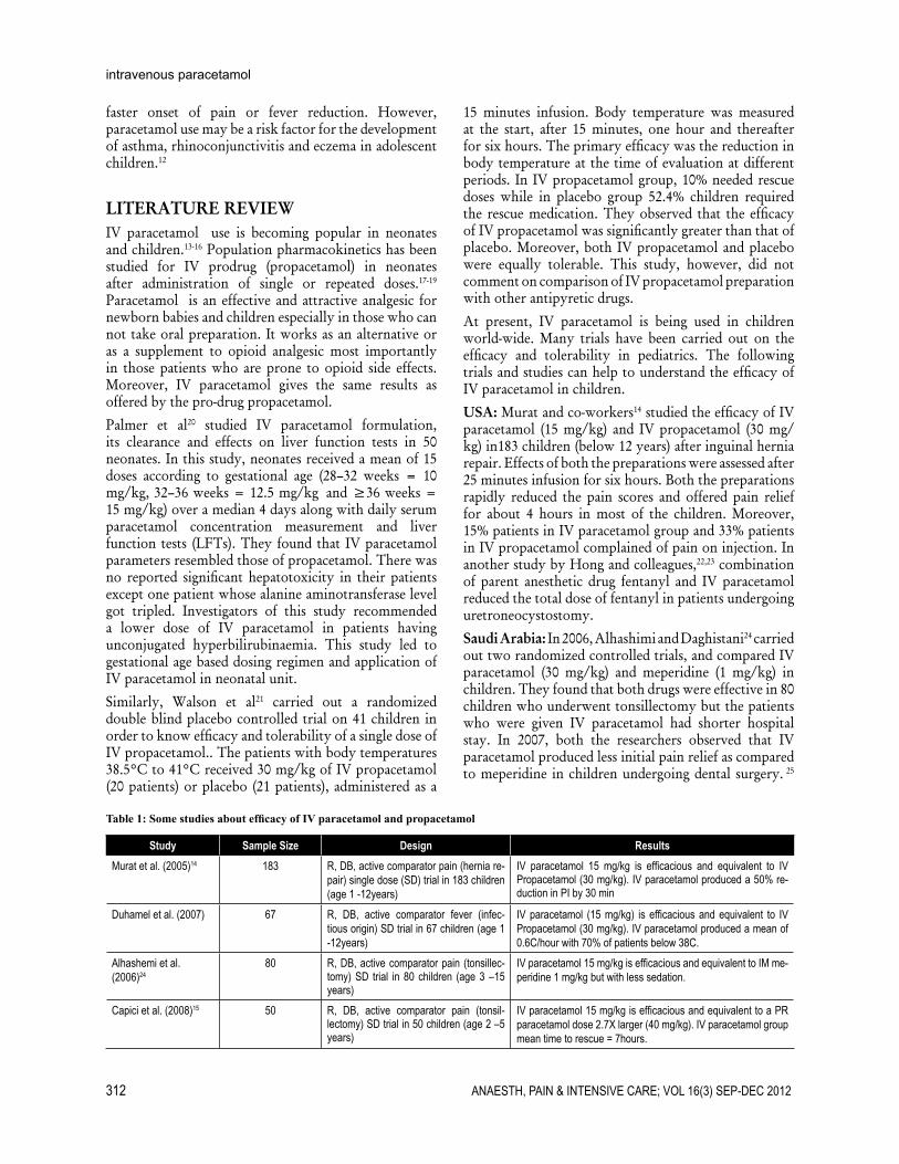
312 ANAESTH, PAIN & INTENSIVE CARE; VOL 16(3) SEP-DEC 2012
intravenous paracetamol
faster onset of pain or fever reduction. However, paracetamol use may be a risk factor for the development of asthma, rhinoconjunctivitis and eczema in adolescent children.12
LITERATURE REVIEWIV paracetamol use is becoming popular in neonates and children.13-16 Population pharmacokinetics has been studied for IV prodrug (propacetamol) in neonates after administration of single or repeated doses.17-19 Paracetamol is an effective and attractive analgesic for newborn babies and children especially in those who can not take oral preparation. It works as an alternative or as a supplement to opioid analgesic most importantly in those patients who are prone to opioid side effects. Moreover, IV paracetamol gives the same results as offered by the pro-drug propacetamol.
Palmer et al20 studied IV paracetamol formulation, its clearance and effects on liver function tests in 50 neonates. In this study, neonates received a mean of 15 doses according to gestational age (28–32 weeks = 10 mg/kg, 32–36 weeks = 12.5 mg/kg and ≥36 weeks = 15 mg/kg) over a median 4 days along with daily serum paracetamol concentration measurement and liver function tests (LFTs). They found that IV paracetamol parameters resembled those of propacetamol. There was no reported significant hepatotoxicity in their patients except one patient whose alanine aminotransferase level got tripled. Investigators of this study recommended a lower dose of IV paracetamol in patients having unconjugated hyperbilirubinaemia. This study led to gestational age based dosing regimen and application of IV paracetamol in neonatal unit.
Similarly, Walson et al21 carried out a randomized double blind placebo controlled trial on 41 children in order to know efficacy and tolerability of a single dose of IV propacetamol.. The patients with body temperatures 38.5°C to 41°C received 30 mg/kg of IV propacetamol (20 patients) or placebo (21 patients), administered as a
15 minutes infusion. Body temperature was measured at the start, after 15 minutes, one hour and thereafter for six hours. The primary efficacy was the reduction in body temperature at the time of evaluation at different periods. In IV propacetamol group, 10% needed rescue doses while in placebo group 52.4% children required the rescue medication. They observed that the efficacy of IV propacetamol was significantly greater than that of placebo. Moreover, both IV propacetamol and placebo were equally tolerable. This study, however, did not comment on comparison of IV propacetamol preparation with other antipyretic drugs.
At present, IV paracetamol is being used in children world-wide. Many trials have been carried out on the efficacy and tolerability in pediatrics. The following trials and studies can help to understand the efficacy of IV paracetamol in children.
USA: Murat and co-workers14 studied the efficacy of IV paracetamol (15 mg/kg) and IV propacetamol (30 mg/kg) in183 children (below 12 years) after inguinal hernia repair. Effects of both the preparations were assessed after 25 minutes infusion for six hours. Both the preparations rapidly reduced the pain scores and offered pain relief for about 4 hours in most of the children. Moreover, 15% patients in IV paracetamol group and 33% patients in IV propacetamol complained of pain on injection. In another study by Hong and colleagues,22,23 combination of parent anesthetic drug fentanyl and IV paracetamol reduced the total dose of fentanyl in patients undergoing uretroneocystostomy.
Saudi Arabia: In 2006, Alhashimi and Daghistani24 carried out two randomized controlled trials, and compared IV paracetamol (30 mg/kg) and meperidine (1 mg/kg) in children. They found that both drugs were effective in 80 children who underwent tonsillectomy but the patients who were given IV paracetamol had shorter hospital stay. In 2007, both the researchers observed that IV paracetamol produced less initial pain relief as compared to meperidine in children undergoing dental surgery. 25
Table 1: Some studies about efficacy of IV paracetamol and propacetamol
Study Sample Size Design ResultsMurat et al. (2005)14 183 R, DB, active comparator pain (hernia re-
pair) single dose (SD) trial in 183 children (age 1 -12years)
IV paracetamol 15 mg/kg is efficacious and equivalent to IV Propacetamol (30 mg/kg). IV paracetamol produced a 50% re-duction in PI by 30 min
Duhamel et al. (2007) 67 R, DB, active comparator fever (infec-tious origin) SD trial in 67 children (age 1 -12years)
IV paracetamol (15 mg/kg) is efficacious and equivalent to IV Propacetamol (30 mg/kg). IV paracetamol produced a mean of 0.6C/hour with 70% of patients below 38C.
Alhashemi et al. (2006)24
80 R, DB, active comparator pain (tonsillec-tomy) SD trial in 80 children (age 3 –15 years)
IV paracetamol 15 mg/kg is efficacious and equivalent to IM me-peridine 1 mg/kg but with less sedation.
Capici et al. (2008)15 50 R, DB, active comparator pain (tonsil-lectomy) SD trial in 50 children (age 2 –5 years)
IV paracetamol 15 mg/kg is efficacious and equivalent to a PR paracetamol dose 2.7X larger (40 mg/kg). IV paracetamol group mean time to rescue = 7hours.
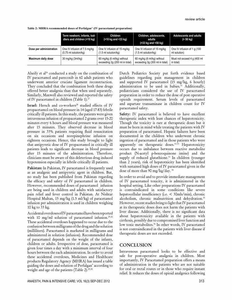
ANAESTH, PAIN & INTENSIVE CARE; VOL 16(3) SEP-DEC 2012 313
review article
Alseify et al26 conducted a study on the combination of IV paracetamol and parecoxib in 60 adult patients who underwent anterior cruciate ligament reconstruction. They concluded that the combination both these drugs offered better analgesia than that when used separately. Similarly, Maxwell also reviewed and reported the safety of IV paracetamol in children (Table 1).27
Israel: Hersch and co-workers28 studied effects of IV propacetamol on blood pressure in 14 (aged 17-83) febrile critically ill patients. In this study, the patients were given intravenous infusion of propacetamol 2 grams over 15-20 minutes every 6 hours and blood pressure was measured after 15 minutes. They observed decrease in blood pressure in 33% patients requiring fluid resuscitation on six occasions and norepinephrine infusion on eighteen occasions. Hence, this study brought to light that antipyretic dose of IV propacetamol in critically ill patients leads to significant decrease in blood pressure after 15 minutes of the administration. Therefore, clinicians must be aware of this deleterious drug induced hypotension especially in febrile critically ill patients.
Pakistan: In Pakistan, IV paracetamol is frequently used as an analgesic and antipyretic agent in children. But, no study has been published from Pakistan regarding the efficacy and safety of IV paracetamol in children. However, recommended doses of paracetamol infusion are being used in children and adults with satisfactory pain relief and fever control in Pakistan. At Nishtar Hospital Multan, 15 mg/kg (1.5 ml/kg) of paracetamol infusion per administration is used in children weighing 10 kg to 33 kg.
Accidental overdoses of IV paracetamol have been reported with 10 mg/ml solution of paracetamol infusion.29,30 These accidental overdoses happened to occur due to the confusion between milligrams of the drug and the solution (milliliters). Paracetamol is marketed in milligrams and administered in solution (infusion). Recommended dose of paracetamol depends on the weight of the infants, children or adults. Irrespective of dose, paracetamol is given four times a day with a minimum interval of four hours between the each administration. In order to avoid these accidental overdoses, Medicines and Healthcare products Regulatory Agency (MHRA) has issued a table guiding the doses and solution of Perfalgan® according to weight and age of the patients (Table 2).29,30
Dutch Pediatrics Society put forth evidence based guidelines regarding pain management in children and supported IV paracetamol (15 mg/kg, 6 hourly) administration to be used in babies.31 Additionally, pediatricians considered the use of IV paracetamol preparation in order to reduce the dose of post operative opioids requirement. Serum levels of paracetamol and aspartate transaminase in children count for IV paracetamol safety.
Safety: IV paracetamol is believed to have excellent therapeutic index with least chances of hepatotoxicity. Though the toxicity is rare at therapeutic doses but it must be born in mind while treating the patients with IV preparation of paracetamol. Hepatic failures have been documented in the children who underwent chronic ingestion of paracetamol and in those patients who were apparently on therapeutic doses.31-33 Hepatotoxicity occurs due to imbalance between reactive metabolite product (N-acetyl p-benzoquinone imine) and the supply of reduced glutathione.31 In children (younger than 2 years), risk of hepatotxicity has been identified with sustained high doses of IV paracetamol given in the dose of more than 90 mg/kg/day. 34
In order to avoid and to provide immediate management of IV paracetamol toxicity, it is administered in the hospital setting. Like other preparations IV paracetamol is contraindicated in some conditions like severe hepatocellular insufficiency (i.e. < 30mls/min), chronic alcoholism, chronic malnutrition and dehydration.35 However, recent studies bring to light that IV paracetamol at its therapeutic doses does not harm the patients with liver disease. Additionally, there is no significant data about hepatotoxicity available in the patients with cirrhosis, possibly due to compromised liver function and low toxic metabolites.36 In other words, IV paracetamol is not contraindicated in the patients with liver disease if therapeutic doses are not exceeded.
CONCLUSIONIntravenous paracetamol looks to be effective and safe for post-operative analgesia in children. Most importantly, IV Paracetamol preparation offers a means of administration in the patients who are not suitable for oral or rectal routes or in those who require instant relief. It reduces the doses of opioid analgesics following
Table 2: MHRA recommended doses of Perfalgan® (IV paracetamol preparation)
Term newborn, infants, tod-dlers and children (<10 kg)
Children (>10 kg and <33 kg)
Children, adolescents, adults
(>33 kg and <50 kg)
Adolescents and adults (> 50 kg)
Dose per administration One IV infusion of 7.5 mg/kg (0.75 ml solution/kg)
One IV infusion of 15 mg/kg (1.5 ml solution/kg)
One IV infusion of 15 mg/kg (1.5 ml solution/kg)
One IV infusion of 1 g (100 ml solution)
Maximum daily dose 30 mg/kg (3ml/kg) 60 mg/kg (6 ml/kg) without exceeding 2g (200 ml in total)
60 mg/kg (6 ml/kg) without exceeding 3g (300 ml in total)
Must not exceed 4 g (400 ml in total)
314 ANAESTH, PAIN & INTENSIVE CARE; VOL 16(3) SEP-DEC 2012
intravenous paracetamol
surgical procedures and subsequently the post-operative sedation. Doses of IV paracetamol are different from those taken orally. It is safe in pediatrics but special care is required while calculating doses for neonates and critically ill patients. No hepatotoxicity has been reported
1. Routledge P, Vale JA, Bateman DN, Johnston GD, Jones A, Judd A, et al. Paracetamol (acetaminophen) poisoning. No need to change current guidelines to accident departments. BMJ. 1998 Dec 12;317(7173):1609–10. [Medline]
2. Sheen CL, Dillon JF, Bateman DN, Simpson KJ, Macdonald TM. Paracetamol toxicity: epidemiology, prevention and costs to the health-care system. QJM. 2002;95(9):609-619. [Medline]
3. Ogbru O. Acetaminophen, Tylenol and Others [Internet]. 2007 Sep 21. Available from: http://www.medicinenet.com/acetaminophen/article.htm. (Accessed on 25 June 2012).
4. Darce’ K. Cadence Pharma wins FDA approval for IV acetaminophen [Internet]. 2010 Nov 2. Available from: http://www.utsandiego.com/news/2010/nov/02/cadence-pharma-wins-fda-approval-iv-acetaminophen/%20 (Accessed on 25 June 2012).
5. Viscusi RR, Singla N, Gonzalez A, Saad N, Stepanian J. IV acetaminophen improves pain management and reduces opioid requirements in surgical patients: a review of the clinical data and case-based presentations [Internet]. New York: McMahon Publishing; 2012 April. Available from: www.pharmacypracticenews.com/download/SR122_WM.pdf. (Accessed on 29 November 2012).
6. Granry JC, Rod B, Monrigal JP, Merckx J, Berniere J, Jean N, Boccard E. The analgesic efficacy of an injectable prodrug of acetaminophen in children after orthopaedic surgery. Paediatr Anaesth. 1997;7:445-9. [Medline]
7. Prins SA, Van Dijk M, Van Leeuwen P, Searle S, Anderson BJ, Tibboel D, Mathot RA. Pharmacokinetics and analgesic effects of intravenous propacetamol vs rectal paracetamol in children after major craniofacial surgery. Paediatr Anaesth. 2008;18:582-92. [Medline]
8. Zhou TJ, Tang J, White PF. Propacetamol versus ketorolac for treatment of acute postoperative pain after total hip or knee replacement. Anesth Analg. 2001;92(6):1569-75. [Medline]
9. Duhamel JF, Le Gall E, Dalphin ML, Payen-Champenois C. Antipyretic efficacy and safety of a single intravenous administration of 15 mg/kg paracetamol versus 30 mg/kg propacetamol in children with acute fever due to infection. Int J Clin Pharmacol Ther. 2007 Apr;45(4):221-9. [Medline]
10. Graham GG, Scott KF. Mechanism of action of paracetamol. Am J Ther. 2005 Jan-Feb;12(1):46-55.
11. Kuczynska J. What is the evidence to support the use of IV paracetamol for the treatment of pain? [Internet]. 2011 Jan 4. Available from: http://www.nelm.nhs.uk/en/NeLM-Area/Evidence/Medicines-Q--A/What-is-the-evidence-to-support-the-use-of-IV-paracetamol-for-the-treatment-of-pain/. (Accessed on 25 June 2012).
12. Beasley RW, Clayton TO, Crane J, Lai CK, Montefort SR, Mutius Ev, Stewart AW; ISAAC Phase Three Study Group. Acetaminophen Use and Risk of
Asthma, Rhinoconjunctivitis, and Eczema in Adolescents: International Study of Asthma and Allergies in Childhood Phase Three. Am J Respir Crit Care Med. 2011 Jan 15;183(2):171-8. doi: 10.1164/rccm.201005-0757OC. [Medline]
13. Palmer GM, Chen SP, Smith KR, Hardikar W. Introduction and audit of intravenous paracetamol at a tertiary pediatric teaching hospital. Anaesth Intensive Care. 2007 Oct;35(5):702-6. [Medline]
14. Murat I, Baujard C, Foussat C, Guyot E, Petel H, Rod B, Ricard C. Tolerance and analgesic efficacy of a new i.v. paracetamol solution in children after inguinal hernia repair. Paediatr Anaesth. 2005 Aug;15(8):663-70. [Medline]
15. Capici F, Ingelmo PM, Davidson A, Sacchi CA, Milan B, Sperti LR, et al. Randomized controlled trial of duration of analgesia following intravenous or rectal acetaminophen after adenotonsillectomy in children. Br J Anaesth. 2008 Feb;100(2):251-5 [Medline]
16. Kumpulainen E, Kokki H, Halonen T, Heikkinen M, Savolainen J, Laisalmi M. Paracetamol (acetaminophen) penetrates readily into the cerebrospinal fluid of children after intravenous administration. Pediatrics. 2007 Apr;119(4):766-71. [Medline]
17. Allegaert K, Van der Marel CD, Debeer A, Pluim MA, Van Lingen RA, Vanhole C, et al. Pharmacokinetics of single dose intravenous propacetamol in neonates: effect of gestational age. Arch Dis Child Fetal Neonatal Ed. 2004 Jan;89(1):F25-8. [Medline]
18. Anderson BJ, Pons G, Autret-Leca E, Allegaert K, Boccard E. Pediatric intravenous paracetamol (propacetamol) pharmacokinetics: a population analysis. Paediatr Anaesth. 2005 Apr;15(4):282-92. [Medline]
19. Allegaert K, Anderson BJ, Naulaers G, de Hoon J, Verbesselt R, Debeer A, et al. Intravenous paracetamol (propacetamol) pharmacokinetics in term and preterm neonates. Eur J Clin Pharmacol. 2004 May;60(3):191-7. [Medline]
20. Palmer GM, Atkins M, Anderson BJ, Smith KR, Culnane TJ, McNally CM, et al. IV acetaminophen pharmacokinetics in neonates after multiple doses. Br J Anaesth. 2008 Oct;101(4):523-30. doi: 10.1093/bja/aen208. [Medline]
21. Walson PD, Jones J, Chesney R, Rodarte A. Antipyretic efficacy and tolerability of a single intravenous dose of the acetaminophen prodrug propacetamol in children: a randomized, double-blind, placebo-controlled trial. Clin Ther. 2006 May;28(5):762-9. [Medline]
22. Hong JY, Kim WO, Koo BN, Cho JS, Suk EH, Kil HK. Fentanyl-sparing effect of acetaminophen as a mixture of fentanyl in intravenous parent-/nurse-controlled analgesia after pediatric ureteroneocystostomy. Anesthesiology. 2010 Sep;113(3):672-7. doi: 10.1097/ALN.0b013e3181e2c34b. [Medline]
23. Hong JY, Won Han S, Kim WO, Kil HK. Fentanyl
sparing effects of combination ketorolac and acetaminophen for outpatient inguinal hernia repair in children. J Urol. 2010 Apr;183(4):1551-5. doi: 10.1016/j.juro.2009.12.043. [Medline]
24. Alhashemi JAS, Daghistani MF. Effects of intraoperative IV acetaminophen vs IM meperidine on post-tonsillectomy pain in children. Br J Anaesth. 2006 Jun;96(6):790-5. [Medline]
25. Alhashemi JAS, Daghistani MF. Effects of intraoperative intravenous acetaminophen vs intramuscular meperidine on pain and discharge time after pediatric dental restoration. Eur J Anaesthesiol. 2007 Feb;24(2):128-33. [Medline]
26. Elseify ZA, El-Khattab SO, Khattab AM, Atta EM, Ajjoub LF. Combined parecoxib and I.V. paracetamol provides additional analgesic effect with better postoperative satisfaction in patients undergoing anterior cruciate ligament reconstruction. Saudi J Anaesth. 2011 Jan;5(1):45-9. doi: 10.4103/1658-354X.76510. [Medline]
27. Maxwell A. IV acetaminophen in children: Is there evidence for safety and efficacy [Internet]. Philadelphia: The children’s hospital of Philadelphia; 2007. Available from: www.pac.chla-accm.org/Abstracts/.../48thMaxwellapap.pdf. (Accessed on 30 November 2012).
28. Hersch M, Raveh D, Izbicki G. Effect of intravenous propacetamol on blood pressure in febrile critically ill patients. Pharmacotherapy. 2008 Oct;28(10):1205-10. doi: 10.1592/phco.28.10.1205. [Medline]
29. Intravenous paracetamol (Perfalgan): risk of accidental overdose, especially in infants and neonates. MHRA. 2010 July;12(3). [Medline]
30. Van Ganzewinkel CJLM, Mohns T, van Lingen RA, Derijks LJJ, Andreissen P. Paracetamol serum concentrations in preterm infants treated with paracetamol intravenously: a case series. J Med Case Rep. 2012 Jan 4;6(1):1. [Medline]
31. Heubi J, Barbacci MB, Zimmerman HJ. Therapeutic misadventures with acetaminophen: hepatotoxicity after multiple doses in children. J Pediatr.1998;132:22-27. [Medline]
32. Miles FK, Kamath R, Dorney SFA, Gaskin KJ, O’Loughlin EV. Accidental paracetamol overdosing and fulminant hepatic failure in children. Med J Aust. 1999;171:472-475. [Medline]
33. Allegaert K, Anderson BJ, Naulaers G, de Hoon J, Verbesselt R, Debeer A, et al. Intravenous paracetamol (propacetamol) pharmacokinetics in term and preterm neonates. Eur J Clin Pharmacol. 2004;60:191-197. [Medline]
34. Kearns GL, Leeder JS, Wasserman GS. Acetaminophen overdose with therapeutic intent. J Pediatr. 1998;132:5-8. [Medline]
35. Bristol Myers Squibb Pharmaceuticals. Perfalgan Australian approved product information. 2004.
36. McIntyre N. The general management of liver disease. In: Bircher J, Benhamou JP, McIntyre N, editors. Oxford textbook of clinical hepatology. Oxford: Oxford University Press. 1999:1917-23.
REFERENCES
with analgesic and antipyretic doses of IV paracetamol. However, it may lead to drug induced hypotension in critically ill patients. Therefore, health care providers must bear in mind the circulatory status of the patients before embarking on high doses of IV paracetamol.
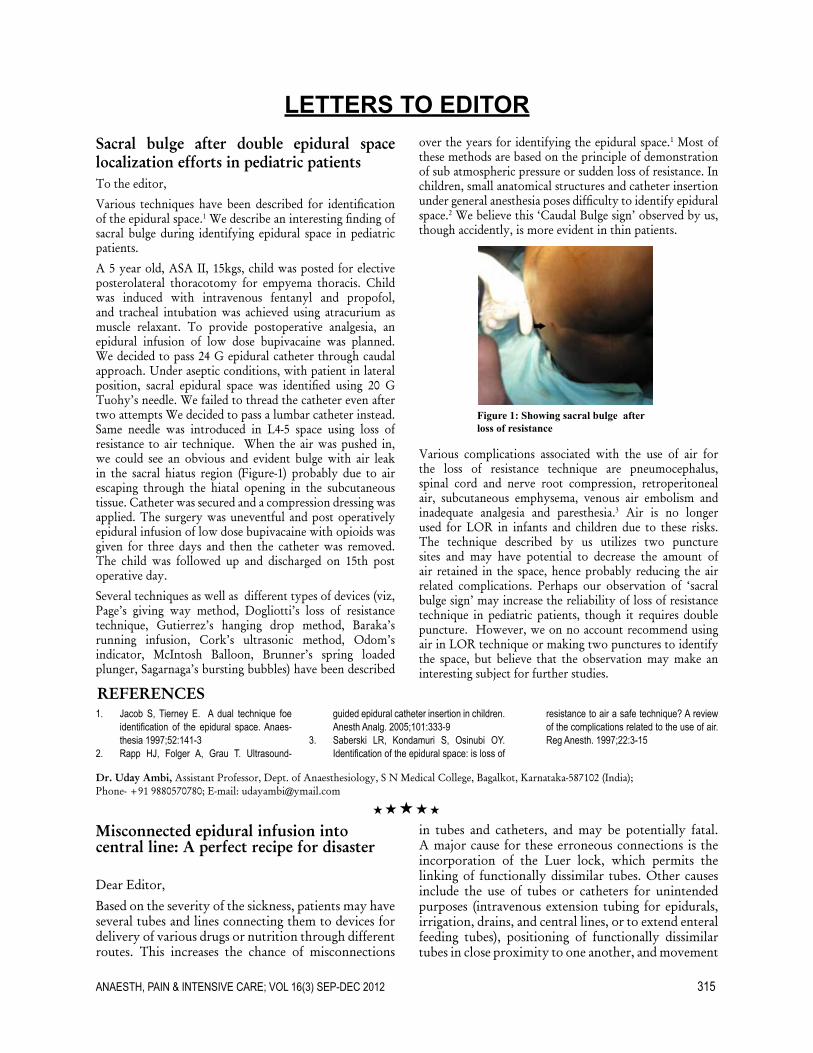
LETTERS TO EDITORSacral bulge after double epidural space localization efforts in pediatric patientsTo the editor,
Various techniques have been described for identification of the epidural space.1 We describe an interesting finding of sacral bulge during identifying epidural space in pediatric patients.
A 5 year old, ASA II, 15kgs, child was posted for elective posterolateral thoracotomy for empyema thoracis. Child was induced with intravenous fentanyl and propofol, and tracheal intubation was achieved using atracurium as muscle relaxant. To provide postoperative analgesia, an epidural infusion of low dose bupivacaine was planned. We decided to pass 24 G epidural catheter through caudal approach. Under aseptic conditions, with patient in lateral position, sacral epidural space was identified using 20 G Tuohy’s needle. We failed to thread the catheter even after two attempts We decided to pass a lumbar catheter instead. Same needle was introduced in L4-5 space using loss of resistance to air technique. When the air was pushed in, we could see an obvious and evident bulge with air leak in the sacral hiatus region (Figure-1) probably due to air escaping through the hiatal opening in the subcutaneous tissue. Catheter was secured and a compression dressing was applied. The surgery was uneventful and post operatively epidural infusion of low dose bupivacaine with opioids was given for three days and then the catheter was removed. The child was followed up and discharged on 15th post operative day.
Several techniques as well as different types of devices (viz, Page’s giving way method, Dogliotti’s loss of resistance technique, Gutierrez’s hanging drop method, Baraka’s running infusion, Cork’s ultrasonic method, Odom’s indicator, McIntosh Balloon, Brunner’s spring loaded plunger, Sagarnaga’s bursting bubbles) have been described
over the years for identifying the epidural space.1 Most of these methods are based on the principle of demonstration of sub atmospheric pressure or sudden loss of resistance. In children, small anatomical structures and catheter insertion under general anesthesia poses difficulty to identify epidural space.2 We believe this ‘Caudal Bulge sign’ observed by us, though accidently, is more evident in thin patients.
Various complications associated with the use of air for the loss of resistance technique are pneumocephalus, spinal cord and nerve root compression, retroperitoneal air, subcutaneous emphysema, venous air embolism and inadequate analgesia and paresthesia.3 Air is no longer used for LOR in infants and children due to these risks. The technique described by us utilizes two puncture sites and may have potential to decrease the amount of air retained in the space, hence probably reducing the air related complications. Perhaps our observation of ‘sacral bulge sign’ may increase the reliability of loss of resistance technique in pediatric patients, though it requires double puncture. However, we on no account recommend using air in LOR technique or making two punctures to identify the space, but believe that the observation may make an interesting subject for further studies.
Figure 1: Showing sacral bulge after loss of resistance
1. Jacob S, Tierney E. A dual technique foe identification of the epidural space. Anaes-thesia 1997;52:141-3
2. Rapp HJ, Folger A, Grau T. Ultrasound-
guided epidural catheter insertion in children. Anesth Analg. 2005;101:333-9
3. Saberski LR, Kondamuri S, Osinubi OY. Identification of the epidural space: is loss of
resistance to air a safe technique? A review of the complications related to the use of air. Reg Anesth. 1997;22:3-15
REFERENCES
Dr. Uday Ambi, Assistant Professor, Dept. of Anaesthesiology, S N Medical College, Bagalkot, Karnataka-587102 (India); Phone- +91 9880570780; E-mail: [email protected]
Misconnected epidural infusion into central line: A perfect recipe for disaster
Dear Editor,
Based on the severity of the sickness, patients may have several tubes and lines connecting them to devices for delivery of various drugs or nutrition through different routes. This increases the chance of misconnections
in tubes and catheters, and may be potentially fatal. A major cause for these erroneous connections is the incorporation of the Luer lock, which permits the linking of functionally dissimilar tubes. Other causes include the use of tubes or catheters for unintended purposes (intravenous extension tubing for epidurals, irrigation, drains, and central lines, or to extend enteral feeding tubes), positioning of functionally dissimilar tubes in close proximity to one another, and movement
ANAESTH, PAIN & INTENSIVE CARE; VOL 16(3) SEP-DEC 2012 315
1. The Joint Commission, Joint Commission International, World Health Organisation. Avoiding catheter and tubing mis-connections [Internet]. WHO Press;2007. Available from: http://www.who.int/patientsafety/solutions/patientsafety/PS-Solution7.pdf.
2. The Joint Commission. Tubing misconnections—a persistent and potentially deadly occurrence [Internet]. Sentinel Event Alert;2006 Apr 3 [cited 2009 Nov 4].
Available from: http://www.jointcommission.org/assets/1/18/SEA_36.PDF
3. Knudsen K, Beckman Suurküla M, Blomberg S, Sjövall J, Edvardsson N. Central nervous and cardiovascular effects of IV infusions of ropivacaine, bupivacaine and placebo in volunteers. Br J Anaesth. 1997;78(5):507-14. [Medline]
4. Pennysylvania Patient Safety Advisory. Tubing misconnections: making the
connection to patient safety [Internet]. Pa Patient Saf Advis 2010 Jun;7(2):41-5. Available from: http://patientsafetyauthority.org/ADVISORIES/AdvisoryLibrary/2010/Jun7(2)/Pages/41.aspx.
5. Guenter P, Hicks RW, Simmons D, Crowley J, Joseph S, Croteau R et al. Enteral feeding misconnections: a consortium position statement. Jt Comm J Qual Patient Saf 2008 May;34(5):285-92,245.
REFERENCES
Rudrashish Haldar, MBBS, MD, PDCC and Prakhar Gyanesh, MBBS, MD, PDCCDept Of Anaesthesiology, SGPGI, Lucknow (India); [email protected]
of the patient from one setting or service to another.1 We came across such a potentially dangerous situation, where the extension tubing of the syringe containing epidural drug was inadvertently connected with one of the lumens of a central line.
A 62-year-old patient underwent Whipple’s procedure under combined general and epidural anaesthesia. After shifting the patient to the post anesthesia care unit, various lines and tubings were attached by the on-duty nurse. At the time of hand-over, it was discovered that the extension tubing for connecting the syringe containing the epidural solution (50 ml solution of 0.25% ropivacaine and 2 mcg/ml fentanyl), had been attached with one of the ports of the double lumen central line via another smaller extension tubing. This smaller extension tubing (20 cm) was attached to the central line for CVP monitoring. Through this connection, the epidural solution had been running intravenously for the past three hours at the rate of 5 ml/hr. The infusion was immediately stopped, and the extension tubing disconnected. The patient was monitored for any signs of toxicity and a close watch was kept on the vitals of the patient. Fortunately, no untoward incident occurred.
The Sentinel Event Alert, issued by Joint Commission
in 2006, had cited different tubing and catheter misconnections, leading to eight deaths and one permanent loss of function.2 We were fortunate that no serious harm occurred to our patient, probably because the dose of ropivacaine (37.5 mg), administered inadvertently, was low. Moreover, the extension tubing, which was not primed with the epidural solution, would have accommodated some amount of the drug. Ropivacaine itself provided a degree of safety due to its lesser central nervous system and cardiovascular system effects in humans, as compared to bupivacaine.3 This incident emphasizes the necessity of anaesthesia providers to be extremely vigilant during connecting disconnected tubings, during and after transport. Rechecking and tracing of tubings to their origin, as a part of hand over process, saved the day for us. We would also like to voice our support for the recommendations of equipment design solution, e.g. labeling of high-risk catheter, routing tubings with standardized directions and limiting the use of adapters.4 The risk reduction strategy of labeling or color-coding feeding tubes and connectors, with necessary modifications, and educating the staff on this system,5 needs to be given due consideration.
letters to editor
316 ANAESTH, PAIN & INTENSIVE CARE; VOL 16(3) SEP-DEC 2012
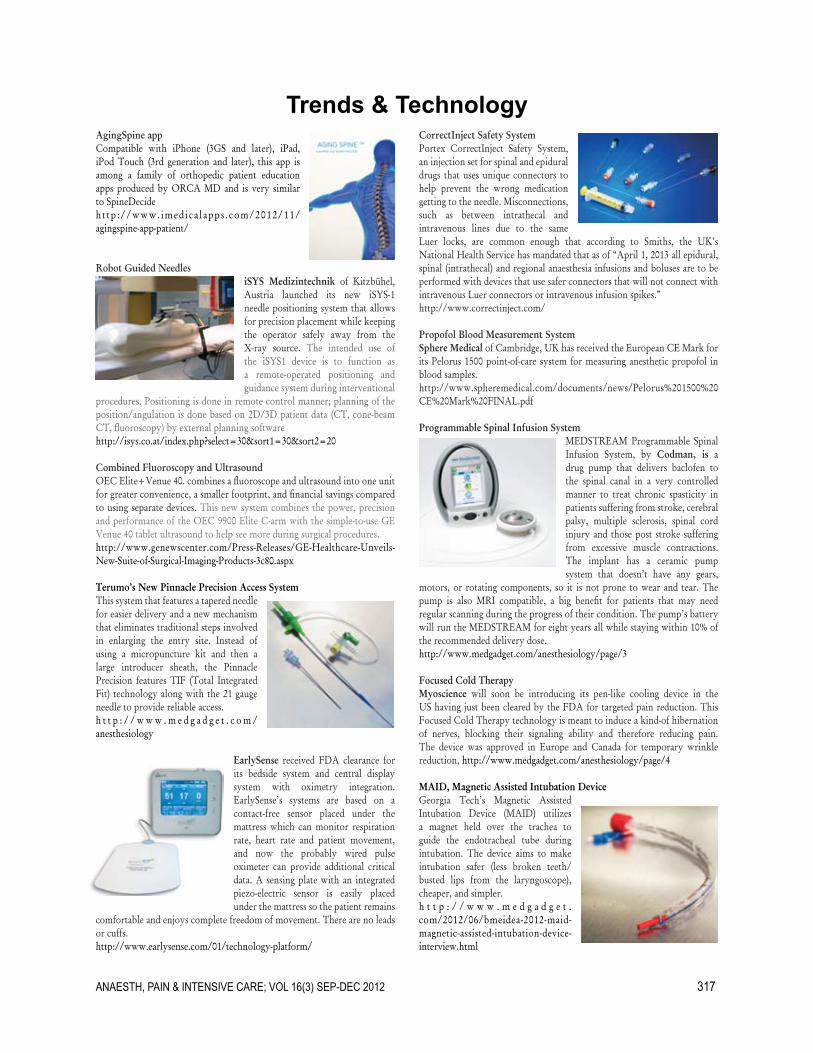
Trends & TechnologyAgingSpine app Compatible with iPhone (3GS and later), iPad, iPod Touch (3rd generation and later), this app is among a family of orthopedic patient education apps produced by ORCA MD and is very similar to SpineDecideht tp ://www. imed ica l apps . com/2012/11/agingspine-app-patient/
Robot Guided NeedlesiSYS Medizintechnik of Kitzbühel, Austria launched its new iSYS-1 needle positioning system that allows for precision placement while keeping the operator safely away from the X-ray source. The intended use of the iSYS1 device is to function as a remote-operated positioning and guidance system during interventional
procedures. Positioning is done in remote control manner; planning of the position/angulation is done based on 2D/3D patient data (CT, cone-beam CT,fluoroscopy)byexternalplanningsoftwarehttp://isys.co.at/index.php?select=30&sort1=30&sort2=20
Combined Fluoroscopy and Ultrasound OECElite+Venue40.combinesafluoroscopeandultrasoundintooneunitforgreaterconvenience,asmallerfootprint,andfinancialsavingscomparedto using separate devices. This new system combines the power, precision and performance of the OEC 9900 Elite C-arm with the simple-to-use GE Venue 40 tablet ultrasound to help see more during surgical procedures.http://www.genewscenter.com/Press-Releases/GE-Healthcare-Unveils-New-Suite-of-Surgical-Imaging-Products-3c80.aspx
Terumo’s New Pinnacle Precision Access SystemThis system that features a tapered needle for easier delivery and a new mechanism that eliminates traditional steps involved in enlarging the entry site. Instead of using a micropuncture kit and then a large introducer sheath, the Pinnacle Precision features TIF (Total Integrated Fit) technology along with the 21 gauge needle to provide reliable access. h t t p : / / w w w . m e d g a d g e t . c o m /anesthesiology
EarlySense received FDA clearance for its bedside system and central display system with oximetry integration. EarlySense’s systems are based on a contact-free sensor placed under the mattress which can monitor respiration rate, heart rate and patient movement, and now the probably wired pulse oximeter can provide additional critical data. A sensing plate with an integrated piezo-electric sensor is easily placed under the mattress so the patient remains
comfortable and enjoys complete freedom of movement. There are no leads or cuffs.http://www.earlysense.com/01/technology-platform/
CorrectInject Safety System Portex CorrectInject Safety System, an injection set for spinal and epidural drugs that uses unique connectors to help prevent the wrong medication getting to the needle. Misconnections, such as between intrathecal and intravenous lines due to the same Luer locks, are common enough that according to Smiths, the UK’s National Health Service has mandated that as of “April 1, 2013 all epidural, spinal (intrathecal) and regional anaesthesia infusions and boluses are to be performed with devices that use safer connectors that will not connect with intravenous Luer connectors or intravenous infusion spikes.”http://www.correctinject.com/
Propofol Blood Measurement SystemSphere Medical of Cambridge, UK has received the European CE Mark for its Pelorus 1500 point-of-care system for measuring anesthetic propofol in blood samples.http://www.spheremedical.com/documents/news/Pelorus%201500%20CE%20Mark%20FINAL.pdf
Programmable Spinal Infusion SystemMEDSTREAM Programmable Spinal Infusion System, by Codman, is a drug pump that delivers baclofen to the spinal canal in a very controlled manner to treat chronic spasticity in patients suffering from stroke, cerebral palsy, multiple sclerosis, spinal cord injury and those post stroke suffering from excessive muscle contractions. The implant has a ceramic pump system that doesn’t have any gears,
motors, or rotating components, so it is not prone to wear and tear. The pump is also MRI compatible, a big benefit for patients that may needregular scanning during the progress of their condition. The pump’s battery willruntheMEDSTREAMforeightyearsallwhilestayingwithin10%ofthe recommended delivery dose.http://www.medgadget.com/anesthesiology/page/3
Focused Cold TherapyMyoscience will soon be introducing its pen-like cooling device in the US having just been cleared by the FDA for targeted pain reduction. This Focused Cold Therapy technology is meant to induce a kind-of hibernation of nerves, blocking their signaling ability and therefore reducing pain. The device was approved in Europe and Canada for temporary wrinkle reduction, http://www.medgadget.com/anesthesiology/page/4
MAID, Magnetic Assisted Intubation DeviceGeorgia Tech’s Magnetic Assisted Intubation Device (MAID) utilizes a magnet held over the trachea to guide the endotracheal tube during intubation. The device aims to make intubation safer (less broken teeth/busted lips from the laryngoscope), cheaper, and simpler.h t t p : / / w w w . m e d g a d g e t .com/2012/06/bmeidea-2012-maid-magnetic-assisted-intubation-device-interview.html
ANAESTH, PAIN & INTENSIVE CARE; VOL 16(3) SEP-DEC 2012 317
Q. 1: A 19 year old male presented to ER with history of ingestion of some toxic substance 2 hours ago. The patient was drowsy and had rotten fish smell from mouth. On examination the pulse was thready with a rate of 124/min, BP: 90/60 mmHg and RR: 40/min. Which of the following poisons can cause these manifestations?a. Cyanideb. Organophosphorus (OP)c. Chloral hydrated. Celphos (aluminium hydroxide)
Q. 2: Miosis is not a feature of which of the following poisons?a. Opioidsb. Organophosphorus poisoningc. Organochlorine poisoningd. Phencyclidine
Q. 3: Death within 24 hours of celphos poisoning (aluminium hydroxide) occurs frequently due to:a. Acute Respiratory failureb. Cardiogenic shockc. ARDSd. Septicemia
Q.4: Hyperthermia is not a feature of which of the following poisons?
a. Ecstasy [methylenedioxymeth-amphetamine (MDMA)]
b. Cocainec. Alcohold. Haloperidol
Q. 5: Which of the following is not observed in OP poisoning?a. Bradycardia and hypotensionb. Hyperpyrexia and hypoglycemiac. Increased sweating, salivation, lacrimation and
bronchoconstrictiond. Hypertension and tachycardia.
CLINIQUIZ
Poisoning Pranav Bansal*, Gaurav Jain**, Mahipal Singh*, Meenu Agarwal*
* Dept. of Anaesthesiology, BPS GMCW, Khanpur Kalan, Sonepat Haryana (India)** Teerthanker Mahaveer Medical College, Moradabad, U.P. (India)
Q. 6: Which of the following is not a clinical phase of OP poisoning?a. Attention deficit disorderb. Intermediate syndromec. Delayed polyneuropathyd. Acute cholinergic crisisQ. 7: Which of the following is not helpful for the management of intermediate syndrome?a. Fresh frozen plasma (FFP)b. Dialysisc. Obidoximed. Ventilatory Support
Q. 8: Which of the following regarding cyanide poisoning is false?a. It can be absorbed rapidly by via skin absorption
or inhalation.b. Venous oxygen saturation may be elevated
(>90%. in severe poisoningsc. Activated Charcoal has high affinity for binding
ingested cyanide d. Hydroxycobalamin is safer than amyl nitrite as
an antidote
Q. 9: Which of the following is not a specific antidote for treatment in poisoning?a. Glucagonb. Fomepizole (4-methylpyrazole)c. Levosimendand. Digibind
Q. 10: Which acute poisoning of the following is not an indication for hemodialysis?e. Methanolf. Arsenicg. Salicylatesh. Lithium
ANSWERS
Ans. 1 (d) Hemodynamic and respiratory disturbances are similar following ingestion of any of these poisons. The diagnostic clue may be reached by the odour of
318 ANAESTH, PAIN & INTENSIVE CARE; VOL 16(3) SEP-DEC 2012
poison. Rotten fish like odour is present after ingestion of celphos, Almond like odour with cyanide, fruity or pear like odour with chloral hydrate and garlic like odour with OP poisons.
Ans. 2 (c) Organochlorine poisoning is associated with mydriasis, the rest are associated with miosis.
Ans. 3 (b) Death in celphos poisoning is commonly due to due to pump failure and progressive fall in ejection fraction and cardiac output leading to dialated cardiomyopathy and
cardiogenic shock.
Ans. 4 (c) Overdose of MAO inhibitors may cause agitation, hyperactivity, myoclonus, and hyperthermia (‘serotonin syndrome’). Antipsychotic agents can cause rigidity and hyperthermia (neuroleptic malignant syndrome). Alcohol poisoning is commonly associated with hypothermia.
Ans. 5 (b) Hypotension and bradycardia, though is seen only in severe poisoning, occur due to direct myocardial depressant effect of acetyl choline following widespread anticholinesterase inhibition (muscarinic action). Hypertension and tachycardia may occur due to ganglionic stimulation (nicotinic action). Abdominal cramps, diarrhoea, lacrimation, bronchoconstriction, sweating and miosis are muscarinic actions of OP poison. Hyperpyrexia is not a feature of OP poisoning.
Ans. 6 (a) The clinical phase after OP poisoning consists of acute cholinergic crisis Intermediate syndrome and delayed polyneuropathy. Attention deficit disorder has been reported in few cases following recovery from OP
poisoning which may persist for months.
Ans 7 (b) FFP, obidoxime and atropine have shown to reduce the severity of intermediate syndrome while ventilatory support may be required if respiratory muscle weakness is pronounced. Dialysis does not help in reducing the severity of symptoms as confirmatory change in acetylcholine receptors have ensued following irreversible binding of receptors with acetylcholine esterase.
Ans. 8 (c) Cyanide inhibits cytochrome oxidase and blocks tissue oxygen uptake so venous oxygen saturation rises paradoxically. Acitvated charcoal has a low affinity for binding cyanide, though the usual doses of 60–100 g are adequate to bind typically ingested lethal doses (100–200 mg), so is recommended in cases of cyanide ingestion. Nitrites induce methemoglobinemia which binds cyanide but also may induce hypotension and may raise methemoglobin to dangerous levels.
Ans. 9 (c) Glucagon is an antidote for poisoning with beta blockers, fomeprizole for methanol poisoning, Digibind (digoxin Immune Fab) for digoxin. Levosimendan is a calcium sensitizer indicated in congestive cardiac failure.
Ans. 10 (b) Hemodialysis is indicated in poisoning with methanol, if serum level > 50 mg/dL; in salicylate poisoning with CNS symptoms, level > 100 mg/dL (acute overdose) or > 60 mg/dL (chronic intoxication) and in lithium poisoning if serum level > 4 mEq/lit for more than 12 hours after last dose. Hemodialysis has no role in arsenic poisoning.
1. Olson KR. Poisoning. in Current medical diagnosis and treatment 2011.Ch 38. pp1499-1527.
2. Poojara L, Vasudevan D, Kumar ASA, Kamat V. Organophosphate poisoning, diagnosis of
intermediate syndrome. IJCCM 2003; 17(4): 94-102.
3. Cherian MA, Roshini C, Peter JV, Cherian AM. Oximes in organophsphorus poisons. IJCCM 2005; 9(3): 155-63.
4. Clark RF. Insecticides: Organic Phosphorus Compounds and Carbamates. Ch 109. Civetta toxicology 8th ed. pp1497-1511.
REFERENCES
cliniquiz
ANAESTH, PAIN & INTENSIVE CARE; VOL 16(3) SEP-DEC 2012 319
Q 1: Which of the following statement is correct in terms of asthma characteristic or prevalence?a. Asthma is a disease of reversible airway obstruction
characterized by a triad of bronchial smooth muscle hyperplasia, airway inflammation, and increased secretions.
b. The mortality rate of asthma is increasing and in the UK around 14000 patients died each year.
c. Inability to complete sentences in one breath is an important characteristic of fatal asthma
d. Arterial PaCO2 (35-45 mmHg) is the characteristic of life threatening asthma during an acute attack.
Q 2: The pathopysiological characteristic of acute severe asthma is characterized bya. Impaired gas trapping b. Diaphragmatic hyperactivityc. Sever Hyperinflation and lung volume reaches up to
total lung capacityd. Decrease air entry on auscultation.
Q 3: The best pharmacological therapy in acute asthma is described as:a. Magnesium sulphate as infusion till bronchospasm
relieved.b. Systemic steroid in adequate doses to all patients with
life threatening asthma.c. Inhaled epinephrine should be avoided. d. Inhaled long beta agonist.
Q 4: What is the indication of mechanical ventilation in acute severe asthma?a. Silent chest.b. Intrinsic PEEP.c. Inability to complete one sentence in one breath.d. Fatigue and somnolence.
Q 5: What is the initial mechanical ventilation strategy in treating status asthmaticus?a. Correction of acidosis is the initial target.b. Decrease in inspiratory time in order to reduce
dynamic hyperinflation.c. Use of muscle relaxant is the treatment of choice in
first 48 hours to reduce the work of breathing.d. Frequent endobronchial suctioning helps in reduction
of bronchospasm
After 2 hours of mechanical ventilation, arterial blood gas improved to 7.24, PaCO2 65 mmHg, PaO2 120 mmHg & HCO3 22 mEq/L. The ventilation setting was assist controlled mode, respiratory rate 12/min, tidal volume 7 ml/kg, inspiratory flow rate 70 liter/minute, PEEP 05 cmH2O and FiO2 of 1.0.Q 6: What will be the next strategy for this patient?a. Allow permissive hypercapnia if plateau pressure is
>30 cmH2Ob. Continue with same oxygen therapy for one hour
morec. Reduce tidal volume if peak airway pressure
increases.d. Increase respiratory rate
Q 7: This patient has intrinsic PEEP of more than 10cmH2O. What is the best strategy to reduce this intrinsic PEEP?a. Deep sedation and relaxation.b. Pressure control ventilationc. Low inspiratory time in respiratory cycled. Keep extrinsic PEEP low to prevent barotrauma.
Q 8: What is the correct arterial blood gas analysis in mild asthma?a. Hypoxemia, hypercapnia and respiratory acidosisb. Hypoxemia, hypocapnia and metabolic alkalosisc. Hypoxemia, hypocapnia and respiratory alkalosisd. Hypoxemia, hypercapnia and metabolic acidosis.
Q 9: Dynamic hyper inflammation in acute severe asthma is primarily due toa. Severe anxiety leading to inadequate managementb. Short inspiratory timec. Short expiratory timed. High inspiratory airway pressure
Q 10: What is the common cause of hypotension in acute severe asthma?a. Dehydration due to insensible fluid lossb. Decrease cardiac output due to severe hypoxiac. SVT induced hypotensiond. Dynamic hyperinflation.
Management of severe asthma in ICUMuhammad Faisal Khan
Department of Critical Care, King Khalid University Hospital, Riyadh (KSA)
43 years old lady known case of asthma for five years presented in emergency room with respiratory distress. Patient’s initial vitals were heart rate 140/min, blood pressure 70/42 mmHg, respiratory rate 35/min and SpO2 78% on pl-ethysmography. Arterial blood gas revealed pH 7.09, PaCO2 95 mmHg, PaO2 60.0 mmHg and HCO3 20.4 mEq/L. Laboratory exams revealed hemoglobin 13.4 g/dl, hematocrit 40.8 percentage, leukocyte count 15.7x10⁹ /L and platelet 225x 109 /L.(Please choose one best option).
cliniquiz
320 ANAESTH, PAIN & INTENSIVE CARE; VOL 16(3) SEP-DEC 2012
ANSWERS1. d. Asthma is a disease of reversible airway obstruction
characterized by a triad of bronchial smooth muscle constriction, airway inflammation, and increased secretions. The mortality rate of asthma is decreasing and in the UK around 1400 patients died each year. Inability to complete sentences in one breath is an important characteristic of severe asthma. Patient with acute severe asthma initially presents with hypocarbia but PaCO2 normalization is the characteristic of life threatening asthma during an acute attack.
2. c. The pathophysiological characteristic of asthma is increase gas trapping, diaphragmatic flattening and hyperinflation is so severe that lung volume reaches up to total lung capacity. Decrease air entry on auscultation is the clinical sign of severe asthma.
3. b. Magnesium sulphate is usually given as single bolus dose but infusion is not proven. There is no role of long acting beta agonist in acute severe asthma. Systemic steroid and inhaled epinephrine is used in life threatening asthma.
4. d. These are the characteristic of acute severe asthma for instance silent chest, intrinsic PEEP and inability to complete one sentence in one breath. Absolute indications of mechanical ventilation are respiratory arrest, coma or fatigue and severe acute hypoxemia.
5. b. Correction of acidosis is not an urgent priority. Dynamic hyperinflation is one of the characteristic of life threatening asthma it can be reduced by increasing expiratory and decreasing inspiratory time. Muscle relaxant can be given but not the treatment of choice. Endobronchial suctioning should be avoided as it exacerbates acute attack.
6. a. Permissive hypercapnia is a proven protective lung ventilation strategy. It can be use in cases of high peak airway pressure to protect barotrauma. Increase in respiratory rate is not the ideal strategy in severe asthmatic patients as it can increase the risk of dynamic hyperinflation. Oxygen should be tapered as early as possible if blood gas oxygen shows good result.
7. c. In a respiratory cycle if expiratory time increases it decreases the duration of inspiratory time. Increase in expiratory time improves the emptying of air in the alveoli and risk of intrinsic PEEP decreases.
8. c. Analysis of arterial blood gases usually reveals hypoxemia, hypocapnia and respiratory alkalosis in mild asthma. If patient’s clinical status deteriorates it can manifest as a non-anion-gap metabolic acidosis. As the severity of airflow obstruction increases, arterial carbon dioxide (PaCO2) first normalizes and subsequently increases.
9. c. Lung hyperinflation in acute severe asthma is primarily due to increase airway expiratory resistance and shortens expiratory time.
10. d. Dynamic hyperinflation is an important determinant of hypotension in acute severe asthma. In expiration, because of the effects of dynamic hyperinflation, the systemic venous return decreases significantly, and again rapidly increases in the next respiratory phase. Rapid right ventricular filling in inspiration leads to shifting the interventricular septum toward the left ventricle. It may lead to left ventricular diastolic dysfunction and incomplete filling. These all could be contributing factors of hypotension in acute severe asthma.
1 Stanely D, Tunnicliffe W. Management of life-threatening asthma in adults. Continuing Education in Anaesthesia, Critical Care & Pain | Volume 8 Number 3 2008.
2 Papiris, S. Kotanidou, A. Malagari, K. Roussos, C. Clinical review: severe asthma. Crit Care. 2002 Feb;6(1):30-44. Epub 2001 Nov 22.
3 Pilbeam SP, Cairo JM. Mechanical ventilation:
physiological and clinical application. 4th edition. Mosby Elsevier; 2006. Chapter 8, Final consideration in ventilator set up, pp 127-147.
REFERENCES
cliniquiz
ANAESTH, PAIN & INTENSIVE CARE; VOL 16(3) SEP-DEC 2012 321
ANESTHESIA CONFERENCES:Association of Anaesthetists of Great Britain and Ireland Winter Scientific Meeting 2013 (AAGBI 2013) 16-18 January, 2013 at London. http://www.aagbi.org
Annual conference of Society for Pediatric Anesthesia(SPA) in collaboration with American Academy of Pediatrics (AAP) March 14-17, 2013 at Las Vegas, Nevada. http://www.pedsanesthesia.org/meetings/index.iphtml
South African Society of Anaesthesiologists Congress2-6 March, 2013 at Port Elizabeth, South Africa. http://www.sasaweb.com
4th NWAC (Networking World Anesthesia Convention) 23-27 April, 2013 at Bangkok Thailand. www.nwac.org
SAMBA 28th Annual Meeting - Society for Ambulatory Anesthesia 16-19th May at Scottsdale, Arizona. http://www.sambahq.org
1st Global Congress of the International Federation of Intravenous Anesthesia: 9-12 May, 2013 at Cartagena, Colombia. http://www.eurosiva.eu
SEA 2013 Spring Annual Meeting - Society for Education of Anesthesia, 31st May -02nd June 2013 in Salt Lake City at Utah. http://www.seahq.net
International Symposium on the Pediatric Airway 31st May – 02nd June 2013, Children’s Hospital Colorado, Department of Pediatric Anesthesiology, 16th Avenue, Aurora, Colorado. http://www.pedsairwaysymposium.org
Annual Congress of the European Society for Paediatric Anaesthesiology: 5-7 September, 2013 in Geneva, Switzerland. http://www.euroespa.org
Anesthesiology 2013 - American Society Of Anesthesiologists’ Annual Meeting, 12-16 October, 2013 at Moscone Center, San Francisco, California. http://www.asahq.org/Annual-Meeting.aspx
6th Middle East Anaesthesia Conference 30-31st January, 2013 at Dubai. http://www.arabhealthonline.com/AHCongress/Conferences1/Conf1/Overview
CRITICAL CARE CONFERENCES:
42nd Critical care congress is organized by Society of Critical Care Medicine (SCCM) 19-23 January, 2012 at Puerto Rico. http://www.sccm.org/Annual_Congress .
26th ESICM Annual Congress 5-9 October, 2013 at Paris, France. www.esicm.org
PAIN AND REGIONAL ANESTHESIA CONFERENCES.31st Conference for Pain Management and Palliative Care in Developing Countries 7-9 February, 2013 at Cairo, Egypt. This congress is sponsored by the Egyptian Society for the Management of Pain (ESMP, IASP chapter). www.egyptianpainsociety.com
3rd World Congress of Regional Anaesthesia and Pain Therapy 3- 7 February, 2013 at Sydney, Australia. http://www.wcrapt2013.com
ISSPS2013: International Symposium of Spine and Paravertebral Sonography for Anaesthesia and Pain Medicine 2013 5 to 7 April 2013 at Prince of Wales Hospital, Shatin, Hong Kong; www.usgraweb.hk/issps2013/
7th NYSORA Asia Symposium on Regional Anesthesia and Ultrasonography in Perioperative Medicine 23-24 February, 2013 at Manila, Philippines. http://www.nysora.com/nysora_future_meetings/index.1.htm
Interventional Pain Hands-on Cadaver Workshop & SymposiumUniversity of Malta Medical School, MaltaMarch 16-17, 2013 [email protected]; www.comedical.nl
5th Association of South-East Asian Pain Societies (ASEAPS) Conference April 28 - May 5, 2013, Singapore. www.aseaps2013.org
9th International Symposium on Pediatric Pain 17-20 June, 2013 at Stockholm, Sweden. The premier international meeting on research and clinical improvements for all children in pain, sponsored by the IASP SIG on Pain in Childhood. http://www.ispp2013.org
“Persistent Pain: A National Challenge,” 33rd Annual Scientific Meeting of the Australian Pain Society (IASP chapter) 17-20 March, 2013 at National Convention Centre, Canberra. http://www.iasp-pain.org
32nd Annual Scientific Meeting of the American Pain Society (IASP chapter) 9-11 May, New Orleans, LA. www.APSScientificMeeting.org
12th Asian & Oceanic Society of Regional Anaesthesia & Pain Medicine Congress (AOSRA 2013) 19-22 June 2013 at Kuching, Sarawak, Malaysia. www.aosra2013.org.
Cardiothoracic Anesthesia:Egyptian Cardiothoracic Anesthesia Society annual meeting 7- 9 February, 2013 at Cairo, Egypt. http://www.ectasegypt.org
World Congress of Regional Anaesthesia and Pain Therapy February3to7,2013atDarlingHarbourConventionCentreinSydney, Australiawww.wcrapt2013.com; [email protected] 15th International Annual Congress of Anaesthesiology & Critical Care (PSA) Safe Conduct of Anaesthesia in Peripheries 15-16 February 2013 at Pearl Continental [email protected] 6th Annual Conference on Pain Management (STSP) [email protected] International Symposium on Spine and Paravertebral Sonography for Anaesthesia and Pain Medicine 2013 (ISSPS2013)5-7April2013www.usgraweb.hk/issps2013/ ; Email: [email protected]
CALENDAR OF EVENTS
322 ANAESTH, PAIN & INTENSIVE CARE; VOL 16(3) SEP-DEC 2012
ANAESTH, PAIN & INTENSIVE CARE; VOL 16(3) SEP-DEC 2012 323
OBITUARy
Dr. Wajahat Shahab Malik passed away at a private hospital in Karachi on 22 September 2012, at the age of 61, after a protracted illness with cardiomyopathy and heart failure for the last four years.
Dr. Wajahat Shahab Malik was born in Karachi on 16 October 1950. He graduated from Dow Medical College Karachi in 1979 and after completing his house job, joined Department of Anesthesia Civil Hospital Karachi. While working there he looked after administrative affairs of the department and contributed a lot in the upgradation of Department of Anesthesia and the Surgical Intensive Care Unit. Meanwhile, in 1987 he passed MCPS (Anesthesiology) examination from CPSP.
He was an active member of Pakistan Society of Anesthesiologist PSA) and took part in organizing academic activities of PSA. At the same time he was also a member of Pakistan Medical Association (PMA) and took active part in her affairs till his last breath.
Dr. Wajahat was a mild mannered and calm individual, deeply passionate about his work, his relationships and his commitments in the walk of life. He was always a welcome help to everyone including his colleagues as well as friends.
In 1984 he got married to Dr. Yasmeen Inayat. His family includes two daughters, Hira and Lalarukh, and a son Hassam. Dr. Wajahat was a loving husband and a caring father, who encouraged his children to extract the best out of life; he steered them towards higher education, to do well in life and above all be good human beings.
He will always be missed by all of us.
(May Allah Almighty bless his soul with eternal peace, AAmeen!)
Dr Amin Khuwaja, General Secretary PSA KarachiDr Rehana Yaseen, Associate professor, NICVD, Karachi.
Dr. Wajahat Shahab Malik, MCPS
OBITUARy
An icon in the specialty of anesthesia in this country, Prof. Dr Rahat Sultan Sahibzada, passed away on 21 September, 2012 and left for her heavenly abode, succumbing finally to chronic renal failure after a prolonged and courageous fight against her illness.
Daughter of late Col. A.K Sahibzada, she was born on 2 February, 1939 in Mardan, KPK. She hailed from a noble, educated and highly respected family of Sahibzadas. She graduated from Fatimah Jinnah Medical College, Lahore in 1964, proceeded to UK and obtained her DA while working as resident in anaesthesia at Farnborough Hospital, Kent (UK) in 1968. She later on proceeded to USA and while working as resident in anaesthesia at Beth Israel Hospital, Harvard Medical School, Boston, Massachusetts, became a diplomat of the American Board of Anesthesiology in 1974. As a fellow she worked in the same hospital upto 1976. She later on served as clinical assistant professor, consultant and instructor in anesthesiology at University of South Dakota, Sioux Falls, J.B. Thomas hospital Peabody, Massachusetts and University of Mississippi from 1976 to 1980. From 1980-1989 she was chief of anesthesiology Veterans Administration Hospital, Mississippi Jackson and assistant professor in anesthesiology in University of Massachusetts, Worcester.
In 1989, she finally came back to Pakistan and joined Postgraduate Medical Institute, Lady Reading Hospi-tal Peshawar as professor and head of department of anesthesiology. She pioneered and helped in establish-ing Cardiac Surgery Unit at Lady Reading Hospital, the first of its kind in KPK province, besides starting Diploma in Anaesthesia (DA) course in Peshawar University. After retiring from active government ser-vice, she joined Department of Anaesthesia and Intensive Care in Rehman Medical Institute and Naseer Teaching Hospital, associated with Kabir Medical College Peshawar as professor and head of department.
She was a dedicated teacher, an adorable colleague, a mentor and a competent professional. She will always be remembered for her forthrightness and contributions to the specialty of anesthesiology.
May her soul rest in eternal peace. Aameen!
Col Riaz Ahmed KhanHoD, Anesthesiology & Intensive CareRehman Medical Complex, Peshawar (Pakistan)
Prof. Dr Rahat Sultan Sahibzada
324 ANAESTH, PAIN & INTENSIVE CARE; VOL 16(3) SEP-DEC 2012
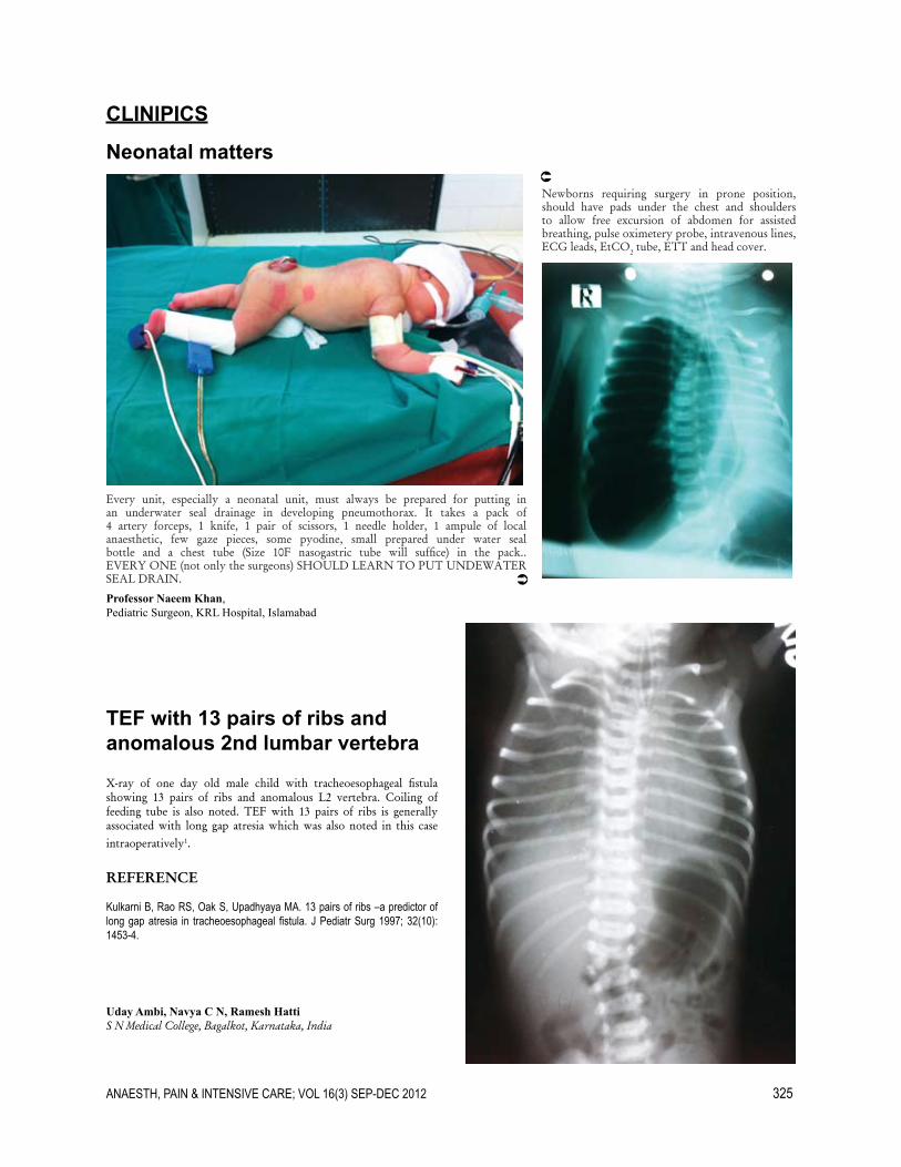
CLINIPICS
Neonatal matters
TEF with 13 pairs of ribs and anomalous 2nd lumbar vertebra
Every unit, especially a neonatal unit, must always be prepared for putting in an underwater seal drainage in developing pneumothorax. It takes a pack of 4 artery forceps, 1 knife, 1 pair of scissors, 1 needle holder, 1 ampule of local anaesthetic, few gaze pieces, some pyodine, small prepared under water seal bottle and a chest tube (Size 10F nasogastric tube will suffice) in the pack.. EVERY ONE (not only the surgeons) SHOULD LEARN TO PUT UNDEWATER SEAL DRAIN.
X-ray of one day old male child with tracheoesophageal fistulashowing 13 pairs of ribs and anomalous L2 vertebra. Coiling of feeding tube is also noted. TEF with 13 pairs of ribs is generally associated with long gap atresia which was also noted in this case intraoperatively1.
REFERENCE
Kulkarni B, Rao RS, Oak S, Upadhyaya MA. 13 pairs of ribs –a predictor of long gap atresia in tracheoesophageal fistula. J Pediatr Surg 1997; 32(10): 1453-4.
Professor Naeem Khan, Pediatric Surgeon, KRL Hospital, Islamabad
Newborns requiring surgery in prone position, should have pads under the chest and shoulders to allow free excursion of abdomen for assisted breathing, pulse oximetery probe, intravenous lines, ECG leads, EtCO2 tube, ETT and head cover.
Uday Ambi, Navya C N, Ramesh HattiS N Medical College, Bagalkot, Karnataka, India
ANAESTH, PAIN & INTENSIVE CARE; VOL 16(3) SEP-DEC 2012 325