an unusual feruloyl esterase belonging to family viii esterases and displaying a broad substrate...
TRANSCRIPT

Ad
CAa
b
a
ARRAA
KMAFF
1
geuomo
i
t
B
m7
n
b
h1
Journal of Molecular Catalysis B: Enzymatic 118 (2015) 79–88
Contents lists available at ScienceDirect
Journal of Molecular Catalysis B: Enzymatic
j ourna l ho me pa ge: www.elsev ier .com/ locate /molcatb
n unusual feruloyl esterase belonging to family VIII esterases andisplaying a broad substrate range
olin W. Ohlhoffa,4, Bronwyn M. Kirbya,∗, Leonardo Van Zyla, David L.R. Mutepfab,na Casanuevaa,2, Robert J. Huddya,3, Rolene Bauera,5, Don A. Cowana,1, Marla Tuffina,∗∗
Institute for Microbial Biotechnology and Metagenomics, University of the Western Cape, Bellville, Cape Town, South AfricaCentre for Microbial Ecology and Genomics, University of Pretoria, Pretoria, South Africa
r t i c l e i n f o
rticle history:eceived 2 December 2014eceived in revised form 19 February 2015ccepted 19 April 2015vailable online 28 April 2015
eywords:etagenome
lkaliphilic
a b s t r a c t
A thermophilic compost metagenomic library constructed in Escherichia coli was functionally screenedfor novel esterases. Of the 110,592 fosmid clones screened, 25 clones demonstrated degradative activ-ity on glyceryl tributyrate (a hit rate of 1:4,423). Four clones displayed ferulic acid esterase activityand were sequenced using 454 Titanium sequencing technology. EstG34, a 410 amino acid protein, wasidentified as having high sequence identity with a number of bacterial �-lactamases. EstG34 has theS-X-X-K motif which is conserved in class C �-lactamases and family VIII carboxylesterases. Purifiedrecombinant EstG34 had a molecular mass of 42 kDa and displayed hydrolytic activity towards a vari-ety of p-nitrophenyl esters, hydroxycinnamic acid esters and �-naphthol acetate. EstG34 represents the
eruloyl Esteraseamily VIII carboxylesterase
first family VIII carboxylesterase and �-lactamase fold enzyme, able to hydrolyse ferulate and a numberof other hydroxycinnamic acid esters. In addition, EstG34 is the first reported FAE to not adopt the �/�hydrolase conformation. The sequence similarity and wide substrate utilization capability of this esterasecomplicates its placement within current classification systems, but also draws attention to the enzyme’spotential versatility.
. Introduction
Microbial communities are a rich source of metabolic andenomic diversity, and are critical components of functioning
cosystems. Both direct cultivation techniques and indirect molec-lar methods have been used to investigate and exploit this wealthf microbial diversity. It is now widely accepted that traditionalethods involving the cultivation and isolation of microorganismsnly provide limited access to the total genetic information within
∗ Corresponding author. Tel.: +27 219593033.∗∗ Corresponding author. Tel.: +27219599725; +27 21 9591506.
E-mail addresses: [email protected], [email protected] (B.M. Kirby),[email protected] (M. Tuffin).
1 Current address: Centre for Microbial Ecology and Genomics, University of Pre-oria, Pretoria, South Africa.
2 Current address: Technology Transfer Office, University of the Western Cape,ellville, Cape Town, South Africa.3 Current address: Centre for Bioprocess Engineering Research (CeBER), Depart-ent of Chemical Engineering, University of Cape Town, Rondebosch, Cape Town
701, South Africa.4 Current address: Fruitique (Pty) Ltd, 27 Scheckter Rd, Killarney Gardens, Mil-erton, Cape Town 7441, South Africa.5 Current address: Launch Lab, Stellenbosch University, Matieland 7602, Stellen-
osch, South Africa.
ttp://dx.doi.org/10.1016/j.molcatb.2015.04.010381-1177/© 2015 Published by Elsevier B.V.
© 2015 Published by Elsevier B.V.
an environment [1]. Metagenomic methods entailing the isolationand cloning of microbial DNA directly from environmental samplescircumvent the limitations of culturing and have been used toidentify a variety of biotechnologically relevant microbial products[2,3].
Composting is a self-heating, aerobic, solid-phase biological sys-tem which accelerates the natural process of biodegradation andmineralisation of organic matter. Microorganisms are essential tothe composting process and compost microbial communities havebeen shown to produce a wide range of robust lignocellulolyticenzymes [4]. Carboxylic esterases are a diverse class of hydrolaseswhich catalyze the cleavage and formation of ester bonds, includingester linkages in plant cell wall polysaccharides. They are com-mercially significant biocatalysts in biotechnological processes, andhave a wide range of industrial and medicinal applications [5,6].Carboxylic esterases are ubiquitous in nature, having been identi-fied in all domains of life (Bacteria, Archaea and Eukaryotes), as wellas in some viruses [7]. Within the carboxylic ester hydrolase family,there are two recognized groups; the lipases (EC 3.1.1.1, triacyg-
lycerol hydrolases) and esterases (EC 3.1.1.3, carboxylester hydro-lases). The current sub-classification of esterases is based on bio-chemical and physiological properties of the enzymes, particularlysubstrate specificities, as well as primary and tertiary structure [8].
8 ar Cat
fs
2
2
Gafe2ssTwoc
2
mtPtltwapwagCnpCMctlteA
2p
mwsfaabrmofipiP
0 C.W. Ohlhoff et al. / Journal of Molecul
Here we present the use of metagenomics to identify a novelamily VIII esterase with broad feruloyl and aromatic ester substratepecificity.
. Materials and methods
.1. Samples and strains
Escherichia coli strains EPI300 (Epicentre® Biotechnologies),eneHogs (Invitrogen) and Rosetta pLysS (Novagen) were used forll cloning and expression studies. Compost samples were collectedrom a commercial compost production facility located in the West-rn Cape Province of South Africa during the summer season of009 (GPS position 34◦S 2′ 53.35′′, 18◦E 31′ 45.71′′). The compostource material consisted of an unspecified mix of wood chips andawdust, with lesser amounts of plant debris and bovine manure.he average temperature of the compost at the point of samplingas 70 ◦C and the pH was 6.1. Elemental analysis (BemLab Lab-
ratories, Strand, South Africa) indicated the composition of theompost was as follows: C 25.8%; N 1.1%; K 0.72%.
.2. Metagenomic fosmid library construction
Metagenomic DNA was extracted using the chemical lysisethod described by Zhou et al. [9] with slight modifications (addi-
ion of SDS (2%, w/v) and PVPP (0.5%, w/v) to the extraction buffer).ersistent humic- and phenolic compounds were removed by elec-rophoresis in low melting point (LMP) agarose. DNA samples wereoaded onto a 0.7% LMP agarose gel in 1× TAE buffer and elec-rophoresis was performed overnight at 1.5 V/cm. High-moleculareight (>35 kb) DNA fragments were excised from LMP agarose
nd treated with agarase (Fermentas) followed by isopropanolrecipitation according to the manufacturer’s instructions. DNAas quantitated using the Quanti-iT dsDNA BR assay kit with
Qubit fluorimeter as described by the manufacturer (Invitro-en). The metagenomic DNA library was constructed using theopyControlTM Fosmid Library production kit (Epicentre® Biotech-ologies) according to the manufacturer’s guidelines. Briefly,urified metagenomic DNA was end-repaired and ligated to theopyControl pCC1FosTM vector. Fosmid clones were packaged byaxPlax Lambda phage and transfected into E. coli EPI300-T1®
ells. Transformants were selected on Luria-Bertani (LB) agar con-aining chloramphenicol (12.5 �g mL−1). To verify the size of theibrary fosmid DNA was extracted from 24 clones and digestedo completion with HindIII and EcoRI (Fermentas). Insert size wasstimated by agarose gel electrophoresis and visualization with anlphaImager 3400 imaging system.
.3. Construction of 16S rRNA gene clone library andhylogenetic analysis
Transformants plated on LB agar were pooled and total fos-id DNA extracted using a Qiagen®Midi kit. The 16S rRNA geneas amplified from the purified fosmid DNA using the univer-
al bacterial primers E9F and U1510R [10,11]. Cycling conditionsor the universal bacterial primers included denaturation (4 mint 94 ◦C), followed by 30 cycles of denaturation (30 s at 94 ◦C),nnealing (30 s at 52 ◦C), and extension (105 s at 72 ◦C), followedy a final extension (10 min at 72 ◦C). PCR was carried out in 50 �Leaction volumes containing 2 mM MgCl2, 1 U DreamTaq poly-erase (Thermo Scientific), 1× PCR buffer, 200 �M dNTPs, 0.5 �M
f each primer and 100 ng fosmid DNA. PCR products were puri-
ed witha GFXTPCR DNA purification kit (Illustra) and cloned intoGEM®-T Easy vector (Promega) according to the manufacturer’snstructions. Putative recombinant clones were screened by colonyCR using universal M13 primers. Compost fosmid library (CFL)
alysis B: Enzymatic 118 (2015) 79–88
clones were grouped manually into ribotypes based on ARDRArestriction patterns generated by single digestions using AluI andBsuRI (isoschizomer of HaeIII) (Fermentas). Partial 16S rRNA genesequences were obtained for at least one representative clonefrom each ribotype. Chromatograms were edited with Chromassoftware version 7.0.5.2 [12] and sequences were assembled inDNAMAN version 4.13 (Lynnon BioSoft). Local alignments wereobtained by performing a standard nucleotide-nucleotide BLASTsearch (BLASTn) [13] of the GenBank database. For phylogeneticanalysis reference strains identified from the BLAST search wereselected for comparison. Sequences were aligned using CLUSTAL W[14] and checked manually for errors. Phylogenetic analyses wereconducted using MEGA version 5.0 [15] and trees were constructedusing the neighbour-joining [16] algorithm. The robustness of treetopology was evaluated by bootstrap analysis [17] based on 1,000resamplings.
2.4. High-throughput screening of fosmid library clones
The library (approximately 105 clones) was inoculated into 384-well microtitre plates containing 50 �L LB broth supplementedwith 12.5 �g mL−1 chloramphenicol using a QPix2 automatedcolony picker (Genetix). Primary screening was performed bygridding the library from the 384-well microtitre plates onto22 cm × 22 cm Q-trays (Genetix) containing 500 mL basal medium(LB agar) supplemented with 12.5 �g mL−1 chloramphenicol(Sigma) and 0.01% (w/v) l-arabinose (Sigma). Clones were screenedfor general esterase activity on basal agar containing 1% (w/v) GumArabic (Sigma) and 0.1% (v/v) glycerol tributyrate (Sigma). A clearhalo surrounding a colony indicated lipolytic and/or esterase activ-ity [18]. Positive esterase clones were screened for ferulic acidesterase activity by plating onto basal agar supplemented with0.4% (w/v) ethyl 4-hydroxy-3 methoxycinnamate (ethyl ferulate)(Sigma). A clear halo surrounding a colony indicated ferulic acidesterase activity [19].
2.5. Fosmid pyrosequencing and ORF analysis
Equimolar concentrations of DNA from 36 fosmids were pooledand sequenced on a Roche 454 GS FLX sequencer by Inqaba Biotech-nology, Pretoria, South Africa. Sequence reads were assembledwith CLC Genomics Workbench software (www.clcbio.com) andSequencher® sequencing software (www.genecodes.com) to pro-duce contiguous sequences. ORFs were predicted using Softberry’sbacterial operon and gene prediction tool, FGENESB (http://linux1.softberry.com) [20]. Signal peptides were predicted with SignalPversion 4.0 [21]. Homology and similarity searches of the trans-lated sequences of the predicted ORFs were performed using theBLASTp program [13,22,23].
2.6. DNA sequence analysis
Multiple alignments of the predicted amino acid sequence ofEstG34 were performed using MEGA 6.0 [24]. A phylogenetic tree ofEstG34 with other family VIII esterases, class-C �-lactamases, fer-ulic acid esterases and DD-peptidases was constructed using theneighbour-joining method [16]. The robustness of tree topologywas evaluated by bootstrap analysis [17] based on 1,000 resam-plings.
2.7. Expression and purification of recombinant EstG34
The EstG34 gene was amplified using fosmid 6-C1 as the tem-plate and the following primers: 5′-GCT CAT ATG GAC GCC CAA TCTCAG TGG-3′ and 5′-TAT CTC GAGGTC GAC GAT GGC CGA ATA-3′
(nucleotides in bold denote NdeI and XhoI restriction enzyme sites,

ar Cat
rtEsictfwbfiriLp4Badu
2
opmtaarai4saNopdwe1
2
2
eadca
2
O
TK
C.W. Ohlhoff et al. / Journal of Molecul
espectively). The EstG34 gene was cloned into the expression vec-or pET-21a (+) and the recombinant plasmid was transformed into. coli Rosetta pLysS cells. Cells were cultured to an optical den-ity of approximately 0.8 at 600 nm at 30 ◦C, after which 0.8 mMsopropyl �-d-1-thiogalactopyranoside (IPTG) was added to theulture for the induction of protein expression. Following 5 h of cul-ivation at 30 ◦C, cells were harvested by centrifugation (6,000 × gor 15 min at 4 ◦C) and resuspended in 1× PBS buffer (pH 7.4). Cellsere disrupted by sonication and cellular debris was harvested
y centrifugation (8,000 × g for 15 min at 4 ◦C). EstG34 was puri-ed by HIS-tag nickel affinity chromatography using the HIS-Bind®
esin and buffer kit (Novagen, USA) according to the manufacturer’snstructions. The eluate was dialysed overnight in a 6 mL Slide-A-yzer® cassette (Thermo Fisher Scientific) against 50 mM sodiumhosphate; 50 mM sodium chloride (pH 7.9) buffer and stored at◦C. The protein concentration was estimated by the method ofradford [25] using the Bio-Rad protein assay kit with bovine serumlbumin as a standard. Protein purity was examined by sodiumodecyl sulphate-polyacrylamide gel electrophoresis (SDS-PAGE)nder denaturing conditions, as described by Laemmli [26].
.8. Esterase activity assay
Esterase activity assays were performed using a standard col-rimetric method by measuring the release of p-nitrophenol from-nitrophenyl (p-NP) esters. The release of p-nitrophenol wasonitored continuously at 410 nm using a Cary 50 Bio spec-
rophotometer (Varian, CA, USA). All assays were prepared andnalysed in triplicate. Enzyme activity was measured at 25 ◦Cnd one unit of activity was defined as the amount of enzymeeleasing 1 �mol of p-nitrophenol per minute under the definedssay conditions. The effect of pH on esterase activity was stud-ed by measuring activities on p-NP decanoate over a pH range of.5–11. The following range of buffer systems were used: 100 mModium acetate (pH 4.5–5.5), 100 mM morpholineethanesulfoniccid (MES) (pH 5.5–7), 100 mM TRIS-HCl (pH 7–9), and 100 mM-cyclohexyl-3-aminopropanesulfonic acid (CAPS) (pH 9–11). Theptimal temperature for enzyme activity was determined for a tem-erature range of 20–50 ◦C using the standard assay towards p-NPecanoate. To test thermal inactivation the recombinant enzymeas pre-incubated at a range of temperatures for 1 h. Residual
sterase activity was assayed on p-NP propanoate at 25 ◦C in00 mM sodium phosphate pH 7.5, 100 mM sodium chloride.
.9. Substrate specificity assays
.9.1. p-nitrophenyl estersThe hydrolytic activity of EstG34 against different fatty acid
sters was investigated using the following p-nitrophenyl esters:cetate (C2); propanoate (C3); octanoate (C8); decanoate (C10);odecanoate (C12) and hexadecanoate (C16) in 1 mL reactionsontaining 100 mM sodium phosphate (pH 7.8), 100 mM NaCl, 1%cetonitrile and 0.5 mM p-NP ester substrate.
.9.2. Hydroxycinnamic acid methyl estersActivity towards the synthetic substrates methyl ferulate (Key
rganics Ltd, Cornwall, UK), methyl sinapate, methyl caffeate
able 1inetic parameters for EstG34 on selected synthetic substrates.
Substrate KM (mM) Specific ac
p-Nitrophenyl acetate 0.011 ± 0.001 26.25 ±p-Nitrophenyl propanoate 0.023 ± 0.004 151 ± 11p-Nitrophenyl ferulate 0.213 ±0.017 10.34±�-Naphthyl acetate 0.84 ± 0.09 1337 ± 56
alysis B: Enzymatic 118 (2015) 79–88 81
and methyl p-coumarate (APIN chemicals, Abingdon, Oxon, UK)were based on the absorption difference of the free acid and therespective methyl ester. The reaction was initiated by the addi-tion of enzyme solution to the assay buffer (100 mM sodiumphosphate pH 7.5, 100 mM sodium chloride) containing 50 �Msynthetic substrate and was performed at 25 ◦C for 1 h in aSPECTROstarNanomicroplate reader (BMG Labtech). Absorbancewas measured at 1 min intervals at 340 nm and activity was deter-mined using calibration curves of the substrate/product. Relativeactivities were expressed as a percentage of the highest activity.
2.9.3. 4-Nitrophenyl ferulateFerulic esterase activity of EstG34 was determined quantita-
tively using 4-nitrophenyl ferulate (4-NPF) as the substrate [27].The substrate was synthesized according to the method of Hegdeet al. [28]. The assay was carried out in 100 mM sodium phosphate(pH 7.5); 100 mM sodium chloride buffer containing 1 mM 4-NPF.The liberated free p-nitrophenol was measured at 410 nm. One unitof enzyme activity is defined was the amount of enzyme releasing1 �mol of p-nitrophenol from 4-NPF in 1 min at 25 ◦C.
Feruloyl esterase activity was also determined using the sub-strates 4-nitrophenyl 5-O-trans-feruloyl-�-L-arabinofuranoside(NPh-5-Fe-Araf) and 4-nitrophenyl 2-O-trans-feruloyl-�-L-arabinofuranoside (NPh-2-Fe-Araf). These substrates weresynthesised as detailed by Mastihubova et al. [27]. The assaywas carried out in 100 mM sodium phosphate buffer (pH 6.5)containing 12 �L DMSO and 3 �L Tween 20 per mL substrate (NPh-5-Fe-Araf or NPh-2-Fe-Araf at 2.5 mM) [29]. The reaction mixturewas incubated to 30 ◦C before addition of EstG34. Measurementsof absorbance at 420 nm were taken over a period of 3 h and 1unit of activity was defined as an absorbance change of 1.0 min−1
(A420).
2.9.4. Naphthyl acetateDeacetylase activity was determined by measuring �-naphthol
released from naphthyl acetate using the Fast Garnet liquid assayas described by Koseki et al. [30]. The assay was performedin 50 mM sodium phosphate buffer (pH 7.5) with 0.8 �mol �-naphthyl acetate (Fluka). After incubation at 37 ◦C for 10 min, thereaction was terminated by the addition of 110 �L Fast Garnet GBC(6 mg mL−1 stock solution in 10% (w/v) SDS) and the absorbancemeasured at 560 nm. One unit (U) of acetyl xylan esterase (AXE)activity was defined as the amount of enzyme required to produce1 �mol of product per min under the defined assay conditions.
2.9.5. Acetyl xylan hydrolysisTo determine hydrolytic activity of EstG34 on acetyl xylan,
an endoxylanase-free 1% solution of acetylxylan in 0.1 M sodiumphosphate buffer (pH 6.5) was prepared. The reaction mixtureswith varying amounts of the enzyme (5–50 �L) were incubatedovernight at 37 ◦C in sealed, thin-walled glass test tubes. Precip-itation in the reaction mixtures was visually determined [31].
2.9.6. Nitrocefin�-Lactam hydrolysis activity was determined spectrophotomet-
rically using the chromogenic substrate nitrocefin, according to themethod outlined in the Oxoid Manual (6th Edition 1990, Unipath
tivity (U mg−1) kcat (s−1) kcat/KM (mM−1 s−1)
30 19.70 ± 30 1790 ± 204 113.25 ± 90 4913 ± 2870.70 7.49 ± 0.81 35.13 ± 4.24
969 ± 48 1154 ± 74
82 C.W. Ohlhoff et al. / Journal of Molecular Catalysis B: Enzymatic 118 (2015) 79–88
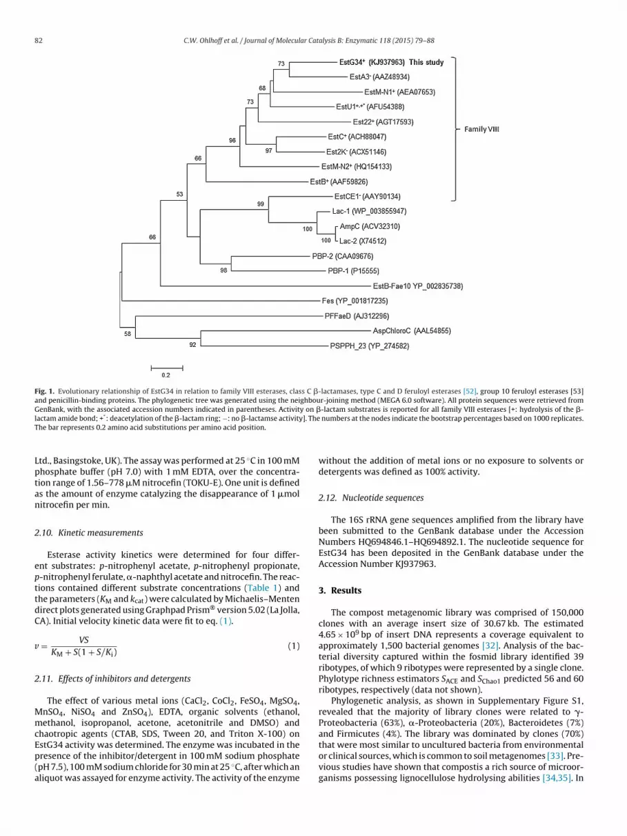
Fig. 1. Evolutionary relationship of EstG34 in relation to family VIII esterases, class C �-lactamases, type C and D feruloyl esterases [52], group 10 feruloyl esterases [53]and penicillin-binding proteins. The phylogenetic tree was generated using the neighbour-joining method (MEGA 6.0 software). All protein sequences were retrieved fromGenBank, with the associated accession numbers indicated in parentheses. Activity on �-lactam substrates is reported for all family VIII esterases [+: hydrolysis of the �-l ]. TheT
Lptan
2
epttdC
v
2
MmcEp(a
actam amide bond; +*: deacetylation of the �-lactam ring; −: no �-lactamse activityhe bar represents 0.2 amino acid substitutions per amino acid position.
td., Basingstoke, UK). The assay was performed at 25 ◦C in 100 mMhosphate buffer (pH 7.0) with 1 mM EDTA, over the concentra-ion range of 1.56–778 �M nitrocefin (TOKU-E). One unit is defineds the amount of enzyme catalyzing the disappearance of 1 �molitrocefin per min.
.10. Kinetic measurements
Esterase activity kinetics were determined for four differ-nt substrates: p-nitrophenyl acetate, p-nitrophenyl propionate,-nitrophenyl ferulate, �-naphthyl acetate and nitrocefin. The reac-ions contained different substrate concentrations (Table 1) andhe parameters (KM and kcat) were calculated by Michaelis–Mentenirect plots generated using Graphpad Prism® version 5.02 (La Jolla,A). Initial velocity kinetic data were fit to eq. (1).
= VS
KM + S(1 + S/Ki)(1)
.11. Effects of inhibitors and detergents
The effect of various metal ions (CaCl2, CoCl2, FeSO4, MgSO4,nSO4, NiSO4 and ZnSO4), EDTA, organic solvents (ethanol,ethanol, isopropanol, acetone, acetonitrile and DMSO) and
haotropic agents (CTAB, SDS, Tween 20, and Triton X-100) on
stG34 activity was determined. The enzyme was incubated in theresence of the inhibitor/detergent in 100 mM sodium phosphatepH 7.5), 100 mM sodium chloride for 30 min at 25 ◦C, after which anliquot was assayed for enzyme activity. The activity of the enzymenumbers at the nodes indicate the bootstrap percentages based on 1000 replicates.
without the addition of metal ions or no exposure to solvents ordetergents was defined as 100% activity.
2.12. Nucleotide sequences
The 16S rRNA gene sequences amplified from the library havebeen submitted to the GenBank database under the AccessionNumbers HQ694846.1–HQ694892.1. The nucleotide sequence forEstG34 has been deposited in the GenBank database under theAccession Number KJ937963.
3. Results
The compost metagenomic library was comprised of 150,000clones with an average insert size of 30.67 kb. The estimated4.65 × 109 bp of insert DNA represents a coverage equivalent toapproximately 1,500 bacterial genomes [32]. Analysis of the bac-terial diversity captured within the fosmid library identified 39ribotypes, of which 9 ribotypes were represented by a single clone.Phylotype richness estimators SACE and SChao1 predicted 56 and 60ribotypes, respectively (data not shown).
Phylogenetic analysis, as shown in Supplementary Figure S1,revealed that the majority of library clones were related to �-Proteobacteria (63%), �-Proteobacteria (20%), Bacteroidetes (7%)and Firmicutes (4%). The library was dominated by clones (70%)
that were most similar to uncultured bacteria from environmentalor clinical sources, which is common to soil metagenomes [33]. Pre-vious studies have shown that compostis a rich source of microor-ganisms possessing lignocellulose hydrolysing abilities [34,35]. In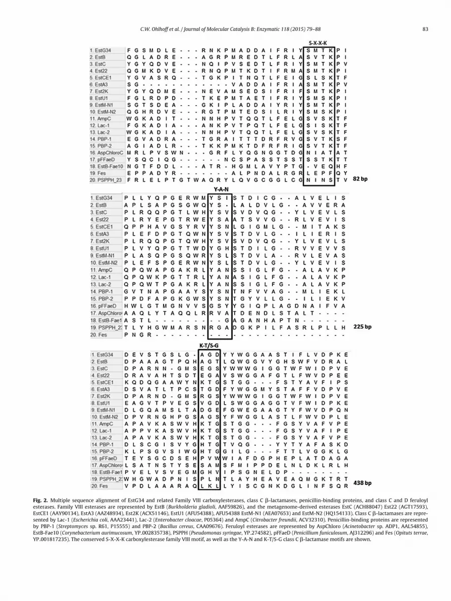
C.W. Ohlhoff et al. / Journal of Molecular Catalysis B: Enzymatic 118 (2015) 79–88 83
Fig. 2. Multiple sequence alignment of EstG34 and related Family VIII carboxylesterases, class C �-lactamases, penicillin-binding proteins, and class C and D feruloylesterases. Family VIII esterases are represented by EstB (Burkholderia gladioli, AAF59826), and the metagenome-derived esterases EstC (ACH88047) Est22 (AGT17593),EstCE1 (AAY90134), EstA3 (AAZ48934), Est2K (ACX51146), EstU1 (AFU54388), AFU54388 EstM-N1 (AEA07653) and EstM-N2 (HQ154133). Class C �-lactamases are repre-sented by Lac-1 (Escherichia coli, AAA23441), Lac-2 (Enterobacter cloacae, P05364) and AmpC (Citrobacter freundii, ACV32310). Penicillin-binding proteins are representedby PBP-1 (Streptomyces sp. R61, P15555) and PBP-2 (Bacillus cereus, CAA09676). Feruloyl esterases are represented by AspChloro (Acinetobacter sp. ADP1, AAL54855),EstB-Fae10 (Corynebacterium aurimucosum, YP 002835738), PSPPH (Pseudomonas syringae, YP 274582), pFFaeD (Penicillium funiculosum, AJ312296) and Fes (Opituts terrae,YP 001817235). The conserved S-X-X-K carboxylesterase family VIII motif, as well as the Y-A-N and K-T/S-G class C �-lactamase motifs are shown.
84 C.W. Ohlhoff et al. / Journal of Molecular Catalysis B: Enzymatic 118 (2015) 79–88
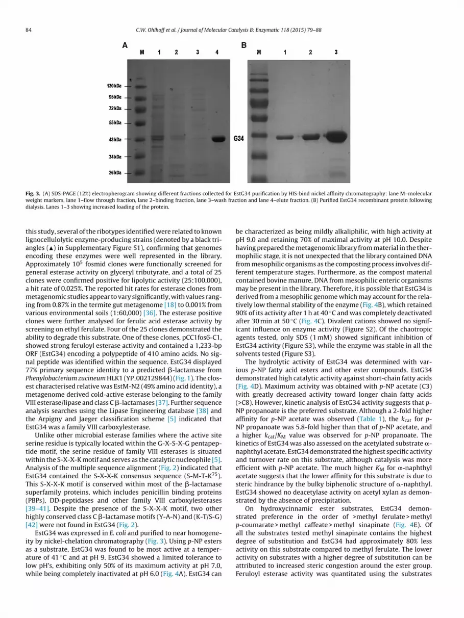
Fig. 3. (A) SDS-PAGE (12%) electropherogram showing different fractions collected for EstG34 purification by HIS-bind nickel affinity chromatography: lane M–molecularw h fracd
tlaeAgcamivcsasOn7PemVatE
stwAETs([h[
iaalw
eight markers, lane 1–flow through fraction, lane 2–binding fraction, lane 3–wasialysis. Lanes 1–3 showing increased loading of the protein.
his study, several of the ribotypes identified were related to knownignocellulolytic enzyme-producing strains (denoted by a black tri-ngles (�) in Supplementary Figure S1), confirming that genomesncoding these enzymes were well represented in the library.pproximately 105 fosmid clones were functionally screened foreneral esterase activity on glyceryl tributyrate, and a total of 25lones were confirmed positive for lipolytic activity (25:100,000),
hit rate of 0.025%. The reported hit rates for esterase clones frometagenomic studies appear to vary significantly, with values rang-
ng from 0.87% in the termite gut metagenome [18] to 0.001% fromarious environmental soils (1:60,000) [36]. The esterase positivelones were further analysed for ferulic acid esterase activity bycreening on ethyl ferulate. Four of the 25 clones demonstrated thebility to degrade this substrate. One of these clones, pCC1fos6-C1,howed strong feruloyl esterase activity and contained a 1,233-bpRF (EstG34) encoding a polypeptide of 410 amino acids. No sig-al peptide was identified within the sequence. EstG34 displayed7% primary sequence identity to a predicted �-lactamase fromhenylobacterium zucineum HLK1 (YP 002129844) (Fig. 1). The clos-st characterised relative was EstM-N2 (49% amino acid identity), aetagenome derived cold-active esterase belonging to the familyIII esterase/lipase and class C �-lactamases [37]. Further sequencenalysis searches using the Lipase Engineering database [38] andhe Arpigny and Jaeger classification scheme [5] indicated thatstG34 was a family VIII carboxylesterase.
Unlike other microbial esterase families where the active siteerine residue is typically located within the G-X-S-X-G pentapep-ide motif, the serine residue of family VIII esterases is situatedithin the S-X-X-K motif and serves as the catalytic nucleophile [5].nalysis of the multiple sequence alignment (Fig. 2) indicated thatstG34 contained the S-X-X-K consensus sequence (S-M-T-K75).his S-X-X-K motif is conserved within most of the �-lactamaseuperfamily proteins, which includes penicillin binding proteinsPBPs), DD-peptidases and other family VIII carboxylesterases39–41]. Despite the presence of the S-X-X-K motif, two otherighly conserved class C �-lactamase motifs (Y-A-N) and (K-T/S-G)42] were not found in EstG34 (Fig. 2).
EstG34 was expressed in E. coli and purified to near homogene-ty by nickel-chelation chromatography (Fig. 3). Using p-NP esters
s a substrate, EstG34 was found to be most active at a temper-ture of 41 ◦C and at pH 9. EstG34 showed a limited tolerance toow pH’s, exhibiting only 50% of its maximum activity at pH 7.0,hile being completely inactivated at pH 6.0 (Fig. 4A). EstG34 can
tion and lane 4–elute fraction. (B) Purified EstG34 recombinant protein following
be characterized as being mildly alkaliphilic, with high activity atpH 9.0 and retaining 70% of maximal activity at pH 10.0. Despitehaving prepared the metagenomic library from material in the ther-mophilic stage, it is not unexpected that the library contained DNAfrom mesophilic organisms as the composting process involves dif-ferent temperature stages. Furthermore, as the compost materialcontained bovine manure, DNA from mesophilic enteric organismsmay be present in the library. Therefore, it is possible that EstG34 isderived from a mesophilic genome which may account for the rela-tively low thermal stability of the enzyme (Fig. 4B), which retained90% of its activity after 1 h at 40 ◦C and was completely deactivatedafter 30 min at 50 ◦C (Fig. 4C). Divalent cations showed no signif-icant influence on enzyme activity (Figure S2). Of the chaotropicagents tested, only SDS (1 mM) showed significant inhibition ofEstG34 activity (Figure S3), while the enzyme was stable in all thesolvents tested (Figure S3).
The hydrolytic activity of EstG34 was determined with var-ious p-NP fatty acid esters and other ester compounds. EstG34demonstrated high catalytic activity against short-chain fatty acids(Fig. 4D). Maximum activity was obtained with p-NP acetate (C3)with greatly decreased activity toward longer chain fatty acids(>C8). However, kinetic analysis of EstG34 activity suggests that p-NP propanoate is the preferred substrate. Although a 2-fold higheraffinity for p-NP acetate was observed (Table 1), the kcat for p-NP propanoate was 5.8-fold higher than that of p-NP acetate, anda higher kcat/KM value was observed for p-NP propanoate. Thekinetics of EstG34 was also assessed on the acetylated substrate �-naphthyl acetate. EstG34 demonstrated the highest specific activityand turnover rate on this substrate, although catalysis was moreefficient with p-NP acetate. The much higher KM for �-naphthylacetate suggests that the lower affinity for this substrate is due tosteric hindrance by the bulky biphenolic structure of �-naphthyl.EstG34 showed no deacetylase activity on acetyl xylan as demon-strated by the absence of precipitation.
On hydroxycinnamic ester substrates, EstG34 demon-strated preference in the order of >methyl ferulate > methylp-coumarate > methyl caffeate > methyl sinapinate (Fig. 4E). Ofall the substrates tested methyl sinapinate contains the highestdegree of substitution and EstG34 had approximately 80% less
activity on this substrate compared to methyl ferulate. The loweractivity on substrates with a higher degree of substitution can beattributed to increased steric congestion around the ester group.Feruloyl esterase activity was quantitated using the substrates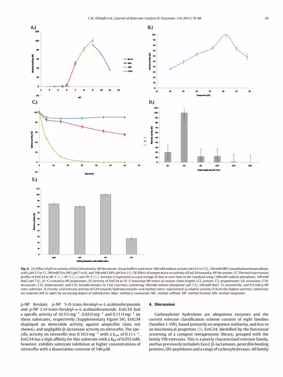
C.W. Ohlhoff et al. / Journal of Molecular Catalysis B: Enzymatic 118 (2015) 79–88 85
Fig. 4. (A) Effect of pH on activity of EstG34 toward p-NP decanoate. Assay buffers used were 100 mM sodium acetate (pH 4.5 to 5.5), 100 mM MES (morpholineethanesulfonicacid) (pH 5.5 to 7), 100 mM Tris-HCl (pH 7 to 9), and 100 mM CAPS (pH 9 to 11). (B) Effect of temperature on activity of EstG34 toward p-NP decanoate. (C) Thermal inactivationprofile of EstG34 at 40 ◦C ( ), 45 ◦C ( ) and 50 ◦C ( ). Activity is expressed as a percentage of that at zero time in the standard assay (100 mM sodium phosphate, 100 mMNaCl (pH 7.5), 25 ◦C) toward p-NP propionate. (D) Activity of EstG34 at 25 ◦C towardsp-NP esters of various chain lengths (C2, acetate; C3, propanonate; C8, octanoate; C10,d ng 100e id mea arate
paatdscEhn
ecanoate; C12, dodecanoate; and C16, hexadecanoate) in 1 mL reactions containister substrate. (E) Ferulic acid esterase activity of G34 towards hydroxycinnamic acre ordered (left to right) by increasing degree of substitution. MpC: methyl p-coum
-NP ferulate, p-NP 5-O-trans-feruloyl-�-L-arabinofuranosidend p-NP 2-O-trans-feruloyl-�-L-arabinofuranoside. EstG34 had
specific activity of 10.3 U mg−1, 0.65 U mg−1 and 0.11 U mg−1 onhese substrates, respectively (Supplementary Figure S4). EstG34isplayed no detectable activity against ampicillin (data nothown), and negligible �-lactamase activity on nitrocefin. The spe-
ific activity on nitrocefin was 0.16 U mg−1 with a kcat of 0.11 s−1.stG34 has a high affinity for this substrate with a KM of 0.055 mM,owever, exhibits substrate inhibition at higher concentrations ofitrocefin with a dissociation constant of 546 �M.mM sodium phosphate (pH 7.5), 100 mM NaCl, 1% acetonitrile, and 0.5 mM p-NPthyl esters, represented as relative activity (% from the highest activity). Substrates, MC: methyl caffeate, MF: methyl ferulate, MS: methyl sinapinate.
4. Discussion
Carboxylester hydrolases are ubiquitous enzymes and thecurrent esterase classification scheme consists of eight families(families 1-VIII), based primarily on sequence similarity, and less soon biochemical properties [5]. EstG34, identified by the functional
screening of a compost metagenomic library, grouped with thefamily VIII esterases. This is a poorly characterised esterase family,and has previously included class C �-lactamases, penicillin bindingproteins, DD-peptidases and a range of carboxylesterases. All family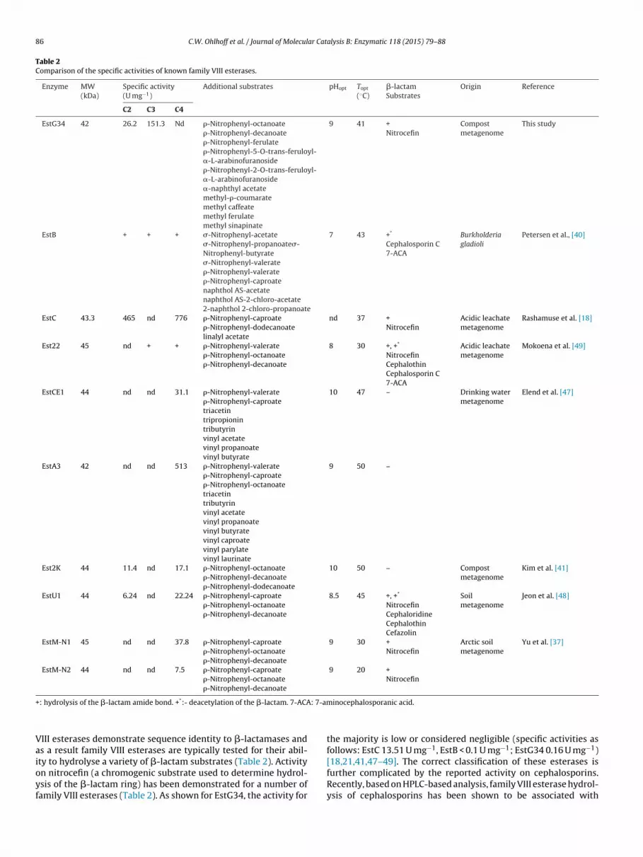
86 C.W. Ohlhoff et al. / Journal of Molecular Catalysis B: Enzymatic 118 (2015) 79–88
Table 2Comparison of the specific activities of known family VIII esterases.
Enzyme MW(kDa)
Specific activity(U mg−1)
Additional substrates pHopt Topt
(◦C)�-lactamSubstrates
Origin Reference
C2 C3 C4
EstG34 42 26.2 151.3 Nd �-Nitrophenyl-octanoate�-Nitrophenyl-decanoate�-Nitrophenyl-ferulate�-Nitrophenyl-5-O-trans-feruloyl-�-L-arabinofuranoside�-Nitrophenyl-2-O-trans-feruloyl-�-L-arabinofuranoside�-naphthyl acetatemethyl-�-coumaratemethyl caffeatemethyl ferulatemethyl sinapinate
9 41 +Nitrocefin
Compostmetagenome
This study
EstB + + + �-Nitrophenyl-acetate�-Nitrophenyl-propanoate�-Nitrophenyl-butyrate�-Nitrophenyl-valerate�-Nitrophenyl-valerate�-Nitrophenyl-caproatenaphthol AS-acetatenaphthol AS-2-chloro-acetate2-naphthol 2-chloro-propanoate
7 43 +*
Cephalosporin C7-ACA
Burkholderiagladioli
Petersen et al., [40]
EstC 43.3 465 nd 776 �-Nitrophenyl-caproate�-Nitrophenyl-dodecanoatelinalyl acetate
nd 37 +Nitrocefin
Acidic leachatemetagenome
Rashamuse et al. [18]
Est22 45 nd + + �-Nitrophenyl-valerate�-Nitrophenyl-octanoate�-Nitrophenyl-decanoate
8 30 +, +*
NitrocefinCephalothinCephalosporin C7-ACA
Acidic leachatemetagenome
Mokoena et al. [49]
EstCE1 44 nd nd 31.1 �-Nitrophenyl-valerate�-Nitrophenyl-caproatetriacetintripropionintributyrinvinyl acetatevinyl propanoatevinyl butyrate
10 47 – Drinking watermetagenome
Elend et al. [47]
EstA3 42 nd nd 513 �-Nitrophenyl-valerate�-Nitrophenyl-caproate�-Nitrophenyl-octanoatetriacetintributyrinvinyl acetatevinyl propanoatevinyl butyratevinyl caproatevinyl parylatevinyl laurinate
9 50 –
Est2K 44 11.4 nd 17.1 �-Nitrophenyl-octanoate�-Nitrophenyl-decanoate�-Nitrophenyl-dodecanoate
10 50 – Compostmetagenome
Kim et al. [41]
EstU1 44 6.24 nd 22.24 �-Nitrophenyl-caproate�-Nitrophenyl-octanoate�-Nitrophenyl-decanoate
8.5 45 +, +*
NitrocefinCephaloridineCephalothinCefazolin
Soilmetagenome
Jeon et al. [48]
EstM-N1 45 nd nd 37.8 �-Nitrophenyl-caproate�-Nitrophenyl-octanoate�-Nitrophenyl-decanoate
9 30 +Nitrocefin
Arctic soilmetagenome
Yu et al. [37]
EstM-N2 44 nd nd 7.5 �-Nitrophenyl-caproate�-Nitrophenyl-octanoate�-Nitrophenyl-decanoate
9 20 +Nitrocefin
+ : 7-am
Vaioyf
: hydrolysis of the �-lactam amide bond. +*:- deacetylation of the �-lactam. 7-ACA
III esterases demonstrate sequence identity to �-lactamases ands a result family VIII esterases are typically tested for their abil-
ty to hydrolyse a variety of �-lactam substrates (Table 2). Activityn nitrocefin (a chromogenic substrate used to determine hydrol-sis of the �-lactam ring) has been demonstrated for a number ofamily VIII esterases (Table 2). As shown for EstG34, the activity forinocephalosporanic acid.
the majority is low or considered negligible (specific activities asfollows: EstC 13.51 U mg−1, EstB < 0.1 U mg−1; EstG34 0.16 U mg−1)
[18,21,41,47–49]. The correct classification of these esterases isfurther complicated by the reported activity on cephalosporins.Recently, based on HPLC-based analysis, family VIII esterase hydrol-ysis of cephalosporins has been shown to be associated with
ar Cat
dTnptandat(fstbatba(hsltbVr
hacethfdicAsewgdrFb1aifinFasivuw[ftt
s
[
[
[[
[
C.W. Ohlhoff et al. / Journal of Molecul
eacetylation and not amide bond hydrolysis (EstB, Est22, EstU1,able 2). To date EstU1 (which was also isolated from a metage-omic library) represents the only family VIII esterase whichossibly hydrolyses both the amide and ester bonds of first genera-ion cephalosporins (with no detectable activity against ampicillinnd second/third generation cephalosporins), although this hasot been conclusively shown [48,49]. The distinction betweeneacetylation or lactamase activity is a crucial determinant and
key differentiation between family VIII carboxylesterases andhe “true” �-lactamases. It is clear from the phylogenetic treeFig. 1) that the true �-lactamases cluster separately from theamily VIII carboxylesterases. However, the resolution of primaryequence comparisons does not sufficiently demonstrate the func-ional evolution (clustering based on deacetylation versus amideond hydrolysis, Fig. 1), which is expected to have resulted from
few critical amino acid changes in the catalytic site, as opposedo global structural modifications. Moreover, for all family VIII car-oxylesterases characterised thus far it is evident that �-lactamsre poor substrates and esters are clearly the preferred substratesTable 2). Consequently, it has been suggested that these esterasesave evolved from the class C �-lactamases (and vice versa), whereome have maintained this remnant activity while others haveost the capability, due to steric interference resulting from struc-ural evolution [18,41,50]. Due to the clear functional distinctionetween class C �-lactamases and the carboxylesterases in familyIII, for clarification of classification we propose that this family beeferred to as carboxylesterases and not �-lactamase fold enzymes.
EstG34 hydrolysed a wide variety of model esters, includingydroxycinnamic acids, ferulated p-nitrophenyl, and �-naphthylcetate, showing considerable preference for the acetylated esterompounds. While there are relatively few characterised feruloylsterases in the literature [43–45] EstG34 activity is comparable towo characterised fungal feruloyl esterases [46]. Specificity towardsydroxycinnamic acids has previously been used to sub-classify
erulic acid esterases (FAEs) as type A, B, C or D [51]. Interestingly,espite the fact that EstG34 demonstrated hydrolytic properties
ndicative of type C and D feruloyl esterases, it showed no signifi-ant sequence identity to type C and D FAEs (Fig. 2). However, the-D classification system is restricted to sequence similarity andubstrate specificity on four model substrates only [51,52]. Consid-ring that FAEs belong to highly divergent protein superfamilies,ith each family having a different evolutionary pathway to the
enesis of FAE specificity, a more robust classification scheme, theescriptor-based classification system, has been developed to moreeliably group functionally related FAEs [52]. Within this schemeAEs belonging to all protein superfamilies representing fungal,acterial and plant origins, are classified into 12 clusters (FEF1-2), which can be further sub-grouped based on the constellationnd distance between the catalytic triad residues. Furthermore,ts robustness is applicable even to poorly characterised enzymeamilies. EstG34 was predicted to group in FEF10, which does notnclude any of the type C or D FAEs. Furthermore, EstG34 doesot show sequence similarity to FEF10 representatives (Fig. 2). AllEF1-10 representatives contain the catalytic triad (Ser, His, Asp),s well as the consensus “nucleophilic elbow” (GXSXG, the univer-ally conserved pentapeptide in which the catalytic serine residues located) [5], although the positioning of these in the sequencearies greatly. This further complicates classification of EstG34sing the descriptor-based system since these conserved motifs,hich EstG34 lacks, are a prerequisite for the correct classification
52]. Furthermore, feruloyl estersases are characteristically �/�-old hydrolases, another characteristic that EstG34 lacks. EstG34 is
he first FAE to be described which lacks the characteristics tradi-ionally used to classify feruloyl esterases.This presents a fascinating question regarding the general clas-ification of feruloyl esterases. Both in this study and others,
[[[[
alysis B: Enzymatic 118 (2015) 79–88 87
phylogenetic clustering of carboxylesterases does not correlatewith substrate specificity [52]. EstG34 also demonstrates that novelbacterial FAEs adopting different structural folds and conservedmotifs are yet to be discovered. While a preference for short tomedium chain length p-NPesters remains the benchmark for clas-sifying an enzyme as a ‘true carboxylesterase’, the results presentedhere suggest that a refinement of the classification of feruloylesterases, and particularly the family VIII esterases, is required.Specifically, the FAE classification now needs to consider the Fam-ily VIII type esterases which harbour the (Ser, Lys, Tyr) catalytictriad where the serine is located in the conserved (SXXK) motif.Given that many carboxylesterases show broad substrate ranges,and that a limited number of key amino acid substitutions in theactive site can dramatically affect substrate specificity, we suggestthat classification of these enzymes by ‘substrate preference’ maybe misleading. Certainly, characterisation using a limited range ofsynthetic nitrophenyl-ester substrates offers little information oneither the in vivo substrate or the classification of the enzyme.EstG34, with high sequence identity to class C �-lactamases andfalling within the current classification scheme of both family VIIIesterases and feruloyl esterases, emphasises the current classifi-cation dilemma. As more EstG34 homologues are discovered, andmore in depth substrate characterisations of family VIII esterasesis conducted, its classification will become clearer.
Conflict of interest
The authors declare that they have no conflict of interest.
Acknowledgements
This work was funded by The National Research Foundation(NRF) and the Technology Innovation Agency (TIA), South Africa(Project number PB99/08).
Appendix A. Supplementary data
Supplementary data associated with this article can be found, inthe online version, at http://dx.doi.org/10.1016/j.molcatb.2015.04.010
References
[1] R.I. Amann, W. Ludwig, K.H. Schleifer, Microbiol. Rev. 59 (1995) 143–169.[2] C.J. Duan, L. Xian, G.C. Zhao, Y. Feng, H. Pang, X.L. Bai, J.L. Tang, Q.S. Ma, J.X. Feng,
Appl. Microbiol. 107 (2009) 245–256.[3] N. Ilmberger, D. Meske, J. Juergensen, M. Schulte, P. Barthen, U. Rabausch, A.
Angelov, M. Mientus, W. Liebl, R.A. Schmitz, W.R. Streit, Appl. Environ. Micro-biol. 95 (2012) 135–146.
[4] M. Allgaier, A. Reddy, J.I. Park, N. Ivanova, P. D’haeseleer, S. Lowry, R. Sapra, T.C.Hazen, B.A. Simmons, J.S. Van der Gheynst, P. Hugenholtz, PLoS One 5 (2010)e8812.
[5] J.L. Arpigny, K.-E. Jaeger, Biochem. J. 343 (1999) 177–183.[6] R. Gupta, N. Gupta, P. Rathi, Appl. Microbiol. Biotechnol. 64 (2004) 763–781.[7] M. Levisson, J. van der Oost, S.W.M. Kengen, Extremophiles 13 (2009) 567–581.[8] U.T. Bornsheuer, FEMS Microbiol. Rev. 26 (2002) 73–81.[9] J. Zhou, M.A. Bruns, J.M. Tiedje, Appl. Environ. Microbiol. 62 (1996) 316–322.10] A.-L. Reysenbach, N.R. Pace, in F.T. Robb, A.R. Place (Eds.), Archaea: A Laboratory
Manual–Thermophiles, Cold Spring Harbour Laboratory Press, New York, 1995,pp 101–107.
11] M.C. Hansen, T. Tolker-Neilson, M. Givskow, S. Molin, FEMS Microbiol. Ecol. 26(1998) 141–149.
12] T.A. Hall, Nucl. Acid Symp. Ser. 41 (1999) 95–98.13] S.F. Altschul, T.L. Madden, A.A. Schaffer, J.H. Zhang, Z. Zhang, W. Miller, D.J.
Lipman, Nucleic Acids Res. 25 (1997) 3389–3402.14] J.D. Thompson, D.G. Higgins, T.J. Gibson, Nucleic Acids Res. 22 (1994)
4673–4680.
15] S. Kumar, K. Tamura, M. Nei, Brief. Bioinform. 5 (2004) 150–163.16] N. Saito, M. Nei, Mol. Biol. Evol. 4 (1987) 406–425.17] J. Felsenstein, Evolution 39 (1985) 783–791.18] K. Rashamuse, V. Magomani, T. Ronneburg, D. Brady, Appl. Microbiol. Bio-technol. 83 (2009) 491–500.
8 ar Cat
[
[
[
[[
[
[[[
[[
[
[
[[
[
[[
[
[
[
[
[
[
[
[
[
[[
[
[
[
8 C.W. Ohlhoff et al. / Journal of Molecul
19] J. Donaghy, P.F. Kelly, A.M. McKay, Appl. Microbiol. Biotechnol. 50 (1998)257–260.
20] V. Solovyev, A. Salamov, R.W. Li (Eds.), Automatic Annotation of MicrobialGenomes and Metagenomic Sequences, Nova Science Publishers, 2011, pp.61–78.
21] T.N. Petersen, S. Brunak, G. von Heijne, H. Nielsen, Nature Methods 8 (2011)785–786.
22] S.F. Altschul, J. Theor. Biol. 138 (1989) 297–309.23] A. Marchler-Bauer, J.B. Anderson, P.F. Cherukuri, C. De Weese-Scott, L.Y. Geer,
M. Gwadz, S. He, D.I. Hurwitz, J.D. Jackson, Z. Ke, C.J. Lanczycki, C.A. Liebert,C. Liu, F. Lu, G.H. Marchler, M. Mullokandov, B.A. Shoemaker, V. Simonvan, J.S.Song, P.A. Thiessen, R.A. Yamashita, J.J. Yin, D. Zhang, S. Bryant, Nucleic AcidsRes. 33 (2005), http://dx.doi.org/10.1093/nar/gki069
24] K. Tamura, D. Peterson, N. Peterson, G. Stecher, M. Nei, S. Kumar, Mol. Biol. Evol.28 (2011) 2731–2739.
25] M.M. Bradford, Anal. Biochem. 72 (1976) 248–254.26] U.K. Laemmli, Nature 15 (1970) 680–685.27] V. Mastihuba, L. Kremnicky, M. Mastihubová, J.L. Willett, G.L. Côté, Anal.
Biochem. 309 (2002) 96–101.28] S. Hegde, P. Srinivas, G. Muralikrisha, Anal. Biochem. 387 (2009) 128–129.29] P. Biely, M. Mastihubová, W.H. van Zyl, B.A. Prior, Anal. Biochem. 311 (2002)
68–75.30] T. Koseki, K. Takahashi, S. Fushinobu, H. Iefuji, K. Iwano, K. Hashizume, H. Mat-
suzawa, Biochim. Biophys. Acta 1722 (2005) 200–208.31] K. Poutanen, M. Sundberg, H. Korte, J. Puls, Appl. Microbiol. Biotechnol. 33
(1990) 506–510.32] E.M. Gabor, W.B.L. Alkema, D.B. Janssen, Environ. Microbiol. 6 (2004) 879–886.33] S.J. Joseph, P. Hugenholtz, P. Sangwan, C.A. Osborne, P.H. Janssen, Appl. Environ.
Microbiol. 69 (2003) 7210–7215.
34] M.C. Vargas-Garcia, F. Suárez-Estrella, M.J. Lopez, J. Morena, Int. Biodeterior.Biodegrad. 59 (2007) 322–328.35] C.M. Wang, C.L. Shyu, S.P. Ho, S.H. Chiou, Lett. Appl. Microbiol. 47 (2008) 46–53.36] Y.-J. Kim, G.-S. Choi, S.-B. Kim, G.-S. Yoon, Y.-S. Kim, Y.-W, Protein Expr. Purif.
45 (2006) 315–323.
[
[
alysis B: Enzymatic 118 (2015) 79–88
37] E.Y. Yu, M.-A. Kwon, M. Lee, J.Y. Oh, J.-E. Choi, J.Y. Lee, B.-K. Song, D.-H. Hahm,J.K. Song, Appl. Environ. Microbiol. 90 (2011) 573–581.
38] J. Pleiss, M. Fischer, M. Peiker, C. Thiele, R.D. Schmid, J. Mol. Catal. B: Enzym. 10(2000) 491–508.
39] J.A. Kelly, O. Dideberg, P. Charlier, J.P. Wery, M. Libert, P.C. Moews, J.R. Knox, C.Duez, C. Fraipont, B. Joris, J. Dusart, J.M. Frere, J.M. Ghuysen, Science 231 (1986)1429–1431.
40] E.I. Petersen, G. Valinger, B. Solkner, G. Stubenrauch, H. Schwab, J. Biotechnol.89 (2001) 11–25.
41] Y.H. Kim, E.J. Kwon, S.K. Kim, Y.S. Jeong, J. Kim, H.D. Yun, H. Kim, Biochem.Biophys. Res.Commun. 393 (2010) 45–49.
42] B. Joris, J.M. Ghuysens, G. Dive, A. Renard, O. Dideberg, P. Charlier, J.M. Frere,J.A. Kelly, J.C. Boyington, P.C. Moews, J. Biochem. 250 (1988) 313–324.
43] M. Estaban-Torres, I. Reverón, J.M. Mancheno, B. de las Rivas, R. Munoz, Appl.Environ. Microbiol. 79 (2013) 5130–5136.
44] D.W.S. Wong, V.J. Chan, H. Liao, M.J. Zidwick, J. Ind. Microbiol. Biotechnol. 40(2013) 287–295.
45] K. Rashamuse, T. Ronneburg, W. Sanyika, K. Mathiba, E. Mmutlane, D. Brady,Appl. Microbiol. Biotechnol. 98 (2014) 727–737.
46] S. Hegde, G. Muralikrishna, World J. Microb. Biot. 25 (2009) 1963–1969.47] C. Elend, C. Schmeisser, C. Leggewie, P. Babiak, J.D. Carballeira, H.L. Steele,
J.-L. Reymond, K.-E. Jaeger, W. Streit, Appl. Environ. Microbiol. 72 (2006)3637–3645.
48] J.H. Jeon, S.-J. Kim, Y.S. Lee, S.-S. Cha, J.H. Lee, S.-H. Yoon, B.-S. Koo, C.-M. Lee,S.H. Choi, S.H. Lee, S.G. Kang, J.-H. Lee, Appl. Environ. Microbiol. 77 (2011)7830–7836.
49] N. Mokoena, K. Mathiba, T. Tsekoa, P. Steenkamp, K. Rashamuse, Biochem.Biophys. Res. Comm. 437 (2013) 342–348.
50] U.G. Wagner, E.I. Petersen, H. Schwab, C. Kratky, Protein Sci. 11 (2002)
467–478.51] V.F. Crepin, C.B. Faulds, I.F. Connerton, Appl. Microbiol. Biotechnol. 63 (2004)647–652.
52] D.B. Udatha, I. Kouskoumvekaki, L. Olsson, G. Panagiotou, Biotechnol. Adv. 29(2011) 94–110.