an overview of diabetic foot ulcers and associated problems
TRANSCRIPT

Citation: Baig, M.S.; Banu, A.;
Zehravi, M.; Rana, R.; Burle, S.S.;
Khan, S.L.; Islam, F.; Siddiqui, F.A.;
Massoud, E.E.S.; Rahman, M.H.; et al.
An Overview of Diabetic Foot Ulcers
and Associated Problems with
Special Emphasis on Treatments with
Antimicrobials. Life 2022, 12, 1054.
https://doi.org/10.3390/
life12071054
Academic Editor: Milan Kolár
Received: 17 June 2022
Accepted: 12 July 2022
Published: 14 July 2022
Publisher’s Note: MDPI stays neutral
with regard to jurisdictional claims in
published maps and institutional affil-
iations.
Copyright: © 2022 by the authors.
Licensee MDPI, Basel, Switzerland.
This article is an open access article
distributed under the terms and
conditions of the Creative Commons
Attribution (CC BY) license (https://
creativecommons.org/licenses/by/
4.0/).
life
Review
An Overview of Diabetic Foot Ulcers and Associated Problemswith Special Emphasis on Treatments with AntimicrobialsMirza Shahed Baig 1, Ahmadi Banu 2, Mehrukh Zehravi 3, Ritesh Rana 4 , Sushil S. Burle 5, Sharuk L. Khan 6,* ,Fahadul Islam 7 , Falak A. Siddiqui 6, Ehab El Sayed Massoud 8,9,10, Md. Habibur Rahman 11,*and Simona Cavalu 12,*
1 Department of Pharmaceutical Chemistry, Y. B. Chavan College of Pharmacy, Aurangabad 431001, India;[email protected]
2 Department of Pharmacology, Vishnu Institute of Pharmaceutical Education & Research,Narsapur 502313, India; [email protected]
3 Department of Clinical Pharmacy Girls Section, Prince Sattam Bin Abdul Aziz University,Alkharj 11942, Saudi Arabia; [email protected]
4 Department of Pharmaceutics, Adarsh Vijendra Institute of Pharmaceutical Sciences, Shobhit University,Gangoh, Saharanpur 247341, India; [email protected]
5 Department of Pharmacology, Smt. Kishoritai Bhoyar College of Pharmacy, Kamptee, Nagpur 441002, India;[email protected]
6 Department of Pharmaceutical Chemistry, MUP’s College of Pharmacy (B Pharm), Degaon, Risod,Washim 444504, India; [email protected]
7 Department of Pharmacy, Faculty of Allied Health Sciences, Daffodil International University,Dhaka 1207, Bangladesh; [email protected]
8 Biology Department, Faculty of Science and Arts in Dahran Aljnoub, King Khalid University,Abha 62529, Saudi Arabia; [email protected]
9 Research Center for Advanced Materials Science (RCAMS), King Khalid University, Abha 61413, Saudi Arabia10 Agriculture Research Centre, Soil, Water and Environment Research Institute, Giza 3725004, Egypt11 Department of Global Medical Science, Wonju College of Medicine, Yonsei University, Wonju 26426, Korea12 Faculty of Medicine and Pharmacy, University of Oradea, Pta 1 Decembrie 10, 410087 Oradea, Romania* Correspondence: [email protected] (S.L.K.); [email protected] (M.H.R.);
[email protected] (S.C.)
Abstract: One of the most significant challenges of diabetes health care is diabetic foot ulcers (DFU).DFUs are more challenging to cure, and this is particularly true for people who already havea compromised immune system. Pathogenic bacteria and fungi are becoming more resistant toantibiotics, so they may be unable to fight microbial infections at the wound site with the antibioticswe have now. This article discusses the dressings, topical antibacterial treatment, medications anddebridement techniques used for DFU and provides a deep discussion of DFU and its associatedproblems. English-language publications on DFU were gathered from many different databases, suchas Scopus, Web of Science, Science Direct, Springer Nature, and Google Scholar. For the treatment ofDFU, a multidisciplinary approach involving the use of diagnostic equipment, skills, and experienceis required. Preventing amputations starts with patient education and the implementation of newcategorization systems. The microbiota involved in DFU can be better understood using noveldiagnostic techniques, such as the 16S-ribosomal DNA sequence in bacteria. This could be achievedby using new biological and molecular treatments that have been shown to help prevent infections,to control local inflammation, and to improve the healing process.
Keywords: diabetic foot ulcers; diabetes mellitus; Wagner grade; diabetic neuropathy; antimicrobials;biofilms
1. Introduction
More than 415 million people throughout the world are diagnosed with diabetes, andthat number is expected to climb to 640 million (1 in 10) by the year 2040, according to the
Life 2022, 12, 1054. https://doi.org/10.3390/life12071054 https://www.mdpi.com/journal/life

Life 2022, 12, 1054 2 of 18
International Diabetes Federation’s 2015 study (IDF 2015). A further 12 percent of globalhealth budgets are allocated to the treatment of people with diabetes (USD 673 billion) [1,2].People with diabetes are more likely to suffer from skin wounds, particularly chronic ulcers,due to neuropathy (nerve damage) and arterial (blood vessel) disease or trauma. Peripheralneuropathy (nerve dysfunction in the feet) and peripheral artery disease (both) are commonin persons with diabetes. People with diabetes have immune system impairments thathave yet to be discovered, limiting their ability to avoid or treat illnesses. Foot ulcers area common complication in people with diabetes because they are more likely to developin persons with the disease [3]. It is estimated that a person with diabetes’ lifetime riskof developing a foot ulcer is 25%, with an uninfected ulcer costing EUR 10,000 and anuntreated ischemic ulcer costing EUR 17,000 in 2008 [4]. When these wounds becomeclinically infected, they cause a large amount of morbidity. A person with diabetes isamputated of a lower limb every 20 s, on average, according to worldwide statistics. Whenat least two typical signs or symptoms of inflammation (pain or tenderness, warmth,redness, and swelling) or purulent discharges appear in a diabetic foot ulcer, an infectionhas occurred (pus) [5]. Patients with diabetes now spend more time in the hospital dueto foot issues than any other diabetic complication. In patients with diabetes, diabeticfoot infections, particularly those that extend to the bone, are the primary cause of lower-extremity amputation, which results in an increased risk of mortality and a higher costburden [6]. To avoid these bad outcomes, it is essential to prevent foot infections or, if thatis not possible, to take care of wounds that have not been treated. There are a lot of methodsto provide antimicrobial therapy: intravenous injections, injections into muscles, and othermeans. One of the most popular kinds of antibiotic treatment is to administer the drugstopically, in other words, locally. Even if the patient has neuropathy or vascular diseases, itis frequently difficult to tell whether a diabetic foot ulcer is infected. Furthermore, even inclinically uninfected wounds, the sheer presence of microorganisms might delay woundhealing, especially if they are pathogenic or present in huge numbers [7]. Some doctorsbelieve that antibiotics (especially topical ones) may effectively treat high-risk wounds thatare clinically uninfected [8,9].
DFU treatments should follow a multidisciplinary approach that uses various diagnos-tic tools, is performed by various specialists, and requires years of experience in treatingthe condition. Patients must be educated to prevent amputations, and new categories mustbe used to guide treatment [10,11]. To learn more about DFU microbiota, it will be requiredto apply cutting-edge diagnostic tools such as the 16S ribosomal DNA sequence in bacteria.In addition to wound characteristics, local epidemiology-based antibiograms, personalizedtreatment, regular debridement, periodic wound assessment, and dressing changes, DFU issaid to have a range of distinctive properties [12]. Infection prevention, local inflammationmanagement, and cicatrizing efficiency may all be improved by bio-molecular therapy andmany other characteristics of the human body. In particular, this survey will look at themost recent developments in antimicrobial treatments, such as dressings; topical thera-pies; medications; debridement techniques; cellular, gene, and molecular therapies; plantextracts; antimicrobial peptides; growth factors; devices; and energy-based treatments.
2. Methodology
The following databases were used: PubMed, Scopus, and Web of Science. Theterminology diabetic foot ulcers, antimicrobials, biofilms, and multidrug-resistant wereused. Up until 2022, English research reports, reviews, and original research articles werechosen and examined. According to Page et al.’s [13] guidelines, an algorithm that followedthe flowchart in Figure 1 and included all of the processes and requirements for selectingthe necessary literature was utilized.

Life 2022, 12, 1054 3 of 18Life 2022, 12, x FOR PEER REVIEW 3 of 19
Figure 1. A flowchart illustrating the steps required for choosing published data to be used in the current study is shown; n = number of literature reports.
3. Diabetic Foot Ulcers (DFUs) DFUs, which are usually skin ulcers that progress across the entire lower limb with
various degrees of peripheral vasculopathy and neuropathy, morbidity, disease, death, and psychosocial distress. Osteomyelitis and gangrene also accompany DFU. With ex-treme DFU, amputation of a significant leg is often used to manage long-term recurrence [14,15]. That is why there are so many various categorization systems for when a foot ulcer responds to therapy. As of yet, it has not been proven to be a commercial success. Thera-peutic data-recording devices are mostly a matter of convenience, rather than clinical or theoretical usefulness, for most people with diabetes [16]. In order to determine the sever-ity of an ulcer, the presence of osteomyelitis or gangrene, and the need for an amputation, the Wagner ulcer classification system uses the following criteria: Wagner grade 0: intact skin; Wagner grade I: superficial ulcer of skin or subcutaneous tissue; Wagner grade II: ulcers extend into tendon, bone, or capsule; Wagner grade III: deep ulcer with osteomye-litis or abscess; Wagner grade IV: partial foot gangrene; and Wagner grade V: whole foot gangrene [17].
An amputation is now required in 90 percent of patients with diabetic foot ulcers with Wagner grade III or above. Approximately 45 percent of patients with diabetic foot ulcers in China have a Wagner grade of III or above, with amputation rates from 18 to 28 percent, according to a nationwide study. Patients with DFU had mortality rates of 11% or higher. The 5-year mortality rate of DFU in Tianjin, China, was found to be 32.7 percent. In the United States, the cost of treating DFU in 2017 was USD 727 billion, while in China,
Figure 1. A flowchart illustrating the steps required for choosing published data to be used in thecurrent study is shown; n = number of literature reports.
3. Diabetic Foot Ulcers (DFUs)
DFUs, which are usually skin ulcers that progress across the entire lower limb withvarious degrees of peripheral vasculopathy and neuropathy, morbidity, disease, death, andpsychosocial distress. Osteomyelitis and gangrene also accompany DFU. With extremeDFU, amputation of a significant leg is often used to manage long-term recurrence [14,15].That is why there are so many various categorization systems for when a foot ulcer respondsto therapy. As of yet, it has not been proven to be a commercial success. Therapeutic data-recording devices are mostly a matter of convenience, rather than clinical or theoreticalusefulness, for most people with diabetes [16]. In order to determine the severity of an ulcer,the presence of osteomyelitis or gangrene, and the need for an amputation, the Wagnerulcer classification system uses the following criteria: Wagner grade 0: intact skin; Wagnergrade I: superficial ulcer of skin or subcutaneous tissue; Wagner grade II: ulcers extendinto tendon, bone, or capsule; Wagner grade III: deep ulcer with osteomyelitis or abscess;Wagner grade IV: partial foot gangrene; and Wagner grade V: whole foot gangrene [17].
An amputation is now required in 90 percent of patients with diabetic foot ulcers withWagner grade III or above. Approximately 45 percent of patients with diabetic foot ulcersin China have a Wagner grade of III or above, with amputation rates from 18 to 28 percent,according to a nationwide study. Patients with DFU had mortality rates of 11% or higher.The 5-year mortality rate of DFU in Tianjin, China, was found to be 32.7 percent. In theUnited States, the cost of treating DFU in 2017 was USD 727 billion, while in China, itwas USD 110 billion [17,18]. Including the fact that endovascular operations and vascular

Life 2022, 12, 1054 4 of 18
bypass surgery are the recommended treatments for ischemia foot ulcers, 40% of patientswith DFU and serious limb ischemia may not follow the criteria. Consequently, amputationis often considered the safest choice for many patients with DFU. In the five years afteramputation, the death rate was around 25–50 percent. Traditional therapy has recurrencerates of 40 percent after one year, 60 percent after three years, and 65 percent after fiveyears. Because of this, new treatments are urgently needed to improve DFU healing andlimb preservation rates [19].
All patients with osteomyelitis must have their DFUs discarded. If a bone sampleis indicated in the case of a suspected fracture, C-reactive protein (CRP), ankle–brachialindex (ABI), and X-ray/MRI imaging should all be performed. Primary care settingsare constrained in their ability to conduct regular health evaluations due to the lack oftime available for foot inspections. Neuropathy; peripheral artery disease (PAD); immunesystem variables; and in certain instances, recurring external or mild damage are among therisk factors for diabetes (which lead to skin breakdown and ultimately to the developmentof infection). Toe deformities (such bunions and hammertoes) are also considered riskfactors since they may produce trigger points on the foot (potential locations for ulceration).Figure 2 illustrates the many risk and predisposing variables that might lead to DFUs.Patients with neuropathy are thought to have more mechanical pain than people withdiabetes without the disease. Inflammation is the most common cause of amputation,which occurs in people with severe diseases, further tissue loss, and organ failure acrossthe body. Patients with anemia (a hemoglobin level below 11 µg/dL), those who areolder, and those who suffer from PAD are at greater risk of infection and, as a result, ofamputations [20,21].
Life 2022, 12, x FOR PEER REVIEW 4 of 19
it was USD 110 billion [17,18]. Including the fact that endovascular operations and vascu-lar bypass surgery are the recommended treatments for ischemia foot ulcers, 40% of pa-tients with DFU and serious limb ischemia may not follow the criteria. Consequently, am-putation is often considered the safest choice for many patients with DFU. In the five years after amputation, the death rate was around 25–50 percent. Traditional therapy has recur-rence rates of 40 percent after one year, 60 percent after three years, and 65 percent after five years. Because of this, new treatments are urgently needed to improve DFU healing and limb preservation rates [19].
All patients with osteomyelitis must have their DFUs discarded. If a bone sample is indicated in the case of a suspected fracture, C-reactive protein (CRP), ankle–brachial in-dex (ABI), and X-ray/MRI imaging should all be performed. Primary care settings are con-strained in their ability to conduct regular health evaluations due to the lack of time avail-able for foot inspections. Neuropathy; peripheral artery disease (PAD); immune system variables; and in certain instances, recurring external or mild damage are among the risk factors for diabetes (which lead to skin breakdown and ultimately to the development of infection). Toe deformities (such bunions and hammertoes) are also considered risk fac-tors since they may produce trigger points on the foot (potential locations for ulceration). Figure 2 illustrates the many risk and predisposing variables that might lead to DFUs. Patients with neuropathy are thought to have more mechanical pain than people with diabetes without the disease. Inflammation is the most common cause of amputation, which occurs in people with severe diseases, further tissue loss, and organ failure across the body. Patients with anemia (a hemoglobin level below 11 μg/dL), those who are older, and those who suffer from PAD are at greater risk of infection and, as a result, of ampu-tations [20,21].
Figure 2. DFUs are caused by a combination of risk and predisposing factors.
4. Pathophysiology of DFUs Diabetic neuropathy and PAD are the major causes of DFUs, with trauma acting as a
starting trigger. At various points in the healing process, both of these factors contribute to the development of ulcers.
4.1. Diabetic Neuropathy
Figure 2. DFUs are caused by a combination of risk and predisposing factors.
4. Pathophysiology of DFUs
Diabetic neuropathy and PAD are the major causes of DFUs, with trauma acting as astarting trigger. At various points in the healing process, both of these factors contribute tothe development of ulcers.

Life 2022, 12, 1054 5 of 18
4.1. Diabetic Neuropathy
Neuropathy in the sensitive, motor, and autonomous nerves is caused by oxidativestress in the nerve cells caused by hyperglycemia. When the hexosamine metabolic routeis activated, it reduces the amount of aldose reductase and sorbitol dehydrogenase pro-duced by the polyol metabolic pathway, which absorbs nicotinamide adenine dinucleotidephosphate (NADPH). These enzymes are responsible for the transformation of glucose intosorbitol and fructose [22]. Myoinositol synthesis in nerve cells reduces as these sugar prod-ucts pile up, resulting in decreased neuronal conduction, increased levels of antioxidantssuch glutathione, and an increase in reactive oxygen species (ROS) formation [23]. In addi-tion to the increased flow of hexosamine and polyol pathway, the altered development ofsubstance P, nerve growth factor, and calcitonin gene-related peptide all lead to additionalnerve damage and ischemia [24]. For example, when there is damage to motor neuronsin the foot muscles, an imbalance in flexor and extender muscles might occur, resultingin anatomical deformity and skin ulcers. Skin breakdown may occur as a consequence ofdamage to the autonomic nervous system because of a decrease in sweat gland activity andan inability to moisturize the feet [25]. If peripheral sensation in the skin is reduced, it ispossible that patients will be more cautious about acquiring foot wounds because the skinis less likely to contain intra-epidermal nerve fiber endings of the afferent A-delta and C-fibers, the majority of which are nociceptor nerve endings that are only stimulated by pain.Diabetes-related neuropathic illnesses, such as vitamin B12 insufficiency, alcohol toxicity,and renal failure towards the end of life, might exacerbate this condition. Epidemiologicalstudies suggest that fat lipoproteins, high blood pressure, and smoking all have roles in thedevelopment of PAD. Charcot’s foot, the most well-known sign of motor neuropathy, isonly one of several. It is crucial to keep in mind that the foot’s skin sheaths, tendons, andsoft tissues make it vulnerable to infection (such as plantar aponeurosis and fascia) [26–28].
4.2. DFUs Pathogenesis: Immunological Involvement
The immune system of individuals with diabetes is characterized by a reduced healingresponse in DFUs. There are many examples, including T-lymphocyte apoptosis; proinflam-matory cytokines; degradation of polymorphonuclear cell functions such as chemotaxis,adhesion, and intracellular killing; inhibition of fibrocyte proliferation; and impaired basallayer of keratinocytes with reduced migration of epidermal cells [27,28]. Bacteria, particu-larly aerobic Gram-positive cocci, such as Staphylococcus aureus (S. aureus) and hemolyticstreptococci, flourish at high blood glucose levels. Carbohydrates, fibroblasts, and collagensynthesis are all affected by diabetes’ metabolic insufficiency as well as other structuralinadequacies. Serum glucose concentrations more than or equal to 150 mL/dL were alsoconsidered indicative of immune system dysfunction. These traits are likely to lead to along-term inflammatory disease [22,23].
4.3. PAD
Almost 80% of individuals with DFU already suffer from PAD [29]. When bloodsugar levels are too high, they cause changes in the foot’s peripheral arteries, which beginat the cell level. The malfunction of endothelial cells is the most important aspect ofmicrocirculation dysfunction. This is because endothelial cell dysfunction causes a decreasein the generation of vasodilators, most notably nitric oxide. Persistent vasoconstriction andhypercoagulation increase plasma thromboxane A2 levels, increasing the risk of ischemiaand ulceration [30]. It is possible that the endothelium will show signs of reduced localangiogenesis, endocrine cell proliferation, basement membrane thickness, blood viscosity,changes in microvascular sound, and antioxidant potential. It might also show signs ofreduced smooth muscle cell proliferation [31].
5. Infection of DFUs
“Infection” is defined as the invasion and proliferation of dangerous bacteria insidetissues, according to the international working group on the diabetic foot. Patients with dia-

Life 2022, 12, 1054 6 of 18
betic foot infections (DFIs) are at greater risk of having a leg amputated and of experiencinga higher risk of death [32]. Ulcer complications such as DFU infections are common andserious. It is estimated that DFU infection is the cause of 80% of non-traumatic lower-limbamputations, with 50% of DFUs being compromised at the time of diagnosis. Some patientswith DFI are hospitalized and given many doses of antibiotics. Skin infections may delayrecovery and lead to systemic health issues if cared for incorrectly. Wound microbiology isa major factor in the onset of foot infection [33–35]. An organism’s level of microbiota aswell as its ability to interact with other microorganisms are important considerations. Whenthe number of bacteria per gram of tissue surpasses 105, the condition is referred to as aninfection [36]. It is possible for skin commensal bacteria to colonize the wound left behindby DFUs, even when the wound is not infected since the host’s immune system has notyet been activated [36]. Triggers might be physical, chemical, or mechanical in nature. TheDFU is sensitive to infection because of ischemia, neuropathy, edema, inflammation, and areduced immune system [37]. It is possible to determine whether an ulcer has been infectedby using recommendations issued by the Infectious Diseases Society of America (IDSA).The infection is detected if at least two of the following symptoms are present during aclinical examination: inflammation, induration, perilesional erythema, hyperesthesia, pain,local fire, and purulent exudate [38]. According to research, 78% of those who undergoDFU already suffer from PAD. Endothelial cell dysfunction is the most critical featureof microcirculation dysfunction, since it results in decreased production of vasodilators,particularly nitric oxide. Chronic vasoconstriction is caused by high plasma thromboxaneA2 levels, which increases the risk of ischemia and ulceration [39–41].
5.1. Bacterial Species of the DFUs
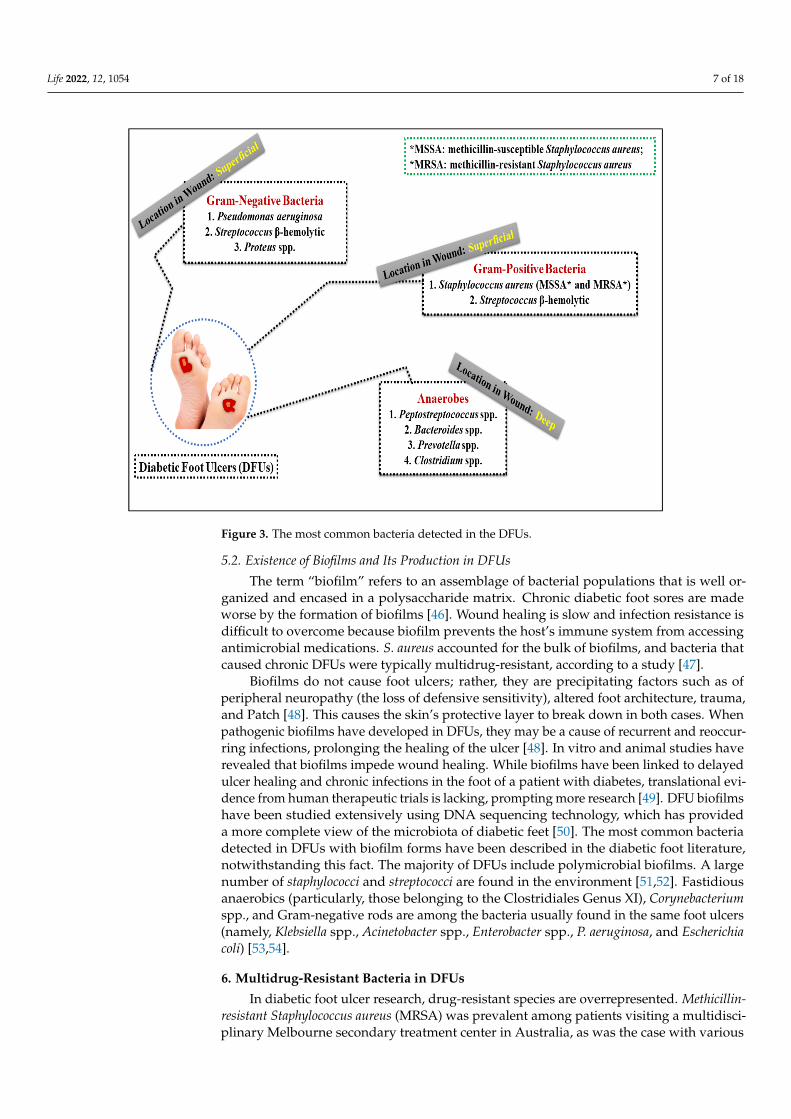
The DFUs’ microbiome has been studied extensively. The organism’s immune systemand physio-pathological features heavily influence the composition of this microbiota.Using molecular tools, researchers have found the polymicrobial nature of chronic woundssuch as DFUs, which comprise Gram-negative and Gram-positive bacteria, as well as anaer-obic bacteria and certain fungi [37]. In the past, conventional bacterial culture methodsfocused on a single bacterium, which was the only one present (Gram-positive bacte-ria). The microbiome of diabetes and non-diabetic ulcers differed, with Gram-negativeand Gram-positive bacteria being found in different proportions. In a microbiologicalexamination of DFI by another author, Gram-negative bacteria were found to outweighGram-positive ones (59 percent vs. 41 percent) [37]. Microorganisms in DFUs have “pre-ferred locations”, which are defined by the amount of oxygen they take up when present. Incontrast to anaerobes, which live deeper inside the niches given by aerobic oxygen intake,aerobic bacteria may be found at the surface, where oxygen levels are quite high [42]. WhilePseudomonas species are the most commonly isolated Gram-negative bacteria, Escherichia coli,Proteus species, Enterobacter species, and Citrobacter species are the most commonly isolatedGram-positive bacteria. S. aureus was found in 72% of culture-positive samples in a mi-crobiome analysis of fresh and chronic DFUs using 16S amplicon sequencing. Geographyhas a significant role in the genesis of DFUs [43]. Gram-positive aerobic cocci are the mostcommon microbe in Western countries, whereas Gram-negative bacilli are more common inwarmer climates (particularly Asia and Africa). Normal procedures yielded the most com-mon bacteria in Mexico: Staphylococcus aureus. In Bangladesh, the most common bacteriain DFUs samples were Pseudomonas spp. (22/29 percent), Enterobacter spp. (22/7 percent),and Staphylococcus spp. (13/13 percent) [44]. India also had the highest percentage ofGram-negative infections (58.5%), indicating the prevalence of Gram-negative bacteria inEastern countries [43]. Up to 95% of all instances of anaerobes found in severe diabeticwounds were caused by Peptostreptococcus spp., Bacteroides spp., and Prevotella spp. [45].Figure 3 depicts the location of DFUs in relation to the most common bacteria detectedin the wound. As a result, DFIs are more prone to develop larger, more frequent ulcersaccompanied with ischemia, necrosis, or unpleasant odors [45].
Life 2022, 12, 1054 7 of 18
Life 2022, 12, x FOR PEER REVIEW 7 of 19
found in severe diabetic wounds were caused by Peptostreptococcus spp., Bacteroides spp., and Prevotella spp. [45]. Figure 3 depicts the location of DFUs in relation to the most com-mon bacteria detected in the wound. As a result, DFIs are more prone to develop larger, more frequent ulcers accompanied with ischemia, necrosis, or unpleasant odors [45].
Figure 3. The most common bacteria detected in the DFUs.
5.2. Existence of Biofilms and Its Production in DFUs The term “biofilm” refers to an assemblage of bacterial populations that is well orga-
nized and encased in a polysaccharide matrix. Chronic diabetic foot sores are made worse by the formation of biofilms [46]. Wound healing is slow and infection resistance is diffi-cult to overcome because biofilm prevents the host’s immune system from accessing an-timicrobial medications. S. aureus accounted for the bulk of biofilms, and bacteria that caused chronic DFUs were typically multidrug-resistant, according to a study [47].
Biofilms do not cause foot ulcers; rather, they are precipitating factors such as of pe-ripheral neuropathy (the loss of defensive sensitivity), altered foot architecture, trauma, and Patch [48]. This causes the skin’s protective layer to break down in both cases. When pathogenic biofilms have developed in DFUs, they may be a cause of recurrent and reoc-curring infections, prolonging the healing of the ulcer [48]. In vitro and animal studies have revealed that biofilms impede wound healing. While biofilms have been linked to delayed ulcer healing and chronic infections in the foot of a patient with diabetes, trans-lational evidence from human therapeutic trials is lacking, prompting more research [49]. DFU biofilms have been studied extensively using DNA sequencing technology, which has provided a more complete view of the microbiota of diabetic feet [50]. The most com-mon bacteria detected in DFUs with biofilm forms have been described in the diabetic foot literature, notwithstanding this fact. The majority of DFUs include polymicrobial biofilms. A large number of staphylococci and streptococci are found in the environment [51,52]. Fas-tidious anaerobics (particularly, those belonging to the Clostridiales Genus XI), Coryne-bacterium spp., and Gram-negative rods are among the bacteria usually found in the same foot ulcers (namely, Klebsiella spp., Acinetobacter spp., Enterobacter spp., P. aeruginosa, and Escherichia coli) [53,54].
Figure 3. The most common bacteria detected in the DFUs.
5.2. Existence of Biofilms and Its Production in DFUs
The term “biofilm” refers to an assemblage of bacterial populations that is well or-ganized and encased in a polysaccharide matrix. Chronic diabetic foot sores are madeworse by the formation of biofilms [46]. Wound healing is slow and infection resistance isdifficult to overcome because biofilm prevents the host’s immune system from accessingantimicrobial medications. S. aureus accounted for the bulk of biofilms, and bacteria thatcaused chronic DFUs were typically multidrug-resistant, according to a study [47].
Biofilms do not cause foot ulcers; rather, they are precipitating factors such as ofperipheral neuropathy (the loss of defensive sensitivity), altered foot architecture, trauma,and Patch [48]. This causes the skin’s protective layer to break down in both cases. Whenpathogenic biofilms have developed in DFUs, they may be a cause of recurrent and reoccur-ring infections, prolonging the healing of the ulcer [48]. In vitro and animal studies haverevealed that biofilms impede wound healing. While biofilms have been linked to delayedulcer healing and chronic infections in the foot of a patient with diabetes, translational evi-dence from human therapeutic trials is lacking, prompting more research [49]. DFU biofilmshave been studied extensively using DNA sequencing technology, which has provideda more complete view of the microbiota of diabetic feet [50]. The most common bacteriadetected in DFUs with biofilm forms have been described in the diabetic foot literature,notwithstanding this fact. The majority of DFUs include polymicrobial biofilms. A largenumber of staphylococci and streptococci are found in the environment [51,52]. Fastidiousanaerobics (particularly, those belonging to the Clostridiales Genus XI), Corynebacteriumspp., and Gram-negative rods are among the bacteria usually found in the same foot ulcers(namely, Klebsiella spp., Acinetobacter spp., Enterobacter spp., P. aeruginosa, and Escherichiacoli) [53,54].
6. Multidrug-Resistant Bacteria in DFUs
In diabetic foot ulcer research, drug-resistant species are overrepresented. Methicillin-resistant Staphylococcus aureus (MRSA) was prevalent among patients visiting a multidisci-plinary Melbourne secondary treatment center in Australia, as was the case with various

Life 2022, 12, 1054 8 of 18
other populations. Among 653 specimens from 379 patients, MRSA was found in just 23%of cases [55].
In a French study in 2008, 188 individuals brought to the hospital with an untreatedfoot ulcer had their recovery rates monitored by the MDR [56]. Two-thirds of the ulcerswere categorized from moderate to severe in the study, revealing their intricacy. It has beenestablished that 70% of the ulcers are neuro-ischemic ulcers, with a fifth of the lesions beingresistant to antibiotics [57]. Lower-limb amputation was more common in patients withMDR microorganisms than in those with non-MDR infections (35.6 percent compared to11.2 percent). The majority of these amputations (87.5 percent) were moderate. Multivariateanalysis, however, showed that the presence of MDR bacteria had minimal impact onhealing time after adjusting for other factors [58–60].
7. Therapeutic Methods Used to Manage DFU Infections
DFIs may end in amputation of a section or more of a patient’s foot or leg, as well asin death in severe situations. An infected diabetic foot, particularly paired with ischemia,remains one of the most severe challenges in the management of DFUs. Due to thepresence of bacteria, local and systemic cytokines are generated, which may lead to systemicinflammatory response and shock, underscoring the requirement of infection care forDFUs. Based on the intensity of the illness, a number of antimicrobial drugs and physicaltechniques are commonly utilized, ranging from topical and oral remedies for light andmoderate infections to intravenous therapy for more severe infections. If an antibiotictreatment is started, it must be completed until all clinical symptoms have disappearedand test results have returned to normal. The wound should be frequently evaluated (at adressing change or on a bi-weekly schedule) throughout infection management to assessthe effectiveness of the treatment [61–63].
7.1. Removal of the Bacterial Biofilm (Debridement)
Because debridement eliminates both the bacteria biofilm and dead tissue from thelesion, it is essential in treating a foot ulcer infection. Tissue for microbiological culture andwound healing may be obtained from the wound, but it also allows for a more detailedevaluation of the wound [64]. The necrotic tissue that accumulates around a wound duringthe normal healing process is called necrotic debris. Debridement speeds up wound healingby removing dead tissue that would otherwise obstruct the growth of new tissue. Isotonicsaline solutions must be used for wound cleansing and debridement prior to antibiotictreatment (0.9 percent NaCl) [65]. Sharp debridement typically reduces the bioburdenof hyperkeratotic margins of plantar neurotrophic ulcers. Every seven to fourteen days,this procedure should be performed [66]. Active and autolytic debridement methods areused in the clinic. Surgical debridement, which removes dead tissue using a scalpel andtweezers while causing the wound bed to bleed, is an example of active debridement [67].Hydro-surgical debridement involves the use of a solid stream of water to remove deadtissue. In outpatient settings, the use of ultrasound-assisted debridement is beneficial. Inthis procedure, low-frequency waves (25 kHz) and irrigation fluids are employed. Asthe moisture in the wound increases, natural tissue shedding occurs. This is commonlyachieved by the use of hydrocolloids and hydrogels [68]. According to a review, therewas no difference in wound duration between clostridial collagenase ointment (CCO) andtraditional hydrogel treatment at six and twelve weeks [69,70].
7.2. Dressings
Wound dressings are an essential aspect of treatment for DFUs right now. To bettertreat DFUs, clinicians have come to appreciate the need for the use of wound dressingsthat promote faster healing, prevent the spread of bacteria, and enhance the overall healingprocess [71]. Silver dressing has been shown in several trials to be helpful in treatingDFUs. Researchers have shown that silver ion dressings may destroy germs, enhance thewound-healing environment, hydrate and soften necrotic tissue, and clean the wound, all

Life 2022, 12, 1054 9 of 18
while releasing silver ions [72]. Bacteria that are negatively charged are drawn to silver ionsbecause of their negative charge, which enhances the permeability of the outer membraneand causes apoptosis. Toxic effects on the fibroblasts of patients with diabetes may limittheir cell activity and collagen production and may dramatically alter the cell’s shapedue to silver dressing. Silver dressings dramatically reduce odor, alleviate pain-relatedsymptoms, decrease wound exudate, and have a more extended dressing wear period thanother treatments in nonhealing and infected chronic wounds [73].
In order to promote the growth of new tissue and to speed the healing process, autolyticdebridement is used [74]. Proteolysis is facilitated by the autolytic breakdown of dead ordiseased tissue due of endogenous proteolytic enzymes. Hydroxylated starch and alginates,and hydrospheres are only a few of the materials that may be used in clothing. Absorbentdressings are used to treat wet wounds, which are intended to absorb wounds [75]. Evenwith the two types of dressings, all are equally successful in hastening the process ofhealing [76].
7.3. Types of Antibacterial Agents Used to Treat DFUs
For chronic wounds, topical antimicrobials are not suggested because of their inabilityto maintain a stable moisture balance and autolytic debridement. Because of their minimaltoxicity to the host tissue, topical antimicrobials are not favored. Many topical antisep-tics/antimicrobials such as 10 percent solution for povidone iodine, chlorhexidine, aceticacid (5 percent), treatment with compounds containing silver, and hydrogen peroxide(H2O2) for DFIs are were used [20,76,77].
7.4. Systemic Therapy with Antibiotics
When symptoms of localized, progressing, or systematic infections occur, systemicantibiotic treatment is suggested. The course of administration and antimicrobial agent tobe employed is determined by the results of a microbiological culture, the clinical symptoms,the body composition, and thet patient’s immune competence [63]. It is common practiceto start with a broad-spectrum antibiotic during normal therapy before moving on toone with a more narrow focus once the results of the bacterial culture are clear. Extreme,non-responsive, spreading infections or suspicions of significant osteomyelitis may needhospitalization and intravenous antibiotic (IV) treatment [44]. Gram-positive staphylococciand streptococci may be treated with oral antibiotics. If a particular antibiotic fails to treat theinfection, a second one is injected. If the patient has a history of infection, if the communityhas a high frequency of MRSA infection, or if the illness is resistant to medicine, empiricalMRSA treatment may be considered [78]. IDSA suggests antibiotics for one to two weeksfor mild infections and for two to three weeks for moderate to serious infections, althoughantibiotics will normally be stopped after clinical signs and effects of infections are resolved.The broad-spectrum drugs most often used are beta-lactam or beta-lactamase inhibitorcombos, such as piperacillin/tazobactam, ampicillin/sulbactam, and ticarcillin/clavulanicacid [79–81].
8. Some Emerging Therapies in Brief for the Treatment of DFUs
Several new therapies are being developed to speed up healing of the ulcer and theydiffer from the usual DFU therapy. Some examples include the use of adjuvant growthfactors, inflammatory modulators, herbal extracts, and blood products; biological treat-ment; hazardous pressure injuries; hyperbaric oxygen therapy; and skin replacements.Supplemental treatments, on the other hand, do not replace the requirement for regulardiabetic foot care [82–87]. Current treatment plans for DFUs include enhanced adjuvanttreatments. To treat these resistant ulcerations, biologic therapies, such as recombinantgrowth factors, platelet-rich plasma (PRP), and other treatments, may be essential to induc-ing healing, avoiding limb loss, and enhancing the quality of life for patients. Recombinantplatelet-derived growth factor and a pair of cell-based treatments (bioengineered skinequivalents and dermal substitutes) are the biologic therapies for DFUs with the most

Life 2022, 12, 1054 10 of 18
scientific backing. Larger wounds, more severe wound grades, longer duration, and alonger time to treatment with advanced biologic therapies have all been linked to a longertime to healing, independent of the advanced biologic treatment employed [88,89].
Using stem cell therapy to treat DFUs has emerged as a possible treatment option.Cell recruitment, immunomodulation, extracellular matrix remodeling, angiogenesis, andneuroregeneration are all promoted by the cytokines produced and secreted by stem cells,all of which aid in wound healing and tissue regeneration. Stem cells can differentiateinto various cell types, including keratinocytes, myofibroblasts, pericytes, and endothelialcells. For certain patients who have exhausted all other revascularization options, stemcell therapy is presently employed as an alternative to amputation. The design of thesubsequent randomized clinical trials may be aided by the agreement between preclinicaland clinical investigations [90,91]. Farideh Davani et al. developed vancomycin andimipenem/cilastatin-loaded core–shell nanofibers to facilitate the treatment of DFUs [92].Oral antibiotics that cover skin flora such as streptococci and Staphylococcus aureus may treatpatients with minor infections in outpatient settings. Effective options include medicationssuch as cephalexin, dicloxacillin, amoxicillin-clavulanate, or clindamycin [93]. There area number of innovative treatments for treating DFUs that have been published in theliterature (Figure 4).
Life 2022, 12, x FOR PEER REVIEW 10 of 19
treatment; hazardous pressure injuries; hyperbaric oxygen therapy; and skin replace-ments. Supplemental treatments, on the other hand, do not replace the requirement for regular diabetic foot care [82–87]. Current treatment plans for DFUs include enhanced adjuvant treatments. To treat these resistant ulcerations, biologic therapies, such as recom-binant growth factors, platelet-rich plasma (PRP), and other treatments, may be essential to inducing healing, avoiding limb loss, and enhancing the quality of life for patients. Re-combinant platelet-derived growth factor and a pair of cell-based treatments (bioengi-neered skin equivalents and dermal substitutes) are the biologic therapies for DFUs with the most scientific backing. Larger wounds, more severe wound grades, longer duration, and a longer time to treatment with advanced biologic therapies have all been linked to a longer time to healing, independent of the advanced biologic treatment employed [88,89].
Using stem cell therapy to treat DFUs has emerged as a possible treatment option. Cell recruitment, immunomodulation, extracellular matrix remodeling, angiogenesis, and neuroregeneration are all promoted by the cytokines produced and secreted by stem cells, all of which aid in wound healing and tissue regeneration. Stem cells can differentiate into various cell types, including keratinocytes, myofibroblasts, pericytes, and endothelial cells. For certain patients who have exhausted all other revascularization options, stem cell therapy is presently employed as an alternative to amputation. The design of the sub-sequent randomized clinical trials may be aided by the agreement between preclinical and clinical investigations [90,91]. Farideh Davani et al. developed vancomycin and imipenem/cilastatin-loaded core–shell nanofibers to facilitate the treatment of DFUs [92]. Oral antibiotics that cover skin flora such as streptococci and Staphylococcus aureus may treat patients with minor infections in outpatient settings. Effective options include med-ications such as cephalexin, dicloxacillin, amoxicillin-clavulanate, or clindamycin [93]. There are a number of innovative treatments for treating DFUs that have been published in the literature (Figure 4).
Figure 4. A collection of innovative treatments for DFUs that have been published in the literature.
9. Recent Upgrade in the Field of DFUs Maggot debridement treatment is the deliberate application of live, “medical-grade”
fly larvae to wounds to induce debridement; disinfection; and eventually, wound healing.
Figure 4. A collection of innovative treatments for DFUs that have been published in the literature.
9. Recent Upgrade in the Field of DFUs
Maggot debridement treatment is the deliberate application of live, “medical-grade”fly larvae to wounds to induce debridement; disinfection; and eventually, wound healing.It is well recognized that maggot treatment may be used in chronic wounds to eliminatenecrotic tissue, to promote the growth of granulation tissue, and to eradicate germs. Thistherapy has been utilized as an alternative to traditional treatments for DFUs plaguedby bacterial resistance [94,95]. A 74-year-old female patient with diabetes for more than30 years was treated with maggot treatment utilizing Chrysomya megacephala larvae. Micro-biological samples were taken to determine the cause of the illness. The 43-day treatmentresulted in a decrease in necrosis and a 0.7 cm2 retraction of the ulcer [96]. Using a combina-tion of surgical debridement, maggot treatment, negative pressure wound therapy (NPWT),and silver foam dressings, Naser Parizad et al. presented their experience treating and

Life 2022, 12, 1054 11 of 18
maintaining patients with DFU. Patient’s ulcers were completely cured after three monthsand ten days, and they were released in excellent health. They detected multidrug-resistantbacteria in the patient’s lesions, including Staphylococcus aureus [97].
NPWT and typical saline dressings have been compared in the treatment of DFU byHaraesh Maranna et al., and 45 patients with DFUs of grades 1 and 2 were included inthis randomized controlled experiment. Twenty-two patients in group A received NPWT,while twenty-three patients in group B received saline dressings. NPWT reduced thesize of the ulcer, enhanced the development of granulation tissue, shortened the hospitalstay, and resulted in a lesion that was completely healed. As a result of the prevalence ofDFUs in low- and middle-income countries such as India, early healing allows patientsto return to their daily routines [98]. Patients with DM and DFUs had their cutaneousmicrovascular function tested to see how recurring transcutaneous infusion of gaseous CO2(CO2 treatment) affected it. Patients with DM benefit from repeated CO2 treatment becauseit improves microvascular function without causing systemic harm [99]. Using a newmethod called PTCTD (proximal tibial cortex transverse distraction), doctors have beenable to treat DFU with promising results in terms of wound healing and the preventionof amputation. The chemokine stromal cell-derived factor-1 (SDF-1) may have a rolein increasing neovascularization in addition to osteogenesis when bone displacementoccurs, according to previous research. SDF-1, a chemokine that plays a crucial role inneovascularization and homing, is a key player in the migration of endothelial progenitorcells (EPCs) and mesenchymal stem cells (BMSCs). Experiments conducted in the lab andon animals have shown that bone distraction increases the expression and plasma levels ofSDF-1. SDF-1 deficiency has been linked to poor neovascularization in patients with DFUsand wounds, according to certain studies [100].
Autologous platelet-rich plasma (PRP) is becoming more commonly used in thetreatment of DFUs [101]. PRP preparation is prohibitively expensive and time-consuming,making it unsuitable for widespread use. L-PRF (leukocyte- and platelet-rich fibrin) ispredicted to be used more often in the future since it is simple and inexpensive. It is possibleto wait 1–2 weeks between L-PRF injections because the fibrin network in L-PRF servesas both a biological matrix for tissue regeneration and a release site for growth factorsthat are released over time. L-PRF is a viable treatment option for diabetics with chronicwounds because of its many advantages [102]. Adipose-derived mesenchymal stem cellinjection into chronic DFUs was studied for its safety and effectiveness. It has been shownthat allogeneic adipose-derived mesenchymal stem cell injections are a safe and effectivetherapy for chronic DFUs, accelerating wound healing [103]. Clinical trials were conductedon patients with DFUs to determine the therapeutic effect of continuous oxygen diffusion(CDO) combined with conventional moist wound dressing (MWD). The research foundthat the combination group had a faster rate of wound healing, a lower white blood cellcount, and lower levels of high-sensitivity C-reactive protein compared with the MWD andCDO groups. During a one-year follow-up, the combination group had an amputation rateof 0%, which was much lower than the other two groups. The combination of MWD andCDO was beneficial in encouraging healing and reducing infection in DFUs, suggestingthat it may represent a novel method for treating this serious clinical problem [104]. In thecase of individuals with DFUs who underwent surgical off-loading concurrently with footulcer closure and did not experience recurrence for a period of two years following surgery,there were no signs of recurrence of the foot ulcer after two years, and the patients wereable to carry out daily tasks on their own after that time. It is less likely to interfere withdaily activities if both reconstructive surgery and surgical off-loading are performed at thesame time, and it helps to reduce ulcer recurrence [105].
The rising prevalence of extensively drug-resistant bacteria (XDR) in individuals withchronic DFU poses a major hazard of foot amputation. The optimal dosage estimatesfor currently available drugs are becoming insufficient against widespread drug-resistantpathogens. The use of antibiotic concentration regimes has been overlooked due to theresistance mechanisms of the potent pathogens, and as a result, piperacillin monother-

Life 2022, 12, 1054 12 of 18
apy, piperacillin-tazobactam, ceftalozane-tazobactam, etc., have been recommended fora long time as a treatment for persistent cases of DFU. Two isolates, VIT PC 7 and VITPC 9, were found to be resistant to all five classes of antibiotics, indicating widespreadXDR. Pseudomonas aeruginosa VIT PC 7 and VIT PC 9 whole-genome sequence analysisrevealed the existence of many RND efflux and antibiotic resistance genes. The MICsfor ciprofloxacin and meropenem were determined using the broth microdilution tech-nique. A checkerboard analysis was used to conduct a synergistic test, and the fractionalinhibitory concentration index (FICI) was used to estimate a sub-MIC concentration ofciprofloxacin/meropenem. On the other hand, a test employing antibiotics at or belowtheir minimum inhibitory concentration (MIC) showed the largest reduction in biofilm-forming cells, which proves the effectiveness of both medicines. Thus, by utilizing optimalciprofloxacin/meropenem concentrations in chronic conditions such as diabetic foot ulcers,an expansion of the antimicrobial spectrum can be accomplished. In addition, sub-MICdoses of ciprofloxacin/meropenem may be a promising alternative for predicting thecontinuing drug-resistant problem [106].
Vancomycin and imipenem/cilastatin-loaded nanofibers with a core–shell structurewere developed to aid in the treatment of DFUs. Due to the unique core–shell nanofibers,electrospinning was utilized to produce nanofibers composed of polyethylene oxide inthe shell compartment, chitosan in the core compartment, and imipenem/cilastatin in theshell compartment. Testing various drug-loaded nanofibers against MRSA, Escherichia coli,and Pseudomococcus aureus using disc diffusion, the nano-fibrous mats showed significantantibacterial activities against S. aureus and MRSA, with inhibition zones of 2.9 and 2.5 cm,respectively, and against gram-negative bacteria E. coli and P. aeruginosa, with inhibitionzones of 1.9 and 2.8 cm, respectively. Due to these nano-fibrous mats’ strong antibacterialactivities, they may be employed as effective medication delivery systems not only for DFUinfections but also for other chronic wounds [107].
Adjuvant hyperbaric oxygen therapy (HBOT) improved ulcer healing and decreasedthe rate of amputation in individuals with non-healing DFUs. HBOT is a reasonably safeintervention [108]. The modified tibial transverse transport (mTTT) technology was usedto treat diabetic ischemic DFU in individuals with T2DM, and the technique’s efficacy andsafety were evaluated. The patients did not experience significant amputation, recurrence,or treatment-related problems. mTTT can be used efficiently and safely to treat ischemicDFUs in patients with type 2 diabetic. This technique is a critical component of theischemia DFU therapy system and needs additional investigation [109]. The fabricationof silver nanoparticles (AgNPs) using an aqueous extract of Turbinaria conoides (TC) wasinvestigated by the reduction of Ag+ ions in a silver nitrate solution. The TCAgNPsshowed significant antibacterial efficacy against multidrug resistant isolates of DFUs suchas Staphylococcus aureus, Klebsiella pneumoniae, Pseudomonas aeruginosa, and Enterococcusfaecalis using disc diffusion and the least inhibitory concentration method. The developmentof fresh formulations containing TCAgNPs has been suggested as a possible alternativehealing method for diabetic foot infections [110].
Multicenter randomized controlled trials (RCTs) have recently revealed novel evidence-based medicines. Such evidence may be found in the treatments for neuro-ischemic ulcersthat use sucrose octasulfate and in the treatment for ulcers with or without ischemia thatuse a multi-layered patch of autologous leukocytes, platelets, and fibrin. There is alsogood RCT evidence for placental-derived products, as well as topical and systemic oxygentherapies in the healing of ulcers [111]. The use of micro- and nanoformulations of bioma-terials in wound dressings has recently showed increased therapeutic properties [112,113].Carboxymethyl, dialdehyde, and 2,2,6,6-tetramethylpiperidine-1-oxyl-oxidized cellulosesare typical biomaterials with superior physicochemical and medicinal qualities comparedwith unmodified cellulose [114]. A high prevalence of the pathogen Staphylococcus aureuswas found in the DFU. Real-time polymerase chain reaction (RT-PCR) is the most reliablemethod for detecting and confirming MRSA infection, despite the use of cefoxitin andoxacillin disc diffusion [115]. Dasman Diabetes Institute (DDI) clinic patients in Kuwait

Life 2022, 12, 1054 13 of 18
were analyzed to determine the microbiological profile of DFUs, which are often seen inpatients with type 2 diabetes. Gram-positive and Gram-negative bacteria were found incomparable numbers in both sexes, regardless of age or glucose levels. Gram-positivebacteria predominate in ulcers free of ischemia, while Gram-negative bacteria predominatein ulcers affected by ischemia. Pseudomonas aeruginosa was more frequent in ulcers withinfection and ischemia than Staphylococcus aureus was in ulcers without ischemia [116].The microbiological profile of DFU in Lebanon is similar to that of other nations in theMiddle East and North Africa (MENA) area, with significant variances when comparedwith that of the Western world. Because of this, it is critical to define regional antimicrobialtreatment recommendations for use in hospitals. Considering the high incidence of aerobicgram-negative rods (GNR) in DFU, in addition to the high prevalence of fluoroquinoloneresistance, the selection of empiric antibiotics should be considered. Except in the case ofindividuals with certain risk factors, empiric therapy for MRSA or Pseudomonas does notseem to be essential [36].
10. Concluding Remarks
When people with diabetes develop foot ulcers and infections, they risk their healthand their lives in danger. Diabetes neuropathy, vasculopathy, immunopathy, and inade-quate glucose control all contribute to the development of diabetic foot disease. A thoroughclinical exam of the patient is the first step in making a correct diagnosis of a diabetic foot.This is followed by early care that focuses on prevention. Essential preventive measuresinclude education; regular follow-ups; and direct coordination among a multidisciplinaryteam of doctors, hospitalists, endocrinologists, infectious disease specialists, and woundtreatment experts. Further multicenter randomized controlled trials must inform on treat-ment decisions and intervention approaches. As a public health issue with a significantinfluence on a patient’s quality of life, chronic wounds resulting from diabetes are treatedwith various treatments (biological, technologies, and pharmaceuticals) that have beenproven to be successful. Since none of these treatments accomplish the main aim of com-plete wound healing, the FDA does not approve of them; thus, more controlled studiesare required to evaluate their effectiveness. Further research is necessary for the differentphases of DFUs to benefit from a combination of cell- and gene-based therapy. BecauseDFUs are caused by a multitude of distinct pathogenic pathways, a monotherapy approachwould result in an exceedingly low recovery rate. Therefore, the treatment of DFU requiresa multimodal and interdisciplinary approach.
Author Contributions: Conceptualization, M.S.B. and S.L.K.; methodology, M.S.B. and A.B., R.R.,M.Z. and E.E.S.M.; software, R.R., M.H.R. and S.S.B.; validation, M.S.B., S.L.K. and F.I.; formalanalysis, S.L.K., F.I. and F.A.S.; investigation, M.S.B. and S.L.K.; resources, S.L.K.; data curation,M.S.B. and R.R.; writing—original draft preparation, M.S.B., S.L.K., R.R., M.Z., E.E.S.M., F.I. andM.H.R.; writing—review and editing, M.S.B., S.L.K., F.I., S.C. and F.A.S.; visualization, S.L.K. and F.I.;supervision, S.L.K., M.Z. and E.E.S.M.; project administration, S.C. All authors have read and agreedto the published version of the manuscript.
Funding: This research received no external funding.
Institutional Review Board Statement: Not applicable.
Informed Consent Statement: Not applicable.
Data Availability Statement: Not applicable.
Acknowledgments: The authors express their gratitude to the Deanship of Scientific Research atKing Khalid University, Abha, Saudi Arabia, for the funding through the Research Groups Programunder grant No. R.G.P.2/7/43.
Conflicts of Interest: The authors declare no conflict of interest.

Life 2022, 12, 1054 14 of 18
Abbreviations
IDF International Diabetes FederationDFU Diabetic foot ulcersPAD Peripheral arterial diseaseROS Reactive oxygen speciesNADPH Nicotinamide adenine dinucleotide phosphateDFIs Diabetic foot infectionsIDSA Infectious Diseases Society of AmericaSIR Systemic inflammatory reactionCCO Clostridial collagenase ointmentNPWT Negative pressure wound therapyLD Laser DopplerLTH Local thermal hyperemiaPTCTD Proximal tibial cortex transverse distractionEPCs Endothelial progenitor cellsBMSCs Bone marrow-derived mesenchymal stem cellsPRP Platelet-rich plasmaCDO Continuous diffusion of oxygenMWD Moist wound dressingXDR Extensively drug-resistant bacteriamTTT Modified tibial transverse transportAgNPs Silver nanoparticlesTC Turbinaria conoidesRCTs Multicenter randomized controlled trials
References1. Jiménez, P.G.; Martín-Carmona, J.; Hernández, E.L. Diabetes Mellitus. Medicine 2020, 13, 883–890. [CrossRef]2. Poretsky, L. (Ed.) Principles of Diabetes Mellitus; Springer Science & Business Media: Berlin/Heidelberg, Germany, 2010;
ISBN 9780387098401.3. Aynalem, S.B.; Zeleke, A.J. Prevalence of Diabetes Mellitus and Its Risk Factors among Individuals Aged 15 Years and above
in Mizan-Aman Town, Southwest Ethiopia, 2016: A Cross Sectional Study. Int. J. Endocrinol. 2018, 2018, 9317987. [CrossRef][PubMed]
4. Endris, T.; Worede, A.; Asmelash, D. Prevalence of Diabetes Mellitus, Prediabetes and Its Associated Factors in Dessie Town,Northeast Ethiopia: A Community-Based Study. Diabetes Metab. Syndr. Obes. Targets Ther. 2019, 12, 2799–2809. [CrossRef][PubMed]
5. Nugroho, P.S.; Tianingrum, N.A.; Sunarti, S.; Rachman, A.; Fahrurodzi, D.S.; Amiruddin, R. Predictor Risk of Diabetes Mellitus inIndonesia, Based on National Health Survey. Malays. J. Med. Health Sci. 2020, 16, 126–130.
6. Hussain, Z.; Thu, H.E.; Shuid, A.N.; Katas, H.; Hussain, F. Recent Advances in Polymer-Based Wound Dressings for the Treatmentof Diabetic Foot Ulcer: An Overview of State-of-the-Art. Curr. Drug Targets 2017, 19, 527–550. [CrossRef]
7. Moura, L.I.F.; Dias, A.M.A.; Carvalho, E.; De Sousa, H.C. Recent Advances on the Development of Wound Dressings for DiabeticFoot Ulcer Treatment—A Review. Acta Biomater. 2013, 9, 7093–7114. [CrossRef]
8. Li, W.W.; Li, V.W. Therapeutic Angiogenesis for Wound Healing. Wounds Compend. Clin. Res. Pract. 2003, 15, 2S–12S.9. Singh, M.R.; Saraf, S.; Vyas, A.; Jain, V.; Singh, D. Innovative Approaches in Wound Healing: Trajectory and Advances. Artif. Cells
Nanomed. Biotechnol. 2013, 41, 202–212. [CrossRef]10. Singh, N.; Armstrong, D.G.; Lipsky, B.A. Preventing Foot Ulcers in Patients with Diabetes. J. Am. Med. Assoc. 2005, 293, 217–228.
[CrossRef]11. Ruke, M.G.; Savai, J. Diabetic Foot Infection, Biofilm & New Management Strategy. Diabetes Res. Open Access 2019, 1, 7–22.
[CrossRef]12. Yazdanpanah, L. Literature Review on the Management of Diabetic Foot Ulcer. World J. Diabetes 2015, 6, 37. [CrossRef] [PubMed]13. Page, M.J.; McKenzie, J.E.; Bossuyt, P.M.; Boutron, I.; Hoffmann, T.C.; Mulrow, C.D.; Shamseer, L.; Tetzlaff, J.M.; Akl, E.A.;
Brennan, S.E.; et al. The PRISMA 2020 Statement: An Updated Guideline for Reporting Systematic Reviews. BMJ 2021, 372, n71.[CrossRef] [PubMed]
14. Armstrong, D.G.; Boulton, A.J.M.; Bus, S.A. Diabetic Foot Ulcers and Their Recurrence. N. Engl. J. Med. 2017, 376, 2367–2375.[CrossRef] [PubMed]
15. Zhang, P.; Lu, J.; Jing, Y.; Tang, S.; Zhu, D.; Bi, Y. Global Epidemiology of Diabetic Foot Ulceration: A Systematic Review andMeta-Analysis. Ann. Med. 2017, 49, 106–116. [CrossRef] [PubMed]

Life 2022, 12, 1054 15 of 18
16. Jeffcoate, W.J.; Vileikyte, L.; Boyko, E.J.; Armstrong, D.G.; Boulton, A.J.M. Current Challenges and Opportunities in the Preventionand Management of Diabetic Foot Ulcers. Diabetes Care 2018, 41, 645–652. [CrossRef]
17. Oyibo, S.O.; Jude, E.B.; Tarawneh, I.; Nguyen, H.C.; Harkless, L.B.; Boulton, A.J.M. A Comparison of Two Diabetic Foot UlcerClassification Systems: The Wagner and the University of Texas Wound Classification Systems. Diabetes Care 2001, 24, 84–88.[CrossRef]
18. Whiting, D.R.; Guariguata, L.; Weil, C.; Shaw, J. IDF Diabetes Atlas: Global Estimates of the Prevalence of Diabetes for 2011 and2030. Diabetes Res. Clin. Pract. 2011, 94, 311–321. [CrossRef]
19. Eldor, R.; Raz, I.; Ben Yehuda, A.; Boulton, A.J.M. New and Experimental Approaches to Treatment of Diabetic Foot Ulcers: AComprehensive Review of Emerging Treatment Strategies. Diabet. Med. 2004, 21, 1161–1173. [CrossRef]
20. Alavi, A.; Sibbald, R.G.; Mayer, D.; Goodman, L.; Botros, M.; Armstrong, D.G.; Woo, K.; Boeni, T.; Ayello, E.A.; Kirsner, R.S.Diabetic Foot Ulcers: Part II. Management. J. Am. Acad. Dermatol. 2014, 70, 21.e1–21.e24. [CrossRef]
21. Management, E.; McCulloch, J.M.; Kloth, L.C. Wound Healing: Evidence-Based Management; FA Davis: Philadelphia, PA, USA, 2010.22. Muhammad Ibrahim, A. Diabetic Foot Ulcer: Synopsis of the Epidemiology and Pathophysiology. Int. J. Diabetes Endocrinol. 2018,
3, 23. [CrossRef]23. Rosyid, F.N. Etiology, Pathophysiology, Diagnosis and Management of Diabetics’ Foot Ulcer. Int. J. Res. Med. Sci. 2017, 5, 4206.
[CrossRef]24. Syafril, S. Pathophysiology Diabetic Foot Ulcer. In Proceedings of the IOP Conference Series: Earth and Environmental Science,
Medan, Indonesia, 15–18 November 2017; Volume 125.25. Alavi, A.; Sibbald, R.G.; Mayer, D.; Goodman, L.; Botros, M.; Armstrong, D.G.; Woo, K.; Boeni, T.; Ayello, E.A.; Kirsner, R.S.
Diabetic Foot Ulcers: Part I. Pathophysiology and Prevention. J. Am. Acad. Dermatol. 2014, 70, 1.e1–1.e18. [CrossRef]26. Aumiller, W.D.; Dollahite, H.A. Pathogenesis and Management of Diabetic Foot Ulcers. J. Am. Acad. Physician Assist. 2015,
28, 28–34. [CrossRef] [PubMed]27. Alsanawi, Y.; Alismail, H.; AlabdRabalnabi, M.; Alturki, H.; Alsuhaibani, A.; Mahbub, M.; Junayd, A.; Alamri, A.; Jawahraji, M.;
Aldaghmani, M. Pathogenesis and Management of Diabetic Foot Ulcers. Int. J. Community Med. Public Health 2018, 5, 4953.[CrossRef]
28. Frykberg, R.G. Diabetic Foot Ulcers: Pathogenesis and Management. Am. Fam. Physician 2002, 66, 1655–1662. [PubMed]29. Megallaa, M.H.; Ismail, A.A.; Zeitoun, M.H.; Khalifa, M.S. Association of Diabetic Foot Ulcers with Chronic Vascular Diabetic
Complications in Patients with Type 2 Diabetes. Diabetes Metab. Syndr. Clin. Res. Rev. 2019, 13, 1287–1292. [CrossRef]30. Esteghamati, A.; Aflatoonian, M.; Rad, M.V.; Mazaheri, T.; Mousavizadeh, M.; Nakhjavani, M.; Noshad, S. Association of
Osteoprotegerin with Peripheral Artery Disease in Patients with Type 2 Diabetes. Arch. Cardiovasc. Dis. 2015, 108, 412–419.[CrossRef]
31. Muthiah, A.; Kandasamy, R.S.N.; Madasamy, A. A Study on Diabetic Foot and Its Association with Peripheral Artery Disease. Int.Surg. J. 2017, 4, 1217. [CrossRef]
32. Schaper, N.C. Diabetic Foot Ulcer Classification System for Research Purposes: A Progress Report on Criteria for IncludingPatients in Research Studies. Diabetes Metab. Res. Rev. 2004, 20, S90–S95. [CrossRef]
33. Loesche, M.; Gardner, S.E.; Kalan, L.; Horwinski, J.; Zheng, Q.; Hodkinson, B.P.; Tyldsley, A.S.; Franciscus, C.L.; Hillis, S.L.;Mehta, S.; et al. Temporal Stability in Chronic Wound Microbiota Is Associated with Poor Healing. J. Investig. Dermatol. 2017,137, 237–244. [CrossRef]
34. Andrea Nelson, E.; Backhouse, M.R.; Bhogal, M.S.; Wright-Hughes, A.; Lipsky, B.A.; Nixon, J.; Brown, S.; Gray, J. Concordance inDiabetic Foot Ulcer Infection. BMJ Open 2013, 3, e002370. [CrossRef] [PubMed]
35. Nelson, A.; Wright-Hughes, A.; Backhouse, M.R.; Lipsky, B.A.; Nixon, J.; Bhogal, M.S.; Reynolds, C.; Brown, S. CODIFI(Concordance in Diabetic Foot Ulcer Infection): A Cross-Sectional Study of Wound Swab versus Tissue Sampling in InfectedDiabetic Foot Ulcers in England. BMJ Open 2018, 8, e019437. [CrossRef] [PubMed]
36. Jouhar, L.; Jaafar, R.F.; Nasreddine, R.; Itani, O.; Haddad, F.; Rizk, N.; Hoballah, J.J. Microbiological Profile and AntimicrobialResistance among Diabetic Foot Infections in Lebanon. Int. Wound J. 2020, 17, 1764–1773. [CrossRef] [PubMed]
37. Jneid, J.; Lavigne, J.P.; La Scola, B.; Cassir, N. The Diabetic Foot Microbiota: A Review. Hum. Microbiome J. 2017, 5–6, 1–6.[CrossRef]
38. Pereira, S.G.; Moura, J.; Carvalho, E.; Empadinhas, N. Microbiota of Chronic Diabetic Wounds: Ecology, Impact, and Potential forInnovative Treatment Strategies. Front. Microbiol. 2017, 8, 1791. [CrossRef]
39. Martínez-De Jesús, F.R.; Ramos-De La Medina, A.; Remes-Troche, J.M.; Armstrong, D.G.; Wu, S.C.; Lázaro Martínez, J.L.;Beneit-Montesinos, J.V. Efficacy and Safety of Neutral PH Superoxidised Solution in Severe Diabetic Foot Infections. Int. Wound J.2007, 4, 353–362. [CrossRef]
40. Liu, P.Y.; Shi, Z.Y.; Sheu, W.H.H. Diagnosis and Treatment of Diabetic Foot Infections. J. Intern. Med. Taiwan 2012, 23, 431–441.[CrossRef]
41. Miller, A.O.; Henry, M. Update in Diagnosis and Treatment of Diabetic Foot Infections. Phys. Med. Rehabil. Clin. N. Am. 2009,20, 611–625. [CrossRef]
42. Murali, T.S.; Kavitha, S.; Spoorthi, J.; Bhat, D.V.; Prasad, A.S.B.; Upton, Z.; Ramachandra, L.; Acharya, R.V.; Satyamoorthy, K.Characteristics of Microbial Drug Resistance and Its Correlates in Chronic Diabetic Foot Ulcer Infections. J. Med. Microbiol. 2014,63, 1377–1385. [CrossRef]

Life 2022, 12, 1054 16 of 18
43. Jain, S.; Barman, R. Bacteriological Profile of Diabetic Foot Ulcer with Special Reference to Drug-Resistant Strains in a TertiaryCare Center in North-East India. Indian J. Endocrinol. Metab. 2017, 21, 688–694. [CrossRef]
44. Karmaker, M.; Sanyal, S.K.; Sultana, M.; Hossain, M.A. Association of Bacteria in Diabetic and Non-Diabetic Foot Infection—AnInvestigation in Patients from Bangladesh. J. Infect. Public Health 2016, 9, 267–277. [CrossRef] [PubMed]
45. Smith, K.; Collier, A.; Townsend, E.M.; O’Donnell, L.E.; Bal, A.M.; Butcher, J.; MacKay, W.G.; Ramage, G.; Williams, C. One StepCloser to Understanding the Role of Bacteria in Diabetic Foot Ulcers: Characterising the Microbiome of Ulcers. BMC Microbiol.2016, 16, 54. [CrossRef] [PubMed]
46. Pouget, C.; Dunyach-Remy, C.; Pantel, A.; Schuldiner, S.; Sotto, A.; Lavigne, J.P. Biofilms in Diabetic Foot Ulcers: Significance andClinical Relevance. Microorganisms 2020, 8, 1580. [CrossRef] [PubMed]
47. Donlan, R.M. Biofilms: Microbial Life on Surfaces. Emerg. Infect. Dis. 2002, 8, 881–890. [CrossRef] [PubMed]48. Jain, A.; Gupta, Y.; Agrawal, R.; Khare, P.; Jain, S.K. Biofilms—A Microbial Life Perspective: A Critical Review. Crit. Rev. Ther.
Drug Carr. Syst. 2007, 24, 393–443. [CrossRef] [PubMed]49. Miere, F.; Teus, dea, A.C.; Laslo, V.; Cavalu, S.; Fritea, L.; Dobjanschi, L.; Zdrinca, M.; Zdrinca, M.; Ganea, M.; Pas, c, P.; et al.
Evaluation of In Vitro Wound-Healing Potential, Antioxidant Capacity, and Antimicrobial Activity of Stellaria media (L.) Vill. Appl.Sci. 2021, 11, 11526. [CrossRef]
50. Santos, R.; Veiga, A.S.; Tavares, L.; Castanho, M.; Oliveira, M. Bacterial Biofilms in Diabetic Foot Ulcers: Potential AlternativeTherapeutics. In Microbial Biofilms—Importance and Applications; IntechOpen: London, UK, 2016.
51. Messad, N.; Prajsnar, T.K.; Lina, G.; O’Callaghan, D.; Foster, S.J.; Renshaw, S.A.; Skaar, E.P.; Bes, M.; Dunyach-Remy, C.;Vandenesch, F.; et al. Existence of a Colonizing Staphylococcus Aureus Strain Isolated in Diabetic Foot Ulcers. Diabetes 2015,64, 2991–2995. [CrossRef]
52. Cavalu, S.; Simon, V. Proteins adsorption to orthopaedic biomaterials: Vibrational spectroscopy evidence. J. Optoelectron. Adv.Mater. 2007, 9, 3297–3302.
53. Srivastava, P.; Sivashanmugam, K. Combinatorial Drug Therapy for Controlling Pseudomonas Aeruginosa and Its Associationwith Chronic Condition of Diabetic Foot Ulcer. Int. J. Low. Extrem. Wounds 2020, 19, 7–20. [CrossRef]
54. Johani, K.; Malone, M.; Jensen, S.; Gosbell, I.; Dickson, H.; Hu, H.; Vickery, K. Microscopy Visualisation Confirms Multi-SpeciesBiofilms Are Ubiquitous in Diabetic Foot Ulcers. Int. Wound J. 2017, 14, 1160–1169. [CrossRef]
55. Shahi, S.K.; Kumar, A. Isolation and Genetic Analysis of Multidrug Resistant Bacteria from Diabetic Foot Ulcers. Front. Microbiol.2016, 6, 1464. [CrossRef] [PubMed]
56. Ji, X.; Jin, P.; Chu, Y.; Feng, S.; Wang, P. Clinical Characteristics and Risk Factors of Diabetic Foot Ulcer with Multidrug-ResistantOrganism Infection. Int. J. Low. Extrem. Wounds 2014, 13, 64–71. [CrossRef] [PubMed]
57. Zubair, M.; Malik, A.; Ahmad, J. Clinico-Bacteriology and Risk Factors for the Diabetic Foot Infection with Multidrug ResistantMicroorganisms in North India. Biol. Med. 2010, 2, 22–34. [CrossRef]
58. Xie, X.; Bao, Y.; Ni, L.; Liu, D.; Niu, S.; Lin, H.; Li, H.; Duan, C.; Yan, L.; Huang, S.; et al. Bacterial Profile and Antibiotic Resistancein Patients with Diabetic Foot Ulcer in Guangzhou, Southern China: Focus on the Differences among Different Wagner’s Grades,IDSA/IWGDF Grades, and Ulcer Types. Int. J. Endocrinol. 2017, 2017, 8694903. [CrossRef]
59. Adeyemo, A.T.; Kolawole, B.; Rotimi, V.O.; Aboderin, A.O. Multicentre Study of the Burden of Multidrug-Resistant Bacteria inthe Aetiology of Infected Diabetic Foot Ulcers. Afr. J. Lab. Med. 2021, 10, 1261. [CrossRef] [PubMed]
60. Matta-Gutiérrez, G.; García-Morales, E.; García-Álvarez, Y.; Álvaro-Afonso, F.J.; Molines-Barroso, R.J.; Lázaro-Martínez, J.L. TheInfluence of Multidrug-Resistant Bacteria on Clinical Outcomes of Diabetic Foot Ulcers: A Systematic Review. J. Clin. Med. 2021,10, 1948. [CrossRef]
61. Apelqvist, J.; Larsson, J. What Is the Most Effective Way to Reduce Incidence of Amputation in the Diabetic Foot? Diabetes Metab.Res. Rev. 2000, 16, S75–S83. [CrossRef]
62. Balows, A. Molecular Medical Microbiology. Diagn. Microbiol. Infect. Dis. 2002, 43, 173–174. [CrossRef]63. Tayeb, K.A. Managing Infection: A Holistic Approach. J. Wound Care 2015, 24, 20–30. [CrossRef]64. Attinger, C.; Wolcott, R. Clinically Addressing Biofilm in Chronic Wounds. Adv. Wound Care 2012, 1, 127–132. [CrossRef]65. Kataoka, Y.; Kunimitsu, M.; Nakagami, G.; Koudounas, S.; Weller, C.D.; Sanada, H. Effectiveness of Ultrasonic Debridement
on Reduction of Bacteria and Biofilm in Patients with Chronic Wounds: A Scoping Review. Int. Wound J. 2021, 18, 176–186.[CrossRef] [PubMed]
66. Yarets, Y. Effective Biofilm Removal and Changes in Bacterial Biofilm Building Capacity after Wound Debridement with Low-Frequency Ultrasound as Part of Wound Bed Preparation before Skin Grafting. Chronic Wound Care Manag. Res. 2017, 4, 55–64.[CrossRef]
67. Dhar, Y.; Han, Y. Current Developments in Biofilm Treatments: Wound and Implant Infections. Eng. Regen. 2020, 1, 64–75.[CrossRef]
68. Wolcott, R. Disrupting the Biofilm Matrix Improves Wound Healing Outcomes. J. Wound Care 2015, 24, 366–371. [CrossRef][PubMed]
69. Dreyfus, J.; Delhougne, G.; James, R.; Gayle, J.; Waycaster, C. Clostridial Collagenase Ointment and Medicinal Honey Utilizationfor Pressure Ulcers in US Hospitals. J. Med. Econ. 2018, 21, 390–397. [CrossRef] [PubMed]
70. Waycaster, C.; Carter, M.J.; Gilligan, A.M.; Mearns, E.S.; Fife, C.E.; Milne, C.T. Comparative Cost and Clinical Effectiveness ofClostridial Collagenase Ointment for Chronic Dermal Ulcers. J. Comp. Eff. Res. 2018, 7, 149–165. [CrossRef]

Life 2022, 12, 1054 17 of 18
71. Lázaro-Martínez, J.L.; Álvaro-Afonso, F.J.; Sevillano-Fernández, D.; Molines-Barroso, R.J.; García-Álvarez, Y.; García-Morales, E.Clinical and Antimicrobial Efficacy of a Silver Foam Dressing with Silicone Adhesive in Diabetic Foot Ulcers with Mild Infection.Int. J. Low. Extrem. Wounds 2019, 18, 269–278. [CrossRef]
72. Lin, H.; Bolatai, A.; Wu, N. Application Progress of Nano Silver Dressing in the Treatment of Diabetic Foot. Diabetes Metab. Syndr.Obes. Targets Ther. 2021, 14, 4145–4154. [CrossRef]
73. Huang, C.; Wang, R.; Yan, Z. Silver Dressing in the Treatment of Diabetic Foot: A Protocol for Systematic Review and Meta-Analysis. Medicine 2021, 100, e24876. [CrossRef]
74. Cavalu, S.; Roiu, G.; Pop, O.; Heredea, D.A.P.; Costea, T.O.; Costea, C.F. Nano-Scale Modifications of Amniotic MembraneInduced by UV and Antibiotic Treatment: Histological, AFM and FTIR Spectroscopy Evidence. Materials 2021, 14, 863. [CrossRef]
75. Zhang, X.; Sun, D.; Jiang, G.C. Comparative Efficacy of Nine Different Dressings in Healing Diabetic Foot Ulcer: A BayesianNetwork Analysis. J. Diabetes 2019, 11, 418–426. [CrossRef] [PubMed]
76. Dumville, J.C.; Deshpande, S.; O’Meara, S.; Speak, K. Hydrocolloid Dressings for Healing Diabetic Foot Ulcers. Cochrane DatabaseSyst. Rev. 2013, 2013, CD009099. [CrossRef]
77. Kwon, K.T.; Armstrong, D.G. Microbiology and Antimicrobial Therapy for Diabetic Foot Infections. Infect. Chemother. 2018,50, 11–20. [CrossRef] [PubMed]
78. Haldar, J.; Mukherjee, P.; Mukhopadhyay, S.; Maiti, P.K. Isolation of Bacteria from Diabetic Foot Ulcers with Special Reference toAnaerobe Isolation by Simple Two-Step Combustion Technique in Candle Jar. Indian J. Med. Res. 2017, 145, 97–101. [CrossRef]
79. Lipsky, B.A.; Armstrong, D.G.; Citron, D.M.; Tice, A.D.; Morgenstern, D.E.; Abramson, M.A. Ertapenem versus Piperacillin/Tazobactamfor Diabetic Foot Infections (SIDESTEP): Prospective, Randomised, Controlled, Double-Blinded, Multicentre Trial. Lancet 2005,366, 1695–1703. [CrossRef]
80. Roberts, A.D.; Simon, G.L. Diabetic Foot Infections: The Role of Microbiology and Antibiotic Treatment. Semin. Vasc. Surg. 2012,25, 75–81. [CrossRef] [PubMed]
81. Karri, V.V.S.R.; Kuppusamy, G.; Talluri, S.V.; Yamjala, K.; Mannemala, S.S.; Malayandi, R. Current and Emerging Therapies in theManagement of Diabetic Foot Ulcers. Curr. Med. Res. Opin. 2016, 32, 519–542. [CrossRef]
82. Ahmed, A.; Getti, G.; Boateng, J. Ciprofloxacin-Loaded Calcium Alginate Wafers Prepared by Freeze-Drying Technique forPotential Healing of Chronic Diabetic Foot Ulcers. Drug Deliv. Transl. Res. 2018, 8, 1751–1768. [CrossRef]
83. Yingsakmongkol, N. Clinical Outcomes of WF10 Adjunct to Standard Treatment of Diabetic Foot Ulcers. J. Wound Care 2013,22, 130–136. [CrossRef]
84. Gasca-Lozano, L.E.; Lucano-Landeros, S.; Ruiz-Mercado, H.; Salazar-Montes, A.; Sandoval-Rodríguez, A.; Garcia-Bañuelos, J.;Santos-Garcia, A.; Davila-Rodriguez, J.R.; Navarro-Partida, J.; Bojórquez-Sepúlveda, H.; et al. Pirfenidone Accelerates WoundHealing in Chronic Diabetic Foot Ulcers: A Randomized, Double-Blind Controlled Trial. J. Diabetes Res. 2017, 2017, 3159798.[CrossRef]
85. Ram, M.; Singh, V.; Kumawat, S.; Kumar, D.; Lingaraju, M.C.; Uttam Singh, T.; Rahal, A.; Kumar Tandan, S.; Kumar, D.Deferoxamine Modulates Cytokines and Growth Factors to Accelerate Cutaneous Wound Healing in Diabetic Rats. Eur. J.Pharmacol. 2015, 764, 9–21. [CrossRef] [PubMed]
86. Maderal, A.D.; Vivas, A.C.; Eaglstein, W.H.; Kirsner, R.S. The FDA and Designing Clinical Trials for Chronic Cutaneous Ulcers.Semin. Cell Dev. Biol. 2012, 23, 993–999. [CrossRef] [PubMed]
87. Tecilazich, F.; Dinh, T.L.; Veves, A. Emerging Drugs for the Treatment of Diabetic Ulcers. Expert Opin. Emerg. Drugs 2013,18, 207–217. [CrossRef] [PubMed]
88. Richmond, N.A.; Vivas, A.C.; Kirsner, R.S. Topical and Biologic Therapies for Diabetic Foot Ulcers. Med. Clin. N. Am. 2013,97, 883–898. [CrossRef] [PubMed]
89. Kirsner, R.S.; Warriner, R.; Michela, M.; Stasik, L.; Freeman, K. Advanced Biological Therapies for Diabetic Foot Ulcers. Arch.Dermatol. 2010, 146, 857–862. [CrossRef] [PubMed]
90. Lopes, L.; Setia, O.; Aurshina, A.; Liu, S.; Hu, H.; Isaji, T.; Liu, H.; Wang, T.; Ono, S.; Guo, X.; et al. Stem Cell Therapy for DiabeticFoot Ulcers: A Review of Preclinical and Clinical Research. Stem Cell Res. Ther. 2018, 9, 188. [CrossRef]
91. Cao, Y.; Gang, X.; Sun, C.; Wang, G. Mesenchymal Stem Cells Improve Healing of Diabetic Foot Ulcer. J. Diabetes Res. 2017,2017, 9328347. [CrossRef]
92. Davani, F.; Alishahi, M.; Sabzi, M.; Khorram, M.; Arastehfar, A.; Zomorodian, K. Dual Drug Delivery of Vancomycin andImipenem/Cilastatin by Coaxial Nanofibers for Treatment of Diabetic Foot Ulcer Infections. Mater. Sci. Eng. C 2021, 123, 111975.[CrossRef]
93. Agbi, K.E.; Carvalho, M.; Phan, H.; Tuma, C. Case Report: Diabetic Foot Ulcer Infection Treated with Topical CompoundedMedications. Int. J. Pharm. Compd. 2017, 21, 22–27.
94. Shi, E.; Shofler, D. Maggot Debridement Therapy: A Systematic Review. Br. J. Community Nurs. 2014, 19, S6–S13. [CrossRef]95. Naik, G.; Harding, K. Maggot Debridement Therapy: The Current Perspectives. Chronic Wound Care Manag. Res. 2017, 4, 121–128.
[CrossRef]96. Pinheiro, M.A.R.Q.; Ferraz, J.B.; Junior, M.A.A.; Moura, A.D.; da Costa, M.E.S.M.; Costa, F.J.M.D.; Neto, V.F.A.; Neto, R.M.;
Gama, R.A. Use of Maggot Therapy for Treating a Diabetic Foot Ulcer Colonized by Multidrug Resistant Bacteria in Brazil. IndianJ. Med. Res. Suppl. 2015, 141, 340–342. [CrossRef]

Life 2022, 12, 1054 18 of 18
97. Parizad, N.; Hajimohammadi, K.; Goli, R. Surgical Debridement, Maggot Therapy, Negative Pressure Wound Therapy, andSilver Foam Dressing Revive Hope for Patients with Diabetic Foot Ulcer: A Case Report. Int. J. Surg. Case Rep. 2021, 82, 105931.[CrossRef]
98. Maranna, H.; Lal, P.; Mishra, A.; Bains, L.; Sawant, G.; Bhatia, R.; Kumar, P.; Beg, M.Y. Negative Pressure Wound Therapy inGrade 1 and 2 Diabetic Foot Ulcers: A Randomized Controlled Study. Diabetes Metab. Syndr. Clin. Res. Rev. 2021, 15, 365–371.[CrossRef] [PubMed]
99. Finžgar, M.; Frangež, H.B.; Cankar, K.; Frangež, I. Transcutaneous Application of the Gaseous CO2 for Improvement of theMicrovascular Function in Patients with Diabetic Foot Ulcers. Microvasc. Res. 2021, 133, 104100. [CrossRef]
100. Fang, J.; Xu, J.; Zhang, Y.; Chen, H.; Ma, Z.; Huang, Z.; Hu, J. Stromal Cell-Derived Factor-1 May Play Pivotal Role in Distraction-Stimulated Neovascularization of Diabetic Foot Ulcer. Med. Hypotheses 2021, 149, 110548. [CrossRef] [PubMed]
101. Ratiu, C.; Brocks, M.; Costea, T.; Moldovan, L.; Cavalu, S. PRGF-Modified Collagen Membranes for Guided Bone Regeneration:Spectroscopic, Microscopic and Nano-Mechanical Investigations. Appl. Sci. 2019, 9, 1035. [CrossRef]
102. Shao, S.; Pan, R.; Chen, Y. Autologous Platelet-Rich Plasma for Diabetic Foot Ulcer. Trends Endocrinol. Metab. 2020, 31, 885–890.[CrossRef]
103. Uzun, E.; Güney, A.; Gönen, Z.B.; Özkul, Y.; Kafadar, I.H.; Günay, M.; Mutlu, M. Intralesional Allogeneic Adipose-Derived StemCells Application in Chronic Diabetic Foot Ulcer: Phase I/2 Safety Study. Foot Ankle Surg. 2020, 27, 636–642. [CrossRef]
104. He, S.; Liang, C.; Yi, C.; Wu, M. Therapeutic Effect of Continuous Diffusion of Oxygen Therapy Combined with Traditional MoistWound Dressing Therapy in the Treatment of Diabetic Foot Ulcers. Diabetes Res. Clin. Pract. 2021, 174, 108743. [CrossRef]
105. Terabe, Y.; Kaneko, N.; Nakabayashi, K.; Matsui, A.; Ando, H. Long-Term Efficient Management of Diabetic Foot Ulcer UsingSimultaneous Foot Ulcer Closure and Surgical off-Loading. JPRAS Open 2021, 30, 1–5. [CrossRef]
106. Srivastava, P.; Sivashanmugam, K. Efficacy of Sub-MIC Level of Meropenem and Ciprofloxacin against Extensive Drug-Resistant(XDR) Pseudomonas Aeruginosa Isolates of Diabetic Foot Ulcer Patients. Infect. Genet. Evol. 2021, 92, 104824. [CrossRef][PubMed]
107. Elsadek, N.E.; Nagah, A.; Ibrahim, T.M.; Chopra, H.; Ghonaim, G.A.; Emam, S.E.; Cavalu, S.; Attia, M.S. Electrospun NanofibersRevisited: An Update on the Emerging Applications in Nanomedicine. Materials 2022, 15, 1934. [CrossRef] [PubMed]
108. Fadol, E.M.; Suliman, H.M.; Osman, B.; Abdalla, S.A.; Osman, W.J.A.; Mohamed, E.M.; Abdoon, I.H. Therapeutic OutcomesEvaluation of Adjuvant Hyperbaric Oxygen Therapy for Non-Healing Diabetic Foot Ulcers among Sudanese Patients. DiabetesMetab. Syndr. Clin. Res. Rev. 2021, 15, 102173. [CrossRef] [PubMed]
109. Yuan, Y.; Ding, X.; Jing, Z.; Lu, H.; Yang, K.; Wang, Y.; Xu, H. Modified Tibial Transverse Transport Technique for the Treatment ofIschemic Diabetic Foot Ulcer in Patients with Type 2 Diabetes. J. Orthop. Transl. 2021, 29, 100–105. [CrossRef]
110. Suresh, T.C.; Poonguzhali, T.V.; Anuradha, V.; Ramesh, B.; Suresh, G. Aqueous Extract of Turbinaria conoides (J.Agardh) KutzingMediated Fabrication of Silver Nanoparticles Used against Bacteria Associated with Diabetic Foot Ulcer. Mater. Today Proc. 2021,43, 3038–3043. [CrossRef]
111. Dixon, D.; Edmonds, M. Managing Diabetic Foot Ulcers: Pharmacotherapy for Wound Healing. Drugs 2021, 81, 29–56. [CrossRef]112. Pasca, P.M.; Cavalu, S. The Influence of Propolis Nanoparticles on Dermal Fibroblasts Migration: Premises for Development of
Propolis-Based Collagen Dermal Patches. Dig. J. Nanomater. Biostruct. 2021, 16, 929–938.113. Cavalu, S.; Fritea, L.; Brocks, M.; Barbaro, K.; Murvai, G.; Costea, T.O.; Antoniac, I.; Verona, C.; Romani, M.; Latini, A.; et al. Novel
Hybrid Composites Based on PVA/SeTiO2 Nanoparticles and Natural Hydroxyapatite for Orthopedic Applications: Correlationsbetween Structural, Morphological and Biocompatibility Properties. Materials 2020, 13, 2077. [CrossRef]
114. Alavi, M.; Nokhodchi, A. Antimicrobial and Wound Treatment Aspects of Micro- and Nanoformulations of Carboxymethyl,Dialdehyde, and TEMPO-Oxidized Derivatives of Cellulose: Recent Advances. Macromol. Biosci. 2020, 20, 1900362. [CrossRef]
115. Anwar, K.; Hussein, D.; Salih, J. Antimicrobial Susceptibility Testing and Phenotypic Detection of MRSA Isolated from DiabeticFoot Infection. Int. J. Gen. Med. 2020, 13, 1349–1357. [CrossRef] [PubMed]
116. Alhubail, A.; Sewify, M.; Messenger, G.; Masoetsa, R.; Hussain, I.; Nair, S.; Tiss, A. Microbiological Profile of Diabetic Foot Ulcersin Kuwait. PLoS ONE 2020, 15, e0244306. [CrossRef]