an autosomal dominant locus, nka, mapping to the ly-49 region of a rat natural killer (nk) gene...
TRANSCRIPT
An Autosomal Dominant Locus, Nka, Mapping to the Ly-49 Region of a Rat Natural Killer (NK) Gene Complex, Controls NK Cell Lysis ofAllogeneic Lymphocytes B y Er ik Dissen,* James C. Ryan ,* Wi l l i am E. Seaman,*
and S igb jorn Fossum*
From the *Department of Anatomy, Institute of Basic Medical Sciences, University of Oslo, ?4-0317 Oslo, Norway; and *Department of Medicine, VA Medical Center and University of California, San Francisco, California 94121
Summary Natural Killer (NK) cells can recognize and kill MHC-incompatible normal bone marrow- derived cells. Presently characterized MHC-binding receptors on NK cells, including the Ly-49 family in the mouse, transmit inhibitory signals upon binding to cognate class I MHC ligands. Here we study in vivo NK-mediated lysis of normal allogeneic lymphocytes in crosses between alloreactivity-competent PVG rats and alloreactivity-deficient DA rats. NK cells from both strains are able to lyse standard tumor targets. We identify an autosomal dominant locus, Nka, that controls NK-mediated alloreactivity. Individuals carrying the dominant PVG allele in sin- gle dose were fully competent in eliminating anogeneic target cells, suggesting that Nka en- codes or regulates a gene product inducing or activating alloreactivity. By linkage analysis and pulsed field gel electrophoresis, a natural killer gene complex (NKC) on rat chromosome 4 is described that contains the rat NKR-P1 and Ly-49 multigene families plus a rat NKG2D ho- mologue. Nka maps within the NKC, together with the most telomeric Ly-49 family mem- bers, but separate from NKG2D and the NKR-P1 family. The Nka-encoded response, more- over, correlates with the expression of transcripts for Ly-49 receptors in NK cell populations, as Northern blot analysis demonstrated low expression of Ly-49 genes in D A N K cells, in contrast to high expression in alloreactivity-competent PVG, (DA • PVG)F1, and PVG.1AV1 NK cells. The low Ly-49 expression in DA is not induced by MHC haplotype, as demonstrated by high expression of Ly-49 in the DA MHC-congenic PVG.1AV1 strain. Finally, we have cloned and characterized the first four members of the rat Ly-49 gene family. Their cytoplasmic do- mains demonstrate substantial heterogeneity, consistent with the hypothesis that different Ly-49 family members may subserve different signaling functions.
N atural killer (NK) ~ cells are a subpopulation of lym- phocytes that without prior sensitization lyse various
target cells (1), including mature allogeneic hematopoietic cells (2-4). NK alloreactivity, here defined as the lysis ofal- logeneic cells, is directed against class I major histocompat- ibility complex (MHC) gene products on the target cells, and displays immunological specificity, with clonally dis- tributed target cell specificities (4-7). Despite the many properties in common with T cell alloreactivity, it does not involve the oq3 or ~/B T cell receptors (3, 8, 9). Another feature distinguishing NK from T cell alloreactivity is that the MHC-binding receptors on NK cells as a rule seem to
1Abbreviations used in this paper: CRD, carbohydrate recognition domain; KIR, killer-cell inhibitory receptors; NK, natural killer; NKC, natural killer gene complex; NKLLR, NK lectin-hke receptors; PFGE, pulsed field gel electrophoresis; TDL, thoracic duct lymphocytes.
transmit inhibitory signals upon ligation to their cognate class I MHC ligands. Thus, expression of self class I MHC antigens on target cells generally inhibits lysis by NK cells, conforming with the "missing self" hypothesis on NK rec- ognition (10). Because the target specificities on allogeneic cells are non-self MHC molecules, NK alloreactivity can also be accommodated into this hypothesis.
There is experimental evidence that at least three types of NK cell receptors for class I MHC can mediate this inhi- bition: Killer-cell inhibitory receptors (KIR) on human NK cells (11, 12), and mouse Ly-49A (mLy-49A) (13). The Ly-49 multigene family belongs to a group of mem- brane proteins related by their structure (type II integral membrane proteins with an external C-type lectin domain) and genetic proximity, as well as by their expression on NK cells, collectively referred to as NK lectin-like recep- tors (NKLLR). The region encoding this receptor super-
2197 J. Exp. Med. �9 The Rockefeller University Press �9 0022-1007/96/05/2197/11 $2.00 Volume 183 May 1996 2197-2207
on June 9, 2015jem
.rupress.orgD
ownloaded from
Published May 1, 1996
family has been termed the natural killer gene complex (NKC) (14). The mouse N K C includes the m N K R - P 1 (14, 15) and mLy-49 (16, 17) gene families as well as CD69 (18), and resides on chromosome 6 (14, 18). In the human, the NKG2 family (19), CD94 (20), N K R - P 1 A (21), and CD69 (22) map to an N K C on chromosome 12. In partic- ular, the Ly-49 family has been promoted as specific recep- tors responsible for N K lysis of allogeneic cells in the mouse, based on cytotoxicity studies (13, 23) and the dem- onstrations that the Ly-49 loci are highly polymorphic (24) and that Ly-49 molecules specifically bind certain allelic variants of class I M H C molecules (17, 25, 26). In addition, the level o f expression of individual Ly-49 family members on N K cells is influenced by M H C haplotype (27-29), sug- gesting a shaped repertoire. In contrast to Ly-49 in the mouse, MHC-bind ing inhibitory N K receptors in the hu- man, the recently cloned KIP, molecules, are members o f the Ig superfamily (30-32), suggesting that the same func- tion is mediated by different types o f molecules in the two species, or, alternatively, a redundancy of MHC-bind ing receptors.
N K allorecognition is further complicated by evidence indicating that the expression of class I M H C molecules on target cells can also promote, rather than inhibit, their lysis by N K cells. In the rat, a gene (or genes) within the M H C renders lymphocytic blast targets susceptible to lysis by allo- geneic N K cells (6). Whereas the roles o f m L y - 4 9 A and the human KIP, family have mainly been studied in vitro, we have here studied rat N K alloreactivity in vivo. We have cloned the first four rat Ly-49 genes, identified and mapped a rat NKC, and present evidence that products encoded in the Ly-49 subregion of the N K C may activate NK-medi - ated alloreactivity in the rat.
Materials and Methods
Animals. The inbred rat strains PVG, PVG.1AV1, DA, AO, and LEW, and (DA X PVG)F 1 and F 2 hybrids were reared under conventional conditions (routinely screened for pathogens) in Oslo or obtained commercially (Harlan Olac, Bicester, UK). (DA X PVG)F1 X DA rats were generated from breeding pairs of male (DA X PVG)F1 and female DA rats.
In Vivo N K Alloreactivity Assay. Donor thoracic duct lympho- cytes (TDL) were freshly isolated (33), labeled with SlCr (34) or rain, and then injected as 1.5 X 10⁷ cells in 1 ml of PBS into the lateral tail veins of 6-10-wk-old recipient rats. By 24 h the recip- ients were killed by CO2 overdosage and the right kidney and super- ficial cervical lymph nodes were removed. The organs were weighed and the radioactivity in each sample determined. 111In-labeling was for 10 rain at room temperature, with 1 X 10⁸ cells and 5 }xCi of Hlln-Oxine (Amersham International, Little Chalfont, UK) per ml of PBS, followed by three washes in PBS supple- mented with 5% FCS. Simultaneous counting of both SlCr and 111in was performed by setting the counting windows at 285-370 keV and 390-480 keV, respectively. Spillover from rain into the SlCr window was subtracted based on emission profiles (Cobra Auto-Gamma counter, Packard Instruments, Downers Grove, IL).
LA K Cells. IL-2-activated cells were cultured from nylon wool-passed spleen cells negatively selected for T cells by immu-
nomagnetic beads (Dynal, Oslo, Norway) after incubation with mAb towards the ot[3 TCP. (P.73 [35]) and CD5 (OX19 [36]), yielding a >99% CD3- population (flow cytometry using mAb 1F4 (37); FACScan, Becton Dickinson, Mountain View, CA). Cells were cukured at 37~ in RPMI 1640 (GIBCO BRL, Gaitb- ersburg, MD) supplemented with 10% FCS (GIBCO BILL), 1 mM Na pyruvate, 2 mM glutamine, 5 X 10-SM 2-ME, and rat rlL-2 (38) at concentrations equivalent to 1,000 IU/ml of human rlL-2. At day 14, cells were harvested, and bright expression of NKR-P1 in >98% of the cells was verified by flow cytometry using the mAb 3.2.3 (39).
eDNA Cloning and Sequence Analysis. The nucleotide sequences of rLy-49 genes were derived by homology screening of an F344 rat NK cell cDNA library and by specific priming of the PCR as previously described (14). IL-2-activated NK cells from F344 rats were generated as described (40), and polyadenylated mlKNA was produced using the Fast Track mlKNA isolation method (Invitro- gen, San Diego, CA). An NK cell cDNA library (complexity 1.3 X 10⁶) was produced using a KZAP cDNA Library Kit (Strat- agene, La Jolla, CA) according to the manufacturers' instructions. 2.5 X 10 s plaques were screened with a radiolabeled XhoI/Hindlll fragment (nucleotides 66-1137) of the mLy-49A cDNA (41), with the final wash in 0.25 X SSC, 0.1% SDS at 45~ as de- scribed (14). Three clones, rLy-49.9, rLy-49.12a, and rLy-49.19 were purified to homogeneity. Both strands of each cDNA were sequenced using the Sequenase 2.0 system (United States Bio- chemical, Cleveland, OH), and sequences were analyzed using the Socrates Datalink facilities at the UCSF Computer Center (San Francisco, CA). The nucleotides encoding the cytoplasmic domain of rLy-49.12a were frame-shifted compared to those encoding the homologous stretch of mouse Ly-49A. To rule out a cloning artifact, the sequence specific oligonucleotides 5 ' -ATCAGGGAAGACCACCAGCTGTCCCTCAATTGG- 3' and 5 '-TTTGTTATGGTTCTTTATTTAGTTCTGTGG- GAGTTTATGC-3' were used to selectively amplify additional variants ofrLy-49.12 from F344 rat NK cell cDNA according to standard methods. The library-derived rLy-49.12a cDNA and the PCR-derived rLy-49.12b cDNA were exactly identical, except that these clones contained non-overlapping internal deletions. The consensus sequence presented here is the predicted, nonde- leted rLy-49.12 cDNA deduced from the sequences of rLy- 49.12a and rLy-49.12b. When compared to the rLy-49.12 con- sensus sequence, the library-derived rLy-49.12a contained two internal deletions spanning nucleotides 250-260 and nucleotides 331-493. Each of these deletions shifts the open reading frame of the predicted rLy-49.12 protein. The PC1k-derived rLy-49.12b does not contain the upstream deletions (250-260 and 331-493) encoded in rLy-49.12a, but contains a nonoverlapping deletion spanning nucleotides 801-954 of the consensus sequence. The rLy- 49.19 cDNA sequence predicts a truncated protein. Additional rLy-49.19 clones were generated by PC1K using the rLy-49.19- specific oligonucleotides 5'-AGAAGATACTCCAAATACTCC- CAAGATGAATG-3' and 5 ' -TGTCTTGTCTCCAGAGGG- AAGGAAGATGTCC-3'. Although no rLy-49.19 clones other than the rLy-49.19 pseudogene were obtained, one novel cDNA, termed rLy-49.29, was identified among the PCR clones.
Southern Blot Analysis. DNA was extracted from rat liver, di- gested with restriction endonuclease (New England Biolabs, Bev- erly, MA), subjected to horizontal agarose gel electrophoresis, and then blotted onto nylon membranes (Biotrans membranes; ICN Biomedicals, Irvine, CA) by conventional methods as previ- ously detailed (9). A Southern blot containing EcolKI-digested genomic DNA from several species was obtained commercially
2198 Nka Controls NK Alloreactivity
on June 9, 2015jem
.rupress.orgD
ownloaded from
Published May 1, 1996

(Clontech Laboratories, Palo Alto, CA). Hybridization to radiola- beled probe was performed in 50% formamide, 5• SSC, 50 mM sodium phosphate pH 6.5, 250 p,g/ml sonicated salmon testes DNA (Sigma Chemical Co., St. Louis, MO), 5• Denhardt's so- lution, and 0.1% SDS at 42~ for 16-20 h in a hybridization oven. Membranes were washed 4 • 5 rain in 2• SSC, 0.1% SDS at room temperature, then 2 • 30 rain at 50~ in 0.1• SSC, 0.1% SDS for rat probes, and, typically, 0.5-0.8• SSC, 0.1% SDS for mouse or human probes. Where low stringency hybridization was required, hybridization temperature was lowered to 37~ and final washes were carried out at 45~ in 1 • SSC, 0.1% SDS for 2 • 30 rain.
Northern Blot Analysis. Extraction of total cellular R.NA, form- aldehyde agarose gel electrophoresis and transfer to nylon mem- brane was performed by conventional methods (9), and hybrid- ization to radiolabeled probe and washing was performed as for Southern blots. Probe removal before rehybridization was for 1 h at 65~ in 50% formamide, 10 mM sodium phosphate, pH 6.5. Filters were hybridized to a chicken [3-actin probe (42) to assure equal levels of RNA. Films were preflashed to A540 = 0.15 (Am- ersham).
Pulsed Field Gel Electrophoresis (PFGE). PFGE was performed by standard methods (43), using single-cell suspensions from BN, PVG, or DA rat lymph nodes embedded in agarose plugs. Hori- zontal agarose gels were run in a R.otaphor R.22 electrophoresis chamber (Biometra, G6ttingen, Germany), with microprocessor- controlled cooling system, ramping of voltage, switch time, and field angle. DNA was transferred to nylon membrane by exposing the gel to 302 nm UV light for 45 s, denaturing in 0.5 M NaOH, 1.5 M NaC1, neutralizing in 3 M sodium acetate, pH 5.5, fol- lowed by capillary transfer in 20• SSC buffer for 24 h, and hy- bridized as for ordinary Southern blots.
Probes. The following probes were radiolabeled with (ot-32P)- dCTP by a random nonamer primer protocol (Megaprime DNA Labelling System; Amersham): P1B, an NKR.-P1B-specific 287- bp 3'UTR. fragment of a rat NKP,.-P1B (rNKR-P1B) cDNA (J.C. Ryan and W.E. Seaman, unpublished observation; Gen- Bank accession number: U56936); rLy-49.9 and rLy-49.12 (li- brary-derived) cDNAs; 49-CTM, a fragment containing the cy- toplasmic and transmembrane regions (nucleotides 1-376) of rLy-49.9; 5E6-5', a 142-bp BsmAI fragment from the 5 'UTR of a mLy-49C cDNA (44); human NKG2D (hNKG2D), hNKG2A, and hNKG2C cDNAs (45); a rat Ig K C-region fragment (46); and rat KAP (kidney androgen-regulated protein; reference 47), CD9 (48), PTHLH (parathyroid hormone-like peptide; reference 49), and NKR-P1A (50) cDNAs. The 5E6-5' probe yielded bands that did not hybridize to the rLy-49.9, -12 or -19 cDNA probes, and identified a subgroup of Ly-49 genes.
Statistics and Linkage Map Calculation. The probability of non- linkage was calculated by the • test. Map distances in centiMor- gans (cM) were calculated from recombination frequencies using the Kosambi linkage function (51).
Results
NK-mediated Lysis of Allogeneic Cells Is Controlled by a Dominant Locus, Nka. In the rat, natural killing o f resting allogeneic lymphocytes can be assessed in vivo (52) as well as in vitro (34). Combinations o f effector and target cells from different inbred strains elicit different responses, indi- cating that the response is under genetic control. A differ- ence in M H C haplotype between effector and target strains
2199 Dissen et al.
is necessary, but not always sufficient, for lysis. As demon- strated both in vivo and in vitro, PVG N K cells efficiently lyse lymphocytes from several MHC-incompat ible donor strains, including A O and LEW, whereas DA N K cells do not (6, 34, 52, 53).
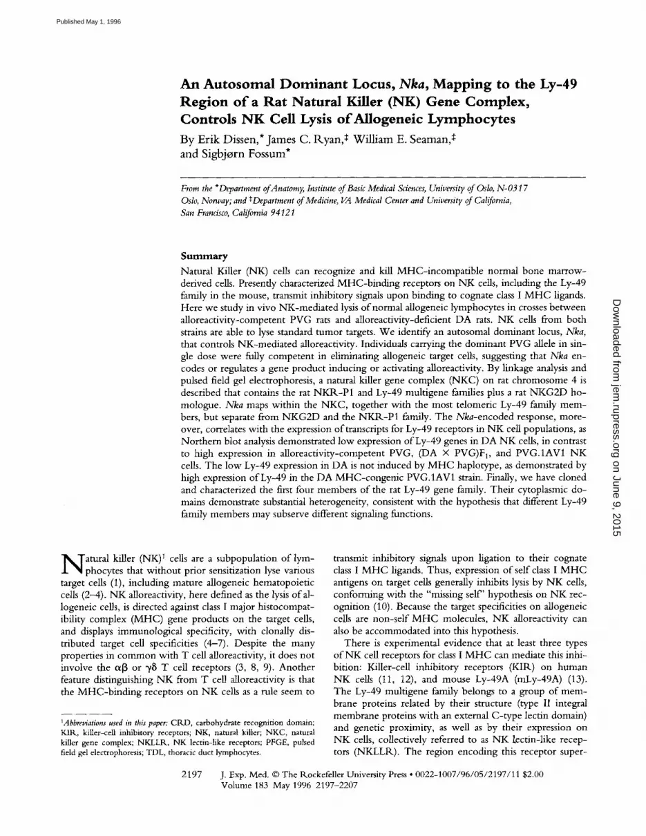
To further define the control o f this response, we as- sessed the in vivo N K cell alloreactivity by offspring o f PVG and DA rats against A O thoracic duct lymphocytes (TDL). All (DA • PVG)F 1 rats displayed high levels ofa l - loreactivity, equal to that o f parental PVG rats. O f 53 F 2
rats, 10 showed low and 43 high alloreactivity (Fig. 1 A). O f 201 (DA • PVG)F 1 • DA back-crossed rats, 96 showed high reactivity (Fig. 1 B). These results are close to 1:3 and 1:1 distributions, respectively, and demonstrate that high alloreactivity segregates as a single autosomal dominant lo- cus, with the PVG allele encoding a high reactivity pheno- type. We have called this locus Nka, for natural killer al- loreactivity.
PVG N K cells lyse LEW as well as A O lymphocytes, and discriminate between the two allotypes (2, 6), implying that different recognition molecules are involved. To de- termine if the responses to these allotypes segregated inde- pendently, 19 (DA • PVG)FI X DA recipients receiving 51Cr-labeled A O cells were simultaneously injected with rain-labeled LEW lymphocytes, and retention o f the two isotopes in the recipient tissues was determined separately. In addition, 9 rats were injected with LEW cells alone. O f the 28 rats, 12 showed high and 16 low reactivity against LEW cells, close to the expected 1:1 distribution for a sin- gle locus showing simple dominant/recessive inheritance. Among the 19 rats that received both A O and LEW cells, there was no segregation of the responses. All 14 rats with low reactivity against A O cells also demonstrated low reac- tivity against LEW cells, whereas the remaining 5 rats had high reactivity against both (not shown). Thus, alloreactiv- ity to A O and LEW cells is controlled by a single gene or by closely linked genes.
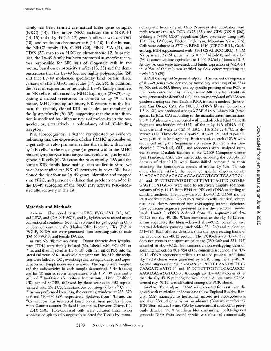
Molecular Cloning and Comparison of Rat Ly-49 cDNAs. W e hypothesized that Nka might be encoded within the NKC. To prepare for mapping of the rat NKC, we isolated four unique rat homologues o f the routine N K receptor Ly-49A (41). The f ly-49.12 sequence is a consensus se- quence derived from the library-derived c D N A rLy-49.12a and the PCP,. clone rLy-49.12b, identical to each other apart from nonoverlapping internal deletions (see Materials and Methods). The nucleotide sequences o f the rat Ly-49 cDNAs (rLy-49.9, rLy-49.12, rLy-49.19, and rLy-49.29) were 68-94% identical to each other and to that o f mLy- 49A (Fig. 2 B). As with members o f the mouse Ly-49 fam- ily, the rLy-49.9, rLy-49.12, and rLy-49.29 cDNAs encode structurally similar polypeptides with putative extracellular calcium-dependent (C-type) lectin domains, rLy--49.19, which is most closely related to fLy-49.12, is a pseudogene in that a stop codon truncates the translated peptide at amino acid 31 (not shown). The predicted mLy-49A, rLy- 49.9, -12, and -29 proteins are 62-81% identical. The rat Ly-49-proteins diverge in both the cytoplasmic and lectin domains (Fig. 2, A and C). Whereas the extracellular lectin
on June 9, 2015jem
.rupress.orgD
ownloaded from
Published May 1, 1996

A
_(3
O G2
O O
E _>,
-40
20
:.:-:
.lo
i �9 ,�9 �9 ,%.. . . . . .~
B N K C ~"
0 e-- c~g.
E
N K C ~
L40 "i." "i" ,,%.
. ;:{,, �9 �9
�9 ***%. ".r
. z �9
2O
. ' k "~." ":.:.:"
:|11|I:I:I :ql l l ! i : . . X . . . .
%.,.
P V G D A F 1 F 2 ( D A x P V G ) F I x D A
Figure 1. In vivo NK aUoreactivity assay towards AO donor TDL. (A) AI- loreactivi W levels in PVG (n - 10), DA (n = 10), (DA • PVG)F 1 (n = 7), or (DA • PVG)F 2 (n = 53) recipients. (B) (DA X PVG)F 1 X DA rats (n = 201) were injected with AO donor TDL. The animals were later genotyped (see below) as either heterozygous at the nat- ural killer gene complex (NKC) (de- noted N K C ~/b, n = 93) or homozygous for the DA NKC haplotype (NKC a/", n = 108). Alloreactivity was expressed as the cervical lymph node/kidney (cpm/mg tissue) ratio. High values re- flect survival of the donor lymphocytes (homing to lymph nodes), low values reflect rejection and secretion of released 51Cr through the kidneys (52). The re- sults have been normalized by multiply- ing data from each individual experi- ment by a linear factor (40/[rn~ig h + mtow] ) calculated from the median values o f the high and low alloreactivity groups. Genotyping was by Southern blot RFLP analysis (Fig. 5). The three observations marked with an asterisk probably represent false positive values (see Fig. 5).
A
rLy-49.9
rLy-49.12 rLy-49.29 mLy-49A
1 MSEQEVTYSSV~SKSSGLQNQ%rKPEETKGPKEAGHR ECYVPWHLIVIALGILCTLLLLTVAVLVTI IFQYSQEKHELQETL*NHH~-~AMQRDIDLKE
1 -N--BF-F-TA--H---V .... ERT---QR-RK--N- V-***-QIT-T ...... FFR-VS---M-IN ............... S-L-J-Y~T--N--N--- 1 -N .... IF-TE--H ......... R .... QSSRK--P- V-S--CL ........ **P-R-VIA-A--SH ...... K--G---IP*---J--~S--S ...... 1 ......... M---H--A---K--R .... ~--~---Y~ R-SFH-KF ....... F-F---VA-S--AIK .... ,-,-',---,-'-------,--,--,
exon 2 exon 3 exon 4
rLy-49. 9 rLy-49. 12 rLy-49.29 reLy- 49A
i00 EMLBIKISIDCSPGNDLLESF~KNRWYSKTKA~HK GSEIETYWFCYGIKCYYVIKDGKSWDECKQTC~-~LFLLKIDDEDE RKFLQQQLIPDN 98 .... DM-TEY-AV-HF-DFL--E---C~_~-T-LD .... S -RGV-MH .......... F-M-R-T-SG-T---J-FJ-P--T ...... LM--HLLVT--S 98 ..... K ..... T---F---LK-EQK ....... T ..... P-T ..................................................... ~ T 99 ---K~[K_~-**** ..... L--DQ--L--~_Z_J-T-LD-L--T -RGDKV ...... M .... FVM-R-T-SG ...... S---S ......... L .... LWPS-S
exon 5 exon 6
rLy-49.9 rLy-49.12 rLy-49.29 mLy-49A
B
cDNA
200 YWIGFSYDKEKKEWAWIENGPSK LASNTMKFNKKLGGCkrFLSKTRLDHTDCINLYSCICGKKLNKFPDLPLQLVLK+ 275 198 .... L---NK-SD-T--D-N ..... L--R-Y-I-D ............ NIN-D--F ...... R-D .... § 265 198 .................... HLN -P ....... E .................................... LSN+ 269 195 C-V-L---NK--D .... D-R ..... L--R-Y-IRD---ML ....... NGN-DQVFI ..... R-D---H+ 262
exon 7
rLy-49.9 fLy-49.12 rLy-49.19 rLy-49.29
fLy-49.9 i00/i00 rLy-49.12 71/63 100/100 rLy-49.19 68/-- 94/-- 100/--
rLy-49.29 90/81 70/62 71/-- 100/100
mLy-49A 71/69 69/71 68/-- 74/60 mLy-49B 69/53 65/51 64/-- 69/46
mLy-49C 72/62 69/60 69/-- 73/58 mLy-49D 73/61 78/69 77/-- 76/61 mLy-d9E 73/64 73/62 69/-- 75/60
mLy-49F 74/59 72/60 70/-- 75/59 mLy-49G 79/60 78/65 76/-- 78/58 mLy-49H 70/60 66/56 66/-- 71/60
C
exon 2 rely-49A
rLy-49.9
mLy- 49C
mLy-49F
reLy- 49E
reLy- 49G
reLy-49B
mLy-49H
rLy-49.29
rLy-49.12
mLy-49D
exon 5 mLy-49C
reLy-49H
mLy-49E
mLy-49F
mLy-49A
reLy-49D
reLy-49G
rLy-49.12
i r L y - 4 9 . 9
rLy-49.29
mLy-49B
2 2 0 0 N k a Controls N K Alloreactivity
on June 9, 2015jem
.rupress.orgD
ownloaded from
Published May 1, 1996

domain o f rLy-49.9 is nearly identical to that o f rLy-49.29 at the amino acid (96%) and at the nucleotide (98%) level, the Ly-49.9 cytoplasmic domain is more similar to the cor- responding domains o f mLy-49A, -C , -E, -F, and - G than to those o f rLy-49.12 o r - 2 9 . The cytoplasmic domains o f rLy-49.9, mLy-49A, and mLy-49D, but not o f rLy-49.12 or rLy-49.29, encode putative inhibi tory G-pro te in (GO binding motifs (Fig. 2 A). This 10 -26 -amino acid mo t i f is defined by at least two individual basic residues upstream of BBXB, BBXXB, or B B X X Z , where B is a basic amino acid, X is non-basic, and Z is an aromatic amino acid (54, 55). The structural requirements for this mo t i f are con- tained in amino acids 29-40 0 K G P K E A G H R E C Y ) o f fLy- 49.9 and in amino acids 29-41 ( K G P R E A G Y R R C S F ) o f mLy-49A. Al though the functional significance o f this m o - t i f is not known, the divergence o f the fLy-49 cytoplasmic domains suggests that distinct Ly-49 receptors may deliver different signals to N K cells.
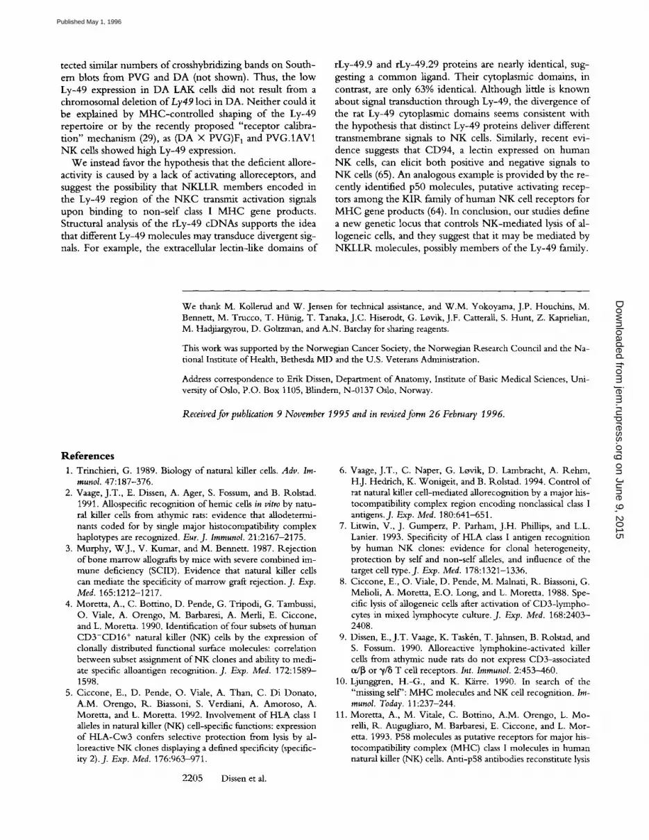
A Rat Gene Crosshybridizes to Human NKG2D. South- ern blots o f genomic D N A from monkey, rat, mouse, dog, cow, and rabbit p roduced one or more bands hybridizing to h N K G 2 D . N o hybridizat ion was detected to chicken or yeast D N A (not shown). The h N K G 2 A probe did not yield detectable bands in the rat, although bands were clearly present for dog and cow D N A . The h N K G 2 C probe did not produce detectable bands except with human and mon- key D N A (Fig. 3). U n d e r condit ions o f low stringency, the h N K G 2 D probe, containing the entire open reading frame, hybridized to only one band in most digests, indicating ho- mology within only a short segment o f genomic D N A (not shown). W e have recently cloned and sequenced this rat N K G 2 D homologue , which is 60% identical to human N K G 2 D at the amino acid level (S. Berg et al., manuscript in preparation).
Mapping of the Rat NKC and Localization to Chromosome 4. To map the N K C , Southern blots o f PVG and D A liver D N A digested with 40 different restriction enzymes were hybridized to probes for genes encoded wi thin or near to the human or mouse N K C . Informative RFLPs were found for a number o f genes, including r N K R - P 1 B , rLy-49.9, rLy-49.12, and the previously undefined rat homologues o f mLy-49C and h N K G 2 D (Fig. 4).
R F L P patterns were then investigated on liver D N A ex- tracted from 223 (DA • PVG)F 1 • D A rats (for some
Figure 3. Search for NKG2 homologues in the rat by Southern blot analysis. DNA from several mammal species were digested with EcoRl, separated by electrophoresis, transferred to nylon membrane, and hybrid- ized under low stringency conditions to cDNA probes for hNKG2A, -C and -D. Lane 1, human; lane 2, monkey; lane 3, rat; lane 4, mouse; lane 5, dog; lane 6, cow; lane 7, rabbit. The NKG2D probe hybridized to a band in the rat, whereas the -A and -C probes did not hybridize detect- ably above background. Approximate molecular weights in kb are indi- cated.
RFLPs, only 85 animals). By this analysis, the members o f the rat N K C were closely linked; h N K G 2 D , r N K R - P 1 B , and all o f the mouse and rat Ly-49 RFLPs cosegregated in all but one o f 223 rats (Fig. 5). A single crossover was ob- served within the N K C , splitting the Ly49 genes into a centromeric group associated with the N K R - P 1 family and with rat N K G 2 D , while the remaining, telomeric Ly49 genes were associated with rat homologues o f mLy-49C. This is similar to the mouse N K C , where mLy-49C maps telomeric to at least one other Ly49 locus (44).
In the mouse, the N K C is l inked to several unrelated genes, including Kap and Igkc (56). R F L P analysis revealed a similar linkage in the rat. A single recombinat ion oc- curred be tween Kap and Ly49, placing Kap on the te lo- meric side o f the N K C . The N K C also lay ~ 3 9 . 5 cM from Igkc, 7.1 cM from Cd9, and 9.5 cM from Pthlh. The Igkc and Pthlh loci have previously been mapped to rat ch romo- some 4 (57). Earlier karyotyping studies have shown con- siderable G-banding similarities be tween mouse ch romo- some 6 and rat chromosome 4 (57). The orientation o f mouse and rat loci within these regions is therefore likely
Figure 2. (A) rLy-49.9, -12 and -29 are type II integral membrane proteins with extracellular C-type lectin domains. For comparison, the mLy-49A sequence (41) is shown, the amino acid sequences are divided into six putative exon sequences as predicted by the genomic structure of mLy-49A (66). The transmembrane region is underlined and sites for N-glycosylation are boxed. The residues defining putative inhibitory G-protein (G3 binding sites are underlined twice. (B) Per cent identical residues between rat and mouse Ly-49 sequences at the nucleotide/amino acid levels. (C) Dendrograms com- paring the amino acid sequences of the putative cytoplasmic domains (exon 2) and amino-terminal exon encoding the lectin domain (exon 5) of the three rat sequences with eight mouse Ly-49 proteins (16, 17) (mLy-49G corresponding to -49G2 in reference 16 and mLy-49H renamed as announced by the authors in reference 17). rLy-49.9 is an 1193 bp cDNA encoding a 275-amino acid protein with a predicted molecular weight of 31,889. The consensus rLy-49.12 cDNA is a 1163--bp cDNA encoding a 265-amino acid protein with a predicted molecular weight of 31,215. rLy-49.19 is a1152-bp cDNA pseudogene encoding a truncated peptide of only 31 amino acids (not shown). When compared with the consensus sequences of the other rat Ly-49 cDNAs, the rLy-49.19 sequence contains a single-base insertion (G) at nt 210, deletion of an 8-nt consensus segment (ATTTTTCA) between bases 333 and 334, and a single base substitution (G to A) producing a stop codon at nt 492 (not shown). The 919-bp fLy-49.29 cDNA encodes a 269-amino acid protein with a molecular weight of 31,140. Nucleotide sequences are available from GenBank (accession numbers rLy-49.9: U56863; fLy-49.12 (consen- sus sequence): U56822; rLy-49.19: U56823; rLy-49.29: U56824).
2201 Dissen et al.
on June 9, 2015jem
.rupress.orgD
ownloaded from
Published May 1, 1996

Figure 4. Southern blot 1<FLP analyses of liver DNA from PVG and DA rats hybrid- ized to the probes indicated below (restriction enzymes in parenthesis). Left lanes, PVG; right lanes, DA. Panel 1, lg K C-region (HinclI); panel 2, CD9 (HaeI[I) panel 3, KAP (Kpnl); panel 4, PTHLH (BstXI); panel 5, P1B (HindlIl); panel 6, 5E6-5' (EcoRV); panel 7, hNKG2D (BstXl); panel 8, rLy49.9 (EcoO- 109I); panel 9, rLy49.12 (BstXl); panel 10, rLy49.12 (EcoO1091); panel 11, rLy49.12 (EcoR.l);
panel 12, rLy49.12 (HindlII); panel 13, 5E6-5' (BstXI); panel 14, 5E6-5' (Eco1<I). The PVG-specific bands are indicated with arrows. Because of crosshy- bridization between Ly-49 genes, Ly-49 RFLPs need not have been specific for the particular genes used as probes. For simplicity, the Ly-49 RFLPs were provisionally termed Ly49-1<1 (panel 12), -R2 and -1<3 (panel 9, short and long bands, respectively), -R.4 (panel 11), -1<5 and -R6 (panel 10, short and long bands, respectively), -R7 (panel 8), -R8 (panel 13), -R9 (panel I4), and -R10 (panel 6).
to be similar, giving the following orientation: centromere- lgkc-Cd9-NKC-Kap-Pthlh. Our findings thus localize and orient the rat N K C within rat chromosome 4 and demon- strate conserved linkage of a large chromosomal region ("-,50 cM) between the mouse and the rat (Fig. 6 A).
PFGE Mapping of the NKC. To further map the loci within the NKC, we prepared PFGE Southern blots of rat DNA. PFGE of SalI- and Sill-digested B cell D N A re- vealed fragments that spanned large parts of the NKC, some of which overlapped. By sequentially hybridizing
,oKc [ ]D[ ] i n N [] i i �9 i i [ ] [ ] co9 [ ] [ ] E l D D D �9 m m �9 i i ~ NI<'R-P~B [ ] D D D ~ Q ~ D D [ ] D D �9 �9 i l l i ~ D ~
Ly49-R, ~%[][][] @ND [] [3D �9 �9 mime ~ @ N Ly49-R2 [~ [~ [~E] [~ [~ @ D [ ] [] [ ] D �9 �9 mime DDD ,.,.,~,-,,4 D G [ ] D s s [] D D �9 m m ~
.~,., [ ] ~ D ~ ~[][] [] ~ [] �9 mini []~
L~9-~s ) ~ ~ D ~ [ ] [ ] ~ [] �9 i i l i ~ [ ] ~
N~A-AO [] IN[] [] [] [] �9 �9 in mim , . -~-~ [ ] [ ] [ ] [ ] �9
~ ~ N ~ [] []~ [] [] miNi ~[][]
31 6 3 1 7 1 0 3 5 2 3 2 2 I 3 2 ] 1" S 1 2 9 I * I I
Figure 5. Linkage analysis of Nka and other loci on rat chromosome 4. 223 (DA X PVG)F~ • DA rats were genotyped as either homozygous (DA/DA) or heterozygous (DA/PVG) at the indicated loci by 1<FLP analysis of liver DNA. Rats are displayed in different groups according to the typed loci and the location of crossovers. The Nka phenotypes of 210 of these animals were determined, either towards AO (n = 182), LEW (n = 9), or both AO and LEW (n = 19) donor lymphocytes (Fig. t B, LEW data not shown). A high alloreactivity phenotype was attributed to heterozygosity at Nka. The rLy-49.9 and rLy-49.12 probes crosshybrid- ized extensively to other Ly-49 related genes. Therefore, several Ly-49 1<FLPs were applied, to detect and localize as many members of the Ly-49 gene family as possible. Two rats showing high NK alloreactivity were homozygous at the surrounding parts of the chromosome (the two far right columns). These results, which were noninformative tbr map- ping of Nka, could stern from double crossover, gene conversion or false positive alloreactivity results. The two columns marked with an asterisk represented conflicting results regarding the order of Nka and Kap.
these blots to several N K C probes and assembling the over- lapping fragments, a tentative map of the rat N K C could be drawn (Fig. 6/3). W e were not able to generate probes spe- cific for each of the Ly-49 members, but two Ly-49 probes (49-CTM and 5E6-5') did not cohybridize, except to very long fragments, indicating that they identify distinct chro- mosomal regions. The 4 9 - C T M and h N K G 2 D probes co- hybridized to several fragments, the shortest of which (XhoI, not shown) constrained the region containing N K G 2 D and at least one Ly-49 gene to less than 250 kb. Probes for r N K R - P 1 A and r N K R - P 1 B hybridized exclusively to a single 120-kb ClaI fragment, demonstrating close proxim- ity of these genes. It was not clear from our data whether N K G 2 D was flanked by Ly-49 genes on both sides or just on the telomeric side, as schematically drawn in Fig. 6 B.
Nka Is Located within the Rat NKC. O f the 223 rats used for RFLP analysis, 201 had been typed for in vivo NK al- loreactivity toward donor TDL from AO rats, and 28 had been tested for alloreactivity toward TDL from LEW do- nors (including 19 that were tested for both). Cosegrega- tion between Nka and the N K C genes was found for 198 out of the 201 rats typed with AO donor TDL (Fig. 1 B). The three discordant rats probably reflected double cross- over or gene conversion, or were false positives (Fig. 5, the three far right columns). The single rat with a recombina- tion inside the Ly-49 region (injected with LEW donor cells) demonstrated the high alloreactivity (PVG) Nka phe- notype, being homozygous (DA/DA) centromeric to the crossover, and heterozygous (PVG/DA) on the telomeric side (Fig. 5). This crossover placed Nka telomeric to the re- gion containing the centromeric portion of the Ly-49 gene family. It also localized Nka to a chromosomal region dis- tinct from Nkrplb and Nkg2d. A second crossover, with low alloreactivity, placed Nka centromeric to Kap. Thus, from these two crossovers Nka lay between the centro- meric portion of the Ly-49 family and Kap. A third cross- over, with high alloreactivity, conflicted with this result, placing Nka telomeric to Kap (Fig. 5). The findings with this rat, however, were discounted because injection was
2202 Nka Controls NK AUoreactivity
on June 9, 2015jem
.rupress.orgD
ownloaded from
Published May 1, 1996

A
39,5
- Igkc
/ / /
/ /
Cd9 / / / 0,4 7,1 , /
~NKC
9,5 l ~ \ ' \ . \ 0,4
Pthlh \ \
telomeric
B
w
S t i l l I I I I I I Nkrp I a, -b
-Nkg2d Ly49 NKR-P1 N KG2~..j.- L y - / , 9 ~ , , , , , AB
Ly49 "Nka S a i l : : : : : I I I I
Kap
I Im
telomeric
' 400 800kb
Figure 6. (A) Linkage map of rat chromosome 4 based on seg- regation of alleles in 223 back- crossed rats. Map distances are shown in cM. The localization of Nka was based on data from both AO and LEW donor cells. (B) Physical map of the rat NKC. PFGE Southern blots were generated with several rare- cutting restriction enzymes and sequentially hybridized to probes for NKC genes. Incomplete Sall and Sill digests produced a num- ber of partially overlapping frag- ments. The 49-CTM, 5E6-5' and hNKG2D probes were used for the telomeric part of the complex. 49-CTM did not hy- bridize to the two most telo-
meric Sall- fragments. 5E6-5', however, hybridized to these bands but not to the four most centromeric bands. The subgroup of Ly-49 genes related to mLy-49C (5E6) are indicated at the telomeric end. One 810-kb band, marked with an asterisk, hybridized to both probes. Two of the overlapping bands summed up to 740 kb, leaving 70 kb that could not be incorporated precisely. The centromeric/telomeric direction of the NKR_-P1 segment was not known. The number of genes in the Ly-49 family is unknown, therefore the Ly-49 genes were drawn schematically. The NKR-PIA and P1B probes both hybridized to a single 120-kb ClaI fragment, and the hNKG2D and 49- CTM probes cohybridized to a 250-kb fragment with XhoI (not shown).
associated with extravasation o f cells, which would result in reduced uptake o f labeled donor cells, mimicking allorejec- t ion (high reactivity). W e thus favor the interpretat ion supplied by the first two crossovers, which constrain Nka to a region o f ~ 0 . 4 cM centromeric to Kap, containing the telomeric group o f Ly-49 genes. This association o f Nka with the Ly-49 gene family was further supported by stud- ies relating Nka to the expression o f Ly-49 transcripts.
Association of NK Alloreactivity with the Expression of Ly-49 Transcripts. The genetic linkage be tween Nka and the Ly-49 family o f receptors led us to consider the hypothesis that NK-med ia t ed lysis o f allogeneic targets may be medi - ated by members o f the Ly-49 family. Al though mLy-49A mediates inhibit ion o f natural killing, it binds to selective class I M H C antigens (13, 25, 26). Thus, it is capable o f a l - lorecognit ion, raising the possibility that other members o f "the Ly-49 family, particularly in the rat, might activate nat- ural killing o f allogeneic targets.
To further examine a possible association be tween Nka and Ly49, Nor thern blots were prepared from IL-2-act i - vated N K cells derived from DA, PVG, (DA • PVG)F 1, and the M H C - c o n g e n i c strain PVG.1AV1, which carries the D A M H C haplotype (RT~ avl) on a PVG background. N K cells from all strains expressed transcripts for N K R - P 1 , although levels in D A N K cells were somewhat higher than in the other strains tested (Fig. 7). In contrast, as as- sessed by hybridization to probes for rLy-49.9, rLy-49.12, rLy-49.19, rLy-49.29, mLy-49A, and mLy-49C, the level o f transcripts for members o f the Ly-49 family was mark- edly reduced in N K cells from the nonresponder D A strain compared with N K cells from al loreact ivi ty-competent PVG, (DA • PVG)F1 and PVG.1AV1 rats (6). Similar to Nka, the higher expression o f Ly-49 genes in PVG appears to be a dominant trait, as deduced from the high level o f
2203 Dissen et al.
expression in (DA • PVG)FI cells. The low expression o f Ly-49 genes in D A N K cells (RT1 avl) apparently did not result from M H C - c o n t r o l l e d repertoire selection, since PVG.1AV1 cells (RT1 avl) expressed levels o f Ly-49 that were similar to those in PVG cells (Fig. 7). These findings
Figure 7. Northern blot anal- ysis of IL-2-activated NK cells derived from DA, PVG, (DA • PVG)F1 or PVG.1AV1 rats. The same pattern of low expression of Ly-49 genes in DA was also found with two mouse Ly-49 cDNA probes (mLy-49A and mLy-49C, not shown). Approxi- mate molecular weights are indi- cated.
on June 9, 2015jem
.rupress.orgD
ownloaded from
Published May 1, 1996

suggest that Nka may be a member of the Ly-49 family or represents a regulatory element controlling transcription of Ly-49 genes.
Discuss ion
In these studies, we have (a) identified a rat NKC region containing members of the NKR-P1 and Ly-49 families as well as a rat NKG2D homologue; (b) mapped the rat NKC to the telomeric part of chromosome 4; (c) demonstrated that the alloreactivity mediated by rat NK cells is controlled by a genetic locus, Nka, that lies within the NKC where it colocalizes with the telomeric group of Ly49 loci and is distinct from Nkrp 1 and Nkg2d; (d) cloned four rat Ly-49 cDNAs and demonstrated considerable heterogeneity within their cytoplasmic domains; and (e) shown that the high re- sponder Nka phenotype correlates with increased expres- sion of Ly-49 transcripts.
All the NKLLR probes tested by Southern blotting mapped to the rat NKC. Although the many bands ob- tained with the Ly-49 probes indicate that there are several more rat Ly-49 genes, no Ly-49 related RFLPs mapped outside the NKC. This suggests that all Ly-49 genes reside in the complex, clustered within a region of 400-1,100 kb. NKG2 genes have previously not been described in ro- dents, hNKG2D is sufficiently distantly related to other NKG2 genes to merit classification as a separate group of NKLLRs, e.g., the COOH-terminal part of its carbohy- drate recognition domain (CRD) is more similar to the CRDs of NKR-P1 than to other NKG2 molecules (58, S. Berg et al., manuscript in preparation). Its presence, there- fore, does not suggest the existence of rat homologues to other NKG2 genes.
Our studies demonstrate the presence within the NKC of a locus, Nka, controlling NK-mediated alloreactivity. Similar to Nka, NK cell-mediated resistance to murine cy- tomegalovirus infection in the mouse is encoded by a locus (Cmvl) that maps to the NKC region on mouse chromo- some 6 (59). Also, a correlation between NK cell alloreac- tivity and NKI.1 (mNKR-P1C) expression in crosses of inbred mouse strains has previously been described (60).
The nature of the Nka locus is not known. However, al- though D A N K cells are deficient in the lysis of allogeneic blasts, they are competent in the lysis of tumor targets (61), indicating that Nka influences target-specific interactions rather than nonspecific steps in the lytic pathway. This is supported by the finding that Nka maps to the NKC, which encodes families of lectin-like receptors. As in the mouse, rat alloreactivity is directed against M H C encoded gene products (2, 6). Our hypothesis is therefore that Nka encodes or regulates an allele-specific receptor for class I M H C molecules. In the mouse, at least one member of the Ly-49 receptor family, Ly-49A, binds to specific target class I M H C antigens. It is therefore particularly noteworthy that Nka is linked to the genes encoding the Ly-49 family of receptors and that the activity of Nka correlates with lev- els of Ly-49 expression.
The striking correlation between levels of Ly-49 expres-
sion and alloreactivity in DA, PVG, (DA • PVG)F1, and PVG.1AV1 NK cells does not prove a causal relationship. In particular, we have only investigated expression in total NK cell populations. Furthermore, there is recent evidence that immature NK cells cultured from mouse fetal liver do not express Ly-49 and do not exhibit alloreactivity, but kill tumor targets (62). Their phenotypic similarity to D A N K cells raises the possibility that the lack of alloreactivity and the low Ly-49 expression are mutually independent fea- tures of D A N K cells with a common cause, e.g., blocked or erratic NK cell maturation. This would imply, however, that Nka controls NK cell maturation. Given the genomic localization of Nka in the Ly-49 region of the NKC, we consider it more likely that Nka encodes or regulates the expression of Ly-49 molecules or related as yet undefined NKLLR members.
The interaction between mouse Ly-49A and cognate M H C inhibits natural killing (13), in accord with the "missing self" hypothesis, that natural killing is inhibited by target cell M H C class I. In contrast, the high responder Nka pvG phenotype is dominant in Nka evC/DA heterozy- gotes, demonstrating that this phenotype is not due to the loss of an inhibitory (DA) allele, but to the inheritance of an allele (PVG) promoting alloreactivity. The possibility that the PVG allele of Nka represents a dominant negative mutation, somehow blocking an inhibitory receptor func- tion, cannot be entirely dismissed, but does not explain the correlation between alloreactivity and Ly-49 expression. In spite of the weak signals obtained from D A N K cells on Northern blots, we cannot exclude normal expression of a single or very few Ly-49 genes. However, the probes used crosshybridized extensively to other Ly-49 genes. More- over, DA rats are deficient in NK-mediated alloreactivity to lymphocytes from a large panel of donor strains (52). We therefore consider it unlikely that the inability of D A N K cells to lyse allogeneic targets reflects the activity of inhibi- tory Ly-49 molecules. The most likely interpretation of our observations is therefore that Ly-49 members or other co- regulated, as yet undefined NKLLR molecules encoded in the same region of the rat NKC activate rather than inhibit natural killing.
Several studies indicate that NK cells can be activated, rather than inhibited by M H C encoded structures. In cold target inhibition experiments with PVG NK cells, blasts from the LEW.1LM1 strain, which is genetically identical to LEW except for a 100-kb homozygous chromosomal deletion in the RT1. C region of the MHC, failed to specif- ically inhibit lysis of LEW blasts (6). Similarly, NK cells in irradiated C57BL/6 mice recognize and eliminate hemato- poietic cells from H-2D a transgenic C57BL/6 donors (63). In the human, recent studies have further demonstrated that some NK clones can be activated rather than inhibited by the selective expression of M H C antigens on target cells (64, J. Gumperz, Stanford, personal communication). These observations suggest the existence of an activating NK cell receptor for M H C gene products.
PFGE analysis of the DA and PVG NKC regions failed to detect insertion/deletion patterns, and Ly-49 probes de-
2204 Nka Controls NK Alloreactivity
on June 9, 2015jem
.rupress.orgD
ownloaded from
Published May 1, 1996
tected similar numbers of crosshybridizing bands on South- ern blots from PVG and DA (not shown). Thus, the low Ly-49 expression in D A LAK cells did not result f rom a chromosomal deletion o f Ly49 loci in DA. Nei ther could it be explained by MHC-con t ro l l ed shaping o f the Ly-49 repertoire or by the recently proposed "receptor calibra- t ion" mechanism (29), as (DA • PVG)F1 and PVG.1AV1 N K cells showed high Ly-49 expression.
W e instead favor the hypothesis that the deficient allore- activity is caused by a lack o f activating alloreceptors, and suggest the possibility that N K L L R members encoded in the Ly-49 region of the N K C transmit activation signals upon binding to non-self class I M H C gene products. Structural analysis o f the rLy-49 cDNAs supports the idea that different Ly-49 molecules may transduce divergent sig- nals. For example, the extracellular lectin-hke domains o f
rLy-49.9 and rLy-49.29 proteins are nearly identical, sug- gesting a c o m m o n hgand. Their cytoplasmic domains, in contrast, are only 63% identical. Although httle is known about signal transduction through Ly-49, the divergence of the rat Ly-49 cytoplasmic domains seems consistent with the hypothesis that distinct Ly-49 proteins dehver different transmembrane signals to N K cells. Similarly, recent evi- dence suggests that CD94, a lectin expressed on human N K cells, can elicit both positive and negative signals to N K cells (65). An analogous example is provided by the re- cently identified p50 molecules, putative activating recep- tors among the K I R family of human N K cell receptors for M H C gene products (64). In conclusion, our studies define a new genetic locus that controls NK-media ted lysis o f al- logeneic cells, and they suggest that it may be mediated by N K L L R molecules, possibly members of the Ly-49 family.
We thank M. Kollerud and W. Jensen for technical assistance, and W.M. Yokoyama, J.P. Houchins, M. Bennett, M. Trucco, T. Hiinig, T. Tanaka, J.C. Hiserodt, G. Lovik, J.F. Catterall, S. Hunt, Z. Kaprielian, M. Hadjiargyrou, D. Goltzman, and A.N. Barclay for sharing reagents.
This work was supported by the Norwegian Cancer Society, the Norwegian Research Council and the Na- tional Institute of Health, Bethesda MD and the U.S. Veterans Administration.
Address correspondence to Erik Dissen, Department of Anatomy, Institute of Basic Medical Sciences, Uni- versity of Oslo, P.O. Box 1105, Blindern, N-0137 Oslo, Norway.
Received for publication 9 November 1995 and in revised form 26 February 1996.
References 1. Trinchieri, G. 1989. Biology of natural killer cells. Adv. Im-
raunol, 47:187-376. 2. Vaage, J.T., E. Dissen, A. Ager, S. Fossum, and B. Rolstad.
1991. Allospecific recognition of hemic cells in vitro by natu- ral killer cells from athymic rats: evidence that allodetermi- nants coded for by single major histocompatibility complex haplotypes are recognized. Eur.J. Immunol. 21:2167-2175.
3. Murphy, W.J., V. Kumar, and M. Bennett. 1987. Rejection of bone marrow allografts by mice with severe combined im- mune deficiency (SCID). Evidence that natural killer cells can mediate the specificity of marrow graft rejection. J. Exp. Med. 165:1212-1217.
4. Moretta, A., C. Bottino, D. Pende, G. Tripodi, G. Tambussi, O. Viale, A. Orengo, M. Barbaresi, A. Merli, E. Ciccone, and L. Moretta. 1990. Identification of four subsets of human CD3-CD16 + natural killer (NK) cells by the expression of clonally distributed functional surface molecules: correlation between subset assignment of NK clones and ability to medi- ate specific alloantigen recognition. J. Exp. Med. 172:1589- 1598.
5. Ciccone, E., D. Pende, O. Viale, A. Than, C. Di Donato, A.M. Orengo, lk. Biassoni, S. Verdiani, A. Amoroso, A. Moretta, and L. Moretta. 1992. Involvement of HLA class I alleles in natural killer (NK) cell-specific functions: expression of HLA-Cw3 confers selective protection from lysis by al- loreactive NK clones displaying a defined specificity (specific- ity 2).J. Exp. Med. 176:963-971.
2205 Dissen et al.
6. Vaage, J.T., C. Naper, G. Lovik, D. Lambracht, A. R.ehm, H.J. Hedrich, K. Wonigeit, and B. Rolstad. 1994. Control of rat natural killer cell-mediated allorecognition by a major his- tocompatibility complex region encoding nonclassical class I antigens.J. Exp. Med. 180:641-651.
7. Litwin, V., J. Gumperz, P. Parham, J.H. Phillips, and L.L. Lanier. 1993. Specificity of HLA class I antigen recognition by human NK clones: evidence for clonal heterogeneity, protection by self and non-self alleles, and influence of the target cell type.J. Exp. Med. 178:1321-1336.
8. Ciccone, E., O. Viale, D. Pende, M. Malnati, R. Biassoni, G. Melioli, A. Moretta, E.O. Long, and L. Moretta. 1988. Spe- cific lysis of allogeneic cells after activation of CD3-1ympho- cytes in mixed lymphocyte culture. J. Exp. Med. 168:2403- 2408.
9. Dissen, E., J.T. Vaage, K. Task6n, T. Jahnsen, B. Rolstad, and S. Fossum. 1990. Alloreactive lymphokine-activated killer cells from athymic nude rats do not express CD3-associated ~x/J3 or ~//5 T cell receptors. Int. Immunol. 2:453-460.
10. Ljunggren, H.-G., and K. ICirre. 1990. In search of the "missing self': MHC molecules and NK cell recognition. Im- munol. Today. 11:237-244.
11. Moretta, A., M. Vitale, C. Bottino, A.M. Orengo, L. Mo- relli, R. Augugliaro, M. Barbaresi, E. Ciccone, and L. Mor- etta. 1993. P58 molecules as putative receptors for major his- tocompatibility complex (MHC) class I molecules in human natural killer (NK) cells. Anti-p58 antibodies reconstitute lysis
on June 9, 2015jem
.rupress.orgD
ownloaded from
Published May 1, 1996

of MHC class I-protected cells in NK clones displaying dif- ferent specificities. J. Exp. Med. 178:597-604.
12. Gumperz, J.E., V. Litwin, J.H. Phillips, L.L. Lanier, and P. Parham. 1995. The Bw4 public epitope of HLA-B molecules confers reactivity with natural killer cell clones that express NK131, a putative HLA receptor. J. Exp. Med. 181:1133- 1144.
13. Karlhofer, F.M., R.K. Ribaudo, and W.M. Yokoyama. 1992. MHC class I alloantigen specificity of Ly-49 + IL-2-activated natural killer cells. Nature (Lond.). 358:66-70.
14. Yokoyama, W.M., J.C. Ryan, J.J. Hunter, H.R.C. Smith, M. Stark, and W.E. Seaman. 1991. cDNA cloning of mouse NKR-P1 and genetic linkage with Ly-49. Identification of a natural killer cell gene complex on mouse chromosome 6. J. Immunol. 147:3229-3236.
15. Giorda, R., and M. Trucco. 1991. Mouse NKR-P1. A fam- ily of genes selectively coexpressed in adherent lymphokine- activated killer cells.J. Immunol. 147:1701-1708.
16. Smith, H.R.C., F.M. Karlhofer, and W.M. Yokoyama. 1994. Ly-49 multigene family expressed by IL-2-activated NK cells.J. Immunol. 153:1068-1079.
17. Brennan, J., D. Mager, W. Jefferies, and F. Takei. 1994. Ex- pression of different members of the Ly-49 gene family de- fines distinct natural killer cell subsets and cell adhesion prop- erties.J. Exp. Med. 180:2287-2295.
18. Ziegler, S.F., S.D. Levin, L. Johnson, N.G. Copeland, D.J. Gilbert, N.A. Jenkins, E. Baker, G.R. Sutherland, A.L. Feldhaus, and F. Ramsdell. 1994. The mouse CD69 gene: structure, expression, and mapping to the NK complex. J. Immunol. 152:1228-1236.
19. Yabe, T., C. McSherry, F.H. Bach, P. Fisch, R.P. Schall, P.M. Sondel, and J.P. Houchins. 1993. A multigene family on human chromosome 12 encodes natural killer-cell lectins. Immunogenetics. 37:455-460.
20. Chang, C., A. Rodriguez, M. Carretero, M. L6pez-Botet, J.H. Phillips, and L.L. Lanier. 1995. Molecular characteriza- tion of human CD94: a type II membrane glycoprotein re- lated to the C-type lectin superfamily. Eur. J. Immunol. 25: 2433-2437.
21. Lanier, L.L., C. Chang, andJ.H. Phillips. 1994. Human NKR- P1A. A disulfide-linked homodimer of the C-type lectin su- perfamily expressed by a subset of NK and T lymphocytes.J. Immunol. 153:2417-2428.
22. Schnittger, S., J. Hamann, C. Dannenberg, H. Fiebig, M. Strauss, and C. Fonatsch. 1993. Regional sublocalization of the human CD69 gene to chromosome bands 12p12.3- p13.2, the predicted region of the human natural killer cell gene complex. Eur.J. Immunol. 23:2711-2713.
23. Sentman, C.L., V. Kumar, and M. Bennett. 1991. Rejection of bone marrow cell allografts by natural killer cell subsets: 5E6 + cell specificity for Hh-1 determinant 2 shared by H-2 d and H-2 f. Eur.J. Immunol. 21:2821-2828.
24. Yokoyama, W.M., P.J. Kehn, D.I. Cohen, and E.M. Shevach. 1990. Chromosomal location of the Ly-49 (AI, YE1/48) multigene family. Genetic association with the NK 1.1 anti- gen.J. Immunol. 145:2353-2358.
25. Kane, K.P. 1994. Ly-49 mediates EL4 lymphoma adhesion to isolated class I major histocompatibility complex molecules. J. Exp. Med. 179:1011-1015.
26. Daniels, B.F., F.M. Karlhofer, W.E. Seaman, and W.M. Yokoyama. 1994. A natural killer cell receptor specific for a major histocompatibility complex class I molecule. J. Exp. Med. 180:687-692.
27. Sykes, M., M.W. Harry, F.M. Karlhofer, D.A. Pearson, G. Szot, and W.M. Yokoyama. 1993. Hematopoietic cells and radioresistant host elements influence natural killer cell differ- entiation.J. Exp. Med. 178:223-229.
28. Karlhofer, F.M., R. Hunziker, A. Reichlin, D.H. Margulies, and W.M. Yokoyama. 1994. Host MHC class I molecules modulate in vivo expression of a NK cell receptor. J. Immu- nol. 153:2407-2416.
29. Olsson, M.Y., K. K~irre, and C.L. Sentman. 1995. Altered phenotype and function of natural killer cells expressing the major histocompatibility complex receptor Ly-49 in mice transgenic for its ligand. Proc. Natl. Acad. Sci. USA. 92:1649- 1653.
30. Colonna, M., and J. Samaridis. 1995. Cloning of immuno- globulin-superfamily members associated with HLA-C and HLA-B recognition by human natural killer cells. Science (Wash. DC). 268:405-408.
31. Wagtmann, N., R. Biassoni, C. Cantoni, S. Verdiani, M.S. Malnati, M. Vitale, C. Bottino, L. Moretta, A. Moretta, and E.O. Long. 1995. Molecular clones of the p58 NK cell re- ceptor reveal immunoglobulin-related molecules with diver- sity in both the extra- and intracellular domains. Immunity. 2: 439-449.
32. D'Andrea, A., C. Chang, K. Franz-Bacon, T. McClanahan, J.H. Phillips, and L.L. Lanier. 1995. Molecular cloning of NKB1. A natural killer cell receptor for HLA-B allotypes. J. Immunol. 155:2306-2310.
33. Ford, W.L. 1978. The preparation and labelling of lympho- cytes. In Handbook of Experimental Immunology. Vol. 2. D.M. Weir, editor. Blackwell Scientific Publications, Ox- ford. 23.1-23.22.
34. Rolstad, B., and S. Fossum. 1987. Allogeneic lymphocyte cy- totoxicity (ALC) in rats: establishment of an in vitro assay, and direct evidence that cells with natural killer (NK) activity are involved in ALC. Immunology. 60:151-157.
35. Hiinig, T., H.-J. Wallny, J.K. Hartley, A. Lawetzky, and G. Tiefenthaler. 1989. A monoclonal antibody to a constant de- terminant of the rat T cell antigen receptor that induces T cell activation.J. Exp. Med. 169:73-86.
36. Dallman, M.J., M.L. Thomas, and J.R. Green. 1984. MRC OX-19: a monoclonal antibody that labels T lymphocytes and augments in vitro proliferative responses. Eur. J. Immunol. 14:260-267.
37. Tanaka, T., T. Masuko, H. Yagita, T. Tamura, and Y. Hash- imoto. 1989. Characterization of a CD3-1ike rat T cell sur- face antigen recognized by a monoclonal antibody. J. lmmu- nol. 142:2791-2795.
38. McKnight, A.J., and B.J. Classon. 1992. Biochemical and im- munological properties of rat recombinant interleukin-2 and interleukin-4. Immunology 75:286-292.
39. Chambers, W.H., N.L. Vujanovic, A.B. DeLeo, M.W. Olszowy, R.B. Herberman, and J.C. Hiserodt. 1989. Mono- clonal antibody to a triggering structure expressed on rat nat- ural killer cells and adherent lymphokine-activated killer cells._/. Exp. Med. 169:1373-1389.
40. Ryan, J.C., E.C. Niemi, R.D. Goldfien, J.C. Hiserodt, and W.E. Seaman. 1991. NKR-P1, an activating molecule on rat natural killer cells, stimulates phosphoinositide turnover and a rise in intracellular calcium. J. Immunol. 147:3244-3250.
41. u W.M., L.B. Jacobs, O. Kanagawa, E.M. Shevach, and D.I. Cohen. 1989. A murine T lymphocyte antigen be- longs to a supergene family of type II integral membrane pro- teins.J. Immunol. 143:1379-1386.
2206 Nka Controls NK Mloreactivity
on June 9, 2015jem
.rupress.orgD
ownloaded from
Published May 1, 1996

42. Cleveland, D.W., M.A. Lopata, P,..J. MacDonald, N.J. Cowan, W.J. Rutter, and M.W. Kirschner. 1980. Number and evolutionary conservation of or- and [3-tubulin and cyto- plasmic [3- and y-actin genes using specific cloned cDNA probes. Cell. 20:95-105.
43. Birren, B., and E. Lai. 1993. Pulsed Field Gel Electrophore- sis: a Practical Guide. Academic Press, San Diego.
44. Stoneman, E.R.., M. Bennett, J. An, K.A. Chesnut, E.K. Wakeland, J.B. Scheerer, M.J. Siciliano, V. Kumar, and P.A. Mathew. 1995. Cloning and characterization of 5E6(Ly- 49C), a receptor molecule expressed on a subset of murine natural killer cells.J. Exp. Med. 182:305-313.
45. Houchins, J.P., T. Yabe, C. McSherry, and F.H. Bach. 199l. DNA sequence analysis of NKG2, a family of related cDNA clones encoding type II integral membrane proteins on hu- man natural killer cells.J. Exp. Med. 173:1017-1020.
46. Sheppard, H.W., and G.A. Gutman. 1981. Allelic forms of rat K chain genes: Evidence for strong selection at the level of nucleotide sequence. Proc. Natl. Acad. Sci. USA. 78:7064- 7068.
47. Niu, E.M., A. Crozat, and J.F. Catterall. 1995. Cell-specific and hormonal regulation of the rat kidney androgen-regu- lated protein (KAP) gene. Mot. Cell. Biol. Submitted.
48. Kaprelian, Z., K.-O. Cho, M. Hadjiargyrou, and P.H. Patterson. 1995. CD9, a major platelet cell surface glycopro- tein, is a R O C A antigen and is expressed in the nervous sys- tem.J. Neurosci. 15:562-573.
49. Yasuda, T., D. Banville, S.A. P,.abbani, G.N. Hendy, and D. Goltzman. 1989. Rat parathyroid hormone-like peptide: comparison with the human homologue and expression in malignant and normal tissue. Mol. Endocrinol. 3:518-525.
50. Giorda, R., W.A. Rudert, C. Vavassori, W.H. Chambers, J.C. Hiserodt, and M. Trucco. 1990. NKR.-P1, a signal transduction molecule on natural killer cells. Science (Wash. DC). 249:1298-1300.
51. Green, E.L. 1981. Genetics and probability in animal breed- ing experiments. Macmillan Publishers, London.
52. Heslop, B.F. 1994. Allogeneic lymphocyte cytotoxicity (ALC) in rats: allogeneic recognition vs. failed self recogni- tion. In Natural immunity to normal hemopoietic cells. B. Rolstad, editor. CR.C Press, Inc., Boca R.aton, FL. 1-32.
53. Fossum, S., A. Ager, and B. R.olstad. 1987. Specific inhibi- tion of natural killer (NK) activity against different alloanti- gens. Immunogenetics. 26:329-338.
54. Ikezu, T., T. Okamoto, E. Ogata, and I. Nishimoto. 1992. Amino acids 356-372 constitute a Gi-activator sequence of the ot2-adrenergic receptor and have a Phe substitute in the G protein-activator sequence motif. FEBS Lett. 311:29-32.
55. Okamoto, T., T. Katada,- Y. Murayama, M. Ui, E. Ogata, and I. Nishimoto. 1990. A simple structure encodes G pro- tein-activating function of the IGF-II/mannose 6-phosphate receptor. Cell. 62:709-717.
56. Elliott, R..W., and K.J. Moore. 1994. Mouse chromosome 6. Mamm. Genome. 5:$79-$103.
57. Levan, G., J. Szpirer, C. Szpirer, K. Klinga, C. Hanson, and M.Q. Islam. 1991. The gene map of the Norway rat (Rattus norvegicus) and comparative mapping with mouse and man. Genomics. 10:699-718.
58. Adamkiewicz, T.V., C. McSherry, F.H. Bach, and J.P. Houchins. 1994. Natural killer lectin-like receptors have di- vergent carboxy-termini, distinct from C-type lectins. Immu- nogenetics. 39:218.
59. Scalzo, A.A., N.A. Fitzgerald, C.R. Wallace, A.E. Gibbons, Y.C. Smart, R.C. Burton, and G.R.. Shellam. 1992. The ef- fect of the Cmv-I resistance gene, which is linked to the nat- ural killer cell gene complex, is mediated by natural killer cells.J. Immunol. 149:581-589.
60. Sentman, C.L., V. Kumar, G. Koo, and M. Bennett. 1989. Effector cell expression of NKI.1, a murine natural killer cell-specific molecule, and ability of mice to reject bone mar- row allografts.J. Immunol. 142:1847-1853.
61. Lovik, G., J.T. Vaage, C. Naper, H.B. Benestad, and B. Rolstad. 1994. P, ecmitment of alloreactive natural killer cells to the rat peritoneum by a transfected cell line secreting rat recombinant interleukin-2.J, lmmunol. Methods. 179:59-69.
62. Sivakumar, P.V., M. Bennett, and V. Kumar. 1995. Ontog- eny and function of Ly49C receptor. Nat. Immun. 14:62 (Abstr.).
63. C)hl~n, C., G. Kling, P. HSglund, M. Hansson, G. Scangos, C. Bieberich, G. Jay, and K. K~irre. 1989. Prevention ofallo- geneic bone marrow graft rejection by H-2 transgene in do- nor mice. Science (Wash. DC). 246:666-668.
64. Moretta, A., S. Sivori, M. Vitale, D. Pende, L. Morelli, R. Augugliaro, C. Bottino, and L. Moretta. 1995. Existence of both inhibitory (p58) and activatory (p50) receptors for HLA-C molecules in human natural killer cells. J. Exp. Med. 182:875-884.
65. P&ez-Villar, J.J., I. Melero, A. R.odrlguez, M. Carretero, J. Aramburu, S. Sivori, A.M. Orengo, A. Moretta, and M. L6pez-Botet. 1995. Functional ambivalence of the Kp43 (CD94) NK cell-associated surface antigen. J. Immunol. 154: 5779-5788.
66. Kubo, S., Y. Itoh, N. Ishikawa, R.. Nagasawa, T. Mitarai, and N. Maruyama. 1993. The gene encoding mouse lym- phocyte antigen Ly-49: structural analysis and the 5'-flanking sequence. Gene (Amst.) 136:329-331.
2207 Dissen et al.
on June 9, 2015jem
.rupress.orgD
ownloaded from
Published May 1, 1996