an adaptive mixed reality training system for stroke rehabilitation
TRANSCRIPT

IEEE TRANSACTIONS ON NEURAL SYSTEMS AND REHABILITATION ENGINEERING, VOL. 18, NO. 5, OCTOBER 2010 531
An Adaptive Mixed Reality Training Systemfor Stroke Rehabilitation
Margaret Duff, Student Member, IEEE, Yinpeng Chen, Member, IEEE, Suneth Attygalle, Member, IEEE,Janice Herman, Hari Sundaram, Member, IEEE, Gang Qian, Member, IEEE, Jiping He, Senior Member, IEEE, and
Thanassis Rikakis, Member, IEEE
Abstract—This paper presents a novel mixed reality rehabilita-tion system used to help improve the reaching movements of peoplewho have hemiparesis from stroke. The system provides real-time,multimodal, customizable, and adaptive feedback generated fromthe movement patterns of the subject’s affected arm and torsoduring reaching to grasp. The feedback is provided via innovativevisual and musical forms that present a stimulating, enriched en-vironment in which to train the subjects and promote multimodalsensory-motor integration. A pilot study was conducted to test thesystem function, adaptation protocol and its feasibility for strokerehabilitation. Three chronic stroke survivors underwent trainingusing our system for six 75-min sessions over two weeks. Afterthis relatively short time, all three subjects showed significantimprovements in the movement parameters that were targetedduring training. Improvements included faster and smootherreaches, increased joint coordination and reduced compensatoryuse of the torso and shoulder. The system was accepted by thesubjects and shows promise as a useful tool for physical andoccupational therapists to enhance stroke rehabilitation.
Index Terms—Mixed reality, motion analysis, reach and grasp,stroke rehabilitation, upper extremity.
I. INTRODUCTION
S TROKE is the leading cause of chronic adult disability inthe United States. Improving the motor abilities, including
physiological recovery, body function recovery, and activityrecovery, of stroke survivors generally requires a long therapyprocess and research efforts are currently devoted to findingthe most effective forms of therapy. Studies have shown func-tional improvements (measured by standard clinical scales)and induced neural plasticity in people who are a year or morepost-stroke when appropriate therapy, based on motor learningprinciples, is administered [1], [2]. Improvements in certainkinematic or functional parameters of the upper extremityhave been achieved through a number of different methods:
Manuscript received March 17, 2009; revised October 06, 2009; acceptedJune 07, 2010. Date of publication June 28, 2010; date of current version Oc-tober 08, 2010. This work was supported in part by NSF IGERT Grant 0504647,in part by NSF CISE RI Grant 0403428, and in part by a State of ArizonaBiomedical Research grant.
J. He is with the School of Biological and Health Systems Engineering, Ari-zona State University, Tempe, AZ 85287 USA (e-mail: [email protected]).
M. Duff and S. Attygalle are with the School of Arts, Media and Engineeringand School of Biological and Health Systems Engineering, Arizona State Uni-versity, Tempe, AZ 85287 USA (e-mail: [email protected]; [email protected]).
Y. Chen, J. Herman, H.Sundaram, G. Qian, and T. Rikakis are with theSchool of Arts, Media and Engineering at Arizona State University, Tempe,AZ 85287 USA (e-mail: [email protected]; [email protected];[email protected]; [email protected]; [email protected]).
Digital Object Identifier 10.1109/TNSRE.2010.2055061
repetitive movement therapy, constraint induced therapy androbotic assisted therapy [3]–[5], and virtual reality therapy [6].Research has shown that effective stroke rehabilitation therapyfocuses on helping the patient regain the ability to success-fully complete functional tasks while relearning pre-morbidmovement patterns. This therapy may minimize the use ofcompensatory strategies and increase the range-of-motion ofdistal joints [7]–[9], although subjects experience discomfortas they are asked to reverse their learned non-use and/or com-pensatory strategies [9]. Therapy can be made more engagingand effective when enhanced by external feedback, to augmentinformation gained from intrinsic sensory organs that may havedeteriorated. The feedback can offer guidance, motivation, andencouragement, all integral parts of motor learning, and adap-tive feedback can reduce boredom and monotony, which candepress motor learning [10]. Interactive environments, whichcan provide this kind of feedback, encourage sensorimotorintegration, promote motor learning, and help stroke survivorsto gain confidence in the use of the affected limb [11], [12].
Digital feedback systems are able to tailor the feedback tofocus on certain movement parameters [13] and monitor whichtypes of feedback will elicit a positive performance responsefrom the patient. The feedback can be connected to observableparameters of physical action (i.e., hand trajectory) or less ob-vious parameters (i.e., joint coordination). This information canbe presented as true feedback about the current movement orused to aid in feed-forward planning of the next movement.Feedback can be presented in audio, visual, or tangible forms, oras an integration of those media. The ideal interactive environ-ment should combine all types of feedback relevant to a specificfunction, and present them to the participant in a meaningful andintuitive way [7], [11], [14]. The task and feedback should en-courage active physical and cognitive participation with the endgoal of the patient learning generalizable movement strategies[11]. The task and feedback must also be adaptable to the sub-ject’s individual abilities, allowing the patient to be challengedwithout causing frustration [6], [15]. These principles would bebest used within a stimulating environment, as animal studieshave shown that enriched environments can lead to increasedneurogenesis and motor learning [16], [17].
Mixed (combined virtual and physical) reality environmentscan provide complex, adaptive scenes for interactive practiceand feedback that engage the user physically and mentally [7].When used with advanced motion capture, these environmentscan measure detailed kinematic parameters used to assessimprovements in arm movement patterns and provide veryaccurate feedback on the movement [6], [18], [19]. Patient
1534-4320/$26.00 © 2010 IEEE

532 IEEE TRANSACTIONS ON NEURAL SYSTEMS AND REHABILITATION ENGINEERING, VOL. 18, NO. 5, OCTOBER 2010
Fig. 1. Photograph taken during one of the training sessions. The therapist(right) monitors and instructs the subject (left) during the entire interactivesession.
interactions with such an environment have been shown toimprove cognitive and physical function, increase self-esteem,and lead to feelings of greater self-efficacy and empowerment[14], [15]. Feedback systems also provide the patient withthe ability to implicitly learn the movements and self-correct.Implicit learning has been shown to be more effective inpromoting motor learning than explicit instructions in indi-viduals with strokes that occurred in the supplementary motorcortex and basal ganglia regions [20]. This paper presents anovel mixed reality system that aims to extend the benefitsof mediated rehabilitation through the use of interactive artsprinciples, real-time motion analysis, a mixed (physical andvirtual) training environment and computational adaptationof system parameters. A picture of a stroke survivor and thetherapist using the system is shown in Fig. 1. This paper willalso present kinematic and clinical assessment results from apilot study of three chronic stroke survivors who were trainedon the system for two weeks.
II. MIXED REALITY REHABILITATION SYSTEM
Our mixed reality rehabilitation system aims to increase sub-ject engagement, empowerment, and learning by taking advan-tage of the parallels between a successful artistic experience anda successful rehabilitation experience. A successful artistic ex-perience transfers the subjects from the frustration of everydayreality to an alternative reality, where they are empowered toovercome cognitive, physical, and psychological limitations anduse their experience to tackle their problems in a new way [21],[22]. Often this alternative reality is achieved through the use ofabstract artistic metaphors that increase the scope and reach ofthe artistic experience and allow the message to be communi-cated through the handling of artistic form
People with movement disabilities may be empowered by ex-periencing their movement in an artistic context that is not re-lated to the frustrations they face everyday. Our system intro-duces this new context by mapping movements during a tradi-tional therapy task (reaching to grasp/touch a target) to feedbacklinked to the composition of an interactive artwork. Initially, the
subject can primarily focus on developing movement strategiesthat will complete the virtual composition, with the focus grad-ually shifting to the physical task as therapy progresses. Thefeedback communicates measures of performance and improve-ment directions for many kinematic aspects of the therapy taskand the abstract nature of the composition encourages general-izable learning
Online adaptation of therapy is crucial to meet the uniqueneeds and abilities of each subject and to adjust the focus of therehabilitation based on progress and performance variability be-tween and within sessions. Kinematic data is used to drive thefeedback, as well as for assessing the movement and adaptingthe parameters linked to the feedback presentation and the phys-ical environment that determines the type of task. The sequenceand intensity of tasks can also be varied per subject. We have de-veloped customized computational algorithms and tools to as-sist the therapist in making the decisions related to adjusting thefeedback and task to better address each subject’s rehabilitationneeds. This section will present a description of the adaptivemixed reality rehabilitation system, including the motion cap-ture setup, the kinematic features derived from the motion cap-ture data, how the features are used to create audio and visualfeedback, and how this feedback relates to interactive arts prin-ciples and rehabilitation theory.
A. Experimental Setup
Kinematic features are derived from hand, arm, shoulder, andtrunk movements recorded with a 10-camera 3-D infrared pas-sive motion capture system from the Motion Analysis Corpo-ration. Reflective markers are placed on the torso, and rightshoulder, upper arm, elbow, wrist, and hand. Marker movementdata is collected at 100 Hz and low pass filtered to suppressnoise. The subject is seated at a height- and position-adjustabletable in front of a large display screen that provides visual feed-back and two speakers that provide audio feedback.
B. Training Task
During training, subjects perform a reaching task, either byreaching to a target, reaching to touch a target or reaching tograsp a target. Reaching movements start from a consistent restposition. The target can be a physical object or virtual (no phys-ical object). This results in the following four different trainingenvironments: 1) virtual (no physical target, interactive audioand visual feedback); 2) mixed two (a physical target is addedbut the subject still experiences audio and visual feedback); 3)mixed one (the physical target is present, along with audio feed-back only); 4) physical (no audio or visual feedback). The con-trolled interplay of these environments helps connect learning invirtual space to tasks in physical space. The physical target canbe a non-moveable 5-in-tall cone in which 25 force-sensing re-sistors are embedded or a 3-in-diameter touch-sensitive plasticbutton. Data from these sensors is synchronized with the motioncapture data and used to differentiate the reach and grasp/touchstages of the movement.
Subjects are trained to reach towards four target locations thatare placed according to each subject’s reaching ability (eachtarget is places at a standard percentage of the subject’s ac-tive assisted reach). The targets are: supported ipsilateral (SI)

DUFF et al.: AN ADAPTIVE MIXED REALITY TRAINING SYSTEM FOR STROKE REHABILITATION 533
TABLE ISTRUCTURE OF THE TRAINING PROTOCOL AND FEEDBACK MAPPINGS
—target is on the table and on the subject’s right side, supportedmidline (SM)—target is on the table and at the subject’s mid-line, against gravity ipsilateral (AGI) —target is 6-in above thetable and on the subject’s right side, and against gravity mid-line (AGM)— target is 6-in above the table and at the subject’smidline.
C. Major Kinematic Parameters
The assessment of each reach includes measures of goalcompletion, speed, trajectory, accuracy, velocity profile, rangeof joint angles, joint coordination and compensatory shoulderand torso movements. Average values and consistency acrossreaches are calculated for all these parameters to assess qualityof movement. Table I shows the major kinematic parameters,the assessment focus of each parameter and the interactivefeedback that is mapped to each parameter, which is describedin the following sections.
1) End Point Parameters: The hand position during reachingis calculated in terms of a local coordinate system that is definedby: z’, a vector between the rest position and the target (parallelto the table), and two vectors perpindicular to z’, x’ (parallel tothe table) and y’ (perpendicular to the table). The hand position,measured over time, is used to calculate: target acquisition accu-racy, trajectory efficiency (measured by length and shape), peakvelocity, reach duration, jerkiness, and velocity profile (bell-ness) of the reach. These parameters represent improvementsin activity level recovery, as they relate directly to task comple-tion. The bellness is a measure of how well the velocity profilematches the ideal bell curve, which indicates a smooth, contin-uous reach with no hesitation or adjustments when approachingthe target. These adjustments, designated by a change in sign ofthe velocity profile slope, segment the reach into phases [19].The bellness is measured by 1) the number of phases, or localminima, between the first phase and the target acquisition and 2)the normalized adjust area, a ratio between the area under the ve-locity curve following the first phase and the area under the ve-locity curve after the peak. A lower normalized adjust area andlower number of phases will indicate a smoother, more directreach to the target. Jerkiness is calculated by finding the third
derivative of the hand marker in all directions over the wholereach and then taking the square root of the sum of their squaresand finding the integral of that square root over the duration ofthe reach.
2) Joint Angle Parameters: Joint angles are measured by therange of movement of individual joints and the coordination ofthese joints, measured by their correlation during the reach.
3) Compensatory Strategy Parameters: Stroke survivorsmay increase the use of their shoulder and torso to compensatefor deficiencies in the range-of-motion of their distal joints[23]. Our system measures both types of compensation as away to assess the subject’s body function during the reach.Shoulder upward (elevation) compensation is measured by theangular relationship of the projection of the shoulder marker onthe torso plane (determined by the three markers on the back)and shoulder forward (protraction) compensation is measuredby the distance from the shoulder marker to the torso plane.Torso forward compensation (torso flexion) and torso twistcompensation (rotation around the midline) are measured indegrees by two rotation angles of the torso plane. All compen-sation measures are computed as the difference between theraw subject measurement and reference measurements derivedfrom reaching tasks performed by six unimpaired subjects andare matched as a function of normalized distance from the handto the target. This approach allows the system to differentiateand react to compensatory strategies that exceed those usedby unimpaired subjects, which often results in compensationmeasures that are smaller in magnitude than reported elsewhere.
D. Online Assessment and Interactive Feedback
Motion capture data and kinematic parameters are archivedand visualized during the therapy to assess the subject’s perfor-mance during the current or past training sessions. Real-timevisual and audio feedback engines map selected motion param-eters into features of a multimodal, abstract, interactive arts en-vironment. The resulting multimodal environment aims to bothencourage the subject to perform the required training task andto offer the subject an intuitive way to self-assess their move-ment performance, understand the cause of error and developan improvement strategy.

534 IEEE TRANSACTIONS ON NEURAL SYSTEMS AND REHABILITATION ENGINEERING, VOL. 18, NO. 5, OCTOBER 2010
1) Visual Feedback: The visual sense is well suited to re-laying spatial cues [24] and communicating explicit narrativesof multimedia compositions [25]. Our system uses visual feed-back to inform the subject of task completion, target acquisi-tion accuracy, movement trajectory, and hand orientation. At thestart of each reach, a picture appears on the center of the screenand then breaks into hundreds of particles that scatter across thescreen. The picture can be any image chosen by the subject tohelp promote excitement and desire to complete the task. Thescattering of the picture into particles moving away from thecenter of the screen (seemingly towards the subject), creates theintuitive need for the subject to react and balance the scatteringmotion coming towards their body [26]. The particles coalesceas the subject’s hand moves towards the target and the picturereassembles fully when the target is reached. If the subject de-viates from the set trajectory sensitivity tolerance zone (hull),in either the x’ or y’ directions, the particles move in the di-rection of the deviation. The particles move back towards thecenter as the subject corrects the deviation. The particles haveturbulence, a random though contained movement, throughoutthe reach. The movement of particles during the reach from anunorganized distribution over a large area towards a focal pointcreates a sense of gravity that helps carry the arm of the sub-ject towards the goal [27]. If the reach task includes supinationof the wrist, the subject’s forearm rotation controls the rotationof the image. If the wrist is not supinated to the proper degreewhen the subject reaches the target, the picture will be askewand may be used as a condition for task completion.
2) Audio Feedback: Auditory feedback in the form of struc-tured music is a powerful tool for movement training, particu-larly of timing aspects, because music helps the brain connectbody, space, and action in a highly intuitive manner [28], [29].Multimodal compositions like film rely on music to commu-nicate implicit messages and emotional states [25], [30]. Thefeedback uses musical rhythm and harmony to drive the timingof the reach over space and musical affect to promote comple-tion of the task using full joint range-of-motion and reducedcompensation.
The velocity of the subject’s hand controls the rhythm of themusic, such that a higher velocity results in a higher density ofsounds per beat (a faster rhythm). The subject is encouraged toperform their movement with smooth acceleration and decelera-tion in order to hear a gradual rise and fall in rhythmic sequencerather than abrupt changes in the pace of the music. Likewise, afamiliar harmony, common to many popular songs, is controlledby the normalized distance of the hand to the target along the z’axis, which motivates the subject to complete the reaching taskto hear the complete musical phrase [30], [31]. Association ofthe rise and fall in the rhythm with the corresponding harmoniesof the musical phrase helps the listener to connect the spatialand temporal aspects of their movement. The volume and du-ration of the notes of a background musical line performed bystring instruments reflects the extension of the subject’s elbow.As the foreground melody (controlled by hand velocity) reachesits peak and begins to drop, rich string sounds in the backgroundbegin to swell (controlled by elbow extension). By linking theseauditory cues, the subject is coaxed to fully extend the elbow,
Fig. 2. Illustrative example of a reach. When the hand moves out of the x-z hull,the image is perturbed in the direction of the movement. When the hand reachesthe coalescing point, the image is identifiable and when the hand reaches thegrasping zone (an area around the virtual target indicating reach success), theimage is frozen into place.
coordinated in space and time with their endpoint trajectory andvelocity [32].
Auditory cues are also provided to discourage certain unde-sired movements. If the subject elevates (shrugging) or protractstheir shoulder past a predetermined threshold during the reach,a metal scraping sound is produced. Excessive leaning forwardor trunk rotation triggers a wood crackling sound. These soundsare readily perceived as jarring and distinctive, thus remindingand encouraging the subject to adjust their shoulder and trunk tothe desired position in order to avoid interruption of the pleasantmusic.
The human brain is known to have a strong memory for mu-sical constructs [31], [33]. Thus, the subject begins to connectsuccessful reaches with the structure of the musical phrasesheard. The memory of the successful musical phrase is then usedto plan future successful movements. The majority of this typeof music-assisted movement learning happens subconsciously,similar to the intuitive learning happening during dance [28].The subject explicitly focuses on successfully completing thevisual task (reconstructing the image) while the music implic-itly trains the timing structure [34].
3) Feedback Sensitivity: The sensitivity of each mappingof a movement parameter to feedback is controlled through avariable tolerance zone called a hull. The shape and size ofthese hulls determine how large an error is needed to elicit cor-rective feedback. For example, in Fig. 2, the grey oval showsan example x’-z’ trajectory hull. When the trajectory of thehand moves outside that region (at the far right black marker)the system recognizes error and provides spatial location visualfeedback. The sizes of the hull and the target zone (the targetingaccuracy required to signal task completion) are adaptable. Thisallows the therapist to vary the difficulty of the task to rewardthe subject’s efforts during early training and gradually increasethe challenge.

DUFF et al.: AN ADAPTIVE MIXED REALITY TRAINING SYSTEM FOR STROKE REHABILITATION 535
4) Ensuring Feedback Understanding: All feedback streamswere previously tested on unimpaired subjects and results showthat those subjects, when reaching in a virtual space with ac-companying feedback, performed similarly to physical reaching[35]. In these trials, all learning was implicit with the subjectsexploring the feedback to learn the correspondence. While weencourage our stroke survivors to also figure out how theirmovements relate to the feedback, the therapist and systemcontroller are always available to prompt understanding andprovide explicit explanations if necessary. The adaptive natureof our system’s feedback also allows for any feedback to beturned off or the sensitivity to be decreased if a certain streamis producing frustration in a certain subject.
E. Adaptive Therapy Sequence
The training generally starts at the target location with thesimplest joint space (SI) and gradually moves to the target withthe most complex joint synergies (AGM). The training for eachtarget is realized through a sequence of numbered training steps,which are broken into a sequence of lettered training tasks. Eachtraining task focuses on improving a specific movement param-eter while maintaining or further advancing gains made in ear-lier tasks. Each task has an associated set of system parameters(e.g., the size of the trajectory hull) and training environment(virtual, mixed I, mixed II, or physical), although these can bechanged as directed by the therapist. As the training steps andtraining tasks progress, the complexity of the feedback and theself-assessment increases. Each training step, each of which canbe used at all target locations, is summarized below and is shownin greater detail in Table I.Step 1: (Tasks 1A-1E) focuses on the recovery of a simple
reaching activity. The first element to performingthis task is the subject being able to move theirhand to a large target area (4–6 cm radius) in space.Following this, the system can focus on trainingreaching speed and duration, reaching time consis-tency, trajectory efficiency, and performing the en-tire task by reaching out to touch a target while re-taining movement characteristics gained in the pre-vious tasks. At the end of this step the subject shouldbe able to reach the target zone accurately, with anefficient trajectory and improved velocity profile.
Step 2: (Tasks 2A-2E) focuses on recovery of body function,including multijoint coordination of the arm and re-duction of torso and shoulder compensation, whichmay assist in activity recovery [41]. At the end ofthis step, the subject should show improved targetacquisition ability and trajectory efficiency as wellas improved shoulder and elbow joint synergy and areduction of trunk rotation or shoulder elevation orprotraction.
Step 3: (Tasks 3A-3E) focuses on integrating the previoustwo steps into a more complex movement ofreaching to grasping. The subject works on im-proving endpoint accuracy by reaching a smallerand more complex target with a more efficienttrajectory. This step uses the cone target whoseaffordance compels the subject to supinate their
forearm when approaching it. The target zone canbe gradually reduced so that task completion re-quires a full grasp of the cone. Lastly, this stepfocuses on the subject using their shoulder, elbowand forearm rotation. At the end of this step, thesubject should be able to use all the previouslylearned skills together to successfully and smoothlyreach and grasp the cone.
The system recommends a progression from one step to thenext based on performance thresholds and retention of goalcompletion, activity recovery and body function recovery [36]related to the task being trained, as evaluated by improvementsin multiple kinematic parameters. These adaptation suggestionsare based on our unique deficit-training-improvement (DTI)correlation framework, whose principles can be generalized foruse with any rehabilitation program. This framework is basedon the correlations between the subject’s initial movementdeficit , the training implemented through our system ,and the improvement in the subject’s movement . Deficitand Improvement are calculated from kinematic parametersand Training is a computational summary of which trainingtasks and their associated parameters have been implemented.The DTI correlation tells us about the effect of the therapyby showing the subject’s progress , and the correlationbetween the improvement and training . With this frame-work, we can evaluate and compare the different trainingprocedures implemented through our mixed reality system. Adetailed explanation of the adaptation process and computa-tional tools can be found in [42]. The therapist can utilize theDTI calculations and suggestions, and the visualizations of thekinematic data, when deciding how to adapt the feedback ortask.
III. FEASIBILITY PILOT STUDY OF THE SYSTEM
Small pilot studies have been conducted with unimpaired sub-jects and stroke survivors to test key elements and concepts ofthis system [35], [38]. The present study assesses if a formalizedimplementation of the adaptive mixed reality training is feasibleand beneficial to stroke rehabilitation. One major objective wasto assess whether adaptation decisions could be made acrosssubjects in an organized, principled, and reproducible manner.Another objective was to evaluate whether this type of trainingpromotes functional recovery, both at the activity and body func-tion levels, and whether there is a correlation between the im-proved movement parameters post-training and the parameterstargeted by the adaptive training. The study protocol has beenreviewed and approved by the Institutional Review Board at Ari-zona State University. A medical monitor was present at all ses-sions of the study to ensure the safety of the subject.
A. Subject Selection
We limited our subject pool to stroke survivors presentingclinical symptoms consistent with left-sided motor area lesionsresulting in right-sided hemiparesis. Subjects were categorizedas having mild or mild-to-moderate impairments by an expe-rienced rehabilitation doctor. Specifically, the subjects were re-quired to have a right arm active range-of-motion that met or ex-ceeded the following thresholds to ensure they could complete

536 IEEE TRANSACTIONS ON NEURAL SYSTEMS AND REHABILITATION ENGINEERING, VOL. 18, NO. 5, OCTOBER 2010
the task: shoulder flexion of at least 45 , elbow flexion/exten-sion of at least 30 –90 , forearm pronation or supination of atleast 20 , wrist extension of at least 20 , and at least 10 ex-tension in the thumb and any two fingers. Four subjects wererecruited from direct referrals from medical care providers orthrough previous research studies. One subject was excluded be-cause he had nearly normal arm kinematics and had little poten-tial benefit from the study. The three subjects that participated inthe study had the following characteristics at the start of training:Subject 1 was a 77 year old male, 14 months post-stroke; Sub-ject 2 was a 76 year old male, 20 months post-stroke; and Sub-ject 3 was a 71 year old female, 32 months post-stroke. All sub-jects were right hand dominant before the stroke, had correctedvision of at least 20/40, no confounding mental illness (veri-fied by a score greater than 24 on the Mini Mental State Exam)and acceptable levels of audio and visual perception, as con-firmed by a sensory perception test. The sensory perception testincludes standard measures of perception (i.e., a standard colorblindness test and the ability to detect basic properties of mu-sical sounds, such as pitch, timbre, loudness [39]) but also teststhe subject’s ability to perceive structural characteristics of thefeedback such as movement of images and rhythm acceleration.In addition to being used as a screening criterion, the results ofthis test were also used when adapting the feedback during thetraining. For example, a subject with limited hearing will veryrarely be trained using two concurrent audio feedback streams.
B. Study Procedures
Each subject had two evaluation visits and six training visits.The pre-training evaluation was performed immediately prior totraining and the post-training evaluation was performed imme-diately following training. Prior to each evaluation visit, eachsubject and his or her caregiver were asked to complete and re-turn the Motor Activity Log (MAL) and the Stroke Impact Scale(SIS), with study staff were available to answer any questionsby phone. The MAL asks subjects to rate their hemiparetic armon the amount of use and quality of use of that arm during var-ious activities of daily living. The MAL has been evaluated tobe reliable and valid measure of the use of the affected arm andhand during activities of daily living in mild to moderate strokesurvivors [40]. The SIS asks subjects to rate aspects of their re-covery such as strength, mobility, social function, and emotion.This questionnaire has been validated as reliable and sensitive tochange over recovery for mild to moderate stroke survivors [41].For a standardized measure of arm function, subjects performedthe upper extremity portion of the Wolf Motor Function Test(WMFT). The WMFT is a series of functional tasks relevant toactivities of daily living that is timed and rated for quality by atrained therapist [42]. Subjects also performed eight reach andgrasp movements, with no feedback, using the pressure sensitivecone for the target, to each of the four locations (SI, SM, AGI,AGM) for a total of 32 reach and grasp trials. All reaches wereself-paced, but the subject was asked to briefly rest after eachreach (2–3 s) to discourage mechanical rhythmic movement andaid in the segmentation of the data. Subjects rested for 2–3 minbetween targets. The WMFT and reach and grasp movements
were performed while recording motion capture data, as de-scribed in Section II, and were conducted by the same therapistwho performed the therapy.
Each training visit lasted 90 min, including 20–30 min ofsetup, and consisted of approximately 120 reaches (12 sets of10 reaches). Each subject’s therapy protocol was customized tofit their personal movement challenges as determined by boththe therapist and system’s evaluation of the movement. Eachsubject’s training profile, below, shows the movement parame-ters that were targeted for improvement during training. Otherparameters were also measured and trained as an integrated partof the therapy task, but the therapist determined the followingaspects of each subject’s movement to be fundamental to theirrehabilitation.
Subject 1 focused on improving the efficiency of his reachto grasp movements by increasing his reaching speed, reducingjerkiness, and improving the bellness (smoother accelerationand deceleration during reaching) and the consistency of his ve-locity profile. He also worked on reducing torso compensationat the end stage of the reach. Subject 2 focused on increasing thespeed and the consistency of his reaches. He also worked on re-laxing his elbow and shoulder before the movement started andsynchronizing his shoulder and elbow joints during the reach toimprove his trajectory and target acquisition accuracy. Subject 3focused on increasing her shoulder and elbow ranges of motionand improving joint synergy while reducing shoulder and torsocompensation.
C. Data Analysis
Clinical scale scores and reaching kinematic data were ob-tained from each subject at the pre- and post-training sessions.All kinematic parameters given in Table I were tracked and as-sessed for each subject. The differences in the kinematic per-formance measures from pre- and post-training were analyzedusing the Wilcoxon rank-sum test. This non-parametric alter-native to the t-test was used due to the small sample size ofeight reaches at each target. Statistical significance was mea-sured at two levels: and , which cor-rects for the multiple comparisons of eight parameters at fourdifferent targets. Because of the individual nature of each sub-ject’s impairments and therapy protocol, statistical comparisonsof kinematics are made individually for each subject and are notcombined across subjects. Clinical scale results are presentedqualitatively with no statistical comparisons.
IV. RESULTS
A. Clinical Scale Results
The MAL (scoring range 0–5, with five representing move-ment frequency or quality at pre-stroke levels) scores forsubjects 1 and 2 show increases in their average amount of use(AOU) of 1.08 and 1.16 points, respectively, and quality ofmovement (QOM) of 1.41 and 0.52 points, respectively aftertraining. The third subject had a slight worsening in the amountof movement of points and slight increase in the qualityof movement of 0.28 points after training. The SIS scores (nor-malized score of 0–100 with 100 representing full recovery)of all subjects show an average increase of 5.7 points in their

DUFF et al.: AN ADAPTIVE MIXED REALITY TRAINING SYSTEM FOR STROKE REHABILITATION 537
TABLE IIMOTOR ACTIVITY LOG AVERAGE AOU AND QOM, THE STROKE IMPACT
SCALE NORMALIZED SCORES AND THE WOLF MOTOR FUNCTION TEST
AVERAGE FAS AND TOTAL TIME OF MOVEMENT
scores after training. The Wolf Motor Function Test did notshow any consistent trends among the three subjects. Subjects 2and 3 increased their average Functional Ability Score (scoringrange 0–5, with 5 representing unimpaired movement quality)slightly during the post-test and Subject 1 decreased slightly.The total time to complete the tasks was slightly longer for bothSubject 1 and 3 during the post-test while Subject 2 reducedhis time by more than half during the post-test. Average scores,across the rated daily activities, for each subject’s amount andquality portions of the MAL, normalized SIS scores (Subject1’s score does not include Section 8 of the SIS due to missingdata), and the average Functional Ability Score (FAS) and totaltime of the WMFT are shown for each subject in Table II.
B. Kinematic Results For Reach and Grasp
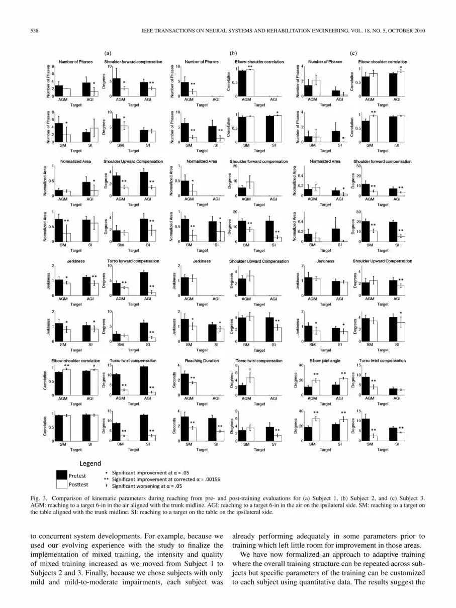
The results presented here are for each target, comparing pre-and post-training evaluations, and are presented in the contextof the subject’s individual training protocol. Despite the shorttraining period of two weeks, all three subjects showed improve-ment trends in activity recovery combined with partial recoveryof pre-morbid body function. Specific improvements are de-scribed below and results for the eight most important aspectsfor each subject are shown in Fig. 3.
Subject 1 showed significant improvements in velocity as-pects (bellness and jerkiness) and elbow and shoulder joint cor-relation during reaches to at least two of the targets. Torso andshoulder compensation was significantly reduced in most tar-gets, with many of these improvements holding even with thestricter significance level. These results are shown in Fig. 3(a).
Target AGI was not included in any analyses for Subject 2due to missing data during the post-training evaluation. Sub-ject 2 demonstrated improved velocity measures (normalizedadjust area, number of phases, and jerkiness) at most targets,with all normalized adjust area being significant at the correctedlevel. This subject also significantly reduced the average reachduration at all targets. Subject 2 had mixed results for bothelbow and shoulder joint correlation and compensation mea-surements, mainly showing significant improvements in the sup-ported target reaching. These results are shown in Fig. 3(b).
Subject 3 made the most improvements in body function, bothjoint range-of-motion and correlation and shoulder and torsocompensation. Subject 3 increased the extension of the elbowsignificantly (at the corrected level) during the reach at all fourtargets. Elbow and shoulder correlations were all higher duringthe post-training evaluation, with two targets improving signif-icantly. Shoulder and torso compensation were significantly re-duced for a majority of the targets. These results are shown inFig. 3(c).
V. DISCUSSION
The kinematic results show that all three subjects improvedtheir reaching movements after training with the interactivemixed reality system, especially the targeted parameters. Dueto the limited training period of only two weeks, however,we did not expect the clinical assessments to show significantfunctional changes. One possible explanation is that the finerchanges in movement that are detected by kinematic analysisare not reliably detected by the clinical tests [6], [36].
After training, two subjects showed an improvement in theamount and quality in performing the activities of daily livingpresented on the Motor Activity Log. While the MAL is avalidated measure, it may be influenced by the subject’s moodor other cognitive biases at time of survey completion. TheMAL also may not be sensitive to changes in recovery after ashort intervention period [43]. The SIS scores also show a trendof improvement, but the underlying cause of these changesis not clear. And while improvements seen in the kinematicparameters were detected by both the therapist and system, theymay not have been apparent to the subject, and therefore notreported. Further work will be done to determine how and whenchanges in kinematics become functionally relevant and willproduce a substantial change in the subjects’ self-assessments.These scales also do not distinguish between compensationand recovery of pre-morbid movement patterns, whereas thekinematic and therapist evaluations do. There were no obvioustrends of improvement in WMFT scores. Subject 2 decreasedhis time to completion by over 100 s during the pretest, butthis was due mainly to being able to complete a task (checkerstacking) that he was unable to complete during the pretest.However, when this task was removed from the totals, he stillshows a decrease of about 18 s. The training period may havebeen too short to induce general functional improvement, as thegeneralization of specific motor task training into functionalimprovement requires extensive training [11], [18].
Each subject showed significant improvements in theirreaching kinematics in merely six sessions, specifically formovement parameters on which their training was focused.This indicates that our approach of customized, adaptable andinteractive feedback in a mixed reality environment is appro-priate and beneficial to the rehabilitation of people who havemild-to-moderate hemiparesis resulting from stroke. Whileperforming repetitive reaching movements alone may have im-proved some parameters of the subjects’ end effector behavior(velocity, trajectory, etc.), each subject’s improvements in ac-tivity performance were correlated to improvements in relevantbody function parameters (joint synergy, compensation, etc.)for which they had received targeted feedback. Furthermore,the improvements in activity recovery parameters showed alevel of stylization (i.e., consistent velocity profile across tar-gets) that can rarely be achieved simply through repetition [32],[34]. This suggests subjects used the mixed reality feedback toinform their motor plans and make improvements. However,the two-week training period may have been too short to fullyaddress issues of the physical apparatus (like lack of musclestrength) or complete the full training sequence for each targetlocation. There were also inconsistencies in the training due
538 IEEE TRANSACTIONS ON NEURAL SYSTEMS AND REHABILITATION ENGINEERING, VOL. 18, NO. 5, OCTOBER 2010
Fig. 3. Comparison of kinematic parameters during reaching from pre- and post-training evaluations for (a) Subject 1, (b) Subject 2, and (c) Subject 3.AGM: reaching to a target 6-in in the air aligned with the trunk midline. AGI: reaching to a target 6-in in the air on the ipsilateral side. SM: reaching to a target onthe table aligned with the trunk midline. SI: reaching to a target on the table on the ipsilateral side.
to concurrent system developments. For example, because weused our evolving experience with the study to finalize theimplementation of mixed training, the intensity and qualityof mixed training increased as we moved from Subject 1 toSubjects 2 and 3. Finally, because we chose subjects with onlymild and mild-to-moderate impairments, each subject was
already performing adequately in some parameters prior totraining which left little room for improvement in those areas.
We have now formalized an approach to adaptive trainingwhere the overall training structure can be repeated across sub-jects but specific parameters of the training can be customizedto each subject using quantitative data. The results suggest the

DUFF et al.: AN ADAPTIVE MIXED REALITY TRAINING SYSTEM FOR STROKE REHABILITATION 539
feedback is intuitively and effectively communicating measuresof performance and direction for improvement to the subjects.We were successful in integrating mixed (physical-virtual) en-vironments into the training to promote motor learning bridgingthe virtual and physical worlds. Movement improvements madeduring training in the virtual and mixed environments success-fully transferred to their post-training physical reaching tests.Subjects improved at four different targets locations (supportedand against gravity to locations ipsilateral to the affected sideand at the midline) pointing to the potential of the system forpromoting generalizable learning.
Other studies [3]–[5] have shown improvements using con-straints of the unimpaired limb, trunk restraints or robotic assis-tive devices. However, these methods use external interventionsthat physically guide the subject to move in a certain way orrestrain their body such that they must use the affected limb.Conversely, our approach allows the subject to be free to moveas they wish, while providing mediated incentives to the subjectto move in a more efficient way and mediated deterrents fromusing compensatory or inefficient movements. This allows thesubject to actively, yet often subconsciously, construct his or herown strategies, reducing dependencies on external constraints.Our system also helps subjects progressively integrate strategieslearned for each kinematic parameter to form a complete move-ment strategy. Finally, the system effectively trains the subject tointegrate the motor tasks with input from their audio, visual andtactile sensory streams which could promote increased motorlearning and neural plasticity. The enthusiastic acceptance ofthe system by the therapist and subjects during the pilot studysuggests that the mixed reality system is suited for therapeuticapplication in the clinic.
This study has also led to improvements in the system in-frastructure. The setup for each visit took 20–30 min per sub-ject, which became tiresome for both the subject and researchteam. This setup time was prohibitive to running multiple sub-jects in one day or running subjects for an extended trainingperiod. A revised setup uses predefined rigid body motion cap-ture markers, which are more easily identified by the motioncapture software with less calibration. While this change doesprevent us from gathering data from smaller joints, such as thefingers, we are working on creating smart sensing objects to de-tect tangible interaction without the data provided by detailedhand motion capture. We have also developed more advancedcontrol software to make adaptations to the therapy protocol andvisualizing data faster and easier. This helps to better utilize thetherapy time with the patient and ensure that the patient will becompletely and consistently engaged in the training.
VI. CONCLUSION AND CURRENT WORK
The presented study has successfully shown a proof ofprinciple that adaptive mixed reality rehabilitation system canprovide customized reaching and grasping training for chronicstroke survivors and elicit improvements in important move-ment parameters. This system can help therapists to structuretherapy based on kinematic performance and to target specificaspects a functional task, with the help of interactive audio andvisual feedback.
The outcomes of the presented pilot study lay the foundationfor a current clinical study at the Rhodes Rehabilitation Insti-tute at Banner Baywood Medical Center. This study is beingconducted with a stable system, using a larger group of sub-jects and will include a matched control group who will receivetraditional repetitive task training of equal intensity. Our expec-tation is that mediated rehabilitation will yield better clinicalscale results, improve movement kinematics in a faster time,help the subjects create generalizable movement strategies andbe as well received by subjects as traditional therapy. Subjectdata from this study will further inform the system’s adaptationand training protocols. The data will also be used to draw cor-relations between feedback and performance to determine howsubjects utilize each feedback stream. Work is also being doneto create low-cost portable systems, based on the fixed clinicalsystem, for use in the home.
ACKNOWLEDGMENT
The interactive visuals for the system were developed byL. Olson. The interactive sounds were developed by I. Wallisand T. Ingalls. Database and visualization structures weredeveloped by W. W. Xu and M. Joshi. Dr. R. Herman served asmedical monitor and advisor. Dr. S. Wolf, at the Emory Uni-versity School of Medicine, was a consultant for the project.
REFERENCES
[1] J. J. Daly and R. L. Ruff, “Construction of efficacious gait and upperlimb functional interventions based on brain plasticity evidence andmodel-based measures for stroke patients,” Sci. World J., vol. 7, pp.2031–2045, Dec. 2007.
[2] J. J. Daly et al., “Response to upper-limb robotics and functional neu-romuscular stimulation following stroke,” J. Rehabil. Res. Dev., vol.42, pp. 723–736, Nov./Dec. 2005.
[3] S. Wolf et al., “Effect of constraint-induced movement therapy onupper extremity function 3 to 9 months after stroke,” JAMA, vol. 296,pp. 2095–104, Nov. 2006.
[4] S. M. Michaelsen, R. Dannenbaum, and M. F. Levin, “Task-specifictraining with trunk restraint on arm recovery in stroke: Randomizedcontrol trial,” Stroke, vol. 37, pp. 186–92, Dec. 2005.
[5] S. E. Fasoli, H. I. Krebs, J. Stein, W. R. Frontera, and N. Hogan, “Ef-fects of robotic therapy on motor impairment and recovery in chronicstroke,” Arch. Phys. Med. Rehabil., vol. 84, pp. 477–82, Apr. 2003.
[6] L. Piron, P. Tonin, F. Piccione, V. Iaia, E. Trivello, and M. Dam, “Vir-tual environment training therapy for arm motor rehabilitation,” Pres-ence, vol. 14, pp. 732–740, Dec. 2005.
[7] M. K. Holden, “Virtual environments for motor rehabilitation: Review,”Cyberpsychol Behav., vol. 8, pp. 187–211, Jun. 2005.
[8] T. Sarkamo et al., “Music listening enhances cognitive recovery andmood after middle cerebral artery stroke,” Brain, vol. 131, pp. 866–876,Feb. 2008.
[9] J. Carr and R. Shepherd, Physiotherapy in Disorders of the Brain.London, U.K.: William Heinemann Medical, 1980.
[10] R. A. Schmidt and C. A. Wrisberg, Motor Learning and Performance:A Situation-Based Learning Approach. Champaign, IL: Human Ki-netics, 2007.
[11] R. A. Schmidt, “Motor learning principles for physical therapy,” inProc. II STEP Conf. Contemporary Management of Motor ControlProblems, Alexandria, VA, 1991, pp. 49–62.
[12] S. Subramanian, L. A. Knaut, C. Beaudoin, B. J. McFadyen, A. G.Feldman, and M. F. Levin, “Virtual reality environments for post-strokearm rehabilitation,” J. Neuroeng. Rehabil., vol. 4, pp. 20–24, Jun. 2007.
[13] Y. Chen, W. Xu, H. Sundaram, T. Rikakis, and S.-M. Liu, “Mediaadaptation framework in biofeedback system for stroke patient rehabil-itation,” presented at the ACM Multimedia, Bavaria, Germany, 2007.
[14] H. Sveistrup, “Motor rehabilitation using virtual reality,” J. Neuroeng.Rehabil., vol. 1, pp. 10–17, Dec. 2004.

540 IEEE TRANSACTIONS ON NEURAL SYSTEMS AND REHABILITATION ENGINEERING, VOL. 18, NO. 5, OCTOBER 2010
[15] Y. Jung, S. Yeh, and J. Stewart, “Tailoring virtual reality technology forstroke rehabilitation: A human factors design,” in Conf. Human FactorsComput. Syst., Apr. 2006, pp. 929–934.
[16] M. Komitova, B. Mattsson, B. B. Johansson, and P. S. Eriksson, “En-riched environment increases neural stem/progenitor cell proliferationand neurogenesis in the subventricular zone of stroke-lesioned adultrats,” Stroke, vol. 36, pp. 1278–1282, Jun. 2005.
[17] A. Ronnback et al., “Gene expression profiling of the rat hippocampusone month after focal cerebral ischemia followed by enriched environ-ment,” Neurosci. Lett., vol. 385, pp. 173–178, Sep. 2005.
[18] M. C. Cirstea and M. F. Levin, “Improvement of arm movement pat-terns and endpoint control depends on type of feedback during practicein stroke survivors,” Neurorehabil. Neural Repair, vol. 21, p. 398, Oct.2007.
[19] J. M. Wagner, J. A. Rhodes, and C. Patten, “Reproducibility andminimal detectable change of three-dimensional kinematic analysis ofreaching tasks in people with hemiparesis after stroke,” Phys. Ther.,vol. 88, pp. 652–663, May 2008.
[20] L. A. Boyd and C. J. Winstein, “Explicit information interferes with im-plicit motor learning of both continuous and discrete movement tasksafter stroke,” J. Neurol. Phys. Therapy, vol. 30, pp. 46–57, Jun. 2006.
[21] A. Schopenhauer, The World as Will and Representation. New York:Dover, 1969.
[22] R. Wagner, Richard Wagner’s Prose Works V1: The Artwork of theFuture. Whitefish, MT: Kessinger, 2008.
[23] M. C. Cirstea and M. F. Levin, “Compensatory strategies for reachingin stroke,” Brain, vol. 123, pp. 940–953, May 2000.
[24] S. Yantis, Visual Perception: Key Readings, 1st ed. New York: Psy-chology Press, 2000.
[25] M. Chion, C. Gorbman, and W. Murch, Audio-Vision. New York:New York Columbia Univ. Press, 1994.
[26] P. Dourish, Where the Action Is: The Foundations of Embodied Inter-action. Cambridge, MA: MIT Press, 2004.
[27] I. Xenakis, Formalized Music: Thought and Mathematics in Composi-tion. Hillsdale, NY: Pendragon, 2001.
[28] M. Thaut and Rhythm, Music and the Brain: Scientific Foundationsand Clinical Applications. New York: Routledge, 2005.
[29] J. L. Chen, V. B. Penhune, and R. J. Zatorre, “Listening to musicalrhythms recruits motor regions of the brain,” Cerebral Cortex, vol. 18,pp. 2844–2845, Dec. 2008.
[30] D. Huron, Sweet Anticipation: Music and the Psychology of Expecta-tion. Cambridge, MA: MIT Press, 2006.
[31] D. J. Levitin, This is Your Brain on Music: The Science of a HumanObsession. New York: Dutton Adult, 2006.
[32] B. H. Repp, “The embodiment of musical structure: Effects of mu-sical context on sensorimotor synchronization with complex timingpatterns,” in Common Mechanisms in Perception and Action: Attentionand Performance XIX, W. Prinz and B. Hommel, Eds. Oxford, U.K.:Oxford Univ. Press, 2002, pp. 245–265.
[33] S. McAdams and E. Bigand, Thinking in Sound: The Cognitive Psy-chology of Human Audition. Oxford, U.K.: Clarendon, 1993.
[34] B. H. Repp and A. Penel, “Rhythmic movement is attracted morestrongly to auditory than to visual rhythms,” Psychological Res., vol.68, pp. 252–270, Aug. 2004.
[35] Y. Chen et al., “The design of a real-time, multimodal biofeedbacksystem for stroke patient rehabilitation,” in ACM Int. Multimedia Conf.,Santa Barbara, CA, 2006, pp. 763–772.
[36] M. F. Levin, J. A. Kleim, and S. L. Wolf, “What do motor “recovery”and “compensation” mean in patients following stroke?,” Neurore-habil. Neural Repair, vol. 23, pp. 313–19, May 2009.
[37] Y. Chen, “Constraint-aware computational adaptation framework tosupport real-time multimedia applications,” Ph.D. dissertation, Dept.Elect. Eng., Arizona State Univ., Tempe, AZ, 2009.
[38] H. Huang, T. Ingalls, L. Olson, K. Ganley, T. Rikakis, and J. He, “In-teractive multimodal biofeedback for task-oriented neural rehabilita-tion,” in Conf. Proc. IEEE Eng. Med. Biol. Soc., Aug. 2005, vol. 3, pp.2547–2550.
[39] D. Deutsch, The Psychology of Music. San Diego, CA: Academic,1999.
[40] G. Uswatte, E. Taub, D. Morris, M. Vignolo, and K. McCulloch, “Re-liability and validity of the upper-extremity motor activity Log-14 formeasuring real-world arm use,” Stroke, vol. 36, pp. 2493–2496, Nov.2005.
[41] P. W. Duncan, D. Wallace, S. M. Lai, D. Johnson, S. Embretson, and L.J. Laster, “The stroke impact scale version 2.0. evaluation of reliability,validity, and sensitivity,” Stroke, vol. 30, pp. 2131–2140, Oct. 1999.
[42] S. L. Wolf, P. A. Catlin, M. Ellis, A. L. Archer, B. Morgan, and A.Piacentino, “Assessing Wolf motor function test as outcome measurefor research in patients after stroke,” Stroke, vol. 32, pp. 1635–9, Jul.2001.
[43] J. H. van der Lee, H. Beckerman, D. L. Knol, H. C. de Vet, and L.M. Bouter, “Clinimetric properties of the motor activity log for theassessment of arm use in hemiparetic patients,” Stroke, vol. 35, pp.1410–1414, Jun. 2004.
Margaret Duff (S’10) received the B.S. degree in biomedical engineering fromBoston University, Boston, MA. She is currently working toward the Ph.D. de-gree in bioengineering at Arizona State University, Tempe.
Her research interests include creating complex sensing objects to measuretangible interactions within a mixed reality rehabilitation environment and dataanalysis of kinematic and clinical scale data.
Yinpeng Chen (M’08) received the Ph.D. degree from the Department of Elec-trical Engineering, Arizona State University, Tempe, in 2009.
He is currently an Assistant Research Professor at Arizona State University,Tempe. His research interests include adaptation modeling in mixed reality re-habilitation, dynamic decision-making and experiential media system.
Suneth Attygalle (M’10) received the B.S. degree in biological engineeringfrom Cornell University, Ithaca, NY, in 2007. He is currently working towardthe M.S. degree in bioengineering under the supervision of J. He at ArizonaState University, Tempe.
His research interests include neuroscience and human–computer interaction.
Janice Herman received the M.S. degree in physical therapy.Since 1978 she has performed clinical work with mostly neurological patients
of all ages. She has also performed research in gross motor patterns after strokeor spinal cord injury.
Hari Sundaram (M’03) received the Ph.D. degree from the Department ofElectrical Engineering at Columbia University, in 2002.
He is an Associate Professor with the School of Arts Media and Engineeringat Arizona State University, Tempe. His research interests include analysis ofsocial network activity and the design of adaptive multimedia environments.He is an Associate Editor for ACM Transactions on Multimedia Computing,Communications and Applications.
Dr. Sundaram has won several best paper awards from the IEEE and the ACM.He also received the Eliahu I. Jury Award for best Ph.D. dissertation in 2002.
Gang Qian (M’01) is an Assistant Professor with the School of Arts, Media andEngineering and the School of Electrical, Computer and Energy Engineering atArizona State University, Tempe. His research interests include computer visionand pattern analysis, sensor fusion and information integration, human-centeredinteractive systems, machine learning for computer vision, as well as multi-modal sensing and analysis of human activities for embodied learning and move-ment rehabilitation.

DUFF et al.: AN ADAPTIVE MIXED REALITY TRAINING SYSTEM FOR STROKE REHABILITATION 541
Jiping He (S’86–M’89–SM’97) was born in Shanghai, China. He received theB.S. degree in control engineering from Huazhong University of Science andTechnology, Wuhan China, in 1982, the M.S. and Ph.D. degrees in electricalengineering from the University of Maryland, College Park, in 1984 and 1988,respectively. He then spent one and a half years as a postdoctoral fellow in theCenter for Biological Information Processing and Artificial Intelligence Labo-ratory, Massachusetts Institute of Technology, Cambridge.
In 1990, he joined Functional Neurosurgery Division of Thomas JeffersonUniversity (TJU), Philadelphia, PA, as a Research Assistant Professor and Ad-junct Professor of Physical Therapy. He was a Visiting Scientist at Human Infor-mation Processing Program of Princeton University, Princeton, NJ during 1991and 1992. He has been a faculty at Arizona State University, Tempe, since 1994and is now Professor of Bioengineering, Director of Center for Neural InterfacesDesign. His primary research and teaching interests include the application ofadvanced control theory to the analysis and control of neuromuscular systemsfor posture and movement, implantable neural interface technology, cortical andspinal cord recording and stimulation for sensorimotor adaptation and control,application of robotics and virtual reality research to neural rehabilitation andprosthetic devices. He is also actively involved in the curriculum developmentof bio-control and neural engineering, and reform in interdisciplinary graduateeducation and research training.
Dr. He is a Senior Editor for the IEEE TRANSACTIONS ON NEURAL SYSTEMS
AND REHABILITATION ENGINEERING. He is active in professional services byorganizing and chairing international conferences.
Thanassis Rikakis (M’09) is a Professor and the founding Director of theSchool of Arts Media and Engineering (AME) at Arizona State University. Hisresearch and creative work is in the areas of experiential media, mixed realityrehabilitation, interdisciplinary education, sound perception, computer music,and media arts systems for education. He is Principal Investigator of a currentNSF IGERT grant in experiential media. His educational background is inmusic composition and computer music.