alpha-conotoxin imi disrupts central control of swimming in the medicinal leech
TRANSCRIPT
Alpha-conotoxin ImI Disrupts Central Control of
Swimming in the Medicinal Leech
Daniel A. Wagenaar1∗, Ruben Gonzalez2, David C. Ries2,
William B. Kristan Jr2, and Kathleen A. French2
1. Broad Fellows Program and Division of Biology
California Institute of Technology
Pasadena CA 91125
2. Neurobiology Section
University of California, San Diego
La Jolla CA 92093
∗ To whom correspondence should be addressed: Caltech 216-76, Pasadena CA 91125.
[email protected]. +1-626-395-2577.
Abstract
Medicinal leeches (Hirudo spp.) swim using a metachronal, front-to-back undula-
tion. The behavior is generated by central pattern generators (CPGs) distributed
along the animal’s midbody ganglia and is coordinated by both central and periph-
eral mechanisms.Here we report that a component of the venomofConus imperialis,
α-conotoxin ImI, known to block nicotinic acetylcholine receptors in other species,
disrupts swimming. Leeches injected with the toxin swam in circles with exagger-
ated dorsoventral bends and reduced forward velocity. Fictive swimming in isolated
nerve cords was even more strongly disrupted, indicating that the toxin targets the
CPGs and central coordination, while peripheral coordination partially rescues the
behavior in intact animals.
Keywords: Swimming; Central pattern generator; Intersegmental coordination;
Medicinal leech; Hirudo verbana.
Introduction
In studying the neuronal basis of behaviors, repetitive and rhythmic behaviors have received
much attention [1]. Locomotion in many species depends on waves of contraction sweeping
along the body under the control of a chain of semi-autonomous segmental central pattern
generators (CPGs) with intersegmental connections. Well-studied examples include the swim-
merets of crayfish [2, 3] and the sinuous swimming of lampreys [4, 5]. Swimming behavior in
medicinal leeches (Hirudo spp.) provides a particularly successful focus of study, because their
nervous systems are relatively simple, their bodies sturdy, and their behaviors readily quan-
tifiable [6]. Accordingly, Hirudo’s nervous system, and in particular its swimming circuit, is
the best-understood among annelids.
Leech swimming requires coordination of muscles along the entire body. The basic swim
pattern is generated by core CPGs that are repeated in each segment. Connections among core
CPGs are spread among several adjacent ganglia in the ventral nerve cord; muscle contrac-
tion and relaxation in each segment is controlled by motor neurons in that segment’s ganglion.
Whole-body coordination of swimming is achieved by two mechanisms: central interganglionic
connections through the ventral nerve cord, and peripheral sensory feedback through stretch
1
receptors embedded in the dorsal and ventral longitudinal muscles [7, 8]. The central connec-
tions are sufficient to produce the motor pattern: electrically stimulating a nerve causes an
isolated nerve cord to generate coordinated motor neuron output in the absence of body wall
[9]. Remarkably, the peripheral mechanism is also sufficient to impose coordination: after tran-
secting the nerve cord between two midbody ganglia, the anterior and posterior parts of the
animal still produce swimming movements, and the phase relationship between swim strokes
in the two parts remains largely unaltered [8].
To tease apart central and peripheral mechanisms, we screened venoms from cone snails.
All previously characterized components of these venoms target neurons or muscles. We found
that venom from the annelid-hunting snail Conus imperialis produced recognizable, but reli-
ably abnormal, swimming. Chemical fractionation (by the laboratory of Dr. Baldomero Olivera,
University of Utah) revealed that the active fraction was α-conotoxin ImI, which blocks nico-
tinic acetylcholine receptors in several vertebrates [10, 11, 12]. We treated intact leeches or
isolated nerve cords with ImI and compared the results to ask whether the apparent redun-
dancy between central and peripheral control makes the system less vulnerable to external
disruptions.
Methods
Behavior
Leeches (Hirudo verbana, Carolina Biological Supply, Burlington NC) were maintained at
15 ◦C in artificial pond water (36 mg/L Instant Ocean salts; Aquarium Systems, Mentor OH).
Leeches were fed cow blood semiannually (no feeding occurred within 1 month of experiments)
and weighed about 1 gram at the time of experiments unless otherwise noted. ImI (from
Dr. Baldomero Olivera, University of Utah, or Peptides International, Louisville KY) was in-
jected intramuscularly in the dorsal body wall near midbody segment M15 at a concentration
of 10 nanomoles/gram body mass in 25 µL HEPES-buffered leech saline [13]. For ImI concen-
trations between 6 nmol/g and 50 nmol/g, the latency to behavioral onset decreased from 8 to
3.5 minutes, but the response itself did not qualitatively change. Effects persisted from days to
weeks, but all leeches eventually returned to normal behavior. Control animals were injected
with 25 µL saline. All the behavioral observations were done blinded.
Following injection, each leech was placed individually in shallow (2 cm deep) water, which
forced it to swim on its side rather than in the normal dorsal-up posture. Swimming in this un-
usual orientation facilitated videotaping from above, but was otherwise indistinguishable from
normal swimming in deeper water. When necessary, leeches were gently prodded to encourage
swimming.
Analysis of Behavior
Video clips of swimming leeches were recorded at 30 frames/s and digitized using standard
equipment. The location and body shape of the leech were identified in each frame using the
“Wormfinder” algorithm [14], which yields a curve along the midline of the animal from head
to tail (Supplemental Figure 1a). Sections of this curve where the ventral longitudinal muscles
contracted (causing the dorsal surface to be convex) were termed peaks, whereas sections where
the ventral longitudinal muscles contracted (causing the dorsal surface to be concave) were
termed troughs.
The forward velocity of a swimming leech was calculated as the velocity of its center of mass
after low-pass filtering of the trajectory to exclude the undulatory motion perpendicular to the
2
swimming direction.
Electrophysiology of the Isolated Cord
The central nervous system of the leech was isolated as previously described [9]. Briefly, leeches
were anesthetized in ice-cold saline, pinned down on wax, and opened along the dorsal midline.
The entire nerve cord minus the head brain was extracted and placed in a Petri dish. (Removal
of the head brain promotes fictive swimming [15, 16].) Dorsal posterior (DP) nerves and the
first branch (B1) of anterior anterior (AA) nerves were exposed in selected segments to allow
recording of motor neuron activity.
Data Acquisition
Suction electrodes where placed on DP and AA nerves (Supplemental Figure 1b). Signals were
amplified 10,000x and bandpass filtered (300 Hz to 5 kHz) using a differential AC amplifier
(A-M Systems, Sequim WA) and were digitized at 10 kHz (DigiData 1320A, Axon Instruments,
Sunnyvale CA). Fictive swimming was elicited by electrical stimulation of one of the DP nerves
from ganglia M10–12 using a Grass (West Warwick RI) S88 stimulator. Stimuli consisted of
trains of 5–10 pulses (1–2 V, 1 ms long) at 10 Hz. We initially recorded 5–10 evoked swim
episodes in normal saline, at 1–3 minute intervals (to avoid habituation). We then replaced
the saline with 10–20 µM ImI (in fresh saline) and continued the same stimulation protocol.
Recordings made within 10 minutes of ImI application were discarded to avoid the transition
period.
Analysis of Electrophysiological Data
Several motor neurons project through the DP nerves. Based on spike amplitude, we isolated
action potentials attributable to motor neuron DE-3, which provides excitatory drive to the
dorsal longitudinal muscles and plays a major role in swimming behavior [6]. Spikes from
cell VE-108, which excites ventral longitudinal muscles, were similarly isolated from AA-B1
recordings [17]. Bursts were defined as groups of at least 2 spikes separated from other groups
by at least 100 ms. The duration of a burst was the time between the first and last spikes in the
burst. The period of bursting was the time between the middle spikes in consecutive bursts.
Bursts shorter than 750 ms were classified as swim-like if either or both of the preceding and
following burst periods were shorter than 1500 ms. A fictive swim episode was defined as a
stretch of time during which at least one ganglion fired a sequence of at least three contiguous
swim-like bursts. To quantify burst propagation along the cord, bursts recorded from adjacent
pairs of electrodes were matched up, and interganglionic delays were calculated as the latency
between the middle spikes in matching bursts, divided by the number of segments separating
the electrodes. For this calculation, we included only bursts that occurred while at least one
ganglion produced recognizable fictive swimming.
Results
Behavior
Treatment with ImI profoundly disrupted the kinematics of swimming.While saline-injected
control leeches swam with a nearly symmetric dorsoventral stroke that produced effective for-
ward motion (Figure 1a), ImI-injected animals craned their heads backward during part of
their stroke and exaggerated the magnitude of troughs (Figure 1b1). As a result, they typically
3
0.0 s0.0 s0.0 s0.0 s0.0 s0.0 s0.0 s0.0 s0.0 s0.0 s0.0 s0.0 s0.0 s0.0 s0.0 s0.0 s0.0 s0.0 s0.0 s0.0 s0.0 s0.0 s0.0 s0.0 s0.0 s0.0 s 0.1 s0.1 s0.1 s0.1 s0.1 s0.1 s0.1 s0.1 s0.1 s0.1 s0.1 s0.1 s0.1 s0.1 s0.1 s0.1 s0.1 s0.1 s0.1 s0.1 s0.1 s0.1 s0.1 s0.1 s0.1 s0.1 s
0.2 s0.2 s0.2 s0.2 s0.2 s0.2 s0.2 s0.2 s0.2 s0.2 s0.2 s0.2 s0.2 s0.2 s0.2 s0.2 s0.2 s0.2 s0.2 s0.2 s0.2 s0.2 s0.2 s0.2 s0.2 s0.2 s 0.3 s0.3 s0.3 s0.3 s0.3 s0.3 s0.3 s0.3 s0.3 s0.3 s0.3 s0.3 s0.3 s0.3 s0.3 s0.3 s0.3 s0.3 s0.3 s0.3 s0.3 s0.3 s0.3 s0.3 s0.3 s0.3 s
0.4 s0.4 s0.4 s0.4 s0.4 s0.4 s0.4 s0.4 s0.4 s0.4 s0.4 s0.4 s0.4 s0.4 s0.4 s0.4 s0.4 s0.4 s0.4 s0.4 s0.4 s0.4 s0.4 s0.4 s0.4 s0.4 s
a1 a2
a3
0.0 s0.0 s0.0 s0.0 s0.0 s0.0 s0.0 s0.0 s0.0 s0.0 s0.0 s0.0 s0.0 s0.0 s0.0 s0.0 s0.0 s0.0 s0.0 s0.0 s0.0 s0.0 s0.0 s0.0 s0.0 s0.0 s 0.1 s0.1 s0.1 s0.1 s0.1 s0.1 s0.1 s0.1 s0.1 s0.1 s0.1 s0.1 s0.1 s0.1 s0.1 s0.1 s0.1 s0.1 s0.1 s0.1 s0.1 s0.1 s0.1 s0.1 s0.1 s0.1 s
0.2 s0.2 s0.2 s0.2 s0.2 s0.2 s0.2 s0.2 s0.2 s0.2 s0.2 s0.2 s0.2 s0.2 s0.2 s0.2 s0.2 s0.2 s0.2 s0.2 s0.2 s0.2 s0.2 s0.2 s0.2 s0.2 s 0.3 s0.3 s0.3 s0.3 s0.3 s0.3 s0.3 s0.3 s0.3 s0.3 s0.3 s0.3 s0.3 s0.3 s0.3 s0.3 s0.3 s0.3 s0.3 s0.3 s0.3 s0.3 s0.3 s0.3 s0.3 s0.3 s
0.4 s0.4 s0.4 s0.4 s0.4 s0.4 s0.4 s0.4 s0.4 s0.4 s0.4 s0.4 s0.4 s0.4 s0.4 s0.4 s0.4 s0.4 s0.4 s0.4 s0.4 s0.4 s0.4 s0.4 s0.4 s0.4 s 0.5 s0.5 s0.5 s0.5 s0.5 s0.5 s0.5 s0.5 s0.5 s0.5 s0.5 s0.5 s0.5 s0.5 s0.5 s0.5 s0.5 s0.5 s0.5 s0.5 s0.5 s0.5 s0.5 s0.5 s0.5 s0.5 s
b1 b2
b3
0
0.5
1
1.5
Tim
e (
s)
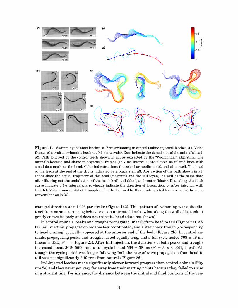
Figure 1. Swimming in intact leeches. a. Free swimming in control (saline-injected) leeches. a1. Video
frames of a typical swimming leech (at 0.1-s intervals). Dots indicate the dorsal side of the animal’s head.
a2. Path followed by the control leech shown in a1, as extracted by the “Wormfinder” algorithm. The
animal’s location and shape in sequential frames (16.7 ms intervals) are plotted as colored lines with
small dots marking the head. Color indicates time; the color bar applies to b2 and c2 as well. The head
of the leech at the end of the clip is indicated by a black star. a3. Abstraction of the path shown in a2.
Lines show the actual trajectory of the head (magenta) and the tail (cyan), as well as the same data
after filtering out the undulations of the head (red), tail (blue), and center (black). Dots along the black
curve indicate 0.1-s intervals; arrowheads indicate the direction of locomotion. b. After injection with
ImI. b1. Video frames. b2–b3. Examples of paths followed by three ImI-injected leeches, using the same
conventions as in (a).
changed direction about 90◦ per stroke (Figure 1b2). This pattern of swimming was quite dis-
tinct from normal cornering behavior as an untreated leech swims along the wall of its tank: it
gently curves its body and does not crane its head (data not shown).
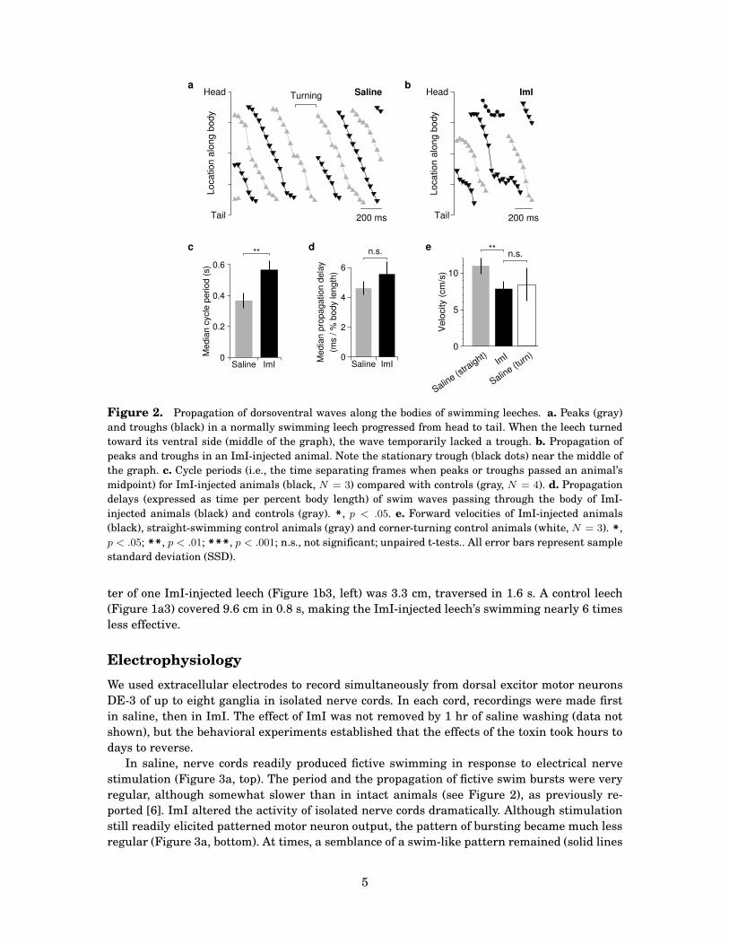
In control animals, peaks and troughs propagated linearly from head to tail (Figure 2a). Af-
ter ImI injection, propagation became less coordinated, and a stationary trough (corresponding
to head craning) typically appeared at the anterior end of the body (Figure 2b). In control an-
imals, propagating peaks and troughs lasted equally long, and a full cycle lasted 368 ± 48 ms
(mean ± SSD, N = 3, Figure 2c). After ImI injection, the durations of both peaks and troughs
increased about 30%–50%, and a full cycle lasted 568 ± 58 ms (N = 3, p < .001, t-test). Al-
though the cycle period was longer following ImI, the rate of wave propagation from head to
tail was not significantly different from controls (Figure 2d).
ImI-injected leeches made significantly slower forward progress than control animals (Fig-
ure 2e) and they never got very far away from their starting points because they failed to swim
in a straight line. For instance, the distance between the initial and final positions of the cen-
4
Tail
Head
Lo
ca
tio
n a
lon
g b
od
y200 ms
SalineTurninga
Tail
Head
Lo
ca
tio
n a
lon
g b
od
y
200 ms
ImIb
**
0
0.2
0.4
0.6
Media
n c
ycle
period (
s)
Saline ImI
c n.s.
0
2
4
6
(ms / %
body length
)
Saline ImI
Media
n p
ropagation d
ela
y
d **n.s.
0
5
10
Velo
city (
cm
/s)
Saline (s
traight)
ImI
Saline (t
urn)
e
Figure 2. Propagation of dorsoventral waves along the bodies of swimming leeches. a. Peaks (gray)
and troughs (black) in a normally swimming leech progressed from head to tail. When the leech turned
toward its ventral side (middle of the graph), the wave temporarily lacked a trough. b. Propagation of
peaks and troughs in an ImI-injected animal. Note the stationary trough (black dots) near the middle of
the graph. c. Cycle periods (i.e., the time separating frames when peaks or troughs passed an animal’s
midpoint) for ImI-injected animals (black, N = 3) compared with controls (gray, N = 4). d. Propagation
delays (expressed as time per percent body length) of swim waves passing through the body of ImI-
injected animals (black) and controls (gray). *, p < .05. e. Forward velocities of ImI-injected animals
(black), straight-swimming control animals (gray) and corner-turning control animals (white, N = 3). *,p < .05; **, p < .01; ***, p < .001; n.s., not significant; unpaired t-tests.. All error bars represent sample
standard deviation (SSD).
ter of one ImI-injected leech (Figure 1b3, left) was 3.3 cm, traversed in 1.6 s. A control leech
(Figure 1a3) covered 9.6 cm in 0.8 s, making the ImI-injected leech’s swimming nearly 6 times
less effective.
Electrophysiology
We used extracellular electrodes to record simultaneously from dorsal excitor motor neurons
DE-3 of up to eight ganglia in isolated nerve cords. In each cord, recordings were made first
in saline, then in ImI. The effect of ImI was not removed by 1 hr of saline washing (data not
shown), but the behavioral experiments established that the effects of the toxin took hours to
days to reverse.
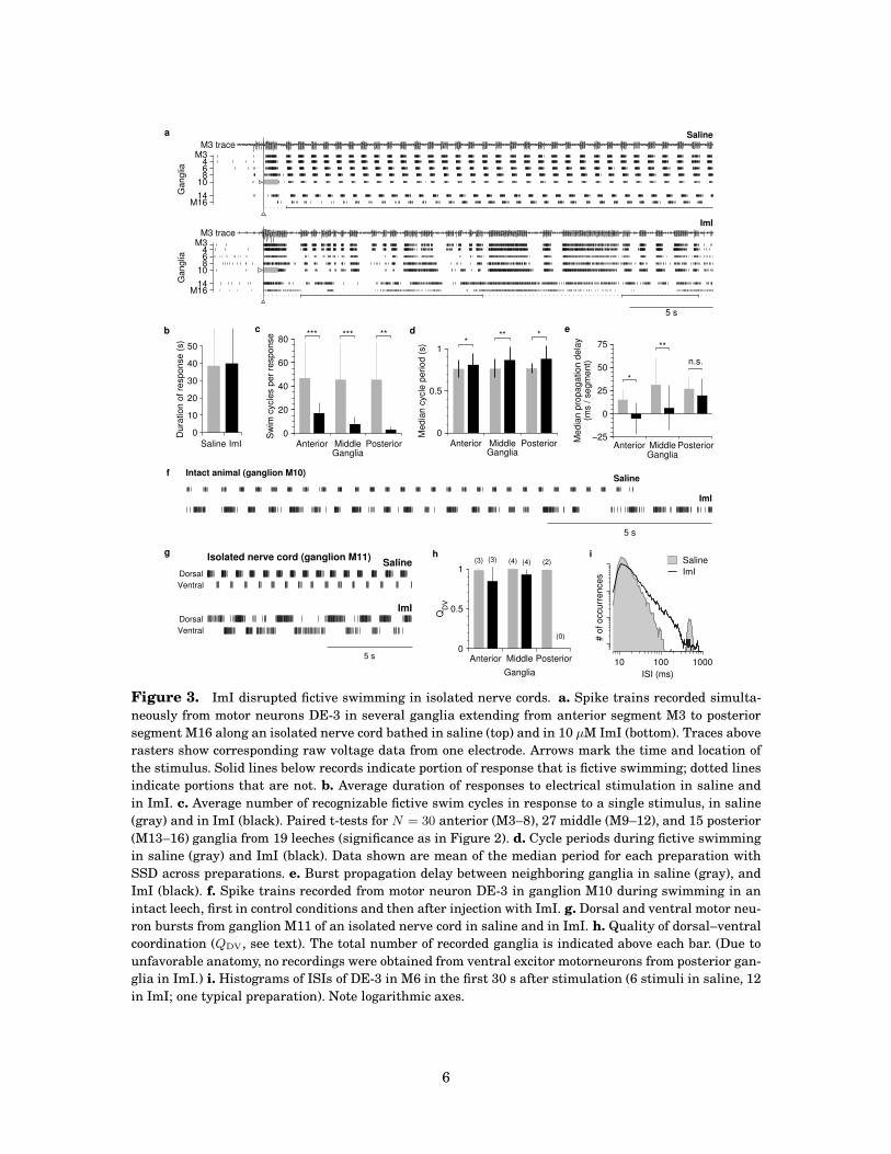
In saline, nerve cords readily produced fictive swimming in response to electrical nerve
stimulation (Figure 3a, top). The period and the propagation of fictive swim bursts were very
regular, although somewhat slower than in intact animals (see Figure 2), as previously re-
ported [6]. ImI altered the activity of isolated nerve cords dramatically. Although stimulation
still readily elicited patterned motor neuron output, the pattern of bursting became much less
regular (Figure 3a, bottom). At times, a semblance of a swim-like pattern remained (solid lines
5
Saline
M1614
10864
M3
Ga
ng
lia
M3 trace
ImI
M1614
10864
M3
Ga
ng
lia
M3 trace
5 s
a
Saline ImI
0
10
20
30
40
50
Du
ratio
n o
f re
sp
on
se
(s)
b
Anterior Middle PosteriorGanglia
0
20
40
60
80
Sw
im c
ycle
s p
er
resp
on
se *** *** **
c
Anterior Middle PosteriorGanglia
0
0.5
1
Me
dia
n c
ycle
pe
rio
d (
s) *
** *d
Anterior Middle PosteriorGanglia
−25
0
25
50
75
(ms /
se
gm
en
t)M
ed
ian
pro
pa
ga
tio
n d
ela
y
*
**
n.s.
e
Saline
ImI
Intact animal (ganglion M10)
5 s
f
Dorsal
Ventral
Saline
Dorsal
Ventral
ImI
Isolated nerve cord (ganglion M11)
5 s
g(3) (4) (2)(3) (4)
(0)
Anterior Middle Posterior
Ganglia
0
0.5
1
QD
V
h
10 100 1000
ISI (ms)
# o
f o
ccu
rre
nce
s
Saline
ImI
i
Figure 3. ImI disrupted fictive swimming in isolated nerve cords. a. Spike trains recorded simulta-
neously from motor neurons DE-3 in several ganglia extending from anterior segment M3 to posterior
segment M16 along an isolated nerve cord bathed in saline (top) and in 10 µM ImI (bottom). Traces above
rasters show corresponding raw voltage data from one electrode. Arrows mark the time and location of
the stimulus. Solid lines below records indicate portion of response that is fictive swimming; dotted lines
indicate portions that are not. b. Average duration of responses to electrical stimulation in saline and
in ImI. c. Average number of recognizable fictive swim cycles in response to a single stimulus, in saline
(gray) and in ImI (black). Paired t-tests for N = 30 anterior (M3–8), 27 middle (M9–12), and 15 posterior
(M13–16) ganglia from 19 leeches (significance as in Figure 2). d. Cycle periods during fictive swimming
in saline (gray) and ImI (black). Data shown are mean of the median period for each preparation with
SSD across preparations. e. Burst propagation delay between neighboring ganglia in saline (gray), and
ImI (black). f. Spike trains recorded from motor neuron DE-3 in ganglion M10 during swimming in an
intact leech, first in control conditions and then after injection with ImI. g. Dorsal and ventral motor neu-
ron bursts from ganglion M11 of an isolated nerve cord in saline and in ImI. h. Quality of dorsal–ventral
coordination (QDV, see text). The total number of recorded ganglia is indicated above each bar. (Due to
unfavorable anatomy, no recordings were obtained from ventral excitor motorneurons from posterior gan-
glia in ImI.) i. Histograms of ISIs of DE-3 in M6 in the first 30 s after stimulation (6 stimuli in saline, 12
in ImI; one typical preparation). Note logarithmic axes.
6
below the rasters), but the swim bursts were less regular, especially toward the posterior end
of the cord (M14–16). Output also commonly included extended periods (dotted lines) of rel-
atively unpatterned motor neuron activity devoid of swim-like bursts, which was extremely
rare in controls. Whereas ImI did not change the total duration of responses to identical stim-
uli (Figure 3b), responses contained far fewer recognizable swim cycles (Figure 3c), especially
in posterior ganglia. Interestingly, the cycle period during those portions of the behavior that
resembled fictive swimming changed only slightly (Figure 3d). To quantify regularity of bursts,
we calculated histograms of interspike intervals (ISIs; Figure 3i). In saline, ISIs within bursts
were much shorter than between bursts, resulting in two distinct peaks in the histogram. ImI
changed ISIs only slightly on average, but they became more variable, and interburst intervals
became so irregular that they no longer produced a distinct peak in the histogram.
We also recorded electrophysiologically fromDE-3 in ganglionM10 of several intact animals
using cuff electrodes (see Supplemental Methods). Simultaneous video recording allowed us to
directly identify episodes of swimming. In control conditions, swim bursts were very regular
(Figure 3f) and had periods of 408 ± 61 ms (N = 3), matching behavioral observations. After
injection with 20 nmol ImI (in 250 µL saline) periods lengthened to 566 ± 114 ms (N = 3),
again matching behavioral observations. Burst patterns became less regular, but—unlike in
recordings from isolated cords—excessively long burst periods (> 1.5 s) were never observed in
swimming intact animals.
Interganglionic coordination
For a swim stroke to effectively propel a leech forward, coordination between segments is as
important as the quality of the motor neuron output within individual segments. Even in iso-
lated cords—which lack sensory feedback—interganglionic coordination was tightly controlled
when cords were bathed in saline, as evidenced by the precise anteroposterior propagation of
bursts (Figure 3a). Interganglionic propagation delays were slightly longer in the middle of the
cord than near the ends (Figure 3e), perhaps because ganglia are physically closer together at
the ends of the cord, but the variability of delays was low, and bursts always propagated antero-
posteriorly. In the presence of ImI, propagation was severely disrupted. Except at the posterior
end of the cord, median propagation delays were no longer significantly different from zero,
indicating that bursts either propagated equally often in both directions, or that they did not
propagate at all, but rather occurred simultaneously in several ganglia.
Dorsoventral coordination
In addition to interganglionic coordination, dorsoventral coordination is critical for effective
swimming. To produce undulation, rather than tensing or shortening, dorsal and ventral con-
tractions must occur in antiphase, and under control conditions they did (Figure 3g): The frac-
tion of ventral spikes that occurred outside of dorsal bursts (QDV) was close to one (Figure 3h),
indicating near perfect alternation of dorsal and ventral bursts without overlap. Remarkably,
in ImI, despite the general degradation of the swim pattern, dorsal and ventral bursts still
alternated without much overlap, and QDV did not decrease significantly relative to controls.
Thus, dorsoventral coordination was preserved in ImI, despite the disruption of many other
parameters of the swim motor pattern.
7
Discussion
Treating intact leeches with α-conotoxin ImI severely compromised their usually elegant and
effective swimming, yet they still swam. Similarly, exposing isolated ventral nerve cords to the
toxin disrupted the rhythmic production of motor neuron bursts that drive swimming behavior,
but did not abolish it.
Following treatment with ImI, leeches swam in a petal-shaped circular pattern (Figure 1b)
rather than in the typical straight path of control animals (Figure 1a), apparently as a result
of exaggerated bending toward the dorsal side as the swim wave passed through the body
and a maintained backward craning of the head. Because the neuronal circuitry underlying
swimming is well-studied [6], we have formed hypotheses regarding the basis of the disruption.
First, consider what ImI does not disrupt. Normal swimming is a bilaterally symmetric
behavior: homologous pairs of motor neurons on the two sides of each ganglion activate longi-
tudinal muscles on both sides of the segment in synchrony. This bilateral symmetry in muscle
contraction is not hard-wired: during local bending [18], longitudinal muscles on one side of the
leech contract while homologous contralateral muscles relax. Synchronous contractions prob-
ably depend on two features of the circuitry: bilateral pairs of homologous motor neurons are
electrically coupled to one another [19] and pairs of homologous neurons receive input from
the swim CPG simultaneously [6]. The persistence of this symmetry following ImI treatment
strongly suggests that the toxin does not affect these connections among and onto the motor
neurons.
In addition, the strictly alternating contraction of dorsal and ventral longitudinal mus-
cles and the strictly alternating activity in the motor neurons that drive these muscles were
largely unchanged following treatment with ImI (Figure 3g, h). This alternation, too, is not a
given: during crawling and shortening, dorsal and ventral longitudinal muscles in a segment
co-contract [6]. Contraction in antiphase depends at least in part on connections between ex-
citatory and inhibitory motor neurons within each ganglion: During swimming, when motor
neurons that excite dorsal longitudinal muscles are active, motor neurons that inhibit ventral
longitudinal muscles are also active. These inhibitory motor neurons not only project to mus-
cles in the periphery, they also make inhibitory synapses onto motor neurons that drive those
same muscles [6]. The swim CPG feeds onto this set of chemical-synaptically connected motor
neurons, providing another layer of control [6], and the strict preservation of antiphasic acti-
vation of dorsal and ventral longitudinal muscles after ImI treatment suggests that the toxin
spares this level of circuitry as well.
What ImI did change was the timing of the motor output and especially its intersegmen-
tal coordination. When isolated nerve cords were exposed to ImI, the coordinated front-to-
back propagation of activity broke down. Instead, bursts of activity occurred simultaneously
in several ganglia (Figure 3a, e). However, in ImI-injected whole animals, propagation was
moderately well preserved (Figure 2b, d), suggesting that peripheral biomechanical feedback
through stretch receptors located in the muscles of the body wall [8, 20] partially rescued in-
tersegmental coordination in spite of disorganization among the CPGs. (Differences between
intact animals and isolated cords cannot be explained by possible concentration differences,
since—in both preparations—a wide range of concentrations produced qualitatively the same
results.)
Why were interganglionic connections more affected than the connections within the CPG
and from the CPG neurons onto the motor neurons? It is possible that the former are less
numerous or synaptically weaker than the latter. More interestingly, interganglionic synapses
could differ pharmacologically from their intraganglionic counterparts, rendering them intrin-
sically more sensitive to ImI. (Across phyla, ImI acts as an antagonist to nicotinic acetylcholine
8
receptors [10, 11, 12].) If so, ImI could be used to differentially block intersegmental particular
components of the CPG, which would allow us to deepen our understanding of how the swim-
ming CPG, a relatively small set of interconnected neurons, generates well-coordinated and
plastic locomotory behavior.
An exploration of the balance between intersegmental control and intraganglionic local con-
trol during swimming in the leech, aided by a drug like ImI which spares motorneuronal con-
nections but disrupts interneuronal networks, will help reveal fundamental principles of motor
control against which metachronal locomotion in these and other species can be compared.
Acknowledgments
We thank Dr. Baldomero Olivera and his laboratory for supplyingConus venoms and ImI in the
early phases of this work. This work was supported by NIH Research Grants MH43396 and
NS35336 (to WBK), by a Senior Research Fellowship from the Broad Foundations (to DAW)
and by Microsoft Research Labs. DAW holds a Career Award at the Scientific Interface from
the Burroughs Wellcome Fund.
References
[1] Marder E, Calabrese RL. Principles of rhythmic motor pattern generation. Physiol Rev
76 (1996), 687–717.
[2] Mulloney B, Skinner FK, Namba H, Hall WM. Intersegmental coordination of swimmeret
movements: mathematical models and neural circuits. Ann N Y Acad Sci 860 (1998),
266–280.
[3] Mulloney B, Hall WM. Local and intersegmental interactions of coordinating neurons and
local circuits in the swimmeret system. J Neurophysiol 98 (2007), 405–413.
[4] Grillner S, Wallen P. Cellular bases of a vertebrate locomotor system-steering, interseg-
mental and segmental co-ordination and sensory control. Brain Res Brain Res Rev 40
(2002), 92–9106.
[5] Ayali A, Fuchs E, Ben-Jacob E, Cohen A. The function of intersegmental connections in
determining temporal characteristics of the spinal cord rhythmic output. Neuroscience
147 (2007), 236–246.
[6] Kristan Jr WB, Calabrese RL, Friesen WO. Neuronal control of leech behavior. Prog
Neurobiol 76 (2005), 279–327.
[7] Brodfuehrer PD, Debski EA, O’Gara BA, Friesen WO. Neuronal control of leech swim-
ming. J Neurobiol 27 (1995), 403–418.
[8] Yu X, Nguyen B, Friesen WO. Sensory feedback can coordinate the swimming activity of
the leech. J Neurosci 19 (1999), 4634–4643.
[9] Kristan WB, Calabrese RL. Rhythmic swimming activity in neurons of isolated nerve cord
of leech. J Exp Biol 65 (1976), 643–668.
[10] McIntosh JM, Yoshikami D, Mahe E, Nielsen DB, Rivier JE, Gray WR, Olivera BM. A
nicotinic acetylcholine-receptor ligand of unique specificity, alpha-conotoxin-imi. J Biol
Chem 269 (1994), 16733–16739.
9
[11] Pereira EF, Alkondon M, McIntosh JM, Albuquerque EX. Alpha-conotoxin-imi: a competi-
tive antagonist at alpha-bungarotoxin-sensitive neuronal nicotinic receptors in hippocam-
pal neurons. J Pharmacol Exp Ther 278 (1996), 1472–1483.
[12] Kehoe J, McIntosh JM. Two distinct nicotinic receptors, one pharmacologically similar
to the vertebrate alpha7-containing receptor, mediate cl currents in aplysia neurons. J
Neurosci 18 (1998), 8198–8213.
[13] Wagenaar DA, Hamilton MS, Huang T, Kristan WB, French KA. A hormone-activated
central pattern generator for courtship. Curr Biol 20 (2010), 487–495.
[14] Wagenaar DA, Kristan Jr WB. Automated video analysis of animal movements using
gabor orientation filters. Neuroinformatics 8 (2010), 33–42.
[15] Brodfuehrer PD, Friesen WO. Control of leech swimming activity by the cephalic ganglia.
J Neurobiol 17 (1986), 697–705.
[16] Brodfuehrer PD, Kogelnik AM, Friesen WO, Cohen AH. Effect of the tail ganglion on
swimming activity in the leech. Behav Neural Biol 59 (1993), 162–166.
[17] Kristan WB, Stent GS, Ort CA. Neuronal control of swimming in medicinal leech. 3.
Impulse patterns of motor neurons. J Comp Physiol 94 (1974), 155–176.
[18] Kristan WB. Sensory and motor neurons responsible for the local bending response in
leeches. J Exp Biol 96 (1982), 161–180.
[19] Fan RJ, Marin-Burgin A, French KA, Friesen WO. A dye mixture (neurobiotin and alexa
488) reveals extensive dye-coupling among neurons in leeches; physiology confirms the
connections. J Comp Physiol A 191 (2005), 1157–1171.
[20] Cang J, Yu X, Friesen WO. Sensory modification of leech swimming: interactions between
ventral stretch receptors and swim-related neurons. J Comp Physiol A 187 (2001), 569–
79.
10
Supplemental Methods
Electrophysiology during Intact Swimming
Electrode Assembly
Cuff electrodes were fabricated from PE-50 tubing (Intramedic; Becton, Dickinson) and 75-µm
Teflon-coated silver wire (Supplemental Figure 1c), according to the methods of [21]. Briefly,
the wire was inserted into a short length of the tubing through a small hole. A knot was made
in the wire to keep it from being pulled back out through the hole. The Teflon coating was
stripped off the end of the wire, which was then formed into a hook.
θtrough
θpeak
a
c
bpeak
trough
Supplemental Figure 1. Recording methods. a. Single frame from a video of a swimming leech (top)
and skeletonization by the “Wormfinder” algorithm (bottom). Asterisks indicate the head and the dorsal
side of the animal. The bending points (dots) of the skeleton curve were used to define the transition
points between peaks and troughs; the angle (named θpeak or θtrough) between the tangential lines (gray
lines) at adjacent transition points were used to define the magnitudes of peaks and troughs. b. Schematic
drawing of an isolated nerve cord showing the sites of extracellular recordings and a typical motor neu-
ronal burst pattern during swimming. c. Schematic of the cuff electrode used for recording from nerves
in a behaving intact animal.
Electrode Placement
Larger leeches (3–6 g) were anesthetized in 8% ethanol, 60–90 minutes prior to surgery. A
small ventral incision was made between the ventral midline and the most medial ventral sen-
sillum in segment M8 or M10. Dissecting away muscle tissue exposed the DP nerve, which was
then caught in the wire hook and gently pulled up into the tubing. The other end of the tubing
was filled with a mixture of oil and Vaseline petroleum jelly to provide electrical insulation, and
11
the tubing was cut to size. The electrode was placed parallel to the incision, which was sutured
shut afterwards. The electrode was secured using a loop in the wire near the tubing, so the
sutures could pull it snugly against the inside of the body wall, to prevent electrode movement
during active swimming. Leeches recovered in cold normal saline for about 60 minutes after
surgery.
Data Acquisition and Analysis
Cuff electrode signals were amplified as for isolated cord recordings and digitized at 5 kHz
using a National Instruments AT-MIO-16E-10 data acquisition system. Spikes from the dorsal
longitudinal excitor motor neuron DE-3 could readily be identified in the resulting recordings.
Electrophysiology and simultaneously acquired video recordings were synchronized using a
flash of light. Spikes and bursts were defined and analyzed as described above.
Supplemental reference
[21] JA Murray, RJA Wilson, WB Kristan Jr, 1996. Motorneuron activity in freely swimming
medicinal leeches. Society for Neuroscience 22nd annualmeeting (1996) abstract no 107.9.
12