alcohol dehydrogenase in drosophila: isolation and characterization of messenger rna and cdna clone
TRANSCRIPT
Nucleic Acids Research
Alcohol dehydrogenase .in Drosophila: isolation and characterization of messenger RNA andcDNA clone
Cheeptip Benyajati*#, Nancy Wang**, Aijula Reddy+, Eric Weinberg' and William Sofer**
Department of Biology, The Johns Hopkins University, Baltimore, MD 21218, USA
Received 3 September 1980
ABSTRACTThe mRNA for alcohol dehydrogenase (ADH) in D. melanoqaster has been
identified by translation in a cell-free system. The in vitro synthesizedpolypeptide, specifically precipitated by anti-ADH antTl5671ias identicalsubunit molecular weight (25,000 daltons) and tryptic peptide profile tothe in vivo synthesized ADH. The poly A containing ADH-mRNA has beenpurified specific precipitation of ADH-polysomes using anti-ADH antibodyand S. aureus. Transformation of E. coli with the dA-tailed ADH-mRNA-complementary DNA hybrid annealed to the dT-tailed pBR322 yielded oneplasmid which has been identified as the ADH-cDNA clone. The identi-fication involved hybridization selection of ADH-mRNA and in vitrotranslation, in situ hybridization to the Adh locus on salivary glandpolytene chromosomes and DNA sequencing. fliTs ADH-cDNA plasmid contains349 bases of the C-terminal protein coding and 180 bases of the 3'untransl ated region.
INTRODUCTIONAlcohol dehydrogenase (ADH) in Drosophila melanogaster is a well
studied gene-enzyme system with several interesting biochemical,genetic and regulatory features. Because of its relative abundance (itcomprises 1-2% of the total soluble protein of mature adults) ADH proteinhas proven easy to purify (1,2). The availability of the pure protein has,in turn, allowed for the enzyme to be extensively characterized. Thesestudies have revealed Drosophila ADH to be quite different from themammalian liver and yeast ADH's; it has a smaller subunit molecular weight(25,000 vs. 39,800 for the liver enzyme and 35,300 for the yeast) and lacksthe bound Zn2+ (3) required for catalysis by the other ADH's. The
completed sequence of the protein (4,5) has shown very little homologybetween Drosophila ADH and either liver or yeast enzymes.
From a genetic point of view, Drosophila ADH is virtually unique amonggenes in higher organisms because chemical selection procedures have been
developed that allow screening for flies that lack enzyme activity (6,7).The large number of homozygous viable mutants recovered that lack active
© IRL Press Limited, 1 Falconberg Court, London W1V 5FG, U.K.
Volume 8 Number 23 1980
5649
Nucleic Acids Research
enzyme indicate that enzyme activity is not required for survival orfertility (8). These mutants have also made it possible to precisely mapthe location of the Adh structural gene both genetically (9,10) andcytologically (10,11,12).
Finally, the expression of the Adh gene appears to be regulated.Activity is high at certain stages of development (in mature adults andlate third instar larvae) and low at others (in pupae). Moreover, certaintissues, such as fat body, intestine and Malpighian tubules have relativelyhigh enzymatic activity; others, like salivary glands and imaginal discshave little or none (13).
We have been interested in the regulation of gene expression of theAdh gene. Our strategy has been to use the selective procedures that we
developed to secure mutants at the Adh locius. Among these mutants we hopeto find some whose lesion is in the regulatory machinery the gene. Inorder to carry out this type of analysis we required a probe for the Adhgene. In this report, we first describe the isolation and characterizationof ADH-mRNA. This mRNA was then used to produce an ADH-cDNA clone whichwe in turn identified and characterized. The cloned cDNA will allow us to
analyze wild type and mutant strains of Drosophila for the presence andstructure of the Adh gene and ADH-mRNA.
MATERIALS AND METHODS
Organisms. The ADH-positive strains, AdhF and AdhD pr cn which arehomozygous for the ADHF and ADHD electrophoretic variants respectively,were the sources of Drosophila melanogaster RNAs. The symbols pr (purple)and cn (cinnabar) refer to eye color markers. E. coli K-12, strainHB101, was obtained from Dr. H. Smith of the Microbiology Department, JohnsHopkins University.
DNA and Enzymes. pBR322 DNA was used as the vector to transformE. coli HB101. Plasmid DNA was purified according to Clewell (14). Avianmyeloblastosis virus reverse transcriptase was provided by Dr. J. W. Beard,the Office of Program Resources and Logistics, NCI. Bacterial alkalinephosphatase and terminal transferase were purchased from Bethesda ResearchLabs. Restriction enzymes were from Bethesda Research Labs and New EnglandBioLabs. Polynucleotide kinase, nucleotides and p(dT)12_18 were fromPL Biochemicals.
Radioactive materials were purchased from New England Nuclear andAmersham.
5650
Nucleic Acids Research
RNA. All glassware and solutions were either autoclaved, filtersterilized or diethylpyrocarbonate treated (15). For the preparationof total cytoplasmic RNA from Drosophila, four- to six-day-old imagoes(live or stored frozen in liquid N2) were ground in liquid N2 in a mortarto a fine powder. They were then homogenized in a loose-fitting Douncehomogenizer in 0.25 M sucrose, 0.25 NaCl, 0.25 M NH4Cl, 0.05 M MgCl2,0.025 M Tris-HCl, pH 7.5, 0.005 M EGTA, 0.005 M N-ethyl maleimide,100 -ig/ml heparin, 0.5% (v/v) B-mercaptoethanol; 0.001 M cytosinemonophosphate and 50 pg/ml spermine (20 ml/g flies). (This bufferrecipe was recommended to us by Dr. D. Sullivan, Syracuse University.) Thehomogenate was centrifuged (10 min at 10,000 rpm, 4°C in a Sorvall HB4rotor) and the resulting supernatant made 1% NP40. This solution wasextracted once with phenol (saturated with 0.01 M Tris-HCl, pH 7.5) andthree times with chloroform. RNA was precipitated from the aqueous phaseby adding 0.1 volume of 3 M sodium acetate and 2.5 volumes of cold ethanoland chilling in a dry ice/ethanol bath. The precipitate was collectedby centrifugation washed once with 70% ethanol, air-dried and resuspendedin distilled H20. The RNA was stored either in ethanol at -200C or asan aqueous solution at -20°C.
For the isolation of polyadenylated RNA, poly U-sepharose (Pharmacia)was employed using deionized formamide containing buffers.
Partial purification of RNA was accomplished using 15-30% (w/v)sucrose gradients containing 0.01 M Tris-HCl, pH 7.5 and 0.5% SDS.Sedimentation was for 19 hr at 26,000 rpm at 200C in a Beckman SW27 rotor.RNA was ethanol precipitated from the collected fractions and washedseveral times with 70% ethanol in order to eliminate sucrose.
Isolation and immunoprecipitation of polysomes. Flies were groundin liquid N2, homogenized and the homogenate centrifuged as describedabove. After adding NP40 to 1%, polysomes were purified by sedimentationfor 5 hr at 50,000 rpm at 40C in a Beckman Ti 60 rotor through a cushionof 2 M sucrose in 0.3 M NH4Cl, 0.05 M MgCl2, 0.05 M Tris-HCl, pH 7.5
and 0.5% 8-mercaptoethanol. The polysomes were resuspended in 0.3 MNaCl, 0.005 M MgCl2 and 0.05 M Tris-HCl, pH 7.5 at 100-200 A260/ml.
Immunoprecipitation of polysomes was done according to the methodsof Gough and Adams (16) using goat anti-ADH antibody. RNA was then purifiedusing the phenol/chloroform extraction procedure described above.
In vitro cell-free translation. Drosophila RNA was translated ina cell-free protein synthesizing system prepared from nuclease-inactivated
5651
Nucleic Acids Research
rabbit reticulocyte lysates (17). Tritiated leucine was used to followincorporation. The translation products were electrophoresed in SDS-polyacrylamide gels (18) and visualized by fluorography (19).
To isolate cross-reacting material from the translation products, aprocedure similar to that of Milman et al. (20) was used (21). Thetranslation mixture was diluted 30-fold with 0.03 M KCl, 0.01 M Tris-HCl,pH 7.4, and 0.5% Triton X-100 and subjected to chromatography on a columnof Sepharose linked to goat anti-ADH. The column was washed with 0.03 MKC1, 1 M NaCl, 1% Triton X-100, 1% sodium deoxycholate and 0.01 M Tris-HCl,pH 7.5 and then with deionized H20. ADH was eluted from the column with0.2% SDS. The eluate was lyophilized and resuspended in SDS-gel samplebuffer and electrophoresed as above.
Tryptic peptide analysis. In order to show that the radioactive
product synthesized in vitro and isolated by immunochromatography was
ADH, the in vitro translation mixture was mixed with purified 14C-leucinelabelled ADHF (labelled in vivo), immunoprecipitated and subjected to
SDS-polyacrylamide gel electrophoresis. The gel was fractionated and the14C-ADH containing band TCA precipitated. The TCA was removed by etherextraction and the protein was digested with trypsin. The resultantpeptides were fractionated on an Aminex A-5 ion exchange column (22).
Construction of recombinant plasmids. cDNA was synthesized from bothsucrose gradient-enriched RNA and from antibody precipitated polysomalRNA. Each 50 il reaction mix contained 0.1 M Tris-HCl, pH 8.7, 6 mMMgCl2, 60 mM KCl, 30 nti B-mercaptoethanol, 100 jig BSA/ml, 20 jgp(dT)12 18/ml, 800 pig RNA/ml (heated at 68%C, 10 min and quick-cooledbefore addition), 1 nM each of dGTP, dCTP, dATP and TTP and 10 ljCieach of 3H-dNTPs as tracers. Reactions were started by the addition of32 units of reverse transcriptase. Incubation was for 1 hr at 45°C atwhich point another 32 units of enzyme were added anl the incubationcontinued for an additional hr. The reactions were stopped by centrifuqingthe incubation mixtujres through packed Sephadex G-50 in microfuge tubes toeliminate unincorporated nucleotides and for desalting. The cDNAsynthesized under these conditions were generally greater than 600nucleotides in length, and represented about 5% of the input RNA.
The mRNA-cDNA hybrid was then "tailed" (23) in 50 l of a reactionmixture which contained 0.14 M potassium cacodylate, 0.03 M Tris base,1 mM CoC12, 100 M ~-mercaptoethanol, 100 mM dATP, 200 jig BSA/ml,200 units terminal transferase/ml and about 1 .g of 3H-cDNA-RNA hybrid.
5652
Nucleic Acids Research
After 100 min of incubation at 37°C, the reaction was stopped by additionof EDTA to 5 MM. The incubation time was calculated to yield 50-75 addeddA residues per 3' end. pBR322 DNA (8 jg) was linearized with Pst I andtailed similarly for 4 min at 37°C using 100 ^I TTP. About 50-70 dTresidues were added per 3' end.
Equimolar quantities of the dA-tailed mRNA-cDNA hybrids (averagecDNA length = 1 kb) and the dT-tailed pBR322, both freed of low molecularweight components by centrifugation through Sephadex G-50, were combinedin 0.1 M NaCl, 1 nM EDTA and 0.01 M Tris-HCl, pH 7.5. The mixture washeated at 680C for 10 min and annealed at 430C for 2 hr. The resultanthybrid molecules were used to transform E. coli K-12, strain HB101by the transfection procedure of M4andel and Higa (24).
We obtained 74 tetracycline resistant, ampicillin sensitivetransformants from 0.5 jg of cDNA synthesized from suicrose gradientenriched RNA. The DNA inserts range from 500 to 2500 bases with themajority larger than 900 bases. Forty-six transformants were obtained from0.4 pjg of cDNA synthesized from antibody selected polysomal RNA. TheDNA inserts range from 200 to 1300 bases with the majority being 800 bases.
Hybridization-selection and translation. DNA (1 pjg) from pBR322,bacteriophage X and pBR322 containing Drosophila sequences was immobilizedon nitrocellulose filters (Schleicher and Schull, BA85) and uItilizedin hybridization-selection and translation according to Ricciardi et al. (25).
Istu ybid n. Drosophila salivary gland chromosomes were
prepared for in situ hybridization by a modification of procedure of Pardueand Gall (26) as described by Strobel et al. (27). Nick-translation was
performed according to Rigby et al. (28) using tritiated dNTPs.DNA sequencing. 5' termninal labelling of restriction fragments by
polynucleotide kinase and DNA sequencing were performed as described byMaxam and Gilbert (29). Polyacrylamide gels of 0.4 mm thick were used.The sequences were analyzed by the program of the Stanford Molgen Projectand the NIH SUMEX-AIM facility.
Northern hybridization. RNA was glyoxalated and separated on agarosegels according to McMaster et al. (30). The RNA was transferred to diazo-
benzyloxymethyl (DBM)-paper and hybridized with 32P-labelled nick-translatedDNA (31).
Resul tsIdentification of ADH-mRNA. Mature adult flies have been observed to
5653
Nucleic Acids Research
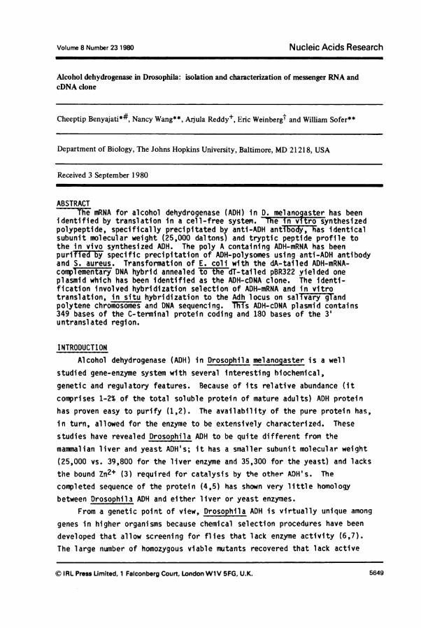
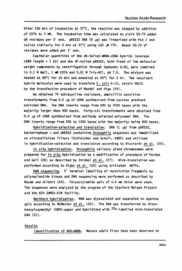
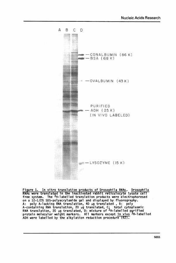
synthesize ADH (32) indicating that they possess functional ADH-mRNA. Toidentify this ADH-mRNA we isolated RNA from wild-type flies and translatedit in a mRNA dependent rabbit reticulocyte cell-free system. The resultant3H-labelled translation products were separated by SDS-gel electrophoresisand visualized by fluorography. Figure 1 shows the results of thisexperiment. The polypeptides synthesized when total cytoplasmic, poly A-containing and poly A-lacking RNA were translated in vitro are shown inlanes C, B and A respectively. The total cytoplasmic and poly A-containingpreparations (these show the bulk of translational activity) code for a
great variety of proteins, with one prominent band, accounting for 1-2%of the total translational activity, migrating to the same position on SDS-polyacrylamide gel as ADH monomer (corresponding to a molecular weight of25,000).
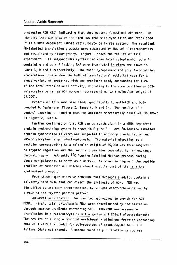
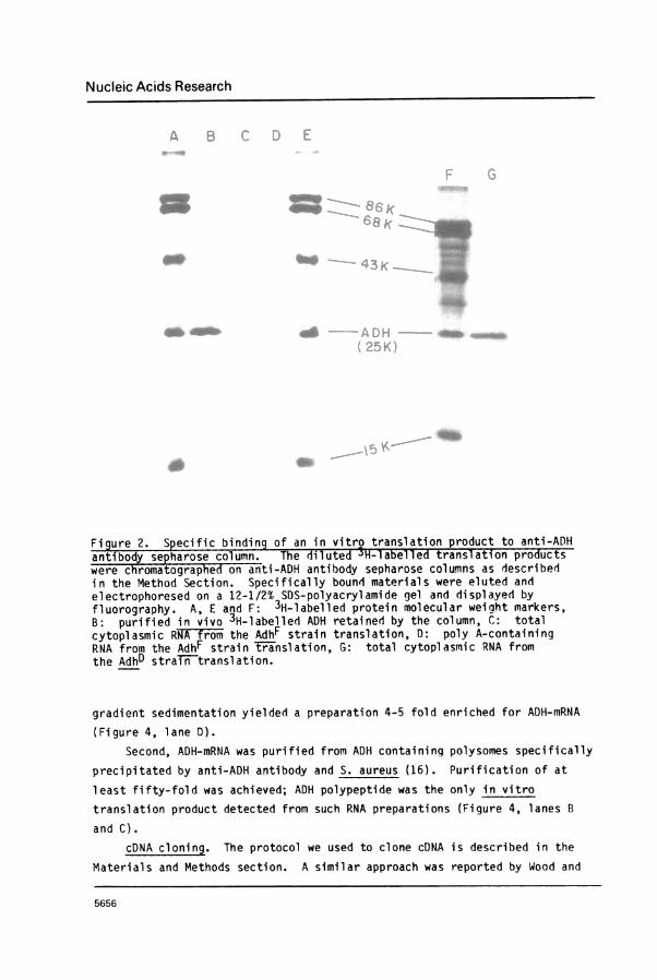
Protein of this same size binds specifically to anti-ADH antibodycoupled to Sepharose (Figure 2, lanes C, D and G). The results of a
control experiment, showing that the antibody specifically binds ADH is shownin Figure 2, lane B.
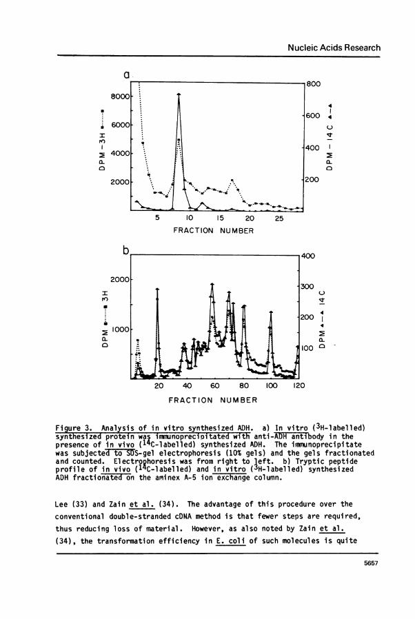
Further confirmation that ADH can be synthesized in a M'RNA dependentprotein synthesizing system is shown in Figure 3. Here 3H-leucine labelledprotein synthesized in vitro was subjected to antibody precipitation and
SDS-polyacrylamide gel electrophoresis. The material migrating at a
position corresponding to a molecular weight of 25,000 was then subjectedto tryptic digestion and the resultant peptides separated by ion exchangechromatography. Authentic 14C-leucine labelled ADH was present duringthese manipulations to serve as a marker. As shown in Figure 3 the peptideprofiles of authentic ADH matches almost exactly that of the in vitro
synthesized product.From these experiments we conclude that Drosophila adults contain a
polyadenylated mRNA that can direct the synthesis of ADH. ADH was
identified by antibody precipitation, by SDS-gel electrophoresis and byvirtue of its tryptic peptide pattern.
ADH-mRNA purification. We used two approaches to enrich for ADH-mRNA. First, total cytoplasmic RNAs were fractionated by sedimentationthrough sucrose gradients containing SDS. ADH-mRNA was assayed by
translation in a reticulocyte in vitro system and SDSgel electrophoresisThe results of a single round of enrichment yielded one fraction containing
RNAs of 11-13S that coded for polypeptides of about 23,000 to 35,000daltons (data not shown). A second round of purification by sucrose
5654
Nucleic Acids Research
A B C D
_ -CONALBUMIN (86 K iNow-BSA (68 K)
-OVALBUMIN (43 K)
PUR IFlID--ADH (25K)
(IN VIVO LABELED)
-LYSOZYME (15K)
Figure 1. In vitro translation products of Drosophila RNAs. DrsohlaRNAs were transl-ated in the inactivated rabbit reticulocyte lysate cllfree system. The 3H-labelled translation products were electrophoresedon a 12-1/2% SDS-polyacrylamide gel and displayed by fluorography.A: poly A-lacking RNA translation, 40 ug translated , B: polyA-containing RNA translation, 20 iug translated, C: total cytoplasmicRNA translation, 20 ug translated, D: mixture of 3H-l abelled pqrifiedprotein molecular weight markers. All markers exceptin vivo3JH-labelledADH were labelled by the alkylation reduction procedur-eTT2F.
5655
Nucleic Acids Research
Figure 2. Specific binding of an in vitro translation product to anti-ADHantibody sepharose column. The diluted '3H-labelled translation productswere chromatographed on anti-ADH antibody sepharose columns as describedin the Method Section. Specifically bound materials were eluted andelectrophoresed on a 12-1/2% SDS-polyacrylamide gel and displayed byfluorography. A, E and F: 3H-labelled protein molecular weiqht markers,B: purified in vivo 3H-labelled ADH retained by the column, C,: totalcytoplasmic RNA from the AdhF strain translation, D: poly A-containingRNA from the AdhF strain translation, G: total cytoplasmic RNA fromthe AdhD straiFntranslation.
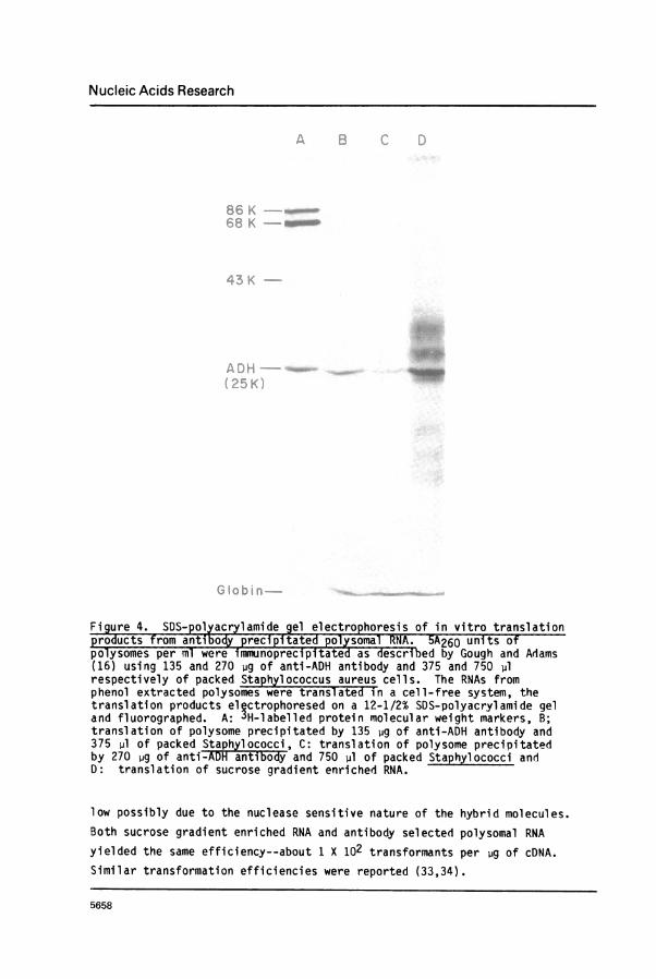
gradient sedimentation yielded a preparation 4-5 fold enriched for ADH-mRNA
(Figure 4, lane D).Second, ADH-mRNA was purified from ADH containing polysomes specifically
precipitated by anti-ADH antibody and S. aureus (16). Purification of at
least fifty-fold was achieved; ADH polypeptide was the only in vitro
translation product detected from such RNA preparations (Figure 4, lanes B
and C).cDNA cloning. The protocol we used to clone cDNA is described in the
Materials and Methods section. A similar approach was reported by Wood and
5656
Nucleic Acids Research
a
il
I
a-0
I
a~
5 10 15 20 25
FRACTION NUMBER
4
4
0
IT
0
4
4
0 -
20 40 60 80 100 120
FRACTION NUMBER
Figure 3. Analysis of in vitro synthesized ADH. a) In vitro (3H-labelled)synthesized protein waC inmunoprecioitated with anti-ADH anti body in thepresence of in vivo (1'C-labelled) synthesized ADH. The immunoprecipitatewas subjecte& tEo SS-gel electrophoresis (10% gels) and the gels fractionatedand counted. Electrophoresis was from right to left. b) Tryptic peptideprofile of in vivo (1 C-labelled) and in vitro (3H-labelled) synthesizedADH fractionated on the aminex A-5 ion exchange column.
Lee (33) and Zain et al. (34). The advantage of this procedure over theconventional double-stranded cDNA method is that fewer steps are required,thus reducing loss of material. However, as also noted by Zain et al.
(34), the transformation efficiency in E. coli of such molecules is quite
5657
Nucleic Acids Research
Figure 4. SDS- polyacrylamide gel electrophore_sis of in vitro translationproducts from antibody precipitated polysom-alRNA. 5A260 units ofpolysomes per ml were immunoprecipitated as deescribed by Gough and Adams(16) using 135 and 270 4g of anti-ADH antibody and 375 and 750 plrespectively of packed Staphylococcus aureus cells. The RNAs fromphenol extracted polysomes were translate in a cell-free system, thetranslation products electrophoresed on a 12-1/2% SDS-polyacrylamide geland fluorographed. A: 3H-labelled protein molecular weight markers, B;translation of polysome precipitated by 135 jg of anti-ADH antibody and375 pl of packed Staphylococci, C: translation of polysome precipitatedby 270 jig of anti-ADH antibody and 750 pl of packed Staphylococci andD: translation of sucrose gradient enriched RNA.
low possibly due to the nuclease sensitive nature of the hybrid molecules.Both sucrose gradient enriched RNA and antibody selected polysomal RNAyielded the same efficiency--about 1 X 10² transformants per ug of cDNA.Similar transformation efficiencies were reported (33,34).
5658
Nucleic Acids Research
2 3 4 5 6 7a_. r%*.,0%. $..t.# PS
4a
8 9 10 11 12 13 14
.0
m-86 K_e-68 K
co
-43 K
-ADH(25 K)
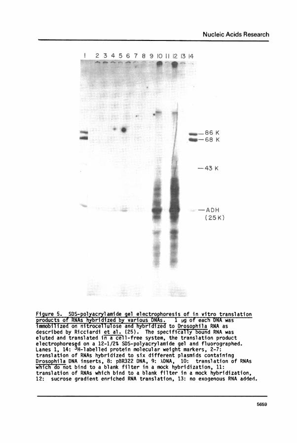
Figure 5. SDS-polyacrylamide gel electrophoresis of in vitro translationproducts of RNAs hybridized by various DNAs. 1 jg of each DNA wasimmobilized on nitrocellulose and hybridized to Drosophila RNA asdescribed by Ricciardi et al. (25). The specificall3y bound RNA waseluted and translated in a cell-free system, the translation productelectrophoresed on a 12-1/2% SDS-polyacrylamide gel and fluorographed.Lanes 1, 14: "H-labelled protein molecular weight markers, 2-7:translation of RNAs hybridized to six different plasmids containingDrosophila DNA inserts, 8: pBR322 DNA, 9: XDNA, 10: translation of RNAswhich -donot bind to a blank filter in a mock hybridization, 11:translation of RNAs which bind to a blank filter in a mock hybridization,12: sucrose gradient enriched RNA translation, 13: no exogenous RNA added.
5659
t38g§}
Nucleic Acids Research
4 'V
I. "
5660
Nucleic Acids Research
From a transformation experiment using antibody selected polysomalRNA-cDNA hybrid molecules, one plasmid carrying ADH-cDNA (pADH*cDNA) hasbeen identified by the three following criteria. First, using thetechnique of hybridization selection and translation (25), we demonstratedthat DNA from this clone was capable of hybridizing to an RNA species thatwas translatable into ADH (Figure 5, lane 6). Control DNA from bacterio-phage x, pBR322 and other plasmids failed to do this.
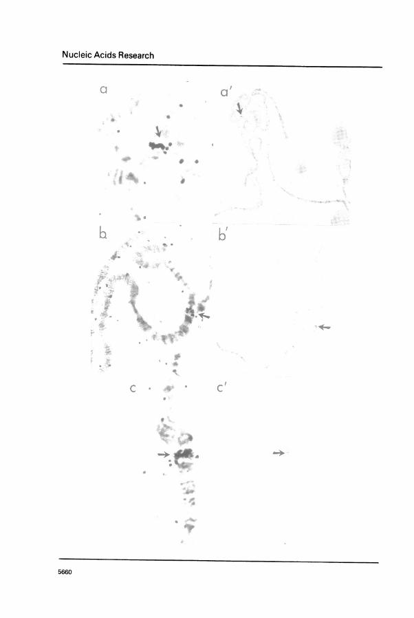
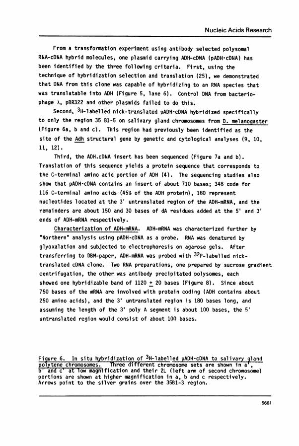
Second, 3H-labelled nick-translated pADH*cDNA hybridized specificallyto only the region 35 B1-5 on salivary gland chromosomes from D. melanogaster(Figure 6a, b and c). This region had previously been identified as the
site of the Adh structural gene by genetic and cytological analyses (9, 10,11, 12).
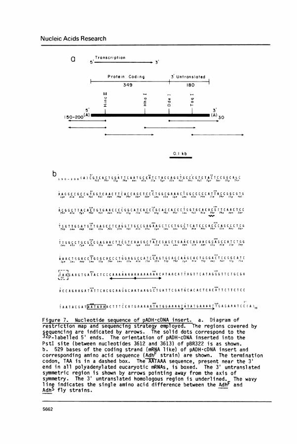
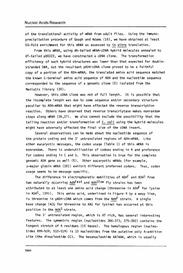
Third, the ADH.cDNA insert has been sequenced (Figure 7a and b).Translation of this sequence yields a protein sequence that corresponds tothe C-terminal amino acid portion of ADH (4). The sequencing studies alsoshow that pADH*cDNA contains an insert of about 710 bases; 348 code for116 C-terminal amino acids (45% of the ADH protein), 180 representnucleotides located at the 3' untranslated region of the ADH-mRNA, and theremainders are about 150 and 30 bases of dA residues added at the 5' and 3'ends of ADH-mRNA respectively.
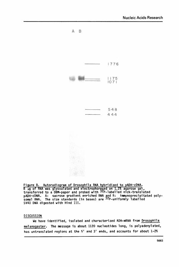
Characterization of ADH-mRNA. ADH-mRNA was characterized further by"Northern" analysis using pADH-cDNA as a probe. RNA was denatured byglyoxalation and subjected to electrophoresis on agarose gels. Aftertransferring to DBM-paper, ADH-mRNA was probed with 32P-labelled nick-translated cDNA clone. Two RNA preparations, one prepared by sucrose gradientcentrifugation, the other was antibody precipitated polysomes, eachshowed one hybridizable band of 1120 + 20 bases (Figure 8). Since about750 bases of the mRNA are involved with protein coding (ADH contains about250 amino acids), and the 3' untranslated region is 180 bases long, and
assuming the length of the 3' poly A segment is about 100 bases, the 5'untranslated region would consist of about 100 bases.
Figure 6. In situ hybridization of 3H-labelled pADH-cDNA to salivary glandpolytene chromosomes. Three different chromosome sets are shown in a,
an Cia ow magnification and their 2L (left arm of second chromosome)portions are shown at higher magnification in a, b and c respectively.Arrows point to the silver grains over the 35B1-3 region.
5661
Nucleic Acids Research
a T r a n s c r i p t i on5' b 3
Protein Coding 3 Untranslated
349 180
5 3~~~~~Q o @v~~a
I I 0
150-200_(A)_ (A)30
0.1 kb
Val Thr CPh.Ph* At a
It.II# CTrl Val Pro Val Tyr See GZy h
r
A 5 G G 2 C G C A C AGGT CAC A A AT T'C AC AC T G CT CA C' TAG C C A ACGT GCCCA T CT ACT'ACC GA CA TC
AAAACCACCATAATCAACTTCACCAGCTCCCTAACAAAACTAGCCCCCATTACCAGCATALys Ata Ala Vat Vat A, Ph. TrP S*r S.r Le Ala Lys Lou Ata Pro IIt Thr Gly Val
A C AG C T TA C ACT T G A A C C C C CATC A C CC C A C C A C C C T T C ACA TT C CThr Ata Tyr Thr Vat A*', Pro Gly Ite Thr Arg Thr Thr Leu Val His Thr Ph. Asn Ser
_~
TAATTAAATATT6AAACCTCAGATTACC6AGAAGACTCCTAACTCATCCCACCCAACCCTCGTrp Lou A*p Vat Gtl Pro CtGu Vat Ala Glu, Lys Leu Leu Ala His Pro ihr Gt Pro Ser
TTGGCCTGCGCCGAGAACTTCGTCAAAGCTATCGAGCTGAACCAGAACGAAGCCATCT A,GLe. Ata Cy* Ala Gtu As. Phe V.1 Lys Ala Ile Ghu L a Asn Gln As. GCly Ala ItZ Trp
AAACTAAACCTAGACACCCTAGAGGCCATCCAATAGACCAAACACTUGGAACTCCAACATCLyia Le. Asp Lou Gly Thr Leu Gtu Ala Ile Slm Trp ThFr Lye His Pup Asp S.r Sly Ite
r.-- ,,,.. -...IT A A:GA A GTGA TACT C C C A A A A A A A A A A A A A A A C A TA A CAT TAG T T C A TAG AG T T C TG C GA
ACCAGAAAAATATTCACACAAGACAATAAAACTAATTCAAT6CACACTCACATTCTTCTCC
TA AT A C G A T A CT T T C A TA A A A A AT"AT A A A A A A TAT AT GA A A ATTA A GA A A T C C A30
Figure 7. Nucleotide sequence of pADH-cDNA insert. a. Diagram ofrestriction map and sequencing strategy employed. The regions covered bysequencing are indicated by arrows. The solid dots correspond to the32P-labelled 5' ends. The orientation of pADH-cDNA inserted into thePstI site (between nucleotides 3612 and 3613) of pBR322 is as shown.b. 529 bases of the coding strand (mRNA like) of pADH cDNA insert andcorresponding amino acid sequence (AdhF strain) are shown. The terminationcodon, TAA is in a dashed box. TheATAAA sequence, present near the 3'end in all polyadenylated eucaryotic mRNAs, is boxed. The 3' untranslatedsymmetric region is shown by arrows pointing away from the axis ofsymmetry. The 3' untranslated homologous region is underlined. The wavyline indicates the single amino acid difference between the AdhF andAdhS fly strains.
5662
Nucleic Acids Research
A B
i 7 6- ~~i-g
=S~~~~~rJ 4 tW.A _@4 4 4
Figure 8. Autoradiogramn of Drosophila RNA hybridized to pADH*cDNA.8 jig of RNA was glyoxalated and el ectrophorped on 1.2% agarose gel,transferred to a DBM-paper and probed with tP-labelled nick-translatedpADH cDNA. A: sucrose gradient enriched RNA and B: imnunoprecipitated poly-somal RNA. The size standards (in bases) are 32P-uniformly labelledSV40 DNA digested with Hind III.
DISCUSSIONWe have identified, isolated and characterized ADH-mRNA from Drosophila
melanogaster. The message is about 1120 nucleotides long, is polyadenylated,has untranslated regions at the 5' and 3' ends, and accounts for about 1-2%
5663
Nucleic Acids Research
of the translational activity of mRNA from adult flies. Using the immuno-
precipitation procedure of Gough and Adams (16), we have obtained at least50-fold enrichment for this mRNA as assessed by in vitro translation.
From this mRNA, using dA-tailed mRNA-cDNA hybrid molecules annealed to
dT-tailed pBR322, we have constructed a cDNA clone. The transformation
efficiency of such hybrid structures was lower than that expected for double-
stranded DNA, but the resultant pADH cDNA clone proved to be a faithfulcopy of a portion of the ADH-mRNA; the translated amino acid sequence matchedthe known C-terminal amino acid sequence of ADH and the nucleotide sequencecorresponded to the sequence of a genomic clone (5) isolated from the
Maniatis library (35).However, this cDNA clone was not of full length. It is possible that
the incomplete length was due to some sequence and/or secondary structure
peculiar to ADH-mRNA that might have affected the reverse transcriptionreaction. Others have observed that reverse transcriptase makes nonrandom
stops along mRNA (36,37). We also cannot exclude the possibility that the
tailing reaction and/or transformation of E. coli using the hybrid molecules
might have adversely affected the final size of the cDNA insert.Several observations can be made about the nucleotide sequence of
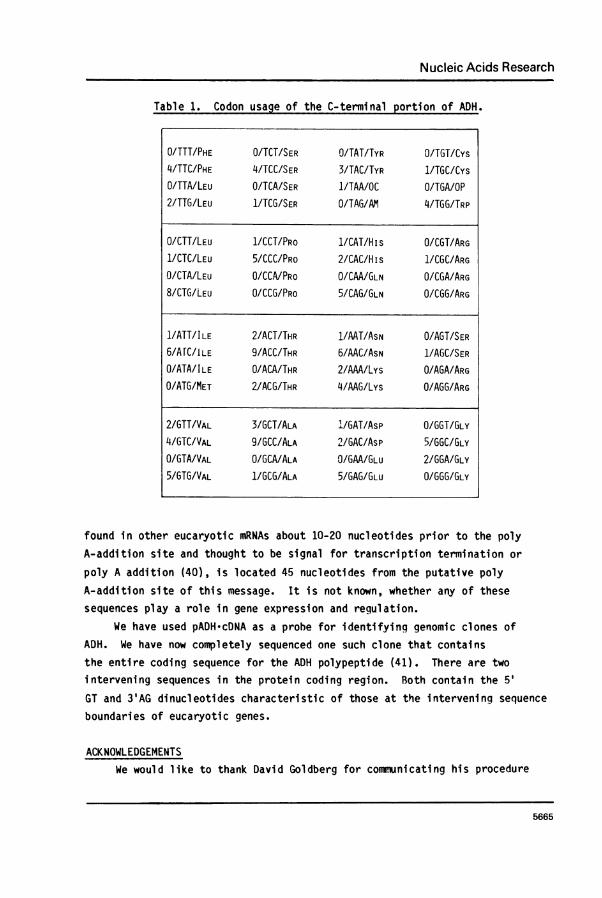
the protein coding and the 3' untranslated regions of ADH-mRNA. Likeother eucaryotic messages, the codon usage (Table 1) of this mRNA isnonrandom. There is underutilization of codons ending in A and preferencefor codons ending in C and G. This observation is true for the completegenomic ADH gene as well (5). Other eucaryotic mRNAs (for example,s-major globin mRNA (38)) exhibit different preferred codons. Thus, codon
usage seems to be message specific.The difference in electrophoretic mobilities of ADHF and ADHS from
two naturally occurring AdhFast and AdhSlOw fly strains has been
attributed to at least one amino acid change (threonine in ADHF for lysinein ADHS, (39)). This amino acid, underlined in Figure 9 by a wavy line,is threonine in pADH*cDNA which comes from the AdhF strain. A singlebase change (ACG for threonine to AAG for lysine) has occurred at this
position in the AdhS strain.The 3' untranslated region, which is AT rich, has several interesting
features. The symmetric region (nucleotides 366-373; 375-382) contains thelongest stretch of A residues (15 bases). The homologous region (nucleo-tides 499-509; 510-519) is 10 nucleotides from the putative poly A-additionsite (the dinucleotide CC). The hexanucleotide AATAAA, which is usually
5664
Nucleic Acids Research
Table 1. Codon usage of the C-terminal portion of ADH.
found in other eucaryotic mRNAs about 10-20 nucleotides prior to the polyA-addition site and thought to be signal for transcription termination or
poly A addition (40), is located 45 nucleotides from the putative polyA-addition site of this message. It is not known, whether any of these
sequences play a role in gene expression and regulation.We have used pADH'cDNA as a probe for identifying genomic clones of
ADH. We have now completely sequenced one such clone that containsthe entire coding sequence for the ADH polypeptide (41). There are twointervening sequences in the protein coding region. Both contain the 5'
GT and 3'AG dinucleotides characteristic of those at the intervening sequenceboundaries of eucaryotic genes.
ACKNOWLEDGEMENTS
We would like to thank David Goldberg for communicating his procedure
5665
O/TTT/PHE O/TCT/SER O/TAT/TYR O/TGT/Cys4/TTC/PHE 4/TCC/SER 3/TAC/TYR 1/TGC/CysO/TTA/LEU O/TCA/SER 1/TAA/OC O/TGA/OP2/TTG/LEU 1/TCG/SER O/TAG/AM 4/TGG/TRP
O/CTT/LEU 1/CCT/PRO 1/CAT/H is O/CGT/ARG1/CTC/LEU 5/CCC/PRO 2/CAC/His 1/CGC/ARGO/CTA/LEU O/CCA/PRo O/CAA/GLN O/CGA/ARG8/CTG/LEU 0/CCG/PRO 5/CAG/GLN O/CGG/ARG
1/ATT/ILE 2/ACT/THR 1/AAT/ASN O/AGT/SER6/AfC/ILE 9/ACC/THR 6/AAC/ASN 1/AGC/SERO/ATA/ILE O/ACA/THR 2/AAA/Lys O/AGA/ARG0/ATG/MET 2IACG/THR 4/AAG/Lys O/AGG/ARG
2/GTT/VAL 3/GCT/ALA 'L/GAT/Asp O/GGT/GLY4/GTC/VAL 9/GCC/ALA 2/GAC/Asp 5/GGC/GLYO/GTA/VAL O/GCA/ALA O/GAA/GLU 2/GGA/GLY5/GTG/VAL 1/GCG/ALA 5/GAG/GLU O/GGG/GLY
Nucleic Acids Research
for mRNA-cDNA hybrid cloning. This work was supported by grants GM21322-01to E. W., GM-18257, NIEHS (ES-01527) and a contract from Department ofEnergy (EY-76-S-02-2965) to W.S.C.B. was a postdoctoral fellow of the DamonRunyon-Walter Winchell Cancer Fund, DRG-175-F.
Present Address:
*Cancer Biology Program, Frederick Cancer Research Center, P.O. Box B,Frederick, MD 21701
**Waksman Institute of Microbiology, Rutgers University, P.O. Box 759,Piscataway, NJ 08854
+School of Life Sciences, Central University of Hyderabad, Hyderabad, IndiatDepartment of Biology, Biology Building/G5, University of Pennsylvania,Philadelphia, PA 19104
#To whom correspondence should be addressed.
REFERENCES1. Sofer, W. and Ursprung, H. (1968) J. Biol. Chem. 243, 3110-3115.2. Schwartz, M., Gerace, L., O'Donnell, J. and Sofer, W. (1975) inInternational Conference on Isozymes, Vol. 1, pp. 725-751, AcademicPress, New York.3. Place, A.R., Powers, D. and Sofer, W. (1980) manuscript submitted.4. Thatcher, D.R. (1980) Biochem. J. 187, 875-886.5. Benyajati, C., Place, A.R., Powers, D. and Sofer, W., manuscriptin preparation.6. Sofer, W. and Hatkoff, M. (1972) Genetics 72, 545-549.7. O'Donnell, J., Gerace, L., Leister, F. and Sofer, W. (1975) Genetics,79, 73-83.8. Schwartz, M. and Sofer, W. (1976) Genetics 83, 125-136.9. Grell, E.H., Jacobson, K.B. and Murphy, J.B. (1965) Science 149, 80-82.10. O'Donnell, J., Mandel, H.C., Krauss, M. and Sofer, W. (1977) Genetics86, 553-566.
11. Woodruff, R.C. and Ashburner, M. (1979) Genetics 92, 117-132.12. Woodruff, R.C. and Ashburner, M. (1979) Genetics 92, 133-149.13. Ursprung, H., Sofer, W. and Burroughs, N. (1970) Wilhelm Roux'Archiv. 164, 201-208.14. Clewell, D.B. (1972) J. Bacteriol. 110, 667-676.15. Palmiter, R.D. (1974) Biochemistry 13, 3606-3615.16. Gough, N.M. and Adams, J.M. (1978) Biochemistry 17, 5560-5566.17. Pelham, H.R.B. and Jackson, R.S. (1976) Eur. J. Biochem. 67, 247-257.18. Laemmli, U.K. (1970) Nature 227, 680-685.19. Laskey, R.A. and Mills, A.D. (1975) Eur. J. Biochem 56, 335-341.20. Milman, G., Krauss S.W. and Olsen, A.S. (1977) Proc. Natl. Acad.Sci. USA 74, 926-930.21. Pelliccia, J.G. (1980) Ph.D. Thesis, The Johns Hopkins University,Bal timore.22. Reddy, A.R., Pelliccia, J.G. and Sofer, W. (1980) BiochemicalGenetics 18, 339-351.
5666
Nucleic Acids Research
23. Roychoudhury, R., Jay, E. and Wu, R. (1976) Nucleic Acids Res.3, 863-877.
24. Mandel, M. and Higa, A. (1970) J. Mol. Biol. 53, 159-162.25. Ricciardi, R.P., Miller, J.S. and Roberts, B.E. (1979). Proc. Natl.
Acad. Sci. USA 76, 4927-4931.26. Pardue, M.L. and Gall, J.G. (1975). in Methods in Cell Biology,
Prescott, D.M., Ed., Vol. 10, pp. 1-16, Academic Press, New York.27. Strobel, E., Dunsmuir, P. and Rubin, G.M. (1979) Cell 17, 429-439.28. Rigby, P.W.J., Dieckmann, M., Rhodes, C. and Berg, P. (1977)
J. Mol. Biol. 113, 237-251.29. Maxam, A.M. and Gilbert, W. (1980) in Methods in Enzymology,
Grossman, L, and Moldave, K. Eds., Vol. 65, pp. 499-560 AcademicPress, New York.
30. McMaster, G.K. and Carmichael, G.G. (1977) Proc. Natl. Acad. Sci.USA 74, 4835-4838.
31. Alwine, J.C., Kemp, D.J. and Stark, G.R. (1977) Proc. Natl. Acad.Sci. USA 74, 5350-5354.
32. Pelliccia, J.G., and Sofer, W. (1980) manuscript submitted.33. Wood, K.0. and Lee J.C. (1976) Nucleic Acids Res. 3, 1961-1971.34. Zain, S., Sambrook, J., Roberts, R.J., Keller, W., Fried, M.
and Dunn, A.R (1979) Cell 16, 851-861.35. Maniatis, T., Hardison, R.C., Lacy, E., Lauer, J., O'Connell, C. and
Quon, D. (1978) Cell 15, 687-701.36. Friedman, E.Y. and Rosbash, M. (1977). Nucleic Acids Res. 4, 3455-
3471.37. Buell, G.N., Wickers, M.P., Payvar, F. and Schimke, R.T. (1978)
J. Biol. Chem. 253, 2471-2482.38. Konkel, D.A., Tilghman, S.M. and Leder, P. (1978) Cell 15, 1125-1132.39. Fletcher, T.S., Ayala, F.J., Thatcher, D.R. and Chambers, G.K. (1978)
Proc. Natl. Acad. Sci. USA 75, 5609-5612.40. Proudfoot, N.J. and Brownlee, G.G. (1976) Nature 263, 211-214.41. Benyajati, C. and Sofer, W. (1980) manuscript in preparation.42. Rice, R.H. and Means, G.E. (1971) J. Biol. Chem. 246, 831-832.
5667