after the double helix
TRANSCRIPT
After the Double HelixRosalind Franklin’s Research on Tobacco
mosaic virus
By Angela N. H. Creager* and Gregory J. Morgan**
ABSTRACT
Rosalind Franklin is best known for her informative X-ray diffraction patterns of DNAthat provided vital clues for James Watson and Francis Crick’s double-stranded helicalmodel. Her scientific career did not end when she left the DNA work at King’s College,however. In 1953 Franklin moved to J. D. Bernal’s crystallography laboratory at BirkbeckCollege, where she shifted her focus to the three-dimensional structure of viruses,obtaining diffraction patterns of Tobacco mosaic virus (TMV) of unprecedented detail andclarity. During the next five years, while making significant headway on the structuraldetermination of TMV, Franklin maintained an active correspondence with both Watsonand Crick, who were also studying aspects of virus structure. Developments in TMVresearch during the 1950s illustrate the connections in the emerging field of molecularbiology between structural studies of nucleic acids and of proteins and viruses. They alsoreveal how the protagonists of the “race for the double helix” continued to interactpersonally and professionally during the years when Watson and Crick’s model for thedouble-helical structure of DNA was debated and confirmed.
F OR MOLECULAR BIOLOGY, the 1950s was in many ways the decade of the helix:the alpha helix of proteins, the double helix of DNA, and the helical nature of Tobacco
mosaic virus (TMV) were all major discoveries.1 This essay examines the interplay
* Department of History and Program in History of Science, 136 Dickinson Hall, Princeton University,Princeton, New Jersey 08544.
** Department of Philosophy, Spring Hill College, 4000 Dauphin Street, Mobile, Alabama 36608.Our research was supported by the National Science Foundation, through a CAREER grant, SBE 98-75012
(ANHC), and a Dissertation Research Improvement Grant, SBE 99-10891 (GJM). For access to correspondenceand other historical materials we thank Donald Caspar, Aaron Klug, Jeremy Norman, Shannon Bohle at the ColdSpring Harbor Laboratory Archives, and archivists at the Bancroft Library, the Caltech Archives, the ChurchillArchives Centre, the Novartis Foundation, the Royal Institution of Great Britain, the University of Maryland,Baltimore County, and the University of Melbourne Archives. We acknowledge the valuable feedback wereceived on versions of this essay presented at “Molecular Biology in the Twentieth Century: A Meeting to Markthe Fiftieth Anniversary of the Determination of the Structure of DNA,” organized by Frank James and held on28–29 Apr. 2003 at the Royal Institution, London, and at a session organized by Karen-Beth Scholthof and PaulPeterson at the American Phytopathological Society Meeting on 1 Aug. 2005 in Austin, Texas. We also thankDonald Caspar, Nathaniel Comfort, John Finch, Michael Gordin, Michael Keevak, Aaron Klug, KennethHolmes, Bernard Lightman, Karen-Beth Scholthof, Judith Swan, Sue Tolin, Daniel Trambaiolo, Doogab Yi, andthree anonymous referees for their suggestions, corrections, and criticisms. The authors alone bear responsibilityfor the essay, including its interpretations and any remaining errors.
1 We have followed current nomenclature in italicizing the full names of virus species.
Isis, 2008, 99:239-272©2008 by The History of Science Society. All rights reserved.0018-9902/2008/9902-0001$10.00
239
between structural studies of DNA and of viruses by focusing on the research of thecrystallographer Rosalind Franklin. Franklin is now remembered principally for her X-raydiffraction patterns of DNA, which provided data that—still unpublished and examinedwithout her consent—informed James Watson and Francis Crick’s double-helical model.2
Neither the degree to which Watson and Crick had access to her results nor the possibilitythat her data contributed to, rather than corroborated, their model came to public light untilthe 1968 publication of Watson’s The Double Helix. This confessional account, with itsunflattering and inaccurate portrayal of Franklin, was published ten years after heruntimely death from ovarian cancer. While Watson’s book has become a classic, so, too,has Franklin become a martyr figure, a woman scientist denied credit for her keycontribution to our understanding of DNA.3 Indeed, developments in the last four de-cades—as DNA came to occupy center stage in the life sciences and the women’smovement drew attention to the pervasive sexism in elite fields, including science—haveintensified this sentiment. The publication of Brenda Maddox’s enlightening biography ofFranklin just prior to the fiftieth anniversary of Watson and Crick’s famous paper kept thevexed issue of scientific credit for the double helix at the center of media accounts of the2003 commemorations.4
The relentless focus on the dramatic elements of the double-helix story, however, yieldsan incomplete and misleading understanding of Franklin’s overall career and contribu-tions—as well as of molecular biology at that time. By the late 1950s, Franklin hadachieved a strong international reputation as a scientist, in large part because of herimpressive contributions to structural virology. At Birkbeck College, in the laboratory ofJohn Desmond Bernal, she used many of the same techniques she had employed withDNA to produce the finest diffraction patterns of TMV available.5 Though she abandonedher experimental work on DNA when she left King’s College, London, in 1953, sheremained in the same small circle of biophysicists: Watson, Crick, and Maurice Wilkinswere all engaged in structural studies of viruses. Watson determined that TMV was helicalin structure; Franklin and her group confirmed this insight but corrected his model byshowing that TMV had forty-nine subunits per three turns of the helix. Franklin andDonald Caspar determined that the RNA in TMV was not situated in the center of the
2 James D. Watson and Francis H. C. Crick, “A Structure for Deoxyribose Nucleic Acid,” Nature, 1953,171:737–738. There are many accounts of the relationship of Franklin’s diffraction patterns to Watson andCrick’s model; for a recent appraisal that cites others see Lynne Osman Elkin, “Rosalind Franklin and the DoubleHelix,” Physics Today, 2003, 56:42–48.
3 James D. Watson, The Double Helix: A Personal Account of the Discovery of the Structure of DNA, ed.Gunther S. Stent (1968; New York: Norton, 1980) (hereafter cited as Watson, Double Helix). For analysis ofthe mythic dimensions of the representation of Franklin as a martyr, in which she is cast as the “Sylvia Plath ofmolecular biology,” see Brenda Maddox, “The Double Helix and the ‘Wronged Heroine,’” Nature, 2003,421:407–408. Anne Sayre’s book played a crucial role in drawing public attention to the injustices of Watson’sportrayal: Anne Sayre, Rosalind Franklin and DNA (New York: Norton, 1975).
4 Brenda Maddox, Rosalind Franklin: The Dark Lady of DNA (New York: Harper Collins, 2002) (hereaftercited as Maddox, Rosalind Franklin). A few examples of the media accounts are Tara Pepper, “Genes, Girls,and Gall,” Newsweek, 5 Aug. 2002, p. 54; Jim Holt, “Photo Finish: Rosalind Franklin and the Great DNA Race,”New Yorker, 28 Oct. 2002, p. 102; Bernadine Healy, “Let’s Remember Rosy,” U.S. News and World Report, 24Feb. 2003, p. 47; and Denise Grady, “A Revolution at Fifty: Fifty Years Later, Rosalind Franklin’s X-ray FuelsDebate,” New York Times, 25 Feb. 2003, p. 2. The PBS program Nova produced a segment on Franklin that wasbroadcast on 22 Apr. 2003. For an analysis of the commemorations see Pnina G. Abir-Am, “DNA at Fifty:Institutional and Biographical Perspectives,” Minerva, 2004, 42:191–213.
5 Franklin’s publications on TMV are cited in the course of this essay; for an overview of her contributionssee Kenneth C. Holmes, “Rosalind Franklin and the Tobacco Mosaic Virus,” in DNA 50: The Secret of Life, ed.Miriam Balaban (London: Faircount, 2003), pp. 200–208.
240 AFTER THE DOUBLE HELIX
rod-shaped virus, as previously thought, but at 40 Ångstroms (Å) radius. Franklin wasadmired for her virtuosity as an experimentalist, but she also took risks to establish herleadership in the field, publishing speculative models of the virus structure.6
Franklin’s research at Birkbeck was highly collaborative.7 Her efforts from 1953 to1958 to determine the structure of TMV involved Aaron Klug, John Finch, and KennethC. Holmes and drew on resources from three rival groups of biochemists—led by GerhardSchramm in Tubingen, Heinz Fraenkel-Conrat in Berkeley, and Barry Commoner in St.Louis—that were also working on the virus. Close analysis of Franklin’s research duringthe last five years of her life reveals how relationships among the main protagonists of thedouble-helix story developed after the spring of 1953—and shows the practical andconceptual connections between key research objects, especially nucleic acids and viruses,in the new field of molecular biology. Her work on virus structure makes it clear that theseresearchers in the 1950s were putting to rest debates about the physical and chemicalnature of biological materials that had persisted for two decades. Even as the project ofshowing that proteins, nucleic acids, and viruses have regular molecular structures gaveway to new concerns about information and sequence, a few central model systems,including viruses such as TMV and proteins such as hemoglobin and insulin, remainedcentral to biophysical research.8
The history of molecular biology in the 1950s is hardly understudied, yet a focus onFranklin’s late work provides a new perspective from which to view familiar develop-ments.9 Two aspects of our analysis merit highlighting at the outset. First, this accountoffers an alternative narrative to the popular double-helix story, one that emphasizes thecontinuing interactions between Franklin, Watson, Crick, and their collaborators as theymoved on from DNA structure to new problems. In the mid-1950s, once the hereditaryrole of nucleic acids had been settled, molecular biologists sought to determine thefunctional correlation between proteins and nucleic acids (as well as to differentiate theroles of DNA and RNA with respect to protein synthesis). Accounts of this period tend toemphasize the researchers who sought to crack this “coding” problem by viewing genesas carriers of information amenable to theoretical and computational approaches.10 Bycontrast, Franklin was centrally involved in a network of scientists who used structuralmethods to investigate the nucleic acid–protein relationship; they often worked with
6 This risk taking stands in contrast to the image of Franklin as such a cautious experimentalist that she resistedstructural speculation; see Watson, Double Helix, pp. 45, 95–96.
7 This, too, contradicts the impression—drawn from her work on DNA at King’s College and based largelyon the portrayal offered in The Double Helix—of Franklin as a solitary investigator. (Even there, she workedclosely with Raymond Gosling, if not with Wilkins.)
8 On the importance of hemoglobin and insulin to biochemistry and biophysics see Soraya de Chadarevian,“Sequences, Conformation, Information: Biochemists and Molecular Biologists in the 1950s,” Journal of theHistory of Biology, 1996, 29:361–386; de Chadarevian, “Following Molecules: Hemoglobin between the Clinicand the Laboratory,” in Molecularizing Biology and Medicine: New Practices and Alliances, 1910s–1970s, ed.de Chadarevian and Harmke Kamminga (Amsterdam: Harwood, 1998), pp. 171–201; and de Chadarevian,Designs for Life: Molecular Biology after World War II (Cambridge: Cambridge Univ. Press, 2002) (hereaftercited as de Chadarevian, Designs for Life). Secondary literature on virus research is cited throughout this essay.
9 For an insightful synoptic account that cites the secondary literature up to the mid-1990s see MichelMorange, A History of Molecular Biology, trans. Matthew Cobb (Cambridge, Mass.: Harvard Univ. Press, 1998).
10 On the coding problem see Lily E. Kay, Who Wrote the Book of Life? A History of the Genetic Code(Stanford, Calif.: Stanford Univ. Press, 2000); and Horace Freeland Judson, The Eighth Day of Creation (NewYork: Simon & Schuster, 1979), Chs. 5–8. Both Kay and Judson make it clear that researchers employingcomputational and theoretical methods (largely members of the RNA Tie Club) were not successful in actuallycracking the code; this was accomplished by Heinrich Matthei and Marshall Nirenberg using biochemicalmethods in the early 1960s.
ANGELA N. H. CREAGER AND GREGORY J. MORGAN 241

viruses, since their nucleic acid and protein components could be studied separately andjointly. This was not the only motivation for undertaking structural studies of viruses, butthe pertinence of this approach to the coding problem and to understanding proteinsynthesis has been largely overlooked, despite Watson’s and Crick’s participation in theeffort. In part, this omission persists because other groupings, such as the “RNA TieClub,” lend themselves to such colorful historical accounts.11 However beguiling suchtales may be, they miss important players and contributions and tend to reproduce patternsof exclusion that operated at the time. The RNA Tie Club actually included severalscientists known for their work on biological structures—Watson, Linus Pauling, andAlexander Rich—but Franklin (who did not wear a necktie, after all) was never invitedto join.
Second, we build on the insights of others in emphasizing the importance of transat-lantic exchanges, both material and informational, among the first generation of molecularbiologists.12 Surviving correspondence makes clear the challenges these researchers facedin the 1950s as they sought to navigate disciplinary boundaries, fend off competitors, andestablish scientific priority—while at the same time trying to contribute sufficiently toscientific exchange networks to “earn” access to others’ research materials and unpub-lished results.13 The increasingly international circulation of materials, knowledge, andresearchers in the postwar decade did not diminish the relevance of differences betweenlocal cultures of science.14 British researchers, who were accustomed to accommodatinginstitutional prerogatives in selecting their research problems (one thinks of how DNAstructure had been viewed as belonging to King’s College), were confronted by entrepre-neurial American competitors and the massive research funds made available by the U.S.federal government.15 Franklin lacked a permanent institutional position in the UnitedKingdom, with its attendant supports and constraints. Yet she showed herself remarkably
11 The RNA Tie Club was part joke and part scientific network. Watson, George Gamow, and Leslie Orgellaunched this clique of scientific correspondents to encourage work to resolve the structure of RNA and toexplicate its role in forming proteins, specifically by providing a forum for speculative ideas and untestedtheories. Other founding members included Crick, Gunther Stent, and Alexander Rich. See Judson, Eighth Dayof Creation, pp. 264–265; and de Chadarevian, Designs for Life, pp. 186–198. On structural models of proteinsynthesis (notably the template theory of Linus Pauling) see Bruno Strasser, “A World in One Dimension: LinusPauling, Francis Crick, and the Central Dogma of Molecular Biology,” History and Philosophy of the LifeSciences, 2006, 28:491–512.
12 Pnina G. Abir-Am, “From Multidisciplinary Collaboration to Transnational Objectivity: InternationalSpaces as Constitutive of Molecular Biology, 1930–1970,” in Denationalizing Science: The Contexts ofInternational Scientific Practice, ed. Elisabeth Crawford, Terry Shinn, and Sverker Sorlin (Dordrecht: Kluwer,1992), pp. 153–186; de Chadarevian, Designs for Life; Jean-Paul Gaudilliere, Inventer la biomedecine: LaFrance, l’Amerique et la production des savoirs du vivant (1945–1965) (Paris: Decouverte, 2002); and BrunoJ. Strasser, La fabrique d’une nouvelle science: La biologie moleculaire a l’age atomique (1945–1964)(Florence: Olschki, 2006).
13 These were transactions, if not monetary ones: scientists exchanged materials and results in return for creditor in the expectation of reciprocity. This understanding of the circulation of scientific information and objectsin terms of “gift exchange” draws on economic anthropology and sociology; for an excellent discussion andreferences see Warwick Anderson, “The Possession of Kuru: Medical Science and Biocolonial Exchange,”Comparative Studies in Society and History, 2000, 42:713–744, esp. pp. 714–716.
14 For a nice example of differences in expectations between French and American cultures of molecularbiology see Jean-Paul Gaudilliere, “Paris–New York Roundtrip: Transatlantic Crossings and the Reconstructionof the Biological Sciences in Post-war France,” Studies in History and Philosophy of Biological and BiomedicalSciences, 2002, 33:389–417, esp. pp. 406–408. On this general issue see Soraya de Chadarevian and BrunoStrasser, “Molecular Biology in Postwar Europe: Towards a ‘Global’ Picture,” ibid., pp. 361–365.
15 For an insightful account of how this played out at Cambridge see de Chadarevian, Designs for Life, esp.Ch. 10. On the perception of King’s College’s prerogative regarding the DNA structure problem see Watson,Double Helix, pp. 13–14.
242 AFTER THE DOUBLE HELIX
adept in maneuvering within the interdisciplinary and international arena and at managingrelations with rivals, collaborators, and allies (often the same people in different roles overtime) in order to obtain the materials and support she needed to succeed.
TMV: ONE MOLECULE OR MANY?
When Franklin began research on TMV in Bernal’s laboratory in 1953, she was tacklingan experimental subject with a long pedigree. TMV was the first virus discovered—byDmitri Ivanovskii in 1892 and, independently, by Martinus Beijerinck in 1898; the lattermade the additional claim, on the basis of its unusual traits, that it was not a bacterium atall but a contagium vivum fluidum.16 Thereafter the term “virus” was used in the literatureto designate a filterable, possibly nonbacterial, submicroscopic pathogen. In 1935 WendellStanley announced that he had crystallized TMV as a protein, and the suggestion that thispathogen might be as chemically simple as table salt caught the imagination of scientistsand journalists alike. The oversimplifications of Stanley’s findings were soon made clear,thanks to the efforts of two British scientists, the plant pathologist Frederick C. Bawdenand the biochemist Norman Wingate Pirie. They repeated Stanley’s procedure and foundthat he had missed the presence of nucleic acid. They also contended, on the basis of theircollaboration with the British crystallographer J. D. Bernal and the American crystallog-rapher Isidore Fankuchen, that Stanley’s “crystals” exhibited regularity only in twodimensions and so were not true three-dimensional crystals. They were more accuratelydescribed as liquid crystalline substances, or paracrystals.17
In the late 1930s Bernal and Fankuchen went on to investigate the X-ray diffractionpatterns of TMV, in which the particles were oriented either in dried specimens or inliquid-crystalline gels. They found that TMV did not exhibit X-ray patterns characteristicof “an indefinite repetition of identical units in three-dimensional space” but, instead,showed regularities within the virus structure. They inferred that the virus was made upof smaller “submolecules” of dimension 22 Å ! 20 Å ! 20 Å, each of which wascomposed of two identical units. They published a full account of their findings on TMVstructure in the Journal of General Physiology in 1941 as a three-part paper reporting ondiffraction patterns of several plant viruses.18
16 Dmitri Ivanovskii, “Uber die Mosaikkrankheit der Tabakspflanze,” Bulletin de l’Academie Imperiale desSciences de St. Petersbourg, Ser. 3, 1892, 35:67–70, trans. James Johnson and rpt. as “Concerning the MosaicDisease of the Tobacco Plant,” Phytopathological Classics, 1942, 7:27–30; and M. W. Beijerinck, “Uber einContagium vivum fluidum als Ursache der Fleckenkrankheit der Tabaksblatter,” Verhandelingen der KoninklijkeAkademie van Wetenschappen te Amsterdam, Afdeeling Natuurkunde, 1898, 6:3–21, trans. Johnson and rpt. as“Concerning a Contagium vivum fluidum as a Cause of the Spot-Disease of Tobacco Leaves,” Phytopatholog.Classics, 1942, 7:33–52. For more on the significance of early research on TMV see Ton van Helvoort, “WhatIs a Virus? The Case of Tobacco Mosaic Disease,” Studies in History and Philosophy of Science, 1991,22:557–588; Angela N. H. Creager, The Life of a Virus: Tobacco Mosaic Virus as an Experimental Model,1930–1965 (Chicago: Univ. Chicago Press, 2002) (hereafter cited as Creager, Life of a Virus), Ch. 2; andKaren-Beth G. Scholthof, John G. Shaw, and Milton Zaitlin, eds., Tobacco Mosaic Virus: One Hundred Yearsof Contributions to Virology (St. Paul, Minn.: American Phytopathological Society Press, 1999).
17 For Stanley’s announcement see W. M. Stanley, “Isolation of a Crystalline Protein Possessing the Propertiesof Tobacco-Mosaic Virus,” Science, 1935, 81:644–645. On the reception of Stanley’s paper as a scientificsensation see Lily E. Kay, “W. M. Stanley’s Crystallization of the Tobacco Mosaic Virus, 1930–1940,” Isis,1986, 77:450–472; and Creager, Life of a Virus, Ch. 3. For the clarification see F. C. Bawden, N. W. Pirie, J. D.Bernal, and I. Fankuchen, “Liquid Crystalline Substances from Virus-Infected Plants,” Nature, 1936, 138:1051–1052. These authors illustrated the spontaneous birefringence of TMV, which indicated the presence of highlyelongated particles, with a photograph showing the pattern left by a goldfish swimming in a solution of the virus.
18 Bernal and Fankuchen prepared both “wet” and “dry” gels of TMV; the wetter preparations, particularly one
ANGELA N. H. CREAGER AND GREGORY J. MORGAN 243

Bernal and Fankuchen’s pioneering X-ray diffraction analysis of the structure of TMVdid not elicit much follow-up in the 1940s, no doubt on account of the difficulty of theirfifty-five-page paper and the disruption of World War II. But there was also disagreementover whether TMV, once chemically isolated, retained the same structure as the infectiousvirus in vivo. Stanley interpreted the sedimentation behavior of purified TMV in theultracentrifuge as evidence that the virus was a single huge molecule (estimates of itsmolecular weight ran as high as 50 million daltons).19 Colloidal chemists, who believed allproteins to be aggregates of smaller units, contested Stanley’s claim, as did Bawden andPirie—not because they held a colloidal view of biological material, but because theybelieved that the ultracentrifuged particles were artifactual aggregates unlike the nativeTMV that infects plants.20 The first electron micrographs of TMV, published in 1939,seemed to support Stanley’s claim, showing the virus particles to be rods approximately3,000 Å long and twenty times as long as they were wide. This result helped explain whythe virus precipitated as needle-shaped paracrystals. The decline of the colloidal view ofproteins also bolstered a conception of the huge virus particle as a true macromoleculerather than an aggregate. In this context, chemists such as Stanley were indifferent toevidence for viral subunits.21
Bernal and Fankuchen’s finding that the virus particle was composed of smaller, regularsubunits received biochemical confirmation in 1943. Gerhard Schramm, in Tubingen,found that placing TMV in alkaline solution produced homogeneous protein fragmentsthat were roughly the same size (but not the same shape) as Bernal and Fankuchen’srepeated units. Strikingly, by lowering the pH again he was able to induce these fragments(later called “A” protein) to reassemble into rods resembling normal TMV. But, as Watsonlater explained, Americans had little interest—or faith—in this result.22 Even so, Watson
in which the long particles were oriented in capillary tubes, gave the clearest diffraction patterns, with hundredsof distinct spots. The analysis of X-ray diagrams from these materials is based not on crystallography proper buton fiber-diffraction methods. See J. D. Bernal and I. Fankuchen, “Structure Types of Protein ‘Crystals’ fromVirus-Infected Plants,” Nature, 1937, 139:923–924, on p. 923; and Bernal and Fankuchen, “X-ray and Crys-tallographic Studies of Plant Virus Preparations, I: Introduction and Preparation of Specimens; II: Modes ofAggregation of the Virus Particles; III,” Journal of General Physiology, 1941, 25:111–146 (Pts. I and II),147–165 (Pt. III). For a discussion of Bernal and Fankuchen’s diffraction analysis of TMV see Robert C. Olby,The Path to the Double Helix: The Discovery of DNA (1974; New York: Dover, 1994), pp. 164–165, 259–263.
19 The Svedberg used sedimentation studies to estimate a molecular weight for TMV of 17 million daltons in1937; by 1940 Stanley had revised this figure to 50 million daltons, on the basis of the assumption that the viruswas cylindrical rather than spherical in shape. See Inga-Britta Eriksson-Quensel and Theodor Svedberg,“Sedimentation and Electrophoresis of the Tobacco-Mosaic Virus Protein,” Journal of the American ChemicalSociety, 1936, 58:1863–1867; W. M. Stanley, “The Biochemistry of Viruses,” Annual Review of Biochemistry,1940, 9:545–570; and Creager, Life of a Virus, Ch. 4.
20 See F. C. Bawden and N. W. Pirie, “Contribution to Aggregation of Purified Tobacco Mosaic Virus,”Nature, 1938, 142:842–843. On these debates see Creager, Life of a Virus, Ch. 4.
21 The first electron micrographs of TMV were published in G. A. Kausche, E. Pfankuch, and H. Ruska, “DieSichtbarmachung von pflanzlichen Virus im Ubermikroskop,” Naturwissenschaften, 1939, 27:292–299. Thedevelopment of the RCA electron microscope led Stanley to collaborate on micrographs of his TMV preparation;see W. M. Stanley and Thomas F. Anderson, “A Study of Purified Viruses with the Electron Microscope,”Journal of Biological Chemistry, 1941, 139:325–338. Measurements from their micrographs led Stanley toassign a length of 2,800 Å and a width of 150 Å. Bawden and Pirie interpreted the long rods visualized inelectron micrographs of TMV as artifactual aggregates, believing that the biologically active virus particles weremuch smaller and possibly even spherical. See F. C. Bawden, “Virus Diseases of Plants,” Journal of the RoyalSociety of Arts, 1946, 94:136–168, esp. p. 166. On electron microscopy see Nicolas Rasmussen, Picture Control:The Electron Microscope and the Transformation of Biology in America, 1940–1960 (Stanford, Calif.: StanfordUniv. Press, 1997). On the decline of the colloidal view of proteins see Joseph Fruton, “From Colloids toMacromolecules,” in Molecules and Life: Historical Essays on the Interplay of Chemistry and Biology (NewYork: Wiley-Interscience, 1972), pp. 131–148.
22 Gerhard Schramm, “Uber die Spaltung des Tabakmosaikvirus in niedermolekulare Proteine und die
244 AFTER THE DOUBLE HELIX
himself was one among several scientists who set out to determine the precise structure ofTMV, in hope of understanding how these complex molecules infected and replicatedthemselves in plants.
TMV, NUCLEIC ACIDS, AND HELICES
When Franklin joined John Randall’s large biophysics group at King’s College in thespring of 1951, Maurice Wilkins was already working on DNA.23 But Wilkins was alsoinvestigating the structure of TMV, at the instigation of Gerald Oster, who had arrivedfrom Stanley’s Virus Lab in Berkeley.24 Using a polarizing microscope to probe thestructure of crystalline TMV detectable in inclusion bodies of tobacco leaf-hair cells,Wilkins, Alexander R. Stokes, William E. Seeds, and Oster found a banded structureconsistent with side-by-side layers of virus rods. While they could not determine theprecise length of the virus particles, their observations—which were based on structuresin vivo—were consistent with representations from electron microcopy and ultracentri-fugation of purified particles. Thus their study undermined Bawden and Pirie’s assertionthat the particles of infectious TMV in plants were smaller than the rods observed inbiophysical studies. They also claimed, contra Bawden and Pirie’s detection of viral RNA,that their work “makes it appear even more likely than before that the crystals are purevirus protein.”25 Stokes suggested that the TMV particles might be arranged helically, butlight microscopy offered little help in revealing molecular structure within individualparticles.26
Watson was also working on TMV structure after his arrival in Cambridge in the fallof 1951.27 He recognized that helical diffraction theory, as set out by Crick, William
Ruckbildung hochmolekularen Proteins aus den Spaltstucken,” Naturwissenschaften, 1943, 31:94–96.Schramm’s techniques are discussed further below. According to Watson, “There already existed biochemicalevidence for protein building blocks. Experiments of the German Gerhard Schramm, first published in 1944,reported that TMV particles in mild alkali fell apart into free RNA and a large number of similar, if not identical,protein molecules. Virtually no one outside Germany, however, thought that Schramm’s story was right. Thiswas because of the war. It was inconceivable to most people that the German beasts would have permitted theextensive experiments underlying his claims to be routinely carried out during the last years of a war they wereso badly losing. It was all too easy to imagine that the work had direct Nazi support and that his experimentswere incorrectly analyzed”: Watson, Double Helix, p. 68. On Stanley’s skepticism about Schramm’s result seeCreager, Life of a Virus, pp. 249–253.
23 On the institutionalization of biophysics in the postwar United Kingdom, including Randall’s laboratory atKing’s College, see de Chadarevian, Designs for Life, Ch. 3. Maddox sheds new light on why Franklin’s arrivalto work on DNA created misunderstandings and friction with Wilkins in Rosalind Franklin, pp. 114–116,128–129, 149–150.
24 M. H. F. Wilkins, “The Molecular Configuration of Nucleic Acids,” in Nobel Lectures in Physiology orMedicine (Amsterdam: Elsevier for the Nobel Foundation, 1964), Vol. 3, pp. 754–782, on p. 755; and Olby, Pathto the Double Helix (cit. n. 18), p. 331. Another first-hand account of this collaboration can be found in GeraldOster to Wendell Stanley, 26 Apr. 1949, Wendell M. Stanley Papers, Bancroft Library, University of California,Berkeley, 78/18c (hereafter cited as Stanley Papers), carton 11, folder Oster.
25 M. H. F. Wilkins, A. R. Stokes, W. E. Seeds, and G. E. Oster, “Tobacco Mosaic Virus Crystals andThree-Dimensional Microscopic Vision,” Nature, 1950, 166:127–129, on p. 127. They estimated the length ofvirus rods at 2,800 Å. In his autobiography, Wilkins credits Oster with inspiring him to pursue the DNA structureusing X-ray diffraction; see Maurice Wilkins, The Third Man of the Double Helix (Oxford: Oxford Univ. Press,2003), p. 116.
26 In addition, Robley Williams at the Berkeley Virus Lab strongly challenged the claim of Wilkins and hiscollaborators that the pattern of banded striations in polarizing light demonstrated a “zig-zag” orientation of virusparticles. See correspondence between Robley Williams and M. H. F. Wilkins, Nov. 1952, Feb. 1953, RobleyC. Williams Papers, Bancroft Library, University of California, Berkeley (hereafter cited as Williams Papers),73/7c, carton 5, folder W. On Stokes’s helical interpretation see Wilkins, Third Man of the Double Helix, p. 116.
27 Watson went to Europe on a Merck National Research Council Fellowship, which was cut short in 1952
ANGELA N. H. CREAGER AND GREGORY J. MORGAN 245

Cochran, and Vladimir Vand in 1952, could explain the strange diffraction spots fromTMV that had puzzled Bernal and Fankuchen, foiling their attempts to assign a unit cellin agreement with the intermolecular measurements. As Robert Olby put it, “under Crick’sexcellent tutorship Watson learnt a lot of crystallography.” Watson’s insight was rein-forced when he realized that TMV could be thought of as a small helical crystal that growsby adding material to “cozy corners”—much as suggested by F. Charles Frank’s theory ofcrystal growth.28
Watson’s reasoning that the TMV rods were helices represented a breakthrough. Heneeded new diffraction patterns, though, to improve on Bernal and Fankuchen’s structurefindings by determining the number of units per helical turn. Preparing dry paracrystallinespecimens of TMV from Roy Markham at the Molteno Institute at Cambridge, and withthe help of Hugh Huxley, Watson spent several months collecting photographs of TMV.As he has recounted, “The way to reveal a helix was to tilt the oriented TMV sample atseveral angles to the X-ray beam.”29 Using this technique, Watson obtained pictures inJune 1952 with what appeared to be the critical reflection. His data supported the existenceof a helical structure and confirmed Bernal and Fankuchen’s general claim that the viruswas composed of many equivalent subunits (though his own photographs lacked the largenumber of distinct reflections they had obtained). Of course, his first-hand familiarity withhelical diffraction theory also enabled him to perceive the structural information containedin Franklin’s “Photograph 51” of the B-form of DNA when Wilkins showed it to him inearly 1953.30
Watson specified the parameters for the TMV helix: a repeat of three turns in 68 Å, with3n " 1 protein subunits per helical repeat. The strong diffraction he saw arced on themeridian of the thirty-first layer line led him to assign a value of 10 to n, for thirty-onesubunits per three turns of the TMV helix. This interpretation implied that each subunitwould have a molecular weight of 35,000 daltons and that a virus particle would containabout twelve hundred such subunits.31 By analogy with Turnip yellow mosaic virus
because of his decision to leave Copenhagen for Cambridge; see Watson, Double Helix, p. 66. Thereafter, MaxDelbruck helped arrange a fellowship for Watson through the National Foundation for Infantile Paralysis.See Watson, Double Helix, p. 66; Olby, Path to the Double Helix (cit. n. 18), p. 378; and Victor K.McElheny, Watson and DNA: Making a Scientific Revolution (Cambridge, Mass.: Perseus, 2003), p. 46. Onthe role of the National Foundation for Infantile Paralysis in supporting basic virus research see Creager,Life of a Virus, Ch. 5.
28 W. Cochran, F. H. C. Crick, and V. Vand, “The Structure of Synthetic Polypeptides, I: The Transform ofAtoms on a Helix,” Acta Crystallographica, 1952, 5:581–586; Bernal and Fankuchen, “X-ray and Crystallo-graphic Studies of Plant Virus Preparations” (cit. n. 18), p. 148; and Olby, Path to the Double Helix, pp. 260,311–312, 316 (quotation). For Frank’s theory see F. C. Frank, “The Influence of Dislocations on CrystalGrowth,” Discussions of the Faraday Society, 1949, 5:48–54; and Frank, “Crystal Growth and Dislocations,”Advances in Physics, 1952, 1:91–109.
29 Watson, Double Helix, p. 69.30 Ibid., p. 98. According to Maddox, Watson’s visit to King’s College, during which Wilkins indiscreetly
showed him Franklin’s “Photograph 51,” took place on 30 Jan. 1953; see Maddox, Rosalind Franklin, pp.193–197.
31 J. D. Watson, “The Structure of Tobacco Mosaic Virus, I: X-ray Evidence of a Helical Arrangement ofSub-units around the Longitudinal Axis,” Biochimica et Biophysica Acta, 1954, 13:10–19. Watson took picturesof both wet and dry TMV preparations, but the table of meridional reflections he used to come up with an n of10 was based on work with the dry specimen. Regarding the protein subunit number, contemporary—andunexpected—biochemical evidence from proteolytic digests of TMV in the Berkeley Virus Lab gave an estimatecloser to three thousand; see J. Ieuan Harris and C. Arthur Knight, “Action of Carboxypeptidase on TobaccoMosaic Virus,” Nature, 1952, 170:613–614. On these developments at Berkeley see Creager, Life of a Virus, pp.266–270; on the various kinds of evidence for TMV subunits from crystallography, physical chemistry, andbiochemistry see Donald D. L. Caspar, “The Radial Structure of Tobacco Mosaic Virus” (Ph.D. diss., YaleUniv., 1955), Introduction.
246 AFTER THE DOUBLE HELIX

(TYMV) and T2 bacteriophage, viruses whose nucleic acid was thought to be located inthe center of a protein shell, Watson suggested that the TMV ribonucleic acid formed a35 Å–diameter core in the center of the protein helix, although his own diffraction datadid not resolve its location.
Franklin moved to Birkbeck College and began her structural determination of TMV inthe spring of 1953. In addition to Bernal and Fankuchen’s 1941 publication, she hadaccess to Watson’s manuscript on the evidence for a helical arrangement of subunits.Watson submitted the paper to Biochimica et Biophysica Acta on 16 April, the weekbefore the famous Watson and Crick, Wilkins, and Franklin and Gosling DNA paperswere published in Nature.32 Whether Franklin’s new choice of subject was Bernal’s or herown, it is hard not to be struck by the fact that she was setting herself up in competitionwith Watson at this juncture. Franklin’s first task at Birkbeck was to install an up-to-datediffraction apparatus and camera. She spent the first several months in Bernal’s laboratory(mid-March to November 1953) familiarizing herself with the literature on plant viruseswhile waiting for pieces of her equipment to arrive. During this time she also continuedanalyzing the X-ray diffraction patterns of DNA and their Patterson functions.33 As others(most recently Maddox) have noted, when Franklin left King’s College Randall orderedher to leave the DNA work behind. Franklin ignored Randall’s ban, however, andcontinued working with Raymond Gosling on his doctoral thesis and their joint publica-tions.34
Franklin’s work on TMV got off to a slow start.35 Some of the challenges were apparentfrom Bernal and Fankuchen’s paper. Fiber-diffraction diagrams obtained from TMV weremore complicated than those of DNA, and there was no simple way to index thereflections. While Franklin assembled her apparatus, Bernal asked Randall if she mightborrow a camera she had used while at King’s, only to be put off.36 Not all of the newobstacles were technical; Franklin was critical of how the “narrow-mindedness” of some
32 Franklin cites Watson’s unpublished paper in Annual Report, 1 Jan. 1953 to 1 Jan. 1954, Anne SayreCollection of the American Society for Microbiology Archives at the University of Maryland, Baltimore County(hereafter cited as Sayre Collection), box 3, folder 6, p. 3. For the published version see Watson, “Structure ofTobacco Mosaic Virus.”
33 Rosalind Franklin, Annual Report, 1 Jan. 1953 to 1 Jan. 1954, Sayre Collection, box 3, folder 6. It seemslikely that completing the DNA work occupied most of Franklin’s time. One virologist has commented thatreading through the relevant sources in the plant virus literature could not have taken her more than a few weeks:Karen-Beth Scholthof, personal communication to Angela N. H. Creager, 21 May 2007.
34 Maddox quotes from Randall’s letter asking Franklin not only to cease working on DNA but to stopthinking about it; Franklin found this absurd: Maddox, Rosalind Franklin, pp. 212–213, 221. There werefive joint publications: Rosalind E. Franklin and R. G. Gosling, “Molecular Configuration in SodiumThymonucleate,” Nature, 1953, 171:740 –741; Franklin and Gosling, “Evidence for Two-Chain Helix inCrystalline Structure of Sodium Desoxyribonucleate,” ibid., 1953, 172:156 –157; and Franklin and Gosling,“The Structure of Sodium Thymonucleate Fibres, I: The Influence of Water Content; II: The CylindricallySymmetrical Patterson Function; III: The Three-Dimensional Patterson Function,” Acta Crystallog., 1953,6:673– 677, 678 – 685; 1955, 8:151–156. Commentators have speculated on how long it might have takenFranklin to deduce the double-helical structure of DNA during these months had Watson and Crick notalready published their model. Franklin’s near recognition of the structure is argued in A. Klug, “RosalindFranklin and the Double Helix,” Nature, 1974, 248:787–788; and Elkin, “Rosalind Franklin and the DoubleHelix” (cit. n. 2); and is taken into account by Maddox, Rosalind Franklin. A more skeptical assessmentis offered by Horace Freeland Judson, “Reflections on the Historiography of Molecular Biology,” Minerva,1980, 18:369 – 421.
35 “I’m sorry you are not having much luck. I’m enclosing a specimen of TMV—which is very highlyaggregated indeed. It is also as clean as or cleaner than the specimen which Watson used”: Roy Markham toRosalind Franklin, 23 Nov. 1953, Papers of Rosalind Franklin, Churchill Archives Centre, Churchill College,Cambridge (hereafter cited as Franklin Papers), FRNK 2/33.
36 Franklin to A. L. Patterson, 1 Dec. [1953], Sayre Collection, box 3, folder 1 (complications of TMV fiber
ANGELA N. H. CREAGER AND GREGORY J. MORGAN 247
of Bernal’s left-wing associates interfered with a productive laboratory environment. Onthe other hand, Bernal’s ideological commitment to equality between the sexes benefitedwomen scientists, including Franklin. She found working at Birkbeck College to be amarked improvement over working at King’s—“as it couldn’t fail to be,” she remarked ina letter to Anne and David Sayre.37 Her professional circumstances continued to improvein early 1954. Franklin gained an important collaborator in Aaron Klug, who had arrivedat Birkbeck in December 1953 to work on the structure of ribonuclease with HarryCarlisle but decided to switch to virus research after he met Franklin on the stairs of theTorrington Square house-turned-laboratory and was excited by her beautiful diffractionpatterns of TMV.38
During the same year that Franklin shifted her attention from DNA to TMV, Watsonalso changed course. Moving to Caltech in September 1953, initially working with MaxDelbruck and still supported by his fellowship from the National Foundation for InfantileParalysis, Watson shifted from working on DNA and TMV to pursuing the structure ofRNA using X-ray diffraction.39 Alex Rich, who was finishing a Ph.D. with Linus Paulingat Caltech, became his collaborator in this venture. Watson’s hope was that determiningthe structure of RNA would clarify its role in protein synthesis, one of the major questionsof the day, just as determining the structure of DNA had suggested an answer to thequestion of how the genetic material was replicated.40
diagrams); and Maddox, Rosalind Franklin, p. 229, citing J. D. Bernal to John Randall, 10 Oct. 1953, andRandall to Bernal, 4 Nov. 1953: Franklin Papers, FRNK 2/31.
37 Regarding both the narrow-mindedness of Bernal’s left-wing associates and the virtues of working atBirkbeck see Franklin to Anne and David Sayre, 17 Dec. [1953], Sayre Collection, box 3, folder 1. DorothyCrowfoot Hodgkin, another preeminent woman crystallographer, had been in Bernal’s laboratory ten yearsearlier; see Georgina Ferry, Dorothy Hodgkin: A Life (London: Granta, 1998). Bernal had done his owncrystallographic training with William H. Bragg, who had also been supportive of women, most notablyKathleen Lonsdale. Marcel Mathieu played a similarly welcoming role for women crystallographers in France;see Maureen M. Julian, “Women in Crystallography,” in Women of Science: Righting the Record, ed. G.Kass-Simon and Patricia Farnes (Bloomington: Indiana Univ. Press, 1990), pp. 335–383. On the significance ofsocialism in opening research opportunities in radioactivity to women see the excellent article by Maria Rentetzi,“Gender, Politics, and Radioactivity Research in Interwar Vienna: The Case of the Institute for RadiumResearch,” Isis, 2004, 95:359–393. As in the case of X-ray crystallography, the fact that radioactivity research“involved meticulous, routine, and repetitive work” has been used to explain the relatively high numbers ofwomen in the field (ibid., p. 360). Rentetzi instead encourages a serious consideration of the role of progressivepolitics, an interpretation that seems equally compelling in accounting for the women in X-ray crystallography,at least in Bernal’s laboratory.
38 Interviews, Gregory J. Morgan with Aaron Klug, Cambridge, 2 June 1999, 17 July 2000; and AndrewBrown, J. D. Bernal: The Sage of Science (Oxford: Oxford Univ. Press, 2005), p. 353.
39 As Watson explains in his Nobel lecture, he actually took a few diffraction pictures of RNA in 1952 whilehe was working on TMV, but these were “very diffuse”: James D. Watson, “The Involvement of RNA in theSynthesis of Proteins,” in Nobel Lectures in Physiology or Medicine (cit. n. 24), Vol. 3, pp. 785–808, on p. 787.
40 Alexander Rich and J. D. Watson, “Some Relations between DNA and RNA,” Proceedings of the NationalAcademy of Sciences, USA, 1954, 40:759–764, esp. p. 759. While others (especially Crick) shared Rich andWatson’s presumption that DNA was the genetic material and RNA was responsible for protein synthesis, thisview was complicated by the fact that in most plant viruses, including TMV, no DNA was present, so the viralRNA was presumed to act as the genetic material—assuming the viral protein was not genetic. Rich and Watsonstate that in “these viruses the genetic material must be the RNA component or the protein component, orpossibly both.” They point out that if RNA served as genetic material in some viruses, you might expect the baseratios to be complementary in those cases, but in fact the opposite result was observed: “Plant virus RNA’s showgreat departure from the 1:1 ratio, while RNA’s from sources to which we need not necessarily postulate agenetic role (e.g., microsomes, mitochondria) often provide beautiful examples of complementarity. We have noexplanation for this finding” (ibid., p. 763). Writing retrospectively of his renewed interest in TMV in 1954,Watson states, “Always troublesome to me was the apparent necessity to postulate both genetic and proteinsynthesis roles for RNA”: Watson, “Early Speculations and Facts about RNA Templates,” in A Passion for DNA:Genes, Genomes, and Society (Cold Spring Harbor, N.Y.: Cold Spring Harbor Laboratory Press, 2000), pp.23–32, on pp. 27–28. On Watson’s research at Caltech see Watson, Genes, Girls, and Gamow: After the Double
248 AFTER THE DOUBLE HELIX

In March 1954 Watson, Leslie Orgel, and George Gamow launched their whimsicalRNA Tie Club—which would be composed of four honorary members and twenty regularmembers, one for each amino acid. By definition, it was an all-male enclave. Watsonwrote Delbruck (who was in Germany for the spring) informing him of this “very secretsociety”—Delbruck was assigned tryptophan—and updating him on the progress he wasmaking on RNA structure. He and Rich found that changing the humidity of the prepa-ration altered the RNA fiber’s length—in a way that correlated with changes in the X-raypattern.41 They extended this investigation to RNA samples from a variety of sources:TMV, TYMV, calf liver, calf thymus, and yeast. They found that all of these RNAsproduced very similar X-ray diffraction patterns and showed the humidity-induced trans-formation. While the biological meaning of this observation remained unclear, it isstriking how closely Watson and Rich followed Wilkins’s and Franklin’s approach (sosuccessful with DNA) of looking for a reversible humidity-induced change in nucleic acidstructure.42
Whether RNA was in the same double-helical configuration as DNA proved hard to pindown. In March, Watson wrote Delbruck that the X-ray patterns suggested that RNAmight possess a helical structure like that of DNA. This result disappointed Watson, whofound the emerging picture “queer and paradoxical,” since RNA lacked the consistentcomplementary base ratios of DNA.43 However, in a coathored paper published in Naturethat May, Watson and Rich emphasized that the X-ray patterns of RNA from varioussources differed from those of DNA. In June, Watson concluded discouragingly, “OurRNA work is at a standstill. We need a cute idea or a much better X-ray photograph andneither possibility seems in the air.” Rich left Caltech for a post at the National Institutesof Health, commencing X-ray diffraction studies of TMV there.44 The world of crystal-lographers studying biological materials was a small one, with the same specialistsengaged in structural studies of nucleic acids and viruses.
Helix (New York: Knopf, 2002) (hereafter cited as Watson, Genes, Girls, and Gamow), esp. Chs. 6, 15; andFrederic Lawrence Holmes, Meselson, Stahl, and the Replication of DNA: A History of “The Most BeautifulExperiment in Biology” (New Haven, Conn.: Yale Univ. Press, 2001), Ch. 1.
41 See James D. Watson to Max Delbruck, 25 Mar. 1954, Max Delbruck Papers, California Institute ofTechnology Archives, Pasadena (hereafter cited as Delbruck Papers), 23.23, on this result and on the “verysecret society.” On the RNA Tie Club see also Watson, Genes, Girls, and Gamow, pp. 67–68. Rich and Watson’sfirst publication also focuses on changes in the diffraction patterns from raising the relative humidity of the RNAsamples: Alexander Rich and J. D. Watson, “Physical Studies on Ribonucleic Acid,” Nature, 1954, 173:995–996.
42 Attention to the effects of humidity on fiber length originated with Wilkins before being adapted sosuccessfully by Franklin, who controlled the specimen’s water content using saturated salt solutions, enablingher to detect different structural conformations of the DNA. See M. H. C. Wilkins, R. G. Gosling, and W. E.Seeds, “Physical Studies of Nucleic Acid: Nucleic Acid: An Extensible Molecule?” Nature, 1951, 167:759–760;Jeremy Bernstein, “A Sorry and a Pity: Rosalind Franklin and The Double Helix,” in Experiencing Science (NewYork: Basic, 1978), pp. 143–162, esp. p. 153; Wilkins, Third Man of the Double Helix (cit. n. 25), pp. 122–124;and Aaron Klug, “The Discovery of the DNA Double Helix,” Journal of Molecular Biology, 2004, 335:3–26.
43 Watson to Delbruck, 25 Mar. 1954, Delbruck Papers, 23.23; Rich and Watson, “Some Relations betweenDNA and RNA” (cit. n. 40); and Alexander Rich, “The Nucleic Acids: A Backward Glance,” Annals of the NewYork Academy of Sciences, 1995, 758:97–142.
44 Rich and Watson, “Physical Studies on Ribonucleic Acid” (cit. n. 41); and Watson to Delbruck, 1 June 1954,Delbruck Papers, 23.23. One paper resulting from Rich’s work at the NIH is Alexander Rich, J. D. Dunitz, andP. Newmark, “Abnormal Protein Associated with Tobacco Mosaic Virus: Structure of Polymerized TobaccoPlant Protein and Tobacco Mosaic Virus,” Nature, 1955, 175:1074–1075.
ANGELA N. H. CREAGER AND GREGORY J. MORGAN 249

FRANKLIN: NEW DIFFRACTION PATTERNS AND COLLABORATORS
Franklin obtained her first X-ray diffraction photographs of TMV at the end of 1953, usinga virus sample from Roy Markham. As she had done with DNA, she examined howpreparing virus specimens with varying amounts of water influenced the reflections. A wetgel preparation of TMV yielded exquisite X-ray patterns: by the spring of 1954, she hadobtained diagrams with more than three hundred distinct maxima, and she was calculatingthe cylindrical Patterson function to see if her results confirmed Watson’s postulation ofa helical arrangement.45 That spring she was also invited—on the basis of her earlier workon coal structure—to participate in a late summer Gordon Conference on Coal in theUnited States. In order to raise the necessary travel money, she contacted several scientistsabout giving lectures on her work on coal structure or TMV.46 Her October itineraryincluded lectures at Caltech and at the University of California, Berkeley, where shevisited Stanley’s Virus Laboratory. Both at Woods Hole early in her trip and then inCalifornia, Franklin met with Watson and brought him up to date on her new work.Following her visit to Caltech, Watson wrote Crick that he and Orgel had spent time“trying to make more sense out of TMV (promoted by Rosie’s visit—very amia-ble!).”47 While in Berkeley, she arranged with Virus Lab biochemists C. Arthur Knightand Heinz Fraenkel-Conrat to obtain samples of their purified TMV for her continuingX-ray diffraction studies. The Berkeley preparations included a heavy-metal deriva-tive in which a mercury atom bound each viral protein subunit at its single cysteineresidue.48
By the early 1950s, an intense rivalry had emerged between the virologists in theBerkeley Virus Lab and those in Gerhard Schramm’s group at the Max Planck Institute forVirus Research in Tubingen.49 Franklin navigated the fractious community of TMV
45 Rosalind Franklin, Annual Report, 1 Jan. 1953 to 1 Jan. 1954, Sayre Collection, box 3, folder 6; Markhamto Franklin, 23 Nov. 1953, Franklin Papers, FRNK 2/33 (on the virus preparation); and Franklin to Stanley, 7May 1954, Stanley Papers, carton 8, folder Franklin, Rosalind. See similar information in Franklin to ErnestPollard (of Yale), 13 Apr. 1954, Franklin Papers, FRNK 2/34, quoted in Maddox, Rosalind Franklin, pp.234–235.
46 Regarding the invitation and Franklin’s efforts to raise sufficient funds for the trip, including an applicationto the Rockefeller Foundation, see Anne Sayre’s notes on “REF 1954 US journey,” Sayre Collection, box 3,folder 6. To complicate her efforts, Franklin was initially denied a visa by the American consul in London“owing to a misunderstanding about whether she was to be compensated over and above her expenses” (ibid.).On the effort to schedule lectures see Franklin to Stanley, 7 May 1954, Stanley Papers, carton 8, folder Franklin,Rosalind. According to Judson, Crick helped facilitate contacts for her schedule of talks; see Judson, Eighth Dayof Creation (cit. n. 10), p. 268.
47 On the planned visit to the Virus Laboratory see Franklin to Stanley, 6 July 1954, Stanley Papers,carton 8, folder Franklin, Rosalind. For Watson’s report see Watson to Francis Crick, 15 Oct. 1954, FrancisCrick Papers, posted on National Library of Medicine, Profiles in Science: http://profiles.nlm.nih.gov/SC/B/B/J/Q/_/scbbjq.pdf; this meeting with Watson is also described in Maddox, Rosalind Franklin, pp.240 –241.
48 Franklin to Stanley, 14 Oct. 1954, Stanley Papers, carton 8, folder Franklin, Rosalind.49 On the formation and early accomplishments of the German group see Hans-Jorg Rheinberger,
“Virusforschung an den Kaiser-Wilhelm-Instituten fur Biologie und Biochemie, 1937–1945,” in Episte-mologie des Konkreten: Studien zur Geschichte der modernen Biologie (Frankfurt am Main: Suhrkamp,2006), pp. 185–218; Christina Brandt, Metapher und Experiment: Von der Virusforschung zum genetischenCode (Gottingen: Wallstein, 2004); and Jeffrey Lewis, “From Virus Research to Molecular Biology:Tobacco Mosaic Virus in Germany, 1936 –1956,” J. Hist. Biol., 2004, 37:259 –301. For an account of therivalry from the Berkeley side see Creager, Life of a Virus, pp. 251–273. The Max Planck Institute for VirusResearch gained independent status in 1954; earlier in the postwar period it was a Division of VirusResearch within the Institute for Biochemistry.
250 AFTER THE DOUBLE HELIX
biochemists with remarkable facility.50 In addition to the heavy-metal derivative of TMVshe obtained from Fraenkel-Conrat as a result of her American trip, she acquired a sampleof Schramm’s “A” protein—presumably the TMV protein subunit—in order to determinethe structure of repolymerized nucleic acid–free TMV-like helices. Barry Commoner atWashington University in St. Louis was examining a material very like the “A” protein,which he called “B8” protein, a small RNA-free protein that also polymerized intoTMV-resembling rods. Franklin was interested in Commoner’s findings and visited himwhile in the United States. They too began to collaborate, publishing a joint paper in 1955that compared X-ray diffraction patterns of “B8” protein and TMV and argued for astructural relationship between the two proteins.51 Not all of Franklin’s joint ventures weresuccessful, however. Pirie, Stanley’s early critic, proved to be an unreliable collaborator.After providing Franklin with purified TMV early in her research, he was so unhappy withher support for the identification of the virus with the 3,000 Å rods that he refused herfurther material. As Maddox has pointed out, Pirie became an influential adversary ofFranklin’s because of his influence in British governmental funding bodies.52
Late in 1954, Franklin drafted a paper on the structural features of TMV she haddiscerned from her X-ray diagram and calculations. After circulating the paper amongseveral colleagues, including Watson, Crick, Max Perutz, and Pirie, she published herfindings and structural model in February 1955 (see Figure 1 and Frontispiece). Her dataconfirmed “most satisfactorily” Watson’s conclusion that the subunits were arranged in ahelical structure.53 Analysis of the diffraction data with Klug indicated that there weregrooves along the external surface of the TMV helix. The surface area afforded by theseextensive grooves helped explain some of the biochemical properties, particularly “thesurprising variety and extent of chemical modifications which it is possible to make intobacco mosaic virus without breaking up the particle and, in some cases, withoutdestroying its infectivity.” Franklin also calculated the distribution of density along theradius of the TMV particle—to determine how the atoms were distributed from the centerout to the edge of the cylinder. Her initial calculations (based on an incorrect phasing ofthe data) revealed a peak of high density at 55 Å from the central axis, and she speculatedthat this was the nucleic acid.54 This meant that the RNA might not be located at the center
50 When appealing for funding, however, Franklin argued that her group’s reliance for material on biochemistsin other countries was “a most unsatisfactory situation” and reflected the inadequacies of virus research inEngland in the mid-1950s; see Rosalind Franklin, “X-ray Diffraction and the Structure of Viruses,” 17 Oct. 1955,appended to “Note on the Future of the A.R.C. Research Group in Birkbeck College Crystallography Labora-tory,” 9 Mar. 1956, Franklin Papers, FRNK 2/36.
51 Rosalind E. Franklin and Barry Commoner, “Abnormal Protein Associated with Tobacco Mosaic Virus:X-ray Diffraction by an Abnormal Protein (B8) Associated with Tobacco Mosaic Virus,” Nature, 1955,175:1077–1082. By “abnormal,” the authors meant that the protein was not found in uninfected tobacco leavesbut was due to the presence of the virus.
52 Pirie criticized Franklin severely for accepting that the 3,000 Å viral rods were not artifacts and for assumingthat there was only one type of protein subunit: N. W. Pirie to Franklin, 6 Dec. 1954, Franklin Papers, FRNK2/33. On Pirie’s influence see Maddox, Rosalind Franklin, pp. 251–253.
53 See Watson to Franklin, 3 Dec. 1954, and Crick to Franklin, 8 Dec. 1954: Franklin Papers, FRNK 2/33; andRosalind E. Franklin, “Structure of Tobacco Mosaic Virus,” Nature, 1955, 175:379–381, on p. 380. In this papershe cited biochemical studies of TMV subunits from both the Berkeley Virus Lab and the Max Planck Institutefor Virus Research in Tubingen, as well as work from the laboratories of William N. Takahashi (University ofCalifornia, Berkeley), Commoner (Washington University, St. Louis), and Raymond Jeener (University ofBrussels) on a low-molecular-weight virus-like protein from infected plants that polymerizes into rods. Franklinmay have gotten the idea that the subunits were divided into two from Crick: Donald Caspar, personalcommunication to Morgan, 14 Nov. 2007.
54 Franklin, “Structure of Tobacco Mosaic Virus,” p. 381. The helical grooving also helped account for theunusually close packing of TMV in dry gels. As Franklin explained, “there are holes between the particles in the
ANGELA N. H. CREAGER AND GREGORY J. MORGAN 251

of the virus—as suggested by the electron micrographs, in which it appeared like the wickof a candle—but was instead closely associated with the protein subunits.55 She acknowl-edged an alternative explanation: that the RNA was in the center after all, but in a hydratedand therefore less dense state.56
Using her superior X-ray pictures, Franklin was able to evaluate Watson’s estimate ofthirty-one subunits per three turns of the TMV helix. She found that Watson’s interpre-tation of a meridional reflection on the thirty-first layer line was mistaken; her results ledher to calculate a value for n of 12, yielding thirty-seven subunits per three turns of thehelix.57 (She and members of her group would later correct this estimate to forty-ninesubunits per three turns.) The unit molecular weight for the virus subunits would then be29,000 daltons. She argued that these units were subdivided into two equivalent or
strongly dried material, which is inevitable if the particle contour is helically grooved” (ibid., p. 380). See alsoRosalind Franklin and Aaron Klug, “The Nature of the Helical Groove on the Tobacco Mosaic Virus Particle,”Biochim. Biophys. Acta, 1956, 19:403–415.
55 Schramm himself used this metaphor of the candlewick; see Gerhard Schramm, “Neuere Untersuchungenuber die Struktur des Tabakmosaikvirus und ihre biologische Bedeutung,” Zentralblatt fur Bakteriologie,Parasitenkunde, Infektionskrankheiten und Hygiene, Sect. 2, 1956, 109:322–324, on p. 322.
56 This was the one aspect of Franklin’s structure that Pirie found plausible (on his objections see note 52,above): Pirie to Franklin, 6 Dec. 1954, Franklin Papers, FRNK 2/33.
57 As Franklin wrote Watson in June 1954: “My measurements of the innermost reflection of each layer lineagree very well with yours (except for the 31st which is definitely split . . .). So the dimensions you give for theoutermost helix are likely to turn up again in my work, but I’m hoping the measurements over the wholephotograph will tell us something about the ‘stuffing’ of the rod.” Franklin to Watson, 4 June [1954], James D.Watson Papers, Cold Spring Harbor Laboratory Archives, Cold Spring Harbor, New York (hereafter cited asWatson Papers).
Figure 1. Franklin’s X-ray diffraction pattern of TMV. Klug thought her diagrams were “beautiful”because they showed many more distinct intensity maxima (spots) than other researchers hadobtained, which indicated both the quality of her sample preparation and her superior diffractiontechniques. These maxima appear on horizontal layer lines that are perpendicular to the fiber axis.Reprinted with permission from Rosalind E. Franklin, “Structure of Tobacco Mosaic Virus,” Nature,2005, 175:379–381, on p. 379. Copyright 1955 Macmillan Magazines Limited.
252 AFTER THE DOUBLE HELIX

near-equivalent subunits and that these smaller units in turn correlated with the proteinsubunits detected through chemical methods. Making use of Watson’s suggestion that thepitch of the helix was sufficient to allow for a double layer of virus proteins (if they were!-helical) on each turn, Franklin offered a schematic representation of the protein subunitarrangement in TMV, as seen in Figure 2. Her description of this representation, further-more, acknowledged helpful conversations with Crick, who himself advised againstpublishing such a speculative drawing.58
The correspondence among Franklin, Watson, and Crick on her early work on TMVwas civil, even friendly. Franklin was, it should be added, keenly aware of her competitorson virus structure, and perhaps it was the realization during the fall of 1954 that three otherpeople were pursuing X-ray diffraction studies of TMV that spurred her to write up andpublish her results after returning from the United States. It appears that her experiencewith DNA structure sensitized Franklin to the importance of receiving acknowledgmentfor her work and led her to publish more speculative ideas. For his part, Watson’s responseto her draft seems to have been aimed at blunting the degree to which she critiqued hisearlier interpretation. As he wrote, “I was not so emphatic on the location of the RNA—Ibelieve I was quite cautious with ‘ifs.’” Franklin annotated the letter, noting that she “onlysaid ‘suggested’” when citing his paper. Two months later, however, he was not so readyto concede the point about RNA placement; he wrote Franklin about new electronmicroscopic evidence from Berkeley that indicated that “the RNA forms a central core”of the virus.59
In November 1954 Franklin received a long letter from another of her competitors onTMV structure: the biophysicist Donald Caspar. Caspar conducted his Ph.D. research on
58 “Experience in the past has shown that it is rash to include a drawing with speculation features. It turns upfor years and years, and one’s reservations get lost in the process”: Crick to Franklin, 8 Dec. 1954, FranklinPapers, FRNK 2/33. His appeal for caution apparently did not deter Franklin.
59 Watson to Franklin, 3 Dec. 1954 (with Franklin’s annotations), 28 Feb. 1955, Franklin Papers, FRNK 2/33.
Figure 2. Franklin’s schematic diagram of the three-dimensional structure of TMV, showing herproposed arrangement of the protein components: (a) offers a view of a short segment of the virusparticle, showing subunits on six turns of the helix (the hatched lines indicate a subdivision of eachsubunit into two near-equivalent parts); (b) depicts the transverse section of the virus rod, showingtwelve triangular subunits in one turn of the helix. Reprinted with permission from Rosalind E.Franklin, “Structure of Tobacco Mosaic Virus,” Nature, 2005, 175:379–381, on p. 381. Copyright1955 Macmillan Magazines Limited.
ANGELA N. H. CREAGER AND GREGORY J. MORGAN 253
TMV at Yale University before going to Caltech as a postdoctoral fellow in December1954.60 In contrast to Franklin’s photographic apparatus, Caspar used a Geiger counter tocollect fiber-diffraction data from both regular TMV and TMV bound with lead acetate.His goal, like Franklin’s, was to determine the distribution of density along the radius ofthe cylindrical TMV particle. The crucial parameters were the phases of the diffractedX-rays; these Caspar could not measure directly. He gleaned some phase information fromthe shape of the intensity curves and the rest through comparison with the lead derivativesof TMV—drawing inspiration from the successes of Max Perutz, who had used the samemethod (heavy-atom isomorphous replacement) to investigate the structure of hemoglo-bin.61 This new technique enabled crystallographers to estimate which phase assignmentis correct by measuring how the binding of a heavy atom perturbs the pattern of reflectionintensities. After arriving at Caltech, where he collaborated with Watson, Caspar contin-ued analyzing the diffraction data from his lead derivative of TMV, calculating a radialdensity profile of the helical virus. His results indicated peaks of density at 24 Å and 40Å from the central axis of the TMV cylinder. Moreover, there was no significant densityat the center of the cylinder—indicating that the TMV rods were hollow. Thus his resultsshowed, independently of Franklin’s, that the viral RNA did not form an axial core, aselectron microscopic studies had suggested.62 Watson and Caspar initially took theinnermost peak (24 Å) to be the RNA.
Caspar’s intended postdoctoral project was to use Pauling’s X-ray diffraction facilitiesto study the structure of spherical plant viruses, especially Southern bean mosaic virus(SBMV). As it turned out, Caspar could not delineate anything more than the diameter ofthis virus. He made even less progress analyzing samples of Tomato bushy stunt virus(BSV), another spherical virus obtained from Knight of Stanley’s Virus Lab in Berkeley,though he did manage to grow some crystals.63 As Watson’s interests were focused onRNA structure, Caspar and Watson considered the structure of TMV RNA and itsrelationship to the helical protein shell.64 At the time, the best estimates of the molecularweight of TMV RNA suggested that there must be more than one piece of RNA in eachparticle.
Watson and Caspar proposed a model of TMV with ten to twelve chains of TMV RNAbound by chemical bonds between the phosphates into pairs of chains that followed ahelical grid on the inside of the particle (see Figure 3). According to this early model, as
60 Caspar to Franklin, 1 Nov. 1954, Franklin Papers, FRNK 2/33. Regarding Caspar’s Ph.D. research and hismove to Caltech see Caspar to George Beadle, 16 Dec. 1953; Caspar to Beadle, 10 June 1954; and Caspar toDavid Powell, 30 July 1954: Biology Division Papers, California Institute of Technology Archives, Pasadena(hereafter cited as Biology Division Papers), 21.23. According to Caspar’s letter to Powell, he expected to finishhis dissertation by the fall of 1954, although the Ph.D. from Yale would be dated June 1955. In the end, havingcollected data in 1953 and 1954, Caspar continued working on the dissertation after going to California, and thebibliography included papers from early 1955, including Franklin’s paper in Nature, “Structure of TobaccoMosaic Virus” (cit. n. 53). Caspar’s work at Caltech was supported by a U.S. Public Health Service Fellowship.
61 On the development of Perutz’s technique see de Chadarevian, Designs for Life, pp. 125–126.62 It was some time before the X-ray diffraction data was taken as more definitive on the issue of RNA
location; in Feb. 1955 Watson wrote Franklin that he had seen electron micrographs from Stanley’s Virus Lab(taken by Roger Hart and Robley Williams) that “definitely establish that the RNA forms a central core ofdiameter somewhere between 30 Å and 50 Å”: Watson to Franklin, 28 Feb. 1955, Franklin Papers, FRNK 2/33.This followed up Watson’s letter dated 3 Dec. 1954 cited in note 59, above.
63 Both virologists in the 1950s and those working today use acronyms extensively to refer to the viruses, asdo we, but in the case of Tomato bushy stunt virus we have departed from the currently accepted TBSV to useBSV, in accordance with the usage of researchers in the 1950s.
64 See Biology Division Annual Report, 1955, Biology Division Papers, 21.23; and Watson to Franklin, 28Feb. 1955, Franklin Papers, FRNK 2/33.
254 AFTER THE DOUBLE HELIX
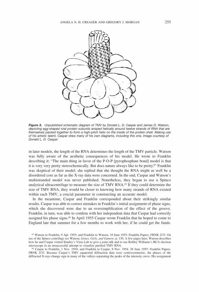
in later models, the length of the RNA determines the length of the TMV particle. Watsonwas fully aware of the aesthetic consequences of his model. He wrote to Franklindescribing it: “The main thing in favor of the P-O-P [pyrophosphate bond] model is thatit is very very pretty stereochemically. But does nature always like to be pretty?” Franklinwas skeptical of their model: she replied that she thought the RNA might as well be adisordered core as far as the X-ray data were concerned. In the end, Caspar and Watson’smultistranded model was never published. Nonetheless, they began to use a Spincoanalytical ultracentrifuge to measure the size of TMV RNA.65 If they could determine thesize of TMV RNA, they would be closer to knowing how many strands of RNA existedwithin each TMV, a crucial parameter in constructing an accurate model.
In the meantime, Caspar and Franklin corresponded about their strikingly similarresults. Caspar was able to correct mistakes in Franklin’s initial assignment of phase signs,which she discovered were due to an oversimplification of the effect of the groove.Franklin, in turn, was able to confirm with her independent data that Caspar had correctlyassigned his phase signs.66 In April 1955 Caspar wrote Franklin that he hoped to come toEngland late that summer for a few months to work with her, if he could get the funds.
65 Watson to Franklin, 9 Apr. 1955; and Franklin to Watson, 10 June 1955: Franklin Papers, FRNK 2/33. Onuse of the Spinco centrifuge see Watson, Genes, Girls, and Gamow, p. 130. A few pages later, Watson describeshow he and Caspar visited Stanley’s Virus Lab to give a joint talk and to use Robley Williams’s RCA electronmicroscope in an unsuccessful attempt to visualize purified TMV RNA.
66 Caspar to Franklin, 1 Nov. 1954; and Franklin to Caspar, 8 Nov. 1954, 28 June 1955: Franklin Papers,FRNK 2/33. Because Caspar’s TMV equatorial diffraction data were centrosymmetric, the phases of thediffracted X-rays change sign in many of the valleys separating the peaks of the intensity curve. His assignment
Figure 3. Unpublished schematic diagram of TMV by Donald L. D. Caspar and James D. Watson,depicting egg-shaped viral protein subunits arrayed helically around twelve strands of RNA that arethemselves packed together to form a high-pitch helix on the inside of the protein shell. Making useof his artistic talent, Caspar drew many of his own diagrams, including this one. Image courtesy ofDonald L. D. Caspar.
ANGELA N. H. CREAGER AND GREGORY J. MORGAN 255

She responded that he would be welcome, “although I am afraid you will find we havevery little to offer in the way of facilities and space.”67
The reason Franklin’s rather small laboratory was so crowded was that her virusresearch group doubled in size that year. First she hired John Finch as a research assistant.Then, in late summer 1955, the Ph.D. student Kenneth Holmes joined her group. Franklinput Holmes and Finch on parallel Ph.D. tracks. (Because Franklin did not have a facultyposition at the University of London, Bernal served as their nominal advisor.) Finch beganto investigate the effect of relative humidity of TMV gels, work similar to Franklin’s onDNA that established the A and B forms, but he switched to work on the spherical virusTYMV with Klug after Holmes arrived. This would be the second virus that Franklin’sgroup would study in detail.68 Holmes continued to work with TMV and would eventuallywrite his dissertation on the comparison of diffraction patterns from different strains of thevirus. Franklin was building up a full research group at Birkbeck, but to do this withouta regular faculty appointment and on soft money was risky. A grant from the AgriculturalResearch Council (ARC) supported the laboratory from 1955 through the end of 1957, butthe ARC refused to pay Franklin the full salary requested by the college. More worrisomewere the obstacles she encountered in trying to use her funding to keep Klug in her groupafter his Nuffield Foundation fellowship ended.69
By late 1955 Franklin had managed to use Schramm’s “A” protein to obtain diffractionpatterns of viral-like rods lacking any nucleic acid. After a number of attempts, shemanaged to nurse the specimens into an oriented gel without the proteins disaggregating.70
The resulting X-ray patterns showed that repolymerized “A” protein was much closer instructure to native TMV than rods formed from Commoner’s “B8” protein. In fact, itappeared to be just like the native TMV particle, but without the RNA.
In the late summer of 1955, Caspar and Watson traveled separately to Europe. Watsonhad already accepted an assistant professorship at Harvard, but a National ScienceFoundation fellowship gave him a year at the Cavendish before his move to Massachu-setts. Crick wrote Franklin ahead of Watson’s arrival to clarify areas of overlapping
of signs was confirmed by Franklin when she discovered that in the high-resolution, low-angle diffraction datathere was a small peak in the data between the first two subsidiary intensity maxima.
67 Caspar to Franklin, 9 Apr. 1955; and Franklin to Caspar, 19 May 1955: Franklin Papers, FRNK 2/33.68 In contrast to TMV, one could obtain true three-dimensional crystals from spherical plant viruses such as
TYMV and analyze their X-ray diffraction patterns using crystallographic methods rather than those of fiberdiffraction. Finch switched projects prompted by Caspar’s discovery of TYMV crystals in Harry Carlisle’srefrigerator (see below). Bernal’s role as advisor to Holmes and Finch is noted in Brown, J. D. Bernal (cit. n.38), p. 356. For more on the history of spherical virus crystallography see Gregory J. Morgan, “HistoricalReview: Viruses, Crystals, and Geodesic Domes,” Trends in Biochemical Sciences, 2003, 28:86–90; Morgan,“Early Theories of Virus Structure,” in Conformational Proteomics of Macromolecular Architecture, ed. R.Holland Cheng and Lena Hammar (Singapore: World Scientific, 2004), pp. 3–40; Morgan, “Virus Design,1955–1962: Science Meets Art,” Phytopathology, 2006, 96:1287–1291; and Morgan, “Why There Was a UsefulPlausible Analogy between Geodesic Domes and Spherical Viruses,” Hist. Phil. Life Sci., 2006, 28:215–236.
69 See Maddox, Rosalind Franklin, pp. 251–252, 256, 263. This was despite the remarkable results Franklinand Klug had obtained together. Especially impressive was their demonstration that the number of subunits perthree turns of the helix varied slightly—by hundredths of a subunit—among different strains of the virus. SeeRosalind E. Franklin and A. Klug, “The Splitting of Layer Lines in X-ray Fibre Diagrams of Helical Structures:Application to Tobacco Mosaic Virus,” Acta Crystallog., 1955, 8:777–780.
70 Aaron Klug manuscript, “Bernal and Virus Research at Birkbeck,” p. 6, Franklin Papers, FRNK 2/37; andRosalind E. Franklin, “Structural Resemblance between Schramm’s Repolymerized A-Protein and TobaccoMosaic Virus,” Biochim. Biophys. Acta, 1955, 18:313–314. Schramm’s method involved disaggregating theTMV particles in alkali, separating the protein and nucleic acid components with electrophoresis, and thenreaggregating the protein component in a mild acid. See Gerhard Schramm, “Uber die Spaltung des Tabakmo-saikvirus und die Wiedervereinigung der Spaltstucke zu hohermolekularen Proteinen, II: Versuche zur Wied-ervereinigung der Spaltstucke,” Zeitschrift fur Naturforschung, 1947, 2(B):249–257.
256 AFTER THE DOUBLE HELIX

interest. He explained that Watson “is interested in TMV from the point of view of RNAstructure and in particular is wondering whether the helical symmetries of the two parts(protein and RNA) may be related.” This line of research followed up his collaborationwith Caspar. Watson also wished to commence work on Potato virus X (PVX), anotherhelical virus that he had taken some diffraction patterns of three years earlier. In order tosecure material, he had Crick ask Roy Markham whether the Molteno Institute wouldgrow some for him.71 Markham, in turn, told Crick that Franklin had already requested apreparation of this particular virus. Since Watson was coming to his laboratory, Crick puthimself in the position of negotiating between Watson and Franklin on the matter of PVX;so as to avoid duplication of effort, he asked Franklin about her plans regarding this virus.
Franklin replied immediately: “I have always intended to work on it as soon as I can gethold of some. I have asked both Roy and Pirie for it, but got no answer from either. Atyour suggestion I shall now write again to Roy.” She was clearly not going to cede PVXto Watson. In the fall he pursued the project anyway: “In the mornings and manyevenings,” Watson recounts, “I was in the Cavendish taking X-ray photographs ofRNA-containing potato virus X. It was my response to an overnight visit from RosalindFranklin, who stayed with the Cricks. Listening to her treat Don and me as insignificantplayers in tobacco mosaic virus (TMV) research, I felt the need for another plant virus tocall my own.” If Watson saw her attitude as disparaging, this is not the view Franklinherself conveyed in correspondence. As she wrote to Paul Kaesberg at the University ofWisconsin in a letter asking for a sample of Pea streak virus, “Jim Watson (of the DNAmodel) is back in Cambridge, and is also interested in these things, and between us wewant to look at as many viruses as possible.”72 On the one hand, Franklin vigorouslyprotected her own research interests regarding precious virus samples; on the other, sherepresented herself and Watson as cooperating in their efforts to investigate the full rangeof plant viruses.
Caspar met Rosalind Franklin for the first time on 12 September 1955.73 He began towork on BSV at Cambridge, and in October he obtained evidence that this virus possessedfive-fold symmetry. Caspar also wanted to work on TYMV, another spherical virus. In hissearch for virus preparations, Caspar went to Birkbeck to rummage in the refrigerator ofBernal’s assistant Harry Carlisle, where he found BSV and TYMV crystals. However,Franklin and her group were also interested in using these crystals, and she insisted thatthe TYMV preparation remain at Birkbeck.74 In his autobiography, Watson describesCaspar as angry that she would “put herself in competition with him.” Watson claims heserved as the mediator, attempting to persuade Franklin “of the unfairness of her climbingup Don’s back.” A letter Watson wrote describing the incident suggests that the “Rosy”persona later made familiar in The Double Helix was already taking shape in his
71 Crick to Franklin, 3 June 1955, Franklin Papers, FRNK 2/33. Watson expressed an interest in PVX inWatson to Crick, 27 May 1955, Crick Papers, National Library of Medicine, Profiles in Science: http://profiles.nlm.nih.gov/SC/B/B/J/J/_/scbbjj.pdf.
72 Franklin to Crick, 6 June 1955, Franklin Papers, FRNK 2/33; Watson, Genes, Girls, and Gamow, p. 180;and Franklin to Paul Kaesberg, 18 July 1955, Franklin Papers, FRNK 2/33.
73 Interview, Morgan with Donald Caspar, Tallahassee, Fla., 4–5 Dec. 1998. Caspar recalls that together theyattended the opening performance of the Japanese Azuma Kabuki dancers and musicians in Covent Garden.
74 Watson, Genes, Girls, and Gamow, p. 183 (evidence of five-fold symmetry); and personal communication,Caspar to Morgan, 18 Apr. 2003. As Caspar more recently described the cache in Carlisle’s fridge, “Finding thesparkling BSV and TYMV crystals was like finding a hoard of diamonds in a secret cavern.” Franklin agreedthat Caspar could have the BSV crystals he found at Birkbeck. Personal communication, Caspar to Morgan, 14Nov. 2007.
ANGELA N. H. CREAGER AND GREGORY J. MORGAN 257

perceptions: “Rosy was in Cambridge yesterday. As usual I felt exhausted by the time shedeparted. Don was still mad at her for her low blow with the crystals so it was left for meto save the situation with charm and diplomacy.”75 Caspar’s memory of the crystalsincident is strikingly different; he recalls being in a bad mood upon his return that dayfrom Birkbeck, more because he accidentally burned a brand-new suit jacket on a Bunsenburner in Franklin’s laboratory while they were talking than because of her appropriationof the crystals. He recalls an amicable division of labor, such that Franklin and Klugwould work on the scarce TYMV crystals and he would continue his work on BSV.76
These negotiations over materials, territory, and credit provide evidence of a moraleconomy at work—albeit not without friction—among virus crystallographers.77
The scientific interactions that developed between Franklin and Caspar over TMVbecame congenial and highly productive.78 By comparing the radial density functions ofher repolymerized “A” protein with the native TMV density functions he had obtained,they were able to conclude that the TMV RNA was neither in the center nor 24 Å fromthe center (as Caspar and Watson had thought) but instead lay #40 Å from the center.When writing Pirie of this finding Franklin made an effort to credit the others properly:“We are working on this together with Watson and Caspar.” Franklin wanted to publishthe comparative analysis but first required that Caspar publish his results. According toCaspar, his procrastination in writing up the results of his dissertation led Franklin to drafthis paper herself. She had finished a “hasty version” by 10 February, mailing copies toWatson and Caspar that day with requests for rewriting. Franklin’s and Caspar’s papersappeared as consecutive articles in Nature on 19 May 1956.79
While in Cambridge, Watson made one last effort with Alex Rich, who was also there
75 Watson, Genes, Girls, and Gamow, p. 188; and Watson to Christa Mayr, undated [Nov. 1955] lettercirculated with an advance copy of Watson’s Genes, Girls, and Gamow (obtained courtesy of Donald Caspar).
76 Personal communication, Caspar to Morgan, 18 Apr. 2003 (burnt jacket). Caspar expressed his take on thedivision of labor: “My memory and Aaron Klug’s memory is somewhat different from Jim Watson’s memory.What we had decided was that Rosalind and Aaron would work on Turnip yellow mosaic virus crystals and Iwould carry on with work on the BSV crystals.” Interview, Morgan with Caspar, 4–5 Dec. 1998. Aaron Klugconfirms Caspar’s memory: communication with Morgan, 16 Aug. 2007.
77 Several historians of science have elaborated on how moral economies of science function, takinginspiration in various ways from E. P. Thompson’s classic usage: “The Moral Economy of the English Crowdin the Eighteenth Century” and “The Moral Economy Reviewed,” in E. P. Thompson, Customs in Common (NewYork: New Press, 1991), pp. 185–258, 259–351. See, e.g., Steven Shapin, A Social History of Truth: Civility andScience in Seventeenth-Century England (Chicago: Univ. Chicago Press, 1994); Lorraine Daston, “The MoralEconomy of Science,” Osiris, N.S., 1995, 10:3–24; and Robert E. Kohler, “Moral Economy, Material Culture,and Community in Drosophila Genetics,” in The Science Studies Reader, ed. Mario Biagioli (New York:Routledge, 1999), pp. 243–257. We draw predominantly on Kohler’s model, given our interest in the circulationof materials and allocation of credit. Along these lines see also Anderson, “Possession of Kuru” (cit. n. 13); andPaula Findlen, “The Economy of Scientific Exchange in Early Modern Italy,” in Patronage and Institutions:Science, Technology, and Medicine at the European Court, 1500–1750, ed. Bruce T. Moran (Rochester, N.Y.:Boydell, 1991), pp. 5–24. Needless to say, our pointing to a moral economy at work does not imply that all ofthe participants were virtuous or exemplary; Watson and Crick’s failure fully to credit Franklin’s experimentaldata on DNA in their 1953 paper was an abrogation of the usual conventions even at the time and remains atroubling aspect of the history.
78 Caspar had sent Franklin his Ph.D. dissertation in June 1955, before he went to England.79 Franklin to Pirie, 3 Feb. 1956, Franklin Papers, FRNK 2/33. Information regarding Franklin’s initial drafting
of Caspar’s paper comes from interview, Morgan with Caspar, 4–5 Dec. 1998. See also Franklin to Watson, 10Feb. 1956, Jeremy Norman Collection (this collection—hereafter cited as Norman Collection—was in SanFrancisco when Morgan used it but has since been auctioned); Watson quotes her cover letter in full in Genes,Girls, and Gamow, p. 203. The published versions of the papers are Donald L. D. Caspar, “Radial DensityDistribution in the Tobacco Mosaic Virus Particle,” Nature, 1956, 177:928; and Rosalind E. Franklin, “Locationof the Ribonucleic Acid in the Tobacco Mosaic Virus Particle,” ibid., pp. 928–930.
258 AFTER THE DOUBLE HELIX

working with Crick for six months, to decipher the X-ray pattern of RNA.80 His ambitionto work out the arrangement of RNA in TMV was sidelined by Franklin’s determinationof its placement within the virus protein. Her finding also suggested that the structure ofviral RNA was likely entirely different from that of RNA in vitro, making the relevanceof the structure of the latter less clear.81 Partly in response to the rapid progress of others(especially Franklin and Caspar) on plant virus structure, Watson and Crick resumed atheoretical project they had already begun, synthesizing current results to come up with ageneral theory of virus structure. Their joint paper, published alongside a communicationfrom Caspar on BSV’s five-fold symmetry, appeared in Nature on 10 March 1956. Theyobserved that “almost all small viruses are either rods or spheres” and raised the questionof why this is so. “The purpose of this article is to explain this observation by means ofthe following simple hypothesis: a small virus contains identical sub-units, packed to-gether in a regular manner. It has been suggested before that viruses are constructed fromsub-units; but the idea has not previously been described in precise terms or put forwardas a general feature of all small viruses.” The best structural evidence for Crick andWatson’s theory that every virus is composed of identical subunits came from plantviruses, both helical viruses like TMV and spherical viruses such as BSV and TYMV.Symmetry considerations alone, in their view, suggested that all viruses are constructedfrom structural subunits that surround the nucleic acid. Relatedly, simple constructionrules constrain the number of viral structures that could be built. The elegant simplicity ofthis idea prompted the witticism, attributed to Crick, that “any child could make a virus.”82
In the case of TMV, both biochemical and X-ray diffraction analysis had already shownthe virus to be composed of identical protein subunits arranged symmetrically in a helix.For spherical viruses, Crick and Watson emphasized Caspar’s finding that BSV possessesicosahedral, or 532, symmetry, meaning that the virus particle exhibits rotational sym-metry along two-fold, three-fold, and five-fold axes.83 TYMV, Crick and Watson ob-served, had also been found to possess cubic symmetry—that is, four three-fold rotationalaxes. They extrapolated that other spherical viruses did as well, for any viral assemblywith cubic symmetry must be “built up by the regular aggregation of small asymmetricalbuilding blocks.” (Caspar wrote that BSV was “built up of sixty structurally identicalasymmetric units,” even as preliminary biochemical evidence indicated up to threehundred chemical subunits.)84 Crick and Watson ventured that viral ribonucleic acid might
80 Watson, Genes, Girls, and Gamow, pp. 189–190. On Rich’s stay in Cambridge and his work there withCrick on the triple-helical structure of collagen see Alexander Rich, “Fifty Years with Double-Stranded RNA,”Scientist, 2006, 20:34–39.
81 Watson, Genes, Girls, and Gamow, pp. 202–203.82 F. H. C. Crick and J. D. Watson, “Structure of Small Viruses,” Nature, 1956, 177:473–475, on p. 473;
D. L. D. Caspar, “Structure of Bushy Stunt Virus,” ibid., pp. 475–476; and Crick, as quoted by Aaron Klug inhis historical introduction to “Session I: Particle Structure,” at “Symposium on Tobacco Mosaic Virus:Pioneering Research for a Century,” sponsored by the Royal Society of Edinburgh in association with the RoyalSociety of London, 7 Aug. 1998, Edinburgh. Klug dates the remark to the late 1950s: personal communicationto Creager, 17 Apr. 2001. Caspar dates Crick’s remark to around 1955; see D. L. D. Caspar, “Movement andSelf-Control in Protein Assemblies: Quasi-Equivalence Revisited,” Biophysical Journal, 1980, 32:103–138.
83 A few familiar objects possess the same kind of symmetry. E.g., a soccer ball constructed from twelve blackpentagons and twenty white hexagons has 532 symmetry: five-fold symmetries though the centers of thepentagons, three-fold symmetries through the centers of the hexagons, and two-fold symmetries through theedges between the hexagons. Dorothy Hodgkin had earlier speculated that BSV has cubic symmetry but had notdrawn general conclusions; see Dorothy Crowfoot Hodgkin, “X-ray Analysis and Protein Structure,” ColdSpring Harbor Symposia on Quantitative Biology, 1950, 14:65–78. For more detail on Caspar’s work seeMorgan, “Early Theories of Virus Structure” (cit. n. 68).
84 Crick and Watson, “Structure of Small Viruses” (cit. n. 82), p. 474; and Caspar, “Structure of Bushy Stunt
ANGELA N. H. CREAGER AND GREGORY J. MORGAN 259

also be made up of smaller identical subunits, but they did not attempt to explain how thegenetic role of RNA was related to its structural role.
The goal of determining the structure of viral RNA and its relation to protein was alsoof central importance to Franklin and her group. TMV was the central model for thiseffort, for both the RNA and the protein components could be isolated, making itconceivable that the correlation of nucleic acid and protein might be determined chemi-cally. As Franklin wrote in March 1956 in an attempt to obtain continued ARC funding:“[Our] work is concerned with what is probably the most fundamental of all questionsconcerning the mechanism of living processes, namely the relationship between proteinand nucleic acid in the living cell. . . . The plant viruses consist of ribonucleic acid andprotein, and provide the ideal system for the study of the in vivo structure of bothribonucleic acid and protein and of the structural relationship of the one to the other.”85
To her chagrin, her ARC funding was renewed for only one final year, despite theappeals of other virologists, including Watson.86 However, in 1957 she secured a three-year grant from the U.S. Public Health Service, at a level of £10,000 per year. As RobleyWilliams commented to Francis Crick, the merit of her grant hinged on the “uniquenesscriterion,” satisfied by the “dearth elsewhere” of structural studies of viruses like thosebeing conducted by Franklin and her coworkers.87
THE CIBA FOUNDATION SYMPOSIUM ON THE NATURE OF VIRUSES
In the three years since Watson proposed a helical structure, X-ray diffraction methodsand analyses had brought the overall structure of TMV into clear view. The location of theRNA was established, the parameters of the TMV helix were known with a reasonabledegree of confidence, and the number of subunits per three turns of the helix had beencorrected to its current value of forty-nine using chemical and structural reasoning.88
Virus” (cit. n. 82), p. 476. These results are not inconsistent if each structural subunit consists of multiplechemical subunits.
85 Franklin, “Note on the Future of the A.R.C. Research Group in Birkbeck College CrystallographyLaboratory,” 9 Mar. 1956, Franklin Papers, FRNK 2/36.
86 In the letter quoted above concerning the incident of Caspar and the TYMV crystals, Watson stated: “Thismorning was spent persuading Victor Rothschild that Rosy should be supported in spite of her continued insultsto the administrative heads of the Agriculture Research Council. This weekend I must write a long report to besubmitted to ARC, otherwise she will hopelessly flounder without adequate financial support.” Watson to Mayr,undated [Nov. 1955] letter circulated with an advance copy of Watson’s Genes, Girls, and Gamow. In hisautobiography, Watson describes how he spoke on behalf of Franklin’s needs to Rothschild earlier that summeras well and reprints a letter he wrote to Franklin with advice on how to secure funds for a much-needed upgradeto her crystallography equipment: Watson, Genes, Girls, and Gamow, pp. 154–156.
87 Williams to Crick, 20 Sept. 1957, Williams Papers, carton 4, folder C. On the grant from the Public HealthService see Maddox, Rosalind Franklin, pp. 290–292. Williams provided advice to Franklin on her application.See Franklin to Williams, 19 Oct. 1956; Williams to Franklin, 24 Oct. 1956; and Franklin to Williams, 6 Dec.1956: Williams Papers, carton 4, folder F. Franklin immediately used the American funds to pay for a researchassistant at the Molteno Institute to grow and purify more TYMV for crystals, which were in short supply:Franklin to Kenneth Smith, 17 July 1957, and Smith to Franklin, 16 Oct. 1957, Norman Collection. Franklin hadrequested a letter from Watson supporting her application to the U.S. Public Health Service; her letter to Watsonindicates how instrumental Williams was in helping her apply successfully and how crucial the American grantwas to enable her and Klug to continue working together. She also mentioned that, since her return from theUnited States, “I’ve spent all my time being ill”: Franklin to Watson, 14 Nov. [1956], Watson Papers.
88 Franklin and Holmes looked for the influence of the mercury atom on the outer parts of the zero layer lineand were able to infer that n $ 16 was the correct solution to Watson’s equation (i.e., 3n " 1 $ 49): KennethC. Holmes, personal communication to Morgan, 24 Dec. 2006. See Rosalind E. Franklin and Kenneth C.Holmes, “Tobacco Mosaic Virus: Application of the Method of Isomorphous Replacement to the Determinationof the Helical Parameters and Radial Density Distribution,” Acta Crystallog., 1958, 11:213–220. On the state of
260 AFTER THE DOUBLE HELIX

Franklin’s leadership in this area was impossible to deny, and Watson commended hercontributions in a letter to Stanley:
Rosalind Franklin’s x-ray analysis of TMV is becoming more and more exciting. The RNAseems now to be [at] a radius of 42 Å, not 24 Å as Caspar and I originally suspected. Also thesubunit number has been revised. Using Fraenkel-Conrat’s Hg"" substituted TMV, she nowhas good evidence that there are 49 subunits/3 turns and so there is good reason for believingthat only one chemical subunit is present per crystallographic unit. If so the subunit number is#2150 and the TMV molecular weight around 40 million.
It is really quite fascinating how the facts are beginning to fit together.89
The time was opportune for Franklin to sum up the remarkable progress in the structuraldetermination of TMV, and an exclusive Ciba Foundation meeting in London in March1956 provided the occasion.
Founded in 1949, the Ciba Foundation held small, informal meetings of elite research-ers from around the world in its elegant establishment at 41 Portland Place. In the viewof originator Frank G. Young, an unstated goal for the March 1956 symposium on “TheBiophysics and the Biochemistry of Viruses” was to “revivify” virology in England. Themajor centers for basic viral research were represented: Cambridge, Berkeley, Birkbeck,and Tubingen. Franklin petitioned to have Schramm invited, but the Tubingen Max PlanckInstitute for Virus Research was represented by Werner Schafer instead. Maurice Wilkinswas scheduled to attend, but it appears that at the last minute his place was taken by AaronKlug.90 Of the thirty-four participants, six went on to win Nobel prizes.
The Ciba symposium on viruses proved to be a meeting of the old and the new. Amongthe representatives of the “new” were Crick, Watson, Franklin, Caspar, Klug, and otherswho were convinced that the application of new physical techniques would transformbiological research and knowledge. The “old” were established virologists of an earliergeneration, who were often medically trained and were accustomed to relying on immu-nological and biochemical techniques. The organizers of the meeting, Marinus van denEnde and Young, sought to bring these two groups together in the hope of meaningfulexchange. The differing responses to the meeting’s most startling news, Robley Wil-liams’s assertion that TMV nucleic acid alone was infectious, showed how much of a gapexisted between the two groups of participants.91 To the surprise of Watson and Crick,
knowledge in 1956 see Franklin to Pirie, 3 Feb. 1956, Franklin Papers, FRNK 2/33. One missing piece ofinformation was that the handedness of the TMV helix was not revealed by diffraction data.
89 Watson to Stanley, 27 Jan. 1956, Stanley Papers, carton 13, folder Wa Misc. There are reasons to becautious about taking this letter at face value, even though we find Watson’s assent to Franklin’s standing in thefield to be notable. Stanley was a major power broker in the field, and Franklin was collaborating closely withmembers of his lab. At the same time, Watson did not find Stanley’s Virus Lab very scientifically stimulating;see Watson to Crick, 11 Dec. 1954, Crick Papers, National Library of Medicine, Profiles in Science: http://profiles.nlm.nih.gov/SC/B/B/J/N/_/scbbjn.pdf. Moreover, he had been recommended for an opening there butfailed to receive an offer; see Watson to Gunther S. Stent, 9 Dec. 1954, Gunther S. Stent Papers, BancroftLibrary, University of California, Berkeley, 99/149z, box 15, folder Watson, J. D.
90 On the symposium as an effort to revivify virology in England see G. E. W. Wolstenholme to Franklin, 21June 1955, Franklin Papers, FRNK 2/34; and Frank Macfarlane Burnet to Lady Burnet, 13 Mar. 1956, FrankMacfarlane Burnet Papers, University of Melbourne Archives, 2/18. For Franklin’s attempt to include Schrammsee Franklin to Wolstenholme, 28 June 1955, Franklin Papers, FRNK 2/34. The Ciba/Novartis scrapbook (heldat the Ciba Foundation, London) has a printed list of participants in which Wilkins’s name is crossed out andKlug’s is written in by hand.
91 “It now begins to appear (Fraenkel-Conrat, 1956) that infection can be obtained from solutions in whichessentially no full-length TMV particles, either native or reconstituted, are present. The active solutions arebelieved to be pure RNA, and are infectious when rubbed upon tobacco plants in sufficiently high concentra-tions”: Robley C. Williams, “Structure and Substructure of Viruses as Seen under the Electron Microscope,” in
ANGELA N. H. CREAGER AND GREGORY J. MORGAN 261

some of the participants did not appreciate the gravity of Williams’s finding or found ithard to accept.92 In the question session, Bawden asked for quantitative results and moreexperiments to control for other enhancing or inhibiting substances in the inoculum:“Unless we have answers to such questions as these, how are we to know what value toattach to the exciting statement at the end of your talk?”93 Watson later attributed thedifference to the failure of older researchers to understand a key tenet of molecularbiology: “They were not at home with the concept that information flows unidirectionallyfrom nucleic acids to proteins and never backwards.”94
Delivering their talk after Williams, Franklin and her colleagues Klug and Holmesunveiled a more detailed proposal for the structure of TMV (see Figure 4).95 Unlike theearlier 1955 illustration, this model depicted the hollow core, the corrected number ofprotein subunits per turn, and the radial location of RNA at 40 Å. The group’s results alsoshowed the high degree of regularity in the structural repeats of the TMV protein helix.Both Watson and Crick contributed to the discussion after Franklin’s paper; Watsonpointed out that the X-ray diffraction evidence was just as consistent with many RNAstrands as with one per TMV particle.
Crick and Watson also presented a paper at the Ciba Foundation conference, articulat-ing general principles for the construction of spherical viruses. They built on theirjust-published joint paper in Nature by asking why viruses were made of identicalsubunits at all. Their explanation focused on the limited information that could be storedin a relatively small viral genome. The way to build a virus given this constraint, theyargued, was to make a large number of identical protein subunits from the same infor-mation encoded in the viral genome and then assemble them into a structure in which eachsubunit occupies the same environment as any other. Two types of structures could resultfrom such an assembly process: helical rods such as TMV and spherical structures withcubic symmetry such as BSV. Crick and Watson suggested the possibility that the“arrangement of the RNA may be practically the same in all spherical viruses . . . sincethe ways of folding a fibrous molecule so that it has cubic symmetry may be ratherlimited.”96 Both because of its genetic properties and because it might solve the codingproblem, plant viral RNA was a key object of research. As Watson had written to Crick
Ciba Foundation Symposium on the Nature of Viruses, ed. G. E. W. Wolstenholme and Elaine C. P. Millar(Boston: Little, Brown, 1957), pp. 19–33, on p. 31. The observation by Heinz Fraenkel-Conrat appeared in “TheRole of the Nucleic Acid in the Reconstitution of Active Tobacco Mosaic Virus,” J. Amer. Chem. Soc., 1956,78:882–883; but Alfred Gierer and Gerhard Schramm made the same discovery independently and gave itgreater prominence in “Infectivity of Ribonucleic Acid from Tobacco Mosaic Virus,” Nature, 1956, 177:702–703.
92 By the same token, Watson has described a hoax he played on Williams, falsifying a telegram from Stanleyconveying the news that the TMV protein was infectious; Watson claims that Williams downplayed his newresults with nucleic acid in his Ciba Foundation symposium talk as a result: Watson, Genes, Girls, and Gamow,p. 217.
93 F. C. Bawden, in discussion following Williams, “Structure and Substructure of Viruses as Seen under theElectron Microscope,” in Ciba Foundation Symposium on the Nature of Viruses, ed. Wolstenholme and Millar(cit. n. 91); the remark appears on p. 35. Pirie argued in the same discussion that Williams should not restricthis search for TMV particles to those 2,000–3,000 Å long, as he might be missing smaller infective units (ibid.,p. 36).
94 Watson, Passion for DNA (cit. n. 40), p. 28. This tenet was codified by Crick as the “Central Dogma.” SeeJudson, Eighth Day of Creation (cit. n. 10), pp. 332–336; and Strasser, “World in One Dimension” (cit. n. 11).
95 Rosalind E. Franklin, A. Klug, and K. C. Holmes, “X-ray Diffraction Studies of the Structure andMorphology of Tobacco Mosaic Virus,” in Ciba Foundation Symposium on the Nature of Viruses, ed.Wolstenholme and Millar (cit. n. 91), pp. 39–52.
96 F. H. C. Crick and J. D. Watson, “Virus Structure: General Principles,” in Ciba Foundation Symposium onthe Nature of Viruses, ed. Wolstenholme and Millar, pp. 5–13, on p. 12.
262 AFTER THE DOUBLE HELIX
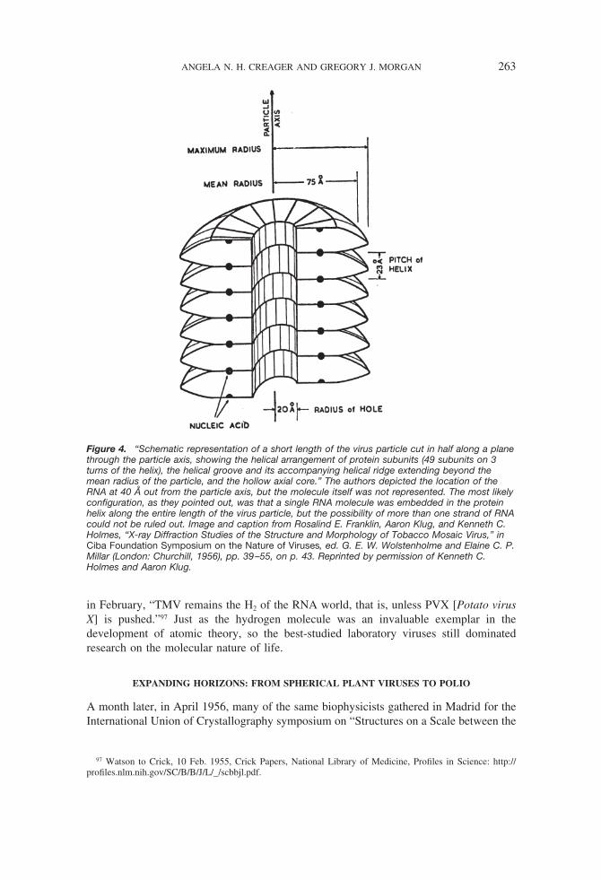
in February, “TMV remains the H2 of the RNA world, that is, unless PVX [Potato virusX] is pushed.”97 Just as the hydrogen molecule was an invaluable exemplar in thedevelopment of atomic theory, so the best-studied laboratory viruses still dominatedresearch on the molecular nature of life.
EXPANDING HORIZONS: FROM SPHERICAL PLANT VIRUSES TO POLIO
A month later, in April 1956, many of the same biophysicists gathered in Madrid for theInternational Union of Crystallography symposium on “Structures on a Scale between the
97 Watson to Crick, 10 Feb. 1955, Crick Papers, National Library of Medicine, Profiles in Science: http://profiles.nlm.nih.gov/SC/B/B/J/L/_/scbbjl.pdf.
Figure 4. “Schematic representation of a short length of the virus particle cut in half along a planethrough the particle axis, showing the helical arrangement of protein subunits (49 subunits on 3turns of the helix), the helical groove and its accompanying helical ridge extending beyond themean radius of the particle, and the hollow axial core.” The authors depicted the location of theRNA at 40 Å out from the particle axis, but the molecule itself was not represented. The most likelyconfiguration, as they pointed out, was that a single RNA molecule was embedded in the proteinhelix along the entire length of the virus particle, but the possibility of more than one strand of RNAcould not be ruled out. Image and caption from Rosalind E. Franklin, Aaron Klug, and Kenneth C.Holmes, “X-ray Diffraction Studies of the Structure and Morphology of Tobacco Mosaic Virus,” inCiba Foundation Symposium on the Nature of Viruses, ed. G. E. W. Wolstenholme and Elaine C. P.Millar (London: Churchill, 1956), pp. 39–55, on p. 43. Reprinted by permission of Kenneth C.Holmes and Aaron Klug.
ANGELA N. H. CREAGER AND GREGORY J. MORGAN 263
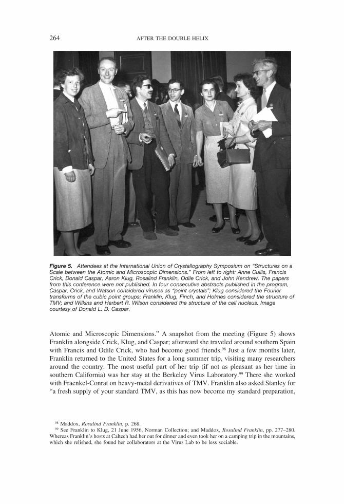
Atomic and Microscopic Dimensions.” A snapshot from the meeting (Figure 5) showsFranklin alongside Crick, Klug, and Caspar; afterward she traveled around southern Spainwith Francis and Odile Crick, who had become good friends.98 Just a few months later,Franklin returned to the United States for a long summer trip, visiting many researchersaround the country. The most useful part of her trip (if not as pleasant as her time insouthern California) was her stay at the Berkeley Virus Laboratory.99 There she workedwith Fraenkel-Conrat on heavy-metal derivatives of TMV. Franklin also asked Stanley for“a fresh supply of your standard TMV, as this has now become my standard preparation,
98 Maddox, Rosalind Franklin, p. 268.99 See Franklin to Klug, 21 June 1956, Norman Collection; and Maddox, Rosalind Franklin, pp. 277–280.
Whereas Franklin’s hosts at Caltech had her out for dinner and even took her on a camping trip in the mountains,which she relished, she found her collaborators at the Virus Lab to be less sociable.
Figure 5. Attendees at the International Union of Crystallography Symposium on “Structures on aScale between the Atomic and Microscopic Dimensions.” From left to right: Anne Cullis, FrancisCrick, Donald Caspar, Aaron Klug, Rosalind Franklin, Odile Crick, and John Kendrew. The papersfrom this conference were not published. In four consecutive abstracts published in the program,Caspar, Crick, and Watson considered viruses as “point crystals”; Klug considered the Fouriertransforms of the cubic point groups; Franklin, Klug, Finch, and Holmes considered the structure ofTMV; and Wilkins and Herbert R. Wilson considered the structure of the cell nucleus. Imagecourtesy of Donald L. D. Caspar.
264 AFTER THE DOUBLE HELIX

and if I have to change to Cambridge or any other preparation I should have to repeat alarge amount of work on the basic measurements.”100
Perhaps the most surprising results she saw in Berkeley were some electron micro-graphs taken by Russell Steere. He used a freeze-shadowing replica technique that wasbetter able to illuminate the substructure of TYMV, the virus that her colleagues Klug andFinch were working on back in London. Franklin wrote an excited letter to Klug about themicrographs.101 If TYMV particles had 532 symmetry and consisted of shells with sixtysubunits, then Franklin assumed one would see a significant fraction of rings of five“knobs.” To her surprise, she did not. Three days later she sent Klug some of the electronmicrographs and a more considered opinion: “I have looked at these and others—particularly ones which show particles in a wide variety of orientations, and there seemsvery little doubt that the knobs lie at the vertices of a cubeoctahedron, i.e., 12 knobs. . . .Among hundreds of particles I have only found 2 that look remotely 5-folded and Icertainly do not believe they are all like that.” She wrote a similar letter to Caspar on thesame day. If TYMV resembled a cubeoctahedron—a semiregular solid formed by trun-cating the corners of a cube—then its symmetry differed markedly from that of BSV, theother well-studied spherical virus.102 Franklin and her coworkers were unable to reconcilethis interpretation with their results.
By December Klug and Franklin had a paper on TYMV ready for publication thatFrancis Crick thought “reads very well indeed.” Crick suggested that they publish inBiochimica et Biophysica Acta rather than Nature because the article was long andtechnical in nature. Klug, Finch, and Franklin submitted a shorter and less technical paperto Nature on 11 January 1957. This paper presented the crystal structure of TYMV andsuggested that, like BSV, TYMV possessed 532 symmetry. A month later the threesubmitted the longer paper to Biochimica et Biophysica Acta.103 In the penultimate sectionthey addressed Steere’s electron micrographs, suggesting that the high percentage ofammonium sulfate (50 percent by weight) in the samples he used might have introducedartifacts not present in the X-ray crystallography. Both electron micrographs taken byKaesberg using a different technique (shadow casting) and their own work indicated thatthe symmetry was 532 or icosahedral.
At this time, biophysicists were intrigued by the morphological and biochemical
100 Franklin to Stanley, 4 July 1956, Stanley Papers, carton 8, folder Franklin, Rosalind. Franklin’s thank-younote to Stanley, written on 30 Aug. (and in the same folder of the Stanley Papers), makes it clear that she stayedin Berkeley for three weeks.
101 “The most important thing here is that I’ve recently seen some electron micrographs of TYM by Steere’sfreezing-shadowing replica technique which show a magnificently clear surface structure. This only happenedtoday, and seems very exciting, so excuse the muddle. The prominent feature is an array of six knobs on eachparticle around a central one. . . . The inter-knob distance is #1/4 inter-particle distance, which is consistent withyour 60 Å, but the thing does not look 5 folded. . . . If it is not 5-folded, the question arises, was the fivefoldedness in the RNA. . . . Believe it or not, he [Robley Williams] says he had a slide of this with him at Cibabut did not show it in case the effect was due to amm. sulphate!!” Franklin to Klug, 27 July 1956, NormanCollection.
102 Franklin to Klug, 30 July 1956, Norman Collection. A cubeoctahedron has 432 symmetry, not the 532symmetry of an icosahedron such as BSV. A week later, Franklin was still thinking about Steere’s electronmicrographs, but now from the point of view that the photographs pertained to the determination of the unit cellof the crystal: Franklin to Klug, 5 Aug. 1956, Norman Collection.
103 Crick to Klug, 14 Dec. 1956, Norman Collection; A. Klug, J. T. Finch, and Rosalind E. Franklin, “Structureof Turnip Yellow Mosaic Virus,” Nature, 1957, 179:683–684; and Klug, Finch, and Franklin, “The Structure ofTurnip Yellow Mosaic Virus: X-ray Diffraction Studies,” Biochim. Biophys. Acta, 1957, 25:242–252. For moreon the symmetry of TYMV see A. Klug and J. T. Finch, “The Symmetries of the Protein and Nucleic Acid inTurnip Yellow Mosaic Virus: X-Ray Diffraction Studies,” J. Molec. Biol., 1960, 2:201–215.
ANGELA N. H. CREAGER AND GREGORY J. MORGAN 265

similarities between RNA-containing spherical viruses and microsomes (now calledribosomes), the roundish particles of protein and RNA found in the cytoplasm andimplicated in protein synthesis.104 As Watson recalled in 1962, “All RNA was thought toexist either as a viral component or be combined with protein in ribonucleoproteinparticles.” Klug was keen to extend their studies of spherical viruses to microsomes—andin particular to determine if the ribonucleoprotein particles were composed of identicalsubunits. He alerted Bernal to the fact that the structural determination of microsomes wasan area of intense competition: “we already have very good powder diagrams of TYMVtaken on the focusing camera, from which it is possible to deduce that the particles aremade up of subunits. We could do the same for microsomes if we had some material, andI think it would be quite a scoop if they turned out to have this sort of sub-structure.”Caspar, Crick, and Watson were all interested in pursuing this possibility.105 Franklin usedher California connections to obtain material from Howard Schachman at Berkeley (yeastmicrosomes) and Jerome Vinograd at Caltech (pea seedling microsomes). ComparingX-ray powder diagrams of microsomes with those of plant viruses, Franklin’s group foundcertain similarities. In particular, the structure of the RNA in microsomes and viruses wascompletely different from that of isolated RNA. Her group concluded that “the structureis essentially determined by a well-defined protein matrix in the interstices of which liesthe RNA.” This seemed in contrast to DNA-protein assemblies, for which the protein“conforms to the structural configuration of the nucleic acid.”106 The observed structuralarrangements did not resolve the coding problem. Even so, the rapid achievement ofresults with microsomes at Birkbeck attests to Franklin’s ability to procure precioussamples from far-flung collaborators.
In 1957 Franklin and her coworkers Klug and Finch began to study poliovirus inaddition to their continuing work on TYMV. Poliovirus was the first animal viruscrystallized, in work done by Frederick Schaffer and Carleton Schwerdt of the BerkeleyVirus Laboratory in 1955. Crystals large enough for single-crystal X-ray diffraction tooka year to grow. The Berkeley researchers gave a sample of these to Franklin in 1957.107
Some of the people working in the Birkbeck laboratories were not pleased that Franklinwas working with poliovirus, believing that it posed an unacceptable risk of humaninfection. They convened a meeting regarding her use of poliovirus at Birkbeck, with the
104 On the history of research on cytoplasmic particles see Nicolas Rasmussen, “Mitochondrial Structure andthe Practice of Cell Biology in the 1950s,” J. Hist. Biol., 1995, 28:381–429; Hans-Jorg Rheinberger, “Com-paring Experimental Systems: Protein Synthesis in Microbes and in Animal Tissue at Cambridge (Ernest F.Gale) and at the Massachusetts General Hospital (Paul C. Zamecnik), 1945–1960,” ibid., 1996, 29:387–416;Rheinberger, “Cytoplasmic Particles in Brussels (Jean Brachet, Hubert Chartrenne, Raymond Jeener) and atRockefeller (Albert Claude), 1935–1955,” Hist. Phil. Life Sci., 1997, 19:47–67; and Rheinberger, Toward aHistory of Epistemic Things: Synthesizing Proteins in the Test Tube (Stanford, Calif.: Stanford Univ. Press,1997).
105 Watson, “Involvement of RNA in the Synthesis of Proteins” (cit. n. 39), p. 787; Klug to Bernal, 31 May1956, Norman Collection (emphasis in original); and Franklin to Klug, 17 July 1956, Norman Collection(interest of Caspar, Crick, and Watson). On the analogy between spherical viruses and microsomes see Crick andWatson, “Virus Structure” (cit. n. 96), p. 12.
106 Franklin to Klug, 27 July 1956, Norman Collection (regarding Franklin’s California connections forobtaining material); and Rosalind E. Franklin, A. Klug, J. T. Finch, and K. C. Holmes, “On the Structure of SomeRibonucleoprotein Particles,” Discussions Faraday Soc., 1958, 25:197–198.
107 F. L. Schaffer and C. E. Schwerdt, “Crystallization of Purified MEF-1 Poliomyelitis Virus Particles,” Proc.Nat. Acad. Sci., USA, 1955, 41:1020–1023. For a discussion of their work see Creager, Life of a Virus, Ch. 5.Polio crystals were discussed at the International Poliomyelitis Conference in Geneva in July 1957, whereFranklin heard Schwerdt give a talk on the crystals he had grown: Franklin notebook entry, 10 July 1957,Franklin Papers, FRNK 3/14.
266 AFTER THE DOUBLE HELIX

result that Franklin was prohibited from using the virus in her lab. The crystals were thentaken from Birkbeck to the London School of Hygiene and Tropical Medicine, which hadbetter facilities for dealing with highly infectious pathogens and was a safer place tomount the crystals. Franklin also needed a powerful X-ray tube if she was to takediffraction pictures. Klug contacted Bragg at the Royal Institution, which by then had arotating anode X-ray tube copied from the design of the tube at Cambridge. Bragg wasquite receptive to the work’s being done at the Royal Institution; Klug had to getpermission to work with an infectious virus, but there were no formal guidelines and sohe invented his own and submitted them to an inspector.108
Franklin’s attempts to mount the polio crystals in capillary tubes in early to mid-1957were unsuccessful. Each crystal would spontaneously dissolve in the capillary before shecould get any diffraction pictures. Franklin attributed this instability to an alkaline reactionoccurring in the borosilicate glass of the capillary, such that salts were leaching out of theglass. She then tried using acid-treated capillaries, which delayed the dissolution of thecrystals but not enough for her to get any data. In the month before her death she wroteto Bawden suggesting that Pyrex tubes might be better. Franklin also corresponded withRonald W. Douglas, of the Department of Glass Technology at Sheffield, who recom-mended that she use “neutral glass.” Franklin did not live long enough to see the resultsof the poliovirus project, but her colleagues continued the work. With the arrival of a newbatch of crystals from Berkeley, Klug discovered that quartz capillary tubes worked muchbetter than glass ones and allowed the virus crystals to be mounted without dissolving.When crystals were at last successfully mounted in capillaries, they were transportedacross town, to the Davy Faraday Laboratory of the Royal Institution, in a thermos. Finchand Klug found that poliovirus, like BSV and TYMV, possesses icosahedral symmetry,showing that spherical viruses from plants and animals exhibit the same structuralorganization.109
In the summer of 1956, Sir Lawrence Bragg had written several biophysicists to enlisttheir help in preparing exhibits for the upcoming 1958 World’s Fair in Brussels. As he toldCrick, the organizers wanted “to make a big feature of the nucleic acid in the biologysection. Wilkins will have to be brought in too and I am hoping to borrow his model orsome later version of it.” He continued,
The organizers of the biology section also want to make an important show of the work on thehelical and spherical viruses. I am writing to Miss Franklin but you and Watson come in heretoo, so it will be a case of general collaboration. I have been so deeply impressed by all thegrand work you have done on these complex structures and I wish to see that it has a properplace in the Exhibition.110
108 Interview, Morgan with John Finch, Cambridge, 18 July 2000.109 For correspondence regarding the attempts to mount polio crystals see Franklin to Caspar, 16 Mar. 1958;
Franklin to Bawden, 20 Mar. 1958; and Franklin to R. W. Douglas, 24 Mar. 1958: Franklin Papers, FRNK 2/33.The results were published in J. T. Finch and A. Klug, “Structure of Poliomyelitis Virus,” Nature, 1959,183:1709–1714.
110 W. L. Bragg to Crick, 26 June 1956, William Lawrence Bragg Papers, Royal Institution, London (hereaftercited as Bragg Papers), 83P/1. Bragg was appointed by the president of the Royal Society (Cyril Hinshelwood)to serve as the U.K. representative on the Scientific Committee for the Brussels exhibition: Bragg Papers, 83G/1,2, 3. Bragg also wrote Frederick Sanger about his interest in including his work on insulin chains and to Perutzabout his wish to include X-ray crystallographic work on proteins and Huxley’s work on muscle: Bragg Papers,83P/3, 84A/1.
ANGELA N. H. CREAGER AND GREGORY J. MORGAN 267
As it developed, the exhibits were less a “case of general collaboration” than a matter forcareful coordination, with delicate negotiations about which contributions would bejointly prepared.111 Wilkins offered to contribute, with Crick, a model of DNA; and Cricksaid he would take care of collagen. Their letters expose the tensions that arose whenscientists from King’s College and Cambridge were asked to collaborate on presentingnucleic acid structure. Franklin’s name never came up in connection with the DNAmodels, but she and Klug contributed an exhibit of large-scale structural models of TMVand TYMV.112 Once built, this model of a segment of the TMV helix was over five feethigh (see cover and Figure 6).
On 16 April 1958, just as the Brussels exhibition opened to international acclaim,Franklin succumbed to cancer. That summer Caspar presented a paper coauthored withKlug at the fiftieth anniversary meeting of the American Phytopathological Society inplace of Franklin. They wrote a review article of X-ray diffraction studies of virusesdrawing largely from Franklin’s work and posthumously added her as first author. Theirsubsequent collaboration culminated in the Caspar-Klug theory of virus structure, aproposal that extended Crick and Watson’s earlier speculation by suggesting that sphericalviruses were structured like microscopic geodesic domes.113 Klug continued his work onTMV and spherical viruses and developed a general method for three-dimensional imagereconstruction. The scientific recognition he received included the 1982 Nobel Prize inChemistry. He dedicated approximately a third of his Nobel lecture to the structure anddynamics of TMV. As he commented, Franklin herself might have stood on that platform“had her life not been cut tragically short.”114
CONCLUSIONS
The history of structural studies of viruses provides a useful vantage point for assessingthe consolidation of molecular biology in the 1950s, one that sheds new light on thebetter-known double-helix episode. At a personal level, the relations among Watson,Crick, and Franklin around DNA should be viewed as part of a longer historical trajectory.In fact, their interactions intensified after 1953. After leaving King’s College Franklinviewed Watson and Crick not as enemies but as colleagues and, at times, competitors.Crick became a friend.115 Her relations with Watson were clearly more complicated. After1953, Watson hoped that his new work elucidating the structure of RNA would clarify the
111 E.g., Crick noted in his response to Bragg’s letter: “Miss Franklin will cover viruses and Wilkins will, Ithink, be responsible for DNA. I should be quite happy to look after collagen, but I think that to collaborate withKing’s on this would cause unnecessary friction.” Crick to Bragg, 8 Dec. 1956, Bragg Papers, 83P/37.
112 See Bragg to Franklin, 26 June 1956, Bragg Papers, 85B/164; Franklin to Bragg, 23 July 1956, BraggPapers, 85B/165; and copies of correspondence in the same box between Franklin and D. C. Phillips. This TMVmodel, whose design and construction were overseen by Klug, ended up at the Cambridge MRC Laboratory.
113 Rosalind E. Franklin, D. L. D. Caspar, and A. Klug, “The Structure of Viruses as Determined by X-rayDiffraction,” in Plant Pathology: Problems and Progress, 1908–1958, ed. C. S. Holton et al. (Madison: Univ.Wisconsin Press, 1959), pp. 447 461; and Caspar and Klug, “Physical Principles in the Construction of RegularViruses,” Cold Spring Harbor Symp. Quant. Biol., 1962, 27:1–24. On the development of this theory seeMorgan, “Early Theories of Virus Structure” (cit. n. 68).
114 Aaron Klug, “From Macromolecules to Biological Assemblies,” in Nobel Lectures: Chemistry, 1981–1990,ed. Tore Frangsmyr and Bo G. Malmstrom (Singapore: World Scientific, 1992), pp. 77–109, on p. 79; alsoquoted in Maddox, Rosalind Franklin, p. 325. Klug was the sole recipient of that year’s prize in chemistry; hefelt Franklin would have been recognized earlier had she lived.
115 As Maddox recounts and others have noted, Franklin went to the Cricks’ home in Oct.–Nov. 1956 torecover from a second surgery rather than continuing to stay with her family: Maddox, Rosalind Franklin, p. 289.
268 AFTER THE DOUBLE HELIX

molecular mechanisms of gene expression and protein synthesis, but this was a dead end.At the same time, it proved difficult for him to continue the work on TMV that he hadstarted; Franklin had taken over this area, building on his early achievement but also,clearly, surpassing it. Instead, his most important subsequent contributions here resultedfrom his theoretical work with Crick, particularly their two important papers of 1956 onvirus structure. As in the case of their earlier work on DNA, their collaboration on thesepapers consisted of reviewing and interpreting the results of other experimentalists,spurred in this case by Caspar’s diffraction patterns of BSV. During this period Watson’sinteractions with Franklin were friendly, but the friction of competition also comesthrough in letters and reminiscences.
Franklin’s work on viruses reveals a willingness to publish speculative models based onearly interpretations of the data. In fact, this readiness led her initially to publish incorrect
Figure 6. Photograph of TMV model constructed for the International Exhibit in Brussels in 1958.This large model now sits in a stairwell at the Cambridge Laboratory of Molecular Biology. Thecable exposed under the egg-shaped protein subunits represents the strand of viral RNA.Copyright Gregory J. Morgan.
ANGELA N. H. CREAGER AND GREGORY J. MORGAN 269

helical parameters for TMV. This attitude seems in strong contrast to her perceivedapproach to the DNA structure and contradicts the impression, attributable largely toWatson and Crick, that she was a talented experimentalist who did not know how tointerpret her own data.116 Whether this image of cautious hesitation was never accurate orwhether her willingness to stake scientific claims in her work on plant virus structure wasa response to being denied credit for the helical structure of DNA cannot be ascertained.Regardless, her ambitions were undiminished by her unhappy stint at King’s College. Shefought hard to be recognized and remunerated on a par with other senior scientists, asevidenced by her protracted but ultimately successful struggle to win long-term fundingfor her internationally recognized research program at Birkbeck.117 In the end, the mostdirect beneficiary of Franklin’s labors was Klug, who moved her research program andgroup to Cambridge in 1962 to join Perutz in the new Laboratory of Molecular Biology.118
By placing structural research on viruses in the foreground rather than in the back-ground of the DNA story, we can also recover some of the key research questions thatmotivated molecular biologists in the mid-1950s. Significantly, the elucidation of thethree-dimensional structure of TMV, including the location of its protein subunits andRNA, took place at a time when the value of the Watson-Crick double-stranded model forDNA was still insecure. As Frederic Lawrence Holmes has argued, the topologicalproblems associated with the Watson-Crick model—namely, the fact that what MaxDelbruck called an “unwindase” was needed—meant that acceptance of the model wastentative.
119The Meselson-Stahl experiment, published in 1958, gave strong support to the
Watson-Crick model by demonstrating the semiconservative mode of DNA replication.But between 1952 and 1958, even though the hereditary role of DNA was accepted, therewas uncertainty about the precise relationship between nucleic acid genes and theirproducts—namely proteins—and about the role of RNA in this connection.
Amidst the more mathematical approaches to the coding problem, as it was known,
116 The characterization of Franklin as so cautious an experimentalist that she distrusted both model buildingand theoretical intuition has been prominent in the historiography since Watson’s Double Helix. Needless to say,the depiction draws on gender stereotypes by portraying a woman scientist as attentive to detail, patient,unoriginal, and intellectually timid. In his account of Franklin, Robert Olby points to J. D. Bernal’s influence onher. Bernal criticized Linus Pauling’s “deductive” model building, arguing that scientists should rely oninductive methods such as “deriving chain types from Patterson sections”: Olby, Path to the Double Helix (cit.n. 18), p. 374. This observation explains Franklin’s choice of approach and her alleged antihelical stance in 1952,but it does not seem fully to account for the repeated claims that she lacked or distrusted intuition. At the endof her biography, Maddox offers an astute analysis of how the image of Franklin as meticulous and unimagi-native gained traction; this characterization was offered by Crick as well as by Watson, and the depiction worksto excuse both of them for using her data by suggesting that she did not seem to know how to interpret it herself.See Francis Crick, “How to Live with a Golden Helix,” Sciences, 1979, 19(7):6–9. Maddox points to Franklin’swork on coal and on viruses as evidence to the contrary; we feel the evidence is even stronger for TMV thanMaddox suggests. See Maddox, “Epilogue: Life after Death,” in Rosalind Franklin, pp. 311–328.
117 Franklin’s combination of institutional marginality and scientific achievement is reminiscent of that of hercontemporary Barbara McClintock. For the definitive account of the institutional obstacles that women scientistsfaced in the mid-twentieth century United States see Margaret W. Rossiter, Women Scientists in America: BeforeAffirmative Action, 1940–1972 (Baltimore: Johns Hopkins Univ. Press, 1995). On McClintock see Evelyn FoxKeller, A Feeling for the Organism: The Life and Work of Barbara McClintock (San Francisco: Freeman, 1983);and Nathaniel C. Comfort, The Tangled Field: Barbara McClintock’s Search for the Patterns of Genetic Control(Cambridge, Mass.: Harvard Univ. Press, 2001).
118 Negotiations for this move predated Franklin’s death and were motivated by Bernal’s impending retire-ment. In addition to Klug, Finch, Holmes, and Reuben Leberman moved from Birkbeck to Cambridge; see deChadarevian, Designs for Life, p. 252.
119 Holmes, Meselson, Stahl, and the Replication of DNA (cit. n. 40), Ch. 1. On the gradual acceptance ofWatson and Crick’s model see also de Chadarevian, Designs for Life, esp. Ch. 6; and Robert Olby, “Quiet Debutfor the Double Helix,” Nature, 2003, 421:402–405.
270 AFTER THE DOUBLE HELIX
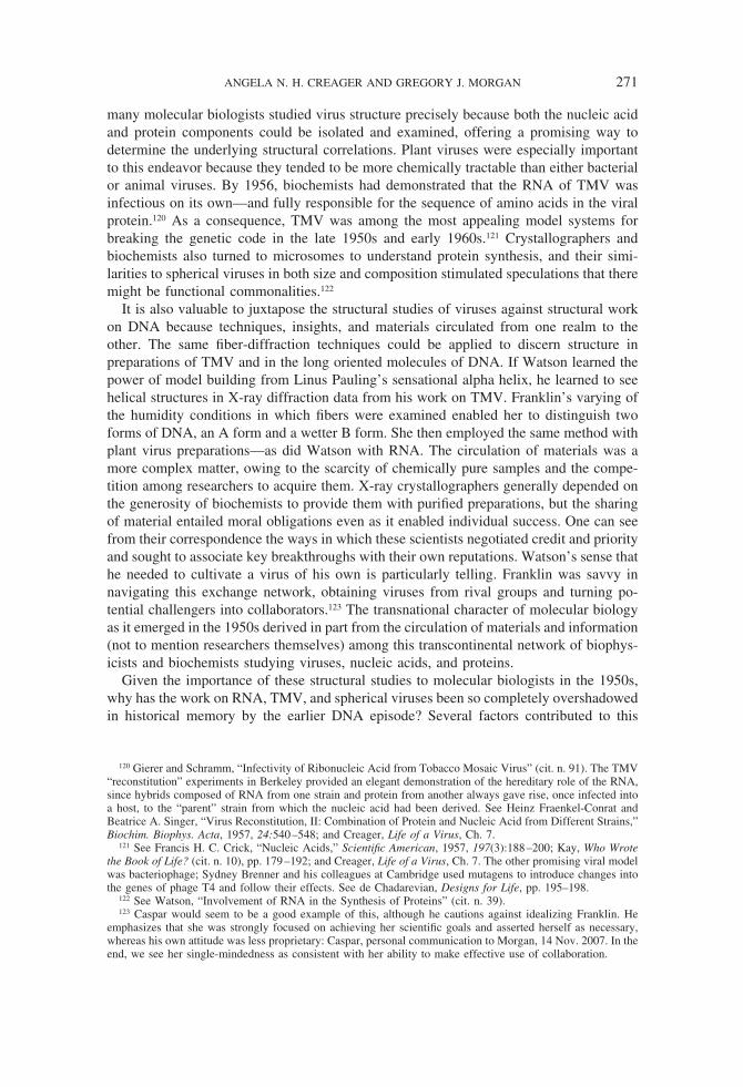
many molecular biologists studied virus structure precisely because both the nucleic acidand protein components could be isolated and examined, offering a promising way todetermine the underlying structural correlations. Plant viruses were especially importantto this endeavor because they tended to be more chemically tractable than either bacterialor animal viruses. By 1956, biochemists had demonstrated that the RNA of TMV wasinfectious on its own—and fully responsible for the sequence of amino acids in the viralprotein.120 As a consequence, TMV was among the most appealing model systems forbreaking the genetic code in the late 1950s and early 1960s.121 Crystallographers andbiochemists also turned to microsomes to understand protein synthesis, and their simi-larities to spherical viruses in both size and composition stimulated speculations that theremight be functional commonalities.122
It is also valuable to juxtapose the structural studies of viruses against structural workon DNA because techniques, insights, and materials circulated from one realm to theother. The same fiber-diffraction techniques could be applied to discern structure inpreparations of TMV and in the long oriented molecules of DNA. If Watson learned thepower of model building from Linus Pauling’s sensational alpha helix, he learned to seehelical structures in X-ray diffraction data from his work on TMV. Franklin’s varying ofthe humidity conditions in which fibers were examined enabled her to distinguish twoforms of DNA, an A form and a wetter B form. She then employed the same method withplant virus preparations—as did Watson with RNA. The circulation of materials was amore complex matter, owing to the scarcity of chemically pure samples and the compe-tition among researchers to acquire them. X-ray crystallographers generally depended onthe generosity of biochemists to provide them with purified preparations, but the sharingof material entailed moral obligations even as it enabled individual success. One can seefrom their correspondence the ways in which these scientists negotiated credit and priorityand sought to associate key breakthroughs with their own reputations. Watson’s sense thathe needed to cultivate a virus of his own is particularly telling. Franklin was savvy innavigating this exchange network, obtaining viruses from rival groups and turning po-tential challengers into collaborators.123 The transnational character of molecular biologyas it emerged in the 1950s derived in part from the circulation of materials and information(not to mention researchers themselves) among this transcontinental network of biophys-icists and biochemists studying viruses, nucleic acids, and proteins.
Given the importance of these structural studies to molecular biologists in the 1950s,why has the work on RNA, TMV, and spherical viruses been so completely overshadowedin historical memory by the earlier DNA episode? Several factors contributed to this
120 Gierer and Schramm, “Infectivity of Ribonucleic Acid from Tobacco Mosaic Virus” (cit. n. 91). The TMV“reconstitution” experiments in Berkeley provided an elegant demonstration of the hereditary role of the RNA,since hybrids composed of RNA from one strain and protein from another always gave rise, once infected intoa host, to the “parent” strain from which the nucleic acid had been derived. See Heinz Fraenkel-Conrat andBeatrice A. Singer, “Virus Reconstitution, II: Combination of Protein and Nucleic Acid from Different Strains,”Biochim. Biophys. Acta, 1957, 24:540–548; and Creager, Life of a Virus, Ch. 7.
121 See Francis H. C. Crick, “Nucleic Acids,” Scientific American, 1957, 197(3):188–200; Kay, Who Wrotethe Book of Life? (cit. n. 10), pp. 179–192; and Creager, Life of a Virus, Ch. 7. The other promising viral modelwas bacteriophage; Sydney Brenner and his colleagues at Cambridge used mutagens to introduce changes intothe genes of phage T4 and follow their effects. See de Chadarevian, Designs for Life, pp. 195–198.
122 See Watson, “Involvement of RNA in the Synthesis of Proteins” (cit. n. 39).123 Caspar would seem to be a good example of this, although he cautions against idealizing Franklin. He
emphasizes that she was strongly focused on achieving her scientific goals and asserted herself as necessary,whereas his own attitude was less proprietary: Caspar, personal communication to Morgan, 14 Nov. 2007. In theend, we see her single-mindedness as consistent with her ability to make effective use of collaboration.
ANGELA N. H. CREAGER AND GREGORY J. MORGAN 271

tendency. The pertinence of structural studies of viruses (as well as microsomes) to thegenetic code ended in the 1960s; thereafter TMV and other well-studied viruses began tobe viewed as models of self-assembly instead.124 Along similar lines, research on proteinsynthesis made it clear that several specific types of RNA—messenger RNA, transferRNA, ribosomal RNA—played distinct roles in protein synthesis, making viral RNA aless useful model for understanding the transcription and translation of genes.125 And,perhaps above all, the double helix itself became a potent icon for molecular biology. AsSoraya de Chadarevian has suggested, this did not occur immediately in response to theappearance of Watson and Crick’s structural model for DNA but, rather, developedgradually.126 The publication of The Double Helix and the subsequent advent of bothrecombinant DNA techniques in the 1970s and the biotechnology industry in the 1980sgave the structure a wider circulation and cultural valence. By contrast, DNA was not theonly helix of significance in the 1950s. An examination of Franklin’s virus research atclose range not only enables a fuller appreciation of her scientific accomplishments butalso reveals the exchange networks and international alliances from which the disciplineof molecular biology crystallized.
124 P. Jonathan G. Butler and Aaron Klug, “The Assembly of a Virus,” Sci. Amer., 1978, 239(5):62–69; andMorgan, “Early Theories of Virus Structure” (cit. n. 68).
125 See Judson, Eighth Day of Creation (cit. n. 10), Chs. 5–8; and Rheinberger, Toward a History of EpistemicThings (cit. n. 104), esp. Chs. 10, 12, 13.
126 De Chadarevian, Designs for Life, Chs. 6, 8. On the popular uses of the double helix see Dorothy Nelkinand M. Susan Lindee, The DNA Mystique: The Gene as a Cultural Icon (New York: Freeman, 1995).
272 AFTER THE DOUBLE HELIX