affinities of the family sollasellidae (porifera, demospongiae
TRANSCRIPT
Contributions to Zoology, 75 (3/4) 133-144 (2006)
Affi nities of the family Sollasellidae (Porifera, Demospongiae). I. Morphological evidence
R.W.M. van Soest1, J.N.A. Hooper2, E. Beglinger1, D. Erpenbeck2, 3
1 Zoologisch Museum, Universiteit van Amsterdam, P.O. Box, 94766, 1090 GT Amsterdam, Netherlands, e-mail: [email protected]; 2 Queensland Museum, P.O. Box 3300, South Brisbane, Qld. 4101, Australia; 3 Dept. of Geobiology, Geoscience Centre Göttingen, 37077 Göttingen, Germany
Key words: sponges, classifi cation, Sollasellidae, Raspailiidae, Sollasella, Raspailopsis, Australia, Oman
Abstract
Comparison of Sollasella digitata Lendenfeld, 1888, up until the present assigned to its own family Sollasellidae Lendenfeld, 1887 in the order Hadromerida, and Raspailopsis cervicornis Burton, 1959, assigned to Raspailiidae Nardo, 1833 in the order Poecilo-sclerida, leads to the conclusion that both should be considered congeneric and are best assigned to a single genus Sollasella. This conclusion is based on examination of habit and skeletal characters of the type material of S. digitata and both type and freshly col-lected material of S. cervicornis. The conclusion is strengthened by the discovery of a new species, Sollasella moretonensis n.sp. collected in North Australia (primarily in the northeastern coast, but also an isolated record from the northwestern Australian coast), which possesses in addition to the characteristic surface pattern and skeletal structure, genuine echinating acanthostyles. The re-defi ned genus Sollasella shares axial / extra-axial arrangement of the skeleton, special surface brushes of oxeas surrounding a single protruding style, and vestigial occurrence of acanthostyles with many Raspailia s.l. Nevertheless, it is retained as a separate genus, on account of its peculiar polygonal arrangement of surface pores. The distribution of the genus is disjunctive including both (south-east, northeast and northwest) Australian and Western Indian Ocean localities, but so far no intermediate records. Based on this mor-phological evidence, it is proposed – pending publication of cor-roborating molecular evidence to be presented in a follow-up study – to reassign Sollasella and the family Sollasellidae to the poecilo-sclerid family Raspailiidae.
Contents
Introduction .................................................................................... 133Material and methods ................................................................... 134Results ............................................................................................. 134 Ordinal and familial affi liations of Sollasella digitata .... 134 Systematic descriptions ......................................................... 135Discussion ...................................................................................... 143Acknowledgements ....................................................................... 144References ...................................................................................... 144
Introduction
The recent update of the sponge classifi cation ‘Sys-tema Porifera’ (Hooper and van Soest, 2002a) typifi ed the family Sollasellidae and its sole representative the East Australian species Sollasella digitata Lendenfeld, 1888 as ‘incertae sedis’ and ‘poorly known’ (p. 170), and commented that ‘its true affi nities remain to be established’ (p. 280). Nevertheless, it was assigned to the order Hadromerida on the grounds that it pos-sessed a cortex and strongly radiating skeletal archi-tecture. The species is only known from its original description and from an excellent re-description of the type material by Hallmann (1914). The species is arborescent and hispid. It has a strongly axial-radiate architecture, with a thick axial column made up of oxeas and styles, long protruding extra-axial styles and a dense ectosomal palisade of oxeas. Between this ‘cortical’ palisade and the axial column there is a region relatively lightly spiculated. The surface of S. digitata has a very characteristic polygonal pattern of ‘perforations’ visible to the naked eye. Previous authors attempting to classify this enigmatic species arrived at divergent conclusions: Axinellidae, order Halichondrida (cf. Hallmann, 1914), Coppatiidae, order Astrophorida, (cf. Hooper and Wiedenmayer, 1994), whereas Polymastiidae and Stylocordylidae, both order Hadromerida were named as closely re-lated families by van Soest (2002). Recently, we obtained freshly collected specimens of a branching hispid sponge from Oman waters showing the same characteristic surface perforation patterns and essentially a similar skeletal architecture and spiculation. This material was initially identifi ed as Raspailopsis cervicornis Burton, 1959 - a species
134 van Soest et al. - Affi nities of the sponge family Sollasellidae
that was previously described from Oman waters - based on the characters presented in the very brief description. Recent examination of type material in the Natural History Museum, London, confi rmed that Burton’s species was identical to our Oman material, but also that R. cervicornis is indeed very similar to Sollasella digitata from southeast Australia. These observations led to the discovery of a further similar species from subtropical Australia, which shared the surface perforations but possessed in addition abun-dant echinating acanthostyles, confi rming the ras-pailiid affi nity. This paper is intended to present morphological evidence for the close similarities of Sollasella and Raspailia, and to discuss the classifi -cation of the family Sollasellidae whose position in Hadromerida is proposed to be untenable. We will follow this up with further corroborating evidence from molecular sequence data (Erpenbeck et al. submitted).
Material and methods
Material was examined from the collections of the Natural History Museum, London (BMNH), the Australian Museum Sydney (AM), the Queensland Museum Brisbane (QM) and the Zoological Mu-seum of the University of Amsterdam (ZMA). Details of the specimens are given below with the treatment of the species. Thick sections and spicule mounts for light and SEM microscopy were made following the usual methodology (cf. de Voogd and van Soest, 2002).
Results
Ordinal and familial affi liations of Sollasella digitata
Hadromerida are defi ned (cf. Hooper and van Soest, 2002b) as comprising sponges with peripherally ra-diating skeleton built from tylostyles, styles, or oc-casionally oxeas, usually with a smaller category in an ectosomal palisade. The radiating architecture becomes confused or plumose in the choanosome, where the larger spicule categories predominate. Microscleres (euasters, spirasters, amphiasters) occur in many families, usually as an ectosomal cover, but
are absent in several families. Family Sollasellidae and Sollasella digitata were hesitatingly assigned (cf. van Soest, 2002) to Hadromerida for want of a better placement. Shared characters with several Hadromer-ida are the arborescent shape, ectosomal palisade, confused choanosomal skeleton, robust oxeas and styles with faint tyle, but these features are widely distributed throughout the Demospongiae, not com-prising clear synapomorphies with any particular order or family. The polygonally arranged surface pores are slightly reminiscent of the polygonal sur-face plates separated by grooves found in the family Placospongiidae, but they appear distinctly unique. Finally, the hispidity is shared with many Hadromer-id genera, but can hardly be suffi cient for ordinal identity. Compared to Hadromerida, affi nities of Sollasella digitata with the order Poecilosclerida also seem tenuous. This order is defi ned (Hooper and van Soest, 2002c) as possessing a skeleton of spicules and spongin, with distinct regionalization into ectosomal and choanosomal components. Among the spicules feature meniscoid microscleres (sigmas, chelae, toxas) and acanthose styles as characteristic. How-ever, some families lack all or some of these. Sol-lasella digitata - in retrospect - shares several features with Poecilosclerid representatives, such as a distinct regionalization into ectosomal, extra-axial and axial skeletal arrangement, surface brushes of smaller megascleres surrounding single protruding long megascleres (‘raspailid ectosome’) and confused axial-longitudinal skeletal arrangement. Arborescent shapes with distinct hispidity like Sollasella digitata are very common features in microcionid Poecilo-sclerida. The recent rediscovery of fresh material of South Arabian Raspailopsis cervicornis Burton, 1959 (see above and below), with its essentially similar surface pattern and structure, and an undescribed species from subtropical northeastern Australia ‘Sp.1245’ (see below) with the same surface pattern but in ad-dition clear possession of acanthostyles, swung the balance defi nitely towards Poecilosclerida - Ra-spailiidae. Table 1 summarizes the different charac-ter states of Sollasella and representative families from Hadromerida and Poecilosclerida discussed above. Heuristic search with PAUP 3.1 (Swofford, 1993) using the characters unordered confi rmed the morphological affi nity of Sollasella with Raspailiidae
135Contributions to Zoology, 75 (3/4) – 2006
Definition (emended). Raspailiidae with strong axial column of confusedly aligned oxeas and styles, and with extra-axial columns of short oxeas and long and short styles positioned at right angles to the axial column. At the surface there is a charac-teristic ornamentation of polygonally arranged in-halant (?) pores and the oxeas form a continuous palisade of brushes of oxeas pierced by long styles. A low proportion of short acanthostyles may be present.
Sollasella digitata Lendenfeld, 1888Figs 1A-F, 2A
Sollasella digitata Lendenfeld, 1888: 56; Hallmann, 1914: 287, pl. XV fi gs 1-2, text-fi g. 1.
Material examined: Lectotype (per Hallmann, 1914): BMNH 1886.8.27.639 (spirit specimen), including 2 slides.Paralectotype: AM G9107 – ‘syntype’ mentioned by Hooper and Wiedenmayer (1994: 144).
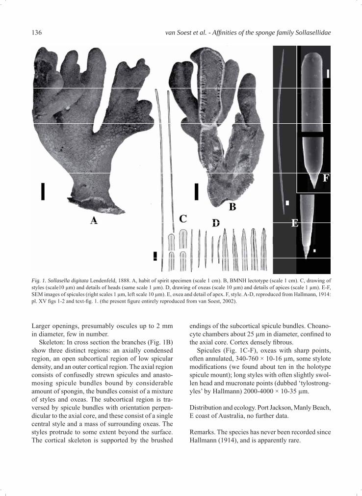
Description (mostly from Hallmann, 1914, summa-rized in van Soest, 2002). Habit: Stalked ramose sponge (Figs. 1A, B, 2A) of up to 14 cm long, with rounded short branches. Consistency very fi rm, tough. Surface hispid due to protruding spicules, and provided with a character-istic polygonal pattern of round pores (Fig. 1A), presumed to be inhalant openings by Hallmann.
(11 shared characters in Table 1), rather than with Suberitidae (7 shared characters) (tree not shown). A full phylogenetic reconstruction will be provided in a second contribution on this topic, incorporating molecular evidence. In the molecular sequence analyses Sollasella clearly clusters inside the Ras-pailiidae clade, and distant from hadromerid taxa (Erpenbeck et al, submitted).
Systematic descriptions
The three species discussed here are assigned to a redefi ned genus Sollasella, provisionally assigned to Raspailiidae, pending further support from molecular studies. They will all be diagnosed below and mor-phological evidence for their congeneric status will be illustrated with habit and microscopical images.
Phylum PoriferaClass DemospongiaeOrder PoeciloscleridaFamily Raspailiidae Nardo, 1833Subfamily Raspailiinae Nardo, 1833
Genus Sollasella Lendenfeld, 1887
Synonym. Raspailopsis Burton, 1959
Type species. Sollasella digitata Lendenfeld, 1888 (by monotypy)
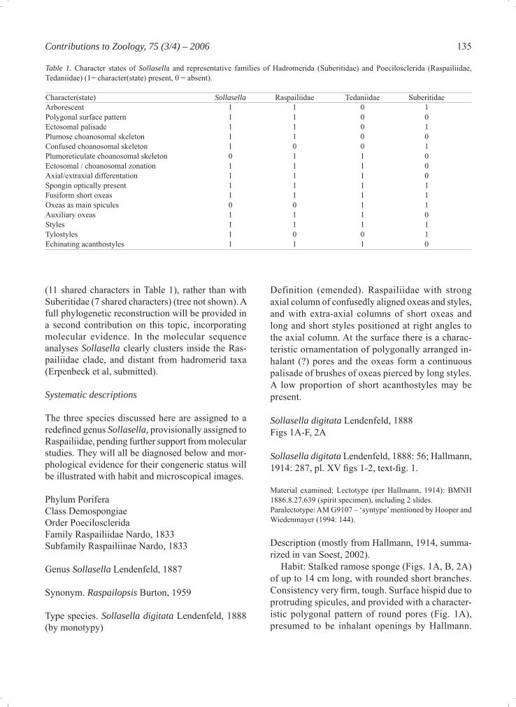
Table 1. Character states of Sollasella and representative families of Hadromerida (Suberitidae) and Poecilosclerida (Raspailiidae, Tedaniidae) (1= character(state) present, 0 = absent). Character(state) Sollasella Raspailiidae Tedaniidae SuberitidaeArborescent 1 1 0 1Polygonal surface pattern 1 1 0 0Ectosomal palisade 1 1 0 1Plumose choanosomal skeleton 1 1 0 0Confused choanosomal skeleton 1 0 0 1Plumoreticulate choanosomal skeleton 0 1 1 0Ectosomal / choanosomal zonation 1 1 1 0Axial/extraxial differentation 1 1 1 0Spongin optically present 1 1 1 1Fusiform short oxeas 1 1 1 1Oxeas as main spicules 0 0 1 1Auxiliary oxeas 1 1 1 0Styles 1 1 1 1Tylostyles 1 0 0 1Echinating acanthostyles 1 1 1 0
136 van Soest et al. - Affi nities of the sponge family Sollasellidae
Larger openings, presumably oscules up to 2 mm in diameter, few in number. Skeleton: In cross section the branches (Fig. 1B) show three distinct regions: an axially condensed region, an open subcortical region of low spicular density, and an outer cortical region. The axial region consists of confusedly strewn spicules and anasto-mosing spicule bundles bound by considerable amount of spongin, the bundles consist of a mixture of styles and oxeas. The subcortical region is tra-versed by spicule bundles with orientation perpen-dicular to the axial core, and these consist of a single central style and a mass of surrounding oxeas. The styles protrude to some extent beyond the surface. The cortical skeleton is supported by the brushed
endings of the subcortical spicule bundles. Choano-cyte chambers about 25 μm in diameter, confi ned to the axial core. Cortex densely fi brous. Spicules (Fig. 1C-F), oxeas with sharp points, often annulated, 340-760 × 10-16 μm, some stylote modifi cations (we found about ten in the holotype spicule mount); long styles with often slightly swol-len head and mucronate points (dubbed ‘tylostrong-yles’ by Hallmann) 2000-4000 × 10-35 μm.
Distribution and ecology. Port Jackson, Manly Beach, E coast of Australia, no further data.
Remarks. The species has never been recorded since Hallmann (1914), and is apparently rare.
Fig. 1. Sollasella digitata Lendenfeld, 1888. A, habit of spirit specimen (scale 1 cm). B, BMNH lectotype (scale 1 cm). C, drawing of styles (scale10 μm) and details of heads (same scale 1 μm). D, drawing of oxeas (scale 10 μm) and details of apices (scale 1 μm). E-F, SEM images of spicules (right scales 1 μm, left scale 10 μm). E, oxea and detail of apex. F, style. A-D, reproduced from Hallmann, 1914: pl. XV fi gs 1-2 and text-fi g. 1. (the present fi gure entirely reproduced from van Soest, 2002).
137Contributions to Zoology, 75 (3/4) – 2006
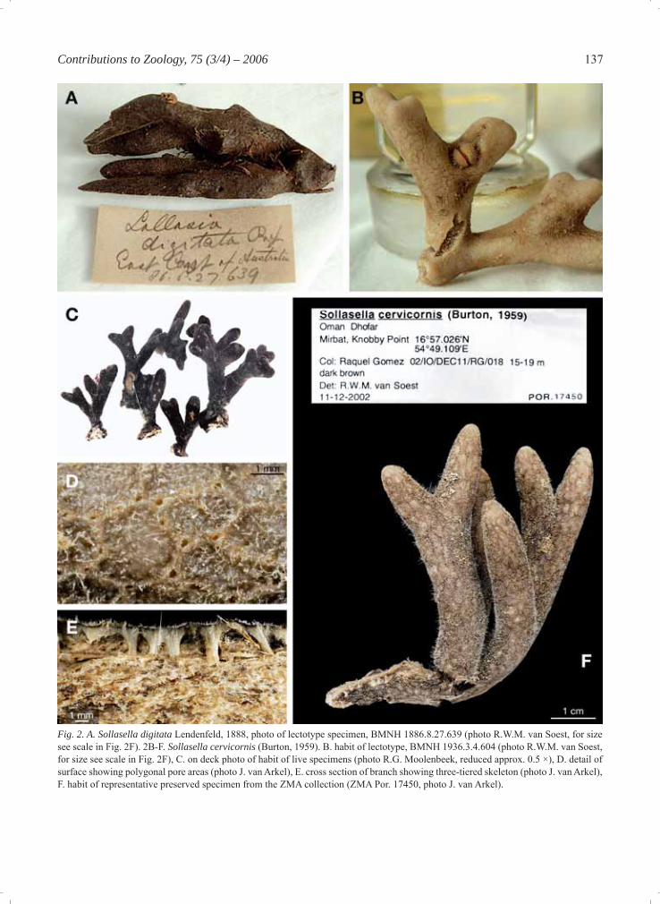
Fig. 2. A. Sollasella digitata Lendenfeld, 1888, photo of lectotype specimen, BMNH 1886.8.27.639 (photo R.W.M. van Soest, for size see scale in Fig. 2F). 2B-F. Sollasella cervicornis (Burton, 1959). B. habit of lectotype, BMNH 1936.3.4.604 (photo R.W.M. van Soest, for size see scale in Fig. 2F), C. on deck photo of habit of live specimens (photo R.G. Moolenbeek, reduced approx. 0.5 ×), D. detail of surface showing polygonal pore areas (photo J. van Arkel), E. cross section of branch showing three-tiered skeleton (photo J. van Arkel), F. habit of representative preserved specimen from the ZMA collection (ZMA Por. 17450, photo J. van Arkel).
138 van Soest et al. - Affi nities of the sponge family Sollasellidae
Sollasella cervicornis (Burton,1959)Figs. 2B-F, 3A-E
Raspailopsis cervicornis Burton, 1959: 256, fi g. 33Raspailia (Parasyringella) cervicornis; Hooper, 2002: 478
Material examined: Holotype, BMNH 1936.3.4.604 (original designation): John Murray Exped. stat. 53, Oman, 19°22’36”N 57°53’E, 13.5 m, 02-11-1933. BMNH 1936.3.4.521, John Murray Exped. stat. 27, Somalia, Gulf of Aden, 11°57’12”/11°56’24”N 50°35’E/50°39’12”E, 37 m, 12-10-1933.BMNH 1936.3.4.523, John Murray Exped. stat. 27, Somalia, Gulf of Aden, 11°57’12”/11°56’24”N 50°35’E/50°39’12”E, 37 m, 12-10-1933.BMNH 1936.3.4.522, John Murray Exped. stat. 45, Oman, 18°03’30”N 57°02’30”E, 38 m, 29-10-1933.BMNH 1936.3.4.466, John Murray Exped. stat. 111, Tanzanya, Zanzibar, 05°04’18”S 39°14’12”E, 73-165 m, 14-01-1934.ZMA Por. 17450, Oman, Dhofar, Mirbat, Knobby Point, 16°57.026’N 54°49.109’E, 15-19 m, fi eld number 02/IO/DEC11/RG/018, coll. Raquel Gomez, 02-12-2002.
Description Habit (Figs. 2B-F): Thickly arborescent sponges, branching dichotomously. Branches have a ten-dency to coalesce and the Burton material contains one ‘caliculate’ specimen (from Zanzibar, BMNH 1936.3.4.466). Surface hispid through protruding spicules. Colour dark brown in life (Fig. 2C), paler grey in alcohol (Figs. 2B,F). Tough consistency, easily broken. Branch endings rounded and slightly swollen. Holdfast broader than stem, evenly round-ed, spreading out equally. Height up to 12 cm, branch diameter 1-1.2 cm, specimens retained as ZMA 17450 are typically 5 or 6 cm high. A striking feature of all branches is the polygonal pattern of surface pores (Figs. 2B, D, F). Upon collection, when lifted out of the water and with slight pressure, these open-ings emitted tiny water jets, indicating they may be exhalant contrary to what is assumed here. Polygo-nal areas are elongate, on average 4 × 2 mm (Fig. 2D), and uniformly spread over the branches (Fig. 2F). No separate larger oscules have been ob-served. Skeleton (Figs. 2E, 3A): In cross section the branches contain three distinct regions recognized by eye (Fig. 2E) and differently coloured in life: a thick axial column, reddish orange in life, pale orange when preserved, a relatively fi brous extra axial re-gion, and a beige coloured cortical layer at the sur-
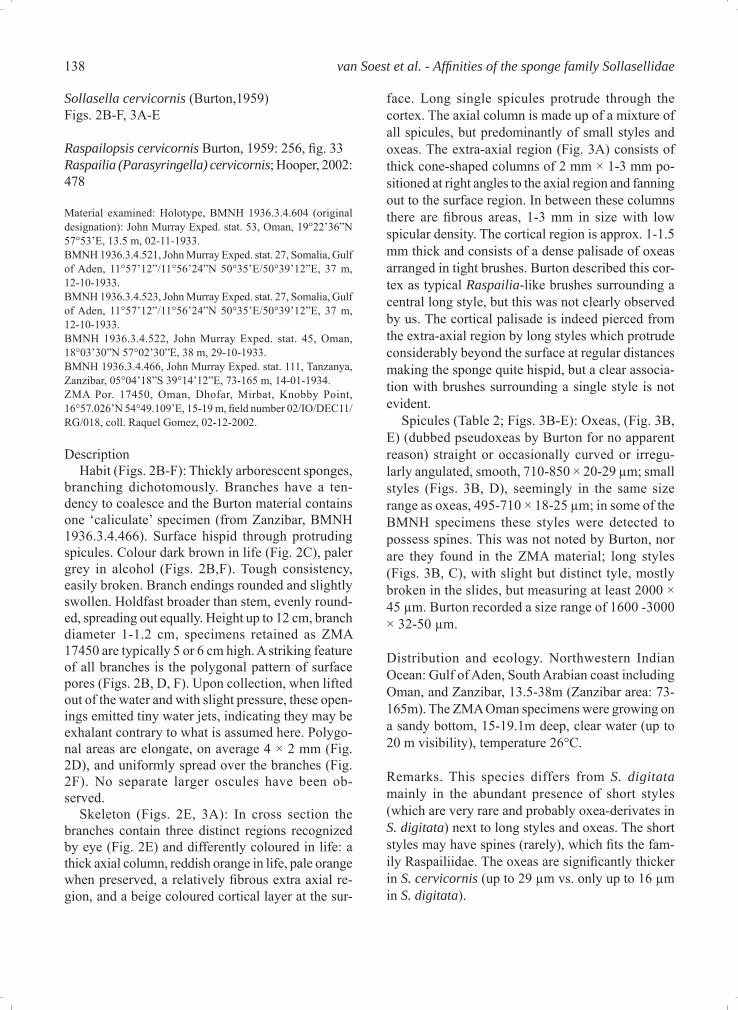
face. Long single spicules protrude through the cortex. The axial column is made up of a mixture of all spicules, but predominantly of small styles and oxeas. The extra-axial region (Fig. 3A) consists of thick cone-shaped columns of 2 mm × 1-3 mm po-sitioned at right angles to the axial region and fanning out to the surface region. In between these columns there are fi brous areas, 1-3 mm in size with low spicular density. The cortical region is approx. 1-1.5 mm thick and consists of a dense palisade of oxeas arranged in tight brushes. Burton described this cor-tex as typical Raspailia-like brushes surrounding a central long style, but this was not clearly observed by us. The cortical palisade is indeed pierced from the extra-axial region by long styles which protrude considerably beyond the surface at regular distances making the sponge quite hispid, but a clear associa-tion with brushes surrounding a single style is not evident. Spicules (Table 2; Figs. 3B-E): Oxeas, (Fig. 3B, E) (dubbed pseudoxeas by Burton for no apparent reason) straight or occasionally curved or irregu-larly angulated, smooth, 710-850 × 20-29 μm; small styles (Figs. 3B, D), seemingly in the same size range as oxeas, 495-710 × 18-25 μm; in some of the BMNH specimens these styles were detected to possess spines. This was not noted by Burton, nor are they found in the ZMA material; long styles (Figs. 3B, C), with slight but distinct tyle, mostly broken in the slides, but measuring at least 2000 × 45 μm. Burton recorded a size range of 1600 -3000 × 32-50 μm.
Distribution and ecology. Northwestern Indian Ocean: Gulf of Aden, South Arabian coast including Oman, and Zanzibar, 13.5-38m (Zanzibar area: 73-165m). The ZMA Oman specimens were growing on a sandy bottom, 15-19.1m deep, clear water (up to 20 m visibility), temperature 26°C.
Remarks. This species differs from S. digitata mainly in the abundant presence of short styles (which are very rare and probably oxea-derivates in S. digitata) next to long styles and oxeas. The short styles may have spines (rarely), which fi ts the fam-ily Raspailiidae. The oxeas are signifi cantly thicker in S. cervicornis (up to 29 μm vs. only up to 16 μm in S. digitata).
139Contributions to Zoology, 75 (3/4) – 2006
The shared characters are more numerous:- polygonal arrangement of pores is a striking feature
of both and a strong synapomorphy. There are sev-
eral other sponge genera showing a polygonal sur-face pattern (e.g. Myrmekioderma and Didiscus in Halichondrida: Heteroxyidae, and Tethya and Pla-
Fig. 3. Sollasella cervicornis (Burton, 1959), SEM photos of ZMA specimens (made by E. Beglinger). A. cross section of branch show-ing axial column and extra-axial palisade, B. overview of spicules (long and short styles, oxea) photographed at same scale (scale bar =100 μm), C. head of long style (scale = 50 μm), D. short style (scale = 100 μm), E. oxea (scale = 100 μm).
140 van Soest et al. - Affi nities of the sponge family Sollasellidae
cospongia in Hadromerida), but the similarities appear superfi cial because in these genera polygonal areas are separated by grooves, rather than separate pores.
- branching habit and size are essentially similar- live colour is unknown for S. digitata, but likely to
be brown as is the colour in S. cervicornis- skeletal architecture with the three regions visible
to the naked eye is closely similar in both- long styles are closely similar, including a slight
tyle in most
Sollasella moretonensis sp. nov.(Figs. 4A-D, 5A-H)
Material examined: Holotype: QM G303227: Middle Reef, N of North Stradbroke Island, Queensland, Australia, 27.40083°S, 153.53°E, 30m, coll. Hooper, J.N.A. and Kennedy, J.A., scuba, 04-06-1993. Paratype: QM G321402: Inner Gneerings Shoals, Sunshine Coast, Queensland, Australia, 26.64512°S, 153.16111°E, 13m, coll. Cook, S., Crowther, A., Carini, G., Ekins, M. and Sut-cliffe, T., scuba, 18-08-2004.Specimens (Queensland, Australia): QM G303996: Mudjimba I., N side of island, off Mooloolabah, 26.60194°S, 153.1025°E, 11m, coll. Hooper, J.N.A., Hobbs, L.J., Kennedy, J.A. and Cook, S.D.,
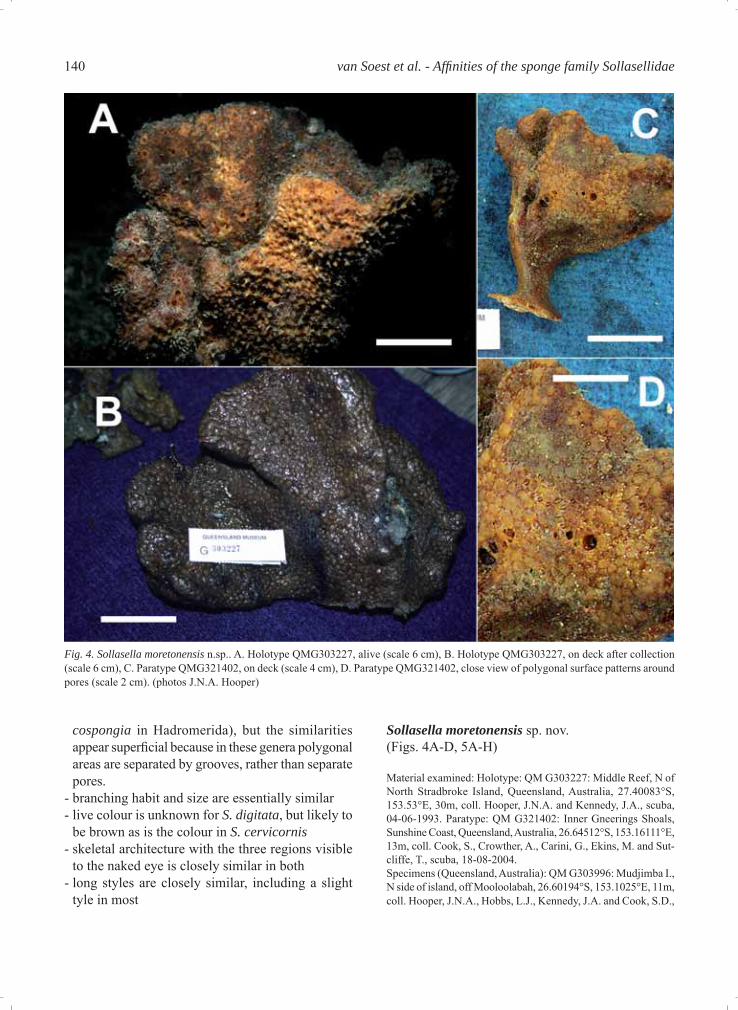
Fig. 4. Sollasella moretonensis n.sp.. A. Holotype QMG303227, alive (scale 6 cm), B. Holotype QMG303227, on deck after collection (scale 6 cm), C. Paratype QMG321402, on deck (scale 4 cm), D. Paratype QMG321402, close view of polygonal surface patterns around pores (scale 2 cm). (photos J.N.A. Hooper)
141Contributions to Zoology, 75 (3/4) – 2006
scuba, 09-02-1994. QM G313599: Hutchinson Shoal, N. of Cape Moreton, 26.943516°S, 153.487426°E, 21.4m, coll. Hooper, J.N.A., Kennedy, J.A. and Cook, S.D., scuba, 01-12-1997. QM G315649: Sunshine Reef off Sunshine Coast, 26.412778°S, 153.13472°E, 27m, coll. Cook, S.D., Kennedy, J.A., List-Armit-age, S.E., Adams, C.L. and Woerheide, G., scuba, 11-10-1999. QM G315759: North Halls off Sunshine Coast, 26.3461°S, 153.067°E, 21m, coll. Cook, S.D., Kennedy, J.A., List-Armitage, S.E., Adams, C.L. and Woerheide, G., scuba, 13-10-1999. QM G303205: Boat Rock, Point Lookout, N of North Stradbroke I., 27.41778°S, 153.55083°E, 25m, coll. Hooper, J.N.A. and Cook, S.D., scuba, 02-06-1993. QM G315719: Jew Shoal off Sunshine Coast, 26.37556°S,153.12583°E, 18m, coll. Cook, S.D., Kennedy, J.A., List-Armitage, S.E., Adams, C.L. and Woerheide, G., scuba, 12-10-1999. QM G303059: NE Cape Grenville, Shelburne Bay, 11.61722°S, 143.06889°E, 31m, coll. Cook, S.D. on FV ‘Clipper Bird’, trawl, 26-03-1993. Specimens (Western Australia): QM G306153: SW of Cape Jaubert, NW of Western Australia, 19.76667°S, 118.21667°E, 37m, coll. Cook, S.D. on CSIRO RV ‘Southern Surveyor’, beam trawl, 05-09-1995.
Description Habit (Figs 4A-D): Vasiform, lobate, fan-shaped, cup-shaped or subvasiform habits, 11-28 cm high, 8-27 cm wide at apex, with thick lamellae 0.5-2 cm thick, bearing rounded ‘lumpy’ or convoluted mar-gins. Specimens are usually attached to the substrate by a short cylindrical woody holdfast, ranging from 3-10 cm long, 1.5-3 cm diameter. Texture: Harsh (slightly hispid), fi rm, stiff but compressible in life, more rigid when preserved. Surface: Dead specimens are covered with a polygonal plate-like pattern formed by shallow pits surrounding low conules, 3-5 mm in diameter, 1-2 mm high, with some (but not all) perforated by an oscule 2-5 mm in diameter sitting at the apex of the conule. When preserved most oscules and pits are collapsed, leaving only the polygonal pattern of conules reminiscent of typical Polymastia or massive Cliona surfaces (Fig. 4B-D), and conules appear to be distributed equally on both the inner and outer surfaces of the lamellae, extend-
ing across all lamellae to the upper part of the hold-fast. In life, however, it is apparent (Fig. 4A) that oscules occur mostly on the inner surface of lamel-lae, less commonly on the upper margins of the outer surface, are about 4 mm in diameter, and are surrounded by a large raised membranous lip, pig-mented darker brown than the surrounding area. The outer surface of living specimens is highly pitted and rendered convoluted by pointed conules surrounding large pits, interconnected by prominent ridges of fl esh forming stellate patterns reminiscent of dic-tyodendrillid sponges. This external sculpturing of pits and conules presumably represents the inhalant aquiferous system. Colour: In life the colour is bright orange to orange-brown, fading to darkish brown upon preservation. Skeleton (Figs. 5A-E): In cross section the skel-eton appears to be differentiated into three distinct regions: an open reticulate very fi brous core, a more plumo-reticulate extra-axial region, and a distinct radial ectosomal skeleton. There is no obvious axial compression, but in the core of the lamellae there are heavily collagenous fi bres and multispicular bundles of large styles and oxeas, forming an open but highly reticulated skeleton. Fibres are moderately large, 180-260 μm in diameter, forming open mesh-es in some places over 1 mm in diameter. Bundles of oxeas and styles appear to run predominantly longi-tudinally through lamellae. At the core of the skeleton fi bres are sparsely echinated by acanthostyles. In the extra-axial region the skeleton becomes more plumo-reticulate, with thicker, more densely compacted and heavy collagenous fi bres together with radial bundles of large choanosomal styles and oxeas,with the latter becoming more radial towards the periphery. The ectosomal skeleton has only occasional large oxeas or styles protruding from the subectosomal regions, with the remainder consisting of a nearly continuous, erect palisade of ectosomal oxeas. In some cases
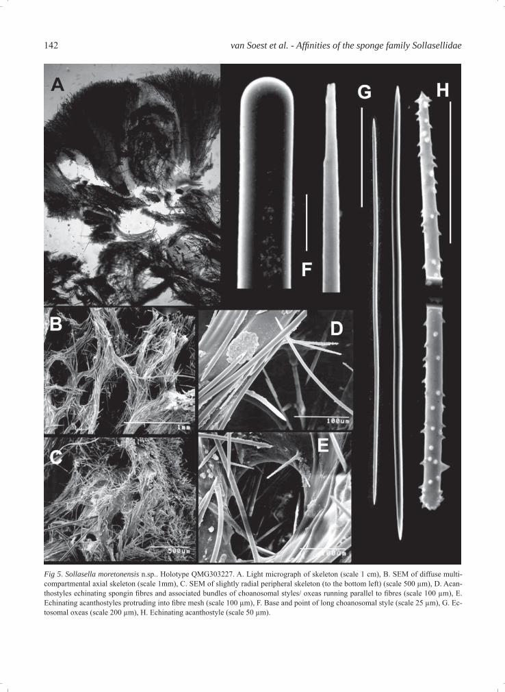
Table 2. Comparison of spicule sizes of Sollasella species. Spicule category S. digitata S. cervicornis S. moretonensis n.sp.Long styles 2000-4000 × 10-35 μm 1600-3000 × 32-50 μm 2500->4000 × 20-55 μmShort styles Modifi cations of oxeas smooth 495-710 × 18-25 μm Absent occasionally spinedEchinating acanthostyles Absent Not differentiated 95-165 × 3-5 μmOxeas (1) 340-760 × 10-16 μm 710-850 × 20-29 μm 660-940 × 14-20 μmOxeas (2) Not differentiated Not differentiated 360-515 × 5-8 μm
142 van Soest et al. - Affi nities of the sponge family Sollasellidae
Fig 5. Sollasella moretonensis n.sp.. Holotype QMG303227. A. Light micrograph of skeleton (scale 1 cm), B. SEM of diffuse multi-compartmental axial skeleton (scale 1mm), C. SEM of slightly radial peripheral skeleton (to the bottom left) (scale 500 μm), D. Acan-thostyles echinating spongin fi bres and associated bundles of choanosomal styles/ oxeas running parallel to fi bres (scale 100 μm), E. Echinating acanthostyles protruding into fi bre mesh (scale 100 μm), F. Base and point of long choanosomal style (scale 25 μm), G. Ec-tosomal oxeas (scale 200 μm), H. Echinating acanthostyle (scale 50 μm).
143Contributions to Zoology, 75 (3/4) – 2006
these bundles of ectosomal oxeas appear to be sur-rounding the larger protruding subectosomal spi-cules, an apomorphy of the Raspailiidae, but this is rare. The mesohyl is sparse at the core of the skel-eton but is more heavily invested in collagen towards the periphery. Spicules (Table 2, Figs. 5F-H): The longer (sub-ectosomal) spicules are predominantly styles, occa-sionally oxeas or anisoxeas, are not common, and appear to extend from the core of the skeleton to the surface, or close to it. They are completely smooth, more-or-less straight, and have either evenly round-ed bases or abruptly pointed bases, and either sharp tapering points or taper to very fi ne slightly telescoped points, approximately 2500->4000 × 20-55 μm). The (choanosomal) oxeas are far more abundant and comprise most of the skeletal spiculation. They are usually slightly curved at the centre, sometimes straight, with sharp, tapering points, 660-940 × 14-20 μm). Ectosomal oxeas are straight, slightly curved at the centre or asymmetrically curved, with slightly tapering sharp points, 360-515 × 5-8 μm). Echinating acanthostyles are straight or slightly curved at the centre, have slightly subtylote bases and are slightly rounded or blunt at the apex, and have a light, evenly distributed spination along the length of the spicule, slightly heavier spines at the tip, and spines are short and conical, (95-165 × 3-5 μm).
Etymology. This species is named for its predomi-nantly subtropical distribution in the vicinity of the Moreton Bay region, SE Queensland.
Distribution and ecology. Moderately prevalent in southern Queensland inshore waters (Brisbane and Sunshine Coast regions), associated with muddy substrata at the base of coral reefs. The two isolated records in Far North Queensland (Shelburne Bay) and NW of Western Australia (Cape Jaubert) were from commercial and scientific trawls made in deeper muddy substrates, and suggest that the species may have a wider distribution than presently known, or a discontiguous distribution in tropical Australia given that there has been intensive sampling of areas in between without trace of this species.
Remarks. The new species S. moretonensis shares the polygonal arrangement of surface pores, posses-sion of three distinct skeletal regions and geometry
of spiculation with the other two species, but differs in possessing echinating acanthostyles, a differenti-ated category of ectosomal oxeas, a vasiform growth form, orange live colouration and specifi c dimensions of spicules. The discovery of this new species con-fi rms the allocation of Sollasella to the Raspailiidae in possessing of the important raspailiid apomorphies – viz. echinating acanthostyles and bundles of ecto-somal oxeas surrounding protruding subectosomal styles/oxeas (albeit rare), in conjunction with a con-tinuous ectosomal palisade common to the other two species.
Discussion
In the latest revision of the Raspailiidae (Hooper, 2002), Raspailopsis was assigned to the synonymy of Raspailia (Parasyringella). Since three distinct species with disjunctive distributions share a number of unique features not shared with other Raspailia species, there is suffi cient justifi cation to abandon Hooper’s synonymy decision and unite them in a separate genus. Accordingly we propose to retain Sollasella as a distinct genus in Raspailiidae, differ-ing from all other genera in this family by the po-lygonally arranged surface pores and the continuous cortical palisade of long, robust oxeas.
The family Sollasellidae Lendenfeld, 1887 was erected a year before the description of the type spe-cies, Sollasella digitata Lendenfeld, 1888. Despite this slight deviation from the nomenclatural rules, it was accepted as a valid family name by subsequent revisors (Hallmann, 1914; de Laubenfels, 1936; van Soest, 2002). In view of the fact that the type species Sollasella digitata is now proposed to be assigned to Raspailiidae, the family name Sollasellidae falls into synonymy with that family name. The ‘Systema Porifera’ (Hooper and van Soest, 2002) gives Hent-schel, 1923 as the author and date of Raspailiidae, which would potentially jeopardize the well-estab-lished name Raspailiidae. However, this authorship and date is predated by Nardo, 1833 who already employed a taxon name Raspeliae of the family group level (corrected to Raspailiae in Nardo, 1847). Thus, authorship of Raspailiidae should be attributed to Nardo, 1833 and this name remains the oldest avail-able name. Sollasellidae Lendenfeld, 1887 is a junior synonym of Raspaillidae Nardo, 1833.
144 van Soest et al. - Affi nities of the sponge family Sollasellidae
Acknowledgements
Raquel Gomez and Rob G. Moolenbeek (ZMA) col-lected and photographed the Oman specimens. Uni-Bioscreen S.A., Brussels funded the 2002 collecting trip. Special thanks are due to Dr Ali Amer Al Ki-yumi, Director General of the Ministry of Regional Municipalities, Environment and Water Resources, Muscat Sultanate of Oman, Dr Barry P. Jupp, Marine Pollution & Coastal Management section, Muscat, and Dr Ahmed Bin Mohamed Al-Mazrooei, Director Marine Science and Fisheries Center, Muscat, for providing local facilities, and permits to collect and transport the samples to the Netherlands for scientic research. A reference collection was deposited in the Marine Science and Fisheries Center, Muscat. Rob van Soest acknowledges the EU-SYNTHESYS / GB-TAF grant 538, extended to him for a 2-weeks visit to the collections of the Natural History Mu-seum, London, hosted by Clare Valentine. Dirk Er-penbeck acknowledges fi nancial support of the Eu-ropean Union under a Marie-Curie outgoing fellow-ship (MOIF-CT-2004). Jan van Arkel (IBED, Uni-versity of Amsterdam) made the detailed photos of the surface of Oman specimens and assisted in com-piling Figure 2. Thanks also to Connie Wörheide for preparing SEM photographs of the new species, S. moretonensis.
References
Burton M. 1959. Sponges. Scientifi c Reports John Murray Ex-pedition, 1933-34. 10 (5): 151-281.
Hallmann EF. 1914. A revision of the monaxonid species de-scribed as new in Lendenfeld’s ‘Catalogue of the Sponges in the Australian Museum’. Part I, II, III. Proc.Linn.Soc. New South Wales 39: 263-315, 327-376, 398-446, pls XV-XXIV.
Hentschel E. 1923. Erste Unterabteilung der Metazoa: Parazoa, Porifera-SchwämmeIn: Kükenthal W, Krumbach T, Eds. Handbuch der Zoologie. Eine Naturgeschichte der Stämme des Tierreiches. Vol. 1, Protozoa, Porifera, Coelenterata, Mesozoa. Berlin and Leipzig, Walter de Gruyter und Co., 307-418.
Hooper JNA. 2002. Family Raspailiidae.Hentschel, 1923. In: Hooper JNA, van Soest RWM, Eds. Systema Porifera. A guide to the classifi cation of sponges. New York, Kluwer Aca-demic / Plenum Publishers, 469-510.
Hooper JNA, van Soest RWM, Eds. 2002a. Systema Porifera. A guide to the classifi cation of sponges. New York, Kluwer Academic / Plenum Publishers, 1-1708, i-xlviii.
Hooper JNA, van Soest RWM, Eds. 2002b. Order Hadromer-ida Topsent, 1894. In: Hooper JNA, van Soest RWM, Eds. Systema Porifera. A guide to the classifi cation of sponges. New York, Kluwer Academic / Plenum Publishers, 169-172.
Hooper JNA, van Soest RWM 2002c. Order Poecilosclerida Topsent, 1928. In: Hooper JNA, van Soest RWM, Eds. Sys-tema Porifera. A guide to the classifi cation of sponges. New York, Kluwer Academic / Plenum Publishers, 403-411.
Hooper JNA, Wiedenmayer F. 1994. Porifera. In: Wells A. Ed. Zoological Catalogue of Australia. Melbourne, CSIRO, 1-621.
Laubenfels MW de. 1936. A discussion of the sponge fauna of the Dry Tortugas in particular, and the West Indies in general, with material for a revision of the families and orders of the Porifera. Carnegie Institute of Washington Publication. Papers of the Tortugas Laboratory 30: 1-225.
Lendenfeld R von. 1887 [1886]. On the systematic position and classifi cation of sponges. Proc. Zool. Soc. London 1886: 558-662.
Lendenfeld R von. 1888. Descriptive catalogue of the sponges in the Australian Museum, Sidney. London, Taylor and Fran-cis, i–xiv,1-260.
Nardo GD. 1833. Auszug aus einem neuen System der Spongi-arien, wonach bereits die Aufstellung in der Universitäts-Sammlung zu Padua gemacht ist. In: Isis, oder Encyclopä-dische Zeitung Coll. Jena, Oken, 519-523.
Nardo GD. 1847. Prospetto della fauna marina volgare del Veneto Estuario con cenni sulle principali specie commesti-bili dell’Adriatico, sulle venete pesche, sulle valli, ecc. In: Venezia e le sue lagune. Venezia, G. Antonelli, 113-156.
Soest RWM van. 2002. Family Sollasellidae Lendenfeld, 1887. In: Hooper JNA, van Soest RWM, Eds. Systema Porifera. A guide to the classifi cation of sponges. New York, Kluwer Academic / Plenum Publishers, 279-280.
Swofford DL. 1993. PAUP version 3.1.1. computer program. Smithsonian Institution, Washington. Distributed by the Illi-nois Natural History Survey, Champaign, Illinois.
Voogd NJ de, van Soest RWM. 2002. Indonesian sponges of the genus Petrosia Vosmaer (Demospongiae: Haplosclerida). Zool. Meded. Leiden 76 (16): 193-209.
Received: 28 February 2006Accepted: 6 September 2006