adhesive clathrin-coated structures supports
TRANSCRIPT
ADHESIVE CLATHRIN-COATED STRUCTURES SUPPORTS 3D
HAPTOTAXIS THROUGH LOCAL FORCE TRANSMISSION
Thèse de doctorat de l'Université Paris-Saclay préparée à Gustave Roussy
École doctorale n°582 : cancérologie : biologie - médecine - santé (CBMS)
Thèse présentée et soutenue à Villejuif, le 13 Décembre 2019, par
Enzo Bresteau Composition du Jury : Christian POÜS Professeur des universités, Université Paris Sud Président
Nathalie SAUVONET Directeur de recherche, Institut Pasteur Rapporteur
Pablo VARGAS Chargé de recherche, Institut Curie Rapporteur
Patrick CASWELL Senior Lecturer, Université de Manchester Examinateur
Guillaume MONTAGNAC Directeur de recherche, Gustave Roussy Directeur de thèse
NN
T :
20
19
SA
CLS
546


GUSTAVE ROUSSY INSTITUTE
- PARIS SACLAY UNIVERSITY –
DOCTORAL THESIS
ADHESIVE CLATHRIN-COATED
STRUCTURES SUPPORTS 3D HAPTOTAXIS
THROUGH LOCAL FORCE TRANSMISSION
By
ENZO BRESTEAU
Under the supervision of
GUILLAUME MONTAGNAC
13th DECEMBER 2019
JURY
Christian POÜS - University of Paris Sud
Nathalie SAUVONET - Pasteur Institute
Pablo VARGAS - Curie Institute
Patrick CASWELL - University of Manchester

AKNOWLEDGMENTS | 2
(An, Lin, and Brodsky 2016; Appert-Collin et al. 2015; Barczyk, Carracedo, and Gullberg 2010;
Barriga and Mayor 2015; Beglova and Blacklow 2005; Bhattacharjee and Bansal 2005; Boekhorst and
Friedl 2016; Campellone and Welch 2010; Collins et al. 2011a; Condeelis and Segall 2003; De
Pascalis and Etienne-Manneville 2017; Doyle et al. 2015; Elkhatib et al. 2017; Faelber et al. 2013;
Ferguson and De Camilli 2012; Goley and Welch 2006; Göstring 2011; Higgins and McMahon 2002;
A. C. Humphries and Way 2013; Ivaska and Heino 2011; Jansen et al. 2018; Kanchanawong et al.
2010; Kedrin et al. 2007; J. Lee 2018; Legerstee et al. 2019; Lemmon and Schlessinger 2010;
Poincloux et al. 2011, 2011; Pollard and Borisy 2003; Qualmann, Koch, and Kessels 2011; Reig,
Pulgar, and Concha 2014; Schmid and McMahon 2007; Sergina and Moasser 2007; Svitkina et al.
1997; Tojkander, Gateva, and Lappalainen 2012; Ulrich and Heisenberg 2009; Vicente-Manzanares et
al. 2009; Villaseñor et al. 2015; W. Wang, Eddy, and Condeelis 2007; Wyckoff et al. 2004; Yamada
and Sixt 2019)
Par amour du goût

3 | AKNOWLEDGMENTS
AKNOWLEDGMENTS
Je tiens d’abord à remercier les membres de mon jury pour avoir pris le temps de juger
mon manuscrit et la présentation de mes travaux de thèse. J’aimerais ensuite remercier
Guillaume pour m’avoir pris comme étudiant en thèse. Travailler avec toi est à la fois agréable
et très enrichissant, je mesure la chance que j’ai eu. J’aimerais aussi remercier Nadia et
Francesco pour le temps phénoménal qu’ils ont pris sur leurs propres projets pour m’aider à
faire le mien. Sans leur aide, ce manuscrit aurait été largement différent. Je remercie aussi tous
les gens avec qui j’ai travaillé et qui ont participé indirectement à ce manuscrit. Je dois aussi
remercier Nathalie pour ne m’avoir jamais crié dessus. Enfin et surtout, je remercie mes parents
pour leur amour et leur soutien constant, il est le plus important.

TABLE OF CONTENTS | 4
TABLE OF CONTENTS
AKNOWLEDGMENTS ......................................................................................................................... 3
TABLE OF CONTENTS ........................................................................................................................ 4
ABBREVIATIONS ................................................................................................................................. 8
INTRODUCTION ................................................................................................................................. 11
I - CELL MIGRATION .................................................................................................................... 11
I.1 - WHERE AND WHEN CELLS MIGRATE .......................................................................... 11
I.1.1 - Cell migration in development and homeostasis ............................................................ 11
I.1.2 - Cell migration in pathology ............................................................................................ 12
I.2 - DIFFERENT MODES OF MIGRATION ............................................................................. 14
I.2.1 - Mesenchymal versus amoeboid migration ..................................................................... 14
I.2.1.1 - Mesenchymal-like migration ................................................................................... 14
I.2.1.2 - Amoeboid migration ................................................................................................ 15
I.2.2 - Collective cell migration ................................................................................................. 16
I.3 - MECHANISMS OF CELL LOCOMOTION ........................................................................ 16
I.3.1 - The actin cytoskeleton: the engine of cell migration ...................................................... 18
I.3.1.1 - Actin and actin regulators ........................................................................................ 18
I.3.1.2 - The lamellipodia ...................................................................................................... 20
I.3.1.3 - Other actin structures in cell migration .................................................................... 22
I.3.2 - Adhesion structures ........................................................................................................ 26
I.3.2.1 - ECM receptors ......................................................................................................... 26
I.3.2.2 - Focal adhesions ........................................................................................................ 29
I.3.2.3 - Adhesive clathrin-coated structures ......................................................................... 34
I.3.3 - Orientation of cell migration........................................................................................... 34
I.3.3.1 - General mechanisms of chemotaxis and haptotaxis ................................................ 35
I.3.3.2 - Regulation of directed migration ............................................................................. 35
I.4 - THE EXTRACELLULAR MATRIX .................................................................................... 38
I.4.1 - Composition and structure .............................................................................................. 39
I.4.1.1 - Glycosaminoglycans and proteoglycans .................................................................. 39
I.4.1.2 - Fibrillar proteins ...................................................................................................... 40
I.4.2 - Collagen type I ................................................................................................................ 40
I.4.2.1 - Collagen structure and fibrillogenesis ..................................................................... 41
I.4.2.2 - Collagen binding proteins ........................................................................................ 42
I.4.3 - Growth factors and the ECM .......................................................................................... 43
I.4.3.1 - Growth factors binding the ECM ............................................................................. 44

5 | TABLE OF CONTENTS
I.4.3.2 - Growth factor gradients in vivo ............................................................................... 44
I.4.4 - 2D versus 3D ECM and consequences for cell migration .............................................. 45
I.4.4.1 - Matrix degradation ................................................................................................... 46
I.4.4.2 - Cell adhesion in 3D ................................................................................................. 46
II - CLATHRIN-MEDIATED ENDOCYTOSIS .............................................................................. 48
II.1 - CLATHRIN-COATED STRUCTURES .............................................................................. 48
II.1.1 - Main actors of CCSs ...................................................................................................... 49
II.1.1.1 - Adaptor protein 2 .................................................................................................... 50
II.1.1.2 - Clathrin ................................................................................................................... 51
II.1.1.3 - Accessory proteins ................................................................................................. 52
II.1.2 - Life cycle of CCSs ........................................................................................................ 53
II.1.2.1 - CCS nucleation ....................................................................................................... 53
II.1.2.2 - CCS maturation ...................................................................................................... 54
II.1.2.3 - Vesicle fission ........................................................................................................ 56
II.1.2.4 - Coat disassembly .................................................................................................... 57
II.2 - FUNCTIONS OF CLATHRIN-COATED STRUCTURES ................................................ 58
II.2.1 - Canonical consequences of CCSs as endocytic structures ............................................ 58
II.2.1.1 - Regulation of plasma membrane composition ....................................................... 59
II.2.1.2 - Regulation of nutrients acquisition ......................................................................... 59
II.2.1.3 - Regulation of signaling pathways .......................................................................... 60
II.2.1.4 - CME and cell migration ......................................................................................... 61
II.2.2 - Non-canonical functions of CCSs ................................................................................. 62
II.2.2.1 - Role of CCSs as signaling platforms ...................................................................... 62
II.2.2.2 - Role of CCSs in adhesion ....................................................................................... 64
III - THE EPIDERMAL GROWTH FACTOR RECEPTOR ........................................................... 66
III.1 - EGF AND EGFRs FAMILY .............................................................................................. 66
III.1.1 - The receptor tyrosine kinase family ............................................................................. 66
III.1.2 - The EGFR family ......................................................................................................... 67
III.1.2.1 - EGFR family members ......................................................................................... 68
III.1.2.2 - Ligands of the EGFR family ................................................................................. 69
III.1.2.3 - The epidermal growth factor ................................................................................. 69
III.2 - PHYSIOLOGY OF THE EGFR ......................................................................................... 69
III.2.1 - EGFR activation ........................................................................................................... 70
III.2.1.1 - EGFR dimerization ............................................................................................... 70
III.2.1.2 - EGFR transphosphorylation .................................................................................. 71
III.2.1.3 - EGFR inhibitors .................................................................................................... 71
III.2.2 - EGFR signaling ............................................................................................................ 71
III.2.3 - EGFR endocytosis ........................................................................................................ 74
TABLE OF CONTENTS | 6
III.2.3.1 - Mechanisms of EGFR uptake ............................................................................... 74
III.2.3.2 - EGFR intracellular trafficking .............................................................................. 76
III.3 - EGFR IN CELL MIGRATION .......................................................................................... 77
III.3.1 - EGFR in cell motility ................................................................................................... 77
III.3.2 - EGFR in chemotaxis .................................................................................................... 78
III.3.3 - EGFR and the ECM ..................................................................................................... 80
AIM OF THE THESIS .......................................................................................................................... 82
RESULTS .............................................................................................................................................. 84
Abstract ............................................................................................................................................. 85
Main Text .......................................................................................................................................... 86
INTRODUCTION ......................................................................................................................... 86
RESULTS ...................................................................................................................................... 87
Production and characterization of ligand-decorated collagen fibers ..................................... 87
Exacerbated accumulation of TCALs along ligand-decorated fibers ....................................... 88
Local TCALs accumulation regulates local forces applied on collagen fibers ......................... 90
Local TCALs accumulation orients cell migration in 3D .......................................................... 91
METHODS .................................................................................................................................... 94
References ....................................................................................................................................... 103
Acknowledgment ............................................................................................................................ 105
Figure legends ................................................................................................................................. 106
Supplementary figure legends ......................................................................................................... 110
Figures ............................................................................................................................................. 113
Figure 1. TCALs preferentially accumulate along ligands-decorated fibers. ............................. 113
Figure 2. Increased nucleation rate of TCALs on EGF-decorated fibers. ................................... 114
Figure 3. TCALs allow cells to preferentially remodel ligands-decorated fibers. ..................... 115
Figure 4. TCALs regulate 3D haptotaxis towards EGF-decorated fibers. .................................. 116
Supplementary figures ..................................................................................................................... 117
Supplementary Figure 1. Characterization of ligands association with collagen networks. ....... 117
Supplementary Figure 2. EGF on collagen fibers is functional. ................................................. 118
Supplementary Figure 3. Gefitinib treatment does not prevent EGFR recruitment at CCSs. ..... 119
Supplementary Figure 4. β1-integrin and vinculin equally distribute between EGF-decorate and
non-decorated fibers. ................................................................................................................... 120
Supplementary Figure 5. Characterization of collagen fibers remodelling. ................................ 121
DISCUSSION ..................................................................................................................................... 122
1. - At least some CCS ligands can bind to collagen fibers ............................................................ 122
2. - Collagen-bound EGF is active and can be internalized ............................................................ 123
3. - Accumulation of TCALs along collagen fibers decorated with CCS ligands ........................... 124
4. - Cells protrude more on EGF-coated collagen fibers ................................................................. 126

7 | TABLE OF CONTENTS
5. - Cells pulls more on CCS ligands-decorated collagen fibers ..................................................... 126
6. - In 3D, cells migrate towards CCS ligands-decorated collagen networks ................................. 128
7. - Model and conclusion ............................................................................................................... 128
ANNEX ............................................................................................................................................... 130
Tubular clathrin/AP-2 lattices pinch collagen fibers to support 3D cell migration ......................... 130
REFERENCES .................................................................................................................................... 139

ABBREVIATIONS | 8
ABBREVIATIONS
ADAMS A Disintegrin And Metalloproteinase with Thrombospondin Motifs
ADF Actin Depolymerizing Factor
ADP Adenosine DiPhosphate
AP180 Adaptor Protein 180
AP-2 Adaptor Protein 2
ARH Autosomal Aecessive Hypercholesterolemia
Arp2/3 Actin-Related Proteins 2/3
ATP Adenosine TriPhosphate
BAD Bcl-2-Associated Death promoter
BAR Bin-Amphiphysin-Rvs
BMP Bone Morphogenetic Protein
BSE Bundle Signaling Element
CCP Clathrin-Coated Pits
Cbl Casitas b-cell lymphoma
CCR Chemokine Receptor
CCS Clathrin-Coated Structure
Cdc42 Cell division cycle 42
CHC Clathrin Heavy Chain
CALM Clathrin Assembly Lymphoid Myeloid leukemia
CLC Clathrin Light Chain
CME Clathrin Mediated Endocytosis
Dab2 Disabled Homolog 2
DAG Diglyceride
DDR Discoidin Domain Receptor
DMEM Dulbecco's Modified Eagle Medium
ECM Extra Cellular Matrix
EGF Epidermal Growth Factor
EGFR Epidermal Growth Factor Receptor
EMT Epithelial–mesenchymal transition
Eps15 EGFR pathway substrate 15
ER Endoplasmic Reticulum
ErbB avian Erythroblastosis oncogene B
Erk Extracellular regulated kinase
ERM Ezrin Radixin Moesin

9 | ABBREVIATIONS
FA Focal Adhesion
FAK Focal Adhesion Kinase
FBS Foetal Bovine Serum
FCHo Fer/Cip4 Homology domain-only proteins
FGF Fibroblast Growth Factor
FRAP Fluorescence Recovery After Fhotobleaching
GAG GlycosAminoGlycans
GFP Green Fluorescent Protein
GPCR G protein-coupled receptor
Grb2 Growth factor receptor-bound protein 2
GTP Guanosine TriPhosphate
GEF Guanine nucleotide Exchange Factor
HB-EGF Heparin-Binding EGF-like Growth Factor
HER Human Epidermal growth factor Receptor
HGF Hepatocyte Growth Factor
HS Heparan Sulfate
HSC70 Heat Shock Cognate 70
HSPG Heparan Sulfate Proteoglycans
IGF Insulin-like Growth Factor
IP3 Inositol trisPhosphate
LDL Low Density Lipoprotein
LDLR Low Density Lipoprotein Eeceptor
MAPK Mitogen Activated Protein Kinase
MMP Matrix Metalloprotease
mRNA Messenger Ribonucleic acid
MTOC MicroTubule Organizing Center
mTOR mechanistic Target Of Rapamycin
NPF Nucleation-Promoting Factor
PBS Phosphate Buffered Saline
PDGF Platelet-derived growth factor
PG ProteoGlycan
PH Pleckstrin Homology
PTB PhosphoTyrosine Binding
PI3K PhosphoInositide 3-Kinase
PIP2 PhosphatidylInositol-(4,5)- bisPhosphate
PIP3 PhosphatidylInositol-(3,4,5)-trisPhosphate
PKC Protein Kinase C
PLC PhosphoLipase C
ABBREVIATIONS | 10
PVR Polio Virus Receptor
PRD Proline Rich Domain
PTB PhosphoTyrosine Binding
PTEN Phosphatase and TENsin homolog
Rac Ras-related C3 botulinum toxin substrate 1
Raf Rapidly Accelerated Fibrosarcoma
Ras Rat sarcoma
Rho Ras homolog
ROCK Rho-associated protein kinase
RLC Regulatory Light Chain
RTK Receptor Tyrosine Kinase
siRNA small interfering RiboNucleic Acid
Sos Son of sevenless
SH2 Src Homology-2
STAT Signal Transducer and Activator of Transcription
TCAL Tubular Clathrin/AP-2 Lattices
TfR Transferrin Receptor
TGF Transforming Growth Factor
TKD Tyrosine Kinase Domain
TIRF Total Internal Reflection Fluorescence
TKI Tyrosine Kinase Inhibitor
VASP VAsodilator-Stimulated Phosphoprotein
VCA Verprolin momology/Cofilin/Acidic
VEGF Vascular Endothelial Growth Factor
VLDL Very Low Density Lipoprotein
WASH Wiskott-Aldrich Syndrom protein and Scar Homolog
WASP Wiskott-Aldrich Syndrom Protein
WAVE WASP family Verprolin Homologous Protein
WH2 WASP Homology domain 2
WHAMM WASP homologue associated with actin, membranes and microtubules

11 | INTRODUCTION
INTRODUCTION
I - CELL MIGRATION
In physics, a movement is the displacement of a body relative to a fixed point in space
called a reference frame. Movement requires energy, which can be from external or internal
origins. This energy also needs to be transformed into a work force that produces movement.
There are several ways to transform a source of energy into a work force. Cell migration, or
cell motility, refers to the ability of a cell to actively move relative to its environment. Its energy
comes from internal metabolic origins as active migration is by definition cell-autonomous. It
is most of the time translated into a work force through dynamic remodeling of the actin
cytoskeleton. Finally, these forces need to be applied on the cell environment in order to
produce movement. Thus, cell motility is heavily dependent on its environment and, in some
cases, the environment can dictate the basic parameters of motion.
I.1 - WHERE AND WHEN CELLS MIGRATE
Cell migration is a fundamental process during the development and homeostasis of
multicellular organisms. It also occurs in unicellular organisms as for example amoeba migrate
as single cells to colonize new environments. In the case of more complex, multicellular
organisms, cell migration occurs during the whole life of every individual. It is especially
important during embryonic development, but it is also needed for maintaining physiological
functions and, at last, it also plays an important role in several pathology.
I.1.1 - Cell migration in development and homeostasis
During development, cells from different lineages migrate through the embryo to reach
their final destination. There are multiple well-studied examples of cell migration during
development: the neural crest migration, the gastrulation movements, primordial germ cells

INTRODUCTION | 12
migration or angiogenesis. Defects in cell migration during embryogenesis leads to severe
consequences ranging from embryo malformation to lethality (Aman and Piotrowski 2010).
Cell migration is also at the center of the immune response. Monocytes and granulocytes
migrate chase pathogens in the organisms through migration. Dendritic cells collect antigens
before migrating to lymphoid tissues to trigger T lymphocytes activation. Once activated, T
lymphocytes migrate towards peripheral tissues, for example to eliminate infected cells (Parkin
and Cohen 2001).
Another striking example of the importance of cell migration in homeostasis is during
wound healing. After the initial injury, leukocytes migrate towards the damaged tissue to clean
it from dead cells and pathogens. Then, an angiogenic process driven by the directed migration
of endothelial cells allows to form new blood vessels. Finally, epithelial cells migrate
collectively to close the wound and reseal the epithelium (Gurtner et al. 2008).
I.1.2 - Cell migration in pathology
Deregulated cell migration can play a role in many diseases, through impacting the
biological processes cited above and that rely on cell migration. In addition, cell migration plays
a central role in cancers. Most solid cancers are from epithelial origins. During the development
of the disease, tumoral cells acquire the capacity to migrate, leading to cancer cells leaving the
primary tumor and disseminating in the organism to form distant metastases (Figure 1). This is
of central importance in the disease as metastases development is widely accepted to be the
main cause of cancer-associated mortality. To undergo metastasis dissemination, cancer cells
are believed to reactivate a cell migration program through the epithelial to mesenchymal
transition (EMT), a process initially describe during embryogenesis (Kalluri and Weinberg
2009). Once able to move, tumor cells degrade the basal membrane separating the epithelium
from the stroma in order to leave the primary tumor and migrate through the stroma in order to
reach the circulatory system. Circulating cancer cells can then extravasate from blood or
lymphatic vessels and migrate until they start to proliferate again to form a metastasis (Lambert,
Pattabiraman, and Weinberg 2017; Figure 1). In addition tumor cells also hijack angiogenesis,
attracting and directing endothelial cell migration in order to support their own growth (Ide
1939; Greenblatt and Shuvi 1968)

13 | INTRODUCTION
CONNECTIVE TISSUE: COLLAGEN NETWORK
Figure 1. To disseminate, cancer cells need
to escape from the primary tumor and
degrade the basement membrane. They
further migrate through the connective
tissue that’s mainly made of collagen I.
They enter the circulatory network
(lymphatic or blood circulation) that they
use to disseminate in the whole body.
Finally, they extravasate into distant organs
to form a metastasis.
Figure 2. Plasticity of cancer cell migration. Cancer cells can change their migration strategies to fit
their environment. Over time, they can switch between single or collective migration, mesenchymal or
amoeboid mode of migration. Sub-strategies exists within these mains categories. (From Boekhorst et
al, 2016)
INTRODUCTION | 14
I.2 - DIFFERENT MODES OF MIGRATION
In order to migrate, cells can adopt different strategies. While it is generally assumed
that certain cell types move in a given manner, it is more and more clear that some cells, and
especially cancer cells, can switch between different modes of migration depending on the
conditions of the microenvironment as well as on some internal properties (Figure 2).
I.2.1 - Mesenchymal versus amoeboid migration
When considering single-cell migration, two main modes of locomotion can be
discriminated: mesenchymal-like or amoeboid-like migration. Even if they are here presented
distinctly, they must be considered as the two extremes of a continuum. Indeed, although cells
often show characteristics predominantly evoking a defined mode of migration, they usually
express some characteristics of both types.
I.2.1.1 - Mesenchymal-like migration
Mesenchymal migration is defined by an analogy to the mode of migration of
mesenchymal cells, i.e. mostly fibroblasts. This is characterized by cells adopting an elongated,
polarized shape with a protruding leading edge at the front and a trailing edge at the back of the
cell. This type of migration is usually observed in relatively slow processes as mesenchymal
cell velocity typically range around 1 μm.min-1 in 3D collagen matrix, which is quite slow as
compared to the amoeboid migration of certain cells (Niggemann et al. 1997). The
mesenchymal strategy is used by most of the migrating cells during embryogenesis (Kurosaka
and Kashina 2008), by fibroblasts and keratinocytes in wound healing (Pilcher et al. 1997;
Schmidt et al. 1993), by endothelial cells during angiogenesis (Rousseau et al. 2000), by
fibroblasts to maintain a healthy stroma and finally by most carcinoma, sarcoma or melanoma
cells (Friedl and Wolf 2003). During mesenchymal migration, actin polymerization at the
leading edge pushes onto the membrane, thus forming a protrusion. This protrusion is stabilized
by newly formed adhesions which strongly link the extracellular matrix to the actin network.
This leading edge also secretes proteases in order to degrade the extracellular matrix (ECM),
allowing to excavate passageways in the dense matrix. Acto-myosin-regulated cell contraction
allows the cell to pull itself forward while the trailing edge retracts following adhesion
structures disassembly. Repetition of this cycle of protrusion, adhesion to the substrate,

15 | INTRODUCTION
disassembly of adhesions at the rear and retraction is the very core of mesenchymal cell
migration (Lauffenburger and Horwitz 1996).
I.2.1.2 - Amoeboid migration
Amoeboid migration is famously used during the immune response and so is also
observed in immune cells-associated cancers such as lymphoma and leukemia. Even if
amoeboid migration mechanisms remain largely unknown, general models have been drawn
during the last decades.
Amoeboid cells usually exhibit a roundish form that is completely different from the
elongated shape of mesenchymal cells. Amoeboid-like migration strategy is based on cells
changing shape to squeeze through its environment. Consequently, amoeboid migration is
usually regarded as independent of matrix remodeling (Wolf et al. 2003) even though this vision
has been challenged (Orgaz et al. 2014). Amoeboid movement is based on actin contraction at
the rear of the cell, rather than actin polymerization at the front as it is the case for mesenchymal
migration. Strong actomyosin cortex contractions at the rear compress the cytoplasm,
generating a flux that consequently pushes the plasma membrane forward by forming large
membrane blebs (Yumura, Mori, and Fukui 1984). Bleb formation is due to either local
breakage in the actomyosin cortex, or local detachment of the cortex from the plasma membrane
(Keller and Eggli 1998), as a direct consequence of cytoplasmic pressure. Upon bleb expansion,
the actin cortex reforms inside the bled leading either to its retraction (not usefull for migration)
or to its stabilization in order to initiate the translocating process. Bleb formation and
stabilization induce a center of mass displacement that, together with nucleus translocation
resulting from contractions at the rear, lead to the final movement.
Opposite to mesenchymal-like migrating cells, the amoeboid-like migrating cells make
no strong interaction with its environment and is based on light contacts rather than proper,
strong adhesive structures.
Because it uses cytoplasm fluxes, cell velocity is not limited by actin polymerization
speed. This migration mode is used for fast processes such as the immune response as the speed
typically ranges around 10 μm.min-1 in 3D collagen matrices (Niggemann et al. 1997)
INTRODUCTION | 16
I.2.2 - Collective cell migration
In addition to single cell migration, cells can also move collectively. This strategy is
notably used in tissue remodeling, like during morphogenesis or wound healing. Collective
migration can take different forms, cells can move as a mono or multilayer, in 2D or 3D, as
sheets, strands or streams. They can form ducts and create an internal lumen, as for example
during angiogenesis or the mammary gland formation. They can also move as small clusters;
drosophila’s border cells use this strategy to reach the eggs in the ovary (Niewiadomska, Godt,
and Tepass 1999). Collective migration is also used by cancer cells and all these strategies have
been observed during tumor cell invasion.
If collective migration regroups very different strategies, they all have common
mechanisms in their organization. Maintenance of cell-cell junction allows the cells to remain
cohesive and to migrate as a cohort. These junctions also allow to coordinate the actin
cytoskeleton within the cohort, and it remains a supracellular structure with coordinated
movement.
The type of locomotion used by cell aggregates is similar to the mesenchymal cycle
movement with polarization, protrusion, adherence and retraction. Cells are collectively
polarized with leader cells at the front having distinct role from followers at the rear. Leader
cells are specialized in protrusion, guidance and matrix degradation. Then, follower cells
provide the retraction movement through coordinated cytoskeleton contraction. It has been
recently proposed that, in certain conditions, collective cell migration can rely on amoeboid-
like mechanisms with global aggregates movement being based on pure actomyosin contraction
rather than actin polymerization.
In addition, it has been demonstrated that collectively migrating cancer cells can switch
to individual cell migration, if properly challenged (Aman and Piotrowski 2010).
I.3 - MECHANISMS OF CELL LOCOMOTION
I will here describe in depth the mechanisms of mesenchymal migration of single cells,
as it is the best understood mode of migration and also the mode of migration of the breast
cancer cells used during my PhD. The engine of cell locomotion is the actin cytoskeleton that
produces forces required to move. However, this engine would be useless if it was not somehow

17 | INTRODUCTION
connected to the substrate on which the cell moves. This is allowed through adhesions to the
extracellular matrix (ECM) that use specific receptors to bind the components of the ECM. The
interplay between adhesions and the actin network generates a cycle of events that drives
migration. First, the cell polarizes, under the control of extracellular cues, to define a front and
a rear. Then, the cell extends a protrusion at its leading edge. This protrusion is stabilized be
generation of new adhesions to the ECM. Finally, the cell’s rear retracts to allow forward
translocation (Figure 3). In addition to these critical steps, cells sometimes need to degrade the
ECM, particularly in dense, 3D environments, in order to excavate a passageway for migration.
I will here mostly describe the role of the actin network and adhesions in mesenchymal cell
migration.
Figure 3. Mesenchymal cell migration is a
repetition of 4 steps.
1) Actin polymerization extend the
lamellipodia at the leading edge.
2) New adhesion forms in the new
lamellipodia.
3) Actin contraction induce a translocation
of the cell body toward the front
4) Retraction of the rear leads to
detachment of old adhesions.
(From https://www.mechanobio.info)

INTRODUCTION | 18
I.3.1 - The actin cytoskeleton: the engine of cell migration
The actin cytoskeleton is the engine of cell migration by providing the forces required
to move. A branched actin network polymerizes at the front of the cell to generate forces that
push the plasma membrane of the leading edge forward. In addition, actin stress fibers provide
contractile forces required to translocate forward the rear of the cell. Because of these numerous
roles, actin regulation involves many proteins that control actin filament assembly, length,
renewal or elongation speed among other characteristics (Figure 3).
I.3.1.1 - Actin and actin regulators
Actin is a globular protein that exists as a monomeric, or globular form (G-actin) that
can assemble into a double helix-shaped filament (F-actin) (Holmes et al. 1990). Actin filament
formation starts with a nucleation step, the assembly of three actin monomers. Actin monomers
have a weak affinity for each other, and in the cell the nucleation step is both helped and
regulated by actin nucleators. After initial nucleation, G-actin is added at the barbed end (+ end)
of the growing filament during the elongation step. Importantly, barbed-ends are oriented
towards the plasma membrane. Thus, a new monomer has to “squeeze” between the plasma
membrane and the extremity of the filament in order to be incorporated. This is what actually
produces the force that pushes the membrane forward. However, because the plasma membrane
opposes a resistance to the pushing force, the incorporation of new monomers at the barbed-
end actually results in a net displacement of the filament towards the cell center in a process
known as actin retrograde flow. It is thus necessary to connect actin filaments to adhesion
structures so that the pushing force can result in a net forward movement of the plasma
membrane (Figure 4).
Several proteins regulate actin polymerization. Profilin is bound to cytoplasmic actin
monomers. It prevents nucleation and leave the monomer upon its integration into the filament
(Pollard and Cooper 1984). ADF/cofilin binds to actin monomers in the actin filament and
promotes filament severing by creating a fragile zone (Andrianantoandro and Pollard 2006).
Capping proteins bind to the barbed end, until actively removed, and block the filament
elongation (Bearer 1991). It helps to orient global actin polymerization toward the plasma
membrane. Cortactin activates the Arp2/3 complex, a central actor in the formation of the
branched actin network, and stabilizes actin branched network (Lai et al. 2009).

19 | INTRODUCTION
Figure 4. In the lamellipodia, actin filaments polymerize by insertion of new monomers at the extremity
facing the plasma membrane. Filaments elongation thus pushes the plasma membrane forward. A) When the
actin network is not engaged with the substratum, this leads to an actin retrograde flow and no forward
migration. B) When the actin network is engaged with the substratum this lead to membrane displacement
and initiate cell migration. (From Lee, 2018)
Figure 5. The different actin nucleators and their roles in the cells. A) Arp2/3 induces branched actin network
assembly while formins (mDia/FMN), Spire and COBL and WH2 induce filaments formation. B) Each actin
nucleator and its activator only localize in specific areas of the cell where they fulfill precise functions. (From
Goley and Welch, 2006; Campellone and Welch, 2010)

INTRODUCTION | 20
Actin nucleators generate the initial the F-actin nucleus from which actin filament
elongates (Figure 5). They control the future network organization and regulate actin
organization by skewing the limiting steps. Arp2/3 is the only nucleator able to generate a
branched actin network. It fulfills many roles and can be found in the lamellipodia but also
associated at clathrin-coated structures and on endosomes. It is constitutively inactive and can
be activated by nucleation promoting factor through a conformational change (Rouiller et al.
2008). Once activated, Arp2/3 polymerize a new filament on the flank of a pre-existing with at
70° orientation as compared to the mother filament (Mullins, Heuser, and Pollard 1998). This
is what produces the branched network. Formins are another familily of actin nucleators and
are notably involved in the generation of long filaments as seen in stress fibers or in filopodia
(Goode and Eck 2007). Other nucleators involve WH2 domains that bind actin monomers to
promote nucleation and filament elongation. Spire (Quinlan et al. 2005) or Cordon bleu (Ahuja
et al. 2007) are two members of this family.
Actin nucleators are under the control of nucleation promoting factors (NPFs) that all
contain a Verprolin Homology/Cofilin/Acidic (VCA) domain. The VCA domain binds to an
actin monomer and activate Arp2/3 by a conformational change (Espinoza-Sanchez et al. 2018).
NPFs families have distinct roles and are spatially restricted (Figure 5). One of them, the
WAVE complex, regulates the lamellipodia formation and is itself activated by the small
GTPase Rac (Chen et al. 2010). It is recruited at the plasma membrane by phosphatidyl-inositol-
3,4,5-phosphate (PIP3) (Oikawa et al. 2004). WASP and N-WASP participate in endocytosis
and podosome formation (Benesch et al. 2005; Merrifield et al. 2004; Mizutani et al. 2002).
They are activated by phosphorylation and binds to PIP2 and cdc42 (Higgs and Pollard 2000;
Rohatgi et al. 1999). Finally, WASH regulates endosome fission and WHAMM is involved in
trafficking events. In all cases, NPFs are recruited and activated by Rho GTPases like Rac at
the level of membranes. As a consequence, actin polymerization always occurs against a
membrane.
I.3.1.2 - The lamellipodia
In migrating mesenchymal-like cels, actin polymerization at the leading edge occurs in
a very thin area at the very edge of the cell. This structure is called the lamellipodia and is home
to a dense branched actin network (Svitkina and Borisy 1999; Figure 6). Its polymerization
requires few necessary proteins but much more regulators. This actin network polymerizes
21 | INTRODUCTION
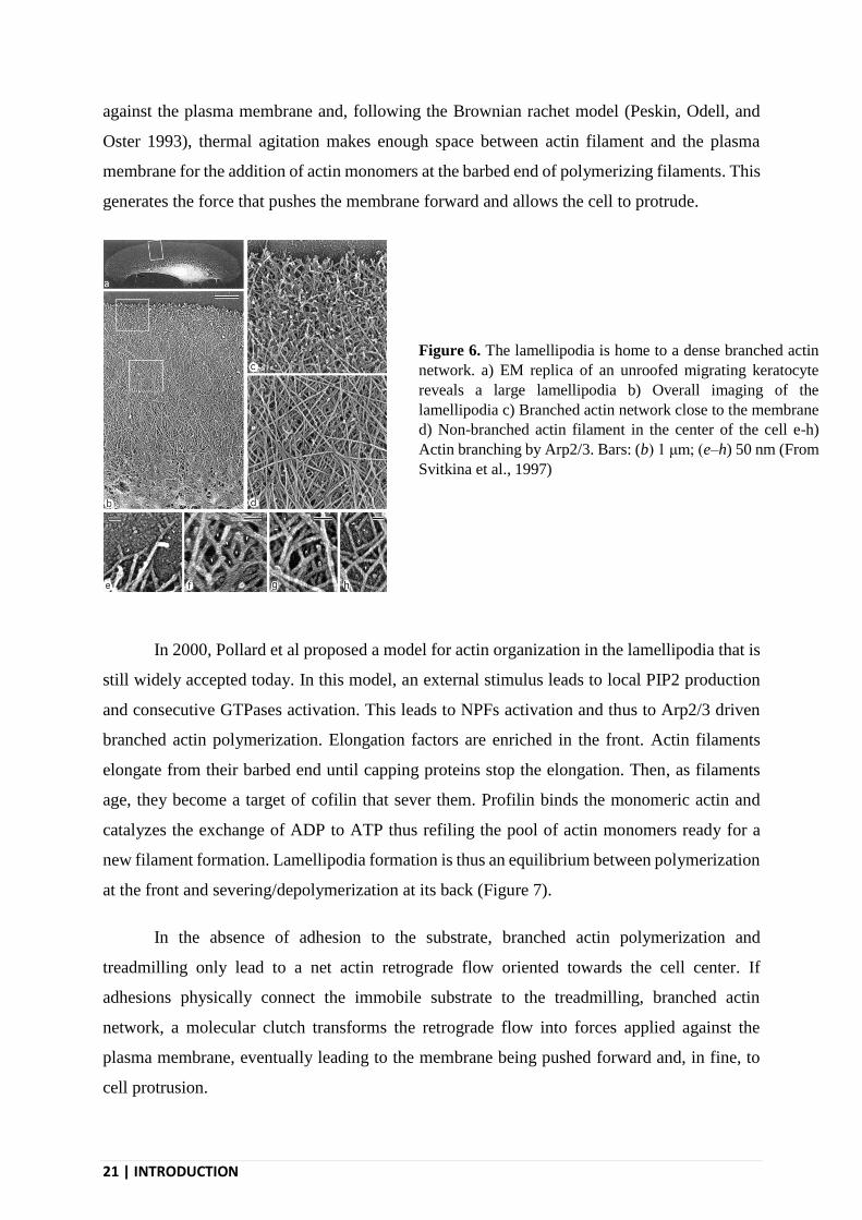
against the plasma membrane and, following the Brownian rachet model (Peskin, Odell, and
Oster 1993), thermal agitation makes enough space between actin filament and the plasma
membrane for the addition of actin monomers at the barbed end of polymerizing filaments. This
generates the force that pushes the membrane forward and allows the cell to protrude.
In 2000, Pollard et al proposed a model for actin organization in the lamellipodia that is
still widely accepted today. In this model, an external stimulus leads to local PIP2 production
and consecutive GTPases activation. This leads to NPFs activation and thus to Arp2/3 driven
branched actin polymerization. Elongation factors are enriched in the front. Actin filaments
elongate from their barbed end until capping proteins stop the elongation. Then, as filaments
age, they become a target of cofilin that sever them. Profilin binds the monomeric actin and
catalyzes the exchange of ADP to ATP thus refiling the pool of actin monomers ready for a
new filament formation. Lamellipodia formation is thus an equilibrium between polymerization
at the front and severing/depolymerization at its back (Figure 7).
In the absence of adhesion to the substrate, branched actin polymerization and
treadmilling only lead to a net actin retrograde flow oriented towards the cell center. If
adhesions physically connect the immobile substrate to the treadmilling, branched actin
network, a molecular clutch transforms the retrograde flow into forces applied against the
plasma membrane, eventually leading to the membrane being pushed forward and, in fine, to
cell protrusion.
Figure 6. The lamellipodia is home to a dense branched actin
network. a) EM replica of an unroofed migrating keratocyte
reveals a large lamellipodia b) Overall imaging of the
lamellipodia c) Branched actin network close to the membrane
d) Non-branched actin filament in the center of the cell e-h)
Actin branching by Arp2/3. Bars: (b) 1 μm; (e–h) 50 nm (From
Svitkina et al., 1997)

INTRODUCTION | 22
I.3.1.3 - Other actin structures in cell migration
Besides the lamellipodia that is the protruding region of the cell, other actin-rich
structures play important functions during cell migration. Behind the lamellipodia, is a larger
region called the lamella in which nascent adhesions mature to form proper focal adhesion
structures. It is also the region where actin stress fibers are formed and play a role in migration.
A very important player of cell migration is found in this area, and is actually excluded from
the lamellipodia: myosin-II. Myosin-II is a molecular motor that, opposite to other myosins, do
not move along actin filament but is able to slide two anti-parallel filaments through
Figure 7. Proposed model for actin organization in the lamellipodia. An external stimulus leads to local PIP2
production and consecutive GTPases activation. This leads to NPFs activation and thus to Arp2/3 driven
branched actin polymerization. Elongation factors are enriched at the front. Actin filaments elongate from
their barbed-end until capping proteins stop the elongation. Then, as filaments age, they become a target of
cofilin that severs them. Profilin binds the monomeric actin and catalyzes the exchange of ADP to ATP thus
refiling the pool of actin monomers ready for a new filament formation. Lamellipodia formation is thus an
equilibrium between polymerization at the front and severing/depolymerization at its back. (From Pollard and
Borisy, 2003)
23 | INTRODUCTION
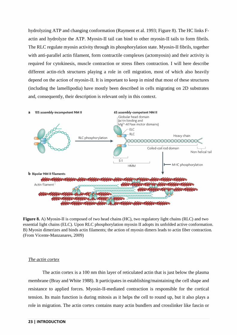
hydrolyzing ATP and changing conformation (Rayment et al. 1993; Figure 8). The HC links F-
actin and hydrolyze the ATP. Myosin-II tail can bind to other myosin-II tails to form fibrils.
The RLC regulate myosin activity through its phosphorylation state. Myosin-II fibrils, together
with anti-parallel actin filament, form contractile complexes (actomyosin) and their activity is
required for cytokinesis, muscle contraction or stress fibers contraction. I will here describe
different actin-rich structures playing a role in cell migration, most of which also heavily
depend on the action of myosin-II. It is important to keep in mind that most of these structures
(including the lamellipodia) have mostly been described in cells migrating on 2D substrates
and, consequently, their description is relevant only in this context.
The actin cortex
The actin cortex is a 100 nm thin layer of reticulated actin that is just below the plasma
membrane (Bray and White 1988). It participates in establishing/maintaining the cell shape and
resistance to applied forces. Myosin-II-mediated contraction is responsible for the cortical
tension. Its main function is during mitosis as it helps the cell to round up, but it also plays a
role in migration. The actin cortex contains many actin bundlers and crosslinker like fascin or
Figure 8. A) Myosin-II is composed of two head chains (HC), two regulatory light chains (RLC) and two
essential light chains (ELC). Upon RLC phosphorylation myosin II adopts its unfolded active conformation.
B) Myosin dimerizes and binds actin filaments; the action of myosin dimers leads to actin fiber contraction.
(From Vicente-Manzanares, 2009)

INTRODUCTION | 24
α -actinin. It contains both actin bundle and branched actin and it is mainly nucleated via Arp2/3
and formins (Bovellan et al. 2014). It is bound to the plasma membrane by ezrin, radixin and
moesin (ERM proteins). It is a very dynamic structure that contain many actin regulators.
Cortical actin is particularly important during amoeboid migration as the dynamic interplay
between the cortex and the plasma membrane is responsible for bleb formation and resolution
(Blaser et al. 2006).
Actin stress fibers
Stress fibers are long cables (up to 10 μm) of antiparallel actin filaments that are linked
together by myosin-II and α-actinin. In addition to myosin-II and α-actinin, many actin binding
proteins are associated with stress fibers, including the actin recruiter tropomyosins. These
proteins have a fast turnover, making stress fibers dynamic structures (Tojkander et al. 2011).
Stress fibers are found from the lamella to the rear of the cell (Cramer, Siebert, and Mitchison
1997). Thanks to myosin sliding along antiparallel actin filaments, stress fibers are contractile
structures and are, most of the time, under tension. They can be associated to focal adhesions
(FAs) at one or both of their extremities and the tension generated by stress fibers helps FAs to
mature (Vogel 2006). They participate in mechanotransduction, transmit forces and are
necessary for the retraction of the rear of the cell during migration.
Stress fibers can be divided into three subtypes that also represents different maturation
steps of these structures (Figure 9). Transverse arcs are located in the lamella and oriented
parallel to the leading edge. They are not associated with adhesion sites but their contraction
participates to the actin retrograde flow that is also observed in the lamella (Zhang et al. 2003).
Dorsal stress fibers are located on the dorsal side of the cell, perpendicular to the direction of
leading edge extension and are only attached to the substrate through one adhesion site. Ventral
stress fibers are anchored to FAs at both their can thus exert strong contractile forces. Ventral
stress fibers are at the back and promote rear contraction, detachment and displacement
(Mitchison and Cramer 1996).
Stress fibers arise from existing actin filaments coming from the lamellipodia
reorganization. Cofilin-severed actin filaments at the rear of the lamellipodia start to be enriched
in myosin-II and α-actinin when entering the lamella, and to reorganize into transverse arc of
antiparallel actin filament bundles (Burnette et al. 2011; Shemesh et al. 2009). Nucleation

25 | INTRODUCTION
activity associated with transverse arc dynamics relies on formins (mDia and FHOD1) via the
Rho/ROCK pathway (Watanabe et al. 1997). Dorsal fibers come from actin polymerization at
adhesion sites. Ultimately, myosin-II mediated contractions leads to transverse arc and dorsal
fibers reorganization into ventral stress fibers (Hotulainen and Lappalainen 2006).
Filopodia
Filopodia are small, pointy membrane outgrowth that form mostly in the lamellipodia.
They serve to sense and explore the cell environment. They form in contact zones with the ECM
and their bundled, parallel actin filaments structure is connected to nascent adhesions (Steketee
and Tosney 2002). The particular organization of actin filaments in filopodia is controlled by
fascin that can only bundle parallel filaments. Formins, located at the tip of filopodia, ensure
further filament elongation (Yang and Svitkina 2011). Myosin X transports receptors to
filopodia tips for effective sensing (Jacquemet, Hamidi, and Ivaska 2015). Among these
receptors are integrins, cell-cell receptors, growth factor receptors and chemokines receptors.
Filopodia are mostly used to sense the environment, to transduce external stimuli into signaling.
They are extensively used at the beginning of cell spreading (Albuschies and Vogel 2013) but
C A
Figure 9. A) In the cell, stress fibers can be sorted into 3 categories: dorsal stress fibers that have
one extremity associated with FAs, ventral stress fibers that have two extremities associated with
FAs and transverse arc that are not associated to FAs. B) Actin staining showing dorsal stress fibers
(red), ventral stress fibers (green) and transverse arc (yellow). C) Structure and composition of a
ventral stress fiber. (Adapted from Tojkander et al., 2012)
INTRODUCTION | 26
also during cell migration where they have been proposed to regulate contact inhibition
(Arjonen et al. 2014) or to recognize the ECM topography. They have also been proposed to
produce retraction forces (Jacquemet, Hamidi, and Ivaska 2015).
I.3.2 - Adhesion structures
Adhesions to the ECM are critical for cell migration as they allow the actin machinery
to be physically connected to the ECM. This allows the actin retrograde flow to be slowed down
and consequently, the barbed ends of lamillipodia actin filaments to efficiently push the plasma
membrane forward. Adhesions are formed around ECM receptors but also contains many
cytosolic proteins that are critical for adhesion sites functions. These proteins fulfil different
functions and are recruited at adhesion sites through direct or indirect binding of the cytosolic
tails of ECM receptors. To date, different types of adhesion complexes have been described,
that, for most of them, display a similar composition and depends on connections with the actin
cytoskeleton. Besides the classical focal adhesions (FAs), other structures sharing to some
extend similarities with FAs such as invadopodia, podosomes and hemi-desmosomes can exist
in some cells and, in addition to adhesion, fulfil some specialized functions. More recently, new
types of adhesion structures have been proposed that do not rely on actin but that contain
clathrin and clathrin-associated endocytic machinery components. I will here describe the most
canonical adhesion structures (that will be termed “focal adhesions” for practical reasons)
before briefly evoking clathrin-coated structures.
I.3.2.1 - ECM receptors
Several families of ECM receptors co-exist in cells, but the integrin family is widely
considered to be the main one as integrins bind to most of the ECM components. In addition to
integrins, other receptors such as the discoidin domain receptors, GPV1, LAIR-1 and the
mannose receptor can take part in adhesion (Leitinger and Hohenester, 2007). On the top of
their role ECM binding, these receptors also activate downstream signaling pathways with
important physiological consequences.

27 | INTRODUCTION
Discoidin domain receptors
Discoidin domain receptors (DDRs) are a family of collagen receptors containing two
members, DDR1 and DDR2 (Shrivastava et al. 1997). DDR1 is mainly found in epithelial cell
and DDR2 in mesenchymal cells. They can recognize different collagen types, but both bind to
collagen type I. They bind to the GVMGFO collagen region (Xu et al. 2000) but only in the
triple helical collagen configuration (Vogel et al. 1997).
DDRs are the only Receptor Tyrosine Kinases (RTK) that bind to the ECM. Like other
RTKs DDRs dimerize upon ligand binding. This leads to autophosphorylation of the receptors
and activation of downstream effectors like Cdc42, ERK1/2-MAPK or members of the STAT
family (Ongusaha et al. 2003; Wang et al. 2006). Interestingly, DDRs require a long ligand
presentation, 2 hours, and remain activated for 16 hours (Shrivastava et al. 1997; Vogel et al.
1997). They are involved in adhesion, migration, ECM remodeling and proliferation.
Integrins
The integrin family of ECM receptors is mainly involved in cell-matrix adhesion but can also
play a role in cell-cell adhesion. They serve to analyze the properties and composition of the
cell environment and they transform these physical and chemical external stimuli into the
adapted intracellular response.
Integrins role can be divided into two part as they are both adhesion molecules and
signaling receptors. As adhesion molecule they link the cell to the ECM and initiate the
formation of large adhesion complexes. As signaling receptors they control cell survival,
cytoskeleton dynamic, cell cycle progression, polarization or mechanosensing. Integrins
subunit expression level can vary over time or depending of the cell type and this defines to
what component of its surrounding matrix the cell can bind.
Integrins are heterodimer composed of non-covalently bound α and β subunit. α subunits
are composed of around 1000 amino acids and β subunits a bit shorter, being composed of
around 750 amino acids. Their heterodimerized, large N-term extracellular domains form the
structural entity endowed with the capacity to bind to ECM proteins and thus provide dimer-
ligand specificity. The transmembrane domains regulate heterodimerization. Integrin
dimerization initially occurs in the endoplasmic reticulum (ER), prior to reaching the cell

INTRODUCTION | 28
surface (M. J. Humphries 2000). The short C-term intracellular domains provide binding to
actin and other adhesion proteins. Integrins are non-enzymatic and thus rely on their partners
for signaling.
18 α and 8 β different subunits can associate into a total of 24 different heterodimers
reported so far (Sheppard 2000). A specific heterodimer is mostly associated to one type of
ECM component. On the opposite, a given ECM protein can be recognized by several
heterodimers (Humphries, Byron, and Humphries 2006; Figure 10A). As a consequence, a cell
adhering to a particular ECM protein can recruit different heterodimers to do so. As an example,
collagen type I is recognized by 5 integrin heterodimers: α1β1, α2β1, α10β1, α11β1 and αXβ2
(Humphries, Byron, and Humphries 2006).
Integrins have been historically identified as the receptor for the RGD (arg-gly-asp)
domain of fibronectin. RGD domains are also responsible for the integrins binding to
vitronectin or osteopontin. Nevertheless, even if RGD sequence is found in collagen, integrins
that bind to collagen actually recognize the triple helical GFOGER sequence (Knight et al.
1998), the later identified GLOGER sequence or the GROGER motif (Raynal et al. 2006).
Finally, integrin’s binding to its ligand is also dependent on cation such as Ca2+, Mn2+, Mg2+
that are required to support the proper structural organization of the high affinity dimers (Hu,
Barbas, and Smith 1996).
Integrins can be found in two conformations (Figure 10B). In the inactive, folded
conformation, the extracellular domains are bent and show a low affinity for the ligand (Nishida
et al. 2006). In the active conformation, the heterodimer is unfolded and displays a very strong
affinity for its ligand (Jahed et al. 2014). The dimerization of the transmembrane domains favors
the inactive state (Wegener et al. 2007). Integrin activation can be regulated by different
phenomenon. Inactivated integrins diffuse freely at the plasma membrane and can be activated
by both ligand binding or intracellular partners like talin.

29 | INTRODUCTION
I.3.2.2 - Focal adhesions
Focal adhesions (FAs) are clusters of proteins forming around activated integrins. They
not only physically link the cell to the ECM but also play a major role in signaling in response
to physical cues from the environment and in interacting with the actin cytoskeleton, thus
playing a major role in cell migration.
General description of FAs
Many proteins take part in FAs assembly and dynamics and, together, they form what
is generally described as “the adhesome”. Studies found 148 resident proteins at Fas and another
84 associated proteins (Winograd-Katz et al. 2014). FAs proteins can be divided into 4 main
groups:
- Adaptor proteins, scaffold, contact protein (eg. talin, ILK, paxillin, vinculin)
- Cytoskeleton binding proteins (eg. α -actinin)
- Enzymes (eg. FAK, Src, PKC, PI3K)
- Small GTPases and GTPases regulators (eg. Rho, Rac, cdc42)
Figure 10. A) Representation of integrin-ligand interactions. Integrin-ligand interaction specificity
is given by the dimer composition. (From Barczyk, 2010) B) Inactive conformation of integrin (left)
and a hypothetical model of the open, active form (right), with a fibrinogen peptide in red and a talin
domain in magenta. The α integrin is in blue and the β integrin is in green. (From
https://pdb101.rcsb.org/motm/134)

INTRODUCTION | 30
The many binding sites that are found in these proteins make FAs a very complex and
interconnected network. Nevertheless improvement in imaging technics allowed to start
uncovering the substructural organization of FAs (Figure 11; Kanchanawong et al. 2010). These
studies have proposed an organization in three functional layers:
- The integrin-signaling layer
This is the most plasma membrane proximal layer and comprises the cytosolic tail of
integrins as well as cytosolic factors directly binding this cytosolic tail. Talin is a central
regulator of integrin affinity and FAs dynamics. Talin is a large protein that adopt an elongated
shape. While the head of talin is very close to the plasma membrane, the protein also spans
across the two other layers of FAs. Talin binding to β integrin subunits leads to the separation
of α and β cytoplasmic tails. This favors the active, high affinity state of integrin dimers and
leads to strong ECM engagement (Wegener et al. 2007). Talin interacts with many other
proteins including signaling proteins. It makes the first direct link with cytoskeleton as it also
binds F-actin in more distal layers (Lee et al. 2004). Its four integrin binding sites participate in
integrin clustering (Klapholz et al. 2015). Another central player of this layer is the Focal
adhesion kinase (FAK). FAK is both a scaffold for other FAs-associated proteins and the major
FAs signaling protein. Its phosphorylation by Src leads to the activation of Rho-GTPases like
RhoA, cdc42 or Rac-1 (Schaller 2010). It is not required for adhesion formation or actin linking
but regulates FAs dynamics (Mitra, Hanson, and Schlaepfer 2005). Paxilin is a signaling and
Figure 11. Focal adhesions are composed of different layers that are defined by their role,
composition and distance to the plasma membrane. (From Kanchanawong et al., 2010)
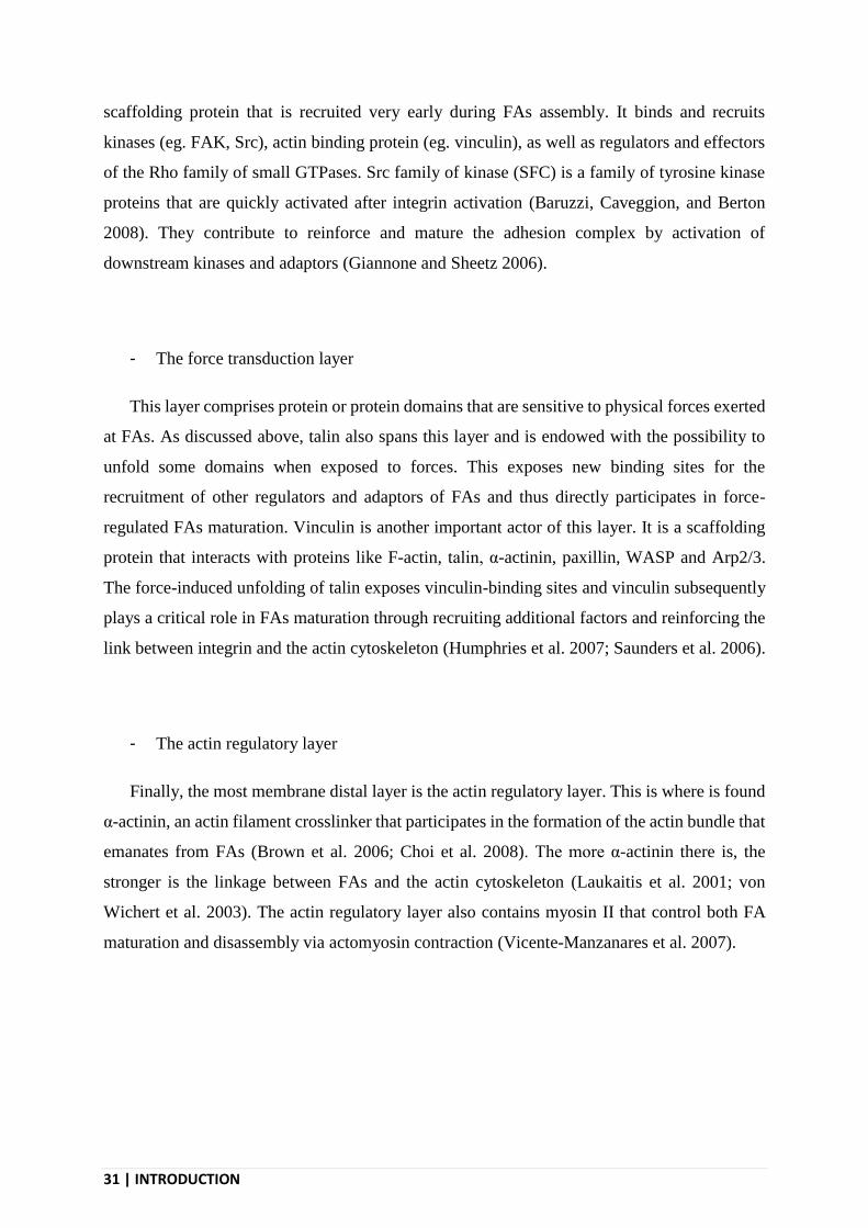
31 | INTRODUCTION
scaffolding protein that is recruited very early during FAs assembly. It binds and recruits
kinases (eg. FAK, Src), actin binding protein (eg. vinculin), as well as regulators and effectors
of the Rho family of small GTPases. Src family of kinase (SFC) is a family of tyrosine kinase
proteins that are quickly activated after integrin activation (Baruzzi, Caveggion, and Berton
2008). They contribute to reinforce and mature the adhesion complex by activation of
downstream kinases and adaptors (Giannone and Sheetz 2006).
- The force transduction layer
This layer comprises protein or protein domains that are sensitive to physical forces exerted
at FAs. As discussed above, talin also spans this layer and is endowed with the possibility to
unfold some domains when exposed to forces. This exposes new binding sites for the
recruitment of other regulators and adaptors of FAs and thus directly participates in force-
regulated FAs maturation. Vinculin is another important actor of this layer. It is a scaffolding
protein that interacts with proteins like F-actin, talin, α-actinin, paxillin, WASP and Arp2/3.
The force-induced unfolding of talin exposes vinculin-binding sites and vinculin subsequently
plays a critical role in FAs maturation through recruiting additional factors and reinforcing the
link between integrin and the actin cytoskeleton (Humphries et al. 2007; Saunders et al. 2006).
- The actin regulatory layer
Finally, the most membrane distal layer is the actin regulatory layer. This is where is found
α-actinin, an actin filament crosslinker that participates in the formation of the actin bundle that
emanates from FAs (Brown et al. 2006; Choi et al. 2008). The more α-actinin there is, the
stronger is the linkage between FAs and the actin cytoskeleton (Laukaitis et al. 2001; von
Wichert et al. 2003). The actin regulatory layer also contains myosin II that control both FA
maturation and disassembly via actomyosin contraction (Vicente-Manzanares et al. 2007).

INTRODUCTION | 32
Maturation and life-cycle of FAs
Upon integrin clustering, FAs mature from small proteins cluster often nascent
adhesions into larger structures by recruiting more and more proteins (Figure 12A). Their shape,
signaling and role in adhesion evolve with their composition (Vicente-Manzanares and Horwitz
2011). First formed are nascent adhesions that can first mature into focal complexes before
further maturation into proper FAs that ultimately disassemble at the rear of the cell, allowing
the cell body to translocate forward (Figure 12B). These distinctions are mostly practical as
FAs maturation is more a continuum than a distinct succession of discrete class of adhesions.
Nascent adhesions for at clustered integrin sites as dot-shaped structures of about 200nm
in diameter. Their nucleation mechanism remains unclear, but they only form at the cell front
(Galbraith, Yamada, and Galbraith 2007). Nascent adhesions formation requires F-actin
polymerization that occurs at the very leading edge of the cell in a structure called the
lamellipodia (Alexandrova et al. 2008; Choi et al. 2008). They exhibit strong phosphorylation
Figure 12. A) Focal adhesions mature by sequential recruitment of proteins B) As they mature and
recruit proteins focal adhesions elongate. They disassemble when once at the rear of the cell. (From
De Pascalis and Etienne-Manneville., 2017) C) Zyxin (Z) and vinculin (Vi) staining showing the
different shapes of focal adhesion in U20S cells (From Legerstee et al., 2019)

33 | INTRODUCTION
capacities (at FAK, Src, Paxilin) that participate to protein recruitment and promote branched
actin polymerization in the lamellipodia. They could be responsible for the strong traction force
observed at the very front of the cell (Beningo et al. 2001; Munevar, Wang, and Dembo 2001).
Most of these short-lived structures (30s) (Choi et al. 2008) disappears as they are
dragged backward by the actin retrograde flow that results from the intense actin polymerization
against the plasma membrane of the leading edge, in the lamellipodia. The few ones associated
with actin bundles can persists to form focal complexes (Oser and Condeelis 2009).
Focal complexes are up to 1 μm elongated structures. Their maturation relies on myosin
II contraction (del Rio et al. 2009) and coincide with the transition from the lamellipodia to the
more distal lamella. Their composition doesn’t change as compared to nascent adhesions (Choi
et al. 2008). Focal complexes then disassemble or mature into proper FAs.
FAs are ovoid structures of 2 μm to 10 μm long. Their turnover is quite slow with a
lifetime of several minutes. The more rigid is the substrate the larger FAs are (Balaban et al.
2001). Phospotyrosine levels are reduced over FAs maturation as the adhesion progressively
loses its signaling role for a more adhesive one (Ballestrem et al. 2006). Their composition
changes as compared to focal complexes with more vinculin, paxillin or α-actinin being
recruited and they also start to associate with actin stress fibers. Activation of myosin II in stress
fibers leads to fiber contraction and thus to more forces being exerted at FAs (Katoh et al. 2001).
These forces participate to the growth of FAs to a certain threshold.
Finally, integrin disengagement and adhesion disassembly at the cell’s rear are required
to end signaling and for the cell to move for forward. FAs disassembly is driven by many
mechanisms. One possible mechanism involves the protease calpain that cleaves integrins or
talin (Franco et al. 2004). Myosin II-driven actin contraction can drive FAs maturation to a
certain threshold as mentioned above. However, when forces become too important, they can
lead to FAs disassembly by weakening integrin interaction with ECM or physically
disconnecting FAs from the actin cytoskeleton (Chrzanowska-Wodnicka and Burridge 1996).
In that latter case, FAs can detach from the cell and remain attached to the ECM. Microtubules
also play a role in FAs disassembly, potentially through bringing a relaxing factor to FAs
(Kaverina, Krylyshkina, and Small 1999). Finally, integrin endocytosis participates in FAs
disassembly in a FAK- and dynamin-dependent manner (Ezratty et al. 2009; Ezratty, Partridge,
and Gundersen 2005). All these mechanisms collaborate to regulate efficient FAs disassembly.

INTRODUCTION | 34
I.3.2.3 - Adhesive clathrin-coated structures
Clathrin-coated structures (CCSs) are primarily endocytic structures and will be
described in greater detail in the second chapter of this manuscript. Nevertheless, because CCSs
were observed in thigh contact with the substrate (Batchelder and Yarar 2010; Maupin and
Pollard 1983) and to contain ECM-engaged integrins (Batchelder and Yarar 2010; De Deyne et
al. 1998; Tawil, Wilson, and Carbonetto 1993), it was proposed that they could also serve as
adhesion structures (Lampe, Vassilopoulos, and Merrifield 2016). This hypothesis has become
more popular recently as a new type of αvβ5-enriched adhesions have been described and
shown to actually correspond to a formerly identified subset of CCSs called clathrin-coated
plaques. The formation and dynamics of these atypical adhesions are independent of actin but
depends on clathrin and clathrin-adaptors that recruits integrins such as Numb, Dab2 and ARH.
Although seemingly not connected to the actin cytoskeleton, these structures are
mechanosensitive as they assemble as a function of the substrate rigidity (Baschieri et al. 2018).
In addition, our team also recently reported the existence of another type of clathrin-
based adhesions (Elkhatib et al. 2017). These structures are called Tubular Clathrin/AP-2
Lattices (TCALs) because they form at contact sites with collagen fibers and wrap around and
pinch the fibers. TCALs are enriched in β1 integrins and are used by cells migrating in 3D to
grab collagen fibers thus facilitating the stabilization of long cell protrusions, in coordination
with FAs.
These atypical adhesive structures will be discussed in greater detail in the second part
of this manuscript.
I.3.3 - Orientation of cell migration
Cells that migrate not only move, but, very often, they actually move in a direction that
is controlled by both external and internal factors. Cells can sense and interpret many kinds of
external physical and chemical cues. For instance, cells can migrate by following rigidity
gradients (eg. durotaxis; Lo et al. 2000), electric fields (eg. galvanotaxis; Zhao et al. 1996)
gradients of soluble attractants (eg. chemotaxis; McCutcheon, 1946) or substrate-bound factors
(eg. haptotaxis; Carter 1965). Directed cell migration is a key process in embryogenesis where
it allows cells to move to their final destination (Reig, Pulgar, and Concha 2014). It is also
central to immunity (Luster 1998) or nervous system wiring (Hatten 2002). I will here describe

35 | INTRODUCTION
more specifically the cases of chemotaxis and haptotaxis because they are the most prominent
forms of directed cell migration in the organism and because it is at the heart of my PhD project.
I.3.3.1 - General mechanisms of chemotaxis and haptotaxis
Many different kinds of secreted proteins have been shown to be able to attract cells:
growth factors (EGF, FGF, VEGF, IGF-1, PDGF, HGF, TGF- β…), chemokines (CCR1,
CCR2, CCR3, CXR1, CXR3…) and matrix proteins (fibronectin, vitronectin, collagen I….).
However, an isotropic distribution of these cues leads to random migration. In order to orientate
cell migration, extracellular cues need to be asymmetrically distributed, in a gradient. The
gradient depth also needs to be adequate, as the difference of cues concentration at the front
versus the rear of the cell must be sensed with sufficient sensitivity by the cell. Indeed, directed
cell migration implies that the asymmetrically distributed external cues are sensed and
integrated into signaling pathways that polarizes the migration machinery inside the cell (Parent
and Devreotes 1999; Vorotnikov 2011).
Several chemotaxis models have been proposed and are not necessarily mutually
exclusive depending on chemoattractants and cell types considered. The “chemotaxis bias”
model is based on a proposed stabilization of the protrusion facing the stronger concentration
of chemattractant (Andrew and Insall 2007; Arrieumerlou and Meyer 2005; Insall 2010). In this
model the cell randomly generates lamellipodia in every direction. If the lamellipodia contact
enough chemoattractant it will stabilize, and otherwise it will retract. In the “compass” model,
the lamellipodia only form towards the more concentrated area of the gradient as a consequence
of local accumulation of second messengers following receptor signaling (eg. PIP3) (Rickert et
al. 2000; Swaney, Huang, and Devreotes 2010; Figure 13).
I.3.3.2 - Regulation of directed migration
Actin is the engine of locomotion and this engine can be steered by external factors in
order to produce a directed migration phenotype. These cues are sensed by cell-surface
receptors that transduce intracellular signals that, collectively, continuously reshape the
organization of the actin network in order to move in a given direction. Classically,
chemoattractant bind to two main receptor families: 1) G protein-coupled receptors (GPCRs)

INTRODUCTION | 36
and, 2) receptor tyrosine kinases (RTKs). Once activated by their ligands, these receptors
triggers intracellular signaling pathways by recruiting different adaptors and effectors.
Chemoattractants usually trigger two signals: 1) a locally restricted activating signal and, 2) a
largely diffusing inhibiting signal (Xiong et al. 2010) thus ensuring a local control on the actin
machinery.
One of the most important signaling factor generated upon receptors activation is
phosphatidylinositol(3,4,5)-trisphosphate (PIP3) (Sasaki et al. 2000). The phosphoinositide 3-
kinase (PI3K) is produce as a consequence of both GPCRs and RTKs activation that control a
localized PIP3 production and accumulation at the cell front (Figure 14). Many actin regulators
have Pleckstrin Homology (PH) domains that can interact with PIP3 (Yin and Janmey 2003).
While PI3K produces some PIP3 at the cell front, the phosphatase PTEN that accumulates at
the cell rear hydrolyzes PIP3 into PIP2 thus reinforcing cell polarization (Funamoto et al. 2002;
Iijima and Devreotes 2002). Receptor activation also leads to calcium and proton entry into the
cell. Calcium is an important second messengers that also accumulate at the front during
migration (Brundage et al. 1991). In addition, pH gradient within the cell is also known to
modulate actin related proteins such as talin, cofilin or cdc42 (Frantz et al. 2007) and seems to
be implicated in many directed migration (Martin et al. 2011; Tarbashevich et al. 2015).
Figure 13. Two model coexist to explain
chemotaxis. A) In the “chemotaxis bias” model the
cell randomly generates lamellipodia in every
direction. If the lamellipodia contact enough
chemoattractant it will be stabilized, and otherwise
it will retract. B) In the “compass” model, the
lamellipodia only form towards the more
concentrated area of the gradient as a consequence
of local accumulation of second messengers
following receptor signaling. (From Reig et al.,
2014)
37 | INTRODUCTION
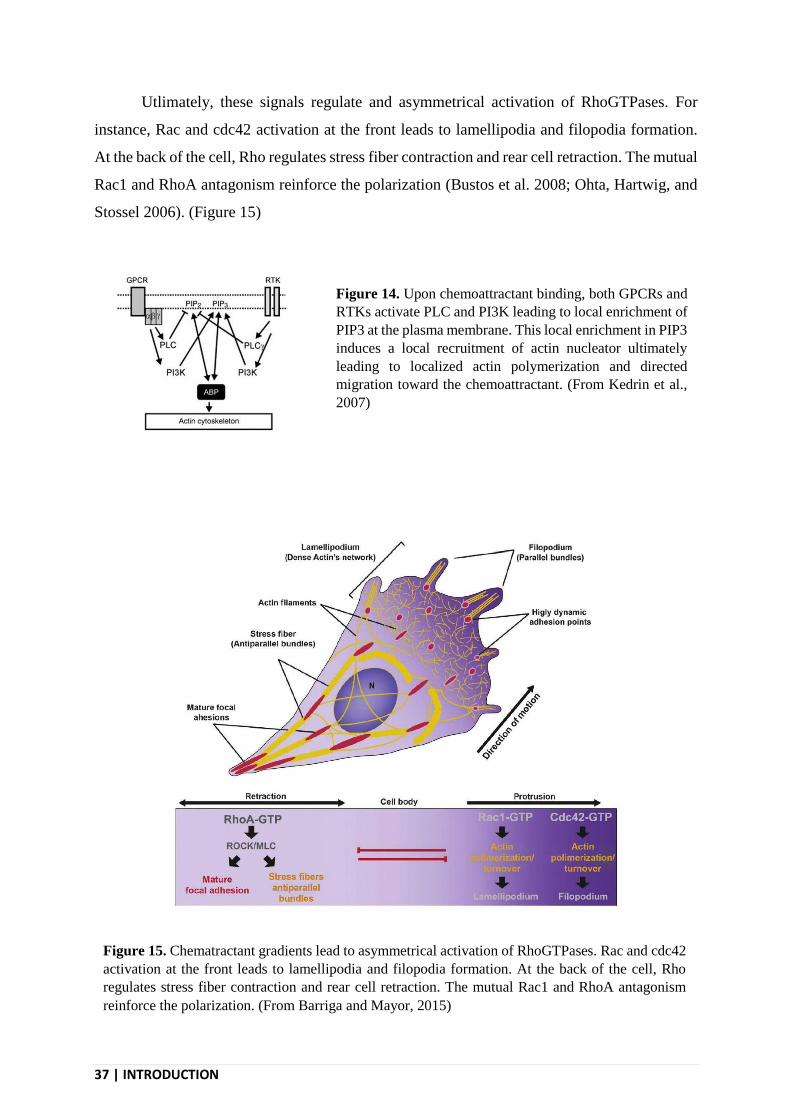
Utlimately, these signals regulate and asymmetrical activation of RhoGTPases. For
instance, Rac and cdc42 activation at the front leads to lamellipodia and filopodia formation.
At the back of the cell, Rho regulates stress fiber contraction and rear cell retraction. The mutual
Rac1 and RhoA antagonism reinforce the polarization (Bustos et al. 2008; Ohta, Hartwig, and
Stossel 2006). (Figure 15)
Figure 14. Upon chemoattractant binding, both GPCRs and
RTKs activate PLC and PI3K leading to local enrichment of
PIP3 at the plasma membrane. This local enrichment in PIP3
induces a local recruitment of actin nucleator ultimately
leading to localized actin polymerization and directed
migration toward the chemoattractant. (From Kedrin et al.,
2007)
Figure 15. Chematractant gradients lead to asymmetrical activation of RhoGTPases. Rac and cdc42
activation at the front leads to lamellipodia and filopodia formation. At the back of the cell, Rho
regulates stress fiber contraction and rear cell retraction. The mutual Rac1 and RhoA antagonism
reinforce the polarization. (From Barriga and Mayor, 2015)

INTRODUCTION | 38
Microtubules also participate in directed cell migration. Microtubules regulate proteins
and mRNAs traffic, RhoGTPases and FAs turnover (Tran et al. 2007). The microtubule
organization center (MTOC) is oriented toward the front of migrating cell to send microtubules
to the leading edge (R. Li and Gundersen 2008). If microtubules are no required to establish
cell polarity they participate in its maintenance (Etienne-Manneville 2013).
Haptotaxis towards substrate-anchored growth factors or chemokines is believed to rely
on similar mechanisms. However, in the case of haptotaxis towards ECM components,
integrins, rather than GPCRs and RTKs are used to sense the gradient (Oudin et al. 2016). In
the specific case of a fibronectin gradient, fibronectin binding to α5β1 leads to Mena
recruitment on their cytoplasmic tails. As Mena is a member of the Ena/VASP family that
promotes actin filaments elongation, this leads to localized actin reorganization. Besides a few
specific examples, and as mentioned above, haptotaxis is poorly studied and understood but is
generally believed to rely on similar mechanisms as chemotaxis.
Besides a few specific examples, and as mentioned above, haptotaxis is poorly studied
and understood but is generally believed to rely on similar mechanisms as chemotaxis.
Nevertheless, a recent study (C. Wu et al. 2012) compared the migration mechanisms involved
in chemotaxis and haptotaxis in 2D. They used Arp2/3-depleted fibroblasts to investigate the
role of lamellipodia in migration in response to gradient of soluble PDGF or substrate-bound
matrix proteins. They found that cells lacking Arp2/3 and lamellipodia could still migrate
toward a soluble PDGF gradient, most likely using filopodia sensing. On the opposite, Arp2/3
and lamellipodia were shown to be required to follow a substrate-bound gradient of matrix
protein. Nevertheless, this study doesn’t allow to draw a general mechanism of chemotaxis
versus haptotaxis as different chemotactic cues were used (growth factor vs matrix proteins)
and these two families of migrating cues bind to very different receptors. It is likely that, in
addition to the way the ligands is presented to the cell (soluble or attached to the substrate), the
mechanism driving directed migration is specific to the class of chemoattractant used in the
different assays.
I.4 - THE EXTRACELLULAR MATRIX
Cell migration occurs on a substrate that, in vivo, is the ECM. Even in vitro, cells do not
migrate directly on the glass or the plastic but on ECM components that either derive from the

39 | INTRODUCTION
serum or are secreted by cells themselves, and that are firmly sticking to the glass of the plastic.
Thus, ECM is crucial for cell migration and its properties are known to influence the way cell
migrate. Indeed, signaling from ECM receptors helps the cells to interpret its surrounding as
the matrix can vary in rigidity, density, topology as well as biochemical composition. In
response to the matrix properties, the cell adapts its phenotype. For example, ECM elasticity
influences stem cell lineage differentiation (Engler et al. 2006) and modulates cell proliferation,
survival and migration (Wells 2008; Zaman et al. 2006). The ECM also regulates the activation
and availability of many secreted proteins that diffuse in the extracellular space. In fact, by
binding and delivering growth factors, the ECM also controls cell proliferation, survival and
migration. Finally, an important factor to take into account is the dimensionality of the ECM.
While most studies have long been performed on 2D substrates coated with ECM components,
the ECM in which many cells migrate in vivo actually forms a 3D environment. This is a major
difference that is now acknowledged to deeply impact the way cells migrate.
I.4.1 - Composition and structure
The matrix composition and organization vary from one tissue to another. Its most
abundant components are glycosaminoglycans (GAG), proteoglycans (PG) and fibrillar
proteins (Frantz, Stewart, and Weaver 2010; Hynes and Naba 2012; Naba et al. 2012) that are
notably secreted by fibroblasts (Green and Goldberg 1964). Nevertheless, other proteins can be
found within the ECM. These proteins regulate both the matrix (eg. metalloproteases) and
associated cells (eg. growth factors). Fast matrix protein turnover and constant remodeling
make the matrix a very dynamic structure. The ECM can be organized into different structures
such as the basement membrane, a very thin (100 nm) and dense ECM sheet that supports
epithelia, or as a 3D scaffold supporting connective tissues. I will here only describe the
composition and organization of these latter form of organization as this is the one that supports
the migration of cancer cells used during my PhD.
I.4.1.1 - Glycosaminoglycans and proteoglycans
GAGs are repetition of disaccharides and PG (eg. decorin) are glycoproteins form by
the association of a protein chain and a glucidic chain (GAG). Most of ECM space is occupied
by PG (Järveläinen et al. 2009). These structures are very hydrophilic and participate to the

INTRODUCTION | 40
formation of a hydrate gel that help the cells resisting compression and strengthening (Mouw,
Ou, and Weaver 2014). They also bind to fibrillar components of the ECM. A very common
GAG is heparan sulfate (HS) that form heparan sulfate proteoglycans (HSPG) that can be both
purely extracellular (eg. perlecan or collagen XVIII) or membrane-bound (eg. syndecan).
HSPG are known to bind growth factors and thus play an important role in gradient formation.
HSPG-bound growth factors are protected from proteolysis and HSPGs also regulates their
diffusion through degradation of the HS chains (Sarrazin, Lamanna, and Esko 2011).
I.4.1.2 - Fibrillar proteins
Among fibrillar proteins found in the ECM, there are large glycoproteins with a
structural role (eg. elastin and collagen) and non-collagenic glycoproteins that mostly bring
resistance to the ECM (eg. laminin and fibronectin). All these ECM proteins binds to each other
to form a deeply interconnected network. In addition, collagens can also be crosslinked into
more or less dense networks (Hynes and Naba 2012). Once secreted, the precursor tropoelastin
assembles into fibers before to be covered with glycoproteins microfibrils (Wise and Weiss
2009). Laminin is a heterotrimeric protein that self-assemble into a cross-linked web and is
critical component of basement membranes. Fibronectin helps to organize the ECM and plays
a major role in cell adhesion. It is secreted by many cell types. It can help cells to sense ECM
tension as cryptic integrin binding sites are uncovered in the fibronectin structures when it
stretches (Smith et al. 2007). Fibronectin is first deposited on an existing ECM network before
to be reorganized into a dense interconnected network (Chernousov et al. 1991).
I.4.2 - Collagen type I
The collagen superfamily comprises 28 members in vertebrates and collagens are the
most abundant proteins in our body. Collagens act as structural scaffolds that can also bind
ECM partners and growth factors. Type I collagen is ubiquitous but other collagen types
expression can be restricted to specific tissues. Type I collagen is produced by fibroblasts,
endothelial and epithelial cells (Bosman and Stamenkovic 2003). It is the major constituent of
the stroma in which cancer cells escaping from the primary tumors have to migrate.

41 | INTRODUCTION
I.4.2.1 - Collagen structure and fibrillogenesis
Type I collagen expression is regulated by EGF and Integrin α2β1 activation (Creely et
al. 1990; Ivaska et al. 1999). Collagen is secreted as a triple helical protein called tropocollagen.
It is composed of three α chains that can be identical or not. Chains contains Gly-X-Y repetition,
X and Y often being proline and hydroxyproline. Presence of proline favors chain folding and
hydroxyproline makes hydrogen bond between chains. Tropocollagens presents a 300 nm long
and 2 nm wide triple helix structure that can further establish covalent bounds between them to
form microfibrils. These microfibrils can then assemble into fibers that can be up to 500 nm
wide and 1 cm long (Craig et al. 1989) (Figure 16). The process of fibrillogenesis stabilizes the
triple helix of each tropocollagen subunits, providing a strong resistance to tensile forces
(Buehler 2006). The process of fibrilogenesis is autonomous and do not require the assistance
of cells. Consequently, Type I collagen I can conveniently polymerize and form a network in
vitro (Gross and Kirk 1958; Wood and Keech 1960). The fibers diameter and network density
vary with the polymerization conditions and particularly with the temperature (Doyle et al.
2015; Figure 17). In vivo, fibroblasts can further remodeled collagen fibers into bigger
structures like sheets and cables.
Figure 16. Collagen fiber formation
starts with the secretion of
tropocollagen, a triple helical protein.
Upon cleavage of both its extremities,
tropocollagen can assemble into
microfibrils. These microfibrils can
then assemble into fibers that can be
up to 500 nm wide and 1 cm long
(Bhattacharjee and Bansal, 2005)

INTRODUCTION | 42
I.4.2.2 - Collagen binding proteins
Type I collagen can interact with many different proteins (Figure 18). It first interacts
with its receptors and notably integrins. One microfibrils exposes on integrin binding site,
allowing the clustering of integrins on collagen fibers (Sweeney et al. 2008). Numerous GAGs
also bind to collagens to form a complex matrix. Many matrix metalloproteases (MMPs) bind
Figure 17. Effect of the temperature (22/26/30/37°C) on collagen network polymerization in vitro.
Row 1 show confocal reflection images and row 2/3 show SEM images at different magnification.
The scale bars represent 20 µm (rows 1 and 2) and 200 nm (row 3) (From Jansen et al., 2018)
Figure 18. Fibrillar collagen
can interact with many
proteins including its receptors
at the cell membrane, GAG
and PG, other fibrillar proteins
form the matrix, soluble
growth factors diffusing in the
extracellular matrix and the
different collagenases. (From
Ann and Brodsky, 2016)
43 | INTRODUCTION
and cut collagen fibers. Such cleavages can expose RGD sites in the collagen structure that can
then be bound by αvβ3 integrins (Petitclerc et al. 1999). Factors that are only found in the blood
stream such as fibrin (Reyhani et al. 2014), the von Willebrand factor (Pietu et al. 1987) and
the Platelet Collagen Receptor Glycoprotein VI (Miura et al. 2002) also bind to collagen.
Collagen is only exposed to blood in case of vessel breakage and in that case these proteins
helps the binding of platelet to collagen and thus the clot formation (Farndale et al. 2004).
Fibronectin binding to collagen (Pearlstein 1976) can guide migration through haptotaxis
(Attieh et al. 2017). Collagen also bind growth factors like hepatocyte growth factor (HGF)
(Schuppan et al. 1998) and transforming growth factor β (TGF-β) (Paralkar, Vukicevic, and
Reddi 1991).
I.4.3 - Growth factors and the ECM
Growth factors are secreted proteins that regulate cell functions such as apoptosis, cell
cycle progression, differentiation and migration. The ECM can modulate the activity of growth
factors in many ways. First, the ECM regulates the availability of many growth factors. Indeed,
binding of growth factors to the ECM modulates their diffusion rate and protects them from
proteolysis. Thus, ECM can be seen as reservoir for growth factors. ECM also regulates growth
factors spatial distribution. This regulation is key to establish gradients that are necessary for
wound healing or morphogenesis (Kreuger et al. 2004). Because the ECM binds to both
integrins and growth factors, it creates local formation of signaling complexes (Rahman et al.
2005). In addition, ECM-bound growth factors can display delayed endocytosis and
degradation that could lead to more sustained signaling (Platt et al. 2009).
MMPs play a major role in regulating growth factors activity in the ECM. MMPs free
peptides trapped in the ECM (Imai et al. 1997; Suzuki et al. 1997). Proteases can also cleave
growth factors, leading to their activation (Lyons et al. 1990; Naldini et al. 1992). In addition,
laminin and tenascin cleavage releases EGF-like domains that can activate the EGF receptor
(Schenk et al. 2003).

INTRODUCTION | 44
I.4.3.1 - Growth factors binding the ECM
Many growth factors bind to the ECM. Fibroblast growth factor (FGF) binding to HSPG
is involved in neural development (Nurcombe et al. 1993).Vascular endothelial growth factor
(VEGF) binding to the ECM plays a role in angiogenesis (Gerhardt et al. 2003). ECM-
associated hepatocyte growth factor (HGF) regulates mammary gland ductal branching (Garner
et al. 2011) and heparin-binding EGF-like growth factor (HB-EGF) stimulates smooth muscle
cells migration (Higashiyama, Abraham, and Klagsbrun 1993). HGF binds to fibronectin and
vitronectin. This leads to c-Met and integrins forming signaling complexes (Rahman et al.
2005). Tumor necrosis factor α (TNF-α) binding to fibronectin is involved in clot formation
(Alon et al. 1994). VEGF binding to fibronectin and tenascin promotes cell proliferation
(Ishitsuka et al. 2009; Wijelath et al. 2006).
Much less growth factors are known to bind to collagen type I. Platelet-derived growth
factor (PDGF) and HGF bind to collagen type I and this modulates their bioavailability
(Schuppan et al. 1998; Somasundaram and Schuppan 1996). Collagen-bound HGF maintain its
activity. Collagen type I also binds to TGF-β (Paralkar, Vukicevic, and Reddi 1991).
In addition, many research efforts are put in the creation of fusion proteins associating
growth factors and ECM-binding domains to generate clinically useful growth factors-
containing matrix scaffolds. Applied to a wound, these medicines would be more stable and
less diffusive and show some clinical advantages.
I.4.3.2 - Growth factor gradients in vivo
Gradients of growth factors have been observed in several model organism. In the
Drosophila egg chamber, border cells use EGF receptor and polio virus receptor (PVR) to
follow TGF- α and PDGF- and VEGF-related factor 1 (PV1) gradients (Duchek et al. 2001;
Duchek and Rørth 2001). Bone morphogenetic protein (BMP) gradients drive lateral cells
positioning during zebrafish gastrulation (von der Hardt et al. 2007). Heparan-bound VEGF
gradients guide endothelial cells migration-dependent vascular sprouting during mice retina
formation (Gerhardt et al. 2003). Heparan-bound CCL21 gradients in mouse skin drive
dendritic cell haptotaxis toward lymphatic vessel (Weber et al. 2013).
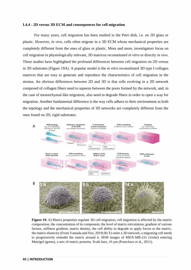
45 | INTRODUCTION
I.4.4 - 2D versus 3D ECM and consequences for cell migration
For many years, cell migration has been studied in the Petri dish, i.e. on 2D glass or
plastic. However, in vivo, cells often migrate in a 3D ECM whose mechanical properties are
completely different from the ones of glass or plastic. More and more, investigators focus on
cell migration in physiologically relevant, 3D matrices reconstituted in vitro or directly in vivo.
These studies have highlighted the profound differences between cell migration on 2D versus
in 3D substrates (Figure 19A). A popular model is the in vitro reconstituted 3D type I collagen
matrices that are easy to generate and reproduce the characteristics of cell migration in the
stroma. An obvious differences between 2D and 3D is that cells evolving in a 3D network
composed of collagen fibers need to squeeze between the pores formed by the network, and, in
the case of mesenchymal-like migration, also need to degrade fibers in order to open a way for
migration. Another fundamental difference is the way cells adhere to their environment as both
the topology and the mechanical properties of 3D networks are completely different from the
ones found on 2D, rigid substrates.
Figure 19. A) Matrix properties regulate 3D cell migration, cell migration is affected by the matrix
composition, the concentration of its component, the level of matrix reticulation, gradient of various
factors, stiffness gradient, matrix density, the cell ability to degrade or apply forces to the matrix,
the matrix elasticity (From Yamada and Sixt, 2019) B) To enter a 3D network, a migrating cell needs
to progressively remodel the matrix around it. SEM images of MDA-MB-231 (violet) entering
Matrigel (green), a mix of matrix proteins. Scale bars, 10 μm (Poincloux et al., 2011).

INTRODUCTION | 46
I.4.4.1 - Matrix degradation
In order to move in dense 3D environments, cells need to clear the way in front of them
(Figure 19B). To do so, cells can secrete metalloproteases that degrade the surrounding matrix.
Some cells that efficiently move in 2D cannot move in 3D gels because they do not express
proteases. There are two mains metalloproteases families (P. Lu et al. 2011), the matrix
metallopeptidases (MMPs) that mostly degrade fibrillar proteins and the ADAMS (A
Disintegrin And Metalloproteinase with Thrombospondin Motifs) that mostly degrade
proteoglycans.
There are 23 MMPs in human. MMP-1 (or collagenase I), MMP-8 and MMP-13 are the
three secreted collagenases. Membrane-type 1 MMP (MT1-MMP) is a transmembrane MMP
that degrades type I collagen (Sabeh et al. 2004). MMPs expression can be induced by many
growth factors including EGF (Kajanne et al. 2007). In addition collagen induces MMPs
expression through DDR signaling (Vogel et al. 1997). Like many proteases, MMPs are
secreted as inactive proenzymes that are then activated, notably by others MMPs. MT1-MMP
is targeted to the cell front where it both degrades the matrix and locally activate other MMPs
(Mori et al. 2002).
I.4.4.2 - Cell adhesion in 3D
Mechanisms of cell migration have been revealed through studies performed on 2D,
rigid environments (glass/plastic). However, in vivo, cell migrates in softer 3D matrices. In
addition with rigidity and pliability many parameters vary between 2D plastic surface and 3D
matrices. Topology in 3D is very different, ligands are not presented evenly distributed as on
planar surfaces but tightly clustered on fibers. In addition, the elasticity of a 3D collagen
network is much smaller than the one of glass or plastic (approximately 200 Pascals versus 1
gigaPascals).
Substrate rigidity positively regulates FAs size and stress fibers formation. Thus, it is
not surprising that the existence of these structures is less easy to spot in 3D matrices (Burridge
et al. 1988; Fraley et al. 2010), even if they have been observed (Cukierman et al. 2001; Kubow
and Horwitz 2011; Figure 20). In 3D, FAs are smaller and stress fibers are less aligned with the
major cell axis of the cell (Discher, Janmey, and Wang 2005; Prager-Khoutorsky et al. 2011).

47 | INTRODUCTION
FAs composition changes in 3D. In a landmark study, vinculin and FAK were confirmed to be
present at FAs in 3D networks, but FAK phosphorylation levels were shown to be much weaker
than on 2D. On the opposite, α5 integrins and paxillin colocalize in 3D but not in 2D
(Cukierman et al. 2001). 3D FA components have faster turnover than their 2D counterparts
(Doyle et al. 2012). As a conclusion, molecular players remain the same in 3D but organization,
dynamics and phosphorylation state vary.
We recently reported that cancer cells migrating in a 3D network use another type of
adhesive structures, in addition to FAs. We observed that collagen fibers pressing on the plasma
membrane generates a local membrane curvature that is a signal for local nucleation of clathrin-
coated structures (CCSs) (Elkhatib et al. 2017). These particular CCSs are enriched in β1-
integrin, a collagen receptor, and adopt the rod-like shape of fibers by forming tubular
structures. These so-called tubular clathrin/AP-2 lattices (TCALs) actually wrap around fibers
as they try and fail to internalize them. As a consequence, TCALs pinch collagen fibers and
provide new anchoring points to the 3D substrate. This helps the cell to stabilize long
protrusions by reducing the tension that raises across the protrusion as it grows. Thus, the
particular topology of 3D collagen networks allows the formation of a new class of adhesion
structures that fully participates in the migration process (see more details in the second chapter
of this manuscript).
Figure 20. Cells migrating in 3D also show focal adhesions at the leading edge. A human fibroblast
expressing EGFP-talin migrates inside an in vitro polymerized collagen network and seems to use
focal adhesions similarly as described in 2D migration. Scale bars: 10µm (Doyle and Yamada, 2015)

INTRODUCTION | 48
II - CLATHRIN-MEDIATED ENDOCYTOSIS
Endocytosis is a fundamental process that allows the cell to acquire some elements from
its environments. It relies on the formation of plasma membrane invaginations that surround
the material to be internalized. It is an active process requiring energy and that culminates in
the detachment of the invagination from the plasma membrane as it buds inside the cytosol.
The first described endocytosis process was phagocytosis (from the Greek “cell eating”).
Besides phagocytosis, a number of pinocytosis (from the Greek “cell drinking”) pathways have
then been described. The major function of these pinocytosis pathways is actually not to
internalize extracellular fluids but rather to control the uptake of cell-surface receptors and their
ligands.
Clathrin-mediated endocytosis (CME) is the major and best described pinocytosis
pathway and is used by all eukaryotic cells. It happens during the whole cell life with a notable
decrease during mitosis (Warren, Davoust, and Cockcroft 1984). During CME, cells internalize
membrane portions and their associated receptors or cargoes (eg. GPCR, RTKs, integrins and
ions channels). These receptors can be internalized with or without their ligands. They are
internalized into vesicles that further traffic inside the cytoplasm.
Because of the large range of receptors it internalizes, CME is key to many cellular
processes. CME helps to regulate the membrane composition in time and space by removing
some of its components. By controlling the composition of the membrane, it regulates most of
the cell’s interaction with its environment. It participates to the sampling of the environment. It
regulates nutrient uptake. CME also controls signaling pathways through regulating
endocytosis of signaling receptors. CME plays a major role in cell migration, notably through
FAs turnover and polarized redistribution of receptors.
II.1 - CLATHRIN-COATED STRUCTURES
CME relies on the formation of clathrin-coated structures (CCSs) at the internal leaflet
of the plasma membrane. CCSs formation is a multistep process starting with the nucleation of
these structures at the plasma membrane followed by cargo selection, coat maturation, scission
and uncoating (McMahon and Boucrot 2011). During the nucleation, a small core of proteins
including clathrin and its adaptor AP-2 are recruited at the plasma membrane and defines the

49 | INTRODUCTION
endocytic site. Cargoes (i.e. receptors) are recruited by adaptors proteins and accumulate within
the endocytic site that concomitantly starts to invaginate. At this stage, CCSs are often referred
to as “clathrin-coated pits” because of the particular shape of this structures as seen in electron
microscopy pictures. The continuous invagination process then leads to the formation of a
vesicle that is only linked to the plasma membrane by a thin neck. Dynamin-mediated scission
of this neck frees the vesicle in the cytosol. Once in the cytosol, the different constituents of the
clathrin coat dissemble, and the vesicle starts to traffic inside the cell. From then, receptors will
be either degraded in lysosomes or be recycled back to the plasma membrane (Figure 21).
II.1.1 - Main actors of CCSs
If CME is named after clathrin, over 50 other cytosolic proteins are involved in the
formation and dynamics of CCSs. Interactions between these proteins are highly organized, and
the regulation of their assembly drives the maturation of a CCSs into a vesicle (Figure 22). The
BAR proteins FCHO1,2 play a key role during nucleation. Adaptor Protein 2 (AP-2) is central
Figure 21. A) CME initiates with the binding of adaptor proteins to membrane receptors. Then
clathrin is recruited and polymerizes into a coat. The clathrin coated region invaginates
progressively to from a vesicle that finally separates from the plasma membrane and traffics inside
the cell. The protein coat disassembles and is available for a new cycle. B) EM image of a carbon
platinum replica showing different steps of CME in a fibroblast. Scale bar = 200 nm. C) EM images
of cryosliced neuron showing different steps of CME. Scale Bar = 200 nm (Higgins and McMahon,
2002).
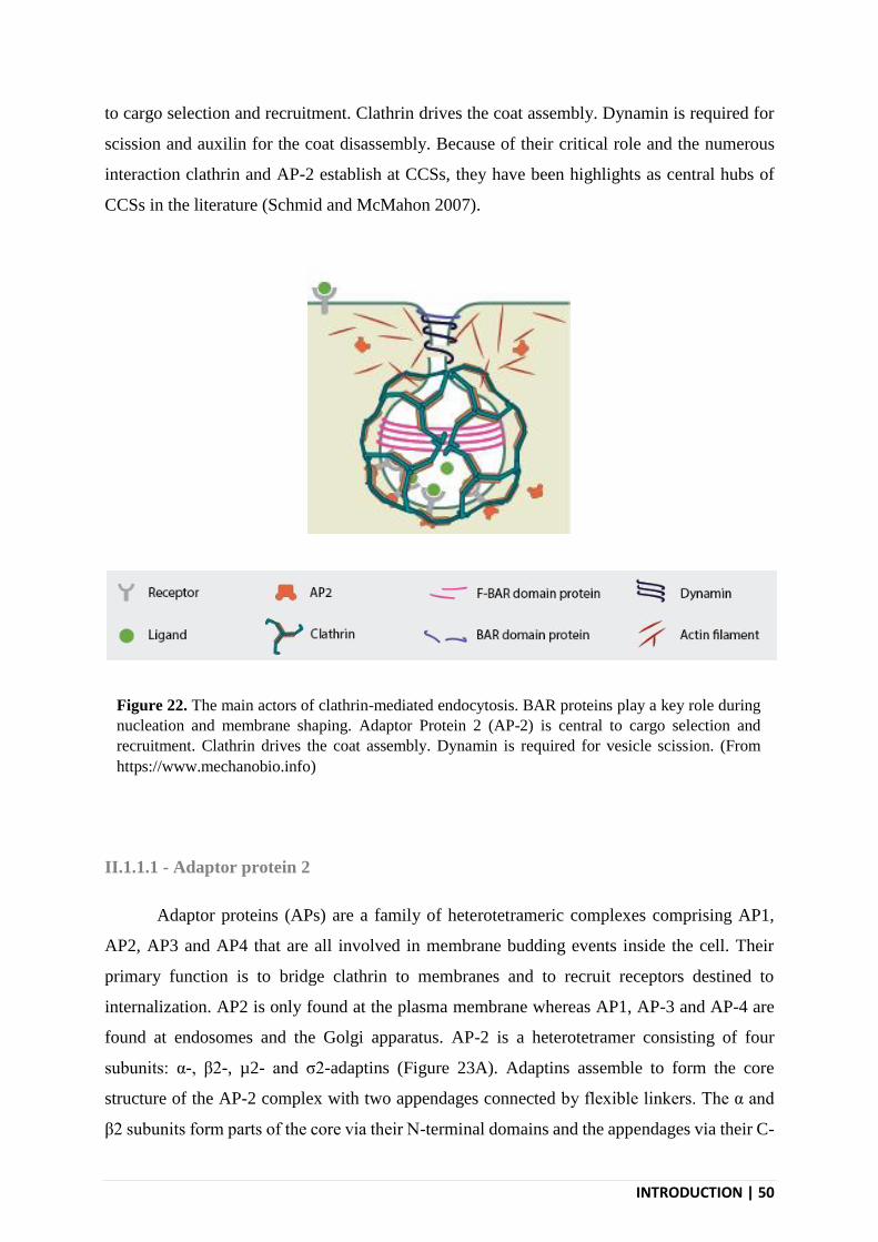
INTRODUCTION | 50
to cargo selection and recruitment. Clathrin drives the coat assembly. Dynamin is required for
scission and auxilin for the coat disassembly. Because of their critical role and the numerous
interaction clathrin and AP-2 establish at CCSs, they have been highlights as central hubs of
CCSs in the literature (Schmid and McMahon 2007).
II.1.1.1 - Adaptor protein 2
Adaptor proteins (APs) are a family of heterotetrameric complexes comprising AP1,
AP2, AP3 and AP4 that are all involved in membrane budding events inside the cell. Their
primary function is to bridge clathrin to membranes and to recruit receptors destined to
internalization. AP2 is only found at the plasma membrane whereas AP1, AP-3 and AP-4 are
found at endosomes and the Golgi apparatus. AP-2 is a heterotetramer consisting of four
subunits: α-, β2-, µ2- and σ2-adaptins (Figure 23A). Adaptins assemble to form the core
structure of the AP-2 complex with two appendages connected by flexible linkers. The α and
β2 subunits form parts of the core via their N-terminal domains and the appendages via their C-
Figure 22. The main actors of clathrin-mediated endocytosis. BAR proteins play a key role during
nucleation and membrane shaping. Adaptor Protein 2 (AP-2) is central to cargo selection and
recruitment. Clathrin drives the coat assembly. Dynamin is required for vesicle scission. (From
https://www.mechanobio.info)
51 | INTRODUCTION
terminal domains. The flexible domain of β2-adaptin is essential to interact with clathrin (Shih,
Gallusser, and Kirchhausen 1995) and the appendage domains of both α- and β2-adaptins are
involved in interactions with other adaptors and accessory proteins that participate in CCSs
formation and dynamics. α-adaptin trunk domain binds to phosphatidylinositol-(4,5)-
bisphosphate (PIP2), a phospholipid specifically found at the plasma membrane, and thus
provides anchorage to and specificity for the plasma membrane. µ2-adaptin and a structural
domain formed by of α2- and σ-adaptins are responsible for direct interaction with receptors
through their tyrosine-based YxxΦ (with Φ being a bulky hydrophobic amino-acid) or
dileucine-based (D/E)xxxL(L/I) internalization motifs, respectively. Phosphorylation of μ2-
adaptin induces a conformational change that increases AP-2 affinity for both cargos and PIP2
(Höning et al. 2005). The conformational changes occur after initial, weak interaction with PIP2
at the plasma membrane and is thus crucial to ensure early stages of CCSs assembly. It has been
suggested that AP-2 may not be necessary for the formation of all CCSs in particular
experimental conditions. However, in physiological conditions, AP-2 is present in all CCSs and
its depletion by siRNAs leads to an almost complete depletion of CCSs. Because of the many
interactions it has with other CCSs components and its prominent role in CCSs formation, AP-
2 is considered as the central hub of CCSs (Conner and Schmid 2003; Schmid and McMahon
2007).
II.1.1.2 - Clathrin
Clathrin doesn’t directly bind to membranes and thus needs adaptors to be recruited at
endocytic sites. Clathrin assembles into a hexamer constituted by three clathrin heavy chains
(CHC, 190 Kda) each associated with a clathrin light chain (CLC, 30 Kda) (S. H. Liu et al.
1995; Ungewickell and Ungewickell 1991). The heavy and light chains assemble into a
triskelion that can polymerize by themselves into a basket-like cage in vitro, in particular buffer
conditions (Crowther and Pearse 1981; Keen, Willingham, and Pastan 1979; Ungewickell and
Branton 1981; Figure 23B). In vivo, adaptors-assisted polymerization of clathrin triskelia on
membranes forms a honey comb-like structure that is constituted either only of hexagons when
the clathrin coat is flat, or of a mixture of hexagons and pentagons when the clathrin-coat is
curved (Fotin et al. 2004, 2006). The role of clathrin has long been seen as a driver of membrane
curvature generation and indeed, in vitro studies suggest that it could fulfil this function
(Dannhauser and Ungewickell 2012). However, many other proteins of CCSs play a role in

INTRODUCTION | 52
membrane curvature and clathrin may only be there to stabilize this curved state (B. L. Scott et
al. 2018).
II.1.1.3 - Accessory proteins
In addition to clathrin and AP-2, many other proteins participates in the formation and
dynamics of CCSs. Most of them can bind to both PIP2 and AP-2 and/or clathrin. These proteins
are involved in cargo selection, membrane bending, clathrin recruitment and assembly, scission
or uncoating. The functions of these accessory proteins are highly regulated as they are recruited
at very precise stages of CCSs maturation and at very precise substructural locations (Mettlen
et al. 2018; Taylor, Perrais, and Merrifield 2011). Below, I will mention and discuss some of
these proteins in further details when describing the different steps of CCSs dynamics.
Figure 23. A) AP-2 consists of four subunits: α-, β2-, µ2- and σ2-adaptins. Theses subunits
mediate interaction with most of the actors of CME. AP-2 is a key actor of CME as it bridges
together the plasma membrane, clathrin and their cargoes. B) Clathrin assembles into a
hexamer of three clathrin heavy chains associated with three clathrin light chains. The heavy
and light chains assemble into a triskelion that can polymerize by themselves into a basket-
like cage in vitro.

53 | INTRODUCTION
II.1.2 - Life cycle of CCSs
Canonical CCSs are small (approximately 100 nm in diameter), dynamic structures
whose life cycle is precisely regulated. This cycle starts with nucleation, followed by a
maturation step that corresponds to membrane bending and vesicle formation, and then a
scission step that allows the physical separation of the nascent vesicle from the plasma
membrane, and finally an uncoating step that removes the clathrin and adaptor coat from the
vesicle. Depending on the cell type, this cycle can last between 30s and 1 min approximately.
II.1.2.1 - CCS nucleation
CCSs formation starts with the recruitment of core endocytic proteins that progressively
recruit the entire coat. A random walk of theses core proteins on the internal leaflet of the
plasma membrane was proposed to lead to CCSs formation at random positions at the plasma
membrane (Ehrlich et al. 2004). Yet, the precise molecular time course of CCSs nucleation is
still a matter of debate (Cocucci et al. 2012; Henne et al. 2010). Both AP-2 and the BAR domain
protein FCHo1/2 have been proposed as key regulators of CCS initiation. AP-2 was the first
proposed master regulator of initiation, notably because of its interaction with numerous
endocytic proteins. As it binds both clathrin and PIP2, it is key to clathrin recruitment at the
plasma membrane. AP-2 is also one of the earliest arriving protein at the endocytic site (Cocucci
et al. 2012). Nevertheless some studies suggested that AP2 depletion doesn’t completely
abolish the formation of CCSs (Aguet et al. 2013; Motley et al. 2003) but this may depend on
experimental conditions . In 2010, Henne and colleague proposed that FCHo1/2 is the main
regulator of CCSs nucleation through generating an initial, shallow membrane curvature but
this model has been challenged (Cocucci et al. 2012). Finally, PIP2 is also known to play a
major role in CME initiation (Jost et al. 1998). It helps to the recruitment of several CCSs
proteins at the plasma membrane and its depletion inhibits CCSs formation (Antonescu et al.
2011)
Receptors themselves have been proposed to be able to control the de novo formation
of CCSs upon their activation (Wilde et al. 1999). However, most studies suggest that activated
receptors are actually targeted to preexisting CCSs in order to be internalized (Rappoport and
Simon 2009; Scott et al. 2002). Yet, it is also now clear that cargo recruitment affects CCS
dynamics and stabilizes them upon nucleation (Henry et al. 2012). In addition, receptor
INTRODUCTION | 54
overexpression, or experimentally-induced clustering, leads to an increased CCSs nucleation
(Liu et al. 2010; Mettlen et al. 2010). Some viruses are also able to nucleate their own CCSs in
order to be internalized (Rust et al. 2004), but others were shown to reach preformed CCSs
(Ehrlich et al. 2004). Finally, we recently demonstrated that collagen fibers contacting the
plasma membrane induce local CCSs nucleation (Elkhatib et al., 2017). This is most likely due
to collagen fiber inducing a local plasma membrane curvature that is probably a signal
triggering local CCSs nucleation. In conclusion, CCS nucleation causes are not completely
understood, and this probably reflects variability and redundancy in the nucleation mechanisms.
II.1.2.2 - CCS maturation
Upon nucleation, CCSs may not necessarily pursue to a full endocytosis process and,
indeed, studies have suggested the existence of an “endocytic checkpoint” determined by the
coat composition and especially receptors recruitment (Aguet et al. 2013). If CCSs do not
contain enough cargos, maturation is delayed and, in some cases, the coat disassembles without
proceeding to endocytosis (Loerke et al. 2009). This allows to prevent the formation of “empty”
endocytic vesicles.
A wide array of receptors are internalized through CCSs. Receptor’s recruitment can
either be constitutive (i.e. no need for ligand binding) or require ligand-mediated activation of
the receptor. Nutrient receptors are mainly internalized constitutively whereas the
internalization of signaling receptors usually requires ligand-dependent activation. For
example, the transferrin receptor (TfR) is constitutively recruited to CCSs through a direct
interaction with AP-2 (Nesterov et al. 1999). On the opposite, the epidermal growth factor
receptor (EGFR) recruitment at CCSs only happens upon ligand binding and receptor
dimerization and involves multiple endocytosis motifs linking different CCSs proteins (Goh et
al. 2010). A full set of adaptors proteins recognize specific motifs in cargos and recruit them to
CCSs. Both FCHo1/2 and AP2 can bind to some cargoes and thus, receptors recruitment already
happens during the nucleation step (Henne et al. 2010). In addition to AP-2, there are a number
of accessory proteins that are dedicated to the recruitment of specific receptors. For instance,
β-arrestins are specialized in the recruitment of activated GPCRs (Zhang et al. 1999). Ubiquitin
Interacting Motif (UIM)-containing proteins such as Eps15 and epsin can recognize and recruit
ubiquitinated receptors to CCSs (Hawryluk et al. 2006). The β integrins domain NPX[FY] and
the FDNPVY motif of the low density lipoprotein receptor (LDLR) are recognized by the

55 | INTRODUCTION
clathrin adaptors ARH, Numb and Dab2 via their phosphotyrosine binding (PTB) domains
(Maurer and Cooper 2006).
In order to form a vesicle, CCSs need to progressively invaginate. Membrane bending
occurs mostly during the maturation step and requires different types of endocytic proteins.
Clathrin can form cages in solution (Keen, Willingham, and Pastan 1979) but can also shape
membranes into spherical buds in vitro (Dannhauser and Ungewickell 2012). It was proposed
that progressive clathrin polymerization impose a curvature to the membrane (Kirchhausen and
Harrison 1981). However, it was recently shown that the forces produced by clathrin
polymerization are not sufficient to account for the observed membrane deformation, especially
when membranes are under tension (Saleem et al. 2015). Yet, other endocytic proteins are
proposed to increase the stiffness of the clathrin coat, potentially allowing such deformations
(Lherbette et al. 2019). In vivo, clathrin polymerization synergizes with other mechanisms to
produce efficient membrane invagination. Protein with BAR domains also play a role in
membrane bending during CME. These proteins display a banana shape that allow them to
sense and/or produce membrane curvature (Figure 24A). The F-BAR proteins FCHo1/2 arrives
very early at CCSs nucleation sites and only produces a shallow invagination (Henne et al.
2010). N-BAR proteins like amphiphysin and endophilin are more potent curvature inducers
but arrive much later and are believed to organize the shape of the neck of still plasma-
membrane associated clathrin-coated vesicles (Peter et al. 2004). CLAM/AP180 and epsin are
proteins that interact with AP-2, clathrin and PIP2 and that generate curvature through of a N-
terminal amphipathic helix in the internal leaflet of the plasma membrane (Miller et al. 2015).
Finally, actin was also proposed to push the membrane in order to induce curvature (Figure
24B). Live and electron microscopy imaging confirmed the presence of actin at endocytic site
(Collins et al. 2011; Li et al. 2015) but inhibiting the actin machinery revealed that actin play
a non-obligatory role during endocytosis (Fujimoto et al. 2000). Actin polymerization actually
seems to be required in case of high membrane tension, a mechanical state that tend to oppose
budding (Boulant et al. 2011).
To conclude, many of the accessory proteins in charge of recruiting receptors can also
bend the plasma membrane making the maturation step the privileged stage of both CCSs
invagination and receptor sorting. In fine, all the mentioned mechanisms cooperate to bend the
plasma membrane until the formation of a receptor-containing vesicle that is still connected to
the plasma membrane through a thin neck.

INTRODUCTION | 56
II.1.2.3 - Vesicle fission
To be released inside the cell, the vesicle needs to break this thin neck. If there is no
doubt about dynamin requirement in this process, the exact mechanism of vesicle scission
remains unclear. There are 3 different dynamins but only dynamin-2 is ubiquitously expressed.
Dynamin-2 is a GTPase of about 100 kDa that assembles into a helix surrounding the vesicle
neck (Hinshaw and Schmid 1995). Dynamin-2 is divided into five domains: the PH domain,
the GTPase domain, the bundle signaling element (BSE), the stalk domain and the proline rich
domain (PRD) (Figure 25A). The PH domain allows membrane anchoring through PIP2
binding (Zheng et al. 1996). The BSE domain allows the transmission of conformational change
between domains. The stalk domain is required for interactions with other dynamin monomers.
The PRD interacts with other endocytic proteins.
Dynamin-2 is recruited preferentially to highly invaginated membrane such as the
vesicle neck (Roux et al., 2010). Dynamin is also recruited at clathrin structure via the PRD
interaction with endocytic proteins such as amphiphysin (Takei et al. 1999). Once at the neck,
dynamin-2 interacts with other dynamin monomers through the stalk domain. Dynamin
oligomerization progressively forms a helix around the neck. Helix formation enhances the
dynamin affinity for GTP (Song, Leonard, and Schmid 2004). Once the helix formed, GTP
hydrolysis induces a helical twist that constricts the neck (Antonny et al. 2016; Figure 25B). It
is proposed that the helix twist brings the neck membranes close enough to each other so that
Figure 24. Different mechanisms of membrane bending cooperate to produce the membrane
invagination during CME. A) BAR proteins display a banana shape that allows them to
sense and/or produce membrane curvature. The different BAR proteins cover a large range
of membrane curvature and thus are sequentially recruited to fit the increasing invagination
of the membrane. (From Qualmann et al., 2011). B) Actin polymerization has been proposed
to participate to membrane bending. It polymerizes against to neck and participates in its
constriction. (From Collins et al., 2011)

57 | INTRODUCTION
they spontaneously fuse due to thermal fluctuation (Bashkirov et al. 2008). Dynamin inhibition
completely abolishes CME (Macia et al. 2006) and cells mutated for the GTPase domain display
deeply invaginated CCSs with elongated necks (Damke et al. 2001). In addition, actin has been
proposed to produce extra forces required for neck constriction during vesicle scission (Collins
et al. 2011).
II.1.2.4 - Coat disassembly
After its scission from the plasma membrane, the newly formed clathrin-coated vesicle
needs to get rid of its clathrin and adaptors coat in order to complete endocytosis. This step will
allow future inter vesicular fusion events and/or fusion of vesicles with endosomes. Coat
disassembly relies on the coordination of two mains mechanisms: disruption of the adaptor
lattice by PIP2 depletion, and action of the ATPase HSC70 directly on clathrin (Schlossman et
al. 1984). Once disassembled, the proteins of the coat can be reuse for a new round of CCS
formation.
PIP2 dephosphorylation on the vesicle by synaptojanin is involved in the coat
disassembly (Cremona et al. 1999). A specific isoform of synaptojanin binds to dynamin and
endophilin and is thus recruited at late stages of CME. PIP2 plays a major role in the coat
assembly and its disruption destabilize the coat, especially AP-2. HSC70 is recruited to clathrin-
coated vesicles by its co-factor auxilin. Auxilin binds to both clathrin and dynamin and is
recruited around the time of vesicle scission. ATPase activity of HSC70 is increased by the
Figure 25. A). 3D structure of dynamin dimers. Dynamin binds the membrane through its
pH domain and dimerizes through its stalk domains. B) Dynamin dimers polymerize to form
a helix around the vesicle neck. GTP hydrolysis leads to helix constriction and to membrane
scission. (From Faelber et al., 2013; Ferguson and De Camilli, 2012)

INTRODUCTION | 58
formation of an HSC70-auxilin-clathrin complex. ATP consumption leads to HSC70-controlled
modification of the structural arrangement of clathrin triskelia, ultimately leading to clathrin
disassembly (Kaksonen and Roux 2018).
Upon uncoating, the endocytic vesicles can fuse with the endosomal system, delivering
there the internalized receptors. From there, some receptors can be readdressed to the plasma
membrane in a process called recycling, or follow endosomal maturation until being degraded
into lysosomes.
II.2 - FUNCTIONS OF CLATHRIN-COATED STRUCTURES
The primary function of CCSs is to regulate the uptake of cell surface receptors and their
ligands. By doing so, CCSs allow the cell to control in time and space the composition of the
plasma membrane, to acquire nutrients and to regulate signaling pathways. Indeed, many
signaling receptors are endocytosed through CCSs and this is known to modulate the quality,
strength and duration of the signal elicited by these receptors. Of notes, all of these functions
can impact cell migration and indeed, CME has long been recognized to play a major role in
cell motility. In additions to these roles that are purely related to their endocytic function, it has
recently become more and more clear that at least some subsets of CCSs may play some non-
endocytic-related functions. I will here briefly describe the canonical consequences of CME,
and especially its role in cell migration, before discussing non-canonical CCSs functions, in
particular as signaling platforms and as adhesion structures.
II.2.1 - Canonical consequences of CCSs as endocytic structures
The importance of CME is highlighted by the fact that genetic disruption of this pathway
is lethal in most eukaryotes. It is at the heart of the cell program as it regulates directly or
indirectly all aspects of cellular functions.

59 | INTRODUCTION
II.2.1.1 - Regulation of plasma membrane composition
The first consequence of CCSs action as endocytic structures is that they endow the cell with
the possibility to control the composition of its plasma membrane. Selective endocytosis of
some receptors can lead to the depletion of these receptors from the cell surface, or from specific
areas of the plasma membrane. For instance, in the case of polarized epithelial cells, CME plays
a critical role by maintaining the differential receptor content between the apical and the
basolateral side of the cell (Eaton and Martin-Belmonte 2014). Cells round-up when undergoing
mitosis and this is achieved by an endocytosis-assisted depletion of adhesion receptors from the
plasma membrane. There are many other situations in which CME plays a role by controlling
plasma membrane composition and I will below discuss the specific case of cell migration.
II.2.1.2 - Regulation of nutrients acquisition
One way CME and CCSs regulates nutrient acquisition is through controlling the
availability of transmembrane nutrient transporters at the cell surface. For example, glucose
receptors or the Na+/K+-pumps are internalized by CCSs (Liu et al. 2004; Wu et al. 2013). But
CCSs also directly regulates the acquisition of some non-soluble nutrients like iron and
cholesterol (Anderson, Brown, and Goldstein 1977; M. S. Brown and Goldstein 1979; Harding,
Heuser, and Stahl 1983; Hentze et al. 2010). The iron carrier trasferrin is internalized by CME
through binding its receptor TfR. TfR endocytosis is constitutive through directly binding to
AP-2. Once in endosomes, the acidic pH leads to iron being released from transferrin. Iron is
further transported into the cytosol while TfR/transferrin complexes are recycled towards the
plasma membrane. The low density lipoprotein (LDL) is the main carrier for cholesterol. It is
composed of cholesterol, phospholipids and a single apolipoprotein B-100 protein. LDL but
also VLDL bind to the LDLR in order to be internalized at CCSs. The LDLR receptor binds to
apo-B100 and is constitutive internalize through binding to the PTB domains of the clathrin and
AP-2 binding proteins ARH or DAB2. Once in the endosomes, the acidic pH lead to LDL being
released from LDLR. Endosomes then fuse with lysosomes where the proteins are degraded,
and the cholesterol made available for the cell, while LDLR is recycled (Figure 26).
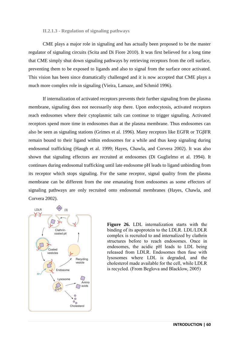
INTRODUCTION | 60
II.2.1.3 - Regulation of signaling pathways
CME plays a major role in signaling and has actually been proposed to be the master
regulator of signaling circuits (Scita and Di Fiore 2010). It was first believed for a long time
that CME simply shut down signaling pathways by retrieving receptors from the cell surface,
preventing them to be exposed to ligands and also to signal from the surface once activated.
This vision has been since dramatically challenged and it is now accepted that CME plays a
much more complex role in signaling (Vieira, Lamaze, and Schmid 1996).
If internalization of activated receptors prevents their further signaling from the plasma
membrane, signaling does not necessarily stop there. Upon endocytosis, activated receptors
reach endosomes where their cytoplasmic tails can continue to trigger signaling. Activated
receptors spend more time in endosomes than at the plasma membrane. Thus endosomes can
also be seen as signaling stations (Grimes et al. 1996). Many receptors like EGFR or TGβFR
remain bound to their ligand within endosomes for a while and thus keep signaling during
endosomal trafficking (Haugh et al. 1999; Hayes, Chawla, and Corvera 2002). It was also
shown that signaling effectors are recruited at endosomes (Di Guglielmo et al. 1994). It
continues during endosomal trafficking until late endosome pH leads to ligand unbinding from
its receptor which stops signaling. For the same receptor, signal quality from the plasma
membrane can be different from the one emanating from endosomes as some effectors of
signaling pathways are only recruited onto endosomal membranes (Hayes, Chawla, and
Corvera 2002).
Figure 26. LDL internalization starts with the
binding of its apoprotein to the LDLR. LDL/LDLR
complex is recruited to and internalized by clathrin
structures before to reach endosomes. Once in
endosomes, the acidic pH leads to LDL being
released from LDLR. Endosomes then fuse with
lysosomes where LDL is degraded, and the
cholesterol made available for the cell, while LDLR
is recycled. (From Beglova and Blacklow, 2005)
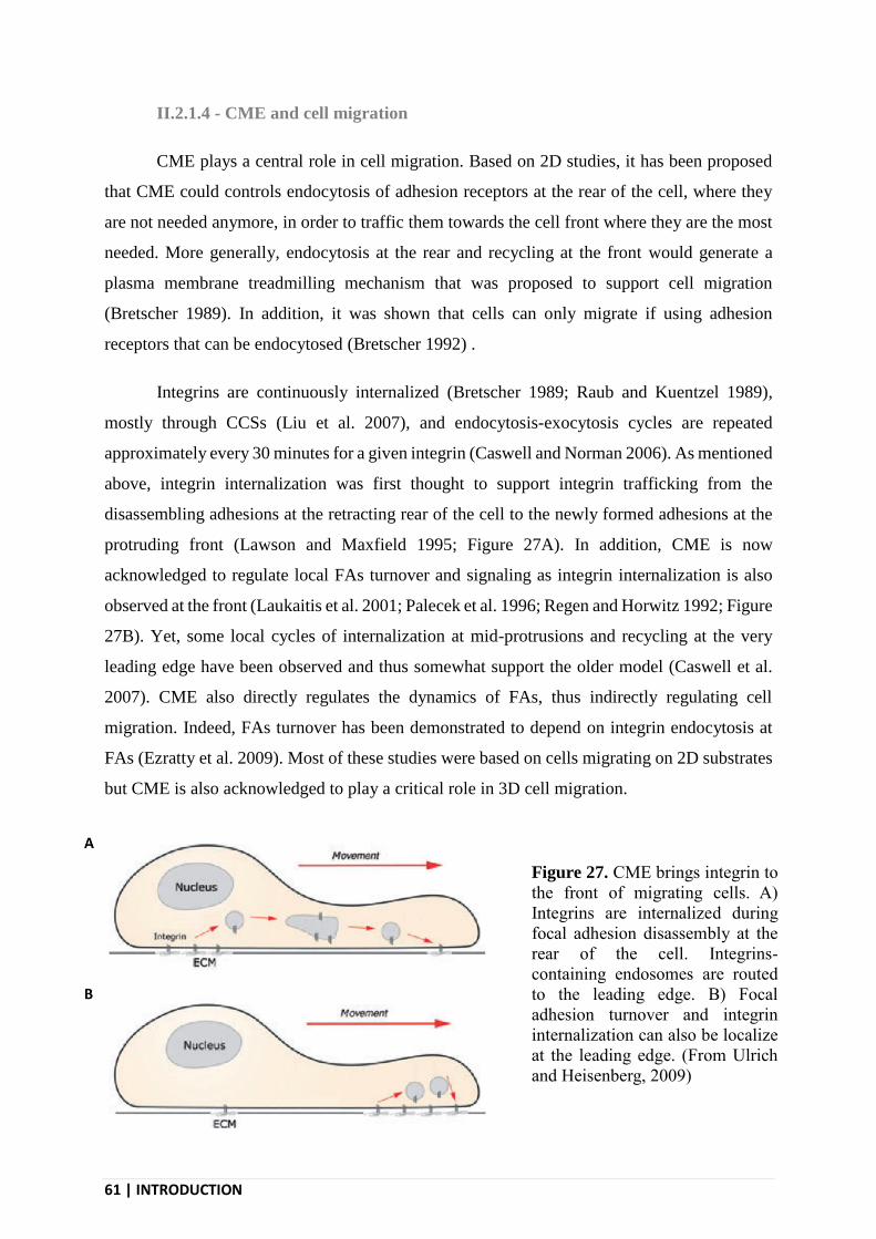
61 | INTRODUCTION
II.2.1.4 - CME and cell migration
CME plays a central role in cell migration. Based on 2D studies, it has been proposed
that CME could controls endocytosis of adhesion receptors at the rear of the cell, where they
are not needed anymore, in order to traffic them towards the cell front where they are the most
needed. More generally, endocytosis at the rear and recycling at the front would generate a
plasma membrane treadmilling mechanism that was proposed to support cell migration
(Bretscher 1989). In addition, it was shown that cells can only migrate if using adhesion
receptors that can be endocytosed (Bretscher 1992) .
Integrins are continuously internalized (Bretscher 1989; Raub and Kuentzel 1989),
mostly through CCSs (Liu et al. 2007), and endocytosis-exocytosis cycles are repeated
approximately every 30 minutes for a given integrin (Caswell and Norman 2006). As mentioned
above, integrin internalization was first thought to support integrin trafficking from the
disassembling adhesions at the retracting rear of the cell to the newly formed adhesions at the
protruding front (Lawson and Maxfield 1995; Figure 27A). In addition, CME is now
acknowledged to regulate local FAs turnover and signaling as integrin internalization is also
observed at the front (Laukaitis et al. 2001; Palecek et al. 1996; Regen and Horwitz 1992; Figure
27B). Yet, some local cycles of internalization at mid-protrusions and recycling at the very
leading edge have been observed and thus somewhat support the older model (Caswell et al.
2007). CME also directly regulates the dynamics of FAs, thus indirectly regulating cell
migration. Indeed, FAs turnover has been demonstrated to depend on integrin endocytosis at
FAs (Ezratty et al. 2009). Most of these studies were based on cells migrating on 2D substrates
but CME is also acknowledged to play a critical role in 3D cell migration.
Figure 27. CME brings integrin to
the front of migrating cells. A)
Integrins are internalized during
focal adhesion disassembly at the
rear of the cell. Integrins-
containing endosomes are routed
to the leading edge. B) Focal
adhesion turnover and integrin
internalization can also be localize
at the leading edge. (From Ulrich
and Heisenberg, 2009)
INTRODUCTION | 62
CCSs regulate cell migration not only through internalizing integrins but also by
regulating signaling pathways. Regulation of the cell-surface level of signaling receptors
directly impacts the cell ability to answer to external cues. As already mentioned, activated
signaling receptors accumulate into CCSs to be internalized. This limit signaling from the PM
itself and, in the case of prolonged exposure to a ligand, it leads to reduced levels of receptors
at the membrane. This negative feedback loop prevents excessive signaling. For example,
receptor internalization has been shown to be essential during the directed migration of cells
following a gradient of growth factor (Maryse Bailly et al. 2000). In addition, perturbing
endocytosis results in unbalanced signaling that prevents directed migration (Minina,
Reichman-Fried, and Raz 2007). CME can also spatially restricts signaling events in response
to extracellular cues. This allows restricted RTKs signaling at the leading edge of Drosophila
border cells, thus preserving the spatial information of ligand gradients and hence their use for
directional migration (Jékely et al. 2005). Finally it is worth saying that growth factors regulate
integrin endocytosis and recycling, thus impacting cell migration in another way (Mai et al.
2014)
II.2.2 - Non-canonical functions of CCSs
In addition to the canonical role of CCSs in supporting receptor endocytosis, non-
endocytic functions of CCSs have been reported over the years. I will here review some of these
atypical functions, in particular the adhesive role of CCSs in cell migration.
II.2.2.1 - Role of CCSs as signaling platforms
CCSs regulate receptor signaling through controlling their endocytosis as mentioned
above. But CCSs were also demonstrated to regulate signaling in an endocytosis-independent
manner.
First CCSs accumulating at the front of migrating cells have been shown to shape the
microtubule network. The growing extremity of microtubules oriented towards the leading edge
can occasionally contact some CCSs. The tubulin acetyltransferase αTAT1 is recruited at CCSs
through binding to AP-2. When a microtubule contacts a αTAT1-enriched CCS, this leads to
the acetylation and stabilization of the microtubule. This mechanism ensures the correct

63 | INTRODUCTION
orientation of microtubules towards the leading edge and is required for efficient persistent
migration in a given direction (Montagnac et al. 2013).
It has also become clear that CCSs can serve as signaling platforms for receptor
signaling, independently of their endocytic function (Garay et al. 2015). Along this line,
prolonged CCSs lifetime was associated with stronger signaling (Eichel, Jullié, and von
Zastrow 2016). Why is it the case is not clear, but it is likely that CCSs represent a favorable
environment for signaling by clustering in small areas not only the receptors but also many
scaffolding proteins and effectors. Some particular subsets of CCSs seem to represent an
extreme versions of such signaling platforms. Indeed, it has long been observed that some cell
types display particular clathrin-coated structures on the top of the canonical, endocytosis-
competent CCSs. These large, long-lived and flat clathrin structures are termed clathrin-coated
plaques (Figure 28). Although canonical CCSs can bud at the rim of plaques, the core of plaques
themselves remains flat for hours and thus, do not support endocytosis. It has recently been
demonstrated that the formation of these plaques relies on αvβ5 integrin (Baschieri et al. 2018;
Zuidema et al. 2018). This integrin strongly anchors the clathrin machinery to the substrate,
thus opposing budding forces. Signaling receptors such as the EGFR and c-Met can still
accumulate at plaques and this seems to reinforce their signaling abilities. Although the precise
mechanisms of this signaling reinforcement are not known, the very long-lived nature of
plaques may play a role. In addition, it was shown that the formation of plaques depends on the
rigidity of the substrate as these structures only assemble on rigid environments. Because their
formation do not depend on the actin cytoskeleton, this makes plaques very peculiar
mechanosensing structures that instruct the cell about substrate stiffness through regulating
receptors signaling.
Figure 28. Clathrin structures can
adopt two different shapes at the
inner leaflet of the plasma
membrane. They can either be
invaginated clathrin-coated pits
that perform endocytosis and
which lattices are composed of
both pentagons and hexagons.
Clathrin can also adopt a flat
hexagon-only structure which do
not perform endocytosis. (From
Humphries and Way, 2013)

INTRODUCTION | 64
II.2.2.2 - Role of CCSs in adhesion
Plaques have also been proposed to serve as adhesive structures (Maupin and Pollard
1983) and indeed, the recent discovery of their enrichment in αvβ5 integrin supports this
hypothesis (Baschieri et al. 2018). Independently of this latter report, αvβ5-rich reticular
adhesions have been described and reported to play a critical role in cell adhesion during
mitosis. These reticular adhesions are also independent of the actin cytoskeleton and proteomic
analyzes revealed the presence of many CCSs components in these structures. Clathrin-coated
plaques and reticular adhesions are probably actually the same structures (as discussed in Lock
et al. 2019), highlighting the prominent role of at least some CCSs in cell adhesion. Although
plaques have been proposed to slow down migration (Saffarian, Cocucci, and Kirchhausen
2009), presumably because of their strong adhesion to the substrate, observations from the lab
show that many highly migratory cell types such as fibroblasts display such structures.
We also recently discovered a new type of CCSs that forms at contact sites between the
plasma membrane and collagen fibers and that are called tubular clathrin/AP-2 lattices (TCALs)
(Elkhatib et al. 2017; Figure 29). TCALs specifically nucleate along collagen fibers as a
consequence of the deformation the fiber imposes to the plasma membrane. This creates a local
membrane curvature that is a signal triggering nucleation. Indeed, many CCSs proteins show a
preferential binding to curved membranes (Pucadyil and Holkar 2016). In addition, TCALs are
longer-lived than non-fiber-engaged CCSs probably because they are rich in β1-integrin that
bind the fiber. While canonical, budding CCSs display a spherical shape, TCALs adopt the
morphology of the fiber to form a tubular clathrin structure that wrap around and pinch collagen
fibers (Figures 29). We showed that, in addition to FAs, TCALs allow the cell to efficiently
grab collagen fibers. This adhesive role of TCALs is completely independent of endocytosis as
clathrin inhibition does not perturb their formation and function. This may seem contradictory
but, in the absence of clathrin, the core of AP-2 and other adaptors can still assemble, whether
on fibers or at any location of the plasma membrane, and can still recruit receptors (Elkhatib et
al. 2017). However, in the absence of clathrin, budding and endocytosis are completely
inhibited. In the 3D environment composed of collagen fibers, FAs are less prominent than in
2D and are only detected at the very leading edge and the very back of the cell. As a
consequence, when the cell extends a protrusion, the tension quickly raises across the

65 | INTRODUCTION
protrusion. Because TCALs form anchoring points to the fibers all around the cell, they reduce
these tension as measured in laser ablation experiments (Elkhatib et al. 2017). This allows the
cell to develop long, stable protrusions and in fine, it allows the cell to migrate efficiently in the
3D environment.
Figure 29. We have recently shown that CCSs can be used as adhesion structures. To do so they
engage and accumulate along collagen fibers (left). They form tubular structures that pinch the
fibers (right) and that were termed tubular clathrin/AP-2 lattices (TCALs) (From Elkhatib et al.,
2017)
INTRODUCTION | 66
III - THE EPIDERMAL GROWTH FACTOR RECEPTOR
Growth factors are membrane anchored or secreted proteins that regulate many aspects
of cellular functions. Some cell types secrete growth factors in the extracellular space. These
growth factors then diffuse in the extracellular space until they bind and activate their receptor
on the same cell (autocrine activation) or on other cells. This process allows is at the heart of
cell-cell communication. Signaling induced by growth factor receptor affects most of cells
function including cell proliferation, differentiation, migration, survival, metabolism tuning and
cell-cycle control. Among the many described growth factors, the epidermal growth factor
(EGF) is known to bind to its receptor (EGFR) that is a member of the receptor tyrosine kinase
family (RTKs).
III.1 - EGF AND EGFRs FAMILY
58 RTKs have been identified in human, they are classified within 20 families. Among
these families there is the family of EGFR, insulin receptor, PDGFR, VEGFR, FGFR or c-Met.
If all RTKs families are different they still share some similarities in molecular architecture,
activation mechanisms and induced signaling pathways.
III.1.1 - The receptor tyrosine kinase family
Regarding their molecular architecture, RTKs are transmembrane proteins that all share
an extracellular ligand-binding domain and a tyrosine kinase domain (TKD) in their
cytoplasmic tail. In the accepted scheme of RTKs activation, ligand-induced RTK dimerization
leads to the transphosphorylation of the dimer through TKDs of individual monomers coming
in close contact to the other monomer’s cytoplasmic tail (Ullrich and Schlessinger 1990).
Ligand-induced dimerization of RTK can happen through two main kind of mechanisms: the
two ligands are either physically bound to form a bridge between the two receptors or the
ligands induce conformational change that favors direct interaction of the monomers (Figure
30). Phosphorylated tyrosines in specific domains of receptor cytoplasmic domains represent
docking sites for effectors containing phosphotyrosine-binding (PTB) or Src homology-2 (SH2)

67 | INTRODUCTION
domains. PTB and SH2 domains-containing scaffolding proteins further recruit other signaling
complexes and effectors. This ultimately leads to signaling pathways activation.
Many of the signaling proteins are common to multiple RTKs but each RTK have a very
precise cellular effect. This raises the question of how activation of the same effectors by
different RTKs lead to different final consequences. RTKs are mostly internalized through
CME (Goh and Sorkin 2013). Like all transmembrane proteins, RTKs are nonspecifically
constitutively internalized at a very low rate. Because the constitutive recycling rate is higher
than the constitutive endocytosis rate, RTKs are mostly localized at the plasma membrane at
steady state. Ligand-binding induces RTKs internalization at a much higher rate and can lead
to RTKs degradation in the endosomal system. These two mechanisms synergize to control the
depletion of RTKs from the cell surface upon their acute stimulation, thus protecting the cell
from overstimulation.
III.1.2 - The EGFR family
The EGFR family is one of the most studied RTK due to its early discovery and its major role
in physiology and diseases, including cancers. They are ubiquitously expressed in both
epithelial and mesenchymal cells.
Figure 30. A) Ligand-induced dimerization of RTK can
happen through two main kinds of mechanisms: the two
ligands are either physically bound to form a bridge
between the two receptors (e.g. the receptors TrkA and
KIT) or the ligands induce conformational change that
favors direct interaction of receptor monomers (e.g. the
receptors FGFR and ErbB). (From Lemmon and
Schlessinger, 2011
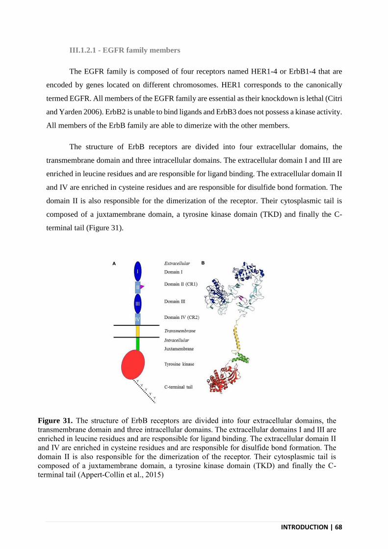
INTRODUCTION | 68
III.1.2.1 - EGFR family members
The EGFR family is composed of four receptors named HER1-4 or ErbB1-4 that are
encoded by genes located on different chromosomes. HER1 corresponds to the canonically
termed EGFR. All members of the EGFR family are essential as their knockdown is lethal (Citri
and Yarden 2006). ErbB2 is unable to bind ligands and ErbB3 does not possess a kinase activity.
All members of the ErbB family are able to dimerize with the other members.
The structure of ErbB receptors are divided into four extracellular domains, the
transmembrane domain and three intracellular domains. The extracellular domain I and III are
enriched in leucine residues and are responsible for ligand binding. The extracellular domain II
and IV are enriched in cysteine residues and are responsible for disulfide bond formation. The
domain II is also responsible for the dimerization of the receptor. Their cytosplasmic tail is
composed of a juxtamembrane domain, a tyrosine kinase domain (TKD) and finally the C-
terminal tail (Figure 31).
Figure 31. The structure of ErbB receptors are divided into four extracellular domains, the
transmembrane domain and three intracellular domains. The extracellular domains I and III are
enriched in leucine residues and are responsible for ligand binding. The extracellular domain II
and IV are enriched in cysteine residues and are responsible for disulfide bond formation. The
domain II is also responsible for the dimerization of the receptor. Their cytosplasmic tail is
composed of a juxtamembrane domain, a tyrosine kinase domain (TKD) and finally the C-
terminal tail (Appert-Collin et al., 2015)

69 | INTRODUCTION
III.1.2.2 - Ligands of the EGFR family
The EGFR family have eleven known ligands: EGF, heparin-binding epidermal growth
factor-like growth factor (HB-EGF), betacellulin, TGF-α, epiregulin, amphiregulin, epigen and
4 different neuregulins. All these ligands bind specifically to the different members of the
EGFR family. All ErbB dimers combinations, plus all ligands combinations, give a total of a
614 theoretically possible dimer/ligand complexes (Roskoski 2014). The different ligands lead
to dimers with different stability and this finally leads to different signaling outcomes (Freed et
al. 2017). All ligands are first exocytosed as transmembrane precursors at the surface of the
secreting cell prior to be cleaved and thus released in the extracellular space.
III.1.2.3 - The epidermal growth factor
EGF is the prototypic ligand of the EGFR family. It was first identified in the
submaxillary gland (Cohen 1960). EGF is a small peptide of 53 amino acids and, like all ErbB
ligands, it is derived from a larger precursor, the pre-pro-EGF comprising 1207 amino acids.
Pre-pro EGF contains a transmembrane domain and mature, soluble EGF is later released upon
cleavage by a metalloprotease (Le Gall et al. 2003). Soluble EGF is folded via three disulfide
bonds. EGF has a strong affinity for EGFR with a Kd of 1.77 x 10⁻⁷ M (Kuo et al. 2015). Within
the ErbB family, EGF is only able to bind to EGFR.
EGF can be found in many body fluids including milk, saliva and plasma (Carpenter
and Cohen 1979). EGF plasma level in mice is about 1 ng/mL (Byyny et al. 1974). EGF have
positive effects on cell survival, proliferation and migration and (Hernandez et al. 2009) is
notably involved in cancer cell invasion (Hernandez et al. 2009).
III.2 - PHYSIOLOGY OF THE EGFR
Because of its importance in physiology and diseases, the EGFR is one of the most
studied receptors. It is also the target for different clinically relevant drugs. Although it is now
studied for decades, many of its basics are still not completely understood, or are still a matter
of debate.
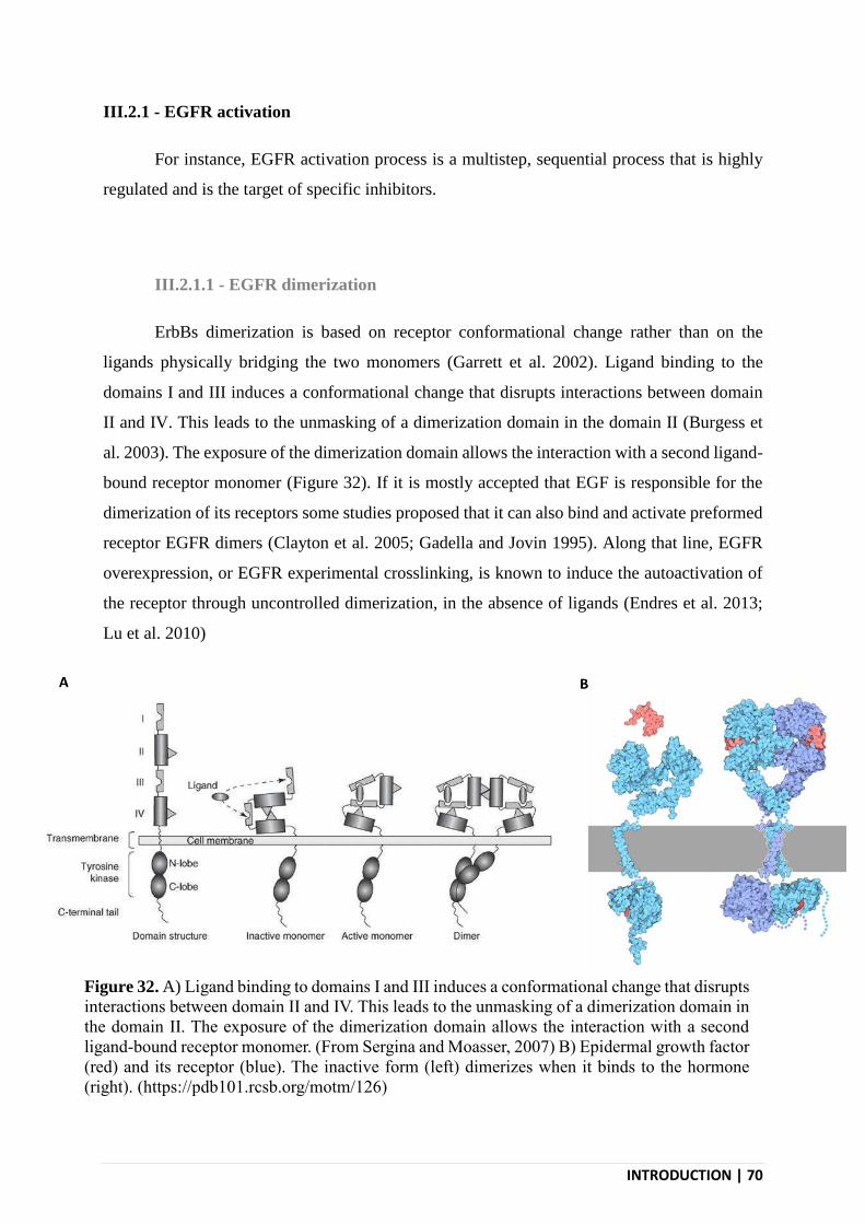
INTRODUCTION | 70
III.2.1 - EGFR activation
For instance, EGFR activation process is a multistep, sequential process that is highly
regulated and is the target of specific inhibitors.
III.2.1.1 - EGFR dimerization
ErbBs dimerization is based on receptor conformational change rather than on the
ligands physically bridging the two monomers (Garrett et al. 2002). Ligand binding to the
domains I and III induces a conformational change that disrupts interactions between domain
II and IV. This leads to the unmasking of a dimerization domain in the domain II (Burgess et
al. 2003). The exposure of the dimerization domain allows the interaction with a second ligand-
bound receptor monomer (Figure 32). If it is mostly accepted that EGF is responsible for the
dimerization of its receptors some studies proposed that it can also bind and activate preformed
receptor EGFR dimers (Clayton et al. 2005; Gadella and Jovin 1995). Along that line, EGFR
overexpression, or EGFR experimental crosslinking, is known to induce the autoactivation of
the receptor through uncontrolled dimerization, in the absence of ligands (Endres et al. 2013;
Lu et al. 2010)
Figure 32. A) Ligand binding to domains I and III induces a conformational change that disrupts
interactions between domain II and IV. This leads to the unmasking of a dimerization domain in
the domain II. The exposure of the dimerization domain allows the interaction with a second
ligand-bound receptor monomer. (From Sergina and Moasser, 2007) B) Epidermal growth factor
(red) and its receptor (blue). The inactive form (left) dimerizes when it binds to the hormone
(right). (https://pdb101.rcsb.org/motm/126)

71 | INTRODUCTION
III.2.1.2 - EGFR transphosphorylation
Activation of the tyrosine kinase domains of the ErbB family is different from the
process at stake in other RTKs. Reaching the active conformation does not require the activation
loop phosphorylation. Upon ligand binding, TKDs from the two EGFR monomers interact
together. This interaction induces a conformational change in TKDs, allowing them to act as
genuine tyrosine kinases (Zhang et al. 2006). Once activated, the TKDs control the
transphosphorylation of different tyrosine residues on the cytosolic tail of each EGFR
monomers. This initiates the activation of the signaling cascade through allowing the
recruitment of specific effectors.
III.2.1.3 - EGFR inhibitors
Because EGFR signaling plays a major role in cancers and other diseases, many
inhibitors have been developed that aim at inhibiting its activation. These drugs can be divided
in two categories: monoclonal antibodies that target the extracellular domains of EGFR, and
tyrosine kinase inhibitors (TKI). Monoclonal antibodies have been developed against ErbB1
and 2 and are used in clinics (eg. cetuximab and trastuzumab). They prevent ligand
binding/dimerization, which leads to signaling inhibition. TKIs have been developed against
all members of the ErbB family (eg. gefitinib and erlotinib). They act intracellularly by binding
to the ATP-binding site of TKDs, thus preventing ATP binding and kinase activity. Unlike
monoclonal antibodies, TKIs do not prevent ligand binding and dimerization (Lichtner et al.
2001).
III.2.2 - EGFR signaling
Activated EGFR displays multiple phosphorylated tyrosine on its cytoplasmic tail.
These phosphorylated tyrosine residues, together with surrounding residues, form docking sites
for adaptor proteins that control different downstream signaling pathways, some of which are
briefly described below (Roskoski 2014; Figure 33).
Among these pathways, the extracellular regulate kinas (Erk) pathway of the mitogen
activated protein kinase (MAPK) pathways is one of the most important. This specific MAPK
pathway is triggered by the recruitment of Grb2 to activated EGFR through its SH2 domain.
This leads to the activation of the guanine nucleotide exchange factor (GEF) Sos. Sos activates

INTRODUCTION | 72
Ras by catalyzing the exchange of GDP for a GTP on this GTPase. Ras then activates Raf (a
serine/threonine protein kinase). Raf dimerizes and autophosphorylates before to
phosphorylate/activate the Mek1/2 kinases which then phosphorylate/activates the Erk1/2
kinases. Upon activation, Erk1/2 translocates from the cytosol to the nucleus where it activates
other kinases and transcription factors. Ultimately, the Erk/MAPK pathway regulates the
transcription of many different genes whose expression level mostly regulate cell survival,
proliferation and cell differentiation.
Another critical signaling pathway regulated by the EGFR is the PI3K/Akt pathway.
PI3K is a heterodimer made of the association of a p85 regulatory subunit and a p110 (or
PI3KCA) catalytic subunit. PI3K can be directly activated by EGFR-associated Grb2. PI3K
phosphorylates PIP2 into PIP3. PIP3 then recruits the kinase Akt at the plasma membrane
through its PIP3-binding PH domain. Akt then phosphorylates both mTOR and BAD. The
phosphorylation events controlled by Akt activates mTOR but inhibits BAD. The PI3K/Akt
pathways is involved in cell survival and apoptosis. By hydrolyzing PIP3 into PIP2 PTEN
negatively regulate the PI3K pathway.
The PLC-γ/PKC pathway relies on the SH2 domain-dependent recruitment of PLC-γ to
activated EGFR. PLC-γ hydrolyzes PIP2 into IP3 and DAG. These second messengers
regulates many intracellular events, and in particular, they synergize to activate the protein
kinase C (PKC). PKC phosphorylates many substrates including Raf. This pathway regulates
cell proliferation, cell survival, migration and cell adhesion.
EGFR activation also leads to the activation of Signal Transducers and Activators of
Transcription (STATs) through controlling their phosphorylation. This depends on the
recruitment of the non-receptor kinase Src onto activated EGFR that then phosphorylates
STATs. In addition, EGFR-regulated Erk1/2 can also phosphorylates STATs on different
residues, reinforcing their activity. Once activated, STATs translocate to the nucleus to act as
transcription factors regulating proliferation, differentiation, cell survival and apoptosis.
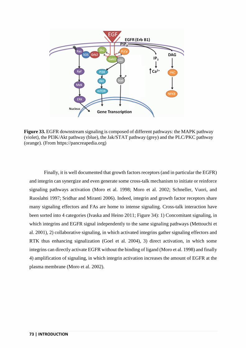
73 | INTRODUCTION
Finally, it is well documented that growth factors receptors (and in particular the EGFR)
and integrin can synergize and even generate some cross-talk mechanism to initiate or reinforce
signaling pathways activation (Moro et al. 1998; Moro et al. 2002; Schneller, Vuori, and
Ruoslahti 1997; Sridhar and Miranti 2006). Indeed, integrin and growth factor receptors share
many signaling effectors and FAs are home to intense signaling. Cross-talk interaction have
been sorted into 4 categories (Ivaska and Heino 2011; Figure 34): 1) Concomitant signaling, in
which integrins and EGFR signal independently to the same signaling pathways (Mettouchi et
al. 2001), 2) collaborative signaling, in which activated integrins gather signaling effectors and
RTK thus enhancing signalization (Goel et al. 2004), 3) direct activation, in which some
integrins can directly activate EGFR without the binding of ligand (Moro et al. 1998) and finally
4) amplification of signaling, in which integrin activation increases the amount of EGFR at the
plasma membrane (Moro et al. 2002).
Figure 33. EGFR downstream signaling is composed of different pathways: the MAPK pathway
(violet), the PI3K/Akt pathway (blue), the Jak/STAT pathway (grey) and the PLC/PKC pathway
(orange). (From https://pancreapedia.org)

INTRODUCTION | 74
III.2.3 - EGFR endocytosis
EGFR endocytosis is a very complex, highly regulated process that involves different
endocytosis motifs carried by its cytosolic tails, but also different adaptors and even different
endocytosis pathways. Once internalized, the fate of the EGFR may also vary depending on the
conditions of its stimulation.
III.2.3.1 - Mechanisms of EGFR uptake
The EGFR can be internalized through CME but also through some clathrin-
independent pathways. At low EGF concentration (below 10ng/ml), the EGFR is internalized
only via CME. At higher concentrations, the EGFR is also internalized through clathrin-
Figure 34. Crosstalk between RTKs and integrins can be sorted into four mechanisms. a)
Concomitant signaling, in which integrins and RTKs signal independently to the same signaling
pathways, b) collaborative signaling, in which activated integrins gather signaling effectors and
RTKs thus enhancing signalization, c) direct activation, in which some integrins can directly
activate RTKs without the binding of ligand and finally d) amplification of signaling, in which
integrin activation increases the amount of RTKs at the plasma membrane. (From Ivaska and
Heilo, 2011)

75 | INTRODUCTION
independent pathways. EGFR endocytosis in clathrin-independent pathways relies on the
ubiquitination of its cytosolic tail (Sigismund et al. 2005). CME of EGFR is associated with
sustained signaling and recycling to the plasma membrane. On the opposite, when EGFR is
endocytosed trough clathrin-independent pathways, it is more likely to reach lysosomes and to
be degraded (Sigismund et al. 2008). The mechanisms supporting this clathrin-independent
pathway are still elusive but may involve a recently proposed, very fast pathway relying on
endophilins (Boucrot et al. 2015). However, EGFR internalization through clathrin-
independent pathways is still a matter of debate (Madshus and Stang 2009) and the observed
differences could be cell type specific.
There is a larger consensus that EGFR uptake is supported by CME. Yet, precise
mechanisms of EGFR recruitment into CCSs are also debated. Some studies reported that
EGFR activation (autophosphorylation) is required for its efficient recruitment at CCSs
(Lamaze and Schmid 1995; Sorkina et al. 2002). Other studies, however, proposed that EGFR
dimerization is sufficient to trigger its endocytosis and can thus happen without activation of
the kinase domain of the receptor (Wang, Chen, and Wang 2015; Wang, Villeneuve, and Wang
2005). These studies proposed that the dimerization allows the formation of a pair of dileucine
(LL)-based endocytic motifs that constitute the basic unit for binding to AP-2. Finally, there
are also evidences that experimentally-induced EGFR clustering is sufficient to trigger its
endocytosis (Heukers et al. 2013).
More precisely, some mechanisms have been proposed to regulate EGFR recruitment at
CCSs. First, a direct interaction have been reported between EGFR and AP-2 (Sorkin et al.
1996). Also, activated EGFR recruit the ubiquitin ligase Cbl that ubiquitinates EGFR itself as
well as some EGFR effectors. Ubiquitynated EGFR is recognized by the clathrin accessory
proteins Eps15 and epsin that participate in the recruitment of EGFR into CCSs (Kazazic et al.
2009). Grb2 as well as lysine residues acetylation are also involved in addressing the EGFR to
CCSs (Goh et al. 2010). It is also worth saying that the intracellular domain of EGFR is the
only among the ErbB family to promote active endocytosis (Baulida et al. 1996). In any case,
it is clear that EGFR is targeted to CCSs through different, redundant mechanisms that
cooperate to ensure that this receptor does not stay at the plasma membrane once activated.
Some studies have suggested that, upon EGF-binding, the EGFR is able to trigger a local
CCSs nucleation in order to promote its own endocytosis (Johannessen et al. 2006). On the
opposite, some other investigators found that activated EGFR is targeted to pre-existing CCSs

INTRODUCTION | 76
(Rappoport and Simon 2009). Similarly intriguing, the absolute requirement of AP-2 for EGFR
internalization is also debated (Motley et al. 2003; Pascolutti et al. 2019). It appears that the
experimental conditions, like EGF incubation at 4°C before performing endocytosis assays,
critically affect EGFR physiology (Benmerah and Lamaze 2007). In physiological conditions,
AP-2 is required for EGFR endocytosis (Huang et al. 2004) and EGFR is targeted to pre-
existing CCSs. However, these different findings highlight the complexity of EGFR
endocytosis and its sensitivity to environmental factors.
III.2.3.2 - EGFR intracellular trafficking
2 to 5 minutes after its endocytosis, EGFR-containing-vesicles fuse with early
endosomes. The pH in early endosome is not low enough to promote EGF unbinding from the
receptor and thus the receptor continues to signal from endosomes. The early endosomes then
mature to progressively become late endosomes and multivesicular bodies. EGFR is observed
in perinuclear MVBs 15-20 min after its endocytosis (Carpentier et al. 1987; Figure 35). As
EGF does not separate from its receptor, the entire EGF/EGFR complex is recycled to the
plasma membrane. As mentioned above, a portion of internalized EGFR can recycle back to
the plasma membrane, especially if the receptor was internalized through CME. Recycling can
occur through a fast pathway, from early endosomes, or through a slow pathway, from late
endosomes and specialized Rab11-positive recycling endosomes (Sorkin et al. 1991). Most of
the internalized EGFR, however, is not recycled to the plasma membrane but rather routed to
lysosomes for degradation. EGFR ubiquitynation is essential for lysosomal targeting (Huang et
al. 2006). Of note, the different possible ligands of EGFR lead to different endocytic fates.
TGF-α associates weakly to EGFR as compared to EGF and thus dissociates from EGFR in the
acidic environment of early endosomes, leading to more EGFR being recycled (Longva et al.
2002).

77 | INTRODUCTION
III.3 - EGFR IN CELL MIGRATION
EGFR regulates several aspects of cell migrations. First, EGFR directly modulates cell
motility by regulating both cell adhesion to its substrate and the cytoskeleton that powers cell
movement. In addition, gradients of EGFR ligands can lead to directed cell migration. Finally,
EGFR also regulates the organization of the ECM supporting cell migration.
III.3.1 - EGFR in cell motility
EGFR activation is known to enhance cell motility. Both TGF-α and EGF are able to
promote migration in different cell lines (Morelli et al. 1992; Westermark, Magnusson, and
Heldin 1982). EGFR overexpression in tumor cells leads to enhanced motility and invasion
(Xue et al. 2006). EGF is also known to promote cell migration by reactivating the epithelial to
mesenchymal transition (Grände et al. 2002). EGFR regulates directly cell migration by fine
tuning the activity of two major actors of the motility machinery: adhesions to the substrate and
the actin cytoskeleton.
Figure 35. EGF endocytosis. A) EGF binding to its receptor leads to their recruitment into CCSs.
The EGF/EGFR complex traffic into endosomes until they are either degraded or recycled back
to the plasma membrane. (Adapted from Göstring, 2011) B) Immunofluorescence images of the
progressive EGFR activation and internalization. Scale bars, 10 μm. (From Villaseñor et al.,
2015).

INTRODUCTION | 78
EGFR first modulates cell adhesion by regulating the composition of FAs (Eberwein et
al. 2015). EGFR also regulates FAs turnover by promoting their disassembly (Xie et al.
1998)through downregulating FAK (Z. Lu et al. 2001). EGFR activation can also regulate
adhesion by regulating the expression level of integrins, their expression at the cell surface as
well as their own signaling activity at the plasma membrane (Laura Moro et al. 2002). Finally,
EGFR can increase cell spreading in cells adhering to rigid matrices (Saxena et al. 2017).
EGFR signaling also affects migration by regulating the actin cytoskeleton. EGF
stimulation modulates actin rearrangement (Rijken et al. 1991) and promotes lamellipodia
extension (Segall et al. 1996). EGF-induced actin rearrangement relies on cofilin, which severs
actin filaments. EGF stimulation leads to PLC activation which, in turn, activates cofilin. This
leads to barbed ends availability and thus more sustained actin polymerization (Mouneimne et
al. 2004)
III.3.2 - EGFR in chemotaxis
EGFR also regulates directed cell migration both in vitro and in vivo. In vitro, EGFR
has been shown to regulate the persistence of migration in one given direction in 2D (Maryse
Bailly et al. 2000) and in 3D (Raja et al. 2010). Cells are also able to migrate towards an
immobilized gradient of EGF in 3D (Fisher et al. 2018). Both EGFR expression level and EGF
gradient shape modulate the cell ability to migrate towards EGF (Fisher et al. 2018; S.-J. Wang
et al. 2004)
In vitro, when exposed to an EGF gradient, cells protrude and show F-actin enrichment
towards the gradient (M. Bailly et al. 1998). To migrate up the gradient, the cell needs to
polarize its actin machinery. This is possible because EGF/EGFR-mediated regulation of actin
polymerization remains localized (Kempiak et al. 2003). While EGFR localization is
homogeneous at the plasma membrane, its endocytosis is asymmetrically regulated (Bailly et
al. 1998) and thus could explain the local effect of EGF. In fact, cofilin regulation by EGF
remains local via LIM kinase that spatially restrict activated cofilin at the leading edge. This
induces local actin polymerization and further directed migration (Yamaguchi and Condeelis
2007; Figure 36)

79 | INTRODUCTION
In vivo, the directed migration of cancer cells toward an EGF source has been observed
by intravital imaging (Wyckoff et al. 2004); Figure 37). A paracrine loop involving EGF and
CSF-1 is proposed to drive this cancer cell invasion. EGF secreted by macrophages attract
tumors cells which in turn secrete CSF-1 that recruit macrophage. This leads to a positive
recruitment loop that promotes directed invasion (Wyckoff et al. 2004); Figure 37).
Figure 36. A model for EGF-driven chemotaxis. In the cell, cofilin is globally inactivated by
the LIMK activity. At the front of the cell, higher concentration of EGF induces a higher
activation of cofilin that locally overcomes the LIMK activity. Thus actin polymerization only
pushes the membrane at the leading edge, maintaining directed migration towards the EGF
gradient. (From Wang et al., 2007)

INTRODUCTION | 80
III.3.3 - EGFR and the ECM
EGFR also indirectly influences cell migration through regulation of the ECM. EGFR
can both trigger ECM protein secretion and remodeling of the existing matrix. Conversely,
matrix properties also influences EGFR signaling.
EGF stimulation leads to increased expression and secretion of collagen (Laato et al.
1987) and fibronectin (Mimura et al. 2004). EGFR can also participate to matrix remodeling,
notably through regulating the expression of matrix metalloproteases. EGFR activation leads to
increased expression of collagenase (Pilcher et al. 1999) and the expression of MMPs is
impaired in EGFR deficient cell (Kajanne et al. 2007). Nevertheless, EGF can increase or
decrease MMPs expression depending on cell types and experimental conditions (Bouchard et
al. 2010; S. Kim et al. 2009). The increase in MMPs expression take part in the larger program
of EGF-induced EMT (Wilkins-Port and Higgins 2007). Finally, the EGF ability to induce cell
Figure 37. A) Cells leaving a tumor to migrate towards an EGF gradient diffusing from a needle
(*) (Wyckoff et al., 2004). B) A paracrine loop involving EGF and CSF-1 is proposed to drive
this cancer cell invasion. EGF secreted by macrophages attract tumors cells which in turn secrete
CSF-1 that recruits macrophage. This leads to a positive recruitment loop that promotes directed
invasion (Condeelis and Segall, 2003)

81 | INTRODUCTION
contraction and thus matrix remodeling is still debated (Kim et al. 2015; Yang, Lin, and Yu
1997).
If EGFR signaling affects the ECM, the presence of some ECM components also
increases EGFR signaling (Cybulsky, McTavish, and Cyr 1994). Changes in the matrix
composition leads to modifications of the EGF-induced gene expression pattern (Yarwood and
Woodgett 2001). Finally, the substrate rigidity is well known to affect EGFR signaling via
integrin/EGFR crosstalk signaling (Saxena et al. 2017) and this play an important role in
regulating cell migration.

AIM OF THE THESIS | 82
AIM OF THE THESIS
Cell migration occurs during the whole life of every individual as a part of embryonic
development and maintenance of physiological functions but it also plays a role in several
pathologies. It is powered by the actin cytoskeleton that provides the force required to push
forward the plasma membrane of the leading edge. This force leads to an actual movement of
the cell only if the actin cytoskeleton is physically engaged with the extracellular matrix via
adhesion structures. To date, different types of adhesion complexes have been described, that,
for most of them, display a similar composition and depend on connections with the actin
cytoskeleton. In addition we recently proposed that CCSs could also serve as adhesion
structures.
CCSs are primarily known as the structures supporting clathrin-mediated endocytosis
(CME). During CME, cells internalize membrane portions and their associated receptors or
cargoes. Because of the large range of receptors it internalizes, CME is key to many cellular
processes. It starts with the recruitment of receptors and their ligands inside membrane
invagination that are called clathrin-coated pits. Some of these cargoes have been proposed to
initiate CCSs formation and modulate their dynamics. These pits progressively invaginate until
scission from the plasma membrane and formation of a vesicle that further traffic inside the
cell.
We showed that a subset of CCSs, called tubular Clathrin/AP-2 lattices (TCALs), can
also serve as adhesion structures to collagen fibers. Cell adhesion and the cell’s capacity to grab
collagen fibers were inhibited by disruption of TCALs. We first observed that CCSs
accumulated along collagen fibers in the 3D environments. TCALs nucleation is triggered by
membrane curvature induced by collagen fibers contacting the plasma membrane. TCALs
further adopt the tubular morphology of collagen fibers, wrapping around and pinching the
fiber. This morphology is rather different from the circular shape adopted by canonical CCSs
that regulate cargoes internalization.
From these, we hypothesized that matrix-bound CCS ligands could link the two roles of
CCSs: their well-documented role as endocytic structures and their newly discovered role as
adhesion structures. These matrix-bound ligands could modulate TCAL dynamics and thus

83 | AIM OF THE THESIS
affect 3D cell migration. In extension, gradient of matrix-bound CCS ligands could lead to
directed migration.
The main objective of my PhD was thus to establish the link between the endocytic and
adhesive role of CCSs during cell migration. This main question can be divided in several
specific objectives:
- Obtain amenable collagen fibers coated with CCSs ligand, as 3D networks are difficult
to manipulate in order to design specific experiments.
- Investigate the consequences of ligand-coated collagen fibers on TCALs and their
dynamic.
- Investigate the forces applied to ligand-coated collagen fibers and the potential role of
TCALs.
- Investigate a potential effect of collagen-bound ligands on 3D, directed migration and
the potential role of TCALs in this process.
I started by developing a protocol to obtain individual collagen fibers coated with EGF
or LDL. I then found that collagen-bound ligands increase the local nucleation of CCSs, leading
to more TCALs being associated with EGF/LDL-coated collagen fibers. I later found that this
increased in TCALs recruitment induces an increase in the forces applied by the cell to fibers.
I finally observed that in 3D, cells migrate toward EGF-coated collagen fibers using a
mechanism that relies on TCALs. In conclusion, I propose a model of directed migration driven
by TCALs in response to substrate-bound CCS-ligands. In this model, an asymmetrical
distribution of ligands induces an asymmetrical nucleation and thus distribution of adhesive
clathrin structures. This leads to an asymmetric distribution of forces applied to the ECM,
ultimately leading to directed migration.

RESULTS | 84
RESULTS
Title:
Clathrin-coated structures support 3D haptotaxis through local force transmission
Authors:
Enzo Bresteau1, Francesco Baschieri1, Nadia Elkhatib1 and Guillaume Montagnac1
Affiliations:
1 Inserm U1170, Gustave Roussy Institute, Université Paris-Saclay, Villejuif, France
Corresponding authors: [email protected] and

85 | RESULTS
Abstract
Migrating cells navigates in complex environments through sensing and interpreting
biochemical and/or mechanical cues. Gradients of cytokines or growth factors orient cell
migration through a complex integration of local, intracellular signaling events. Similar
mechanisms are believed to govern cell migration during haptotaxis, when cells follow signals
emanating from substrate-anchored chemoattractants. Here, we report that recently identified
tubular clathrin/AP-2 lattices (TCALs), a subset of clathrin-coated structures (CCSs) that pinch
collagen fibers, mechanically control haptotaxis along fibers decorated with ligands of CCS
cargoes, in 3D environments. We observed that epidermal growth factor (EGF) or low density
lipoprotein (LDL) bound to collagen fibers lead to an increased local nucleation and
accumulation of TCALs. By using engineered, mixed collagen networks, we demonstrate that
this mechanism selectively increases local forces applied on ligand-decorated fibers. We show
that these effects depend on ligand’s receptors but do not rely on their ability to trigger signaling
events. The accumulation of TCALs along ligand-decorated fibers steers migration in 3D
environments. We conclude that ligand-regulated, local TCAL nucleation results in asymmetric
force distribution that orient cell migration in 3D environments.

RESULTS | 86
Main Text
INTRODUCTION
Clathrin-mediated endocytosis is a fundamental process that regulates the uptake of a wide
diversity of cell-surface receptors and their ligands. By doing so, clathrin-coated structures
(CCSs) impinge on many cellular functions including cytokinesis (Montagnac, Echard, and
Chavrier 2008), cell migration, and cell invasion (Maritzen, Schachtner, and Legler 2015). We
recently demonstrated that in 3D environments composed of collagen fibers, β1-integrin-
enriched CCSs wrap around and pinch the fibers, thus offering many anchoring points that
facilitate cell migration (Elkhatib et al. 2017). These collagen fiber-pinching CCSs, also called
tubular clathrin/AP-2 lattices (TCALs), are frustrated in nature as they try and fail to internalize
fibers that are longer than the cell itself. Yet, although their lifetime is longer as compared to
non fiber-engaged CCSs, it is still limited in time suggesting that they may be able to bud and
to produce endocytic vesicles after an initial period of frustration on fibers. Collagen fibers are
sticky structures to which many proteins can bind or adsorb. Besides other extracellular matrix
(ECM) components, some cytokines and growth factors can also directly or indirectly bind to
collagen fibers (Hynes 2009; Schuppan et al. 1998). For example, the epidermal growth factor
(EGF) was reported to bind to collagen fibers with an estimated affinity of 1.706 μM (Yang et
al. 2009). The EGF receptor (EGFR) is mostly internalized through CCSs and this has been
suggested to play a role in chemotaxis towards EGF gradients (Wyckoff et al., 2004; S.-J. Wang
et al. 2004). Low density lipoprotein (LDL), the ligand of another major CCS cargo, was also
reported to bind to collagen fibers (Nievelstein-Post et al. 1994). We set out here to investigate
the relationship between TCALs and two major CCSs cargos, and their role in orienting cell
migration in 3D environments.

87 | RESULTS
RESULTS
Production and characterization of ligand-decorated collagen fibers
We observed that incubating Alexa488-labelled EGF with a pre-polymerized fibrillar
collagen gel leads to an accumulation of EGF along fibers (Supplementary Fig. 1a), thus
confirming previous findings (Yang et al. 2009). Because such a gel is difficult to handle in
order to address specific questions, we setup a protocol to produce, decorate with EGF, and
manipulate individual collagen fibers (Fig. 1a). Our protocol produces collagen fibers of quite
homogenous length that can be spotted on glass or incorporated into a 3D network (Fig. 1b and
c). These fibers were homogenously decorated with Alexa488-EGF (Fig. 1b and c) and similar
results were obtained when using Dil-LDL (Supplementary Fig. 1b). Fluorescence recovery
after photobleaching (FRAP) assays showed that the fluorescence associated with Alexa488-
EGF does not recover when engineered fibers were spotted on 2D surfaces or incorporated into
3D networks (Supplementary Fig. 1c and d). If free EGF was added in the medium before the
FRAP experiments, a mobile fraction of approximately 50% was detected (Supplementary Fig.
1c and d). The immobile fraction observed in these latter experiments most likely correspond
to the EGF fraction that is stably associated with collagen fibers recovered at the end of our
production protocol.
We observed that fiber-associated EGF was able to activate the mitogen activated
protein kinase Erk as efficiently as free EGF alone, or free EGF added together with non-
decorated fibers, when MDA-MB-231 cells and fibers were incubated together in suspension
(Supplementary Fig. 2a and b). Akt could not be activated by free EGF alone in these
conditions, but was equally activated by EGF-decorated fibers and by free EGF added together
with non-decorated fibers (Supplementary Fig. 2a and c). These latter results probably reflect
the requirement of integrins engagement for efficient activation of the Akt pathway, this is
conflit with previously reported experiment (Velling, Stefansson, and Johansson 2008). In

RESULTS | 88
addition, MDA-MB-231 cells were able to internalize fiber-bound EGF as evidenced by the
transfer of EGF-associated fluorescence from the fibers to internal compartments
(Supplementary Fig. 2d). Together, these data demonstrate that collagen fiber-associated EGF
is functional and can be sensed and internalized by cells.
Exacerbated accumulation of TCALs along ligand-decorated fibers
We next took advantage of our protocol to generate composite 2D substrates by
sequentially spotting EGF-decorated and non-decorated fibers on glass coverslips (Fig. 1d).
When MDA-MB-231 cells were seeded for 15 min on this mixed, 2D network, we noticed that
CCSs marked with the -adaptin subunit of the clathrin adaptor AP-2 accumulated along non-
decorated fibers as previously described (Elkhatib et al. 2017) (Fig. 1d). However, CCSs
accumulated approximately 30 % more along EGF-decorated fibers as compared to naked
fibers, whether using Alexa-488-labelled EGF or unlabeled EGF (Fig. 1d and e). We also
observed that the EGFR accumulated along EGF-decorated fibers but not along non-decorated
fibers (Fig. 1f and g image and quantif). The accumulation of TCALs along EGF-decorated
fibers was dependent on the expression of the EGFR but was insensitive to Gefitinib, a drug
that inhibits the kinase activity of EGFR (Cohen et al. 2004) (Fig. 1e). We controlled that
Gefetinib was indeed able to inhibit EGFR-triggered Erk activation (Supplementary Fig. 3a and
b). These results suggest that EGFR activation (autophosphorylation) and downstream
signaling pathways are not playing a role in the accumulation of TCALs along EGF-decorated
fibers. We next performed live cell imaging of genome-edited MDA-MB-231 cells engineered
to express GFP-tagged, endogenous 2-adaptin subunit of AP-2. When these cells were allowed
to spread for 15 min on the composite, 2D network, we measured that TCALs average lifetime
was slightly increased on EGF-decorated as compared to non-decorated fibers, although the
difference was not statistically significant (Fig 2a-c). However, TCALs had approximately 20%

89 | RESULTS
more chances to nucleate on EGF-decorated fibers as compared to control fibers (Fig. 2b and
d). EGFR knockdown, but not Gefetinib treatment, abolished the preferential TCALs
nucleation rate on EGF-decorated fibers (Fig. 2e). It has been suggested that, in particular
experimental setups, EGF/EGFR complexes could induce the de novo formation of CCSs
(Wilde et al. 1999; Johannessen et al. 2006). In addition, receptors are known to take an
important part in the maturation of nascent CCSs (Ehrlich et al. 2004; Loerke et al. 2009) and
experimental clustering of cell-surface receptors can induce the de novo formation of CCSs
(Liu et al. 2010). Thus, it is possible that local accumulation of EGFR driven by collagen fiber-
associated EGF results in local TCAL nucleation/maturation as suggested by our data. The role
of EGFR kinase activity in EGFR interaction with CCSs has been supported by several studies
(Lamaze and Schmid 1995; Sorkina et al. 2002) but some others suggested that EGF-induced
receptor dimerization or clustering is sufficient to be recruited at CCSs (Wang, Villeneuve, and
Wang 2005; Wang, Chen, and Wang 2015; Heukers et al. 2013). In any case, our results suggest
that EGF/EGFR-regulated local TCAL nucleation does not require EGFR activation. Along this
line, we observed that EGF still accumulates at CCSs in cells seeded on glass and treated with
Gefetinib, although less efficiently than in control cells (Supplementary Fig. 3c and d). It is
possible that the particular conditions of TCALs assembly, which is driven by a cooperation
between local membrane curvature and β1-integrin engagement (Elkhatib et al. 2017), are
further favored by high local concentrations of functional CCS cargoes. In agreement with that,
we observed that TCALs also preferentially accumulated along Dil-LDL-decorated fibers as
compared to non-decorated ones (Fig. 2f and g). Similar to EGF-decorated fibers, CCS
nucleation rate was increased on LDL-decorated fibers as compared to naked fibers (Fig. 2h).
Thus, local accumulation of CCS ligands drives the local accumulation of TCALs through
increased CCS nucleation.

RESULTS | 90
Local TCALs accumulation regulates local forces applied on collagen fibers
We noticed that cells spreading on the mixed 2D network developed more protrusions
along EGF-decorated fibers as compared to non-decorated fibers (Fig. 3a and b). Yet, we did
not observed any differential enrichment of β1-integrin on one or the other type of fiber
(Supplementary Fig. 4a and b). Similarly, the focal adhesion marker vinculin was equally
distributed between EGF-decorated and non-decorated fibers (Supplementary Fig. 4c and d)
suggesting that these adhesion structures did not participate in the differential protrusion
activity on the two different types of fibers. We previously reported that TCALs help cells to
develop long protrusions in 3D collagen networks by providing several anchoring points to
collagen fibers (Elkhatib et al. 2017).Here, our data suggest that the increased accumulation of
TCALs along EGF-decorated fibers allow the cell to develop longer protrusions on these fibers
as compared to non-decorated fibers. To check whether cells specifically exert more forces on
ligand-decorated fibers as compared to normal fibers, we developed a collagen fiber remodeling
assay. EGF-decorated and non-decorated fibers were sequentially spotted on a glass coverslip
and MDA-MB-231 cells were allowed to remodel this composite network for 2 hours. Collagen
fiber remodeling was characterized by a transition from rod-like shape to circular aggregates as
cells pulled on and packed fibers on their dorsal surface. In the course of this process, TCALs
were observed colocalizing and moving together with collagen fibers on the dorsal surface of
cells (Supplementary Fig. 5a and b). In order to precisely quantify fiber remodeling, we
measured the evolution of fibers circularity over time. The data show that EGF-decorated fibers
are almost twice as much remodeled as compared to non-decorated fibers over a 120 min period
(Fig. 3c-e). The preferential remodeling of EGF-decorated fibers was dependent on EGFR
expression (Fig. 3f and Supplementary Fig. S5c). However, Gefetinib treatment did not
modulate the preferential remodeling of EGF-decorated fibers indicating that EGFR activation
is not required (Fig. 3f and Supplementary Fig. S5c). Knockdown of AP-2 subunits, but not

91 | RESULTS
clathrin heavy chain (CHC), abrogated the preferential remodeling of collagen fibers (Fig. 3f
and Supplementary Fig. S5c). This is consistent with our previous characterization of TCALs
showing that clathrin is not required for the accumulation of AP-2-positive structures along
collagen fibers and that it does not play a role in the adhesive function of TCALs (Elkhatib et
al. 2017). Of note, inhibiting the formation of focal adhesions by using talin-specific siRNAs
strongly reduced the global fiber remodeling rates but did not inhibit the preferential remodeling
of EGF-decorated fibers (Fig. 3f and Supplementary Fig. S5c). Together, our data show that
both EGFR and TCALs are required for the preferential remodeling of EGF-decorated collagen
fibers. In addition, we observed that LDL-decorated fibers were also preferentially remodeled
over non-decorated fibers and that this also depended on AP-2 (Fig. 3g and h). Together, our
data suggest that the accumulation of TCALs along CCS ligands-decorated fibers leads to more
forces being applied on them.
Local TCALs accumulation orients cell migration in 3D
We previously reported that TCALs help cells to migrate in 3D environments by serving
as adhesive structures to collagen fibers (Elkhatib et al. 2017). We hypothesized that the
preferential accumulation of TCALs along ligands-decorated fibers could further favor cell
migration in 3D networks. We first observed that MDA-MB-231 cells located in 3D network
composed of EGF-decorated fibers were more elongated as compared to cells located in a non-
decorated network (Fig. 4a-c). Together with our previous results showing that TCALs help
cells to build long protrusions required for migration (Elkhatib et al. 2017), these data suggested
that EGF on collagen fibers further potentiates cell elongation through favoring TCALs
formation. However, cells migrated with a similar velocity in the EGF-decorated and non-
decorated networks (Supplementary Fig. 5d). It is possible that a homogenous distribution of
EGF-decorated fibers around the cell leads to a global stabilization of all cell protrusions,

RESULTS | 92
without a net consequence on cell displacement in one given direction. Stable protrusions
pointing in different directions could hinder a potential effect of EGF-decorated fibers on cell
migration. To test this possibility, we aimed at observing cells located in an asymmetric network
in which EGF-decorated fibers would be restricted to a defined area of the gel. For this, EGF-
decorated, or non-decorated, chopped collagen fibers produced as in Fig. 1a were added to a
mixture of non-polymerized collagen in a 1:1 ratio. A first mix containing the non-decorated
fibers was deposited as a 50 μl drop on a glass coverslip at 4°C. 6 μl of the second mix,
containing the EGF-decorated fibers, was carefully injected into the first one and the composite
gel was then allowed to polymerize. This protocol enabled the production of a composite
network with a clear segregation between EGF-decorated and non-decorated fibers, without
creating a physical interface between the two regions (Fig. 4d). We observed that cells located
in the homogenous EGF-decorated or non-decorated areas of the gel migrated randomly in all
directions (Fig. 4e). However, cells located at the boundaries between the two areas had
approximately twice as much chances to migrate towards the EGF-decorated network rather
than towards the non-decorated network (Fig. 4e and f). This preferential migration towards
EGF-decorated fibers was dependent on EGFR and CCSs (Fig. 4f). Together, our results show
that TCALs accumulation on CCSs ligands-decorated fibers allow cells to migrate towards this
type of fibers.
Overall, we have found that TCALs strongly accumulate on CCSs ligands-decorated
fibers because ligand/receptor complexes favor the local nucleation of TCALs on these fibers.
The preferential accumulation of TCALs on ligands-decorated fibers allows the cell to exert
more forces on these fibers as compared to non-decorated fibers. As a consequence, cells
preferentially migrate towards ligands-decorated fibers in 3D environments. Strikingly, this
haptotactic mode of migration does not depend on the transduction of signaling pathways but

93 | RESULTS
only on the local accumulation of TCALs that allow cells to pull harder on collagen fibers
decorated with CCSs ligands. Thus, it is important to consider that haptotaxis is not a simple
variation of chemotaxis but is actually driven by completely different mechanisms, at least in
the case of EGF and LDL in 3D collagen networks. Because ECM fibers are abundant in
complex organisms, and many chemoattractants and other potential CCSs ligands are known to
bind to ECM, the mechanism we described here may play a central role in cell migration in
different contexts, from development to cancers.
RESULTS | 94
METHODS
Cell lines and constructs
MDA-MB-231 cells (a gift from P. Chavrier, Institut Curie, Paris, France) or genome edited
MDA-MB-231 cells engineered to express an endogenous GFP-tagged or mCherry-tagged μ2
subunit (a gift from D. Drubin, University of California-Berkeley, California, USA) were
grown in DMEM Glutamax (Gibco) supplemented with 10% foetal calf serum (Gibco) at 37°C
in 5% CO2.
Antibodies, growth factors and drugs
Rabbit polyclonal anti-α-adaptin antibodies (M300) were purchased from Santa Cruz
Biotechnology Inc. Mouse monoclonal anti-α-adaptin antibodies (ab2807) were purchased
from Abcam. Activated integrin (4B4) antibody (6603113) was obtained from Beckman
coulter. Alexa488-conjugated anti-mouse (A21202) or anti-rabbit (A21202) antibodies and
Alexa545-labelled phalloidin (A22283) were from Molecular Probes. Anti phospho-Akt
(9271), Erk1/2 (9102) and phospho-Erk1/2 (9101) antibodies were purchased from Cell
Signaling. HRP-conjugated anti-mouse antibodies (115-035-062) for western blot and Cy3-
conjugated anti-rabbit (711-165-152) or anti-mouse (711-165-152) antibodies were purchased
from Jackson ImmunoResearch Laboratories. HRP-conjugated anti-rabbit antibodies (A0545)
for western blot were purchased from Sigma.
Gefitinib (CDS022106) was purchased from Sigma and used at a final concentration of 10 μM
unless otherwise stated. Before experiment, cells with gefitinib were pre-treated for 30 min at
37°C. Alexa Fluor® 488 (E-13345)- or 647 (E35351)-labelled EGF and DiI-conjugated LDL
(L3482) were purchased from Thermo Fisher. Rat tail Collagen-I (CA10483-01) was purchased
from GIBCO. Human recombinant EGF (E9644) was purchased from Sigma.

95 | RESULTS
RNA interference
For siRNA depletion, MDA-MB-231 cells were plated at 50% confluence and treated with the
indicated siRNA (30 nM) using RNAimax (Invitrogen, Carlsbad, CA) according to the
manufacturer's instruction. Cells were used after 72 h or 120 h of siRNA treatment as shown
by immunoblotting analysis with specific antibodies. Equal loading of the cell lysates was
verified by immunoblotting with anti-tubulin antibodies.
The following siRNAs were used: Talin, 5'-ACAAGAUGGAUGAAUCAAUUUU-3'; µ2-
adaptin, 5’-AAGUGGAUGCCUUUCGGGUCA-3’; Clathrin heavy chain (CHC),
5’GCUGGGAAAACUCUUCAGATT-3’; α-adaptin, 5’- AUGGCGGUGGUGUCGGCUCTT-
3’; Epidermal growth factor receptor (EGFR) 5’- GAGGAAAUAUGUACUACGA-3' (EGFR-
1) and 5’- GCAAAGUGUGUAACGGAAUAGGUAU-3' (EGFR-2); non-targeting siRNAs
(siControl), ON-TARGETplus Non-Targeting SMARTpool siRNAs (Dharmacon D-001810-
01).
Western Blots
For Western Blot experiments, cells were lysed in ice cold MAPK buffer (100mM NaCl, 10
nM EDTA, 1% IGEPAL ® CA-630, 0.1% SDS, 50mM TRIS-HCl pH 7.4) supplemented with
protease and phosphatase inhibitors. Protein concentration was measured using Pierce™
Coomassie Plus (Bradford) Assay Kit (1856210) according to the manufacturer’s instructions
in order to load equal amount of proteins. Antibodies were diluted at 1:1000 in PBS - 0.1%
Tween - 5% BSA or 5% non-fat dried milk. Analyzes of bands densitometry were performed
using ImageJ.
For testing Gefitinib-mediated inhibition of EGFR signaling, 200 000 MDA-MB-231 were
serum-starved for 2h, and then stimulated or not with 10 ng/ml EGF in the presence or not 10

RESULTS | 96
μM gefitinib for 5 minutes at 37°C. Cells were then harvested, lysed and analyzed by western
blot as describe above.
EGF/LDL-decorated collagen fibers and 3D networks
200 μl of a mix containing a 10:1 ratio of unlabeled collagen type I and Alexa548(or Alexa488
or Alexa647)-labelled collagen type I (Gibco, A1048301) at a final concentration of 1.1 mg/ml
was allowed to polymerize in a 1.5 ml Protein LoBind Tube (Eppendorf, 0030108116) for 12
min at room temperature. 200 μl of PBS were then added on ice to the mix before 3 rounds of
10s sonication were performed using a Q125 sonicator at 40% amplitude (Qsonica sonication).
2.5 μl of EGF/LDL was then added (or not) to the mix before incubation at room temperature
for 2.5h (final concentration EGF: 1.25 μg/ml and LDL: 6.25 mg/ml). 600 μl of PBS was then
added to the mix before 12 rounds of 10s sonication were performed at 40% amplitude.
Polymerized, sonicated, EGF-or LDL- or non-decorated collagen fibers were then pelleted by
centrifugation for 1h at 45000 rpm (centrifuge 5427R; rotor FA-45-30-11; Eppendorf) at 4°C.
The pellet was washed twice with cold PBS before to be either resuspended in 1 ml cold PBS
(for coverslip spotting experiments) or incorporated into another mix for 3D collagen network
preparation. Fiber solution was kept on ice to prevent collagen fibers aggregation.
For collagen fibers deposition on glass (2D), 100 μl of the mix containing polymerized,
sonicated collagen fibers were spotted for 10 min on a 12 mm coverslip (Marienflied, 0111520)
or in a glass-bottom 96 well plate (Greiner, 07-000-630) at room temperature before to be
washed twice with PBS. For sequential deposition experiments, naked fibers were spotted as
described, then washed twice with PBS before EGF- or LDL-decorated fibers were spotted for
10 min as well and washed twice using PBS.
For incorporation into 3D networks, the pellet composed of polymerized, sonicated collagen
fibers was resuspended in a 200 μl mix containing non-polymerized collagen and 45 μl of this

97 | RESULTS
new mix were deposited in a glass-bottom 96 well plates pretreated with poly-L-lysin 0.1%
(Sigma, 8920) for 10 min. For some experiments, the mix contained 200 cells/μl. The mix was
allowed to polymerize at room temperature for 30 min before to be covered with complete
medium. For generating composite 3D networks, 7 μl of a mix containing EGF- or LDL-
decorated, polymerized, sonicated fibers and 200 cells/μl was gently pipetted inside a 45 μl mix
containing naked, polymerized, sonicated fibers and 200 cells/μl that was deposited on glass a
few seconds before. All steps were performed at 4°C to prevent polymerization. The composite
3D network was then allowed to polymerize at room temperature for 30 min before to be
covered with complete medium.
Indirect immunofluorescence microscopy and fluorescence quantification
MDA-MB-231 cells plated for 15 min on the top of naked and EGF- or LDL-decorated fibers
spotted on coverslips were fixed in ice-cold methanol or PFA and processed for
immunofluorescence microscopy by using the indicated antibodies. Cells were imaged through
a 100× 1.40NA UPlanSApo objective lens of a wide-field IX73 microscope (Olympus)
equipped with an Orca-Flash2.8 CMOS camera (Hamamatsu) and steered by CellSens
Dimension software (Olympus).
For anti-vinculin staining, cells were briefly extracted for 1 min using 0.1% Triton prior to
fixation.
For calculating the degree of CCSs or integrins or vinculin alignment along collagen fibers,
naked and decorated fibers were segmented using ImageJ software and the average fluorescence
intensity of the anti-α-adaptin or anti-integrin or anti-vinculin staining in fibers area was
measured for both type of fibers and for each individual cells and normalized to the area
occupied by respective collagen fibers. For protrusions quantification, the proportion of

RESULTS | 98
protrusions associated with each type of fibers were manually counted in ImageJ. At least 50
cells per conditions were quantified in 3-5 independent experiments.
Total internal reflection fluorescence microscopy
For total internal reflection fluorescent microscopy (TIRF), MDA-MB-231 cells seeded on
glass or onto collagen-fibers-coated glass coverslips were imaged through a 100x 1.49 NA
TIRF objective lens on a Nikon TE2000 (Nikon France SAS, Champigny sur Marne, France)
inverted microscope equipped with a QuantEM EMCCD camera (Roper Scientific SAS, Evry,
France / Photometrics, AZ, USA), a dual output laser launch, which included 491 and 561 nm
50 mW DPSS lasers (Roper Scientific), and driven by Metamorph 7 software (MDS Analytical
Technologies, Sunnyvale, CA, USA). A motorized device driven by Metamorph allowed the
accurate positioning of the illumination light for evanescent wave excitation.
To measure EGFR accumulation on collagen fibers, MDA-MB-231 cells were transfected with
a EGFR-GFP encoding plasmid (Addgene #32751) using linear PEI (MW 25.000,
Polysciences) and following manufacturer’s instructions. Cells were allowed to spread for
approximately 15 min on the top of a mixed network composed of naked and EGF-decorated
fibers spotted onto a glass-bottom fluorodish (World Precision Instruments, FD35-100) before
to be imaged for 5 min. EGFR-associated average fluorescence intensity was quantified in
ImageJ using the above described segmentation protocol at time points showing the greatest
accumulation of EGFR on fibers. At least 20 cells per condition were analyzed in 2 independent
experiments.
To monitor EGF recruitment at CCSs, genome-edited MDA-MB-231 cells on fluorodishes
were starved for 2 h and treated or not with 10 mM Gefitinib for 1h before to be incubated in
the presence of the same concentration of Gefitinib and 50 ng/ml Alexa488-EGF before to be
imaged for 10 min. For quantification, CCSs were individually segmented using ImageJ and
average EGF-associated fluorescence was measured at the time point showing the maximum
99 | RESULTS
colocalization between EGF and CCSs. At least 2000 CCSs from at least 5 cells per condition
and per experiments were quantified in 2 independent experiments.
Fluorescence Recovery After Photobleaching (FRAP)
EGF-decorated collagen fibers were spotted on glass or embedded in a 3D gel as described
above in the presence or not of 8 μg/ml Alexa488-EGF. FRAP was performed on a Leica Sp8
confocal microscope equipped with a Pecon incubation chamber to maintain the cells at 37°C
and 5% CO2 and using the FRAP wizard of the Leica software. One fiber was manually selected
and subjected to 100% laser power. One frame was collected before photo-bleaching, and 40
frames were collected after bleaching to analyze fluorescent recovery at the frequency of 1
frame/30 sec. Data were analyzed using the ImageJ FRAP Profiler plugin (McMaster
University, Canada) to extract recovery curves and calculate the half-time recovery.
Fiber-associated EGF stimulation and internalization assays in suspension
For fiber-associated EGF stimulation assays, MDA-MB-231 cells were starved for 2h before to
be harvested using trypsin and incubated in suspension alone or in the presence of 50 ng/ml
Alexa-488 EGF, or naked fibers, or Alexa488-EGF-decorated fibers, or naked fibers and 50
ng/ml Alexa488-EGF as indicated. The amount of fibers used in the assay was chosen so that
the concentration of Alexa488-EGF on EGF-decorated fibers was equivalent to 50 ng/ml
soluble EGF as determined by SDS-PAGE followed by EGF-associated fluorescence
quantification using a gel imager (Biorad ChemiDoc XRS+ System) and ImageJ software. Cells
were incubated at 37°C for 5 or 30 minutes before cells were harvested at 4°C and subsjected
to lysis in cold MAPK buffer followed by western-blot analyzis using the indicated antibodies.
For fiber-associated EGF internalization assays, the same protocol was used except that fibers
were labelled using Alexa548-conjugated collagen and cells were incubated only with

RESULTS | 100
Alexa488-EGF-decorated fibers at 37°C for 5, 10, 20 or 40 minutes before to be harvested and
fixed using PFA. Fixed cells were centrifuged for 2 min at 500g and resuspended in 500 μL
PBS. Cells were then spotted on a coverslip and imaged by epifluorescence microscopy.
Spinning disk microscopy
Control or siRNA-treated, genome edited MDA-MB-231 cells were imaged for exposure times
of 200 ms at 5 s intervals for the indicated time using a spinning disk microscope (Andor) based
on a CSU-W1 Yokogawa head mounted on the lateral port of an inverted IX-83 Olympus
microscope equipped with a 60x 1.35NA UPLSAPO objective lens and a laser combiner
system, which included 491 and 561 nm 100 mW DPSS lasers (Andor). Images were acquired
with a Zyla sCMOS camera (Andor). The system was steered by IQ3 software (Andor).
For calculating CCSs lifetime on collagen fibers, naked or EGF- or LDL-decorated collagen
fibers were segmented using ImageJ software and CCSs were tracked using the trackmate
plugin. Tracks corresponding to CCSs detected on only one frame were discarded. For
calculating the CCSs nucleation index, each new appearance of a µ2-adaptin-mCherry marked
CCSs were manually counted on segmented fibers and results were expressed as a function of
fiber length and time. At least 700 CCSs from at least 15 cells per condition and per experiments
were analyzed in 3-5 independent experiments.
Collagen fibers remodeling assay
Naked and EGF- or LDL-decorated fibers were sequentially spotted on glass-bottom 96-well
plates before 40 000 control or siRNA-treated or Gefitinib-treated MDA-MB-231 cells were
seeded per well in DMEM supplemented with 1% FCS. Plates were then immediately imaged
at 37°C and 5% CO2 by spinning disk microscopy. One frame was collected every 20 minutes
for 6 hours. Naked and decorated fibers were individually segmented and remodeling over time

101 | RESULTS
was quantified as a function of the evolution of collagen fibers circularity index using the
following ImageJ macro:
for (i = 0; i < 10; i++) {
run("Subtract Background...", "rolling=10 stack");
setAutoThreshold("Default dark");
//run("Threshold...");
setOption("BlackBackground", false);
setThreshold(XX,100000);
run("Convert to Mask", "method=Default background=Dark");
run("Remove Outliers...", "radius=0.5 threshold=50 which=Dark stack");
run("Analyze Particles...", "size=10-Infinity show=Nothing summarize stack");
close();
}
The threshold (XX) was manually defined for each fiber type and for each experiment. At least
6 wells per condition and per experiments were analyzed in 3-4 independent experiments.
3D migration assays
For spreading analysis in 3D, MDA-MB-231 were embedded in uniform 3D networks
composed of either naked or EGF-coated collagen fibers in 96-well plates and imaged with
awide field microscope 24h latter. Cell circularity was measured using ImageJ. At least 70 cells
per condition and per experiments were quantified in 3 independent experiments. Data are
expressed as ranked, inversed circularity.
For migration assays in composite 3D networks, control or siRNAs-treated MDA-MB-231 cells
in the presence of DMEM supplemented with 2% FCS were imaged 24h after being embedded
in the gel by spinning disk microscopy through a 10x objective by focusing on areas of the gel
were both non-decorated and decorated regions were visible. Frames were collected every 20

RESULTS | 102
min for 10 hours. Cells were manually tracked using Image J and separated into 3 categories:
cells only evolving in the non-decorated network, cells only evolving in the decorated network,
and cells reaching at some point the interface between decorated and non-decorated networks.
In that latter case, initial tracking point was set when cells reached the interface, if they were
not already at the interface at the beginning of the movie. At least 20 cells per condition and
per experiments were quantified in at least 3 independent experiments. Data are represented via
rose plot produced by the ImageJ plugin chemotaxis tool (Ibidi).
Statistical analyses
Statistical analyses in Fig. 1e, Fig. 2e, Fig. 3efgh, Fig. 4f and Fig. S5c have been performed
using Kruskal-Wallis One Way Analysis of Variance (ANOVA) followed by an All Pairwise
Multiple Comparison Procedure (Tukey Test). Data in Fig. 1g, Fig. 2cdgh, Fig. 3b, Fig. 4c, Fig.
S3d, Fig. S4bd and Fig. S5d have been tested using Student’s t-test. All statistical analyses were
performed using SigmaStat software.
103 | RESULTS
References
Cohen, Martin H. et al. 2004. “United States Food and Drug Administration Drug Approval
Summary: Gefitinib (ZD1839; Iressa) Tablets.” Clinical Cancer Research: An Official
Journal of the American Association for Cancer Research 10(4): 1212–18.
Ehrlich, Marcelo et al. 2004. “Endocytosis by Random Initiation and Stabilization of Clathrin-
Coated Pits.” Cell 118(5): 591–605.
Elkhatib, Nadia et al. 2017. “Tubular Clathrin/AP-2 Lattices Pinch Collagen Fibers to Support
3D Cell Migration.” Science (New York, N.Y.) 356(6343).
Heukers, Raimond et al. 2013. “Endocytosis of EGFR Requires Its Kinase Activity and N-
Terminal Transmembrane Dimerization Motif.” Journal of Cell Science 126(Pt 21):
4900–4912.
Hynes, Richard O. 2009. “Extracellular Matrix: Not Just Pretty Fibrils.” Science (New York,
N.Y.) 326(5957): 1216–19.
Johannessen, Lene E. et al. 2006. “Activation of the Epidermal Growth Factor (EGF)
Receptor Induces Formation of EGF Receptor- and Grb2-Containing Clathrin-Coated
Pits.” Molecular and Cellular Biology 26(2): 389–401.
Lamaze, C., and S. L. Schmid. 1995. “Recruitment of Epidermal Growth Factor Receptors
into Coated Pits Requires Their Activated Tyrosine Kinase.” The Journal of Cell
Biology 129(1): 47–54.
Liu, Allen P., François Aguet, Gaudenz Danuser, and Sandra L. Schmid. 2010. “Local
Clustering of Transferrin Receptors Promotes Clathrin-Coated Pit Initiation.” The
Journal of Cell Biology 191(7): 1381–93.
Loerke, Dinah et al. 2009. “Cargo and Dynamin Regulate Clathrin-Coated Pit Maturation.”
PLOS Biology 7(3): e1000057.
Maritzen, Tanja, Hannah Schachtner, and Daniel F. Legler. 2015. “On the Move: Endocytic
Trafficking in Cell Migration.” Cellular and molecular life sciences: CMLS 72(11):
2119–34.
Montagnac Guillaume, Arnaud Echard, and Philippe Chavrier. 2008. “Endocytic Traffic in
Animal Cell Cytokinesis.” Current Opinion in Cell Biology 20(4): 454–61.
Nievelstein-Post, P., G. Mottino, A. Fogelman, and J. Frank. 1994. “An Ultrastructural Study
of Lipoprotein Accumulation in Cardiac Valves of the Rabbit.” Arteriosclerosis and
Thrombosis: A Journal of Vascular Biology 14(7): 1151–61.
Schuppan, D. et al. 1998. “Collagens in the Liver Extracellular Matrix Bind Hepatocyte
Growth Factor.” Gastroenterology 114(1): 139–52.
Sorkina, Tatiana, Fangtian Huang, Laura Beguinot, and Alexander Sorkin. 2002. “Effect of
Tyrosine Kinase Inhibitors on Clathrin-Coated Pit Recruitment and Internalization of
RESULTS | 104
Epidermal Growth Factor Receptor.” The Journal of Biological Chemistry 277(30):
27433–41.
Velling, Teet, Anne Stefansson, and Staffan Johansson. 2008. “EGFR and Β1 Integrins Utilize
Different Signaling Pathways to Activate Akt.” Experimental Cell Research 314(2):
309–16.
Wang, Qian, Xinmei Chen, and Zhixiang Wang. 2015. “Dimerization Drives EGFR
Endocytosis through Two Sets of Compatible Endocytic Codes.” Journal of Cell
Science 128(5): 935–50.
Wang, Qian, Greg Villeneuve, and Zhixiang Wang. 2005. “Control of Epidermal Growth
Factor Receptor Endocytosis by Receptor Dimerization, Rather than Receptor Kinase
Activation.” EMBO reports 6(10): 942–48.
Wang, Shur-Jen et al. 2004. “Differential Effects of EGF Gradient Profiles on MDA-MB-231
Breast Cancer Cell Chemotaxis.” Experimental Cell Research 300(1): 180–89.
Wilde, A. et al. 1999. “EGF Receptor Signaling Stimulates SRC Kinase Phosphorylation of
Clathrin, Influencing Clathrin Redistribution and EGF Uptake.” Cell 96(5): 677–87.
Yang, Yifan et al. 2009. “Collagen-Binding Human Epidermal Growth Factor Promotes
Cellularization of Collagen Scaffolds.” Tissue Engineering. Part A 15(11): 3589–96.

105 | RESULTS
Acknowledgment
The authors wish to thank the imaging facilities of Gustave Roussy and Institut Curie for help
with image acquisition. Core funding for this work was provided by the Gustave Roussy
Institute and the Inserm and additional support was provided by grants from ATIP/Avenir
Program, la Fondation ARC pour la Recherche sur le cancer, Le Groupement des Entreprises
Françaises dans la LUtte contre le Cancer (GEFLUC) and from Institut Nationale du Cancer
(INCA 2018-1-PL BIO-02-IGR-1) and the Fondation pour la Recherche Médicale (FRM
DEQ20180339205) to GM. This project was supported by grant "Taxe d'apprentissage Gustave
Roussy - 2016 - EB"
E.B designed and performed experiments, analysed results and wrote the manuscript. F.B and
N.E performed experiments. G.M supervised the study, designed experiments and wrote the
manuscript.
The authors declare no competing interests. Correspondence and requests for materials should
be addressed to [email protected] or to [email protected]

RESULTS | 106
Figure legends
Figure 1. TCALs preferentially accumulate along ligands-decorated fibers. a, Scheme
representing the different steps of collagen fibers production and decoration with ligands. b,
Alexa555-labelled (left panel), Alexa488-EGF (right panel)-decorated fibers produced as in a
were spotted on a glass coverslip. Scale bar: 5 µm. c, Alexa488-EGF-decorated fibers (right
panel) produced as in a were embedded in a 3D collagen network. Scale bar: 20 µm. d,
Alexa488-labelled, EGF-decorated fibers (green) and Alexa555-labelled, non-decorated fibers
(Naked, blue) were sequentially spotted on a glass coverslip and MDA-MB-231 cells were
allowed to spread on this composite network for 15 min before to be fixed and stained for -
adaptin (red). Scale bar: 10 µm. e, Quantification of the enrichment of average -adaptin
staining fluorescence intensity on Alexa488EGF-, or native EGF-decorated fibers (as indicated)
as compared to non-decorated fibers in MDA-MB-231 cells as in d and treated with the
indicated siRNA or with Gefitinib. Data are expressed as the mean ± SD percentage over
average fluorescence on non-decorated fibers (* P<0.01, One Way Analysis of Variance –
ANOVA. N=3). f, MDA-MB-231 cells expressing GFP-tagged EGFR were allowed to spread
on a composite network as in d. Higher magnification of boxed area are shown. Scale bar: 10
µm. f, Quantification of the enrichment of average GFP-EGFR fluorescence intensity on
Alexa488EGF-decorated fibers as compared to non-decorated fibers in MDA-MB-231 cells as
in d and treated or not with Gefitinib, as indicated. Data are expressed as the mean ± SD ratio
of EGF-fibers- versus naked fibers-associated GFP fluorescence.
Figure 2. Increased nucleation rate of TCALs on EGF-decorated fibers. a, Alexa488-
labelled, EGF-decorated fibers (green) and Alexa555-labelled, non-decorate fibers (Naked,
blue) were sequentially spotted on a glass coverslip and genome-edited MDA-MB-231 cells

107 | RESULTS
expressing mCherry-tagged 2-adaptin (AP-2, red) were allowed to spread on this composite
network for 15 min. Scale bar: 10 µm. b, Kymographs of regions boxed in a over a 2 min time
period. c, Quantification of the average lifetime of CCSs located on EGF-decorated or on non-
decorated fibers as indicated (ns: non-significant, Student’s t-test. N=3). d, Quantification of
the average nucleation rate of CCSs located on EGF-decorated or on non-decorated fibers as
indicated (* P<0.01, Student’s t-test. N=3). e, Quantification of the ratio of the average
nucleation rate of CCSs located on EGF-decorated versus non-decorated fibers in MDA-MB-
231 cells treated with the indicated siRNA or with Gefitinib (* P<0.001, One Way Analysis of
Variance – ANOVA. N=3). f, Alexa555-labelled, DiL-LDL-decorated fibers (green) and
Alexa488-labelled, non-decorated fibers (Naked, blue) were sequentially spotted on a glass
coverslip and MDA-MB-231 cells were allowed to spread on this composite network for 15
min before to be fixed and stained for -adaptin (red). Scale bar: 10 µm. g, Quantification of
the average -adaptin staining fluorescence intensity on Dil-LDL-decorated and non-decorated
(Naked) fibers (* P<0.01, Student’s t-test. N=3). Values for non-decorated fibers were set to
100%. h, Quantification of the average nucleation rate of CCSs located on Dil-LDL-decorated
or on non-decorated fibers as indicated (* P<0.01, Student’s t-test. N=3). All results are
expressed as mean ± SD.
Figure 3. TCALs allow cells to preferentially remodel ligands-decorated fibers. a,
Alexa488-labelled, EGF-decorated fibers (green) and Alexa555-labelled, non-decorate fibers
(Naked, blue) were sequentially spotted on a glass coverslip and MDA-MB-231 cells were
allowed to spread on this composite network for 20 min before to be fixed and stained with
phalloidin (red). Scale bar: 10 µm. b, Quantification of the average percentage of non-
decorated (Naked) or EGF-decorated fibers associated with protrusions in cells as in a (*

RESULTS | 108
P<0.01, Student’s t-test. N=3). c, Alexa488-labelled, EGF-decorated fibers (green) and
Alexa555-labelled, non-decorate fibers (Naked, red) were sequentially spotted on a glass
coverslip. Scale bar: 100 µm. d, Kymographs depicting collagen fibers remodelling in the boxed
area shown in c upon seeding MDA-MB-231 cells on the composite network for 120 min. e,
Quantification of the average evolution of EGF-decorated and non-decorated (Naked) collagen
fibers circularity upon cell seeding as in d (* P<0.01, Student’s t-test. N=3). f, Quantification
of the ratio of EGF-decorated fibers versus non-decorated fibers circularity at t=120 min as
depicted in e when using cells transfected with the indicated siRNAs or treated with Gefitinib
(* P<0.001, One Way Analysis of Variance – ANOVA. N=3). g, Quantification of the average
evolution of LDL-decorated and non-decorated (Naked) collagen fibers circularity upon cell
seeding (* P<0.01, Student’s t-test. N=3). h, Quantification of the ratio of LDL-decorated fibers
versus non-decorated fibers circularity at t=120 min as depicted in g when using cells
transfected with the indicated siRNAs (* P<0.001, One Way Analysis of Variance – ANOVA.
N=3). All results are expressed as mean ± SD.
Figure 4. TCALs regulate 3D haptotaxis towards EGF-decorated fibers. a, MDA-MB-231
cells were embedded in a non-decorated fibers- (upper panel) or in EGF-decorated fibers (lower
panel)-containing 3D collagen network and imaged 24h latter (representative images of three
independent experiments). Scale bar: 10 µm. b, Quantification of cell elongation index
corresponding to the inverse circularity index in 458 cells embedded in naked fibers (red)- or
EGF fibers (green)-containing 3D networks from three independent experiments. Cells are
sorted along the x axis from the most (left) to the less (right) elongated (1 on y axis=perfectly
circular cell). c, Quantification of the mean ± SD elongation index of cells embedded in naked
fibers- or in EGF fibers-containing 3D networks, as indicated (* P<0.01, Student’s t-test. N=3).

109 | RESULTS
d, Engineered composite 3D network comprising a non-decorated fibers (Naked, red)-
containing area and an EGF-decorated fibers (green)-containing area in a supporting collagen
gel was imaged by spinning disk microscopy. A phase contrast image of the same region of the
composite network is shown. Scale bar: 20 µm. e, Rose plots representing the angular
distribution of migration of MDA-MB-231 cells located in the EGF-fibers-containing area (top,
green), the non-decorated fibers-containing area (bottom, red) or at the interface between the
two areas (middle). f, Box plots representing the average ratio of cells initially located at the
interface between the two areas as depicted in d and migrating towards the EGF-fibers-
containing area versus the non-decorated fibers area SD (* P<0.001, One Way Analysis of
Variance – ANOVA). A ratio of 1 indicates no preferential migration towards one or the other
area.
RESULTS | 110
Supplementary figure legends
Supplementary Figure 1. Characterization of ligands association with collagen networks. a,
Pre-polymerized, 3D collagen network was incubated for 30 min with 100 nM Alexa488-
labelled EGF before to be imaged by spinning disk microscopy. Collagen fibers (left panel)
were stained using an anti-collagen I antibody. Scale bar: 10 µm. b, Alexa555-labelled (left
panel), Dil-LDL (right panel)-decorated fibers produced as in Fig. 1a were spotted on a glass
coverslip. Scale bar: 5 µm. c, d, FRAP experiments of Alexa488-EGF on collagen fibers
produced as in Fig. 1a and spotted on a glass coverslip (a) or embedded in a 3D network (b)
with or without adding 100nM Alexa488-EGF (free EGF) in the medium 30 min before the
beginning of the experiment, as indicated.
Supplementary Figure 2. EGF on collagen fibers is functional. a, MDA-MB-231 cells were
serum-starved for 2h before to be trypsinized and incubated in suspension at 37°C with
Alexa488-EGF-decorated or non-decorated fibers produced as in Fig. 1a and supplemented or
not with soluble 30nM Alexa488-EGF for 5 or 30 min, as indicated. Cells were then harvested
at 4°C and subjected to Western blot analysis using the indicated antibodies. Actin was used as
a loading control. Control cells were incubated in suspension at 37°C for 5 min in the absence
of fibers or EGF. b, c, Densitometry analyses of bands depicted in a when using the anti-
phospho-Erk antibody (b) or the anti-phospho-Akt antibody (c). Controls were set to 1. d,
MDA-MB-231 cells were serum-starved for 2h before to be trypsinized and incubated in
suspension at 37°C with Alexa488-EGF-decorated fibers produced as in Fig. 1a for the
indicated time periods. Cells were then fixed and stained with DAPI before to be imaged by
spinning disk microscopy. Note that the initially green fibers (EGF-decorated) progressively

111 | RESULTS
become only red (Alexa555-labelled collagen) as EGF is being internalized by cells. Scale bar:
5 µm.
Supplementary Figure 3. Gefitinib treatment does not prevent EGFR recruitment at CCSs.
a, MDA-MB-231 cells were serum-starved for 2h before to be stimulated or not with 10nM
EGF with or without 10M Gefitinib for 5 min. Cells were then harvested and subjected to
Western-blot analyses using the indicated antibodies. Total Erk (t-Erk) antibodies were used as
a loading control. b, Densitometry analyses of bands depicted in a when using the anti-phospho-
Erk antibody. Control was set to 1. c, Genome-edited MDA-MB-231 cells expressing mCherry-
tagged 2-adaptin were serum-starved for 2h before to be stimulated with 30nM Alexa488-
EGF for 5min in the presence or not of 10M Gefitinib, as indicated, and imaged by TIRF
microscopy. Scale bar: 2 µm. d, Quantification of the average enrichment of Alexa488-EGF
fluorescence intensity in CCSs over background (non-CCSs areas of the plasma membrane; *
P<0.01, Student’s t-test. N=3). Values for background were set to 0. All results are expressed
as mean ± SD.
Supplementary Figure 4. 1-integrin and vinculin equally distribute between EGF-decorate
and non-decorated fibers. a, Alexa488-labelled, EGF-decorated fibers (green) and Alexa555-
labelled, non-decorated fibers (Naked, blue) were sequentially spotted on a glass coverslip and
MDA-MB-231 cells were allowed to spread on this composite network for 15 min before to be
fixed and stained for 1-integrin (red). Scale bar: 10 µm. b, Quantification of the average 1-
integrin staining fluorescence intensity on EGF-decorated and non-decorated (Naked) fibers
(ns: non-significant, Student’s t-test. N=3). Values for non-decorated fibers were set to 100%.
c, Alexa488-labelled, EGF-decorated fibers (green) and Alexa555-labelled, non-decorated

RESULTS | 112
fibers (Naked, blue) were sequentially spotted on a glass coverslip and MDA-MB-231 cells
were allowed to spread on this composite network for 15 min before to be fixed and stained for
vinculin (red). Scale bar: 10 µm. d, Quantification of the average vinculin staining fluorescence
intensity on EGF-decorated and non-decorated (Naked) fibers (ns: non-significant, Student’s t-
test. N=3). Values for non-decorated fibers were set to 100%. All results are expressed as mean
± SD.
Supplementary Figure 5. Characterization of collagen fibers remodelling. a, Genome-edited
MDA-MB-231 cells expressing mCherry-tagged 2-adaptin (red) were seeded onto Alexa488-
labelled collagen fibers (green) spotted on a glass coverslip for 1h and imaged by spinning disk
microscopy by focusing on the dorsal surface of the cells. Scale bar: 10 µm. b, Kymograph
depicting the sequence of events occurring over time in the boxed area shown in a. c,
Quantification of the average evolution of EGF-decorated (green) and non-decorated (Naked,
red) collagen fibers circularity upon cell seeding on a composite 2D network as in Fig. 3d and
when using cells treated with the indicated siRNAs or with Gefitinib (* P<0.01, Student’s t-test.
N=3). d, Quantification of the average cell velocity of MDA-MB-231 cells evolving in
homogenous 3D networks composed of non-decorated or of EGF-decorated fibers, as indicated
(ns: non-significant, Student’s t-test. N=3). All results are expressed as mean ± SD.

113 | RESULTS
Figures
Figure 1. TCALs preferentially accumulate along ligands-decorated fibers.

RESULTS | 114
Figure 2. Increased nucleation rate of TCALs on EGF-decorated fibers.

115 | RESULTS
Figure 3. TCALs allow cells to preferentially remodel ligands-decorated
fibers.

RESULTS | 116
Figure 4. TCALs regulate 3D haptotaxis towards EGF-decorated fibers.

117 | RESULTS
Supplementary figures
Supplementary Figure 1. Characterization of ligands association with
collagen networks.

RESULTS | 118
Supplementary Figure 2. EGF on collagen fibers is functional.
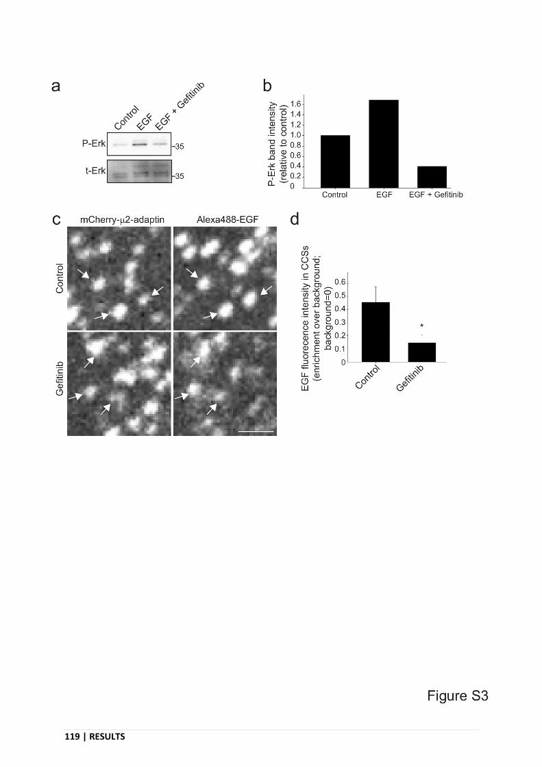
119 | RESULTS
Supplementary Figure 3. Gefitinib treatment does not prevent EGFR
recruitment at CCSs.

RESULTS | 120
Supplementary Figure 4. 1-integrin and vinculin equally distribute between
EGF-decorate and non-decorated fibers.

121 | RESULTS
Supplementary Figure 5. Characterization of collagen fibers remodelling.
DISCUSSION | 122
DISCUSSION
Cell movement is driven by polymerizing actin pushing the plasma membrane at the
front of the cell. Actin polymerization can drive efficient migration only if the actin
cytoskeleton is physically engaged with the extracellular matrix. Focal adhesions are the best
documented adhesion structures. They cluster matrix receptors and link them to the actin
cytoskeleton, thus playing a key role in migration.
We recently discovered that clathrin-coated structures (CCSs), previously known only
as endocytic structures, can also serve as adhesion structures to collagen fibers. CCSs
accumulate along collagen fibers because 1) CCSs nucleation is triggered by the curvature that
the fiber imposes to the plasma membrane and 2) CCSs cluster β1 integrin, a collagen receptor.
These adhesive CCSs were termed tubular clathrin/AP-2 lattices (TCALs) because they adopt
the tubular morphology of the collagen fibers, wrapping around and pinching the fibers.
During my PhD, I discovered a mechanism of 3D haptotaxis that relies on TCALs and
local force transmission. I found that collagen fibers-bound CCS ligands are even more covered
by TCALs than normal collagen fibers. This results from the preferential nucleation of CCSs
on these fibers, leading to a local accumulation of adhesive structures which in turn leads to
cell exerting more forces on ligand-decorated fibers as compared to non-decorated fibers.
Finally, this mechanism is used by the cell to migrate directionally, by following matrix-bound
protein in 3D networks.
1. - At least some CCS ligands can bind to collagen fibers
This project started with the observation that two CCS ligands (EGF and LDL) bind to
in vitro polymerized collagen networks. These observations were already reported in the
literature as EGF was shown to interact with monomeric collagen (Y. Yang et al. 2009) and
electron microscopy imaging revealed that LDL can associate with collagen fibers in rabbit’s
cardiac valve (Nievelstein-Post et al. 1994). Because it is difficult to manipulate 3D collagen
123 | DISCUSSION
network to address precise questions, I developed a protocol to produce short and manipulable
collagen fibers and to decorate them with specific ligands.
Nothing is known about the mechanism of EGF or LDL interaction with collagen fibers
as neither EGF nor LDL have known collagen binding domains. A study reported that the
interaction of EGF with collagen display a Kd of 1.77 x 10⁻⁷ M (Kuo et al. 2015). This affinity
seems week but was measured using monomeric collagen and not with collagen assembled into
fibers. It is possible that EGF have a much stronger affinity for polymerized collagen.
Visualization of EGF-coated fibers were done using EGF labelled with the fluorophore
Alexa488. Actually, Alexa488 is associated with streptavidin that binds biotin-decorated EGF.
The Alexa488-streptavidin-biotin-EGF complex could impact on the apparent affinity/stability
of EGF on collagen fibers as measured in my FRAP experiments. Indeed, more than 50% of
EGF associated with collagen fibers was immobile, confirming the stability I observed. FRAP
experiments on initially non-decorated collagen fibers in the presence of soluble EGF revealed
that the strength of EGF/collagen interaction is essentially the same as for EGF-decorated fibers
produce using my protocol. This rules out the possibility that EGF would better associate with
polymerizing collagen than with already assembled fibers. Regarding LDL, nothing is known
about its mechanism of binding to collagen and previous in vitro experiments revealed a poor
binding of LDL to collagen (Pentikäinen et al. 1997). The observed LDL/collagen interaction
have been proposed to be mediated through the collagen-binding ECM protein decorin. I
however observed that LDL can bind to collagen fibers in the absence of any added factor.
Using cells transfected with fluorescent EGFR, I also confirmed that EGFR
preferentially accumulate along EGF-decorated fibers as compared to normal collagen fibers.
This accumulation is not affected by the EGFR inhibitor gefitinib. This is not surprising as
gefitinib act through biding and inhibiting the kinase domain of the receptor that is not involved
in EGF-EGFR interaction. In fact, gefitinib has been shown to increase EGFR affinity for its
ligand (Björkelund, Gedda, and Andersson 2011) through unknown mechanisms.
2. - Collagen-bound EGF is active and can be internalized
I also observed that collagen-bound EGF could activate EGFR-dependent signaling
pathways. Indeed, Erk was equally activated by similar concentrations of soluble or fiber-

DISCUSSION | 124
associated EGF. However, while Erk can be activated by EGF alone in the experimental
conditions I used (cells in suspension), Akt activation required the presence of both EGF and
collagen fibers. This confirms the existence of crosstalk signaling between integrins and EGFR,
at least for Ekt activation below EGFR signaling. These findings are in opposition with some
results from the literature. For instance, Erk activation upon EGF stimulation was reported to
be strongly decreased in non-adherent cells (Short, Talbott, and Juliano 1998). On the opposite,
Akt was shown to be activated by EGF alone in cells in suspension (Velling, Stefansson, and
Johansson 2008). Theses contradictory results may be explained by the different cell types used
in these studies as compared to the MDA-MB-231 cells I used.
It also seems that Erk signaling is more sustained in time when EGF is associated with
collagen fibers. This could result from the EGF/collagen interaction as the cell could take more
time to internalize collagen-bound EGF, leading to a longer signaling period. This could also
be due to the different pattern of EGF presentation to the cell. In one case, EGF is soluble and
thus presented evenly distributed all around the cell. On the opposite, collagen fiber-bound EGF
is presented as patches of local, high concentration and most of the rest of the membrane is free
of EGF. In the case of soluble EGF, all receptors are internalized upon stimulation. When EGF
is presented bound to collagen, most of the EGFR may remain available to sustain the signal.
In any case, more precise kinetic experiments would be required to draw a definitive conclusion.
I also confirmed that cell internalize collagen-bound EGF. CCSs accumulation along
EGF-decorated fibers suggest that collagen-bound EGF is directly internalized from the fibers.
Nevertheless, it remains possible that a portion of EGF/EGFR complexes detach from the
collagen fibers prior to be recruited at non-fiber associated CCSs. Here also, more experiments
would be required to unambiguously demonstrate that CCSs that accumulate on fibers are
actually able to internalize fiber-associated EGF.
3. - Accumulation of TCALs along collagen fibers decorated with CCS ligands
I observed that TCALs accumulate more on EGF/LDL-decorated collagen fibers as
compared to non-decorated ones. The accumulation of TCALs along EGF-decorated collagen
fibers is lost in cells depleted for EGFR but not in cells treated with gefitinib. TCALs
preferential accumulation along ligand-coated fibers could results from an increase of either

125 | DISCUSSION
CCS local nucleation rate and/or lifetime. I observed that CCS lifetime was only slightly
increased on EGF-coated fibers. On the opposite, CCS nucleation rate was significantly
increased along ligand-decorated collagen fibers as compared to naked fibers. EGFR depletion
but not gefitinib treatment abolished the increased CCS nucleation rate on EGF-coated fibers.
These observations could bring new insights on two ongoing debates: the requirement of EGFR
phosphorylation for its internalization and the ability of receptors to mediate de novo formation
of CCSs.
First, the requirement of EGFR phosphorylation for its recruitment into CCSs, and
hence its internalization, remains unclear in the literature. Some studies found that the tyrosine
kinase activity is required for EGFR recruitment at CCS (Sorkina et al. 2002) and
internalization (Lamaze and Schmid 1995). Other studies suggested that receptor dimerization
is sufficient for EGFR internalization, regardless of the phosphorylation state (Q. Wang,
Villeneuve, and Wang 2005). Finally, a study found that artificial clustering of EGFR is
sufficient to induce internalization (Heukers et al. 2013). In our case, gefitinib treatment did not
affect the preferential accumulation of TCALs along EGF-coated collagen fibers nor the
recruitment of EGF into CCSs. This suggests that EGFR tyrosine kinase activity, and hence
autophosphorylation, is not required for EGFR recruitment at CCSs and subsequent
internalization. This means that EGFR signaling and endocytosis are uncoupled, EGF-induced
dimerization of its receptors regulating both signaling and endocytosis independently.
Second, the ability of receptors, especially EGFR, to induce de novo formation of CCSs
is also debated. Some studies found that EGFR can induce CCSs nucleation in a signaling
dependent manner (Johannessen et al. 2006; Wilde et al. 1999). On the other side, some
observed that EGFR is internalized through preformed CCSs (Rappoport and Simon 2009). If
the constitutively internalized LDLR was never reported to induce de novo formation of CCSs,
a study found that clustering of the constitutively internalized transferrin receptor induces CCS
nucleation (A. P. Liu et al. 2010). In our case, collagen fiber-bound ligands induce CCS
nucleation using a signaling independent mechanism as gefitinib did not affect this parameter,
and LDLR is not known to elicit any specific signaling event. Either the observed EGF/LDL-
induced nucleation relies on different mechanisms or they rely on similar mechanisms
potentially induced by their association with collagen fibers. For EGFR, we can hypothesize
that dimerization reveals a functional endocytosis motif, as suggested, that further recruits the
endocytic machinery, leading to CCS formation. Another non-exclusive and very likely

DISCUSSION | 126
hypothesis is that the accumulation of EGF or LDL along collagen fibers induce the clustering
of their receptors. These clustered receptors could induce CCS formation as reported for TfR
in experimental clustering conditions (A. P. Liu et al. 2010).
4. - Cells protrude more on EGF-coated collagen fibers
I observed that cells tend to protrude more on EGF-decorated fibers spotted on 2D
coverslips. Around 70% of EGF-coated fibers were associated with a protrusion and this
proportion drop to 50% for non-coated fibers. Protrusion formation is driven by actin
polymerization and EGF ability to locally induce actin polymerization (Kempiak et al. 2003)
could explain the preferential protrusion activity on EGF-decorated fibers. Another possibility
is that the preferential protrusion activity on EGF-coated collagen fibers is linked to the
preferential CCSs accumulation on these fibers (see next section). On the opposite, neither
integrins nor focal adhesions seemed to be involved in this mechanism as they distributed
evenly between naked and EGF-coated collagen fibers. Further experiments are required to
determine the actors and mechanisms involved in preferential protrusion along EGF-coated
collagen.
5. - Cells pulls more on CCS ligands-decorated collagen fibers
I observed that when cells are plated on the top of collagen fibers sparsely spotted on a
coverslip, they pull on the fibers and pack them on their dorsal surface as condensed aggregates.
We took advantage of this to estimate cells remodeling capacities by measuring the evolution
over time of fibers circularity. I observed that cells preferentially remodel collagen fibers that
are decorated with CCS ligands as compared to naked fibers.
In the case of both EGF- and LDL-decorated fibers, AP-2 depletion reduced the
preferential remodeling. This means that more forces are applied on decorated-fibers in a CCSs-
dependent manner. However, although the preferential remodeling is lost in these conditions, it
was not the consequence of a reduced remodeling of decorated fibers but was rather due to an
increased remodeling of non-decorated ones. Observations from the lab showed that focal
adhesions are increased in size upon CCSs depletion, probably reflecting the role of endocytosis
in regulating the dynamics of these structures. This could explain the overall increase in
remodeling levels observed after AP-2 depletion. However, clathrin depletion did not reduce
127 | DISCUSSION
the preferential remodeling of EGF-coated fibers. This is in agreement with our previously
published model in which TCALs role as adhesive structures were actually shown to be
independent of clathrin. In the absence of clathrin, a structural core made of AP-2 and other
adaptors remains, still able to cluster receptors, and still able to wrap around and pinch collagen
fibers (Elkhatib et al. 2017). In addition, the fact that clathrin depletion did not inhibit the
preferential remodeling of decorated fibers indicate that this process is independent of
endocytosis and probably rely only on the adhesive role of TCALs as we previously reported.
EGFR depletion also abolished the preferential remodeling of EGF-coated fibers.
Decorated fibers were specifically less remodeled in these conditions as compared to control
cells. This confirms the role of EGFR in the preferential remodeling of EGF-coated fibers, most
likely through its role in promoting local TCALs nucleation on decorated fibers. In agreement
with my observations that tyrosine kinase activity is not involved in TCALs nucleation on EGF-
decorated fibers, its inhibition using gefitinib did not affect the preferential remodeling of these
fibers. The observed preferential remodeling of LDL-coated fibers also advocates for a
signaling-independent mechanism.
Finally, talin depletion did not affect the preferential remodeling of EGF-decorated
fibers. However, the global levels of remodeling were strongly reduced. This suggests that focal
adhesions play an important role in fibers remodeling but do not allow to discriminate between
the different types of fiber, in agreement with my observations that focal adhesions are equally
distributed between decorated and non-decorated fibers.
We already reported that TCALs help the cell to grab and move collagen fibers. My data
here suggest that TCALs are responsible for the increased forces applied on decorated fibers.
Force production in the cell is usually provided by the actin network but it is not known if and
how TCALs can connect actin to transmit forces. Focal adhesion directly interact with the actin
network and this slows down the actin retrograde flow, leading to effective pushing forces and
generation of protrusions. A similar mechanism could be at play here. Observations from the
lab show that, in cells crawling on a 2D surface, CCSs located at the leading edge move toward
the cell center before budding and actually follow the actin retrograde flow. This means that
they are somehow connected to the actin cytoskeleton. Several components found in CCSs are
able to bind actin and thus, CCSs anchored to the substrate could potentially slow down the
actin retrograde flow. It is also possible that actin interacts more sterically than specifically with

DISCUSSION | 128
the TCALs/CCSs. The actin retrograde flow could simply bump into the membrane
invagination formed by CCSs or bump into the fibers upon TCALs-mediated membrane
wrapping around fibers. In any case our data suggest that the more TCALs on a fiber, the more
forces are applied on this fiber and thus more forces are applied on CCS ligands-decorated
fibers.
6. - In 3D, cells migrate towards CCS ligands-decorated collagen networks
To see if collagen-bound CCSs ligands affect 3D adhesion and migration, naked or
decorated collagen fibers were incorporated into 3D collagen networks. Cells were more spread
in EGF-decorated 3D collagen network as compared to regular collagen network. The increased
spreading could be due to more TCALs accumulating on EGF-decorated fibers and facilitating
cell adhesion to surrounding fibers. This still needs to be confirmed by ablating TCALs
formation. Yet, cell velocity was not increased in EGF-decorated networks. This may reflect a
global stabilization of protrusions pointing in different direction so that in fine no net advantage
for motility can be measured in these conditions. However, when at the interface between
regular and EGF-decorated networks, cells preferentially migrated towards the EGF-decorated
network. This directed migration was lost upon depletion of EGFR or AP-2. These findings
reveal an EGF-dependent haptotaxis in 3D that relies on clathrin structures.
At the interface, cells most likely protrude in random directions, but it is likely that the
extra accumulation of TCALs along EGF-decorated fibers may stabilize protrusions pointing
towards these types of fibers. As a consequence, cells would preferentially migrate towards
these fibers, leading to the directed migration I observed. Additional experiments on protrusion
lifetime at the interface are required, though, to test and validate this model.
7. - Model and conclusion
Together, my results allow to draw a general model for haptotaxis mediated by CCS
ligands. Asymmetric distribution of fiber-associated CCS ligands around the cell leads to an
asymmetrical distribution of adhesive TCALs because of local increased nucleation rate on
ligands-decorated fibers. Because we previously reported that TCALs help to stabilize
protrusions, this asymmetric distribution of TCALs may lead to an asymmetric stabilization of
protrusions and an asymmetric distribution of forces applied on the ECM. This mechanism

129 | DISCUSSION
would induce a directed migration towards ECM-anchored CCS ligands. Interestingly, the local
accumulation of TCALs that drives this 3D haptotaxis does not rely on activation of signaling
pathways as proposed for classical chemotaxis.
These results suggest that haptotaxis is mediated by different mechanisms from
chemotaxis, at least in the conditions used in this study. In the case of EGF, chemotaxis is
mediated by an asymmetric distribution of actin polymerization in response to EGFR signaling.
In our model, EGF haptotaxis is driven by an asymmetric distribution of TCALs, independently
of EGFR signaling. In vivo, directed migration towards EGF gradients is probably a
combination of both mechanisms.
I here provide evidences for haptotaxis mediated by EGF or LDL, but the described
mechanism could apply to any CCS ligands able to bind to the ECM. Many CCS ligands are
known to associate with collagen or with other ECM components. 3D haptotaxis mediated by
TCALs could thus be found in numerous examples of directed migration in vivo, from
embryogenesis to cancer invasion.
In addition I set up a protocol allowing the study of haptotaxis independently from
chemotaxis. The effect of decorated-collagen fibers can be tested in cell in suspension, in 2D
or in 3D collagen networks. This allows to study the induced signaling activation, to visualize
the localized effect of decorated fibers and to test the ability of collagen-bound chemoattractants
to induce directed migration. This protocol could be used for other CCS ligands but also to
decorate collagen fibers with some ECM components in order to build and assemble complex
network in a precise manner. In conclusion, this protocol may prove useful for many studies in
different fields.

ANNEX | 130
ANNEX
Tubular clathrin/AP-2 lattices pinch collagen
fibers to support 3D cell migration
Nadia Elkhatib, Enzo Bresteau, Francesco Baschieri, Alba López Rioja,
Guillaume van Niel, Stéphane Vassilopoulos and Guillaume Montagnac
Science. 2017 Jun 16;356(6343). pii: eaal4713. doi: 10.1126/science.aal4713.

131 | ANNEX

ANNEX | 132

133 | ANNEX
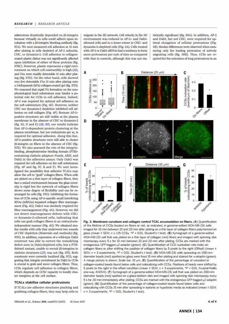
ANNEX | 134

135 | ANNEX

ANNEX | 136

137 | ANNEX

ANNEX | 138
139 | REFERENCES
REFERENCES
Aguet, François et al. 2013. ‘Advances in Analysis of Low Signal-to-Noise Images Link Dynamin and AP2 to the Functions of an Endocytic Checkpoint’. Developmental Cell 26(3): 279–91.
Ahuja, Rashmi et al. 2007. ‘Cordon-Bleu Is an Actin Nucleation Factor and Controls Neuronal Morphology’. Cell 131(2): 337–50.
Albuschies, Jörg, and Viola Vogel. 2013. ‘The Role of Filopodia in the Recognition of Nanotopographies’. Scientific Reports 3: 1658.
Alexandrova, Antonina Y. et al. 2008. ‘Comparative Dynamics of Retrograde Actin Flow and Focal Adhesions: Formation of Nascent Adhesions Triggers Transition from Fast to Slow Flow’. PLOS ONE 3(9): e3234.
Alon, R. et al. 1994. ‘TNF-Alpha Binds to the N-Terminal Domain of Fibronectin and Augments the Beta 1-Integrin-Mediated Adhesion of CD4+ T Lymphocytes to the Glycoprotein’. Journal of Immunology (Baltimore, Md.: 1950) 152(3): 1304–13.
Aman, Andy, and Tatjana Piotrowski. 2010. ‘Cell Migration during Morphogenesis’. Developmental Biology 341(1): 20–33.
An, Bo, Yu-Shan Lin, and Barbara Brodsky. 2016. ‘Collagen Interactions: Drug Design and Delivery.’ Advanced drug delivery reviews 97: 69–84.
Anderson, R. G., M. S. Brown, and J. L. Goldstein. 1977. ‘Role of the Coated Endocytic Vesicle in the Uptake of Receptor-Bound Low Density Lipoprotein in Human Fibroblasts’. Cell 10(3): 351–64.
Andrew, Natalie, and Robert H. Insall. 2007. ‘Chemotaxis in Shallow Gradients Is Mediated Independently of PtdIns 3-Kinase by Biased Choices between Random Protrusions’. Nature Cell Biology 9(2): 193–200.
Andrianantoandro, Ernesto, and Thomas D. Pollard. 2006. ‘Mechanism of Actin Filament Turnover by Severing and Nucleation at Different Concentrations of ADF/Cofilin’. Molecular Cell 24(1): 13–23.
Antonescu, Costin N., François Aguet, Gaudenz Danuser, and Sandra L. Schmid. 2011. ‘Phosphatidylinositol-(4,5)-Bisphosphate Regulates Clathrin-Coated Pit Initiation, Stabilization, and Size’. Molecular Biology of the Cell 22(14): 2588–2600.
Antonny, Bruno et al. 2016. ‘Membrane Fission by Dynamin: What We Know and What We Need to Know’. The EMBO journal 35(21): 2270–84.
Appert-Collin, Aline, Pierre Hubert, Gérard Crémel, and Amar Bennasroune. 2015. ‘Role of ErbB Receptors in Cancer Cell Migration and Invasion’. Frontiers in Pharmacology 6. https://www.ncbi.nlm.nih.gov/pmc/articles/PMC4657385/ (October 16, 2019).

REFERENCES | 140
Arjonen, Antti et al. 2014. ‘Mutant P53-Associated Myosin-X Upregulation Promotes Breast Cancer Invasion and Metastasis’. The Journal of Clinical Investigation 124(3): 1069–82.
Arrieumerlou, Cécile, and Tobias Meyer. 2005. ‘A Local Coupling Model and Compass Parameter for Eukaryotic Chemotaxis’. Developmental Cell 8(2): 215–27.
Attieh, Youmna et al. 2017. ‘Cancer-Associated Fibroblasts Lead Tumor Invasion through Integrin-Β3–Dependent Fibronectin Assembly’. The Journal of Cell Biology 216(11): 3509–20.
Bailly, M. et al. 1998. ‘Regulation of Protrusion Shape and Adhesion to the Substratum during Chemotactic Responses of Mammalian Carcinoma Cells’. Experimental Cell Research 241(2): 285–99.
Bailly, Maryse et al. 2000. ‘Epidermal Growth Factor Receptor Distribution during Chemotactic Responses’. Molecular Biology of the Cell 11(11): 3873–83.
Balaban, N. Q. et al. 2001. ‘Force and Focal Adhesion Assembly: A Close Relationship Studied Using Elastic Micropatterned Substrates’. Nature Cell Biology 3(5): 466–72.
Ballestrem, Christoph et al. 2006. ‘Molecular Mapping of Tyrosine-Phosphorylated Proteins in Focal Adhesions Using Fluorescence Resonance Energy Transfer’. Journal of Cell Science 119(Pt 5): 866–75.
Barczyk, Malgorzata, Sergio Carracedo, and Donald Gullberg. 2010. ‘Integrins’. Cell and Tissue Research 339(1): 269–80.
Barriga, Elias H., and Roberto Mayor. 2015. ‘Embryonic Cell-Cell Adhesion: A Key Player in Collective Neural Crest Migration’. Current Topics in Developmental Biology 112: 301–23.
Baruzzi, A., E. Caveggion, and G. Berton. 2008. ‘Regulation of Phagocyte Migration and Recruitment by Src-Family Kinases’. Cellular and molecular life sciences: CMLS 65(14): 2175–90.
Baschieri, Francesco et al. 2018. ‘Frustrated Endocytosis Controls Contractility-Independent Mechanotransduction at Clathrin-Coated Structures’. Nature Communications 9(1): 1–13.
Bashkirov, Pavel V. et al. 2008. ‘GTPase Cycle of Dynamin Is Coupled to Membrane Squeeze and Release, Leading to Spontaneous Fission’. Cell 135(7): 1276–86.
Batchelder, Erika M., and Defne Yarar. 2010. ‘Differential Requirements for Clathrin-Dependent Endocytosis at Sites of Cell–Substrate Adhesion’. Molecular Biology of the Cell 21(17): 3070–79.
Baulida, J. et al. 1996. ‘All ErbB Receptors Other than the Epidermal Growth Factor Receptor Are Endocytosis Impaired’. The Journal of Biological Chemistry 271(9): 5251–57.
Bearer, E. L. 1991. ‘Direct Observation of Actin Filament Severing by Gelsolin and Binding by GCap39 and CapZ’. The Journal of Cell Biology 115(6): 1629–38.
Beglova, Natalia, and Stephen C. Blacklow. 2005. ‘The LDL Receptor: How Acid Pulls the Trigger’. Trends in Biochemical Sciences 30(6): 309–17.
Benesch, Stefanie et al. 2005. ‘N-WASP Deficiency Impairs EGF Internalization and Actin Assembly at Clathrin-Coated Pits’. Journal of Cell Science 118(14): 3103–15.

141 | REFERENCES
Beningo, K. A. et al. 2001. ‘Nascent Focal Adhesions Are Responsible for the Generation of Strong Propulsive Forces in Migrating Fibroblasts’. The Journal of Cell Biology 153(4): 881–88.
Benmerah, Alexandre, and Christophe Lamaze. 2007. ‘Clathrin-Coated Pits: Vive La Différence?’ Traffic 8(8): 970–82.
Bhattacharjee, Arnab, and Manju Bansal. 2005. ‘Collagen Structure: The Madras Triple Helix and the Current Scenario’. IUBMB life 57(3): 161–72.
Björkelund, Hanna, Lars Gedda, and Karl Andersson. 2011. ‘Comparing the Epidermal Growth Factor Interaction with Four Different Cell Lines: Intriguing Effects Imply Strong Dependency of Cellular Context’. PloS One 6(1): e16536.
Blaser, Heiko et al. 2006. ‘Migration of Zebrafish Primordial Germ Cells: A Role for Myosin Contraction and Cytoplasmic Flow’. Developmental Cell 11(5): 613–27.
Boekhorst, Veronika Te, and Peter Friedl. 2016. ‘Plasticity of Cancer Cell Invasion-Mechanisms and Implications for Therapy.’ Advances in cancer research 132: 209–64.
Bosman, Fred T., and Ivan Stamenkovic. 2003. ‘Functional Structure and Composition of the Extracellular Matrix’. The Journal of Pathology 200(4): 423–28.
Bouchard, Frédéric, Simon D. Bélanger, Katherine Biron-Pain, and Yves St-Pierre. 2010. ‘EGR-1 Activation by EGF Inhibits MMP-9 Expression and Lymphoma Growth’. Blood 116(5): 759–66.
Boucrot, Emmanuel et al. 2015. ‘Endophilin Marks and Controls a Clathrin-Independent Endocytic Pathway’. Nature 517(7535): 460–65.
Boulant, Steeve et al. 2011. ‘Actin Dynamics Counteract Membrane Tension during Clathrin-Mediated Endocytosis’. Nature Cell Biology 13(9): 1124–31.
Bovellan, Miia et al. 2014. ‘Cellular Control of Cortical Actin Nucleation’. Current biology: CB 24(14): 1628–35.
Bray, D., and J. G. White. 1988. ‘Cortical Flow in Animal Cells’. Science (New York, N.Y.) 239(4842): 883–88.
Bretscher, M S. 1989. ‘Endocytosis and Recycling of the Fibronectin Receptor in CHO Cells.’ The EMBO Journal 8(5): 1341–48.
Bretscher, M S. 1992. ‘Cells Can Use Their Transferrin Receptors for Locomotion.’ The EMBO Journal 11(2): 383–89.
Brown, Claire M. et al. 2006. ‘Probing the Integrin-Actin Linkage Using High-Resolution Protein Velocity Mapping’. Journal of Cell Science 119(24): 5204–14.
Brown, M. S., and J. L. Goldstein. 1979. ‘Receptor-Mediated Endocytosis: Insights from the Lipoprotein Receptor System’. Proceedings of the National Academy of Sciences of the United States of America 76(7): 3330–37.
Brundage, R. A., K. E. Fogarty, R. A. Tuft, and F. S. Fay. 1991. ‘Calcium Gradients Underlying Polarization and Chemotaxis of Eosinophils’. Science (New York, N.Y.) 254(5032): 703–6.

REFERENCES | 142
Buehler, Markus J. 2006. ‘Nature Designs Tough Collagen: Explaining the Nanostructure of Collagen Fibrils’. Proceedings of the National Academy of Sciences of the United States of America 103(33): 12285–90.
Burgess, Antony W et al. 2003. ‘An Open-and-Shut Case? Recent Insights into the Activation of EGF/ErbB Receptors’. Molecular Cell 12(3): 541–52.
Burnette, Dylan T. et al. 2011. ‘A Role for Actin Arcs in the Leading-Edge Advance of Migrating Cells’. Nature Cell Biology 13(4): 371–81.
Burridge, Keith et al. 1988. ‘Focal Adhesions: Transmembrane Junctions Between the Extracellular Matrix and the Cytoskeleton’. Annual Review of Cell Biology 4(1): 487–525.
Bustos, Rodrigo I., Marie-Annick Forget, Jeffrey E. Settleman, and Steen H. Hansen. 2008. ‘Coordination of Rho and Rac GTPase Function via P190B RhoGAP’. Current biology: CB 18(20): 1606–11.
Byyny, R. L., D. N. Orth, S. Cohen, and E. S. Doyne. 1974. ‘Epidermal Growth Factor: Effects of Androgens and Adrenergic Agents’. Endocrinology 95(3): 776–82.
Campellone, Kenneth G., and Matthew D. Welch. 2010. ‘A Nucleator Arms Race: Cellular Control of Actin Assembly’. Nature Reviews Molecular Cell Biology 11(4): 237–51.
Carpenter, G., and S. Cohen. 1979. ‘Epidermal Growth Factor’. Annual Review of Biochemistry 48: 193–216.
Carpentier, J. L., M. F. White, L. Orci, and R. C. Kahn. 1987. ‘Direct Visualization of the Phosphorylated Epidermal Growth Factor Receptor during Its Internalization in A-431 Cells’. The Journal of Cell Biology 105(6 Pt 1): 2751–62.
Carter, S. B. 1965. ‘Principles of Cell Motility: The Direction of Cell Movement and Cancer Invasion’. Nature 208(5016): 1183–87.
Caswell, Patrick T. et al. 2007. ‘Rab25 Associates with Alpha5beta1 Integrin to Promote Invasive Migration in 3D Microenvironments’. Developmental Cell 13(4): 496–510.
Caswell, Patrick T., and Jim C. Norman. 2006. ‘Integrin Trafficking and the Control of Cell Migration’. Traffic (Copenhagen, Denmark) 7(1): 14–21.
Chen, Zhucheng et al. 2010. ‘Structure and Control of the Actin Regulatory WAVE Complex’. Nature 468(7323): 533–38.
Chernousov, M. A., F. J. Fogerty, V. E. Koteliansky, and D. F. Mosher. 1991. ‘Role of the I-9 and III-1 Modules of Fibronectin in Formation of an Extracellular Fibronectin Matrix’. The Journal of Biological Chemistry 266(17): 10851–58.
Choi, Colin K. et al. 2008. ‘Actin and Alpha-Actinin Orchestrate the Assembly and Maturation of Nascent Adhesions in a Myosin II Motor-Independent Manner’. Nature Cell Biology 10(9): 1039–50.
Chrzanowska-Wodnicka, M., and K. Burridge. 1996. ‘Rho-Stimulated Contractility Drives the Formation of Stress Fibers and Focal Adhesions’. The Journal of Cell Biology 133(6): 1403–15.
143 | REFERENCES
Citri, Ami, and Yosef Yarden. 2006. ‘EGF-ERBB Signalling: Towards the Systems Level’. Nature Reviews. Molecular Cell Biology 7(7): 505–16.
Clayton, Andrew H. A. et al. 2005. ‘Ligand-Induced Dimer-Tetramer Transition during the Activation of the Cell Surface Epidermal Growth Factor Receptor-A Multidimensional Microscopy Analysis’. The Journal of Biological Chemistry 280(34): 30392–99.
Cocucci, Emanuele, François Aguet, Steeve Boulant, and Tom Kirchhausen. 2012. ‘The First Five Seconds in the Life of a Clathrin-Coated Pit’. Cell 150(3): 495–507.
Cohen, Stanley. 1960. ‘PURIFICATION OF A NERVE-GROWTH PROMOTING PROTEIN FROM THE MOUSE SALIVARY GLAND AND ITS NEURO-CYTOTOXIC ANTISERUM*’. Proceedings of the National Academy of Sciences of the United States of America 46(3): 302–11.
Collins, Agnieszka, Anthony Warrington, Kenneth A. Taylor, and Tatyana Svitkina. 2011a. ‘Structural Organization of the Actin Cytoskeleton at Sites of Clathrin-Mediated Endocytosis’. Current biology: CB 21(14): 1167–75.
Collins, A et al. 2011b. ‘Structural Organization of the Actin Cytoskeleton at Sites of Clathrin-Mediated Endocytosis’. Current biology: CB 21(14): 1167–75.
Condeelis, John, and Jeffrey E. Segall. 2003. ‘Intravital Imaging of Cell Movement in Tumours’. Nature Reviews. Cancer 3(12): 921–30.
Conner, Sean D., and Sandra L. Schmid. 2003. ‘Regulated Portals of Entry into the Cell’. Nature 422(6927): 37–44.
Craig, A. S., M. J. Birtles, J. F. Conway, and D. A. Parry. 1989. ‘An Estimate of the Mean Length of Collagen Fibrils in Rat Tail-Tendon as a Function of Age’. Connective Tissue Research 19(1): 51–62.
Cramer, L. P., M. Siebert, and T. J. Mitchison. 1997. ‘Identification of Novel Graded Polarity Actin Filament Bundles in Locomoting Heart Fibroblasts: Implications for the Generation of Motile Force’. The Journal of Cell Biology 136(6): 1287–1305.
Creely, J. J. et al. 1990. ‘Effects of Epidermal Growth Factor on Collagen Synthesis by an Epithelioid Cell Line Derived from Normal Rat Kidney.’ The American Journal of Pathology 136(6): 1247–57.
Cremona, O. et al. 1999. ‘Essential Role of Phosphoinositide Metabolism in Synaptic Vesicle Recycling’. Cell 99(2): 179–88.
Crowther, R. A., and B. M. Pearse. 1981. ‘Assembly and Packing of Clathrin into Coats.’ The Journal of Cell Biology 91(3): 790–97.
Cukierman, E., R. Pankov, D. R. Stevens, and K. M. Yamada. 2001. ‘Taking Cell-Matrix Adhesions to the Third Dimension’. Science (New York, N.Y.) 294(5547): 1708–12.
Cybulsky, A. V., A. J. McTavish, and M. D. Cyr. 1994. ‘Extracellular Matrix Modulates Epidermal Growth Factor Receptor Activation in Rat Glomerular Epithelial Cells’. The Journal of Clinical Investigation 94(1): 68–78.
Damke, Hanna et al. 2001. ‘Dynamin GTPase Domain Mutants Block Endocytic Vesicle Formation at Morphologically Distinct Stages’. Molecular Biology of the Cell 12(9): 2578–89.

REFERENCES | 144
Dannhauser, Philip N., and Ernst J. Ungewickell. 2012. ‘Reconstitution of Clathrin-Coated Bud and Vesicle Formation with Minimal Components’. Nature Cell Biology 14(6): 634–39.
De Deyne, P. G. et al. 1998. ‘The Vitronectin Receptor Associates with Clathrin-Coated Membrane Domains via the Cytoplasmic Domain of Its Beta5 Subunit’. Journal of Cell Science 111 ( Pt 18): 2729–40.
De Pascalis, Chiara, and Sandrine Etienne-Manneville. 2017. ‘Single and Collective Cell Migration: The Mechanics of Adhesions’. Molecular Biology of the Cell 28(14): 1833–46.
Di Guglielmo, G. M. et al. 1994. ‘Compartmentalization of SHC, GRB2 and MSOS, and Hyperphosphorylation of Raf-1 by EGF but Not Insulin in Liver Parenchyma’. The EMBO journal 13(18): 4269–77.
Discher, Dennis E., Paul Janmey, and Yu-Li Wang. 2005. ‘Tissue Cells Feel and Respond to the Stiffness of Their Substrate’. Science (New York, N.Y.) 310(5751): 1139–43.
Doyle, Andrew D. et al. 2012. ‘Micro-Environmental Control of Cell Migration--Myosin IIA Is Required for Efficient Migration in Fibrillar Environments through Control of Cell Adhesion Dynamics’. Journal of Cell Science 125(Pt 9): 2244–56.
Doyle, A et al. 2015. ‘Local 3D Matrix Microenvironment Regulates Cell Migration through Spatiotemporal Dynamics of Contractility-Dependent Adhesions’. Nature Communications 6. https://www.ncbi.nlm.nih.gov/pmc/articles/PMC4643399/ (October 13, 2019).
Duchek, P. et al. 2001. ‘Guidance of Cell Migration by the Drosophila PDGF/VEGF Receptor’. Cell 107(1): 17–26.
Duchek, P., and P. Rørth. 2001. ‘Guidance of Cell Migration by EGF Receptor Signaling during Drosophila Oogenesis’. Science (New York, N.Y.) 291(5501): 131–33.
Eaton, Suzanne, and Fernando Martin-Belmonte. 2014. ‘Cargo Sorting in the Endocytic Pathway: A Key Regulator of Cell Polarity and Tissue Dynamics’. Cold Spring Harbor Perspectives in Biology 6(10). https://www.ncbi.nlm.nih.gov/pmc/articles/PMC4176004/ (October 14, 2019).
Eberwein, Philipp et al. 2015. ‘Modulation of Focal Adhesion Constituents and Their Down-Stream Events by EGF: On the Cross-Talk of Integrins and Growth Factor Receptors’. Biochimica Et Biophysica Acta 1853(10 Pt A): 2183–98.
Ehrlich, Marcelo et al. 2004. ‘Endocytosis by Random Initiation and Stabilization of Clathrin-Coated Pits’. Cell 118(5): 591–605.
Eichel, K., D. Jullié, and M. von Zastrow. 2016. ‘β-Arrestin Drives MAP Kinase Signalling from Clathrin-Coated Structures after GPCR Dissociation’. Nature Cell Biology 18(3): 303–10.
Elkhatib, Nadia et al. 2017. ‘Tubular Clathrin/AP-2 Lattices Pinch Collagen Fibers to Support 3D Cell Migration’. Science (New York, N.Y.) 356(6343).
Endres, Nicholas F. et al. 2013. ‘Conformational Coupling across the Plasma Membrane in Activation of the EGF Receptor’. Cell 152(3): 543–56.
Engler, Adam J., Shamik Sen, H. Lee Sweeney, and Dennis E. Discher. 2006. ‘Matrix Elasticity Directs Stem Cell Lineage Specification’. Cell 126(4): 677–89.

145 | REFERENCES
Espinoza-Sanchez, Sofia et al. 2018. ‘Conformational Changes in Arp2/3 Complex Induced by ATP, WASp-VCA, and Actin Filaments’. Proceedings of the National Academy of Sciences 115(37): E8642–51.
Etienne-Manneville, Sandrine. 2013. ‘Microtubules in Cell Migration’. Annual Review of Cell and Developmental Biology 29: 471–99.
Ezratty, Ellen J., Claire Bertaux, Eugene E. Marcantonio, and Gregg G. Gundersen. 2009. ‘Clathrin Mediates Integrin Endocytosis for Focal Adhesion Disassembly in Migrating Cells’. The Journal of Cell Biology 187(5): 733–47.
Ezratty, Ellen J., Michael A. Partridge, and Gregg G. Gundersen. 2005. ‘Microtubule-Induced Focal Adhesion Disassembly Is Mediated by Dynamin and Focal Adhesion Kinase’. Nature Cell Biology 7(6): 581–90.
Faelber, Katja et al. 2013. ‘Oligomerization of Dynamin Superfamily Proteins in Health and Disease’. Progress in Molecular Biology and Translational Science 117: 411–43.
Farndale, R. W., J. J. Sixma, M. J. Barnes, and P. G. de Groot. 2004. ‘The Role of Collagen in Thrombosis and Hemostasis’. Journal of thrombosis and haemostasis: JTH 2(4): 561–73.
Ferguson, Shawn M., and Pietro De Camilli. 2012. ‘Dynamin, a Membrane-Remodelling GTPase’. Nature Reviews. Molecular Cell Biology 13(2): 75–88.
Fisher, Stephanie A. et al. 2018. ‘Photo-Immobilized EGF Chemical Gradients Differentially Impact Breast Cancer Cell Invasion and Drug Response in Defined 3D Hydrogels’. Biomaterials 178: 751–66.
Fotin, Alexander et al. 2004. ‘Molecular Model for a Complete Clathrin Lattice from Electron Cryomicroscopy’. Nature 432(7017): 573–79.
Fotin, A et al. 2006. ‘Structure Determination of Clathrin Coats to Subnanometer Resolution by Single Particle Cryo-Electron Microscopy’. Journal of Structural Biology 156(3): 453–60.
Fraley, Stephanie I. et al. 2010. ‘A Distinctive Role for Focal Adhesion Proteins in Three-Dimensional Cell Motility’. Nature Cell Biology 12(6): 598–604.
Franco, Santos J. et al. 2004. ‘Calpain-Mediated Proteolysis of Talin Regulates Adhesion Dynamics’. Nature Cell Biology 6(10): 977–83.
Frantz, Christian et al. 2007. ‘Positive Feedback between Cdc42 Activity and H+ Efflux by the Na-H Exchanger NHE1 for Polarity of Migrating Cells’. The Journal of Cell Biology 179(3): 403–10.
Frantz, Christian, Kathleen M. Stewart, and Valerie M. Weaver. 2010. ‘The Extracellular Matrix at a Glance’. Journal of Cell Science 123(Pt 24): 4195–4200.
Freed, Daniel M. et al. 2017. ‘EGFR Ligands Differentially Stabilize Receptor Dimers to Specify Signaling Kinetics’. Cell 171(3): 683-695.e18.
Friedl, Peter, and Katarina Wolf. 2003. ‘Tumour-Cell Invasion and Migration: Diversity and Escape Mechanisms’. Nature Reviews. Cancer 3(5): 362–74.
Fujimoto, L. Miya, Robyn Roth, John E. Heuser, and Sandra L. Schmid. 2000. ‘Actin Assembly Plays a Variable, but Not Obligatory Role in Receptor-Mediated Endocytosis’. Traffic 1(2): 161–71.

REFERENCES | 146
Funamoto, Satoru et al. 2002. ‘Spatial and Temporal Regulation of 3-Phosphoinositides by PI 3-Kinase and PTEN Mediates Chemotaxis’. Cell 109(5): 611–23.
Gadella, T. W., and T. M. Jovin. 1995. ‘Oligomerization of Epidermal Growth Factor Receptors on A431 Cells Studied by Time-Resolved Fluorescence Imaging Microscopy. A Stereochemical Model for Tyrosine Kinase Receptor Activation’. The Journal of Cell Biology 129(6): 1543–58.
Galbraith, Catherine G., Kenneth M. Yamada, and James A. Galbraith. 2007. ‘Polymerizing Actin Fibers Position Integrins Primed to Probe for Adhesion Sites’. Science (New York, N.Y.) 315(5814): 992–95.
Garay, Camilo et al. 2015. ‘Epidermal Growth Factor-Stimulated Akt Phosphorylation Requires Clathrin or ErbB2 but Not Receptor Endocytosis’. Molecular Biology of the Cell 26(19): 3504–19.
Garner, Omai B. et al. 2011. ‘Stage-Dependent Regulation of Mammary Ductal Branching by Heparan Sulfate and HGF-CMet Signaling’. Developmental Biology 355(2): 394–403.
Garrett, Thomas P. J. et al. 2002. ‘Crystal Structure of a Truncated Epidermal Growth Factor Receptor Extracellular Domain Bound to Transforming Growth Factor Alpha’. Cell 110(6): 763–73.
Gerhardt, Holger et al. 2003. ‘VEGF Guides Angiogenic Sprouting Utilizing Endothelial Tip Cell Filopodia’. The Journal of Cell Biology 161(6): 1163–77.
Giannone, Grégory, and Michael P. Sheetz. 2006. ‘Substrate Rigidity and Force Define Form through Tyrosine Phosphatase and Kinase Pathways’. Trends in Cell Biology 16(4): 213–23.
Goel, Hira Lal et al. 2004. ‘Selective Modulation of Type 1 Insulin-like Growth Factor Receptor Signaling and Functions by Β1 Integrins’. The Journal of Cell Biology 166(3): 407–18.
Goh, Lai Kuan et al. 2010. ‘Multiple Mechanisms Collectively Regulate Clathrin-Mediated Endocytosis of the Epidermal Growth Factor Receptor’. The Journal of Cell Biology 189(5): 871–83.
Goh, Lai Kuan, and Alexander Sorkin. 2013. ‘Endocytosis of Receptor Tyrosine Kinases’. Cold Spring Harbor Perspectives in Biology 5(5): a017459.
Goley, Erin D., and Matthew D. Welch. 2006. ‘The ARP2/3 Complex: An Actin Nucleator Comes of Age’. Nature Reviews Molecular Cell Biology 7(10): 713–26.
Goode, Bruce L., and Michael J. Eck. 2007. ‘Mechanism and Function of Formins in the Control of Actin Assembly’. Annual Review of Biochemistry 76: 593–627.
Göstring, Lovisa. 2011. ‘Cellular Studies of HER-Family Specific Affibody Molecules’. http://urn.kb.se/resolve?urn=urn:nbn:se:uu:diva-156730 (October 17, 2019).
Grände, Mats et al. 2002. ‘Transforming Growth Factor-Beta and Epidermal Growth Factor Synergistically Stimulate Epithelial to Mesenchymal Transition (EMT) through a MEK-Dependent Mechanism in Primary Cultured Pig Thyrocytes’. Journal of Cell Science 115(Pt 22): 4227–36.
Green, H., and B. Goldberg. 1964. ‘COLLAGEN AND CELL PROTEIN SYNTHESIS BY AN ESTABLISHED MAMMALIAN FIBROBLAST LINE’. Nature 204: 347–49.

147 | REFERENCES
Greenblatt, Melvin, and Philippe Shuvi. 1968. ‘Tumor Angiogenesis: Transfilter Diffusion Studies in the Hamster by the Transparent Chamber Technique’. JNCI: Journal of the National Cancer Institute 41(1): 111–24.
Grimes, M. L. et al. 1996. ‘Endocytosis of Activated TrkA: Evidence That Nerve Growth Factor Induces Formation of Signaling Endosomes’. The Journal of Neuroscience: The Official Journal of the Society for Neuroscience 16(24): 7950–64.
Gross, J., and D. Kirk. 1958. ‘The Heat Precipitation of Collagen from Neutral Salt Solutions: Some Rate-Regulating Factors’. The Journal of Biological Chemistry 233(2): 355–60.
Gurtner, Geoffrey C., Sabine Werner, Yann Barrandon, and Michael T. Longaker. 2008. ‘Wound Repair and Regeneration’. Nature 453(7193): 314–21.
Harding, C., J. Heuser, and P. Stahl. 1983. ‘Receptor-Mediated Endocytosis of Transferrin and Recycling of the Transferrin Receptor in Rat Reticulocytes’. The Journal of Cell Biology 97(2): 329–39.
von der Hardt, Sophia et al. 2007. ‘The Bmp Gradient of the Zebrafish Gastrula Guides Migrating Lateral Cells by Regulating Cell-Cell Adhesion’. Current biology: CB 17(6): 475–87.
Hatten, Mary E. 2002. ‘New Directions in Neuronal Migration’. Science (New York, N.Y.) 297(5587): 1660–63.
Haugh, J. M. et al. 1999. ‘Internalized Epidermal Growth Factor Receptors Participate in the Activation of P21(Ras) in Fibroblasts’. The Journal of Biological Chemistry 274(48): 34350–60.
Hawryluk, Matthew J. et al. 2006. ‘Epsin 1 Is a Polyubiquitin-Selective Clathrin-Associated Sorting Protein’. Traffic (Copenhagen, Denmark) 7(3): 262–81.
Hayes, Susan, Anil Chawla, and Silvia Corvera. 2002. ‘TGFβ Receptor Internalization into EEA1-Enriched Early Endosomes’. The Journal of Cell Biology 158(7): 1239–49.
Henne, William Mike et al. 2010. ‘FCHo Proteins Are Nucleators of Clathrin-Mediated Endocytosis’. Science (New York, N.Y.) 328(5983): 1281–84.
Henry, Anastasia G. et al. 2012. ‘Regulation of Endocytic Clathrin Dynamics by Cargo Ubiquitination’. Developmental Cell 23(3): 519–32.
Hentze, Matthias W., Martina U. Muckenthaler, Bruno Galy, and Clara Camaschella. 2010. ‘Two to Tango: Regulation of Mammalian Iron Metabolism’. Cell 142(1): 24–38.
Hernandez, Lorena et al. 2009. ‘The EGF/CSF-1 Paracrine Invasion Loop Can Be Triggered by Heregulin Beta1 and CXCL12’. Cancer Research 69(7): 3221–27.
Heukers, Raimond et al. 2013. ‘Endocytosis of EGFR Requires Its Kinase Activity and N-Terminal Transmembrane Dimerization Motif’. Journal of Cell Science 126(Pt 21): 4900–4912.
Higashiyama, S., J. A. Abraham, and M. Klagsbrun. 1993. ‘Heparin-Binding EGF-like Growth Factor Stimulation of Smooth Muscle Cell Migration: Dependence on Interactions with Cell Surface Heparan Sulfate’. The Journal of Cell Biology 122(4): 933–40.
Higgins, Matthew K., and Harvey T. McMahon. 2002. ‘Snap-Shots of Clathrin-Mediated Endocytosis.’ Trends in biochemical sciences 27(5): 257–63.

REFERENCES | 148
Higgs, Henry N., and Thomas D. Pollard. 2000. ‘Activation by Cdc42 and Pip2 of Wiskott-Aldrich Syndrome Protein (Wasp) Stimulates Actin Nucleation by Arp2/3 Complex’. The Journal of Cell Biology 150(6): 1311–20.
Hinshaw, J. E., and S. L. Schmid. 1995. ‘Dynamin Self-Assembles into Rings Suggesting a Mechanism for Coated Vesicle Budding’. Nature 374(6518): 190–92.
Holmes, Kenneth C., David Popp, Werner Gebhard, and Wolfgang Kabsch. 1990. ‘Atomic Model of the Actin Filament’. Nature 347(6288): 44–49.
Höning, Stefan et al. 2005. ‘Phosphatidylinositol-(4,5)-Bisphosphate Regulates Sorting Signal Recognition by the Clathrin-Associated Adaptor Complex AP2’. Molecular Cell 18(5): 519–31.
Hotulainen, Pirta, and Pekka Lappalainen. 2006. ‘Stress Fibers Are Generated by Two Distinct Actin Assembly Mechanisms in Motile Cells’. The Journal of Cell Biology 173(3): 383–94.
Hu, Dana D., Carlos F. Barbas, and Jeffrey W. Smith. 1996. ‘An Allosteric Ca2+ Binding Site on the Β3-Integrins That Regulates the Dissociation Rate for RGD Ligands’. Journal of Biological Chemistry 271(36): 21745–51.
Huang, Fangtian et al. 2006. ‘Differential Regulation of EGF Receptor Internalization and Degradation by Multiubiquitination within the Kinase Domain’. Molecular Cell 21(6): 737–48.
Huang, Fangtian, Anastasia Khvorova, William Marshall, and Alexander Sorkin. 2004. ‘Analysis of Clathrin-Mediated Endocytosis of Epidermal Growth Factor Receptor by RNA Interference’. The Journal of Biological Chemistry 279(16): 16657–61.
Humphries, Ashley C., and Michael Way. 2013. ‘The Non-Canonical Roles of Clathrin and Actin in Pathogen Internalization, Egress and Spread’. Nature Reviews Microbiology 11(8): 551–60.
Humphries, Jonathan D. et al. 2007. ‘Vinculin Controls Focal Adhesion Formation by Direct Interactions with Talin and Actin’. The Journal of Cell Biology 179(5): 1043–57.
Humphries, Jonathan D., Adam Byron, and Martin J. Humphries. 2006. ‘Integrin Ligands at a Glance’. Journal of Cell Science 119(Pt 19): 3901–3.
Humphries, M. J. 2000. ‘Integrin Structure’. Biochemical Society Transactions 28(4): 311–39.
Hynes, Richard O., and Alexandra Naba. 2012. ‘Overview of the Matrisome--an Inventory of Extracellular Matrix Constituents and Functions’. Cold Spring Harbor Perspectives in Biology 4(1): a004903.
Ide, AG. 1939. ‘Vascularization of the Brown Pearce Rabbit Epithelioma Transplant as Seen in the Transparent Ear Chamber’. AJR Am J Roentgenol 42: 891–99.
Iijima, Miho, and Peter Devreotes. 2002. ‘Tumor Suppressor PTEN Mediates Sensing of Chemoattractant Gradients’. Cell 109(5): 599–610.
Imai, K et al. 1997. ‘Degradation of Decorin by Matrix Metalloproteinases: Identification of the Cleavage Sites, Kinetic Analyses and Transforming Growth Factor-Beta1 Release.’ Biochemical Journal 322(Pt 3): 809–14.
Insall, Robert H. 2010. ‘Understanding Eukaryotic Chemotaxis: A Pseudopod-Centred View’. Nature Reviews. Molecular Cell Biology 11(6): 453–58.

149 | REFERENCES
Ishitsuka, Taichi, Tomoki Ikuta, Hiroyoshi Ariga, and Ken-Ichi Matsumoto. 2009. ‘Serum Tenascin-X Strongly Binds to Vascular Endothelial Growth Factor’. Biological & Pharmaceutical Bulletin 32(6): 1004–11.
Ivaska, Johanna et al. 1999. ‘Integrin Α2β1 Mediates Isoform-Specific Activation of P38 and Upregulation of Collagen Gene Transcription by a Mechanism Involving the Α2 Cytoplasmic Tail’. The Journal of Cell Biology 147(2): 401–16.
Ivaska, Johanna, and Jyrki Heino. 2011. ‘Cooperation between Integrins and Growth Factor Receptors in Signaling and Endocytosis’. Annual Review of Cell and Developmental Biology 27: 291–320.
Jacquemet, Guillaume, Hellyeh Hamidi, and Johanna Ivaska. 2015. ‘Filopodia in Cell Adhesion, 3D Migration and Cancer Cell Invasion’. Current Opinion in Cell Biology 36: 23–31.
Jahed, Zeinab, Hengameh Shams, Mehrdad Mehrbod, and Mohammad R. K. Mofrad. 2014. ‘Mechanotransduction Pathways Linking the Extracellular Matrix to the Nucleus’. International Review of Cell and Molecular Biology 310: 171–220.
Jansen, Karin A. et al. 2018. ‘The Role of Network Architecture in Collagen Mechanics’. Biophysical Journal 114(11): 2665–78.
Järveläinen, Hannu et al. 2009. ‘Extracellular Matrix Molecules: Potential Targets in Pharmacotherapy’. Pharmacological Reviews 61(2): 198–223.
Jékely, Gáspár, Hsin-Ho Sung, Carlos M. Luque, and Pernille Rørth. 2005. ‘Regulators of Endocytosis Maintain Localized Receptor Tyrosine Kinase Signaling in Guided Migration’. Developmental Cell 9(2): 197–207.
Johannessen, Lene E. et al. 2006. ‘Activation of the Epidermal Growth Factor (EGF) Receptor Induces Formation of EGF Receptor- and Grb2-Containing Clathrin-Coated Pits’. Molecular and Cellular Biology 26(2): 389–401.
Jost, M. et al. 1998. ‘Phosphatidylinositol-4,5-Bisphosphate Is Required for Endocytic Coated Vesicle Formation’. Current biology: CB 8(25): 1399–1402.
Kajanne, Risto et al. 2007. ‘EGF-R Regulates MMP Function in Fibroblasts through MAPK and AP-1 Pathways’. Journal of Cellular Physiology 212(2): 489–97.
Kaksonen, Marko, and Aurélien Roux. 2018. ‘Mechanisms of Clathrin-Mediated Endocytosis’. Nature Reviews. Molecular Cell Biology 19(5): 313–26.
Kalluri, Raghu, and Robert A. Weinberg. 2009. ‘The Basics of Epithelial-Mesenchymal Transition’. The Journal of Clinical Investigation 119(6): 1420–28.
Kanchanawong, Pakorn et al. 2010. ‘Nanoscale Architecture of Integrin-Based Cell Adhesions’. Nature 468(7323): 580–84.
Katoh, Kazuo et al. 2001. ‘Rho-Kinase–Mediated Contraction of Isolated Stress Fibers’. The Journal of Cell Biology 153(3): 569–84.
Kaverina, Irina, Olga Krylyshkina, and J. Victor Small. 1999. ‘Microtubule Targeting of Substrate Contacts Promotes Their Relaxation and Dissociation’. The Journal of Cell Biology 146(5): 1033–44.

REFERENCES | 150
Kazazic, Maja et al. 2009. ‘Epsin 1 Is Involved in Recruitment of Ubiquitinated EGF Receptors into Clathrin-Coated Pits’. Traffic (Copenhagen, Denmark) 10(2): 235–45.
Kedrin, Dmitriy et al. 2007. ‘Cell Motility and Cytoskeletal Regulation in Invasion and Metastasis’. Journal of Mammary Gland Biology and Neoplasia 12(2–3): 143–52.
Keen, James H., Mark C. Willingham, and Ira H. Pastan. 1979. ‘Clathrin-Coated Vesicles: Isolation, Dissociation and Factor-Dependent Reassociation of Clathrin Baskets’. Cell 16(2): 303–12.
Keller, H., and P. Eggli. 1998. ‘Protrusive Activity, Cytoplasmic Compartmentalization, and Restriction Rings in Locomoting Blebbing Walker Carcinosarcoma Cells Are Related to Detachment of Cortical Actin from the Plasma Membrane’. Cell Motility and the Cytoskeleton 41(2): 181–93.
Kempiak, Stephan J., Shu-Chin Yip, Jonathan M. Backer, and Jeffrey E. Segall. 2003. ‘Local Signaling by the EGF Receptor’. The Journal of Cell Biology 162(5): 781–88.
Kim, Daehwan et al. 2015. ‘Epidermal Growth Factor Improves the Migration and Contractility of Aged Fibroblasts Cultured on 3D Collagen Matrices’. International Journal of Molecular Medicine 35(4): 1017–25.
Kim, Sangmin et al. 2009. ‘EGF-Induced MMP-9 Expression Is Mediated by the JAK3/ERK Pathway, but Not by the JAK3/STAT-3 Pathway in a SKBR3 Breast Cancer Cell Line’. Cellular Signalling 21(6): 892–98.
Kirchhausen, T., and S. C. Harrison. 1981. ‘Protein Organization in Clathrin Trimers’. Cell 23(3): 755–61.
Klapholz, Benjamin et al. 2015. ‘Alternative Mechanisms for Talin to Mediate Integrin Function’. Current biology: CB 25(7): 847–57.
Knight, C. G. et al. 1998. ‘Identification in Collagen Type I of an Integrin Alpha2 Beta1-Binding Site Containing an Essential GER Sequence’. The Journal of Biological Chemistry 273(50): 33287–94.
Kreuger, Johan, Lidia Perez, Antonio J. Giraldez, and Stephen M. Cohen. 2004. ‘Opposing Activities of Dally-like Glypican at High and Low Levels of Wingless Morphogen Activity’. Developmental Cell 7(4): 503–12.
Kubow, Kristopher E., and Alan Rick Horwitz. 2011. ‘Reducing Background Fluorescence Reveals Adhesions in 3D Matrices’. Nature Cell Biology 13(1): 3–5; author reply 5-7.
Kuo, Wei-Ting et al. 2015. ‘Quantitative Analysis of Ligand-EGFR Interactions: A Platform for Screening Targeting Molecules’. PLoS ONE 10(2). https://www.ncbi.nlm.nih.gov/pmc/articles/PMC4344348/ (October 18, 2019).
Kurosaka, Satoshi, and Anna Kashina. 2008. ‘Cell Biology of Embryonic Migration’. Birth Defects Research. Part C, Embryo Today: Reviews 84(2): 102–22.
Laato, M., V. M. Kähäri, J. Niinikoski, and E. Vuorio. 1987. ‘Epidermal Growth Factor Increases Collagen Production in Granulation Tissue by Stimulation of Fibroblast Proliferation and Not by Activation of Procollagen Genes’. The Biochemical Journal 247(2): 385–88.

151 | REFERENCES
Lai, Frank P.L. et al. 2009. ‘Cortactin Promotes Migration and Platelet-Derived Growth Factor-Induced Actin Reorganization by Signaling to Rho-GTPases’. Molecular Biology of the Cell 20(14): 3209–23.
Lamaze, C., and S. L. Schmid. 1995. ‘Recruitment of Epidermal Growth Factor Receptors into Coated Pits Requires Their Activated Tyrosine Kinase’. The Journal of Cell Biology 129(1): 47–54.
Lambert, Arthur W., Diwakar R. Pattabiraman, and Robert A. Weinberg. 2017. ‘Emerging Biological Principles of Metastasis’. Cell 168(4): 670–91.
Lampe, Marko, Stéphane Vassilopoulos, and Christien Merrifield. 2016. ‘Clathrin Coated Pits, Plaques and Adhesion’. Journal of Structural Biology 196(1): 48–56.
Lauffenburger, D. A., and A. F. Horwitz. 1996. ‘Cell Migration: A Physically Integrated Molecular Process’. Cell 84(3): 359–69.
Laukaitis, C. M., D. J. Webb, K. Donais, and A. F. Horwitz. 2001. ‘Differential Dynamics of Alpha 5 Integrin, Paxillin, and Alpha-Actinin during Formation and Disassembly of Adhesions in Migrating Cells’. The Journal of Cell Biology 153(7): 1427–40.
Lawson, M. A., and F. R. Maxfield. 1995. ‘Ca(2+)- and Calcineurin-Dependent Recycling of an Integrin to the Front of Migrating Neutrophils’. Nature 377(6544): 75–79.
Le Gall, Sylvain M., Rodolphe Auger, Catherine Dreux, and Philippe Mauduit. 2003. ‘Regulated Cell Surface Pro-EGF Ectodomain Shedding Is a Zinc Metalloprotease-Dependent Process’. The Journal of Biological Chemistry 278(46): 45255–68.
Lee, Ho-Sup et al. 2004. ‘Characterization of an Actin-Binding Site within the Talin FERM Domain’. Journal of Molecular Biology 343(3): 771–84.
Lee, Juliet. 2018. ‘Insights into Cell Motility Provided by the Iterative Use of Mathematical Modeling and Experimentation’. AIMS Biophysics 5(2): 97.
Legerstee, Karin, Bart Geverts, Johan A. Slotman, and Adriaan B. Houtsmuller. 2019. ‘Dynamics and Distribution of Paxillin, Vinculin, Zyxin and VASP Depend on Focal Adhesion Location and Orientation’. Scientific Reports 9(1): 1–18.
Leitinger, Birgit, and Erhard Hohenester. 2007. ‘Mammalian Collagen Receptors’. Matrix Biology: Journal of the International Society for Matrix Biology 26(3): 146–55.
Lemmon, Mark A., and Joseph Schlessinger. 2010. ‘Cell Signaling by Receptor-Tyrosine Kinases’. Cell 141(7): 1117–34.
Lherbette, Michael et al. 2019. ‘The AP2 Adaptor Enhances Clathrin Coat Stiffness’. The FEBS Journal 0(0). https://febs.onlinelibrary.wiley.com/doi/abs/10.1111/febs.14961 (October 14, 2019).
Li, Dong et al. 2015. ‘Extended-Resolution Structured Illumination Imaging of Endocytic and Cytoskeletal Dynamics’. Science 349(6251): aab3500.
Li, Rong, and Gregg G. Gundersen. 2008. ‘Beyond Polymer Polarity: How the Cytoskeleton Builds a Polarized Cell’. Nature Reviews. Molecular Cell Biology 9(11): 860–73.

REFERENCES | 152
Lichtner, R. B. et al. 2001. ‘Signaling-Inactive Epidermal Growth Factor Receptor/Ligand Complexes in Intact Carcinoma Cells by Quinazoline Tyrosine Kinase Inhibitors’. Cancer Research 61(15): 5790–95.
Liu, Allen P., François Aguet, Gaudenz Danuser, and Sandra L. Schmid. 2010. ‘Local Clustering of Transferrin Receptors Promotes Clathrin-Coated Pit Initiation’. The Journal of Cell Biology 191(7): 1381–93.
Liu, Jiang et al. 2004. ‘Ouabain Induces Endocytosis of Plasmalemmal Na/K-ATPase in LLC-PK1 Cells by a Clathrin-Dependent Mechanism’. Kidney International 66(1): 227–41.
Liu, Li et al. 2007. ‘Tetraspanin CD151 Promotes Cell Migration by Regulating Integrin Trafficking’. The Journal of Biological Chemistry 282(43): 31631–42.
Liu, S. H., M. L. Wong, C. S. Craik, and F. M. Brodsky. 1995. ‘Regulation of Clathrin Assembly and Trimerization Defined Using Recombinant Triskelion Hubs’. Cell 83(2): 257–67.
Lo, C. M., H. B. Wang, M. Dembo, and Y. L. Wang. 2000. ‘Cell Movement Is Guided by the Rigidity of the Substrate’. Biophysical Journal 79(1): 144–52.
Lock, John G. et al. 2019. ‘Clathrin-Containing Adhesion Complexes’. The Journal of Cell Biology 218(7): 2086–95.
Loerke, Dinah et al. 2009. ‘Cargo and Dynamin Regulate Clathrin-Coated Pit Maturation’. PLOS Biology 7(3): e1000057.
Longva, Karianne E. et al. 2002. ‘Ubiquitination and Proteasomal Activity Is Required for Transport of the EGF Receptor to Inner Membranes of Multivesicular Bodies’. The Journal of Cell Biology 156(5): 843–54.
Lu, Chafen et al. 2010. ‘Structural Evidence for Loose Linkage between Ligand Binding and Kinase Activation in the Epidermal Growth Factor Receptor’. Molecular and Cellular Biology 30(22): 5432–43.
Lu, Pengfei, Ken Takai, Valerie M. Weaver, and Zena Werb. 2011. ‘Extracellular Matrix Degradation and Remodeling in Development and Disease’. Cold Spring Harbor Perspectives in Biology 3(12).
Lu, Zhimin, Guoqiang Jiang, Peter Blume-Jensen, and Tony Hunter. 2001. ‘Epidermal Growth Factor-Induced Tumor Cell Invasion and Metastasis Initiated by Dephosphorylation and Downregulation of Focal Adhesion Kinase’. Molecular and Cellular Biology 21(12): 4016–31.
Luster, A. D. 1998. ‘Chemokines--Chemotactic Cytokines That Mediate Inflammation’. The New England Journal of Medicine 338(7): 436–45.
Lyons, R. M., L. E. Gentry, A. F. Purchio, and H. L. Moses. 1990. ‘Mechanism of Activation of Latent Recombinant Transforming Growth Factor Beta 1 by Plasmin’. The Journal of Cell Biology 110(4): 1361–67.
Macia, Eric et al. 2006. ‘Dynasore, a Cell-Permeable Inhibitor of Dynamin’. Developmental Cell 10(6): 839–50.

153 | REFERENCES
Madshus, Inger Helene, and Espen Stang. 2009. ‘Internalization and Intracellular Sorting of the EGF Receptor: A Model for Understanding the Mechanisms of Receptor Trafficking’. Journal of Cell Science 122(Pt 19): 3433–39.
Mai, Anja et al. 2014. ‘Distinct C-Met Activation Mechanisms Induce Cell Rounding or Invasion through Pathways Involving Integrins, RhoA and HIP1’. Journal of Cell Science 127(Pt 9): 1938–52.
Martin, Christine, Stine F. Pedersen, Albrecht Schwab, and Christian Stock. 2011. ‘Intracellular PH Gradients in Migrating Cells’. American Journal of Physiology. Cell Physiology 300(3): C490-495.
Maupin, P., and T. D. Pollard. 1983. ‘Improved Preservation and Staining of HeLa Cell Actin Filaments, Clathrin-Coated Membranes, and Other Cytoplasmic Structures by Tannic Acid-Glutaraldehyde-Saponin Fixation’. The Journal of Cell Biology 96(1): 51–62.
Maurer, Meghan E., and Jonathan A. Cooper. 2006. ‘The Adaptor Protein Dab2 Sorts LDL Receptors into Coated Pits Independently of AP-2 and ARH’. Journal of Cell Science 119(Pt 20): 4235–46.
McCUTCHEON, M. 1946. ‘Chemotaxis in Leukocytes’. Physiological Reviews 26(3): 319–36.
McMahon, Harvey T., and Emmanuel Boucrot. 2011. ‘Molecular Mechanism and Physiological Functions of Clathrin-Mediated Endocytosis’. Nature Reviews. Molecular Cell Biology 12(8): 517–33.
Merrifield, Christien J., Britta Qualmann, Michael M. Kessels, and Wolfhard Almers. 2004. ‘Neural Wiskott Aldrich Syndrome Protein (N-WASP) and the Arp2/3 Complex Are Recruited to Sites of Clathrin-Mediated Endocytosis in Cultured Fibroblasts’. European Journal of Cell Biology 83(1): 13–18.
Mettlen, Marcel et al. 2010. ‘Cargo- and Adaptor-Specific Mechanisms Regulate Clathrin-Mediated Endocytosis’. The Journal of Cell Biology 188(6): 919–33.
Mettlen, M et al. 2018. ‘Regulation of Clathrin-Mediated Endocytosis’. Annual Review of Biochemistry 87: 871–96.
Mettouchi, A. et al. 2001. ‘Integrin-Specific Activation of Rac Controls Progression through the G(1) Phase of the Cell Cycle’. Molecular Cell 8(1): 115–27.
Miller, Sharon E. et al. 2015. ‘CALM Regulates Clathrin-Coated Vesicle Size and Maturation by Directly Sensing and Driving Membrane Curvature’. Developmental Cell 33(2): 163–75.
Mimura, Yoshihiro et al. 2004. ‘Epidermal Growth Factor Induces Fibronectin Expression in Human Dermal Fibroblasts via Protein Kinase C δ Signaling Pathway’. Journal of Investigative Dermatology 122(6): 1390–98.
Minina, Sofia, Michal Reichman-Fried, and Erez Raz. 2007. ‘Control of Receptor Internalization, Signaling Level, and Precise Arrival at the Target in Guided Cell Migration’. Current biology: CB 17(13): 1164–72.
Mitchison, T. J., and L. P. Cramer. 1996. ‘Actin-Based Cell Motility and Cell Locomotion’. Cell 84(3): 371–79.

REFERENCES | 154
Mitra, Satyajit K., Daniel A. Hanson, and David D. Schlaepfer. 2005. ‘Focal Adhesion Kinase: In Command and Control of Cell Motility’. Nature Reviews. Molecular Cell Biology 6(1): 56–68.
Miura, Yoshiki, Tsuyoshi Takahashi, Stephanie M. Jung, and Masaaki Moroi. 2002. ‘Analysis of the Interaction of Platelet Collagen Receptor Glycoprotein VI (GPVI) with Collagen. A Dimeric Form of GPVI, but Not the Monomeric Form, Shows Affinity to Fibrous Collagen’. The Journal of Biological Chemistry 277(48): 46197–204.
Mizutani, Kiyohito et al. 2002. ‘Essential Role of Neural Wiskott-Aldrich Syndrome Protein in Podosome Formation and Degradation of Extracellular Matrix in Src-Transformed Fibroblasts’. Cancer Research 62(3): 669–74.
Montagnac, Guillaume et al. 2013. ‘ΑTAT1 Catalyses Microtubule Acetylation at Clathrin-Coated Pits’. Nature 502(7472): 567–70.
Morelli, Joseph G et al. 1992. ‘Leukotriene C4 and TGF-Alpha Are Stimulators of Human Melanocyte Migration In Vitro’. Journal of Investigative Dermatology 98(3): 290–95.
Mori, Hidetoshi et al. 2002. ‘CD44 Directs Membrane-Type 1 Matrix Metalloproteinase to Lamellipodia by Associating with Its Hemopexin-like Domain’. The EMBO Journal 21(15): 3949–59.
Moro, L et al. 1998. ‘Integrins Induce Activation of EGF Receptor: Role in MAP Kinase Induction and Adhesion-Dependent Cell Survival.’ The EMBO Journal 17(22): 6622–32.
Moro, Laura et al. 2002. ‘Integrin-Induced Epidermal Growth Factor (EGF) Receptor Activation Requires c-Src and P130Cas and Leads to Phosphorylation of Specific EGF Receptor Tyrosines’. The Journal of Biological Chemistry 277(11): 9405–14.
Motley, Alison, Nicholas A. Bright, Matthew N. J. Seaman, and Margaret S. Robinson. 2003. ‘Clathrin-Mediated Endocytosis in AP-2-Depleted Cells’. The Journal of Cell Biology 162(5): 909–18.
Mouneimne, Ghassan et al. 2004. ‘Phospholipase C and Cofilin Are Required for Carcinoma Cell Directionality in Response to EGF Stimulation’. The Journal of Cell Biology 166(5): 697–708.
Mouw, Janna K., Guanqing Ou, and Valerie M. Weaver. 2014. ‘Extracellular Matrix Assembly: A Multiscale Deconstruction’. Nature Reviews. Molecular Cell Biology 15(12): 771–85.
Mullins, R. Dyche, John A. Heuser, and Thomas D. Pollard. 1998. ‘The Interaction of Arp2/3 Complex with Actin: Nucleation, High Affinity Pointed End Capping, and Formation of Branching Networks of Filaments’. Proceedings of the National Academy of Sciences 95(11): 6181–86.
Munevar, Steven, Yu-li Wang, and Micah Dembo. 2001. ‘Distinct Roles of Frontal and Rear Cell-Substrate Adhesions in Fibroblast Migration’. Molecular Biology of the Cell 12(12): 3947–54.
Naba, Alexandra et al. 2012. ‘The Matrisome: In Silico Definition and in Vivo Characterization by Proteomics of Normal and Tumor Extracellular Matrices’. Molecular & cellular proteomics: MCP 11(4): M111.014647.
Naldini, L. et al. 1992. ‘Extracellular Proteolytic Cleavage by Urokinase Is Required for Activation of Hepatocyte Growth Factor/Scatter Factor’. The EMBO journal 11(13): 4825–33.

155 | REFERENCES
Nesterov, A et al. 1999. ‘Inhibition of the Receptor-Binding Function of Clathrin Adaptor Protein AP-2 by Dominant-Negative Mutant Mu2 Subunit and Its Effects on Endocytosis.’ The EMBO Journal 18(9): 2489–99.
Nievelstein-Post, P., G. Mottino, A. Fogelman, and J. Frank. 1994. ‘An Ultrastructural Study of Lipoprotein Accumulation in Cardiac Valves of the Rabbit’. Arteriosclerosis and Thrombosis: A Journal of Vascular Biology 14(7): 1151–61.
Niewiadomska, P., D. Godt, and U. Tepass. 1999. ‘DE-Cadherin Is Required for Intercellular Motility during Drosophila Oogenesis’. The Journal of Cell Biology 144(3): 533–47.
Niggemann, Bernd et al. 1997. ‘Locomotory Phenotypes of Human Tumor Cell Lines and T Lymphocytes in a Three-Dimensional Collagen Lattice’. Cancer Letters 118(2): 173–80.
Nishida, Noritaka et al. 2006. ‘Activation of Leukocyte Beta2 Integrins by Conversion from Bent to Extended Conformations’. Immunity 25(4): 583–94.
Nurcombe, V., M. D. Ford, J. A. Wildschut, and P. F. Bartlett. 1993. ‘Developmental Regulation of Neural Response to FGF-1 and FGF-2 by Heparan Sulfate Proteoglycan’. Science (New York, N.Y.) 260(5104): 103–6.
Ohta, Yasutaka, John H. Hartwig, and Thomas P. Stossel. 2006. ‘FilGAP, a Rho- and ROCK-Regulated GAP for Rac Binds Filamin A to Control Actin Remodelling’. Nature Cell Biology 8(8): 803–14.
Oikawa, Tsukasa et al. 2004. ‘PtdIns(3,4,5)P3 Binding Is Necessary for WAVE2-Induced Formation of Lamellipodia’. Nature Cell Biology 6(5): 420–26.
Ongusaha, Pat P. et al. 2003. ‘P53 Induction and Activation of DDR1 Kinase Counteract P53-Mediated Apoptosis and Influence P53 Regulation through a Positive Feedback Loop’. The EMBO journal 22(6): 1289–1301.
Orgaz, Jose L. et al. 2014. ‘Diverse Matrix Metalloproteinase Functions Regulate Cancer Amoeboid Migration’. Nature Communications 5(1): 1–13.
Oser, Matthew, and John Condeelis. 2009. ‘The Cofilin Activity Cycle in Lamellipodia and Invadopodia’. Journal of Cellular Biochemistry 108(6): 1252–62.
Oudin, Madeleine J. et al. 2016. ‘Tumor Cell-Driven Extracellular Matrix Remodeling Drives Haptotaxis during Metastatic Progression’. Cancer Discovery 6(5): 516–31.
Palecek, S. P., C. E. Schmidt, D. A. Lauffenburger, and A. F. Horwitz. 1996. ‘Integrin Dynamics on the Tail Region of Migrating Fibroblasts’. Journal of Cell Science 109 ( Pt 5): 941–52.
Paralkar, Vishwas M., Slobodan Vukicevic, and A. H. Reddi. 1991. ‘Transforming Growth Factor β Type 1 Binds to Collagen IV of Basement Membrane Matrix: Implications for Development’. Developmental Biology 143(2): 303–8.
Parent, C. A., and P. N. Devreotes. 1999. ‘A Cell’s Sense of Direction’. Science (New York, N.Y.) 284(5415): 765–70.
Parkin, J., and B. Cohen. 2001. ‘An Overview of the Immune System’. Lancet (London, England) 357(9270): 1777–89.

REFERENCES | 156
Pascolutti, Roberta et al. 2019. ‘Molecularly Distinct Clathrin-Coated Pits Differentially Impact EGFR Fate and Signaling’. Cell Reports 27(10): 3049-3061.e6.
Pearlstein, Edward. 1976. ‘Plasma Membrane Glycoprotein Which Mediates Adhesion of Fibroblasts to Collagen’. Nature 262(5568): 497–500.
Pentikäinen, M. O., K. Oörni, R. Lassila, and P. T. Kovanen. 1997. ‘The Proteoglycan Decorin Links Low Density Lipoproteins with Collagen Type I’. The Journal of Biological Chemistry 272(12): 7633–38.
Peskin, C S, G M Odell, and G F Oster. 1993. ‘Cellular Motions and Thermal Fluctuations: The Brownian Ratchet.’ Biophysical Journal 65(1): 316–24.
Peter, Brian J. et al. 2004. ‘BAR Domains as Sensors of Membrane Curvature: The Amphiphysin BAR Structure’. Science (New York, N.Y.) 303(5657): 495–99.
Petitclerc, E. et al. 1999. ‘Integrin Alpha(v)Beta3 Promotes M21 Melanoma Growth in Human Skin by Regulating Tumor Cell Survival’. Cancer Research 59(11): 2724–30.
Pietu, G. et al. 1987. ‘Binding of Human von Willebrand Factor to Collagen and to Collagen-Stimulated Platelets’. The Journal of Laboratory and Clinical Medicine 109(6): 637–46.
Pilcher, B. K. et al. 1997. ‘The Activity of Collagenase-1 Is Required for Keratinocyte Migration on a Type I Collagen Matrix’. The Journal of Cell Biology 137(6): 1445–57.
Pilcher, Brian K. et al. 1999. ‘Keratinocyte Collagenase-1 Expression Requires an Epidermal Growth Factor Receptor Autocrine Mechanism’. Journal of Biological Chemistry 274(15): 10372–81.
Platt, Manu O. et al. 2009. ‘Sustained Epidermal Growth Factor Receptor Levels and Activation by Tethered Ligand Binding Enhances Osteogenic Differentiation of Multi-Potent Marrow Stromal Cells’. Journal of Cellular Physiology 221(2): 306–17.
Poincloux, Renaud et al. 2011. ‘Contractility of the Cell Rear Drives Invasion of Breast Tumor Cells in 3D Matrigel’. Proceedings of the National Academy of Sciences 108(5): 1943–48.
Pollard, Thomas D., and Gary G. Borisy. 2003. ‘Cellular Motility Driven by Assembly and Disassembly of Actin Filaments’. Cell 112(4): 453–65.
Pollard, Thomas D., and John A. Cooper. 1984. ‘Quantitative Analysis of the Effect of Acanthamoeba Profilin on Actin Filament Nucleation and Elongation’. Biochemistry 23(26): 6631–41.
Prager-Khoutorsky, Masha et al. 2011. ‘Fibroblast Polarization Is a Matrix-Rigidity-Dependent Process Controlled by Focal Adhesion Mechanosensing’. Nature Cell Biology 13(12): 1457–65.
Pucadyil, Thomas J., and Sachin S. Holkar. 2016. ‘Comparative Analysis of Adaptor-Mediated Clathrin Assembly Reveals General Principles for Adaptor Clustering’. Molecular Biology of the Cell 27(20): 3156–63.
Qualmann, Britta, Dennis Koch, and Michael Manfred Kessels. 2011. ‘Let’s Go Bananas: Revisiting the Endocytic BAR Code’. The EMBO Journal 30(17): 3501–15.
Quinlan, Margot E., John E. Heuser, Eugen Kerkhoff, and R. Dyche Mullins. 2005. ‘Drosophila Spire Is an Actin Nucleation Factor’. Nature 433(7024): 382–88.
157 | REFERENCES
Rahman, Salman et al. 2005. ‘Novel Hepatocyte Growth Factor (HGF) Binding Domains on Fibronectin and Vitronectin Coordinate a Distinct and Amplified Met-Integrin Induced Signalling Pathway in Endothelial Cells’. BMC cell biology 6(1): 8.
Raja, Waseem Khan et al. 2010. ‘A New Chemotaxis Device for Cell Migration Studies’. Integrative biology : quantitative biosciences from nano to macro 2(0): 696–706.
Rappoport, Joshua Z., and Sanford M. Simon. 2009. ‘Endocytic Trafficking of Activated EGFR Is AP-2 Dependent and Occurs through Preformed Clathrin Spots’. Journal of Cell Science 122(Pt 9): 1301–5.
Raub, Thomas J., and Sandra L. Kuentzel. 1989. ‘Kinetic and Morphological Evidence for Endocytosis of Mammalian Cell Integrin Receptors by Using an Anti-Fibronectin Receptor β Subunit Monoclonal Antibody’. Experimental Cell Research 184(2): 407–26.
Rayment, I. et al. 1993. ‘Structure of the Actin-Myosin Complex and Its Implications for Muscle Contraction’. Science (New York, N.Y.) 261(5117): 58–65.
Raynal, Nicolas et al. 2006. ‘Use of Synthetic Peptides to Locate Novel Integrin Alpha2beta1-Binding Motifs in Human Collagen III’. The Journal of Biological Chemistry 281(7): 3821–31.
Regen, C. M., and A. F. Horwitz. 1992. ‘Dynamics of Beta 1 Integrin-Mediated Adhesive Contacts in Motile Fibroblasts’. The Journal of Cell Biology 119(5): 1347–59.
Reig, Germán, Eduardo Pulgar, and Miguel L. Concha. 2014. ‘Cell Migration: From Tissue Culture to Embryos’. Development (Cambridge, England) 141(10): 1999–2013.
Reyhani, Vahid et al. 2014. ‘Fibrin Binds to Collagen and Provides a Bridge for ΑVβ3 Integrin-Dependent Contraction of Collagen Gels’. The Biochemical Journal 462(1): 113–23.
Rickert, Paula et al. 2000. ‘Leukocytes Navigate by Compass: Roles of PI3Kγ and Its Lipid Products’. Trends in cell biology 10(11): 466.
Rijken, P. J. et al. 1991. ‘Epidermal Growth Factor Induces Rapid Reorganization of the Actin Microfilament System in Human A431 Cells’. Journal of Cell Science 100 ( Pt 3): 491–99.
del Rio, Armando et al. 2009. ‘Stretching Single Talin Rod Molecules Activates Vinculin Binding’. Science (New York, N.Y.) 323(5914): 638–41.
Rohatgi, R. et al. 1999. ‘The Interaction between N-WASP and the Arp2/3 Complex Links Cdc42-Dependent Signals to Actin Assembly’. Cell 97(2): 221–31.
Roskoski, Robert. 2014. ‘The ErbB/HER Family of Protein-Tyrosine Kinases and Cancer’. Pharmacological Research 79: 34–74.
Rouiller, Isabelle et al. 2008. ‘The Structural Basis of Actin Filament Branching by the Arp2/3 Complex’. The Journal of Cell Biology 180(5): 887–95.
Rousseau, S. et al. 2000. ‘Vascular Endothelial Growth Factor (VEGF)-Driven Actin-Based Motility Is Mediated by VEGFR2 and Requires Concerted Activation of Stress-Activated Protein Kinase 2 (SAPK2/P38) and Geldanamycin-Sensitive Phosphorylation of Focal Adhesion Kinase’. The Journal of Biological Chemistry 275(14): 10661–72.
REFERENCES | 158
Rust, Michael J., Melike Lakadamyali, Feng Zhang, and Xiaowei Zhuang. 2004. ‘Assembly of Endocytic Machinery around Individual Influenza Viruses during Viral Entry’. Nature Structural & Molecular Biology 11(6): 567–73.
Sabeh, Farideh et al. 2004. ‘Tumor Cell Traffic through the Extracellular Matrix Is Controlled by the Membrane-Anchored Collagenase MT1-MMP’. The Journal of Cell Biology 167(4): 769–81.
Saffarian, Saveez, Emanuele Cocucci, and Tomas Kirchhausen. 2009. ‘Distinct Dynamics of Endocytic Clathrin-Coated Pits and Coated Plaques’. PLOS Biology 7(9): e1000191.
Saleem, Mohammed et al. 2015. ‘A Balance between Membrane Elasticity and Polymerization Energy Sets the Shape of Spherical Clathrin Coats’. Nature Communications 6(1): 1–10.
Sarrazin, Stephane, William C. Lamanna, and Jeffrey D. Esko. 2011. ‘Heparan Sulfate Proteoglycans’. Cold Spring Harbor Perspectives in Biology 3(7). https://www.ncbi.nlm.nih.gov/pmc/articles/PMC3119907/ (October 13, 2019).
Sasaki, T. et al. 2000. ‘Function of PI3Kgamma in Thymocyte Development, T Cell Activation, and Neutrophil Migration’. Science (New York, N.Y.) 287(5455): 1040–46.
Saunders, Ruth M. et al. 2006. ‘Role of Vinculin in Regulating Focal Adhesion Turnover’. European Journal of Cell Biology 85(6): 487–500.
Saxena, Mayur et al. 2017. ‘EGFR and HER2 Activate Rigidity Sensing Only on Rigid Matrices’. Nature Materials 16(7): 775–81.
Schaller, Michael D. 2010. ‘Cellular Functions of FAK Kinases: Insight into Molecular Mechanisms and Novel Functions’. Journal of Cell Science 123(Pt 7): 1007–13.
Schenk, Susann et al. 2003. ‘Binding to EGF Receptor of a Laminin-5 EGF-like Fragment Liberated during MMP-Dependent Mammary Gland Involution’. The Journal of Cell Biology 161(1): 197–209.
Schlossman, D. M., S. L. Schmid, W. A. Braell, and J. E. Rothman. 1984. ‘An Enzyme That Removes Clathrin Coats: Purification of an Uncoating ATPase’. The Journal of Cell Biology 99(2): 723–33.
Schmid, Eva M., and Harvey T. McMahon. 2007. ‘Integrating Molecular and Network Biology to Decode Endocytosis’. Nature 448(7156): 883–88.
Schmidt, C. E., A. F. Horwitz, D. A. Lauffenburger, and M. P. Sheetz. 1993. ‘Integrin-Cytoskeletal Interactions in Migrating Fibroblasts Are Dynamic, Asymmetric, and Regulated’. The Journal of Cell Biology 123(4): 977–91.
Schneller, M., K. Vuori, and E. Ruoslahti. 1997. ‘Alphavbeta3 Integrin Associates with Activated Insulin and PDGFbeta Receptors and Potentiates the Biological Activity of PDGF’. The EMBO journal 16(18): 5600–5607.
Schuppan, D. et al. 1998. ‘Collagens in the Liver Extracellular Matrix Bind Hepatocyte Growth Factor’. Gastroenterology 114(1): 139–52.
Scita, Giorgio, and Pier Paolo Di Fiore. 2010. ‘The Endocytic Matrix’. Nature 463(7280): 464–73.

159 | REFERENCES
Scott, Brandon L. et al. 2018. ‘Membrane Bending Occurs at All Stages of Clathrin-Coat Assembly and Defines Endocytic Dynamics’. Nature Communications 9(1): 1–9.
Scott, Mark G. H., Alexandre Benmerah, Olivier Muntaner, and Stefano Marullo. 2002. ‘Recruitment of Activated G Protein-Coupled Receptors to Pre-Existing Clathrin-Coated Pits in Living Cells’. The Journal of Biological Chemistry 277(5): 3552–59.
Segall, Jeffrey E. et al. 1996. ‘EGF Stimulates Lamellipod Extension in Metastatic Mammary Adenocarcinoma Cells by an Actin-Dependent Mechanism’. Clinical & Experimental Metastasis 14(1): 61–72.
Sergina, Natalia V., and Mark M. Moasser. 2007. ‘The HER Family and Cancer: Emerging Molecular Mechanisms and Therapeutic Targets’. Trends in Molecular Medicine 13(12): 527–34.
Shemesh, Tom et al. 2009. ‘Role of Focal Adhesions and Mechanical Stresses in the Formation and Progression of the Lamellum Interface’. Biophysical Journal 97(5): 1254–64.
Sheppard, D. 2000. ‘In Vivo Functions of Integrins: Lessons from Null Mutations in Mice’. Matrix Biology: Journal of the International Society for Matrix Biology 19(3): 203–9.
Shih, W., A. Gallusser, and T. Kirchhausen. 1995. ‘A Clathrin-Binding Site in the Hinge of the Beta 2 Chain of Mammalian AP-2 Complexes’. The Journal of Biological Chemistry 270(52): 31083–90.
Short, Sarah M., Gregory A. Talbott, and Rudolph L. Juliano. 1998. ‘Integrin-Mediated Signaling Events in Human Endothelial Cells’. Molecular Biology of the Cell 9(8): 1969–80.
Shrivastava, A. et al. 1997. ‘An Orphan Receptor Tyrosine Kinase Family Whose Members Serve as Nonintegrin Collagen Receptors’. Molecular Cell 1(1): 25–34.
Sigismund, Sara et al. 2005. ‘Clathrin-Independent Endocytosis of Ubiquitinated Cargos’. Proceedings of the National Academy of Sciences of the United States of America 102(8): 2760–65.
Sigismund, S et al. 2008. ‘Clathrin-Mediated Internalization Is Essential for Sustained EGFR Signaling but Dispensable for Degradation’. Developmental Cell 15(2): 209–19.
Smith, Michael L. et al. 2007. ‘Force-Induced Unfolding of Fibronectin in the Extracellular Matrix of Living Cells’. PLoS biology 5(10): e268.
Somasundaram, R., and D. Schuppan. 1996. ‘Type I, II, III, IV, V, and VI Collagens Serve as Extracellular Ligands for the Isoforms of Platelet-Derived Growth Factor (AA, BB, and AB)’. The Journal of Biological Chemistry 271(43): 26884–91.
Song, Byeong Doo, Marilyn Leonard, and Sandra L. Schmid. 2004. ‘Dynamin GTPase Domain Mutants That Differentially Affect GTP Binding, GTP Hydrolysis, and Clathrin-Mediated Endocytosis’. The Journal of Biological Chemistry 279(39): 40431–36.
Sorkin, A. et al. 1991. ‘Recycling of Epidermal Growth Factor-Receptor Complexes in A431 Cells: Identification of Dual Pathways’. The Journal of Cell Biology 112(1): 55–63.
Sorkin, A et al. 1996. ‘Epidermal Growth Factor Receptor Interaction with Clathrin Adaptors Is Mediated by the Tyr974-Containing Internalization Motif’. The Journal of Biological Chemistry 271(23): 13377–84.
REFERENCES | 160
Sorkina, Tatiana, Fangtian Huang, Laura Beguinot, and Alexander Sorkin. 2002. ‘Effect of Tyrosine Kinase Inhibitors on Clathrin-Coated Pit Recruitment and Internalization of Epidermal Growth Factor Receptor’. The Journal of Biological Chemistry 277(30): 27433–41.
Sridhar, S. C., and C. K. Miranti. 2006. ‘Tetraspanin KAI1/CD82 Suppresses Invasion by Inhibiting Integrin-Dependent Crosstalk with c-Met Receptor and Src Kinases’. Oncogene 25(16): 2367–78.
Steketee, Michael B., and Kathryn W. Tosney. 2002. ‘Three Functionally Distinct Adhesions in Filopodia: Shaft Adhesions Control Lamellar Extension’. The Journal of Neuroscience: The Official Journal of the Society for Neuroscience 22(18): 8071–83.
Suzuki, M. et al. 1997. ‘Matrix Metalloproteinase-3 Releases Active Heparin-Binding EGF-like Growth Factor by Cleavage at a Specific Juxtamembrane Site’. The Journal of Biological Chemistry 272(50): 31730–37.
Svitkina, T. M., and G. G. Borisy. 1999. ‘Arp2/3 Complex and Actin Depolymerizing Factor/Cofilin in Dendritic Organization and Treadmilling of Actin Filament Array in Lamellipodia’. The Journal of Cell Biology 145(5): 1009–26.
Svitkina, T. M., A. B. Verkhovsky, K. M. McQuade, and G. G. Borisy. 1997. ‘Analysis of the Actin-Myosin II System in Fish Epidermal Keratocytes: Mechanism of Cell Body Translocation’. The Journal of Cell Biology 139(2): 397–415.
Swaney, Kristen F., Chuan-Hsiang Huang, and Peter N. Devreotes. 2010. ‘Eukaryotic Chemotaxis: A Network of Signaling Pathways Controls Motility, Directional Sensing, and Polarity’. Annual Review of Biophysics 39: 265–89.
Sweeney, Shawn M. et al. 2008. ‘Candidate Cell and Matrix Interaction Domains on the Collagen Fibril, the Predominant Protein of Vertebrates’. The Journal of Biological Chemistry 283(30): 21187–97.
Takei, K., V. I. Slepnev, V. Haucke, and P. De Camilli. 1999. ‘Functional Partnership between Amphiphysin and Dynamin in Clathrin-Mediated Endocytosis’. Nature Cell Biology 1(1): 33–39.
Tarbashevich, Katsiaryna, Michal Reichman-Fried, Cecilia Grimaldi, and Erez Raz. 2015. ‘Chemokine-Dependent PH Elevation at the Cell Front Sustains Polarity in Directionally Migrating Zebrafish Germ Cells’. Current biology: CB 25(8): 1096–1103.
Tawil, N., P. Wilson, and S. Carbonetto. 1993. ‘Integrins in Point Contacts Mediate Cell Spreading: Factors That Regulate Integrin Accumulation in Point Contacts vs. Focal Contacts’. The Journal of Cell Biology 120(1): 261–71.
Taylor, Marcus J., David Perrais, and Christien J. Merrifield. 2011. ‘A High Precision Survey of the Molecular Dynamics of Mammalian Clathrin-Mediated Endocytosis’. PLOS Biology 9(3): e1000604.
Tojkander, Sari et al. 2011. ‘A Molecular Pathway for Myosin II Recruitment to Stress Fibers’. Current biology: CB 21(7): 539–50.
Tojkander, Sari, Gergana Gateva, and Pekka Lappalainen. 2012. ‘Actin Stress Fibers--Assembly, Dynamics and Biological Roles’. Journal of Cell Science 125(Pt 8): 1855–64.
161 | REFERENCES
Tran, Andy Dong-Anh et al. 2007. ‘HDAC6 Deacetylation of Tubulin Modulates Dynamics of Cellular Adhesions’. Journal of Cell Science 120(Pt 8): 1469–79.
Ullrich, A., and J. Schlessinger. 1990. ‘Signal Transduction by Receptors with Tyrosine Kinase Activity’. Cell 61(2): 203–12.
Ulrich, Florian, and Carl-Philipp Heisenberg. 2009. ‘Trafficking and Cell Migration’. Traffic (Copenhagen, Denmark) 10(7): 811–18.
Ungewickell, E., and H. Ungewickell. 1991. ‘Bovine Brain Clathrin Light Chains Impede Heavy Chain Assembly in Vitro’. The Journal of Biological Chemistry 266(19): 12710–14.
Ungewickell, Ernst, and Daniel Branton. 1981. ‘Assembly Units of Clathrin Coats’. Nature 289(5796): 420–22.
Velling, Teet, Anne Stefansson, and Staffan Johansson. 2008. ‘EGFR and Β1 Integrins Utilize Different Signaling Pathways to Activate Akt’. Experimental Cell Research 314(2): 309–16.
Vicente-Manzanares, Miguel et al. 2007. ‘Regulation of Protrusion, Adhesion Dynamics, and Polarity by Myosins IIA and IIB in Migrating Cells’. The Journal of Cell Biology 176(5): 573–80.
Vicente-Manzanares, Miguel, and Alan Rick Horwitz. 2011. ‘Cell Migration: An Overview’. Methods in Molecular Biology (Clifton, N.J.) 769: 1–24.
Vicente-Manzanares, Miguel, Xuefei Ma, Robert S. Adelstein, and Alan Rick Horwitz. 2009. ‘Non-Muscle Myosin II Takes Centre Stage in Cell Adhesion and Migration’. Nature Reviews. Molecular Cell Biology 10(11): 778–90.
Vieira, Amandio V., Christophe Lamaze, and Sandra L. Schmid. 1996. ‘Control of EGF Receptor Signaling by Clathrin-Mediated Endocytosis’. Science 274(5295): 2086–89.
Villaseñor, Roberto et al. 2015. ‘Regulation of EGFR Signal Transduction by Analogue-to-Digital Conversion in Endosomes’ ed. Suzanne R Pfeffer. eLife 4: e06156.
Vogel, Viola. 2006. ‘MECHANOTRANSDUCTION INVOLVING MULTIMODULAR PROTEINS: Converting Force into Biochemical Signals’. Annual Review of Biophysics and Biomolecular Structure 35(1): 459–88.
Vogel, W., G. D. Gish, F. Alves, and T. Pawson. 1997. ‘The Discoidin Domain Receptor Tyrosine Kinases Are Activated by Collagen’. Molecular Cell 1(1): 13–23.
Vorotnikov, A. V. 2011. ‘Chemotaxis: Movement, Direction, Control’. Biochemistry. Biokhimiia 76(13): 1528–55.
Wang, Chau-Zen et al. 2006. ‘A Discoidin Domain Receptor 1/SHP-2 Signaling Complex Inhibits Α2β1-Integrin–Mediated Signal Transducers and Activators of Transcription 1/3 Activation and Cell Migration’. Molecular Biology of the Cell 17(6): 2839–52.
Wang, Qian, Xinmei Chen, and Zhixiang Wang. 2015. ‘Dimerization Drives EGFR Endocytosis through Two Sets of Compatible Endocytic Codes’. Journal of Cell Science 128(5): 935–50.
Wang, Qian, Greg Villeneuve, and Zhixiang Wang. 2005. ‘Control of Epidermal Growth Factor Receptor Endocytosis by Receptor Dimerization, Rather than Receptor Kinase Activation’. EMBO reports 6(10): 942–48.

REFERENCES | 162
Wang, Shur-Jen et al. 2004. ‘Differential Effects of EGF Gradient Profiles on MDA-MB-231 Breast Cancer Cell Chemotaxis’. Experimental Cell Research 300(1): 180–89.
Wang, Weigang, Robert Eddy, and John Condeelis. 2007. ‘The Cofilin Pathway in Breast Cancer Invasion and Metastasis’. Nature reviews. Cancer 7(6): 429–40.
Warren, G, J Davoust, and A Cockcroft. 1984. ‘Recycling of Transferrin Receptors in A431 Cells Is Inhibited during Mitosis.’ The EMBO Journal 3(10): 2217–25.
Watanabe, N. et al. 1997. ‘P140mDia, a Mammalian Homolog of Drosophila Diaphanous, Is a Target Protein for Rho Small GTPase and Is a Ligand for Profilin’. The EMBO journal 16(11): 3044–56.
Weber, Michele et al. 2013. ‘Interstitial Dendritic Cell Guidance by Haptotactic Chemokine Gradients’. Science 339(6117): 328–32.
Wegener, Kate L. et al. 2007. ‘Structural Basis of Integrin Activation by Talin’. Cell 128(1): 171–82.
Wells, Rebecca G. 2008. ‘The Role of Matrix Stiffness in Regulating Cell Behavior’. Hepatology 47(4): 1394–1400.
Westermark, B., A. Magnusson, and C. H. Heldin. 1982. ‘Effect of Epidermal Growth Factor on Membrane Motility and Cell Locomotion in Cultures of Human Clonal Glioma Cells’. Journal of Neuroscience Research 8(2–3): 491–507.
von Wichert, Götz, Beatrice Haimovich, Gen-Sheng Feng, and Michael P. Sheetz. 2003. ‘Force-Dependent Integrin–Cytoskeleton Linkage Formation Requires Downregulation of Focal Complex Dynamics by Shp2’. The EMBO Journal 22(19): 5023–35.
Wijelath, Errol S. et al. 2006. ‘Heparin-II Domain of Fibronectin Is a Vascular Endothelial Growth Factor-Binding Domain: Enhancement of VEGF Biological Activity by a Singular Growth Factor/Matrix Protein Synergism’. Circulation Research 99(8): 853–60.
Wilde, A. et al. 1999. ‘EGF Receptor Signaling Stimulates SRC Kinase Phosphorylation of Clathrin, Influencing Clathrin Redistribution and EGF Uptake’. Cell 96(5): 677–87.
Wilkins-Port, Cynthia E., and Paul J. Higgins. 2007. ‘Regulation of Extracellular Matrix Remodeling Following Transforming Growth Factor-Beta1/Epidermal Growth Factor-Stimulated Epithelial-Mesenchymal Transition in Human Premalignant Keratinocytes’. Cells, Tissues, Organs 185(1–3): 116–22.
Winograd-Katz, Sabina E., Reinhard Fässler, Benjamin Geiger, and Kyle R. Legate. 2014. ‘The Integrin Adhesome: From Genes and Proteins to Human Disease’. Nature Reviews. Molecular Cell Biology 15(4): 273–88.
Wise, Steven G., and Anthony S. Weiss. 2009. ‘Tropoelastin’. The International Journal of Biochemistry & Cell Biology 41(3): 494–97.
Wolf, Katarina et al. 2003. ‘Compensation Mechanism in Tumor Cell Migration: Mesenchymal-Amoeboid Transition after Blocking of Pericellular Proteolysis’. The Journal of Cell Biology 160(2): 267–77.
Wood, G. C., and M. K. Keech. 1960. ‘The Formation of Fibrils from Collagen Solutions. 1. The Effect of Experimental Conditions: Kinetic and Electron-Microscope Studies’. The Biochemical Journal 75: 588–98.
163 | REFERENCES
Wu, Congying et al. 2012. ‘Arp2/3 Is Critical for Lamellipodia and Response to Extracellular Matrix Cues but Is Dispensable for Chemotaxis’. Cell 148(5): 973–87.
Wu, Ning et al. 2013. ‘AMPK-Dependent Degradation of TXNIP upon Energy Stress Leads to Enhanced Glucose Uptake via GLUT1’. Molecular Cell 49(6): 1167–75.
Wyckoff, Jeffrey et al. 2004. ‘A Paracrine Loop between Tumor Cells and Macrophages Is Required for Tumor Cell Migration in Mammary Tumors’. Cancer Research 64(19): 7022–29.
Xie, H. et al. 1998. ‘EGF Receptor Regulation of Cell Motility: EGF Induces Disassembly of Focal Adhesions Independently of the Motility-Associated PLCgamma Signaling Pathway’. Journal of Cell Science 111 ( Pt 5): 615–24.
Xiong, Yuan, Chuan-Hsiang Huang, Pablo A. Iglesias, and Peter N. Devreotes. 2010. ‘Cells Navigate with a Local-Excitation, Global-Inhibition-Biased Excitable Network’. Proceedings of the National Academy of Sciences 107(40): 17079–86.
Xu, Y. et al. 2000. ‘Multiple Binding Sites in Collagen Type I for the Integrins Alpha1beta1 and Alpha2beta1’. The Journal of Biological Chemistry 275(50): 38981–89.
Xue, Chengsen et al. 2006. ‘Epidermal Growth Factor Receptor Overexpression Results in Increased Tumor Cell Motility in Vivo Coordinately with Enhanced Intravasation and Metastasis’. Cancer Research 66(1): 192–97.
Yamada, Kenneth M., and Michael Sixt. 2019. ‘Mechanisms of 3D Cell Migration’. Nature Reviews. Molecular Cell Biology.
Yamaguchi, Hideki, and John Condeelis. 2007. ‘Regulation of the Actin Cytoskeleton in Cancer Cell Migration and Invasion’. Biochimica Et Biophysica Acta 1773(5): 642–52.
Yang, Changsong, and Tatyana Svitkina. 2011. ‘Filopodia Initiation: Focus on the Arp2/3 Complex and Formins’. Cell Adhesion & Migration 5(5): 402–8.
Yang, Chin-Chiang, Sin-Daw Lin, and Hsin-Su Yu. 1997. ‘Effect of Growth Factors on Dermal Fibroblast Contraction in Normal Skin and Hypertrophic Scar’. Journal of Dermatological Science 14(2): 162–69.
Yang, Yifan et al. 2009. ‘Collagen-Binding Human Epidermal Growth Factor Promotes Cellularization of Collagen Scaffolds’. Tissue Engineering. Part A 15(11): 3589–96.
Yarwood, S. J., and J. R. Woodgett. 2001. ‘Extracellular Matrix Composition Determines the Transcriptional Response to Epidermal Growth Factor Receptor Activation’. Proceedings of the National Academy of Sciences of the United States of America 98(8): 4472–77.
Yin, Helen L., and Paul A. Janmey. 2003. ‘Phosphoinositide Regulation of the Actin Cytoskeleton’. Annual Review of Physiology 65: 761–89.
Yumura, S., H. Mori, and Y. Fukui. 1984. ‘Localization of Actin and Myosin for the Study of Ameboid Movement in Dictyostelium Using Improved Immunofluorescence.’ The Journal of Cell Biology 99(3): 894–99.
Zaman, Muhammad H. et al. 2006. ‘Migration of Tumor Cells in 3D Matrices Is Governed by Matrix Stiffness along with Cell-Matrix Adhesion and Proteolysis’. Proceedings of the National Academy of Sciences of the United States of America 103(29): 10889–94.

REFERENCES | 164
Zhang, J. et al. 1999. ‘Cellular Trafficking of G Protein-Coupled Receptor/Beta-Arrestin Endocytic Complexes’. The Journal of Biological Chemistry 274(16): 10999–6.
Zhang, Xiao-Feng et al. 2003. ‘Rho-Dependent Contractile Responses in the Neuronal Growth Cone Are Independent of Classical Peripheral Retrograde Actin Flow’. Neuron 40(5): 931–44.
Zhang, Xuewu et al. 2006. ‘An Allosteric Mechanism for Activation of the Kinase Domain of Epidermal Growth Factor Receptor’. Cell 125(6): 1137–49.
Zhao, M., A. Agius-Fernandez, J. V. Forrester, and C. D. McCaig. 1996. ‘Orientation and Directed Migration of Cultured Corneal Epithelial Cells in Small Electric Fields Are Serum Dependent’. Journal of Cell Science 109 ( Pt 6): 1405–14.
Zheng, J. et al. 1996. ‘Identification of the Binding Site for Acidic Phospholipids on the PH Domain of Dynamin: Implications for Stimulation of GTPase Activity’. Journal of Molecular Biology 255(1): 14–21.
Zuidema, Alba et al. 2018. ‘Mechanisms of Integrin ΑVβ5 Clustering in Flat Clathrin Lattices’. Journal of Cell Science 131(21): jcs221317.
RESUME LONG EN FRANÇAIS
La migration cellulaire est un mécanisme fondamental qui se produit pendant toute la vie de
chaque individu. Elle joue notamment un rôle dans le cadre du développement embryonnaire, du
maintien des fonctions physiologiques normale, mais est également impliquée dans le développement
de plusieurs pathologies. La migration cellulaire repose sur le cytosquelette d'actine. C’est la
polymérisation d’actine contre la membrane plasmique qui permet le mouvement des cellules vers
l’avant. Cette force ne se traduit par un réel mouvement de la cellule uniquement si des structures
d'adhésion engagent physiquement le cytosquelette d'actine à la matrice extracellulaire. À ce jour,
différents types de structures d'adhésion ont été décrites. Elles présentent pour la plupart une
composition similaire et dépendent d’interactions avec le cytosquelette d'actine. Cependant, nous avons
récemment proposé que les structures recouvertes de clathrine puissent également servir de structures
d’adhésion.
Les structures recouvertes de clathrine sont principalement connue pour être au cœur de
l’endocytose dépendante de la clathrine (EDC). Au cours de l’EDC, les cellules internalisent des
portions de membrane et les récepteurs ou cargos qui s’y trouvent. L’EDC participe à la régulation de
nombreux récepteur membranaire et est ainsi centrale dans de nombreux processus cellulaire. L’EDC
débute par l’accumulation de récepteurs et de leurs ligands dans des invaginations de la membrane
plasmique appelés puits recouvert de clathrine. Ces invaginations augment progressivement jusqu’à
leur séparation de la membrane ce qui conduit à la formation de vésicules diffusant à l'intérieur de la
cellule.
Nous avons donc précédemment montré qu’une sous population de ces PRCs, appelé structures
tubulaires de clathrin/AP2 (STCA), peut également servir de structure d’adhésion sur des fibres de
collagène. Nous avons d’abord observé que des structures de clathrine s’accumulent le long des fibres
de collagène dans des réseaux en 3D. Cette accumulation s’explique par une augmentation locale de la
nucléation des structures de clathrine. La nucléation des structure de clathrine étant induite la courbure
de la membrane associé à la fibre collagène. Les STAC vont prendre suivre la courbure membranaire
et pincer la fibre de collagène. L’accumulation d’intégrines dans ces STAC les transforme en structure
d’adhésion que les cellules utilisent pour migrer dans des réseaux en trois dimensions. Mon travail de
thèse vient à la suite de ce projet et cherche à faire le lien entre le rôle d’adhésion et le rôle d’endocytose
des structures de clathrine. Plus précisément, j’ai postulé que des ligands lié à la matrice extracellulaire
pourraient influencer la dynamique des STCA et ainsi orienter la migration cellulaire.
J’ai d’abord montré que deux ligand connus pour être internalisés par la voie dépendante de la
clathrine, le facteur de croissance épidermique (EGF) et les lipoprotéines de basse densité (LDL), sont
capable de se lier a des réseaux de collagène. J’ai ensuite mis au point un protocole permettant de
transformer ces réseaux décorés en fibres individuelles. Ces fibres sont plus faciles à manier que les
réseaux 3D et permettent une meilleure précision dans l’étude de leurs effets sur les cellules. J’ai ensuite
déposé des cellules sur un réseau 2D de fibres nues et décoré afin d’observer la distribution des
structures de clathrine. J’ai observé que les fibres décorées étaient associées à plus de structures de
clathrine que les fibres nues. Cette accumulation est due à une augmentation de la nucléation des
structures de clathrin associées aux fibres décorées. Cette nucléation préférentielle dépend notamment
du récepteur à l’EGFR mais pas de son activation. J’ai également montré que les cellules produisent
plus de protrusions sur les fibres décorées par de l’EGF. Toujours en 2D, j’ai également montré que les
cellules appliquent des forces plus importantes à des fibres décorées par rapport à des fibres nues. Ce
surplus de forces appliqué aux fibres décorées nécessite les récepteurs des ligands mais également la
présence de structure de clathrine. Ces résultats suggèrent que des STCA sont les structures d’adhésion
responsable des forces appliqué préférentiellement aux fibres décorées. J’ai ensuite développé un
protocole pour créer une interface en un réseau 3D de collagène nu et un réseau de 3D de collagène
décoré par de l’EGF. J’ai ainsi pu observer que les cellules migrent préférentiellement en direction du
réseau de collagène décoré par de l’EGF. A nouveau, cette migration préférentielle est dépendante à la
fois du récepteur à l’EGF mais aussi de la présence de structure de clathrine. Ces résultats révèlent un
haptotactisme des cellules pour l’EGF, le fait que les cellules migrent en suivant une molécule liée au
substrat. Ces résultats suggèrent également que les structures de clathrine sont les structures
d’adhésions à l’œuvre dans cette migration dirigée.
En conclusion, nous proposons un modèle ou les cellules peuvent migrer en suivant des ligands
liés à la matrice extracellulaire. Dans notre modèle, une distribution asymétrique de ligands entraine
une distribution asymétrique des structures de clathrine à l’intérieur de la cellule. Ces structures
adhésives régulent les forces appliquées à la matrice, et donc la distribution asymétrique des forces se
conclue par la migration dirigée des cellules. Ce mécanisme permet ainsi aux cellules de suivre des
gradients de ligands liés à la matrice et ainsi de s’orienter dans l’organisme.


Université Paris-Saclay Espace Technologique / Immeuble Discovery
Route de l’Orme aux Merisiers RD 128 / 91190 Saint-Aubin, France
Titre : Les structures de clathrine dirigent la migration haptotactique en 3D
Mots clés : Haptotactisme, EGF, Clathrine, Migration
Résumé : La migration cellulaire est un
processus fondamental au maintien des fonctions
physiologiques de l’organisme. Elle est
également centrale dans de nombreuses pathologies et entre notamment en jeu lors de la
dissémination métastatique. Lorsqu’elles
migrent, les cellules utilisent des structures d’adhésion afin de s’appuyer sur leur
environnement. Nous avons récemment montré
que les puits recouverts de clathrine, plus connus
pour leur rôle dans l’endocytose, peuvent
également servir de structures d’adhésion. Dans
ce manuscrit, je démontre que certains ligands
internalisés par la voie d’endocytose clathrine
peuvent également se lier à la matrice et orienter
la migration cellulaire en régulant les structures adhésives de clathrine.
J’ai commencé par montrer que le collagène est
associé à plus de structures de clathrine et a plus
de protrusions lorsqu’il est recouvert par des
ligands. J’ai ensuite montré que les cellules appliquaient plus de forces sur des fibres de
collagènes décorées par des ligands et que ce
surplus de force nécessite la présence de structures de clathrine. Enfin j’ai montré que les
cellules suivent les ligands liés à des réseaux de
collagène en 3D et que cette migration dirigée
nécessite également la présence de structures de
clathrine. Ce mécanisme de migration pourrait
notamment permettre aux cellules de suivre des
gradients de ligands liés à la matrice in vivo et
ainsi de s’orienter dans l’organisme.
Title: Adhesive clathrin structures support 3D haptotaxis through local force transmission
Keywords: Haptotaxis, EGF, Clathrin, Migration
Abstract: Cell migration is a fundamental
process in the development and homeostasis of
multicellular organisms. It is also central to
many pathologies and it is especially important
for metastatic dissemination. When migrating,
cells use adhesion structures to push on their substrate in order to move forward. We recently
showed that clathrin coated structures, primarily
known as endocytic structures, can also serve as adhesion structures. In this manuscript, I show
that some ligands internalized through clathrin
mediated endocytosis can also bind to the extracellular matrix and orient cell migration
using adhesive clathrin structures.
I first showed that ligand-decorated collagen
fibers are associated with more clathrin
structures and more protrusions. I then showed
that cells applied more forces to the ligand-
decorated collagen fibers and this extra amount
of forces requires the presence of clathrin structures. Finally, I showed that cells can
migrate following collagen-bound ligands in
3D, this directed migration also requiring the presence of clathrin structures. Such migration
mechanism could be used by cells to follow in
vivo gradient of matrix-bound ligands and thus find their way when migrating inside the body.