acute inhibition of a cortical motor area impairs vocal control in singing zebra finches
TRANSCRIPT

Acute inhibition of a cortical motor area impairs vocalcontrol in singing zebra finches
Yoko Yazaki-Sugiyama,1 Shin Yanagihara,1 Patrick M. Fuller2 and Michael Lazarus31Neuronal Mechanism for Critical Period Unit, Okinawa Institute of Science and Technology (OIST) Graduate University, 1919-1Tancha, Onna-son, Okinawa 904-0495, Japan2Department of Neurology, Beth Israel Deaconess Medical Center, Harvard Medical School, Boston, MA, USA3International Institute for Integrative Sleep Medicine (WPI-IIIS), University of Tsukuba, Tsukuba, Japan
Keywords: chemogenetic tools, motor pattern generation, singing behavior, song motif
Abstract
Genetically targeted approaches that permit acute and reversible manipulation of neuronal circuit activity have enabled an unprec-edented understanding of how discrete neuronal circuits control animal behavior. Zebra finch singing behavior has emerged asan excellent model for studying neuronal circuit mechanisms underlying the generation and learning of behavioral motorsequences. We employed a newly developed, reversible, neuronal silencing system in zebra finches to test the hypothesis thatensembles of neurons in the robust nucleus of the arcopallium (RA) control the acoustic structure of specific song parts, but notthe timing nor the order of song elements. Subunits of an ivermectin-gated chloride channel were expressed in a subset of RAneurons, and ligand administration consistently suppressed neuronal excitability. Suppression of activity in a group of RA neuronscaused the birds to sing songs with degraded elements, although the order of song elements was unaffected. Furthermore somesyllables disappeared in the middle or at the end of song motifs. Thus, our data suggest that generation of specific song parts iscontrolled by a subset of RA neurons, whereas elements order coordination and timing of whole songs are controlled by a higherpremotor area.
Introduction
As in human speech, vocalization is recognized and generated as asequence of motor patterns. To generate and recognize specific pat-terned vocalizations, well-coordinated neuronal circuits are required.Bird songs serve as the premier model for studying central motor cir-cuits subserving song motor pattern generation and learning. Thezebra finch is the most commonly used model by virtue of its stereo-typed song patterns and well-described brain anatomy. Zebra finchsongs generally consist of a few acoustic elements, called syllables,which emerge in a stereotyped sequence. In zebra finches, brain areassubserving song production and learning, i.e. the ‘song system’, arewell identified. The premotor area, HVC (used as a proper name), sitsat the apex of the song system, and in turn projects to a secondarypremotor region, called the robust nucleus of the arcopallium (RA)(Nottebohm et al., 1982; Wild, 1993). The RA comprises thousandsof neurons, each of which topographically projects to subregions ofthe hypoglossal motor nucleus, which in turn directly innervates thevocal organ syrinx, or dorsomedial nucleus of the midbrain intercollic-ular complex (Gurney, 1981; Vicario & Nottebohm, 1988; Vicario,1991). The temporal organization of zebra finch song pattern genera-tion is linked to sparse spike firing by HVC neurons (Hahnloser et al.,2002), which have been suggested to produce bursts in RA neurons at
different points in song motifs (Fee et al., 2004; Leonardo & Fee,2005). RA neurons show high-frequency bursts that are preciselyreproduced during song motif repetition (Yu & Margoliash, 1996; Chi& Margoliash, 2001; Leonardo & Fee, 2005), and their pattern isdependent on notes (subsyllable) type, suggesting that the activity ofRA neurons controls the acoustic structure of syllables. These obser-vations further suggest that RA neuronal stereotyped firing is neces-sary for the stereotype of bird song.Numerous studies employing neurotoxic lesioning (Ashmore
et al., 2008), electrophysiological recordings (e.g. McCasland &Konishi, 1981; Mooney, 2000; Fee & Leonardo, 2001; Long & Fee,2010) or pharmacological approaches (Olveczky et al., 2005;Charlesworth et al., 2012) have revealed the neuronal circuit mecha-nism for zebra finch singing behavior, i.e. the stereotyped motor out-puts. Given the recent development of genetically targeted systemsthat enable acute and reversible activation or inactivation of discreteneuronal circuit elements, we tested the hypothesis of previous stud-ies that ensembles of RA neurons control the acoustic structure ofsong elements. Although the lack of efficient transgenic zebrafinches has hampered the use of genetic tools in this model system(Agate et al., 2009), viral vector gene delivery systems (Robertset al., 2012) could theoretically enable development of a zebra finchmodel system for studying the detailed circuit basis of motor patterngeneration and learning. To generate such a model, we used adeno-associated viral (AAV; serotype 9) vectors to deliver 2 subunits(a and b) of Caenorhabditis elegans glutamate- and ivermectin
Correspondence: Y. Yazaki-Sugiyama, as above.E-mail: [email protected]
Received 27 June 2014, revised 6 September 2014, accepted 22 September 2014
© 2014 Federation of European Neuroscience Societies and John Wiley & Sons Ltd
European Journal of Neuroscience, pp. 1–12, 2014 doi:10.1111/ejn.12757

(IVM)-gated chloride channels (GluCl; Slimko et al., 2002) to RAneurons in zebra finch brains. This allowed us to examine the effectsof reversible silencing of these neurons on song motor generation(Lerchner et al., 2007) and more specifically on acoustic structuresof each song element. Here we show that ensembles of RA neuronscontribute to the acoustic structure of specific song parts, but not tothe timing or syllable sequence of the songs.
Materials and methods
All experimental procedures were performed with the approval ofthe Animal Care Committee at Okinawa Institute of Science andTechnology (OIST) Graduate University and in accordance withNIH guidelines for the care and use of animals. Every effort wasmade to minimize the number of animals used.
AAV production
AAVs of serotype 9 for AAV-optGluCla-EYFP-pACP (AAV-Glu-Cla) and AAV-optGluClbY182F-ECFP-pACP (AAV-GluClb) (Liet al., 2002; Slimko & Lester, 2003) were generated by tripartite
A
B
Fig. 1. (A) Construction of adeno-associated viral (AAV) vectors to expresseither subunit (a or b) of IVM-gated chloride channels (optGluCl) from Ca-enorhabditis elegans. (B) Virus was injected into the zebra finch forebrainpremotor area, robust nucleus of the arcopallium (RA). Expression of taggedfluorescent proteins CFP and YFP could be detected after more than2 weeks. In most cases, expression of both CFP and YFP was detected.
Alexa647CFP YFP
20um
IVM 15 min after wash
CFP/YFP negative
Before
CFP/YFP positive
0
20
40
60
80
100
120
wash 25min
Nor
mal
ized
Firi
ng R
ate
IVM
Pre IVM wash 15min0
50
100
150
200 IVM
wash 25minPre IVM wash 15min
–60
–40
–20
0
20
40
60
pA10s
A
B
C
Fig. 2. Suppression of neuronal activity with ivermectin application to acute slices. (A) Histological verification of a recorded neuron (arrowhead) with expres-sion of CFP and YFP in confocal images. The recorded cell was labeled with Alexa647 delivered through the recording electrode (right) and expressed bothCFP (left) and YFP (middle). (B) Sample trace of activity from an RA neuron that expressed both CFP and YFP, voltage clamped at 0 mV before and during i-vermectin (IVM) bath application, and 15 min after washing. (C) Firing rate measured as action-potential currents during time progression (five events each)from before, during, 15 min after, and 25 min after IVM application, and normalized with the rate before IVM application, recorded in CFP- and YFP-positiveneurons (left) and CFP- and YFP-negative neurons (right). Each symbol denotes recording from different neurons (four neurons each for CFP/YFP-positive and-negative).
© 2014 Federation of European Neuroscience Societies and John Wiley & Sons LtdEuropean Journal of Neuroscience, 1–12
2 Y. Yazaki-Sugiyama et al.

transfection (AAV-rep2/cap9 expression plasmid, adenovirus helperplasmid and AAV-vector plasmid) into 293A cells. Virus titer wasthen determined using quantitative PCR (AAV-GluCla: 4.2 9 1012
and AAV-GluClb: 3.1 9 1012). (YFP and CFP denote yellow fluo-rescent protein and cyan fluorescent protein, respectively.)
Zebra finches
Adult male zebra finches were purchased from commercial sourcesand maintained in the laboratory. To express GluCl channel subun-its, we injected a mixture of AAV-GluCla and AAV-GluClb intoboth sides of RAs (1 lL over 10–15 min) using glass pipettes con-nected to a pressure injector (IM300; Narishige, Tokyo, Japan) withstereotaxic coordination under Equithesin anesthesia (30 lL perbody). IVM (Tocris, Ellisville, MO, USA) solution in propylene gly-col or vehicle solution was injected i.p. (1.25 mg/kg body weight)before and after injection of AAVs. During this process, zebrafinches were housed in individual sound attenuation chambers andtheir songs were recorded (Avisoft, Glienicke, Germany) and analy-sed later.
Electrophysiology in vitro
Sagittal brain slices (400 lm thick) were made from zebra finchesthat had been injected more than 2 weeks previously with a mixtureof AAV-GluCla and AAV-GluClb. Throughout experiments, sliceswere continually perfused with artificial cerebrospinal fluid. Whole-cell patch clamp recordings were made from RA neurons withelectrodes having a resistance of 5–8 MΩ, when filled with pipettesolution of the following composition: 135 mM potassium gluconate,
5 mM EGTA, 0.5 mM CaCl2, 2 mM MgCl2, 10 mM HEPES, 2 mM
Mg-ATP and 0.1 mM GTP. Voltage clamp recordings were per-formed to monitor spontaneous activity and changes in activity fol-lowing application of IVM (Tocris; 20 nM in 1% dimethylsulfoxide/artificial cerebrospinal fluid) in the bath. To examine theeffect of IVM application, we monitored spiking events in five 25-srecordings taken at each of four time periods: before, during,15 min after and 25 min after drug application. After recording,neurons in 50-lm frozen sections were visualized with Alexa647injected through the recording electrode, and examined for theexpression of YFP and CFP.
Electrophysiology in vivo
Zebra finches that were injected with AAV-GluCla and AAV-GluClb were subjected to electrophysiological recording under iso-fluorane (1.3%) anesthesia. Extracellular single unit activity wasrecorded from RA by stereotaxic cordination using a tetrode. Spon-taneous neuronal activity was recorded before, during and aftersystemic IVM injection (1.25 mg/kg). Single units were isolated bypost-hoc analysis (Plexon, Dallas, TX, USA).
Song analysis
To see whether birds sang stable songs, we measured song similaritybetween each bird’s songs at different time points and the typicaltemplate songs. We created typical syllable templates using thebird’s own songs, sung more than 24 h before each IVM injection,before and after AAV injections, and performed template matchingof songs (a total of 30 song motifs) each day, and then calculated
A
B C
Fig. 3. Systemic injection of IVM decreased spontaneous spiking activity of a RA neuron recorded in an anesthetized, virus-infected bird. (A) Firing rate ofRA spontaneous activity. After systemic injection of IVM (1.25 mg/kg body weight), the firing rate decreased. (B) Representative traces of extracellular RAneuronal spiking activity before (top) and after (bottom) IVM injection. (C) Interspike-interval (ISI) histogram of RA spontaneous activity measured during 10-min periods before and after IVM injection (shown as an open rectangle in A).
© 2014 Federation of European Neuroscience Societies and John Wiley & Sons LtdEuropean Journal of Neuroscience, 1–12
A subset of neurons controls a specific song part 3

the percentage of syllables that matched the typical syllable template(coefficient efficiency > 0.5) as a similarity index. The typical sylla-ble template constitutes a standard or normal pattern for each bird.We calculated similarity at ten different time points more than 24 hbefore the IVM injection day and used it as the baseline. Then wenormalized song similarity before and after IVM injection using thebaseline.
Histological analysis
After the behavioral investigation, birds were transcardially perfu-sed with saline, followed by 4% paraformaldehyde and were then
post-fixed and cryoprotected overnight with 30% sucrose/paraform-aldehyde. Brains were then sliced into 40-lm sections using afreezing microtome (Leica, Wetzlar, Germany). Sections were im-munohistochemically stained with GFP- [Molecular Probes (Invitro-gen), Carlsbad, CA, USA; Cat. No. A6455] and NeuN- (EMDMillipore, Billerica, MA, USA; Cat. No. MAB377) primary anti-body, and subsequently stained with a fluorescent secondary anti-body (Molecular Probes). Confocal images (Olympus FV-1000) ofthe RA brain region were captured, and green fluorescent protein(GFP)- or NeuN-positive cells, as well as YFP- or CFP-expressingcells were counted by an observer who was blind to the birds’ treat-ment histories.
A
B
C D
Fig. 4. Effects of suppression of RA neuronal activity by IVM injection on singing behavior. (A) Timeline of experimental procedures. IVM (1.25 mg/kg bodyweight) was test-injected before virus injection to check innate sensitivity to IVM and to establish a baseline effect on singing behavior. Then AAVGluCla andAAVGluClb were injected into the RA with stereotaxic coordination. More than 2 weeks later, which was sufficient for full expression of a and b subunits ofthe GluCl channel in a group of RA neurons, IVM was injected again to determine the effect of suppression of RA neuronal activity. A third injection of IVMwas made 3 weeks thereafter to confirm the effect of silencing of RA neuronal activities. (B) Zebra finch songs were transiently degraded by inactivation of asubset of RA neurons, but not when only IVM was applied. Song similarity at each time point, normalized to the birds’ own template songs, which wererecorded just before the test and IVM injection day, was determined. Whereas IVM injection did not affect song similarity before virus injection, it decreasedsong similarity to the template significantly after expression of GluCl channels via the viral infection. The same effect could be repeated by the second injectionof IVM after recovery. Each symbol denotes similarity of different birds (n = 7). (C) Average number of syllables in a motif of songs during the time course ofIVM injection in five birds with degraded songs after IVM injection. (D) Mean number of identified syllables matching the typical template in song motifs dur-ing the course of IVM action in five birds with degraded songs after IVM injection. Each symbol denotes data from a specific bird. Symbols in B, C and Ddenote data from the same birds. Note that some birds did not change the number of syllables, but decreased the number of identifiable syllables (degraded).Others decreased the number of syllables in a motif (stopped in the middle or dropped syllables).
© 2014 Federation of European Neuroscience Societies and John Wiley & Sons LtdEuropean Journal of Neuroscience, 1–12
4 Y. Yazaki-Sugiyama et al.
Results
Expression of GluCl channel subunits in zebra finch brain
The feasibility of GluCl/IVM-mediated silencing of neuronal activityin vivo has been previously established, but only in mice (Lerchneret al., 2007). We therefore tested whether the GluCl/IVM channelsystem could be successfully employed in the zebra finch model.We injected a mixture of AAV-GluCla and AAV-GluClb into RA,and 2 weeks after the AAV injections we observed both YFP- andCFP-labeled cells in and around the injection site. Because IVM-dependent inhibition of neuronal activity is only possible in cellsthat express both the a and the b subunits of heteromeric GluClchannels, we quantified coexpression of both subunits by visualizingendogenous YFP and CFP fluorescence using confocal microscopy.We found that 61.4% of YPF-expressing cells coexpressed CFP and92.9% of CFP-expressing cells also expressed YFP (78/127 YFP-labeled cells and 78/84 CFP-labeled cells from 13 sections fromthree birds), demonstrating a high level of subunit coexpression(Fig. 1B).
IVM-dependent neuronal silencing in vitro and in vivo
To confirm the inhibitory effect of IVM on GluCl-expressing neu-rons in zebra finch brain, we performed whole-cell patch clamprecording from acutely prepared slices. We monitored spontaneousactivity of RA neurons in voltage-clamp mode in slices taken frombirds that had been injected with the AAV-GluCla/AAV-GluClbmixture. As we were unable to reliably detect YFP and CFP expres-sion in the 400-lm-thick slices used for patch clamp recording, weinstead randomly recorded from the RA area and identified therecorded neuron histologically post-hoc using Alexa647 labeling,which was injected through the recording electrode (Fig. 2A). Theeffect of IVM was determined by monitoring the effect of IVM bathapplication on the spontaneous activity of RA neurons. RA neuronsdisplayed spontaneous high-frequency firing, which we monitored asaction-potential current with voltage clamp recordings (Fig. 2B,‘Before’). We recorded eight RA neurons from seven virus-injectedbirds with successful post-hoc histological identification. The rate ofaction potential current was decreased by the bath application of20 nM IVM in 4/8 cells that expressed both YFP and CFP (Fig. 2C,left). In fact, in 3/4 YFP/CFP-positive neurons, firing activity wascompletely absent after IVM applications, and did not recover even25 min after washing, as previously reported (Lerchner et al.,2007). In the remaining four neurons in which we could not detectYFP or CFP fluorescence, the rate of the action-potential current didnot change, or occasionally increased after bath application of IVM(Fig. 2C, right). Normalized firing rate in neurons that expressedboth a and b subunits of GluCl channels was significantly lowerwhen compared with neurons that did not express either subunit dur-ing IVM perfusion (normalized firing rate during IVM infusion inYFP/CFP-positive neurons vs. YFP/CFP-negative neurons,20.2 � 19.5 vs. 129.0 � 18.4, n = 4 and 4, mean � SE, P < 0.01,unpaired t-test). These in vitro experiments confirmed IVM-depen-dent suppression of neuronal firing in neurons that expressed a andb subunits of GluCl channels.We next sought to determine if IVM could cross the zebra finch
blood–brain barrier, which would enable use of the GluCl/IVM sys-tem in vivo. To do so, we performed extracellular recordings ofspontaneous RA neuronal activity in AAV-GluCla/AAV-GluClb-injected birds. To maximize the chances of recording from AAV-GluCla/AAV-GluClb-transduced neurons, we inserted the recordingelectrode through the same pipette track used for AAV injection. As
reported previously (Yu & Margoliash, 1996), RA neurons showeda high rate (> 10 Hz) of spontaneous firing, and this firing was sig-nificantly suppressed following i.p. administration of IVM at1.25 mg/kg (Fig. 3) [firing rates 10 min before and 20 min afterIVM administration were 15.5 � 2.0 and 3.1 � 1.4 Hz, n = 5,P < 0.05, Wilcoxon signed-rank test (one-tailed)]. These results sug-gest that the GluCl/IVM system can be successfully applied to thezebra finch brain in vivo to suppress neuronal activity, as has beensuccessfully done in a mouse model (Lerchner et al., 2007).
Effect of suppression of RA neuronal activity on singingbehavior
Following validation of the GluCl system in zebra finch brain, weused this system to define the specific contribution of neurons in thesong premotor area RA to zebra finch singing behavior. To elucidatethe role of zebra finch RA neuronal activity in motif generation, weinjected AAV-GluCla/AAV-GluClb into the RA and investigatedthe effect of silencing RA neurons on singing behavior. Systemicadministration of IVM in AAV-uninjected zebra finches (binomialtest, P > 0.05; one bird showed slightly decreased similarity) andexpression of GluCla/GluClb in the absence of ligand had no effecton the acoustical structure of zebra finch stereotypic song motifs(Fig. 4A and B). It has been reported that expression of only a sin-gle channel is insufficient to induce behavioral effects (Lerchneret al., 2007). Upon IVM administration, however, birds with con-firmed GluCl expression in the RA exhibited specific changes intheir songs. Five of seven birds with GluCl expression in the RAchanged their songs after IVM injection (decreased song similarityto template songs, Fig. 4B and C, P < 0.05 binomial test). In twoof these five birds, songs were degraded when they started to singagain within a few hours after the first IVM injection with AVVinfection, but baseline recovery was observed within 24 h of the
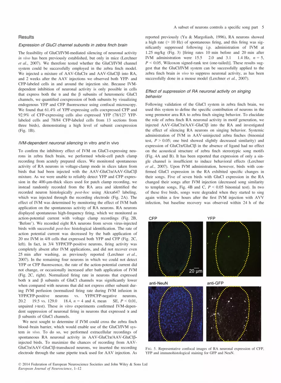
Fig. 5. Representative confocal images of RA neuronal expression of CFP,YFP and immunohistological staining for GFP and NeuN.
© 2014 Federation of European Neuroscience Societies and John Wiley & Sons LtdEuropean Journal of Neuroscience, 1–12
A subset of neurons controls a specific song part 5
injections. In the remaining three birds, singing did not resume untilthe next morning (approximately 24 h post-injection), and their sing-ing did not return to normal for up to 8 days. The time course ofIVM-mediated behavioral effect was similar to results reported inmice (Lerchner et al., 2007); the earliest effect was detected 6–12 hafter IVM injection, and was maintained on average for 2–3 days,with maximum of 14 days. Histological analysis revealed a subsetof RA neurons (approximately 5% compared with NeuN-labeled
cells in RA) in adult male zebra finches that expressed GluCl chan-nels. To maximize the detection of GluCl channel expression, welabeled neurons with anti-GFP antibody, although it did not allowus to distinguish YFP expression from CFP expression (Fig. 5). Wecounted the number of cells labeled with anti-GFP antibody, as wellas the number of neurons labeled with anti-NeuN antibody withinthe entire RA brain region in both hemispheres. On average, 9.69%of NeuN-labeled RA neurons were also labeled with anti-GFP
A
B C
D E
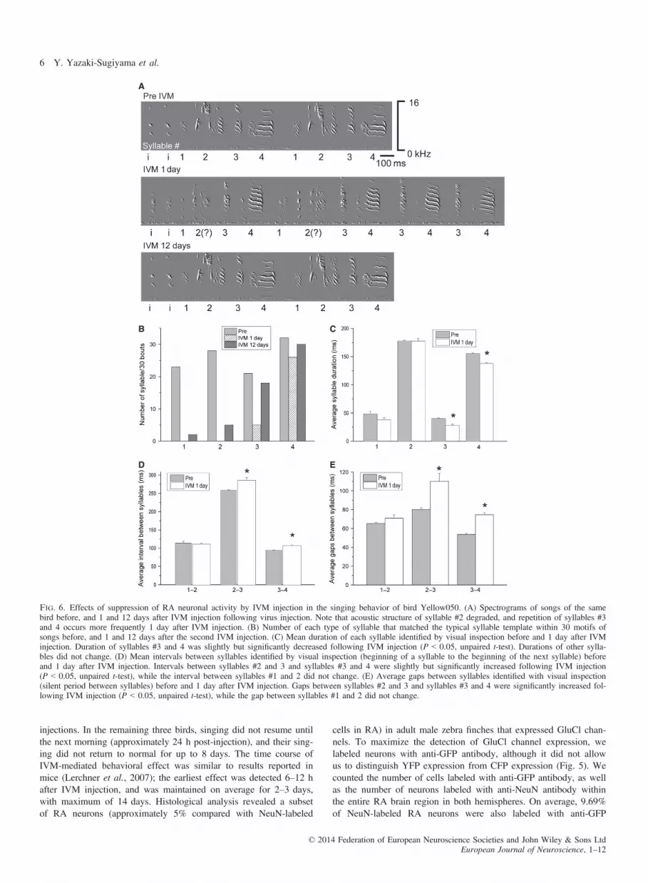
Fig. 6. Effects of suppression of RA neuronal activity by IVM injection in the singing behavior of bird Yellow050. (A) Spectrograms of songs of the samebird before, and 1 and 12 days after IVM injection following virus injection. Note that acoustic structure of syllable #2 degraded, and repetition of syllables #3and 4 occurs more frequently 1 day after IVM injection. (B) Number of each type of syllable that matched the typical syllable template within 30 motifs ofsongs before, and 1 and 12 days after the second IVM injection. (C) Mean duration of each syllable identified by visual inspection before and 1 day after IVMinjection. Duration of syllables #3 and 4 was slightly but significantly decreased following IVM injection (P < 0.05, unpaired t-test). Durations of other sylla-bles did not change. (D) Mean intervals between syllables identified by visual inspection (beginning of a syllable to the beginning of the next syllable) beforeand 1 day after IVM injection. Intervals between syllables #2 and 3 and syllables #3 and 4 were slightly but significantly increased following IVM injection(P < 0.05, unpaired t-test), while the interval between syllables #1 and 2 did not change. (E) Average gaps between syllables identified with visual inspection(silent period between syllables) before and 1 day after IVM injection. Gaps between syllables #2 and 3 and syllables #3 and 4 were significantly increased fol-lowing IVM injection (P < 0.05, unpaired t-test), while the gap between syllables #1 and 2 did not change.
© 2014 Federation of European Neuroscience Societies and John Wiley & Sons LtdEuropean Journal of Neuroscience, 1–12
6 Y. Yazaki-Sugiyama et al.

[9.69 � 4.05 (mean � SE), n = 7]. However one bird had an espe-cially high infection level (33.7%), which affected the mean. With-out this bird, the mean decreased to 5.68 � 0.74).Degradation of songs was also detected as a decrease of identified
syllable variety in a motif because birds changed acoustic structuresof some syllables (Figs 6 and 7). Furthermore in some birds, thetotal number of syllables in a motif decreased as they stopped asong in the middle (Figs 8 and 9) or dropped a syllable (Fig. 10).
Syllable-specific control by ensembles of RA neurons
An earlier study on unilateral inactivation of the RA using microle-sions showed a diminution of almost all parts of a song motif (Ash-more et al., 2008). In the present study, IVM-mediated suppressionof RA neuronal activity induced changes only in specific parts ofsong motifs, essentially on specific syllables. Although the numberof affected syllables differed between birds, within a single bird,
A
B C
D E
Fig. 7. Effects of suppression of RA neuronal activity by IVM injection in the singing behavior of bird Orange058. (A) Spectrograms of songs of the samebird before, and 3 h and 1 day after IVM injection following virus injection. Note that acoustic structure of syllable #3 degraded, while syllable #2 changed lit-tle. (B) Number of each type of syllable that matched the typical syllable template in 30 song motifs before, and 3 h and 1 day after the first IVM injection.(C) Mean duration of each syllable identified by visual inspection before and 3 h after IVM injection. Durations of all syllables were slightly but significantlyincreased following IVM injection (P < 0.05, unpaired t-test). (D) Mean intervals between syllables identified by visual inspection (beginning of a syllable tothe beginning of the next syllable) before and 3 h after IVM injection. The interval between syllables #2 and 3 was slightly but significantly increased followingIVM injection (P < 0.05, unpaired t-test), while the interval between syllables #1 and 2 did not change. (E) Mean gaps between syllables identified by visualinspection (silent period between syllables) before and 3 h after IVM injection. The gap between syllables #1 and 2 was slightly but significantly decreased fol-lowing IVM injection (P < 0.05, unpaired t-test), while the gap between syllables #2 and 3 did not change.
© 2014 Federation of European Neuroscience Societies and John Wiley & Sons LtdEuropean Journal of Neuroscience, 1–12
A subset of neurons controls a specific song part 7

some syllables showed a decrease in the degree of template match-ing after IVM injection, whereas other syllables did not (Figs 6 and10B). Regardless, IVM application did not change the syllableorder, supporting the previously suggested idea that ensembles ofRA neurons control the acoustic structure of song elements. Interest-ingly, songs stopped in the middle of motifs in two birds, and sub-sequent syllables were not sung (Figs 8 and 9A). In one bird, one
syllable (#4) was dropped following inactivation of a subset of RAneurons (Fig. 10A). This bird’s song motif consisted of six types ofsyllables, and we counted 176 syllables in 30 motifs before theIVM injection. However, the number of identified syllablesdecreased to 132, representing only five types of syllables, 24 hpost-IVM injection. Recovery of the sixth syllable type occurred3 days post- injection (Fig. 10B).
A
B C
D E
Fig. 8. Effects of suppression of RA neuronal activity by IVM injection in the singing behavior of bird Orange064. (A) Spectrograms of songs of the samebird before, and 2 and 4 days after IVM injection following virus injection. Note that songs stopped at syllable #4 after the IVM injection. (B) Number of eachtype of syllable that matched the typical syllable template within 30 song motifs before, and 2 and 4 days after the first IVM injection. Durations of other sylla-bles did not change. (C) Mean duration of each syllable identified by visual inspection before and 2 days after IVM injection. Durations of syllables #2, 3 and4 were slightly but significantly decreased, while the duration of syllable #1 increased following IVM injection (P < 0.05, unpaired t-test). (D) Mean intervalsbetween syllables identified by visual inspection (beginning of a syllable to the beginning of the next syllable) before and 2 days after IVM injection. Intervalsbetween syllables #1 and 2 and syllables #2 and 3 were slightly but significantly increased following IVM injection (P < 0.05, unpaired t-test), while the inter-val between syllables #3 and 4 decreased (P < 0.05, unpaired t-test). (E) Mean gaps between syllables identified by visual inspection (silent period between syl-lables) before and 2 days after IVM injection. The gap between syllables #2 and 3 was increased following IVM injection (P < 0.05, unpaired t-test), while thegap between other syllables did not change.
© 2014 Federation of European Neuroscience Societies and John Wiley & Sons LtdEuropean Journal of Neuroscience, 1–12
8 Y. Yazaki-Sugiyama et al.

Phasic bursting of RA neurons time-locked to a song motif hasbeen suggested to play an important role in generating stereotype inzebra finch songs. Two motor control models within the forebrainpremotor circuit have been suggested and discussed. In one model,a chain of sparse firing of HVC neurons, many of which projectonto single RA neurons to drive the precise bursting that producesthe song motifs, generates precise timing and controls a whole songmotif (Fee et al., 2004; Leonardo & Fee, 2005). In another model,
time-precise bursting is generated by the interaction of HVC inputand RA local circuits, controlling stereotyped song motifs (Yu &Margoliash, 1996; Chi & Margoliash, 2001; Amador et al., 2013).To examine the possibility of temporal pattern control by anupstream area, we further investigated the duration, interval betweensyllables (beginning of a syllable to the beginning of the next sylla-ble) and the gap between each pair of syllables (silent periodbetween syllables) before and after IVM injection. In all five birds
A
B C
D E
Fig. 9. Effects of suppression of RA neuronal activity by IVM injection in the singing behavior of bird Green001. (A) Spectrograms of songs of the same birdbefore, and 5 and 8 days after IVM injection following virus injection. Note that songs stopped at syllable #2 or 3 with degraded acoustic structure after theIVM injection. (B) Number of each type of syllable that matched the typical syllable template within 30 song motifs before, and 5 and 8 days after the firstIVM injection. (C) Mean duration of each syllable identified with visual inspection before and 5 days after IVM injection. Durations of syllables #1 and 2 weresignificantly decreased (P < 0.05, unpaired t-test). There was no syllable #5 detected by visual inspection. (D) Mean intervals between syllables identified byvisual inspection (beginning of a syllable to the beginning of the next syllable) before and 5 days after IVM injection. Interval between syllables #1 and 2 wasslightly but significantly decreased while interval between syllables #3 and 4 increased following IVM injection (P < 0.05, unpaired t-test). Interval betweensyllables #2 and 3 did not change after the IVM injection. Interval between syllables #4 and 5 could not be measured as there was no syllable #5 detected. (E)Mean gaps between syllables identified by visual inspection (silent period between syllables) before and 5 days after IVM injection. The gap between syllables#2 and 3 was significantly increased following IVM injection (P < 0.05, unpaired t-test), while the gaps between other syllables did not change. Gaps betweensyllables #4 and 5 could not be measured as there was no syllable #5 detected.
© 2014 Federation of European Neuroscience Societies and John Wiley & Sons LtdEuropean Journal of Neuroscience, 1–12
A subset of neurons controls a specific song part 9

that changed songs with IVM injections, the duration of syllables orintervals between syllables changed. However, interestingly, elonga-tion or shortening of syllable duration occurred only in some songelements, rather than throughout the entire song, when HVC activitywas decreased by cooling (Long & Fee, 2008). The changes in dura-tion of each syllable and the interval were relatively large comparedwith the change in the whole motif duration (Fig. 11). We comparedthe duration of the whole motif before and after IVM injection (inbirds that stopped songs in the middle of a motif after IVM injec-tion, we compared the duration of the remaining parts). Durations ofthe whole motif were significantly different before and after IVM
injection in all five birds. However, the difference was small(approximately 5%) and was not equal to the sum of differences induration and intervals of each syllable. These observations suggestthat changes in the duration or interval of each syllable are moreaffected by changes in acoustic structures, although the precise tim-ing of each syllable could be modified by activity changes of RAlocal circuits, as suggested by the fact that time-precise RA neuronalbursting is thought to be produced in RA local circuits. These datashow that silencing of groups of RA neurons had effects on acousticstructures of specific syllables without changing their sequences,supporting the idea that the sequence had already been generated
A
B C
D E
Fig. 10. Effects of suppression of RA neuronal activity by IVM injection in the singing behavior of bird Orange060. (A) Spectrograms of songs of the samebird before, and 1 and 3 days after IVM injection following virus injection. Note that there is no syllable #4 after the IVM injection. (B) Number of each typeof syllable that matched the typical syllable template within 30 motifs of songs before, and 1 and 3 days after the first IVM injection. No syllable #4 was identi-fied 1 day after injection, and the number of syllables #2 and 3 had not changed at that time. (C) Mean durations of each syllable identified by visual inspectionbefore and 1 day after IVM injection. Duration of syllables did not change. (D) Mean intervals between syllables identified by visual inspection (beginning of asyllable to the beginning of the next syllable) before and 1 day after IVM injection. Intervals between syllables #3 and 4 or syllables #4 and 5 could not bemeasured, as there was no syllable #4. Interval between syllables #2 and 3 was slightly but significantly increased following IVM injection (P < 0.05, unpairedt-test), while intervals between other syllables did not change after IVM injection. (E) Mean gaps between syllables identified with visual inspection (silent per-iod between syllables) before and 1 day after IVM injection. Gaps between syllables did not change.
© 2014 Federation of European Neuroscience Societies and John Wiley & Sons LtdEuropean Journal of Neuroscience, 1–12
10 Y. Yazaki-Sugiyama et al.
within a neuronal circuit in the upstream premotor area, probablywithin the microcircuit in HVC. By contrast, the timing of eachsmall song parts and acoustic structure of syllables are controlled bythe interaction between HVC inputs to the RA local circuit.One of the advantages of chemogenetic tools is reversible control
of neuronal activity and repetitive control of the same circuit. Wetherefore injected IVM a second time more than 3 weeks after thefirst injection. We then confirmed that all five birds that showedsong degradation with the first IVM injection again sang degradedsongs with a second IVM injection (Fig. 4B). Before the secondinjection, song similarity to the template song recovered to baseline,but again decreased after the second IVM injection to a level com-parable to the first injection. These results suggest the presence offirmly formed neuronal circuits subserving generation of specificsyllables within RA.
Discussion
Genetic methods have provided powerful experimental techniquesfor investigating the neuronal mechanisms underlying animal behav-ior. In particular, opto- and chemogenetic tools have enabled investi-gation of links between activities of discrete circuits or cellpopulations and behavioral and physiological outcomes (Boydenet al., 2005; Li et al., 2005; Nagel et al., 2005). In this study, wesuccessfully applied the GluCl/IVM system to reversibly suppressneuronal activity of a subset of RA neurons in behaving zebrafinches. Using this method, we found that ensembles of neurons inthe zebra finch forebrain premotor area RA contribute to generationof specific elements of sequenced motor activities.Zebra finch singing behavior is a well-characterized learned motor
sequence. Brain areas necessary for song production and learning,i.e. the ‘song system’, are well identified. The premotor HVC com-prises three types of neurons, i.e. projecting neurons for two differ-ent brain areas and interneurons (Mooney, 2000). Among them,neurons that project to the RA fire sparsely, but are precisely time-locked to specific parts of song motifs during singing (Hahnloseret al., 2002). A model in which RA neurons receive inputs frommultiple neurons in the HVC, causing multiple bursting at differentpoints in the song motifs, has been suggested (Fee et al., 2004;
Leonardo & Fee, 2005). In this model, temporal coding and songsyllable sequences for the whole motif are generated within a localcircuit in the upstream premotor area, HVC (Long & Fee, 2010).Our results partially support the model that syllable sequence isalready coded in HVC. However, inactivating ensembles of RA neu-rons resulted in changing of acoustic structure as well as local tem-poral coding of specific syllables. In addition, although the sequenceof syllables did not change, some birds dropped syllables in themiddle or end of motifs. This suggests that the timing and acousticstructure of syllables are generated within ensembles of RA neuronsor RA local circuits with HVC input interaction. This would supportthe idea that precise RA bursting controls each song note. Suppres-sion of just a group of RA neurons resulted in degradation of acous-tic structures only in specific parts of the motifs, without changingthe other parts, or syllable sequences. Seven syringeal muscles arethought to contribute to song generation (Greenewalt, 1968), andthey are innervated by fibers from the hypoglossal motor nucleus.There are about 8000 RA neurons, some of which topographicallyproject to subregions of the hypoglossal motor nucleus (Gurney,1981; Vicario, 1991). However, the precise connectivity betweenRA neurons and brainstem motor units is not yet known, and therelationship between syringeal muscle forces and vocal output canbe complex (Fee et al., 1998). Some models suggest that just asmall number of muscles can directly control acoustic parameters ofsongs (Gardner et al., 2001; Mindlin et al., 2003). In addition, RAneurons independently project to other parts of the brainstem, thenucleus retroambigualis (Ram) and nucleus parambigualis (PAm),where the neurons are involved in vocal–respiratory coordination(Wild, 1993). In this study, about 5% of RA neurons were inacti-vated, which would be expected to result in only partial loss of acti-vation of vocal muscles or of coordination with respiratory rhythm,resulting in degradation of syllables controlled by the inactivatedneurons. This vocal generation circuit appears to be firmly formed,as silencing of the same group of neurons at different times causesthe acoustical alteration of the same song parts.Bird song singing and learning provide the premier model for
learning of sequenced motor generation, as well as neuronal plastic-ity during the developmental critical period. In addition to thebehavioral analysis of singing and learning processes during devel-opment, many physiological, anatomical and pharmacological tech-niques have been applied to reveal underlying neuronal mechanismsfor this behavior. Although genetically modified songbirds have notyet been established, recent viral vector-based methods have pro-vided new tools for model development (Roberts et al., 2010,2012). The present study employed a newly developed chemoge-netic tool in zebra finches, providing a model that will allow furtherinvestigation into generation of motor sequences involved in singingbehavior and song learning within the developmental time window.
Acknowledgements
We thank Dr Hiroshi Takagi for his technical support for the in vi-tro electrophysiological study, Dr Elizabeth Ko Mitamura for hertechnical support for the generation of AAVs and Drs Aryesh Muk-herjee, Mahesh M. Bandi and Colm Connaughton for thoughtful dis-cussions about the mechanics of the avian syrinx. We are alsograteful for generous support to Y.Y-S. from the Okinawa Instituteof Science and Technology Graduate University. This research waspartially supported by the Ministry of Education, Culture, Sports,Science, and Technology (MEXT), a Grant-in-Aid for ScientificResearch (C) (24500403) to Y.Y-S., a Grant-in-Aid for ScientificResearch (B) (24300129) to M.L. and by the MEXT World Premier
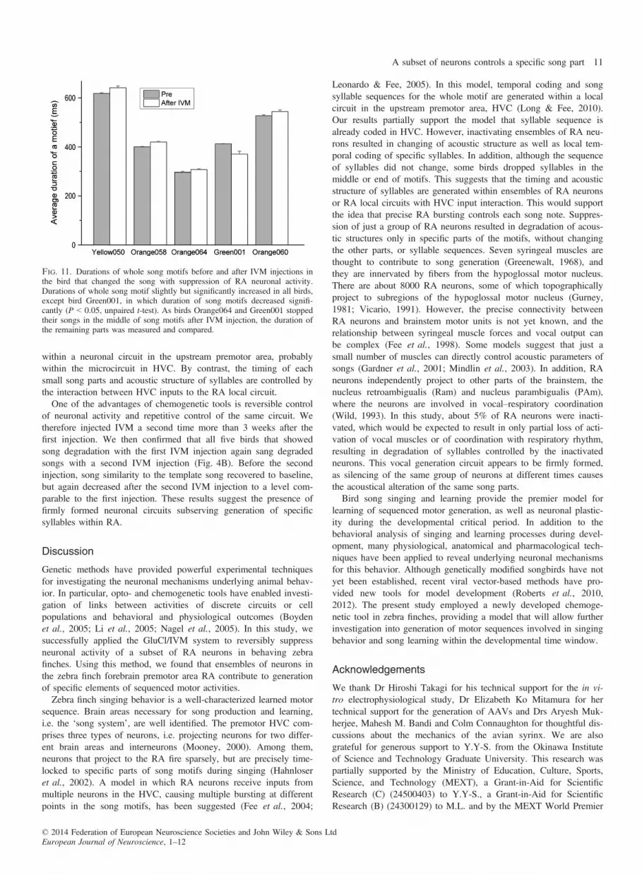
Fig. 11. Durations of whole song motifs before and after IVM injections inthe bird that changed the song with suppression of RA neuronal activity.Durations of whole song motif slightly but significantly increased in all birds,except bird Green001, in which duration of song motifs decreased signifi-cantly (P < 0.05, unpaired t-test). As birds Orange064 and Green001 stoppedtheir songs in the middle of song motifs after IVM injection, the duration ofthe remaining parts was measured and compared.
© 2014 Federation of European Neuroscience Societies and John Wiley & Sons LtdEuropean Journal of Neuroscience, 1–12
A subset of neurons controls a specific song part 11
International Research Center Initiative (WPI) to M.L. and USNational Institutes of Health grant NS073613 to P.M.F.
Abbreviations
AAV, adeno-associated viral; CFP, cyan fluorescent protein; GFP, greenfluorescent protein; GluCl, glutamate- and ivermectin-gated chloride;IVM, ivermectin; RA, robust nucleus of the archistriatum; YFP, yellowfluorescent protein.
References
Agate, R.J., Scott, B.B., Haripal, B., Lois, C. & Nottebohm, F. (2009) Trans-genic songbirds offer an opportunity to develop a genetic model for vocallearning. Proc. Natl. Acad. Sci. USA, 106, 17963–17967.
Amador, A., Perl, Y.S., Mindlin, G.B. & Margoliash, D. (2013) Elementalgesture dynamics are encoded by song premotor cortical neurons. Nature,495, 59–64.
Ashmore, R.C., Bourjaily, M. & Schmidt, M.F. (2008) Hemispheric coordi-nation is necessary for song production in adult birds. J. Neurophysiol.,99, 373–385.
Boyden, E.S., Zhang, F., Bamberg, E., Nagel, G. & Deisseroth, K. (2005)Millisecond-timescale, genetically targeted optical control of neural activ-ity. Nat. Neurosci., 8, 1263–1268.
Charlesworth, J.D., Warren, T.L. & Brainard, M.S. (2012) Covert skill learn-ing in a cortical-basal ganglia circuit. Nature, 486, 251–255.
Chi, Z. & Margoliash, D. (2001) Temporal precision and temporal drift inbrain and behavior of zebra finch song. Neuron, 32, 899–910.
Fee, M.S. & Leonardo, A. (2001) Miniature motorized microdrive and com-mutator system for chronic neural recordings in small animals. J. Neurosci.Methods, 112, 83–94.
Fee, M.S., Shraiman, B., Pesaran, B. & Mitra, P.P. (1998) The role of non-linear dynamics of the syrinx in the vocalizations of a songbird. Nature,395, 67–71.
Fee, M.S., Kozhevnikov, A.A. & Hahnloser, R.H.R. (2004) Neural mecha-nisms of vocal sequence generation in the songbird. Ann. NY Acad. Sci.,1016, 153–170.
Gardner, T., Cecchi, G., Magnasco, M., Laje, R. & Mindlin, G.B. (2001)Simple gestures for birdsongs. Phys. Rev. Lett., 87, 208101.
Greenewalt, C.H. (1968) J Bird Song: Acoustics and Physiology. SmithsonianInstitution, Washington, DC.
Gurney, M.E. (1981) Hormonal control of cell form and number in the zebrafinch song system. J. Neurosci., 1, 658–673.
Hahnloser, R.H.R., Kozhevnikov, A.A. & Fee, M.S. (2002) An ultra-sparsecode underlies the generation of neural sequences in a songbird. Nature,419, 65–70.
Leonardo, A. & Fee, M.S. (2005) Ensemble coding of vocal control in bird-song. J. Neurosci., 25, 652–661.
Lerchner, W., Xiao, C., Slimko, E.M., Van Trigt, L., Lester, H.A. & Ander-son, D.J. (2007) Reversible silencing of neuronal excitability in behavingmice by a genetically targeted, ivermectin-gated Cl channel. Neuron, 54,35–49.
Li, X., Slimko, E.M. & Lester, H.A. (2002) Selective elimination of gluta-mate activation and introduction of fluorescent proteins into a Caenor-habditis elegans chloride channel. FEBS Lett., 528, 77–82.
Li, X., Gutierrez, D.V., Hanson, M.G., Han, J., Mark, M.D., Chiel, H.,Hegemann, P. & Landmesser, L.T. (2005) Fast noninvasive activation andinhibition of neural and network activity by vertebrate rhodopsin andgreen algae channel rhodopsin. Proc. Natl. Acad. Sci. USA, 102, 17816–17821.
Long, M.A. & Fee, M.S. (2008) Using temperature to analyse temporaldynamics in the songbird motor pathway. Nature, 456, 189–194.
Long, M.A. & Fee, M.S. (2010) Support for a synaptic chain model of neu-ronal sequence generation. Nature, 468, 394–399.
McCasland, J. & Konishi, M. (1981) Interaction between auditory and motoractivities in an avian song control nucleus. Proc. Natl. Acad. Sci. USA, 78,7815–7819.
Mindlin, G.B., Gardner, T.J., Goller, F. & Suthers, R. (2003) Experimentalsupport for a model of birdsong production. Phys. Rev. E., 68, 041908.
Mooney, R. (2000) Different subthreshold mechanisms underlie song selec-tivity in identified HVc neurons of the zebra finch. J. Neurosci., 201,5420–5436.
Nagel, G., Brauner, M., Liewald, J.F., Adeishvili, N., Bamberg, E. & Gotts-chalk, A. (2005) Light activation of channel rhodopsin-2 in excitable cellsof Caenorhabditis elegans triggers rapid behavioral responses. Curr. Biol.,15, 2279–2284.
Nottebohm, F., Kelley, D.B. & Paton, J.A. (1982) Connections of vocal con-trol nuclei in the canary telencephalon. J. Comp. Neurol., 207, 344–357.
Olveczky, B.P., Andalman, A.S. & Fee, M.S. (2005) Vocal experimentationin the juvenile songbird requires a basal ganglia circuit. PLoS Biol., 3,e153.
Roberts, T.F., Tschida, K.A., Klein, M.E. & Mooney, R. (2010) Rapid spinestabilization and synaptic enhancement at the onset of behavioural learn-ing. Nature, 463, 948–952.
Roberts, T.F., Gobes, S.M., Murugan, M., €Olveczky, B.P. & Mooney, R.(2012) Motor circuits are required to encode a sensory model for imitativelearning. Nat. Neurosci., 10, 1454–1459.
Slimko, E.M. & Lester, H.A. (2003) Codon optimization of Caenorhabditiselegans GluCl ion channel genes for mammalian cells dramaticallyimproves expression levels. J. Neurosci. Meth., 124, 75–81.
Slimko, E.M., McKinney, S., Anderson, D.J., Davidson, N. & Lester, H.A.(2002) Selective electrical silencing of mammalian neurons in vitro bythe use of invertebrate ligand-gated chloride channels. J. Neurosci., 22,7373–7379.
Vicario, D.S. (1991) Organization of the zebra finch song control system. II.Functional organization of outputs from nucleus Robustus archistriatalis.J. Comp. Neurol., 309, 486–494.
Vicario, D.S. & Nottebohm, F. (1988) Organization of the zebra finch songcontrol system: I. Representation of syringeal muscles in the hypoglossalnucleus. J. Comp. Neurol., 271, 346–354.
Wild, J.M. (1993) Descending projections of the songbird nucleus Robustusarchistriatalis. J. Comp. Neurol., 338, 225–241.
Yu, A.C. & Margoliash, D. (1996) Temporal hierarchical control of singingin birds. Science, 273, 1871–1875.
© 2014 Federation of European Neuroscience Societies and John Wiley & Sons LtdEuropean Journal of Neuroscience, 1–12
12 Y. Yazaki-Sugiyama et al.