acoustic focusing - thermo fisher scientific
TRANSCRIPT

Acoustic Focusing for Multiparameter Flow Cytometry
Sponsored by:
Article collection


Contents
4 Introduction
5Fundamentals of Acoustic CytometryBY MICHAEL D. WARD, AND GREGORY KADUCHAKCurrent Protocols in Cytometry
20Light-Triggered Drug Release from Red Blood Cells Suppresses Arthritic InflammationBY EMILIA M. ZYWOT, NATALIA ORLOVA, SONG DING, RISHI
R. RAMPERSAD, EMILY M. RABJOHNS, VICTORIA A. WICKENHEISSER, QUNZHAO WANG, JOSHUA G. WELFARE, LAUREN HAAR, AMANDA M. EUDY, TERESA K. TARRANT, AND DAVID S. LAWRENCE
Advanced Therapeutics
31Cell-Based Therapy for Canavan Disease Using Human iPSC-Derived NPCs and OPCsBY LIZHAO FENG, JIANFEI CHAO, E TIAN, LI LI, PENG YE,
MI ZHANG, XIANWEI CHEN, QI CUI, GUIHUA SUN, TAO ZHOU, GERARDO FELIX, YUE QIN, WENDONG LI, EDWARD DAVID MEZA, JEREMY KLEIN, LUCY GHODA, WEIDONG HU, YONGLUN LUO, WEI DANG, DAVID HSU, JOSEPH GOLD, STEVEN A. GOLDMAN, REUBEN MATALON, AND YANHONG SHI
Advanced Science
50WJMSC-derived small extracellular vesicle enhance T cell suppression through PD-L1BY MEIZHANG LI, RUPAL SODER, SUNIL ABHYANKAR,
HAITHAM ABDELHAKIM, MITCHELLW. BRAUN, CAMILLE V. TRINIDAD, HARSH B. PATHAK, ZIYAN PESSETTO, CLAYTON DEIGHAN, SIDDHARTHA GANGULY, BUDDHADEB DAWN, JOSEPH MCGUIRK, NEIL DUNAVIN, AND ANDREW K. GODWIN
Journal of Extracellular Vesicles
Cover Image ©Thermo Fisher Scientific
3

IntroductionBY JEREMY PETRAVICZ, PH.D., EDITOR, CURRENT PROTOCOLS
T he hardware underlying flow cytometers can be broadly divided into two categories: detection and fluidics. Detection encompasses the lasers, filters,
and detector modules of the cytometer. The fluidics system includes the sample reservoir, injection stream and flow cell. The precision of identifying and characterizing single cells relies not only on the detection system of the cytome-ter, but also on the fluidics system. Integral to this is the ability to process the sample into a stream that enables the detection of single cells as they pass the detectors using a process known as hydrodynamic focusing.
In hydrodynamic focusing, the sample or central stream and the sheath fluid run parallel to each other at different rates, with the sheath fluid the slower of the pair. This crea-tes a laminar flow that aligns the cells in the sample stream based on the configuration of the flow cell. There are limits to hydrodynamic focusing: increasing the sample volume input rate leads to corresponding decreases in single cell resolution necessary for applications such as immunophe-notyping. To overcome this limitation, flow cytometers can combine hydrodynamic focusing with a process known as acoustic focusing. Acoustic focusing can be thought of as a “pre-alignment” step of the cells in the sample stream prior to being introduced into the sheath fluid. The use of acoustic focusing can enable the user to sort cells at hig-her rates and with higher precision than hydrodynamic focus alone. However, as discussed in Ward and Kaduchak (2018), there are considerations when deciding if the use of acoustic focusing is appropriate for your sample type. Additionally, acoustic focusing systems can be combined with high-speed brightfield cameras (such as in the Attune CytPix Flow Cytometer system) to obtain images of the cell populations as it is being sorted to examine morphology or the presence of doublet/triplet cells.
This article collection highlights the application of this technology to the development of cell-based therapies, utilizing the strengths of acoustic focusing combined with multiparameter flow cytometry to characterize both cells designed to deliver therapeutics and the impact of cell-ba-sed therapies on target cells. Ward and Kaduchak (2018) provides a technical overview of acoustic focusing flow cy-tometry to familiarize the reader. This overview discusses the strengths and limitations of acoustic focusing, as well as mitigating any potential impacts that it can have on the cells being processed. Next, Zywot et al (2021) demonstrate how acoustic focusing and imaging flow cytometry can be utilized to analyze the viability and morphology of modified red blood cells (mRBCs) under development as a potential cell-based therapy. The mRBCs were designed to deliver an anti-inflammatory agent in a light dependent manner with higher efficacy than more conventional delivery methods.
The third article is Feng et al (2020) which details the de-velopment of an induced pluripotent stem cell (iPSC) based therapy for Canavan disease (CD), a leukodystrophy that leads to loss of myelin and brain degeneration. Acoustic focusing flow cytometry was utilized to obtain candidate cells for generation of human iPSCs from CD patients. The-se iPSCs were then engineered to carry a functional copy of the ASPA gene which is mutated in CD. Treatment with the-se engineered cells in a mouse model of CD rescued many of the major pathological features of the disease and in-creased their survivability. Lastly, Li et al (2020) examined the role of small extracellular vesicles (sEVs) derived from Wharton’s Jelly-derived mesenchymal stem cells (WJMSCs) to modulate the immune response to organ transplant. The authors found WJMSCs secrete sEVs enriched in a ligand that modulates T-cell responses. Through acoustic focusing flow cytometry, they were able to characterize the inhibito-ry action of the ligand on T-cell activation and reduce the pathological response in acute graft versus host disease.
Through the concepts and applications presented in this research article collection we hope to educate scientists on how acoustic focusing and imaging flow cytometry can ad-vance their research and gain deeper insight cell populati-ons. For more information regarding acoustic flow cytome-try, we encourage you to visit the Thermo Fisher Scientific Attune Flow Cytometer page and explore the possibilities presented there for your research.
References
Ward, M. D., & Kaduchak, G. (2018). Fundamentals of Acoustic Cytome-try. Current Protocols in Cytometry, 84, e36. doi: https://doi.org/10.1002/cpcy.36
Zywot, E.M., Orlova, N., Ding, S., Rampersad, R.R., Rabjohns, E.M., Wi-ckenheisser, V.A., Wang, Q., Welfare, J.G., Haar, L., Eudy, A.M., Tarrant, T.K. and Lawrence, D.S. (2022), Light-Triggered Drug Release from Red Blood Cells Suppresses Arthritic Inflammation. Adv. Therap. 2100159. https://doi.org/10.1002/adtp.202100159
Feng, L., Chao, J., Tian, E., Li, L., Ye, P., Zhang, M., Chen, X., Cui, Q., Sun, G., Zhou, T., Felix, G., Qin, Y., Li, W., Meza, E. D., Klein, J., Ghoda, L., Hu, W., Luo, Y., Dang, W., Hsu, D., Gold, J., Goldman, S. A., Matalon, R., Shi, Y., Cell-Based Therapy for Canavan Disease Using Human iPSC-Derived NPCs and OPCs. Adv. Sci. 2020, 7, 2002155.t
Li, M, Soder, R, Abhyankar, S, et al. WJMSC-derived small extracellular vesicle enhance T cell suppression through PD-L1. J Extracell Vesicles. 2021; 10:e12067. https://doi.org/10.1002/jev2.12067
4

Fundamentals of Acoustic CytometryMichael D. Ward1 and Gregory Kaduchak1
1ThermoFisher Scientific, Eugene, Oregon
Acoustic cytometry uses radiation pressure forces instead of or in addition tohydrodynamic focusing to position cells or particles in a flowing stream foranalysis. Commercial implementations to date combine both hydrodynamic andacoustic focusing together to enable high precision analysis of a broad dynamicrange of volumetric sample input rates up to an order of magnitude higherthan is practical with hydrodynamic focus alone. This capability allows greatflexibility in reducing assay time or modifying or eliminating concentrationrequirements or concentration steps in sample preparation protocols. It alsoprovides a practical method for processing sub-microliter volumes using sampledilution. In order to take full advantage of this dynamic range, it is necessaryto understand the fundamental benefits and limitations of acoustic focusing asapplied to flow cytometry. C© 2018 by John Wiley & Sons, Inc.
Keywords: acoustic focus � acoustic cytometry � high-speed flow cytometry� hydrodynamic focus � sample preparation
How to cite this article:Ward, M. D., & Kaduchak, G. (2018). Fundamentals of Acoustic
Cytometry. Current Protocols in Cytometry, 84, e36. doi:10.1002/cpcy.36
INTRODUCTION
Hydrodynamic focusing uses sheath flow to confine injected sample fluid to a small“sample core” that is typically accelerated to meters-per-second velocity through an op-tical interrogation region defined by a tightly focused laser beam. The first descriptionof acoustic focusing applied in flow cytometry (Goddard et al. 2006) mimicked thisprocess without requiring sheath flow by exploiting physical differences between cellsor particles relative to the background carrier medium to position the particles or cellsin a single, focused line along the central axis of a glass capillary. Subsequent commer-cially available acoustic focusing cytometers have combined hydrodynamic focusing andacoustic focusing together by applying the acoustic focus to a similar ultrasonic capillarydevice used to inject the sample into a sheath manifold (Acoustic Focusing Overview,4/12/2017). By focusing cells into a tight line prior to injection in the sheath manifold,precise alignment of cells in interrogating lasers can be maintained at much higher volu-metric inputs than possible with hydrodynamic focus alone (Figure 1). These higher inputrates enable dilute samples to be processed quickly, without resorting to centrifugationor other concentration steps. Alternately, they allow dilution of concentrated samples ormodifying of protocols for lower cell concentrations, without fear of long analysis times.
Acoustic cytometry as referred to herein uses acoustic radiation pressure to align cellsin flow for analysis in an interrogation region using optical detectors. It should notbe confused with flow cytometry that uses acoustic energy to interrogate cells, (Roos& Apfel, 1988) or that detects acoustic energy from the photoacoustic effect, whichis stimulated by light pulses. (Galanzha & Zharov, 2012). Acoustic alignment couldhowever be used together with acoustic interrogation, and detection and photoacousticanalysis has already been combined with acoustic alignment in vivo (Galanzha et.al.,2016).
Current Protocols in Cytometry e36, April 2018Published online April 2018 in Wiley Online Library (wileyonlinelibrary.com).doi: 10.1002/cpcy.36Copyright C© 2018 John Wiley & Sons, Inc.
Flow CytometryInstrumentation
5

Figure 1 Comparison of hydrodynamic focus alone (left) versus Acoustic-assisted hydrodynamicfocus (right) at a high volumetric sample input rate. Acoustic focusing of particles before sampleinjection into the sheath manifold allows velocity and illumination precision to be maintained forlarge sample cores resulting from these rates.
Figure 2 Schematic of a line-driven capillary depicting tight single line focusing of particles in aflowing liquid using acoustic radiation pressure.
Fundamentals ofAcoustic
Cytometry
Current Protocols in Cytometry
6

The effect of acoustic radiation pressure on particles in a medium was first describedby Kundt and Lehmann (1874) after they witnessed dust particles levitated in organpipes. This effect has been applied to aqueous solutions for the separation of bioparticles(Coakley, Bardsley, Grundy, Zamani, & Clarke, 1989, 2000; Curtis & Stephans, 1982;Jonsson, Nilsson, Petersson, Allers, & Laurell, 2005; Yasuda, Haupt, & Unemura, 1997).Use of acoustic fields for separation of cells and or positioning cells for analysis continuesto be an active area of research today, with wide ranging applications including bulkprocessing of algae for bio fuels and cells for cell therapy, multiple focused lines of cellsfor high throughput flow cytometry (Piyasena et al., 2012), and single cell manipulationslike cell sorting (Ren et al., 2015). Ren et al. used acoustic traveling waves and a surfaceacoustic wave device or SAW to sort cells, but the bulk of these studies use resonantsquare or rectangular cavities to create acoustic standing waves. The acoustic cytometersdescribed here use standing waves generated by a circular focusing device called aline-driven capillary. The line-driven capillary, described by Kaduchak et al. (2008), isan acoustically resonant device that focuses cells or particles into a single line using acapillary driven by a single piezoelectric transducer or line-source (Figure 2). The theoryof this device is described by Goddard and Kaduchak (2005).
THE ACOUSTIC-ASSISTED HYDRODYNAMIC FOCUSING CYTOMETER
Commercial instruments employing these acoustic-focusing devices are more aptlynamed “acoustic-assisted hydrodynamic focusing cytometers” because they use the line-driven capillary to inject sample into a sheath manifold, in much the same way as asample injection probe is used in a conventional cytometer (Figure 3).
In contrast, the first acoustic cytometer focused particles without sheath flow (Goddard,Martin, Graves, & Kaduchak, 2006). There are advantages to eliminating sheath, butpractical challenges as well. In a sheathless cytometer, the sample is free to contact andcontaminate optical flow cell walls. In addition, particle velocity, and therefore dwelltime in an interrogating laser, is linearly dependent on volumetric sample input rate. Aprimary advantage of acoustic focus is that data precision can be maintained over a largedynamic range of volumetric sample input rates. However, in order to take advantage ofthis range without sheath flow, the electronics and software would need to accommodate
Figure 3 Generic illustration of an acoustic assisted hydrodynamic focusing analyzer. Sampleis forced into the capillary, acoustically focused, injected into a sheath manifold, analyzed, andtransferred to waste. As for a conventional flow cytometer, the analysis stage includes a laserbeam focused at the position of the particles in the optical cell. The scatter/fluorescent signal isconditioned by appropriate optical filters before optical detection. Driving and control electronicsare added to ensure the piezoelectric device is driven at the acoustic resonance frequency of thepiezoelectric element/capillary.
Flow CytometryInstrumentation
Current Protocols in Cytometry
7

Figure 4 Schematic drawings of hydrodynamic focus only (A) and acoustic assisted hydrody-namic focus at low (left) and high (right) sample input rates. Directly under each schematic isa corresponding cell cycle histogram of FxCycleTMViolet area taken at low (12 µl/min) and high(1000 µl/min) flow rates.
signals across the same large dynamic range of velocities and laser dwell times. For theinstrument used to collect the data presented in Figure 4, the flow rate spans nearly twoorders of magnitude (12 µl/min to 1000 µl/min).
Apart from the acoustic capillary and its driving electronics, all the other componentsin Fig 3 could be used to construct a conventional cytometer. In fact, the instrument canbe used as a hydrodynamic focus only instrument by simply turning off the acousticdriver board. This board drives the vibration of the line-driven capillary device usingfeedback control that ensures that the resonant frequency required for a tight focus ismaintained. This frequency varies with the capillary diameter and wall thickness. A300-μm inner diameter capillary, for example, may have a resonance near 3 MHz,whereas a 600-μm inner diameter capillary resonates at a proportionately lower frequencynear 1.5 MHz. The resonance also varies with temperature and fluid properties. Thevariations for the range of temperatures and samples used in flow cytometry are relativelysmall, with resonant frequency changes on the order of a few percent, but the feedbackcontrol is still essential to ensure optimum performance over this entire range.
During operation of the cytometer, a discreet flow rate between 12 and 1000 µl/min ischosen and the instrument adjusts its sheath rate such that the ratio of sample to sheath ishighest at the lowest rate and lowest at the highest rate. Figure 4 shows schematics for lowand high sample input rates with acoustic focusing turned on or off. Cell cycle histogramsfor actively growing alcohol fixed and FxCycleTM Violet stained Jurkat cells are pairedwith each schematic. See supplementary material for protocol details. Figure 4A showsthe two rates with the acoustic field off (hydrodynamic focus only). The 12 µl/min rateshows the good precision required for cell cycle analysis, whereas the 1000 µl/min rateprecision is only useful for counting cells. With the acoustic field off, the cells are free todistribute across the large core, and signal precision is degraded by increased variationof cell velocity and illumination intensity of the laser. For this instrument, the precisiondrop due to illumination position variation is partly mitigated by a flat top laser beamprofile, but the G1 cell cycle stage coefficient of variation (CV) is a high 6.73% and theG2 cell cycle peak disappears entirely. With the acoustic field turned on in Figure 4B,velocity and illumination intensity precision are high for both low and high rates. TheCV benefit for the acoustic focus vs. no acoustic focus at 12 µl/min is small at 2.35%
Fundamentals ofAcoustic
Cytometry
Current Protocols in Cytometry
8

versus 2.43%, respectively, because of the high ratio of sheath to sample at this rate. Theprecision benefit of acoustic focus tapers off with the decrease in sample core diameter,providing no additional benefit as the core diameter approaches the cell or particle size.In other words, acoustic focus does not push a 10 µm diameter cell any closer to thecenter of a 10 µm diameter sample core than does hydrodynamic focus.
The high precision demonstrated in Figure 4B at 1000 µl/min enables running of samplesup to 10 times faster than in cytometers without acoustic focus. This does not mean, how-ever, that all samples should be run this fast. Understanding how best to take advantage ofthis increased throughput can be made easier by answering the following questions: (1)How are different cells or particles focused by the acoustic field? (2) What is the acousticconcentration effect and how does it pertain to sample concentration and injection rates?Once these questions are answered, one can begin to ask about how acoustic cytometrycan help answer questions in biology, chemistry, and medicine.
ACOUSTIC FORCE ON PARTICLES IN A MEDIUM
The answer to the question of how cells or particles are affected by acoustic focus startswith the mechanical properties of both the particles and the medium they are carried in.Equation 1 gives the acoustic force U exerted on a particle in a carrier medium, Gorkov(1962):
U = 4
3πa3
[(βo
�p2�2
)f1 − 3
2
(ρo�v2�
2
)f2
]
Equation 1
Here, a is the particle radius, β0 is the compressibility of the surrounding fluid, and ρ0 isthe density of the surrounding fluid. The pressure and velocity of the acoustic field in theabsence of the particle are described by p and v, respectively, and the brackets correspondto a time-averaged quantity. The terms f1 and f2 are the contrast terms that determinehow the mechanical properties (compressibility and density, respectively) of the particlediffer from the background medium. They are given by the following Equations 2a and2b:
f1 = 1 − βp
βo
Equation 2a
f2 = 2 (ρp − ρo)
(2ρp + ρo)
Equation 2b
The subscript p corresponds to intrinsic properties of the particle. The force F acting ona particle is related to the gradient of the force potential U by Equation 3:
F = −∇U
Equation 3
Particles will be localized at positions where the potential U displays a minimum (stableequilibrium). The acoustic contrast of a particle (or medium) is determined by the densityand compressibility differences between it and the background medium as defined byterms f 1 and f 2 in Equations 2a and 2b. The relative magnitudes and signs of f 1 and
Flow CytometryInstrumentation
Current Protocols in Cytometry
9

Figure 5 Calculated acoustic force potential in the cross-section of an acoustically driven cap-illary. Particles with positive acoustic contrast are focused toward the force potential trap in thecenter of the cross-section. Note that that the acoustic field is asymmetric, with stronger gra-dients along the axis of the piezo driver. This asymmetry can improve precision of analysis ofnon-spherical cells by helping to orient them in interrogating lasers.
f 2 determine the behavior of the radiation force potential U and thus determine themagnitude and direction of the acoustic radiation pressure force. As an example, if aparticle and the background medium share the same density value (ρp = ρ0), then f 2 iszero and the acoustic contrast is due only to compressibility differences in f 1. If both f 1and f 2 are zero, then there is no acoustic contrast. Figure 5 displays the force potential Ufor an erythrocyte within the cross section of an acoustically driven capillary containingphosphate buffered saline. Particles traveling through the capillary experience a timeaveraged force that transports them into the deep potential well centered along the axisof the capillary.
It should be noted that nearly all particles and cells of interest have acoustic contrastvalues in water or aqueous buffers, which force them to migrate to the central axis ofthe capillary (as shown in Figure 5). These particles have positive acoustic contrast intheses fluids. There are a few materials such as fat particles and gas bubbles that havenegative acoustic contrast which forces their migration toward the wall of the capillaryin the acoustic field.
Effects on Cell Health and Viability
Detrimental acoustic effects on cells are invariably at the top of the list of concerns formany cell biologists when introduced to the topic of acoustic focusing. This is becauseultrasonic energy is routinely used for lysis of cells as hardy as bacterial spores. Likemost acoustic resonators designed for cell manipulation (Wiklund, 2012), line drivenacoustic capillaries used for cytometry are different from devices designed to lyse cells infundamentally important ways. First, acoustic lysis is typically done using sub-megahertzfrequencies that can create cavitation, a phenomenon in which tiny gas bubbles form andcollapse with tremendous local shear and heating. The acoustic focusing capillaries usedhere operate at a frequency well above 1 MHz without cavitation. Second, acoustic lysisis performed at very high energy levels where acoustic streaming and rapid fluid heatingare common. Acoustic cytometry is performed with relatively low energy levels of tensof milliwatts maximum electrical input power at the high sample flow rate of a milliliterper minute. At lower flow rates, this power is progressively scaled down. The acousticenergy dissipated in the fluid is also significantly less than the electrical input energy. Thedesign of the acoustically driven capillary spreads this energy over the entire length of thecapillary and there is very little sample heating. Goddard, Sanders, Martin, Kaduchak,and Graves (2007) showed that the viability of Chinese hamster ovary (CHO) cells was
Fundamentals ofAcoustic
Cytometry
Current Protocols in Cytometry
10

Figure 6 Calculated trajectories of different diameter microspheres as they travel along the axisof the acoustic focusing capillary. The vertical axis is the particle position relative to the capillaryaxis. The horizontal axis is the particle position along the length of the capillary. Sample flows fromleft to right at a rate of 1000 µl/min.
not significantly affected by the acoustic field created by a large acoustically drivencapillary even in the sub-megahertz region. The higher, gentler megahertz frequenciesnow utilized in commercial cytometers are routinely used in safe medical imaging ofpatients and are thought to be gentle enough for cell separations where cell health orrecovery are critical, such as cell therapy (FloDesign Sonics) or circulating tumor cellseparation. (Li et al., 2015). Pre-focusing in the injector with acoustics also serves toreduce the acceleration of cells required in the subsequent hydrodynamic focus wherecells can undergo significant shear forces.
Particle or Cell Size
While many cells and microbes have similar acoustic contrast, the acoustic force ondifferent sized particles varies widely. As can be seen from Equation 1, the acoustic forceis proportional to the third power of the particles’ radius. The force resisting movementof the particle is the Stokes drag force Fd, which can be approximated by the followingEquation 4 for a hard sphere with particle Reynolds numbers <0.1:
Fd = 6πrpηur
Equation 4
Here rp is the cell or particle radius, η is the medium viscosity, and ur is the acousti-cally induced velocity in the radial direction. Fd is linearly proportional to radius, sothe net result is that overall force is proportional to the particles’ radius squared withsmall particles moving more slowly than large particles of similar acoustic contrast.Figure 6 shows the predicted trajectories of various sizes of polystyrene beads in a sam-ple flowing in the axial direction in the acoustic capillary. As can be seen from this figure,it takes longer for the smaller particles to reach the capillary axis. This in turn dictates thatvolumetric sample throughput should be reduced when processing smaller particles suchas bacteria. If the residence time within the capillary of particles or cells of a given size isnot long enough, the variation in position of the particles/cells about the central capillaryaxis will be greater and the coefficient of variation (CV) of the optical measurement will
Flow CytometryInstrumentation
Current Protocols in Cytometry
11

suffer at the higher sample input rates. Acoustics may contribute to focus and may alsoalign asymmetric cells but smaller particles should generally be analyzed at conventionalsample input rates, so that the additional hydrodynamic focusing can help ensure higherprecision.
If the particle is so small that the acoustic force is weaker than Brownian motion, theacoustic field will not have a focusing effect, such that positioning precision will dependsolely on the hydrodynamic focus. This size cutoff is a function of acoustic contrastfactors, acoustic power, and frequency and is beyond the scope of this unit, but ingeneral, nano particles, like exosomes and viruses, should be analyzed with low sampleinput rates/high sheath to sample ratios, as in any flow cytometer.
THE ACOUSTIC CONCENTRATION EFFECT
The focusing of all cells in the capillary volume to a line in the center of the capillarycreates a local effective cell concentration that can be many times higher than the initialstarting concentration. This enables much faster analysis of dilute samples, but it necessi-tates the addition of sheath fluid at higher cell concentrations in order to maintain singleparticle analysis. Figure 7 illustrates the acoustic concentration effect in the absenceof sheath. Whole blood diluted to about 2 × 10⁷ (Figure 7A and B) and 2 × 10⁶ redblood cells per ml (Figure 7C) was imaged in a quartz flow cell after pumping through acapillary with the acoustic field turned off (7A) and on (7.B and C). When the acousticfield is on, all of the positive acoustic contrast particles in the capillary are forced tothe center before being pumped into the flow cell. This effect in relatively concentratedsamples results in a rope-like sheet of particles that can be many particles in width likethat seen in Figure 7B.
In the instrument, this rope is injected into a sheath manifold where sheath fluid speedsup and separates the cells, creating single particle spacing dependent on the sample tosheath ratio. A 1:10 sample to sheath ratio for example, would create spacing similar tothat seen in the 10-fold dilution of sheathless sample in Figure 7.C. Rope-like conditionssimilar to Figure 7B can be created in the laser interrogation zone of the instrument itselfby diluting less and running at high sample input rates. For a 10-fold dilution of blood
Figure 7 Micrographs of dilute acoustically focused whole blood pumped into in a square quartzflow cell without sheath. (A, B) 2 × 10⁷ RBCs per ml (C) 2× 10⁶ RBCs per ml. In the instrument,where sheath flow accelerates the sample and separates the cells, a 100 fold and a 10 fold higherconcentration respectively run at 1000 µl /min would be needed to produce similar concentrationsto B and C in the laser interrogation zone.
Fundamentals ofAcoustic
Cytometry
Current Protocols in Cytometry
12

Figure 8 Plot of number of events versus arrival interval at the interrogation laser for acousticallyfocused microspheres in flow at 1 ml/min without sheath flow. The black line is an exponentialdistribution. The experimental data (gray bars) closely matches this prediction.
containing 5 × 10⁹ RBCs per ml and injected at 1000 µl/min, approximately 8 millionRBCs per second pass through the instrument. For a core velocity of 8 meters/second,this averages about 10 cells per 10 micron length of sample core. With several cells inthe laser focus at all times, scatter is completely useless, but fluorescence data can stillbe collected from white blood cells. This sounds attractive from a throughput standpoint,because white blood cell coincidence under these conditions is still relatively rare forwhole blood, but the quality of fluorescence data is degraded. Higher concentration ofunbound fluorophore, combined with non-specific staining, reduces sensitivity such thatthis technique can typically only be used for high density antigens with bright labels.An additional effect is the absorbance of violet laser light by hemoglobin, which furtherdiminishes signals for violet excitable probes.
Volumetric Throughput, Poisson Rate and Coincidence
Although acoustic cytometers can process sample volumes an order of magnitude fasterthan conventional cytometers, this does not mean that all samples should be processedthis fast. This is often desirable for more dilute samples, but at higher concentrations,all cytometers, including acoustic ones with or without sheath, are limited by coincidentevents as governed by Poisson statistics. Figure 8 shows that the inter-arrival times ofparticles that have been acoustically focused follow an exponential distribution, whichis in agreement with a Poisson process.
Poisson statistics predict the likelihood of one cell, no cells, or more than one cellbeing present in an event window. As sample throughput increases with higher sampleconcentration, the probability of a cell being present in an interrogating laser beam in anygiven window of time increases, but the probability of more than one cell being presentin the laser also increases (a coincident event).
Coincidence in an event window should generally be kept low by using mean rates ofless than one event per ten event windows for most assays (van den Engh, 2000). Thiscondition theoretically corresponds to a 10% coincidence rate. While speed of electronicscan also limit event rates, most modern cytometers are capable of electronic event rates
Flow CytometryInstrumentation
Current Protocols in Cytometry
13

Figure 9 Linear versus log (top) and log versus log (bottom) plots for cell event rate versusinitial sample concentration for sample input rates of 12, 100, and 1000 μl/min. Event rates aretheoretical and exclude the impact of coincidence. Linear plotting of event rate emphasizes thelow event rates obtained with cell concentrations below one million per milliliter at conventionalrates. Log plotting of event rate shows both the need for high throughput rates at the lowestcell concentrations and the danger of exceeding maximum instrument event rates at high cellconcentrations.
that significantly exceed their 10% Poisson rate, which is governed by the magnitudeand variation of cell velocity.
Large variation in cell velocity for large sample cores limits sample core size and conse-quently volumetric throughput in a conventional cytometer. The size of an event windowdictates the Poisson rate, and in a cytometer with spatially separated lasers, the eventwindow must typically be extended to account for different laser to laser arrival times forparticles or cells having different velocities. In a large core without acoustic focus, thelarge spread in transit times requires larger event windows which decrease the Poissonrate.
If lower coincidence is desired or higher coincidence is acceptable, sample concentrationand or volumetric sample rate should be adjusted accordingly with a correspondingdecrease or increase in particle throughput. Figure 9 shows linear (top) and log (bottom)plots of theoretical event rates that exclude the impact of coincidence. Event rates arein cells per second as a function of the log of sample concentration for three differentvolumetric flow rates: 12, 100, and 1000 μl/min. The first two rates cover a similar
Fundamentals ofAcoustic
Cytometry
Current Protocols in Cytometry
14
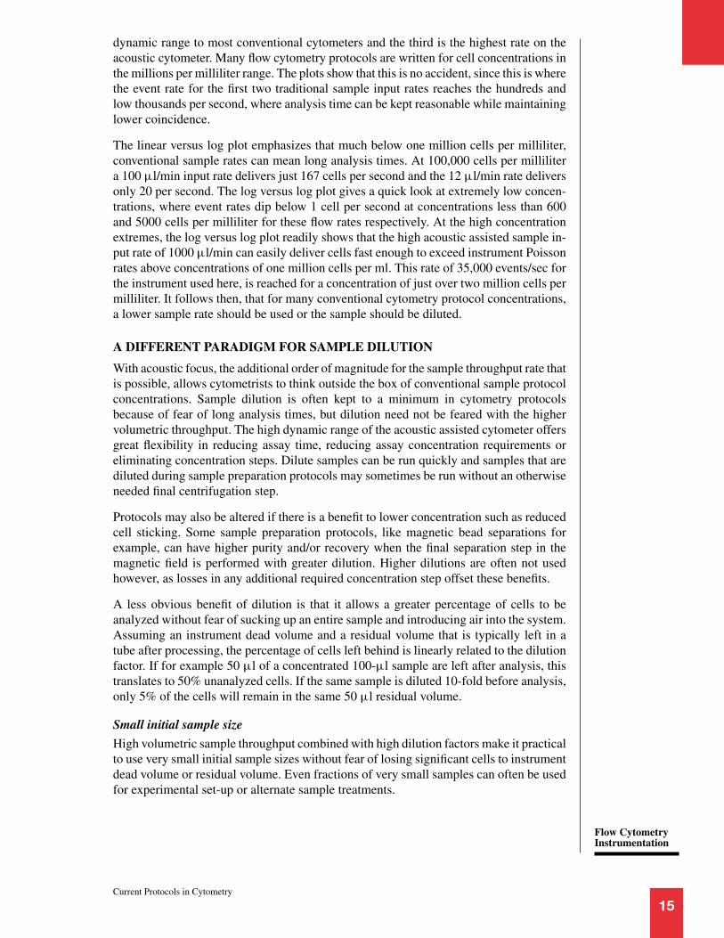
dynamic range to most conventional cytometers and the third is the highest rate on theacoustic cytometer. Many flow cytometry protocols are written for cell concentrations inthe millions per milliliter range. The plots show that this is no accident, since this is wherethe event rate for the first two traditional sample input rates reaches the hundreds andlow thousands per second, where analysis time can be kept reasonable while maintaininglower coincidence.
The linear versus log plot emphasizes that much below one million cells per milliliter,conventional sample rates can mean long analysis times. At 100,000 cells per millilitera 100 μl/min input rate delivers just 167 cells per second and the 12 μl/min rate deliversonly 20 per second. The log versus log plot gives a quick look at extremely low concen-trations, where event rates dip below 1 cell per second at concentrations less than 600and 5000 cells per milliliter for these flow rates respectively. At the high concentrationextremes, the log versus log plot readily shows that the high acoustic assisted sample in-put rate of 1000 μl/min can easily deliver cells fast enough to exceed instrument Poissonrates above concentrations of one million cells per ml. This rate of 35,000 events/sec forthe instrument used here, is reached for a concentration of just over two million cells permilliliter. It follows then, that for many conventional cytometry protocol concentrations,a lower sample rate should be used or the sample should be diluted.
A DIFFERENT PARADIGM FOR SAMPLE DILUTION
With acoustic focus, the additional order of magnitude for the sample throughput rate thatis possible, allows cytometrists to think outside the box of conventional sample protocolconcentrations. Sample dilution is often kept to a minimum in cytometry protocolsbecause of fear of long analysis times, but dilution need not be feared with the highervolumetric throughput. The high dynamic range of the acoustic assisted cytometer offersgreat flexibility in reducing assay time, reducing assay concentration requirements oreliminating concentration steps. Dilute samples can be run quickly and samples that arediluted during sample preparation protocols may sometimes be run without an otherwiseneeded final centrifugation step.
Protocols may also be altered if there is a benefit to lower concentration such as reducedcell sticking. Some sample preparation protocols, like magnetic bead separations forexample, can have higher purity and/or recovery when the final separation step in themagnetic field is performed with greater dilution. Higher dilutions are often not usedhowever, as losses in any additional required concentration step offset these benefits.
A less obvious benefit of dilution is that it allows a greater percentage of cells to beanalyzed without fear of sucking up an entire sample and introducing air into the system.Assuming an instrument dead volume and a residual volume that is typically left in atube after processing, the percentage of cells left behind is linearly related to the dilutionfactor. If for example 50 μl of a concentrated 100-μl sample are left after analysis, thistranslates to 50% unanalyzed cells. If the same sample is diluted 10-fold before analysis,only 5% of the cells will remain in the same 50 μl residual volume.
Small initial sample size
High volumetric sample throughput combined with high dilution factors make it practicalto use very small initial sample sizes without fear of losing significant cells to instrumentdead volume or residual volume. Even fractions of very small samples can often be usedfor experimental set-up or alternate sample treatments.
Flow CytometryInstrumentation
Current Protocols in Cytometry
15

Figure 10 Analysis of nucleated cells from 940 nl (A) and 94 nl (B) of whole blood with 850-fold and 8500-fold dilution, respectively. Nucleated cells are plotted on a log log histogram ofDyeCycleTMRuby fluorescence showing the threshold level used for collection and a linear 405 nmviolet SSC-H versus FSC-H differential scatter plot showing white blood cell populations Granulo-cytes (Gran), Monocytes (Mono), and Lymphocytes (Lymph).
For precious samples where analyzing every cell is important, combining a “no lyse, nowash” protocol with high dilution prevents cell loss from centrifugation or lysis reagentsand decreases cells lost in residual volume. For Figure 10A a 1 μl sample of wholeblood was diluted into 850 μl of DyeCycleTMRuby nucleic acid staining buffer and800 μl, of this dilution was run at 1000 μl/min with an analysis time of about 46 sec. ForFigure 10B, the 1 μl sample is diluted as for A and 85 μl of this dilution was dilutedanother 10-fold and run as in A. Nucleated cell events are captured by thresholding onDyeCycle Ruby high events.
For each sample, 800 μl of an 850 μl sample or 94% of the sample is analyzed, equating to940 nl (A) and 94 nl (B) of the original whole blood sample. Each sample is plotted with alog log histogram of DyeCycle Ruby fluorescence and a corresponding differential whiteblood cell scatter plot using 405 nm violet SSC-H versus FSC-H (488 nm blue). For fullprotocol and instrument setup, see supplementary material. Note that for whole unlysedblood on this instrument, the position of granulocytes in FSC is shifted significantly tothe left relative to ammonium chloride lysed blood. Red blood cell lysis protocols canchange white blood cell morphology, particularly for granulocytes. The differences inFSC seen from morphology changes are dependent on interrogating light parameters
Fundamentals ofAcoustic
Cytometry
Current Protocols in Cytometry
16

including wavelength, scatter collection angles and laser focus and alignment. (Petrizet al., 2017).
For “no-lyse, no-wash” protocols and for any protocol in which a wash step may beremoved, it is important to understand how assay background can either be increased bythe large sample cores generated at high volumetric input rates or reduced by dilution ofthe free fluorophore in a sample.
Background reduction from dilution
One concern that arises when proposing elimination or significant reduction of sheathratios is that the benefit of squeezing the sample core to a very small size, such that verylittle free fluorophore is excited by the laser, is lost. If the sample core is large, the laserwill excite free fluorophores throughout the beam focus, resulting in higher backgroundfor unwashed samples. This effect is mitigated somewhat by the tight Gaussian focusof the laser beam and by spatial filtration in the collection optics, but for a given con-centration of unbound fluorophore, fluorescence background is higher than for a tightlyhydrodynamically focused core. With dilution, however, the concentration of free labelis reduced by the dilution factor, reducing the fluorescence background. Dilution, likewashing by centrifugation, will disturb the binding equilibrium, but for higher affinitylabels, dissociation will be insignificant if it is performed within a reasonable time beforeanalysis. For many antibodies with useful affinity, dissociation half-lives are on the orderof hours or days. If dissociation for lower affinity ligands is of concern, rapid dilution fol-lowed by analysis with a high volumetric sample rate can be used as a quicker alternativeto centrifugation for background reduction.
As a frame of reference for background reduction, a single round of centrifugation, de-pending on operator and dilution prior to and after centrifugation, is typically comparableto about a 300-fold dilution. For a properly titrated immunophenotyping experiment withhigh affinity antibodies, non-specific binding contributes more to background than doesunbound fluorophore at this dilution, and continued dilution beyond 500 to 800-fold maynot significantly reduce background levels.
Note that for lower affinity reagents like nucleic acid stains, dilution can disturb equi-librium in a short period of time and that for some high precision assays like cellcycle analysis, the dilution buffer should contain equilibrium concentrations of theselow affinity stains. This can increase background in large cores, even for dyes consid-ered “non-fluorescent” until bound, depending on the dye concentration and the ratio offluorescence enhancement upon binding.
SUMMARY AND OUTLOOK
Use of acoustic fields for separation and positioning of cells and particles has been anactive and growing area of research for nearly four decades. Diverse uses of these fields inflow cytometry have been suggested, including pre-analysis sample preparation, acousticcell sorting, multi-stream analysis and sheathless triggered stopped or even reverse flowanalysis. Commercial implementations to date have focused on combining sheath andacoustic focusing to create instruments capable of high precision analysis over a highdynamic range of volumetric sample inputs from 12 μl/min to 1000 μl/min.
This expansion of dynamic range enables up to an order of magnitude faster analysis timesversus conventional hydrodynamic focusing alone, particularly for dilute samples, andprovides greater flexibility in sample preparation protocols. Protocols can be modified torun lower concentrations of cells, eliminate extra concentration steps or dilute to extendthe number of experiments possible or increase the percentage of cells analyzed in verysmall volumes. Flexibility for sample dilution ratios is particularly useful for optimization
Flow CytometryInstrumentation
Current Protocols in Cytometry
17

of no lyse no wash assays where red blood cell lysis and centrifugation are avoided tominimize potential sample preparation artifacts.
Increasing availability of more and more parameters in flow cytometry has spurreddiscovery of new cell types, more correlation of phenotyping with live cell function andincreasing scrutiny of smaller and smaller phenotypic and functional cell subpopulations.The concern that sample preparation causes loss or alteration of specific fragile cells hasgrown in the face of this research, making protocols that can minimize impact on livecells and their response to environment and stimuli highly desirable.
Understanding the fundamental advantages and limitations of acoustic focusing as ap-plied to flow cytometry can enable users to better leverage the technology, not only toincrease throughput and save time but also to modify and improve sample preparationand minimize its effects on cell biology.
Acknowledgements
The authors thank Marc DeJohn ofBiomeme Inc. and the late Carl Stew-art and Patrick Turner for their signifi-cant contributions to the implementationof acoustic focusing in flow cytometry.We also thank Jolene Bradford of ThermoFisher Scientific for valuable support andadvice.
Conflicts of Interest
The authors are employees of ThermoFisher Scientific, which is in the businessof selling flow cytometers and flow cytom-etry reagents.
Literature CitedAcoustic Focusing Overview, 4/12/(2017). Re-
trieved from https://www.thermofisher.com/us/en/home/life-science/cell-analysis/flow-cytometry/flow-cytometers/acoustic-focusing-technology-overview.html. Accessed November 28,2017.
Coakley, W. T., Bardsley, D. W., Grundy, M. A.,Zamani, F., & Clarke, D. J. (1989). Cell manip-ulation in ultrasonic standing wave fields. Jour-nal of Chemical Technology and Biotechnology,44, 43–62. doi: 10.1002/jctb.280440106.
Coakley, W. T., Hawkes, J. J., Sobanski, M.A., Cousins, C. M., & Spengler, J. (2000).Analytical scale ultrasonic standing wavemanipulation of cells and microparticles.Ultrasonics, 38, 638–641. doi: 10.1016/S0041-624X(99)00151-1.
Curtis, H. W., & Stephans, E. J. (1982). Ultrasoniccontinuous flow plasmapheresis separator. IBMTechnical Disclosure Bulletin, 25(1).
Galanzha, E. I., & Zharov, V. P. (2012).Photoacoustic flow cytometry. Methods (SanDiego, Calif.), 57(3), 280–296. doi: 10.1016/j.ymeth.2012.06.009.
Galanzha, E. I., Viegas, M. G., Malinsky, T. I.,Melerzanov, A. V., Juratli, M. A., Sarimol-
laoglu, M., . . . Zharov, V. P. (2016). In vivoacoustic and photoacoustic focusing of circu-lating cells. Scientific Reports, 6, 21531. doi:10.1038/srep21531.
Goddard, G., & Kaduchak, G. (2005). Ultra-sonic particle concentration in a line-drivencylindrical tube. Journal of the AcousticalSociety of America, 117, 3440–3447. doi:10.1121/1.1904405.
Goddard, G., Martin, J. C., Graves, S. W., & Ka-duchak, G. (2006). Ultrasonic particle concen-tration for sheathless focusing of particles foranalysis in a flow cytometer. Cytometry, 69, 66–74. doi: 10.1002/cyto.a.20205.
Goddard, G. R., Sanders, C. K., Martin, J. C., Ka-duchak, G., & Graves, S. W. (2007). Analyticalperformance of an ultrasonic particle focusingflow cytometer. Analytical Chemistry, 79, 8740–8746. doi: 10.1021/ac071402t.
Gorkov, L. P. (1962). Forces acting on a small par-ticle in an acoustic field within an ideal fluid.Soviet Physics-Doklady, 6, 773–775.
Jonsson, H., Nilsson, A., Petersson, F., Allers, M.,& Laurell, T. (2005). Particle separation us-ing ultrasound can be used with human shedmediastinal blood. Perfusion, 20, 39–43. doi:10.1191/0267659105pf782oa.
Kaduchak, G., Goddard, G., Salzman, G., Sinha,D., Martin, J. C., Kwiatkowski, C. S., & Graves,S. W. (2008). Ultrasonic Particle Concentra-tion and Application in Flow Cytometry. UnitedStates Patent, 7, 340, 957.
Kundt, A., & Lehmann, O. (1874). Longitudinalvibrations and acoustic figures in cylindricalcolumns of liquids. Annalen der Physik undChemie (Poggendorff ’s Annalen), 153, 1–11.
Li, P., Mao, Z., Peng, Z., Zhou, L., Chen, Y., Huang,P-H., . . . Huang, T. J. (2015). Acoustic sepa-ration of circulating tumor cells. Proceedings ofthe National Academy of Sciences of the UnitedStates of America, 112(16), 4970–4975. doi:10.1073/pnas.1504484112.
Petriz, J., Bradford, J. A., & Ward, M. D.(2017). No lyse no wash flow cytometryfor maximizing minimal sample preparation.
Fundamentals ofAcoustic
Cytometry
Current Protocols in Cytometry
18

Methods (San Diego, Calif.), pii, S1046–2023(17)30159–30157. https://doi.org/10.1016/j.ymeth.2017.12.012.
Piyasena, M. E., Suthanthiraraj, P. P. A., Apple-gate, R. W. Jr., Goumas, A. M., Woods, T. A.,Lopez, G. P., & Graves, S. W. (2012). MultinodeAcoustic Focusing for Parallel Flow Cytome-try. Analytical Chemistry, 84, 1831–1839. doi:10.1021/ac200963n.
Ren, L., Chen, Y., Lia, P., Maoa, Z., Huanga,P.-H., Rufoa, J., . . . Huang, T. J. (2015).A high-throughput standing surface acous-tic wave (SSAW)-based cell sorter. Lab onA Chip, 15(19), 3870–3879. doi: 10.1039/C5LC00706B.
Roos, M. S., & Apfel, R. E. (1988). Applicationof 30-MHz acoustic scattering to the study of
human red blood cells. Journal of the Acous-tical Society of America, 83, 1639–1644. doi:10.1121/1.395918.
van den Engh, G. (2000). High speed cell sort-ing. In Emerging Tools for Single-Cell Analysis:Advances in Optical Measurement Technologies(pp. 21–48). G. Durack, & J. P. Robinson (Eds.),New York: John Wiley and Sons.
Wiklund, M. (2012). Acoustofluidics 12: Bio-compatibility and cell viability in microfluidicacoustic resonators. Lab on A Chip, 12, 2018–2028.doi: 10.1039/c2lc40201g.
Yasuda, K., Haupt, S. S., & Unemura, S. (1997).Using acoustic radiation force as a concentra-tion method for erythrocytes. The Journal of theAcoustical Society of America, 102, 642–645.doi: 10.1121/1.421009.
Flow CytometryInstrumentation
Current Protocols in Cytometry
19

RESEARCH ARTICLEwww.advtherap.com
Light-Triggered Drug Release from Red Blood CellsSuppresses Arthritic Inflammation
Emilia M. Zywot, Natalia Orlova, Song Ding, Rishi R. Rampersad, Emily M. Rabjohns,Victoria A. Wickenheisser, Qunzhao Wang, Joshua G. Welfare, Lauren Haar,Amanda M. Eudy, Teresa K. Tarrant,* and David S. Lawrence*
Arthritis is a leading cause of disability in adults, which can be intenselyincapacitating. The location and intensity of the pain is both subjective andchallenging to manage. Consequently, patient-directed delivery ofanti-inflammatories is an essential component of future therapeutic strategiesfor the management of this disorder. The design and application of alight-responsive red blood cell (RBC)-conveyed dexamethasone (Dex)construct that enables targeted drug delivery upon illumination of theinflamed site is described. The red wavelength (650 nm) responsive nature ofthe phototherapeutic is validated using tissue phantoms mimicking the lightabsorbing properties of various skin types. Furthermore, photoreleased Dexhas the same impact on cellular responses as conventional Dex. Murine RBCscontaining the photoactivatable therapeutic display comparable circulationproperties as fluorescently labeled RBCs. In addition, a single dose oflight-targeted Dex delivery is fivefold more effective in suppressinginflammation than the parent drug, delivered serially over multiple days.These results are consistent with the notion that the circulatory system beused as an on-command drug depot, providing the means to therapeuticallytarget diseased sites both efficiently and effectively.
1. Introduction
Joint pain and inflammation are a leading cause of disabilityamong working age adults with staggering societal costs.[1] For
E. M. Zywot, N. Orlova, S. Ding, Q. Wang, L. Haar, D. S. LawrenceDivision of Chemical Biology and Medicinal ChemistryUniversity of North CarolinaChapel Hill, NC 27599, USAE-mail: [email protected]. R. Rampersad, E. M. Rabjohns, V. A. Wickenheisser, A. M. Eudy,T. K. TarrantDepartment of MedicineDivision of Rheumatology and ImmunologyDuke UniversityDurham, NC 27710, USAE-mail: [email protected]. G. Welfare, D. S. LawrenceDepartment of ChemistryUniversity of North CarolinaChapel Hill, NC 27599, USA
The ORCID identification number(s) for the author(s) of this articlecan be found under https://doi.org/10.1002/adtp.202100159
DOI: 10.1002/adtp.202100159
example, the economic burden associatedwith arthritis is estimated to be greater than$300 billion in theUnited States alone.[2] Asa consequence of the persistent nature ofthese diseases, frequent and long-term ther-apeutic administration is required, whichresults in moderate to severe undesiredside effects. Furthermore, the therapeuticneeds are accentuated during periods ofprofoundly increased disease activity, whichcan be intensely debilitating. Efforts to im-prove efficacy and reduce undesired sys-temic toxicity have focused on technolo-gies, which can selectively deliver therapeu-tics to inflamed joints.[3,4] Indeed, it hasbeen known for decades that the directinjection of glucocorticoids into arthriticjoints provides temporary benefits;[5] how-ever,multiple injections into joints on a rou-tine basis is not an acceptable therapeuticoption.[3] Nonetheless, the repeated deliveryof therapeutic agents to afflicted joints is re-quired to silence local inflammation and re-pair damage.[6] A stimuli-responsive drug
delivery system could potentially be used to intermittently dis-pense therapeutic agent(s) at the diseased site in a patient-directed, as-needed, fashion. Specifically, light as a stimulus en-joys a number of potentially useful attributes, including that itcan be easily focused on inflamed and painful joints using read-ily available 600 to 1000 nm laser and light-emitting diode (LED)light sources.[7] A light-activated form of an anti-inflammatoryagent, such as dexamethasone (Dex), would ideally be main-tained in the circulatory system in an inactive state, and subse-quently released using light by the patient when needed. Indeed,the circulatory system represents an opportune drug depot sinceall cells in the human body are positioned within 100 μm of ablood vessel. However, repeated delivery to inflamed joints overthe course of days or weeks requires a circulatory presence sig-nificantly longer than the half-life (a few hours) of the parentdrug. We have addressed this issue by installing a photoactivableDex inside red blood cells (RBCs) and have employed these en-gineered cells to successfully treat a mouse model of inflamma-tory arthritis. For clinical relevance, the overwhelming majorityof RBC drug loading studies have been performed with humanerythrocytes (hRBCs). However, in preparation for animal stud-ies, loaded mouse RBCs were also formulated and characterized.
Adv. Therap. 2021, 2100159 © 2021 Wiley-VCH GmbH
RESEARCH ARTICLEwww.advtherap.com
Light-Triggered Drug Release from Red Blood CellsSuppresses Arthritic Inflammation
Emilia M. Zywot, Natalia Orlova, Song Ding, Rishi R. Rampersad, Emily M. Rabjohns,Victoria A. Wickenheisser, Qunzhao Wang, Joshua G. Welfare, Lauren Haar,Amanda M. Eudy, Teresa K. Tarrant,* and David S. Lawrence*
Arthritis is a leading cause of disability in adults, which can be intenselyincapacitating. The location and intensity of the pain is both subjective andchallenging to manage. Consequently, patient-directed delivery ofanti-inflammatories is an essential component of future therapeutic strategiesfor the management of this disorder. The design and application of alight-responsive red blood cell (RBC)-conveyed dexamethasone (Dex)construct that enables targeted drug delivery upon illumination of theinflamed site is described. The red wavelength (650 nm) responsive nature ofthe phototherapeutic is validated using tissue phantoms mimicking the lightabsorbing properties of various skin types. Furthermore, photoreleased Dexhas the same impact on cellular responses as conventional Dex. Murine RBCscontaining the photoactivatable therapeutic display comparable circulationproperties as fluorescently labeled RBCs. In addition, a single dose oflight-targeted Dex delivery is fivefold more effective in suppressinginflammation than the parent drug, delivered serially over multiple days.These results are consistent with the notion that the circulatory system beused as an on-command drug depot, providing the means to therapeuticallytarget diseased sites both efficiently and effectively.
1. Introduction
Joint pain and inflammation are a leading cause of disabilityamong working age adults with staggering societal costs.[1] For
E. M. Zywot, N. Orlova, S. Ding, Q. Wang, L. Haar, D. S. LawrenceDivision of Chemical Biology and Medicinal ChemistryUniversity of North CarolinaChapel Hill, NC 27599, USAE-mail: [email protected]. R. Rampersad, E. M. Rabjohns, V. A. Wickenheisser, A. M. Eudy,T. K. TarrantDepartment of MedicineDivision of Rheumatology and ImmunologyDuke UniversityDurham, NC 27710, USAE-mail: [email protected]. G. Welfare, D. S. LawrenceDepartment of ChemistryUniversity of North CarolinaChapel Hill, NC 27599, USA
The ORCID identification number(s) for the author(s) of this articlecan be found under https://doi.org/10.1002/adtp.202100159
DOI: 10.1002/adtp.202100159
example, the economic burden associatedwith arthritis is estimated to be greater than$300 billion in theUnited States alone.[2] Asa consequence of the persistent nature ofthese diseases, frequent and long-term ther-apeutic administration is required, whichresults in moderate to severe undesiredside effects. Furthermore, the therapeuticneeds are accentuated during periods ofprofoundly increased disease activity, whichcan be intensely debilitating. Efforts to im-prove efficacy and reduce undesired sys-temic toxicity have focused on technolo-gies, which can selectively deliver therapeu-tics to inflamed joints.[3,4] Indeed, it hasbeen known for decades that the directinjection of glucocorticoids into arthriticjoints provides temporary benefits;[5] how-ever,multiple injections into joints on a rou-tine basis is not an acceptable therapeuticoption.[3] Nonetheless, the repeated deliveryof therapeutic agents to afflicted joints is re-quired to silence local inflammation and re-pair damage.[6] A stimuli-responsive drug
delivery system could potentially be used to intermittently dis-pense therapeutic agent(s) at the diseased site in a patient-directed, as-needed, fashion. Specifically, light as a stimulus en-joys a number of potentially useful attributes, including that itcan be easily focused on inflamed and painful joints using read-ily available 600 to 1000 nm laser and light-emitting diode (LED)light sources.[7] A light-activated form of an anti-inflammatoryagent, such as dexamethasone (Dex), would ideally be main-tained in the circulatory system in an inactive state, and subse-quently released using light by the patient when needed. Indeed,the circulatory system represents an opportune drug depot sinceall cells in the human body are positioned within 100 μm of ablood vessel. However, repeated delivery to inflamed joints overthe course of days or weeks requires a circulatory presence sig-nificantly longer than the half-life (a few hours) of the parentdrug. We have addressed this issue by installing a photoactivableDex inside red blood cells (RBCs) and have employed these en-gineered cells to successfully treat a mouse model of inflamma-tory arthritis. For clinical relevance, the overwhelming majorityof RBC drug loading studies have been performed with humanerythrocytes (hRBCs). However, in preparation for animal stud-ies, loaded mouse RBCs were also formulated and characterized.
Adv. Therap. 2021, 2100159 © 2021 Wiley-VCH GmbH20

www.advancedsciencenews.com www.advtherap.com
Figure 1. Assembly of Dex-Cbl-Cy5 RBC phototherapeutics. A) Structures of photoactivatable Dex-Cbl-Cy5 (1) and control compound H2O-Cbl-Cy5(2). B) Schematic representation of the isotonic-to-hypotonic-to-isotonic method by which phototherapeutics are loaded into RBCs and subsequentlyphotochemically released. Dex is represented by the blue sphere. Pore formation in the RBC membrane occurs in the presence of 1 or 2 under hypotonicconditions (4 °C for 40 min). Pores are subsequently resealed by direct addition of high salt followed by incubation at 37 °C for 20 min. Conjugates 1or 2 remain trapped inside the RBC due to the membrane impermeability of the B12 anchor. Upon photolysis of the C─Co bond, the now membranepermeable Dex is released from the RBC carrier.
of the RBC. Third, although the corrin ring of Cbl absorbs onlyshort wavelength light (330–575 nm), installation of Cy5 on theCbl[12–14] adjusts photorelease of the appended drug to a longer,tissue-penetrating wavelength (650 nm).[15]
2.1.1. Synthesis of Cbl Conjugates
Both Dex-Cbl-Cy5 (1) and H2O-Cbl-Cy5 (2) (Figure 1A) were syn-thesized and introduced into RBCs to assess drug photodeliveryas a potential therapeutic strategy for the treatment of inflam-matory arthritis. H2O-Cbl-Cy5 serves as an inactive control thatlacks the Dex therapeutic agent. In brief, Dex was appended tothe aminopropyl ligand on the Co and Cy5 subsequently cou-pled to an ethylenediamine linker on the ribose of Cbl (SchemesS1 and S2 and Figures S1–S4, Supporting Information). TheCy5 fluorophore extends the light capturing wavelengths of Cblfrom 330-575 nm to 650 nm, where the latter displays greater tis-sue penetration than that of the former (Figure S5, SupportingInformation).[11,13] LC-MS analysis of the resulting mixture afterphotolysis of Dex-Cbl-Cy5 confirms the expected photoproducts,
2. Results and Discussion2.1. Design of a Circulating Photoresponsive Anti-Inflammatory Drug Depot
Novel therapies designed to maintain Dex’s circulatory presence while minimizing its systemic side effects employ slow release carrier-based systems such as liposomes, polymeric-drug conju-gates, and RBCs.[8] Of these carriers, RBCs present a potentially elegant solution to the challenge of creating a long-term drug de-pot that circulates throughout the body in an innocuous, dormant form.[9,10] Indeed, internally loaded RBCs have been reported to circulate for at least 1 month.[10] The strategy outlined herein em-ploys engineered RBCs that stably house a Dex derivative that is released upon exposure to red light (650 nm). A key element of the design strategy is the covalent attachment of Dex to vitamin B12 (cobalamin, Cbl), where Cbl serves three roles (Figure 1A).[11]First, Cbl is membrane impermeable, which ensures that the in-ternally loaded Drug-Cbl is retained by the RBC (Figure 1B). Sec-ond, Dex is appended to the central Co of Cbl via a light cleav-able C-Co bond. Exposure to the appropriate wavelength severs Dex from the Cbl anchor, enabling the drug to freely diffuse out
21

www.advancedsciencenews.com www.advtherap.com
Table 1. Mean corpuscular volume (MCV), mean cell hemoglobin (MCH), and mean corpuscular hemoglobin concentration (MCHC) in human andmouse RBCs. Data presented as mean ± SD, n = 3; where NA = not applicable and Q = quantitative.
Properties Nativea)
DiIa)
1a)
2a)
Nativeb)
DiIb)
1b)
2b)
MCV [fL] 98 ± 5 100 ± 1 70 ± 7 76 ± 5 34 ± 1 36 ± 3 29 ± 3 33 ± 4
MCH [pg] 28 ± 2 30 ± 1 18 ± 1 21 ± 2 13 ± 1 14 ± 1 8 ± 1 8 ± 1
MCHC [g dL−1] 29 ± 1 30 ± 1 25 ± 3 28 ± 2 38 ± 1 40 ± 1 27 ± 4 23 ± 1
Cell recovery after loading NA Q Q Q NA Q 25–50% 25–50%
a)Human RBCs;
b)Mouse RBCs.
are surface loaded with DiI display values similar to native RBCs(Table 1). Trends are comparable for both mRBCs and hRBCs,consistent with the notion that opening and resealing RBCs re-sults in the loss of some of the intracellular contents. In addi-tion, although the recovery of hRBCs following loading is es-sentially quantitative, a significantly lower recovery was obtainedfor mRBCs. These results are consistent with the observationthat hRBCs are more stable, under ex vivo conditions, than theirmurine counterparts.[11]
Imaging flow cytometry was used to qualitatively visualize thevarious RBC populations and to quantitively assess changes indiameter and SSC (side light scattering) (Figure S9, SupportingInformation). These studies confirm that RBCs containing either1 or 2 exhibit a decrease in both size (diameter) and a decreasingshift in the SSC compared to native or DiI surface-loaded RBCs(Figure 2B,C,E,F). The decrease in diameter of internally loadedRBCs is consistent with hemocytometry MCV analysis and sug-gests that loss of some of the hemoglobin during the drug load-ing process is responsible for the smaller cell size. SSC is com-monly related to the internal complexity (microparticles) of thecell. Since RBCs are presumed to have a relatively homogenousrefractive index,[23] this implies that the refractive index is alteredupon the partial replacement of hemoglobin with the Cbl conju-gates 1 or 2.We also assessed the uniformity of drug loading in RBCs and
quantified the amount of Dex loaded. Fluorescent imaging flowcytometry revealed a uniform peak of loaded 1 and slightly lessuniform 2 in hRBCs and mRBCs (Figure S10, Supporting Infor-mation). The amount of Dex-Cbl-Cy5 and H2O-Cbl-Cy5 loadedwas quantified by ethanol extraction of loaded RBC pellets andsubsequent measurement of the absorbance of Cy5 at 649 nm(Figures S11 and S12, Supporting Information). mRBCs contain0.4 μg Dex in 100 μL of a mRBC pellet. Approximately 5% of Dexis loaded into RBCs.
2.3. Red Light Stimulates the Release of Dex from RBCs
We subsequently examined the light-triggered release of Dexfrom loaded hRBCs, which was quantified by LC-MS (Figure 3A;Figure S13 and Scheme S3, Supporting Information). In addi-tion, following treatment, the RBC-containing solution was cen-trifuged and the presence of free Dex was examined in both thepellet and the supernatant. We did not detect free Dex in the ab-sence of illumination (Figure 3A). By contrast, 5 min exposure toa 645 nm light source released the vast majority (90 ± 5.8%) ofDex that had been loaded into RBCs, consistent with the notion
namely free Dex and H2O-Cbl-Cy5 (Scheme S3 and Figure S5, Supporting Information).
2.1.2. Assembly of Phototherapeutic RBCs
RBCs internally loaded with either 1 or 2 were prepared using a hypotonic swelling procedure (Figure 1B).[11,12,16] Exposure of RBCs to a low ionic strength buffer solution induces cell swelling and pore formation within the cell membrane, which enables otherwise impermeable compounds to enter RBCs. The pores are subsequently resealed upon exposure to a high salt solution to reestablish an isotonic environment, which internally entraps the Cbl derivatives inside the RBCs (Figures S6 and S7, Support-ing Information).[17] Although loading conditions are well estab-lished for hRBCs, mouse RBCs (mRBCs) are less stable than their human counterparts as demonstrated by their accelerated hemolysis and aging.[11,18] We found that modification of the es-tablished loading protocol improves the stability of the loaded mRBCs.[19] Key optimized parameters include lengthening the drug loading and membrane resealing times, the high salt ad-dition to return the RBCs to an isotonic environment, and the presence of adenosine triphosphate (ATP) (see the Experimental Section for details).[20]
2.2. Characterization of Phototherapeutic RBCs
The hRBCs and mRBCs loaded with 1 or 2 were assessed for overall volume and hemoglobin content, cellular distribution of the phototherapeutic, and loading homogeneity and quan-tity of Dex in RBCs. The results from these studies were com-pared with those obtained for unmodified RBCs and for RBCs surface-loaded with the lipidated indocarbocyanine fluorophore DiI (1,1′-dioctadecyl-3,3,3′,3′-tetramethylindocarbocyanine per-chlorate; 𝜆𝜆ex 550 nm, 𝜆𝜆em 570 nm). DiI is noncovalently anchored via insertion of the lipid tails into the outer membrane sheath of the RBC. In addition, the DiI surface-loaded cells were used as a control circulation population serving as a comparison to in-ternally loaded RBCs for in vivo studies (Figure S8, Supporting Information).[21] Specifically, no pores were opened in the mem-brane of DiI surface-loaded cells and, as a consequence, their in-ternal contents are not perturbed. The mean corpuscular volume (MCV), mean corpuscular hemoglobin (MCH), and mean cor-puscular hemoglobin concentration (MCHC) of both hRBCs and mRBCs were assessed by automated hemocytometry (Table 1).[22]RBCs internally loaded with 1 or 2 are smaller and contain less
hemoglobin on average than native RBCs. As expected, RBCs that
22

www.advancedsciencenews.com www.advtherap.com
Figure 2. Characterization of native and loaded RBCs. A) Imaging flow cytometry of hRBCs. Bright-field images (left) and fluorescent images (right)of native, surface-loaded (DiI), and internally loaded (1 or 2) hRBCs. Scale bar represents 7 μm. B,C) Plot of SSC versus diameter of loaded hRBCs.B) hRBCs loaded with 1 (red) display a greater variability in SSC compared to DiI (yellow) and native hRBCs (black x). C) hRBCs loaded with 2 (blue)exhibit morphological changes comparable to those displayed by hRBCs containing 1 (red). D) Imaging flow cytometry of mRBCs. Bright-field images(left) and fluorescent images (right) of native, surface-loaded (DiI), and internally loaded (1 or 2) mRBCs. Scale bar represents 7 μm. E,F) Plot of SSCversus diameter of loaded mRBCs. E) mRBCs loaded with 1 (red) display a greater variability in SSC compared to surface-loaded DiI (yellow) and nativemRBCs (black x). F) mRBCs loaded with 2 (blue) exhibit morphological changes comparable to those displayed by mRBCs containing 1 (red). The minorpopulation of cells with the low SSCs in panels (B)–(F) are likely RBC ghosts.[24] Imaging data was used to calculate the diameter of each cell/event aspreviously described.[25]
Figure 3. Photolysis of Dex-Cbl derivatives and release from hRBCs. A) hRBCs containing 1 were kept in the dark or exposed to 645 nm light for 5 min.Following illumination, the RBCs were centrifuged and the presence of free Dex in the supernatant (black bars) and pellet (white bars) was quantifiedvia LC-MS. Free Dex is not detected in the absence of illumination whereas, upon illumination, 90 ± 5.8% of free Dex is present in the supernatant.In addition, the photolyzed byproduct, H2O-Cbl-Cy5 (2) is not detected in the supernatant. B) Illumination (645 nm) of a buffered solution of 1 in thepresence of Fitzpatrick phantom solutions. After 2 min of light exposure, there was no significant difference detected in Dex-Cbl-Cy5 photolysis with anyof the Fitzpatrick types as measured by one-way ANOVA (n = 3, NS). C) Illumination (645 nm) of 1 embedded in hRBCs in the presence of Fitzpatrickphantom solutions. After 2 min of light exposure, there was no significant difference detected in Dex-Cbl-Cy5 photolysis from RBCs with any of theFitzpatrick types as measured by one-way ANOVA (n = 3, NS). All samples were illuminated with a light intensity of 1.0 mW cm−2 for up to 6 min.
23

www.advtherap.com
taining either 1 or 2. By contrast, robust GR𝛼𝛼 nuclear localiza-tion is clear in Dex exposed HeLa cells. Analogous experimentswere performed in the presence of 660 nm light. Untreated HeLacells, as well as those co-incubated with hRBCs containing 2 (neg-ative control), fail to display a nuclear GR𝛼𝛼 migration. However,hRBCs loaded with 1 trigger the anticipated GR𝛼𝛼 migration in afashion consistent with that observed with the parent glucocor-ticoid. These experiments were recapitulated using primary FLScells from RA patients. We do note that FLS cells are phenotyp-ically heterogeneous[32] and do not display the near 100% uni-formGR𝛼𝛼 nuclear migration observed with HeLa cells. However,both 660 nm exposed hRBCs bearing 1, and Dex itself, trigger thesame degree of GR𝛼𝛼 relocation in FLS cells (Figure 4B; FiguresS17 and S18, Supporting Information).
2.3.2. Photoreleased Dex Does Not Impact FLS Viability
Glucocorticoids are known to induce apoptosis in certain celltypes, which serves as both a mechanism of action for anti-inflammatory effects as well as a contributor to certain negativeside effects (such as osteoporosis).[29] With this in mind, we ex-amined the impact that free Dex, or hRBCs containing 1 or 2,have on Dex resistant and sensitive cells. FLS cells experienceminimal growth inhibition in response to treatment with Dex(Figures S19 and S20, Supporting Information). The viability ofFLS cells is likewise minimally impacted upon exposure to illu-minated hRBCs bearing either 1 or 2 (Figure 4C). However, un-like synoviocytes, B cells are known to be sensitive toDex (FiguresS21 and S22, Supporting Information).[33] We examined the ef-fect of hRBCs loaded with either 1 or 2, in the presence of 660 nmlight, on Sup-B15 lymphoma B cell viability. As expected, bothDex photoreleased from Dex-Cbl-Cy5 hRBCs, as well as the par-ent drug (Dex), impact B cell viability (Figure 4C). These resultssuggest that Dex, delivered to the site of inflammation, should re-duce the localized immune response responsible for symptomsassociated with arthritis. Furthermore, since synoviocytes play akey role in producing extracellular components of the synovialfluid, it is reassuring that the photorelease of Dex from RBCsdoes not impact FLS viability.
2.4. Photoactivated Treatment of Collagen Antibody-InducedArthritis in a Mouse Model
2.4.1. The Circulatory Integrity of Internally and Externally ModifiedRBCs Is Similar
The circulatory integrity of modified RBCs was examined with a1:1 mixture of two mRBC populations: i) internally loaded withH2O-Cbl-Cy5 (2) and ii) surface-loaded with DiI (Figure 5A). Amixture of the two populations were tail vein injected into miceand a blood sample was subsequently acquired after 20min. Flowcytometry revealed that both cell types constitute ≈3% of all cir-culating RBCs. Subsequent blood sampling after 1 h revealed in-significant loss of circulatingmodifiedRBCs, indicating that Dex-loaded mRBCs circulate to the same extent as their fluorophoresurface labelled counterparts.
www.advancedsciencenews.com
that Dex, once released from the membrane impermeable Cbl, is free to diffuse out of the cellular carrier (Figure 3A). Our previous studies with an alkyl-Cbl-Cy5 derivative revealed a quantum yield of ≈0.1.[26]The Dex-Cbl-Cy5 phototherapeutic is designed to respond to
red photons, which are not as extensively absorbed by tissue as compared to blue light. Indeed, wavelengths in the far red/near IR achieve tissue depths of up to several cm under optimized conditions.[27] We explored the relative efficacy of Dex photore-lease from Dex-Cbl, which lacks a red light absorbing antenna, and Dex-Cbl-Cy5, in the presence of the Fitzpatrick series of tis-sue phantoms. The latter are devised to mimic the tissue ab-sorbing properties of human skin color, from lightly to heavily pigmented (Figure 3B,C).[26,28] We examined the photolysis of 1 filtered through Fitzpatrick skin phantom solutions that repro-duced the properties of light (Type I–II; [melanin] = 8.8 μgmL−1), brown (Type III–IV; [melanin] = 66 μg mL−1), and dark (Type V–VI; [melanin] = 130 μg mL−1) skin.[26] Illumination (510 nm) through the Fitzpatrick phantoms fails to produce significant photolysis of a buffered solution of Dex-Cbl (Scheme S4 and Fig-ure S14, Supporting Information). Nearly 90% of Dex-Cbl is un-photolyzed even after 6 min of illumination in the presence of type I/II Fitzpatrick phantom (Figure S15, Supporting Informa-tion). As expected, tissue phantoms containing greater melanin concentrations are even more effective at blocking photolysis. By contrast, analogous experiments performed at 645 nm with 1 result in the near complete photolysis after only 3 min (Fig-ure 3B). Finally, we examined the photorelease (645 nm) of Dex from hRBCs containing 1 in the presence of the tissue phantoms (Figure 3C). Reassuringly, illumination triggers the rapid photol-ysis of 1 and subsequent release of Dex from RBCs.
2.3.1. Photoreleased Dex Triggers Glucocorticoid Receptor𝛼𝛼 Translocation
Our initial studies on the potential therapeutic efficacy of pho-toactivatable Dex were performed on a variety of cultured hu-man cells. Dex mediates its therapeutic action via the glucocor-ticoid receptor 𝛼𝛼 (GR𝛼𝛼), which is normally found in the cyto-plasm but localizes to the nucleus in response to glucocorticoid binding.[29] Upon entry into the nucleus, GR𝛼𝛼 associates with glucocorticoid-responsive elements and stimulates or represses target gene expression.[29,30] We assessed the ability of Dex, pho-toreleased from RBCs bearing 1, to trigger GR𝛼𝛼 in HeLa cells and primary fibroblast-like synoviocytes (FLSs). HeLa cells were used as a model cell line due to their established sensitivity to conven-tional Dex (Figure S16, Supporting Information).[31] In contrast, FLS are nonimmune cells that participate in rheumatoid arthri-tis (RA) pathogenesis. The FLS employed in this study were col-lected and cultured from RA patients.Both HeLa and FLS cells were exposed to hRBCs bearing 1
in the dark and at 660 nm. In addition, RBCs containing 2 (i.e., no Dex) was used as a negative control and Dex itself was em-ployed as a positive control. HeLa cells respond in a manner con-sistent with literature precedent (Figure 4A).[31] In the absence of 660 nm exposure, GR𝛼𝛼 is primarily retained in the cytoplasm in untreated HeLa cells and in cells co-incubated with hRBCs con-
24

www.advancedsciencenews.com www.advtherap.com
Figure 4. Efficacy and toxicity of Dex-Cbl-Cy5 hRBCs. A) Immunocytochemical analysis of GR𝛼𝛼 nuclear localization in HeLa cells. HeLa cells were culturedin serum free media for 24 h and then left untreated (column 1), exposed to hRBCs containing 2 (column 2), Dex (250 × 10−9 m; column 3), or hRBCscontaining 1 (column 4) in the absence (row 1) or presence of 660 nm light (row 2). Cells were fixed and permeabilized, exposed to anti-GR𝛼𝛼, andsubsequently antirabbit secondary antibodies conjugated to Alexa Fluor 488 (green). Cells were also labeled with the nuclear Hoescht 33342 stain(blue). Images display merged green and blue channels where scale bars represent 30 μm (representative 1 of 5). B) Immunocytochemical analysis ofGR𝛼𝛼 nuclear localization in FLS cells. FLS cells were exposed to the conditions for HeLa cells as described above without serum starvation. Scale barsrepresent 50 μm (representative 1 of 4). C) The effect of 660 nm illuminated hRBCs loaded with 1 or 2 on the viability of FLS and Sup-B15 B cells. hRBCtreatments with 1 have a modest impact on FLS cell viability, but a more substantial effect on Sup-B15 cells (n = 3, *P < 0.05 and **P < 0.01).
25

www.advancedsciencenews.com www.advtherap.com
Figure 5. Light-stimulated Dex treatment of CAIA mice. A) Percent of loaded mRBCs circulating in mice (n = 3) at 20 and 60 min. White bars representcontrol surface-loaded DiI RBCs, while black bars represent RBCs containing 2. Statistical analysis confirmed no significant change in circulating DiI orRBCs containing 2 over 1 h (NS = not significant, P> 0.1). B) There is significantly less inflammation (*P = 0.01) in mice treated with RBCs containing1 (dotted line) than mice exposed to RBCs loaded with 2 (gray line) after the first 24 h of treatment. IP Dex serves as a positive treatment control (blackline) but does not achieve a statistically significant improvement at 24 h. C) Over multiple days, mice treated with mRBCs loaded with 1 and treated withserial laser (dotted line, n = 13) continued to maintain remission of inflammation while arthritis in IP Dex treated mice (black line, n = 11, ) improvedafter 1–4 injections, depending on the individual mouse. Mice in both treatment groups (mRBCs containing 1, IP Dex) display a comparable responseto therapy(P = 0.09), and both groups are significantly different from the illuminated-daily inactive control (2) loaded mRBCs group, which continue toworsen (gray line, n = 12) despite daily laser treatment (P < 0.0002). D) Shaded bars furnish the ratio of mice that received additional doses (black) of IPDex over the treatment course to achieve remission and the cumulative dose received. Dex from loadedmRBCs (gray, n= 13) is compared to the averageIP Dex (hatched, n = 11). Mice treated with 1 loaded mRBCs only required 1 intravenous infusion on day 0 and subsequent daily laser application to thearthritic paw to achieve remission of inflammation (gray). The RBC group containing 1 received 78% less Dex (mg kg−1) than mice treated with IP Dexto achieve similar clinical remission of inflammation (P < 0.0001).
maximal redness and swelling of the entire paw and digits, withor without ankylosis.[37] CAIA mice were tail vein injected witha 90% hematocrit of allogenic, strain-specific mRBCs containing1 or 2 (negative control). A single arthritic paw was illuminatedwith a 3mW 635 nm laser for 5 min (Scheme S5 and Figures S23and S24, Supporting Information). In addition, a positive controltreatment group was treated using a standard protocol[38] (0.5 mgkg−1) dexamethasone sodium phosphate i.p. (“IP Dex”). Within24 h following treatment, only mice exposed to mRBCs contain-ing 1, with illumination of the inflamed paw, displayed statisti-cally diminished arthritic severity relative to animals treated withmRBCs containing 2 (P = 0.01). By contrast, the difference inarthritic severity of laser treated paws of animals receiving IPDexat 24 h was not significant when compared to inactive control 2mRBC treatment (Figure 5B). If clinical paw swelling was stillpresent in an IP Dex-treated mouse, IP Dex was continued in anindividual mouse until clinical remission was achieved. Thus, IPDex mice received between 1 and 4 doses of steroid, dependingon severity of disease (Figure 5C). Specifically, on the second day
2.4.2. Phototherapeutic RBCs Suppress Inflammation in an Arthritic Mouse Model
The phototherapeutic efficacy of mRBCs containing 1 was evalu-ated using the collagen-antibody induced arthritis (CAIA) mouse model. CAIA is an accepted animal model of inflammatory arthritis,[34] including a dependence on inflammatory mononu-clear cells in acute inflammation and joint erosion in end-stage disease.[35] Particular advantages to the CAIA model include its rapid onset and highly consistent penetrance of disease with min-imal interexperimental variability.[36] CAIA was induced by an intraperitoneal (i.p.) injection of arthrogenic mAb 5-clone cock-tail into DBA1/J mice on day 0 followed by i.p. injection of lipopolysaccharide (LPS) on day 2 according to manufacturer in-structions. Arthritis was measured daily by a blinded observer using a clinical disease score index 0–4, where 0 = normal paw; 1 = mild but definite swelling of the ankle or digits; 2 = mod-erate redness and swelling of an ankle ± any number of digits; 3 = moderate redness and swelling of the entire paw; and 4 =
26

www.advancedsciencenews.com www.advtherap.com
of treatment, 8 of 11 mice IP Dex mice had clinical inflamma-tion and were treated with a second dose of IP Dex. Similarly,mice treated with RBCs containing 1 had serial laser treatmentson the arthritic paw until clinical inflammation was resolved. Alltreatments were completed, and the experiment terminated after4 days for all groups. The disease curves of both IP Dex and mR-BCs containing 1 achieve statistical difference in arthritis sever-ity when compared to the RBC 2 inactive control (P = 0.0001 andP = 0.0002, respectively) and are comparable to each other (P =0.9). However, it is notable that 78% less Dex was administered inthe single 1mRBC treatment dose with serial laser treatments ascompared to average systemic treatments of the IPDex to achievecomparable remission (Figure 5D). It is also noteworthy that theRBC group containing 2 had progressive worsening of clinical in-flammation in the paw that was treated with laser daily for 4 days(Figure 5C).
3. Conclusion
Arthritis therapies are constrained by 1) the failure to deliversufficient drug quantities to the inflamed site to achieve the de-sired therapeutic effect, 2) the range of moderate to severe sideeffects associated with long-term systemic exposure, and 3) theinability of the patient to self-administer therapeutics in a site-targeted as-needed fashion. There is a compelling biomedicalneed to develop a technology to address these issues, especiallygiven the chronic nature and prevalence of arthritis, the capri-cious acute episodes of pain and inflammation, interpatient het-erogeneity, and different disease subtypes of arthritis that affecta broad swath of the population. We have developed a light acti-vation strategy that triggers the delivery of the anti-inflammatoryagent, Dex, in a site-specific fashion. Given the therapeutic po-tential established by clinical studies of internally loaded RBCsand the Red Cell Loader and ERY CAPS RBC loading devices,RBCs conveying a photoactivatable therapeutic offer the oppor-tunity to target inflamed diseased sites in a patient-directed fash-ion by employing the circulatory system as a drug depot.[10,39] Wehave shown that RBCs harboring the phototherapeutic agent dis-play properties similar to those of native and surface-labeled ery-throcytes. Furthermore, given the red-light responsiveness of thecarrier system, drug release is efficiently triggered through tis-sue phantoms that mimic the entire range of pale to dark skintypes. We have demonstrated that the RBC-conveyed photo-anti-inflammatory agent not only induces the desired reduction in in-flammation but does so at a dose that is significantly lower thanthe standard of care for the parent drug. Finally, we note that theinherent flexibility of the drug delivery platform, particularly withrespect to designating the wavelength of photorelease, may ulti-mately enable a variety of arthritis drugs to be separately deliveredto designated sites via wavelength modulation. These studies arein progress.
4. Experimental Section
propylamine-Cbl-Fmoc (0.041 mmol) was added to the solution, and thereaction vessel was placed on a shake plate for 4 h. After 4 h, the solutionwas diluted with diethyl ether (to 50mL) and spun down to pellet the prod-uct. Then, the supernatant was decanted. The Dex-Cbl-Fmoc pellet was al-lowed to dry for 4 h, and then dissolved in dry DMF (5 mL). Piperidine(0.29 mmol) was added to the solution to deprotect the Fmoc group. Thesolution was stirred for 4 h, and then diluted with diether ethyl (to 50 mL)to precipitate the product, which was pelleted via centrifugation. The pel-let was dissolved in MeOH (10 mL) and then diluted with deionized (DI)water (to 40 mL). A 12 g Biotage C18 column with sample was preppedwith 5:95 MeOH:H2O (0.1% TFA) and the solution was eluted using anMeOH:H2O (0.1% TFA) gradient from 10% to 80%MeOH. Fractions wereanalyzed for purity via LC-MS (Figure S1, Supporting Information) andcombined, concentrated under reduced pressure, and lyophilized to yielda powdered orange solid. 1H NMR (Figure S2, Supporting Information;500 MHz, MeOH-d4).
Synthesis of Dex-Cbl-Cy5 (1; Scheme S2, Supporting Information): Cy5(0.0204 g, 0.04 mmol) was dissolved in dry DMF (5 mL) creating adark blue solution. DIPEA (0.0481 g, 0.37 mmol) and HATU (0.0113 g,0.03 mmol) were added to solution and the reaction mixture was placedon a shake plate for 10 min. Dex-Cbl-Eda (0.0495 g, 0.025 mmol) was thenadded to the solution and the reaction vessel was shaken for an addi-tional 2.5 h. The solution was diluted with diethyl ether (to 50 mL) andcentrifuged to pellet the Cbl conjugate. Residual ether was removed by“air drying” for 4 h in the dark. The pellet was then dissolved in MeOH(4 mL) and diluted with DI H2O (to 15 mL). A 30 g Biotage C18 columnwith sample was prepped with MeOH:H2O (0.1% TFA) and the solutionwas eluted using an MeOH:H2O (0.1% TFA) gradient from 0% to 80%MeOH. Fractions were analyzed for purity via LC-MS (Figure S3, Support-ing Information) and combined, concentrated under reduced pressure,and lyophilized to yield a blue solid (49.2 g, 80.2% yield). 1H NMR (FigureS4, Supporting Information; 500 MHz, MeOH-d4).
Encapsulation of 1 and 2 in mRBCs: Mouse blood was collected fromDBA-1J mice, and the RBCs immediately isolated by filtering throughFicoll-Paque at 400 × g for 30 min. The serum and white blood cells wereremoved and packed mRBCs were washed three times with 1× PBS at 500× g for 4 min. RBCs were then immediately loaded with Cbl derivatives aspreviously described with slight modifications to allow for stable loadingof larger RBC pellets.[11] Most commonly, mRBC pellets (400 μL, 100%hematocrit) were prepared for loading by addition of a solution of 1 (13.8× 10⁻³ m, 8 μL) or 2 (19 × 10−3 m, 6 μL) in DMSO and diluent C (164 μL)to create a mixture of mRBCs (70% hematocrit) and Cbl conjugate (200 ×10−6 m). The RBC/Cbl mixture was dialyzed for 40 min in 400 mL of dial-ysis buffer (80 mOSm L−1 PBS, 0.25% glycerol, 10 × 10−3 m glucose, 2 ×10⁻³ m ATP) at 4 °C. RBCs were removed from dialysis tubing and resealedby adding 0.1 volume 10× PBS per vol of dialyzed RBCs and by incubatingfor 20 min at 37 °C. Loaded, resealed RBCs were washed 5× with 1× PBSor 3× with 1× PBS and 2× with FBS. All manipulations of light-responsivecompounds occurred in the dark room to prevent exposure to ambientlight. The entire isolation and loading of mRBCs was completed within6 h.
Imaging Flow Cytometry: RBCs loaded with the Cbl conjugates 1 or2 or surface-loaded with DiI were washed with 1× PBS and then diluted1000-fold with 1× PBS to achieve a concentration of 9 × 10⁷ RBCs mL−1.Loaded cells were then evaluated using the ImageStreamXMark II (Amnis,Seattle, USA). Samples were run at a speed of 25 μL min−1 and 100 000unique cells were imaged in two bright-field channels (intensities of 64.6and 93.94 mW), a side scatter channel (785 nm laser at an intensity of2 mW with a 740–800 nm filter), a DiI channel (488 nm solid state laserat an intensity of 200 mW with a 560–595 nm filter), and a Cy5 Channel(658 nm diode laser at an intensity of 150 mW with a 660–740 nm filter)at 60×magnification. Cells were gated, first filtering for focus by requiringa gradient root mean square greater than 60, then analyzing for singletcells using a gate within an Area versus Aspect Ratio scatter plot (FigureS9, Supporting Information). The fluorescence of the cells was then ana-lyzed using an intensity histogram of the appropriate fluorescence chan-nel. General morphology of cells was analyzed by comparing cells usingthe diameter feature (Erode mask, pixel 03) as established in previous
Synthesis of Dex-Cbl-Ethylenediamine (Dex-Cbl-EDA; Scheme S1, Sup-porting Information): Succinyl-Dex[40] (0.041 mmol) was dissolved in dry DMF (5 mL) along with DIPEA (0.32 mmol) and HATU (0.047 mmol). The solution was stirred for 5 min to activate the carboxylic acid. After 5 min,
27

www.advtherap.com
RBCs-(1) at 10% hematocrit (7.9 × 10⁸ cells mL−1), or 100 μL of RBCs-(2)at 10%hematocrit (8.5× 10⁸ cellsmL−1), or 500 μLDex at 250× 10−9 m, or500 μL ethanol vehicle in plain DMEM usingMillicell Hanging Cell CultureInserts (1 μm, polyethylene terephthalate, Millipore). Light treated sam-ples were exposed to a 660 nm LED board for 30 min at RT. After 30 minof exposure, hanging wells were removed and cells were incubated for anadditional 30 min at 37 °C in a humidified environment at 5% CO2. Cellswere then washed 3× with 1× PBS, fixed with 4% PFA in PBS for 10 min,washed with 2 × 1× PBS (1 mL), and treated with methanol for 5 min.Fixed cells were treated with a blocking buffer (5% Donkey serum; 0.1%Triton X-100; PBS) and stained for GR𝛼𝛼 using monoclonal rabbit anti-GR𝛼𝛼antibody (Abcam 181327) at 1:200 dilution in antibody dilution buffer (1%BSA; 0.1%Triton-X-100; PBS). Cells were thenwashedwith PBS (3× 5min)while stirring before incubation with antirabbit AlexaFluor 488 secondaryantibody (Life Technologies A11034) at 1:500 dilution in antibody dilutionbuffer for 1 h at room temperature. Cells were washed (1 × 5 min) withPBS and imaged on an inverted Olympus IX81 microscope equipped witha Hamamatsu FLASH 4V3, 60× oil objective, and a FITC filter cube (Sem-rock). Images were analyzed using ImageJ. FLS cells and quantified usingImageJ’s Coloc 2 program. Regions of Interest (created from the Hoechstnuclear stain) were analyzed for colocalization between the Hoechst nu-clear stain and anti-GR𝛼𝛼 488 fluorescence in the nuclear space. Pearson’scoefficients without thresholding were analyzed from four replicates (Fig-ure S18, Supporting Information).
Cell Viability upon Exposure to Dex: Sup-B15 and FLS cell seeding den-sities were optimized to ensure cells linearly responded to MTS (FiguresS19 and S20, Supporting Information). Sup-B15 cells were then seededin 24-well glass-bottom plates at a density of 1.7 × 10⁶ cells mL−1 andallowed to settle for 1 h, while primary FLS cells were plated at a densityof 1 × 10⁴ cells mL−1 and allowed to adhere overnight. Cells were thentreated with 100 μL of RBCs-(1) at 50% hematocrit (4.45× 10⁹ cells mL−1),or 100 μL of RBCs-(2) at 50% hematocrit (4.3 × 10⁹ cells mL−1), or 500 μLfree Dex at 2 × 10⁻⁶ m, or 500 μL plain DMEM, which was added to wellsin Millicell Hanging Cell Culture Inserts (1 μm, polyethylene terephthalate,Millipore). Samples were exposed to a 660 nm LED board for 30 min at RT.After 30 min of exposure, hanging wells were removed and cells were incu-bated for 24 h at 37 °C in a humidified environment at 5% CO2. Cells werethen treated with 100 μL MTS/1 mL media for 2 h (Abcam 197010) afterwhich absorption at 492 nm was measured using a HTS 7000 BioAssayReader (Perkin Elmer, Waltham, Ma).
Assessment of Circulatory Viability of Modified mRBCs: Whole blood ofDBA-1J, 8–10 week, male donors was harvested on the day of loading bycardiac puncture. Internally loaded and control RBCs were prepared asdescribed above. mRBCs loaded with 2 were resuspended in PBS at 50%hematocrit and 100 μL of cell suspension was drawn into syringes circum-ferentially covered with tape to block light. Surface-loaded control mRBCs(DiI) and internally (2) loadedmRBCs weremixed 1:1 v/v and 100 μL of themixture was loaded in a syringe at 90% hematocrit and circumferentiallytaped to block light. Samples were injected into DBA-1J mice via the tailvein. At the described time points, blood was collected from mice via tailnicks. Mice were euthanized via CO2 inhalation according to approvedUNC IACUC protocol #19-048.0. Whole blood was diluted ≈1000-1500-fold and analyzed on the Attune NxT flow cytometer as described below.
Attune NxT Flow Cytometry: mRBCs loaded with the Cbl conjugates 1or 2, or surface-loaded with DiI, or unmodified were diluted 1000-fold with1× PBS to achieve a concentration of 1 × 10⁷ RBCs mL−1. Diluted sam-ples were analyzed using the Attune NxT (ThermoFisher). First, a scatterplot of side scatter area (SSC-A) to side scatter height (SSC-H) was used togate for single cells. Then, Cy5 from 2 was excited and measured using the637 nm 100 mW laser and the RL1 detector (Mirror 654LP, Filter 670/14).Data were processed using FCS Express v.7.04 ensuring even flow of sam-ple through the instrument by plotting time versus forward scatter area(FSC-A). Final data allowed for RBC size characterization via scatter plotsof FSC-A versus SSC-A and loading uniformity via Cy5 fluorescence-RL1-area and DiI fluorescence-BL1-area.
CAIA Animal Model: CAIA was induced in forty-five 8–10 week oldmale DBA/IJ mice via the i.p. injection of Chondrex Arthrogen-CIA5-Clone Cocktail (1.6 mg per mouse) followed by an i.p. injection of
www.advancedsciencenews.com
methods and intensity of channel 6 (the side scatter channel).[25] Images were analyzed using the IDEAS Software. Characterization of mRBCs was performed on the same day as their isolation and loading.
Automated Hemocytometry: hRBCs were prepared as 10% hematocrit hRBC samples as described above and then analyzed via the Siemens Advia 2120i - automated CBC. RBCs were sphered and fixed and then automatically processed through a Flowcell where RBC Count, MCV, and hemoglobin content were measured using a laser diode. Hemoglobin was measured in a colorimeter by lysing all RBCs to free hemoglobin. The free hemoglobin was converted to methemoglobin and then porphyrin and the color change was measured at 546 nm. MCH was calculated by the ana-lyzer using the following equation: MCH = Hgb × 10/RBC. mRBCs were prepared as 10% hematocrit mRBC samples as described above. Samples were then analyzed using the IDEXX ProCyte Dx automated hematology in-strument (software version 00-34 Build57) using settings for mouse whole blood. Cell recovery was assessed by measuring the RBC pellet volume be-fore and after loading.
Photorelease of Dex from Dex-Cbl-Cy5 (1) hRBCs (Scheme S3, Supporting Information): Dex-Cbl-Cy5 (loaded into hRBCs, 20% hematocrit in hu-man platelet rich plasma, 500 μL) were photolyzed as described below. Re-lease of Dex into supernatant after photolysis was assessed by extracting Dex from plasma and pellet with acetonitrile followed by LC-MS analysis, comparing photolyzed product fragment ion 510 and 373 in the super-natant and pellet. Exposure to the light source was performed 5–10 min so that Dex-Cbl-Cy5 was fully photolyzed, as revealed by the absence of unphotolyzed starting material via LC-MS (Figure S13, Supporting Infor-mation).
Photolysis of Dex-Cbl-Cy5 (1) and Dex-Cbl with Fitzpatrick Skin Phantom Solutions: Fitzpatrick skin phantom solutions were prepared following a previously reported protocol.[26] Hematocrit lysate stock solution (25%hematocrit) was prepared via sonicated lysis of RBCs in water followed by centrifuge to remove residual membranes. All skin phantom solutions contained 0.3% v/v Intralipid, 1% hemoglobin, and erythrocyte lysate so-lution (1% v/v) in PBS. Melanin stock solution (synthetic melanin from Sigma-Aldrich; 𝜖𝜖 at 645 nm ≈1–3 mg mL−1 cm−1; 3 mg mL−1 in 100 ×10−3 m NH4OH solution) was added at varying concentrations depend-ing on Fitzpatrick skin type (I–II ≈8.8 μg mL−1; III–IV ≈66 μg mL−1; V–VI ≈130 μg mL−1). The skin phantom solution was inserted between the sample and the light source before illumination. Dex-Cbl (20 × 10−6 m in MeOH/PBS 1:1, 200 μL) or Dex-Cbl-Cy5 (20 × 10−6 m in MeOH/PBS 1:1, 100 μL) were placed in a cuvette and illuminated with an Oriel Xe Flash lamp (Model 60000) with a 510 nm filter (Newport 10BPF10-510, band width 10 nm) for Dex-Cbl and a 645 nm filter (Newport HPM645-50, band width 50 nm) for Dex-Cbl-Cy5. The sample was exposed to a light intensity of 1.0 mW cm−2. For Dex-Cbl, the progress of photolysis was monitored by observing an absorbance increase at 350 nm as previously described.[41]For Dex-Cbl-Cy5, the progress of photolysis was monitored by observing the fluorescence decrease using at 675 nm (with 𝜆𝜆ex at 645 nm). Comple-tion of photolysis was confirmed by LC-MS. Analogous experiments were conducted using Dex-Cbl-Cy5-loaded human RBCs (20% hematocrit in hu-man platelet rich plasma, 100 μL), which were added to 0.5 mL of human platelet rich plasma (Zen-Bio, Inc). Photolysis of the samples was per-formed using an Oriel Xe Flash lamp (Model 60000) (800 mJ, 62 Hz) as the light source with selective bandpass filters for 645 ± 10 for 5 min (dark samples were incubated in the dark for 5 min). Photolyzed RBC suspen-sions were centrifuged at 1000 × g for 3 min, and the supernatant was then analyzed by LC-MS (solvent A: 0.1% formic acid/H2O; solvent B: 0.1% formic acid/methanol). The pellet was extracted with 0.5 mL of ace-tonitrile and analyzed by LC-MS as well. The photolyzed Dex product was monitored by its fragment ions 510 (Dex-aldehyde) and 373 (hydrolyzed Dex) and compared with standard photolyzed product from photolysis in PBS/MeOH (1:1). Acetonitrile was added to the supernatant in a 1:1 ratio, centrifuged at 21 000 × g for 5 min, and removed from the pellet.
GR𝛼𝛼 Localization in Human Cells: Human HeLa or FLS cells were seeded in 24-well glass-bottom plates treated with polylysine at a density of 5 × 10⁴ cells mL−1 and allowed to adhere overnight. HeLa cells were then serum starved for 24 h while FLS cells were immediately treated after ad-hering overnight. The cell cultures were subsequently exposed to 100 μL of
28

www.advtherap.com
Supporting InformationSupporting Information is available from the Wiley Online Library or fromthe author.
AcknowledgementsD.S.L. and T.K.T. thank the Rheumatology Research Foundation and D.S.L.thanks theNational Institutes ofHealth (1R01HL153744). E.M.Z. was sup-ported from a T32 CA71341 training grant. Research reported in this pub-lication was supported in part by the North Carolina Biotech Center In-stitutional Support Grants 2017-IDG-1025, 1UM2AI30836-01, and 1 S10OD017984-01A1. The UNC Flow Cytometry Core Facility is supported inpart by P30 CA016086 Cancer Center Core Support Grant to the UNCLineberger Comprehensive Cancer Center. Animal clinical services wereperformed by the Animal Histopathology & Laboratory Medicine Core atthe University of North Carolina, which is supported in part by an NCI Cen-ter Core Support Grant (5P30CA016086-41) to the UNC Lineberger Com-prehensive Cancer Center. Animal studies were performed within the UNCLineberger Animal Studies Core Facility and Duke Division of LaboratoryAnimal Resources at the University of North Carolina at Chapel Hill andDuke University, respectively. The UNC Lineberger Animal Studies Core issupported in part by an NCI Center Core Support Grant (P30 CA16086) tothe UNC Lineberger Comprehensive Cancer Center.
Conflict of InterestThe authors declare no conflict of interest.
Data Availability StatementResearch data are not shared.
Keywordsarthritis, dexamethasone, drug delivery, photoactivatable therapeutics, redblood cells
Received: July 20, 2021Revised: October 1, 2021
Published online:
[1] K. A. Theis, D. W. Roblin, C. G. Helmick, R. Luo, Disabil. Health J.2018, 11, 108.
[2] Centers for Disease Control and Prevention, https://www.cdc.gov/chronicdisease/resources/publications/factsheets/arthritis.htm (ac-cessed: July 2021).
[3] S. Mitragotri, J. W. Yoo, Arch. Pharmacal Res. 2011, 34, 1887.[4] a) W. Ulbrich, A. Lamprecht, J. R. Soc., Interface 2010, 7, S55; b) J.
Bullock, S. A. A. Rizvi, A. M. Saleh, S. S. Ahmed, D. P. Do, R. A.Ansari, J. Ahmed, Med. Princ. Pract. 2018, 27, 501; c) C. Fiehn, Clin.Exp. Rheumatol. 2010, 28, S40.
[5] J. L. Hollander, E. M. Brown Jr., R. A. Jessar, C. Y. Brown, J. Am. Med.Assoc. 1951, 147, 1629.
[6] A. Reum Son, D. Y. Kim, S. Hun Park, J. Yong Jang, K. Kim, B. Ju Kim,X. Yun Yin, J. Ho Kim, B. Hyun Min, D. Keun Han, M. Suk Kim, Sci.Rep. 2015, 5, 14713.
[7] V. A. Wickenheisser, E. M. Zywot, E. M. Rabjohns, H. H. Lee, D. S.Lawrence, T. K. Tarrant, Curr. Allergy Asthma Rep. 2019, 19, 37.
www.advancedsciencenews.com
lipopolysaccharide from Escherichia coli (25 mg) 3 days later, as per themanufacturer’s instructions. Arthritis was measured daily by a blinded observer with a clinical disease score index and measurement of paw swelling. The clinical disease score index was performed with the following scoring system: 0 = normal paw; 1 = mild but definite swelling of the ankle or digits; 2 = moderate redness and swelling of an ankle ± any number of digits; 3 = moderate redness and swelling of the entire paw; and 4 = moderate redness and swelling of all three joints in the paw.[37,42] The maximum score/paw was 4. This method had been previously validated in inflammatory arthritis models.[42] Assessment of disease occurred while the mouse was anesthetized using isoflurane. All procedures performed were approved by the Institutional Animal Care and Use Committees of UNC-CH (#19-048.0) and Duke University Medical Center (#A185-17-08).
Treatment of Mice with IP Dex or RBCs Containing 1 or 2 (Scheme S5, Supporting Information): Mice were randomized after symptom onset in at least one paw with a clinical score ≥1 to one of three groups that received the following injections: mRBCs containing 1 delivered intra-venously (1 mRBC; experimental treatment); mRBCs containing 2 deliv-ered intravenously (2 mRBC; inactive control, no drug); or Dex sodium phosphate solution delivered i.p. (IP Dex; positive control). The 1 mRBC group received mRBCs (90% hematocrit, 100 μL) internally loaded with ≈0.0065 mg Dex-Cbl-Cy5 (1), equivalent to 0.31 mg kg−1 per mouse, and the IP Dex group received 0.5 mg kg−1 per mouse. Individual mice in the IP Dex group with persistent disease would be treated with additional in-jections of 0.5 mg kg−1 daily to achieve a clinical score of 0 in the arthritic paw. Injections of loaded RBCs were performed via the tail vein by a single, blinded technician using a 27G needle. A laser was applied to the first ob-served affected joint (only one paw per mouse) in all RBC groups for 5 min immediately following injection and each day until experiment terminationat day 4. Mice were euthanized via CO2 inhalation according to approved Duke IACUC protocol #A1851708. An RS laser diode (Power Technology Inc., Alexander AR, USA), 635 nm, 3 mW (Figure S23, Supporting Informa-tion) was used for laser treatment of the arthritic paw (clinical score ≥ 1). If two or more joints were simultaneously affected, then the laser-treated paw was selected at random. A single, blinded experimenter performed all laser treatments and both lower and upper extremity paws were treated in the study. The laser was applied to the selected joint at an angle that al-lowed for illumination of the entire joint and paw and was held in position for a total of 5 min per animal. Mice were anesthetized using isoflurane during laser treatment. Treatment was applied to the same paw daily from day of injection until termination.
Statistical Analysis: All plots were presented as the mean ± standard deviation. Data were analyzed by an unpaired Student’s t test. GraphPad Prism software was used for statistical analysis. A P-value of less than 0.05 was statistically significant. It had been previously demonstrated that 10 animals per group are needed to achieve 80% power to detect a medium effect size (0.22) at a 0.05 level of significance in the CAIA model.[42] There were >10 animals per group for each of the three experimental groups in this study. The area under the curve (AUC) of the arthritis severity score–time curve was calculated for laser paws in each treatment group. AUC differences were estimated by ANOVA. Comparisons were made across all three treatment groups, and pairwise comparisons were performed using Tukey’s test based on the LSMEANS statement. All analyses were conducted using SAS 9.4 (Cary, North Carolina). A one-way ANOVA was used for comparing laser-paw treatment groups at 24 h and was found to be significant (P = 0.05). A subsequent Mann–Whitney U test com-paring laser-paw Dex-Cbl-Cy5 (1) RBCs (n = 13) with H2O-Cbl-Cy5 (2) RBC control (n = 12) identified significant difference (P = 0.01) at 24 h, whereas IP DEX (n = 11) did not achieve statistical significance from the control. An unpaired t-test was used to analyze the cumulative steroid dose used to achieve clinical remission of the laser-treated paw between IP Dex (n = 11) mice and Dex-Cbl-Cy5 (1) RBCs (n = 13) mice, where IP Dex mice were treated daily with 0.5 mg kg−1 IP until the arthritis subsided (ranging between 1 and 4 steroid doses + laser) and Dex-Cbl-Cy5 (1) RBC mice received a single dose (0.31 mg kg−1 intracellularly loaded within 50% Hct) injected intravenously followed by daily laser for 3 additional days.
29

www.advancedsciencenews.com www.advtherap.com
[8] F. Lühder, H. M. Reichardt, Int. J. Mol. Sci. 2017, 18, 1836.[9] a) S. Ding, C. P. O’Banion, J. G. Welfare, D. S. Lawrence, Cell Chem.
Biol. 2018, 25, 648; b) X. Han, C. Wang, Z. Liu, Bioconjugate Chem.2018, 29, 852; c) L. Koleva, E. Bovt, F. Ataullakhanov, E. Sinauridze,Pharmaceutics 2020, 12, 276; d) T. Hanley, R. Vankayala, C.-H. Lee, J.C. Tang, J. M. Burns, B. Anvari, Biomolecules 2021, 11, 729; e) D. Sun,J. Chen, Y. Wang, H. Ji, R. Peng, L. Jin, W. Wu, Theranostics 2019, 9,6885.
[10] V. Bourgeaux, J. M. Lanao, B. E. Bax, Y. Godfrin, Drug Des., Dev. Ther.2016, 10, 665.
[11] C. M. Marvin, S. Ding, R. E. White, N. Orlova, Q. Wang, E. M. Zywot,B. M. Vickerman, L. Harr, T. K. Tarrant, P. A. Dayton, D. S. Lawrence,Small 2019, 15, 1901442.
[12] R. M. Hughes, C. M. Marvin, Z. L. Rodgers, S. Ding, N. P. Oien, W. J.Smith, D. S. Lawrence, Angew. Chem., Int. Ed. Engl. 2016, 55, 16080.
[13] T. A. Shell, D. S. Lawrence, Acc. Chem. Res. 2015, 48, 2866.[14] T. A. Shell, J. R. Shell, Z. L. Rodgers, D. S. Lawrence, Angew. Chem.,
Int. Ed. Engl. 2014, 53, 875.[15] a) R. Weinstain, T. Slanina, D. Kand, P. Klan, Chem. Rev. 2020, 120,
13135; b) H. Kobayashi, M. Ogawa, R. Alford, P. L. Choyke, Y. Urano,Chem. Rev. 2010, 110, 2620.
[16] V. Bourgeaux, J. M. Lanao, B. E. Bax, Y. Godfrin, Drug Des., Dev. Ther.2016, 10, 665.
[17] A. V. Gothoskar, Pharm. Technol. 2004, 28, 140.[18] A. T. Makley, M. D. Goodman, L. A. W. Friend, J. A. Johannigman, W.
C. Dorlac, A. B. Lentsch, T. A. Pritts, Shock 2010, 34, 40.[19] a) F. Pierigè, N. Bigini, L. Rossi, M. Magnani, Wiley Interdiscip. Rev.:
Nanomed. Nanobiotechnol. 2017, 9, e1454; b) G. L. Dale, D. G. Villa-corte, E. Beutler, Biochem. Med. 1977, 18, 220; c) J. Deloach, G. Ihler,Biochim. Biophys. Acta 1977, 496, 136.
[20] H. Aujla, M.Wozniak, T. Kumar, G. J. Murphy, REDJUVENATE Investi-gators, Vox Sang. 2018, 113, 509 https://onlinelibrary.wiley.com/doi/full/10.1111/vox.12666.
[21] a) S. R. Stowell, J. S. Liepkalns, J. E. Hendrickson, K. R. Girard-Pierce,N. H. Smith, C. M. Arthur, J. C. Zimring, J. Immunol. 2013, 191, 5013;b) R.Mukthavaram, G. Shi, S. Kesari, D. Simberg, J. Controlled Release2014, 183, 146.
[22] K. Doig, B. Zhang, Clin. Lab. Sci. 2017, 30, 173.[23] C. Martinez-Torres, B. Laperrousaz, L. Berguiga, E. Boyer-Provera, J.
Elezgaray, F. E. Nicolini, V. Maguer-Satta, A. Arneodo, F. Argoul, J.Biomed. Opt. 2015, 20, 096005.
[24] J. Pichler, D. Printz, D. Scharner, D. Trbojevic, J. Siekmann, G. Fritsch,Cytometry 2002, 50, 231.
[25] S. M. Johnson, A. Banyard, C. Smith, A. Mironov, M. G. McCabe, Int.J. Mol. Sci. 2020, 21, 8723.
[26] Z. L. Rodgers, R. M. Hughes, L. M. Doherty, J. R. Shell, B. P. Molesky,A.M. Brugh,M. D. Forbes, A.M.Moran, D. S. Lawrence, J. Am. Chem.Soc. 2015, 137, 3372.
[27] C. E. Tedford, S. DeLapp, S. Jacques, J. Anders, Lasers Surg.Med. 2015,47, 312.
[28] a) A. N. Sharma, B. C. Patel, StatPearls, StatPearls Publishing, Trea-sure Island, FL 2021; b) A. E. Karsten, J. E. Smit, Photochem. Photobiol.2012, 88, 469; c) J. E. Smit, A. F. Grobler, R. W. Sparrow, Photochem.Photobiol. 2011, 87, 64.
[29] R. S. Hardy, K. Raza, M. S. Cooper, Nat. Rev. Rheumatol. 2020, 16,133.
[30] S. Ramamoorthy, J. A. Cidlowski, Rheum Dis. Clin. North Am. 2016,42, 15.
[31] A. G. Karra, M. Konstantinou, M. Tzortziou, I. Tsialtas, F. D. Kalousi,C. Garagounis, J. M. Hayes, A. G. Psarra, Int. J. Mol. Sci. 2018, 20, 94.
[32] P. V. Kasperkovitz, T. C. Timmer, T. J. Smeets, N. L. Verbeet, P. P. Tak,L. G. van Baarsen, B. Baltus, T. W. Huizinga, E. Pieterman,M. Fero, G.S. Firestein, T. C. van der Pouw Kraan, C. L. Verweij, Arthritis Rheum.2005, 52, 430.
[33] a) K. Andreau, C. Lemaire, V. Souvannavong, A. Adam, Immunophar-macology 1998, 40, 67; b) D. Lill-Elghanian, K. Schwartz, L. King, P.Fraker, Exp. Biol. Med. 2002, 227, 763.
[34] a) G. Kollias, P. Papadaki, F. Apparailly, M. J. Vervoordeldonk, R.Holmdahl, V. Baumans, C. Desaintes, J. Di Santo, J. Distler, P. Gar-side, M. Hegen, T. W. Huizinga, A. Jungel, L. Klareskog, I. McInnes, I.Ragoussis, G. Schett, B. Hart, P. P. Tak, R. Toes, W. van den Berg,W. Wurst, S. Gay, Ann. Rheum. Dis. 2011, 70, 1357; b) R. Dinser,Best Pract. Res., Clin. Rheumatol. 2008, 22, 253; c) K. K. Keller, L. M.Lindgaard, L. Wogensen, F. Dagnaes-Hansen, J. S. Thomsen, S. Sak-aguchi, K. Stengaard-Pedersen, E. M. Hauge, Rheumatol. Int. 2013,33, 1127; d) N. Sakaguchi, T. Takahashi, H. Hata, T. Nomura, T.Tagami, S. Yamazaki, T. Sakihama, T. Matsutani, I. Negishi, S. Nakat-suru, S. Sakaguchi, Nature 2003, 426, 454.
[35] K. S. Nandakumar, R. Holmdahl, Arthritis Res. Ther. 2006, 8, 223.[36] T. K. Tarrant, P. Liu, R. R. Rampersad, D. Esserman, L. R. Rothlein, R.
G. Timoshchenko, M. W. McGinnis, D. J. Fitzhugh, D. D. Patel, A. M.Fong, Arthritis Rheum. 2012, 64, 1379.
[37] L. M. Khachigian, Nat. Protoc. 2006, 1, 2512.[38] J. J. Inglis, G. Criado, M. Medghalchi, M. Andrews, A. Sandison, M.
Feldmann, R. O. Williams, Arthritis Res. Ther. 2007, 9, R113.[39] C. H. Villa, A. C. Anselmo, S. Mitragotri, V. Muzykantov, Adv. Drug
Delivery Rev. 2016, 106, 88.[40] A. D. McLeod, D. R. Friend, T. N. Tozer, J. Pharm. Sci. 1994, 83, 1284.[41] A. Juzeniene, Z. Nizauskaite, J. Photochem. Photobiol., B. 2013, 122,
7.[42] a) P. M. Giguere, M. J. Billard, G. Laroche, B. K. Buckley, R. G. Tim-
oshchenko, M. W. McGinnis, D. Esserman, O. Foreman, P. Liu, D. P.Siderovski, T. K. Tarrant,Mol. Immunol. 2013, 54, 193; b) T. K. Tarrant,M. J. Billard, R. G. Timoshchenko, M. W. McGinnis, D. S. Serafin, O.Foreman, D. A. Esserman, N. J. Chao,W. E. Lento, D.M. Lee, D. Patel,D. P. Siderovski, J. Leukocyte Biol. 2013, 94, 1243.
30

FULL PAPERwww.advancedscience.com
Cell-Based Therapy for Canavan Disease Using HumaniPSC-Derived NPCs and OPCs
Lizhao Feng, Jianfei Chao, E Tian, Li Li, Peng Ye, Mi Zhang, Xianwei Chen, Qi Cui,Guihua Sun, Tao Zhou, Gerardo Felix, Yue Qin, Wendong Li, Edward David Meza,Jeremy Klein, Lucy Ghoda, Weidong Hu, Yonglun Luo, Wei Dang, David Hsu, Joseph Gold,Steven A. Goldman, Reuben Matalon, and Yanhong Shi*
Canavan disease (CD) is a fatal leukodystrophy caused by mutation of theaspartoacylase (ASPA) gene, which leads to deficiency in ASPA activity,accumulation of the substrate N-acetyl-L-aspartate (NAA), demyelination, andspongy degeneration of the brain. There is neither a cure nor a standardtreatment for this disease. In this study, human induced pluripotent stem cell(iPSC)-based cell therapy is developed for CD. A functional ASPA gene isintroduced into patient iPSC-derived neural progenitor cells (iNPCs) oroligodendrocyte progenitor cells (iOPCs) via lentiviral transduction orTALEN-mediated genetic engineering to generate ASPA iNPC or ASPA iOPC.After stereotactic transplantation into a CD (Nur7) mouse model, theengrafted cells are able to rescue major pathological features of CD, includingdeficient ASPA activity, elevated NAA levels, extensive vacuolation, defectivemyelination, and motor function deficits, in a robust and sustainable manner.Moreover, the transplanted mice exhibit much prolonged survival. Thesegenetically engineered patient iPSC-derived cellular products are promisingcell therapies for CD. This study has the potential to bring effective celltherapies, for the first time, to Canavan disease children who have notreatment options. The approach established in this study can also benefitmany other children who have deadly genetic diseases that have no cure.
Dr. L. Feng, Dr. J. Chao, Dr. E Tian, Dr. L. Li, Dr. P. Ye, Dr. M. Zhang,Dr. X. Chen, Dr. Q. Cui, Dr. T. Zhou, G. Felix, Dr. Y. Qin, Dr. W. Li,E. D. Meza, J. Klein, Dr. L. Ghoda, Prof. Y. ShiDivision of Stem Cell Biology ResearchDepartment of Developmental and Stem Cell BiologyBeckman Research Institute of City of Hope1500 E. Duarte Rd., Duarte, CA 91010, USAE-mail: [email protected]. G. SunDiabetes and Metabolism Research Institute at City of Hope1500 E. Duarte Rd., Duarte, CA 91010, USA
The ORCID identification number(s) for the author(s) of this articlecan be found under https://doi.org/10.1002/advs.202002155
© 2020 The Authors. Published by Wiley-VCH GmbH. This is an openaccess article under the terms of the Creative Commons AttributionLicense, which permits use, distribution and reproduction in anymedium, provided the original work is properly cited.
DOI: 10.1002/advs.202002155
1. Introduction
Canavan disease (CD) is a rare, autoso-mal recessive neurodevelopmental disor-der that affects children from infancy.[1]
Most children with infantile-onset CD, themost prevalent form of the disease, will diewithin the first decade of life. There is nei-ther a cure nor a standard treatment forthis disease. CD is caused by genetic mu-tation in the aspartoacylase (ASPA) gene,which encodes a metabolic enzyme synthe-sized by oligodendrocytes in the brain.[1]
The ASPA enzyme breaks down N-acetyl-aspartate (NAA), an amino acid derivativein the brain. The cycle of production andbreakdown of NAA appears to be criticalfor maintaining the white matter of thebrain, which consists of nerve fibers cov-ered by myelin. Mutation of the ASPAgene results in a deficiency in the ASPAenzyme, which in turn leads to accumu-lation of the NAA substrate, spongy de-generation (vacuolation), and myelinationdefect in the brain. The clinical symptoms
G. FelixIrell & Manella Graduate School of Biological SciencesBeckman Research Institute of City of Hope1500 E. Duarte Rd., Duarte, CA 91010, USADr. W. HuDepartment of Molecular Imaging and TherapyBeckman Research Institute of City of Hope1500 E. Duarte Rd., Duarte, CA 91010, USADr. Y. LuoDepartment of BiomedicineAarhus UniversityAarhus 8000, DenmarkDr. W. Dang, Dr. D. Hsu, Dr. J. GoldCenter for Biomedicine and GeneticsBeckman Research Institute of City of Hope1500 E. Duarte Rd., Duarte, CA 91010, USAProf. S. A. GoldmanCenter for Translational NeuromedicineUniversity of Rochester Medical CenterRochester, NY 14642, USA
Adv. Sci. 2020, 7, 2002155 © 2020 The Authors. Published by Wiley-VCH GmbH
31

www.advancedsciencenews.com www.advancedscience.com
of CD include impaired motor function, mental retardation, andearly death.[2]
There is currently no approved therapy for this condition.The closest therapeutic candidate under clinical development forthis disease is the delivery of a functional ASPA gene directlyinto the brain via adeno-associated viral (AAV) transduction[3]
or liposome-mediated transfection.[4] The AAV product has un-dergone a phase 1 clinical trial with 13 patients, while the li-posome ASPA gene transfer has been tested in 2 patients. Theresults of the studies showed reasonable safety profiles, how-ever, the clinical benefits to the patients were limited.[3,4] Thereis a clear, unmet medical need for an effective therapy forCD.Stem cell technology holds great promise for the treatment of
intractable human diseases. Several clinical trials are ongoing us-ing cells derived from human embryonic stem cells or humaninduced pluripotent stem cells.[5] iPSCs could provide an autolo-gous and expandable donor source for the generation of specificsomatic cell types and tissues from individual patients.[6] Further-more, patient-specific iPSCs are tailored to specific individuals,and therefore could reduce the potential for immune rejection.Neural progenitor cells (NPCs) have been used in clinical trialsand shown a favorable safety profile.[7] The high expandabilityand short differentiation time[8] make iPSC-derived NPCs (iN-PCs) a desirable cell source for cell therapy.Because CD is a demyelination disease with oligodendrocyte
loss in the brain of CD patients, oligodendrocyte progenitor cells(OPCs), the precursor cells of oligodendrocytes, could also be agood candidate for CD cell therapy.[9] OPCs have been success-fully derived from human iPSCs.[10] They are highly migratoryafter intracerebral engraftment, and can differentiate into oligo-dendrocytes and myelinate dysmyelinated loci throughout thebrain.[10a,b,11]
In this study, we developed good manufacturing practice(GMP)-compatible processes for human iPSC derivation, ex-pansion, and differentiation. We generated iPSCs from CD pa-tients and differentiated CD iPSCs into iNPCs using GMP-compatible processes we established. A functional ASPA genewas introduced into CD iNPCs by lentiviral transduction. Theresultant ASPA iNPCs were transplanted into the brains of animmunodeficient CD (Nur7) mouse model. The efficacy andpreliminary safety of the transplanted ASPA iNPCs were evalu-ated. Furthermore, we introduced a wild type ASPA gene intoa defined locus in CD iPSCs by TALEN-mediated gene edit-ing. These gene-edited iPSCs were further differentiated intoOPCs. The resultant ASPA iOPCswere also transplanted into CD(Nur7) mouse brains to determine their efficacy and preliminarysafety.
2. Results
2.1. Manufacturing Canavan Disease Patient iPSCs andDifferentiating Them into iNPCs
The objective of this study is to establish human iPSC-basedcell therapies for CD. We have demonstrated that research-gradeNPCs derived from CD patient iPSCs that were transduced witha wild type ASPA gene are able to ameliorate disease phenotypesin a CD (Nur7) mouse model in our developmental stage study.In order to move the therapeutic candidate to the clinic, we devel-oped GMP-compatible processes to manufacture the CD patientiPSC-derived cellular product, in order to transfer the processesto GMP manufacturing. We established a GMP-compatible pro-cess to derive human iPSCs by episomal reprogramming[12] inan integration-free, xeno-free, and feeder-free manner. We fur-ther developed methods to expand human iPSCs and differenti-ate them iNPCs under chemically defined, xeno-free, and feeder-free, GMP-compatible conditions.We derived iPSCs from fibroblasts generated from six CD pa-
tients using the GMP-compatible manufacturing process we es-tablished. The cohort of the CD patients include patients CD#59and CD#60 who carry the G176E and A305E mutations in theASPA gene, patient CD#68 who carries the E285A mutation inthe ASPA gene, patient CD#92 who has one nucleotide inser-tion in exon 2 of the ASPA gene, CD#00 who has a H244R mu-tation in the ASPA gene, and CD#01 who has a deletion andtwo point mutations in the ASPA gene (Figure 1A). Among theASPA mutations, A305E is the most common mutation (60%)in non-Jewish CD patients,[13] while E285A is the predominantmutation (accounting for over 82% of mutations) among theAshkenazi Jewish population.[14] The CD patient-derived fibrob-last cells were reprogrammed via nucleofection to generate iP-SCs using episomal vectors encoding the reprogramming factorshuman OCT4, SOX2, KLF4, L-MYC, and LIN28. At least threeiPSC colonies with typical human embryonic stem cell (ESC)morphology and marker expression (Figure S1A, Supporting In-formation) were selected and expanded for each patient fibroblastline.For each patient, one line of iPSCs that express the pluripo-
tency genes and human ESC surface markers (Figure S1A, Sup-porting Information) and exhibit normal karyotype (Figure S2A,Supporting Information) was selected for in-process testing. Allsix lines were negative for microbial and mycoplasma contam-ination (Table S1, Supporting Information). Short tandem re-peat (STR) analysis confirmed that all CD iPSC clones exhib-ited the same STR pattern as their parental fibroblast cells onall loci tested (Table S2, Supporting Information). For each CDpatient iPSC line, flow cytometry analysis showed that morethan 90% cells express the pluripotency marker OCT4 andthe human ESC surface marker SSEA4 (Table S3, SupportingInformation).Reverse transcription polymerase chain reaction (RT-PCR)
analysis was performed to confirm the activation of the endoge-nous pluripotency genes and detect any residual exogenous re-programming factors in each CD iPSC line. The activation ofthe endogenous OCT4, SOX2, and NANOG gene expression wasdetected in iPSCs derived from each CD patient fibroblast line,whereas the exogenous reprogramming factors, OCT4, KLF4,
Prof. S. A. GoldmanCenter for Translational NeuromedicineFaculty of Health and Medical SciencesUniversity of CopenhagenCopenhagen DK-2200, DenmarkProf. R. MatalonDepartment of Pediatricsthe University of Texas Medical Branch at Galveston 301 University Blvd, Galveston, TX 77555-0359, USA
32

www.advancedsciencenews.com www.advancedscience.com
Figure 1. Characterization of the ASPA iNPCs. A) iPSC lines used in the study. B) The ASPA iNPC release test results. The ASPA activity was expressed asthe increase of aspartic acid in nmol per mg cell lysates per hour at 37 °C. %NPC was determined as the percent of CD133+SSEA4− cells in ASPA iNPCsby FACS. % residual iPSCs was determined as the percent of SSEA4+ cells by FACS or the percent of REX1+ cells by RT-qPCR. C) Immunostaining ofASPA iNPCs for the NPC markers NESTIN and SOX1. Scale bar: 50 µm. D) RT-PCR analysis of ASPA iNPCs for the expression of the NPC markers SOX1and PAX6 and lack of expression of the pluripotency factors OCT4 and NANOG. ACTIN was included as a loading control. E) Flow cytometry analysisto determine the percentage of CD133+SSEA4− NPC population and the residual SSEA4+ iPSC population in ASPA iNPCs. Isotype IgG was included asthe negative control.
33
www.advancedscience.com
2.3. Generation of Immunodeficient CD (Nur7) Mice for ASPAiNPC Transplantation
The Aspanur7/nur7 mouse contains a nonsense mutation (Q193X)in the ASPA gene.[17] Because Aspanur7/nur7 mice exhibit keypathological phenotypes resembling those of CD patients, in-cluding loss of ASPA enzymatic activity, elevated NAA levels,and extensive spongy degeneration in various brain regions,[17]
it is considered a relevant animal model for CD. Therefore, theAspanur7/nur7 mouse provides an excellent platform for testing thetherapeutic effects of the ASPA iNPCs.Because we needed to transplant human cells into CD (Nur7)
mice, we generated an immunodeficient ASPAnur7//nur7 mousemodel by breeding the Aspanur7/nur7 mice with immunodeficientRag2−/− mice, which lack mature B and T lymphocytes.[18] Theresultant Aspanur7/nur7/Rag2−/− mice were termed “CD (Nur7)mice” for short. These mice exhibit a range of pathological fea-tures of CD (see results below) and were used for transplantationstudies to evaluate the efficacy of the ASPA iNPC cellular prod-uct. All CD (Nur7) mice used for transplantation were verified tocarry homozygous nur7 and Rag2 genetic mutations by genotyp-ing. Postnatal day (PND) 1–4 pups of both sexes were used fortransplantation.
2.4. The Distribution and Cell Fate of ASPA iNPCs in theTransplanted CD (Nur7) Mouse Brains
Three lines of ASPA iNPCs derived from three different CDpatients, including CD#59, CD#60, and CD#68, were injectedinto CD (Nur7) mouse brains individually. The injection was per-formed bilaterally into six sites. The injection sites include thecorpus callosum, the subcortical whitematter, and the brain stem(Figure 2A). The ASPA iNPC-transplanted mice were evaluatedat organismal, histological, and biochemical levels. The wild type(WT, ASPA+/+ / Rag2−/−) and/or heterozygous (Het, ASPAnur7/+
/ Rag2−/−) mice were included as the positive control, while thenon-transplanted CD (Nur7) mice (ASPAnur7/nur7 / Rag2−/−) wereincluded as the negative control for the preclinical efficacy stud-ies. In addition, the medium for ASPA iNPCs was injected intoCD (Nur7) mouse brains using the same coordinates and proce-dure as for cell transplantation as a sham control.First, we determined the survival, distribution, and cell fate
of the ASPA iNPCs in brains of the transplanted mice by im-munohistochemical staining for human nuclear antigen (hNu)andmarkers of various neural lineage cells. 3 months after trans-plantation, brains of the transplanted mice were harvested. Thesurvival of the transplanted ASPA iNPCs was determined by im-munostaining the transplanted mouse brains for hNu. We wereable to detect the signal of hNu in multiple regions of the trans-planted brain, including the corpus callosum, the subcortical re-gion, and the brain stem region (Figure S3A, Supporting Infor-mation). The ASPA iNPCs were distributed around the injectionsites, without extensive migration, in the transplanted CD (Nur7)mouse brain (Figure 2B).Double staining of the transplanted brains with antibodies
for hNu and the NPC marker PAX6 revealed that a small por-tion of the ASPA iNPCs was maintained as NPCs (Figure 2C,D;and Figure S3A,B, Supporting Information). Double staining
www.advancedsciencenews.com
MYC, and LIN28, were not detectable in any iPSCs by passage 6 (Figure S1B,C, Supporting Information). Sanger sequencing confirmed that each CD patient-derived iPSC line harbored the same ASPA mutation as the corresponding CD patient (Fig-ure S2B, Supporting Information).After in-process testing, the CD iPSCs that met the specifica-
tions were differentiated into CD iNPCs. The CD iNPCs lines were expanded up to passage 6. At this stage, all CD iNPC lines were tested for sterility and mycoplasma and confirmed to be free of contamination.
2.2. Generating ASPA iNPCs by Lentiviral Transduction of a Functional ASPA Gene into CD iNPCs
Because CD is caused by ASPA gene mutations, which lead to de-ficient ASPA enzymatic activity, a functional ASPA gene was in-troduced into CD iNPCs by transducing CD iNPCs with a lentivi-ral vector. The lentiviral vector consisting of the sequence of a functional human ASPA gene (R132G ASPA) under the control of the constitutive human EF1𝛼𝛼 promoter was called LV-EF1𝛼𝛼-hASPA. The R132G mutation created outside of the catalytic cen-ter for the purpose of tracking did not disrupt the ASPA enzy-matic activity, but increased ASPA activity mildly (Figure S2C, Supporting Information). The LV-EF1𝛼𝛼-hASPA was used for ge-netic modification of CD iNPCs. The resultant cellular product was termed ASPA iNPCs.The ASPA iNPCs were sampled during manufacturing (in-
process, Tables S1–S3, Supporting Information) and at final product stage (Figure 1B; and Table S4, Supporting Information) for characterization. According to the established procedures, the ASPA iNPCs were characterized for sterility, mycoplasma, via-bility at thaw, endotoxin, STR profiling, ASPA transgene copy#, ASPA activity, %NPC (CD133+SSEA4− cells), and % residual iPSC (SSEA4+ cells by FACS and REX1+ cells by RT-qPCR). The copy number of the virally transduced ASPA transgene in the ASPA iNPCs was determined by TaqMan real time PCR fol-lowing a published protocol.[15] The copy number of the trans-gene is less than five in all 6 ASPA iNPC lines. The ASPA activity was measured using a coupled enzymatic reaction[16]and robust ASPA activity was detected in each ASPA iNPC line (Figure 1B).We also characterized ASPA iNPCs to confirm that they ex-
pressed typical NPC markers PAX6, SOX1, NESTIN, and CD133. We showed that all 6 lines of ASPA iNPC lines expressed typical NPC markers, including NESTIN, SOX1, and PAX6, as revealed by immunostaining (for NESTIN and SOX1) and RT-PCR (for SOX1 and PAX6) analyses (Figure 1C,D), whereas no expression of the pluripotency factors OCT4 and NANOG was detected in ASPA iNPCs (Figure 1D). FACS analysis was performed to deter-mine the percentage of CD133+SSEA4− NPC population, which ranged from 93.42% to 97.97% in six lines of ASPA iNPCs, and confirmed the lack of residual iPSCs in ASPA iNPCs (0–0.004%by SSEA4 FACS and <0.003% by REX1 RT-qPCR) (Figure 1E). ASPA iNPCs derived from 6 CD patients all met the release test-ing criteria. In summary, we have successfully established GMP-compatible manufacturing processes and generated genetically modified ASPA iNPCs from CD patients using these processes.
34

www.advancedsciencenews.com www.advancedscience.com
Figure 2. Elevated ASPA activity and reduced NAA level in ASPA iNPC-transplanted CD (Nur7) mouse brains. A) Illustration of the injection sites inmouse brains. The bilateral injection sites were indicated in green dots. B) The ASPA iNPCs were distributed around the injection sites in the transplantedCD (Nur7) mouse brains 3 months after transplantation. The dot map of the human nuclear antigen (hNu) staining is shown. The injection sites wereindicated by arrows. Scale bar: 1 mm. C) The ASPA iNPCs gave rise to neurons, astrocytes, and oligodendroglial lineage cells in the transplanted mousebrains. 3 months after transplantation, the ASPA iNPC-transplanted brains were immunostained for hNu and the NPC marker PAX6, the neuronalmarker NeuN, the astrocyte marker SOX9, and the oligodendroglial lineage marker OLIG2, respectively. The images from the subcortical white matterwas shown. Scale bar: 50 µm. D) The percentage of hNu+ and the neural lineage marker+ cells in the transplanted brains. n = 9 fields from 3 mice foreach group. E,F) Elevated ASPA activity E) and reduced NAA level F) in ASPA iNPC-transplanted CD (Nur7) mouse brains 3months after transplantation.The NAA level was measured using NMR. The ASPA activity was measured by NMR and expressed as reduced NAA level per gram of brain tissue withinan hour (h) (µmol g−1 h−1). Each dot represents the result from an individual mouse. n = 6 mice for the WT, Het, and CD (Nur7) mice, 5 for the CD#59ASPA iNPC, and 6 for the CD#60 ASPA iNPC and CD#68 ASPA iNPC-transplanted mice, respectively. Error bars are SE of the mean. ***p < 0.001 byone-way ANOVA followed by Dunnett’s multiple comparisons test for panels (E,F).
35

www.advancedscience.com
tical white matter, the brain stem and the cerebellum (Figure 3A–C), but not in medium-treated CD (Nur7) mice (Figure S4C,D).The extent of rescue in the cerebellum region was not as exten-
sive as the subcortical white matter and the brain stem regions,presumably because the cerebellum is too far away from the in-jection sites. The ASPA iNPCs derived from three different CDpatients all led to substantial rescue, in a comparable manner(Figure 3A–C). These results indicate that transplantation withASPA iNPCs was able to rescue the spongy degeneration phe-notype in CD (Nur7) mouse brains, supporting the therapeuticpotential of ASPA iNPCs for their ability to ameliorate the patho-logical phenotypes of CD.
2.7. Improved Myelination in ASPA iNPC-Transplanted CD(Nur7) Mouse Brains
It has been suggested that vacuolation results from myelin de-struction in brains of CD (Nur7) mice.[17] Consistent with theextensive vacuolation detected in brains of the CD (Nur7) mice,we observed substantially reduced number of normal myelinsheaths in brains of the CD (Nur7) mice, compared to that ofthe Het mice, as revealed by electron microscopy (EM) analy-sis (Figure 4A,B) and myelin basic protein (MBP) staining (Fig-ure S5, Supporting Information). G ratio, the ratio of the innerdiameter to the outer diameter of myelin sheaths, was also al-tered in CD (Nur7) mouse brains. Increased G ratio was detectedin brains of the CD (Nur7) mice, compared to that in the het-erozygousmice (Figure 4A,C). TransplantationwithASPA iNPCsled to substantially improved myelination in CD (Nur7) mousebrains. The number of normalmyelin sheaths in theASPA iNPC-transplanted CD brains was much higher than that in the con-trol CD brains, reaching the level in the Het mouse brains (Fig-ure 4A,B). Moreover, the G ratio of myelin sheaths in the trans-planted brains resembled that in the Het mouse brains, both ofwhich weremuch lower than that in that in the control CD brains(Figure 4A,C), indicating that the myelin sheaths in the trans-planted brains are thicker than those in the untreated control CDbrains. The reduced myelin sheaths and disordered nerve tractscould also be found in CD (Nur7) mouse brains by immunos-taining for MBP, a marker of myelination (Figure S5, SupportingInformation). Transplantation with the ASPA iNPCs improvedmyelination as revealed by enhanced MBP staining and better-organized nerve tracks (Figure S5, Supporting Information).
2.8. Rescue of Gross Motor and Neuromuscular Function inASPA iNPC-Transplanted CD (Nur7) Mice
Defect in motor performance is typical of CD patients and ani-malmodels.[1,17,19] To determine if transplantationwith the ASPAiNPCs could rescue the defective motor performance in CD(Nur7) mice, we tested the ASPA iNPC-transplanted CD (Nur7)mice in two motor skill paradigms at 3 months after transplan-tation. First, the transplanted mice were tested using an accel-erating rotarod, a device that is designed for testing motor co-ordination and balance.[20] Transplantation with ASPA iNPCsimproved rotarod performance substantially in CD (Nur7) micetransplanted with any of the three ASPA iNPC lines, compared tothe control CD (Nur7) mice (Figure 4D). A grip strength test was
www.advancedsciencenews.com
for hNu and the neuronal marker NeuN, the astrocyte marker SOX9, and the oligodendroglial lineage marker OLIG2, respec-tively, revealed that the ASPA iNPCs could give rise to neurons, astrocytes, and oligodendroglial lineage cells in the transplanted brains (Figure 2C,D; and Figure S3A,B, Supporting Informa-tion). There was no obvious difference in the fate of the trans-planted cells in the regions where they were located, including the corpus callosum, the subcortical, and the brain stem white matters (Figure 2C,D; and Figure S3A,B, Supporting Informa-tion), presumably because they were all white matter tracks.
2.5. Increased ASPA Activity and Reduced NAA Levels in ASPA iNPC-Transplanted CD (Nur7) Mouse Brains
Because the deficiency in ASPA enzymatic activity is the under-lying cause of disease phenotypes in both CD patients and ani-mal models, we sought to determine the ASPA enzymatic activity in ASPA iNPC-transplanted CD (Nur7) mouse brains. 3 months after transplantation, brains of the ASPA iNPC-transplanted mouse brains were evaluated for ASPA enzymatic activity and NAA levels. Potent ASPA enzymatic activity was detected in brains of all ASPA iNPC-transplanted mice, compared to that in control CD (Nur7) mouse brains without transplantation (Fig-ure 2E). In contrast, the medium-treated CD (Nur7) mice ex-hibited deficient ASPA activity, similar to the control CD (Nur7) mice (Figure S4A, Supporting Information). Further comparison revealed that the ASPA activity in the ASPA iNPC-transplanted CD (Nur7) mouse brains is similar to or higher than the ASPA activity in the Het mice. Of interest, both Het human subjects and Het CD (Nur7) mice are phenotypically normal,[17] although the ASPA activity in the Het mouse brains is about 50–60% of that in the WT brains (Figure 2E). It has been shown that ASPA deficiency leads to elevated NAA level in brains of both CD pa-tients and mouse models.[1,17,19] Consistent with elevated ASPA enzymatic activity, we detected reduced NAA level in the ASPA iNPC-transplanted CD (Nur7) mouse brains, compared to that in control CD (Nur7) mouse brains (Figure 2F). In contrast, the NAA level remained to be elevated in medium-treated CD (Nur7) mouse brains (Figure S4B, Supporting Information). These re-sults together indicate that transplantation with the ASPA iNPCs was able to rescue the deficiency of ASPA enzymatic activity and reduce NAA level, both of which are major defects in CD patients and mouse models, and that the therapeutic effect was resulted from the cell products instead of the procedure by itself because medium control exhibited no effect on either ASPA activity or NAA level.
2.6. Rescue of Spongy Degeneration in ASPA iNPC-Transplanted CD (Nur7) Mouse Brains
Extensive spongy degeneration is a key pathological feature of CD patients and mouse models, which is revealed by vacuolation in various brain regions.[1,17,19] Indeed, we observed extensive vac-uolation in brains of the CD (Nur7) mice, compared to brains of the Het mice, which have intact brain parenchyma (Figure 3A–C). In contrast, hematoxylin and eosin (H&E) staining revealed substantially reduced vacuolation in various brain regions of the ASPA iNPC-transplanted CD (Nur7) mice, including the subcor-
36

www.advancedsciencenews.com www.advancedscience.com
Figure 3. Reduced vacuolation in the ASPA iNPC-transplanted CD (Nur7)mouse brains. A) Reduced vacuolation in brains of the ASPA iNPC-transplantedCD (Nur7) mice 3 months after transplantation as revealed by H&E staining. Three whole brain sagittal sections of one mouse from each group areshown. The heterozygous (Het) mice were included as the positive control and the homozygous CD (Nur7) mice as the negative control. Scale bar:2000 µm. B) Enlarged H&E images of the subcortical white matter, the brain stem and the cerebellum are shown. Scale bar: 500 µm. C) Quantification ofthe vacuolation area in the subcortical, the brain stem, and the cerebellum white matter. n = 3 mice per group. Error bars are SE of the mean. *p < 0.05,**p < 0.01, and ***p < 0.001 by one-way ANOVA followed by Dunnett’s multiple comparisons test.
37

www.advancedsciencenews.com www.advancedscience.com
Figure 4. Improved myelination and motor function ASPA iNPC-transplanted CD (Nur7) mice. A) Improved myelination in the ASPA iNPC-transplantedCD (Nur7) mouse brains 3 months after transplantation. Improved myelination was shown by electron microscope and revealed by increased number ofintact myelin sheaths and enhanced thickness of myelin sheaths in brains of the transplantedmice, compared to control CD (Nur7) mice. The subcorticalwhite matter was processed and analyzed. Scale bar: 1 µm. B,C) Quantification showing increased number of intact myelin sheaths B) and enhancedthickness of myelin sheaths as revealed by reduced G ratio C) in brains of the ASPA iNPC-transplanted miceCD (Nur7) mice, compared to that in controlCD (Nur7) mice. n = 15 myelin sheaths from one mouse brain for each group. 3 transplanted brains (one brain for each line) were analyzed. Error barsare SE of the mean. D,E) Improved motor function in ASPA iNPC-transplanted CD (Nur7) mice 3 months after transplantation revealed by rotarod D)and grip strength (GS, E) tests. Each dot represents the result from an individual mouse. n = 8 mice for the WT, Het, and CD (Nur7) mice, 23, 25, and25 for the CD#59 ASPA iNPC, CD#60 ASPA iNPC, and CD#68 ASPA iNPC-transplanted mice, respectively, for panels (D,E). *p < 0.05, **p < 0.01, and***p < 0.001 by one-way ANOVA followed by Tukey’s multiple comparisons test for panels (B,C) and by Dunnett’s multiple comparisons test.
38

www.advancedscience.com
(Nur7) mice (Figure 5F). Considerable enhancement of the gripstrength was also detected in the ASPA iNPC-transplanted CD(Nur7)mice 6months after transplantation, compared to the con-trol CD (Nur7) mice (Figure 5G). This result indicates that theengrafted ASPA iNPCs can sustain improved motor functions inCD (Nur7) mice.
2.10. The ASPA iNPC-Transplanted Mice Exhibit ProlongedSurvival
We monitored the ASPA iNPC-transplanted CD (Nur7) mice forup to 10 months to track their life span. The WT and Het micewere included as the positive control and the CD (Nur7) mice asthe negative control. We observed substantially prolonged lifes-pan in the ASPA iNPC-transplanted mice, compared to the con-trol CD (Nur7) mice (Figure 5H). While 45% of the control CD(Nur7) mice (n = 20) died before 10 months, only one ASPAiNPC-transplanted CD (Nur7) mice out of a total of 20 trans-planted mice died before 10 months. Taken together, the resultsfrom the preclinical efficacy study provide a proof-of-concept thatthe ASPA iNPCs have great therapeutic potential to amelioratethe pathological phenotypes of CD in a robust and sustainablemanner.
2.11. Preliminary Safety of the ASPA iNPCs in the TransplantedCD (Nur7) Mice
For a preliminary safety study, CD (Nur7) mice transplanted withthe ASPA iNPCs were monitored monthly for up to 10 months,and no signs of tumor formation or other adverse effects were ob-served. At the end of 3 and 6 months, brains of the transplantedmice were harvested and analyzed. No tumor tissue was found inthe transplanted brain sections. The lack of tumor formation inthe ASPA iNPC-transplanted brains was confirmed by Ki67 stain-ing. A lowmitotic index, as revealed by the low percentage (1.35–4.32%) of hNu and Ki67 double positive (hNu+Ki67+) cells outof total hNu+ cells, was detected in the ASPA iNPC-transplantedbrains at both 3 and 6 months post-transplantation (Figure 5I–K; and Figure S6D, Supporting Information). Furthermore, al-though separate animal brains were observed at 3- and 6-monthsafter transplantation, the percent of hNu+Ki67+ cells out of totalhNu+ cells appeared not to increase but to actually decrease inthe transplanted brains, from 4.32% (at 3 months) to 2.20% (at 6months) in CD#68 ASPA iNPC-transplanted brains (Figure 5K).These results demonstrate preliminary safety of ASPA iNPCs intransplanted brains.
2.12. The ASPA iOPCs Exhibit Widespread Distribution inTransplanted CD (Nur7) Mice
As an alternative to introducing a functional ASPA gene intoCD iNPCs through lentiviral transduction, we also knocked ina WT ASPA gene into the AAVS1 safe harbor site in CD68 iP-SCs through TALEN-mediated gene editing (Figure 6A). TheWT ASPA gene was linked to a truncated CD19 (CD19t) sur-face marker through T2A. The gene-edited iPSCs were selectedby flow cytometry using a CD19-specific antibody. Single cell-derived colonies were picked and expanded. One of the colonies,
www.advancedsciencenews.com
performed to evaluate the forepaw strength as an indication of neuromuscular function,[21] using a grip strength meter. Sub-stantial enhancement of the grip strength was also detected in CD (Nur7) mice, compared to that in the control CD (Nur7) mice transplanted with any of the three lines of ASPA iNPCs (Fig-ure 4E). In contrast, treatment with the medium control exhibited no effect on either the rotarod performance or the grip strength of the CD (Nur7) mice (Figure S4E, F, Supporting Information). These results indicate that the ASPA iNPCs can substantially im-prove motor functions in a mouse model of CD. These results together provide a proof-of-concept that the ASPA iNPCs have great therapeutic potential to ameliorate the pathological pheno-types of CD.
2.9. Sustained Rescue of Disease Phenotypes in ASPAiNPC-Transplanted CD (Nur7) Mice
The ASPA iNPCs were sustained in brains of the trans-planted mice 6 months after transplantation and the cell fate was largely maintained (Figure S6A,B, Supporting Infor-mation), although there is a mild increase in the astrocyte (hNu+SOX9+) and the oligodendroglial (hNu+OLIG2+) popula-tions, and a mild reduction in the NPC (hNu+PAX6+) and neu-ronal (hNu+NeuN+) populations from the transplanted cells 6 months post-transplantation, compared to 3 months post-transplantation (Figure S6C, Supporting Information).To determine if transplantation with the ASPA iNPCs could
lead to sustained ASPA activity, brains of the CD68 ASPA iNPC-transplanted CD (Nur7) mouse brains were evaluated for ASPA activity 6 months after transplantation. Substantially higher ASPA enzymatic activity was detected in brains of ASPA iNPC-transplanted CD (Nur7) mice, compared to that in control CD (Nur7) mice (Figure 5A). The ASPA activity in the ASPA iNPC-transplanted CD (Nur7) mouse brains is similar to or even slightly higher than the ASPA activity in the Het mice 6 months after transplantation (Figure 5A). Consistent with elevated ASPA enzymatic activity, we detected dramatically reduced NAA level in brains of ASPA iNPC-transplanted CD (Nur7) mice, compared to that in control CD (Nur7) mice (Figure 5B). These results to-gether indicate that transplantation with ASPA iNPCs was able to rescue the deficiency of ASPA enzymatic activity and reduce NAA level in a sustainable manner.To determine is ASPA iNPC transplantation could have long-
term beneficial effect, we examined brains of the ASPA iNPC-transplanted CD (Nur7) mice for vacuolation. Substantially re-duced vacuolation in various brain regions of the CD#68 ASPA iNPC-transplanted CD (Nur7) mice, including the subcortical white matter, the brain stem, and the cerebellum, was detected 6 months after transplantation (Figure 5C,D,E). These results indi-cate that transplantation with the ASPA iNPCs was able to rescue the spongy degeneration phenotype in CD (Nur7) mouse brains in a sustainable manner.To determine if transplantation with the ASPA iNPCs could
lead to sustained improvement of motor function in CD (Nur7) mice, we tested the ASPA iNPC-transplanted CD (Nur7) mice at 6 months after transplantation. The ASPA iNPCs improved rotarod performance in transplanted CD (Nur7) mice substan-tially 6 months after transplantation, compared to the control CD
39

www.advancedsciencenews.com www.advancedscience.com
Figure 5. Sustained efficacy of ASPA iNPCs in transplanted CD (Nur7) mice 6 months after transplantation. A,B) Elevated ASPA activity A) and reducedNAA level B) in ASPA iNPC-transplanted CD (Nur7) mouse brains 6 months after transplantation. The ASPA activity and NAA level was measured usingNMR as described earlier. n = 4 mice for each group. C–E) Reduced vacuolation in brains of ASPA iNPC-transplanted CD (Nur7) mouse brains asrevealed by H&E staining. Quantification is shown in panel (C), and enlarged H&E images are shown in panel (E). n = 3 mice for each group. Scale bar:2000 µm for D and 500 µm for (E). F,G) Improvedmotor function in ASPA iNPC-transplanted CD (Nur7) mice 6months after transplantation, as revealedby rotarod F) and grip strength (GS, G) tests. n = 8 mice for WT, Het and CD (Nur7) mice, respectively, 6 for CD#59 ASPA iNPC, 8 for CD#60 ASPAiNPC, and 7 for CD#68 ASPA iNPC-transplanted mice. H) Life span of ASPA iNPC-transplanted CD (Nur7) mice. The survival of the transplanted micewas monitored over 10 months. The CD (Nur7) mice were included as the negative control and the WT/Het mice as the positive control. n = 20 for CD,14 for WT/Het, and 20 for the transplantedmice. I,J) Lowmitotic index in ASPA iNPC-transplanted CD (Nur7) mouse brains as revealed by hNu and Ki67costaining 3 I) or 6 months J) after transplantation. The images from the subcortical white matter was shown Scale bar: 50 µm. K) The percentage of thehNu+Ki67+ cells out of total hNu+ cells in the transplanted brains. n = 9 fields from 3 mice for each group. Error bars are SE of the mean. ***p < 0.001by one-way ANOVA followed by Dunnett’s multiple comparisons test for panels (A–C,F,G). ***p < 0.001 by Log-rank test between CD (Nur7) mice andASPA iNPC-transplanted mice for panel (H). *p < 0.05 by one-way ANOVA followed by Dunnett’s multiple comparisons test for panel (K).
40

www.advancedsciencenews.com www.advancedscience.com
Figure 6. Characterization of ASPA iOPC. A) Schematic for introducing the WT ASPA gene into the AAVS1 locus in CD iPSCs by TALEN-mediated geneediting. B) Flow cytometry analysis of the CD#68T-13 ASPA iPSCs using CD19-specific antibody. The isotype IgG was included as the negative control(blue). The ASPA-T2A-CD19t-positive cells were show in red. C) Immunostaining of the CD#68T-13 ASPA iOPCs for the oligodendroglial lineagemarkersOLIG2 and O4. D) Flow cytometry analysis of the ASPA iOPCs using CD140a-specific antibody. The isotype IgG was included as the negative control. E)Lack of residual SSEA4-positive iPSCs in ASPA iOPCs as revealed by flow cytometry. The isotype IgG was included as the negative control, which showedsimilar SSEA4+ population to that of SSEA4 antibody-based flow. F) The ASPA iOPCs displayed potent ASPA enzymatic activity, compared to the controlCD iOPCs. n = 3 replicates. **p < 0.01 by Student’s t-test (two tailed). G) Dot map shows widespread distribution of the transplanted ASPA iOPCs inCD (Nur7) mouse brains by immunostaining for hNu 3 months after transplantation. H) Costaining of the transplanted CD (Nur7) mouse brains forhuman nuclear antigen hNu and the oligodendroglial lineage marker OLIG2, the neuronal marker NeuN, or the astrocyte marker SOX9, respectively.The images from the subcortical white matter was shown. I) The percentage of the hNu+NeuN+, hNu+SOX9+, and hNu+OLIG2+ population in theASPA iOPC-transplanted (Nur7) mouse brains. n = 9 fields from 3 mice for each group. Scale bar: 100 µm for (C), 2000 µm for (G), and 50 µm for (H).Error bars are SE of the mean for panels (F,I).
41

www.advancedscience.com
tarod (Figure 7F) and enhanced grip strength (Figure 7G), com-pared to the control CD (Nur7) mice. These results indicate thatASPA iOPCs have the potential to ameliorate the pathologicalphenotypes of CD.Importantly, no sign of tumor formation or other adverse effect
was observed during 3months after ASPA iOPC transplantation.Ki67 staining showed minimal number of hNu+Ki67+ cells outof total hNu+ cells in the ASPA iOPC-transplanted brains (Fig-ure 7H; and Figure S7D, Supporting Information). These resultstogether demonstrate robust preclinical efficacy and preliminarysafety of the ASPA iOPC cell product for CD therapy develop-ment.
3. Discussion
CD is a devastating neurological disease that has neither a curenor a standard treatment.[23] In this study, we established humaniPSC-based cell therapeutic candidates for CD. To facilitate thetransfer of the cell therapeutic candidates to the clinic, we firstestablished GMP-compatible processes for human iPSC deriva-tion, expansion, and differentiation. We then generated iPSCsfrom CD patient fibroblast cells and differentiated these iPSCsinto iNPCs using the GMP-compatible processes we established.To reconstitute ASPA activity which is deficient in both CD pa-tients and mouse models, we developed ASPA iNPCs by intro-ducing a functional ASPA gene through lentiviral transduction.We transplanted the ASPA iNPCs into CD (Nur7) mouse brainsand showed that these cells were able to improve the diseasesymptoms dramatically, as revealed by increased ASPA activity,decreased NAA levels, substantially reduced spongy degenera-tion in various brain regions, and rescued motor functions of thetransplanted mice. The therapeutic effect is long-lasting, show-ing no diminishing effect by 6 months compared to 3 monthspost-transplantation. Moreover, the transplanted CD (Nur7) miceexhibited much prolonged survival.As an alternative strategy to introducing a functional ASPA
gene by lentiviral transduction at the iNPC stage, we introduced aWT ASPA gene together with a truncated CD19 (CD19t) into theAAVS1 safe harbor site in CD iPSCs through TALEN-mediatedgene editing. The CD19t sequence has been used in a previ-ous clinical trial and confirmed to be safe.[24] The CD19t tagprovides a cell surface marker for in vivo tracking of trans-planted cells in patient brains by flow cytometry and immunohis-tochemistry approaches and can induce cell elimination throughantibody-dependent cellular cytotoxicity in case of adverse tu-morigenic events.[24,25] We chose TALEN-based editing for intro-ducing the WT ASPA gene into CD iPSCs to generate the ASPAiOPC cell product because of the low off-target activity associatedwith TALEN.[26] Indeed, our whole genome sequencing revealedno off-target effects in the top 99 potential off-target sites. TheTALEN-edited ASPA iPSCs were differentiated into iOPCs usingan established protocol.[10d,11] After being transplanted into CD(Nur7) mouse brains, these cells showed an ability to rescue theCD phenotype that is comparable to that of ASPA iNPCs. More-over, the ASPA iOPCs had better migration and more than 80%transplanted ASPA iOPCs went to the oligodendroglial lineage.Importantly, no tumorigenesis or other adverse effect was ob-served in mice transplanted with either the ASPA iNPCs or theASPA iOPCs. These results indicate that the ASPA iNPCs and
www.advancedsciencenews.com
CD68T-13 ASPA iPSCs, was chosen for further analysis based on colony morphology. Flow cytometry analysis using CD19-specific antibody confirmed that the CD68T-13 ASPA iPSC colony con-tained more than 99% ASPA-CD19t-positive cells (Figure 6B), confirming successful knock-in. The CD68T-13 ASPA iPSCs ex-hibited normal karyotype (Figure S7A, Supporting Information) and lacked off-target mutation as revealed by whole genome se-quencing (Table S5, Supporting Information).Next we differentiated the CD68T-13 ASPA iPSCs into iOPCs
following a published protocol.[10c,d] The ASPA iPSC were first differentiated into OLIG2+ pre-OPCs, followed by induction into O4+ OPCs (Figure 6C). These ASPA iPSC-derived OPCs were termed ASPA iOPCs. Flow cytometry analysis revealed enrich-ment of CD140a (PDGF𝛼𝛼R)+ OPCs (54.5%) in the differenti-ated cell population[22] (Figure 6D). In contrast, the CD68T ASPA iOPCs contained no detectable SSEA4+ residual pluripotent stem cells (0.13% detected by SSEA4 antibody minus 0.14% by IgG control) (Figure 6E). A pure population of ASPA iOPCs could be obtained by CD140a-directed magnetic-activated cell sorting. The ASPA iOPCs exhibited potent ASPA enzymatic activity, com-pared to control CD68 iOPCs without ASPA knock-in (Figure 6F).The ASPA iOPCs were then transplanted into brains of CD
(Nur7) mice for efficacy evaluation using the same procedure as used for ASPA iNPC transplantation (Figure 2A). The distribu-tion and cell fate of the engrafted ASPA iOPCs were analyzed 3 months after transplantation. In contrast to the ASPA iNPCs, the ASPA iOPCs showed widespread distribution throughout the brain as evidenced by immunostaining with hNu at 3 months af-ter transplantation (Figure 6G). The ASPA iOPCs were detected in the forebrain, the subcortical, and the brain stem regions, although not the cerebellum, which may be too far away from the injection sites. Costaining for hNu and different cell lineage markers revealed that most donor cells were oligodendroglial lin-eage cells. The proportion of hNu+OLIG2+ cell reached 86.35 ± 2.90%. The remaining transplanted cells mostly became astro-cytes (12.92 ± 1.97% hNu+SOX9+ cells). Only very few human cell-derived neurons were detected in the transplanted brain (0.33 ± 0.33% hNu+NeuN+ cells) (Figure 6H,I; and Figure S7B,C, Supporting Information). These results indicate that the ASPA iOPCs could migrate and gave rise to oligodendroglial lineage cells in the transplanted brains.
2.13. The ASPA iOPCs Exhibit Robust Efficacy and Preliminary Safety in Transplanted CD (Nur7) Mice
To determine the efficacy of ASPA iOPCs, we transplanted ASPA iOPCs into CD (Nur7) mice and the transplanted mice were eval-uated 3 months after transplantation. Biochemically, the ASPA iOPCs was able to reconstitute ASPA enzymatic activity and re-duce NAA level in the transplanted CD (Nur7) mouse brains (Fig-ure 7A,B). The spongy degeneration was also rescued substan-tially in brains of the ASPA iOPC-transplanted CD (Nur7) mice, compared to the control CD (Nur7) mice (Figure 7C–E). Trans-plantation with the ASPA iOPCs also improved myelination in CD (Nur7) mice brains as revealed by enhanced MBP staining (Figure S5, Supporting Information). Moreover, the motor func-tion in the ASPA iOPC-transplanted CD (Nur7) mice was im-proved considerably, as revealed by increased latency on the ro-
42

www.advancedsciencenews.com www.advancedscience.com
Figure 7. The ASPA iOPCs rescued multiple deficits in CD (Nur7) mice. A,B) Elevated ASPA activity A) and reduced NAA level B) in ASPA iOPC-transplanted CD (Nur7) mouse brains 3 months after transplantation measured by NMR. The ASPA activity was expressed as reduced NAA level pergram of brain tissue in an hour (µmol g−1 h−1). The same data for the WT, Het, and CD (Nur7) mice from Figure 2E,F as were included here as controls.Each dot represents the result from an individual mouse for panels (A,B). n= 6mice for WT, Het, and CD (Nur7) mice, respectively, and 5 for the CD#68TASPA iOPC-transplanted mice. C–E) Reduced vacuolation in brains of ASPA iOPC-transplanted CD (Nur7) mouse brains as revealed by H&E staining.Quantification is shown in panel (C), and enlarged H&E images are shown in panel (E). n = 9 fields from 3mice for panel (C). Scale bar: 2000 µm for (D)and 500 µm for (E). F,G) Improved motor function in ASPA iOPC-transplanted CD (Nur7) mice 3 months after transplantation as revealed by rotarodF) or grip strength (GS, G) test. Each dot represents the result from an individual mouse for panels (F,G). n = 8 mice for WT, Het, and CD (Nur7) mice,respectively, and 7 for the CD#68T ASPA iOPC-transplantedmice. The same data for the WTmice from Figure 2C were included here as a control. H) TheASPA iOPCs showed low mitotic index in transplanted mouse brains as revealed by hNu and Ki67 costaining and the low percentage of the hNu+Ki67+
cells out of total hNu+ cells. The images from the subcortical white matter was shown. n = 9 fields from 3 mice for panel (H). Error bars are SE of themean. *p < 0.05, **p < 0.01, and ***p < 0.001 by one-way ANOVA followed by Dunnett’s multiple comparisons test for panels (A–C,F,G). ns stands fornot statistically significant (p < 0.05).
43

www.advancedscience.com
human applications.[30] The current protocol for iOPC differen-tiation can only produce limited number of cells and iOPCs arenot as easy to maintain and expand. Further optimized protocolfor iOPC differentiation with shorter differentiation time, sim-pler procedure with less expensive reagents, and higher differen-tiation efficiency may facilitate the application of iOPCs into theclinic in the future.It is worth noting that although the ASPA iNPCs did not mi-
grate in the brain after transplantation, they were able to res-cue the disease phenotypes in a robust and sustainable manner.One explanation for these unexpected results is because NAAtravels in the brain through an intercompartmental cycling viaextracellular fluids, between its anabolic compartment in neu-rons and catabolic compartment in oligodendrocytes[33] or trans-planted ASPA iNPCs in this case. After NAA is released fromneurons, it can move to the transplanted cells that have ASPAactivity through a concentration gradient, therefore leading towidespread reduction of NAA level, and consequently extensiverescue of spongy degeneration and myelination defect in thebrain.Unlimited source of cells derived from iPSCs and the low
risk of immune rejection associated with autologous cell trans-plantation render human iPSC-based autologous cellular prod-ucts great potential for regenerative medicine.[5b] Indeed, thefirst clinical study using human iPSC-based product was initi-ated in 2014, in which autologous retinal pigment epitheliumsheets derived from patient’s own iPSCs were transplanted backto the patient. This treatment has resulted in favorable outcome,halting macular degeneration in the absence of anti-VEGF drugadministration.[34]
Despite the huge advantage associated with human iPSC-derived cellular products, there remain issues related to iPSC-based cell therapy, including teratoma formation and high cost ofindividualized cell products. To address the safety concern associ-ated with potential development of teratoma from iPSC products,we developed an standard operation procedure (SOP) that allowsefficient and reproducible differentiation of iPSCs into iNPCswith undetectable residual iPSCs. Whether there were any resid-ual iPSCs in ASPA iNPCs was tested using both FACS analysisand RT-qPCR assay and a stringent release specification was setfor the ASPA iNPC products. The residual iPSCs in all six ASPAiNPC products were below the detection limit for both FACS andRT-qPCR analyses. Furthermore, continuous monitoring of theASPA iNPC-transplantedmice for up to 10months and theASPAiOPC-transplantedmice for 3months revealed no sign of tumori-genesis. These results suggest the preclinical safety of our cellu-lar products.The use of autologous iPSCs as the source of cell therapy prod-
ucts comes at high cost. Ideally, an off-the-shelf allogenic prod-uct would address this concern. The use of allogeneic iPSCs, inwhich a single lot of cells could be used to treat multiple patients,would bring down the cost for iPSC-based cell product manu-facturing. However, this would come at the price of immune re-jection caused by human leukocyte antigen (HLA) mismatchingand, thus, poses a major challenge for allogeneic transplanta-tion. The rejection issue has typically been addressed throughimmunosuppression, which has been effective but can itself becostly and its serious side effects for long term application[35]
would further complicate the management of these CD patients.
www.advancedsciencenews.com
the ASPA iOPCs both have the potential to serve as cell therapy candidates for CD.Great efforts have been directed toward therapeutic devel-
opment for CD. While most other approaches resulted in limited functional recovery, gene therapy seems a promising clinical option for CD.[23b] When the WT human ASPA gene was delivered into brains of CD animal models by recombi-nant adeno-associated virus (rAAV), encouraging results were seen.[3,4,27] However, the early clinical trial using AAV to deliver the ASPA gene into CD patient brains was unable to reach the desired therapeutic efficacy, although the safety profile was good.[3] Recent studies showed that knockdown of the neuronal NAA-synthesizing enzyme Nat8l by antisense oligonucleotide or AAV-delivered shRNA to reduce NAA level improved disease phenotypes in ASPAnur7/nur7 mice,[28] suggesting that targeting Nat8l could be a candidate approach to treat CD, although how to achieve sustained efficacy using this approach remains to be addressed.Compared to direct gene therapy, the combined cell and gene
therapy approach used in this study allowed extensive in vitro characterization of the genetically modified cells before apply-ing these cells to in vivo study. The ASPA iNPCs were exam-ined for transgene copy number and all six ASPA iNPC lines had less than five copies of the transgene. The ASPA iPSCs that un-derwent TALEN-mediated gene editing were subjected to whole genome sequencing to make sure there were no adverse off-target effects before differentiation and transplantation. Further-more, the lentivirus or TALEN-introduced ASPA transgene will likely be more stable because of integration events, therefore al-lowing sustained ASPA activity in the host brains, unlike AAV-mediated transgene delivery which is episomal, thus can have more transient expression. Our patient iPSC-derived autologous cellular products could also avoid potential immunogenicity as-sociated with the AAV vector,[29] and have the added benefit of regenerative potential linked to cell therapy.[5b]NPCs have been used in clinical trials and shown a favor-
able safety profile.[7a–d] NPCs isolated from human fetal brains have been transplanted into Pelizaeus–Merabacher disease pa-tient brains and exhibited long-term safety after 5 years of fol-low up.[7c,30] No tumors or other long-term adverse effects were observed.[7c] Besides the favorable safety profile, the expandabil-ity and short manufacturing protocol make iNPCs a relatively economic and accessible cell source for cell therapy.OPCs are another desirable cell therapy candidate for
leukodystrophies including Canavan disease.[9,31] This study and previous studies[10b,32] have shown that OPCs can migrate widely after intracerebral transplantation, rendering OPCs a desired vec-tor for widespread delivery. Moreover, it has been shown that the transplanted OPCs can differentiate into oligodendrocytes and myelinate dysmyelinated loci throughout the brain.[10b,11,32] In this study, we showed that the ASPA iOPCs can migrate out of the injection sites, and rescue disease phenotypes dramatically in a leukodystrophy mouse model. However, compared to iNPCs, the differentiation protocol for iOPCs is more complex (requir-ing multiple growth factors), more time-consuming and costly. It takes about 70 days or more to differentiate from human iP-SCs to iOPCs,[10a,c] whereas differentiation from human iPSCs to iNPC only needs 8 days.[8] Moreover, the iNPCs are of high purity and can be easily expanded to produce enough cells for
44

www.advancedsciencenews.com www.advancedscience.com
The approach taken in Japan by using iPSC stocks from HLAhomozygous donors to cover most HLA haplotypes[36] would notlikely be effective in CDwhich is associated with a diverse geneticbackground. An alternative approach manipulates the immuneresponses through gene editing to overcome immune rejectionassociated with allogeneic transplantation.[37] This approach hasgreat potential to generate universal donor cells, but brings itsown safety concerns, for example, the potential of increased tu-morigenicity due to compromised immune surveillance. Fromthe immunological point of view, autologous transplantation isideal for cell therapy because these cells may avoid any potentialimmune-mediated complications. It is anticipated that the cost ofiPSC-based cell therapy manufacturing will be reduced with theavailability of low-cost reagents,[38] and derisking of GMP man-ufacturing through the development of GMP-compatible pro-cesses as described in this study that are cost-effective and easilytransferrable to GMP.
4. Conclusion
Leukodystrophies are among the most debilitating neurologicaldisorders, and are appealing targets for cell therapies becausethere is no need for the grafted cells to integrate into the exist-ing neural network.[9] This study provides important preclinicalefficacy data for developing a therapeutic candidate for CD, a dev-astating leukodystrophy that has neither a cure nor a standardtreatment. This is the first application of human iPSC technol-ogy in developing a stem cell therapy for CD. It provides a robustproof-of-principle for cell therapy development of this and otherdiseases of this kind. The feasibility and efficacy study presentedhere represents a critical step toward bringing the human iPSC-derived cellular products into the clinic for the treatment of CDand related diseases.
5. Experimental Section
12 (DMEM/F12, Thermo Fisher, 11 330 032), 1x N2 (Thermo Fisher,17 502 048), 1x B27 (Thermo Fisher, 12 587 010), 1x NEAA (Gibico,11 140 076), 2 × 10⁻³ m GlutaMAX (Thermo Fisher, 35 050 061), 0.1× 10⁻⁶ m RA (Sigma, R2625), 4 × 10⁻⁶ m CHIR99021 (Cellagen Technol-ogy, C2447), 3 × 10⁻⁶ m SB431542 (Peprocell, 04-0010-10), 2 × 10⁻⁶ mDorsomorphin (Sigma, P5499), and 10 ng mL−1 hLIF (Millipore Sigma,GF342). Cells were cultured in NIM-1 for 2 days, then switched to NeuralInduction Medium 2 (NIM-2) containing DMEM/F12, 1x N2, 1x B27, 1xNEAA, 2 × 10⁻³ m GlutaMAX, 0.1 × 10⁻⁶ m RA, 4 × 10⁻⁶ m CHIR99021,3 × 10−6 m SB431542, and 10 ng mL−1 hLIF with daily medium changefor 5 days. The resultant iNPCs were dissociated and cultured in NeuralProgenitor Maintenance Medium containing DMEM/F12, 1x N2, 1x B27,2× 10−3 mGlutaMAX, 0.1× 10⁻⁶ mRA, 3× 10⁻⁶ mCHIR99021, 2× 10⁻⁶ mSB431542, 10 ng mL−1 EGF (PeproTech, 100–18b), and 10 ng mL−1 FGF(PeproTech, 100–15), with medium change every other day. The CD iNPCswere expanded and cells before passage 6 were used. For immunostaining,dissociated single cells were seeded onMatirgel (Corning, 354 230)-coatedcoverslip in 24 well plates for 2–3 days.
ASPA Viral Preparation and Transduction: The cloned DNA that wasused for genetic modification of CD iNPCs consists of the sequence of afunctional human ASPA gene under the control of the constitutive humanEF1𝛼𝛼 promoter. The human ASPA coding sequence was PCR-amplified us-ing the ASPA cDNA clone MGC:34 517 (IMAGE: 5 180 104) as the tem-plate. The ASPA cDNA was cloned into the pSIN lentiviral vector down-stream of the EF1𝛼𝛼 promoter. The EF1𝛼𝛼 promoter and the ASPA cDNAfragments were subsequently PCR-amplified using the pSIN-ASPA as thetemplate and subcloned into the self-inactivating pHIV7 lentiviral vectordescribed previously.[24,39] The resultant lentiviral vector was called LV-EF1𝛼𝛼-hASPA. In order to track the transplanted cells in patient brains,a point mutation was created in the ASPA gene by changing the codonof Arginine (AGG) at amino acid residue 132 to that of Glycine (GGG).Arginine 132 was selected for mutation because it is located outside ofthe catalytic center of the ASPA protein. To package the ASPA-expressinglentivirus, the LV-EF1𝛼𝛼-hASPA transgene vector, together with the VSV-G, REV, and MDL packaging vectors were transfected into HEK 293Tcells using the calcium phosphate transfection method as was describedpreviously.[40] 48 h after transfection, virus was harvested, concentrated byultracentrifugation and stocked in−80 °C. For lentiviral transduction, 1.5×10⁶ dissociated single NPCs were seed in T25 flask and viruses were addedwhen cells were attached. Then ASPA iNPCs were lifted and expanded insuspension culture. The ASPA iNPC before passage 6 were used for char-acterization and transplantation.
Generation of the ASPA-CD68 iPSCs Using TALEN Editing: The ASPA-CD68 iPSCswere generated by TALEN-mediated gene editing. The hAAVS1TALEN left and right vectors were used for TALEN-mediated targeting ofthe AAVS1 locus as described.[41] The donor vector was constructed us-ing the AAVS1-CAG-hrGFP vector by inserting the EF1𝛼𝛼-ASPA-T2A-CD19tfragment between the AAVS1 left and right arm. The hAAVS1 TALEN leftand right vectors and the donor plasmid were delivered via nucleofectioninto CD68 iPSCs. The transfected iPSCs were sorted by using the CD19antibody and seeded as single cells. The single cell-derived clones werepicked and screened by PCR. Three primers: AAVS1- Fwd, AAVS1- Rev, andASPA-Rev, were designed for genotyping of the iPSC clones. Three iPSCclones with homozygous insertion were chosen, expanded, and stocked.The CD68T-13 iPSC clone was randomly selected from these three colnesfor further experiments. The hAAVS1 TALEN Right, hAAVS1 TALEN Left,and AAVS1-CAG-hrGFP vectors were gifts from Dr. Su-Chun Zhang (TableS6, Supporting Information).
Whole Genome Sequencing and TALEN Off-Target Analysis: GenomicDNA from control CD iPSCs and TALEN-edited ASPA iPSCs were sub-jected to whole genome sequencing using the BGIseq 500 (MGI Tech).High quality genomic DNA was purified from the cells using Wizard SVGenomic DNA Purification System (Promega, A2360) and quantified us-ing Qubit 3.0 fluorometer. For sequencing library generation, the genomicDNA was fragmentated into sizes of 50–800 bp using ultrasound-basedfragmentation (Covaris E220). The fragmented DNA were further selectedwith AMPure XP beads (Beckman Coulter, A63881) to enrich DNA of 100–300 bp, which were then repaired with a blunt ending enzyme and by
CD iPSC Production: CD iPSCs were manufactured using anintegration-free, xeno-free, and feeder-free method by following the spe-cific SOP was established in this study. Specifically, CD patient fibrob-lasts CD59 (Coriell, GM00059), CD60 (Coriell, GM00060), CD68 (Coriell, GM04268), CD92 (ID 21 282, Biobank code FFF0871992, Telethon), CD00 (ID 22 217, Biobank code FFF0282000, Telethon), and CD01 (ID 22 276, Biobank code FFF0082001, Telethon) were reprogrammed using episo-mal vectors expressing human OCT4, SOX2, KLF4, L-MYC, LIN28, and p53 shRNA (sh-p53) (Addgene plasmids pCXLE-hSK, pCXLEhUL, pCXLE-hOCT3/4-shp53-F, and pCXWB-EBNA1, Table S6, Supporting Informa-tion) as described.[12] Cells electroporated with the reprogramming vec-tors using 4D Nucleofector (Lonza) were seeded onto plates coated with recombinant human Laminin-521 matrix (Thermo Fisher, A29249) and maintained in Essential 8 (E8) medium (Thermo Fisher, A1517001), a xeno-free medium. iPSC clones were picked around day 20 and expanded in E8 medium. For immunostaining, iPSCs were passage and seeded on 12-well Laminin-521-coated plates for 2–3 days. The resultant iPSC clones were ready for staining.
Differentiation of CD iPSCs into CD iNPCs: CD iPSCs were differenti-ated iNPCs on recombinant human Laminin-521-coated plates by follow-ing the SOP that was developed following an established protocol.[8] To start neural induction, human iPSCs were dissociated into single cells, seeded onto Laminin-521-coated plate, and cultured in E8 medium. Af-ter 2 days, cells were switched to Neural Induction Medium 1 (NIM-1) containing Dulbecco’s Modified Eagle Medium: Nutrient Mixture F-
45

www.advancedscience.com
for imaging. Images were taken using Nikon ECLIPSE TE2000-S or NikonTi-2. The detailed information of the primary antibodies used was listed inTable S7 (Supporting Information).
Viability Assay: The vials with frozen cells were thawed in a 37 °C wa-ter bath and the content was transferred to a 15 mL conical tube. 3 mLmedium was added drop by drop and the cell suspension was centrifugedat 200 × g for 3 min. The cell pellet was resuspended in Perfusion FluidCNS (CMAP000151, Harvard Apparatus). A small aliquot of cell suspen-sion was further diluted by Trypan blue solution. Live and dead cells werecounted by Hemocytometer. Three cryopreserved vials were tested foreach cell lines.
Sterility and Endotoxin Test: 1–2 mL media were collected from cultur-ing plates or flasks and sent to Department of Pathology in City of Hope totest for sterility. 1 mL media were collected from culturing plates or flasksand sent to Center for Biomedicine and Genetics and Analytical Pharma-cology Core Facility of City of Hope to test for endotoxin.
Karyotype and STR Analysis: iPSCs in culture were directly sent to theCyotogenetics Core of City of Hope for karyotype analysis using stan-dard G-banding method. Total 20 metaphase cells were analyzed for eachsample. For STR assay, DNA was first purified from fibroblasts, iPSCs,and ASPA iNPCs. Geneprint 10 System PCR Amplification Kit (Promega,B9510) was used to generate a 10-locus DNA profile that is unique to eachindividual. PCR products were sent to City of Hope Integrative GenomicsCore for fragment analysis. The results were analyzed using the GeneMap-per Software 5 (Thermo Fisher).
Exon Sequencing of the ASPA Genomic DNA: Genomic DNAs were ex-tracted from CD iPSCs using QuickExtract DNA Extraction Solution (Luci-gen, QE09050). The primers used for sequencing each exon were listed inTable S8 (Supporting Information).
ASPA Enzymatic Activity Assay for ASPA iNPCs: The ASPA enzymaticassay was developed in the laboratory based on a published protocol.[16,45]
Cell lysates were prepared using radioimmunoprecipitation assay buffer(RIPA) buffer with phenylmethylsulfonyl fluoride (PMSF) and protein con-centration was determined by Bradford. For the first reaction, 100 µg pro-tein lysates in 50 µL RIPA buffer was mixed with 50 µL 2 x Assay Buffer Iwith the final concentration of 50 × 10−3 m Tris-HCl, pH8.0, 50 × 10−3 mNaCl, 0.1 × 10⁻³ m DTT, 0.05% IGEPAL CA-630, 2.5 × 10⁻³ m CaCl2, and5 × 10−3 m NAA. The reaction mixture was incubated at 37 °C for 1 h, andthe reaction was stopped by heating the tubes at 100 °C for 3 min. Aftercentrifugation at 15 000 g for 5 min, the supernatant was collected for thesecond reaction. For the second reaction, 90 µL of the first reaction super-natant was added to 90 µL 2 x Assay Buffer II with the final concentrationof 50 × 10−3 m Tris-HCl pH 8.0, 50 × 10−3 m NaCl, 2.5 × 10⁻³ m alpha-ketoglutarate (AKG), 1 mg mL−1 BSA, 5 × 10⁻⁶ m PLP, 0.5 × 10⁻³ m 𝛽𝛽-NADH, 10 units MDH, and 10 unit glutamate-oxalacetate transaminase(GOT). 20 min later, OD340 nm was determined by luminescence reader.The ASPA activity is defined by the production of aspartate in nmol by 1mgprotein lysate in 1 h at 37 °C.
ASPA Transgene Copy Number Analysis: Because the human ASPAtransgene in the lentiviral vector was integrated into the genome togetherwith the PBS/psi region, the copy number of the human ASPA transgenewas measured by detecting the PBS/psi region.[46] Specifically, the ASPAtransgene copy number was detected by TaqMan real time PCR usingStep One Plus real-time PCR system (Applied Biosystems) with primersin the PBS/psi region: PBS/psi-Fwd and PBS/psi-Fwd, and the PBS/psi-TaqMan probe. The Albumin gene is a single copy gene in the genome (2copies per cell). It was included as an internal control and amplified usingprimers: Albumin-Fwd and Albumin-Rev, and the Albumin-TaqMan probe.The gBlock DNA fragment mixtures of psi and albumin with different ratiowere amplified to create a standard curve to determine the relationshipbetween ∆Ct (psi-albumin) and log2(psi copy number). If the log2 (psicopy number) is n for the unknown sample, the transduced hASPA copynumber/Cell = power (2, n). The Ct values were determined by TaqManreal time PCR, and used to calculate the copy numbers of both Albuminand the ASPA transgene based on the standard curves. The primers andgBlocks used were listed in Table S8 (Supporting Information).
RNAPreparation and RT-PCRAnalysis: Total RNAswere extracted fromcells using TRIazol (Invitrogen, 15 596 018). Reverse transcription was
www.advancedsciencenews.com
addition of 3’ A overhang. A *T* tailed adapter was ligated to both ends of the DNA fragments and amplified by PCR (8 cycles). The PCR prod-uct was then denatured and annealed with a single strand bridging DNA that is reverse-complemented to both ends of the PCR product to generate single-strand circular DNA. The single-strand molecule was ligated using a DNA ligase. The excessive linear molecule was digested with the exonu-clease. DNA nanoballs (DNB) were then generated from the single-strand circular DNA according to the manufacturer’s instruction (MGI Tech) and sequenced with BGISEQ-500 using pair-end 100 cycles. For each sam-ple, coverage of over 30X was generated. The sequences of DNBs were base called using the base calling software Zebra call. Calling for vari-ants were carried out with BWA[42] and GATK.[43] Structure variation was analyzed using breakDancer (http://www.nature.com/nmeth/journal/v6/n9/abs/nmeth.1363.html). The potential off-target sites of TALEN were predicted using a genome wide TALEN off-target site prediction tool TALENoffer.[44] A total of 100 sites including the target site and the tope 99 potential off-target sites were export from TALENoffer. The potential off-target sites were evaluated using whole genome sequencing. No mutation was found on any of these sites (Table S5, Supporting Information).
Differentiation of ASPA-CD68 iPSCs into iOPCs: The ASPA-CD68 iP-SCs were differentiated into iOPCs by following a previously published protocol.[10c,d] Briefly, ASPA-CD68 iPSCs were dissociated into single cells and induced by OPC-I Medium containing DMEM/F12, 1x N2, 2 × 10⁻³ m GlutaMAX, 0.1 × 10⁻³ m RA (Sigma, R2625), 10 × 10−3 m SB431542 (Peprocell, 04-0010-10), and 250 × 10−9 m LDN-193189 (Peprocell, 04-0074-10) for 8 days. Then cells were switched to OPC-II Medium contain-ing DMEM/F12, 1x N2, 2 × 10⁻³ m GlutaMAX, 0.1 × 10⁻⁶ m RA, and 1× 10⁻⁶ m SAG (Sigma, ML1314) for another 4 days. After 12 days of cul-ture, cells were dissociated and cultured in flasks for overnight to form spheres. The resultant pre-OPC spheres were switched to OPC-III Medium containing DMEM/F12, 1x N2, 1x B27 minus vitamin A (Thermo Fisher, 12 587 010), 2 × 10⁻³ m GlutaMAX, 0.1 × 10⁻⁶ m RA, and 1 × 10⁻⁶ m SAG-for 8 days, and then switched to PDGF medium containing DMEM/F12, 1x N2, 1x B27 minus vitamin A, 2 × 10⁻³ m GlutaMAX, 10 ng mL−1 PDGF-AA (R&D, 221-AA-050), 10 ng mL−1 IGF-1 (R&D, 291-GG-01M), 5 ng mL−1
HGF (R&D, 294-HG-250), 10 ng mL−1 NT3 (EMD Millipore, GF031; and PeproTech, AF-450-03), 60 ng mL−1 T3 (Sigma, T2877), 100 ng mL−1 Bi-otin (Sigma, 4639), 1 × 10⁻⁶ m cAMP (Sigma, D0627), and 25 µg m L−1Insulin (Sigma, I9278) for 10 days. After 18 days of suspension culture, the spheres were attached on Matrigel-coated plates and cultured for 30–60 days in the PDGF medium. OPCs could be detected by flow cytometry with a CD140a antibody and by live staining with an O4 antibody after 30 days of attached culture. After 30–60 days of attached culture, OPCs were collected for transplantation.
Flow Cytometry: Human H9 ESCs (WiCell, WA09) were used as the positive control for FACS analysis to detect the pluripotency marker OCT4 and the human ESC cell surface marker SSEA4. HEK293T cells were used as the negative cell control for iPSC and NPC marker detection. Cells were dissociated and passed through a 70 µm cell strainer to make single cell suspension. For cell surface marker staining, cells were directly incubated with fluorophore-conjugated primary antibodies for 20 min on ice. The same fluorophore-conjugated IgGs were included as the isotype controls. For intracellular OCT4 staining, cells were first fixed and permeabilized using a Fixation/Permeabilization Solution Kit (BD, 554 714) before in-cubation with the PE-conjugated anti-Oct3/4 primary antibody. The PE-conjugated mouse IgG1 was included as the isotype control. Cells were washed twice and resuspended in phosphate-buffered saline (PBS) con-taining DAPI and 0.1% donkey serum. The samples were run on Attune NxT Flow Cytometer (ThermoFisher Scientific) and data were analyzed by FlowJo v10. The detailed information of all the primary antibodies and iso-type controls used were listed in Table S7 (Supporting Information).
Immunocytochemistry: Cells were fixed with 4% paraformaldehyde (PFA) at room temperature (RT) for 10 min. After fixation, cells were washed with PBS twice and blocked with 5% donkey serum diluted in PBS with 0.1% triton (PBST) for 1 h at RT. The fixed cells were then incubated with primary antibodies at 4 °C for overnight. On the following day, cells were washed with PBS twice, incubated with secondary antibodies at RT for 1 h, and washed. Cells were counterstained with DAPI before mounting
46

www.advancedscience.com
in another tube were incubated at 37 °C for 1 h followed by PAC extraction.6% ice-cold PCA was added into each tube at 5 mL g−1 of wet brain tis-sues, followed by vortexing for 30 s. The samples were incubated on ice foradditional 10 min. The mixture was centrifuged at 12 000 × g for 10 minat 4 °C. The supernatant was transferred into a new tube, neutralized with2 m K2CO3, and placed on ice with lids open to allow CO2 to escape. Eachsample was incubated on ice for 30 min to precipitate the potassium per-chlorate salt. Supernatant was collected and pH was adjusted. Sampleswere centrifuged at 12 000 × g for 10 min at 4 °C. Supernatant was trans-ferred to Eppendorf tubes and frozen on dry ice. Samples were then sub-jected to NMR analysis at the NMR Core Facility of City of Hope. The ASPAactivity was calculated using the difference of NAA levels before and after1 h incubation at 37 °C, and expressed as decreased NAA level in nmol pergram of brain tissue per hour.
Hematoxylin and Eosin Staining and Vacuolation Analysis: A one-in-sixseries of whole brain slides were stained withH&E at the Pathology Core ofCity ofHope. Thewhole slide was scanned underNanozoomerHT (Hama-matsu Photonics, Japan) at the LightMicroscopy Core of City of Hope. Thesurface area of the vacuolated brain regions and the intact brain regionswas measured using Image-Pro Premier 9.2 for all sections. The percentvacuolation = [the area of vacuolated brain region/(the area of vacuolatedbrain region+ the area of intact brain region)] × 100. All sections from onerepresentative slide of each brain were analyzed and at least three brainswere analyzed for each mouse group.
EM and G-Ratio Analysis of Myelin Sheaths: Mice were deeply anes-thetized with isoflurane, and perfused with 0.9% saline followed by 0.1 mMillonig’s buffer containing 4% PFA and 2.5% glutaraldehyde. Brain tis-sues were dissected and postfixed in the same fixative overnight. We fol-lowed a heavy metal staining protocol developed by Dr. Mark Ellisman’sgroup.[49] Target tissues were cut into about150 µm vibratome sectionsusing a Leica VT 1000S vibratome. The subcortical white matter of thebrain was microdissected and embedded in Durcupan ACM resin (Elec-tron Microscopy Sciences). Ultrathin sections were cut using a Leica Ul-tracut UCT ultramicrotome and picked onto EM grids. Transmission elec-tronmicroscopy was performed on an FEI Tecnai 12 transmission electronmicroscope equipped with a Gatan Ultrascan 2K CCD camera at the EMCore Facility of City of Hope. Three to four images were randomly taken foreach sample in the subcortical region (3 images for the HET and the trans-plantedmice, respectively, and four images for CD (Nur7)mice). The inneraxonal diameter and the total outer diameter of total 15myelin sheathes inthe brain of the Het and the transplantedmice, respectively, and 17 myelinsheathes in the brain of the CD (Nur7) mice were measured using Image-Pro Premier 9.2. The g-ratio is the ratio of the inner axonal diameter tothe total outer diameter. The abnormal myelin sheaths were further iden-tified based on the layer structure of the myelin sheaths which exhibitedsubstantial difference between the Het and the CD (Nur7) mice.
Rotarod Test: The motor performance of the ASPA iNPC-transplantedmice was evaluated using a rotarod treadmill (Rotamex, Columbus Instru-ments) as described.[17] Mice were tested for the latency on the rod whenthe rod was rotating at the accelerating speed (2–65 rpm) in a 2 min trialsession. Each mouse was monitored for the latency 4 times per test. Atleast 6 mice for each group were tested.
Grip Strength Test: The forelimb strength of the transplantedmice wasmeasured using a grip strength meter (BIO-GS3, Bioseb) to detect motorcoordination and motor function. Mouse was allowed to grip a metal gridtightly. The grip strength of the mouse was recorded by gently pulling thetail of the mouse backward until release. Four sequential measurementswere performed, and the average strength was calculated. At least 6 micefor each group were tested.
Mycoplasma Test: All cell culture products including iPSCs, iNPCs,and iOPCs were checked for potential mycoplasma contamination us-ing MycoAlert PLUS Mycoplasma Detection Kit (Lonza). 500 µL culturemedium was harvested from each cell line and centrifuged at 200 × gfor 5 min to eliminate cell debris. 100 µL medium was used for eachreaction and duplicate reactions were run for each sample. The resultwas determined by luminescence reading according to the establishedSOP. All cellular products used in this study were mycoplasmanegative.
www.advancedsciencenews.com
performed with 1 µg of RNA using the Tetro cDNA synthesis kit (Bioline, BIO-65043). Real-time PCR was performed using DyNAmo Flash SYBR Green qPCR mix on a StepOnePlus system (Applied Biosciences) and nor-malized to 𝛽𝛽-actin. The primers used for PCR are listed in Table S8 (Sup-porting Information).
Generation and Maintenance of Immunodeficient CD (Nur7) Mice: All animal housing conditions and surgical procedures were approved by and conducted according to the Institutional Animal Care and Use Commit-tee of City of Hope. ASPAnur7/+ (ASPAnur7/J, 0 08607) and Rag2−/− mice (B6(Cg)-Rag2tm1.1Cgn/J, 0 08449) were purchased from the Jackson Lab-oratory. ASPAnur7/+ mice were backcrossed with Rag2−/− mice for four generations and screened for homozygosity of ASPAnur7/nur7 and Rag2−/−mutations. The ASPAnur7/nur7/Rag2−/− mice were called CD (Nur7) mice. The survival of the WT, Het, and CD (Nur7) mice, and the ASPA iNPC-transplanted CD (Nur7) mice was monitored for 10 months. Animal death was not counted during the first 2 months for mice of all genotypes, be-cause death resulted from pathology versus death resulted from events associated with fostering, cannibalization, and weaning occurred during this period was not differentiated.
Stereotaxic Transplantation: Postnatal day 1–4 (PND 1–4) mice were anesthetized on ice for 6–7 min and then placed onto a stereotaxic de-vice. The ASPA iNPCs in suspension were transplanted with about 600 000 cells (in 1.5 µL) per site into six sites in the mouse brain bilaterally using a Hamiliton syringe with 33 gauge needle. The following coordinates, which were modified from a published study,[47] were used for transplantation: the corpus callosum (+3.0, ±1.6, −1.3), the subcortical (0.5, ±1.0, −2.5), and the brain stem (−1.6, ±0.8, −3.0). For pups with weight over 2 g and/or with head size obviously bigger than usual, slightly modified co-ordinates were used: the corpus callosum (+3.5, ±1.7, −1.4), the sub-cortical (0.5, ±1.0, −2.5), and the brain stem (−1.6, ±1.0, −3.1). All the coordinates are (A, L, V) with reference to Lambda. “A” stands for antero-posterior from midline, “L” stands for lateral from midline, and “V” stands for ventral from the surface of brain, respectively. The ASPA iOPCs were transplanted with about 60 000 cells (in 1.5 µL) per site into six sites per mouse brain using the same coordinates.
Immunohistochemistry: Immunohistochemistry was performed on PFA-fixed tissues. Animals were deeply anesthetized and transcardially perfused with ice cold 0.9% saline followed by 4% PFA. Perfused brains were removed and postfixed in 4% PFA, then cryoprotected with 30% su-crose. Cryoprotected brains were flash frozen and stored at −20 °C. Then the brains were serially cryosectioned at sagittal planes. Specifically, slides were first labeled. Serial sections were collected onto labeled slides with one section per slide, until all slides were used for collection. Repeat the procedure until all sections from a brain were collected. For immunohis-tochemistry analysis, brain sections were permeabilized in PBST for 2 × 10 min, blocked with 5% donkey serum in PBST for 1 h at RT. Sections were then incubated with primary antibodies (Table S7, Supporting In-formation) at 4 °C for overnight. Following primary antibody incubation and washes, sections were incubated with secondary antibodies at RT for 2 h, washed with 1 x PBS, counterstained with Dapi, and mounted with the mounting medium. Cell fate and proliferation status were assessed by double immunostaining using the antihuman nuclear antigen (hNA) to-gether with antibodies against PAX6, NeuN, SOX9, OLIG2, or Ki67. Con-focal microscopy was performed on a Zeiss LSM 700 microscope (Zeiss), and the resulting images were analyzed with Zen 2.3 lite software (Zeiss). For quantification, images of transplanted cells in all three targeting sites including the corpus callosum, the subcortical and the brain stem regions were taken. Total human cells and double positive cells were counted for each brain. Three brains were analyzed in each group. Tiled whole section sagittal images were taken using Nikon Ti-2 and dot maps were made us-ing Photoshop CS4 based on the hNu+ signal from the titled whole section sagittal images.
NAA Level and ASPA Activity Measurement in Brain Tissues: Aque-ous metabolites were extracted from mouse brains using the method of perchloric acid (PCA, Sigma, 244 252) as described.[48] Briefly, mouse brains were rapidly chopped into small pieces, mixed well and divided into aliquots. Two aliquots were placed into two 1.5 mL Eppendorf tubes. Brain tissues in one tube were subjected to PCA extraction directly, while tissues
47

www.advancedsciencenews.com www.advancedscience.com
Statistical Analyses: Data are shown as means ± SE as specified in thefigure legends and analyzed with GraphPad Prism 8 (San Diego, CA) andKaleidaGraph 4.0 (Reading, PA). The number of mice analyzed per treat-ment group is indicated as “n” in the corresponding figure legends. Noexclusion criteria were applied. Animals were assigned randomly to treat-ment groups. The study was not blinded. Student’s t-test (two tailed), Log-rank test, and One-Way ANOVA followed by Dunnett’s multiple compar-isons test or Tukey’s multiple comparisons test were used for statisticalanalysis as reported in each figure legend. P < 0.05 was considered statis-tically significant. *P < 0.05, **P < 0.01, and ***P < 0.001.
Supporting InformationSupporting Information is available from the Wiley Online Library or fromthe author.
AcknowledgementsL.F., J.C., E.T., and L.L. contributed equally to this work. The authors wouldlike to thank Dr. J. Zaia for his advice throughout the study, Dr. X. Wangfor sharing the CD19t-expresing vector, Dr. T. Reignier for SOP editing,Shafinez Iqbal, Elizabeth Tannous, Padmaja Thomas, Rebecca Shakeley,and Yasmine Shad for quality assurance, Ariana Silva-Torres, Kevin Zhao,Samuel Crawford, Marvin Huynhle, Cheng Wang, WooSung Ahn, MingziZhang, Xingqiang Li, and Lingzhi Li for technical assistance. This work wassupported by the Louise and Herbert Horvitz Charitable Foundation, theSidell Kagan Foundation, California Institute for Regenerative MedicineTRAN1-08525, and the National Institute of Aging of the National Insti-tutes of Health R01 AG056305, RF1 AG061794, and R56 AG061171 toY.S. Research reported in this publication was also supported by the Na-tional Cancer Institute of the National Institutes of Health under AwardNo. P30CA33572. The content is solely the responsibility of the authorsand does not necessarily represent the official views of the National Insti-tutes of Health.
Conflict of InterestThe authors declare no conflict of interest.
KeywordsCanavan disease, iPSCs, leukodystrophy, neural progenitor cells (NPCs),neurological diseases, oligodendrocyte progenitor cells (OPCs), stem celltherapy
Received: June 9, 2020Revised: August 22, 2020
Published online: October 29, 2020
[1] R. Matalon, K. Michals, D. Sebesta, M. Deanching, P. Gashkoff, J.Casanova, Am. J. Med. Genet. 1988, 29, 463.
[2] a) R. M. Matalon, K. Michals-Matalon, Front Biosci. 2000, 5, D307; b)L.van Bogaert, I. Bertrand, Acta Neurol. 1949, 49, 572.
[3] P. Leone, D. Shera, S. W. McPhee, J. S. Francis, E. H. Kolodny, L. T. Bi-laniuk, D. J. Wang, M. Assadi, O. Goldfarb, H. W. Goldman, A. Freese,D. Young, M. J. During, R. J. Samulski, C. G. Janson, Sci. Transl. Med.2012, 4, 165ra163.
[4] P. Leone, C. G. Janson, L. Bilaniuk, Z. Wang, F. Sorgi, L. Huang, R.Matalon, R. Kaul, Z. Zeng, A. Freese, S. W. McPhee, E. Mee, M. J.During, Ann. Neurol. 2000, 48, 27.
[5] a) M. Desgres, P. Menasche, Cell Stem Cell 2019, 25, 594; b) Y. Shi,H. Inoue, J. C. Wu, S. Yamanaka, Nat. Rev. Drug Discovery 2017, 16,115.
[6] a) K. Takahashi, K. Tanabe, M. Ohnuki, M. Narita, T. Ichisaka, K. To-moda, S. Yamanaka, Cell 2007, 131, 861; b) J. Yu, M. A. Vodyanik, K.Smuga-Otto, J. Antosiewicz-Bourget, J. L. Frane, S. Tian, J. Nie, G.A. Jonsdottir, V. Ruotti, R. Stewart, Slukvin II, J. A. Thomson, Science2007, 318, 1917; c) W. E. Lowry, L. Richter, R. Yachechko, A. D. Pyle,J. Tchieu, R. Sridharan, A. T. Clark, K. Plath, Proc. Natl. Acad. Sci. USA2008, 105, 2883; d) I. H. Park, R. Zhao, J. A. West, A. Yabuuchi, H.Huo, T. A. Ince, P. H. Lerou, M. W. Lensch, G. Q. Daley, Nature 2008,451, 141.
[7] a) E. Curtis, J. R. Martin, B. Gabel, N. Sidhu, T. K. Rzesiewicz, R. Man-deville, S. VanGorp,M. Leerink, T. Tadokoro, S.Marsala, C. Jamieson,M. Marsala, J. D. Ciacci, Cell Stem Cell 2018, 22, 941; b) J. D. Glass, V.S. Hertzberg, N.M. Boulis, J. Riley, T. Federici, M. Polak, J. Bordeau, C.Fournier, K. Johe, T. Hazel, M. Cudkowicz, N. Atassi, L. F. Borges, S.B. Rutkove, J. Duell, P. G. Patil, S. A. Goutman, E. L. Feldman,Neurol-ogy 2016, 87, 392; c) N. Gupta, R. G. Henry, S. M. Kang, J. Strober, D.A. Lim, T. Ryan, R. Perry, J. Farrell, M. Ulman, R. Rajalingam, A. Gage,S. L. Huhn, A. J. Barkovich, D. H. Rowitch, Stem Cell Rep. 2019, 13,254; d) J. A. Steinbeck, L. Studer,Neuron 2015, 86, 187; e) E. S. Rosen-zweig, J. H. Brock, P. Lu, H. Kumamaru, E. A. Salegio, K. Kadoya, J. L.Weber, J. J. Liang, R. Moseanko, S. Hawbecker, J. R. Huie, L. A. Hav-ton, Y. S. Nout-Lomas, A. R. Ferguson, M. S. Beattie, J. C. Bresnahan,M. H. Tuszynski, Nat. Med. 2018, 24, 484.
[8] G. H. Liu, J. Qu, K. Suzuki, E. Nivet, M. Li, N. Montserrat, F. Yi, X.Xu, S. Ruiz, W. Zhang, U. Wagner, A. Kim, B. Ren, Y. Li, A. Goebl, J.Kim, R. D. Soligalla, I. Dubova, J. Thompson, J. Yates, 3rd, C. R. E., I.Sancho-Martinez, J. C. Izpisua Belmonte, Nature 2012, 491, 603.
[9] S. A. Goldman, Cell Stem Cell 2016, 18, 174.[10] a) J. Piao, T. Major, G. Auyeung, E. Policarpio, J. Menon, L. Droms,
P. Gutin, K. Uryu, J. Tchieu, D. Soulet, V. Tabar, Cell Stem Cell 2015,16, 198; b) S. Wang, J. Bates, X. Li, S. Schanz, D. Chandler-Militello,C. Levine, N. Maherali, L. Studer, K. Hochedlinger, M. Windrem, S.A. Goldman, Cell Stem Cell 2013, 12, 252; c) P. Douvaras, V. Fossati,Nat. Protoc. 2015, 10, 1143; d) L. Li, E. Tian, X. Chen, J. Chao, J. Klein,Q. Qu, G. Sun, G. Sun, Y. Huang, C. D. Warden, P. Ye, L. Feng, X. Li,Q. Cui, A. Sultan, P. Douvaras, V. Fossati, N. E. Sanjana, A. D. Riggs,Y. Shi, Cell Stem Cell 2018, 23, 239.
[11] P. Douvaras, J.Wang,M. Zimmer, S. Hanchuk,M. A.O’Bara, S. Sadiq,F. J. Sim, J. Goldman, V. Fossati, Stem Cell Rep. 2014, 3, 250.
[12] a) K. Okita, Y. Matsumura, Y. Sato, A. Okada, A. Morizane, S.Okamoto, H. Hong, M. Nakagawa, K. Tanabe, K. Tezuka, T. Shibata,T. Kunisada, M. Takahashi, J. Takahashi, H. Saji, S. Yamanaka, Nat.Methods 2011, 8, 409; b) K. Okita, T. Yamakawa, Y. Matsumura, Y.Sato, N. Amano, A. Watanabe, N. Goshima, S. Yamanaka, Stem Cells2013, 31, 458; c) C. Kime, T. A. Rand, K. N. Ivey, D. Srivastava, S.Yamanaka, K. Tomoda, Curr. Protoc. Hum. Genet. 2015, 87, 21.2.1.
[13] R. Kaul, G. P. Gao, M. Aloya, K. Balamurugan, A. Petrosky, K. Michals,R. Matalon, Am. J. Hum. Genet. 1994, 55, 34.
[14] a) R. Matalon, K. Michals-Matalon, Adv. Pediatr. 1999, 46, 493; b) R.Kaul, G. P. Gao, K. Balamurugan, R. Matalon, Nat. Genet. 1993, 5,118.
[15] S. Charrier, M. Ferrand, M. Zerbato, G. Precigout, A. Viornery, S.Bucher-Laurent, S. Benkhelifa-Ziyyat, O. W. Merten, J. Perea, A. Galy,Gene Ther. 2011, 18, 479.
[16] C. N. Madhavarao, J. A. Hammer, R. H. Quarles, M. A. Namboodiri,Anal. Biochem. 2002, 308, 314.
[17] M. Traka, R. L.Wollmann, S. R. Cerda, J. Dugas, B. A. Barres, B. Popko,J. Neurosci. 2008, 28, 11537.
[18] Y. Shinkai, G. Rathbun, K. P. Lam, E. M. Oltz, V. Stewart, M. Mendel-sohn, J. Charron, M. Datta, F. Young, A. M. Stall, F. W. Alt, Cell 1992,68, 855.
48

www.advancedsciencenews.com www.advancedscience.com
[19] a) R. Matalon, P. L. Rady, K. A. Platt, H. B. Skinner, M. J. Quast, G. A.Campbell, K. Matalon, J. D. Ceci, S. K. Tyring, M. Nehls, S. Surendran,J. Wei, E. L. Ezell, S. Szucs, J. Gene Med. 2000, 2, 165; b) N. Mers-mann, D. Tkachev, R. Jelinek, P. T. Roth, W. Mobius, T. Ruhwedel, S.Ruhle, W. Weber-Fahr, A. Sartorius, M. Klugmann, PLoS One 2011, 6,e20336.
[20] a) N. W. Dunham, T. S. Miya, J. Am. Pharm. Assoc. 1957, 46, 208; b)B. J. Jones, D. J. Roberts, J. Pharm. Pharmacol. 1968, 20, 302.
[21] J. N. Crawley, Brain Res. 1999, 835, 18.[22] F. J. Sim, C. R. McClain, S. J. Schanz, T. L. Protack, M. S. Windrem, S.
A. Goldman, Nat. Biotechnol. 2011, 29, 934.[23] a) R. Matalon, K. Michals, R. Kaul, J. Pediatr. 1995, 127, 511; b) S. S.
Ahmed, G. Gao, JIMD Rep. 2015, 19, 11; c) H. Hoshino, M. Kubota,Pediatr. Int. 2014, 56, 477.
[24] C. E. Brown, D. Alizadeh, R. Starr, L. Weng, J. R. Wagner, A. Naranjo,J. R. Ostberg, M. S. Blanchard, J. Kilpatrick, J. Simpson, A. Kurien, S. J.Priceman, X. Wang, T. L. Harshbarger, M. D’Apuzzo, J. A. Ressler, M.C. Jensen, M. E. Barish, M. Chen, J. Portnow, S. J. Forman, B. Badie,N. Engl. J. Med. 2016, 375, 2561.
[25] X. Wang, W. C. Chang, C. W. Wong, D. Colcher, M. Sherman, J. R.Ostberg, S. J. Forman, S. R. Riddell, M. C. Jensen, Blood 2011, 118,1255.
[26] a) J. P. Guilinger, V. Pattanayak, D. Reyon, S. Q. Tsai, J. D. Sander, J. K.Joung, D. R. Liu, Nat. Methods 2014, 11, 429; b) D. Hockemeyer, H.Wang, S. Kiani, C. S. Lai, Q. Gao, J. P. Cassady, G. J. Cost, L. Zhang, Y.Santiago, J. C. Miller, B. Zeitler, J. M. Cherone, X. Meng, S. J. Hinkley,E. J. Rebar, P. D. Gregory, F. D. Urnov, R. Jaenisch, Nat. Biotechnol.2011, 29, 731; c) C. Mussolino, R. Morbitzer, F. Lutge, N. Danne-mann, T. Lahaye, T. Cathomen, Nucleic Acids Res. 2011, 39, 9283; d)M. Boettcher, M. T. McManus,Mol. Cell 2015, 58, 575.
[27] a) C. Janson, S. McPhee, L. Bilaniuk, J. Haselgrove, M. Testaiuti, A.Freese, D. J. Wang, D. Shera, P. Hurh, J. Rupin, E. Saslow, O. Gold-farb, M. Goldberg, G. Larijani, W. Sharrar, L. Liouterman, A. Camp,E. Kolodny, J. Samulski, P. Leone, Hum. Gene Ther. 2002, 13, 1391;b) R. Matalon, S. Surendran, P. L. Rady, M. J. Quast, G. A. Campbell,K. M. Matalon, S. K. Tyring, J. Wei, C. S. Peden, E. L. Ezell, N. Muzy-czka, R. J. Mandel,Mol. Ther. 2003, 7, 580; c) S. W.McPhee, J. Francis,C. G. Janson, T. Serikawa, K. Hyland, E. O. Ong, S. S. Raghavan, A.Freese, P. Leone,Mol. Brain Res. 2005, 135, 112; d) S. W. McPhee, C.G. Janson, C. Li, R. J. Samulski, A. S. Camp, J. Francis, D. Shera, L.Lioutermann, M. Feely, A. Freese, P. Leone, J. Gene Med. 2006, 8, 577;e) S. S. Ahmed, H. Li, C. Cao, E. M. Sikoglu, A. R. Denninger, Q. Su, S.Eaton, A. A. Liso Navarro, J. Xie, S. Szucs, H. Zhang, C. Moore, D. A.Kirschner, T. N. Seyfried, T. R. Flotte, R. Matalon, G. Gao, Mol. Ther.2013, 21, 2136; f) J. S. Francis, I. Wojtas, V. Markov, S. J. Gray, T. J.McCown, R. J. Samulski, L. T. Bilaniuk, D. J. Wang, D. C. De Vivo, C.G. Janson, P. Leone, Neurobiol. Dis. 2016, 96, 323; g) S. S. Ahmed, S.A. Schattgen, A. E. Frakes, E. M. Sikoglu, Q. Su, J. Li, T. G. Hampton,A. R. Denninger, D. A. Kirschner, B. Kaspar, R. Matalon, G. Gao,Mol.Ther. 2016, 24, 1030; h) D. J. Gessler, D. Li, H. Xu, Q. Su, J. Sanmiguel,S. Tuncer, C. Moore, J. King, R. Matalon, G. Gao, JCI insight 2017, 2,e90807; i) G.von Jonquieres, Z. H. T. Spencer, B. D. Rowlands, C. B.Klugmann, A. Bongers, A. E. Harasta, K. E. Parley, J. Cederholm, O.Teahan, R. Pickford, F. Delerue, L.M. Ittner, D. Frohlich, C. A.McLean,A. S. Don,M. Schneider, G. D. Housley, C. D. Rae, M. Klugmann, ActaNeuropathol. 2018, 135, 95; j) M. H. Baslow, J. Inherited Metab. Dis.2017, 40, 627; k) S. Starling, Nat. Rev. Neurol. 2018, 14, 4.
[28] a) V. Hull, Y. Wang, T. Burns, S. Zhang, S. Sternbach, J. McDonough,F. Guo, D. Pleasure, Ann. Neurol. 2020, 87, 480; b) P. Bannerman, F.Guo, O. Chechneva, T. Burns, X. Zhu, Y. Wang, B. Kim, N. K. Singhal,J. A. McDonough, D. Pleasure,Mol. Ther. 2018, 26, 793.
[29] P. Colella, G. Ronzitti, F. Mingozzi, Mol. Ther.–Methods Clin. Dev.2018, 8, 87.
[30] N. Gupta, R. G. Henry, J. Strober, S. M. Kang, D. A. Lim, M. Bucci, E.Caverzasi, L. Gaetano, M. L. Mandelli, T. Ryan, R. Perry, J. Farrell, R.
J. Jeremy, M. Ulman, S. L. Huhn, A. J. Barkovich, D. H. Rowitch, Sci.Transl. Med. 2012, 4, 155ra137.
[31] S. A. Goldman, M. Nedergaard, M. S. Windrem, Science 2012, 338,491.
[32] a) M. S. Windrem, S. J. Schanz, M. Guo, G. F. Tian, V. Washco, N.Stanwood, M. Rasband, N. S. Roy, M. Nedergaard, L. A. Havton, S.Wang, S. A. Goldman, Cell Stem Cell 2008, 2, 553; b) M. S. Windrem,M. C. Nunes, W. K. Rashbaum, T. H. Schwartz, R. A. Goodman, G.McKhann, 2nd, N. S. Roy, S. A. Goldman, Nat. Med. 2004, 10, 93.
[33] M. H. Baslow, Neurochem. Res. 2003, 28, 941.[34] M. Mandai, Y. Kurimoto, M. Takahashi, N. Engl. J. Med. 2017, 377,
792.[35] J. I. Pearl, L. S. Kean, M. M. Davis, J. C. Wu, Sci. Transl. Med. 2012, 4,
164ps25.[36] M. Umekage, Y. Sato, N. Takasu, Inflammation Regener. 2019, 39, 17.[37] a) G. G. Gornalusse, R. K. Hirata, S. E. Funk, L. Riolobos, V. S. Lopes,
G. Manske, D. Prunkard, A. G. Colunga, L. A. Hanafi, D. O. Clegg, C.Turtle, D. W. Russell, Nat. Biotechnol. 2017, 35, 765; b) Z. Rong, M.Wang, Z. Hu, M. Stradner, S. Zhu, H. Kong, H. Yi, A. Goldrath, Y. G.Yang, Y. Xu, X. Fu, Cell Stem Cell 2014, 14, 121; c) T. Deuse, X. Hu,A. Gravina, D. Wang, G. Tediashvili, C. De, W. O. Thayer, A. Wahl, J.V. Garcia, H. Reichenspurner, M. M. Davis, L. L. Lanier, S. Schrepfer,Nat. Biotechnol. 2019, 37, 252; d) H. Xu, B. Wang, M. Ono, A. Kagita,K. Fujii, N. Sasakawa, T. Ueda, P. Gee, M. Nishikawa, M. Nomura, F.Kitaoka, T. Takahashi, K. Okita, Y. Yoshida, S. Kaneko, A. Hotta, CellStem Cell 2019, 24, 566.
[38] H. H. Kuo, X. Gao, J. M. DeKeyser, K. A. Fetterman, E. A. Pinheiro,C. J. Weddle, H. Fonoudi, M. V. Orman, M. Romero-Tejeda, M. Jouni,M. Blancard, T. Magdy, C. L. Epting, A. L. George Jr., P. W. Burridge,Stem Cell Rep. 2020, 14, 256.
[39] a) P. Y. Yam, S. Li, J. Wu, J. Hu, J. A. Zaia, J. K. Yee,Mol. Ther. 2002, 5,479; b) D. L. DiGiusto, A. Krishnan, L. Li, H. Li, S. Li, A. Rao, S. Mi, P.Yam, S. Stinson, M. Kalos, J. Alvarnas, S. F. Lacey, J. K. Yee, M. Li, L.Couture, D. Hsu, S. J. Forman, J. J. Rossi, J. A. Zaia, Sci. Transl. Med.2010, 2, 36ra43.
[40] a) Q. Qu, G. Sun, W. Li, S. Yang, P. Ye, C. Zhao, R. T. Yu, F. H. Gage, R.M. Evans, Y. Shi, Nat. Cell Biol. 2010, 12, 31; b) Q. Cui, S. Yang, P. Ye,E. Tian, G. Sun, J. Zhou, G. Sun, X. Liu, C. Chen, K. Murai, C. Zhao,K. T. Azizian, L. Yang, C. Warden, X. Wu, M. D’Apuzzo, C. Brown, B.Badie, L. Peng, A. D. Riggs, J. J. Rossi, Y. Shi, Nat. Commun. 2016, 7,10637; c) Y. Shi, C. D. Lie, P. Taupin, K. Nakashima, J. Ray, R. T. Yu, F.H. Gage, R. M. Evans, Nature 2004, 427, 78.
[41] K. Qian, C. T. Huang, H. Chen, L. W. t. Blackbourn, Y. Chen, J. Cao, L.Yao, C. Sauvey, Z. Du, S. C. Zhang, Stem Cells 2014, 32, 1230.
[42] H. Li, R. Durbin, Bioinformatics 2009, 25, 1754.[43] A. McKenna, M. Hanna, E. Banks, A. Sivachenko, K. Cibulskis, A.
Kernytsky, K. Garimella, D. Altshuler, S. Gabriel, M. Daly, M. A. De-pristo, Genome Res. 2010, 20, 1297.
[44] J. Grau, J. Boch, S. Posch, Bioinformatics 2013, 29, 2931.[45] A. Prokesch, H. J. Pelzmann, A. R. Pessentheiner, K. Huber, C.
T. Madreiter-Sokolowski, A. Drougard, M. Schittmayer, D. Kolb, C.Magnes, G. Trausinger, W. F. Graier, R. Birner-Gruenberger, J. A.Pospisilik, J. G. Bogner-Strauss, Sci. Rep. 2016, 6, 23723.
[46] S. Charrier, M. Ferrand, M. Zerbato, G. Précigout, A. Viornery, S.Bucher-Laurent, S. Benkhelifa-Ziyyat, O. W. Merten, J. Perea, A. Galy,2011, 18, 479.
[47] N. Uchida, K. Chen, M. Dohse, K. D. Hansen, J. Dean, J. R. Buser, A.Riddle, D. J. Beardsley, Y. Wan, X. Gong, T. Nguyen, B. J. Cummings,A. J. Anderson, S. J. Tamaki, A. Tsukamoto, I. L. Weissman, S. G. Mat-sumoto, L. S. Sherman, C. D. Kroenke, S. A. Back, Sci. Transl. Med.2012, 4, 155ra136.
[48] J. E. Le Belle, N. G. Harris, S. R. Williams, K. K. Bhakoo,NMRBiomed.2002, 15, 37.
[49] J. B. West, Z. Fu, T. J. Deerinck, M. R. Mackey, J. T. Obayashi, M. H.Ellisman, Respir. Physiol. Neurobiol. 2010, 170, 202.
49

Received: 17 July 2020 Revised: 4 December 2020 Accepted: 13 January 2021
DOI: 10.1002/jev2.12067
RESEARCH ARTICLE
WJMSC-derived small extracellular vesicle enhance T cellsuppression through PD-L
Meizhang Li Rupal Soder Sunil Abhyankar,, Haitham Abdelhakim
Mitchell W. Braun Camille V. Trinidad Harsh B. Pathak, Ziyan Pessetto
Clayton Deighan Siddhartha Ganguly Buddhadeb Dawn, Joseph McGuirk,
Neil Dunavin, Andrew K. Godwin,,
1 Department of Pathology and LaboratoryMedicine, University of Kansas Medical Center,Kansas City, Kansas, USA2 Midwest Stem Cell Therapy Center, University ofKansas Medical Center, Kansas City, Kansas, USA3 Division of Hematologic Malignancies andCellular Therapeutics, University of KansasMedical Center, Kansas City, Kansas, USA4 The University of Kansas Cancer Center,University of Kansas Medical Center, Kansas City,Kansas, USA5 Department of Microbiology, Molecular Geneticsand Immunology, Kansas City, Kansas, USA6 NanoView Biosciences, Boston, Massachusetts,USA7 Department of Medicine, University of Nevada,Las Vegas, Nevada, USA8 Division of Hematology and Blood and MarrowTransplant, University of California San Francisco,San Francisco, California, USA
CorrespondenceAndrewK.Godwin,University ofKansasMedicalCenter, 3901RainbowBlvd. 4019WahlHall East,MS3040,KansasCity, KS 66160,USA.Email: [email protected]
NeilDunavin andAndrewK.GodwinContributedequally.
Funding informationNIGMS,Grant/AwardNumber: P20GM130423;TheUniversity ofKansasCancerCenter Support,Grant/AwardNumber: P30CA168524
AbstractBoth mesenchymal stem cells (MSCs) and their corresponding small extracellularvesicles (sEVs, commonly referred to as exosomes) share similar immunomodula-tory properties that are potentially beneficial for the treatment of acute graft versushost disease (aGvHD). We report that clinical grade Wharton’s Jelly-derived MSCs(WJMSCs) secrete sEVs enriched in programmed death-ligand 1 (PD-L1), an essen-tial ligand for an inhibitory immune checkpoint. A rapid increase in circulating sEV-associated PD-L1 was observed in patients with aGvHD and was directly associatedwith the infusion time of clinical gradeWJMSCs. In addition, in vitro inhibitory anti-body mediated blocking of sEV-associated PD-L1 restored T cell activation (TCA),suggesting a functional inhibitory role of sEVs-PD-L1. PD-L1-deficient sEVs isolatedfrom WJMSCs following CRISPR-Cas9 gene editing fail to inhibit TCA. Further-more, we found that PD-L1 is essential for WJMSC-derived sEVs to modulate T cellreceptors (TCRs). Our study reveals an important mechanism by which therapeu-tic WJMSCs modulate TCR-mediated TCA through sEVs or sEV-carried immunecheckpoints. In addition, our clinical data suggest that sEV-associated PD-L1 may benot only useful in predicting the outcomes fromWJMSC clinical administration, butalso in developing cell-independent therapy for aGvHD patients.
KEYWORDSacute graft-versus-host disease, PD-L1, small extracellular vesicles, T cell receptor, Wharton’s Jelly-derivedmesenchymal stem cells
INTRODUCTION
Allogeneic hematopoietic cell transplantation (HCT) provides a potential cure for hematological malignancies, which dependsin part on the immune graft-versus-leukemia (GvL) effects mediated by donor T cells (Chakraverty & Sykes, 2007; Ito & Shizuru,1999). However, HCTmay also cause the serious complication of immune acute graft-versus-host disease (aGvHD) due to donor
This is an open access article under the terms of the Creative Commons Attribution License, which permits use, distribution and reproduction in any medium, provided the originalwork is properly cited.© 2021 The Authors. Journal of Extracellular Vesicles published by Wiley Periodicals, LLC on behalf of the International Society for Extracellular Vesicles
J Extracell Vesicles. 2021;10:e12067. wileyonlinelibrary.com/journal/jev2https://doi.org/10.1002/jev2.12067
50

LI et al.
T cells recognizing and attacking the recipient’s non-malignant tissues (Blazar et al., 2012; Ratanatharathorn et al., 2001; Zeiser& Blazar, 2017).Programmed death ligand-1 (PD-L1) is a member of the B7 family, consisting of structurally related, cell-surface proteins that
regulate T cell activation (TCA) and tolerance through the T cell receptor (TCR) (Greaves & Gribben, 2013). Depending on itsinteraction with either receptor PD-1 or CD80 expressed on the T cells, PD-L1 can function differently as either a coinhibitoryor costimulatory checkpoint signal for the T cells (Butte et al., 2007; Butte et al., 2008; Freeman et al., 2000; Fumihiko Tsushimaet al., 2007; Keir et al., 2007). It has been suggested that donor CD8+ T cells expressing high levels of CD80 that leads in PD-L1/CD80 interaction in lymphoid tissue with subsequent CD8+ T cell expansion to promote GvL effects (Cassady et al., 2018; Niet al., 2017). However, in non-malignant target tissues, the binding of PD-L1 to PD-1 induces the anergy, exhaustion or apoptosisto establish donor’s T cell tolerance and prevent GvHD (Cassady et al., 2018; Fujiwara et al., 2014; Kitazawa et al., 2007; Saha et al.,2016).Mesenchymal stem cells (MSCs) have been widely applied to treat aGvHD (Dotoli et al., 2017; Kebriaei et al., 2020; Kurtzberg
et al., 2020; Maziarz et al., 2015; Muroi et al., 2016), and one of their important mechanisms is their immunomodulatory effectsrelated to PD-L1 (Ankrum et al., 2014; Pittenger et al., 2019; Shi et al., 2018). In GvHD patients, infused MSCs inhibit the cellularproliferation and activation of CD4+ and CD8+ T-cells through PD-L1-PD1 signalling (Zhang et al., 2020). The release of PD-L1from MSCs is regulated by pro-inflammatory cytokines (Davies et al., 2017; Francisco et al., 2009; Guan et al., 2018) and is apivotal ‘licensing’ step to maintain MSCs’ immunomodulatory capabilities (Carvalho et al., 2019; Krampera, 2011). The role ofsecretory factors is crucial to explain the immunomodulatory function forMSCs from the bonemarrow in vivo, given thatMSCsmigrate to the lungs and cannot reach systemic circulation in humans after infusion (Barbash et al., 2003; Eggenhofer et al., 2012).
Extracellular vesicles (EVs) represent a heterogeneous group of lipid bilayer membranous structures, which originate from theendosomal system (Van Niel et al., 2018). Small EVs (sEVs, also referred to as exosomes) usually have an average size between30–150 nm. These sEVs can communicate with other cells and reshape surrounding microenvironments by transporting anddelivering bioactive molecules (Barile & Vassalli, 2017). It has been demonstrated that tumour-associated exosomes suppressanti-tumour immune activities through inhibitory PD-L1-PD1 (Boussiotis, 2016; Daassi et al., 2020). For example, exosomal PD-L1 inhibit CD8+ T cells activation associated with anti-PD1-PD-L1 therapy (Chen et al., 2018; Poggio et al., 2019; Ricklefs et al.,2018), and, if exosomal PD-L1 is suppressed, systemic anti-tumour immunity and memory will be recovered (Poggio et al., 2019;Ricklefs et al., 2018). Recent studies also have found that MSC-secreted exosomes inhibited the activation of CD4+ and CD8+ Tcells (Fujii et al., 2018; Lai et al., 2018) and induced the differentiation of regulatory T cells (Ankrum et al., 2014; Dotoli et al., 2017;Maziarz et al., 2015; Muroi et al., 2016; Shi et al., 2018). Like tumoral exosomes, MSC-associated exosomes have been suggestedto be an attractive therapeutic agent for the treatment of immune-mediated disorders through immunomodulation of T cellfunction (Gomzikova et al., 2019); however clinical trials have resulted in variable success and an optimal source of MSC has yetto be defined. Furthermore, there is currently no direct evidence for the intrinsic connection between MSC therapy and theirassociated exosomes, though MSC exosomes potentially contribute to the treatment of GvHDs (Fujii et al., 2018; Kordelas et al.,2014; Lai et al., 2018). For instance, a recent clinical study has shown that MSC-derived exosomes can effectively improve GvHDsymptoms by reducing the pro-inflammatory cytokine response of donor PBMCs (Kordelas et al., 2014). However, their rolesin regulating T cell function, specifically the profiling of checkpoint protein components on sEVs which underly the molecularmechanisms of sEV-mediated therapy, have not been extensively explored.In this study, we determined that PD-L1 was enriched on the surface of WJMSC sEVs. The focus on WJMSC was based on
the importance of maternal-fetal interface immune tolerance, extraembryonic fetal tissues, such as the umbilical cord, and ourrecent clinical study which assessed whether this was a superior tissue source ofMSC tomediate immunomodulation in aGVHD(Soder et al., 2020). To support this clinical trial, we investigated the role of WJMSC sEV-PD-L1 on TCR-mediated TCA. Weshow that sEV-PD-L1 is necessary for theWJMSC-mediated blockage of TCR-mediatedTCA.Mechanistically,membrane-boundPD-L1 provided by WJMSC-associated sEVs is required for the regulation of pZAP70-NFAT signalling pathway downstream ofTCR. Finally, we show that increased circulation of sEV-PDL1 in aGvHD patients is associated withWJMSC infusion. Our workprovides insight into the molecular mechanisms through which MSC-secreted PD-L1+ sEVs target pathological immune cellsrelated to aGvHD.
RESULTS
. Identifying checkpoint PD-L onWJMSC-derived sEVs
Clinical grade WJMSCs (MSCTC-0010) were manufactured by the Midwest Stem Cell Therapy Center (MSCTC) at the Univer-sity of Kansas Medical Center (KUMC). These cells were used in a phase I study (NCT03158896) designed to evaluate the safetyof MSCTC-0010 in the treatment of de novo high risk acute or steroid refractory aGvHD (Soder et al., 2020). We demonstratedthat WJMSCs express characteristic biomarkers such as CD90, CD105, and CD73 (Figure S1A) and maintain their multipoten-tial under in vitro culture (Figure S1B). sEVs (30–150 nm size) were isolated from clinical grade WJMSCs using differential
51

LI et al.
F IGURE PD-L1 is enriched on WJMSC-derived sEVs. (a) Representative TEM imaging of purified sEVs from three clinical grade human WJMSCs(D0001, D0002 and D0003). Scale bar, 100 nm. (b-c) Immunoblotting for stem cell biomarkers and exosome biomarkers (b), and checkpoint PD-L1 and PD-L2 (c) in purified WJMSC sEVs and the whole cell lysates (MSCs). All lanes were loaded with the same amount of protein. (d) A representative TEM imageshowing the immunogold-labelled PD-L1 signals (arrowheads) on the surface of a WJMSC sEV (arrow). Scale bar, 50 nm. (e) Immunoblotting for PD-L1 in thewhole exosome lysates (Total) and exosomal membrane protein extracts (Mem). All lanes were loaded with the same amount of protein. (f) Schematic of opticalbiolayer interferometry (BLI) assay for detecting surface PD-L1 on the WJMSC sEVs. PD-L1-Ab, PD-L1 detection antibody. (g) A typical binding (left) betweenPD-L1-Ab and WJMSC sEVs (red curve) or between Isotype and WJMSC sEVs (blue curve) and quantification analysis based on their wavelength differences(right, n = 3). Data are mean ± s.e.m. and analysed by unpaired one-tailed Student’s t-test
centrifugation (Figure S2A). Isolated WJMSC sEVs were characterized through Transmission Electron Microscopy (TEM, Fig-ure 1a & Figure S2B) and Nanoparticle Tracking Analysis (NTA, Figure S2C). As expected, when compared with the cell lysatecounterparts, WJMSC sEVs were enriched in conventional exosome-associated biomarkers (e.g., CD9, CD63, and CD81) andthe stem cell biomarker CD90 as analysed by immunoblotting (Figure 1b).PD-L1 was detected on the WJMSCs by flow cytometry (Figure S1C-D) and surface PD-L1 expression was confirmed on the
WJMSC sEVs using immunogold TEM (Figure 1d). PD-L1 was consistently enriched in the membrane protein extract isolatedfromWJMSC-derived sEVs (Figure 1e). To confirm these findings, we used biolayer interferometry (BLI) assay to further detectand quantify surface PD-L1 on sEVs using an anti-human PD-L1 antibody (Figure 1f). A measurable binding was observedbetween WJMSC sEVs and the PD-L1 antibody (Figure 1g and Figure S3A-C), supporting that checkpoint PD-L1 was predomi-nantly presented on the surface of WJMSC-derived sEVs. To determine whether PD-L1 was enriched on small EVs, we furtherapplied qEVoriginal/70 nm columns (Izon Science, USA) to separate the exosomes from other EVs (Figure S3D-E). We foundthat PD-L1 was enriched on WJMSC-derived exosomes using our custom PD-L1/BLI assay (Figure S3F-G).
. WJMSC sEVs inhibit TCR-mediated TCA through PD-L
MSC-derived exosomes have been suggested to regulate the activation and differentiation of T cells (Chen et al., 2016; Zhang et al.,2018; Zhang et al., 2018). We investigated howWJMSC sEVs might modulate the activation of T cells mediated by T cell receptor(TCR). CD3/CD28 Dynabeads were used to stimulate peripheral blood mononuclear cells (PBMCs) in vitro, and subsequently,TCR-mediated activation of CD4+ T cells was indicated by de novo CD154 up-regulation (Figure S4A-C). Increased proteinexpression of PD1 was found on activated CD4+ T cell surface (Figure S4D, F-G) and directly correlated with CD154 expression(Figure S4E). There was 40 ± 2.5 % CD4+ T cells co-expressed CD154 and PD1 (n = 6) after stimulation with CD3/CD28Dynabeads (Figure S4F).
52

LI et al.
F IGURE WJMSC sEVs inhibit TCR-mediated CD4+ TCA through PD-L1. (a) Flow cytometry showing the inhibitory effects of WJMSC-derived sEVson CD4+ T cell activation (CD154+) in a dose-dependent manner (left) and quantitative analysis (right). (b) Effects of WJMSC sEVs on unstimulated CD4+T cells (left) and quantitative analysis (right), measured by flow cytometry. (c) Survival of unstimulated CD3+ T cells treated with PBS or 10 μg/ml WJMSCsEVs for 6 days. (d) Representative flow charts (left) and quantitative analysis (right) of activated CD4+ T cells incubated with 10 μg/ml WJMSC sEVs, 3 μg/mlPD-L1-iAb/isotype, or 1 μg/ml hPD-L1 for overnight. (e) Flow cytometry showing the effects of soluble hPD-L1 on activated CD4+ T cells (left) and quantitativeanalysis (right). Peripheral blood mononuclear cell (PBMC)s were stimulated with (Unstim) or without (Stim) CD3/CD28 Dynabead at a dilution of 1:1 ratio(a,d,e). hPD-L1, recombinant human PD-L1 (d,e). PD-L1-iAb, neutralization antibody for human PD-L1 (d). Data are mean ± s.e.m (n = 3) and analysed byone-way ANOVA (a,b,d,e). Experiments independently repeated three times (a,d,e)
Next, we asked whether WJMSC-derived sEVs affected the activation of CD4+ T cells stimulated by CD3/CD28 Dynabeads.We found that WJMSC sEVs significantly inhibited TCR-mediated CD4+ T cell activation (TCA) in a dose-dependent manner(Figure 2a). Normalized with the PBS, we found less expression of CD154 on CD4+ T cells 15 ± 1.2∼47 ± 0.8% (n = 3) aftertreating with 1.25∼20 μg/ml WJMSC sEVs for overnight. However, WJMSC sEVs were not able to either activate naïve CD4+ Tcells (Figure 2b) or enhance their survival (Figure 2c). To confirm this observation, we investigated the effects ofWJMSC-derivedsEVs on TCR-mediated CD8+ TCA (Figure S5A-B). Consistently, we found that WJMSC sEVs significantly (P< 0.05) inhibitedthe activation of CD8+ T cells (IFN-γ+) stimulated by PepTivator cytomegalovirus (CMV) pp65 peptide (Figure S5C).
Exosomal PD-L1 is critical for tumour cells to evade immune attack through coinhibitory regulationmechanisms related to theTCRs (Chen et al., 2018; Ricklefs et al., 2018). Because PD-L1 was also enriched on the WJMSC-derived sEVs, we asked whetherWJMSC sEV-PD-L1 molecularly regulates TCR-mediated CD4+ TCA. After treatment with 10 μg/ml sEVs overnight, TCR-mediated TCA was reduced by 28 ± 6% (n = 3, P < 0.0001), and pre-incubating sEVs with 3 mg/ml neutralization antibody caneliminate their inhibitory effects (Figure 2d). This loss of inhibition can be partially rescued by the addition of soluble humanrecombinant hPD-L1 protein. However, we did not observe the direct effect of recombinant PD-L1 on the activated CD4+ T
53

LI et al.
cells (Figure 2e). Our results support that WJMSC-derived sEVs inhibit TCA by modulating TCR and that PD-L1 is involved insEV-mediated immunosuppression.
. Genetic disruption of PD-L impairs WJMSC sEVs’ capability to inhibit TCR-mediated TCA
To confirm the above observation, we generated PD-L1 knockout (KO) WJMSCs by genetically deleting “-CAGC-” in the 3rd-Exon through gene editing (Figure S6A). Loss of PD-L1 protein in the PD-L–/– WJMSCswas verified by immunoblotting (FigureS6B) and flow cytometry (Figure S6C). These cells demonstrated the same normal phenotype as their wild types (Figure S6C-D)and were not capable of expressing PD-L1 after IFN-γ treatment (Figure S6D-E and Figure S7). The immune inhibitory effectsof PD-L–/– WJMSCs on CD4+ TCA were further tested. We found PD-L–/– WJMSCs failed to block the CD4+ TCA comparedwith the WTWJMSCs by means of CD154 expression by flow cytometry (Figure 3a). Consistently, we found that expression ofPD1 on activated CD4+ T cells was decreased by 58 ± 3% (n = 3, P < 0.0001) after treated with WT WJMSCs. Compared with
F IGURE BothWJMSCs and sEVs decrease their capability to block TCR-mediated TCA after PD-L1 knockout. (a) Flow cytometry showing the 48-hourinhibitory effects of both WT and KO WJMSCs on CD4+ T cell activation (left) and quantitative analysis (right). (b) Expression of PD1 on the CD4+ T cellsmentioned above, measured by flow cytometry (left) and quantitation (right). (c) Representative flow charts (left) and quantitative analysis (right) of activatedCD4+ T cells incubated with 20 μg/ml PD-L1 WT or KO WJMSC sEVs for 12 h or 36 h. PBMCs were stimulated with (Unstim) or without (Stim) CD3/CD28Dynabead at a dilution of 1:1 ratio (a-c) and the ratio of WJMSCs to PBMCs was 1 to 10 (a,b). Data are mean ± s.e.m (n = 3) and analysed by one-way ANOVA(a-c). Data are representative of three independent experiments (a-c)
54

LI et al.
the WT WJMSCs, expression of PD1 on activated CD4+ T cells was decreased by 28.6 ± 11% (n = 3, P < 0.0001) after treatedwith PD-L–/– WJMSCs (Figure 3b).Next, sEVs were purified from PD-L–/– WJMSCs, and the loss of PD-L1 on their corresponding sEVs was verified by
immunoblotting (Figure S8A), TEM (Figure S8B), and BLI (Figure S8C). PD-L1-deficient sEVs demonstrate similar levels ofthe exo-proteins, for example, CD81, PD-L2, HSP70 (Figure S8A & F), morphology (Figure S8B & E), particle yield (Figure S8D)and average sizes (Figure S8D-E) as their wild type counterparts. These sEVs were not induced by IFN-γ to release PD-L1 (Fig-ure S8F-G). Like WJMSCs, we show that PD-L1-deficient WJMSC sEVs significantly lost their capability to inhibit CD3/CD28Dynabead-stimulated CD4+ TCA (Figure 3c). Our results suggest that PD-L1 carried byWJMSC sEVs are required forWJMSCsto inhibit TCR-mediated TCA.
. PD-L carried byWJMSC sEVs is essential for modulating TCR’s functions
To determine whether WJMSC sEVs modulate the TCR signaling pathway through PD-L1, we examined the protein level ofphosphorylated zeta-chain-associated protein kinase 70 (pZAP70), a partner protein that is associated with activated TCRs, inT cells (Boussiotis, 2016; Gaud et al., 2018). In the activated CD4+ T cells (Figure 4a), we found that expression of pZAP70 wasincreased by 60 ± 10% (n = 3, P = 0.014) compared with the unstimulated CD4+ T cells (Figure 4b). Treating them with wildtype WJMSC sEVs significantly decreased the pZAP70’s expression by 89 ± 11% (n = 3, P < 0.0001) in the activated CD4+ Tcells. However, treating them with PD-L1-deficient WJMSC sEVs only decreased the pZAP70’s expression by 16 ± 5% (n = 3,
F IGURE PD-L1 is required forWJMSC sEVs to modulate TCR signalling pathway. (a) Representative flow charts showing the activation of CD4+ T cellsincubated with 20 μg/ml PD-L1WT or KOWJMSC sEVs for 20 min (left) and quantitative analysis (right). (b) Expression of phospho-ZAP70 protein (pZAP70)in activated CD4+ T cells mentioned above, measured by flow cytometry (left) and quantitation (right). (c) Schematic of PD1-NFAT reporting system in JurkatT cells stimulated with 1:1 ratio CD3/CD28 Dynabeads and co-incubated with 20 μg/ml PD-L1 WT and KO WJMSC sEVs plus 3 μg/ml PD-L1-iAb/isotype for12 h or 36 h. LUCNFAT, luciferase gene under the control of NFAT response elements; PD-L1-iAb, neutralization antibody for human PD-L1. (d) Quantitativeanalysis from c (n = 3). Data are mean ± s.e.m and analysed by one-way ANOVA (a,b,d)
55

LI et al.
P= 0.054) in the activated CD4+ T cells. Our results suggest that PD-L1 carried by sEVs is critical for WJMSCs to inhibit CD4+TCA through TCR.Phosphorylation of ZAP70 protein induced its downstream nuclear translocation of nuclear factor of activated T cells (NFAT),
signalling pathways through Ca2+–calcineurin (Boussiotis, 2016; Gaud et al., 2018). To determine whether WJMSC sEVs mod-ulate this signalling pathway downstream of TCR, we examined their effect on Jurkat T cells that were engineered with a PD1-NFAT-luciferase reporter (Figure 4c). PD1-NFAT reporter-Jurkat cells were stimulated by CD3/CD28 Dynabeads (1:1 ratio) for12 h andNFAT luciferase reporter activity was significantly increased by 69± 13% (n= 3, P= 0.001) compared with unstimulatedreporter Jurkat cells (Figure 4d). Our results suggest that reporter-Jurkat cells can be directly activated by CD3/CD28 Dynabeadsand activation of NFAT signalling pathways is directly related to the stimulation of TCR. Next, we found that treating them with20 μg/ml WTWJMSC sEVs significantly decreased the NFAT luciferase reporter activity by 70 ± 10% (n= 3, P= 0.002) in acti-vated reporter-Jurkat cells which could be reversed by neutralizing anti-PD-L1 antibody (Figure 4d). PD-L–/– WJMSCs derivedsEVs did not cause inhibition of the activation of reporter-Jurkat cells (Figure 4d). Similar results were obtained through stimu-lating and treating reporter-Jurkat cells for 36 h. Our results support the concept thatWJMSC sEVs modulate TCR function andregulate its downstream signalling pathway mainly through PD-L1.
. Increased plasma sEVs is associated withWJMSC infusion in aGvHD patients
To address whetherWJMSCs might provide patients therapeutic benefit via sEV-associated PD-L1, we first applied BLI to exam-ine the levels of sEV-PD-L1 in plasma samples from aGvHD patients infused with WJMSCs in a clinical trial at our center(Figure 5a). Compared to the baseline (pre-dose) plasma level, the level of plasma sEV-PD-L1 was significantly increased (by50%, P< 0.01) 30 min afterWJMSC infusion (Figure 5b). A time-dependent decline of plasma sEV-PD-L1 levels was observed atadditional longitudinal screening points (from 1 to 8 h post infusion). Nevertheless, the circulating sEV-PD-L1 levels remainedelevated when compared to the baseline level. In addition, we observed that multiple infusions (Figure 5c,d) and higher celldosage (Figure 5e,f) more efficiently increased and maintained plasma levels of sEV-PD-L1 in aGvHD patients.Next, we used ExoView™ chips (NanoView Bioscience, USA) to enumerate the quantity and population subtypes of sEVs in
the plasma from these patients (Figure S9A). We found that total and CD81+ circulating sEVs captured by the CD81 antibodyappeared significantly increased 1 h after WJMSC administration (Figure S9B-C). Similar results were obtained from CD63- orCD9-capture spots (Figure S9B&D-E). All sEV subpopulations, including CD63+CD81+, CD63+CD9+, andCD9+CD81+, wereobserved to be significantly increased post-infusion (Figure S9C-F). Therefore, our results show that both increased circulationof sEVs and up-regulation of sEV-PD-L1 are associated with WJMSC infusion in aGvHD patients. These findings suggest thatWJMSC-mediated immunomodulatory effects are related to increasing sEV-PD-L1 levels. Extending on recent studies indicatingan immunomodulatory role of MSC-derived exosomes in preventing GvHD (Kordelas et al., 2014; Lai et al., 2018), our resultssuggest for the first time that sEV-PD-L1 may represent a mediator of WJMSC immunosuppressive properties.
. WJMSCs infusion demonstrated clinical responses in patients with advanced aGvHD
In the clinical trial (NCT03158896), 70%of patientswith advanced aGvHDhad an overall response including 40%of patientswitha complete response thought to be attributable to MSCTC-0010 infusions. This therapy was associated with median overall > 1year in this high-risk cohort (Soder et al., 2020). Patients on this trial did not show significant toxicities associated with treatment(Soder et al., 2020). To evaluate the potential therapeutic effects of WJMSCs in patients with aGvHD, we examined the plasmalevels of two prognostic aGvHD biomarkers, that is, suppression of tumorigenicity 2 (ST2) and regenerating islet-derived 3 alpha(REG3 alpha or REG3A) in plasma samples from patients with aGvHD (Dunavin et al., 2019; Levine et al., 2012; Lugt et al.,2013; Ponce et al., 2015; Rowan et al., 2020; Te Boome et al., 2015). We observed decreased plasma levels for both ST2 and REG3A(Figure 6a). Prior toWJMSC infusion (Day 1), themeanplasma ST2 level in aGvHDwas 80 ng/ml.Oneweek after the 2ndWJMSCinfusion (Day 14 fromfirst infusion), we found that themean plasma ST2 level in aGvHDpatients decreased to 74 ng/ml, P< 0.05(Figure A6, left). At post infusion day 21 and day 28, the mean plasma ST2 level decreased to 72 ng/ml (P < 0.05) and 64 ng/ml(P < 0.005), respectively. Consistently, we found that mean plasma REG3A levels decreased at days 7 (44.5 ng/ml, P < 0.05),14 (42.7 ng/ml, P < 0.05) and 21 (44.6 ng/ml, P < 0.01) post WJMSCs infusion in aGvHD patients compared to pre-infusion(57.8 ng/ml) (Figure 6b).
. IFN-γ induced the release of sEV-PD-L fromWJMCs
Interferon-γ (IFN-γ) can promote MSCs to secrete soluble PD-L1 (Carvalho et al., 2019; Davies et al., 2017; Francisco et al., 2009;Guan et al., 2018; Krampera, 2011) and enhance the release of exosome-associated PD-L1 in other cell types (Chen et al., 2018;
56

LI et al.
F IGURE Clinical WJMSC infusion increases plasmasEV-PD-L1 in aGvHD patients. (a) Schematic of measuringplasma sEV-PD-L1 in aGvHD patients clinically receivedWJMSCs. Patients’ plasma samples were collected before(predose) and after (post) WJMSC transplantation within 8h, sEVs were isolated from these plasma samples, and PD-L1on sEVs was examined by BLI. (b-f) Quantitative analysis forcombined patients (b, n = 15) and partial patients receivedwith either 1st infusion (c, n = 8), 2nd infusion (d, n = 7), lowdose: 1.2 × 10⁶ cells/kg (e, n = 8), or high dose: 10 × 106cells/kg (f, n = 7). WJMSC infusion time points wereindicated by red arrows (b-f). Data are mean ± s.e.m. andanalysed by unpaired one-tailed Student’s t-test (b-f).*P < 0.05, **P < 0.01, ***P < 0.005
Ricklefs et al., 2018). To explore whether increased circulating sEV-PD-L1 was related to patient plasma IFN-γ levels, we firstexamined the levels of plasma IFN-γ in aGvHD patients. We observed that the systemic IFN-γ concentration in aGvHD patientswas significantly higher (increased by 69%, P < 0.005) compared with healthy individuals (Figure 7a). In addition, we noticedthatWJMSC infusion did not affect patients’ plasma IFN-γ levels. Second, consistent with previous findings (Carvalho et al., 2019;Davies et al., 2017), we observed that IFN-γ significantly up-regulated expression of cell surface protein (Figure 7b) and mRNA(Figure 7c) of PD-L1 in WJMSCs in vitro. Consistent with these observations, we found that PD-L1 concentration increased by4.5-fold (n = 3, P < 0.001) in the culture medium from IFN-γ-treated WJMSCs (Figure 7d). To determine whether IFN-γ canpotential induced soluble PD-L1 fromWJMSC infusions, we examined the level of PD-L1 protein in complete/unfiltered (Exo+)conditioned medium and exosome-depleted (Exo–) conditioned medium (Figure 7e). Approximately 3.8 ± 1.56 ng/ml PD-L1 inthe untreated culture and 3.1± 2.0 ng/ml PD-L1 in the IFN-γ-treated culture was detected in exosome-free conditional medium.This finding suggests that IFN-γ cannot directly enhance WJMSCs to secrete soluble PD-L1.
Third, we sought to determine the fraction of membrane-bound PDL1 carried by the sEVs to total PDL1 protein in aGvHDpatients’ plasma post WJMSCs infusion. To address this question, we compared the levels of PD-L1 in the total versus
57

LI et al.
F IGURE Decreased plasma ST2 and REG3A levels in aGvHD patients with WJMSC infusion. (a,b) ELISA of ST2 (a) and REG3A (b) in the plasmasamples from aGvHD patients with the WJMSC infusion. Left, combined analysis; Middle, low-dose (1.2 × 10⁶/kg WJMSCs); Right, high-dose (10 × 106/kgWJMSCs). Two cell infusion time points were indicated by red arrows (a,b). Data are mean ± s.e.m. and analysed by unpaired one-tailed Student’s t-test (a,b).*P < 0.05, **P < 0.01, ***P < 0.005
EV-depleted plasma samples. About 19 ± 6 pg/ml PD-L1 (P = 0.0045) was detected in the unfiltered plasma, but only 0.02± 0.009 pg/ml in the exosome-free plasma generated by Ultracentrifugation (Figure 7f). Consistently, significantly lower levelsof PD-L1 were detected in exosome-depleted plasma compared to complete/unfiltered plasma (Figure 7g). These findings suggestthat a majority of circulating PD-L1 in the plasma was sEV membrane-bound and was not soluble. Indeed, WJMSC sEV-PD-L1were able to be directly detected by ELISA (Figure 7h). Our results indicated that exposing WJMSCs to the pro-inflammatorycytokine IFN-γmay be an initial step to induce the release of immunosuppressive sEV-PD-L1 after WJMSC infusion in vivo.
. Binding of WJMSC sEVs to the peripheral blood cells in vivo
A decline in circulating sEV-PD-L1 at later timepoints was observed in patients (Figure 5a). IFN-γ remained constant in aGvHDpatients after WJMSC infusion (Figure 7a), and one possibility might be the direct uptake of sEVs by peripheral blood cells.To address this possibility, we intravenously administrated WJMSC sEVs to immunodeficient NOD SCID gamma (NSG) mice(Figure 8a,i) and measured the presence of human PD-L1 (hPD-L1) on the surface of mouse peripheral blood cells by flowcytometry (Figure 8b). Twelve hours after WJMSC sEV injection, about 8.2 ± 2.0% of the mouse PBMCs and 10.8 ± 1.6% ofthe mouse CD3+ T cells appeared positive for hPD-L1 (Figure 8c). Even though an unavoidable low (background) level of 2.0 ±0.31% PBMCs and 3.0 ± 0.06% CD3+ T cells was detected, our results suggested that binding and subsequent uptake of human
58

LI et al.
F IGURE IFN-γ induces the sEV-PD-L1 release fromWJMSCs. (a) Quantitation of IFN-γ in the plasma samples ofhealthy donors (n = 23) and aGvHD patients (n = 10) beforeor after WJMSC infusion, measured by ELISA. (b) Flowcytometry of PD-L1 protein on the WJMSCs induced by2.5 ng/ml IFN-γ (left) and quantitative analysis (right, n = 3).(c) Quantitation of PD-L1 mRNA in the WJMSCs from b(n = 3). (d), ELISA of PD-L1 secreted from the WJMSC from b(n = 3). (e) PD-L1 in both complete conditioned medium(Exo+ CM) and exosome-depleted conditional medium (Exo-CM) fromWJMSCs licensed by IFN-γ (n = 3). (f,g)Quantitation of PD-L1 in unfiltered or exosome-depletedhuman plasma samples. Exosome-free human plasma wasgenerated by either ultracentrifugation (F, n = 3) or AmiconUltra-4 centrifugal filters (n = 3). (h) ELISA of PD-L1 onWJMSC-derived sEVs (n = 15). Data are mean ± s.e.m. andanalysed by unpaired one-tailed Student’s t-test (a-h)
sEVs by mouse blood cells might be a major reason why hPD-L1 was detectable on mouse cells. We further measured the levelsof human PD-L1 on the isolated sEVs from mouse plasma samples by BLI. For WJMSC sEV-injected mice, we found a two-fold increase (P = 0.011) of human PD-L1 on isolated mouse sEVs compared with PBS-injected mice (Figure 8d). Within 1–4 hafter sEV injection, a higher percentage of hPD-L1-positive mouse PBMCs (42 ± 2.7%∼48 ± 2.2%) and CD3+ T cells (38 ±2.6%∼42± 2.01%) were detected (Figure 8e). To confirm this uptake, biotin-labelledWJMSC sEVs were injected into NSGmice(Figure 8a,ii) and similar elevated levels of biotin positive PBMCs (13 ± 6.3%∼43 ± 6.3%) and CD3+ T cells (22.6 ± 3.2%∼30± 1.4%) were observed (Figure 8f). In addition to inflammatory environment exposure, our results suggest that direct uptake ofsEVs by peripheral blood cells may likely influence circulating sEV-PD-L1 levels.
DISCUSSION
Steroid refractory aGvHD is a major cause of mortality in patients receiving HCT.While the immunomodulatory characteristicsof MSCs represent a potential effective therapy with minimal toxicity, the exact mechanisms for their therapeutic function invivo have been elusive. Gaining insight into the specific contribution of sEVs to WJMSCs clinical effects provides opportuni-ties for enhanced cellular-based therapeutics in patients with aGvHD. We demonstrate for the first time that WJMSCs secrete
59

LI et al.
F IGURE In vivo binding of human WJMSC sEVs on mouse PBMCs. (a) Schematic of intravenously administrating 4 mg/kg sEVs (i) or Biotin-labelledsEVs (ii) to immunodeficient NOD scid gamma (NSG) mice. (b) Flow cytometry of human PD-L1 on the cell surface of PBMCs from NSG mice administratedby PBS or sEVs for 12 h. (c) Quantitation of human PD-L1 on mouse PBMCs (left, n = 3) and CD3+ T cells (right, n = 3) from b. (d) Quantitative analysis ofhuman sEV-PD-L1 in mouse plasma samples from b (n = 4). (e) Flow cytometry showing human PD-L1 on mouse PBMCs from NSG mice before and afterWJMSC sEV injection (left) and quantitation (right, n = 3). (f) Quantitative analysis of biotin labelled WJMSC sEVs (Biotin+) on mouse PBMCs (left, n = 3)and CD3+ T cells (right, n = 3), measured by flow cytometry. Human WJMSC sEVs infusion time points were indicated by red arrows (d,e). Data are mean ±s.e.m. and analysed by unpaired one-tailed Student’s t-test (c-f). *P < 0.05, **P < 0.01, ***P < 0.005
immunosuppressive sEVs after being infused into aGvHDpatients and subsequently target the immune cells via enhancing T cellsuppression. We identified PD-L1, an inhibitory checkpoint protein expressed on MSCs (Carvalho et al., 2019; Krampera, 2011)and involved in their immunomodulation (Davies et al., 2017; Francisco et al., 2009; Guan et al., 2018), as specifically enrichedin WJMSC-derived sEVs. Our study reveals a role of sEV-carried PD-L1 for WJMSC-based therapy in aGvHD patients.A role for MSC-derived sEVs in T cell regulation has been suggested in various settings (Fujii et al., 2018; Ha et al., 2020;
Lai et al., 2018; Zhang et al., 2018; Zhang et al., 2018), mainly in the production and differentiation of T helper cells (Fujii et al.,2018; Lai et al., 2018) and regulatory T cells (Lai et al., 2018; Zhang et al., 2018; Zhang et al., 2018). We report that the inhibitoryPDL1 checkpoint is enriched onWJMSC-derived sEVs, thus suggesting these sEVsmight regulate the functions of T cell receptors(TCR) responsible for recognizing antigen peptides. Indeed, we show thatWJMSC sEVs effectively inhibit both CD4+ andCD8+T cell activation that is mediated by TCR signalling pathway. sEVs only demonstrate their immunosuppressive capability withoutstimulating the activation or enhancing the proliferation of naïve T cells. Consistent to previous studies (Chen et al., 2016; Fujiiet al., 2018), our studies support that sEVs’ functions, exerted on the TCRs, primarily rely on indirectly modulatory regulationrather than the direct antigen presentation.
60

LI et al.
F IGURE Summary of WJMSCs’ contribution to circulation PD-L1 pool through secreting sEVs in aGvHD patients. Transplanted WJMSCs are inducedby IFN-γ and enhance the release of sEV-carried PD-L1 in the blood. Membrane-bound sEV-PD-L1 interacts the PD1 on the T cells and modulate the TCR-mediated TCA associated with aGvHD
We show that PD1, the receptor for PD-L1, is up-regulated on the activated CD4+ T cells, underscoring the roles of PD-L1-PD1axis in sEV-related immunomodulation. Blockage of PD-L1 on the WJMSC sEVs through neutralizing antibodies impairs theirinhibitory ability for TCA. Consistently, PD-L1-deficient sEVs fromPD-L1 nullWJMSCs failed to block the TCA, supporting thatsEVs inhibit T cell activation via sEV-carried checkpoint PD-L1. Furthermore, sEV-PD-L1 is essential for the phosphorylationof pZAP70, an important partner protein associated with the activated TCR and its subsequent induction of downstream sig-nalling pathways. Our study suggests that WJMSC-derived sEVs inhibit T cell activation or enhance T cell suppression throughmembrane-carried inhibitory checkpoints.While MSC therapy faces challenges from either resources or the standard definition of MSCs (Brown et al., 2019; Galipeau
& Sensébé, 2018), sEVs have significant potential as a novel alternative to whole cell therapies because of the low toxicity andease of storage (Galipeau & Sensébé, 2018). In aGvHD patient with WJMSC infusion, we demonstrate that a rapidly increasingplasma sEV-PD-L1 following infusion. Exposing WJMSCs to IFN-γ seems critical to release sEV-PD-L1 from their parentalcells. Secreted sEV-PD-L1 can quickly target the peripheral blood cells through direct uptake of sEVs. Remarkably, we show thatincreased sEV-PD-L1 in patients’ plasma samples is associated with improved GvHD after clinical WJMSC infusion, suggestingthat sEV-PD-L1 is an important mechanism explaining the efficacy of WJMSC in aGvHD patients.Taken together, our findings suggest that infused WJMSCs can significantly contribute to the circulation “PD-L1 pool” by
quickly secreting sEVs (Figure 9) and may represent the fundamental mechanism underlying WJMSC-based therapies. We alsoshowed that sEV-associated PD-L1, rather than its soluble form,mainly contributes to the inducible “PD-L1 pool” though endoge-nous soluble PD-L1 is detected in patients. A more detailed characterization of the effects of sEV-PD-L1 on T cells should shedlight on the contribution of therapeutic exosomes to T cell regulation.Tumour cell-derived exosomes carrying PD-L1 have been suggested to contribute to malignant immunosuppression, but are
often associated with the clinical benefits of compensatory immune checkpoint targeted therapies (Chen et al., 2018; Poggio et al.,2019; Ricklefs et al., 2018). Although tumoral exosome-PD-L1 has been reported to be deleterious for cancer patients (Chen et al.,2018; Poggio et al., 2019; Ricklefs et al., 2018), our findings indicate that WJMSC sEV-PD-L1 might indeed be beneficial for treat-ing aGvHD patients through targeting CD4+ T cells. However, increased sEV-PD-L1 may enhance both inhibitory PD-L1/PD1(Wolfgang Koestner et al., 2011) and stimulatory PD-L1/CD80 (Cassady et al., 2018; Ni et al., 2017) on the donor CD8+ T cellsrelated to the GvL. Regarding the former point, the completed clinical trial (NCT03158896) of patients with hemopoietic malig-nancies have evaluated malignancy recurrence and did not show increased malignancy relapse post-WJMSC therapy even withlonger follow up (Soder et al., 2020). It has been suggested thatMSC-associated exosomesmay reduce tumour growth by impair-ing angiogenesis and increasing apoptosis (Brossa et al., 2020). The identification of sEV-carried PD-L1 further expands thepotential application of WJMSC-derived sEVs for their anti-tumour therapy through PD-L1/CD80 checkpoint regulation. Moreimportantly, regarding their immunomodulation roles (Gomzikova et al., 2019), our study offers, for the first time, a rationale tofurther develop a new generation of cell free therapeutics for aGvHD based on WJMSC-derived sEVs.
61

LI et al.
METHODS
. Production and maintenance of therapeutic WJMSCs
Umbilical cord specimens were obtained from individuals enrolled under the Institutional Review Board-approved protocol(HSC#1546) following good manufacturing practices (GMP). Umbilical cords were collected from healthy women (18∼35-yearold) who underwent elective cesarean section after full-term pregnancy. The cord was thoroughly washed twice with sterilephosphate buffered saline (PBS), blood vessels were removed, and pieces of cord tissue (approximately 2–3mm in diameter) wereseeded onto tissue culture dishes containing StemMACSMSCExpansionMedia (Miltenyi Biotec,Germany), penicillin 100U/ml,and streptomycin 100 μg/ml (Mediatech). Explant cultures were incubated at 37◦C in a humidified atmosphere containing 5%CO2.Mediumwas changed every 3–4 days and tissue explants were removed after 21 days of culture to allow themigration of cellsfrom the explants. Once between 80–90% confluency, adherent cells were trypsinized using TrypLE Select (Life Technologies)and reseeded in tissue culture flasks (Corning) for further culture expansion. Low-passage WJMSCs (< 4 frozen/thawed cycles)were cultured in Stem MACS MSC Expansion Media. High-passage WJMSCs (> 4 < 10 frozen/thawed cycles) were cultured inconditional DMEMmedia supplied with 15% (v/v) fetal bovine serum (FBS).
. In vitro WJMSC differentiation
WJMSCs were differentiated into fat cells, cartilage cells and bone cells using humanMSC differentiation kits following the stan-dard protocol provided by the manufacturer (ThermoFisher Scientific, USA). Differentiated cells were fixed with 4% formalde-hyde (PFA) solution and washed twice with PBS for histochemical analysis. In brief, bone cells were stained with 2%Alizarin RedS (Sigma, USA) prepared in H2O for at least 45 min, cartilage cells were stained with 1% Alcian blue (Millipore, USA) solutionprepared in 0.1 N HCL for 30 min and fat cells were stained with 0.5% Oil red O (Sigma, USA) solution prepared in propyleneglycol for 50–60 min. Images were recorded by using Nikon Digital Imaging Head (Nikon, Japan) and analysed by MetaMorph7.7.0 imaging software (Molecular Devices, USA).
. Flow cytometry
WJMSCs were characterized by flow cytometry using the BD Stemflow HumanMSC analysis kit (BD Biosciences, USA). WJM-SCs were washed with PBS and detached using TrypLE Select and resuspended in staining buffer (1% FBS in PBS). WJ-MSCswere stained with multiple fluorochrome-conjugated antibody cocktails for positive and negative selection markers. Positivemarker cocktail included APC-conjugated anti-CD73, FITC-conjugated anti-CD90, and PerCP-Cy5.5-conjugated anti-CD105.The negative marker cocktail included PE-conjugated antibodies against CD45, CD34, CD11b, CD19, and HLA-DR. WJMSCswere stained with PE-conjugated anti-human PD-L1 (Biolegend, USA). Human PBMCs were stained by anti-human antibod-ies listed as below: Anti- CD3 PE-Cy5/CD4 PE/CD8 FITC Cocktail, APC anti- CD154 and PE-dazzle anti- ZAP70 Phospho(Tyr319)/Syk(Tyr352), and APC/fire anti-PD1 antibodies were purchased from Biolegend (USA). Antibodies anti- CD3, CD4APC, CD8 FITC, CD14PerCP, and IFN-γ PE proteins were purchased fromMiltenyi Biotec (Germany). To stain anti-IFN-γ andpZAP7 antibodies, cells were fixed with 4% formaldehyde and permeabilized with 10 × permeabilization buffer (Invitrogen,USA). Human sEVs on mouse PBMCs were stained by anti-human PD-L1 PE (Biolegend, USA) and rat anti-mouse CD3 FITC(Biolegend, USA). Biotin-labelled human sEVs on mouse PBMCs were stained by streptavidin conjugated with PE/Cyanine 7(Biolegend, USA) and rat anti-mouse CD3 FITC (Biolegend, USA). All antibodies were used at a dilution of 1:100. Flow cytomet-ric analysis was performed using BD LSR II analyser (Becton Dickinson, USA) or Attune NxT multiparameter flow cytometer(Invitrogen, USA).
. sEV enrichment and characterization
Either 500 ml supernatant from WJMSC culture or 1 ml blood plasma sample from aGvHD patients was spun down at 400 xg for 10 min to remove cell debris. Then, the supernatant was spun down at 2000 x g for 30 min to remove apoptotic bodies.This was followed by ultracentrifugation spins at 10,000 x g for 1.5 h and 100,000 x g centrifugation for 1.5 h, both at 4◦C. Thepellet was washed with PBS and spun 100,000 x g centrifugation for 1.5 h at 4◦C. Finally, the pellet was resuspended in PBS. Toenrich for small EVs, extracellular vesicle pellets were further passed through qEVoriginal/70 nm columns (Izon Science, USA)according to the protocol provided by the manufacturer. The size of the sEVs was measured by NanoSight LM10 system and datawas analysed using the NTA software v2.3 (NanoSight Ltd; United Kingdom).
62

LI et al.
. Immunoblotting
WJMSCs and sEVs were lysed with RIPA buffer containing a Halt protease Inhibitor single-use cocktail (Thermo Scientific).Membrane-bound PD-L1 were extracted using Mem-PER™ plus membrane protein extraction kit following the manufacturer’sinstruction (Thermoscientific, USA). Protein lysates were separated by a Mini-protean TGX precast gel (BIO-RAD, USA) andtransferred onto a PVDF membrane (BIO-RAD, USA). Western blots were performed according to the standard techniques.The following antibodies were used at a dilution of 1:1000 in 5% nonfat milk unless otherwise stated: goat anti-human PD-L1 andPDL2 (R&D), Rabbit anti-human CD90, CD105 (Thermo Scientific), Rabbit anti-human CD9, CD81, HSP70, and CD63 (SystemBiosciences) and mouse anti-human β-actin (1:10,000, Sigma). Secondary antibodies include donkey anti-goat-HRP (R&D),goat-anti-rabbit-HRP (Cell Signalling) and goat anti-mouse antibody conjugated with HRP (Sigma). Signals were developed byusing Pierce ECLWestern Blotting Substrate (Thermo Scientific).
. Transmission Electron Microscopy (TEM)
TEM was carried out as previously reported (Chen et al., 2018). In brief, 10 μl WJMSC sEVs were dropped on the spot plate andglow discharge carbon filmed nickel grids (ElectronMicroscopy Sciences, USA) were floated on the drops for 20min. Grids werewashed three times with H2O and fixed with 2.5 glutaraldehyde in 100 mM sodium cacodylate buffer (PH 7.0) for 1 h. Negativestaining was performed by using 3% solution of neutral sodium phosphotungstate for 20 s. For immune-gold staining, grids wereincubated with mouse monoclonal Leaf™ purified anti-human PD-L1 antibody (1:250, Biolegend, USA) for 60 min. Grids werewashed three times with H2O followed by incubation with goat anti-mouse IgG conjugated with gold (1:500, Abcam, USA) for1 h. Again, grids were then washed three times with H2O. Finally, grids were fixed with 1% glutaraldehyde and stained with 1%uranyl acetate for 5 s, dried and viewed under a JEOL JEM-1400 transmission electron microscope (JEOL, USA) equipped witha Lab6 gun at 100 KV.
. Enzyme-linked immunosorbent assay (ELISA)
Samples from blood plasma, cell culture supernatant and purifiedWJMSC sEVs were added to 96-wll plate to detect the humanPD-L1 using a Human/Cynomolgus Monkey B7-H1 Elisa kit according to the manufacturer’s instructions (R&D systems, USA).For detection of IFN-γ, ST2 and REG3 alpha (REG3A), plates were prepared using Duoset human IFN-γ, ST2 and REG3A ELISAkit according to the manufacturers’ instructions (R&D systems, USA). Coated plates were added by the diluted plasma samplesfrom aGvHDpatients and detected by ELISA detection antibodies (R&D systems, USA). O.D. Value was read at an absorbance of450 nm using Infinite 200 PRO plate reader (Tecan US). To determine soluble PD-L1 in conditional medium and human plasmasamples, exosome-free conditional medium or plasma samples were generated by either ultracentrifugation (shown as above) orAmicon Ultra-4 centrifugal filters according to the manufacturer’s instructions (Millipore, USA).
. Quantitative PCR (qPCR)
The mRNA expression of human PD-L1 was examined by qPCRs. Forward primer: 5′-GCAAGGCGATTAAGTTGGGT-3′ andReverse primer: 5′-GTGCTCTTCATCTTGTTGGT-3′ were designed. Total RNA was extracted from cells by using TRIzolreagent (ambio, USA). The first-strand cDNA was synthesized using High Capacity cDNA Reverse Transcription Kit (Appliedbiosystems, USA) according to the manufacture’s protocols. Quantitative PCRs were conducted following the standard proto-cols using Maxima SYBR Green/Rox qPCR Master Mix (2X) (Thermo scientific, USA). Reactions were conducted on CFX96Real-Time System (Bio-Rad, USA) following the standard procedures. qPCR experiments were independently repeated threetimes.
. Immunofluorescence staining
Human PBMCs were isolated with Ficoll-Hypaque solution (STEMCELL, USA) by centrifugation at 400 x g for 30min followedby washing with PBS. CD3+ T cells were further sorted using CD3 micro beads (Miltenyi Biotec, Germany). Sorted CD3+ Tcells were activated by CD3/CD28 Dynabeads (1:1 ratio) and seeded on WJMSCs. PBMCs and WJMSCs were cocultured inconditional DMEM medium supplied with 10% FBS for overnight. Immunofluorescence staining was carried out by followingthe standard procedures. Cells were fixed with 4% PFA and stained by rabbit anti-human CD3 (1:250, Cell Marque, USA) andmouse anti-human PD1 (1:250, eBioscience, USA) for 1 h. These were thenwashed thrice with PBS. Then, the cells were incubated
63

LI et al.
with goat secondary antibodies anti-mouse IgG- Alex594 (1:500, Invitrogen, USA) and anti-rabbit IgG-FITC (1:500, Invitrogen,USA). For PD-L1 staining, WJMSCs were cultured in conditioned DMEM medium supplied with 10% FBS and induced with2.5 ng/ml human recombinant IFN-γ protein (R&D, USA). Cells were stained with rabbit anti-human PD-L1 antibody (1:250,Thermal scientific, USA) and washed thrice with PBS. These cells were stained with anti-rabbit IgG conjugated with Alex 594.Finally, cells were mounted with VECTASHIELD mounting medium with DAPI (Vector, USA). Images were recorded by usingNikon Digital Imaging Head (Nikon, Japan) and analysed by MetaMorph 7.7.0 imaging software (Molecular Devices, USA).
. Clinical blood sample preparation
The Midwest Stem Cell Therapy Center at the University of Kansas Medical Center has developed and manufactured clinicalgrade WJMSCs (MSCTC-0010) for use in humans. Therapeutic WJMSCs have been tested in a Phase I clinical trial in subjectswith high-risk aGvHDand this study showed preliminary safety of two doses of therapeuticWJMSCs by IV administration (Clin-icalTrials.gov Identifier: NCT03158896 – “Evaluation of Umbilical Cord-Derived Wharton’s Jelly Stem Cells for the Treatmentof Acute Graft Versus Host Disease”). A total of 10 subjects were treated in two dose cohorts of five subjects; five patients hadhigh risk acute myelogenous leukaemia, two myelodysplastic syndrome, two myelofibrosis, and on1 had T cell non-Hodgkin’slymphoma. The study was approved by the institutional review board and was conducted in accordance with the principles of theDeclaration of Helsinki and International Conference onHarmonizationGoodClinical Practice Guidelines. All the patients pro-vided written informed consent (additional details regarding clinical trial design and outcomes are in Soder et al., 2020 (Soderet al., 2020)). Cells were administered via IV infusion at a dose of 1.2 × 10⁶ WJMSCs/kg for two doses in the first cohort and10 × 106 WJMSCs/kg in the second cohort and the subjects were followed over a six-month period. Patient blood samples werecollected prior to the first dose of and then weekly for 4 weeks and patients’ aGvHD grades were further evaluated accordingto the clinic diagnostic standards. For dynamic analysis, patient blood samples were collected at individual timepoints beforeWJMSCs administration (pre-dose) and after WJMSCs administration (post-30 min, post-1 h, post-2 h, post-4 h, and post-8 h).
. ExoView
sEV numbers in blood plasma were measured using ExoView™ Tetraspanin kits (NanoView Bioscience, USA) according tomanufacturer’s instructions. In brief, human and mouse plasma samples were diluted with incubation solution (1:1 dilution).35 μl of diluted plasma samples was loaded on the Nanoview chips and incubated overnight. Each chip included three mousecapture antibodies targeted against sEV tetraspaninmarkers (CD9, CD63, CD81). After washing thrice with incubation solution,Nanoview chips were stained with detection antibodies cocktail for 1 h. Detection antibody cocktail contained three mouse anti-bodies anti-human CD81 conjugated with Alex 488, CD63 conjugated with Alex 647 and CD9 conjugated with Alex 555. Again,these were washed thrice with incubation solution, immunofluorescent staining on the chips was recorded with ExoView™ R100automated imager and analysed using ExoScan 2.5.5 acquisition software (NanoView Bioscience, USA).
. In vivoWJMSC sEV injection and blood collection
All animal experiments were performed according to protocols (2017-2387 & 2020–2549) approved by the Institutional AnimalCare and Use Committee (IACUC) of the University of KansasMedical Center. 4 mg/kg humanWJMSC sEVs were injected into8-week immunodeficient NSGmice (NOD.Cg-Prkdcscid IlrgtmWjl/SzJ) through intravenous (IV) injection according to a pre-vious protocol (Ehx et al., 2018). WJMSC sEVs were labelled with biotin using EZLabel™ antibody biotin labelling kit accordingto the standard protocol provided by the manufacturer (Biovision, USA). 4 mg/kg biotin labelled WJMSC sEVs were injectedintomice through intravenous (IV) injection. 1 h, 4 h, or 12 h after injection, mouse facial blood withdrawal was performed using5mm animal lancet (Goldenrod, USA) and about 2–3 drops of blood samples were collected into an EDTAmicro-collection tube(Microtainer, USA). To obtain PBMCs, whole mouse blood was diluted with RBC lysing solution (Biolegend, USA) and kept atRT for 10 min. Cells were spun for 10 min at 250 x g and washed with PBS for one time.
. In vitro TCR-mediated T cell activation
All patients who provided blood samples for this study did so under written informed consent and University of Kansas Med-ical Center Institutional Review Board approved the collection protocol (HSC #5929) and following U.S. Common Rule. De-identified clinical samples were provided by the KU Cancer Center’s Biospecimen Repository Core Facility (BRCF) along withdetailed clinical outcomes and phenotypes. Once the patient provides written, informed consent in accordance with the BRCFIRB protocol, a blood sample is collected, the sample is de-identified to the user, and transported directly to the laboratory for
64

LI et al.
processing. Five ml whole blood was used to prepare PBMCs using lymphocyte separation medium (Corning, USA) and spunfor 30 min at 400 x g through density gradient centrifugation. After washing with PBS, 1 × 10⁵ PBMCs per well were seeded in96-well plates with 1ml of Hyclone™RPMI-1640medium (GE healthcare life sciences, USA) supplemented with 10% FBS, 10 μMHEPES buffer, 100 U/ml penicillin-streptomycin. PBMCs were activated with CD3/CD28 Dynabeads (Gibco, USA) at a dilutionof 1:1 ratio as previously described (Chattopadhyay et al., 2005; Chattopadhyay et al., 2006). T cell activation was measure by dualCD154+/CD4+ or PD1+/CD4+ T cells with flow cytometry. Unstimulated or stimulated PBMCs were treated with 0.2-40 μg/mlWJMSC sEVs. Stimulated PBMCs were treated with 2.5-40 ng/ml human recombinant PD-L1 protein (R&D systems, USA) for12 h. Stimulated PBMCs were treated with 10 μg/ml WJMSC sEVs, 3 μg/ml anti-human PD-L1 internalization antibody/IgG(Novus biologicals, USA), or 1 μg/ml recombinant human PD-L1 protein (R&D systems, USA) for overnight. Stimulated PBMCswere treated with WT (PD-L+/+) or KO (PD-L–/–) WJMSCs for 48 h and the ratio of WJMSCs to PBMCs were 1 to 10. Stim-ulated PBMCs were treated with WT (PD-L+/+) or KO (PD-L–/–) WJMSC-derived sEVs for both 12 h and 36 h. For CD8+ Tcell activation, PBMCs were isolated from donors and stimulated with 1 ng/ml PepTivator CMV-pp65 peptide (Miltenyi Biotec,Germany). Intracellular IFN-γ in CD8+ T cells was examined by using Rapid Cytokine Inspector kit (Miltenyi Biotec, Germany)following the manufacturer’s instruction.
. Biolayer interferometry (BLI)
All solutions were made in octet kinetics buffer (1x PBS with 0.1% BSA and 0.02% Tween-20). Goat anti-human PD-L1 antibodyor IgG control (R&D systems, USA) was diluted in 200 μl kinetics buffer with the final 5 ng/ml concentration. 20 μg WJMSC-derived sEVs and 0–1mg/ml human recombinant PD-L1 protein (R&D systems,USA)were suspended in 200 μl in kinetics buffer.Each kinetic experiment consisted of four steps: (1) protein G biosensors were equilibrated in kinetics buffer for 20 min, (2) goatanti-human PD-L1 antibody/IgG control were loaded onto the protein G biosensors for 400 s (3) a baseline was established inkinetics buffer for 1min, (4) bindingWJMSC-derived sEVs 1–2k s. BLI experiments were recorded in sterile PBS at 25◦ConOctetRed instrument (ForteBio LLC, USA). Kinetic assay was performed at 25◦Cwith 1000 rpm plate rotation. Software provided withthe Octet system (version 7.1) was used to fit the data to a one-to-one model and obtain kon, koff, and KD values.
. PD-L Knockout (KO) inWJMSCs and characterization
PD-L gene in WJMSCs was genetically disrupted by CRIPR-cas9 gene editing as described in the previous studies (Conget al., 2013; Shalem et al., 2014). PdlsgRNA (targeting uCUG): 5′-GGGCCAGTCTCCTCGCCTGC-3′ and PdlsgRNA (target-ing uAUG): 5′-TGGTCCCCAAGCCTCATGCC-3′ were designed and by Shalem et al (Shalem et al., 2014) and cloned intopSpCas9(BB)-2A-GFP (PX458) vector (Addgene no. 48138). Non-targeting control sgRNA was used side-by-side with PD-LsgRNA to rule out phenotypes result from nonspecific editing. HEK293T cells (Addgene) were co-transfected with packagingvectors psPAX2 and pCI-VSVG (Addgene) with above lentiviral vectors using lipofectamine 2000 (Invitrogen). Media of theHEK293T cells was changed 18 h after transfection and viral supernatants were collected 24 and 48 h later and filtered to trans-fect WJMSCs. After infection, WJMSCs were cultured in DMEMmedium supplied 10% FBS and positive clones were screenedby adding 0.5-2 μg/ml puromycin. To confirm PD-L gene knockout, genomic DNAs were isolated fromWJMSCs using QIAampDNA (Qiagen, USA). A 300 nt DNA fragment covered the editing target region was amplified by PCR and cloned into pCRIIvector using TA cloning kit (Thermal fisher, USA). The T7 and Sp6 promoters provide on the pCR II vector were used to sequencecloned DNA fragments. Sequence alignment was performed using bioinformatics software DNASTAR (Dnastar, USA).
. PD/NFAT assay
PD1/NFAT reporter Jurkat T cells were purchased fromBPS Bioscience (USA) and cultured with growthmedium 2A conditionalmedium supplied with 10% FBS. Cells were activated by human T-activator CD3/CD28 Dyna beads (1:1 ratio, Gibco, USA).Activated cells were treated with 20 μg/ml PD-L1 WT or KO WJMSC EVs, 3 μg/ml PD-L1 antibody or isotype control and1 μg/ml recombinant human PD-L1 protein. Luciferase assay was conducted using One-Step Luciferase assay system (BPSBioscience, USA) according to the procedures provided by the manufacturer. O.D. Value was read using Infinite 200 PRO platereader (Tecan, US).
. MTT assay
CD3+ T cells were sorted from healthy human PBSMCs using CD3 micro beads (Miltenyi Biotec, Germany). CD3+ T cells wereseeded into 96-well plates in RPMI-1640 medium supplied with 10% FBS and treated with 10 μg/ml WJMSC sEVs for 6 days.
65

LI et al.
Cell toxicity was examined by using MTT assay kit (BioVision, USA) according to the manufacturer’s instructions. Plates wereread by a micro-plate reader at 490 nm wavelength as above.
. Statistics
Data were analysed and statistics performed in GraphPad Prism 8. Data were expressed as means ± s.e.m (standard error ofmean). A one-tailed, unpaired Student’s t test was used for pair-wise comparison or one-way analysis of variance (ANOVA) withDunnett’s test. For correlation analysis, a two-sided Pearson’s correlation test was used. *P < 0.05, **P < 0.01, ***P < 0.005. andthe exact P values are indicated in the figures.
ACKNOWLEDGEMENTSWe thank Jim Mitchell and the Midwest Stem Cell Therapy Center, which was funded by State legislature and private donorsfor providing WJMSCs. We acknowledge the support from Larysa Stroganova and Pat St. John (Electron Microscopy ResearchLaboratory, KUMC), Richard Hastings (Flow Cytometry Core Laboratory, KUMC), Drs. Safinur Atay (Department of Pathol-ogy and Laboratory Medicine, KUMC), and Andrew Keightley (School of Biological and Chemical Sciences, the Universityof Missouri-Kansas City) for their technical assistances. We thank Dr. Kathy Roby (Department of Anatomy & Cell Biology,KUMC) for breeding and providing NSG mice and Dr. Mihaela E. Sardiu (Stowers Institute for Medical Research) for bioin-formatic support. We acknowledge the University of Kansas Cancer Center’s Biospecimen Repository Core Facility and staff,in particular Ms. Sophia McCormick and Ms. Anh Pham, who are sponsored, in part, by the NIH/NCI Cancer Center Sup-port Grant P30 CA168524. The Electron Microscope Research Laboratory and Flow Cytometry Core Laboratory are supported,in part, by NIH/NIGMS COBRE grant P20 GM104936. The JEOL JEM-1400 transmission electron microscope was purchasedwith funds from NIH grant 1S10RR027564. We acknowledge the Biobanking and Biomarker Validation Core support, which issponsored, in part, by the Kansas Institute for Precision Medicine NIH/NIGMS COBRE grant P20 GM130423. This work wassupported in part by a grant from the NIGMS (P20 GM130423 to A.K.G.) and The University of Kansas Cancer Center SupportGrant (P30 CA168524 to N.D. & A.K.G). Funding for the MSCTC was through the Kansas State Legislature (Senate Bill 199,2012). A.K.G. is the Chancellors Distinguished Chair in Biomedical Sciences Endowed Professor.
CONFL ICTS OF INTERESTThe authors declare no competing interests.
AUTHOR CONTRIBUTIONSMeizhang Li designed and performed the experiments and analysed the data. Buddhadeb Dawn directed the Midwest StemCell Therapy Center and Rupal Soder generated, characterized, and maintained clinical grade WJMSCs. Neil Dunavin, SunilAbhyankar, and Siddhartha Ganguly took charge of clinical trial and collected clinical samples. Haitham Abdelhakim, CamilleV. Trinidad, and Mitch Braun discussed the hypothesis and contributed to data analysis related to in vitro T cell activation.Harsh B. Pathak and Ziyan Pessetto discussed the hypothesis and contributed to data analysis related to clinical trial. ClaytonDeighan performed the ExoView to count exosome number in human plasm samples. Joseph McGuirk, Neil Dunavin, andAndrew K. Godwin coordinated the project, and Meizhang Li, Neil Dunavin, and Andrew K. Godwin interpreted data andwrote the manuscript. All authors reviewed and approved the manuscript. Correspondence and requests for materials should beaddressed to Andrew K. Godwin ([email protected]).
REFERENCESAnkrum, J. A., Ong, J. F., & Karp, J. M. (2014). Mesenchymal stem cells: immune evasive, not immune privileged. Nature Biotechnology, (3), 252.Barbash, I. M., Chouraqui, P., Baron, J., Feinberg, M. S., Etzion, S., Tessone, A., Miller, L., Guetta, E., Zipori, D., Kedes, L. H., Kloner, R. A., & Leor, J. (2003).
Systemic delivery of bonemarrow-derivedmesenchymal stem cells to the infarctedmyocardium: Feasibility, cellmigration, and body distribution.Circulation,(7), 863–868.
Barile, L., & Vassalli, G. (2017). Exosomes: therapy delivery tools and biomarkers of diseases. Pharmacology & Therapeutics, , 63–78.Blazar, B. R., Murphy, W. J., & Abedi, M. (2012). Advances in graft-versus-host disease biology and therapy. Nature Reviews Immunology, (6), 443–458.Boussiotis, V. A. (2016). Molecular and biochemical aspects of the PD-1 checkpoint pathway. New England Journal of Medicine, (18), 1767–1778.Brossa, A., Fonsato, V., Grange, C., Tritta, S., Tapparo,M., Calvetti, R., Cedrino,M., Fallo, S., Gontero, P., Camussi, G., & Bussolati, B. (2020). Extracellular vesicles
from human liver stem cells inhibit renal cancer stem cell-derived tumor growth in vitro and in vivo. International Journal of Cancer, (6), 1694–1706.Brown, C., Mckee, C., Bakshi, S., Walker, K., Hakman, E., Halassy, S., Svinarich, D., Dodds, R., Govind, C. K., & Chaudhry, G. R. (2019). Mesenchymal stem
cells: cell therapy and regeneration potential. Journal of Tissue Engineering and Regenerative Medicine, (9), 1738–1755.Butte, M. J., Keir, M. E., Phamduy, T. B., Sharpe, A. H., & Freeman, G. J. (2007). Programmed death-1 ligand 1 interacts specifically with the B7-1 costimulatory
molecule to inhibit T cell responses. Immunity, (1), 111–122.Butte, M. J., Peña-Cruz, V., Kim, Mi-J., Freeman, G. J., & Sharpe, A. H. (2008). Interaction of human PD-L1 and B7-1.Molecular Immunology, (13), 3567–3572.Carvalho, A. É. S.-., Sousa, M. R. R., Alencar-Silva, T., Carvalho, J. L., & Saldanha-Araujo, F. (2019). Mesenchymal stem cells immunomodulation: the road to
IFN-gamma licensing and the path ahead. Cytokine & Growth Factor Reviews, :32–42.
66

LI et al.
Cassady, K., Martin, P. J., & Zeng, D. (2018). Regulation of GVHD and GVL activity via PD-L1 interaction with PD-1 and CD80. Frontiers in immunology, :3061.
Chakraverty, R., & Sykes, M. (2007). The role of antigen-presenting cells in triggering graft-versus-host disease and graft-versus-leukemia. Blood, (1), 9–17.Chattopadhyay, P. K., Yu, J., & Roederer, M. (2005). A live-cell assay to detect antigen-specific CD4+ T cells with diverse cytokine profiles. Nature Medicine,
(10), 1113–1117.Chattopadhyay, P. K., Yu, J., & Roederer, M. (2006). Live-cell assay to detect antigen-specific CD4+ T-cell responses by CD154 expression. Nature Protocols, (1),
1–6.Chen, G., Huang, A. C., Zhang, W., Zhang, G., Wu, M., Xu, W., Yu, Z., Yang, J., Wang, B., Sun, H., Xia, H., Man, Q., Zhong, W., Antelo, L. F., Wu, B., Xiong, X.,
Liu, X., Guan, L., Li, T., … Guo, W. (2018). Exosomal PD-L1 contributes to immunosuppression and is associated with anti-PD-1 response.Nature, (7718),382.
Chen, W., Huang, Y., Han, J., Yu, L., Li, Y., Lu, Z., Li, H., Liu, Z., Shi, C., Duan, F., & Xiao, Y. (2016). Immunomodulatory effects of mesenchymal stromalcells-derived exosome. Immunologic Research, (4), 831–840.
Cong, L., Ran, F. A., Cox, D., Lin, S., Barretto, R., Habib, N., Hsu, P. D., Wu, X., Jiang, W., Marraffini, L. A., & Zhang, F. (2013). Multiplex genome engineeringusing CRISPR/Cas systems. Science, (6121), 819.
Daassi, D., Mahoney, K. M., & Freeman, G. J. (2020). The importance of exosomal PDL1 in tumour immune evasion. Nature Reviews Immunology, (4),209–215.
Davies, L. C., Heldring, N., Kadri, N., & Le Blanc, K. (2017). Mesenchymal stromal cell secretion of programmed death-1 ligands regulates T cell mediatedimmunosuppression. Stem Cells, (3), 766–776.
Dotoli, G. M., De Santis, G. C., Orellana, M. D., De Lima Prata, K., Caruso, S. R., Fernandes, T. R., Rensi Colturato, V. A., Kondo, A. T., Hamerschlak, N., Simões,B. P., & Covas, D. T. (2017). Mesenchymal stromal cell infusion to treat steroid-refractory acute GvHD III/IV after hematopoietic stem cell transplantation.Bone Marrow Transplantation, (6), 859–862.
Dunavin, N., Braun, M. W., Li, M., Godwin, A. K., Abhyankar, S., & Yankee, T. M. (2019). Biomarker profiling of steroid-resistant chronic GvHD patientsundergoing extracorporeal photopheresis demonstrates high ST2 levels at treatment onset and decline during therapy. Advances in Cell and Gene Therapy,(1), e32.
Eggenhofer, E., Benseler, V., Kroemer, A., Popp, F. C., Geissler, E. K., Schlitt, H. J., Baan, C. C., Dahlke, M. H., & Hoogduijn, M. J. (2012). Mesenchymal stemcells are short-lived and do not migrate beyond the lungs after intravenous infusion. Frontiers in immunology, :297.
Ehx, G., Somja, J., Warnatz, H.-J., Ritacco, C., Hannon, M., Delens, L., Fransolet, G., Delvenne, P., Muller, J., Beguin, Y., Lehrach, H., Belle, L., Humblet-Baron,S., & Baron, F. (2018). Xenogeneic graft-versus-host disease in humanized NSG and NSG-HLA-A2/HHDmice. Frontiers in immunology, :1943.
Francisco, L.M., Salinas, V.H., Brown,K. E., Vanguri, V.K., Freeman,G. J., Kuchroo,V.K., & Sharpe, A.H. (2009). PD-L1 regulates the development,maintenance,and function of induced regulatory T cells. Journal of Experimental Medicine, (13), 3015–3029.
Freeman, G. J., Long, A. J., Iwai, Y., Bourque, K., Chernova, T., Nishimura, H., Fitz, L. J., Malenkovich, N., Okazaki, T., Byrne, M. C., Horton, H. F., Fouser, L.,Carter, L., Ling, V., Bowman, M. R., Carreno, B. M., Collins, M., Wood, C. R., & Honjo, T. (2000).Engagement of the PD-1 immunoinhibitory receptor by anovel B7 family member leads to negative regulation of lymphocyte activation. Journal of Experimental Medicine, (7), 1027–1034.
Fujii, S., Miura, Y., Fujishiro, A., Shindo, T., Shimazu, Y., Hirai, H., Tahara, H., Takaori-Kondo, A., Ichinohe, T., & Maekawa, T. (2018). Graft-versus-host diseaseamelioration by human bone marrow mesenchymal stromal/stem cell-derived extracellular vesicles is associated with peripheral preservation of naive T cellpopulations. Stem Cells, (3), 434.
Fujiwara, H., Maeda, Y., Kobayashi, K., Nishimori, H., Matsuoka, K.-I., Fujii, N., Kondo, E., Tanaka, T., Chen, L., Azuma, M., Yagita, H., & Tanimoto, M. (2014).Programmed death-1 pathway in host tissues ameliorates Th17/Th1-mediated experimental chronic graft-versus-host disease. Journal of Immunology, (5),2565–2573.
Fumihiko Tsushima, S. Y., Shin, T., Flies, A., Flies, S., Xu, H., Tamada, K., Pardoll, D. M., & Chen, L. (2007). Interaction between B7-H1 and PD-1 determinesinitiation and reversal of T-cell anergy. Blood, (1), 6.
Galipeau, J., & Sensébé, L. (2018). Mesenchymal stromal cells: clinical challenges and therapeutic opportunities. Cell Stem Cell, (6), 824–833.Gaud, G., Lesourne, R., & Love, P. E. (2018). Regulatory mechanisms in T cell receptor signalling. Nature Reviews Immunology, (8), 485–497.Gomzikova, M. O., James, V., & Rizvanov, A. A. (2019). Therapeutic application of mesenchymal stem cells derived extracellular vesicles for immunomodulation.
Frontiers in immunology, :2663.Greaves, P., & Gribben, J. G. (2013). The role of B7 family molecules in hematologic malignancy. Blood, (5), 734–744.Guan, Q., Li, Y., Shpiruk, T., Bhagwat, S., &Wall, D. A. (2018). Inducible indoleamine 2,3-dioxygenase 1 and programmed death ligand 1 expression as the potency
marker for mesenchymal stromal cells. Cytotherapy, (5), 639.Ha, D. H., Kim, H.-K., Lee, J., Kwon, H. H., Park, G.-H., Yang, S. H., Jung, J. Y., Choi, H., Lee, J. H.o, Sung, S., Yi, Y. W., & Cho, B. S. (2020). Mesenchymal
stem/stromal cell-derived exosomes for immunomodulatory therapeutics and skin regeneration. Cells, (5), 1157.Ito, M., & Shizuru, J. A. (1999). Graft-vs.-lymphoma effect in an allogeneic hematopoietic stem cell transplantation model. Biology of Blood and Marrow Trans-
plantation, (6), 357–368.Kebriaei, P., Hayes, J., Daly, A., Uberti, J., Marks, D. I., Soiffer, R., Waller, E. K., Burke, E., Skerrett, D., Shpall, E., & Martin, P. J. (2020). A phase 3 randomized
study of remestemcel-L versus placebo added to second-line therapy in patients with steroid-refractory acute graft-versus-host disease. Biology of Blood andMarrow Transplantation, (5), 835–844.
Keir, M. E., Freeman, G. J., & Sharpe, A. H. (2007). PD-1 regulates self-reactive CD8+T cell responses to antigen in lymph nodes and tissues. Journal of Immunol-ogy, (8), 5064–5070.
Kitazawa, Y., Fujino, M.,Wang, Q., Kimura, H., Azuma,M., Kubo, M., Abe, R., & Li, X.-K. (2007). Involvement of the programmed death-1/programmed death-1ligand pathway in CD4+CD25+ regulatory T-cell activity to suppress alloimmune responses. Transplantation, (6), 774–782.
Kordelas, L., Rebmann, V., Ludwig, A.-K., Radtke, S., Ruesing, J., Doeppner, T. R., Epple, M., Horn, P. A., Beelen, D. W., & Giebel, B. (2014). MSC-derivedexosomes: a novel tool to treat therapy-refractory graft-versus-host disease. Leukemia, (4), 970–973.
Krampera, M. (2011). Mesenchymal stromal cell ‘licensing’: a multistep process. Leukemia, (9), 1408.Kurtzberg, J., Abdel-Azim, H., Carpenter, P., Chaudhury, S., Horn, B., Mahadeo, K., Nemecek, E., Neudorf, S., Prasad, V., Prockop, S., Quigg, T., Satwani, P.,
Cheng, A., Burke, E., Hayes, J., & Skerrett, D. (2020). A phase 3, single-arm, prospective study of remestemcel-L, ex vivo culture-expanded adult humanmesenchymal stromal cells for the treatment of pediatric patients who failed to respond to steroid treatment for acute graft-versus-host disease. Biology ofBlood and Marrow Transplantation, (5), 845–854.
Lai, P., Chen, X., Guo, L., Wang, Y., Liu, X., Liu, Y., Zhou, T., Huang, T., Geng, S., Luo, C., Huang, X., Wu, S., Ling, W., Du, X., He, C., &Weng, J. (2018). A potentimmunomodulatory role of exosomes derived from mesenchymal stromal cells in preventing cGVHD. Journal of hematology & oncology, (1), 135.
67
LI et al.
Levine, J. E., Logan, B. R., Wu, J., Alousi, A. M., Bolaños-Meade, J., Ferrara, J. L. M., Ho, V. T., Weisdorf, D. J., & Paczesny, S. (2012). Acute graft-versus-hostdisease biomarkers measured during therapy can predict treatment outcomes: a blood and marrow transplant clinical trials network study. Blood, (16),3854.
Lugt, V., BT, M. T., Hanash, S., Ritz, J., Ho, V. T., Antin, J. H., Zhang, Q., Wong, C. H., Wang, H., Chin, A., Gomez, A., Harris, A. C., Levine, J. E., Choi, S. W.,Couriel, D., Reddy, P., Ferrara, J. L., & Paczesny, S. (2013). ST2 as a marker for risk of therapy-resistant graft-versus-host disease and death. New EnglandJournal of Medicine, (6), 11.
Maziarz, R. T., Devos, T., Bachier, C. R., Goldstein, S. C., Leis, J. F., Devine, S. M., Meyers, G., Gajewski, J. L., Maertens, J., Deans, R. J., Van’t Hof, W., & Lazarus,H. M. (2015).Single and multiple dose MultiStem (multipotent adult progenitor cell) therapy prophylaxis of acute graft-versus-host disease in myeloablativeallogeneic hematopoietic cell transplantation: a phase 1 trial. Biology of Blood and Marrow Transplantation, (4), 720–728.
Muroi, K.,Miyamura, K., Okada,M., Yamashita, T.,Murata,M., Ishikawa, T., Uike, N., Hidaka,M., Kobayashi, R., Imamura,M., Tanaka, J., Ohashi, K., Taniguchi,S., Ikeda, T., Eto, T., Mori, M., Yamaoka, M., & Ozawa, K. (2016). Bone marrow-derived mesenchymal stem cells (JR-031) for steroid-refractory grade III orIV acute graft-versus-host disease: a phase II/III study. International Journal of Hematology, (2), 243–250.
Ni, X., Song, Q., Cassady, K., Deng, R., Jin, H., Zhang, M., Dong, H., Forman, S., Martin, P. J., Chen, Y.-Z., Wang, J., & Zeng, D. (2017). PD-L1 interacts withCD80 to regulate graft-versus-leukemia activity of donor CD8+ T cells. Journal of Clinical Investigation, (5), 1960–1977.
Pittenger, M. F., Discher, D. E., Péault, B. M., Phinney, D. G., Hare, J. M., & Caplan, A. I. (2019). Mesenchymal stem cell perspective: cell biology to clinicalprogress. NPJ Regenerative Medicine, :22.
Poggio, M., Hu, T., Pai, C.-C., Chu, B., Belair, C. D., Chang, A., Montabana, E., Lang, U. E., Fu, Qi, Fong, L., & Blelloch, R. (2019). Suppression of exosomal PD-L1induces systemic anti-tumor immunity and memory. Cell, (2), 414–427.e13.
Ponce, D. M., Hilden, P., Mumaw, C., Devlin, S. M., Lubin, M., Giralt, S., Goldberg, J. D., Hanash, A., Hsu, K., Jenq, R., Perales, M.-A., Sauter, C., Van Den Brink,M. R. M., Young, J. W., Brentjens, R., Kernan, N. A., Prockop, S. E., O’reilly, R. J., Scaradavou, A., … Barker, J. N. (2015). High day 28 ST2 levels predict foracute graft-versus-host disease and transplant-related mortality after cord blood transplantation. Blood, (1), 199–205.
Ratanatharathorn, V., Ayash, L., Lazarus, Hm, Fu, J., & Uberti, Jp. (2001). Chronic graft-versus-host disease: Clinical manifestation and therapy. Bone MarrowTransplantation, (2), 121–129.
Ricklefs, F. L., Alayo, Q., Krenzlin, H., Mahmoud, A. B., Speranza, M. C., Nakashima, H., Hayes, J. L., Lee, K., Balaj, L., Passaro, C., Rooj, A. K., Krasemann, S.,Carter, B. S., Chen, C. C., Steed, T., Treiber, J., Rodig, S., Yang, K., Nakano, I., … Chiocca, E. A. (2018). Immune evasion mediated by PD-L1 on glioblastoma-derived extracellular vesicles. Science Advances, (3), eaar2766.
Rowan, C.M., Pike, F., Cooke, K. R., Krance, R., Carpenter, P. A., Duncan, C., Jacobsohn,D.A., Bollard, C.M., Cruz, C. R. Y.,Malatpure, A., Farag, S. S., Renbarger,J., Liu, H., Bakoyannis, G., Hanash, S., & Paczesny, S. (2020). Assessment of ST2 for risk of death following graft-versus-host disease in the pediatric and adultage groups. Blood, (17), 1428–1437.
Saha, A., O’connor, R. S., Thangavelu, G., Lovitch, S. B., Dandamudi, D. B., Wilson, C. B., Vincent, B. G., Tkachev, V., Pawlicki, J. M., Furlan, S. N., Kean, L. S.,Aoyama, K., Taylor, P. A., Panoskaltsis-Mortari, A., Foncea, R., Ranganathan, P., Devine, S. M., Burrill, J. S., Guo, L., … Blazar, B. R. (2016). Programmeddeath ligand-1 expression on donor T cells drives graft-versus-host disease lethality. Journal of Clinical Investigation, (7), 2642–2660.
Shalem, O., Sanjana, N. E., Hartenian, E., Shi, Xi, Scott, D. A., Mikkelsen, T. S., Heckl, D., Ebert, B. L., Root, D. E., Doench, J. G., & Zhang, F. (2014). Genome-scaleCRISPR-Cas9 knockout screening in human cells. Science, (6166), 84–87.
Shi, Y., Wang, Yu, Li, Q., Liu, K., Hou, J., Shao, C., & Wang, Y. (2018). Immunoregulatory mechanisms of mesenchymal stem and stromal cells in inflammatorydiseases. Nature Reviews Nephrology, (8), 493–507.
Soder, R. P., Dawn, B., Weiss, M. L., Dunavin, N., Weir, S., Mitchell, J., Li, M., Shune, L., Singh, A. K., Ganguly, S., Morrison, M., Abdelhakim, H., Godwin, A.K., Abhyankar, S., & Mcguirk, J. (2020). A phase I study to evaluate two doses of Wharton’s jelly-derived mesenchymal stromal cells for the treatment of denovo high-risk or steroid-refractory acute graft versus host disease. Stem Cell Reviews Reports, (5), 979–991.
Te Boome, L. C. J., Mansilla, C., Van Der Wagen, L. E., Lindemans, C. A., Petersen, E. J., Spierings, E., Thus, K. A., Westinga, K., Plantinga, M., Bierings, M.,Broers, A. E. C., Cuijpers, M. L. H., Van Imhoff, G. W., Janssen, J. J., Huisman, C., Zeerleder, S., Huls, G., Boelens, J. J., Wulffraat, N. M., … Kuball, J. (2015).Biomarker profiling of steroid-resistant acute GVHD in patients after infusion of mesenchymal stromal cells. Leukemia, (9), 1839–1846.
Van Niel, G., D’angelo, G., & Raposo, G. (2018). Shedding light on the cell biology of extracellular vesicles. Nature Reviews Molecular Cell Biology, (4), 213–228.Wolfgang Koestner, M. H., Herbst, J., Klein, C., Welte, K., Fruehauf, J., Flatley, A., Vignali, D. A., Hardtke-Wolenski, M., Jaeckel, E., Blazar, B. R., & Sauer, M.
G. (2011). PD-L1 blockade effectively restores strong graft-versus-leukemia effects without graft-versus-host disease after delayed adoptive transfer of T-cellreceptor gene-engineered allogeneic CD8+ T cells. Blood, (3), 12.
Zeiser, R., & Blazar, B. R. (2017). Acute graft-versus-host disease - biologic process, prevention, and therapy.NewEngland Journal ofMedicine, (22), 2167–2179.Zhang, A., Xiong, Y., Xu, F., Wang, Z., Ma, J., Zhao, N., Hu, T., Yi, J., Zhou, Y., & Luan, X. (2020). IL-1beta enhances human placenta-derived mesenchymal
stromal cells ability to mediate Th1/Th2 and Th1/CD4(+)IL-10(+) T cell balance and regulates its adhesion, proliferation and migration via PD-L1. CellularImmunology, :104113.
Zhang, B., Yeo, R. W. Y., Lai, R. C., Sim, E. W. K., Chin, K. C., & Lim, S. K. (2018). Mesenchymal stromal cell exosome-enhanced regulatory T-cell productionthrough an antigen-presenting cell-mediated pathway. Cytotherapy, (5), 687.
Zhang, Q., Fu, L., Liang, Y., Guo, Z., Wang, L., Ma, C., &Wang, H. (2018). Exosomes originating fromMSCs stimulated with TGF-beta and IFN-gamma promoteTreg differentiation. Journal of Cellular Physiology, (9), 6832–6840.
SUPPORTING INFORMATIONAdditional supporting information may be found online in the Supporting Information section at the end of the article.
68